Note: Descriptions are shown in the official language in which they were submitted.
1
METHODS FOR ISOLATING EQUINE DECELLULARIZED TISSUE
TECHNICAL FIELD OF THE INVENTION
The present invention relates in general to the field of decellularized
tissue, and more particularly, to
compositions and methods for isolating equine decellularized tissue.
BACKGROUND OF THE INVENTION
Without limiting the scope of the invention, its background is described in
connection with tissue
decellularization.
Tissue engineering efforts are ongoing to produce methods and materials for
replacing biological
functions, typically repairing or replacing whole tissues or portions thereof.
In this regard, wound
treatment and skin repair are areas of predominant focus, as the loss of skin
integrity due to illness or
injury can lead to chronic, life threatening complications.
Wound healing involves complex interactions between cells, growth factors, and
extracellular matrix
(ECM) components to reconstitute tissue following injury. The wound healing
process in adult
mammalian tissue has been well characterized and can be broken down into three
stages¨inflammation,
proliferation, and remodeling.
Typically, in response to an incision or trauma the body conveys blood, blood
products, and proteins into
the void (also referred to as the cavity or negative space) formed at the
wound. During early
inflammation, a wound exudate begins to form under the influence of
inflammatory mediators and as a
result of vasodilation. Fibrin and fibronectin present in clotting blood
provide a scaffold over which cells
such as keratinocytes, platelets and leukocytes migrate to the wound site.
Bacteria and debris are
phagocytosed and removed, and growth factors are released that stimulate the
migration and division of
fibroblasts.
The subsequent stage of wound healing involves new tissue formation as fibrous
connective tissue,
termed granulation tissue (composed of fibroblasts, macrophages and
neovasculature) replaces the fibrin
clot. New blood vessels are formed during this stage, and fibroblasts
proliferate and produce a provisional
ECM by excreting collagen and fibronectin. Nearly all mammalian cells require
adhesion to a surface in
order to proliferate and function properly. The ECM fulfills this function.
Initially, the provisional ECM
contains of a network of Type III collagen, a weaker form of collagen that is
rapidly produced. This is
later replaced by the stronger Type I collagen (which contributes to scar
formation). At the same time, re-
epithelialization of the epidermis occurs. During this process, epithelial
cells proliferate and migrate over
the newly forming tissue as proteases such as metalloportineaes (MIVIPs) and
collagenases at the leading
edge of the migrating cells help to invade the clot. These enzymes in addition
to growth factor signaling
(cell-cell interactions) and cell-ECM
Date Recue/Date Received 2021-08-11
CA 03041719 2019-04-24
WO 2018/085852 PCT/US2017/060437
2
interactions (signal transduction from interactions between cells, integrins
(cell surface receptors),
laminin, collagen, fibronectin, and other ECM proteins) stimulate cell
migration into the wound and
ECM degradation.
Finally, in the remodeling phase, collagen is remodeled and realigned along
tension lines and cells
.. that are no longer needed are removed by apoptosis. Wound contraction
occurs as fibroblasts
transform into my-ofibroblasts through their interactions with ECM proteins
and growth factors.
Myofibroblasts then interact with collagen, vitronectin, and other ECM
proteins to contract the
wound. As the remodeling phase proceeds, fibronectin and hyaluronic acid are
replaced by collagen
bundles that lend strength to the tissue.
Subjecting the tissue sample to a decellularization process that maintains the
structural and functional
integrity of the extracellular matrix, by virtue of retaining its fibrous and
non-fibrous proteins,
glycoaminoglycans (GAGs) and proteoglycans. while removing sufficient cellular
components of the
sample to reduce or eliminate antigenicity and immunogenicity for xenograft
purposes is the
manufacturing process (if the tissue is not already acellular from the
beginning).
What is needed are new compositions, methods, tissue culture materials, and
conditions that promote
the growth of skin and other tissue without adverse immunological reactions to
the material
implanted and that have the strength superior to human or other xenografts.
SUMMARY OF THE INVENTION
In one embodiment, the present invention includes an acellular or
decellularized biomaterial
.. produced by the process that comprises: obtaining placental tissue from an
equine animal, which
tissue sample comprises extracellular matrix, and deccllularizing the sample
to retain structural and
functional integrity while removing sufficient cellular components of the
sample to be suitable for
clinical use. hi one aspect, the decellularizing comprises subjecting the
placental tissue to an
alkaline treatment. In another aspect, the process further comprises
subjecting the sample to
sterilization. In another aspect, the decellularized biomaterial further
comprises devitalized cells. In
one aspect, the material has a strength greater that the equivalent human
tissue. In one aspect, the
acellular or decellularized biomaterial further comprises adding to the
acellular or decellularized
biomaterial at least one of: one or more block-copolymers, one or more
osteogenic agent or one or
more osteoinductive agents.
In another embodiment, the present invention includes a tissue graft
comprising extracellular matrix
components derived from a placental tissue from an equine animal. In another
embodiment, the
present invention includes an isolated, decellularized an equine placental
tissue extracellular
membrane, wherein the membrane is inductive and conductive. In one aspect, the
extracellular
matrix is alpha-Gal negative. In another aspect, the extracellular matrix
includes basement
membrane. In another aspect, the extracellular matrix is infused with, coated
with, or attached to an
CA 03041719 2019-04-24
WO 2018/085852 PCT/US2017/060437
3
agent xcnogcnic to equine placental tissue that is a growth factor, a
cytokinc, a chcmokinc, a protein,
a carbohydrate, a sugar. a steroid, an antimicrobial agent, a synthetic
polymer, an adhesive, a drug or
a human agent. In another aspect, the agent is a cell, optionally a human
cell. In another aspect, the
extracellular membrane is a sheet. In another aspect, the sheet includes
perforations. In another
aspect, the extracellular membrane is a dry powder. In another aspect, the
extracellular membrane is
a reconstituted gel. In another aspect, the extracellular membrane is sterile.
In one aspect, the
acellular or decellularized biomaterial further comprises adding to the
acellular or decellularized
biomaterial at least one of: one or more block-copolymers, one or more
osteogenic agent or one or
more osteoinductive agents.
In another embodiment, the present invention includes a package containing an
isolated, sterile,
decellularized equine placental tissue extracellular membrane, wherein the
membrane is inductive
and conductive and optionally adding to the decellularized extracellular
membrane at least one of:
one or more block-copolymers, one or more osteogenic agent, or one or more
osteoinductive agents.
In one aspect, the isolated, sterile, decellularized equine placental tissue
extracellular membrane is a
sheet of isolated, decellularized Equine placental tissue equine tissue
extracellular membrane. In
another aspect, the isolated, sterile, decellularized Equine placental tissue
equine tissue extracellular
membrane is a dry powder. In another aspect, the isolated, sterile,
decellularized equine placental
tissue extracellular membrane is a gel. In one aspect, the acellular or
decellularized biomaterial
further comprises adding to the acellular or decellularized biomaterial at
least one of: one or more
block-copolymers, one or more osteogenic agent or one or more osteoinductive
agents.
In another embodiment, the present invention includes a sterile medical
implant comprising a sterile,
isolated, decellularized equine placental tissue extracellular membrane,
wherein the membrane is
inductive and conductive. In another aspect, the implant is a biocompatible
sheet, mesh, gel, graft,
tissue or device, and optionally adding to the decellularized extracellular
membrane at least one of:
one or more block-copolymers, one or more osteogenic agent, or one or more
osteoinductive agents..
In one aspect, the acellular or decellularized biomaterial further comprises
adding to the acellular or
decellularized biomaterial at least one of: one or more block-copolymers, one
or more osteogenic
agent or one or more osteoinductive agents. In one aspect, the acellular or
decellularized biomaterial
further comprises adding to the acellular or decellularized biomaterial at
least one of: one or more
block-copolymers, one or more osteogenic agent or one or more osteoinductive
agents.
In another embodiment, the present invention includes a material coated with,
impregnated with,
encapsulating, or having attached thereto an isolated, sterile, decellularized
equine placental tissue
extracellular membrane, wherein the membrane is inductive and conductive, and
optionally adding to
the decellularized extracellular membrane at least one of: one or more block-
copolymers, one or
more osteogenic agent, or one or more osteoinductive agents. In one aspect,
the acellular or
decellularized biomaterial further comprises adding to the acellular or
decellularized biomaterial at
CA 03041719 2019-04-24
WO 2018/085852 PCT/US2017/060437
4
least one of: one or more block-copolymers, one or more osteogenic agent or
one or more
osteoinductive agents.
In another embodiment, the present invention includes a tissue culture system
comprising: (a) an
decellularized equine placental tissue extracellular membrane, (b) tissue
culture medium, and (c)
.. mammalian cells, wherein the membrane is inductive and conductive, and
optionally adding to the
decellularized extracellular membrane at least one of: one or more block-
copolymers, one or more
osteogenic agent, or one or more osteoinductive agents. In another aspect, the
mammalian cells are
human cells. In one aspect, the acellular or decellularized biomaterial
further comprises adding to
the acellular or decellularized biomaterial at least one of: one or more block-
copolymers, one or more
osteogenic agent or one or more osteoinductive agents.
In another embodiment, the present invention includes a tissue culture medium
conditioned with an
isolated isolated, sterile, decellularized equine placental tissue
extracellular membrane, wherein the
membrane is inductive and conductive and optionally adding to the
decellularized extracellular
membrane at least one of: one or more block-copolymers, one or more osteogenic
agent, or one or
more osteoinductive agents. In one aspect, the acellular or decellularized
biomaterial further
comprises adding to the acellular or decellularized biomaterial at least one
of: one or more block-
copolymers, one or more osteogenic agent or one or more osteoinductive agents.
In another embodiment, the present invention includes a device comprising at
least two sheets of an
isolated, sterile, decellularized equine placental tissue extracellular
membrane laminated to one
.. another, wherein the membrane is inductive and conductive and optionally
adding to the
decellularized extracellular membrane at least one of: one or more block-
copolymers, one or more
osteogenic agent, or one or more osteoinductive agents. In one aspect, the
acellular or decellularized
biomaterial further comprises adding to the acellular or decellularized
biomaterial at least one of: one
or more block-copolymers, one or more osteogenic agent or one or more
osteoinductive agents.
In another embodiment, the present invention includes a product prepared by
isolating decellularized
equine placental tissue extracellular membrane, and sterilizing the
decellularized equine placental
tissue extracellular membrane, wherein the membrane is inductive and
conductive. In one aspect, the
acellular or decellularized biomaterial further comprises adding to the
acellular or decellularized
biomaterial at least one of: one or more block-copolymers, one or more
osteogenic agent or one or
more osteoinductive agents.
In another embodiment, the present invention includes a method of preparing a
biologic material
comprising: obtaining a tissue sample from an equine, which tissue sample
comprises extracellular
matrix, decellularizing the sample forming a decellularized extracellular
membrane to remove
sufficient cellular components of the sample to reduce or eliminate
antigenicity of the biomaterial as
.. a xenograft, wherein the membrane is inductive and conductive, and
optionally adding to the
5
decellularized extracellular membrane at least one of: one or more block-
copolymers, one or more
osteogenic agent, or one or more osteoinductive agents. In one aspect, the
method further comprises
performing the decellularization in a manner to retain structural and
functional integrity of the
decellularized extracellular matrix membrane sufficient to permit the
decellularized extracellular
membrane to be useful as a matrix upon and within which cells can grow. In
another aspect, the method
further comprises homogenizing the decellularized extracellular membrane to
form a powder. In another
aspect, the method further comprises reconstituting the powder as a gel. In
another aspect, the method
further comprises sterilizing the decellularized extracellular membrane. In
another aspect, the method
further comprises attaching the decellularized extracellular membrane to an
agent xenogenic to an equine.
In another aspect, the extracellular membrane is a-Gal negative. In another
aspect, the decellularlized
equine extracellular membrane has a surface area of greater than 1,000 cm2. In
another aspect, the
decellularlized equine extracellular membrane does not collapse during
isolation. In another aspect; the
decellularlized equine extracellular membrane is not used for ophthalmic use.
In one aspect, the acellular
or decellularized biomaterial further comprises adding to the acellular or
decellularized biomaterial at
least one of: one or more block-copolymers, one or more osteogenic agent or
one or more osteoinductive
agents.
In another embodiment, the present invention includes a method of treating a
wound, burn, or surgical
location with a biologic material comprising: obtaining a decellularized
equine placental tissue
extracellular membrane to remove sufficient cellular components of the sample
to reduce or eliminate
antigenicity of the biomaterial as a xenograft, wherein the membrane is
inductive and conductive; and
placing the decellularized equine placental tissue extracellular membrane in,
or, or about the wound, burn,
or surgical location to treat the wound, burn, or surgical location. In
another aspect, the method further
comprises performing the decellularization in a manner to retain structural
and functional integrity of the
decellularized extracellular matrix membrane sufficient to permit the
decellularized extracellular
membrane to be useful as a matrix upon and within which cells can grow. In
another aspect, the method
further comprises homogenizing the decellularized extracellular membrane to
form a powder. In another
aspect, the method further comprises reconstituting the powder as a gel. In
another aspect, the method
further comprises sterilizing the decellularized extracellular membrane. In
another aspect, the method
further comprises attaching the decellularized extracellular membrane to an
agent xenogenic to an equine.
In another aspect, the decellularlized equine extracellular membrane is not
used for ophthalmic use. In
one aspect, the acellular or decellularized biomaterial further comprises
adding to the acellular or
decellularized biomaterial at least one of: one or more block-copolymers, one
or more osteogenic agent or
one or more osteoinductive agents.
Date Recue/Date Received 2020-08-07
5a
According to one aspect of the invention, there is provided an acellular or
decellularized biomaterial
produced by the process that comprises: obtaining placental tissue from an
equine animal, which tissue
sample comprises an extracellular matrix, and decellularizing the tissue
sample to retain structural and
functional integrity while removing sufficient cellular components of the
sample for clinical use.
According to another aspect of the invention, there is provided a tissue graft
comprising extracellular
matrix components from a placental tissue from an equine animal and at least
one of: one or more block-
copolymers, one or more osteogenic agent or one or more osteoinductive agents,
wherein the tissue graft
is acellular or decellularized.
According to another aspect of the invention, there is provided an isolated,
decellularized an equine
placental tissue extracellular membrane, wherein the membrane is inductive and
conductive.
According to another aspect of the invention, there is provided a package
containing an isolated, sterile,
decellularized equine placental tissue extracellular membrane, wherein the
membrane is inductive and
conductive.
According to another aspect of the invention, there is provided a sterile
medical implant comprising a
sterile, isolated, decellularized equine placental tissue extracellular
membrane, wherein the membrane is
inductive and conductive.
According to another aspect of the invention, there is provided a material
coated with, impregnated with,
encapsulating, or having attached thereto an isolated, sterile, decellularized
equine placental tissue
extracellular membrane, wherein the membrane is inductive and conductive.
According to another aspect of the invention, there is provided a tissue
culture system comprising: (a) an
acellular or decellularized equine placental tissue extracellular membrane,
(b) a tissue culture medium,
wherein the membrane is inductive and conductive.
According to another aspect of the invention, there is provided a tissue
culture medium conditioned with
an isolated, sterile, decellularized equine placental tissue extracellular
membrane, wherein the membrane
is inductive and conductive.
According to another aspect of the invention, there is provided a device
comprising at least two sheets of
an isolated, sterile, decellularized equine placental tissue extracellular
membrane laminated to one
another, wherein the membrane is inductive and conductive.
According to another aspect of the invention, there is provided a product
prepared by isolating
decellularized equine placental tissue extraccllular membrane, and sterilizing
the decellularized equine
placental tissue extracellular membrane, wherein the membrane is inductive and
conductive.
Date Recue/Date Received 2020-08-07
5b
According to another aspect of the invention, there is provided a method of
preparing a biologic material
comprising: obtaining a tissue sample from an equine, which tissue sample
comprises extracellular
matrix; anddecellularizing the sample forming a decellularized extracellular
membrane to remove
sufficient cellular components of the sample to reduce or eliminate
antigenicity of the biomaterial as a
xenograft, wherein the membrane is inductive and conductive by washing in a
hyperisotonic saline at or
near room temperature, scrubbing away cellular debris to obtain a tissue
basement membrane, and
treating with antibiotics.
According to another aspect of the invention, there is provided a use of a
decellularized equine placental
tissue extracellular membrane for treatment of a wound, burn, or surgical
location, wherein the
decellularized equine placental tissue extracellular membrane is made by a
method comprising: obtaining
a decellularized equine placental tissue extracellular membrane to remove
sufficient cellular components
of the sample to reduce or eliminate antigenicity of the biomaterial as a
xenograft, wherein the membrane
is inductive and conductive by washing in a hyperisotonic saline at or near
room temperature, scrubbing
away cellular debris to obtain a tissue basement membrane, and treating with
antibiotics; and placing the
decellularized equine placental tissue extracellular membrane in, or, or about
the wound, burn, or surgical
location to treat the wound, burn, or surgical location.
According to a further aspect of the invention, there is provided a method of
preparing a biologic material
comprising:
obtaining an equine placental tissue, which equine placental tissue comprises
an extracellular
matrix and a bilateral histoarchitecture; and
decellularizing the equine placental tissue forming a decellularized
extracellular membrane that
conserves the bilateral histoarchitecture and removes sufficient cellular
components of the equine
placental tissue to reduce or eliminate antigenicity of the biomaterial as a
xenograft, wherein the
decellularized extracellular membrane that conserves the bilateral
histoarchitecture is inductive and
conductive by washing in a hyperisotonic saline at or near room temperature,
scrubbing away cellular
debris to obtain a tissue basement membrane, and treating with antibiotics.
BRIEF DESCRIPTION OF THE DRAWINGS
Date Recue/Date Received 2021-08-11
CA 03041719 2019-04-24
WO 2018/085852 PCT/US2017/060437
6
For a morc complete understanding of the features and advantages of the
present invention, reference
is now made to the detailed description of the invention along with the
accompanying figures and in
which:
FIGS. IA and 1B show the evaluating of biomarkers for intact basement membrane
with collagen IV
(FIG. 1A) and laminitis (FIG. 1B) biomarkers that stain positively in
processed biomaterial using
i min unohi stochem i cal antibody staining.
FIGS. 2A and FIG. 2B show the evaluation of retained biological properties of
underlining
extracellular matrix (ECM) with collagen 1 (FIG. 2A) and fibronectin (FIG. 2B)
biomarkers when
stain positive in processed biomaterial using Immunohistochemical antibody
staining.
FIGS. 3A and FIG. 3B show the bilateral histoarchitecture is evident as being
retained and the
absence of cell and cellular debris confirms the process renders the material
acellular.
FIG. 3C shows Pico Sirius red birefrigence staining in H shows the relative
ratio of collagen 111 to
collagen 1 as would be expected in a Neotenic material
FIGS. 4A to 4F are SEM images, (El, E2, E3), which show the decellurization of
equine placental
tissue.
FIG. 5 is a graph that shows the degradation of digested collagen normalized
to initial weight.
FIG. 6 is a graph that shows axial pull strength for the material of the
present invention.
DETAILED DESCRIPTION OF THE INVENTION
While the making and using of various embodiments of the present invention are
discussed in detail
below, it should be appreciated that the present invention provides many
applicable inventive
concepts that can be embodied in a wide variety of specific contexts. The
specific embodiments
discussed herein are merely illustrative of specific ways to make and use the
invention and do not
delimit the scope of the invention.
To facilitate the understanding of this invention, a number of terms are
defined below. Terms
defined herein have meanings as commonly understood by a person of ordinary
skill in the areas
relevant to the present invention. Terms such as "a", "an" and "the" are not
intended to refer to only
a singular entity, but include the general class of which a specific example
may be used for
illustration. The terminology herein is used to describe specific embodiments
of the invention, but
their usage does not delimit the invention, except as outlined in the claims.
Ideally, transplantable scaffold products should support cell adhesion,
proliferation and
differentiation and act as an interim synthetic extracellular matrix (ECM) for
cells prior to the
formation of new tissue. Scaffold materials should be biocompatible,
biodegradable and exhibit no,
or low, antigenicity. The implant should degrade at a rate roughly equal to
that of the new tissue
formation. Once implanted, the scaffold must have the mechanical properties
necessary to
CA 03041719 2019-04-24
WO 2018/085852 PCT/US2017/060437
7
temporarily offer structural support until the new tissue has formed.
Additionally, scaffold products
must be porous, providing an appropriate path for nutrient transmission and
tissue ingrowth. Tissue
scaffolds also should promote fast healing and facilitate the development or
regeneration of new
tissue that resembles normal host tissue in both appearance and function. To
this end, implanted
scaffold products should offer (i) bioactive stimulation, e.g., protein and
molecular signaling, to
encourage cell migration, proliferation and differentiation, and (ii)
mechanical or structural support
for these processes.
The invention is useful for preparation of a variety of human and animal
personal care
(cosmeceuticals) and healthcare products (medical devices such as implants,
diagnostic tools,
pharmaceutical preparations, medical research product, etc.). In certain
embodiments, the
composition taught herein can also include using the biomaterial combined with
one or more block
copolymers (e.g., poloxamer). In another embodiment, the novel substrates may
using the methods
of the present invention may also include one or more osteogenic and/or
osteoinductive agents. In
other embodiment, the composition may include both the block co-polymers and
the one or more
osteogenic and/or osteoinductive agents.
The present invention find particular advantages over known extracellular
matrices, such as those
obtained from cadaveric human tissue, in that the present invention provides a
material with a very
large surface area (e.g., greater than 50, 75, 100, 125, 150, 200, 250, 300,
400, 500, 600, 700, 800,
900, 1,000, 2,000, 3000, or 4,000 cm) and a mechanical strength that permits
use of the material for,
e.g., use in mechanically challenging surgeries such as hernias, tendon, or
other orthopedic surgical
needs.
Further, the present invention finds particular uses because it has been
found, surprisingly, that the
equine source material provides a rare combination of being both inductive and
conductive.
As used herein, the term "inductive" refers to a material that is induces to
the growth of certain types
.. of cells into the material, e.g., stem cells from skin, tendon, bone, or
other materials. For example,
the term "osteoinductive" refers to a material that when inserted into a bone
or adjacent a bone would
induce the growth of osteoclasts and other such cells into this osteoinductive
material.
As used therein, the term -conductive" refers to a material that provides a
scaffold that provides
mechanical strength at the location of insertion. When a material is
"osteoconductive" this refers to a
.. material that provide mechanical support in an area in or adjacent to a
bone that provides a material
that provides a scaffold for bone growth.
As used therein, the term "protective" refers to a material that provides a
scaffold that provides
mechanical strength at the location of insertion but also provides
bioprotection against the
environment.
CA 03041719 2019-04-24
WO 2018/085852 PCT/US2017/060437
8
The equine material of the present invention has been found to be both
inductive and conductive
when used as a biomaterial. Further, because of its large surface area and
strength, it was also found
to be protective when used as a replacement for, e.g., skin, or to enhance the
strength of damaged
skin and help protect the underlying tissue from infection and other debris.
The present invention
finds particular uses in burn victims and victims of accidents where a
significant area of skin has
been removed, for example, an area greater than 50 cm2, or even greater than
1,000 cm2. The present
invention finds particular uses as a skin scaffold, for use after surgeries,
e.g., hernia, orthopedic, or
other surgeries that require a material with a strength that exceeds that of
material from human
donors. Other uses include, e.g., wound healing, tissue closure, bulking
tissue, preventing tissue
adhesion, providing structural support to tissue, providing a protective
barrier, and/or correcting a
defect.
As used herein, the terms "decellularization" or "acellular" refers to
biomaterial produced by
decellularizing a tissue sample obtained from equine placental tissue. The
primary constituent of the
resulting equine placental biomaterial is an extracellular membrane (ECM),
possibly with devitalized
epithelial cells, which can retain moisture and otherwise protect a wound-
healing environment.
Equine placental tissue is used as a starting material For the present
invention. Thus, the starting
material that is subjected to decellularization can comprise equine placental
tissue dermis and
basement membrane, with or without epidermis. Even upon decellularization,
moreover, the
biomaterial of the invention can comprise, with the ECM, adjacent epithelial
cells that may be
rendered non-viable by the process. Alternatively, non-cutaneous equine
placental tissues can serve
as the starting material of the invention, particularly those comprising a
basement membrane or
epithelial tissues. Tissues that contain substantial amounts of fibrous
connective tissue, such as
cartilage, tendon, bone, dura mater and fascia, also are illustrative of
appropriate starting materials of
the present invention.
In one example, conventional decellularization methodology can be used on the
equine placental
tissue to remove immunogenic cellular antigens that can induce an inflammatory
response or
immune-mediated tissue rejection, while preserving the structural integrity
and composition of the
associated ECM. Generally, ECM structural components, many if not all of which
remain intact
following decellularization, are well-tolerated by xenogeneic recipients. ECM
components that may
be present in the final biomaterial of the invention include proteins such as
collagen (e.g., fibrous
collagen I and collagen III, as well non-fibrous collagen IV, collagen V and
collagen VII), elastin,
fibronectin, laminin, vitronectin, thrombosponsdins, osteopontin and
tenascins, plus GAGS (e.g., the
proteoglycans, decoran and versican and sulfated GAGs, e.g., heparin sulfate,
keratan sulfate,
dermatan sulfate and chondroitin sulfate) and growth factors such VEGF, BMP,
TGF and FGF. For
some indications the post-decellularization material comprises at least
collagen IV, laminin, sulfated
CA 03041719 2019-04-24
WO 2018/085852 PCT/US2017/060437
9
GAGs and one or more growth factors in amounts that approximate pre-
decellularization levels when
viewed via histological and immunohistological staining.
Suitable techniques for decellularizing tissues, pursuant to the invention,
include physical methods
such as freezing, direct pressure application, sonication, and agitation. In
addition or in the
alternative, chemical methods can be employed, such as alkaline and acid
treatments, application of
detergents (including amphoteric, cationic, anionic and non-ionic detergents),
organic solvents,
hypotonic or hypertonic solutions and chelating agents. Enzymatic approaches
including protease
digestion and treatment with one or more nucleases also may be used to
decellularize equine
placental tissue. In addition or alternatively, the equine placental tissue is
subjected to cleaning,
sterilization, disinfection, antibiotic treatment and/or viral inactivation.
According to one aspect of the invention, a biomaterial is provided. The
material is produced by the
process that includes: (a) obtaining an equine placenta tissue sample from one
of the equine placental
tissues, which tissue sample comprises extracellular matrix, and (b)
decellularizing the sample to
retain structural and functional integrity while removing sufficient cellular
components of the sample
to reduce or eliminate antigcnicity of the biomatcrial as a xcnograft. In some
embodiments,
decellularizing comprises subjecting the tissue sample to an alkaline
treatment. In embodiments, the
process can further comprise subjecting said sample to sterilization. In
embodiments, the process can
further comprise devitalizing cells.
According to one aspect of the invention, a tissue graft is provided. The
graft includes extracellular
matrix components derived from equine placental tissue. In embodiments, the
extracellular matrix
components are substantially free of components that induce an immune response
when implanted as
a xenograft. In embodiments, the extracellular matrix components are non-
toxic.
Definitions.
As used herein, the term "equine placental tissue" refers to maternal and
fetal equine birth tissues
expelled during the birth process to include, but not limited to, all tissues
related to the birth of an
equine (such as placenta body, umbilical cord, amnion, chorioallantois,
amnion, allantoamnion,
extra-amnionic cord, urachus, yolk, decidua, and all related vessels,
membranes (from the underlying
matrix)), fluids and tissues.
As used herein, the term "extracellular matrix (ECM)" refers to maternal and
fetal equine birth
.. tissues that include the basement membrane.
As used herein, the term "biocompatible" refers to a composition and its
normal degradation
products in vivo are substantially non-toxic and non-carcinogenic in a subject
within useful, practical
and/or acceptable tolerances.
CA 03041719 2019-04-24
WO 2018/085852 PCT/US2017/060437
As used herein, the term "bytocompatible" refers to a composition can sustain
the viability and
growth of a population of cells.
As used herein, the term "decellularized ECM" or "acellular ECM" refers to an
extra cellular matrix
sufficiently free of cellular components to eliminate or reduce antigenicity
of the extra cellular matrix
5 to an extent where the matrix would be considered non-toxic as a
xenograft.
As used herein, the term "isolated" when used in connection with the ECM of
the invention refers to
tissue separated from other Equine placental tissue.
As used herein, the term "non-toxic" refers to a composition, when implanted
in a subject, causes
little or no adverse reaction or substantial harm to cells and tissues in the
body, and does not cause a
10 substantial adverse reaction or substantial harm to cells and tissues in
the body, for instance, the
composition does not cause necrosis, an infection, or a substantial immune
response resulting in
harm to tissues from the implanted or applied composition.
As used herein, the term "progenitor cell" refers to a cell that can
differentiate under certain
conditions into a more-differentiated cell type. Non-limiting examples of
progenitor cells include
stem cells that may be totipotent, pluripotent, multipotent stem cells, or
referred to as progenitor
cells. Additional non-limiting examples of progenitor cells include
perivascular stem cells, blastema
cells, arid multilineage progenitor cells.
As used herein, the term "retain structural and functional integrity" used in
connection with the ECM
of the invention refers to retaining sufficient structure and function to
permit and support the use of
the matrix as a substrate for the growth of cells in vivo or in vitro.
As used herein, the term "subject" refers to an animal. In some embodiments
the animal is a
mammal. The mammal can be a dog. cat, a horse, a cow, a goat, a sheep, a pig
or a non-human
primate. In any embodiment the mammal can be a human.
As used herein, the term "treatment" or "treating" refers to the
administration or application to a
subject by any suitable placement, insertion, layering, stitching, or other
medical regimen and route
of administration of the composition with the object of achieving a desirable
clinical/medical end-
point, such as assisting in wound healing, tissue closure, bulking tissue,
preventing tissue adhesion,
providing structural support to tissue, providing a protective barrier,
correcting a defect, etc.
As used herein, the term "equine placental tissue fraction derived from
decellularized Equine
placental tissues ECM" refers to an extract or isolate of decellularized
equine placental tissue ECM
maintaining sufficient characteristics of an Equine placental tissue in terms
of chemical structure
and/or relative chemical concentrations of two (or three, or four, or five or
more) chemical entities in
the extract or isolate to distinguish the extract as obtained from an Equine
placental tissue by any one
or more of electron microscopy, HPLC. immunohistochemistry, and the like.
11
General Preparative Methodology
According to the invention, equine placental tissue samples obtained for
decellularization can be treated
in the manner detailed in US2008/0046095 or US2010/0104539. Thus, tissue
samples may be subjected
to cleaning and chemical decontamination. Briefly, a tissue sample is washed
for approximately 10 to 30
minutes in a sterile basin containing 18% NaCl (hyperisotonic saline) solution
that is at or near room
temperature. Visible cellular debris, such as epithelial cells adjacent to the
tissue basement membrane, is
gently scrubbed away using a sterile sponge to expose the basement membrane.
Using a blunt instrument,
a cell scraper or sterile gauze, any residual debris or contamination also is
removed. Other techniques
including, but not limited to, freezing the membrane, physical removal using a
cell scraper, or exposing
the cells to nonionic detergents, anionic detergents, and nucleases also may
be used to remove cells. In
one embodiment, equine placental tissue is decellularized using alkaline
treatment.
The tissue is placed into a sterile container, such as a Nalgene jar, for the
next step of chemical
decontamination. Thus, each container is aseptically filled with 18% saline
solution and sealed (or closed
with a top). The containers then are placed on a rocker platform and agitated
for between 30 and 90
minutes, which further cleans the tissue of contaminants.
In a sterile environment using sterile forceps, the tissue is gently removed
from the container containing
the 18% hyperisotonic saline solution and placed into an empty container. This
empty container with the
tissue is then aseptically filled with a pre-mixed antibiotic solution.
Preferably, the premixed antibiotic
solution is comprised of a cocktail of antibiotics, such as Streptomycin
Sulfate and Gentamicin Sulfate.
Other antibiotics, such as Polymyxin B Sulfate and Bacitracin, or similar
antibiotics available now or in
the future, are suitable as well. It is preferred that the antibiotic solution
be at room temperature when
added so that it does not change the temperature of or otherwise damage the
tissue. This container
containing the tissue and antibiotics is then sealed or closed and placed on a
rocker platform and agitated
for, preferably, between 60 and 90 minutes. Such rocking or agitation of the
tissue within the antibiotic
solution further cleans the tissue of contaminants and bacteria.
In a sterile environment, the container is opened and, using sterile forceps,
the tissue is gently removed
and placed in a sterile basin containing sterile water or normal saline (0.9%
saline solution). The tissue is
allowed to soak in place in the sterile water/normal saline solution for at
least 10 to 15 minutes. The tissue
may be slightly agitated to facilitate removal of the antibiotic solution and
any other contaminants from
the tissue.
In some cases, the present invention involves treating equine placental tissue
using a chemical
sterilization methodology, as illustrated the TUTAPLASTO and ALLOWASHO
procedures, optionally
in combination with mechanical processes that gently agitate chemical agents,
as in the
Date Recue/Date Received 2020-08-07
CA 03041719 2019-04-24
WO 2018/085852 PCT/US2017/060437
12
BIOCLEANSEV system. Thus, equine placental tissue is subjected to oxidative
and/or alkaline
treatments as well as osmotic treatment to break down cell walls, to
inactivate pathogens, and to
remove bacteria. In addition, tissue may be subjected to delipidization,
solvent dehydration (to permit
room temperature storage of tissue without damaging the collagen structure)
and/or low-dose gamma
irradiation to ensure sterility of the final product.
Efficient cell removal upon decellularization can be verified by various known
methods, including
histological analyses to detect nuclear and cytoplasmic structures,
immunohistochemical or
immunofluorescent assaying for indicative intracellular proteins, and DNA
detection. The nature of
desirable components in the final Equine placental tissue-derived scaffold
biomaterial varies
depending on the clinical indication being treated. Once a particular
indication is identified, the
knowledgeable clinician can determine which components in the equine placental
tissue sample
should be retained in the final scaffold product, and standard methodology can
be employed to
ensure that the desired components are present following decellularization.
Samples may be viewed histologically before, during, and/or after
decellularization to monitor the
process and to confirm that the desired degree of cellular component removal
is reached. For
instance, tissues can be analyzed for cytoskeletal content to gauge sufficient
decellularization.
Intracellular protein content also may be assayed to determine if
decellularization is sufficient. In
addition, the tissue sample thickness and chemical makeup may be monitored to
determine when
sufficient decellularization has been achieved. Periodic monitoring during
processing allows for a
real time response to the observed tissue properties.
In some cases, a sufficiently decellularized tissue comprises no more than 50
ng dsDNA per mg
ECM dry weight. Alternatively, for some indications, a sufficiently
decellularized tissue lacks visible
nuclear material in a tissue section stained with 4',6-diamindino-2-
phenylindole (DAN) or
haematoxyilin and eosin (H&E).
.. In scenarios where removal of an adjacent epithelial cell layer is
required, the presence or absence of
epithelial cells remaining in the sample can be evaluated using techniques
known in the art. For
example, after removal of the epithelial cell layer, a representative tissue
sample from the processing
lot is placed onto a standard microscope examination slide. The tissue sample
is then stained using
Eosin Y Stain and evaluated as described below. The sample is then covered and
allowed to stand.
Once an adequate amount of time has passed to allow for staining, visual
observation is done under
magnification. The presence of cells and cellular material will appear darker
than the areas which
have been de-epithelialized.
Once cellular removal has progressed sufficiently, conventional methods are
employed to confirm
the retention of desired structural and functional properties of the remaining
ECM scaffold. The
specific structural testing that should be conducted depends on the intended
clinical application of the
CA 03041719 2019-04-24
WO 2018/085852 PCT/US2017/060437
13
final scaffold product. In some cases, the equine placental tissue starting
material may be monitored
before, during, and after decelhtlarization to ensure that the desired
structural components and
configuration are maintained in the final product.
One method for determining whether the desired ECM components are present
involves staining
parallel tissue sections and examining them histologically to determine
whether the desired
constituents and structural orientation of the equine placental tissue have
been preserved. For
instance, equine placental tissue can be stained with HezE and
immunoperoxidase stain for laminin to
assess preservation of ECM and laminin. In general, the three-dimensional
configuration of ECM
components remaining in the final biomaterial scaffold product should
approximate that of pre-
decellularized material when viewed via histological staining. Another
component one can assay for
is AMPs, as the ECM of the invention is rich in AMPs.
Accordingly, the Equine placental tissue-derived biomaterial of the invention
comprises ECM
components useful for directing enhanced re-epithelialization and promoting
efficient tissue
regeneration or wound healing. The inventive biomaterial also serves as a
matrix and reservoir for
bioactive peptides such as growth factors, adhesion proteins and AMPs.
Accordingly, the biomaterial
functions effectively as a biological scaffold for tissue regeneration,
providing both the necessary
bioactive stimulation and structural support. The product can be used as is,
cut into smaller pieces or
shapes, laminated to itself or other materials, pre-punctured to provide
openings for securing
attachments, formed into desired three dimensional shapes, as well as other
formats, discussed in
more detail below.
Powders and Gels
In embodiments, the scaffold can be further processed into small grains or a
powder. The fine
particles can be hydrated in water, saline or a suitable buffer or medium to
produce a paste or gel.
This fine material, paste or gel produced from it may be used for a multitude
of purposes, described
in greater detail below.
Although numerous methods exist, two exemplary methods may be used to produce
a particulate
form of the scaffold. The first method involved lyophilizing the disinfected
material and then
immersing the sample in liquid nitrogen. The snap frozen material is then
reduced to small pieces
with a blender so that the particles are small enough to be placed in a rotary
knife mill, such as a
Wiley mill. A #60 screen can be used to restrict the collected powder size to
a desired size, for
example less than 250 mm. A Sonic sifter or other classification device can be
used to remove larger
particles and/or to obtain a particle size distribution within a desired
range. A second method is
similar to the previous method except the sample is first soaked in a 30%
(w/v) NaCl solution for 5
min. The material is then snap frozen in liquid nitrogen to precipitate salt
crystals, and lyophilized to
remove residual water. This material is then comminuted as described in above.
By precipitating
CA 03041719 2019-04-24
WO 2018/085852 PCT/US2017/060437
14
NaCl within the sample, it is expected that the embedded salt crystals would
cause the material to
fracture into more uniformly sized particles. The particles are then suspended
in deionized water and
centrifuged for 5 min at 1,000 rpm three times to remove the NaCl. The
suspension is snap frozen
and lyophilized again. Finally, the powder is placed in a rotary- knife mill
to disaggregate the
.. individual particles. The powder can be hydrated to create a gel, with or
without other gelling
materials to supplement gelling.
The powder, paste or gel can be applied without further processing to treat a
subject. It can be
sprayed, painted, injected or otherwise applied to a wound or surgical site.
The gel can be shaped.
The powder, paste or gel also can be placed inside a "bag", such as a
polymeric synthetic material or
a ECM sheet as described herein to produce a larger three-dimensional
structure, such as an
orthopedic implant for cartilage repair (e.g., knee or TMJ cartilage repair)
or an implant for breast
reconstruction or augmentation. In such a case, a bag of a desirable size and
shape is formed from
sheets of ECM material or other biocompatible polymeric material, and then the
bag or cover can be
filled with the tissue-derived powder or gel described herein. The shape of
the device or implant can
vary with its intended use. The bag may be molded into a useful shape by any
useful molding
technique, such as the shape of cartilage for the car, nose, knee, TMJ, rib,
etc., prior to filling the
molded bag with the scaffold material described herein. In one example, a
biodegradable polymeric
matrix (e.g., PEUU or PEEUU) is sprayed or electrodeposited onto a mold. The
resultant molded
cover can then be filled with the material. Heat, for example, may be used to
seal the cover.
Additives
Generally, the agents include any agent useful in cell culture or as a
therapeutic or therapeutic
adjuvant. The agents can be coated on, infused into or otherwise covalently or
non-covalently
attached to or incorporated onto or into the ECM of the invention. The agents
also can be otherwise
combined with a product that contains the ECM, for example, as by mixing
powders of the agent and
.. ECM together. Each agent may be used alone with the ECM of the invention or
in combination with
other agents. Non-limiting examples of such agents include antimicrobial
agents, growth factors,
cytokines, chemokines. emollients, retinoids, steroids, and cells, including
but not limited to the
subject's own cells.
In certain non-limiting embodiments, the agent is a growth factor. Non-
limiting examples of growth
factors, which can include one or more osteogenic agent or one or more
osteoinductive agents,
include basic fibroblast growth factor (bFGF), acidic fibroblast growth factor
(aFGF), vascular
endothelial growth factor (VEGF), hcpatocyte growth factor (HGF), insulin-like
growth factors 1 and
2 (IGF-1 and IGF-2), platelet derived growth factor (PDGF), stromal derived
factor 1 alpha (SDF-1
alpha), nerve growth factor (NGF), ciliary neurotrophic factor (CNTF),
neurotrophin-3,
neurotrophin-4, neurotrophin-5, pleiotrophin protein (neurite growth-promoting
factor 1), midkine
protein (neurite growth-promoting factor 2), brain-derived neurotrophic factor
(BDNF), tumor
CA 03041719 2019-04-24
WO 2018/085852 PCT/US2017/060437
angiogencsis factor (TAF), corticotrophin releasing factor (CRF). transforming
growth factorsa and
(TGF-a and TGF-13), interleukin-8 (IL-8), granulocyte-macrophage colony
stimulating factor (GM-
CSF), interleukins, and interferons. Commercial preparations of various growth
factors, including
neurotrophic and angiogenic factors, are available from R & D Systems,
Minneapolis, Minn.;
5 Biovision, Inc, Mountain View, Calif.; ProSpec-Tany TechnoGene Ltd.,
Rehovot, Israel; and Cell
Sciences , Canton, Mass.
In certain non-limiting embodiments, the therapeutic agent is an antimicrobial
agent, such as, without
limitation, an anti-microbial peptide, isoniazid, ethambutol, pyrazinamidc,
streptomycin,
clofazimine, rifabutin, fluoroquinolones, ofloxacin, sparfloxacin, rifampin,
azithromycin,
10 clarithromycin, dapsone, tetracycline, erythromycin, ciprofloxacin,
doxycycline, ampicillin,
amphotericin B, ketoconazole, fluconazole, pyrimethamine, sulfadiazine,
clindamycin, lincomycin,
pentamidine, atovaquone, paromomycin, diclazaril, acyclovir, trifluorouridine,
foscarnet, penicillin,
gentamicin, ganciclovir, iatroconazole, miconazole, Zn-pyrithione, and silver
salts such as chloride,
bromide, iodide and periodate.
15 In certain non-limiting embodiments, the therapeutic agent is an anti-
inflammatory agent, such as,
without limitation, an NSAID, such as salicylic acid, indomethacin, sodium
indomethacin trihydrate,
salicylamide, naproxen, colchicine, fenoprofen, sulindac, diflunisal,
diclofenac, indoprofen, sodium
salicylamide; an anti-inflammatory cytokine; an anti-inflammatory protein; a
steroidal anti-
inflammatory agent; or an anti-clotting agents, such as heparin.
Other drugs that may promote wound healing and/or tissue regeneration may also
be included. The
agent may be dispersed within the scaffold by any useful method, e.g., by
adsorption and/or
absorption. For example, the therapeutic agent may be dissolved in a solvent
(e.g., DMSO) and
added to the scaffolding. In another embodiment, the agent is mixed with a
carrier polymer (e.g.,
polylactic-glycolic acid microparticles, agarose, a poly(ester urethane) urea
elastomer (PEUU) or a
poly(ether ester urethane) urea elastomer (PEEUU)), which is subsequently
dispersed within or
applied to the scaffold. By blending the agent with a carrier polymer or
elastomeric polymer, the rate
of release of the therapeutic agent may be controlled by the rate of polymer
degradation and/or by
release from the polymer by diffusion or otherwise. Likewise, a therapeutic
agent may be provided in
any dissolvable matrix for extended release, as are known in the
pharmaceutical arts, including sugar
or polysaccharide matrices. The agent also may be included with the powdered
ECM and gelled with
the powdered ECM. The agent may be covalently attached to the ECM of the
invention. The
foregoing are meant to be non-limiting examples.
Extracts
In addition to the decellularized ECM in its native state or ground as a
particulate or powder, the
invention also provides extracts and isolates of the same. As mentioned above,
the Equine placental
CA 03041719 2019-04-24
WO 2018/085852 PCT/US2017/060437
16
tissue ECM is loaded with antimicrobial peptides, growth promoting factors,
collagen and laminins,
and Equine placental tissue fractions of the ECM are useful according to the
invention.
Extraction buffers are well known in the art. One such buffer is 4 M guanidine
and 2 M urea each
prepared in 50 mM Tris-HC1, pH 7.4. The powder form of the ECM can be
suspended in the relevant
extraction buffer (e.g., 25% iv/v) containing phenylmethyl sulphonyl fluoride,
N-ethylmaleimide,
and benzamidine (protease inhibitors) each at 1 inM and vigorously stirred for
24 hours at 4 C. The
extraction mixture can then be centrifuged and the supernatant collected. The
insoluble material can
be washed in the extraction buffer, centrifuged, and the wash combined with
the original supernatant.
The supernatant can be dialyzed against deionized water. The dialy sate can
then be centrifuged to
remove any insoluble material and the supernatant used immediately or
lyophilized for long term
storage. Such an isolate will contain growth factors in concentrations
specific to Equine placental
tissues.
In another aspect, the extraction is done by conditioning medium. A method of
making Equine
placental tissue-specific extract by taking the powdered ECM, forming a
solution thereby generating
a supernatant and a particulate, wherein the supernatant is an extract and
isolating the extract from
the particulate. One also could grow cells on the ECM, and isolate the
supernatant after a period of
time of cell growth.
Synthetic Materials
Synthetic biocompatible and cyto-compatable material can be combined with the
ECM, such as, for
example, (a) a structural support for a sheet or a gel of the ECM, (b) a
structural support for shaping
the ECM, (c) a coating for the ECM (or a coating containing the particulate
ECM), a supplemental
gelling agent, or (d) a sustained release material for the particulate ECM or
an isolate thereof. Such
polymers have been known to be applied to other ECM materials as a backing
sheet, including
materials that are themselves biodegradable. Suitable synthetic material for a
matrix can be
biocompatible to preclude migration and immunological complications, and can
be able to support
cell growth and differentiated cell function. Some are resorbable, allowing
for a completely natural
tissue replacement. Some can be configurable into a variety of shapes and have
sufficient strength to
prevent collapse upon implantation. Studies indicate that the biodegradable
polyester polymers made
of polyglycolic acid fulfill all of these criteria (Vacanti. et al. J. Ped.
Surg. 23:3-9 (1988); Cima, et al.
Biotechnol. Bioeng. 38:145 (1991); Vacanti, et al. Plast. Reconstr. Surg.
88:753-9 (1991)). Other
synthetic biodegradable support matrices include synthetic polymers such as
polyanhydrides,
polyorthoesters, and polylactic acid. Further examples of synthetic polymers
and methods of
incorporating or embedding cells into these matrices are also known in the
art. See e.g., U.S. Pat.
Nos. 4,298,002 and 5,308,7.
17
As a non-limiting example, the powder may be formulated with one or more block
co-polymers, e.g., tri-
block co-polymers. See international published application W02012131104 and
W02012131106. Non-
limiting examples of include one or more block co-polymers can include:
poloxamers, which are nonionic
triblock copolymers composed of a central hydrophobic chain of
polyoxypropylene (poly(propylene
oxide)) flanked by two hydrophilic chains of polyoxyethylene (poly(ethylene
oxide)). Poloxamers are
also known by the trade name Pluronics (BASF). Certain poloxamers are useful
as sustained release
materials for pharmaceuticals.
Particles of the invention also may be encapsulated into a polymer, hydrogel
and/or surgical sealant. As a
non-limiting example, the polymer, hydrogel or surgical sealant may be PLGA,
ethylene vinyl acetate
(EVAc), POLOXAMER , GELSITE (Nanotherapeutics, Inc. Alachua, Fla.), HYLENEX
(Halozyme
Therapeutics, San Diego Calif), surgical sealants such as fibrinogen polymers
(Ethicon Inc. Cornelia,
Ga.), TISSELLO (Baxter International, Inc Deerfield, Ill.), PEG-based
sealants, and COSEALO (Baxter
International, Inc Deerfield, Ill.). In another embodiment, the particle may
be encapsulated into any
polymer known in the art, which may form a gel when injected into a subject.
As another non-limiting
example, the particle may be encapsulated into a polymer matrix which may be
biodegradable. Additional
examples of polymers for controlled release and/or targeted delivery may also
include at least one
controlled release coating. Controlled release coatings include, but are not
limited to, OPADRYO,
polyvinylpyrrolidone/vinyl acetate copolymer, polyvinylpyrrolidone,
hydroxypropyl methylcellulose,
hydroxypropyl cellulose, hydroxyethyl cellulose, EUDRAGIT RLO, EUDRAGIT RS
and cellulose
derivatives such as ethylcellulose aqueous dispersions (AQUACOATO and
SURELEASEO.
Uses. The decellularized Equine placental tissue ECMs described herein are
useful for growing cells,
tissues, organs in virtually any in vivo, ex vivo, or in vitro use. The ECMs
can be used as a substrate to
facilitate the growth and/or differentiation of cells. In vitro, the ECMs are
useful as a cell growth substrate
to support the growth in culture of cells, including virtually any type of
cells or cell-lines, including stem
cells, progenitor cells or differentiated cells. In one embodiment, the cells
are cancer cells. In one
embodiment, the cancer cells foim nodules when grown on the ECMs. Cells on the
substrate also may be
grown into tissue, organ or body part precursors, or even mature tissues or
structures. Cells grown on
ECMs may be used for implantation, for wound dressings, for in vitro drug
testing, for modeling
differentiation, etc. The cells may be matched in tissue cell type to the ECM
or unmatched. The cells are
xenogenic.
The Equine placental tissue ECM of the invention is useful in vivo as a cell
growth scaffold for tissue
growth for any useful purpose, including repair, replacement or augmentation
of tissue in a subject in
either humans or animals. For example, the materials are useful in repair
and/or replacement of tissue lost
or damaged during trauma or surgery, for example in loss of tissue after tumor
removal. The
Date Recue/Date Received 2020-08-07
CA 03041719 2019-04-24
WO 2018/085852 PCT/US2017/060437
18
materials arc useful for structural repair, such as inguinal hernia repair,
parastomal reinforcement,
soft tissue reinforcement, surgical staple-line reinforcement during, for
example, bariatric surgery or
lung resection, umbilical hernia grafts, Peyronie's repair grafts, incision
grafts and fistula plugs. The
materials are useful for wound dressings, such as for burns, graft and split-
thickness graft coverings,
ulcers including decubitis ulcers and dermal abrasion procedures. The
materials are useful for
cosmetic purposes, for example in breast, lip or buttock augmentation. An
aspect of the invention
particularly appealing for anti-adhesion surgical uses is the properties of
the basement membrane,
which inhibit or prevent adhesion. The presence of the AMPs make the ECM of
the invention
particularly well suited for the foregoing applications.
As mentioned above, the materials described herein can be molded or contained
within a structure to
form desired shapes, such as, for cartilage repair or replacement by seeding
the material with, e.g.,
chondrocytes and/or chondroprogenitor cells. The materials can be ground into
a powder and used to
reconstitute and/or form gels, as cell culture additives, as a powder, spray,
liquid, suspension or
coating for application to (a) a wound, (b) an implant, (c) a wound dressing,
etc.
In one embodiment, for example, adipose stem cells are propagated in the cell
growth scaffolds
described herein. Adipose stem cells are of mesodermal origin. They typically
are pluripotent, and
have the capacity to develop into mesodermal tissues, such as: mature adipose
tissue; bone; heart,
including, without limitation, pericardium, epicardium, epimyocardium,
myocardium, pericardium,
and valve tissue; dermal connective tissue; hemangial tissues; muscle tissues;
urogenital tissues;
pleural and peritoneal tissues; viscera; mesodermal glandular tissues: and
stromal tissues. The cells
not only can differentiate into mature (fully differentiated) cells, they also
can differentiate into an
appropriate precursor cell (for example and without limitation, preadipocytes,
premyocytes,
preosteocytes). Also, depending on the culture conditions, the cells can also
exhibit developmental
phenotypes such as embryonic, fetal, heinatopoetic, neurogenic, or
neuralgiagenic developmental
phenotypes.
In one embodiment, a subject's own cells are dispersed within the matrix. For
example, in the
production of cartilaginous tissue, chondrocytes and/or chondroprogenitor
cells can be dispersed
within the matrix and optionally grown ex vivo prior to implantation.
Likewise, skin cells of a
subject can be dispersed within the scaffolding prior to implantation on a
damaged skin surface of a
subject, such as a burn or abrasion.
When used as a gel, a non-limiting example is injecting the gel into a subject
at a desirable site, such
as in a wound. In one instance, the gel can be injected in a bone breakage or
in a hole drilled in bone
to facilitate repair and/or adhesion of structures, such as replacement
ligaments, to the bone. In
another use, finely comminuted particles can be sprayed onto a surface of a
subject, such as in the
case of large surface abrasions or burns. The scaffold can also be sprayed
onto skin sutures to inhibit
scarring. The equine placental decellularized ECM of the invention can be
place or sutured in place
19
inside the body at a surgical site such as mentioned above. All of these
treatments are embraced by the
present invention.
Equine placental tissue decellularized ECM can be used also for sustained
delivery of therapeutic
molecules, proteins or metabolites, to a site in a host. See, for example,
U.S. 2004/0181240, which
describes an amniotic membrane covering for a tissue surface that may prevent
adhesions, exclude
bacteria or inhibit bacterial activity, or to promote healing or growth of
tissue, and U.S. Pat. No.
4,361,552, which pertains to the preparation of cross-linked amnion membranes
and their use in methods
for treating bums and wounds. The ECMs of the invention can be used in the
same manner.
Pharmaceutical Formulations.
Although the descriptions of pharmaceutical compositions provided herein are
principally directed to
phaimaceutical compositions that are suitable for administration to humans, it
will be understood by the
skilled artisan that such compositions are generally suitable for
administration to any other animal, e.g., to
non-human animals, e.g. non-human mammals. Modification of pharmaceutical
compositions suitable for
administration to humans in order to render the compositions suitable for
administration to various
animals is well understood, and the ordinarily skilled veterinary
pharmacologist can design and/or
perform such modification with merely ordinary, if any, experimentation.
The pharmaceutical compositions described herein may be prepared by any method
known in the art of
phaimacology. In general, such preparatory methods include the step of
bringing the active ingredient into
association with an excipient and/or one or more other accessory ingredients,
and then, if necessary
and/or desirable, dividing, shaping and/or packaging the product into a
desired single- or multi-use
configuration.
The ECM in accordance with the invention may be prepared, packaged, and/or
sold in bulk, as a single
unit dose, and/or as a plurality of single unit doses. For example, the
composition may comprise between
0.1% and 100% (w/w) of the ECM. When other active agents are included,
relative amounts of agents
combined with the ECM of the invention will be known to those of ordinary
skill in the art, similar to
those amounts used in combination with ECM as formulated in the prior art.
Relative amounts also may
vary, depending upon the identity, size, and/or condition of the subject being
treated and further
depending upon the route by which the ECM is to be administered.
Pharmaceutical formulations may additionally comprise a pharmaceutically
acceptable excipient, which,
as used herein, includes, but is not limited to, any and all solvents,
dispersion media, diluents, or other
liquid vehicles, dispersion or suspension aids, surface active agents,
isotonic agents, thickening or
emulsifying agents, preservatives, and the like, as suited to the particular
dosage form desired. Various
excipients for formulating pharmaceutical compositions and techniques for
Date Recue/Date Received 2020-08-07
20
preparing the composition are known in the art. See Remington: THE SCIENCE AND
PRACTICE OF
PHARMACY (21st Ed.), A. R. Gennaro, Lippincott, Williams & Wilkins (Baltimore,
Md., 2006).
Equine placental tissue samples were obtained for decellularization and
treated in the manner detailed in
US 2008/0046095 or US 2010/0104539. Tissue samples were subjected to cleaning
and chemical
decontamination. Finally, a tissue sample was washed for approximately 10 to
30 minutes in a sterile
basin containing 18% NaCl (hyperisotonic saline) solution that is at or near
room temperature. Visible
cellular debris, such as epithelial cells adjacent to the tissue basement
membrane, is gently scrubbed away
using a sterile sponge to expose the basement membrane. Using a blunt
instrument, a cell scraper or
sterile gauze, any residual debris or contamination was also removed, and the
tissue is washed. The tissue
was ready to sterilize.
FIGS. lA and 1B show the evaluating of biomarkers for intact basement membrane
with collagen IV
(FIG. 1A) and laminitis (FIG. 1B) biomarkers that stain positively in
processed biomaterial using
immunohistochemical antibody staining.
FIGS. 2A and FIG. 2B show the evaluation of retained biological properties of
underlining extracellular
matrix (ECM) with collagen 1 (FIG. 2A) and fibronectin (FIG. 2B) biomarkers
when stain positive in
processed biomaterial using Immunohistochemical antibody staining.
FIGS. 3A and FIG. 3B show the bilateral histoarchitecture is evident as being
retained and the absence of
cell and cellular debris confirms the process renders the material acellular.
FIG. 3C shows Pico Sirius red birefrigence staining in H shows the relative
ratio of collagen 111 to
collagen 1 as would be expected in a Neotenic material.
FIGS. 4A to 4F are SEM images, (El, E2, E3), which show the decellurization of
equine placental tissue.
FIG. 5 is a graph that shows the degradation of digested collagen normalized
to initial weight, which
demonstrates the enhanced collagen to normalized ratio.
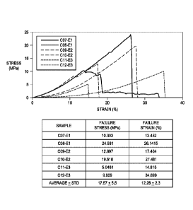
FIG. 6 is a graph that shows axial pull strength for the material of the
present invention, which
demonstrates the enhanced strength of the material.
A Cosmetic Product
Block-copolymers plus equine particulate 250-1000ic used after cosmetic
procedure, such as dermal
abrasion or chemical peel, etc. where Pluronic F127 (PF127) is prepared and
physically blended with
invention in volume to volume ration of 10-35%. Prepared missed compositions
are either stored at room
temperature above gelling temperature, allowing for alginate configuration.
Prior to application of
emollient topical application approx. every 4 hours alignate form or liquid
form a thin layer of the
Date Recue/Date Received 2020-08-07
CA 03041719 2019-04-24
WO 2018/085852 PCT/US2017/060437
21
mixed composition can be applied topically post procedurally thermo-gel
(reverse phase) or spray,
and allowed adequate time (less than 10 minutes) to dry after which the
invention and active
component will be distributed and proximal to the impacted area and have
potential to beneficially
impact consumer by minimized undesirable post procedural signs of irritation
and enhance rate to
which desirable effect of procedure are realized without impairing post
procedural regimen of
reapplication of emollient every 4 hours.
A Medical Research Product
Preparation of the invention in uniform diameter discs compatible with
standard well plates of 6, 12
that are decellularized and prepared sterile are suitable and advantageous
over readily available 2-D
matrix substrate common products, like matrigel, as provided and has
comparable composition and
architecture, particularly known basement membrane components, including
collagen IV, Collagen
VII, Laminin And Fibronectin, but additionally retain 3-D architecture
offering an improved
biomimetic properties. Such a product is advantageous and offers chance to
improve the value of
preclinical toxicity studies for predicting clinical events. Additionally,
improved cell culture is
advantageous to ongoing medical research in the progression of stem cell
phenotype transition during
oncogenesis as well as regenerative medicine research focused on expansion of
cell lines and culture
of stern cells benefitting directly and indirectly regenerative medicine
research. Multi-layer
configurations, and side specific orientation variations which leverage
exposure of various surface
topology, porosity, pore kinetics, adhesion properties, adsorption properties,
and ligand binding
potential can be leveraged to aid understanding of the hierarchy related to
structural-functional
complexity and interdependence that is the basis for in vivo and in vitro
simulation of cell-matrix
interactions under specific conditions. Available 2-D soluble products omit to
simulate key structural
cues and precursors whereas insufficiently decellularized or alter prepared
structural tissue material
products often elicits initiation of DAMP cascade and/or inflammatory or
Fibrosis related cellular
cascades. Only is a sufficient structurally and function preserved tissue
material devoid of low
molecular weight peptides and non-self-genetic material that is a reservoir of
bound bioactivc
peptides and both bio-conductive and bio-inductive provide adequate conditions
for simulation of
mechanical transduction pathways, cell-to-cell or cell-to-matrix interaction,
all which are necessary
elements in accomplishing an in vitro biometric set up that has the potential
to simulate events that
are truly relevant and representative to in vivo events and interaction which
are critical to new
medical discovery and understanding that supports development of new medical
solution and tools,
as well as improving current methods of identifying sufficient safety profiles
early in development
before sufficient time and resources are allocated.
Osteogenic Device
Pluronic preparation blended with osteo-inductive agent such as DBM prepared
via chemical
demineralization or dry mill shearing of hydro-appetite and the biomaterial,
which would provide
CA 03041719 2019-04-24
WO 2018/085852 PCT/US2017/060437
22
substantial improvement over available flow able osteo-inductive products that
arc physically mixed,
because of the additive and increased osteo-inductive potential contributed by
the biomaterial and
added benefit of the anti-microbial peptides and resultant bacteriostatic and
bacteriosydal resultant
product features that commercial available carrier-osteo-inductive agent
physically blended
compositions can potentially offer.
Pharmaceutical Composition
Solubility of the biomaterial and isolation of bioactive peptides having
desirable individual or
collective properties which can be used for additive function action to drug
formulations, can be used
to replace traditional formulations (antimicrobial peptides lack disadvantages
of resistance and
cellular immunity based mechanism of action present) are potentials for, or
used to improve the
phannokinetis associated with active ingredient release profiles of oral and
transdermal administered
formulations by solubilizing the biomaterial in controlled fashion after
uniform section of the
material is performed and exposing it to 1M glacier acetic acid while
agitating to sufficiently
solubilize the material to its secondary structure by which preserving
inherent self-assembly potential
via fibrillogenesis. Temporary preservation of the solubilized fibular
material is optimized by
introduction of polar ionic solution which will inhibit formation of covalent
bonds between polar
organic molecules that otherwise would be difficult to control and inhibit.
While in a controlled
solubilized state a variety of additive can be introduced including not
limited to addition active
ingredient analgesic, antimicrobial compound, intact particulate of the
starting invention material.
Post loading of additive, physiological state, proper neutralization of the
glacier acetic acid with 1M
NAOH and salt precipitation during dehydration via air drying, solvent
dehydration or lyophilization
will sufficient restore physiological conditions necessary for fibro-genesis
to occur and remove ions
inhibiting covalent bond formation that occurs during fibrillogenesis. During
and following
fibrillogenesis events additive introduced will be tethered or bound via
formation of covalent bonds
in contrast to hydrogen bonding that support most carrier agent compositions.
The benefit of the
covalently bonded additive is multifaceted. It preserves active components
that arc low molecular
weight and limited stability susceptible to being consumed post administration
or placement during
acute inflammatory cascades, it also reduces the potential to exacerbate or
prolong inflammatory
response compared to what configurations that have an immediate bolus release
and availability of a
bioactive component (bmp) and the subsequent compensation in product design by
overdosing to
compensate for loss during inflammation therefore reducing risks to patient of
over dose and
consequence of inflated costs of over designed product configuration and
wastefulness of natural
biological resource. The tethered or bound agent is also available naturally
and has increased
potential to persist and provide benefit at numerous points in the healing
cascade therefore not just
indirectly impacting the quality or host tissue by slowing down reformation
and inflammation, but
directly influencing the quality and rate of the formed tissue 14+ days after
administration after
inflammation has resolved. Long term the formation of quality host tissue for
non-vascular limited
23
functioning scar tissue is the essential to achieving long term success with
surgical therapy vs. short term
success currently achieved and necessity for subsequent continued intervention
to address formation of
post-surgical scar tissue which ultimately is excised surgically and results
in negative feedback loop for
patients who elect survival therapy intervention.
It is contemplated that any embodiment discussed in this specification can be
implemented with respect to
any method, kit, reagent, or composition of the invention, and vice versa.
Furthermore, compositions of
the invention can be used to achieve methods of the invention.
It will be understood that particular embodiments described herein are shown
by way of illustration and
not as limitations of the invention. The principal features of this invention
can be employed in various
embodiments without departing from the scope of the invention. Those skilled
in the art will recognize,
or be able to ascertain using no more than routine experimentation, numerous
equivalents to the specific
procedures described herein. Such equivalents are considered to be within the
scope of this invention and
are covered by the claims.
All publications and patent applications mentioned in the specification are
indicative of the level of skill
of those skilled in the art to which this invention pertains.
The use of the word "a" or "an" when used in conjunction with the term
"comprising" in the claims
and/or the specification may mean "one," but it is also consistent with the
meaning of "one or more," "at
least one," and "one or more than one." The use of the term "or" in the claims
is used to mean "and/or"
unless explicitly indicated to refer to alternatives only or the alternatives
are mutually exclusive, although
the disclosure supports a definition that refers to only alternatives and
"and/or." Throughout this
application, the term "about" is used to indicate that a value includes the
inherent variation of error for the
device, the method being employed to determine the value, or the variation
that exists among the study
subjects.
As used in this specification and claim(s), the words "comprising" (and any
form of comprising, such as
-comprise" and -comprises"), -having" (and any form of having, such as -have"
and -has"), -including"
(and any form of including, such as "includes" and "include") or "containing"
(and any form of
containing, such as "contains" and "contain") are inclusive or open-ended and
do not exclude additional,
unrecited elements or method steps. In embodiments of any of the compositions
and methods provided
herein, "comprising" may be replaced with "consisting essentially of' or
"consisting of'. As used herein,
the phrase "consisting essentially of' requires the specified integer(s) or
steps as well as those that do not
materially affect the character or function of the claimed invention. As used
herein, the term "consisting"
is used to indicate the presence of the recited integer (e.g., a feature, an
element, a characteristic, a
Date Recue/Date Received 2020-08-07
24
property, a method/process step or a limitation) or group of integers (e.g.,
feature(s), element(s),
characteristic(s), propertie(s), method/process steps or limitation(s)) only.
The term "or combinations thereof' as used herein refers to all permutations
and combinations of the
listed items preceding the term. For example, "A, B, C, or combinations
thereof' is intended to include at
least one of. A, B, C, AB, AC, BC, or ABC, and if order is important in a
particular context, also BA,
CA, CB, CBA, BCA, ACB, BAC, or CAB. Continuing with this example, expressly
included are
combinations that contain repeats of one or more item or term, such as BB,
AAA, AB, BBC,
AAABCCCC, CBBAAA, CABABB, and so forth. The skilled artisan will understand
that typically there
is no limit on the number of items or terms in any combination, unless
otherwise apparent from the
context.
As used herein, words of approximation such as, without limitation, "about",
"substantial" or
"substantially" refers to a condition that when so modified is understood to
not necessarily be absolute or
perfect but would be considered close enough to those of ordinary skill in the
art to warrant designating
the condition as being present. The extent to which the description may vary
will depend on how great a
change can be instituted and still have one of ordinary skilled in the art
recognize the modified feature as
still haying the required characteristics and capabilities of the unmodified
feature. In general, but subject
to the preceding discussion, a numerical value herein that is modified by a
word of approximation such as
"about" may vary from the stated value by at least 1, 2, 3, 4, 5, 6, 7, 10,
12 or 15%.
All of the compositions and/or methods disclosed and claimed herein can be
made and executed without
undue experimentation in light of the present disclosure. While the
compositions and methods of this
invention have been described in terms of preferred embodiments, it will be
apparent to those of skill in
the art that variations may be applied to the compositions and/or methods and
in the steps or in the
sequence of steps of the method described herein without departing from the
concept and scope of the
invention. All such similar substitutes and modifications apparent to those
skilled in the art are deemed to
be within the scope and concept of the invention.
Date Recue/Date Received 2020-08-07