Note: Descriptions are shown in the official language in which they were submitted.
CA 03041779 2019-04-25
WO 2018/080325
PCT/NZ2017/050141
Improved Sensing for Automated Biological Cell Injection
Field
This disclosure relates to improvements with respect to sensing for automation
of
biological cell injection, such as, for example, injection of cells with
needles carrying
pharmacological, biological or chemical agents. The disclosure further relates
to
sensing for and automation of injection of biological cells using micro-scale
or nano-
scale robotic devices. The disclosure further relates to sensing for and
automation
of injection of biological cells using a parallel array of robotic devices.
Background
Injecting biological cells can be achieved by using a microneedle or
nanoneedle to
penetrate the cell to deliver an agent to be injected. Conventional approaches
involve using a device to move the needle in 3-D. Conventional devices use
micro-
engineered machine (MEMS) technologies involving devices formed from silicon
wafer.
.. There is an accepted need to make biological cell injection operation as
cost-
effective as possible, and to provide an array of needle manipulators which
results
in improved throughput of biological cell injection operations and is readily
controllable.
The applicant has observed potential advantage in injection operations
involving a
number of devices in parallel on a single silicon wafer.
The applicant has also observed a potential advantage in controlling a number
of
devices in parallel on a single silicon wafer.
Summary
In an embodiment, a method of controlling a needle actuator to interact with a
cell
is provided, the method comprising: providing an actuator comprising a tower,
a
stage and a needle, wherein the needle is mounted on the stage; applying an
1
CA 03041779 2019-04-25
WO 2018/080325
PCT/NZ2017/050141
electrostatic potential between the tower and the stage to retract the needle;
moving the actuator towards the cell; reducing the potential so as to allow
the
stage and needle to move towards the cell; applying calibration data to detect
when
the needle has pierced the cell; and reducing the potential further once it
has been
detected that the needle has pierced the cell. The cell can be a biological
cell. The
needle can be a micro-needle and the stage can be a micro-stage.
Alternatively, the cell is held by a cell trap. The cell trap can comprise a
plurality of
microchambers, each microchamber arranged to hold a cell.
Alternatively, the method further comprises applying an electrostatic
potential
between the tower and the stage to retract the needle towards the stage.
Alternatively, the method further comprises reducing the potential to allow
the
stage and needle to move towards the cell while monitoring the potential and
displacement of the stage to detect a fluctuation in voltage versus
displacement to
indicate that the needle has pierced the cell. Alternatively, a laser
interferometer is
used to indicate that the needle has pierced the cell.
Alternatively, the calibration data comprises data defining voltages for
displacements stored against types of cells.
Alternatively, the actuator is provided on an array of actuators, each
interacting
with an individual cell of a plurality of cells.
In another embodiment, a method of generating calibration data for target
voltage
potentials associated with cell-type data is provided, the method comprising:
providing a calibration apparatus comprising a manipulator and a cell trap,
the
manipulator comprising a tower, a stage, and a needle, wherein the needle is
mounted on the stage; identifying a cell type to be calibrated; applying a
voltage
.. so as to pull the stage towards the tower in a retracted position; moving
the
manipulator to within a defined range of the cell-trap configured to house a
cell
type; changing the voltage to allow the stage and mounted needle to be forced
away from the tower and the retracted position while measuring the
displacement
2
CA 03041779 2019-04-25
WO 2018/080325
PCT/NZ2017/050141
of the stage; determining when the needle has reached a target region; and
recording actuation data for use in cell injection for the identified cell
type.
Alternatively, the method further comprises receiving a user input of the cell
type
to a controller provided on the calibration apparatus.
Alternatively, the method further comprises applying a voltage to an actuator
provided on the calibration apparatus, so as to pull the stage towards the
tower in a
retracted position.
Alternatively, the method further comprises moving the manipulator to within
the
defined range of the cell-trap, wherein a camera provided on the calibration
apparatus is programmed to determine if the manipulator is within the defined
range. The camera on the calibration apparatus can alternatively be programmed
to determine if the manipulator is within the defined range of a periphery of
the cell
trap.
Alternatively, the method further comprises reducing the voltage to allow the
stage
and mounted needle to be forced away from the tower and the retracted position
while measuring the displacement of the stage. Measuring the displacement of
the
stage can be performed by a laser interferometer provided in the calibration
apparatus.
Alternatively, the actuation data is selected from a group consisting of a
record
target voltage, a target vertical actuation displacement, a point of
penetration, a
point of poking, a Voltage-Displacement characteristic curve distortion, and
combinations thereof.
In another embodiment, a non-transitory computer-readable medium in which a
program is stored for causing a computer to perform a method for generating
calibration data for target voltage potentials associated with cell-type data
is
provided, the method comprising: providing a calibration apparatus comprising
a
manipulator and a cell trap, the manipulator comprising a tower, a stage, and
a
needle, wherein the needle is mounted on the stage; identifying a cell type to
be
3
CA 03041779 2019-04-25
WO 2018/080325
PCT/NZ2017/050141
calibrated; applying a voltage so as to pull the stage towards the tower in a
retracted position; moving the manipulator to within a defined range of the
cell-trap
configured to house a cell type; changing the voltage to allow the stage and
mounted needle to be forced away from the tower and the retracted position
while
measuring the displacement of the stage; determining when the needle has
reached a target region; and recording actuation data for use in cell
injection for
the identified cell type.
Alternatively, the method further comprises receiving a user input of the cell
type
to a controller provided on the calibration apparatus.
Alternatively, the method further comprises applying a voltage to an actuator
provided on the calibration apparatus, so as to pull the stage towards the
tower in a
retracted position.
Alternatively, the method further comprises moving the manipulator to within
the
defined range of the cell-trap, wherein a camera provided on the calibration
apparatus is programmed to determine if the manipulator is within the defined
range. The camera on the calibration apparatus can be alternatively programmed
to determine if the manipulator is within the defined range of a periphery of
the cell
trap.
Alternatively, the method further comprises reducing the voltage to allow the
stage
and mounted needle to be forced away from the tower and the retracted position
while measuring the displacement of the stage. The measuring the displacement
of
the stage can be performed by a laser interferometer provided in the
calibration
apparatus.
Alternatively, the actuation data is selected from a group consisting of a
record
target voltage, a target vertical actuation displacement, a point of
penetration, a
point of poking, a Voltage-Displacement characteristic curve distortion, and
combinations thereof.
In yet another embodiment, a system for controlling a needle actuation to
interact
4
CA 03041779 2019-04-25
WO 2018/080325
PCT/NZ2017/050141
with a cell is provided, the system comprising: an injection device comprising
a
tower, stage, needle and actuator, the needle mounted on the stage, and the
actuator arranged and configured to apply a voltage potential to the stage to
move
the needle toward and away from the tower; a cell trap configured to house a
cell
to be penetrated by the needle of the injection device; a first camera
configured
and arranged to monitor a proximity of the injection device to the cell trap;
and a
controller configured to control the movement of the injection device. The
first
camera can be configured and arranged to monitor movement on a Z-axis.
Alternatively, the injection device further comprises a plurality of
actuators.
Alternatively, the system further comprises a second camera configured and
arranged to monitor the alignment between the injection device and the cell
trap.
Alternatively, the first camera is configured and arranged to monitor movement
on
a Z-axis, and wherein the second camera is configured and arranged to monitor
movement on the X-axis and Y-axis.
Alternatively, the system further comprises a microscope comprising a second
camera, the microscope configured and arranged to monitor the alignment
between
the injection device and the cell trap. Alternatively, the first camera is
configured
and arranged to monitor movement on a Z-axis, and wherein the microscope is
configured and arranged to monitor movement on the X-axis and Y-axis. The
microscope can be an inverted microscope.
Alternatively, the system further comprising a macro-stage configured and
arranged to control movement of the injection device.
In a further embodiment, a method for controlling a cell injection device is
provided, the method comprising: providing an apparatus comprising a cell
injection device, a cell trap, and a storage device, the cell injection device
comprising a tower, a stage, and a needle, wherein the needle is mounted on
the
stage; identifying a cell type to be injected; retrieving actuation data from
the
storage device; applying a voltage so as to pull the stage towards the tower
in a
5
CA 03041779 2019-04-25
WO 2018/080325
PCT/NZ2017/050141
retracted position; moving the cell injection device to within a defined range
of the
cell-trap configured to house a cell type; applying a varying target actuation
voltage based on retrieved actuation data to allow the stage and mounted
needle to
be forced away from the tower and the retracted position; determining when the
needle has reached a target region; and adjusting the voltage to move the
needle
towards the retracted position.
Alternatively, the method further comprises receiving a user input of the cell
type
to a controller provided on the apparatus.
Alternatively, the method further comprises applying a voltage to an actuator
provided on the injection device, so as to pull the stage towards the tower in
a
retracted position.
Alternatively, the method further comprises moving the injection device to
within
the defined range of the cell-trap, wherein a camera provided on the apparatus
is
programmed to determine if the injection device is within the defined range.
Alternatively, the camera on the apparatus is programmed to determine if the
injection device is within the defined range of a periphery of the cell trap.
Alternatively, the actuation data is selected from a group consisting of a
record
target voltage, a target vertical actuation displacement, a point of
penetration, a
point of poking, a Voltage-Displacement characteristic curve distortion, and
combinations thereof.
In another embodiment, a non-transitory computer-readable medium in which a
program is stored for causing a computer to perform a method for controlling a
cell
injection device is provided, the method comprising: providing an apparatus
comprising a cell injection device, a cell trap, and a storage device, the
cell injection
device comprising a tower, a stage, and a needle, wherein the needle is
mounted
on the stage; identifying a cell type to be injected; retrieving actuation
data from
the storage device; applying a voltage so as to pull the stage towards the
tower in
a retracted position; moving the cell injection device to within a defined
range of
6
CA 03041779 2019-04-25
WO 2018/080325
PCT/NZ2017/050141
the cell-trap configured to house a cell type; applying a varying target
actuation
voltage based on retrieved actuation data to allow the stage and mounted
needle to
be forced away from the tower and the retracted position; determining when the
needle has reached a target region; and adjusting the voltage to move the
needle
towards the retracted position.
Alternatively, the method further comprises receiving a user input of the cell
type
to a controller provided on the apparatus.
Alternatively, the method further comprises applying a voltage to an actuator
provided on the injection device, so as to pull the stage towards the tower in
a
retracted position.
Alternatively, the method further comprises moving the injection device to
within
the defined range of the cell-trap, wherein a camera provided on the apparatus
is
programmed to determine if the injection device is within the defined range.
Alternatively, the camera on the apparatus is programmed to determine if the
injection device is within the defined range of a periphery of the cell trap.
Alternatively, the actuation data is selected from a group consisting of a
record
target voltage, a target vertical actuation displacement, a point of
penetration, a
point of poking, a Voltage-Displacement characteristic curve distortion, and
combinations thereof.
Brief description of the drawings
Additional and further aspects of the present invention will be apparent to
the
reader from the following description of embodiments, given in by way of
example
only, with reference to the accompanying drawings in which:
Figure 1 illustrates a single unit actuator controlled and calibrated for
biological cell
injection, in accordance with various embodiments.
7
CA 03041779 2019-04-25
WO 2018/080325
PCT/NZ2017/050141
Figure 2 illustrates a closer view of a single unit actuator controlled and
calibrated
for biological cell injection, in accordance with various embodiments.
Figures 3a to 3c illustrate a parallel plate actuator model representing
actuation of
a stage of a single unit actuator, in accordance with various embodiments.
Figure 4 illustrates a single unit actuator and a corresponding parallel
injection
device including an array of unit actuators, in accordance with various
em bodim ents.
Figures 5a to 5f depict a biological cell injection process, in accordance
with various
em bodim ents.
Figure 6 depicts a parallel injection device and a corresponding cell trap
array and
cameras, in accordance with various embodiments.
Figure 7 illustrates a system for control and calibration for biological cell
injection,
in accordance with various embodiments.
Figure 8 illustrates a calibration process for biological cell injection, in
accordance
with various embodiments.
Figure 9 depicts a relation between displacement of a stage of the single unit
actuator showing characteristics recorded in a calibration process, in
accordance
with various embodiments.
Figure 10 illustrates a control process for biological cell injection, in
accordance with
various embodiments.
Figure 11 is a block diagram that illustrates a computer system, in accordance
with
various embodiments.
It is to be understood that the figures are not necessarily drawn to scale,
nor are
the objects in the figures necessarily drawn to scale in relationship to one
another.
The figures are depictions that are intended to bring clarity and
understanding to
8
CA 03041779 2019-04-25
WO 2018/080325
PCT/NZ2017/050141
various embodiments of apparatuses, systems, and methods disclosed herein.
Wherever possible, the same reference numbers will be used throughout the
drawings to refer to the same or like parts. Moreover, it should be
appreciated that
the drawings are not intended to limit the scope of the present teachings in
any
way.
Detailed Description
The following description of various embodiments is exemplary and explanatory
only and is not to be construed as limiting or restrictive in any way. Other
embodiments, features, objects, and advantages of the present teachings will
be
apparent from the description and accompanying drawings, and from the claims.
As used herein, the terms "comprise", "comprises", "comprising", "contain",
"contains", "containing", "have", "having" "include", "includes", and
"including" and
their variants are not intended to be limiting, are inclusive or open-ended
and do
not exclude additional, unrecited additives, components, integers, elements or
method steps. For example, a process, method, system, composition, kit, or
apparatus that comprises a list of features is not necessarily limited only to
those
features but may include other features not expressly listed or inherent to
such
process, method, system, composition, kit, or apparatus.
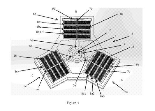
Figure 1 illustrates a nanorobot or microrobot in the form of a single-unit
needle
manipulator 1 which is included in an array of needle manipulators, in
accordance
with various embodiments, the features of which can be used as illustrated or
in
combination with other embodiments disclosed herein.
Figure 2 illustrates a central part of the single-unit needle manipulator,
such as that
illustrated, for example, in Figure 1.
The single-unit manipulator 1 has a manipulation stage 2 on which a needle 3
is
mounted. Needle 3 can be of a type suited to penetrate an object or cell to
deliver,
or inject, an agent to the object or cell interior. The injected object or
cell may be
a biological cell, wherein needle 3 can be of a type suited to penetrate
biological
9
CA 03041779 2019-04-25
WO 2018/080325
PCT/NZ2017/050141
cells to deliver, or inject, an agent to the cell interior and/or cell
nucleus.
The stage 2 can be located above a tower 4 which can be electrically charged
relative to the stage 2 to apply electrostatic forces to the stage 2. The
stage and
tower may be referred to collectively as a parallel-plate actuator, wherein
the
opposing surfaces on the stage and tower are electrostatically charged when a
voltage is applied across them. Electrostatic forces between the tower 4 and
stage
2 can actuate the stage 2 in a Z-axis.
As will be described in detail below in reference to Figure 3, Z-axis
actuation may
be the only actuation needed to provide the movement necessary to affect
appropriate cell or object penetration by needle 3.
If Figure 1, the Z-axis can be considered the central axis of the tower 4. The
stage
2 can also be actuated in different axes lying in an X-Y plane, in the plane
of the
manipulator 1 as shown in Figure 1, by tethers 5a, 5b and 5c. Stage 2 can be
configured to manipulate a needle 3 suitable for penetration of objects on
this scale
of a biological cell. As such, the stage 2 may be referred to as a micro-stage
or
nano-stage.
The tethers 5a, 5b and 5c tether the stage 2 to actuators 6a, 6b and 6c
respectively. The actuators 6 can be located so that forces transferred by the
tethers 5 can be in three different axes in the X-Y plane. Each tether
5a/5b/5c can
apply tensile forces. Actuators 6 can serve to apply forces from three
different
directions A, B and C. For example, the actuators 6 can be arranged at 1200
intervals about stage 2.
Tether beams 7a, 7b and 7c of actuators 6a, 6b and 6c can connect each of
tethers
5a, 5 b and 5c to three support beams 8. The support beams 8 support comb-
features, or comb-like electrostatic actuators 18. In this embodiment the
actuator
6a has support beams 8a1, 8a2, and 8a3. Actuators 6a and 6c similarly have
support beams 8b1/8b2/8b3 and 8c1/8c2/8c3 respectively.
CA 03041779 2019-04-25
WO 2018/080325
PCT/NZ2017/050141
Electrostatic comb features 18 can be located in the same plane as the support
beams 8 shown, for example, in Figure 1. The comb-features may be referred to
as
comb-drive actuators or comb-drives. The comb-features 18 can be configured to
apply force on the support beams 7 in the X-Y axis. The parallel-plate
actuator
including the central micro-stage 2 and the tower 4 underneath it can be
configured
to apply force on the tethers 5 in the Z axis. The actuators can have a set of
comb-
features 18 on the support beams 8 and another opposing set of comb-features
18
on the device. The two opposing sets of comb-features can be charged relative
to
each other to generate an electrostatic force in the X-Y axis, providing a
comb-
drive. Similarly the opposing micro-stage 2 and the tower 4 of the parallel-
plate
actuator can be charged relative to each other to generate an electrostatic
capacitive force in the Z axis.
Spring-flexure beams 9 (9a, 9b and 9c) connect or anchor beams 8a, 8b and 8c
to
a substrate 10 of the manipulator 1. Each actuator 6 can apply a force to the
stage
2 in its respective direction. Individual control of the forces applied to the
stage 2
in the direction of each actuator 6 allows the stage 2 to be actuated so as to
manipulate the needle 3. In so doing, tethers 5 can stretch, and movement of
the
stage 2 can be dependent on stretching, or strain, of the tethers 5 and
flexing of
the spring-flexure beams 9.
As shown in Figure 2, for example, the three tethers 5 of the single-unit
manipulator connect actuators in three respective directions to a central
stage to
provide an elastic support structure for the stage 2.
Figures 3a to 3c illustrate an example of a scheme for out of plane Z-
actuation of
the stage, in accordance with various embodiments, the features of which can
be
used alone (as illustrated) or in combination with other embodiments disclosed
herein. Figure 3a shows the stage 2 and tower 4 as a parallel plate
electrostatic
actuator formed of a movable surface 13 on the stage and a fixed electrostatic
surface 14 on the tower 4. Spring flexure beam 9, which can have the effect of
11
CA 03041779 2019-04-25
WO 2018/080325
PCT/NZ2017/050141
bringing the stage 2 towards a resting position 16 (see Figure 3a) relative to
the
substrate 10, and above the tower 4. Stretch in the tethers 5 may also
contribute
to spring effect. Applying a potential across a movable electrostatic surface
11 on
the underside of the stage 2 and a fixed surface 12 on the upper side of the
tower
can displace the stage 2 and needle 3 towards a retracted state 15 (see Figure
3c),
relatively closer to the tower 4 and substrate 10. Decreasing the potential
can
allow the stage 2 and needle 3 to be actuated by restoring force of the
tethers 5
and spring flexure beams 9 of actuators 6 relatively away from the tower 4 and
substrate 10 towards the resting position 16. This action of decreasing the
potential from a retracting potential to actuate the needle away from the
substrate
10 will be referred to herein as actuating the needle 3 in a Z axis (see
Figure 3b for
example of actuating away from the retracted position, and beyond the resting
position). If a cell is trapped in proximity to the needle 4, when the stage 2
is in a
retracted position injection can be achieved by reducing the potential across
the
movable and fixed electrostatic surfaces to actuate the stage and needle by
reducing the potential to allow the stage to return towards a resting position
16.
The degree to which the stage returns towards the resting position from the
retracted position is one of many key factors critical to usefully injecting a
given
type of biological cell. Calibration and control of this actuation according
to various
embodiments is discussed below.
Figure 4 illustrates a parallel injection device 20 formed of six arrays 21 of
single-
unit needle manipulators 1, in accordance with various embodiments, the
features
of which can be used as illustrated or in combination with other embodiments
disclosed herein. The six arrays of Figure 4 is only an example, and the
number of
arrays on device 20 can vary as needed. The device 20 can be physically
associated
with a cell trapping platform 22 which has an array of micro-wells or micro-
chambers 23, or cell traps with a cell-trap matching each manipulator in the
array.
Injecting a cell with a manipulator 1 in the array 21 involves manipulating
the stage
(and associated needle 3) in the X-Y plane of the device 20 and then actuating
the
stage 2 in the Z axis.
12
CA 03041779 2019-04-25
WO 2018/080325
PCT/NZ2017/050141
Figure 5 (5a to 5f) illustrates a scheme for actuation of a needle 3 of single-
unit
manipulator 1 for biological cell injection, in accordance with various
embodiments,
the features of which can be used as illustrated or in combination with other
embodiments disclosed herein. Figure 5a depicts the manipulator 1 prior to any
operation. As shown, tower 4 is attached to substrate 10. Stage 2 is suspended
by
tethers 5 that bend to represent stretching, and flexing of spring flexure
beams 9 of
actuators 6. The needle 3 is shown mounted on the stage 2. Figure 5a also
shows a
biological cell 24 with a nucleus, or other target within the cell, 25. The
nucleus 25
can be penetrated by the needle 3 to deliver a chemical agent (not shown)
incorporated into the needle 3. Manipulating or actuating a needle 3 at the
micro
and nanoscale successfully generally can include overcoming three primary
adhesion forces that may have an effect on the process: van der Waals,
capillary
attraction and electrostatic.
Figure 5b depicts the step on which an electrostatic potential, or voltage, is
applied
across the fixed and movable surfaces of the tower 4 and stage 2 respectively
to
retract the stage 2 towards the tower 4 and substrate 10. In conjunction with
the
retraction, manipulator 1 as part of the device 20 is brought close to the
cell 24
held in micro-well 23.
Figure Sc depicts decreasing voltage to V1 to allow the stage 2 to return
towards a
resting position relatively away from the substrate 10 compared to the state
shown
in Figure 5b. This step may be understood as actuating the stage 2 and needle
3 in
the direction away from the substrate 10 or towards the cell 24. The cell 24
shown
as indented by the needle, or poked by the needle. The cell 24 is depicted as
exerting a restoring force 26, R1, on the needle 3.
Figure 5d depicts further reduction of the voltage to V2 to actuate the stage
2 and
needle 3 further away from a substrate 10. The cell is depicted as exerting
the
restoring force 26, R2.
Figure 5e depicts the point of further reduction of voltage to V3 with the
cell
exerting restoring force 26 R3, when the needle 3 first penetrates the cell
24.
13
CA 03041779 2019-04-25
WO 2018/080325
PCT/NZ2017/050141
Figure 5f depicts the manipulator 1 in the state in which the voltage has been
reduced to VO where the needle 3 penetrates the nucleus 25 of the cell 24. In
this
particular example the stage 2 is shown as in its resting position. However,
the
voltage may alternatively be reduced sufficiently for the needle 3 to
penetrate the
nucleus but not sufficiently to allow the stage 2 to return to its resting
position.
Among the many factors that can impact voltage level at nucleus penetration,
some
include, for example, the size and type of cell.
Figure 6 illustrates an apparatus used in cell injection operations, in
accordance
with various embodiments, the features of which can be used as illustrated or
in
combination with other embodiments disclosed herein. A parallel injection
device 20
is depicted with opposing cell trapping platform 22 with trapped cells 24. An
X-Y
camera 28 allows monitoring of the alignment between the parallel injection
device
and the cell trapping platform 22. The camera 28 can further monitor cells 24,
manipulators, and/or movement of the device 20 in the X-Y plane of the device
20.
15 AZ camera 29 allows monitoring of the proximity of the parallel
injection device 20
to the cell trapping platform 22.
Figure 7 illustrates a system 27 used in the injection of objects or cells,
such as
biological cells, in accordance with various embodiments, the features of
which can
be used as illustrated or in combination with other embodiments disclosed
herein.
20 The system has a Z camera 29 (which can be, for, example, a high
resolution
camera), an inverted microscope with X-Y camera 28, a cell trapping platform
22, a
parallel injection device 20, and a macro-stage 31 that is able to move the
device
20 macro level towards and away from the cell trapping platform 22. The device
20
can be a parallel array device formed on a substrate. The macro stage can be
configured to move the whole device 20. Alternatively, the macro stage can be
configured and programmed to move individual manipulators 1. Also illustrated
is a
caps DAQ system 32 (hereinafter referred to as controller 32) which controls
individual single unit manipulators 1, both for X-Y manipulation and for Z
actuation.
Also shown is a controller running control software 33, which can include a
set of
instructions stored in on memory media which when executed provides the
14
CA 03041779 2019-04-25
WO 2018/080325
PCT/NZ2017/050141
functionality of the controller.
COMPUTER SYSTEM
Figure 11 is a block diagram that illustrates a computer system 1000, upon
which
embodiments, or portions of the embodiments, of the present teachings may be
implemented. In various embodiments of the present teachings, computer system
1000 can include a bus 1020 or other communication mechanism for
communicating information, and a processor 1040 coupled with bus 1020 for
processing information. In various embodiments, computer system 1000 can also
include a memory 1060, which can be a random access memory (RAM) or other
.. dynamic storage device, coupled to bus 1020 for determining instructions to
be
executed by processor 1040. Memory 1060 also can be used for storing temporary
variables or other intermediate information during execution of instructions
to be
executed by processor 1040. In various embodiments, computer system 1000 can
further include a read only memory (ROM) 1080 or other static storage device
coupled to bus 1020 for storing static information and instructions for
processor
1040. A storage device 1100, such as a magnetic disk or optical disk, can be
provided and coupled to bus 1020 for storing information and instructions.
In various embodiments, computer system 1000 can be coupled via bus 1020 to a
display 1120, such as a cathode ray tube (CRT) or liquid crystal display
(LCD), for
displaying information to a computer user. An input device 1140, including
alphanumeric and other keys, can be coupled to bus 1020 for communicating
information and command selections to processor 1040. Another type of user
input
device is a cursor control 1160, such as a mouse, a trackball or cursor
direction
keys for communicating direction information and command selections to
processor
1040 and for controlling cursor movement on display 1120. This input device
1140
typically has two degrees of freedom in two axes, a first axis (i.e., x) and a
second
axis (i.e., y), that allows the device to specify positions in a plane.
However, it
should be understood that input devices 1140 allowing for 3-dimensional (x, y
and
z) cursor movement are also contemplated herein.
CA 03041779 2019-04-25
WO 2018/080325
PCT/NZ2017/050141
Consistent with certain implementations of the present teachings, results can
be
provided by computer system 1000 in response to processor 1040 executing one
or
more sequences of one or more instructions contained in memory 1060. Such
instructions can be read into memory 1060 from another computer-readable
.. medium or computer-readable storage medium, such as storage device 1100.
Execution of the sequences of instructions contained in memory 1060 can cause
processor 1040 to perform the processes described herein. Alternatively hard-
wired
circuitry can be used in place of or in combination with software instructions
to
implement the present teachings. Thus implementations of the present teachings
are not limited to any specific combination of hardware circuitry and
software.
The term "computer-readable medium" (e.g., data store, data storage, etc.) or
"computer-readable storage medium" as used herein refers to any media that
participates in providing instructions to processor 1040 for execution. Such a
medium can take many forms, including but not limited to, non-volatile media,
volatile media, and transmission media. Examples of non-volatile media can
include, but are not limited to, optical, solid state, magnetic disks, such as
storage
device 1100. Examples of volatile media can include, but are not limited to,
dynamic memory, such as memory 1060. Examples of transmission media can
include, but are not limited to, coaxial cables, copper wire, and fiber
optics,
including the wires that comprise bus 1020.
Common forms of computer-readable media include, for example, a floppy disk, a
flexible disk, hard disk, magnetic tape, or any other magnetic medium, a CD-
ROM,
any other optical medium, punch cards, paper tape, any other physical medium
with patterns of holes, a RAM, PROM, and EPROM, a FLASH-EPROM, any other
memory chip or cartridge, or any other tangible medium from which a computer
can read.
In addition to computer readable medium, instructions or data can be provided
as
signals on transmission media included in a communications apparatus or system
to
provide sequences of one or more instructions to processor 1040 of computer
system 1000 for execution. For example, a communication apparatus may include
a
16
CA 03041779 2019-04-25
WO 2018/080325
PCT/NZ2017/050141
transceiver having signals indicative of instructions and data. The
instructions and
data are configured to cause one or more processors to implement the functions
outlined in the disclosure herein. Representative examples of data
communications
transmission connections can include, but are not limited to, telephone modem
connections, wide area networks (WAN), local area networks (LAN), infrared
data
connections, NFC connections, etc.
It should be appreciated that the methodologies described herein including
flow
charts, diagrams and accompanying disclosure can be implemented using computer
system 1000 as a standalone device or on a distributed network of shared
computer processing resources such as a cloud computing network.
Figure 8 illustrates an example of a process carried out by a single unit
manipulator
1 in a calibration apparatus, which could be part of system illustrated in
Figure 7, in
accordance with various embodiments, the features of which can be used as
illustrated or in combination with other embodiments disclosed herein. The
process
can be for determining target voltage potentials to apply to the tower 4 to
actuate
needle 3 in the Z-axis by targeting the Z-displacement, so as to inject a
target,
such as a nucleus 25, within a given type of biological cell 24. The process
of Figure
8 is carried out by a single unit manipulator only by way of example. The
process
can be used by parallel injection device 20 of the system illustrated, for
example, in
Figure 7.
The start of the process is shown as step P1 51, using the convention P1 51 to
denote Process 1 Step 1.
At P1 S2, the controller 32 of the system 27 receives user inputs from
operator
carrying information on a type of cell and a Young's modulus for the cell
wall.
At P1 S3, the controller stores data carrying information from P1 S2. As
described
later, data carrying information can include information on target voltages
and
target Z-displacements as well as other information is stored at P1 S3.
17
CA 03041779 2019-04-25
WO 2018/080325
PCT/NZ2017/050141
At P1 S4, the controller 32 receives inputs from an operator identifying
whether a
calibration process is required to determine target voltages and target Z-
displacements. This may be referred to as a blind sensing process. The
software will
check if the blind sensing is to be activated. If yes, then the actuation
voltage will
be applied to parallel arrays of towers individually. If no, then the
algorithm will
initiate the z control process from the start. There can be multitude of
reasons
regarding the termination of the blind sensing control and re-start such as
error in
recording the cell types or external noise affecting the macro and micro-
alignment
of the system among others.
At P1 S5 a retraction voltage is applied to the tower to establish an
electrostatic
potential across the tower 4 and stage 2 to retract the needle as shown, for
example, in figure 5b. From this retracted state any reduction in voltage
applied to
the tower 4 will actuate the stage 2 under restoring force of the tethers 5
and
spring flexure beams 9 of actuators 6.
.. At P1 S6 the macro stage incrementally guides the single unit manipulator 1
towards the cell, which can be provided in a cell trapping platform 22. P1 S7
determines, using the camera 29, whether the manipulator 1 is within a defined
range i.e. less than 1 m of the cell trapping platform 22. In process
illustrated by
Figure 8, the controller 32, using the camera 29, determines whether the
manipulator 1 is within, for example, one micro-meter of the cell trapping
platform
22. If the range is satisfied, then the algorithm continues to the subsequent
step.
Else, it will go back to the previous step and instruct the macro stage to
further
incrementally guide the manipulator 1 until the proximity condition is
satisfied. This
is critical due to the non-visual nature of the controller 32 for vertical
manipulation.
In the parallel architecture with a plurality of manipulators 1, controller
32, using
the camera 29, can determine whether the plurality of manipulators 1 are
within,
for example, one micro-meter of the cell trapping platform 22 (sub-micron
proximity condition).
Alternatively, the camera 29 will verify the sub-micron proximity condition
for the
cells on the periphery of the cell trapper. In this circumstance, for the
cells in the
18
CA 03041779 2019-04-25
WO 2018/080325
PCT/NZ2017/050141
internal sections of the cell trapping platform 22, the controller will act
based on the
statistical confidence data of the number of the cells that can be manipulated
at a
time by a single parallel architecture chip.
At P1 S8 the controller 32 will continue reducing the actuation voltage in the
parallel-actuator plate, thereby, the needle starts gradually coming back to
its
original position due to the decrease in electrostatic force between the two
plates of
the vertical actuator of the manipulator 1. The actual displacement is
measured
using a single/double-beam laser interferometer at room temperature. There is
a
continuous change in the applied voltage between the two plates and therefore
the
change in distance between these plates.
At P1 S9, while the actuation voltage is reduced incrementally, the
corresponding
voltage-displacement characteristic curve is plotted as shown, for example, in
Figure 9. The plot primarily relies upon two variable parameters, namely
change of
potential in the parallel-plate actuator in relation to the change of position
of the
central stage 2 relative to the tower 4 as shown in Figure 5. The sudden
significant
decrease in the plot between these two parameters, voltage and displacement,
during vertical motion is used as a signature for feedback to the controller
32 to
enable the biomanipulation. This change in the plot can be attributed, for
example,
to the change in stiffness of the cell membrane that is sensed by the
nanoneedle.
Rather than measuring the force during manipulation (plotting it against
time), the
voltage-displacement principle illustrated by the Figure 9 curve allows the
measuring of the decrease in voltage to identify the points of penetration and
poking into the cell.
At P1 S10, the condition that the needle has arrived at the final cell target
region
such as the nucleus or mitochondria is verified by, for example, the camera 29
(as
shown by the exemplary CMOS camera in Figure 9) or fluorescent microscopy. If
this condition is satisfied, then the algorithm will proceed forward to record
actuation data. If not, then the actuation voltage is further reduced
incrementally
until the final cell target compartment is reached. Once this system is
calibrated for
a cell type, the relationship between the voltage and displacement is fed into
a
19
CA 03041779 2019-04-25
WO 2018/080325
PCT/NZ2017/050141
controller database, such as storage device 1100 of Figure 11, a priori
(discussed
below).
At P1 S11, once the manipulation at the target region inside the cell is
complete,
actuation data such as record target voltage (Vd), target vertical actuation
displacement (Xd), points of penetration and poking, and V-D characteristic
curve
distortion, are recorded and stored into the controller database, such as
storage
device 1100 of Figure 11. This record of data is critical for the parallel
manipulation,
because similar to xy control, for z tracking, closed-loop control can be used
by
using error in position signals as feedback to form a closed-loop. This
calibrated
data will eventually act as a reference input and will employ PID control for
achieving the desired voltage, Vd, and thereby avoiding overshooting of the
nanoneedle.
At P1 S12, the nanoneedle is then pulled out of the cell at a velocity. The
velocity
can range, for example, between 0.5 - 2.5 mm 5ec-1. Biological membranes
.. generally stretch elastically only by approximately 2%-4% before they
rupture.
Cells have an ability to resist fast changes in the membrane tension brought
upon
by external forces such as needle manipulation in our case. There are numerous
tension-sensitive surface area regulation mechanisms that can help the cells
resist
more dramatic and slower changes in the cellular environment. One example is a
small bilayer reservoir that can buffer minor increases in the membrane
tension.
The steps P1 S8, P1 S9, P1 S10 and P1 S11 taken together can be referred to as
the blind sensing mechanism.
Figure 9 shows a plot 33 of the relation of voltage, in Volts, versus
displacement in
micrometres captured at P1 S9 (introduced above). The plot 33 shows a point 36
of
gradual decrease in rate of change of displacement with voltage which is
characteristic of the needle 3 beginning to poke a cell 24 as shown in Figure
Sc.
The plot 33 also shows a point 35 of further decrease in the rate of change of
displacement with voltage which is characteristic of the needle 3 beginning to
penetrate cell 24 as illustrated in Figure 5e. The controller 32 recognises
these
CA 03041779 2019-04-25
WO 2018/080325
PCT/NZ2017/050141
points 36 and 35 by relating data defining the relation of voltage to
displacement
against characterisation data.
Figure 10 shows an example of a process P2 for controlling a parallel
injection
device 20 (or cell injection device), in accordance with various embodiments,
the
features of which can be used as illustrated or in combination with other
embodiments disclosed herein. The process of Figure 10 is carried out by a
single
unit manipulator only by way of example. The process can be used by parallel
injection device 20 of the apparatus/system illustrated, for example, in
Figure 6 and
7. The process starts at P2 Si.
At P2 S2, the controller 32 receives inputs from an operator identifying a
cell type
to be injected by a single unit manipulator 1. The cell type may be identified
for
each single unit manipulator 1 in the parallel injection device 20 to allow
different
types of cells, for example, to be injected in parallel injection operations.
At P2 S3, the controller 32 retrieves data stored in association with for an
identified
cell type, including Young's modulus of the cell type.
At P2 S4, the controller 32 retrieves data carrying the following additional
information: target voltage to be applied to tower 4, target vertical
displacement of
stage 2 captured at P1 S5 in Figure 8, the displacement and/or voltage at
which the
needle 3 begins to penetrate the cell 24 captured at steps P1 S6 to P1 S8 and
the
displacement versus voltage plot captured at P1 S9, for example. Data used to
identify displacement versus voltage plot characteristics for poking and
penetration
can also be retrieved.
At P2 S5, the controller 32 determines whether a cell injection operation
should
start. Controller 32 can determine this by inputs from an operator received by
the
controller 32. The controller can check if the blind sensing is to be
activated. If yes,
then the actuation voltage can be applied to parallel arrays of towers 4
individually.
If no, then the algorithm can return to P2 S2 to initiate the z control
process from
the start. There can be a multitude of reasons for termination of the blind
sensing
21
CA 03041779 2019-04-25
WO 2018/080325
PCT/NZ2017/050141
control and re-start. These can include, for example, error in recording the
cell
types, external noise affecting the macro and micro-alignment of the system,
or
other examples.
At P2 S6 voltages are applied to the tower 4 to retract the stage 2 to the
state
shown, for example, in Figure 5b. By extension, with potential difference
applied to
all of the manipulators 1 in the parallel injection device 20, the voltage is
applied to
each of the tower 4 of each of the manipulators 1 to retract each of the
stages 2.
3SA microrobot is an example of injection device 20.
At P2 S7, the macro stage 31 incrementally guides the parallel injection
device 20
.. towards the cell trapping platform 22 while the Z-camera 29 is used by the
controller 32 to monitor the proximity of the parallel injection device 20 to
the cell
trapping array (or platform) 22. Alternatively, the camera 29 will verify the
proximity of the parallel injection device 20 on the periphery of the cell
trapping
array 22. In this circumstance, for the cells in the internal sections of the
cell
trapping platform 22, the controller will act based on the statistical
confidence data
of the number of the cells that can be manipulated at a time by a single
parallel
architecture chip.
At P2 S8 and P2 S9, the controller 32 determines whether the parallel
injection
device 20 is suitably close to the cell trapping array 22 and returns the
process to
P2 S7 otherwise. The Z camera 29 provides suitable video data at P2 S9 for
this
decision. The sub-micron range proximity information is validated by the Z
camera
29 placed sideways. In the parallel architecture with a plurality of
manipulators 1,
controller 32, using the camera 29, can determine whether the plurality of
manipulators 1 are within, for example, one micrometer of the cell trapping
platform 22 (sub-micron proximity condition).
Alternatively, the camera 29 will verify the sub-micron proximity condition
for the
cells on the periphery of the cell trapper. In this circumstance, for the
cells in the
internal sections of the cell trapping platform 22, the controller will act
based on the
22
CA 03041779 2019-04-25
WO 2018/080325
PCT/NZ2017/050141
statistical confidence data of the number of the cells that can be manipulated
at a
time by a single parallel architecture chip.
Due to the nature of non-visual sensing of the system and mechanism of the
controller 32 that can detect the manipulation through some physical change,
the
sub-micron proximity between the parallel arrays of needles and their
corresponding cells is critical. In addition, because the Z movement of the
needles
has a range such as, for example, between 5-10 pm, the satisfaction of the
proximity condition as a pre-requisite allows most of the cells to be
physically
manipulated. If the range is satisfied, then the algorithm continues to the
subsequent step. Else, it will go back to P2 S7 and will instruct the
macrostage to
further incrementally guide the parallel architecture chips until the
proximity
condition is satisfied.
At P2 S10 the target actuation voltage of P2 S4, for the cell type selected at
P2S2,
is retrieved by the controller 32 and, for the specific manipulator 1 being
controlled,
applied to the tower 4 to actuate the stage 2 and needle to a target so the
needle
3, for example, penetrates the target. This is the target voltage which
actuates the
stage and needle to the target, such as nucleus 25, of the particular cell
type
selected at P2 S2 as illustrated, for example, in Figure 5f.
The blind sensing mechanism is employed during this step when the cell
manipulation actually occurs. Similar to the XY controller, for Z tracking, we
use
closed-loop control by using error in position signals as feedback to form a
closed-
loop. The controller accepts a desired position, Xd, as the reference input
and
employs PID control for achieving the desired voltage, Vd, and therefore
avoids
overshooting of the needle. As discussed earlier, the calibrated values of Xd
and Vd
are pre-programmed into the controller 32 for different types of cells. The
voltage is
now gradually reduced (V to V/ to V2 to V3 to Vd), which decreases the
electrostatic force (E to E/ to E2 to E3 to Ed) between the plates gradually
as
shown, for example, in Figure Sc to 5e. As the controller 32 starts reducing
the
actuation voltage for each needle, they start gradually coming back to their
original
position due to the decrease in electrostatic force between the two plates of
the
23
CA 03041779 2019-04-25
WO 2018/080325
PCT/NZ2017/050141
parallel injection device 20. As needles start coming out of their retracted
state,
they gradually penetrate through the cell membranes because of the vertical
stiffness of the manipulators 1 and the decreasing parallel-plate
electrostatic force,
until they poke through these membranes completely and are in the target site
inside the cells. The primary purpose of the controller 32 is to enable
manipulation
using the blind sensing and prevent overshooting of the needles during the
manipulation process.
There is a continuous change in the applied voltage between the two plates of
the
parallel injection device 20 and therefore the change in distance between
these
plates. Therefore, Xd and Vd are continually changing to guide the arrays of
needles
to the specific target position inside these arrays of cells. The error, Xdiff
in z
positioning precision is calculated from the estimator that uses the blind
sensing
model by comparing the measured position, Xn, with the desired position, Xd.
The
PID controller calculates the desired voltage to drive the needles in the
parallel
architecture to the desired vertical position in Z axis. Once the needles are
inside
the cell, depending on the cell organelle to be manipulated such as nucleus,
the
needles might undergo another motion, resulting in another subsequent decrease
in
the V-D plot as shown as an example in Figure 9, confirming the poking through
the
second cellular organelle.
It is the change in the plot of the voltage-displacement tracking curve that
identifies penetration and subsequent poking through the cell membranes. This
change is the alteration of the cell membrane stiffness sensed by the needle
during
vertical manipulation and is reflected in the force-deflection curve as shown
in
Figure 9. This is primarily a model-based feedback employing a blind control
scheme and therefore the precision of the position of the needle in a Z
coordinate
frame can depend on the accuracy of this blind model. Based on electrostatic
force
law, the output motion of the central stage 2 relative to the bottom tower 4
is
expected to be proportional to the square of the actuation voltage,
oc V2. Due
to this electrostatic nature, this blind sensing scheme is a linear system
with
deterministic behaviour. Nonetheless, immediately following the poking into
the
24
CA 03041779 2019-04-25
WO 2018/080325
PCT/NZ2017/050141
cell, the behaviour becomes non-linear due to the vibration induced into the
system
with the rupture of the cell membrane.
A P2 S11 the voltage on the tower 4 is adjusted to retract the needle 3 as
illustrated in Figure 5b to remove the needle 3 from the cell 24. The process
ends
at P2 S12.
Further detail on the calibration process, such as the one exemplified in
Figure 8, in
accordance with various embodiments, will now be given in reference to steps
in
Figure 8.
The blind sensing mechanism of P1 S8, P1 S9, P1 S10 and P1 S11 can produce
different displacement versus voltage plots, and plot characteristics, for
different
types of cell due to factors such as, for example, cell size, membrane
thickness and
Youngs Modulus of the membrane (elasticity). Therefore, the controller can
request
inputs to identify data carrying information such as cell type and the
corresponding
Youngs Modulus (E), for example, as noted in Table1.
Cell Type E(kPa)
Endothelial cells
HUVEC 10-11
BPAEC
Leukocytes
Leukemia myeloid cells (HL60) 0.2-1.4
Leukemia lymphoid (Jurkat) cells
mgmogggmognAue.ammaiimmmmmmmmmm
Neutrophils 0.2-0.07
CA 03041779 2019-04-25
WO 2018/080325
PCT/NZ2017/050141
Osteoblasts 0.3-20.0
Astrocytes
Fibroblasts
Migrating 3T3 cells 3-12
L929
Epidermal keratocytes ii(P.AS1
Platelets 1 -50
Skeletal muscle cells
Murine C2C12 myoblasts
Myofibrils 40-45
Erythrocytes 14-18
Table 1
For example, injecting Leukaemia myeloid cells (HL60), with an E value of 0.2-
1.4
kPa generally will require comparatively less force in the Z-axis compared to
Erythrocytes, with an E value of 14-18 kPa. Due to such wide differences in E
values, individual cells, from the ones being widely used in research and
clinical
studies to the more rare ones such as Circulating Tumour Cells (CTCs), should
be
calibrated for injection.
26
CA 03041779 2019-04-25
WO 2018/080325
PCT/NZ2017/050141
Once the cell type is entered, the controller can store data carrying this
information
in a control database such as storage device 1100 of Figure 11. The control
database also contains data carrying information for actuation such, for
example, as
target voltage, displacement, points of first penetration and first poking,
among
others. This will be subsequently retrieved for parallel injection operations.
The
displacement versus voltage plot is retrieved as well.
The controller can also check at a step such as, for example, at P1 S4 whether
the
blind sensing mechanism of P1 S8, P1 S9, P1 S10 and P1 S11 is to be activated.
If
yes, then an actuation voltage will be applied to the tower. If no, then the
algorithm
will return the process to P1 S2. For example if there is an error in
recording the
cell type to be manipulated or the system macro-alignment has been compromised
due to some external noise, then the calibration will be terminated. In some
embodiments, steps such as scanning for nuclei, system macro-alignment and
fine
X-Y axis alignment of the needles can occur before the calibration activated.
A macromanipulator, such as MP-285 Sutter Instrument Co., with a coarse
submicron resolution of 0.2 pm and almost 40 nm fine resolution, can be used
to
connect the parallel architecture chip 20 using a fixture to hold it firmly in
place.
Before the vertical movement of the entire chip occurs, a actuation voltage is
applied at a step such as P1 S5 to the tower of the manipulator. The central
stage
will be already at a particular potential applied during the fine X-Y
alignment of
nanoneedles. A retraction voltage can exert an attractive electrostatic force
on the
central stage and pull it back toward a tower and substrate.
When the stage and needle are in retracted state, for example, the parallel
injection
device can undergo a coarse macro-movement at P1 S6 while the
macromanipulator incrementally brings the device down vertically so the
needles
are in close proximity of the cells. The calibration process may require that
the gap
between the needle tip and the upper cell membrane be within sub-micron range
(e.g., less than 1 um) leading to the next step.
27
CA 03041779 2019-04-25
WO 2018/080325
PCT/NZ2017/050141
The sub-micron range proximity information can be verified at a step such as
P1 S7
by a camera such as, for example, a high-resolution CMOS camera. If the range
is
verified, then the controller continues to the subsequent step such as P2 S8.
Otherwise, the controller will go back to the previous step such as P2 S6 and
instruct the macromanipulator to further incrementally guide the device 20
(e.g., a
35A manipulator) until the proximity condition is satisfied. In the parallel
architecture with a plurality of manipulators 1, controller 32, using the
camera 29,
can determine whether the plurality of manipulators 1 are within, for example,
one
micrometer of the cell trapping platform 22 (sub-micron proximity condition).
Alternatively, the camera 29 will verify the sub-micron proximity condition
for the
cells on the periphery of the cell trapper. In this circumstance, for the
cells in the
internal sections of the cell trapping platform 22, the controller will act
based on the
statistical confidence data of the number of the cells that can be manipulated
at a
time by a single parallel architecture chip. This is important due to the non-
visual
nature of the controller for vertical manipulation.
At a step such as P1 S8, the controller can gradually reduce the actuation
voltage
on the tower, allowing the needle to start gradually coming back to its
resting
position due to the decrease in electrostatic force between the stage and
tower.
Displacement of the stage from the tower, or from a resting position or from
retracted position, can be measured by various means including, for example, a
single or double-beam laser interferometer at room temperature. There can also
be
a continuous change in the applied voltage between the two plates and
therefore
the change in distance between these plates.
Moreover, when using a single unit manipulator for calibration, the condition
where
the needle has arrived at a given final cell target region, such as the
nucleus or
mitochondria, can be verified by, for example, a camera (such as, for example,
a
hi-res CMOS camera), or fluorescent microscopy. If this condition is
satisfied, then
the algorithm can proceed to a step such as P1 S11 to record actuation data.
If not,
the process can loop back such that the actuation voltage can be further
reduced in
28
CA 03041779 2019-04-25
WO 2018/080325
PCT/NZ2017/050141
a step, such as P1 S8 (see loop between steps P1 S8 and P1 S10, for example,
on
Figure 8), incrementally until the final cell target compartment is reached.
Once this system is calibrated for a cell type, the relationship between the
voltage
and displacement recorded at a step, such as P1 S9, can be fed into the
controller
database, such as storage device 1100 of Figure 11. While the actuation
voltage is
reduced incrementally, the corresponding voltage-displacement characteristic
curve
is plotted at P1 S9 (see Figure 9 for example). The plot (such as that
provided in
Figure 9) primarily relies upon two variable parameters, namely, change of
potential on the tower and stage, in relation to the change of position of the
stage
relative to the tower as shown, for example, in Figure 1. A sudden significant
decrease in the plot between these two parameters, voltage and displacement,
during vertical motion can be used as a signature characteristic for feedback
to the
blind sensing controller to enable the injection. A defined characteristic
observed in
the plot or development of the plot in real-time can be used as a feedback
signature or prompt. This change in the plot can be attributed, for example,
to the
change in stiffness of the cell membrane that is sensed by the actuated
needle.
Rather than measuring the force during manipulation (plotting it against
time), the
controller can measure the decrease in voltage, or a defined characteristic in
the
voltage vs displacement, to identify points of penetration and poking into the
cell.
Once the manipulation and actuation of the needle to target region inside the
cell is
complete, calibration data carrying actuation information such as, for
example,
record target voltage (Vd), target vertical actuator displacement (Xd), points
of
penetration, points of poking, and V-D characteristic curve distortions, are
recorded
and stored into the controller database, such as storage device 1100 of Figure
11.
This record of data can be used by the controller for manipulation and
actuation of
needles in an injection operation. This information can be particularly useful
for
parallel injection operations. Similar to X-Y control, for Z-tracking, the
controller
can use closed-loop control by using error in position signals as feedback to
form a
closed-loop. This calibration data can provide a reference input for the
controller
29
CA 03041779 2019-04-25
WO 2018/080325
PCT/NZ2017/050141
and can employ PID control for achieving the desired record target voltage, Vd
,
thereby avoiding overshooting of the needle.
The needles can be pulled out of the cell in a step such as P1 S11 at a
velocity in
the range 0.5 - 2.5 mm 5ec-1. Biological membranes typically stretch
elastically by
approximately 2%-4% before they rupture. Cells have an ability to resist fast
changes in the membrane tension brought upon by external forces such as needle
manipulation in our case. This may be due, for example, to a small bilayer
reservoir
that can buffer minor increases in the membrane tension. Moreover, there are
other
known tension-sensitive surface area regulation mechanisms that can help the
cells
resist more dramatic and slower changes in the cellular environment.
A biological cell injection operation process according to various embodiments
will
now be described. The control software receives inputs identifying cell types
to be
injected at a step such as P2 S2. Depending on the type of operation, either a
single cell type (in parallel) or multiple cell type information can be
entered.
Cell data carrying information such as Youngs Modulus for the cell membrane,
membrane thickness, and cell size can be retrieved from the control database
such
as storage device 1100 of Figure 11. This cell data are for Z-motion and
injection.
Calibration data can be retrieved at a step such as P2 S4 for different cell
types and
the relationship between the voltage and displacement can be fed into the
controller, a priori. The calibrated values of Xd and Vd can be pre-programmed
into
the controller for different types of cells. Thus, for a particular cell type,
such as
leukaemia myeloid cells (HL60) with Young's modulus of the membrane between
0.2-1.4 kPa (Table 8.1), the system can be calibrated and Xd and Vd can be
used as
parameters to drive the needle to a desired position inside the cells, such as
nucleus. Moreover, data pertaining to multiple cell types can be retrieved
simultaneously.
The controller can also be configured to check, at a step such as P2 S5,
whether a
cell is to be injected. If yes, then a retrieved actuation voltage will be
applied to
CA 03041779 2019-04-25
WO 2018/080325
PCT/NZ2017/050141
parallel arrays of towers individually. If no, then the controller will
terminate the
blind sensing mechanism and restart the process. There are various reasons
that
can lead to a termination of the blind sensing control and re-start such as
error in
recording the cell types or external noise affecting the macro and micro-
alignment
of the system among others.
As noted above, depending on the check at P2 S5, an actuation voltage can be
applied to the towers in a step P2 S6. The stages in each single unit
manipulator in
the array are already biased during the fine X-Y movement of the needles. The
resulting potential difference, therefore, can retract the stages back as
shown in
Figure 5b. Individual stages can be biased differently based on, for example,
the
cell type or target regions. Moreover, the individual towers can be
differentially
biased to maintain a uniform electrostatic gap in the parallel-plate
actuators.
A vertical macropositioning stage gripping the chips in their retracted state
can then
gradually brings them down to close proximity of the cell in a step such as P2
S7.
The macrostage can be guided by, for example, a camera such as, for example, a
high-resolution Z-camera. As discussed earlier, the condition for the sub-
micron
gap between the needles and the cells is important. During this entire
process, the
needles continue to be pre-aligned in the xyz directions.
As the chip is gradually brought down to close proximity to the cell, or
plurality of
cells, the sub-micron range proximity information can be validated by, for
example,
a camera such as, for example, a high-resolution CMOS camera, in a step such
as
P2 S9. For arrays of parallel architecture chips with a plurality of
manipulators 1,
controller 32, using the camera 29 (e.g, CMOS camera), can determine whether
the
plurality of manipulators 1 are within, for example, one micrometer of the
cell
trapping platform 22 (sub-micron proximity condition).
Alternatively, the camera 29 will verify the sub-micron proximity condition
for the
cells on the periphery of the cell trapper. In this circumstance, for the
cells in the
internal sections of the cell trapping platform 22, the controller (e.g., z
controller)
31
CA 03041779 2019-04-25
WO 2018/080325
PCT/NZ2017/050141
will act based on the statistical confidence data of the number of the cells
that can
be manipulated at a time by a single parallel architecture chip.
Due to the nature of non-visual sensing of the system and mechanism of the z
controller that can detect the injection through some physical change in
various
embodiments, it is advantageous to be able to determine the sub-micron
proximity
between the parallel arrays of needles and their corresponding cells. In
addition, in
some cases, the Z-movement of the needles can be limited in range to, for
example, between 5-10 pm. In those cases, the satisfaction of the proximity
condition may be a pre-requisite so that most of the cells can be physically
manipulated. If the range is verified at a step such as P2 S9, the controller
continues to the subsequent step such as S2 P10. Else, it can go back to a
step
such as P2 S7 and instruct the macrostage to further incrementally guide the
parallel architecture chips until the proximity condition is satisfied.
As stated above, a blind sensing mechanism can be employed during a step when
the cell injection occurs. Similar to the X-Ycontroller, for Z tracking, a
closed-loop
control can use error in position signals as feedback to form a closed-loop.
The
controller can accept a desired position, Xd as the reference input and can
employ
PID control for achieving the retrieved voltage, Vd, thereby avoiding
overshooting of
the needle. Calibration data carrying Xd and Vd cn be stored by the controller
for
different types of cells.
As the voltage is gradually reduced (V to Vito V2 to V3 to Vd), electrostatic
force
decreases (E to E/ to E2 to E3 to Ed) between the plates gradually, as shown,
for
example, in Figures Sc to e. As the controller starts reducing the actuation
voltage
for each needle, they start gradually coming back to their original position
due to
the decrease in electrostatic force between the two plates of the parallel-
plate
actuators. As needles start coming out of their retracted state, they
gradually
penetrate through the cell membranes because of the vertical stiffness of the
manipulators and the decreasing parallel-plate electrostatic force, until they
poke
through these membranes completely and are in the target site inside the
cells.
32
CA 03041779 2019-04-25
WO 2018/080325
PCT/NZ2017/050141
There can be a continuous change in the applied voltage between the two plates
and therefore a continuous change in distance between these plates. Therefore,
Xd
and Vd are continually changing to guide the arrays of nanoneedles to the
specific
target position inside these arrays of cells. The error, Xdtff, in Z-
positioning precision
is calculated from an estimator that uses the blind sensing model by comparing
the
measured position, Xn, with the desired position, Xd. The PID controller
calculates
the desired voltage to drive the nanoneedles in the parallel architecture to
the
desired vertical position in Z axis. Once the needles are inside the cell,
depending
on the cell organelle to be manipulated, such as a nucleus, the needles might
undergo another motion, resulting in another subsequent decrease in the V-D
plot,
confirming the poking through a second cellular organelle.
The change in the plot of the voltage-displacement tracking curve in some
embodiments helps identify penetration and subsequent poking through the cell
membranes. This change is the alteration of the cell membrane stiffness sensed
by
the needle during vertical manipulation and is reflected in the force-
deflection curve
as shown earlier in Figure 9. This is primarily a model-based feedback
employing a
blind control scheme and therefore the precision of the position of the needle
in a Z
coordinate frame can depend on the accuracy of this blind model. Based on
electrostatic force law, the output motion of the stage relative to the tower
can be
expected to be proportional to the square of the actuation voltage, U x ,z CC
17 2 . Due
to this electrostatic nature, our blind sensing scheme can be a linear system
with
purely deterministic behaviour. Nonetheless, immediately following the poking
into
the cell, the behaviour can then become non-linear due to the vibration
induced
into the system with the rupture of the cell membrane.
Once the injection is complete, the needles are pulled back from the cells by
the
vertical macropositioning stage before the next set of injection operation
occurs.
The velocity of motion of the needles can be around 0.5 - 2.5 mm 5ec-1.
Therefore
the frequency associated with this movement can be significantly less compared
to
the resonant frequencies of the 3SA manipulators. The first resonant frequency
of
the manipulator as predicted from finite element analysis can be, for example,
12
33
CA 03041779 2019-04-25
WO 2018/080325
PCT/NZ2017/050141
kHz (in-plane mode for XY) and second resonant frequency can be, for example,
27
kHz (Z mode motion including flexure of comb-finger electrodes). Unless the
cell
manipulation occurs at a very high rate, closer to the resonant frequencies
when
dynamic response analysis becomes important, then the movement of the needles
are static.
It should be noted that injection of objects or cells other than biological
cells can be
performed. These objects can include, for example, viruses, liposomes,
micelles,
reverse micelles, protein capsules, liquid droplets, globular protein
complexes,
protein-DNA complexes, protein-RNA complexes, protein-cofactor complexes, any
object with a discrete volume, or a combination thereof.
Recitation of Embodiments
Embodiment 1: A method of controlling a needle actuator to interact with a
cell,
the method comprising: providing an actuator comprising a tower, a stage and a
needle, wherein the needle is mounted on the stage; applying an electrostatic
potential between the tower and the stage to retract the needle; moving the
actuator towards the cell; reducing the potential so as to allow the stage and
needle
to move towards the cell; applying calibration data to detect when the needle
has
pierced the cell; and reducing the potential further once it has been detected
that
the needle has pierced the cell.
Embodiment 2: The method of Embodiment 1, wherein the cell is a biological
cell.
Embodiment 3: The method of Embodiments 1 and 2, wherein the needle is a
micro-needle and the stage is a micro-stage.
Embodiment 4: The method of any of the preceding Embodiments, wherein the cell
is held by a cell trap.
Embodiment 5: The method of any of the preceding Embodiments, further
comprising applying an electrostatic potential between the tower and the stage
to
retract the needle towards the stage.
34
CA 03041779 2019-04-25
WO 2018/080325
PCT/NZ2017/050141
Embodiment 6: The method of any of the preceding Embodiments, further
comprising reducing the potential to allow the stage and needle to move
towards
the cell while monitoring the potential and displacement of the stage to
detect a
fluctuation in voltage versus displacement to indicate that the needle has
pierced
the cell.
Embodiment 7: The method of any of the preceding Embodiments, wherein the
calibration data comprises data defining voltages for displacements stored
against
types of cells.
Embodiment 8: The method of any of the preceding Embodiments, wherein the
actuator is provided on an array of actuators, each interacting with an
individual
cell of a plurality of cells.
Embodiment 9: The method of any of Embodiments 4 to 8, wherein the cell trap
comprises a plurality of microchambers, each microchamber arranged to hold a
cell.
Embodiment 10: The method of any of Embodiments 6 to 9, wherein a laser
interferometer is used to indicate that the needle has pierced the cell.
Embodiment 11: A method of generating calibration data for target voltage
potentials associated with cell-type data, the method comprising:
providing a calibration apparatus comprising a manipulator and a cell
trap, the manipulator comprising a tower, a stage, and a needle, wherein the
needle is mounted on the stage;
identifying a cell type to be calibrated;
applying a voltage so as to pull the stage towards the tower in a
retracted position;
moving the manipulator to within a defined range of the cell-trap
configured to house a cell type;
changing the voltage to allow the stage and mounted needle to be
forced away from the tower and the retracted position while measuring the
displacement of the stage;
CA 03041779 2019-04-25
WO 2018/080325
PCT/NZ2017/050141
determining when the needle has reached a target region; and
recording actuation data for use in cell injection for the identified cell
type.
Embodiment 12: The method of Embodiment 11, further comprising receiving a
user input of the cell type to a controller provided on the calibration
apparatus.
Embodiment 13: The method of Embodiments 11 and 12, further comprising
applying a voltage to an actuator provided on the calibration apparatus, so as
to
pull the stage towards the tower in a retracted position.
Embodiment 14: The method of Embodiments 11 to 13, further comprising moving
the manipulator to within the defined range of the cell-trap, wherein a camera
provided on the calibration apparatus is programmed to determine if the
manipulator is within the defined range.
Embodiment 15: The method of Embodiment 14, wherein the camera on the
calibration apparatus is programmed to determine if the manipulator is within
the
defined range of a periphery of the cell trap.
Embodiment 16: The method of Embodiments 11 to 15, further comprising
reducing the voltage to allow the stage and mounted needle to be forced away
from
the tower and the retracted position while measuring the displacement of the
stage.
Embodiment 17: The method of Embodiment 16, wherein measuring the
displacement of the stage is performed by a laser interferometer provided in
the
calibration apparatus.
Embodiment 18: The method of Embodiments 11 to 17, wherein the actuation data
is selected from a group consisting of a record target voltage, a target
vertical
actuation displacement, a point of penetration, a point of poking, a Voltage-
Displacement characteristic curve distortion, and combinations thereof.
Embodiment 19: A non-transitory computer-readable medium in which a program
is stored for causing a computer to perform a method for generating
calibration
36
CA 03041779 2019-04-25
WO 2018/080325
PCT/NZ2017/050141
data for target voltage potentials associated with cell-type data, the method
comprising:
providing a calibration apparatus comprising a manipulator and a cell
trap, the manipulator comprising a tower, a stage, and a needle, wherein the
needle is mounted on the stage;
identifying a cell type to be calibrated;
applying a voltage so as to pull the stage towards the tower in a
retracted position;
moving the manipulator to within a defined range of the cell-trap
configured to house a cell type;
changing the voltage to allow the stage and mounted needle to be
forced away from the tower and the retracted position while measuring the
displacement of the stage;
determining when the needle has reached a target region; and
recording actuation data for use in cell injection for the identified cell
type.
Embodiment 20: The method of Embodiment 19, further comprising receiving a
user input of the cell type to a controller provided on the calibration
apparatus.
Embodiment 21: The method of Embodiments 19 and 20, further comprising
applying a voltage to an actuator provided on the calibration apparatus, so as
to
pull the stage towards the tower in a retracted position.
Embodiment 22: The method of Embodiments 19 to 21, further comprising moving
the manipulator to within the defined range of the cell-trap, wherein a camera
provided on the calibration apparatus is programmed to determine if the
manipulator is within the defined range.
Embodiment 23: The method of Embodiment 22, wherein the camera on the
calibration apparatus is programmed to determine if the manipulator is within
the
defined range of a periphery of the cell trap.
37
CA 03041779 2019-04-25
WO 2018/080325
PCT/NZ2017/050141
Embodiment 24: The method of Embodiments 19 to 23, further comprising
reducing the voltage to allow the stage and mounted needle to be forced away
from
the tower and the retracted position while measuring the displacement of the
stage.
Embodiment 25: The method of Embodiment 24, wherein measuring the
displacement of the stage is performed by a laser interferometer provided in
the
calibration apparatus.
Embodiment 26: The method of Embodiments 19 to 25, wherein the actuation data
is selected from a group consisting of a record target voltage, a target
vertical
actuation displacement, a point of penetration, a point of poking, a Voltage-
Displacement characteristic curve distortion, and combinations thereof.
Embodiment 27: A system for controlling a needle actuation to interact with a
cell,
the system comprising:
an injection device comprising a tower, stage, needle and actuator, the
needle mounted on the stage, and the actuator arranged and configured to
apply a voltage potential to the stage to move the needle toward and away
from the tower;
a cell trap configured to house a cell to be penetrated by the needle of
the injection device;
a first camera configured and arranged to monitor a proximity of the
injection device to the cell trap; and
a controller configured to control the movement of the injection
device.
Embodiment 28: The system of Embodiment 27, wherein the first camera is
configured and arranged to monitor movement on a Z-axis.
Embodiment 29: The system of Embodiments 27 and 28, wherein the injection
device further comprises a plurality of actuators.
Embodiment 30: The system of Embodiments 27 to 29, wherein the system further
38
CA 03041779 2019-04-25
WO 2018/080325
PCT/NZ2017/050141
comprises a second camera configured and arranged to monitor the alignment
between the injection device and the cell trap.
Embodiment 31: The system of Embodiment 30, wherein the first camera is
configured and arranged to monitor movement on a Z-axis, and wherein the
second
camera is configured and arranged to monitor movement on the X-axis and Y-
axis.
Embodiment 32: The system of Embodiments 27 to 29, wherein the system further
comprises a microscope comprising a second camera, the microscope configured
and arranged to monitor the alignment between the injection device and the
cell
trap.
Embodiment 33: The system of Embodiment 32, wherein the first camera is
configured and arranged to monitor movement on a Z-axis, and wherein the
microscope is configured and arranged to monitor movement on the X-axis and Y-
axis.
Embodiment 34: The method of Embodiments 32 and 33, wherein the microscope
is an inverted microscope.
Embodiment 35: The method of Embodiments 27 to 34, the system further
comprising a macro-stage configured and arranged to control movement of the
injection device.
Embodiment 36: A method for controlling a cell injection device, the method
comprising:
providing an apparatus comprising a cell injection device, a cell trap,
and a storage device, the cell injection device comprising a tower, a stage,
and a needle, wherein the needle is mounted on the stage;
identifying a cell type to be injected;
retrieving actuation data from the storage device;
applying a voltage so as to pull the stage towards the tower in a
retracted position;
moving the cell injection device to within a defined range of the cell-
39
CA 03041779 2019-04-25
WO 2018/080325
PCT/NZ2017/050141
trap configured to house a cell type;
applying a varying target actuation voltage based on retrieved
actuation data to allow the stage and mounted needle to be forced away
from the tower and the retracted position;
determining when the needle has reached a target region; and
adjusting the voltage to move the needle towards the retracted
position.
Embodiment 37: The method of Embodiment 36, further comprising receiving a
user input of the cell type to a controller provided on the apparatus.
Embodiment 38: The method of Embodiments 36 and 37, further comprising
applying a voltage to an actuator provided on the injection device, so as to
pull the
stage towards the tower in a retracted position.
Embodiment 39: The method of Embodiments 36 to 38, further comprising moving
the injection device to within the defined range of the cell-trap, wherein a
camera
provided on the apparatus is programmed to determine if the injection device
is
within the defined range.
Embodiment 40: The method of Embodiment 39, wherein the camera on the
apparatus is programmed to determine if the injection device is within the
defined
range of a periphery of the cell trap.
Embodiment 41: The method of Embodiments 36 to 40, wherein the actuation data
is selected from a group consisting of a record target voltage, a target
vertical
actuation displacement, a point of penetration, a point of poking, a Voltage-
Displacement characteristic curve distortion, and combinations thereof.
Embodiment 42: A non-transitory computer-readable medium in which a program
is stored for causing a computer to perform a method for controlling a cell
injection
device, the method comprising:
providing an apparatus comprising a cell injection device, a cell trap,
and a storage device, the cell injection device comprising a tower, a stage,
CA 03041779 2019-04-25
WO 2018/080325
PCT/NZ2017/050141
and a needle, wherein the needle is mounted on the stage;
identifying a cell type to be injected;
retrieving actuation data from the storage device;
applying a voltage so as to pull the stage towards the tower in a
retracted position;
moving the cell injection device to within a defined range of the cell-
trap configured to house a cell type;
applying a varying target actuation voltage based on retrieved
actuation data to allow the stage and mounted needle to be forced away
from the tower and the retracted position;
determining when the needle has reached a target region; and
adjusting the voltage to move the needle towards the retracted
position.
Embodiment 43: The method of Embodiment 42, further comprising receiving a
user input of the cell type to a controller provided on the apparatus.
Embodiment 44: The method of Embodiments 42 and 43, further comprising
applying a voltage to an actuator provided on the injection device, so as to
pull the
stage towards the tower in a retracted position.
Embodiment 45: The method of Embodiments 42 to 44, further comprising moving
the injection device to within the defined range of the cell-trap, wherein a
camera
provided on the apparatus is programmed to determine if the injection device
is
within the defined range.
Embodiment 46: The method of Embodiment 45, wherein the camera on the
apparatus is programmed to determine if the injection device is within the
defined
range of a periphery of the cell trap.
Embodiment 47: The method of Embodiments 42 to 47, wherein the actuation data
is selected from a group consisting of a record target voltage, a target
vertical
actuation displacement, a point of penetration, a point of poking, a Voltage-
41
CA 03041779 2019-04-25
WO 2018/080325
PCT/NZ2017/050141
Displacement characteristic curve distortion, and combinations thereof.
In the preceding description and the following claims the word "comprise" or
equivalent variations thereof is used in an inclusive sense to specify the
presence of
the stated feature or features. This term does not preclude the presence or
addition
of further features in various embodiments.
It is to be understood that the present invention is not limited to the
embodiments
described herein and further and additional embodiments within the spirit and
scope of the invention will be apparent to the skilled reader from the
examples
illustrated with reference to the drawings. In particular, the invention may
reside in
any combination of features described herein, or may reside in alternative
embodiments or combinations of these features with known equivalents to given
features. Modifications and variations of the example embodiments of the
invention
discussed above will be apparent to those skilled in the art and may be made
without departure of the scope of the invention as defined in the appended
claims.
42