Note: Descriptions are shown in the official language in which they were submitted.
85243972
1
HIP JOINT DEVICE AND METHOD
This application is a divisional of Canadian Patent Application Number
2,805,021 filed
on July 12,2010.
FIELD OF INVENTION
The present invention relates generally to a medical device for use in a
surgical or
laparoscopic method of treating hip joint osteoarthritis in a human patient.
BACKGROUND
Hip Osteoarthritis is a syndrome in which low-grade inflammation results in
pain in the hip
joints, caused by abnormal wearing of the Cartilage that acts as a cushion
inside if the hip
joint. This abnormal wearing of the cartilage also results in a decrease of
the joints lubricating
fluid called Synovial fluid. Hip Osteoarthritis is estimated to affect 80% of
all people above
65 years of age, in more or less serious forms.
The present treatments for hip osteoarthritis comprises NSAID drugs, local
injections of
Hyaluronic acid or Glucocorticoid to help lubricating the hip joint, and
replacing parts of the
hip joint with a prosthesis through hip joint surgery.
Replacing parts of the hip joint is one of the most common surgeries to date
performed at
hundreds of thousand of patients in the world annually. The most common method
comprises
placing a metal prosthesis in Femur and a plastic bowl in Acetabulum. This
operation is
usually done through a lateral incision in the hip and upper thigh and through
Fascia Lata and
the lateral muscles of the thigh. To get access to the joint, the supporting
Fibrous Capsule
attached to Femur and Ilium needs to be penetrated, making it difficult to get
a fully
functional joint after the surgery. Femur is then cut at the neck with a bone
saw and the
prosthesis is placed in femur either with bone cement or without. Acetabulum
is slightly
enlarged using an Acetabular reamer, and the plastic bowl is positioned using
screws or bone
cement.
The surgery typically requires one week of hospitalization due to the
increased risk of
infection. The recovery process is on average about 6 weeks, but even after
that the patient
should not perform any physical activities that places large strain on the
joint.
CA 3041896 2019-05-01
' 85243972
2
SUMMARY
A medical device for treating hip joint osteoarthritis in a human patient by
providing at least
one artificial hip joint surface is provided. The hip joint comprises an
acetabulum, being a
part of the pelvic bone, and a caput femur being a part of the femoral bone,
and having a
partly spherical form having a largest diameter. The medical device is adapted
to be inserted
through a hole in the pelvic bone from the opposite side from said acetabulum
and to be in
contact with the pelvic bone. Furthermore the medical device is adapted to
transfer a load
from the medical device to the pelvic bone through the contact with the pelvic
bone. The use
of this medical device enables an operational method that could spare the
Fibrous Capsule and
reduce the removal of healthy Femur bone. This could also shorten the time for
recovery of
the patient, and reducing the amount of affected large blood vessels, thus
reducing the risk of
blood clots.
According to one embodiment the medical device has a largest diameter which is
adapted to
be changed during an operation for treating hip joint osteoarthritis in a
human patient by
.. providing at least one artificial hip joint surface. According to one
embodiment the medical
device is adapted to have a construction, allowing the change in the largest
diameter. The
construction could comprise at least one slit, at least one elastic member or
elastic part or at
least two parts adapted to be connected to each other after insertion in a hip
joint to form a
functional artificial hip joint surface.
According to one embodiment, the at least one artificial hip joint surface
comprises an
artificial caput femur surface having a largest diameter. According to another
embodiment
said at least one artificial hip joint surface comprises an artificial
acetabulum surface having a
largest diameter. According to yet another embodiment the at least one
artificial hip joint
surface comprises both an artificial caput femur surface and an artificial
acetabulum surface.
According to one embodiment the artificial caput femur surface is adapted to
have a varying
maximum diameter for insertion through a hole in the pelvic bone from the
opposite side from
acetabulum. The hole has a diameter smaller than the largest diameter of the
caput femur.
According to one embodiment of the medical device, the artificial caput femur
surface is
adapted to have a varying maximum diameter for insertion through a hole in the
pelvic bone
CA 3041896 2019-05-01
WO 2011/00198 PCT/SE2010/050817
3
from the opposite side from acetabulum. The can have a diameter smaller than
the largest
diameter of the artificial caput femur surface, when the artificial caput
femur surface is placed
in a functional hip joint.
According to another embodiment the hole has a diameter which is larger than
the largest
diameter of the caput femur. The hole thus being adapted to allow the caput
femur to pass
through said hole.
According to one embodiment the medical device is adapted to have a varying
maximum
diameter for insertion through a hole in the pelvic bone from the opposite
side from
acetabulum. The hole has a diameter smaller than the largest diameter of the
artificial
acetabulum surface, when said artificial acetabulum surface is placed in a
functional hip joint.
According to yet another embodiment of the medical device the hole has a
diameter and the
largest diameter of the artificial acetabulum surface is larger than the hole,
thus said medical
device being adapted to hinder said artificial acetabulum surface from passing
through said
hole, after being placed in a functional hip joint.
According to yet another embodiment of the medical device the hole has a
diameter and the
largest diameter of the artificial caput femur surface is smaller than the
hole, thus said medical
device being adapted to allow said artificial caput femur surface to pass
through said hole.
The medical device according one embodiment comprises an artificial caput
femur surface
comprising at least two caput femur surface parts. The at least two artificial
caput femur
surface parts are adapted to be connected to each other after insertion in a
hip joint.
According to another embodiment the artificial acetabulum surface comprises at
least two
acetabulum surface parts. The at least two artificial acetabulum surface parts
are adapted to be
connected to each other after insertion in a hip joint of a human patient to
form said artificial
acetabulum surface.
According to yet another embodiment the artificial caput femur surface
comprises at least two
caput femur surface parts. Said at least two artificial caput femur surface
parts are adapted to
CA 3041896 2019-05-01
WO 2011/005198 PCT/SE2010/050817
4
be connected to each other to form an assembled artificial caput femur surface
having a
greatest intemal cross-sectional area. The assembled artificial caput femur
surface is hollow
and has an opening with a cross-sectional area smaller than said greatest
internal cross-
sectional area of said assembled artificial caput femur.
According to one embodiment said artificial caput femur surface comprises a
replacement of
the entire said caput femur, and according to one embodiment the artificial
caput femur is
smaller than said caput femur and adapted to be placed inside a surgically
modified caput
femur.
According to one embodiment the medical device comprises at least an
artificial caput femur
surface comprising a hollow ball shape replacement of the surface of said
caput femur.
The contact in any of the embodiments above could be a direct or indirect
contact. In the
embodiments where the contact is an indirect contact a material could be
placed between said
medical device and said femoral bone, and/or between the medical device and
the pelvic
bone. Said material could be a material selected from a group consisting of:
bone cement, an
at least partly elastic material, glue, adhesive, antibiotic, biocompatible
plastic material,
biocompatible ceramics, and biocompatible metal.
The artificial acetabulum surface according to any of the embodiments above
could comprise
at least one supporting member. According to one embodiment the supporting
member is
adapted to be in connection with the pelvic bone after insertion through said
hole in the pelvic
bone, and further adapted to carry the load placed on caput femur from the
weight of said
human patient by the connection with the pelvic bone, after insertion through
said hole in the
pelvic bone. The supporting member could comprise at least one element
selected from a
group consisting of: screws, adhesive, at least one plate, bone cement, or a
section of said
artificial acetabulum. The at least one supporting member could be positioned
on the
abdominal side of said pelvic bone or on the acetabulum side of said pelvic
bone, for directly
or indirectly carrying said load.
According to one embodiment the said at least one supporting member is in
connection with
the bone surrounding the hole, said bone directly or indirectly carrying said
load.
CA 3041896 2019-05-01
WO 2011/005198 PCT/SE2010/050817
According to another embodiment the at least one supporting member is adapted
to be fixated
to the cortex and/or to the surface of said pelvic bone for carrying said
load.
5 According to one embodiment the medical device comprises a first and
second part, wherein
said second part comprises said supporting member. The second part could be:
displaceable in
relation to the said first part, adapted to carry said load by the connection
with the pelvic
bone, and carry said load when displaced.
The caput femur has a smallest passable area, being an area of a hole through
which said
caput femur can pass, and the medical device has a smallest passable area,
being an area of a
hole through which said medical device can pass. According to one embodiment
the medical
device is solid, and said smallest passable area of said medical device is
smaller than said
smallest passable area of said caput femur.
The medical device could comprise an artificial caput femur surface and/or
artificial
acetabulum surface which can be adapted to be in contact with said artificial
caput femur
surface.
According to one embodiment the artificial acetabulum surface comprises at
least one
supporting member. Said at least one supporting member could be adapted to be
in
connection with the pelvic bone after said medical device has been inserted
through said hole
in the pelvic bone, and said at least one supporting member is further adapted
to carry the load
placed on caput femur from the weight of said human patient by the connection
with the
pelvic bone, after insertion through said hole in the pelvic bone.
The supporting member could comprise at least one element selected from a
group consisting
of screws, adhesive, at least one plate, bone cement, or a section of said
artificial acetabulum.
Said at least one supporting member could be positioned on the abdominal side
and/or on the
acetabulum side of said pelvic bone for directly or indirectly carrying said
load.
CA 3041896 2019-05-01
WO 2011/005198 PCT/SE2010/050817
6
According to one embodiment said at least one supporting member is in
connection with the
bone surrounding said hole, said bone directly or indirectly carrying said
load. The supporting
member could be adapted to be fixated to the cortex of the pelvic bone or the
surface of the
pelvic bone for carrying said load.
According to another embodiment said medical device comprises a first and
second part, and
said second part comprises said supporting member.
METHOD
A method of treating hip joint osteoarthritis in a human patient by providing
at least one
artificial hip joint surface is further provided. The hip joint comprises an
acetabulum, being a
part of the pelvic bone, and a caput femur being a part of the femoral bone,
and having a
partly spherical form having a largest diameter. The method comprises the
steps of: inserting
said at least one artificial hip joint surface through a hole in the pelvic
bone, from the opposite
side from the acetabulum, placing said artificial hip joint surface in contact
with the pelvic
bone direct or indirect and in connection with the caput femur or an
artificial replacement
therefor, and fixating said at least one artificial hip joint surface such
that said artificial hip
joint surface can transfer a load from said at least one artificial hip joint
surface to the pelvic
bone through said direct or indirect contact with the pelvic bone.
Yet another method of treating hip joint osteoarthritis in a human patient by
providing at least
one artificial hip joint surface is provided. The hip joint comprising an
acetabulum, being a
part of the pelvic bone, and a caput femur, being a part of the femoral bone.
The method
comprises the steps of: inserting said at least one artificial hip joint
surface through a hole in
the pelvic bone, from the opposite side from the acetabulum, placing said
artificial hip joint
surface to replace a hip joint surface of at least one of the caput femur hip
joint surface and
the acetabulum hip joint surface.
According to one embodiment the step of inserting said at least one artificial
hip joint surface
through a hole in the pelvic bone, from the opposite side from the acetabulum
comprising the
CA 3041896 2019-05-01
WO 2011/005198 PCT/SE2010/050817
7
step of; laparoscopically operating to provide the at least one artificial hip
joint surface. as to
through the abdominal cavity and providing the at least one artificial hip
joint surface.
According to one embodiment the step of inserting at least one artificial hip
joint surface
through a hole in the pelvic bone, from the opposite side from the acetabulum
comprising the
step of; extra abdominally operating and dissecting as to externally from the
abdominal cavity
providing the at least one artificial hip joint surface.
According to one embodiment the artificial hip joint surface has a largest
diameter, and
wherein the method further comprises the step of changing the largest diameter
during an
operation for treating hip joint osteoarthritis in a human patient.
According to another embodiment the artificial hip joint surface comprises at
least two parts,
and the method comprises the step of connecting the at least two parts to each
other after
insertion in a hip joint of a human patient.
The step of providing at least one hip joint surface could comprise the step
of providing at
least one artificial acetabulum surface.
The step of providing at least one hip joint surface could comprise the step
of providing at
least one artificial caput femur surface.
The at least one artificial acetabulum surface comprises at least two
artificial acetabulum
surface parts. The step of providing at least one artificial acetabulum
surface could comprise
the step of connecting said at least two artificial acetabulum parts to each
other after insertion
in a hip joint of a human patient to form the artificial acetabulum surface.
The at least one artificial caput femur surface could comprise at least two
artificial caput
femur surface parts. The step of providing at least one artificial caput femur
surface could
CA 3041896 2019-05-01
WO 2011/005198 PCT/SE2010/050817
8
comprise the step of connecting the at least two artificial caput femur
surface parts to each
other after insertion in a hip joint of a human patient to form the artificial
caput femur surface.
The step of providing at least one artificial caput femur surface could
comprise the step of
providing a replacement for the entire caput femur.
The step of providing at least one artificial caput femur surface could
comprise the step of
providing an artificial caput femur surface being smaller than the caput
femur, and placing the
artificial caput femur inside a surgically modified caput femur.
According to one embodiment the step of placing the artificial hip joint
surface in contact
with the pelvic bone comprises the step of placing the artificial hip joint
surface in direct
contact with the pelvic bone.
According to one embodiment the step of placing the artificial hip joint
surface in contact
with the pelvic bone could comprise the step of placing the artificial hip
joint surface in
indirect contact with the pelvic bone.
The method could according to one embodiment comprise the step of placing a
material
between said artificial hip joint surface and the pelvic bone.
The artificial acetabulum surface could comprise at least one supporting
member, and the
method could further comprise the steps of: placing at least one supporting
member in
connection with the pelvic bone, after insertion through the hole in the
pelvic bone, and
fixating the at least one supporting member such that the at least one
supporting member can
carry a load placed on caput femur from the weight of said human patient by
the connection
with the pelvic bone, after insertion through the hole in the pelvic bone.
CA 3041896 2019-05-01
85243972
9
The step of placing at least one supporting member could comprise the step of
placing an
element selected from a group consisting of: screws, adhesive, at least one
plate, bone cement,
and a section of said artificial acetabulum.
The method could according to one embodiment comprise the step of placing at
least one
supporting member in connection with the pelvic bone, which could comprise the
step of
placing the at least one supporting member on the abdominal side of the pelvic
bone for
directly or indirectly carrying a load. The step of placing at least one
supporting member in
connection with the pelvic bone could comprises the step of placing at least
one supporting
member on the acetabulum side of the pelvic bone for directly or indirectly
carrying the load.
The step of placing at least one supporting member could further comprise
placing at least one
supporting member comprising a first and second part, wherein the second part
comprises the
supporting member.
According to one embodiment the method could further comprise the steps of:
displacing the
second part in relation to the first part, such that the second part carries
the load when
displaced.
According to one embodiment, there is provided a medical device for
implantation in a hip
joint for providing at least one artificial hip joint surface for a patient,
the hip joint having two
hip joint surfaces; caput femur which is a ball shaped proximal part of the
femoral bone, and
acetabulum which is a bowl shaped part of the pelvic bone adapted to contain
said caput
femur, the medical device comprising a partly bowl shaped artificial hip joint
surface adapted
to replace the surface of the acetabulum, wherein said partly bowl shaped
artificial hip joint
surface has a largest diameter, largest radius or largest cross-sectional
distance, and wherein
said largest diameter, largest radius or largest cross-sectional distance is
variable, such that
the partly bowl shaped artificial hip joint surface can be inserted through a
hole in the pelvic
bone, from the abdominal side of the pelvic bone, having a diameter smaller
than said largest
diameter or cross-sectional distance of said partly bowl shaped artificial hip
joint surface,
wherein a functional opening of the partly bowl shaped artificial hip joint
surface is adapted to
be directed towards the caput femur or an artificial replacement for the caput
femur, wherein
the size of the functional opening is adapted to be variable such that the
functional opening is
Date recue / Date received 2021-11-01
85243972
9a
smaller than the largest diameter of an artificial caput femur surface, if
said artificial caput
femur surface is placed onto the caput femur in a functional hip joint, such
that the partly
bowl shaped artificial hip joint surface is operable to clasp the artificial
caput femur surface,
and wherein the functional opening is adapted to be variable by means of at
least one slit.
.. According to one embodiment, said medical device has a largest diameter,
and wherein said
largest diameter is adapted to be changed during an operation for treating hip
joint
osteoarthritis in a human patient by providing at least one artificial hip
joint surface.
According to one embodiment, said medical device is adapted to have a
construction,
allowing said change in said largest diameter.
According to one embodiment, said medical device further comprises at least
one slit,
allowing said change in said largest diameter.
According to one embodiment, said medical device further comprises at least
one elastic
member or elastic part, allowing said change in said largest diameter.
According to one embodiment, said medical device comprises at least two parts,
and wherein
said at least two parts are adapted to be connected to each other after
insertion in a hip joint of
a human patient to form a functional artificial hip joint surface.
According to one embodiment, said at least one artificial hip joint surface
comprises an
artificial caput femur surface having a largest diameter.
According to one embodiment, said at least one artificial hip joint surface
comprises an
.. artificial acetabulum surface having a largest diameter.
According to one embodiment, said at least one artificial hip joint surface
comprises both an
artificial caput femur surface and an artificial acetabulum surface.
According to one embodiment, said artificial caput femur surface is adapted to
have a varying
maximum diameter for insertion through a hole in the pelvic bone from the
opposite side from
acetabulum of the human patient, said hole having a diameter smaller than said
largest
diameter of the caput femur.
Date recue / Date received 2021-11-01
85243972
9b
According to one embodiment, said artificial caput femur surface is adapted to
have a varying
maximum diameter for insertion through a hole in the pelvic bone from the
opposite side from
acetabulum of the human patient, said hole having a diameter smaller than said
largest
diameter of said artificial caput femur surface, when said artificial caput
femur surface is
placed in a functional hip joint.
According to one embodiment, said hole has a diameter, and wherein said
diameter is larger
than said largest diameter of the caput femur, said hole thus being adapted to
allow the caput
femur to pass through said hole.
According to one embodiment, said artificial acetabulum surface is adapted to
have a varying
maximum diameter for insertion through a hole in the pelvic bone from the
opposite side from
acetabulum of said human patient, said hole having a diameter smaller than
said largest
diameter of said artificial acetabulum surface, when said artificial
acetabulum surface is
placed in a functional hip joint.
According to one embodiment, said hole has a diameter and wherein largest
diameter of said
artificial acetabulum surface is larger than said hole, thus said medical
device being adapted to
hinder said artificial acetabulum surface from passing through said hole,
after being placed in
a functional hip joint.
According to one embodiment, said hole has a diameter, and wherein said
largest diameter of
said artificial caput femur surface is smaller than said hole, thus said
medical device being
adapted to allow said artificial caput femur surface to pass through said
hole.
According to one embodiment, said hole has a diameter, and wherein said
largest diameter of
said artificial acetabulum surface is smaller than said hole, thus said
medical device being
adapted to allow said artificial acetabulum surface to pass through said hole.
According to one embodiment, said artificial caput femur surface comprises at
least two caput
femur surface parts, and wherein said at least two artificial caput femur
surface parts are
adapted to be connected to each other after insertion in a hip joint of a
human patient to form
an artificial caput femur surface.
Date recue / Date received 2021-11-01
85243972
9c
According to one embodiment, said artificial acetabulum surface comprises at
least two
acetabulum surface parts, and wherein said at least two artificial acetabulum
surface parts are
adapted to be connected to each other after insertion in a hip joint of a
human patient to form
said artificial acetabulum surface.
According to one embodiment, said artificial caput femur surface comprises at
least two caput
femur surface parts, and wherein said at least two artificial caput femur
surface parts are
adapted to be connected to each other to form an assembled artificial caput
femur surface
having a greatest internal cross-sectional area, said assembled artificial
caput femur surface
being hollow and having an opening with a cross-sectional area smaller than
said greatest
internal cross-sectional area of said assembled artificial caput femur.
According to one embodiment, said artificial caput femur surface comprises a
replacement of
the entire caput femur.
According to one embodiment, said artificial caput femur surface is smaller
than the caput
femur, and adapted to be placed inside a surgically modified caput femur.
According to one embodiment, said at least one artificial caput femur surface
comprises a
hollow ball shape replacement of the surface of the caput femur.
According to one embodiment, said contact is a direct contact.
According to one embodiment, said contact is an indirect contact.
According to one embodiment, comprising a material adapted to be placed
between said
medical device and the femoral bone.
According to one embodiment, comprising a material adapted to be placed
between said
medical device and the pelvic bone.
According to one embodiment, said material placed between said medical device
and the
pelvic bone comprises any of the materials selected from a group consisting
of: bone cement,
an at least partly elastic material, glue, adhesive, antibiotic, biocompatible
plastic material,
biocompatible ceramics, and biocompatible metal.
Date recue / Date received 2021-11-01
85243972
9d
According to one embodiment, said artificial acetabulum surface comprises at
least one
supporting member, wherein: said at least one supporting member is adapted to
be in
connection with the pelvic bone after insertion through said hole in the
pelvic bone, and said
at least one supporting member is further adapted to carry the load placed on
caput femur
.. from the weight of said human patient by the connection with the pelvic
bone, after insertion
through said hole in the pelvic bone.
According to one embodiment, said supporting member comprises at least one
element
selected from a group consisting of: screws, adhesive, at least one plate,
bone cement, and a
section of said artificial acetabulum.
According to one embodiment, said at least one supporting member is positioned
on the
abdominal side of the pelvic bone for directly or indirectly carrying said
load.
According to one embodiment, said at least one supporting member is placed on
the
acetabulum side of the pelvic bone for directly or indirectly carrying said
load.
According to one embodiment, said at least one supporting member is in
connection with the
bone surrounding said hole, and wherein said bone directly or indirectly
carries said load.
According to one embodiment, said at least one supporting member is adapted to
be fixated to
the cortex of said pelvic bone for carrying said load.
According to one embodiment, said at least one supporting member is adapted to
be fixated to
the surface of said pelvic bone for carrying said load.
According to one embodiment, said medical device comprises a first and second
part, and
wherein said second part comprises said supporting member.
According to one embodiment, said second part is: displaceable in relation to
said first part,
adapted to carry said load by the connection with the pelvic bone, and carries
said load when
displaced.
According to one embodiment, said caput femur has a smallest passable area
being an area of
a hole through which said caput femur can pass, and wherein said medical
device has a
Date recue / Date received 2021-11-01
85243972
9e
smallest passable area, being an area of a hole through which said medical
device can pass,
wherein said medical device: is solid, and wherein said smallest passable area
of said medical
device is smaller than said smallest passable area of said caput femur.
According to one embodiment, said medical device comprises an artificial caput
femur
surface.
According to one embodiment, said medical device further comprises an
artificial acetabulum
surface adapted to be in contact with said artificial caput femur surface.
According to one embodiment, said artificial acetabulum surface comprises at
least one
supporting member, wherein- said at least one supporting member is adapted to
be in
connection with the pelvic bone after said medical device has been inserted
through said hole
in the pelvic bone, and said at least one supporting member is further adapted
to carry the load
placed on caput femur from the weight of said human patient by the connection
with the
pelvic bone, after insertion through said hole in the pelvic bone.
According to one embodiment, said supporting member comprises at least one
element
selected from a group consisting of: screws, adhesive, at least one plate,
bone cement, or a
section of said artificial acetabulum.
According to one embodiment, said at least one supporting member is adapted to
be
positioned on the abdominal side of the pelvic bone for directly or indirectly
carrying said
load.
According to one embodiment, said at least one supporting member is adapted to
be placed on
the acetabulum side of the pelvic bone for directly or indirectly carrying
said load.
According to one embodiment, said at least one supporting member is in
connection with the
bone surrounding said hole, and wherein the bone directly or indirectly
carries said load.
According to one embodiment, said at least one supporting member is adapted to
be fixated to
the cortex of the pelvic bone for carrying said load.
Date recue / Date received 2021-11-01
85243972
9f
According to one embodiment, said at least one supporting member is adapted to
be fixated to
the surface of the pelvic bone for carrying said load.
According to one embodiment, said medical device comprises a first and second
part, and
wherein said second part comprises said supporting member.
According to another embodiment, there is provided a medical device system for
implantation
in a hip joint for providing two artificial hip joint surfaces for a patient,
the medical system
comprising: an artificial caput femur surface; and an artificial acetabulum
surface; wherein the
artificial acetabulum surface comprises: a largest cross-sectional distance
that is variable such
that the artificial acetabulum surface can be inserted through a hole in the
pelvic bone, from
the abdominal side of the pelvic bone, the hole having a diameter smaller than
said largest
cross-sectional distance; and a functional opening adapted to be directed
towards the artificial
caput femur surface, when implanted in the patient, the functional opening
having a size
adapted to be variable such that it is smaller than the largest diameter of
the artificial caput
femur surface, when implanted, such that the artificial acetabulum surface is
configured for
clasping the artificial caput femur surface, wherein the functional opening is
adapted to be
variable by means of at least one slit.
According to one embodiment, said artificial surface has a largest diameter,
and wherein said
largest diameter is adapted to be changed during an operation for treating hip
joint
osteoarthritis in a human patient.
According to one embodiment, said artificial surface comprises a slit,
allowing said change in
said largest diameter.
According to one embodiment, said artificial surface comprises an elastic
member or elastic
part, allowing said change in said largest diameter.
According to one embodiment, said artificial surface comprises at least two
parts, and wherein
.. said at least two parts are adapted to be connected to each other after
insertion in a hip joint of
a human patient to form a functional artificial hip joint surface.
Date recue / Date received 2021-11-01
85243972
9g
According to one embodiment, said artificial hip joint surface includes an
artificial caput
femur surface having a largest diameter.
According to one embodiment, said artificial hip joint surface includes an
artificial
acetabulum surface having a largest diameter.
According to one embodiment, said artificial acetabulum surface comprises at
least one
supporting member, wherein: said at least one supporting member is adapted to
be in
connection with the pelvic bone after insertion through said hole in the
pelvic bone, and said
at least one supporting member is further adapted to carry the load placed on
caput femur
from the weight of said human patient by the connection with the pelvic bone,
after insertion
through said hole in the pelvic bone.
According to one embodiment, said supporting member comprises at least one
element
selected from a group consisting of: screws, adhesive, at least one plate,
bone cement, and a
section of said artificial acetabulum.
According to one embodiment, said at least one supporting member is positioned
on the
abdominal side of the pelvic bone for directly or indirectly carrying said
load.
According to one embodiment, said at least one supporting member is placed on
the
acetabulum side of the pelvic bone for directly or indirectly carrying said
load.
According to one embodiment, said at least one supporting member is in
connection with the
bone surrounding said hole, and wherein said bone directly or indirectly
carries said load.
According to one embodiment said at least one supporting member is adapted to
be fixated to
the cortex of said pelvic bone for carrying said load.
According to one embodiment, said at least one supporting member is adapted to
be fixated to
the surface of said pelvic bone for carrying said load.
According to one embodiment, said medical device comprises a first and second
part, and
wherein said second part comprises said supporting member.
Date recue / Date received 2021-11-01
85243972
9h
According to one embodiment, said second part is: displaceable in relation to
said first part,
adapted to carry said load by the connection with the pelvic bone, and carries
said load when
displaced.
According to one embodiment, said at least one supporting member is at least
one of: in
connection with the bone surrounding said hole, and wherein the bone directly
or indirectly
carries said load; adapted to be fixated to the cortex of the pelvic bone for
carrying said load;
and adapted to be fixated to the surface of the pelvic bone for carrying said
load.
According to one embodiment, the supporting member can be arranged in a first
position
allowing the medical device to pass through the hole in the pelvic bone, and
in a second
position in which the supporting member connects the medical device to the
pelvic bone such
that the medical device is allowed to transfer load from the prosthetic part
or bone plug to the
pelvic bone.
According to one embodiment, the supporting member is configured to be rotated
so as to
change from said first to said second position.
Please note that any method or part of method may be combined with any other
method or
part of method to create any combination of methods or parts of methods.
Date recue / Date received 2021-11-01
WO 20. 11/005198 PCT/SE2010/050817
BRIEF DESCRIPTION OF DRAWINGS
The embodiments is now described, by way of example, with reference to the
accompanying
drawings, in which:
Fig. 1 shows the hip joint in section,
5 Fig. 2 shows different locations of the incisions made in the human body
in the surgical
method,
Fig. 3 shows different locations where small incisions can be made in the
human body in the
laparoscopic method,
Fig. 4 shows the laparoscopic method of operating the hip joint of a human
patient,
10 Fig. 5 shows a lateral view in section of the laparoscopic method,
Fig. 6 shows the hip joint in section when a hole is created in the pelvic
bone,
Fig. 7 shows the hip joint in section when a small hole is created in the
pelvic bone,
Fig. 8 shows the expandable reamer being used in the surgical or laparoscopic
method,
Fig. 9 shows an artificial caput femur surface being larger than equator
frustum spherical,
Fig. 10 shows the artificial caput femur surface according to a sixth
embodiment,
Fig. 11 shows the artificial caput femur surface according to a sixth
embodiment when fixated
to the caput femur,
Fig. 12 shows the artificial caput femur surface according to a first
embodiment,
Fig. 13 shows the artificial caput femur surface according to a second
embodiment,
Fig. 14a-14e shows the artificial caput femur surface according to a third
embodiment,
Fig. 15a shows the artificial caput femur surface according to a fourth
embodiment,
Fig. 15b shows the artificial caput femur surface according to the fourth
embodiment in its
folded state,
Fig. 16a shows the artificial caput femur surface according to a seventh
embodiment,
Fig. 16b shows the artificial caput femur surface according to the seventh
embodiment when
assembled,
CA 3041896 2019-05-01
=
WO 2611/005198 PCT/SE2010/050817
11
Fig. 16c shows the artificial caput femur surface according to the seventh
embodiment with
the connecting members enlarged,
Fig. 17a shows the artificial caput femur surface according to a fifth
embodiment,
Fig. 17b shows the artificial caput femur surface according to the fifth
embodiment in greater
detail,
Fig. 17c shows the artificial caput femur surface according to the fifth
embodiment when
assembled,
Fig. 18 shows a conceptual view of the function of the expandable caput femur
surface,
Fig. 19a shows the step of providing an artificial caput femur surface,
Fig. 19b shows a section of the hip joint after the artificial caput femur
surface has been
provided,
Fig. 20a shows an expandable artificial caput femur surface, according to the
second
embodiment, when travelling through a hole in the pelvic bone.
Fig. 20b shows an expandable artificial caput femur surface, according to the
second
embodiment, when being placed on the caput femur.
Fig. 20c shows an expandable artificial caput femur surface, according to the
second
embodiment, when placed on the caput femur.
Fig. 21a show the insertion of artificial caput femur surface parts into the
hip joint.
Fig. 2 lb shows the artificial caput femur surface parts after they have been
connected inside
of the hip joint forming an artificial caput femur surface.
Fig. 21c shows how the form of the artificial caput femur surface parts
enables the connection
of the artificial caput femur surface parts to form an artificial caput femur
surface.
Fig. 21d shows the hip joint in section when a second hole for a camera is
provided.
Fig. 22 shows an artificial acetabulum surface when being inserted into a hip
joint.
Fig. 23a shows a conceptual view of the function of the expandable acetabulum
surface.
Fig. 23b shows the expandable acetabulum surface when positioned.
Fig. 24 shows an artificial acetabulum surface according to a first
embodiment.
Fig. 25a shows an artificial acetabulum surface according to a second
embodiment.
CA 3041896 2019-05-01
WO 2011/005198 PCT/SE2010/050817
12
Fig. 25b shows an artificial acetabulum surface according to the second
embodiment in
greater detail.
Fig. 25c shows the artificial acetabulum surface when assembled.
Fig. 26a shows an artificial acetabulum surface according to one embodiment.
Fig. 26b shows an artificial acetabulum surface according to one embodiment
when
assembled.
Fig. 26c shows the connection function of the artificial acetabulum surface
according to the
embodiment of figs 26a and 26b.
Fig. 27a shows an artificial acetabulum surface according to a fourth
embodiment,
Fig. 27b shows the function of the artificial acetabulum surface according to
the fourth
embodiment,
Fig. 27c shows an artificial acetabulum surface according to a fourth
embodiment in its folded
state,
Fig. 27d shows an artificial acetabulum surface according to a fourth
embodiment in
perspective,
Fig. 28a shows an artificial acetabulum surface according to a fifth
embodiment,
Fig. 28b shows an artificial acetabulum surface according to the fifth
embodiment in its
folded state,
Fig. 29a-c shows the closing of a hole in the hip joint using a bone plug,
Fig. 30a,b shows the fixation of a bone plug in the pelvic bone,
Fig. 31 shows a part for closing a hole in the pelvic bone having displaceable
supporting
members,
Fig. 32a shows a prosthetic part being used to close a hole in the pelvic
bone,
Fig. 32b shows how sections of a prosthetic part is used as support against
the edges of the
hole in the pelvic bone,
Fig. 33 shows the insertion of a prosthetic part in the hole in the pelvic
bone,
Fig. 34a shows how screws are being used to fixate a bone plug or a prosthetic
part in the hole
in the pelvic bone of a human patient,
CA 3041896 2019-05-01
WO 2011/005198 PCT/SE2010/050817
13
Fig. 34b shows how a supporting plate is being used to fixate a bone plug or a
prosthetic part
in the hole in the pelvic bone of a human patient,
Fig. 34c shows two bone plugs or prosthetic parts being fixated using a
supporting plate,
Fig. 34d shows a section of the hip joint after two holes in the pelvic bone
have been filled
with a fluid,
Fig. 35 shows, schematically, the pelvic bone in section.
Fig. 36a shows an injecting member adapted to inject a fluid into an area of
the hip joint,
Fig. 36b shows an injecting member adapted to inject a fluid into an area of
the hip joint when
injecting a fluid,
Fig. 37 shows an injecting member in further detail,
Fig. 38a shows the step of suturing or stapling in the surgical method,
Fig. 38b shows the step of suturing or stapling in the laparoscopic method.
CA 3041896 2019-05-01
=
WO 2011/005198 PCT/SE2010/050817
14
DETAILED DESCRIPTION OF PREFERRED EMBODIMENTS
Biocompatible material is to be understood as being a material with low level
of immune
response. Biocompatible materials are sometimes also referred to as
biomaterials. Analogous
are biocompatible metals, metals with low immune response such as titanium or
tantalum.
The biocompatible metal could also be a biocompatible alloy comprising at
least one
biocompatible metal.
Elasticity is to be understood as a materials ability to deform in an elastic
way.
Form fitting is to be understood as an element having a part or section which
is adapted to
enable a mechanical connection of said element to at least one other element
using said part or
section. Form fitted structure is a structure of an element which enables form
fitting.
In the following a detailed description of preferred embodiments will be
given. In the drawing
figures, like reference numerals designate identical or corresponding elements
throughout the
several figures. It will be appreciated that these figures are for
illustration only and are not in
any way restricting the scope. Thus, any references to direction, such as "up"
or "down", are
only referring to the directions shown in the figures. Also, any dimensions
etc. shown in the
figures are for illustration purposes.
REQUIRED STEPS BEFORE THE PLACING OF THE MEDICAL DEVICE
Fig. 1 shows the hip joint of a human patient in section. The hip joint
comprises a caput femur
5 which has a partly spherical shape with a diameter, the caput femur 5 is
placed at the very
top of collum femur 6 which is the top part of the femur bone 7. The caput
femur is in
connection with the acetabulum 8 which is a bowl shaped part of the pelvic
bone 9. Both the
caput femur surface 10 and the acetabulum surface 11 is covered with articular
cartilage 13
which acts as a cushion in the hip joint. In patients with hip joint
osteoarthritis, this articular
cartilage 13 is abnormally worn down due to a low grade inflammation. The hip
joint is
surrounded by the hip joint capsule 12 which provides support for the joint
and hinders
luxation. After conventional hip joint surgery, penetrating the hip joint
capsule 12, the capsule
12 is dramatically weakened due to the limited healing possibilities of its
ligament tissue. By
performing hip joint surgery without damaging the hip joint capsule 12 the
patient can fully
recover and place equal amount of strain on an artificial joint as is possible
on a natural one.
CA 3041896 2019-05-01
WO 2011/005108 PCT/SE2010/050817
Fig. 2 shows a frontal view of the body of a human patient. A surgical method
of operating
the hip joint from the opposite side from acetabulum, is according to a first
embodiment
performed starting with an incision 1 in the abdominal wall of the human
patient. The incision
1 passes through the abdominal wall, preferable rectus abdominis and
peritoneum in to the
5 abdomen of the human patent. In a second preferred embodiment the
incision 2 is conducted
in the abdominal wall, preferably through the rectus abdominis and in to the
pelvic area,
below peritoneum. According to a third embodiment the incision 3 is performed
just between
Illium of the pelvic bone and the surrounding tissue, an incision 3 which
could enable the
pelvic bone to be dissected with very little penetration of fascia and
muscular tissue.
10 According to a fourth embodiment the incision 4 is made in the inguinal
region. In all of the
four embodiments the tissue surrounding the pelvic bone 9 (fig.1) in the area
opposite to
acetabulum 8 (fig.1) is removed or penetrated or divided or moved away which
enables the
surgeon to reach the pelvic bone 9 (fig.1). It is obvious that the methods
described may both
be combined or altered reaching the same goal to dissect the pelvic bone on
the opposite side
15 of the acetabulum 8 (fig.1).
Fig. 3 shows a frontal view of the body of a human patient. A laparoscopic
method of
operating the hip joint, from the opposite side from acetabulum, is according
to a first
embodiment performed starting with making small incisions 14 in the abdominal
wall of the
human patient. The small incisions enable the surgeon to insert laparoscopic
trocars into the
abdomen of the human patient. According to the first embodiment the incisions
14 passes
through the abdominal wall, preferably rectus abdominis and peritoneum in to
the abdomen of
the human patent. According to a second preferred embodiment the small
incisions 15 is
conducted through the abdominal wall, preferably rectus abdominis and in to
the pelvic area,
below peritoneum. According to a third embodiment the small incisions 16 is
performed just
between 'Ilium of the pelvic bone and the surrounding tissue, an incision 16
which could
enable the pelvic bone to be dissected with very little penetration of fascia
and muscular
tissue. According to a fourth embodiment the incision 17 is made in the
inguinal region. In all
of the four embodiments the tissue surrounding the pelvic bone 9 (fig.1) in
the area opposite
to acetabulum 8 (fig.1) is removed or penetrated or divided or moved away
which enables the
surgeon to reach the pelvic bone 9 (fig.1).
Fig. 4 shows a frontal view of the body of a human patient, illustrating the
laparoscopic
method of operating the hip joint from the opposite side from acetabulum 8.
The hip joint
CA 3041896 2019-05-01
85243972
16
comprising the acetabulum 8 and the caput femur 5. The small incisions 14 in
the abdominal wall
of the human patient allows the insertion of laparoscopic trocars 33a,b,c into
the body of the
patients. Whereafter one or more camera 34, a surgical instrument 35 adapted
to create a hole in
the pelvic bone 9, or instruments 36 for dissecting, introducing, placing,
connecting, attaching,
creating or filling prosthesis or prosthetic parts, can be inserted into said
body through said
laparoscopic trocars 33a,b,c.
Fig. 5 shows a lateral view of the body of a human patient, with the hip joint
shown in section in
further detail. The hip joint comprises a caput femur 5 placed at the very top
of collum femur 6
which is the top part of the femur bone 7. The caput femur is in connection
with the acetabulum 8
which is a bowl shaped part of the pelvic bone 9. Laparoscopic trocars 33a,b,c
is being used to
reach the hip joint 39 with one or more camera 34, a surgical instrument 35
adapted to create a
hole in the pelvic bone 9, or instruments 36 for dissecting, introducing,
placing, connecting,
attaching, creating or filling prosthesis or prosthetic parts.
After dissecting the pelvic bone 9 a hole 18 is created in the bone 9, shown
in fig. 6. The hole 18
passes through the pelvic bone from the opposite side from acetabulum 8 and
into the hip joint 39.
The pelvic bone comprises an inner and outer cortex comprising cortical bone.
Cortical bone is the
outer, more sclerotic bone. The pelvic bone furthermore comprises a bone
marrow comprising
cancellous bone, which is more fragile.
Fig. 6 shows the hole 18 in the pelvic bone 9 according to a first embodiment,
the hole 18 is large
which allows prosthesis to pass through said hole 18 in their full functional
size. The creation of a
hole 18 creates edges 19 of said hole 18 which comprises an inner cortex, on
the abdominal side
of the pelvic bone, and an outer cortex, on the acetabulum side of the pelvic
bone 9.
Fig. 7 shows a second embodiment wherein the hole 20 created in the surgical
or laparoscopic
method is much smaller, which in turn allows the surgical instrument 35 (fig.
5) creating the hole
to be smaller, and thus the incision and dissection performed in the human
body could be made
smaller.
Before the medical device according to any of the embodiments can be provided,
the hip joint
surfaces could require preparation. The preparation could comprise reaming the
acetabulum 8
and/or the caput femur 5.
Date recue / Date received 202 1-1 1-01
WO 2011/005198 PCT/SE2010/050817
17
Fig. 8 shows an expandable reamer reaming the acetabulum and/or the caput
femur 5. The
reamer can be adapted to be operated manually or by means of a rotating,
vibrating or
oscillating operating device. The reaming prepares the surfaces by removing
some of the
articular cartilage 13 which covers the contacting surfaces of the acetabulum
8 and the caput
femur 5. The removing of the articular cartilage 13 creates room for a medical
device
comprising at least one hip joint surface, at the same time as it prepares the
surfaces for the
fixation of the medical device. The expandable reamer comprises multiple
reaming blades 40
which in turn comprises abrasive elements or particles adapted to remove
material of the hip
joint when the expandable reamer is in use.
THE MEDICAL DEVICE
According to one embodiment, after the preparation of the hip joint surfaces,
a medical device
comprising an artificial caput femur surface is provided.
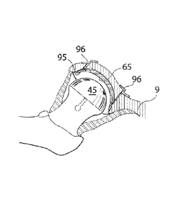
Fig. 9 shows an artificial caput femur surface 45 in section having a greatest
cross-sectional
distance 52 adapted to travel over and beyond the maximum diameter of the
caput femur 5.
The largest diameter of the caput femur 5 being positioned at a corresponding
largest cross
sectional distance 61 of the artificial caput femur surface, a second distance
62 is the distance
that the artificial caput femur surface 45 travels beyond the maximum diameter
of the caput
femur 5. Said distance 62 is the beyond part of the artificial caput femur
surface and is a part
of the mechanical fixation of the artificial caput femur surface 45 to the
caput femur 5.
Fig. 10 shows an artificial caput femur surface according to a first
embodiment, the artificial
caput femur surface 45 is adapted to pass beyond the maximum diameter of the
caput femur
5. This enables a mechanical fixation using the form of said artificial caput
femur surface 45.
In this embodiment the artificial caput femur surface 45 comprises at least
one slit 49 adapted
to make said artificial caput femur surface 45 flexible for traveling over and
beyond the
maximum diameter of the caput femur 5. The construction could further be made
flexible so
that the size of the artificial caput femur surface 45 can vary to become
smaller for insertion
through a hole 18 in the pelvic bone 9 smaller than the full functional size
of the artificial
caput femur surface 45. It is also conceivable that the artificial caput femur
surface 45
comprises two or more artificial caput femur surface arms 50 which have a
cross sectional
distance 52 between each other. This cross sectional distance 52 is according
to one
embodiment shorter than the maximum diameter of the caput femur 5 enabling the
CA 3041896 2019-05-01
W02011/005198 PCT/SE2010/050817
18
mechanical fixation of the artificial caput femur surface 45 by means of said
artificial caput
femur surface arms 50. For further fixation a band, cord or wire 59 can be
placed around the
artificial caput femur surface 45 beyond the maximum diameter of the caput
femur 5. The
band, cord or wire can be mechanically connected using a self locking member
60 for forming
a ring-shaped element able to assist in the fixation of the artificial caput
femur surface 45 to
the caput femur 5.
Fig. 11 shows the artificial caput femur surface 45 when fixated to the caput
femur with the
supporting band, cord or wire placed around the artificial caput femur surface
45 beyond the
maximum diameter of the caput femur 5. The arms may also be adapted to go into
the bone of
caput femur 5 to lock said artificial caput femur surface 45.
Fig. 12 shows the artificial caput femur surface 45 according to one
embodiment. The shaft or
screw placed in the middle of the artificial caput femur surface 45 serves as
a mechanical
attachment 44 penetrating the cortex of the caput femur 5 and fixating the
artificial caput
femur surface 45 to the caput femur 5. However it is also conceivable that
said shaft or screw
is assisted or replaced with screws, welding, sprints, band, adhesive or some
other mechanical
connecting member.
Fig. 13 shows the artificial caput femur surface 45 according to another
embodiment, in
which said artificial caput femur surface 45 comprises at least one slit 49
enabling the
construction of the artificial caput femur surface 45 to be flexible, thus
enabling the largest
diameter 51 to vary for insertion of said artificial caput femur surface 45
through a hole in the
pelvic bone 9 smaller than the full functional size of said artificial caput
femur surface 45.
According to this embodiment the artificial caput femur surface 45 further
comprises artificial
caput femur surface arms 50 located on the sides of said at least one slit 49.
The caput femur
surface arms 50 can be made of a flexible material enabling the insertion
through a hole 20 in
the pelvic bone 9 smaller than the largest diameter 51 of said artificial
caput femur surface 45
when in its full functional size.
According to one embodiment the artificial caput femur surface 45 of said
third embodiment
could be adapted to pass beyond the maximum diameter of the caput femur 5.
This enables a
mechanical fixation using the form of said artificial caput femur surface 45.
In the
embodiment where the artificial caput femur surface 45 travels beyond the
maximum
diameter of the caput femur 5 the construction can be made flexible so that
the size of the
CA 3041896 2019-05-01
WO 2011/005198 PCT/SE2010/050817
19
artificial caput femur surface 45 can vary to become smaller for insertion
through a hole 18 in
the pelvic bone smaller than the full functional size of the artificial caput
femur surface 45,
and have an opening adapter to travel over the caput femur 5 that can be
larger that the same
opening is in the full functional size of the artificial caput femur surface
45 enabling the
artificial caput femur surface 45 to at least partly cover an area beyond the
maximum diameter
of caput femur 5 from the direction of the acetabulum 8. According to a second
embodiment
the artificial caput femur surface 45 comprises two or more artificial caput
femur surface arms
50 which have a cross sectional distance 52 between each other. This cross
sectional distance
52 is according to one embodiment shorter than the maximum diameter of the
caput femur 5
enabling the mechanical fixation of the artificial caput femur surface 45 by
means of said
artificial caput femur surface arms 50.
Fig. 14a,b,c,d,e shows the artificial caput femur surface 45 according to a
fourth embodiment,
in which said artificial caput femur surface 45 comprises a first 53a and a
second 53b section,
as shown in fig. 14b. The first and second sections are displaceable in
relation to each other.
According to a first embodiment said first section 53a can be rotated in
relation to said second
section 53b so that said second section 53b travels underneath said first
section 53a to create a
displaced artificial caput femur surface 54, as shown in fig. 14e, which is
possible to insert
into a hip joint of a human patient through a hole 18 being oval, or at least
having an area
smaller than the cross sectional area of the artificial caput femur surface 45
when in its full
functional size 45, as shown in fig. 14a. According to this embodiment the two
sections are
connected to each other when the artificial caput femur surface 45 is returned
to its full
functional size using a mechanical form fitting 55, as shown in fig 14e.
However it is also
conceivable that said connection is assisted or replaced with screws, welding,
sprints, band,
adhesive or some other mechanical connecting member.
Fig. 15a,b shows the artificial caput femur surface 45 according to another
embodiment, in
which said artificial caput femur surface 45 comprises four slits. The
artificial caput femur
surface 45 is flexible in its construction allowing the four artificial caput
femur arms 50 to be
folded towards the center axis of the artificial caput femur surface 45 thus
allowing the
artificial caput femur surface 45 to be inserted into a hip joint through a
hole smaller than the
full functional size of the artificial caput femur surface 45. The artificial
caput femur surface
45 according to this embodiment can be constructed to go beyond the maximum
diameter of
CA 3041896 2019-05-01
WO 2011/005198 PCT/SE2010/050817
the caput femur 5, in which case the construction with the slits 49 allows the
artificial caput
femur surface 45 to change to both a smaller and a larger size than said full
functional size.
Fig. 15b shows the artificial caput femur surface 45 in section when said
artificial caput femur
surface arms 50 are folded for insertion through a hole 18 with an area
smaller than the largest
5 area of the artificial caput femur surface 45 when in its full functional
size.
Fig 16a shows the artificial caput femur surface 45 according to a sixth
embodiment, in which
said artificial caput femur surface 45 comprises multiple ring-shaped
artificial caput femur
surface parts 63. Said multiple ring-shaped artificial caput femur surface
parts 63 are adapted
to be connected to each other to form an artificial caput femur surface 45,
shown in fig. 16b.
10 According to one embodiment said artificial caput femur surface parts 63
are adapted to be
connected to each other using mechanical connecting members 64a,b. In Fig.
16c, 64a shows
how an individual ring-shaped artificial caput femur surface part 63 can be
connected to itself
to form a continuous ring shape. 64b shows how an individual ring-shaped
artificial caput
femur surface part 63 connects to other ring-shaped artificial caput femur
surface parts 63 to
15 form an artificial caput femur surface 45. The artificial caput femur
surface 45 according to
this embodiment can further be adapted to go beyond the maximum diameter of
the caput
femur 5.
Fig. 17a,b,c shows the artificial caput femur surface 45 according to a sixth
embodiment, in
which said artificial caput femur surface 45 comprises multiple artificial
caput femur surface
20 parts 46. Said multiple artificial caput femur surface parts 46 are
adapted to be connected to
an interconnecting artificial caput femur surface part 56 after insertion into
a hip joint. The
interconnecting artificial caput femur surface part 56, which serves as a base
part, comprises
self locking connecting members 57, shown in fig. 17b, that fits with
corresponding self
locking members 58 of the artificial caput femur surface parts 46. The
artificial caput femur
surface parts 46 create an artificial caput femur surface 45 when connected to
each other,
shown in fig. 17c. The self locking members 57, 58 can be assisted or replaced
with screws,
welding, sprints, band, adhesive or some other mechanical connecting member.
The artificial
caput femur surface 45 according to this embodiment can further be adapted to
go beyond the
maximum diameter of the caput femur 5.
Fig. 18 shows a conceptual way wherein the artificial caput femur surface 45
has a diameter
or cross-sectional distance dl small enough to enable said artificial caput
femur surface 45 to
CA 3041896 2019-05-01
WO 2011/005198 PCT/SE2010/050817
21
travel through a hole 20 in the pelvic bone 9. After the artificial caput
femur surface 45 has
traveled through the hole 20 in the pelvic bone 9 the artificial caput femur
surface 45 is
expanded such that the diameter or cross-sectional distance d2 is large enough
to travel over
the caput femur 5. Finally the artificial caput femur surface 45 is positioned
on the caput
femur 5, in this state the diameter or cross-sectional distance is smaller
than the largest
diameter of the caput femur 5 which mechanically attaches the artificial caput
femur surface
45 to the caput femur 5. d3 is the normal sate cross sectional distance of the
medical device,
i.e. the cross sectional distance that the medical device has when the medical
device is in its
functional position. This figure may also in an alternative embodiment show
the artificial
acetabulum surface mounted onto caput femur or an artificial replacement
therefore with the
same locking principle.
Fig 19a,b shows the hip joint in section with the caput femur 5 placed at the
very top of
collum femur 6, which is the top part of the femur bone 7. The caput femur is
in connection
with the acetabulum 8, which is a bowl shaped part of the pelvic bone 9.
According to a first
embodiment the hole 18 created in the pelvic bone 9 from the opposite side
from acetabulum
8, is larger than said artificial caput femur surface 45, enabling the
insertion of said artificial
caput femur surface 45 in its full functional size. Said insertion of said
artificial caput femur
surface 45 could be performed as a step of the surgical method, as well as a
step of the
laparoscopic method. After the insertion, the artificial caput femur surface
45 is attached to
the caput femur 5, the attaching is performed by means of a mechanical
attachment 44
comprising a shaft or screw penetrating the cortex. It is however also
conceivable that the
mechanical attachment 44 is assisted or replaced by bone cement or adhesive
placed between
caput femur 5 and the artificial caput femur surface 45, or in connection with
said shaft or
screw 44. Alternative ways of attaching the artificial caput femur surface 45
includes: at least
one screw, at least one pin, at least one portion of at least one of the parts
adapted to be
introduced into the other part, the parts being adapted to be sliding into the
other part, form
fitting, welding, adhesive, pin, wire, a ball mounted into a bowl being
portions of said parts, a
male portion of one part mounted into a female portion of the other part, a
key introduced into
a lock being portions of said parts, band, or other mechanical connecting
members.Fig. 19b
shows the hip joint in section with the artificial caput femur surface 45
attached to the caput
femur 5.
CA 3041896 2019-05-01
WO 2011/005198 PCT/SE2010/050817
22
Fig. 20a shows how an expandable artificial caput femur surface 45 is being
inserted through
a hole 18 in the pelvic bone 9.
Fig. 20b shows how an expandable artificial caput femur surface 45 travels
through the hole
18 in the pelvic bone 9 and travels over caput femur 5.
Fig. 20c shows an expandable artificial caput femur surface 45 is after it has
been placed on
said caput femur 5.
Fig. 21a shows the hip joint in section according to a second embodiment in
which the hole
18 in the pelvic bone 9 is smaller than the artificial caput femur surface 45
in its full
functional size. According to this embodiment the artificial caput femur
surface 45 is
introduced into said hip joint through the hole 18 in the pelvic bone 9 form
the opposite side
from acetabulum 8. The artificial caput femur surface parts 46 are connected
to each other
after insertion into said hip joint to form the artificial caput femur surface
45.
Fig. 21b shows the hip joint in section when the artificial caput femur
surface parts 46 are
connected to each other using form fitting 47, however it is conceivable that
the form fitting is
assisted or replaced with adhesive or bone cement. After the artificial caput
femur surface
parts 46 have been introduced and connected in the hip joint, they are
mechanically fixated to
the caput femur 5, the mechanical fixation could be done by means of: at least
one screw, at
least one pin, at least one portion of at least one of the parts adapted to be
introduced into the
other part, the parts being adapted to be sliding into the other part, form
fitting, welding,
adhesive, pin, wire, a ball mounted into a bowl being portions of said parts,
a male portion of
one part mounted into a female portion of the other part, a key introduced
into a lock being
portions of said parts, band, or other mechanical connecting members.
Fig. 21c shows the artificial caput femur surface parts 46 with the parts
supplying the form
fitting 47.
Fig. 21d shows the hip joint in section wherein a second hole 18b in the
pelvic bone 9 enables
the surgeon to place a camera 34 into the hip joint, preferably used in the
laparoscopic
method.
According to one embodiment the medical device comprises an artificial
acetabulum surface
65. In the embodiments where the medical device comprises an artificial caput
femur surface
CA 3041896 2019-05-01
W02911/005198 PCT/SE2010/050817
23
45 and an artificial acetabulum surface 65, the artificial acetabulum surface
is provided after
the artificial caput femur surface.
Fig. 22 shows an artificial acetabulum surface 65 in its full functional size,
as it is being
inserted through a hole 18 in the pelvic bone 9. The hole being large enough
to allow the
artificial acetabulum surface to pass through the hole.
Fig. 23a shows an artificial acetabulum surface in a conceptual way, wherein
the artificial
acetabulum surface 65 has a diameter or cross-sectional distance dl small
enough to enable
said artificial acetabulum surface 65 to travel through a hole 18 in the
pelvic bone 9. After the
artificial acetabulum surface 65 has traveled through the hole 18 in the
pelvic bone 9, the
artificial acetabulum surface is expanded such that the diameter or cross-
sectional distance d2
is large enough to hinder the artificial acetabulum surface 65 from traveling
through the hole
18 in the pelvic bone 9.
Fig. 23b shows the artificial acetabulum surface 65 when positioned in the
acetabulum 8.
Fig. 24 shows an artificial acetabulum surface 65 according to a second
embodiment in which
the artificial acetabulum surface 65 comprises at least one slit 66 enabling
the artificial
acetabulum surface 65 to vary in size for insertion through a hole 18 in the
pelvic bone 9
smaller than the full functional size of the artificial acetabulum surface 65.
The slits are
placed between one or more artificial acetabulum surface arms 67 which are
flexible by
means of the material or by means of a joint affecting said artificial
acetabulum surface arms
67.
Fig. 25a,b,c shows an artificial acetabulum surface 65 according to a second
embodiment in
which the artificial acetabulum surface 65 comprises multiple artificial
acetabulum surface
parts 68. Said multiple artificial acetabulum surface parts 68 are adapted to
be connected to an
interconnecting artificial acetabulum surface part 69 after insertion into a
hip joint. The
interconnecting artificial caput femur surface part 69 comprises self locking
connecting
members 70a, shown in fig. 25b, that fits with corresponding self locking
members 70b of the
artificial acetabulum surface parts 68. The artificial acetabulum surface
parts 68 create an
artificial acetabulum surface 65 when connected to each other, shown in fig.
25c. The self
locking members 70a,b can be assisted or replaced with at least one screw, at
least one pin, at
least one portion of at least one of the parts adapted to be introduced into
the other part, the
CA 3041896 2019-05-01
85243972
24
parts being adapted to be sliding into the other part, form fitting, welding,
adhesive, pin, wire,
a ball mounted into a bowl being portions of said parts, a male portion of one
part mounted
into a female portion of the other part, a key introduced into a lock being
portions of said
parts, band, or other mechanical connecting members.
Fig. 26a,b,c shows an artificial acetabulum surface 65 according to a third
embodiment in
which the artificial acetabulum surface 65 comprises multiple ring-shaped
artificial
acetabulum surface parts 71. Said multiple ring-shaped artificial acetabulum
surface parts 71
are adapted to be connected to each other to form an artificial acetabulum
surface 65 after
insertion in a hip joint. According to one embodiment said artificial
acetabulum surface parts
71 are adapted to be connected to each other using mechanical connecting
members 72a,b.
Fig. 26c shows how an individual ring-shaped artificial acetabulum surface
part 71 can be
connected to itself using the mechanical connecting member 72a to form a
continuous ring
shape. Further 37c shows how an individual ring-shaped artificial acetabulum
surface part 71
connects to other ring-shaped artificial acetabulum surface parts 71 using the
mechanical
connecting member 72b to form an artificial acetabulum surface 65.
Fig. 27a,b,c,d shows an artificial acetabulum surface 65 according to a fourth
embodiment in
which the artificial acetabulum surface 65 comprises a first 73a and a second
73b section,
shown in fig. 27b. The first and second sections are displaceable in relation
to each other.
According to one embodiment said first section 73a can be rotated in relation
to said second
section 73b so that said second section 73b travels underneath said first
section 73a to create a
displaced artificial acetabulum surface 74, as shown in fig. 27c, which is
possible to insert
into a hip joint of a human patient through a hole being oval, or at least
having an area smaller
than the cross sectional area of the artificial acetabulum surface 65 when in
its full functional
size. According to this embodiment the two sections 73a,b are connected to
each other
when the artificial acetabulum surface is returned to its full functional size
using a mechanical
form fitting 75, as shown in fig. 27d. However it is also conceivable that
said connection is
assisted or replaced with at least one screw, at least one pin, at least one
portion of at least one
of the parts adapted to be introduced into the other part, the parts being
adapted to be sliding
into the other part, form fitting, welding, adhesive, pin, wire, a ball
mounted into a bowl being
portions of said parts, a male portion of one part mounted into a female
portion of the other
Date Recue/Date Received 2021-03-29
85243972
part, a key introduced into a lock being portions of said parts, band, or
other mechanical
connecting members.
Fig. 28a shows an artificial acetabulum surface 65 according to a fifth
embodiment in which
the artificial acetabulum surface 65 comprises four slits 66. The artificial
acetabulum surface
5 65 is flexible in its construction allowing the four artificial
acetabulum arms 67 to be folded
towards the center axis of the artificial acetabulum surface 65 thus allowing
the artificial
acetabulum surface to be inserted into a hip joint through a hole smaller than
the full
functional size of the artificial acetabulum surface 65.
Fig. 28b shows the artificial acetabulum surface 65 according to the fifth
embodiment in its
10 folded state.
After the medical device, comprising at least one hip joint surface, has been
provided through
a hole 18 in the pelvic bone 9, in accordance with any of the embodiment
above, said hole 18
needs to be closed. The hole can be closed using the medical device, however
it is also
conceivable that closing of the hole is performed with assistance of an
additional prosthetic
15 part, or a piece of human bone. In the embodiments where the closing is
assisted by a piece of
human bone, that piece could simply be the piece drilled from the pelvic bone
or a modified
piece from the pelvic bone e.g. equipped with additional supporting members.
Fig. 29a shows an embodiment where a solid medical device 600 is fixated to
the collum
femur 6, introduced through a hole 18 in the pelvic bone 9. The stabilizing
member 612 is
20 adapted to stabilize the medical device 600 from the outside of the collum
femur 6
substantially perpendicular to the longitudinal extension of the collum femur
6, and from the
acetabulum side, substantially in line with the longitudinal extension of the
collum femur 6
through the stabilizing member being placed in contact with the outside of the
collum femur 6
and the surface of the section 610 in the collum femur 6. The stabilizing
member is fixated to
25 the outside of the collum femur and/or to the surface of the section in
the collum femur by
means of the adhesive 614. However the adhesive 614 could be replaced or
assisted by bone
cement or a mechanical fixation element 615. The
medical device 600
has a longitudinal member 608 having a smallest passable area being
an area of a hole 18 through which the medical device 600 can pass. To
enable the
solid medical device 600 to pass through said hole in the pelvic bone the
solid medical device
600 is smaller than the caput femur, i.e. the smallest passable area of the
medical device 600,
being an area of a hole through which said medical device 600 can pass is
smaller than the
Date Recue/Date Received 2021-03-29
WO 2011/005198 PCT/SE2010/050817
26
smallest passable area of the caput femur 5, being an area of a hole through
which said caput
femur 5 can pass.
Fig. 29b shows the hip joint in section when the medical device 600 is
positioned on the
collum femur 6. The stabilizing member 612 is here fixated to the collum femur
by means of
adhesive 614 and a mechanical fixation element 615.
Fig. 29c shows the hip joint in section when the medical device 600 is
positioned on the
collum femur 6. The stabilizing member 612 is here fixated to the collum femur
by means of
adhesive 614. An prosthetic part 98 comprising an artificial acetabulum
surface 618 has been
positioned in the hole 18 in the pelvic bone 9. The artificial acetabulum
surface 618 is adapted
to be in direct of indirect connection with the artificial caput femur surface
607. In
embodiments where the artificial acetabulum surface 618 is adapted to be in
indirect
connection with the artificial caput femur surface 607 a lubricating fluid or
a lubricating
material (not shown) can be placed between said artificial acetabulum surface
618 and said
artificial caput femur surface 607. The prosthetic part is adapted to carry
the load placed on
the artificial acetabulum surface 618 from weight of the human patient through
the contact
with the artificial caput femur surface 607 by means of the supporting members
99 in
connection with the pelvic bone. The prosthetic part 98 can further be fixated
to the pelvic
bone 9 by means of bone cement, adhesive, screws, form fitting, welding,
sprints, band or
some other mechanical connecting member. According to this embodiment the
supporting
members 99 are positioned on the acetabulum side of the pelvic bone 9, however
it is also
conceivable that the supporting members 99 are positioned on the abdominal
side of the
pelvic bone 9 or according to fig. 35.
Fig. 30a shows the hip joint of a human patient in section, wherein a bone
plug 31 is placed in
the hole 18 in the pelvic bone 9 to close said hole 18. According to a first
embodiment the
medical device comprises supporting members 95 which carries the load placed
on the
acetabulum 8 from weight of the human patient through the contact with the
caput femur 5.
Said supporting members can be adapted to be displaceable 97 supporting
members. The bone
plug 31 can be attached to the artificial acetabulum surface 11 and/or the
pelvic bone 9 by
means of at least one screw, at least one pin, at least one portion of at
least one of the parts
adapted to be introduced into the other part, the parts being adapted to be
sliding into the other
part, form fitting, welding, adhesive, pin, wire, a ball mounted into a bowl
being portions of
CA 3041896 2019-05-01
= WO 2011/005198
PCT/SE2010/050817
27
said parts, a male portion of one part mounted into a female portion of the
other part, a key
introduced into a lock being portions of said parts, band, or other mechanical
connecting
members.
The connection between the medical device and the pelvic bone 9 can be direct
or indirect. In
the embodiment where the contact between the medical device and the pelvic
bone 9 is
indirect, a material can be provided between said medical device and said
pelvic bone. The
material could comprise bone cement, an at least partly elastic material,
glue, adhesive,
antibiotic, biocompatible plastic material, biocompatible ceramics and/or
biocompatible
metal.
Fig 30b shows the hip joint of a human patient in section wherein the bone
plug 31 placed in
the hole 18 in the pelvic bone 9 is further supported by supporting members 96
placed
between the bone plug 31 and the pelvic bone 9 on the opposite side from
acetabulum 8 using
at lest one of: at least one screw, at least one pin, at least one portion of
at least one of the
parts adapted to be introduced into the other part, the parts being adapted to
be sliding into the
other part, form fitting, welding, adhesive, pin, wire, a ball mounted into a
bowl being
portions of said parts, a male portion of one part mounted into a female
portion of the other
part, a key introduced into a lock being portions of said parts, band, or
other mechanical
connecting members.
Fig. 31 shows a bone plug 31 or a prosthetic part 98 comprising several
displaceable
supporting members adapted to carry the load placed on the acetabulum 8 from
weight of the
human patient through the contact with the caput femur 5. The displaceable
parts 97 are
displaced into a corresponding part in or at the edge of the hole 18 in the
pelvic bone 9.
Fig 32a shows an embodiment wherein the closing of the hole 18 in the pelvic
bone is
performed by means of a prosthetic part 98. Fig. 56a shows the prosthetic part
98 being
inserted into a hole 18 in the pelvic bone 9 from the opposite side from
acetabulum 8.
According to one embodiment the prosthetic part 98 comprises supporting
members 99
adapted to correspond with sections 100 of the hole 18 in the pelvic bone 9.
After the
prosthetic part 98 has been inserted into said hole 18 in the pelvic bone 9 it
is rotated so that
the supporting members 99 comes in contact with the pelvic bone 9 and can
carry the load
placed on the acetabulum 8 from weight of the human patient through the
contact with the
CA 3041896 2019-05-01
WO 2011/005198 PCT/SE2010/050817
28
caput femur 5. Said prosthetic part 98 could also be adapted to serve as
artificial acetabulum
surface 65 according to any of the above mentioned embodiments.
Fig. 32b shows the prosthetic part 98 when rotated to carry the load placed on
the acetabulum
8 from weight of the human patient through the contact with the caput femur 5.
This supporting means could be constructed in many different ways and this
should be seen as
examples.
Fig. 33 shows the hip joint of a human patient in section wherein the
prosthetic part 98 closes
the hole 18 in the pelvic bone 9 and carries the load placed on the acetabulum
8 from weight
of the human patient through the contact with the caput femur 5 by means of
the supporting
members 99. The prosthetic part 98 can farther be fixated to the pelvic bone 9
by means of at
least one screw, at least one pin, at least one portion of at least one of the
parts adapted to be
introduced into the other part, the parts being adapted to be sliding into the
other part, form
fitting, welding, adhesive, pin, wire, a ball mounted into a bowl being
portions of said parts, a
male portion of one part mounted into a female portion of the other part, a
key introduced into
a lock being portions of said parts, band, or other mechanical connecting
members.
Fig. 34a shows the hip joint of a human patient in section wherein bone plug
31 or prosthetic
part 98 is attached to the pelvic bone 9 by means of screws 101 placed from
the opposite side
from acetabulum 8. The screws 101 are possible to place in different angles
depending on
reach or need for support. This construction may be performed in many
different ways, for
example using a plate mounted to the pelvic bone.
Fig 34b shows the hip joint of a human patient in section wherein bone plug 31
or prosthetic
part 98 is attached to the pelvic bone 9 by means of a plate 102 at least
partly covering said
bone plug 31 or prosthetic part 98. According to a first embodiment the plate
102 is attached
to the pelvic bone 9 by means of screws 103 placed from the opposite side from
acetabulum
8. However it is also conceivable that said screws 103 can be replaced or
assisted by bone
cement, adhesive, form fitting, welding, sprints, band or some other
mechanical connecting
member.
Fig. 34c shows the hip joint of a human patient in section wherein two bone
plugs 31 or
prosthetic parts 98 arc attached to the pelvic bone 9 by means of a plate 102
at least partly
CA 3041896 2019-05-01
WO 2011/005198 PCT/SE2010/050817
29
covering said bone plugs 31 or prosthetic parts 98. According to a first
embodiment the plate
102 is attached to the pelvic bone 9 by means of screws 103 placed from the
opposite side
from acetabulum 8. However it is also conceivable that said screws 103 can be
replaced or
assisted by bone cement, adhesive, form fitting, welding, sprints, band or
some other
mechanical connecting member.
Fig 34c also shows the provided artificial acetabulum surface 65 and the
provided caput
femur surface. The members for fixating and covering the hole in the pelvic
bone, together
with the artificial acetabulum surface and the artificial caput femur surface
constitute the
medical device.
Fig. 34d shows the hip joint of a human patient in section wherein two holes
18 in the pelvic
bone has been covered by means of a fluid injected into said holes 18, through
sealing
members 104, said fluid 93 being adapted to harden. Further more a plate 102
has been
provided at least partly covering said holes 18. According to a first
embodiment the plate 102
is attached to the pelvic bone 9 by means of screws 103 placed from the
opposite side from
acetabulum 8. However it is also conceivable that said screws 103 can be
replaced or assisted
by bone cement, adhesive, form fitting, welding, sprints, band or some other
mechanical
connecting member. Fig 34d also shows the provided artificial acetabulum
surface 65, and the
provided artificial caput femur surface 45.
Fig. 35 is a schematic figure of the pelvic bone in section, describing in
further detail the
supporting members shown in for example fig. 35. The pelvic bone comprises an
inner cortex
201a placed on the abdominal side of the pelvic bone AB, and an outer cortex
201b placed on
the acetabulum side of the pelvic bone AC. The inner and outer cortex 201a,b
comprises
cortical bone, which is a more dense sclerotic bone. The pelvic bone further
comprises
cancellous bone 202, placed in the bone marrow between said inner cortex 201a
and said
outer cortex 201 b. The supporting members of the medical device according to
any of the
embodiments above can be adapted to be in contact with the outside of the
inner cortex 201a
as supporting member 203, or be placed inside of the inner cortex 201a as
supporting member
204, which enables the supporting member to carry loads in the direction of
the abdomen AB
as well as in the direction of the acetabulum AC. It is furthermore
conceivable that the
supporting member is placed in the middle of the inner cortex 201a and the
outer cortex 201b,
in the cancellous bone, as supporting member 205, in which case the supporting
member
CA 3041896 2019-05-01
W02011/005198 PCT/SE2010/050817
could be in contact with the inner cortex 201a, on the inside thereof, and the
outer cortex
201b, on the inside thereof, which enables the supporting member to carry
loads in the
direction of the abdomen AB as well as in the direction of the acetabulum AC.
Further, the
supporting members can be adapted to be in contact with the outside of the
outer cortex 201b
5 as supporting member 207, or be placed inside of the outer cortex 201b as
supporting member
206, which enables the supporting member to carry loads in the direction of
the abdomen AB
as well as in the direction of the acetabulum AC.
Fig. 36a shows an injecting member 105 for injecting a fluid adapted to harden
93, preferably
bone cement or adhesive to be used as support in the closing of the hole 18 in
the pelvic bone
10 9. The injecting member 105 comprises a piston 94 that pushes said fluid
93 in to the area
where it is wanted.
Fig. 36b shows the injecting member 105 as it is inserted through the skin 106
of a human
patient in the surgical or laparoscopic method, and is further placed in
connection with the hip
joint through the hole 18 in the pelvic bone 9. The injecting member 105 is
adapted to inject a
15 fluid 93 adapted to harden.
Fig. 37 shows the injecting member 105 according to any of the embodiments
above, adapted
to inject fluid 93 into a mould 81, a sealed area 87 or a connecting area
between the pelvic
bone 9 and a prosthetic part, the pelvic bone 9 and a bone plug 31 or the
caput femur 5 and a
prosthetic part. Said injecting member comprises a container 107 adapted to
hold a fluid for
20 injection. According to a first embodiment said container comprises two
compartments
108a,b adapted to hold two different fluids, said fluids being adapted to
harden when mixed.
In the embodiment when the container 107 is adapted to hold two fluids, it is
conceivable that
the injecting member 105 further comprises a mixing member 109 wherein said
two fluids arc
being mixed before injection. According to a second embodiment (not shown)
said container
25 107 is adapted to keep said fluid sterile. According to a third
embodiment (not shown) said
container 107 is adapted to keep said fluid cold and according to a fourth
embodiment (not
shown) said container 107 is adapted to keep said fluid in a dark environment.
Furthermore a
combination of the above mentioned embodiments is conceivable.
After the step of closing the hole in the pelvic bone of the human patient is
concluded the
30 medical device has been provided and all instruments are retracted. The
final step of a
CA 3041896 2019-05-01
WO 2011/005198 PCT/SE2010/050817
31
surgical or laparoscopic method comprises suturing or stapling the affected
tissue and finally
suturing or stapling the skin of the human patient.
Fig. 38a shows the step of suturing 110 or stapling 111 the skin of the human
patient in the
surgical method, whereas fig. 38b shows the step of suturing 110 or stapling
111 the skin of
the human patient in the laparoscopic method. The laparoscopic method may not
need any
suturing.
Please note that any embodiment or part of embodiment as well as any method or
part of
method could be combined in any way. All examples herein should be seen as
part of the
general description and therefore possible to combine in any way in general
terms.
CA 3041896 2019-05-01