Note: Descriptions are shown in the official language in which they were submitted.
CA 03042588 2019-05-02
WO 2018/090148 PCT/CA2017/051387
METHODS AND SYSTEMS FOR RAPID RETRACTION OF A TRANSCATHETER
HEART VALVE DELIVERY SYSTEM
CROSS-REFERENCE
[0001] The present application claims priority to U.S. Provisional Patent
Application No.
62/424,910 (Attorney Docket No. 53235-712.101), filed on November 21, 2016,
which is
herein incorporated by reference in its entirety.
[0002] The present application is related to: U.S. Patent No. 8,579,964
(Attorney Docket No.
53235-703.201) filed April 28, 2011; and also related to U.S. Publication Nos.
2013/0211508
(Attorney Docket No. 53235-704.201) filed November 16, 2012; 2014/0052237
(Attorney
Docket No. 53235-705.201) filed February 8, 2013; 2014/0155990 (Attorney
Docket No
53235-706.201) filed May 29, 2013; 2014/0257467 (Attorney Docket No. 53235-
707.201)
filed March 3, 2014; and 2014/0343669 (Attorney Docket No. 53235-708.201)
filed April 1,
2014; the entire contents of each of which is incorporated herein by
reference.
BACKGROUND OF THE INVENTION
[0003] 1. Field of the Invention.
[0004] Mitral regurgitation, also known as mitral insufficiency or mitral
incompetence is a
heart condition in which the mitral valve does not close properly thereby
resulting in
abnormal leakage of blood retrograde from the left ventricle through the
mitral valve back
upstream into the left atrium. Persistent mitral regurgitation can result in
congestive heart
failure, a costly and often fatal condition. Traditional surgical repair of
the valve generally
results in a good clinical outcome but requires open heart surgery and a
lengthy and costly
hospital stay along with an extended recovery period. More recently, minimally
invasive
procedures have been developed to deliver a prosthetic heart valve
percutaneously over a
catheter through the patient's vasculature to the heart, or by using a
transapical procedure to
introduce the prosthesis through the chest wall and through the apex of the
heart to the
treatment site. An exemplary prosthesis includes any of the embodiments
described in U.S.
Patent No. 8,579,964, the entire contents of which are incorporated herein by
reference.
These prostheses and delivery procedures appear to be promising, but there is
yet opportunity
to improve procedural outcomes by minimizing the duration of the procedure,
from first
-1-
CA 03042588 2019-05-02
WO 2018/090148 PCT/CA2017/051387
contact with the delivery system by an operator to final withdrawal of the
delivery system and
wound closure in the patient. Therefore, it would be desirable to provide
improved devices,
systems, and methods that reduce the amount of time needed to remove the
delivery system
from the patient, improve ease of use, speed up the procedure, and reduce
risk. At least some
of these objectives will be met by the exemplary embodiments described herein.
[0005] 2. Description of the Background Art. U.S. Patent No. 8,579,964
discloses an
exemplary prosthetic heart valve and trans-catheter delivery system, the
entire contents
previously incorporated herein by reference.
BRIEF SUMMARY
[0006] The present disclosure generally relates to medical systems, devices
and methods, and
more particularly relates to prostheses and delivery systems such as heart
valve delivery
systems that may be used to implant a prosthesis such as a valve, including a
prosthetic mitral
valve, a heart valve, or any other valve. The present disclosure emphasizes
exemplary
embodiments of a prosthetic mitral valve and delivery system, but one of skill
in the art will
appreciate that this is not intended to be limiting.
[0007] In many embodiments, trans-catheter methods and systems of deploying
prosthetic
heart valves and rapid retraction of the delivery system are provided. In
certain embodiments,
the delivery system comprises a trans-apical delivery system that may be used
to implant a
prosthetic heart valve into anatomical position by way of an incision in the
apex of the heart.
The trans-apical delivery system may comprise a system of catheters that may
be
concentrically nested upon one another and that, when combined, may retain a
compressed
heart valve prosthesis. Removal of the constraint provided by certain
catheters may then
facilitate deployment of the heart valve prosthesis into the heart. Further
embodiments of the
trans-apical delivery system that may be used in any of the delivery systems
disclose herein
may allow for the closure of the delivery catheters at an enhanced speed, such
as by way of
translation of catheter components within each other in the opposite direction
to that required
for deployment operation. The operation of such delivery systems may be
facilitated through
the use of actuator mechanisms such as button mechanisms that may be in
communication
with linkage systems, or actuator mechanisms such as button mechanisms that
may be in
communication with flexible members, or even pin coupled components that
simplify use.
-2-
CA 03042588 2019-05-02
WO 2018/090148 PCT/CA2017/051387
[0008] Further embodiments herein may include delivery systems that allow for
alternative
implantation pathways such as through the inferior or superior vena cava, the
aorta, or the
atria.
[0009] In an aspect of the present disclosure, a method of rapidly retracting
a delivery system
comprises providing a delivery system, the delivery system having a plurality
of catheters
used to deliver a heart valve prosthesis, providing a controllable deployment
mechanism, the
controllable deployment mechanism having the ability to preferentially release
a prosthesis
from the catheter, and actuating the controllable deployment mechanism thereby
releasing the
prosthesis from the catheter. The method may also comprise providing a rapid
retraction
mechanism, the rapid retraction mechanism having the ability to rapidly close
the catheter,
actuating the rapid retracting mechanism thereby rapidly closing the catheter.
[0010] The method may comprise trans-apically introducing the delivery system
into an apex
of a heart, or transseptally delivering the delivery system to a heart,
delivering the delivery
system to the heart via a subclavian vein, delivering the delivery system to
the heart via an
aorta, or delivering the delivery system to the heart via a left atrium or a
right atrium.
[0011] Actuating the rapid retraction mechanism may comprise actuating a
button and
linkage. The rapid retraction mechanism may comprise a threaded region and
interference
member, and the method may further comprise constraining movement of the rapid
retraction
mechanism with the threaded region and interference member. The rapid
retraction
mechanism may comprise a flexible interference member, and the method may
further
comprise deflecting the flexible interference member. The rapid retraction
mechanism may
comprise a pin and pin-hole link assembly, and the method may comprise
removing the pin
from the pin-hole link assembly.
[0012] In another aspect of the present disclosure, a delivery device for
delivering a
prosthesis comprises a first actuation mechanism for controlling movement of a
delivery
catheter, wherein the delivery catheter may be configured to carry a
prosthesis therein, and
wherein actuation of the first actuation mechanism may move the delivery
catheter away from
the prosthesis thereby at least partially removing a constraint therefrom, and
a deployment
mechanism for controlling release of the prosthesis from an anchoring
catheter, the anchoring
catheter disposed at least partially in the delivery catheter, and wherein
actuation of the
-3-
CA 03042588 2019-05-02
WO 2018/090148 PCT/CA2017/051387
deployment mechanism may move the anchoring catheter away from the prosthesis
thereby
releasing a constraint therefrom. The delivery system may also comprise an
inner guidewire
catheter having a tapered distal tip, the inner guidewire catheter disposed in
the anchoring
catheter, and a rapid retraction mechanism for controlling movement of the
delivery catheter
relative to the tapered distal tip, wherein actuation of the rapid retraction
mechanism closes
the delivery device such that a proximal end of the distal tip abuts against a
distal end of the
delivery catheter thereby forming a smooth continuous outer surface of the
delivery device.
[0013] The actuation mechanism may comprise a thumbwheel. The deployment
mechanism
may comprise an actuatable button with a linkage coupled thereto. The rapid
retraction
mechanism may comprise a threaded region and interference member, a flexible
interference
member, or a pin and pin-hole linkage assembly.
[0014] In another aspect of the present disclosure, a system for delivering a
prosthesis
comprise the delivery device described above and a prosthesis such as a
prosthetic mitral
valve.
[0015] These and other embodiments are described in further detail in the
following
description related to the appended drawing figures.
INCORPORATION BY REFERENCE
[0016] All publications, patents, and patent applications mentioned in this
specification are
herein incorporated by reference to the same extent as if each individual
publication, patent,
or patent application was specifically and individually indicated to be
incorporated by
reference.
BRIEF DESCRIPTION OF THE DRAWINGS
[0017] The novel features of the present disclosure are set forth with
particularity in the
appended claims. A better understanding of the features and advantages of the
present
disclosure will be obtained by reference to the following detailed description
that sets forth
illustrative embodiments, in which the principles of the present disclosure
are utilized, and the
accompanying drawings of which:
[0018] FIG. 1 shows a perspective view of a trans-apical delivery system
configured to allow
for rapid retraction.
-4-
CA 03042588 2019-05-02
WO 2018/090148 PCT/CA2017/051387
[0019] FIGS. 2A-2E illustrate schematic side views of an operational sequence
of a trans-
apical delivery system configured to allow for rapid retraction.
[0020] FIGS. 3A-3D illustrate partial cross-sectional breakout views of an
operational
sequence of a trans-apical delivery system configured to allow for rapid
retraction.
[0021] FIGS. 4A-4B illustrate isometric partial cross-sectional breakout views
of a sequence
of action of an internal mechanism within a trans-apical delivery system
configured to allow
for rapid retraction.
[0022] FIG. 5 illustrates an exploded view and internal components of a trans-
apical delivery
system configured to allow for rapid retraction.
[0023] FIGS. 6A-6E illustrate schematic side views of an operational sequence
of an alternate
embodiment of a trans-apical delivery system configured to allow for rapid
retraction.
[0024] FIGS. 7A-7D illustrate schematic side views of an operational sequence
of another
alternate embodiment of a trans-apical delivery system configured to allow for
rapid
retraction.
[0025] FIG. 8 illustrates a schematic diagram of an exemplary prosthesis
[0026] FIGS. 9A-9B illustrate exemplary cross-sections of the prosthesis in
FIG. 8.
[0027] FIGS. 10A-10B illustrate a prosthesis coupled to a delivery catheter.
[0028] FIG. 11 illustrates basic human heart anatomy.
[0029] FIGS. 12A-12C illustrate exemplary delivery methods.
[0030] FIGS. 13A-13C illustrate an exemplary method of deploying a prosthesis
in the heart.
DETAILED DESCRIPTION
[0031] In the following detailed description, reference is made to the
accompanying figures,
which form a part hereof. In the figures, similar symbols typically identify
similar
components, unless context dictates otherwise. The illustrative embodiments
described in the
detailed description, figures, and claims are not meant to be limiting. Other
embodiments
may be utilized, and other changes may be made, without departing from the
scope of the
subject matter presented herein. It will be readily understood that the
aspects of the present
disclosure, as generally described herein, and illustrated in the figures, can
be arranged,
substituted, combined, separated, and designed in a wide variety of different
configurations,
all of which are explicitly contemplated herein.
-5-
CA 03042588 2019-05-02
WO 2018/090148 PCT/CA2017/051387
[0032] Although certain embodiments and examples are disclosed below,
inventive subject
matter extends beyond the specifically disclosed embodiments to other
alternative
embodiments and/or uses, and to modifications and equivalents thereof. Thus,
the scope of
the claims appended hereto is not limited by any of the particular embodiments
described
below. For example, in any method or process disclosed herein, the acts or
operations of the
method or process may be performed in any suitable sequence and are not
necessarily limited
to any particular disclosed sequence. Various operations may be described as
multiple
discrete operations in turn, in a manner that may be helpful in understanding
certain
embodiments, however, the order of description should not be construed to
imply that these
operations are order dependent. Additionally, the structures, systems, and/or
devices
described herein may be embodied as integrated components or as separate
components.
[0033] For purposes of comparing various embodiments, certain aspects and
advantages of
these embodiments are described. Not necessarily all such aspects or
advantages are achieved
by any particular embodiment. Thus, for example, various embodiments may be
carried out
in a manner that achieves or optimizes one advantage or group of advantages as
taught herein
without necessarily achieving other aspects or advantages as may also be
taught or suggested
herein.
[0034] FIG. 1 shows a perspective view of a trans-apical delivery system 1
which may be
configured to allow for delivery of a prosthesis such as a prosthetic heart
valve with rapid
retraction of the delivery system after the prosthesis has been delivered,
whereby rapid
retraction herein may comprise the expedient removal of the delivery catheter
8 and dilating
tip 9 from the apex of a patient's heart (not shown) or other treatment region
of the patient.
The trans-apical delivery system 1 may be comprised of a dilating tapered tip
9 which is
delivered directly into the apex of a patient's heart (not shown), a delivery
catheter 8
(sometimes referred to as a sheath catheter), a handle assembly including a
distal handle 3, a
proximal external handle 2, and an actuator mechanism such as a thumbwheel 4
therebetween
which may be configured to actuate said delivery catheter 8 in order to cause
it to slidably
translate away from the dilating tip 9 into an open configuration or open
position. When the
trans-apical delivery system is in the open position, a space for a prosthetic
heart valve 11 or
any other prosthesis may be defined between the dilating tip 9 and the distal
edge of the
-6-
CA 03042588 2019-05-02
WO 2018/090148 PCT/CA2017/051387
delivery catheter 8 (best seen in FIG. 2A). An innermost lumen can be defined
between the
guidewire lumen inlet 13, located at the distal most end of the dilating tip
9, and the guidewire
lumen outlet 14 which may be located within a connector such as a needle hub
12 having a
Luer connector at its proximal end, at the proximal most portion of the
proximal external
handle 2. The guidewire lumen may extend through the guidewire catheter
(sometimes also
referred to as the dilator catheter) which may be axially and concentrically
disposed under the
other catheters including bell catheter 10. The guidewire catheter may be
referred to as a
guidewire catheter. Any of the features describing the delivery catheter 8 may
be applied to
any of the delivery catheter embodiments disclosed herein. Similarly, any of
the prosthetic
heart valve features described for prosthetic heart valve may apply to the
prostheses disclosed
herein.
[0035] Also shown in FIG. 1 is an embodiment of an actuation mechanism 5,
which can
allow a user to control the final release of a prosthesis such as a prosthetic
heart valve from
the delivery system, and can enable further mechanical actions that will be
described below.
The actuation mechanism 5 may be comprised of any actuator such as a button 6,
and a
disposed in a housing 7 which describes a space wherein the button 6 may
translate. The
mechanical details behind the translation will be further described below.
[0036] Turning now to FIG. 2A-2E, an operational sequence of a trans-apical
delivery
system 1 configured to allow for rapid retraction is presented. FIG. 2A shows
the delivery
system 1 in the closed configuration where all catheters may be concentrically
disposed over
one another, and the distal leading edge 22 of delivery catheter 8 may be
disposed against the
proximal end of dilating tip 9 to form a smooth continuous outer surface. A
prosthesis such
as a prosthetic heart valve may be loaded and disposed in the space 11 and
constrained by the
catheters. The closed configuration may be configured for trans-apical
delivery of the
prosthesis to the treatment region in the heart. FIG. 2B shows an arrow
indicating translation
20 of the distal leading edge of the delivery catheter 22 in the proximal
direction. An arrow
indicating rotation 15 of the thumbwheel 4 is also shown, and when the
thumbwheel 4 is
rotated, proximal translation of the distal leading edge of the delivery
catheter 8 may occur by
way of internal component mechanical relationships, such as those described
within U.S.
Patent No. 8,579,964 (also referred to herein as the '964 patent), which is
incorporated herein
-7-
CA 03042588 2019-05-02
WO 2018/090148 PCT/CA2017/051387
by reference. For example, FIGS. 11-15C of the '964 patent describe one
exemplary
embodiment of a delivery system having features which may apply to the present
exemplary
embodiment, and FIGS. 16-20 of the '964 patent describe another exemplary
embodiment
having features which may apply to the present exemplary embodiment. Rotation
of the
thumbwheel in the opposite direction may move the delivery catheter 8 in the
opposite
direction, distally.
[0037] FIG. 2C shows an arrow indicating radially inward translation 16 of an
actuator, here
a button 6. An arrow indicating proximal translation 21 of the distal leading
edge 22 of the
delivery catheter 8 is also shown, and also when the button 6 is depressed
radially inwardly,
the leading edge of the bell catheter 10 may translate proximally away from
and off of an
anchoring catheter (sometimes referred as a hub catheter), anchoring tip 23,
as further
described in the '964 patent, for example, in FIGS. 16-20. By releasing the
leading edge of
the bell catheter 10 (similarly referred to as a bell catheter) from the
anchoring catheter
anchoring tip 23, a prosthesis such as a prosthetic heart valve (not shown)
may be
preferentially released. The internal mechanics of this component relationship
will be further
described in detail below.
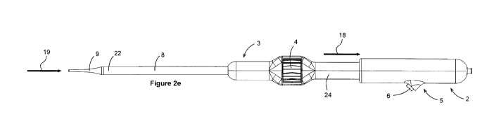
[0038] FIG. 2D depicts the operation of the rapid retraction functionality of
the herein
disclosed trans-apical delivery system 1. By maintaining pressure on the
button 6, the
proximal external handle 2 may be rotated as depicted by the arrow indicating
rotation 17. By
rotating the proximal external handle 2 for one 360 rotation in a first
direction (clockwise
with respect to the operator), the handle can become disengaged from the
middle section of
the internal handle 24 and thus may be free to translate proximally over
internal handle 24, as
depicted by the arrow indicating translation 18 (FIG. 2E). The dilator tip 9
by way of the
connector such as needle hub 12 (FIG. 5), and anchoring catheter 50 (FIG. 5)
by way of the
externally threaded portion of anchoring catheter 51 (FIG. 5) may be mated to
the proximal
external handle 2. The bell catheter proximal end 68 (FIG. 5) may be fastened
to the catheter
carriage 30 (FIG. 5), which may be translated along with the proximal external
handle 2, and
depicted by an arrow indicating translation 19 (FIG. 2E). This movement may
allow the
proximal end of the dilator tip to butt up against the distal end of the
delivery catheter 8 to
form a smooth continuous surface when the delivery system is in a closed
configuration.
-8-
CA 03042588 2019-05-02
WO 2018/090148 PCT/CA2017/051387
[0039] FIGS. 3A-3D more clearly illustrate some of the actuation mechanism.
Turning now
to FIG. 3A, there is illustrated the first view of a sequence of views of an
operational
sequence of a trans-apical delivery system that is configured to allow for
rapid retraction of
the delivery system 1, depicted by way of cross-sectional breakout. The middle
section of the
internal handle 24 is shown, which acts as a support structure for the
proximal external handle
2 to slide thereover. Specifically, an internal circular rib 25 may traverse
the distal-most
portion of the inner diameter of the proximal external handle 2, and in
conjunction with the
external threads 27 of the middle section of the internal handle 24 as well as
an external
circular flange 42, may provide support and location for the middle section of
the internal
handle 24 to translate within the proximal external handle 2 (shown in FIG.
5). In operable
communication with the external threads 27 of the middle section of the
internal handle 24
may be internal threads 28 of the proximal external handle 2, which can allow
for relative
rotation and controlled translation between the two handles without binding or
cocking, prior
to disengagement. An additional feature of embodiments of this device which
may be used in
any embodiment of a delivery system disclosed herein, with specific regards to
the external
threads 27 of the middle section of the internal handle 24 is an internal slot
40 (FIG. 4A), the
details of which will be described further below.
[0040] As previously described, a button 6 may be provided which when pushed
as depicted
by the arrow indicating translation 16 of the button 6, may transmit force and
motion along
the shaft of the button 6, and through a linkage arm 31, thereby applying it
to the catheter
carriage 30 and causing it to translate proximally, as depicted by the arrow
indicating
translation 43 of the catheter carriage 30. Directional control of the
translation of the button 6
may be provided by the button housing 7, which may be cylindrically shaped and
acts as a
piston chamber to guide the similarly cylindrically shaped, piston-like button
6. Functionally,
the combination of button 6, linkage arm 31 and catheter carriage 30 may
behave as a
mechanical linkage. The transmission of force and motion between these
components can be
achieved through pin-and-hole connection of each successive component to the
next; whereas
a plurality of button pins 46 (FIG. 5) on one end of the button 6 may be
concentrically mated
with the distal pin-holes 32 of the linkage arm 31, and a plurality of
catheter carriage pins 47
(FIG. 5) on one end of the catheter carriage 30 may be concentrically mated
with the
-9-
CA 03042588 2019-05-02
WO 2018/090148 PCT/CA2017/051387
proximal pin-holes 33 of the linkage arm 31. The catheter carriage 30 has
several
characteristics that may assist in its ability to translate smoothly without
binding or cocking
within the proximal external handle 2. For example, the catheter carriage 30
may have a
plurality of support bosses 36 (best seen in FIG. 4A) that can allow the
carriage to slide within
the proximal external handle 2 by contacting the inner surface of said
proximal external
handle 2. The catheter carriage 30 may also have a plurality of support fins
35 that can also
assist the sliding of the carriage within the proximal external handle 2 by
contacting the inner
surface of said proximal external handle 2. Additionally, the plurality of
support fins 35 may
also provide locations for the plurality of catheter carriage pins 47 (FIG.
5).
[0041] In order to provide the necessary return force for appropriate valve-
capturing ability
through the distal end of the bell catheter 10, a cylindrical retaining nut 38
may be in contact
with both the catheter carriage 30 and a compression spring 39. This
compression spring 39
can act to push the catheter carriage 30 and bell catheter 10 proximal end 68
and distal end
towards the dilating tip 9 when the button is released due to the bias
provided by the
compression spring 39 causing the bell catheter distal end 10 to slide over
top of the
anchoring catheter anchoring tip 23.
[0042] Continuing on through the sequence of views of an operational sequence
of a trans-
apical delivery system that is configured to allow for rapid retraction of the
delivery system 1,
by turning to FIG. 3B it is shown that further rotation of the proximal
external handle 2 as
depicted by the arrow indicating rotation 17, may allow the external threads
27 of the middle
section of the internal handle 24 and internal threads 28 of the proximal
external handle 2 to
further become disengaged. If this rotation is continued (arrow indicating
continued rotation
41 of the proximal external handle 2, FIG. 3C), the above mentioned threads
may eventually
completely disengage, as illustrated in FIG. 3D. Once the above mentioned
threads are
completely disengaged, the proximal external handle 2 may be free to translate
away from the
distal handle 3 when pulled proximally by an operator, as depicted by the
arrow indicating
translation 18 of the proximal external handle 2. The proximal end of the
dilator tip may now
be butted up against the distal end of the delivery catheter 8 forming a
smooth continuous
outer surface, and all catheters may be nested within one another. This can
complete the rapid
retraction process, whereupon the device can safely be removed from the apex
of a patient's
-10-
CA 03042588 2019-05-02
WO 2018/090148 PCT/CA2017/051387
heart (not shown) or another treatment site. It should be noted that the
internal circular rib 25
located on the distal end of the proximal external handle 2 may acts as a
rigid, physical stop
upon contact with the external circular flange 42 located at the proximal end
of the middle
section 24 of the internal handle. This limits the translation of the proximal
external handle 2
and associated components relative to the internal handle, ensuring the
handles do not become
fully detached from one another.
[0043] As mentioned previously, there is an internal slot 40 (FIG. 4A) located
at the
proximal-most end of the middle section of the internal handle 24. The purpose
of this
internal slot 40 is to provide space wherein a rectangular tab 34 of the
linkage arm 31 may be
placed to prevent unwanted rotation of the proximal external handle 2 relative
to the inner
handle 24 or distal handle 3, and the relationships between these components
is more easily
appreciated when witnessed as depicted in FIG. 4A. The rectangular tab may be
biased to
rest in the slot when the button remains undepressed. One further feature of
the mechanical
linkage defined by the button 6, linkage arm 31 and catheter carriage 30 that
must be
appreciated may be realized by the pressing of the button 6, whereupon the
rectangular tab 34
of the linkage arm 31 becomes fully removed from the internal slot 40, and
full rotation of the
proximal external handle 2 relative to the inner handle 24 is thus enabled.
[0044] Turning now to FIG. 5, there is illustrated an exploded view with
internal components
of a trans-apical delivery system configured to allow for rapid retraction of
delivery system 1.
While many of the elements of FIG. 5 have been previously described herein,
additional
detail will now be given with emphasis to certain elements used to anchor
components within
the handle. The proximal external handle 2 may be comprised of two handle
halves,
specifically an upper section 44 and a lower section 45 which may be fastened
together by
way of commonly used medical device adhesives such as cyanoacrylate UV cure
adhesives
that may be applied to a plurality of pegs 58 for mating of said proximal
external handle
sections 44, 45. Other means for coupling the two handle halves together
include but are not
limited to press fits, screws, ultrasonic welding, etc. The pegs 58 are
illustrated as being
located within the lower section 45 of the proximal external handle 2, and
each peg may have
a complementary boss having an aperture into which it fits in the upper
section 44, although it
is not shown. The relative positions of the pegs and bosses may be transposed.
At the
-11-
CA 03042588 2019-05-02
WO 2018/090148 PCT/CA2017/051387
proximal-most end of each of the sections (upper 44, and lower 45) of the
proximal external
handle 2 there is illustrated a plurality of rectangular slots 60 that may act
to securely locate
and retain the body 59 of the connector such as needle hub 12. Additionally, a
plurality of
pockets 56 for retaining the needle hub flange 57 may be provided in close
proximity to the
plurality of rectangular slots 60, in order to retain and locate a specific
fastening feature of the
needle hub 12, being primarily the needle hub flange 57. Also found within the
upper section
44 and lower section 45 of the proximal external handle 2 may be a plurality
of rectangular
pockets 55, which serve to locate and retain the anchoring nut 48 and also
provide location for
an adhesive bond that secures the anchoring nut into the handle sections. It
will be
remembered that the anchoring nut 48 may provide mechanical fastening and
location of the
anchoring catheter 50 by way of an externally threaded portion 51 on the
anchoring catheter
50 and an internally threaded portion 52 within the anchoring nut 48.
[0045] FIGS. 6A-6E provide illustration of an operational sequence of an
alternate
embodiment of a trans-apical delivery system 1 configured to allow for rapid
retraction. FIG.
6A depicts the first view of an operational sequence, showing another
embodiment of a
proximal external handle 65. In this embodiment of a proximal external handle
65, rapid
retraction may be provided by way of a similar fashion as previously described
herein, but
with alternative means for disengagement of the proximal external handle 65
from another
embodiment of a distal handle section 66. Specifically, the actuator mechanism
in this
embodiment may include latching buttons 49 (FIG. 6A-6C) which may be used to
maintain
this embodiment of the distal handle section 66 coupled to this embodiment of
the proximal
external handle 65. The latching buttons 49 may be in continuous and flexible
connection
with this embodiment of the distal handle section 66, but may be typically
located within a
recess of the proximal external handle embodiment 65. Thus, an interfering
edge 63 of the
latching buttons may be registered against another interfering edge 62 that is
within the
proximal external handle embodiment 65, prior to engagement. As depicted in
FIG. 6D, once
both the latching buttons 49 are depressed (illustrated by arrows 61
indicating
translation/bending of the cantilevered latching buttons 49) the interfering
edge 63 of the
buttons may achieve clearance of the interfering edge 62 of the proximal
external handle 65
by bending flexion (FIG. 6E). Clearance between the components may allow for
translation
-12-
CA 03042588 2019-05-02
WO 2018/090148 PCT/CA2017/051387
of this embodiment of the proximal external handle 65 away from this
embodiment of the
distal handle 66, as depicted by directional arrow 67 indicating translation
of the proximal
external handle embodiment 65 (FIG. 6E). The remaining internal and external
elements of
this embodiment (FIG. 6A-6E) of a trans-apical delivery system 1 may be
configured to
allow for rapid-retraction are as that of the delivery system described in the
'964 patent.
[0046] FIGS. 7A-7D provide illustration of an operational sequence of yet
another alternate
embodiment of a trans-apical delivery system 1 configured to allow for rapid
retraction. FIG.
7A depicts the first view of an operational sequence, showing yet another
embodiment of a
proximal external handle 72. In this embodiment of a proximal external handle
72, rapid
retraction may be provided by way of a similar fashion as previously described
herein, but
with alternative means for disengagement of the proximal external handle 72
from yet another
embodiment of a distal handle section 73. In the embodiment illustrated in
FIG. 7A, a
retaining pin/latch style of handle retention similar to what may be seen in
the modern hand-
grenade may be provided. Specifically, a retaining pin 71 which may be
comprised of a
preferential shaped wire-form having a grasping portion 76 and shafts 75 (FIG.
7C) may be
used to pin a proximal external handle section embodiment 72 to a distal
handle embodiment
73 by disposing the shafts 75 in receiving pin holes 77 (located on the
proximal end of the
distal handle embodiment 73) and pin holes 78 (located on the proximal handle
embodiment
72). A recess 74 (FIG. 7B) in the handle for the retaining pin 71 may provide
a location for
the pin to sit flush with the outer surface of the proximal handle embodiment
72, preventing
the snagging of sterile gloves that may be adorned by the clinical user (not
shown). Operation
of the actuation mechanism here having a retaining pin 71 may be as follows:
after final
deployment of a prosthetic heart valve (not shown) by sustained rotation of
the thumbwheel 4
(FIG. 7B), the user may then grasp the retaining pin 71 and pull it out of the
recess 74 as
depicted by directional arrow 69 indicating translation of the retaining pin
71. Once the
retaining pin shafts 75 are entirely removed from the pin holes 77, 78, the
proximal external
handle embodiment 72 may become free to translate away from the distal handle
embodiment
73 as depicted by directional arrow 70 indicating translation of the proximal
external handle
embodiment 72. The remaining internal and external elements of this embodiment
(FIG. 7A-
-13-
CA 03042588 2019-05-02
WO 2018/090148 PCT/CA2017/051387
7D) of a trans-apical delivery system 1 may be configured to allow for rapid-
retraction are as
that of the delivery system described in '964 patent.
[0047] Prosthesis
[0048] FIG. 8 illustrates a schematic diagram of an exemplary prosthesis 802
which may be
used with any of the delivery catheters disclosed herein. The prosthesis 802
is preferably a
prosthetic valve such as a prosthetic mitral valve, although it may be a
prosthetic valve for
any other region in the body such as a prosthetic triscuspid valve, a
prosthetic aortic valve, or
a prosthetic pulmonary valve. Or it may be a prosthetic venous valve, or any
other prosthetic
valve, or prosthetic device. The prostheses 802 preferably includes an
expandable frame 804
with a prosthetic valve mechanism 806 and preferably includes an anchor
mechanism 808.
The expandable frame may be balloon expandable or self-expanding and the frame
expands
into engagement with the native valve. The prosthetic valve mechanism 806 may
include one,
two, three, or more prosthetic valve leaflets which have an open position
which allows
antegrade fluid flow therepast, and a closed configuration where the
prosthetic valve leaflets
coapt with one another to prevent or minimize retrograde fluid flow therepast.
The fluid may
be blood or another body fluid. The prosthetic leaflets may be pericardial
tissue or other
tissues, or they may be formed from synthetic materials such as polymers or
metals. The
anchor mechanism may be any structure configured to help enagage tissue and
anchor the
prosthesis with the native valve.
[0049] FIGS. 9A-9B illustrates taken along the line A-A in FIG. 8 and show
possible cross-
sections of the frame 804. FIG. 9A shows that the prosthesis may have a
circular cross-
section, and in preferred embodiments, preferably for the mitral valve, the
prosthesis may
have a D-shaped cross-section so that the prosthesis conforms to the native
anatomy.
Additional details about exemplary embodiments of a prosthesis are disclosed
in the '964
patent previously incorporated herein by reference.
[0050] FIGS. 10A-10B illustrate a prosthesis 1008 such as the one described in
FIG. 8
coupled to a delivery catheter 1002. In FIG. 10A, the prosthesis is in a
collapsed
configuration and being carried and constrained by the delivery catheter 1002.
The delivery
catheter 1002 may be any of the delivery catheters described herein. An outer
sheath 1004
constrains the prosthesis 1008 and keeps it in the collapsed configuration and
disposed over
-14-
CA 03042588 2019-05-02
WO 2018/090148 PCT/CA2017/051387
an inner shaft 1006 slidably disposed in the outer sheath 1004. The inner
shaft 1006 may be
any of the inner shafts disclosed herein including the bell catheter
previously disclosed. Other
optional shafts in the delivery catheter are not illustrated for convenience.
As the outer sheath
1004 is retracted proximally, or the bell catheter is advanced distally, the
prosthesis becomes
unconstrained from the outer sheath and begins to self-expand as seen in FIG.
10B. Once the
prosthesis is completely unconstrained, is self-expands into position,
preferably into
engagement with a native valve.
[0051] Delivery
[0052] FIG. 11 illustrates basic human heart anatomy. The heart includes four
chambers, the
right atrium RA, the right ventricle RV, the left atrium LA, and the left
ventricle LV. Several
valves prevent retrograde blood flow. The tricuspid valve TV controls flow
from the right
atrium to the right ventricle, and the pulmonary valve PV controls flow out of
the right
ventricle RV. The mitral valve MV controls flow between the left atrium LA and
the left
ventricle LV, and the aortic valve AOV controls flow out of the aorta AO. The
major vessels
coupled to the heart include the vena cava VC which brings venous blood back
to the right
atrium RA, and the pulmonary artery brings blood from the right ventricle RV
to the lungs
(not illustrated). Oxygenated blood from the lungs returns to the left atrium
LA via the
pulmonary veins PVE, and blood is delivered out of the left ventricle LV to
the body by the
aorta AO.
[0053] FIG. 12A illustrates one exemplary delivery method for treating mitral
valve MV. In
this embodiment, the delivery catheter C which may be any of the delivery
devices disclosed
herein and may have any of the prostheses disclosed herein is advanced
typically from a
femoral vein in the groin up into the vena cava VC into the right atrium RA
and then
transseptally across the atrial septal wall into the left atrium LA and then
downward into
disposition across or adjacent the native mitral valve MV where the prosthesis
may be
deployed as described herein.
[0054] FIG. 12B illustrates another exemplary delivery method for treating a
mitral valve
MV. In this embodiment, the delivery catheter C which may be any of the
delivery devices
disclosed herein and may have any of the prostheses disclosed herein is
advanced typically
from a femoral artery or other artery (e.g. radial artery) up into the aorta
AO in to the left
-15-
CA 03042588 2019-05-02
WO 2018/090148 PCT/CA2017/051387
ventricle LV and then across the mitral valve MV or adjacent thereto for
deployment of the
prosthesis as described herein.
[0055] FIG. 12C illustrates another exemplary delivery method for treating a
mitral valve
MV. In this embodiment, the delivery catheter C which may be any of the
delivery devices
described herein and may have any of the prostheses disclosed herein is
typically advanced
transapically from outside the body, through the chest well, into the apex of
the heart into the
left ventricle LV and then adjacent or across the mitral valve MV where the
prosthesis is then
deployed as disclosed herein.
[0056] FIGS. 13A-13C illustrate an exemplary method of deploying a prosthesis
P in the
heart using a delivery catheter C which may be any of the delivery devices
disclosed herein.
The prosthesis is preferably a mitral valve prosthesis but may be any of the
prostheses
disclosed herein. In FIG. 13A, the delivery catheter is preferably delivered
transapically to
the mitral valve MV. In FIG. 13B, once the prosthesis P has been properly
positioned
relative to the native mitral valve MV, the outer sheath is retracted
proximally (or the inner
bell shaft is advanced distally) so that the prosthesis is unconstrained and
allowed to self-
expand into engagement with the native mitral valve and anchor into position.
After the
prosthetic valve has been deployed and properly positioned and anchored, the
delivery
catheter is then retracted proximally and removed from the heart as seen in
FIG. 13C. The
prosthetic valve now takes over the function of the native mitral valve
allowing antegrade
flow from the left atrium to the left ventricle and preventing or minimizing
regurgitation of
blood from the left ventricle to the left atrium.
[0057] While preferred embodiments of the present invention have been shown
and described
herein, it will be obvious to those skilled in the art that such embodiments
are provided by
way of example only. Numerous variations, changes, and substitutions will now
occur to
those skilled in the art without departing from the invention. It should be
understood that
various alternatives to the embodiments of the invention described herein may
be employed in
practicing the invention. It is intended that the following claims define the
scope of the
invention and that methods and structures within the scope of these claims and
their
equivalents be covered thereby.
-16-