Note: Descriptions are shown in the official language in which they were submitted.
CA 03043325 2019-05-08
WO 2018/089789 PCT/US2017/061099
- I -
SYSTEM, METHOD AND BIOMARKERS FOR AIRWAY OBSTRUCTION
FIELD OF THE INVENTION
The present invention relates to the field of systems and methods for
detecting obstructive
apnea or dyspnea, and biomarkers for obstructive apnea or dyspnea.
BACKGROUND OF THE INVENTION
Airway obstruction can be a critical health emergency, leading to death within
minutes.
Partial obstruction is also possible.
Apnea is suspension of breathing. During apnea, the volume of the lungs
initially remains
unchanged. Depending on how blocked the airways are (patency), there may or
may not be a
flow of gas between the lungs and the environment; gas exchange within the
lungs and cellular
respiration is not acutely affected.
In obstructive apnea, breathing is attempted, which causes increased
activation of the
diaphragm and other muscles of respiration, including the intercostal muscles.
After a few
minutes of prolonged apnea, blood oxygen falls, and various secondary
responses occur.
Epileptic seizure is associated with obstructive apnea. Seizure activity
spreads to laryngeal
motor neurons to cause laryngospasm. Laryngospasm results in partial or
complete airway
occlusion. Seizure activity changes breathing frequency, amplitude,
variability, and can cause
central apnea. Only obstructive apnea was associated with rapid, severe
arterial oxygen
desaturation, bradycardia, and death. Sudden death is the result of
respiratory arrest during
airway obstruction and nearly simultaneous LV dilatation and asystole. Sudden
death in epilepsy
can be the result of seizure induced laryngospasm sufficient to cause
obstructive apnea, which
leads to respiratory arrest and cardiac asystole within tens of seconds.
The recurrent laryngeal nerve (RLN) is a branch of the vagus nerve (cranial
nerve X) that
supplies all the intrinsic muscles of the larynx, with the exception of the
cricothyroid muscles.
These muscles act to open and close the vocal cords, and include the posterior
cricoarytenoid
muscles, the only muscle to open the vocal cords. The nerves supply muscles on
the same side of
the body, with the exception of the interarytenoid muscle, which is innervated
from both sides.
See, en.wikipedia.org/wiki/Recurrent_laryngeal_nerve. The recurrent laryngeal
nerves supply
sensation to the larynx below the vocal cords, gives cardiac branches to the
deep cardiac plexus,
and branches to the trachea, esophagus and the inferior constrictor muscles.
The posterior
cricoarytenoid muscles, the only muscles that can open the vocal cords, are
innervated by this
nerve. The nerves also carry sensory information from the mucous membranes of
the larynx
CA 03043325 2019-05-08
WO 2018/089789 PCT/US2017/061099
- 2 -
below the lower surface of the vocal fold, as well as sensory, secretory and
motor fibers to the
cervical segments of the esophagus and the trachea.
The MORTality in Epilepsy Monitoring Unit Study (MORTEMUS) identified a
consistent
sequence of events in epilepsy patients beginning with a generalized tonic
clonic seizure and
.. ending in death lRyvlin et al., Lancet Neurol. 12:966, 2013]. Ten cases
were used to establish
that the end of the seizure was followed within minutes by terminal apnea and
ultimately cardiac
arrest. Most importantly, this study established a singular pattern for their
SUDEP cases.
U.S. 5,800,470, expressly incorporated herein by reference, discloses a
respiratory muscle
electromyographic rate responsive implantable pacemaker. The directly detected
electromyogram (EMG) signal is amplified and band passed filtered, processed
to remove any
electrocardiogram (ECG) or pacing impulse signal, full-wave rectified,
processed to develop a
moving time average signal from which the peak, the maximal slope, and the
average slope of
the EMG moving time average may be calculated and processed in conjunction
with the
inspiratory and expiratory times between successive slope detections of the
moving time average
EMG to develop a rate control signal representative of ventilation rate. The
EMG may be
selectively picked up from electrodes implanted in or near the parastemal
intercostal muscles, the
external intercostal muscles, the internal intercostal muscles, the diaphragm,
or any other
respiratory muscle such as the scalenes, or the stemocleidomastoid, and
coupled to
conventionally designed or special configuration pacemaker pulse generators
and cardiac
pace/sense lead systems.
U.S. 4,961.423, expressly incorporated herein by reference, proposes to employ
specific
electromyogram or EMG (a graph of electrical signals associated with muscle
activity) signal
processing circuitry in conjunction with a conventional cardiac pacing lead
system to derive a
control signal which reflects the patient's respiration as reflected across
the electrodes in contact
with the patient's heart. By use of specific filtration and signal processing,
it is proposed to
separate the EMG signal from the electrocardiogram (ECG) signal and pacing
stimulation
impulse from the aggregate signal picked up across the pacing tip and can
electrode pair or
across separate electrodes devoted to the detection of the EMG.
Getzel et al., "Variation of Cardiac Pacemaker Rate Relative to Respiration,"
IEEE
Proceedings of 32nd CEMB, 1979, p. 123, and "Variation of Cardiac Pacemaker
Rate Relative to
Respiration," IEEE Trans. on Biomed. Eng., Vol. BME-26, No. 9, September 1979,
p. 526.,
expressly incorporated orated herein by reference, describe the electronic
integration of the
CA 03043325 2019-05-08
WO 2018/089789 PCT/US2017/061099
- 3 -
diaphragm electromyogram to generate a control signal proportional to
respiratory minute
volume for use as the controlling physiological input for a pacemaker.
US 2016/0089540, expressly incorporated herein by reference, a method of
treating a patient,
comprising: sensing a biological parameter indicative of respiration;
analyzing the biological
parameter to identify a respiratory cycle; identifying an inspiratory phase of
the respiratory
cycle; and delivering stimulation to a hypoglossal nerve of the patient,
wherein stimulation is
delivered if a duration of the inspiratory phase of the respiratory cycle is
greater than a
predetermined portion of a duration of the entire respiratory cycle.
It is thus known that there is a respiration artifact in the ECG signal. It is
also known that the
intrinsic ECG signal is respiratory responsive, including R-R interval.
Nakase et al., "Laryngospasm, central and obstructive apnea during seizures:
Defining
pathophysiology for sudden death in a rat model, Epilepsy Research, Volume
128, 126 ¨ 139
(Dec. 2016), DOI:dx.doi.org/10.1016/j.eplepsyres.2016.08.004; www.epires-
journal.com/article/50920-1211(16)30124-3/abstract, describes the
pathophysiology of sudden
death in epilepsy using an animal model, and has several figures that
illustrate laryngospasm,
obstructive apnea, desaturation during obstructive apnea, direct measures of
the forces developed
during attempts to inspire against a closed airway, and evidence of artifacts
in ECG records.
Seizure spread into the autonomic nervous system is thought to play an
important role in
sudden unexpected death in epilepsy (SUDEP; (Bermeo-Ovalle et al., 2015;
Devinsky, 2011;
Lathers et al., 2008; Sakamoto et al., 2008; Shorvon and Tomson. 2011;
Stewart, 2011; Surges
and Sander, 2012; Tolstykh and Cavazos, 2013)). Approximately 1% of the US
population lives
with epilepsy; depending on how one defines sudden death, 2%-17% of deaths in
these patients
are labeled SUDEP (e.g. (Nei and Hays, 2010)). Among adults with epilepsy,
mortality rates are
2-3 times greater than among their non-epileptic counterparts (Langan, 2000;
Thurman et al.,
2014), and SUDEP is the single most common cause of death (Lathers et al.,
1998; Wannamaker,
1985).
Seizures are known to produce significant respiratory changes (reviewed in
(Massey et al.,
2014; Sowers et al., 2013)). fetal apnea (Blum, 2009) is implicated in oxygen
desaturation during
seizures (Bateman eta]., 2008; Seyal et al., 2010). Indeed, animal research
established the
importance of ictal hypoxemia in seizure-induced death, as studies in sheep
have shown that ictal
hypoventilation leads to severe bradycardia and death (Johnston et al., 1995;
Johnston et al.,
1997). Similar findings have been noted in rats (Sakamoto et al., 2008;
Stewart, 2011), cats
(Schraeder and Lathers, 1983), and mice (Faingold et al., 2010; Uteshev et
al., 2010). The
CA 03043325 2019-05-08
WO 2018/089789 PCT/US2017/061099
- 4 -
physiological mechanisms, however, that link seizures to respiratory
dysfunction have not been
fully resolved.
One possible cause of ictal respiratory distress is laryngospasm, a tonic
adduction of the
vocal folds that partially or fully obstructs the upper airway. Laryngospasm
has been observed
during seizures or postictally, evidenced by stridor and a narrowed airway
when attempting to
place an endotracheal tube (Tavee and Morris, 2008) or intensive inspiratory
effort with severe
air hunger (Amir et al., 1983). Cats and piglets experienced hypoventilation
and glottal
obstruction during chemically-induced seizures (Learning et al., 1999;
Terndrup et al., 1995a;
Terndrup et al., 1995b). That pulmonary edema is the most common single
finding at autopsy in
SUDEP cases is also indirect evidence of laryngospasm (Antoniuk et al., 2001;
Morentin and
Alcaraz, 2002; Salmo and Connolly, 2002). Pulmonary edema can occur when
"pulling" against
a closed airway ¨ the inspiratory effort increases pulmonary capillary
pressure (Ead, 2003;
Murray-Calderon and Connolly, 1997; Umbrain and Camu, 1993). Seizures could
cause ictal
laryngospasms by spreading via autonomic medullary motor regions to the
laryngeal branches of
the vagus nerve, the efferent innervation of the vocal folds.
A urethane/kainate rat model (reviewed in (Naggar and Stewart, 2015; Stewart,
2011)) was
used to permit detailed study of laryngospasm during seizure activity. This
model allows
invasive monitoring during seizure activity. Recordings are obtained from the
recurrent laryngeal
nerve, the principal motor output to the larynx (Bartlett, 2011; Brancatisano
et al., 1991; Kuna et
al., 1991; Kuna et al., 1988; Kuna et al., 1990), along with simultaneous
laryngoscopy (Mor et al.,
2014) to define the patterns of RLN activity during seizures, the impact of
seizure activity on
laryngeal function, and the impact of laryngeal dysfunction on breathing.
These data highlight
the complexity of laryngospasm during seizures, and how changes in laryngeal
function can
contribute to death.
In order to monitor heart signals in an ambulatory environment, a number of
options are
available. Bioelectric signals may be acquired from the chest wall, limbs, and
digits. Heart rate
and pulse variability can also be acquired using pulse information, which can
be acquired by
plethysmography and optical sensors on the skin, wrist, ankle, and digits.
See:
www.vitalconnect.com/upload/Documents/EngeryExpenditure2014_MobiHealth_publishe
d.pdf;
www.vitalconnect.com/upload/Documents/Longterm-Remote-
Monitoring_HealthInnovations_2014_published.pdf;
www.vitalconnect.com/upload/Documents/AutomatedPrediction_2014_IEEE_published.p
df;
CA 03043325 2019-05-08
WO 2018/089789 PCT/US2017/061099
- 5 -
www.vitalconnect.com/upload/Documents/2014-Sleep-Abstract.pdf;
www.vitalconnect.com/upload/press/Chan2013EMBC_VitalConnectPatch.pdf;
www.vitalconnect.com/upload/press/Selvaraj2013EMBC_OSAeventDetectionRespiratory
Signals.pdf;
www.vitalconnect.com/upload/press/Chan2013EMBC_RespirationECGandAccelerometer.p
df;
Rosenberg M., Samuel M., Thosani A., Zimetbaum P., "Use of a noninvasive
continuous
monitoring device in the management of atrial fibrillation: a pilot study",
Pacing Clin
Electrophysiol. 2013;36(3): 328-333.
See also, U.S. Patent and Pub. Patent App. Nos. 3942513; 3950799; 4033332;
4169462;
4197856; 4289142; 4350166; 4381788; 4387722; 4391279; 4403215; 4422458;
4446869;
4474185; 4475559; 4506626; 4558708; 4595016; 4630614; 4657026; 4686975;
4694839;
4715367; 4724844; 4732159; 4736749; 4745925; 4757824; 4757825; 4765340;
4802485;
4802486; 4803997; 4806112; 4838279; 4889116; 4895162; 4924860; 4928692;
4928703;
4958638; 4961423; 4982738; 5005234; 5005571; 5016636; 5036852; 5052400;
5095900;
5099836; 5107855; 5131387; 5133346; 5206807; 5271412; 5295490; 5307817;
5309921;
5311875; 5353793; 5360008; 5395301; 5485850; 5495242; 5603316; 5645053;
5671733;
5704345; 5765554; 5769084; 5779631; 5782240; 5792068; 5800360; 5800470;
5825293;
5853005; 5873821; 5879313; 5913826; 5928157; 5954053; 5961447; 5964720;
6017315;
6019732; 6029665; 6045514; 6062216; 6064910; 6134460; 6138675; 6150104;
6150941;
6168568; 6208897; 6223064; 6241683; 6248068; 6261238; 6267730; 6286508;
6290654;
6342039; 6342040; 6363933; 6368287; 6375621; 6390987; 6450168; 6454724;
6470888;
6477710; 6498652; 6510339; 6537228; 6544192; 6549795; 6550478; 6553242;
6553256;
6574507; 6580943; 6580944; 6621278; 6675797; 6721980; 6773404; 6785568;
6816266;
6849049; 6856141; 6881192; 6915705; 6920877; 6970737; 6984993; 6989744;
7020511;
7035432; 7039152; 7074177; 7080554; 7092755; 7117036; 7126467; 7129833;
7148797;
7166123; 7168429; 7170404; 7173525; 7179229; 7225021; 7269459; 7315760;
7320320;
7330127; 7340302; 7363086; 7391316; 7403110; 7415093; 7431700; 7435221;
7460899;
7467012; 7473227; 7477142; 7477143; 7477144; 7508307; 7515059; 7522035;
7533571;
7593764; 7597659; 7611472; 7656287; 7661426; 7667624; 7678058; 7711579;
7715905;
7716767; 7725181; 7730886; 7734335; 7794716; 7800505; 7811234; 7818058;
7827988;
7884735; 7894849; 7899521; 7909764; 7938114; 7942822; 7976470; 7988640;
8011365;
8031080; 8050765; 8106781; 8119134; 8121692; 8136521; 8142343; 8155735;
8161971;
8204580; 8219185; 8226570; 8226571; 8238996; 8255029; 8258973; 8262578;
8273053;
8301219; 8301232; 8360060; 8360983; 8381722; 8393233; 8396537; 8403861;
8417351;
CA 03043325 2019-05-08
WO 2018/089789 PCT/US2017/061099
- 6 -
8442578; 8449473; 8482418; 8483807; 8483811; 8483834; 8489182; 8509882;
8527028;
8538510; 8551010; 8554323; 8560044; 8562526; 8566115; 8568160; 8579792;
8595164;
8616203; 8630704; 8641631; 8683999; 8700137; 8718751; 8721554; 8721560;
8721573;
8731644; 8750987; 8752547; 8768731; 8790270; 8801620; 8828386; 8844525;
8862211;
8868152; 8880207; 8892194; 8923971; 8948854; 8954137; 8983587; 8985106;
9019100;
9022032; 9024781; 9026202; 9037477; 9044362; 9078577; 9089691; 9101277;
9113788;
9131892; 9132250; 9144389; 9177459; 9180270; 9192336; 9198616; 9198617;
9199053;
9202084; 9215075; 9216291; 9220459; 9220460; 9227034; 9269000; 9277867;
9302116;
9307921; 9333318; 9364180; 9370634; 9414787; 9415182; 9445736; 9445740;
9445747;
9451888; 9468835; 9477812; D284697; RE32180; 20010018557; 20010044588;
20020007126;
20020078957; 20020099300; 20020105340; 20020161290; 20020173707; 20030024528;
20030111079; 20030161436; 20030161440; 20030163059; 20030199945; 20030208130;
20030213488; 20030236548; 20040002742; 20040082874; 20040104733; 20040123866;
20040158193; 20040186523; 20040187870; 20040194220; 20040207409; 20040257233;
20050027204; 20050027206; 20050053262; 20050085736; 20050085863; 20050085865;
20050101833; 20050113656; 20050177051; 20050211248; 20050211249; 20050273361;
20050277842; 20050288572; 20060000475; 20060004245; 20060011200; 20060025696;
20060025697; 20060030894; 20060050930; 20060087325; 20060122675; 20060134106;
20060179571; 20060229489; 20060258916; 20060258921; 20060264770; 20070061393;
20070062540; 20070073177; 20070100381; 20070106536; 20070106537; 20070106750;
20070106751; 20070106752; 20070106753; 20070106754; 20070116036; 20070116037;
20070142713; 20070142741; 20070167694; 20070168461; 20070199262; 20070215146;
20070255310; 20070265539; 20070282212; 20080009755; 20080015454; 20080015457;
20080039730; 20080040151; 20080041382; 20080041383; 20080045832; 20080051845;
20080058873; 20080058892; 20080060138; 20080071185; 20080082016; 20080091082;
20080092898; 20080101532; 20080154110; 20080161708; 20080163873; 20080167567;
20080170654; 20080177789; 20080183095; 20080183239; 20080190430; 20080190436;
20080243021; 20080262360; 20080287769; 20080287770; 20080288013; 20080300499;
20080308112; 20080313816; 20080319513; 20090036790; 20090099462; 20090099469;
20090118629; 20090149778; 20090172773; 20090177050; 20090177495; 20090183312;
20090203972; 20090226861; 20090226862; 20090226863; 20090226864; 20090226865;
20090270773; 20090318820; 20090326981; 20100004549; 20100010359; 20100016783;
20100018530; 20100036209; 20100036263; 20100036268; 20100036269; 20100056850;
CA 03043325 2019-05-08
WO 2018/089789
PCT/US2017/061099
- 7 -
20100063438; 20100099963; 20100137723; 20100159426; 20100168578; 20100179396;
20100198083; 20100198289; 20100204550; 20100222685; 20100222689; 20100228120;
20100242965; 20100252037; 20100252039; 20100252040; 20100252041; 20100252042;
20100253505; 20100258123; 20100262035; 20100268093; 20100297129; 20100307500;
20100328075; 20110009722; 20110011402; 20110021970; 20110034820; 20110105921;
20110131057; 20110160601; 20110172552; 20110184298; 20110184304; 20110190651;
20110214676; 20110245706; 20110284003; 20110301435; 20110301439; 20110319777;
20120004749; 20120028504; 20120029362; 20120032778; 20120046709; 20120056746;
20120088998; 20120101690; 20120109027; 20120130204; 20120130446; 20120136231;
20120152252; 20120160243; 20120165711; 20120172689; 20120173470; 20120189632;
20120197144; 20120203491; 20120209096; 20120232398; 20120232416; 20120240934;
20120252709; 20120272429; 20120283527; 20120323104; 20120330123; 20130012827;
20130030711; 20130046151; 20130046156; 20130046157; 20130046160; 20130046161;
20130046184; 20130046185; 20130046186; 20130046187; 20130046188; 20130053674;
20130060480; 20130081479; 20130085688; 20130096447; 20130096450; 20130123654;
20130131522; 20130165809; 20130174847; 20130178719; 20130211210; 20130218030;
20130234535; 20130255683; 20130261485; 20130268030; 20130281815; 20130291060;
20130296660; 20130300575; 20130307685; 20130312752; 20130312757; 20130331663;
20130340500; 20140012145; 20140018779; 20140025141; 20140031705; 20140031709;
20140058274; 20140066741; 20140066798; 20140073969; 20140094669; 20140100628;
20140107501; 20140150793; 20140152467; 20140163343; 20140163386; 20140171783;
20140180148; 20140180154; 20140218210; 20140221859; 20140228651; 20140243934;
20140246024; 20140246025; 20140258743; 20140276287; 20140309568; 20140309943;
20140320309; 20140323946; 20140330155; 20140330540; 20140363740; 20140364706;
20150005646; 20150018660; 20150035643; 20150038867; 20150039110; 20150065891;
20150073285; 20150101609; 20150112202; 20150119739; 20150126847; 20150141766;
20150141862; 20150150500; 20150173672; 20150182132; 20150182842; 20150216448;
20150228176; 20150251016; 20150257653; 20150305634; 20150314098; 20150320588;
20150321022; 20150359964; 20150366511; 20150370320; 20150374328; 20160004820;
20160022145; 20160022204; 20160030689; 20160045134; 20160045166; 20160045167;
20160045654; 20160045695; 20160066805; 20160066809; 20160074606; 20160089540;
20160095997; 20160100798; 20160120430; 20160120431; 20160120433; 20160120434;
20160128209; 20160135706; 20160143594; 20160169930; 20160174857; 20160220197;
CA 03043325 2019-05-08
WO 2018/089789
PCT/US2017/061099
- 8 -
20160228024; 20160228060; 20160256063; 20160256644; 20160263393; 20160278658;
20160296124; 20160302726; and 20160310103.
A fingertip electrometer-based cardiac cycle sensors is disclosed in US
2012/0004523.
CA 03043325 2019-05-08
WO 2018/089789
PCT/US2017/061099
- 9 -
SUMMARY OF THE INVENTION
Seizures are known to produce significant respiratory changes and seizure
spread into the
autonomic nervous system can result in life-threatening cardiovascular and
respiratory
dysfunction. Ictal apnea and/or ictal bradycardia has been well recognized as
a part of the
autonomic manifestation in epileptic seizures. Prolonged peri-ictal apnea and
bradycardia are
both regarded as risk factors for sudden death in epilepsy (SUDEP). SUDEP is
the major cause
of death among persons with epilepsy. However, the physiological mechanisms of
SUDEP are
poorly understood and no specific indicator of SUDEP events is known. One
possible cause of
ictal respiratory distress is laryngospasm, a tonic adduction of the vocal
folds that partially or
fully obstructs the upper airway. Using a rat model, sudden death due to
seizure and hypoxemia-
induced conditions was studied. Based on findings of the inventors, some
seizures cause
laryngospasm that is sufficiently severe to produce complete airway
obstruction. Once occluded,
attempts to inspire against a closed airway get progressively stronger until
attempts stop (the
point of respiratory arrest). These attempts produce clear artifacts in
recordings of
electrocardiogram (ECG) and electroencephalogram (EEG) signals whose
amplitudes highly
correlate with the force of attempted inspiration. Late in the occlusion, the
RR interval variability
is dramatically increased due to an overall slower heart rate in combination
with additional very
short RR intervals closely associated with attempts to inspire.
Artifacts in the ECG and EEG during obstructive apnea caused by laryngospasm
correspond
in time and correlate in size with a direct measure of inspiratory effort in
experimental animals.
Likewise, these inspiration efforts cause strong electromyography (EMG)
signals from muscles
of respiration, including diaphragm and intercostal muscles while the
resulting hypoxemia leads
to bradycardia and an abrupt increase in heart rate variability with very
short RR intervals at the
time of each attempted inspiration.
R waves in ECG can be automatically identified through RR interval analyses
and artifact
detection and quantification from ECG and EEG records.
These physiological effects detected by these signals and analyzed can be used
as practical
biomarkers of obstructive apnea (e.g. laryngospasm). Two particular biomarkers
that are specific
for upper airway occlusion include:
- a high frequency EMG signal superimposed on the ECG signal
- a variation in R-R wave intervals
The high frequency signal has an amplitude that corresponds to inspiratory
effort, and
therefore by monitoring respiration artifacts over time, an adaptive baseline
may be established.
CA 03043325 2019-05-08
WO 2018/089789 PCT/US2017/061099
- 10 -
When an obstructive apnea occurs, the respiratory artifacts are altered in a
distinctive way. The
amplitude increases on successive attempts, and the timing of these attempts
differs from a
normal respiratory rate. Both the high frequency signal and the variation in R-
R wave intervals
are responsive to obstructive apnea and indicative of an apnea activity
pattern of muscles of
respiration, including diaphragm and intercostal muscles.
Because these biomarkers do not require ECG analysis per se, they may be
detected from
electrodes in non-standard locations for cardiac monitoring, such as fingers
or wrist. As such, the
monitoring device may take the form of a wrist-band, ring(s), or other
convenient form. Of
course, traditional chest electrodes may also be employed.
The R-R interval is the basic heart rate, and therefore the rate and its
variability can be
determined in an alternate manner, e.g., without electrocardiographic
electrodes. For example,
physical or optical pulse sensors, acoustic sensors, ballistocardiographic
sensors, etc.
On the other hand, the high frequency electromyographic signal from muscles of
respiration,
including diaphragm and intercostal muscles, superimposed on the
electrocardiographic signal
.. would generally require an electronic sensor for detection. However, other
types of respiratory
sensors and detection may be employed, though when directly measuring
respiration, the need
for a biomarker or indirect measurement for apnea is diminished.
The combination of these biomarkers clearly indicates when a person's
breathing is
obstructed, attempting to breathe, and generating large breathing forces in
these attempts. An
alarm sounded at this point to alert a caretaker will permit enough time to
ensure that respiratory
arrest does not occur or that, if respiratory arrest does occur, resuscitation
steps can be taken to
save a life. These biomarkers can also be applied to past cases and used to
monitor patients to
improve outcomes.
These biomarkers may have application in various types of obstructive apnea.
While a
.. preferred system and method target ictal obstructive apneas, asthmatic
conditions may produce
similar biomarkers. Thus, when an asthmatic attack occurs, airways are
restricted, leading to
reduced chest pressure and large inspiratory efforts. Asthmatic apnea tends to
be an incomplete
blockage, and therefore the pattern over time may differ from a laryngospasm-
induced apnea, but
the biomarkers are sufficiently broad to permit application in various uses.
In the case of asthma, one might seek to determine the extent of blockage,
which is not
always directly apparent, especially in exercise induced-asthma, where
increased demand is
superimposed on the airway restriction. However, the restriction will increase
the efforts
CA 03043325 2019-05-08
WO 2018/089789 PCT/US2017/061099
- 11 -
required, and increase the pressure differentials, and thus the asthmatic
restriction may be
distinguished from the mere increased respiratory rate due to exertion.
Based on these biomarkers, a system and method is provided that can detect the
period of
obstructive apnea and be used to sound an alarm in time to prevent respiratory
arrest or in time to
permit resuscitation.
A particular aspect of the system and method is the extraction of one or both
of the biomarkers from
ECG data, EEG data, or other bioelectric signals. The data used for biomarker
extraction can thus come
from multiple sources. In circumstances where ECG data or EEG data is already
collected and available
for analysis, e.g. any continuous ECG recording or EEG recording in a hospital
setting, such as that used
in Critical Care Units, Epilepsy Monitoring Units, etc., the biomarker
identification algorithms can be
added to the existing instrumentation. In an ambulatory or home setting, ECG
can be obtained by a
minimally intrusive "bracelet" such as those used for popular HR monitoring,
with the exception that a
telemetry component would generally be added to the bracelet and the receiving
station, e.g., smartphone,
would house the biomarker detection software and the hardware used for the
alarm. A hat or scalp
monitor with electrodes can also provide EEG data.
Of course, the data analysis can be provided within the sensor module, and an
audible and/or visual
alarm sounded from the module. Sensing obstruction may incur a latency, of
approximately 10-30
seconds, and the time before permanent damage occurs to the patient is only a
few minutes, providing
only a small window of opportunity to prevent a complete laryngeal obstruction
of the patients airway,
and therefore a local caregiver would need to provide immediate assistance,
and remote monitoring would
likely be ineffective. However, within a hospital or other facility, a remote,
wireless alarm may he useful.
Similarly, in cases of incomplete obstruction, such as bronchial constriction,
the onset and resolution of
the apnea provide a larger window of opportunity for intervention.
Biomarker extraction involves taking the ECG signal and processing it in
different ways for each of
the two biomarkers.
BIOMARKER
The algorithm for biomarker 1 (Artifact Growth) involves the following steps
applied to ECG
recorded with a bandwidth of approximately 10 Hz to > 1 kHz:
1. Secondary filter applied to data to pass approximately 300 Hz to 1 kHz.
2. Detect and measure breathing artifacts by methods such as rectification and
integration or signal
"envelope" quantification.
3. Compare values to amplitude threshold.
4. Hold value and time of events above threshold.
5. Compare interval between successive events with window established for
respiratory rate.
6. Sound alarm if:
= a) 3 successive events are above threshold, and
CA 03043325 2019-05-08
WO 2018/089789 PCT/US2(i17/061099
- 12 -
= b) the interval between events is appropriate for respiratory rate, and
= c) the event amplitude is steady or increasing.
More generally, a bioelectric signal is obtained which includes a contribution
from activity of
muscles of respiration, including diaphragm and intercostal muscles activity.
The bioelectric
.. signal is processed to represent amplitude and timing of inspiratory
efforts. The respiratory rate
is compared with a normal pre-detection rate, and the amplitude of the
bioelectric signal is
compared with a pre-detection normal amplitude. The obstructive apnea is
likely present if a
series of inspiratory efforts are above a normal amplitude and with increasing
amplitude, but at a
normal rate.
BIOMARKER 2
The algorithm for biomarker 2 (Ultrashort RR Intervals) involves analysis of
the acquired ECG signal
with the following steps:
1. Detect R waves.
2. Measure RR intervals.
3. Compare interval to baseline range.
4. If ultrashort interval detected (RR interval is below threshold), store
value and time of event.
5. Immediately successive short intervals are stored as a single event.
6. Compare time between successive events to the window established for
respiratory rate.
7. Sound alarm if:
= a) 3 sets of short intervals are spaced by the respiratory interval.
More generally, the heart rate is determined, and compared to a baseline
average. A normal lower
threshold is established, and if subthreshold events occur (short RR
intervals), a commencement of each
sequence of subthreshold events is compared for a respiratory rate-normalized
window. If the number of
subthreshold events exceeds a minimum for the window, obstructive apnea is
likely present.
The technology may be implemented in any device that receives a bioelectric
signal that includes
electromygraphic signals emanating from muscles of respiration. For example,
an automated external
defribrillator (AED) device may be provided with program instructions that
permit the ECG electrodes to
read the electromygraphic signals, and provide obstructive apnea indication,
in addition to the normal
defribrillator functionality. As noted, the present system seeks to compare a
current bioelectric signal
with a baseline signal, which may not be available in an acute emergency.
Likewise, the AED tends to be
employed with a human user in attendance, who can observe the patient.
However, the user may be
untrained, and therefore automatically monitoring the patient for apnea, and
to distinguish different types
of apnea, may be useful, especially for differential diagnosis where a patient
hooked to the AED has a
normal sinus rhythm, and yet is in distress.
CA 03043325 2019-05-08
WO 2018/089789
PCT/US2017/061099
- 13 -
It is therefore an object to provide a method for detecting obstructive apnea,
comprising:
receiving a bioelectric signal from a mammal comprising electromyographic
activity of muscles
of respiration, including diaphragm and intercostal muscles; processing the
bioelectric signal to
isolate the electromyographic activity; determining a timing and amplitude of
inspiratory efforts
based on the isolated electromyographic activity; determining a baseline
amplitude of inspiratory
efforts; comparing an amplitude of inspiratory efforts with the determined
baseline amplitude of
inspiratory efforts; and determining occurrence of obstructive apnea if a
series of inspiratory
efforts have increasing amplitude over time, above the baseline amplitude.
The method may further comprise determining a baseline timing of inspiratory
efforts, and
comparing the timing of inspiratory efforts with the determined baseline
timing of inspiratory
efforts, wherein the occurrence of obstructive apnea is determined if a series
of inspiratory
efforts have increasing amplitude above the baseline amplitude over time, and
a baseline timing.
The timing and amplitude of inspiratory efforts may be determined over a
series of three
inspiratory efforts before the occurrence of obstructive apnea is determined.
The bioelectric signal may be an electrocardiographic signal. The bioelectric
signal may be
an electroencephalographic signal. The bioelectric signal may be acquired from
a single
extremity.
The method may further comprise generating an audible alarm in response to
determining the
occurrence of obstructive apnea. The method may further comprise generating a
wireless
communication in response to determining the occurrence of obstructive apnea.
The bioelectric signal may be received from a mammal comprising
electromyographic
activity of muscles of respiration, including diaphragm and intercostal
muscles comprises
receiving at least one of an electrocardiographic signal, an
electroencephalographic signal, and
an electromyographic signal. The processing of the bioelectric signal may be
used to isolate
electromyographic activity comprises subjecting the bioelectric signal to a
bandpass filter having
a passband between about 300 Hz and 1 kHz. The processing of the bioelectric
signal may be
used to isolate electromyographic activity comprises determining a signal
power within a
passband over time.
The comparing of an amplitude of inspiratory efforts with the determined
baseline amplitude
.. of inspiratory efforts may comprise comparing a series of amplitudes and
timings of inspiratory
efforts with a baseline window representing a normal range of amplitudes and
timings of
inspiratory efforts.
CA 03043325 2019-05-08
WO 2018/089789 PCT/US2017/061099
- 14 -
The occurrence of obstructive apnea may be determined if a series of
inspiratory efforts have
increasing amplitude over time, above the baseline amplitude, comprises
determining if three
successive inspiratory efforts have an amplitude above a threshold with at
least one of a steady
amplitude and an increasing amplitude, while an interval between inspiratory
efforts is within a
normal range.
It is also an object to provide a method of determining obstructive apnea,
comprising:
determining a baseline inter-heartbeat interval and a normal range of
variation for a respective
respiratory rate within a respiratory interval; determining an inter-heartbeat
interval and a
respiratory rate of a patient; determining a commencement of a series of inter-
heartbeat intervals
which is outside the normal range of variation below the baseline inter-
heartbeat interval for the
respective respiratory rate; and determining commencement of obstructive apnea
if a number of
commencements of the series of at least one inter-heartbeat interval which is
below the baseline
inter-heartbeat interval for the respective respiratory rate within the
respiratory interval is above
a threshold. The threshold may be three.
The inter-heartbeat interval and the respiratory rate may be determined based
on a bioelectric
signal.
The bioelectric signal may be an electrocardiographic signal, an
electroencephalographic
signal, and/or an electromyographic signal. The bioelectric signal may be
acquired from a single
extremity.
The method may further comprise generating an audible alarm in response to
determining the
commencement of obstructive apnea. The method may further comprise generating
a wireless
communication in response to determining the commencement of obstructive
apnea. The
method may further comprise automatically generating an e911 (enhanced 911)
call through a
telephone network in response to determining the commencement of obstructive
apnea.
The inter-heartbeat interval may be determined by determining an R-R interval
of an
electrocardiogram.
The method may further comprise establishing a window distinguishing a normal
inter-
heartbeat interval from a short inter-heartbeat interval for the respective
respiratory rate; and
recording a time of an inter-heartbeat interval which is outside the window
for the respective
respiratory rate.
It is a further object to provide a system for detecting obstructive apnea,
comprising: an input
configured to receive a bioelectric signal from a mammal comprising
electromyographic activity
of muscles of respiration, including diaphragm and intercostal muscles; at
least one processor
CA 03043325 2019-05-08
WO 2018/089789 PCT/US2017/061099
- 15 -
configured to: process the bioelectric signal to isolate electromyographic
activity; determine a
timing and amplitude of inspiratory efforts based on the isolated
electromyographic activity;
determine a baseline amplitude of inspiratory efforts; compare an amplitude of
inspiratory efforts
with the determined baseline amplitude of inspiratory efforts; and determine
occurrence of
.. obstructive apnea if a series of inspiratory efforts have increasing
amplitude over time, above the
baseline amplitude; and an output for communicating an alarm dependent on the
determined
occurrence.
It is another object to provide a system for of determining obstructive apnea,
comprising: an
input configured to receive information defining am inter-heartbeat interval;
at least one
processor configured to: determine a baseline inter-heartbeat interval and a
normal range of
variation for a respective respiratory rate within a respiratory interval;
determine an inter-
heartbeat interval and a respiratory rate of a patient; determine a
commencement of a series of
inter-heartbeat intervals which is outside the normal range of variation below
the baseline inter-
heartbeat interval for the respective respiratory rate; and determine
commencement of
obstructive apnea if a number of commencements of the series of at least one
inter-heartbeat
interval which is below the baseline inter-heartbeat interval for the
respective respiratory rate
within the respiratory interval is above a threshold; and an output for
communicating an alarm
dependent on the determined commencement.
These and other objects will become apparent through a review of the
description hereof.
BRIEF DESCRIPTION OF THE FIGURES
Fig. IA shows artifacts enhanced in EEG and ECG by highpass filtering. Arrows
indicate last breath
attempt.
Fig. 1B shows correlations of ECG and EEG artifacts with peak inspiratory
pressure (PIP).
Fig. IC shows a plot of RR over time (bottom), PIP during obstruction (middle)
and PIP peak
markers (top). RR variance increases late in the occlusion.
Fig. 1D shows the standard deviation of the RR intervals (n=16 animals).
Fig. lE shows plots of RR intervals as function of the time relative to the
PIP (n=16 animals).
Fig. 2 shows extreme increases in RLN activity during a seizure.
Fig. 3 shows plethysmography during kainic acid-induced seizure activity.
Fig. 4A shows a graph of movement of arytenoid cartilage over time, and Fig.
413 shows a
graph of average glottis opening during seizures over time, demonstrating show
irregular vocal
fold movement during seizure activity.
Fig. 5 shows obstructive and central apnea during seizures.
CA 03043325 2019-05-08
WO 2018/089789 PCT/US2017/061099
- 16 -
Fig. 6 shows recurrent laryngeal nerve activity during obstructive and central
apnea.
Fig. 7 shows a laryngoscope view, plethysmograph trace, ECG, and EEG (x2)
tracings in a
baseline state (left), hemiparetic (middle), and laryngospasm (right) states.
Fig. 8 shows concurrent tracings of ECG (top), pulse oximeter (second from
top), airway
pressure (second from bottom), and blood pressure (bottom) during upper airway
occlusion.
Fig. 9 shows echocardiography during controlled airway occlusion.
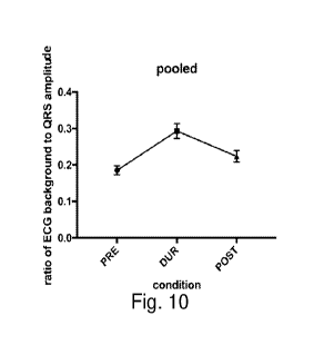
Fig. 10 shows a graph of a pilot test in human subjects of biomarker 1 (the
increased peak-to-
peak amplitude of an ECG recording's background activity, which is due to
increase thoracic
muscular EMG getting included in the ECG signal) during attempts to inspire
against an
occluded upper airway.
Fig. 11 shows a summary of cardiac and respiratory parameters during
controlled airway
occlusion.
Figs. 12 and 13 show various prior art ECG acquisition systems.
Fig. 14 shows a prior art ECG analog acquisition and wireless transmitter
system.
Figs. 15 and 16 show semi-schematic drawings of the prior art ECG wireless
transmitter
system and ECG analog acquisition system of Fig. 14.
Fig. 17 shows a prior art flow diagram for algorithm implementation of the
prior art ECG
analog acquisition and wireless transmitter system according to Fig. 14.
Fig. 18 shows a block diagram of a prior art electronic device according to US
2016/0128209.
Fig. 19 is a diagram illustrating a network environment including an
electronic device
according to US 2016/0128209.
CA 03043325 2019-05-08
WO 2018/089789 PCT/US2017/061099
- 17 -
DETAILED DESCRIPTION OF THE PREFERRED EMBODIMENTS
EXAMPLE 1
Parenteral kainic acid was used to induce recurring seizures in urethane-
anesthetized Sprague
Dawley rats. EEG recordings and combinations of cardiopulmonary monitoring,
including video
laryngoscopy, were performed during multi-unit recordings of recurrent
laryngeal nerve (RLN)
activity or head-out plethysmography with or without endotracheal intubation.
Controlled
occlusions of a tracheal tube were used to study the kinetics of cardiac and
respiratory changes
after sudden obstruction. Seizure activity caused significant firing increases
in the RLN that were
associated with abnormal, high-frequency movements of the vocal folds. Partial
airway
obstruction from laryngospasm was evident in plethysmograms and was prevented
by intubation.
Complete glottic closure (confirmed by laryngoscopy) occurred in a subset of
non-intubated
animals in association with the largest increases in RLN activity, and
cessation of airflow was
followed in all obstructed animals within tens of seconds by ST-segment
elevation, bradycardia,
and death.
Periods of central apnea occurred in both intubated and non-intubated rats
during seizures for
periods up to 33 seconds and were associated with modestly increased RLN
activity, minimal
cardiac derangements, and an open airway on laryngoscopy.
For controlled airway occlusion, a 1-tube was inserted into the distal trachea
of urethane-anesthetized
rats. EEG, ECG, and inspiratory pressure at the sidearm of the T-tube were
bandpass-filtered from 1 Hz to
1 kHz. The open port of the T-tube was occluded for 100 seconds or until
respiratory arrest. Inspiration
artifacts in the EEG and EEG records were isolated with a digital high-pass
filter (corner frequency 367
Hz, rolloff -3 dB/octave) and quantified by full-wave rectification.
Inspiration artifacts matched the
inspiratory pressure extrema during airway occlusion. Correlations (r) of peak
inspiratory pressure to
artifact amplitude in a within-animal comparison were -0.88 (ECG) and -0.75
(EEG), suggesting that
.. artifacts extracted from ECG records may be better than those derived from
EEG records. The average
correlation of artifact magnitude (ECG) with peak inspiratory pressure was -
0.89 0.04 (N=5 rats). The
results suggest that a sudden increase in the amplitude of the inspiratory
artifact in EEG and ECG
recordings indicates an occluded airway, and a very high correlation of
increasing inspiration artifact size
with increasing inspiratory effort was observed. This artifact pattern could
serve as a biomarker in two
important ways: First, to review existing records for the possible
contribution of obstructive apnea to
documented SUDEP cases. Second, to warn about obstructive apnea in patients
being monitored in real
time. The specificity of the biomarker would be further enhanced by marking
decreases in seizure activity
and heart rate. To maximize the sensitivity of the biomarker, EEG and ECG
should be recorded at the
highest bandwidth possible (within the capability of the available equipment,
e.g., up to 10 kHz
CA 03043325 2019-05-08
WO 2018/089789 PCT/US2017/061099
- 18 -
bandwidth). The most attractive feature of this biomarker is that it can be
derived from commonly-used
measures in epilepsy-monitoring units and even potentially portable devices
outside of the hospital.
Using a rat model that permits simultaneous autonomic, cardiovascular, and
respiratory monitoring, it
was demonstrated that seizure-induced laryngospasm caused obstructive apnea,
which stopped the seizure
and persisted until respiratory arrest, followed by cardiac arrest. The
MORTEMUS study used artifacts in
EEG recordings as evidence of respiration. A critical finding herein is that
attempts to breathe during
obstruction generated artifacts in EEG and ECG recordings that resembled
artifacts associated with actual
breaths.
The electrical artifacts of attempts to inspire during airway obstruction can
be used as a practical
biomarker of obstructive apnea. In Fig. 1A, artifacts related to respiration
in ECG and EEG recordings are
shown in conjunction with tracheal pressure. Highpass filtered artifact size
was highly correlated with
peak inspiratory pressure (r2=0.85; n=14 animals). The size of the artifact
itself cannot discriminate
between effective breaths and attempts to breathe. The specific biomarker is
the upward trend in artifact
size as a marker for increasing effort during airway obstruction.
Bradyarrhythmia is present in most patients [Ryvlin et al.] and animals.
[Nakase et al.; Hotta et al.
Epilepsia 50: 923, 2009]. An abrupt change in the ECG RR interval variability
(SDNN; ECG and filtered
ECG) and that the normal lengthening of the RR interval during inspiration
could be reversed during the
late occlusion period. This pattern represents a second biomarker for airway
obstruction, even with short
time samples. Abnormally short RR intervals associated with inspiration
occurred in no animals at
baseline, 4/16 animals during early occlusion, and 15/16 during late
occlusion.
Figs. 1A-1E show a demonstration of inspiration associated artifacts and
changes in RR interval
length during obstruction as bio markers for obstructive apnea.
Fig. lA shows artifacts enhanced in EEG and ECG by highpass filtering. Arrows
indicate last breath
attempt.
Fig. 1B shows correlations of ECG and EEG artifacts with peak inspiratory
pressure (PIP).
Fig. 1C shows a plot of RR over time (black), PIP during obstruction (blue)
and PIP peak markers
(red). RR variance increases late in the occlusion. Relative minima in RR
intervals are ONLY shorter than
baseline during extreme inspiratory effort. Arrows point to the artifact or RR
plot minimum for the breath
just before a missed breath. Heavy black line at the bottom of the graph is
the time shown in the inset.
Fig. 1D shows the standard deviation of the RR intervals (n=16 animals).
Fig. lE shows plots of RR intervals as function of the time relative to the
PIP (n=16 animals). Fitted
curves for baseline and onset use right ordinate. Note the reverse relation of
RR to inspiratory peak.
EXAMPLE 2
The spread of seizure activity over the principal motor nerve of the larynx,
RLN, was studied
in one set of experiments aimed at characterizing RLN activity during normal
quiet breathing
CA 03043325 2019-05-08
WO 2018/089789 PCT/US2017/061099
- 19 -
(baseline) and during seizure activity induced by kainic acid. A tracheal
opening or T-shaped
tracheal tube that preserved RLN bilaterally was used to protect animals from
laryngospasm. In
animals with a tracheal tube, periods of complete glottic closure could be
studied with
laryngoscopy without concern about oxygen desaturation. RLN recordings were
also made
.. during other experiments with the goal of capturing RLN activity during
specific events such as
periods of central and obstructive apnea. EEG, multi-unit RLN activity, and
ECG were recorded
in all animals. Laryngoscopy was performed at intervals during experiments.
The impact of laryngospasm and seizure activity on ventilation was assessed
with head- out
plethysmography in a second set of experiments. One group of animals was
intubated with an
endotracheal tube prior to seizure induction and these animals were compared
with non-
intubated animals. The non-intubated animals comprised two subgroups: one with
no treatment
other than kainic acid to induce seizures, and a second with bilateral
superior laryngeal nerve
transection to prevent reflex laryngospasm performed in the pre-seizure
condition.
Seizure activity was associated with increases in RLN activity and abnormal,
high frequency
movements of vocal folds. Within a single seizure, RLN activity progressively
increased, with
the highest levels of activity most commonly observed near the end of the
seizure. The full
pattern of an RLN activity increase during a single seizure and its decrease
to baseline at the end
of the seizure could be observed when the airway was protected by a tracheal
tube or window
(Fig. 2). Laryngospasm during seizure activity had a significant impact on
respiration
Fig. 2 shows extreme increases in RLN activity during a seizure. Segments from
a complete
seizure are shown with normal respiratory bursting on RLN (top left, even
lines) giving way to
significantly increased firing (right side of top trace with maximum on right
side of second trace)
with eventual firing reductions (bottom trace). EEG is shown on odd lines.
Estimates of seizure
onset and offset (based on changes in low frequency activity and spiking) are
marked with
arrows. In these animals, the airway was protected with a tracheal implant or
opening cut
through the tracheal cartilage so that the entire profile of RLN activity
during individual seizures
might be captured.
During normal tidal breathing under urethane anesthesia, the early expiratory
peak in rats
resembles human breathing (Arito et al., 1997). Three of 14 non-intubated rats
and 1 of 6
intubated rats had seizure activity mainly characterized by low frequency,
repetitive gasping
breaths and were not included in the summary data. Plethysmograph recordings
were taken
before and after SLN lesions in this subgroup, and before and after intubation
in intubated
animals. None of the measured parameters showed a difference due to SLN lesion
or intubation.
CA 03043325 2019-05-08
WO 2018/089789 PCT/US2017/061099
- 20 -
Pre-seizure values used for comparison with seizure values were the baseline
condition for KA-
only animals, the post-intubation condition for intubated animals, and the
post-lesion condition
for SLN lesioned animals. There were no differences between the two subgroups
of non-
intubated rats and their measures were pooled for statistics except when these
two groups were
compared with each other. Examples of flow-volume loops for non- intubated and
intubated rats
are shown in Fig. 3.
Fig. 3 shows plethysmography during kainic acid-induced seizure activity. Head
plethysmography examples from one non-intubated (panels Al, Cl) and one
intubated (panels
Bl, DO rat. The pre-seizure baseline condition for each animal is shown in the
top row (panels
Al, B1) and the corresponding seizure-associated condition is shown below
(panels Cl, D1).
For each panel, 5 minutes' worth of continuous breathing that was analyzed to
produce the flow-
volume loops in each case. The upper horizontal red line on each flow-volume
graph is the mean
peak expiratory flow, the lower horizontal red line is the mean peak
inspiratory flow, and the
vertical red line is the mean tidal volume. Several key features are evident:
1) tidal volumes
during seizure activity are lower for both animals; 2) the variability of
breath flows and volumes
during seizures are increased for both animals; 3) the ratio of peak
inspiratory flow to peak
expiratory flow is decreased for the non-intubated rat and increased for the
intubated rat
(calculated average shown at the upper right of each flow-volume graph).
Seizure activity was associated with large increases in respiratory rate in
all remaining rats
(11 non-intubated and 5 intubated), irrespective of treatment, from mean pre-
seizure rates of 85
11 and 98 17 breaths/min for non-intubated and intubated rats, respectively
to seizure
associated rates of 371 54 and 295 43 breaths/min. Increases were
significant (p<0.0001)
after Scheffe post hoc correction of multi-variate ANOVA. Pre-seizure mean
rates of 89 and 81
breaths/min were observed in the 5 KA-only rats and the 6 non-intubated SLN
lesioned animals,
with seizure-associated mean rates of 371 breaths/min for both groups
(p<0.0001 for both
comparisons).
Other details are given in Table 1, which shows a summary of first and second
order
plethysmography variables. The full set of plethysmography variables measured
are shown with
details for baseline, post manipulation (intubation or SLN lesion) and during
seizure activity.
The manipulations (intubation or SLN lesion) did not change baseline values
significantly for
any parameter, but seizure activity changed many parameters related to
durations and volumes in
all animals. The principal measure to discriminate between non-intubated and
intubated animals
CA 03043325 2019-05-08
WO 2(118/089789 PCT/US2017/061099
-21 -
was the ratio of inspiratory peak flow to expiratory peak flow. Scheffe post-
hoc corrections
applied to one-way ANOVAs. A p value of 0 is used to indicate p<0.0001.
Tidal volume decreased significantly in the non-intubated rats. Mean pre-
seizure tidal
volumes of 1.50 0.36 ml/breath decreased to 0.46 0.14 ml/breath (p<0.000 I
). Subgroup tidal
volumes were each significantly decreased: 1.2 to 0.46, p=0.008 for KA-only
rats and 1.7 to 0.46,
p<0.0001 for SLN lesioned rats). The difference in pre-seizure (1.03 0.69
ml/breath) vs.
seizure (0.53 0.26 ml/breath) tidal volume in intubated animals did not
reach statistical
significance.
Given that ventilation rates increased approximately 3-fold and tidal volumes
decreased
.. approximately 3-fold during seizure activity, the average minute
ventilation during seizure
activity did not differ significantly from baseline, but tended toward lower
values. Mean pre-
seizure values of 124.8 27.3 and 124.2 61.8 ml/min were associated with
mean seizure
values of 100.8 35.7 and 93.2 42.3 ml/min (NS, NS) for non-intubated and
intubated rats.
Only the SLN lesioned subgroup showed a significant decrease in minute
ventilation from 138.8
13.2 to 106 22.4 ml/min (p<0.0001). Mean pre-seizure and seizure values for
the KA-only
rats were not significantly different (108.0 25.0 vs. 94.4 49.7 ml/min;
NS).
The most dramatic differences were seen in the ratio of peak flow during
inspiration to peak
flow during expiration. This parameter is used to identify upper airway
obstruction. Normally,
this ratio is > 1, and values < 1 are indicative of extrathoracic (e.g. upper
airway) obstruction
.. (Blitzer and Meyer, 2006; Miller et al., 1987). Mean pre-seizure ratios
were 1.04 0.25 for non-
intubated rats and 1.02 0.10 for intubated rats. These values changed in
opposite directions for
intubated (increasing to 1.56 0.38;1)=0.011) and non-intubated rats
(decreasing to 0.52 0.32;
p<0.001). The individual subgroups of non-intubated animals each showed
decreases in the ratio
of peak flows during inspiration and expiration: 0.95 10 0.60 for KA-only rats
(NS) and 1.11 to
0.46 for SLN lesioned rats (p=0.001). Whereas the decrease in PF(i)/PF(e) is
consistent with
partial airway obstruction from laryngospasm, the increase in PF(i)/PF(e) is
clearly not from one
of the typical causes of variable intrathoracic obstruction. Since there was
no obstruction in the
intubated animals, the flow-volume characteristics of the intubated animals
reflect seizure-
induced disordered ventilation without contribution from airway narrowing. If
this is true, the
decreased PF(i)/PF(e) seen in non- intubated rats should be considered an
underestimate, more
properly compared with the intubated rats' seizure condition than with their
own pre-seizure
condition.
CA 03043325 2019-05-08
WO 2018/089789
PCT/US2017/061099
- 22 -
Values are summarized in Table 2, which shows summary statistics from
plethysmography
data. To compensate for multiple ANOVAs. a difference score (seizure condition
minus pre-
seizure condition) was computer for each animal on the 4 variables derived
from the
plethysmograph (respiratory rate, tidal volume, minute ventilation, and the
ratio of inspiratory
peak flow to expiratory peak flow). A 2-tailed Mann-Whitney test was conducted
of the
difference of distribution of these change- scores between intubated and
pooled non-intubated
study arms. Bootstrapping (20,000 replications) was used (SAS 9.4 Proc
Multtest) to arrive at
corrected p-values for the four measures, based on independent samples 2-
tailed t-tests
performed on ranked scores.
Figs. 4A and 4B show irregular vocal fold movement during seizure activity.
Laryngoscopy
during seizure activity revealed "shaking" movements of the arytenoid
cartilages consistent with
the findings of partial obstruction from plethysmography and abnormal RLN
activity. In
analyses of video recordings of laryngeal vocal fold and arytenoid cartilage
movements, the
highly correlated movements of the left and right arytenoid cartilages
uncouple partially from an
average Pearson correlation of -0.95 0.04 to -0.79 0.11 (n=10; p=0.0007),
as shown in Fig.
4A. Frame-by-frame analysis of vocal fold and arytenoid cartilage position
during video
recordings of laryngoscopy show the typical coordinated abduction and
adduction (periodic low
frequency trace of upper graph of Fig. 4A) of the vocal folds during
respiration. The position of
the left arytenoid cartilage relative to the midline is shown as an upward
deflection in the top
graph, and the right arytenoid cartilage position is shown as a downward
deflection. The
correlation is high (0.98). During seizure activity (high frequency trace),
the total displacement
is less, the frequency is higher, and the correlation is decreased (0.75).
The distributions of time in quintiles of the peak-to-peak glottic opening
(measured at
baseline) is shown in Fig. 4B. The distribution of times across degrees of
glottic opening (bin
sizes = 20% of minimum to maximum opening in the baseline condition) were
shifted toward
larger openings, but with less variation in glottic opening. In fact, the
average total normalized
glottic opening over 10 seconds was larger during seizure activity than during
baseline (0.36
0.03 baseline, 0.47 0.06 seizure; p=0.00005). At baseline (squares), the
largest fraction of time
is in the closed position, with rapid cycling through open angles. The
distribution of times for a
sine wave are shown for reference (dotted line). During seizure activity, the
profile is changed
significantly (circles) with a larger fraction of time in relatively open
states, which would seem
to mitigate the relatively stationary opening. Data are shown as means
standard deviations.
CA 03043325 2019-05-08
WO 2018/089789 PCT/US2017/061099
- 23 -
Whereas clear evidence of partial airway obstruction due to laryngospasm was
routinely
observed, the modest decreases in minute ventilation suggested that
respiratory derangements
during seizures were adequately compensated. However, complete glottic closure
(confirmed
with laryngoscopy) occurred in a subset of non-intubated animals during
discrete seizures in
association with the largest increases in RLN activity, and cessation of
airflow was followed in
all animals within tens of seconds with ST segment elevations in ECG,
bradycardia, and
eventually death. Complete obstructive apnea occurred in 7 of 11 non-
intubated and 0 of 5
intubated rats (p=0.03, Fisher exact test, two-tailed). All 7 animals died.
The start of the
obstructive apneic period was taken as the time from the point at which peak-
to- peak airflow
reached <10% of the pre-apneic peak-to-peak airflow, and the endpoint was the
time at which the
recording was stopped and the animal removed from the plethysmography chamber
with
evidence of severe bradycardia on ECG that, upon removal from the
plethysmography chamber,
was associated with apparent cardiopulmonary arrest. Only when an artificial
airway was
present was a period of complete glottic closure due to laryngospasm seen to
terminate on its
.. own with a reversion to the normal pattern of opening and closing with each
breath.
Fig. 5 shows obstructive and central apnea during seizures. The top panel
illustrates an
episode of obstructive apnea due to laryngospasm with hypoxic cardiac
arrhythmia. Each set of
traces consists of plethysmography (top); ECG (middle); EEG (bottom). The
obstructive apnea
develops as a rapid, but continuous (several seconds) reduction in the amount
of air per breath
until that amount is negligible. At the time indicated as complete obstruction
(confirmed by
simultaneous laryngoscopy - single frames shown at the right), the ECG
develops clear brady-
arrhythmia with ST segment elevation from hypoxemia develops. The recording is
taken from
the end of a seizure; seizure activity is present from the beginning of the
illustrated data and an
estimate of seizure offset (based on a complete flat-lining of EEG) is marked
by an arrow.
Episodes of central apnea, by contrast, were characterized by an abrupt
cessation of breathing
and air flow, but the vocal folds arrested in an open position (video frame at
right). There were
no cardiac derangements over the same time period. The entire record, taken
from the middle of
a seizure, displays seizure activity.
On plethysmography records, airflow declined rapidly to zero or near zero flow
(Fig. 5, top).
In every case, ST-segment elevation on ECG recordings was clear evidence of
hypoxemia.
Laryngoscopy revealed complete glottic closure. The shortest duration period
of obstructive
apnea to produce apparent cardiopulmonary arrest was 56 seconds. RLN activity
recorded with
laryngoscopic confirmation of glottic closure (n=2) showed intense firing
associated with the
CA 03043325 2019-05-08
WO 2018/089789 PCT/US2017/061099
- 24 -
laryngospasm and ECG evidence of hypoxia (Fig. 5, top). The occurrence of
laryngospasm in
SLN-lesioned animals is further evidence that laryngospasm was not mediated by
pharyngeal/laryngeal reflexes that might have been activated by the
laryngoscope or salivation.
Fig. 6 shows recurrent laryngeal nerve activity during obstructive and central
apnea. RLN
firing (middle trace of each panel) during obstructive apnea (top panel) and
central apnea
(bottom panel) show that the RLN is active during both types of apnea. The RLN
carries motor
output for both laryngeal abductors and adductors. The multi-unit recordings
do not permit
discrimination of nerve activity for abductors or adductors, but adduction
dominates during
obstructive apnea and abduction dominates during central apnea. Video frames
are shown to the
right. Also shown to the right are three ECG sweeps for each type of apnea to
illustrate the
pronounced ST segment elevation and slowing during obstructive apnea and the
uniform PQRST
complexes during central apnea. The recording illustrating obstructive apnea
is taken from the
end of a seizure; seizure activity is present from the beginning of the
illustrated data and an
estimate of seizure offset (based on a complete flat-lining of EEG) is marked
by an arrow.
In contrast to systemic impact of obstructive apnea, periods of central apnea,
characterized
by an abrupt cessation of breathing effort, a completely open glottis,
moderate RLN firing, and
no air flow on plethysmography, were never associated with ST segment
elevation in ECG or
any other evidence that these episodes might be life threatening (Fig. 5,
bottom and Fig. 6,
bottom; Table 3).
Table 3 shows contrasts between obstructive apnea and central apnea. Details
of obstructive
and central apneic periods captured in non-intubated and intubated rats.
Obstructive apnea
appeared only in non-intubated rats, a difference that was significant
(p=0.034). Central apneic
periods averaged durations < 10 seconds, but some periods exceeded 30 seconds
in duration. To
compare the impact of apnea of either type on cardiac activity, HR and the
presence or absence
of ST segment elevation were compared over equivalent 10 second periods from
the onset of
apnea based on plethysmography records. ST segment changes were only seen
during
obstructive apnea periods. Taking the minimum HR over this period in
comparison with baseline,
both obstructive and central apneic periods were associated with significant
bradycardia, but the
changes associated with obstructive apnea were greater. All comparisons were
two-tailed
unpaired t-tests.
As further evidence that the glottic closure was active and not passive (e.g.
resembling vocal
fold paralysis (Morel al., 2014)), plethysmography was performed, and recorded
vocal fold
motion in an animal whose right vocal fold was paralyzed by RLN damage (Fig.
7, hemiparetic).
CA 03043325 2019-05-08
WO 2018/089789
PCT/US2017/061099
- 25 -
During a seizure-induced period of obstructive apnea, the ECG shows ST-segment
elevation and
the plethysmograph shows an absence of air movement. The force of contraction
of the left vocal
fold actually pushed the arytenoid cartilage across the midline in the absence
of resistance from
the right vocal fold.
Fig. 7 also shows a demonstration of the force of contraction during
laryngospasm. In this
example, the normal open and closed states of the arytenoid cartilages are
illustrated in the
baseline panel (left) together with plethysmography, ECG, and EEG records
taken
simultaneously. The tick marks on the plethysmography records indicate the
time of the video
snapshots. Note that the glottis is not completely closed, even at the minimum
of arytenoid
excursions from the midline. In the center panel, the right vocal fold was
paralyzed by crushing
the right RLN to cause hemiparesis. Breathing is changed from regular large
breaths to more
frequent smaller breaths mixed with large gasps. The far right panel shows a
segment taken from
the same rat during seizure induced laryngospasm sufficient to produce
obstructive apnea. The
glottis is completely closed, but note how left side of the larynx actually
crosses the midline
when not opposed by an active right side (white arrow in video snapshot). Also
note the ST-
segment elevations are prominent (asterisk on ECG trace) in contrast with the
other two states.
(Calibrations: 0.5 sec, 0.25 ml (pleth), 0.5 mV (ECG), and 0.1 mV (EEG)).
The lethality of obstructive apnea periods is contrasted with the minimal
impact of central
apnea periods in the same animals. That these transient periods of central
apnea are separate
from the central apnea that characterizes respiratory arrest. Periods of
central apnea were
defined by an abrupt cessation of breathing for periods > 1 second as
evidenced in
plethysmography records. These were recorded during seizure activity in
animals of all groups
with no differences in the frequency or duration of central apneic periods
between groups. No
central apneic periods were ever seen in baseline, pre-seizure/post-
intubation, or pre-
seizure/post-SLN transection conditions. Three of 6 KA-only animals showed
central apneic
periods, compared with 3/5 SLN-lesioned animals, and 5/5 intubated animals.
Central apneic
periods as long as 33 seconds were recorded. The mean durations and counts of
central apneic
periods? 1 s, and the subset of periods whose durations were > 5 s are
detailed in Table 3.
Two findings highlight the contrast between obstructive and central apnea.
First, on
laryngoscopy during central apneic periods, the vocal folds were abducted and
immobile and
held the glottis in a completely open configuration for the entirety of the
apneic period (Figs. 5,
6). The open state of the larynx is an active state, as shown by RLN activity
during central apneic
periods. Second, bradycardia developed to a much greater extent, plus ST-
segment elevation
CA 03043325 2019-05-08
WO 2018/089789 PCT/US2017/061099
- 26 -
was prominent, during periods of obstructive apnea, but not central apnea.
While it is true that
the obstructive apnea periods lasted longer than central apnea periods (all
central apnea periods
ended spontaneously with a return to pre-apneic respiratory patterns), at the
same time from
apnea onset, only obstructive apnea impacted cardiac function. Taking all
central apneic periods
of 15 seconds or greater from all groups into a single pool (n=9 apneic
periods from 6 animals),
the change in heart rate within the time window of 5-15 seconds was examined
for comparison
with the mean heart rate pre-apnea. The mean and minimum heart rate measures
during the 5-15
second time window of obstructive apnea were significantly decreased compared
to pre-apnea
rates. For central apneic periods, the minimum heart rate during 5-15 seconds
was significantly
decreased, but not the mean rate for the 10 second epoch. In comparing the
relative changes,
heart rate decreases during obstructive apnea (-31.4 13.9 % change, n=7)
were significantly
greater than central apnea (-17.3 9.7 % change, n=9) over the same time
frames (Table 3). The
average minimum heart rate for periods of obstructive apnea before stopping
the recordings was
0.86 0.38 beats/s (down from > 6 beats/s at baseline).
A series of experiments were conducted in which a controlled complete
occlusion of the
airway was used to study response kinetics without the uncertainty of when
complete obstruction
would occur during seizure activity. A T-shaped tracheal tube was implanted
after dissecting the
RLN free bilaterally. This enabled securing the tracheal tube in place without
disturbing normal
laryngeal function. A pressure transducer on the tracheal tube sidearm
recorded forces developed
during either normal breathing with the tracheal tube open to the atmosphere
or during complete
closure of the open port with an airtight cap. Complete obstruction of the
airway was performed
for 100 seconds or until 20 s after respiratory arrest occurred, whichever was
earlier. In addition
to tracheal sidearm pressures, ECG and pulse oximetry were recorded
continuously. hi subsets
of animals, echocardiography and/or continuous arterial blood pressure
monitoring were
performed.
During occlusion, respiratory effort to inspire progressively increased, then
ceased, usually in
less than 1 minute (60.4 24.0 s; median = 54.4 s; n=16). Respiratory arrest
was associated with
cardiac dilatation and asystole, an increase of systemic blood pressure (which
collapsed without
resuscitation), and laryngospasm sufficient for complete glottic closure. This
is a type of central
apnea that differs from the central apneic episodes reported earlier that were
associated with an
actively open airway. The LV diastolic cavity size became dilated by about 40%
(0.47 + 0.07 at
baseline to 0.64 0.22 cm 10 sec after respiratory arrest) and the end
systolic LV cavity size
CA 03043325 2019-05-08
WO 2018/089789 PCT/US2017/061099
- 27 -
became dilated by nearly 300% (0.12 0.03 at baseline and 0.44 0.23 cm 10
seconds after
respiratory arrest). The LV ejection fraction fell from 94 2 to 49 23
percent.
An example experiment is illustrated in Fig. 7 and the cardiac and respiratory
function
parameters are summarized in Figs. 8 and 9, which show echocardiography during
controlled
airway occlusion. Fig. 8 shows ECG, pulse oximetry, airway pressure
transducer, and arterial
blood pressure records during a 100 second occlusion of the trachea (onset and
end marked with
arrows). The development of bradyarrhythmia and the progressive inspiratory
effort are clearly
visible. The inspiratory effort is sufficient to significantly impact blood
flow as evidenced by the
larger pulse oximary waves associated with each attempt to breathe. Fig. 9
shows a series of M-
mode echocardiogram panels, each representing a respective 2-second period
which has a
reference number corresponding to the markings in the ECG trace of Fig. 8.
Normal cardiac
function is visible in the pre- occlusion record (panel 0 of Fig. 9). Panels 1-
6 of Fig. 9 occur at
points during the period of occlusion and show rhythm abnormalities and
progressive left
ventricular dilation. By the time respiration has arrested (echo panel 5 of
Fig. 9), the heart is
nearly akinetic. The recovery panel shows the abrupt return of cardiac
performance after the
airway obstruction has been removed and breathing has recovered. Major ticks
on each plot in
top panel: ECG = 0.25 mV; pulse ox = 5% variation (high pass filtered); airway
transducer = 25
mmHg; BP = 10 mm Hg.
EXAMPLE 3
Fig. 10 shows results of a pilot human trial showing the use of biomarker 1
(the increased
peak-to-peak amplitude of an ECG recording's background activity, which is due
to increase
thoracic muscular EMG getting included in the ECG signal) to detect
obstructive apnea. The
preliminary human subject data shows that biomarker 1 appears in a simple
setting where
patients try to inspire by drawing air out of closed 500 ml container.
Significant increases in
ECG background amplitude relative to QRS amplitude (simple ratio of peak-to-
peak ECG signal
to the peak-to-peak amplitude of the QRS complex) occur during the inspiratory
effort.
This demonstrates that biomarker 1 has utility in any condition where EMG
associated with
inspiratory effort can be increased.
In Fig. 10, a statistical analysis of the results showed:
ANOVA: F (1.487, 43.13) = 22.42 P<0.0001
Multiple comparisons:
PRE vs. DUR p=0.0001
PRE vs. POST p=0.0128
CA 03043325 2019-05-08
WO 2018/089789 PCT/US2017/061099
- 28 -
EXAMPLE 4
Tracheal tubes were placed in four rats, and strong seizure activity induced
with kainic acid.
In three rats, systemic variables to describe the sequence of events leading
to death were
monitored. During the period continuous seizure activity (status epilepticus),
shallow, irregular
breathing was mixed with gasping breaths that occurred at a rate of 1/s, but
dropped abruptly to
1/15 s before apparently stopping completely. Although the airway was
completely open,
oxygen saturations of 54 or 77% (no data for 3rd rat) preceded the transition
to very slow or
arrested breathing. The rate of change of oxygen saturation over time was well
fitted with a
straight line (slope = -0.07 0.05 pulse oximetry percentage points per
second, R2 v= 0.93
0.04). From these values, an average 10% drop in oxygen saturation took 135
seconds (2.25
minutes) ¨ compared with times of <10 seconds after onset of laryngospasm-
induced obstructive
apnea or controlled occlusion. Whereas these animals demonstrated that, during
periods of
sustained seizure activity, very low oxygen saturations and death could occur
with an intact
airway, the times for desaturation were so long that this mechanism is
unlikely to be the principal
mechanism for desaturation during discrete seizures. Rather, this mechanism is
likely a distinct
feature of status epilepticus.
Fig. 11 shows a summary of cardiac and respiratory parameters during
controlled airway
occlusion. Changes in heart rate derived from ECG, arterial oxygen saturation
based on pulse
oximetry, respiratory rate and peak inspiratory pressure derived from a
pressure transducer on
the sidearm of a tracheal implant. and left ventricular cavity size and
ejection fraction derived
from echocardiography. A pre-occlusion baseline point is compared with three
time points
during controlled airway occlusion (15 s after onset, at the time of
respiratory arrest, and 10 s
after respiratory arrest), and recovery (60 s after the end of resuscitation
efforts). Each point is
shown as its mean and standard deviation. Obstructed animals (squares) are
also compared with
unobstructed control animals (circles) for some measures. Changes in
respiratory parameters and
oxygen saturation occur, in general, earlier than changes in cardiac
parameters as evidenced by
statistically significant decreases in these parameters by 15 s after the
onset of airway occlusion.
By the time of respiratory arrest, left ventricular performance is
significantly impaired as
illustrated with significant dilatation (enlarged end systolic dimension) and
decreased ejection
fraction. Units for plots: HR (beats/s), oxygen saturation (% saturation),
respiratory rate
(breaths/s), peak inspiratory pressure (mmHg/100), ventricular cavity size
(cm), and ejection
fraction (% diastolic volume ejected during systole).
CA 03043325 2019-05-08
WO 2018/089789
PCT/US2017/061099
- 29 -
Unlike laryngospasm-mediated obstruction where the vocal folds were
continuously
adducted, during the period of obstruction, each attempted inspiration was
associated with an
opening and closing of the airway by vocal fold movements such that the degree
of opening
increased as the inspiratory effort increased. Each glottic opening was
followed by a complete
closure of the airway due to fully apposed arytenoid cartilages and vocal
folds. The maximal
opening angle during the last breath attempt was 56.7 5.3 compared to
baseline values of 27.6
4.6 (p<0.0000I). After the last breath attempt, the airway stayed in this
closed position for an
additional 20-60 seconds before normal breathing and vocal fold motion resumed
after
resuscitation or a small glottic opening became evident when the vocal folds
appeared to relax in
animals that were not resuscitated.
From the point of the last apparent breath, a minimum in heart rate was
reached in 30, 70, or
140 seconds. In two animals, laryngospasm was recorded only after the
appearance of
bradycardia because the larynx was not being continuously monitored. For the
third rat, first
evidence of laryngospasm was captured on video. This showed that breathing
appeared to stop
18 seconds before laryngospasm and cessation of seizure activity as evidenced
by flattening of
the EEG. Whereas hypoxia-induced laryngospasm such as that described in the
controlled
occlusion experiments might account for the laryngospasm observed in the first
two rats,
laryngospasm in the third rat was uncoupled from the respiratory pattern and
apparently still
driven by seizure activity.
The key findings of these studies are that: 1) seizure activity causes large
increases in RLN
activity; 2) seizure activity changes breathing frequency, amplitude,
variability, and can cause
central apnea; 3) seizure activity causes laryngospasm that can result in
partial or complete
airway occlusion (obstructive apnea); 4) only obstructive apnea was associated
with rapid, severe
arterial oxygen desaturation, bradycardia, respiratory arrest, and death; 5)
hypoxemia itself can
cause laryngospasm, significantly prolonging complete airway closure; and 6)
sudden death is
the result of respiratory arrest during airway obstruction and nearly
simultaneous left-ventricle
dilatation and asystole. From this set of findings, it is concluded that
sudden death in any animal
or person experiencing a seizure can be the result of seizure-induced
laryngospasm sufficient to
cause obstructive apnea, which leads to respiratory arrest and cardiac
asystole within tens of
seconds, and which can only be reversed by cardiopulmonary resuscitation (i.e.
spontaneous
recovery is highly unlikely).
Seizures clearly disrupt normal breathing. Respiratory frequency, tidal
volume, and cycle
variability were all changed by seizure activity. More severe outcomes were
marked by periods
CA 03043325 2019-05-08
WO 2018/089789 PCT/US2017/061099
- 30 -
of no airflow at all, either because the drive to breathe ceased while the
glottis was fully open
(central apnea) or because the glottis was closed due to laryngospasm
(obstructive apnea). The
most significant impact on oxygen status and cardiac and respiratory function
was from
obstructive apnea secondary to seizure-induced laryngospasm. A straightforward
interpretation
of these observations is a spread of seizure activity along the pathway from
subiculum to
paraventricular nucleus (PVN) of the hypothalamus (Canteras and Swanson, 1992)
and from
PVN to medullary regions (e.g. (Geerling et al., 2010)), where it impacts
medullary autonomic
nuclei, respiratory centers, and laryngeal motor neurons.
What is it about seizure-induced obstructive apnea that resulted in such rapid
and severe
cardiopulmonary dysfunction? The lack of airflow could not have been the
problem since the
periods of central apnea did not cause the same deterioration. The remarkable
feature of the
obstructive apneas was that the airway was completely shut. The forces of
vocal fold
contractions during seizure-induced laryngospasm were illustrated by the fact
that an active
vocal fold actually crossed the midline when it was not opposed by a paralyzed
vocal fold and
the fact that the usual opening of the vocal folds during attempts to gasp did
not occur. By
contrast, the vocal folds were always in a completely open position during
periods of central
apnea. It is conceivable that the occurrence of laryngospasm merely reflected
a level of seizure
activity that caused cardiopulmonary dysfunction by a mechanism independent of
obstructive
apnea. However, when the airway was manually occluded by closing a tracheal
tube in rats that
had not been treated with kainate and were not undergoing seizures, the same
sequence and time
course of events was observed: oxygen desaturation, bradycardia, and ST-
segment elevation
within seconds, respiratory arrest and serious cardiac mechanical failure
within about one minute,
and cardiac arrest within several minutes. The combination of these findings
indicates that it is
the airway occlusion that triggers cardiopulmonary collapse and death.
A major difference between central and obstructive apnea relates to the
intense autonomic
response that comes during attempts to breathe against a closed airway or
during asphyxiation
(e.g. (Brostrom et al., 2007; Hotta et al., 2009; Weiss et al., 2015)), but
does not occur in the
absence of a drive to breathe. Breath holding can last for long times without
detriment; the
current world record in humans exceeds 11 minutes, or over 22 minutes after
hyperventilation
with pure oxygen (Association Internationale pour le Developpement de l'Apnee;
www.aidainternational.org/). It involves a voluntary reduction of the drive to
breath, but does not
require closing the glottis (Donzelli and Brady, 2004; Mendelsohn and Martin,
1993) and does
not significantly activate the autonomic nervous system. Seizure-induced
central apneas are
CA 03043325 2019-05-08
WO 2018/089789 PCT/US2017/061099
- 31 -
generally harmless because they induce only a minimal autonomic and systemic
response in the
absence of a drive to breathe. Seizure-induced obstructive apneas, in
contrast, are deadly because
the attempt to breathe against a closed airway triggers a strong autonomic co-
activation, on top
of an already raised autonomic tone due to the seizures themselves, that
ultimately results in
cardiopulmonary collapse.
Given the complexity and interdependence of the cardiac, respiratory, and
nervous systems,
the question arises whether laryngospasm is both necessary and sufficient for
sudden death. First,
seizure-induced laryngospasm is not reflex-driven by salivation or other
pharyngeal stimuli
because a subgroup of non-intubated rats had bilateral superior- laryngeal-
nerve lesions that
would abolish the afferent limb of a reflex to drive laryngospasm. All of the
deaths observed
during the plethysmography experiments occurred with a sequence of seizure-
induced
laryngospasm followed by respiratory and then cardiac arrest with exactly the
same temporal
profile observed during the controlled occlusion of tracheal implants.
Therefore, closing the
airway by itself crosses a critical threshold and that seizure-induced
laryngospasm is sufficient
for sudden death.
The MORTEMUS heart rate data show that 9/10 patients experienced the largest
drop in
heart rate during the period of apparent respiration at the end of the seizure
(Figure 3 of Ryvlin et
al., 2013) and before the onset of terminal apnea. The timing of the sharp
drop in heart rate in the
present rat experiments corresponds to a point late in the period of
obstruction, after the seizure
.. would have been terminated (Stewart, 2008), but before the point of
respiratory arrest, which are
believed to correspond to the onset of terminal apnea in the MORTEMUS study
(Ryvlin et al.,
2013). It is not only possible, but probable that the SUDEP cases of the
MORTEMUS study
experienced obstructive apnea as evidenced by the same terminal sequence of
events leading to
respiratory arrest and death as found for rats.
Laryngospasm may contribute to sudden death even in cases when it is not the
initial trigger.
In the manual airway occlusion experiments, laryngospasm was observed after
respiratory arrest.
This complicates the interpretation of clinical case reports since the
presence of laryngospasm
postictally may indicate either laryngospasm-mediated hypoxia or hypoxia-
mediated
laryngospasm. More ominously, whether laryngospasm starts the desaturation or
occurs after
desaturation, it guarantees that death occurs unless cardiopulmonary
resuscitation is initiated
shortly after respiratory arrest (when there is no effort to breathe and the
heart is severely dilated).
A sequence of events is defined that links seizures to sudden death. In
particular, seizure-
induced laryngospasm resulted in cessation of airflow, followed within tens of
seconds by ST-
CA 03043325 2019-05-08
WO 2018/089789 PCT/US2017/061099
- 32 -
segment elevation, bradycardia, and respiratory arrest. These data were
obtained in an
established animal model for seizure experiments (urethane- anesthetized rats
treated with kainic
acid), not in humans, but demonstrate the utility of this rat model for
studying laryngospasm and
obstructive apnea.
Figs. 12 and 13 show prior art ECG acquisition circuits. See www.electro-tech-
online.com/attachments/untitled-gif.26911/; www.electro-tech-
online.com/attachments/ecg-
circuit-png.26416/; and
gasstationwithoutpumps.files.wordpress.com/2012/08/dobrev-amp.jpg,
each of which is expressly incorporated herein by reference in its entirety.
Figs. 14, 15 and 16 show aspects of a prior art ECG analog acquisition and
wireless
transmitter system, See, Fen Miao, Yayu Cheng, Yi He, Qingyun He and Ye Li, "A
Wearable
Context-Aware ECG Monitoring System Integrated with Built-in Kinematic Sensors
of the
Smartphone-, Sensors 2015, 15(5), 11465-11484; doi:10.3390/sI50511465,
www.mdpi.com/1424-8220/15/5/11465/htm, which is expressly incorporated herein
by reference.
In its entirety.
The block diagram of a proposed ECG monitoring system combined a wearable ECG
acquisition sensor with a smartphone is shown in Figure 14. The ECG sensor
follows the
YY1139-2000 standard (a pharmaceutical industry standard of China for single
and multichannel
electrodigraph, which is evolved from EC13 national standard). In the ECG
acquisition sensor,
signal is amplified and filtered by a single chip of AFE module, then in MCU
module the analog
signal from AFE is converted to digital signal. After processed with
compression algorithm, the
digital signal is recorded in SD card or transmitted to smartphone for real-
time display.
Meanwhile, a USB port is equipped in the device for transmitting the signals
which have been
saved in the SD card to personal computers and then to the cloud platform for
further analysis.
The ECG signals transmitted to smartphone are real-time displayed on screen,
with a brief report
provided from the automatic analysis approach in the software or professional
advices provided
from the remote server. The built-in kinematic sensors of the smartphone are
used to recognize
the individual's physical activity and thus help to improve the diagnosis
accuracy for detecting
abnormal patterns.
The block diagram of traditional implementation of ECG acquisition device is
presented in
Fig. 15, in which the circuit consists of a traditional instrument amplifier
and Sallen-Key or
Nyquist low pass filter, and some external function circuits for realistic ECG
detection. The
system employs various discrete components which occupy circuit board area.
CA 03043325 2019-05-08
WO 2018/089789 PCT/US2017/061099
- 33 -
Miao et al. propose an architecture using a fully custom, fully integrated,
low power AFE.
with all the function circuits integrated, as shown in Fig. 16 of the prior
art, with an input/output
buffer, full differential amplifier (DA) with high pass function, second Gm-C
low pass filter,
additional amplifying stage, DRL circuit, lead-off detecting circuit, fast
restore function, and a
power management module to provide a stable working voltage and current.
US 2016/0128209, expressly incorporated herein by reference in its entirety,
discloses an
exemplary hardware platform that can be used to implement the present
technology, as shown in
Figs. 17 and 18.
Referring to Fig. 17 of US 2016/0128209, the electronic device 100 can
constitute at least
one of: at least one AP (application processor) 910, a communication module
920, a SIM
(subscriber identification module) card 924, a memory 930, a sensor module
940, an input device
950, a display 960 (e.g. the display device 13), an interface 970, an audio
module 980, a camera
module 991, a power management module 995, a battery 996, an indicator 997,
and a motor 998.
The AP 910 controls a plurality of hardware or software components connected
to the AP 910 by
driving an operating system or an application program, process various data
including
multimedia data, and perform calculations. The AP 910 can be embodied as, for
example, a
System on Chip (SoC). According to an embodiment, the AP 910 further includes
a Graphic
Processing Unit (GPU). The communication module 920 (e.g. the communication
interface 160)
can perform data transmission/reception in connection with communication with
other electronic
devices connected to the electronic device 100 via a network. According to one
embodiment, the
communication module 920 includes at least one of: a cellular module 921, a Wi-
Fi module 923,
a BT module 925. a GPS module 927, an NFC module 928, and a Radio Frequency
(RF) module
929. The cellular module 921 provides a voice call, a video call, a text
message service, or an
Internet service through a communication network (for example, LTE, LTE-A,
CDMA,
WCDMA, UMTS, WiMax, GSM, 3G, 4G, 5G, or the like). Further, the cellular
module 921
distinguishes and authenticates electronic devices within a communication
network by using a
subscriber identification module (for example, the SIM card 924). According to
an embodiment,
the cellular module 921 performs at least some of functions that the AP 910
provides. For
example, the cellular module 921 can perform at least a part of a multimedia
control function.
The cellular module 921 may include a Communication Processor (CP). Further,
the cellular
module 921 can be implemented by, for example, an SoC. Although components
such as the
cellular module 921 (e.g., the communication processor), the memory 930, or
the power
management module 995 are illustrated to be separate from the AP 910 in Fig.
17, the AP 910
CA 03043325 2019-05-08
WO 2018/089789 PCT/US2017/061099
- 34 -
can be implemented to include at least some of the above described components
(e.g., the
cellular module 921). The AP 910 or the cellular module 921 (for example,
communication
processor) can load a command or data received from at least one of a non-
volatile memory and
other components connected to each of them to a volatile memory and process
the loaded
command or data. Further, the AP 910 or the cellular module 921 can store data
received from or
generated by at least one of the other components in a non-volatile memory.
Each of the Wi-Fi module 923, the BT module 925, the GPS module 927, and the
NFC
module 928 can include, for example, a processor for processing data
transmitted/received
through the corresponding module. In Fig. 17, the cellular module 921, the
WiFi module 923, the
BT module 925, the GPS module 927, and the NFC module 928 are illustrated as
blocks
separated from each other, but, according to an embodiment, at least some (for
example, two or
more) of the cellular module 921, the WiFi module 923, the BT module 925, the
GPS module
927, and the NFC module 928 can be included in one Integrated Chip (IC) or one
IC package.
For example, at least some (for example, a communication processor
corresponding to the
cellular module 921 and a Wi-Fi processor corresponding to the Wi-Fi module
923) of the
processors corresponding to the cellular module 921, the Wi-Fi module 923, the
BT module 925,
the GPS module 927, and the NFC module 928, respectively, can be implemented
by a single
SoC.
The RF module 929 transmits and receives data, for example, an RF signal.
Although not
illustrated, the RF module 929 includes, for example, a transceiver, a Power
Amplifier Module
(PAM), a frequency filter, a Low Noise Amplifier (LNA), or the like. Further,
the RF module
929 further includes a component for transmitting/receiving an electromagnetic
wave in a free
space during a radio communication, such as a conductor or a conducting wire.
Although the
cellular module 921, the Wi-Fi module 923, the BT module 925, the GPS module
927, and the
NFC module 928 are illustrated to share one RF module 929 in Fig. 17, at least
one of the
cellular module 921, the Wi-Fi module 923, the BT module 925, the GPS module
927, and the
NFC module 928 transmits and receives RF signals through a separate RF module.
The SIM card 924 is a card including a subscriber identification module, and
can be inserted
into a slot formed in a particular portion of the electronic device. The SIM
card 924 includes
unique identification information (for example, Integrated Circuit Card
Identifier (ICCID)) or
subscriber information (for example, international Mobile Subscriber Identity
(1MS1)).
The memory 930 (for example, memory 130) includes an internal memory 932 or an
external
memory 934. The internal memory 932 includes at least one of a volatile memory
(for example,
CA 03043325 2019-05-08
WO 2018/089789 PCT/US2017/061099
- 35 -
a Dynamic RAM (DRAM), a Static RAM (SRAM), a Synchronous Dynamic RAM (SDRAM),
and the like) and a non-volatile memory (for example, a One Time Programmable
ROM
(OTPROM), a Programmable ROM (PROM), an Erasable and Programmable ROM (EPROM),
an Electrically Erasable and Programmable ROM (EEPROM), a mask ROM, a flash
ROM, a
NAND flash memory, a NOR flash memory, and the like).
According to an embodiment, the internal memory 932 is a Solid State Drive
(SSD). The
external memory 934 can further include a flash drive, for example, a Compact
Flash (CF), a
Secure Digital (SD). a Micro Secure Digital (Micro-SD), a Mini Secure Digital
(Mini-SD), an
extreme Digital (xD), a memory stick or the like. The external memory 934 can
be functionally
connected to the electronic device 100 through various interfaces. According
to an embodiment,
the electronic device 100 further includes a storage device (or storage
medium) such as a hard
drive.
The sensor module 940 measures a physical quantity or detects an operation
state of the
electronic device 100, and converts the measured or detected information to an
electronic signal.
The sensor module 940 includes, for example, at least one of a gesture sensor
940A, a gyro
sensor 940B, an atmospheric pressure sensor 940C, a magnetic sensor 940D, an
acceleration
sensor 940E, a grip sensor 940F, a proximity sensor 940G, a color sensor 940E-
1 (for example, red,
green, and blue (RGB) sensor), a biometric sensor 9401, a temperature/humidity
sensor 940J, a
luminance sensor 940K, and an Ultra Violet (UV) sensor 940M. Additionally or
alternatively,
the sensor module 940 includes, for example, an E-nose sensor (not
illustrated), an
ElectroMyoGraphy (EMG) sensor (not illustrated), an ElectroEncephaloGram (EEG)
sensor (not
illustrated). an ElectroCardioGram (ECG) sensor (not illustrated), an InfraRed
(IR) sensor, an
iris sensor (not illustrated), a fingerprint sensor (not illustrated) and the
like. The sensor module
940 further includes a control circuit for controlling one or more sensors
included therein.
The input device 950 includes a touch panel 952, a (digital) pen sensor 954, a
key 956, or an
ultrasonic input device 958. The touch panel 952 recognizes a touch input
through at least one of,
for example, a capacitive scheme, a resistive scheme, an infrared scheme, and
an ultrasonic
scheme. The touch panel 952 further includes a control circuit. The capacitive
scheme touch
panel recognizes physical contact or proximity. The touch panel 952 further
includes a tactile
layer. In this case, the touch panel 952 provides a tactile reaction to a
user.
The (digital) pen sensor 954 can be embodied, for example, using a method
identical or
similar to a method of receiving a touch input of a user, or using a separate
recognition sheet.
The key 956 includes, for example, a physical button, an optical key or a
keypad. The ultrasonic
CA 03043325 2019-05-08
WO 2018/089789 PCT/US2017/061099
- 36 -
input device 958 has an input tool, which generates ultrasonic signals, so
that the electronic
device 100 senses sound waves using the microphone 988 and identifies data,
and is capable of
wireless recognition. According to an embodiment, the electronic device 100
receives a user
input from an external device (for example, computer or server) connected
thereto by using the
communication module 920.
The display 960 (e.g. the display device 13) includes a panel 962, a hologram
device 964, or
a projector 966. The panel 962 can be, for example, a Liquid Crystal Display
(LCD), Active-
Matrix Organic Light Emitting Diode (AM-OLED), or the like. The panel 962 can
be embodied
to be, for example, flexible, transparent, or wearable. The panel 962 can be
also configured as
one module together with the touch panel 952. The hologram 964 can show a
stereoscopic image
in the air by using interference of light. The projector 966 can project light
onto a screen to
display an image. For example, the screen can be located inside or outside the
electronic device
100. According to one embodiment, the display 960 can further include a
control circuit for
controlling the panel 962, the hologram device 964, or the projector 966.
The interface 970 includes, for example, a High-Definition Multimedia
Interface (HDMI)
972, a Universal Serial Bus (USB) 974, an optical interface 976, or a D-
subminiature (D-sub)
978. Additionally or alternatively, the interface 970 includes, for example, a
Mobile High-
definition Link (MHL) interface, a Secure Digital (SD) card/Multi-Media Card
(MMC) interface,
or an Infrared Data Association (IrDA) standard interface.
The audio module 980 bi-directionally converts a sound and an electronic
signal. At least
some of the components of the audio module 980 can be included in the
input/output interface.
The audio module 980 processes voice information input or output through, for
example, a
speaker 982, a receiver 984, earphones 986, the microphone 988 or the like.
The camera module 991 is a device which can photograph an image and a dynamic
image.
According to an embodiment, the camera module 291 includes one or more image
sensors (for
example, a front sensor or a back sensor), a lens (not shown), an Image Signal
Processor (ISP)
(not shown) or a flash (not shown) (for example, LED or xenon lamp).
The power management module 995 manages power of the electronic device 100.
Although
not illustrated, the power management module 995 includes, for example, a
Power Management
Integrated Circuit (PMIC), a charger Integrated Circuit (IC), or a battery or
fuel gauge. The
PMIC can be mounted to, for example, an integrated circuit or an SoC
semiconductor. Charging
methods can be classilied into a wired charging method and a wireless charging
method. The
charger IC charges a battery and prevents over voltage or over current from
being flowed from a
CA 03043325 2019-05-08
WO 2018/089789 PCT/US2017/061099
- 37 -
charger. According to an embodiment, the charger IC includes a charger IC for
at least one of the
wired charging method and the wireless charging method. A magnetic resonance
scheme, a
magnetic induction scheme, or an electromagnetic scheme can be exemplified as
the wireless
charging method, and an additional circuit for wireless charging, such as a
coil loop circuit, a
resonance circuit, a rectifier circuit, and the like can be added. The battery
fuel gauge measures,
for example, a remaining quantity of the battery 996, or a voltage, a current,
or a temperature
during the charging. The battery 996 stores or generates electricity, and
supplies power to the
electronic device 100 using the stored or generated electricity. The battery
996 can include, for
example, a rechargeable battery or a solar battery.
The indicator 997 indicates particular states (e.g., a booting state, a
message state, a charging
state, etc.) of the electronic device 100 or a part (e.g., the AP 910) of the
electronic device 900.
The motor 998 converts an electrical signal to a mechanical vibration.
Although not illustrated.
the electronic device 100 includes a processing unit (for example, GPU) for
mobile TV support.
The processing unit for supporting the mobile TV processes media data
according to a standard
of Digital Multimedia Broadcasting (DMB), Digital Video Broadcasting (DVB),
media flow or
the like.
The above described components of the electronic device according to various
embodiments
of the present disclosure can be formed of one or more components, and a name
of a
corresponding component element may be changed based on the type of electronic
device. The
electronic device according to the present disclosure may include one or more
of the
aforementioned components or may further include other additional components,
or some of the
aforementioned components may be omitted. Further, some of the components of
the electronic
device according to the various embodiments of the present disclosure may be
combined to form
a single entity, and thus, may equivalently execute functions of the
corresponding elements prior
to the combination.
The "module" used in various embodiments of the present disclosure may refer
to, for
example, a "unit" including one of hardware, software, and firmware, or a
combination of two or
more of the hardware, software, and firmware. The "module" may be
interchangeable with a
term, such as a unit, a set of logic, e.g., embodied in a non-transitory
computer readable medium,
a logical block, a component, or a circuit. The "module" may be a minimum unit
of an integrated
component element or a part thereof. The "module" may be a minimum unit for
performing one
or more functions or a part thereof. The "module" may be mechanically or
electronically
implemented. For example, the "module" according to various embodiments of the
present
CA 03043325 2019-05-08
WO 2018/089789 PCT/US2017/061099
- 38 -
disclosure may include at least one of an Application-Specific Integrated
Circuit (ASIC) chip, a
Field-Programmable Gate Arrays (FPGAs), and a programmable-logic device for
performing
operations which have been known or are to be developed hereafter.
Fig. 18 of US 2016/0128209 is a diagram illustrating a network environment
including an
electronic device 100 which includes a bus 110, a processor 120, a memory 130,
an input/output
interface 140, a display 150, a communication interface 160, and an
application operation
module 170. The bus 110 is a circuit that connects the above-described
components with each
other and to transfer communication (for example, control messages) between
the above-
described components. For example, the processor 120 can receive instructions
from the
aforementioned other elements (e.g., the memory 130, the input/output
interface 140, the display
150, the communication interface 160, and the application operation module
170) through the
bus 110, decipher the received instructions, and perform calculation or data
processing according
to the deciphered instructions.
The memory 130 stores instructions or data received from the processor 120 or
other
elements (e.g., the input/output interface 140, the display 150, the
communication interface 160,
the application operation module 170, or the like) or generated by the
processor 120 or other
elements. The memory 130 includes programming modules, such as a kernel 130a,
middleware
130b, API (application programming interface) 130c, or an application 130d.
Each of the
programming modules described above can be formed of software, firmware, and
hardware, or a
combination thereof.
The kernel 130a controls or manage system resources (for example, the bus 110,
the
processor 120, the memory 130 or the like) which are used for performing
operations or
functions implemented by other programming modules, for example, the
middleware 130b, the
API 130c or the application 130d. Further, the kernel 130a provides an
interface through which
the middleware 130b, the API 130c, or the application 130d can access and
control or manage
individual components of the electronic device 100.
The middleware 130b serves as an intermediator that allows the API 130c or the
application
130d to communicate with and exchange data with the kernel 130a. Further, in
relation to
requests for an operation received from the application 130d, the middleware
130b controls (for
example, scheduling or load-balancing) the requests for the operation by
using, for example, a
method of determining sequence for using system resources (for example, the
bus 110, the
processor 120, the memory 130, or the like) of the electronic device 100 with
respect to at least
one application among the applications 130d.
CA 03043325 2019-05-08
WO 2018/089789
PCT/US2017/061099
- 39 -
The API 130c is an interface by which the application 130d controls functions
provided from
the kernel 130a or the middleware 130b, and includes, for example, at least
one interface or
function (for example, instructions) for file control, window control, image
processing, or text
control.
According to various embodiments, the application 130d includes a Short
Message Service
(SMS)/Multimedia Message Service (MMS) application, an e-mail application, a
calendar
application, an alarm application, a health care application (for example, an
application for
measuring the amount of exercise or blood sugar), an environmental information
application (for
example, an application for providing atmospheric pressure, humidity, or
temperature), or the
like. Additionally or alternatively, the application 130d can be an
application related to
information exchange between the electronic device 100 and an external
electronic device 104.
The application related to the information exchange can include, for example,
a notification relay
application for transmitting specific information to the external electronic
device, or a device
management application for managing the external electronic device.
For example, the notification relay application can include a function of
transferring
notification information generated in other applications (for example, the
SMS/MMS application,
the e-mail application, the health care application, or the environmental
information application)
of the electronic device 100 to the external electronic device 104.
Additionally or alternatively,
the notification relay application can receive the notification information
from, for example, the
external electronic device 104, and can provide the received notification
information to a user.
The device management application manages (for example, install, delete, or
update), for
example, at least some functions (for example, turning external electronic
device (or some
elements) on or off, or adjusting the brightness (or resolution) of a display)
of the external
electronic device 104 that communicates with the electronic device 100,
applications performed
in the external electronic device, or services (for example, a phone call
service, or a messaging
service) provided by the external electronic device.
According to various embodiments, the application 130d includes applications,
which are
designated according to the attribute (e.g., device type) of the external
electronic device 104. For
example, in a case where the external electronic device is an MP3 player, the
application 130d
includes an application related to the reproduction of music. Similarly, when
the external
electronic device is a mobile medical device, the application 130d includes an
application related
to health care. According to an embodiment, the application 130d includes at
least one of an
CA 03043325 2019-05-08
WO 2018/089789 PCT/US2017/061099
- 40 -
application designated for the electronic device 100 or an application
received from a different
electronic device (for example, a server 106, or an external electronic device
104).
The input/output interface 140 transmits a command or data input from the user
through an
input/output device (for example, sensor, keyboard, or touch screen) to the
processor 120, the
memory 130, the communication interface 160, or the application operation
module 170 through,
for example, the bus 110. For example, the input/output interface 140
provides, to the processor
120, data for a user's touch which is input through the touch screen. Further,
through the
input/output device (for example, a speaker or a display), the input/output
interface 140 outputs
commands or data received from the processor 120, the memory 130, the
communication
interface 160, or the application operation module 170 through the bus 110.
For example, the
input/output interface 140 outputs voice data processed by the processor 120
to the user through
the speaker.
The display 150 displays various pieces of information (for example,
multimedia data or text
data) for the user.
The communication interface 160 makes a communication connection between the
electronic
device 100 and a different electronic device (for example, the external
electronic device 104 or
the server 106). For example, the communication interface 160 connects to a
network 162
through wireless or wired communication to communicate with the external
electronic device.
The wireless communication includes, for example, at least one of Wi-Fi, Wi-Fi
Direct,
Bluetooth (BT), Near Field Communication (NFC), a Global Positioning System
(GPS), or
cellular communication (for example, LTE, LTE-A, CDMA, WCDMA, UMTS, WiMax,
GSM,
3G, 4G, 5G, etc.). The wired communication includes at least one of, for
example, a Universal
Serial Bus (USB, USB 2.0, USB 3.0, USB 3.1, etc.), a High Definition
Multimedia Interface
(HDMI), Ethernet (802.3, etc.), Recommended Standard 232 (RS-232), and a Plain
Old
Telephone Service (POTS) port/interface.
According to an embodiment, the network 162 can be a telecommunications
network. The
communication network can include at least one of a computer network, the
Internet, the Internet
of things, and a telephone network. According to an embodiment, protocols (for
example, a
transport layer protocol, a data link layer protocol, or a physical layer
protocol) for
communication between the electronic device 100 and external electronic
devices can be
supported by at least one of the application 130d, the API 130c, the
middleware 130b, the kernel
130a, and the communication interface 160.
CA 03043325 2019-05-08
WO 2018/089789 PCT/US2017/061099
- 41 -
According to an embodiment, the application operation module 170 supports
driving of the
electronic device 100 by performing at least one of the operations (or
functions) implemented by
the electronic device 100. For example, the server 106 can include a
communication control
server module 108 capable of supporting the application operation module 170
implemented in
the electronic device 100. For example, the communication control server
module 108 can
include at least one component of the application operation module 170, and
can perform (e.g.,
perform as a proxy) at least one of the operations performed by the
application operation module
170.
The application operation module 170 processes at least some of the
information obtained
from other components (for example, the processor 120, the memory 130, the
input/output
interface 140, or the communication interface 160) and utilize the same in
various manners. For
example, the application operation module 170 controls at least some functions
of the electronic
device 100 by using the processor 120 or independently thereof so that the
electronic device 100
can interwork with a different electronic device (e.g., the external
electronic device 104 or the
server 106). The connection control module 170 can be integrated into the
processor 120.
According to an embodiment, at least one component of the application
operation module 170
can be included in the server 106 (for example, the communication control
server module 108)
and can have at least one operation, which is performed by the application
operation module 170,
supported by the server 106.
Although the present disclosure has been described with an exemplary
embodiment, various
changes and modifications may be suggested to one skilled in the art. It is
intended that the
present disclosure encompass such changes and modifications as fall within the
scope of the
appended claims.
What is claimed is:
REFERENCES
Each reference cited herein (including those aforementioned) is expressly
incorporated herein
by reference in its entirety.
Aiba, I., Noebels, J.L., 2015. Spreading depolarization in the brainstem
mediates sudden
cardiorespiratory arrest in mouse SUDEP models. Sci Transl Med 7, 282ra246.
Amir, J., Ashkenazi, S., Schonteld, T., Weitz, R., Nitzan, M., 1983.
Laryngospasm as a single
manifestation of epilepsy. Arch Dis Child 58, 151-153.
Antoniuk, S.A., Oliva, L.V., Bruck, I., Malucelli, M., Yabumoto, S.,
Castellano, J.L., 2001.
CA 03043325 2019-05-08
WO 2018/089789 PCT/US2017/061099
- 42 -
Sudden unexpected, unexplained death in epilepsy autopsied patients. Arq
Neuropsiquiatr 59, 40-45.
Arito, H., Takahashi, M., Iwasaki, T., Uchiyama, I., 1997. Age-related changes
in ventilatory and heart
rate responses to acute ozone exposure in the conscious rat. Ind Health 35,78-
86.
Bartlett, D., 2011. Upper Airway Motor Systems, Comprehensive Physiology. John
Wiley & Sons, Inc.
Bateman, L.M., Li, C.S., Seyal, M., 2008. Ictal hypoxemia in localization-
related epilepsy: analysis of
incidence, severity and risk factors. Brain 131,3239-3245.
Benneo-Ovalle, A.C., Kennedy, J.D., Schuele, S.U., 2015. Cardiac and autonomic
mechanisms
contributing to SUDEP. J Clin Neurophysiol 32,21-29.
Bernard, C., 2015. Spreading depression: epilepsy's wave of death. Sci Transl
Med 7, 282fs214. Blitzer,
A., Meyer, T., 2006. Neurologic disorders of the larynx, in: Bailey, B.J.,
Johnson, J.T.,
Newlands, S.D. (Hs.), Head & Neck Surgery--otolaryngology Lippincott Williams
& Wilkins, pp.
867-882.
Blum, A.S., 2009. Respiratory physiology of seizures. J Clin Neurophysiol 26,
309-315.
Brancatisano, A., Dodd, D.S., Engel, L.A., 1991. Posterior cricoarytenoid
activity and glottic size during
hyperpnea in humans. J Appl Physiol (1985) 71, 977-982.
Bmstrom, A., Johansson, P., Stmmherg, A., Albers, J., Martensson, J.,
Svanborg, E., 2007. Obstructive
sleep apnoea syndrome--patients' perceptions of their sleep and its effects on
their life situation. J Adv Nurs
57, 318-327.
Canteras, N.S., Swanson, L.W., 1992. Projections of the ventral subiculum to
the amygdala, septum,
and hypothalamus: a PHAL anterograde tract-tracing study in the rat. J Comp
Neurol 324, 180-194.
Devinsky, 0., 2011. Sudi len, unexpected death in epilepsy. N Engl J Med 365,
1801-1811.
Donzelli, J., Brady, S., 2004. The effects of breath-holding on vocal fold
adduction: implications for
safe swallowing. Arch Otolaryngol Head Neck Surg 130,208-210.
Ead, H., 2003. Review of laryngospasm and noncardiogenic pulmonary edema.
Dynamics 14,9-12.
Faingold, C.L., Randall, M., Tupal, S., 2010. DBA/1 mice exhibit chronic
susceptibility to audiogenic
seizures followed by sudden death associated with respiratory arrest Epilepsy
Behav 17,436 110.
Geerling, J.C., Shin, J.W., Chimenri, P.C., Loewy, A.D., 2010. Paraventricular
hypothalamic nucleus:
axonal projections to the brainstem. J Comp Neurol 518, 1460-1499.
Greene, E.C., 1968. Anatomy of the rat. Hafner Pub. Co., New York,.
Herreras, 0., Largo, C., lbarz, J.M., Somjen, G.G., Martin del Rio. R., 1994.
Role of neuronal
synchronizing mechanisms in the propagation of spreading depression in the in
vivo hippocampus. J
Neurosci 14,7087-7098.
CA 03043325 2019-05-08
WO 2018/089789
PCT/US2017/061099
- 43 -
Hotta, H., Koizumi, K., Stewart, M., 2009. Cardiac sympathetic nerve activity
during kainic acid-
induced limbic cortical seizures in rats. Epilepsia 50,923-927.
Johnston, S.C., Horn, J.K., Valente, J., Simon, R.P., 1995. The role of
hypoventilation in a sheep model
of epileptic sudden death. Ann Neurol 37, 531-537.
Johnston, S.C., Siedenberg, R., Min, J.K., Jerome, E.H., Laxer, K.D., 1997.
Central apnea and acute
cardiac ischemia in a sheep model of epileptic sudden death. Ann Neurol 42,
588- 594.
Kuna, S.T., Day, R.A., Insalaco, G., Villeponteaux, R.D., 1991. Posterior
cricoarytenoid activity in
normal adults during involuntary and voluntary hyperventilation. J Appl
Physiol (1985) 70, 1377-1385.
Kuna, S.T., Insalaco, G., Woodson G.E., 1988. Thyroarytenoid muscle activity
during wakefulness and
sleep in normal adults. J Appl Physiol (1985) 65, 1332-1339.
Kuna, S.T., Smickley, J.S., Insalaco, G., 1990. Posterior cricoatytenoid
muscle activity during
wakefulness and sleep in normal adults. J Appl Physiol (1985) 68, 1746-1754.
Langan, Y., 2000. Sudden unexpected death in epilepsy (SUDEP): risk factors
and case control studies.
Seizure 9, 179-183.
Lathers, C.M., Schraeder, P.L., Boggs, J.G., 1998. Sudden unexplained death
and autonomic
dysfunction., in: Engel, J., Pedley, T.A. (Eds.), Epilepsy : a comprehensive
textbook. Lippincott-Raven,
Philadelphia, pp. 1943-1955, chapter 1183.
Lathers, C.M., Schraeder, P.L., Bungo, M.W., 2008. The mystery of sudden
death: mechanisms for
risks. Epilepsy Behav 12,3-24.
Lauritzen, M., Dreier, J.P., Fabricius, M., Hartings, J.A., Graf, R., Strong,
A.J., 2011. Clinical relevance
of cortical spreading depression in neurological disorders: migraine,
malignant stroke, subarachnoid and
intracranial hemorrhage, and traumatic brain injury. J Cereb Blood Flow Metab
31,17-35.
Learning, J.M., Temdrup, T.E., Ognibene, S., 1999. Glottal patency during
experimental cortical
seizures in piglets. Acad Emerg Med 6, 682-687.
Leao, A.A., 1986. Spreading depression. Funct Neurol 1, 363-366.
Mascareno, E., Galatioto, J., Rozenberg, I., Salciccioli, L., Kamran, H., I
nznr, J.M., Liu, F., Pedrazzini,
T., Siddiqui, M.A., 2012. Cardiac lineage protein-1 (CLP-1) regulates cardiac
remodeling via transcriptional
modulation of diverse hypertrophic and fibrotic responses and angiotensin II-
transforming growth factor
beta (TGF-betal ) signaling axis. J Biol Chem 287, 13084-13093.
Massey, C.A., Sowers, L.P., Dlouhy, B.J., Richerson, G.B., 2014. Mechanisms of
sudden unexpected
death in epilepsy: the pathway to prevention. Nat Rev Neurol 10, 271-282.
Mendelsohn, M.S., Martin, R.E., 1993. Airway protection during breath-holding.
The Annals of
otology, rhinology, and laryngology 102,941-944.
CA 03043325 2019-05-08
WO 2018/089789
PCT/US2017/061099
- 44 -
Miller, W.F., Scacci, R., Gast, LR., 1987. Laboratory evaluation of pulmonary
function. Lippincott,
Philadelphia.
Mor, N., Naggar, I., Das, 0., Nakase, K., Silverman, J.B., Sundaram, K,
Stewart, M., Kollmar, R.,
2014. Quantitative video laryngoscopy to monitor recovery from recurrent
laryngeal nerve injury in the rat.
Otolaryngology--head and neck surgery : official journal of American Academy
of Otolaryngology-Head
and Neck Surgery 150, 824-826.
Morentin, B., Alcaraz, R., 2002. [Sudden unexpected death in epilepsy in
children and adolescents].
Rev Neurol 34,462-465.
Murray-Calderon, P., Connolly, M.A., 1997. Laryngospasm and noncardiogenic
pulmonary edema.
Journal of perianesthesia nursing : official journal of the American Society
of PeriAnesthesia Nurses /
American Society of PeriAnesthesia Nurses 12,89-94.
Naggar, 1., Stewart, M., 2015. A rat model for exploring the contributions of
ventricular arrhythmias to
sudden death in epilepsy, in: Lathers, C.M., Schmeder, P.L., Ieestma, J.E.,
Wannamaker, B.B., Richard L.
Verrier, F.A.C.C., Schachter, S.C. (Fd.s.), Sudden Unexpected Death in
Epilepsy: Mechanisms and New
Methods for Analyzing Risks. Taylor & Francis, pp. 241-250.
Naggar, I., Uchida, S., Kamran, H., Lazar, J., Stewart, M., 2012. Autonomic
boundary conditions for
ventricular fibrillation and their implications for a novel defibrillation
technique. J Physiol Sci 62,479-492.
Nei, M., Hays, R., 2010. Sudden unexpected death in epilepsy. Curr Neurol
Neurosci Rep 10, 319-326.
Paxinos, G., Watson, C., 1998. The rat brain in steitotaxic coordinates, 4th
ed. Academic Press, San Diego.
Renninger, J.P., 2006. Head-out plethysmography in safety pharmacology
assessment. Curr Protoc
Pharmacol Chapter 10, Unit10 11.
Ryvlin, P., Nashef, L., Lhatoo, S.D., Bateman, L.M., Bird, J., Bleasel, A.,
Boon, P., Crespel, A.,
Dworetzky, B.A., Hogenhaven, H., Lerche, H., Maillard, L, Maker, M.P.,
Marchal, C., Murthy, J.M.,
Nitsche, M., Pataraia, E., Rabben, T., Rheims, S., Sadzot, B., Schulze-
Bonhage, A., Seyal, M., So, E.L,
Spitz, M., Szucs, A., Tan, M., Tao, J.X., Tomson, T., 2013. Incidence and
mechanisms of cardiorespiratory
arrests in epilepsy monitoring units (MORTEMUS): a retrospective study. Lancet
Neurol 12,966-977.
Saito, T., Sakamoto, K., Koizumi, K., Stewart, M., 2006. Repeatable focal
seizure suppression: a rat
preparation to study consequences of seizure activity based on urethane
anesthesia and reversible carotid
artery occlusion.1Neumsci Methods 155, 241-250.
Salcamoto, K., Saito, T., Orman, R., Koizumi, K., Lazar, J.. Salciccioli, L.,
Stewart, M., 2008.
Autonomic consequences of kainic acid-induced limbic cortical seizures in
rats: peripheral autonomic
nerve activity, acute cardiovascular changes, and death. Epilepsia 49,982-996.
CA 03043325 2019-05-08
WO 2018/089789 PCT/US2017/061099
- 45 -
Salmo, E.N., Connolly, C.E., 2002. Mortality in epilepsy in the west of
Ireland: a 10-year review. hi
Med Sci 171,199-201.
Schroeder, P.L., Lathers, C.M., 1983. Cardiac neural discharge and
epileptogenic activity in the cat: an
animal model for unexplained death. Life Sci 32, 1371-1382.
Seyal, M., Bateman, L.M., Albertson, T.E., Lin, T.C., Li, C.S., 2010.
Respiratory changes with seizures
in localization-related epilepsy: Analysis of periictal hypercapnia and
airflow patterns. Epilepsia.
Shorvon, S., Tomson, T., 2011. Sudden unexpected death in epilepsy. Lancet
378,2028-2038. Somjen,
G.G., Aitken, P.G., Czeh, (IL., Herreras, 0., ling, J., Young, J.N., 1992.
Mechanism of spreading
depression: a review of recent findings and a hypothesis. Can J Physiol
Phannacol 70 Suppl, S248-254.
Sowers, L.P., Massey, C.A., Gehlbach, B.K., Grarnier, M.A., Richerson, G.B.,
2013. Sudden
unexpected death in epilepsy: fatal post-ictal respiratory and arousal
mechanisms. Respiratory physiology &
neumbiology 189, 315-323.
Stewart, M., 2008. Is an abrupt "cerebral electrical shutdown" during a
seizure the mechanism of SUDEP?, J
Nand Neurosurg Psychiatry, eLetter: jnnp.bmj.com/cgi/elettersf78/12/1395#3299,
1312 Feb 2008.
Stewart, M., 2011. The urethane/kainate seizure model as a tool to explore
physiology and death
associated with seizures, in: Lathers, C.M., Schraeder, P.L., Bungo, M.W.,
Leetsma, J.E. (Eds.), Sudden
Death in Epilepsy: Forensic and Clinical Issues. Taylor & Francis Group, Boca
Raton, FL, pp. 627-644.
Surges. R., Sander, J.W., 2012. Sudden unexpected death in epilepsy:
mechanisms, prevalence, and
prevention. Cuff Opin Neurol 25,201-207.
Tavee, J., Morris, H., 3rd, 2008. Severe postictal laryngospasm as a potential
mechanism for sudden
unexpected death in epilepsy: a near-miss in an EMU. Epilepsia 49,2113-2117.
Terndrup, T.E., Gregory, M.E., Fordyce, W.E., 1995a. The role of the upper
airway in contributing to
respiratory responses during experimental seizures in piglets. Pediatr Res
38,61-66.
Terndrup, T.E., Kadison, A., Woo, P., 1995b. Glottal patency during
experimental seizures in piglets.
Pediatr Res 38,932-937.
Thurman, D.J., Hesdorffer, D.C., French, J.A., 2014. Sudden unexpected death
in epilepsy: assessing
the public health burden. Epilepsia 55, 1479-1485.
Tolstykh, G.P., Cavazos, J.E., 2013. Potential mechanisms of sudden unexpected
death in epilepsy.
Epilepsy Behav 26,410-414.
Umbrain, V., Camu, F., 1993. Acute pulmonary edema after laryngospasm. Acta
anaesthesiologica
Belgica 44, 149-153.
Uteshev, V.V., Tupal, S., Mhaskar, Y., Faingold, C.L., 2010. Abnormal
serotonin receptor expression in
DBA/2 mice associated with susceptibility to sudden death due to respiratory
arrest. Epilepsy Res 88,183-188.
CA 03043325 2019-05-08
WO 2018/089789 PCT/US2017/061099
- 46 -
Venit, E.L., Shepard, B.D., Seyfried, T.N., 2004. Oxygenation Prevents Sudden
Death in Seizure-prone
Mice. Epilepsia 45, 993-996.
Waimamaker, B.B., 1985. Autonomic nervous system and epilepsy. Epilepsia 26
Suppl 1, S31- 39.
Weiss, J.W., Tamisier, R., Liu, Y., 2015. Sympathoexcitation and Arterial
Hypertension Associated
with Obstructive Sleep Apnea and Cyclic intent-lit-tent Hypoxia. J Appl
Physiol (1985), jap 00315 02015.