Note: Descriptions are shown in the official language in which they were submitted.
CA 03043495 2019-05-10
WO 2018/094469
PCT/AU2017/051298
1
TITLE
DETERMINING A CANCER PROGNOSIS
FIELD
THIS INVENTION relates to cancer. More particularly, this invention relates to
methods of determining the prognosis of cancers, in particular lung cancer.
BACKGROUND
Lung cancer is a leading cause of cancer death and disease burden in many
countries. By way of example, lung cancer in Australia accounts for 1 in every
14
1() deaths in
men and 1 in every 25 deaths in women from any cause. The stratification of
patients into responding and non-responding categories is currently not
possible for
lung cancer.
Surgery is regarded as the optimal treatment for early stage lung cancer in
people who are sufficiently fit for surgical resection. Nonetheless, clinical
staging is
imperfect as people treated by curative intent still have a significant chance
of
recurrence. For instance in stage I, II, or IIIA non-small cell lung cancer
(NSCLC),
about 40 to 50 % of patients with stage IB, 55 to 70 % of stage II, and a
greater
percentage of those with stage IIIA NSCLC eventually recur and die of their
disease
despite potentially curative surgery. In recent times, more active platinum-
based
combinations and a number of large clinical trials demonstrating effectiveness
of
adjuvant chemotherapy for resected NSCLC have led to the use of adjuvant
chemotherapy to improve the outcome in patients with completely resected
NSCLC.
Currently, the pathologic (TNM) staging is the most important prognostic
factor determining the likelihood of relapse for lung cancer. Genomic
biomarkers
have been investigated for their potential prognostic value 3 -5 but at this
time none are
routinely used in the clinic unlike breast cancer where FDA-approved tests are
increasingly being utilised in patients (e.g., Oncotype DX). Similarly, other
biomarkers, including protein expression and proteomics, have been proposed
for use
in lung cancer but are yet to be routinely clinically applied.
Accordingly, there remains a pressing need for accurate prognostic biomarkers
after treatment with curative intent, as a significant proportion of patients
with
NSCLC who undergo complete resection or chemoradiation as primary treatment
for
apparently curable lung cancer, eventually relapse and recur. Prognostic
factors are
required for guiding clinicians in determining which patients may be benefit
from
CA 03043495 2019-05-10
WO 2018/094469
PCT/AU2017/051298
2
adjuvant chemotherapy, and who will suffer potential chemotherapy related
adverse
effects without any benefit.
In addition to the above, conventional validated prognostic biomarkers
generally require the performance of invasive biopsies. However, in NSCLC
patients
co-morbidities and general health problems make 20% of patients unsuitable for
such
biopsies. Furthermore, biopsies themselves may cause injury and inflammation,
contributing to the morbidity and mortality of NSCLC patients. Because of
this, an
improved method of assessing patient outcome from minimally-invasive sampling,
such as blood tests, is required.
SUMMARY
The present invention broadly relates to determining expression levels of one
or more exosomal proteins as prognostic markers of cancer progression in a
subject.
In some aspects, the invention also broadly relates to the treatment of cancer
using
such exosomal proteins to inform treatment selection or decision making. In a
particular form, the cancer is a lung cancer, such as non-small cell lung
cancer.
In a first aspect, the invention provides a method of determining the
aggressiveness of a cancer in a subject, said method including the step of
determining
an expression level of one or a plurality of markers in an exosome sample of
the
subject, wherein the markers comprise one or more of those proteins listed in
Table 1
and/or Table 2 and an expression level of the one or plurality of markers
indicates or
correlates with a level of aggressiveness of the cancer.
In a second aspect, the invention provides a method of determining a
prognosis for a cancer in a subject, said method including the step of
determining an
expression level of one or a plurality of markers in an exosome sample of the
subject,
wherein the markers comprise one or more of those proteins listed in Table 1
and/or
Table 2 and an expression level of the one or plurality of markers indicates
or
correlates with a less or more favourable prognosis for said cancer.
In one embodiment of the method of the above aspects, a relatively decreased
expression level of the one or plurality of markers indicates or correlates
with a more
favourable prognosis and/or a less aggressive cancer; and/or a relatively
increased
expression level of the one or plurality of markers indicates or correlates
with a less
favourable prognosis and/or a highly aggressive cancer.
CA 03043495 2019-05-10
WO 2018/094469
PCT/AU2017/051298
3
Suitably, the method of first and second aspects further includes the step of
diagnosing said subject as having: (i) a highly aggressive cancer or a less
aggressive
cancer; and/or (ii) a less favourable prognosis or a more favourable
prognosis.
In one embodiment of the method of the aforementioned aspects, the cancer
prognosis or aggressiveness is used, at least in part, to determine a
likelihood of
metastasis of the cancer in said subject. Suitably, a relatively decreased
expression
level of the one or plurality of markers indicates or correlates with a
decreased
likelihood of metastasis of said cancer; and/or a relatively increased
expression level
of the one or plurality of markers indicates or correlates with an increased
likelihood
1() of metastasis of said cancer.
In a third aspect, the invention provides a method of predicting the
responsiveness of a cancer to an anti-cancer treatment in a subject, said
method
including the step of determining an expression level of one or a plurality of
markers
in an exosome sample of the subject, wherein the markers comprise one or more
of
those proteins listed in Table 1 and/or Table 2 and an altered or modulated
expression
level of the one or plurality of markers indicates or correlates with
relatively increased
or decreased responsiveness of the cancer to the anti-cancer treatment.
With respect to the invention of the first, second and third aspects, the
method
suitably includes the further step of treating the cancer in the subject.
In a fourth aspect, the invention provides a method of treating cancer in a
subject, said method including the step of determining an expression level of
one or a
plurality of markers in an exosome sample of the subject, wherein the markers
comprise one or more of those proteins listed in Table 1 and/or Table 2, and
based on
the determination made, initiating, continuing, modifying or discontinuing an
anti-
cancer treatment.
Suitably, for the method of the third and fourth aspects, the anti-cancer
treatment comprises administration to the subject of a therapeutically
effective
amount of an anti-cancer agent that decreases the expression and/or an
activity of the
one or plurality of markers.
In one embodiment of the method of the third and fourth aspects, the anti-
cancer treatment comprises administration to the subject of a therapeutically
effective
amount of an anti-cancer agent that prevents or inhibits metastasis of said
cancer.
CA 03043495 2019-05-10
WO 2018/094469
PCT/AU2017/051298
4
In reference to the method of the third and fourth aspects, the anti-cancer
agent
is suitably an antibody or a small molecule (e.g., a small organic or
inorganic
molecule antagonist).
Suitably, the method of the aforementioned aspects further includes the step
of
obtaining the exosome sample from the subject.
With respect to the method of the aforementioned aspects, the one or plurality
of markers are suitably selected from the group consisting of Galectin-3-
Binding
Protein, Transitional endoplasmic reticulum ATPase, Neutral alpha-glucosidase
AB,
60 kDa heat shock protein, Lysyl oxidase homolog 2, Tenascin C, Fatty acid
synthase,
Agrin, Aspartyl aminopeptidase, Proteasome subunit alpha type-1, Proteasome
subunit alpha type-2, Proteasome subunit alpha type-3, Proteasome subunit
alpha
type-4, Proteasome subunit alpha type-5, Proteasome subunit alpha type-6,
Proteasome subunit beta type-1, Proteasome subunit beta type-2, Proteasome
subunit
beta type-3, Proteasome subunit beta type-4, Proteasome subunit beta type-5,
Proteasome subunit beta type-6, Proteasome subunit beta type-7, Proteasome
subunit
beta type-8, Thrombospondin-1, Latent Transforming Growth Factor Beta Binding
Protein 3 and any combination thereof. In one particular embodiment, the one
or
plurality of markers are selected from the group consisting of Galectin-3-
Binding
Protein, Transitional endoplasmic reticulum ATPase, Tenascin C, Proteasome
subunit
alpha type-2, Thrombospondin-1 and any combination thereof
Suitably, the method of the aforementioned aspects further includes the step
of
comparing the expression level of the one or plurality of markers in the
exosome
sample to a reference exosomal expression level of the respective one or
plurality of
markers.
In a fifth aspect, the invention provides a method for identifying or
producing
an agent for use in the treatment of cancer in a subject including the steps
of:
(a) contacting a cell that expresses a marker listed in Table 1 and/or Table
2;
with a candidate agent; and
(b) determining whether the candidate agent modulates the expression and/or
an activity of the marker.
In certain embodiments, the candidate agent, at least partly, reduces,
eliminates, suppresses or inhibits the expression and/or the activity of the
marker.
Suitably, the cancer of the aforementioned aspects is or comprises a lung
cancer. Preferably, the lung cancer includes squamous cell carcinoma,
CA 03043495 2019-05-10
WO 2018/094469
PCT/AU2017/051298
adenocarcinoma, large cell carcinoma, small cell carcinoma and mesothelioma.
Even
more preferably, the lung cancer is non-small cell lung carcinoma.
Suitably, the subject of the above aspects is a mammal, preferably a human.
Unless the context requires otherwise, the terms "comprise", "comprises" and
5 "comprising", or similar terms are intended to mean a non-exclusive
inclusion, such
that a recited list of elements or features does not include those stated or
listed
elements solely, but may include other elements or features that are not
listed or
stated.
The indefinite articles `a' and `an' are used here to refer to or encompass
singular or plural elements or features and should not be taken as meaning or
defining
"one" or a "single" element or feature. For example, "a" cell includes one
cell, one or
more cells and a plurality of cells.
BRIEF DESCRIPTION OF THE FIGURES
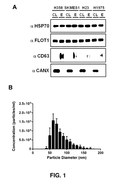
Figure 1. Exosomes are secreted by NSCLC cells. A Protein identification of
exosomes demonstrates the presence of exosome markers, and the absence of non-
exosomal calnexin. B Exosomes secreted by NSCLC have the expected size
distribution. C Hypoxia increases the secretion of exosomes, but does not
modify
exosome size range. D Hypoxia significantly increases exosome secretion of
NSCLC
cells. CL: cell lysate; E: exosome lysate.
Figure 2. Hypoxia modifies exosome content. Exosomes were harvested from
conditioned media from cells cultured for 24 hours under normoxic (21% 02) or
hypoxic (2% 02) conditions. A Scanning electron microscopy demonstrates
classical
exosome morphology. B Quantitative mass spectrometry revealed 55 proteins that
are
commonly upregulated under hypoxia, n = 5, FDR 1%. C,D protein targets were
validated with western blotting and ELISA.
Figure 3. Proteins upregulated correlate to patient disease progression. A
Exosomes
isolated from NSCLC patients show the expected size range and morphology. B, C
Hypoxic protein markers identified in vitro are upregulated in patients that
relapse
within the first 18 months. D ROC curve of combined protein signature (GANAB,
VCP, and Galectin-3-Binding Protein) for identifying patients that relapse
within 12
months. E Disease free survival of patients in relation to their exosome
content.
Patients that had at least 2 of the above markers highly expressed progressed
rapidly,
compared to patients that had only one or no markers expressed in their
exosomes.
CA 03043495 2019-05-10
WO 2018/094469
PCT/AU2017/051298
6
Figure 4. Other upregulated proteins identified in hypoxic exosomes have
prognostic
value. TNC was upregulated under hypoxia and is more abundant in exosomes of
NSCLC patients that progress rapidly.
Figure 5. Individual ROC and survival curves of proteins used in patient
signature.
Figure 6. Hypoxia-induced changes to the protein composition of NSCLC cell-
derived exosomes. a, The morphology of isolated exosomes was assessed using
transmission electron microscopy. Images of normoxic and hypoxic SKMES1-
derived
exosomes (Size bar 200 nm) also indicate clear upregulation of exosome
concentration. b, Nanoparticle analysis using TRPS of exosomes isolated from 4
1() different
NSCLC cell lines demonstrating the majority of exosomes have a size range
between 30 and 150 nm. c, Quantitative mass spectrometry identified 32
proteins to
be commonly upregulated in H358 and SKMES1 exosomes (FDR < 0.1%; n = 5). d,
e, Mass spectrometry results were confirmed using Western blot analysis of VCP
(FLOT1 is used as a loading control), and ELISA for MAC2BP, TNC, PSMA2, and
THBS1 in H358, SKMES1, H23, and H1975 NSCLC cell lines (= ¨ H358, = ¨
SKMES1, 1 ¨ H23, = ¨ H1975). *p<0.05, **p<0.01.
Figure 7. Hypoxic exosome signature prognosticates disease progression in
NSCLC
patients. a, b, Exosomes can be isolated from NSCLC plasma based on morphology
as shown by TEM (size bar 200 nm), and size distribution of 20 ¨ 150 nm. c,
TRPS
demonstrates that there is no difference in exosome concentration in plasma
from
healthy controls or patients that progress within 18 months or patients
without relapse
at 18 months. d, Exosomes isolated from NSCLC patients show an enrichment of
VCP in patients that progress with 18 months compared to patients that did not
relapse and healthy controls (FLOT1 is used as a loading control). e, The
hypoxic
exosome signature is upregulated in exosome derived from patients that
progress with
18 months. f, The number of hypoxic protein markers that exceed Youden's index
threshold value demonstrates a clear separation between patients that progress
within
18 months or patients without relapse at 18 months. g, Kaplan-Meier shows a
clear
separation of patient DFS based on the abundance of proteins from the hypoxic
exosome signature (>3 markers that exceed the Youden's index value). h, ROC
curve
demonstrates that the hypoxic exosome signature is a perfect prognostic marker
of
disease progression (<18 months) in NSCLC patients, while exosome
concentration
does not have prognostic value. i, Kaplan-Meier curve demonstrates the hypoxic
exosome signature also correlates with overall survival in NSCLC patients.
CA 03043495 2019-05-10
WO 2018/094469
PCT/AU2017/051298
7
Figure 8. The hypoxic exosome signature is derived from lung cells that have
undergone EMT. a, GSEA identified the hallmark epithelial-to-mesenchymal
transition gene set was significantly associated with exosomes derived from
hypoxic
NSCLC cells. b, Immunofluorescence of normal lung epithelial (30KT) and
transformed lung mesenchymal cells (30KTP53/KRAs/KB1) demonstrating
oncogenically
induced phenotypic transition to a mesenchymal phenotype. c, western blot in
cell
lysates demonstrates the loss of the epithelial marker E-cadherin and gain of
the
mesenchymal marker vimentin in 30KTP53/KRAsill(B1 cells. d, western blot of
VCP in
exosomes derived from epithelial (30KT) and mesenchymal (30KTP53/KRAsill(B1)
lung
cells (CD9 is used as a loading control). e, ELISA of MAC2BP, TNC, PSMA2, and
THBS1 in exosomes derived from epithelial (30KT) and mesenchymal
(3 oKTp5 3 /KRAS/LKB 1)
lung cells. *p<0.05, **p<0.01
***p<0.001. f,
Immunohistochemistry of primary tumours demonstrates the loss of E-cadherin
expression correlates to the patients that were stratified into the high
signature group
(>3 markers that exceed the Youden's index value).
Figure 9. Confirmation that the hypoxic exosome signature prognosticates
disease
relapse in NSCLC patients. a, b, "F-FDG PET/CT images of 2 patients
(confirmation
cohort) that are tracked in c at indicated points. c, In support of the
discovery cohort,
exosome concentration in patients that relapse within 18 months compared to
patients
that relapse after 18 months was similar, in particular patient 44 and 53 are
indicated.
d, The number of hypoxic protein markers that exceed Youden's index threshold
value demonstrates a clear separation between patients that progress within 18
months
or patients without relapse at 18 months. e, Kaplan-Meier plot of DFS of NSCLC
patients that have low abundance or high abundance of hypoxic exosome proteins
indicates a clear separation in DFS. f, ROC curve analysis again shows a
perfect
classification of patients that will progress within 18 months. g, Kaplan-
Meier plot
confirms the signature is also a prognostic marker of overall survival in
NSCLC
patients.
Figure 10. Hypoxia increases exosome secretion from NSCLC cells. a, Exosome
isolated from NSCLC cell lines express canonical exosome markers HSP70, FLOT1,
and CD63. The cell marker CANX is only found in cell lysates, not exosome
lysates.
b, Hypoxia increases exosome secretion from NSCLC cell lines. n = 3 SEM,
*p<0.05, **p<0.01 ***p<0.001.
CA 03043495 2019-05-10
WO 2018/094469
PCT/AU2017/051298
8
Figure 11. Discovery cohort demonstrates exosomal proteins are associated with
disease progression in NSCLC patients. a ¨ e, Individual Kaplan-Meier and ROC
curves of each protein in the hypoxic exosome signature.
Figure 12. Gene set enrichment analysis (GSEA) identified gene sets that were
significantly elevated in exosomes derived from hypoxic NSCLC cells. A,
Heatmap
of proteins identified in the EMT gene set. b ¨ e, GSEA using the total
exosome
protein expression dataset against hallmark gene sets reveals that hypoxic
exosomes
are enriched in proteins associated with glycolysis, MYC targets, E2F targets,
and
xenobiotic metabolism (FDR < 0.05). NES ¨ Normalised enrichment score.
Figure 13. Reduced E-cadherin expression is correlated to the number of
signature
proteins that exceeds Youden's index threshold values, a, table of IHC scores
in
reference to the signature score. b, Low E-cadherin IHC scores are associated
more
prominently with patients that relapse within 18 months.
Figure 14. Upregulated signature proteins in the confirmation cohort
correlates with
DFS. a, western blot of VCP demonstrates an upregulation of patients that
progress
within 18 months compared to patients that progress after 18 months (FLOT1 is
used
as a loading control). b, individual signature values of patient 44 and 53,
show patient
53 who progresses within 18 months has significantly elevated baseline levels
of the
signature proteins compared to patient 44.
DETAILED DESCRIPTION
The present invention is at least partly predicated on the surprising
discovery
that hypoxia-induced exosomal proteins identified in vitro are accurate
prognostic
biomarkers of cancer progression and aggressiveness in patients.
In one aspect, the invention provides a method of determining the
aggressiveness of a cancer in a subject, said method including the step of
determining
an expression level of one or a plurality of markers in an exosome sample of
the
subject, wherein the markers comprise one or more of those proteins listed in
Table 1
and/or Table 2 and an expression level of the one or plurality of markers
indicates or
correlates with a level of aggressiveness of the cancer.
In a related aspect, the invention provides a method of determining a
prognosis
for a cancer in a subject, said method including the step of determining an
expression
level of one or a plurality of markers in an exosome sample of the subject,
wherein the
markers comprise one or more of those proteins listed in Table 1 and/or Table
2 and
CA 03043495 2019-05-10
WO 2018/094469
PCT/AU2017/051298
9
an expression level of the one or plurality of markers indicates or correlates
with a
less or more favourable prognosis for said cancer.
With respect to the above aspects, the one or plurality of markers are
suitably
selected from the group consisting of Galectin-3-Binding Protein, Transitional
endoplasmic reticulum ATPase, Neutral alpha-glucosidase AB, 60 kDa heat shock
protein, Lysyl oxidase homolog 2, Tenascin C, Fatty acid synthase, Agrin,
Aspartyl
aminopeptidase, Proteasome subunit alpha type-1, Proteasome subunit alpha type-
2,
Proteasome subunit alpha type-3, Proteasome subunit alpha type-4, Proteasome
subunit alpha type-5, Proteasome subunit alpha type-6, Proteasome subunit beta
type-
1, Proteasome subunit beta type-2, Proteasome subunit beta type-3, Proteasome
subunit beta type-4, Proteasome subunit beta type-5, Proteasome subunit beta
type-6,
Proteasome subunit beta type-7, Proteasome subunit beta type-8, Thrombospondin-
1,
Latent Transforming Growth Factor Beta Binding Protein 3 and any combination
thereof In one particular embodiment, the one or plurality of markers are
selected
from the group consisting of Galectin-3-Binding Protein, Transitional
endoplasmic
reticulum ATPase, Tenascin C, Proteasome subunit alpha type-2, Thrombospondin-
1
and any combination thereof
As generally used herein, an expression level of one or more of: (a) the 55
marker proteins identified as upregulated in Table 1; and (b) the 32 marker
proteins
identified as upregulated in Table 2; may refer to the expression level of a
nucleic acid
encoding said protein (e.g., RNA, mRNA and cDNA), the protein itself or both,
unless otherwise specified.
As generally used herein, the terms "cancer", "tumour", "malignant" and
"malignancy" refer to diseases or conditions, or to cells or tissues
associated with the
diseases or conditions, characterized by aberrant or abnormal cell
proliferation,
differentiation and/or migration often accompanied by an aberrant or abnormal
molecular phenotype that includes one or more genetic mutations or other
genetic
changes associated with oncogenesis, expression of tumour markers, loss of
tumour
suppressor expression or activity and/or aberrant or abnormal cell surface
marker
expression.
By "aggressiveness" and "aggressive" is meant a property or propensity for a
cancer to have a relatively poor prognosis due to one or more of a combination
of
features or factors including: at least partial resistance to therapies
available for cancer
CA 03043495 2019-05-10
WO 2018/094469
PCT/AU2017/051298
treatment; invasiveness; metastatic potential; recurrence after treatment; and
a low
probability of patient survival, although without limitation thereto.
In particular embodiments, the proteins provided herein, such as those
provided in Table 1 and Table 2, are prognostic for aggressive disease, and in
5
particular a shorter time to pathological recurrence and/or a shorter patient
survival
time. In further embodiments, the proteins provided herein, such as those
provided in
Table 1 and Table 2, correlate with or indicate metastatic cancer, and more
particularly, metastatic NSCLC. In this regard, it will be apparent that a
number of the
32 proteins provided in Table 2 are also listed in Table 1, with the exception
of, for
10 example, LTBP3.
Cancers may include any aggressive or potentially aggressive cancers,
tumours or other malignancies such as listed in the NCI Cancer Index at
http://www.cancer.govicancertopics/alphalist, including all major cancer forms
such
as sarcomas, carcinomas, lymphomas, leukaemias and blastomas, although without
limitation thereto. These may include breast cancer, lung cancer inclusive of
lung
adenocarcinoma and mesothelioma, cancers of the reproductive system inclusive
of
ovarian cancer, cervical cancer, uterine cancer and prostate cancer, cancers
of the
brain and nervous system, head and neck cancers, gastrointestinal cancers
inclusive of
colon cancer, colorectal cancer and gastric cancer, liver cancer, kidney
cancer, skin
cancers such as melanoma and skin carcinomas, blood cell cancers inclusive of
lymphoid cancers and myelomonocytic cancers, cancers of the endocrine system
such
as pancreatic cancer and pituitary cancers, musculoskeletal cancers inclusive
of bone
and soft tissue cancers, although without limitation thereto.
In particular embodiments, the cancer includes breast cancer, lung cancer,
ovarian cancer, cervical cancer, uterine cancer, prostate cancer, cancer of
the brain
and nervous system, head and neck cancer, colon cancer, colorectal cancer,
gastric
cancer, liver cancer, kidney cancer, bladder cancer, skin cancer, pancreatic
cancer,
pituitary cancer or adrenal cancer. More preferably, the cancer is or
comprises lung
cancer, such as NSCLC.
In particular embodiments, the cancer of the aspects disclosed herein is, or
comprises, a lung cancer. To this end, it would be apparent that lung cancer
may
include any aggressive lung cancers and cancer subtypes known in the art, such
as
non-small cell carcinoma (i.e., squamous cell carcinoma, adenocarcinoma and
large
cell carcinoma), small cell carcinoma and mesothelioma. In one preferred
CA 03043495 2019-05-10
WO 2018/094469
PCT/AU2017/051298
11
embodiment, the lung cancer is or comprises non-small cell lung carcinoma
(NSCLC).
The terms "prognosis" and "prognostic" are used herein to include making a
prognosis, which can provide for predicting a clinical outcome (with or
without
medical treatment), selecting an appropriate course of treatment (or whether
treatment
would be effective) and/or monitoring a current treatment and potentially
changing
the treatment. This may be at least partly based on determining the gene
and/or
protein expression levels of the one or plurality of markers by the methods of
the
invention, which may be in combination with determining the expression levels
of
additional protein and/or other nucleic acid biomarkers. A prognosis may also
include
a prediction, forecast or anticipation of any lasting or permanent physical or
psychological effects of cancer suffered by the subject after the cancer has
been
successfully treated or otherwise resolved. Furthermore, prognosis may include
one or
more of determining metastatic potential or occurrence, therapeutic
responsiveness,
implementing appropriate treatment regimes, determining the probability,
likelihood
or potential for cancer recurrence after therapy and prediction of development
of
resistance to established therapies (e.g., chemotherapy). It would be
appreciated that a
positive prognosis typically refers to a beneficial clinical outcome or
outlook, such as
long-term survival without recurrence of the subject's cancer, whereas a
negative
prognosis typically refers to a negative clinical outcome or outlook, such as
cancer
recurrence or progression.
In one embodiment of the method of the two aforementioned aspects, a
relatively decreased expression level of the one or plurality of markers
indicates or
correlates with a more favourable prognosis and/or a less aggressive cancer;
and/or a
relatively increased expression level of the one or plurality of markers
indicates or
correlates with a less favourable prognosis and/or a highly aggressive cancer.
In one particular embodiment, the cancer prognosis or aggressiveness is used,
at least in part, to determine a likelihood of metastasis of the cancer in
said subject.
As used herein, "metastasis" or "metastatic", refers to the migration or
transfer
of malignant tumour cells, or neoplasms, via the circulatory or lymphatic
systems or
via natural body cavities, typically from the primary focus of tumour, cancer
or a
neoplasia to a distant site in the body, and the subsequent development of one
or more
secondary tumours or colonies thereof in the one or more new locations.
"Metastases"
refers to the secondary tumours or colonies formed as a result of a metastasis
and
CA 03043495 2019-05-10
WO 2018/094469
PCT/AU2017/051298
12
encompasses micro-metastases as well as regional, including lymph node, and
distant
metastases.
Suitably, a relatively decreased expression level of the one or plurality of
markers indicates or correlates with a decreased likelihood of metastasis of
said
cancer; and/or a relatively increased expression level of the one or plurality
of
markers indicates or correlates with an increased likelihood of metastasis of
said
cancer.
In one embodiment, the cancer prognosis or aggressiveness is used, at least in
part, to determine whether the subject would benefit from treatment of the
cancer. By
way of example, a patient with a favourable prognosis and/or a less aggressive
cancer
may be less likely to suffer from rapid local progression of the cancer and/or
metastasis and can be spared from more aggressive monitoring and/or therapy.
In another embodiment, the cancer prognosis or aggressiveness is used, at
least
in part, to develop a treatment strategy for the subject.
In one embodiment, the cancer prognosis or aggressiveness is used, at least in
part, to determine disease progression or recurrence in the subject.
In one embodiment, the cancer prognosis or aggressiveness is used, at least in
part, to determine an estimated time of survival.
For the purposes of this invention, by "isolated" is meant material that has
been removed from its natural state or otherwise been subjected to human
manipulation. Isolated material may be substantially or essentially free from
components that normally accompany it in its natural state, or may be
manipulated so
as to be in an artificial state together with components that normally
accompany it in
its natural state. Isolated material may be in native, chemical synthetic or
recombinant
form.
As used herein a "gene" is a nucleic acid which is a structural, genetic unit
of a
genome that may include one or more amino acid-encoding nucleotide sequences
and
one or more non-coding nucleotide sequences inclusive of promoters and other
5'
untranslated sequences, introns, polyadenylation sequences and other 3'
untranslated
sequences, although without limitation thereto. In most cellular organisms a
gene is a
nucleic acid that comprises double-stranded DNA.
The term "nucleic acid" as used herein designates single- or double-stranded
DNA and RNA. DNA includes genomic DNA and cDNA. RNA includes mRNA,
RNA, RNAi, siRNA, cRNA and autocatalytic RNA. Nucleic acids may also be DNA-
CA 03043495 2019-05-10
WO 2018/094469
PCT/AU2017/051298
13
RNA hybrids. A nucleic acid comprises a nucleotide sequence which typically
includes nucleotides that comprise an A, G, C, T or U base. However,
nucleotide
sequences may include other bases such as inosine, methylycytosine,
methylinosine,
methyladenosine and/or thiouridine, although without limitation thereto.
Also included are, "variant" nucleic acids that include nucleic acids that
comprise nucleotide sequences of naturally occurring (e.g., allelic) variants
and
orthologs (e.g., from a different species) of nucleic acids that respectively
encode the
one or plurality of markers provided herein. Preferably, nucleic acid variants
share at
least 70% or 75%, preferably at least 80% or 85% or more preferably at least
90%,
91%, 92%, 93%, 94%, 95%, 96%, 97%, 98% or 99% sequence identity with a
nucleotide sequence disclosed herein.
Also included are nucleic acid fragments. A 'fragment" is a segment, domain,
portion or region of a nucleic acid, which respectively constitutes less than
100% of
the nucleotide sequence. A non-limiting example is an amplification product or
a
primer or probe. In particular embodiments, a nucleic acid fragment may
comprise,
for example, at least 10, 15, 20, 25, 30 35, 40, 45, 50, 55, 60, 65, 70, 75,
80, 85, 90,
95, 100, 125, 150, 175, 200, 225, 250, 275, 300, 325, 350, 375, 400, 425, 450,
475,
500, 600, 700, 800, 900, 1000, 1500, 2000, 2500, 3000, 3500, 4000, 4500, 5000,
5500, 6000, 6500, 7000 and 7500 contiguous nucleotides of said nucleic acid.
As used herein, a "polynucleotide" is a nucleic acid having eighty (80) or
more contiguous nucleotides, while an "oligonucleotide" has less than eighty
(80)
contiguous nucleotides. A "probe" may be a single or double-stranded
oligonucleotide
or polynucleotide, suitably labelled for the purpose of detecting
complementary
sequences in Northern or Southern blotting, for example. A "primer" is usually
a
single-stranded oligonucleotide, preferably having 15-50 contiguous
nucleotides,
which is capable of annealing to a complementary nucleic acid "template" and
being
extended in a template-dependent fashion by the action of a DNA polymerase
such as
Tag polymerase, RNA-dependent DNA polymerase or SequenaseTM. A "template"
nucleic acid is a nucleic acid subjected to nucleic acid amplification.
By "protein" is meant an amino acid polymer. The amino acids may be
natural or non-natural amino acids, D- or L- amino acids as are well
understood in the
art. As would be appreciated by the skilled person, the term "protein" also
includes
within its scope phosphorylated forms of a protein (i.e., a phosphoprotein)
and/or
glycosylated forms of a protein (i.e. a glycoprotein). A "peptide" is a
protein having
CA 03043495 2019-05-10
WO 2018/094469
PCT/AU2017/051298
14
no more than fifty (50) amino acids. A "polypeptide" is a protein having more
than
fifty (50) amino acids.
Also provided are protein "variants" such as naturally occurring variants
(e.g.
allelic variants) and orthologs or isoforms of the one or plurality of markers
provided
herein, such as those listed in Table 1 and Table 2. Preferably, protein
variants share
at least 70% or 75%, preferably at least 80% or 85% or more preferably at
least 90%,
91%, 92%, 93%, 94%, 95%, 96%, 97%, 98% or 99% sequence identity with an amino
acid sequence of the one or plurality of markers disclosed herein or known in
the art.
To this end, Tables 1 and 2 also include Accession Numbers referencing an
example
of a protein sequence of the recited protein marker, as are well understood in
the art
and are incorporated by reference herein.
Also provided are protein fragments, inclusive of peptide fragments that
comprise less than 100% of an entire amino acid sequence. In particular
embodiments, a protein fragment may comprise, for example, at least 10, 15,
20, 25,
30 35, 40, 45, 50, 55, 60, 65, 70, 75, 80, 85, 90, 95, 100, 125, 150, 175,
200, 225, 250,
275, 300, 325, 350, 375, 400, 425, 450, 475, 500, 550, 600, 650, 700, 750,
800, 850,
900, 950, 1000, 1050, 1100, 1150 and 1200 contiguous amino acids of said
protein.
It would be appreciated by the skilled person that exosomes are small (i.e.,
typically 30-150 nm), cell-derived membrane vesicles of endocytic origin. They
may
contain lipids, nucleic acid and proteins, and are released into the
extracellular
environment upon fusion with the plasma membrane. Generally, exosomes are
characterized by the presence of marker proteins, including CD63, CD9, HSP70,
Flotillin-1 and TSG101, as well as their morphology and size.
In accordance with the methods of the present invention, an exosome sample
containing one or more exosomes may comprise or be obtained from most
biological
fluids including, without limitation, blood, serum, plasma, ascites, cyst
fluid, pleural
fluid, peritoneal fluid, cerebral spinal fluid, tears, urine, saliva, sputum,
nipple
aspirates, lymph fluid, fluid of the respiratory, intestinal, and
genitourinary tracts,
breast milk, intra-organ system fluid, or combinations thereof To this end, an
exosome sample may be isolated or purified from a biological fluid or sample,
such as
those provided above, so as to facilitate the removal of contaminating
proteins,
lipoproteins etc.
To this end, an exosome or exosome sample may be isolated by any means
known in the art, such as, but not limited to, ultracentrifugation, size-
exclusion
CA 03043495 2019-05-10
WO 2018/094469
PCT/AU2017/051298
chromatography, exosome precipitation (e.g., ExoQuick from System
Biosciences),
affinity-based capture of exosomes (e.g., affinity purification with
antibodies to
CD63, CD81, CD82, CD9, Alix, annexin, EpCAM, and Rab5) and any combination
thereof
5 As would be understood by the skilled person, the gene and/or protein
expression level of the one or more proteins provided herein may be relatively
(i)
higher, increased or greater; or (ii) lower, decreased or reduced when
compared to an
expression level in a control or reference sample, or to a threshold
expression level. In
one embodiment, an expression level may be classified as higher increased or
greater
10 if it exceeds a mean and/or median expression level of a reference
population. In one
embodiment an expression level may be classified as lower, decreased or
reduced if it
is less than the mean and/or median expression level of the reference
population. In
this regard, a reference population may be a group of subjects who have the
same
cancer type, subgroup, stage and/or grade as said mammal for which the
expression
15 level is determined.
Terms such as "higher", "increased" and "greater" as used herein refer to an
elevated amount or level of a nucleic acid and/or protein, such as in an
exosome
sample, when compared to a control or reference level or amount. The
expression
level of the nucleic acid and/or protein of the one or plurality of markers
may be
relative or absolute. In some embodiments, the gene and/or protein expression
of the
one or plurality of markers is higher, increased or greater if its level of
expression is
more than about 0.5%, 1%, 2%, 3%, 4%, 5%, 10%, 15%, 20%, 25%, 30%, 35%, 40%,
45%, 50%, 55%, 60%, 65%, 70%, 75%, 80%, 85%, 90%, 95%, 100%, 150%, 200%,
300%, 400% or at least about 500% above the level of gene and/or protein
expression
of the respective or corresponding protein in a control or reference level or
amount.
The terms, "lower", "reduced" and "decreased", as used herein refer to a lower
amount or level of a nucleic acid and/or protein, such as in an exosome
sample, when
compared to a control or reference level or amount. The expression level of
the
nucleic acid and/or protein of the one or plurality of markers provided herein
may be
relative or absolute. In some embodiments, the gene and/or protein expression
of the
one or plurality of markers is lower, reduced or decreased if its level of
expression is
less than about 95%, 90%, 80%, 70%, 60%, 50%, 40%, 30%, 20% or 10%, or even
less than about 5%, 4%, 3%, 2%, 1%, 0.5%, 0.1%, 0.01%, 0.001% or 0.0001% of
the
CA 03043495 2019-05-10
WO 2018/094469
PCT/AU2017/051298
16
level or amount of the gene and/or protein expression of the respective or
corresponding protein in a control or reference level or amount.
The term "control sample" typically refers to a biological sample, such as an
exosome sample, from a (healthy) non-diseased individual not having cancer. In
one
embodiment, the control sample may be from a subject known to be free of
cancer or
a sample that was obtained from the subject at an earlier timepoint.
Alternatively, the
control sample may be from a subject in remission from cancer. The control
sample
may be a pooled, average or an individual sample. An internal control is a
marker
from the same biological sample (e.g., exosome sample) being tested.
As used herein, a gene and/or protein expression level may be an absolute or
relative amount thereof Accordingly, in some embodiments, the gene and/or
protein
expression level of the one or plurality of markers provided herein is
compared to a
control level of expression, such as the level of gene and/or protein
expression of one
or a plurality of "housekeeping" genes and/or proteins in an exosome sample of
the
subject.
In further embodiments, the gene and/or protein expression level of the one or
plurality of markers is compared to a threshold level of expression, such as a
level of
gene and/or protein expression in an exosome sample. A threshold level of
expression
is generally a quantified level of gene and/or protein expression of the one
or plurality
of markers of the invention. Typically, a gene and/or protein expression level
of the
one or plurality of markers in an exosome sample that exceeds or falls below
the
threshold level of expression is predictive of a particular disease state or
outcome. The
nature and numerical value (if any) of the threshold level of expression will
typically
vary based on the method chosen to determine the expression of the one or more
genes, or products thereof, used in determining, for example, a prognosis
and/or a
response to anticancer therapy, in the subject.
A person of skill in the art would be capable of determining a threshold level
of gene and/or protein expression in an exosome sample that may be used in
determining, for example, a prognosis and/or a response to anticancer therapy,
using
any method of measuring gene or protein expression known in the art, such as
those
described herein. In one embodiment, the threshold level is a mean and/or
median
gene and/or protein expression level (median or absolute) of the one or
plurality of
markers in a reference population, that, for example, have the same cancer
type,
CA 03043495 2019-05-10
WO 2018/094469
PCT/AU2017/051298
17
subgroup, stage and/or grade as said subject for which the expression level is
determined. Additionally, the concept of a threshold level of expression
should not be
limited to a single value or result. In this regard, a threshold level of
expression may
encompass multiple threshold expression levels that could signify, for
example, a
high, medium, or low probability of, for example, metastasis of the subject's
cancer.
In one embodiment, a lower gene and/or protein expression level of the one or
plurality of markers provided herein indicates or correlates with relatively
increased
responsiveness of the cancer to the anti-cancer treatment. In alternative
embodiments,
a lower gene and/or protein expression level of the one or plurality of
markers
provided herein indicates or correlates with relatively decreased
responsiveness of the
cancer to the anti-cancer treatment.
The terms "determining", "measuring", "evaluating", "assessing" and
"assaying" are used interchangeably herein and may include any form of
measurement known in the art, such as those described hereinafter.
Determining, assessing, evaluating, assaying or measuring corresponding
nucleic acids of the one or plurality of markers provided herein, such as RNA,
mRNA
and cDNA, may be performed by any technique known in the art. These may be
techniques that include nucleic acid sequence amplification, nucleic acid
hybridization, nucleotide sequencing, mass spectroscopy and combinations of
any
these.
Nucleic acid amplification techniques typically include repeated cycles of
annealing one or more primers to a "template" nucleotide sequence under
appropriate
conditions and using a polymerase to synthesize a nucleotide sequence
complementary to the target, thereby "amplifying" the target nucleotide
sequence.
Nucleic acid amplification techniques are well known to the skilled addressee,
and
include but are not limited to polymerase chain reaction (PCR); strand
displacement
amplification (SDA); rolling circle replication (RCR); nucleic acid sequence-
based
amplification (NASBA), Q-I3 replicase amplification; helicase-dependent
amplification (HAD); loop-mediated isothermal amplification (LAMP); nicking
enzyme amplification reaction (NEAR) and recombinase polymerase amplification
(RPA), although without limitation thereto. As generally used herein, an
"amplification product" refers to a nucleic acid product generated by a
nucleic acid
amplification technique.
CA 03043495 2019-05-10
WO 2018/094469
PCT/AU2017/051298
18
PCR includes quantitative and semi-quantitative PCR, real-time PCR, allele-
specific PCR, methylation-specific PCR, asymmetric PCR, nested PCR, multiplex
PCR, touch-down PCR, digital PCR and other variations and modifications to
"basic"
PCR amplification.
Nucleic acid amplification techniques may be performed using DNA or RNA
extracted, isolated or otherwise obtained from a cell or tissue source. In
other
embodiments, nucleic acid amplification may be performed directly on
appropriately
treated cell or tissue samples.
Nucleic acid hybridization typically includes hybridizing a nucleotide
1() sequence,
typically in the form of a probe, to a target nucleotide sequence under
appropriate conditions, whereby the hybridized probe-target nucleotide
sequence is
subsequently detected. Non-limiting examples include Northern blotting, slot-
blotting,
in situ hybridization and fluorescence resonance energy transfer (FRET)
detection,
although without limitation thereto. Nucleic acid hybridization may be
performed
using DNA or RNA extracted, isolated, amplified or otherwise obtained from a
cell or
tissue source or directly on appropriately treated cell or tissue samples.
It will also be appreciated that a combination of nucleic acid amplification
and
nucleic acid hybridization may be utilized.
Determining, assessing, evaluating, assaying or measuring protein levels of
the
one or plurality of exosomal proteins may be performed by any technique known
in
the art that is capable of detecting such proteins whether on the surface or
internally
expressed in an exosome, or proteins that are isolated, extracted or otherwise
obtained
from the exosome sample of the subject. These techniques include antibody-
based
detection that uses one or more antibodies which bind the protein,
electrophoresis,
isoelectric focussing, protein sequencing, chromatographic techniques and mass
spectroscopy and combinations of these, although without limitation thereto.
Antibody-based detection may include flow cytometry using fluorescently-
labelled
antibodies, ELISA, immunoblotting, immunoprecipitation, radioimmunoassay (MA)
and immuncytochemistry, although without limitation thereto.
It will be appreciated that determining the expression of the one or plurality
of
markers provided herein may include determining both the nucleic acid levels
thereof,
such as by nucleic acid amplification and/or nucleic acid hybridization, and
the
protein levels thereof Accordingly, detection and/or measurement of expression
of
the one or plurality of markers from the exosome sample of the subject may be
CA 03043495 2019-05-10
WO 2018/094469
PCT/AU2017/051298
19
performed by any of those methods or combinations thereof described herein
(e.g
measuring mRNA levels or an amplified cDNA copy thereof and/or by measuring a
protein product thereof), albeit without limitation thereto.
In light of the foregoing, it will further be appreciated that an expression
level
of the one or plurality of markers provided herein may be an absolute or
relative
amount of an expressed gene or gene product thereof, inclusive of nucleic
acids such
as RNA, mRNA and cDNA, and/or protein.
Suitably, the method of the aforementioned aspects further includes the step
of
diagnosing said subject as having: (i) a highly aggressive cancer or a less
aggressive
cancer; and/or (ii) a less favourable prognosis or a more favourable
prognosis.
In a further aspect, the invention provides a method of predicting the
responsiveness of a cancer to an anti-cancer treatment in a subject, said
method
including the step of determining an expression level of one or a plurality of
markers
in an exosome sample of the subject, wherein the markers comprise one or more
of
those proteins listed in Table 1 and/or Table 2 and an altered or modulated
expression
level of the one or plurality of markers indicates or correlates with
relatively increased
or decreased responsiveness of the cancer to the anti-cancer treatment.
As would be understood by the skilled person, the expression level of a gene
or protein may be deemed to be "altered" or "modulated" when the expression
level is
higher/increased or lower/decreased when compared to a control or reference
sample
or expression level, such as a threshold level. In one embodiment, the
expression level
may be classified as high if it is greater than a mean and/or median relative
expression
level of a reference population and the expression level may be classified as
low if it
is less than the mean and/or median expression level of the reference
population. In
this regard, a reference population may be a group of subjects who have the
same
cancer type, subgroup, stage and/or grade as said mammal for which the
expression
level is determined. Furthermore, the expression level may be relative or
absolute.
Suitably, the one or plurality of markers are selected from the group
consisting
of Galectin-3-Binding Protein, Transitional endoplasmic reticulum ATPase,
Neutral
alpha-glucosidase AB, 60 kDa heat shock protein, Lysyl oxidase homolog 2,
Tenascin
C, Fatty acid synthase, Agrin, Aspartyl aminopeptidase, Proteasome subunit
alpha
type-1, Proteasome subunit alpha type-2, Proteasome subunit alpha type-3,
Proteasome subunit alpha type-4, Proteasome subunit alpha type-5, Proteasome
subunit alpha type-6, Proteasome subunit beta type-1, Proteasome subunit beta
type-2,
CA 03043495 2019-05-10
WO 2018/094469
PCT/AU2017/051298
Proteasome subunit beta type-3, Proteasome subunit beta type-4, Proteasome
subunit
beta type-5, Proteasome subunit beta type-6, Proteasome subunit beta type-7,
Proteasome subunit beta type-8, Thrombospondin-1, Latent Transforming Growth
Factor Beta Binding Protein 3 and any combination thereof In one particular
5 embodiment, the one or plurality of markers are selected from the group
consisting of
Galectin-3-Binding Protein, Transitional endoplasmic reticulum ATPase,
Tenascin C,
Proteasome subunit alpha type-2, Thrombospondin-1 and any combination thereof
In one embodiment, a higher expression level of the one or plurality of
markers indicates or correlates with relatively increased responsiveness of
the cancer
10 to the anti-cancer treatment. In alternative embodiments, a higher
expression level of
the one or plurality of markers indicates or correlates with relatively
decreased
responsiveness of the cancer to the anti-cancer treatment.
With respect to the invention of the aforementioned aspects, the method
suitably includes the further step of treating the cancer in the subject.
15 Further aspects of the invention relate to treatment of cancer in a
subject.
In one particular aspect, the cancer treatment is performed in conjunction
with
determining an expression level of one or a plurality of markers in an exosome
sample
of the subject, wherein the markers comprise one or more of those proteins
listed in
Table 1 and/or Table 2, and based on the determination made, initiating,
continuing,
20 modifying or discontinuing the cancer treatment.
Suitably, the one or plurality of markers are selected from the group
consisting
of Galectin-3-Binding Protein, Transitional endoplasmic reticulum ATPase,
Neutral
alpha-glucosidase AB, 60 kDa heat shock protein, Lysyl oxidase homolog 2,
Tenascin
C, Fatty acid synthase, Agrin, Aspartyl aminopeptidase, Proteasome subunit
alpha
type-1, Proteasome subunit alpha type-2, Proteasome subunit alpha type-3,
Proteasome subunit alpha type-4, Proteasome subunit alpha type-5, Proteasome
subunit alpha type-6, Proteasome subunit beta type-1, Proteasome subunit beta
type-2,
Proteasome subunit beta type-3, Proteasome subunit beta type-4, Proteasome
subunit
beta type-5, Proteasome subunit beta type-6, Proteasome subunit beta type-7,
Proteasome subunit beta type-8, Thrombospondin-1, Latent Transforming Growth
Factor Beta Binding Protein 3, and any combination thereof In one particular
embodiment, the one or plurality of markers are selected from the group
consisting of
Galectin-3-Binding Protein, Transitional endoplasmic reticulum ATPase,
Tenascin C,
Proteasome subunit alpha type-2, Thrombospondin-1 and any combination thereof
CA 03043495 2019-05-10
WO 2018/094469
PCT/AU2017/051298
21
In this regard, it would be appreciated that those methods described herein
for
predicting the responsiveness of a cancer to an anti-cancer agent may further
include
the step of administering to the mammal a therapeutically effective amount of
the
anti-cancer treatment, such as an anticancer agent. In a preferred embodiment,
the
anticancer treatment is administered when the gene and/or protein expression
level of
the one or plurality of markers described herein indicates or correlates with
relatively
increased responsiveness of the cancer to the anti-cancer agent.
Suitably, the agent(s) is/are administered to a subject as a pharmaceutical
composition comprising a pharmaceutically-acceptable carrier, diluent or
excipient. In
this regard, any dosage form and route of administration, such as those
provided
therein, may be employed for providing a subject with the composition of the
invention.
Cancer treatments may include drug therapy, such as small organic or
inorganic molecules, chemotherapy, antibody, nucleic acid and other
biomolecular
therapies, radiation therapy, surgery, nutritional therapy, relaxation or
meditational
therapy and other natural or holistic therapies, although without limitation
thereto.
Generally, drugs (e.g., small organic or inorganic molecules), biomolecules
(e.g
antibodies, inhibitory nucleic acids such as siRNA) or chemotherapeutic agents
are
referred to herein as "anti-cancer therapeutic agents" or "anti-cancer
agents".
Methods of treating cancer may be prophylactic, preventative or therapeutic
and suitable for treatment of cancer in mammals, particularly humans. As used
herein,
"treating", "treat" or "treatment" refers to a therapeutic intervention,
course of action
or protocol that at least ameliorates a symptom of cancer after the cancer
and/or its
symptoms have at least started to develop. As used herein, "preventing",
"prevent" or
"prevention" refers to therapeutic intervention, course of action or protocol
initiated
prior to the onset of cancer and/or a symptom of cancer so as to prevent,
inhibit or
delay or development or progression of the cancer or the symptom.
The term "therapeutically effective amount" describes a quantity of a
specified agent sufficient to achieve a desired effect in a subject being
treated with
that agent. For example, this can be the amount of a chemotherapeutic agent
necessary
to reduce, alleviate and/or prevent a cancer or cancer associated disease,
disorder or
condition. In some embodiments, a "therapeutically effective amount" is
sufficient to
reduce or eliminate a symptom of a cancer. In other embodiments, a
"therapeutically
effective amount" is an amount sufficient to achieve a desired biological
effect, for
CA 03043495 2019-05-10
WO 2018/094469
PCT/AU2017/051298
22
example an amount that is effective to decrease or prevent cancer growth
and/or
metastasis.
Ideally, a therapeutically effective amount of an agent is an amount
sufficient
to induce the desired result without causing a substantial cytotoxic effect in
the
subject. The effective amount of an agent useful for reducing, alleviating
and/or
preventing a cancer will be dependent on the subject being treated, the type
and
severity of any associated disease, disorder and/or condition (e.g., the
number and
location of any associated metastases), and the manner of administration of
the
therapeutic composition.
1() Suitably,
the anti-cancer therapeutic agent is administered to a mammal as a
pharmaceutical composition comprising a pharmaceutically-acceptable carrier,
diluent
or excipient.
By "pharmaceutically-acceptable carrier, diluent or excipient" is meant a
solid or liquid filler, diluent or encapsulating substance that may be safely
used in
systemic administration. Depending upon the particular route of
administration, a
variety of carriers, well known in the art may be used. These carriers may be
selected
from a group including sugars, starches, cellulose and its derivatives, malt,
gelatine,
talc, calcium sulfate, liposomes and other lipid-based carriers, vegetable
oils,
synthetic oils, polyols, alginic acid, phosphate buffered solutions,
emulsifiers, isotonic
saline and salts such as mineral acid salts including hydrochlorides, bromides
and
sulfates, organic acids such as acetates, propionates and malonates and
pyrogen-free
water.
A useful reference describing pharmaceutically acceptable carriers, diluents
and excipients is Remington's Pharmaceutical Sciences (Mack Publishing Co.
N.J.
USA, 1991), which is incorporated herein by reference.
Any safe route of administration may be employed for providing a patient with
the composition of the invention. For example, oral, rectal, parenteral,
sublingual,
buccal, intravenous, intra-articular, intra-muscular, intra-dermal,
subcutaneous,
inhalational, intraocular, intraperitoneal, intracerebroventricular,
transdermal and the
like may be employed. Intra-muscular and subcutaneous injection is
appropriate, for
example, for administration of immunotherapeutic compositions, proteinaceous
vaccines and nucleic acid vaccines.
Dosage forms include tablets, dispersions, suspensions, injections, solutions,
syrups, troches, capsules, suppositories, aerosols, transdermal patches and
the like.
CA 03043495 2019-05-10
WO 2018/094469
PCT/AU2017/051298
23
These dosage forms may also include injecting or implanting controlled
releasing
devices designed specifically for this purpose or other forms of implants
modified to
act additionally in this fashion. Controlled release of the therapeutic agent
may be
effected by coating the same, for example, with hydrophobic polymers including
acrylic resins, waxes, higher aliphatic alcohols, polylactic and polyglycolic
acids and
certain cellulose derivatives such as hydroxypropylmethyl cellulose. In
addition, the
controlled release may be effected by using other polymer matrices, liposomes
and/or
mi cro sphere s.
Compositions of the present invention suitable for oral or parenteral
administration may be presented as discrete units such as capsules, sachets or
tablets
each containing a pre-determined amount of one or more therapeutic agents of
the
invention, as a powder or granules or as a solution or a suspension in an
aqueous
liquid, a non-aqueous liquid, an oil-in-water emulsion or a water-in-oil
liquid
emulsion. Such compositions may be prepared by any of the methods of pharmacy
but all methods include the step of bringing into association one or more
agents as
described above with the carrier which constitutes one or more necessary
ingredients.
In general, the compositions are prepared by uniformly and intimately admixing
the
agents of the invention with liquid carriers or finely divided solid carriers
or both, and
then, if necessary, shaping the product into the desired presentation.
The above compositions may be administered in a manner compatible with the
dosage formulation, and in such amount as is pharmaceutically-effective. The
dose
administered to a patient, in the context of the present invention, should be
sufficient
to effect a beneficial response in a patient over an appropriate period of
time. The
quantity of agent(s) to be administered may depend on the subject to be
treated
inclusive of the age, sex, weight and general health condition thereof,
factors that will
depend on the judgement of the practitioner.
In particular embodiments, the anti-cancer treatment and/or agent may be
directed at inhibiting the action of and/or decreasing the expression of the
one or
plurality of markers.
In other embodiments, the anti-cancer treatment and/or agent may be directed
at preventing or inhibiting metastasis of the cancer.
In alternative embodiments, the anti-cancer treatment and/or agent may be
directed at genes or gene products other than the one or plurality of markers
of the
invention. By way of example, the anti-cancer treatment may target genes or
gene
CA 03043495 2019-05-10
WO 2018/094469
PCT/AU2017/051298
24
products that are known to interact, directly or indirectly, with the one or
plurality of
markers.
In a particular embodiment, the invention provides a "companion diagnostic"
with respect to the cancer treatment, whereby the expression level of the one
or
plurality of markers of the invention provides information to a clinician or
the like
that is used for the safe and/or effective administration of said cancer
treatment.
Suitably, the cancer is of a type hereinbefore described, albeit without
limitation thereto.
Referring to the aforementioned aspects, the method suitably includes the
initial step of obtaining the exosome sample from the subject, such as from
those
biological samples and/or isolation methods hereinbefore described.
In a further aspect, the invention provides a method for identifying or
producing an agent for use in the treatment of cancer in a subject including
the steps
of:
(a) contacting a cell that expresses a marker listed in Table 1 and/or Table
2;
with a candidate agent; and
(b) determining whether the candidate agent modulates the expression and/or
an activity of the marker.
In certain embodiments, the candidate agent, at least partly, reduces,
eliminates, suppresses or inhibits the expression and/or the activity of the
marker.
Suitably, the agent possesses or displays little or no significant off-target
and/or nonspecific effects.
Preferably, the agent is an antibody or a small molecule.
Suitably, the marker is selected from the group consisting of Galectin-3-
Binding Protein, Transitional endoplasmic reticulum ATPase, Neutral alpha-
glucosidase AB, 60 kDa heat shock protein, Lysyl oxidase homolog 2, Tenascin
C,
Fatty acid synthase, Agrin, Aspartyl aminopeptidase, Proteasome subunit alpha
type-
1, Proteasome subunit alpha type-2, Proteasome subunit alpha type-3,
Proteasome
subunit alpha type-4, Proteasome subunit alpha type-5, Proteasome subunit
alpha
type-6, Proteasome subunit beta type-1, Proteasome subunit beta type-2,
Proteasome
subunit beta type-3, Proteasome subunit beta type-4, Proteasome subunit beta
type-5,
Proteasome subunit beta type-6, Proteasome subunit beta type-7, Proteasome
subunit
beta type-8, Thrombospondin-1, Latent Transforming Growth Factor Beta Binding
Protein 3 and any combination thereof In one particular embodiment, the one or
CA 03043495 2019-05-10
WO 2018/094469
PCT/AU2017/051298
plurality of markers are selected from the group consisting of Galectin-3-
Binding
Protein, Transitional endoplasmic reticulum ATPase, Tenascin C, Proteasome
subunit
alpha type-2, Thrombospondin-1 and any combination thereof
In embodiments relating to antibody inhibitors, the antibody may be
5 polyclonal or monoclonal, native or recombinant. Well-known protocols
applicable to
antibody production, purification and use may be found, for example, in
Chapter 2 of
Coligan et al., CURRENT PROTOCOLS IN IMMUNOLOGY (John Wiley & Sons
NY, 1991-1994) and Harlow, E. & Lane, D. Antibodies: A Laboratory Manual, Cold
Spring Harbor, Cold Spring Harbor Laboratory, 1988, which are both herein
ix) incorporated by reference.
Generally, antibodies of the invention bind to or conjugate with an isolated
protein, fragment, variant, or derivative of the marker. For example, the
antibodies
may be polyclonal antibodies. Such antibodies may be prepared for example by
injecting an isolated protein, fragment, variant or derivative of the marker
protein
15 product into a production species, which may include mice or rabbits, to
obtain
polyclonal antisera. Methods of producing polyclonal antibodies are well known
to
those skilled in the art. Exemplary protocols which may be used are described
for
example in Coligan et al., CURRENT PROTOCOLS IN IMMUNOLOGY, supra,
and in Harlow & Lane, 1988, supra.
20 Monoclonal antibodies may be produced using the standard method as for
example, described in an article by Kohler & Milstein, 1975, Nature 256, 495,
which
is herein incorporated by reference, or by more recent modifications thereof
as for
example, described in Coligan et al., CURRENT PROTOCOLS IN IMMUNOLOGY,
supra by immortalizing spleen or other antibody producing cells derived from a
25 production species which has been inoculated with one or more of the
isolated marker
protein products and/or fragments, variants and/or derivatives thereof
Typically, the inhibitory activity of candidate inhibitor antibodies may be
assessed by in vitro and/or in vivo assays that detect or measure the
expression levels
and/or activity of the marker protein in the presence of the antibody.
In some embodiments, modulators such as inhibitors may be rationally
designed. These methods may include structural analysis of the marker and the
design
and/or construction of molecules that bind, interact with or otherwise
modulate the
activity of the marker. These methods may particularly include computer-aided
three-
CA 03043495 2019-05-10
WO 2018/094469
PCT/AU2017/051298
26
dimensional modelling of the interaction between the candidate modulator and
the
marker.
In other embodiments, modulators such as small organic molecule inhibitors,
this may involve screening of large compound libraries, numbering hundreds of
thousands to millions of candidate inhibitors (chemical compounds including
synthetic, small organic molecules or natural products, such as inhibitory
peptides or
proteins) which may be screened or tested for biological activity at any one
of
hundreds of molecular targets in order to find potential new drugs, or lead
compounds. Screening methods may include, but are not limited to, computer-
based
("in silico") screening and high throughput screening based on in vitro
assays.
Typically, the active compounds, or "hits", from this initial screening
process
are then tested sequentially through a series of other in vitro and/or in vivo
tests to
further characterize the active compounds. A progressively smaller number of
the
"successful" compounds at each stage are selected for subsequent testing,
eventually
leading to one or more drug candidates being selected to proceed to being
tested in
human clinical trials.
At the clinical level, screening a candidate agent may include obtaining
samples from test subjects before and after the subjects have been exposed to
a test
compound. The levels in the samples, such as exosome samples, of marker
protein
may then be measured and analysed to determine whether the levels and/or
activity of
the marker protein changes after exposure to a candidate agent. By way of
example,
protein product levels in the samples may be determined by mass spectrometry,
western blot, ELISA, electrochemistry and/or by any other appropriate means
known
to one of skill in the art.
In this regard, candidate agents that are identified of being capable of
reducing, eliminating, suppressing or inhibiting the expression level and/or
activity of
the marker may then be administered to patients who are suffering from cancer.
For
example, the administration of a candidate agent which inhibits or decreases
the
activity and/or expression of the marker may treat the cancer and/or decrease
the risk
of cancer, if the increased activity of the biomarker is responsible, at least
in part, for
the progression and/or onset of the cancer..
With respect to the aforementioned aspects, the term "subject" includes but is
not limited to mammals inclusive of humans, performance animals (such as
horses,
CA 03043495 2019-05-10
WO 2018/094469
PCT/AU2017/051298
27
camels, greyhounds), livestock (such as cows, sheep, horses) and companion
animals
(such as cats and dogs). Preferably, the subject is a human.
All computer programs, algorithms, patent and scientific literature referred
to
herein is incorporated herein by reference.
For the present invention, the database accession number or unique identifier
provided herein for a gene or protein, such as those presented in Table 1 and
Table 2,
as well as the gene and/or protein sequence or sequences associated therewith,
are
incorporated by reference herein.
So that preferred embodiments of the invention may be fully understood and
put into practical effect, reference is made to the following non-limiting
examples.
EXAMPLE 1
Recent data suggests that tumour hypoxia is a strong driving force for the
secretion of factors that promote the metastatic dissemination 8'9. A critical
component
of secreted factors that are thought to be involved in enhancing metastasis is
the
release of exosomes. Increasing evidence suggests that the rich array of
proteomic and
genomic information carried by tumour-derived exosomes is a novel mechanism by
which cancer cells modify surrounding stroma and malignant cell behaviour10
.
Exosomes can affect signalling processes involved in neo-angiogenesis 11,
immune
suppression 12, and induce drug resistance and oncogenic transfer 13-15.
Moreover, the
ability of exosomes to induce systemic changes is thought to promote
metastatic
dissemination, which accounts for a majority of patient deaths 16 .
The transfer of oncogenic proteins by exosomes has also been reported 14.
Exosome transfer in glioma cells has recently been demonstrated to enhance
tumorigenesis through delivery of a mutant epidermal growth factor receptor
(EGFRvIII) isoform, resulting in increased expression of anti-apoptotic genes
and
enhanced proliferation 14. Similarly, colon cancer cells with a mutant form of
KRAS
are capable of enhancing the three-dimensional growth of wild-type KRAS colon
cells
via exosomal transfer of mutant KRAS to the wild-type cells. Additionally, non-
metastatic melanoma cells can be induced to become more metastatic by the
uptake of
exosomes derived from a highly metastatic melanoma cell line 17. However,
whether
this change in metastatic potential is permanent remains unclear.
The protein and RNA content of exosomes typically varies significantly
depending on the cell type, tissue, and microenvironment they originate from.
For this
CA 03043495 2019-05-10
WO 2018/094469
PCT/AU2017/051298
28
reason, cancer-secreted exosomes and their molecular contents represent
potential
sources of biomarkers and therapeutic targets in cancer. Accordingly, the
overall aim
of this Example was to establish a means to non-invasively predict disease
progression in NSCLC patients from their blood using exosomes.
Currently, there is a large unmet need to develop non-invasive and informative
diagnostic markers for a variety of solid malignancies. The proteomic and RNA
information contained in tumour-derived exosomes has generated significant
interest
for the use of exosomes as a non-invasive diagnostic tool. As exosome
isolation
techniques are now well established, and because exosomes are stable in bodily
fluids,
including serum, urine and saliva, they demonstrate great potential as
reliable
biomarkers of disease progression 23. Given that exosomes may provide
molecular
signatures of their cell of origin, proteomic and RNA analysis may also
provide an
efficient means to determine oncogenic mutations. Recently, it was shown that
exosome-based proteins, in this case the presence of Glypican-1, can predict
short
disease-free survival in pancreatic cancer patients24.
Moreover, exosomes derived from patients may prove useful in understanding
the progression and treatment options for the disease. This has already been
demonstrated with exosomes isolated from melanoma patients, which exhibited
high
protein content and elevated expression of TYRP2, VLA 4, and HSP70; proteins
that
were enriched in patients with a poor prognosis 16. Furthermore, a number of
different
group have identified retrotransposon RNA transcripts, single-stranded DNA
(ssDNA), mitochondrial DNA, and oncogene amplifications (i.e., cMyc) in
microvesicles as well as double-stranded DNA (dsDNA) in exosomes 25. Amongst
the
oncogenes in exosomes, cMet (melanoma) 16, mutated KRAS and p53 in pancreatic
cancer 26 have so far been reported. Thus, given the presence of these
specific
exosomal biomolecules coupled to their known release by tumour cells, exosomes
may prove a clinically useful enriched template for simplex or multiplexed
diagnostic
biomarkers 27, reviewed by 28.
Materials and Methods
Cell lines and cell culture
Human non-small cell lung cancer (NSCLC) cell lines were purchased from
American Type Culture Collection (ATCC). All cell lines were confirmed by
short
tandem repeat (STR) profiling and were found to be negative for mycoplasma.
All
CA 03043495 2019-05-10
WO 2018/094469
PCT/AU2017/051298
29
cells were maintained in a humidified incubator with 5% CO2 at 37 C. SKMES1
cells were cultured in DMEM, supplemented with 10% FBS (Gibco, Thermo Fisher
Scientific), and penicillin-streptomycin. All other cells were cultured in
RPMI,
supplemented with 10% FBS and penicillin-streptomycin. For hypoxia
experiments,
cells were cultured in a humidified incubator with 2% 02 and 5% CO2 at 37 C.
Exosome isolation
Serum media was removed by washing cells twice with PBS and replacing with
mL of serum-free media. Media was conditioned for 24 hours at normoxia (21%
02), or hypoxia (21% 02). Conditioned media was aliquoted into falcon tubes
and
10 floating cells and debris was removed by centrifugation at 300 x g at 4
C for 10
minutes. The resulting supernatant was filtered through 0.22 p.m filters to
remove the
remaining large particles. Clarified conditioned media was concentrated to 300
¨ 500
[it using a Centricon Plus-70 Centrifugal Filter (Ultracel-PL Membrane, 100
kDa)
device at 3,500 g at 4 C. Exosomes were then purified using an OptiPrepTM
density
15 gradient. Concentrated media was overlaid on a discontinuous iodixanol
gradient and
centrifuged 16 hours at 100,000 gavg (k-factor: 277.5) at 4 C. Exosome
containing
fractions were identified with tunable resistive pulse sensing (TRPS) and
diluted to 20
mL in PBS and centrifuged at 100,000 gavg for 2 hours at 4 C. The resulting
pellet
was resuspended in PBS for further analysis.
Electron Microscopy
Exosomes were visualized using transmission electron microscopy (TEM).
Three tL of exosome suspension was fixed in 50-100 [it of 2% paraformaldehyde.
A
Two microliter aliquot was then transferred onto each of 2 Formvar-carbon
coated
electron microscopygrids and then covered for 20 minutes. The grids were
washed
and transferred to 50 of uranyl-oxalate solution, pH 7, for 5 minutes, then
to a 50
[it drop of methyl-cellulose-UA (a mixture of 4% uranyl acetate and 2% methyl
cellulose in a ratio of 100 lL/900 [it, respectively) for 10 minutes on ice.
The grids
were removed and dried before being observed with JEM 1,011 transmission
electron
microscope at 80 kV.
Tunable resistive pulse sensing (TRPS)
Exosome concentration and size was analysed with TRPS (qNano, Izon Science
Ltd) using a NP100 nanopore at a 45 mm stretch. Exosome concentration and size
was standardized using multi-pressure calibration with 70 nm carboxylated
polystyrene beads at a known concentration.
CA 03043495 2019-05-10
WO 2018/094469
PCT/AU2017/051298
Western Blotting
The following antibodies were used for Western blotting: TSG101 (Santa Cruz,
sc-6037), CD63 (Abeam, ab8219), Flotillin-1 (BD Transduction Laboratories,
610821), HSP70 (Transduction Laboratories, 610608), Calnexin (Cell Signaling
5 Technology, 2679S), VCP (Abeam, ab11433), GANAB (Abeam, ab179805).
Horseradish peroxidase (HRP) conjugated secondary antibodies were purchased
from
Thermo Scientific. Samples were lysed in reducing sample buffer [0.25 M Tris
HC1
(pH 6.8), 40% glycerol, 8% SDS, 5% 2-mercaptoethanol and 0.04% bromophenol
blue] or non-reducing sample buffer (without 2-mercaptoethanol) and boiled for
10
1() minutes at 95 C. Proteins were resolved by SDS-PAGE and transferred to
polyvinylidene fluoride membranes, blocked in 5% non-fat powdered milk in PBS-
T
(0.5% Tween-20) and probed with antibodies. Proteins were detected using X-ray
film
and enhanced chemiluminescence reagent (Amersham ECL Select).
ELISAs
15 Duoset ELISAs were purchased from R & D systems and used according
to
manufacturer's instructions. Briefly, capture antibody was diluted to the
working
concentration in PBS and placed in a 96-well microplate overnight at room
temperature. The capture antibody was then removed and the plates washed with
wash
buffer 3 times. Plates were then blocked with reagent diluent for 2 hours
before being
20 washed 3 times with wash buffer. Standards and samples were then
incubated for 2
hours in plates before being washed as before. Plates were then incubated with
detection antibody for 2 hours and then washed as before. Streptavidin-HRP as
then
added for 20 minutes, and plates subsequently washed again. Colour was
developed
by the addition of substrate solution for 20 minutes, before the reaction was
stopped
25 by the addition of stop solution. The optical density of each well
was determined with
a microplate reader set at 450 nm, and wavelength correction at 540 nm.
TNC ELISA kit was purchased from RayBiotech and used according to
manufacturer's instructions.
Plasma
30 Plasma was thawed on ice and centrifuged at 1,500 g for 10 minutes
at 4 C. The
supernatant was removed, and large vesicles were further removed with another
centrifugation step at 10,000 g for 20 minutes at 4 C. 500 [it was then
overlaid on
qEV size exclusion columns (Izon) followed by elution with PBS. Exosome
positive
CA 03043495 2019-05-10
WO 2018/094469
PCT/AU2017/051298
31
fractions were pooled and concentrated in AmicongUltra-4 10 kDa centrifugal
filter
units to a final volume of 50 - 100 L.
Mass Spectrometry
Protein from disrupted exosomes was subjected to proteolytic digestion and
analysed on LTQ-OrbitrapElite instrument combined with a Waters NanoAcquity
UltraHighPressure Liquid Chromatograph. The number of identifiably discrete
proteins within different exosomes on a quantitative basis was processed via a
number
of purpose-specific software packages
Statistical Analysis
GraphPad Prism version 6.0 and MedCalc version 16.8.4 were used for all
calculations. Unpaired Student's t-test was used to calculate the difference
in
expression values of proteins from exosomes. Receiver operator characteristic
(ROC)
curves were used to determine the sensitivity and specificity of predictive
values.
Threshold values were selected using the Youden index. Univariate analysis
using the
log-rank test was used to assess disease-free survival (Kaplan-Meier curves).
Results
The present study first demonstrated that exosomes were secreted by the
NSCLC cell lines H358, SKMES1, H23 and H1975. Figure la shows the presence of
canonical exosome proteins and the absence of the endoplasmic reticulum
protein
Calnexin from exosomes isolated using the above protocol. Furthermore,
isolated
exosomes exhibit expected morphology and size profiles consistent with pure
exosome preparations (Fig. lb and 2a).
These NSCLC cell lines were then cultured under hypoxic conditions and the
effect on exosome secretion was monitored. As can be observed in Figures lc,
ld and
2a, hypoxic conditions induced the secretion of exosomes from each of the four
cell
lines investigated, but the range of exosome size and morphology was
unchanged.
The present study then sought to determine whether the hypoxia modified the
protein content or signatures of the exosomes secreted by the NSCLC cell
lines.
Quantitative mass spectrometry demonstrated that exosomes from the H358 and
SKMES-1 cell lines had a respective 83 and 156 upregulated proteins with
hypoxia,
of which a total of 55 upregulated proteins were common to both cell lines
(Figure 2b,
Table 1). The present study then sought to validate this mass spectrometry
data. To
this end, two of the upregulated proteins identified by mass spectrometry,
namely
CA 03043495 2019-05-10
WO 2018/094469
PCT/AU2017/051298
32
Neutral alpha-glucosidase AB (GANAB) and Transitional endoplasmic reticulum
ATPase (VCP), were shown to be upregulated in hypoxic exosomes from the four
NSCLC cell lines by western blot and ELISA (Figures 2c and 2d), thereby
supporting
the mass spectrometry data.
The present study then sought to determine whether these proteins upregulated
with hypoxia correlated to patient disease progression in NSCLC. As can be
seen in
Figure 3a, exosomes isolated from the plasma of NSCLC patients demonstrate a
typical size range and morphology. It was then demonstrated by western blot
that the
hypoxic exosomal protein markers of GANAB, VCP, Galectin-3-Binding Protein,
TNC and PMSA2 were significantly upregulated in NSCLC patients with a poorer
prognosis (i.e., those that progress or relapse within the first 12 months
after
treatment) (Figure 3c). The ROC curve in Figure 3d further demonstrates that
the
combined protein signature of GANAB, VCP and Galectin-3-Binding Protein has a
high overall accuracy with respect to identifying NSCLC patients of a poor
prognosis.
This is supported by Figure 3e that reveals that NSCLC patients with
upregulated
exosomal expression of at least 2 of the GANAB, VCP and Galectin-3-Binding
Protein proteins demonstrate a significantly shorter period of disease-free
survival
than those patients with only one or none of these markers highly expressed in
their
exosomes.
In addition to the exosomal protein markers of GANAB, VCP and Galectin-3-
Binding Protein, additional proteins from the original 55 hypoxia protein
signature
identified in NSCLC cell lines may also be of prognostic value. For example,
Figure 4
demonstrates that Tenascin C (TNC) protein levels is also upregulated in the
exosomes of NSCLC patients more likely to progress following treatment.
Additionally, the ROC curve in Figure 4 demonstrates that on its own
demonstrates
considerable accuracy with respect to identifying NSCLC patients of a poor
prognosis.
Individual protein ROC and survival curves for that data with respect to the
patient exosomal proteins of GANAB, VCP and Galectin-3-Binding Protein
(MAC2BP) demonstrated in Figures 3d and 3e are provided in Figure 5. This data
confirms that even on their own, each of these 3 proteins are accurate
prognostic
markers with respect to disease progression in NSCLC patients.
Conclusion
CA 03043495 2019-05-10
WO 2018/094469
PCT/AU2017/051298
33
These data indicate that the above protein markers identified in hypoxic
exosomes in vitro represent potential prognostic biomarkers for disease
progression or
relapse in NSCLC cancer patients. Accordingly, such exosomal biomarkers may
represent reliable and non-invasive prognostic markers for a variety of solid
malignancies.
CA 03043495 2019-05-10
WO 2018/094469 PCT/AU2017/051298
34
Table 1 ¨ Upregulated proteins common to H358 and SKMES-1 cell lines.
Protein name Accession No.
40S ribosomal protein S15 P62841
60 kDa heat shock protein, mitochondrial P10809
Afadin P55196
Agrin 000468
Amyloid beta A4 protein P05067
Aspartyl aminopeptidase Q9ULAO
ATP-citrate synthase P53396
Cal syntenin-1 094985
Complement factor H P08603
Cullin-associated NEDD8-dissociated protein 1 Q86VP6
Fatty acid synthase P49327
Filamin-A P21333
Filamin-B 075369
Fructose-bisphosphate aldolase A P04075
Galectin-3-binding protein Q08380
Glutamate dehydrogenase 1, mitochondrial P00367
Laminin subunit alpha-3 Q16787
Laminin subunit alpha-5 015230
Laminin subunit beta-1 P07942
Laminin subunit beta-2 P55268
Laminin subunit gamma-1 P11047
Lysyl oxidase homolog 2 Q9Y4K0
MIT domain-containing protein 1 Q8WV92
Neutral alpha-glucosidase AB Q14697
Nucleolar protein 56 000567
Prolow-density lipoprotein receptor-related protein 1 Q07954
Proteasome activator complex subunit 1 Q06323
Proteasome subunit alpha type-1 P25786
Proteasome subunit alpha type-2 P25787
Proteasome subunit alpha type-3 P25788
Proteasome subunit alpha type-4 P25789
Proteasome subunit alpha type-5 P28066
Proteasome subunit alpha type-6 P60900
Proteasome subunit beta type-1 P20618
Proteasome subunit beta type-2 P49721
Proteasome subunit beta type-3 P49720
Proteasome subunit beta type-4 P28070
Proteasome subunit beta type-5 P28074
Proteasome subunit beta type-6 P28072
Proteasome subunit beta type-7 Q99436
Proteasome subunit beta type-8 P28062
CA 03043495 2019-05-10
WO 2018/094469
PCT/AU2017/051298
Protein arginine N-methyltransferase 5 014744
Protein LAP2 Q96RT1
Proto-oncogene tyrosine-protein kinase Src P12931
Serine incorporator 5 Q86VE9
Spectrin alpha chain, non-erythrocytic 1 Q13813
Spectrin beta chain, non-erythrocytic 1 Q01082
Splicing factor 3B subunit 3 Q15393
Syntaxin-binding protein 2 Q15833
Tenascin P24821
Tensin-3 Q68CZ2
Thrombospondin-1 P07996
Transitional endoplasmic reticulum ATPase P55072
Translational activator GCN1 Q92616
UDP-glucuronic acid decarboxylase 1 Q8NBZ7
CA 03043495 2019-05-10
WO 2018/094469
PCT/AU2017/051298
36
REFERENCES
1. Thun MJ, Lally CA, Flannery JT, Calle EE, Flanders WD, Heath CW, Jr.
Cigarette smoking and changes in the histopathology of lung cancer [see
comments]. J
Natl Cancer Inst 1997;89:1580-6.
2. Mathers C, Vos T. The burden of disease and injury in Australia-summary
report. Canberra: Australian Institute of Health and Welfare ISBN 1 74024 022
7;
1999.
3. Larsen JE, Pavey SJ, Bowman R, et al. Gene expression of lung squamous
cell
carcinoma reflects mode of lymph node involvement. Eur Respir J 2007;30:21-5.
4. Larsen JE, Pavey SJ, Passmore LH, et al. Expression profiling defines a
recurrence signature in lung squamous cell carcinoma. Carcinogenesis
2007;28:760-6.
5. Larsen JE, Pavey SJ, Passmore LH, Bowman RV, Hayward NK, Fong KM.
Gene expression signature predicts recurrence in lung adenocarcinoma. Clin
Cancer
Res 2007;13:2946-54.
6. S ELA, Mager I, Breakefield XO, Wood MJ. Extracellular vesicles: biology
and emerging therapeutic opportunities. Nat Rev Drug Discov 2013;12:347-57.
7. Valadi H, Ekstrom K, Bossios A, Sjostrand M, Lee JJ, Lotvall JO. Exosome-
mediated transfer of mRNAs and microRNAs is a novel mechanism of genetic
exchange between cells. Nature cell biology 2007;9:654-9.
8. Sceneay J, Chow MT, Chen A, et al. Primary tumor hypoxia recruits
CD11b+/Ly6Cmed/Ly6G+ immune suppressor cells and compromises NK cell
cytotoxicity in the premetastatic niche. Cancer Res 2012;72:3906-11.
9. Chafe SC, Lou Y, Sceneay J, et al. Carbonic anhydrase IX promotes
myeloid-
derived suppressor cell mobilization and establishment of a metastatic niche
by
stimulating G-CSF production. Cancer Res 2015;75:996-1008.
10. Martins VR, Dias MS, Hainaut P. Tumor-cell-derived microvesicles as
carriers of molecular information in cancer. Curr Opin Oncol 2013;25:66-75.
11. Kucharzewska P, Christianson HC, Welch JE, et al. Exosomes reflect the
hypoxic status of glioma cells and mediate hypoxia-dependent activation of
vascular
cells during tumor development. Proc Natl Acad Sci U S A 2013;110:7312-7.
12. Xiang X, Poliakov A, Liu C, et al. Induction of myeloid-derived
suppressor
cells by tumor exosomes. Int J Cancer 2009;124:2621-33.
CA 03043495 2019-05-10
WO 2018/094469
PCT/AU2017/051298
37
13. Al-Nedawi K, Meehan B, Kerbel RS, Allison AC, Rak J. Endothelial
expression of autocrine VEGF upon the uptake of tumor-derived microvesicles
containing oncogenic EGFR. Proc Natl Acad Sci U S A 2009;106:3794-9.
14. Al-Nedawi K, Meehan B, Micallef J, et al. Intercellular transfer of the
oncogenic receptor EGFRvIII by microvesicles derived from tumour cells. Nat
Cell
Biol 2008;10:619-24.
15. Ciravolo V, Huber V, Ghedini GC, et al. Potential role of HER2-
overexpressing exosomes in countering trastuzumab-based therapy. J Cell
Physiol
2012;227:658-67.
16. Peinado H, Aleckovic M, Lavotshkin S, et al. Melanoma exosomes educate
bone marrow progenitor cells toward a pro-metastatic phenotype through MET.
Nature medicine 2012;18:883-91.
17. Demory Beckler M, Higginbotham JN, Franklin JL, et al. Proteomic
analysis
of exosomes from mutant KRAS colon cancer cells identifies intercellular
transfer of
mutant KRAS. Mol Cell Proteomics 2013;12:343-55.
18. Corcoran C, Rani S, O'Brien K, et al. Docetaxel-resistance in prostate
cancer:
evaluating associated phenotypic changes and potential for resistance transfer
via
exosomes. PLoS One 2012;7:e50999.
19. Wysoczynski M, Ratajczak MZ. Lung cancer secreted microvesicles:
underappreciated modulators of microenvironment in expanding tumors. Int J
Cancer
2009;125:1595-603.
20. Lv LH, Wan YL, Lin Y, et al. Anticancer drugs cause release of exosomes
with heat shock proteins from human hepatocellular carcinoma cells that elicit
effective natural killer cell antitumor responses in vitro. J Biol Chem
2012;287:15874-85.
21. Khan S, Jutzy JM, Aspe JR, McGregor DW, Neidigh JW, Wall NR. Survivin
is released from cancer cells via exosomes. Apoptosis 2011;16:1-12.
22. Safaei R, Larson BJ, Cheng TC, et al. Abnormal lysosomal trafficking
and
enhanced exosomal export of cisplatin in drug-resistant human ovarian
carcinoma
cells. Mol Cancer Ther 2005;4:1595-604.
23. Vlassov AV, Magdaleno S, Setterquist R, Conrad R. Exosomes: current
knowledge of their composition, biological functions, and diagnostic and
therapeutic
potentials. Biochim Biophys Acta 2012;1820:940-8.
CA 03043495 2019-05-10
WO 2018/094469
PCT/AU2017/051298
38
24. Melo SA, Luecke LB, Kahlert C, et al. Glypican-1 identifies cancer
exosomes
and detects early pancreatic cancer. Nature 2015;523:177-82.
25. Thakur BK, Zhang H, Becker A, et al. Double-stranded DNA in exosomes: a
novel biomarker in cancer detection. Cell research 2014;24:766-9.
26. Kahlert C, Melo SA, Protopopov A, et al. Identification of double-
stranded
genomic DNA spanning all chromosomes with mutated KRAS and p53 DNA in the
serum exosomes of patients with pancreatic cancer. J Biol Chem 2014;289:3869-
75.
27. Roberson CD, Atay S, Gercel-Taylor C, Taylor DD. Tumor-derived exosomes
as mediators of disease and potential diagnostic biomarkers. Cancer biomarkers
:
section A of Disease markers 2010;8:281-91.
28. Zocco D, Ferruzzi P, Cappello F, Kuo WP, Fais S. Extracellular vesicles
as
shuttles of tumor biomarkers and anti-tumor drugs. Frontiers in oncology
2014;4:267.
29. Wen SW, Everitt SJ, Bedo J, et al. Spleen Volume Variation in Patients
with
Locally Advanced Non-Small Cell Lung Cancer Receiving Platinum-Based Chemo-
Radiotherapy. PLo S One 2015;10 : e0142608.
30. Lobb RJ, Becker M, Wen SW, et al. Optimized exosome isolation protocol
for
cell culture supernatant and human plasma. Journal of extracellular vesicles
2015;4:27031.
CA 03043495 2019-05-10
WO 2018/094469
PCT/AU2017/051298
39
EXAMPLE 2
Despite significant therapeutic advances, lung cancer remains the leading
cause of cancer-related death worldwide'-. Non-small cell lung cancer (NSCLC)
patients have a very poor overall five-year survival rate as low as 15 /0-1.
Biopsies are
used to diagnose and subtype NSCLC, and TNM staging is the most important
factor
for predicting survival and guiding clinical interventions;-. However, a
significant
proportion of early stage and locoregionally-confined NSCLC patients have
therapy-
refractory disease or develop metastatic disease despite curative intent
treatment with
surgery radiotherapy or chemoradiotherapy, demonstrating that TNM staging
alone is
insufficient in guiding disease management. Therefore, there is a significant
unmet
clinical need to identify these patients who respond poorly to current
treatments and
would allow for a tailoring of treatment interventions. Prognostic biomarkers
¨ in
particular non-invasive liquid biomarkers ¨ could allow clinicians to triage
patients
who require intensification of treatment or adjuvant treatment interventions.
Small extracellular vesicles, termed exosomes, have been shown to serve as a
non-invasive method for identifying outcome in pancreatic cancer. Exosomes are
secreted, membrane enclosed vesicles with a size-range of 30-150 nm in
diameter.
Originating from the inward budding of multivesicular bodies, exosomes contain
a
variety of nucleic acids, lipids and proteins derived from their cell of
origin. Upon
fusion with the plasma membrane, exosomes are released into the extracellular
environment and capable of entering the circulation'. It is for this reason
that exosome
isolation from the body fluids of patients serves as a potential source of
novel markers
that can serve to characterise NSCLC in more detail compared with currently
available clinical techniques.
It is well established that hypoxia occurs early during tumour development and
causes an aggressive, invasive and metastatic phenotype. We postulated that
NSCLC cells exposed to hypoxic conditions would secrete exosomes with a
distinct
proteome profile, indicative of an aggressive phenotype of the cell of origin.
To
address if hypoxia causes changes to exosomal protein content, we isolated
exosomes
secreted by human NSCLC lines (H358, SKMES1, H23, and H1975) cultured under
normoxic (21% 02), or hypoxic (2% 02) conditions (Figure 6A & B and Figure 10)
using established methods:'-. Exosomes displayed typical size distribution
when
measured by tunable resistive pulse sensing (TRPS), and contained canonical
CA 03043495 2019-05-10
WO 2018/094469
PCT/AU2017/051298
exosome markers HSP70, FLOT1 and CD63 (Figure 6B; Figure 10A). Interestingly,
transmission electron microscopy (TEM) and TRPS nanoparticle analysis revealed
NSCLC cells significantly increased exosome secretion in response to hypoxia
(Figure 6A & B; Figure 10B). The proteomes of normoxic and hypoxia-derived
5 exosomes from the adenocarcinoma H358 and squamous cell carcinoma SKMES1
cells were evaluated using mass spectrometry. Label-free quantification by
spectral
counting identified 32 proteins that were upregulated under low oxygen tension
in
both H358 and SKMES1 exosomes (16 cytoplasmic, 10 secreted, and 6
transmembrane) (Figure 6C; Tables 2 & 3). Based on the previous association
with
1() cancer progression, an exosome signature based on 5 of these
proteins (2 cytoplasmic
[VC132, PSMA2], 2 secreted [TNC11'i-2-, THBS1], and 1 transmembrane protein
[MAC2B131=-1]) was selected. All 5 proteins were confirmed to be contained at
higher
abundances in exosomes derived from additional hypoxic NSCLC cell lines
(Figure 1
D & E).
15 We then postulated that hypoxic-induced exosomal changes could be
utilised
as a prognostic biomarker for disease progression in early-stage NSCLC.
Exosomes
were isolated from the plasma of a 32 patient treatment naive stage I-III
NSCLC
discovery cohort sampled at the time of diagnosis (Figure 7A & B). Although
hypoxia
increases exosome secretion from NSCLC cells (Figure 10B), we surprisingly
found
20 that exosome concentration in the plasma of NSCLC patients had no
prognostic value
for clinical relapse within 18 months as a categorical variable (Figure 7C).
Interestingly, the combined 5 protein exosome signature (VCP, MAC2BP, TNC,
PSMA2, and THBS1) was specifically increased in exosomes derived from NSCLC
subjects who relapsed (Figure 7D). Each protein from the exosome signature was
25 individually an excellent prognostic biomarker of disease relapse
(Figure 11).
Interestingly, we were able to generate a clear separation in the disease-free
survival
(DFS) of patients based on the abundance of these 5 exosomal proteins that
exceeded
Youden's threshold value (<2 = No relapse; >3 = Relapse) (Figure 7F & G).
Importantly, the receiver operating characteristic (ROC) curve demonstrates
that these
30 5 exosomal proteins have the capacity to prognosticate disease
progression at 100%
specificity and sensitivity (Figure 7F) within this discovery cohort.
Moreover, the
exosome signature was capable of separating patients overall survival (OS) in
the
discovery cohort (Figure 71), indicating that both relapse and OS is linked to
the
abundance of the exosome signature.
CA 03043495 2019-05-10
WO 2018/094469
PCT/AU2017/051298
41
On the basis of the prognostic value of the exosome signature we investigated
the potential mechanism underpinning this exosomal signature. We have recently
demonstrated the protein content of exosomes can reflect the phenotype of the
cell of
origin', we performed gene set enrichment analysis (GSEA) on total protein
abundance in exosomes derived from normoxic or hypoxic conditions. A number of
gene sets were significantly enriched in NSCLC cell-derived exosomes isolated
under
hypoxic conditions (Figure 12), including glycolysis, MYC targets, E2F
targets, and
xenobiotic metabolism. Interestingly, the top ranked gene set enriched in
hypoxic
exosomes was associated with EMT (Figure 8A; Figure 12A). Given that hypoxia
is a
strong inducer of EMT in cancer cells¨, we postulated that a mesenchymal
phenotype
alone could be sufficient to cause the exosomal signature secretion. To
determine if
the 5 exosomal proteins are secreted by normal or transformed lung epithelial
cells,
we isolated exosomes from an isogenic human bronchial epithelial cell (HBECs)
line.
Strikingly, HBECs that underwent oncogenically-induced EMT (Figure 8B & C)
through p53 knockdown, Kras v12 overexpression and LKB1 knockdown
(30KTP53/KRAsiu(B1)12, secreted elevated exosomal signature proteins even
under
normoxic conditions (Figure 8D & E). To validate the link of mesenchymal lung
cancer cells secreting the exosome signature we then analysed E-cadherin
expression
in patient tumour biopsies from the discovery cohort. Immunohistochemistry of
tumour biopsies revealed a significant correlation (R2 = 0.458, p <0.001)
(Figure 13)
of reduced E-cadherin expression in tumours from patients with a high exosome
signature score of > 3 compared to patients with an exosome signature score of
< 2
(Figure 8F). These data support the notion that EMT in oncogenically
transformed
lung cells is causative for the elevated proteins levels found in our exosome
signature
both in vitro and in vivo in NSCLC patients.
The phenotypic depolarisation of epithelial cells into elongated mesenchymal
cells not only promotes an aggressive and metastatic phenotype of cancer
cells, but
18
also chemotherapy resistance------. Therefore, for independent validation, we
evaluated
20 locally advanced NSCLC subjects (confirmation cohort) receiving standard of
care
chemoradiation, consisting of conformal RT (60Gy/30 fractions, 6 weeks) with
concomitant chemotherapy (either cisplatin/etoposide or
carboplatin/paclitaxel).
Patients were monitored at baseline, Day 10, Day 24 and Day 90 with 18F-FDG
PET/CT and with standard CT-scan at three monthly intervals for 12 months and
six
monthly intervals thereafter (Figure 9A & B; Table 5). Exosome concentration
was
CA 03043495 2019-05-10
WO 2018/094469
PCT/AU2017/051298
42
measured at baseline using TRPS. Subjects who relapsed within 18 months had no
significant differences in circulating exosome abundance (Figure 9C). In
agreement
with the discovery cohort, the exosomal protein signature showed significant
elevation and prognostic value in subjects who relapsed within 18 months,
compared
to those who did not relapse within 18 months (Figure 9D; Figure 14). Using
the same
threshold values and algorithm (< 2 markers = low risk of relapsing within 18
months; > 3 = high risk of relapsing within 18 months) established in the
discovery
cohort, the signature clearly separated patients that relapsed within 18
months and
patients that relapsed after 18 months (Figure 9D & E). ROC curve analysis
further
confirmed the specificity and sensitivity of the exosome signature for disease
relapse
(Figure 9F). In further agreement with the discovery cohort, the exosome
signature
could separate patients on the basis of OS, indicating the exosomal protein
signature
is an ideal classifier of subjects who relapse early and have poor overall
survival.
Given the association of EMT with metastasis and chemoresistance'6'''(), these
data identify a mechanism for the short disease-free survival seen in both
cohorts of
NSCLC patients in this study. This work demonstrates that hypoxia/EMT-related
exosomal biomarkers are very promising for identifying early stage NSCLC
patients
at risk of early recurrence and poor clinical outcome. Hypoxia has diverse
functions in
promoting tumour growth and metastasis5''21, including the induction of the
developmental EMT program, thereby promoting metastasis and chemoresistance in
cancer cells 2
Importantly, the capability of non-invasively, and reliably,
detecting hypoxia and/or EMT in NSCLC may serve as a potential prognostic
screening tool in early stage NSCLC, facilitating curative therapies and
reducing
overall mortality. Our results provide strong initial evidence for a newly
discovered
exosomal protein signature as a marker of disease progression in NSCLC.
Further
work will be carried out to determine if the exosome signature is a predictive
biomarker in the setting of chemoradiation, or whether the exosome signature
is a
prognostic biomarker in the setting of NSCLC in general. Although TNM staging
provides significant benefit in patient management and will remain key in
clinical
management of NSCLC patients, the exosome signature has the potential to
complement TNM staging and allow for specific tailoring of treatment
interventions
to improve clinical outcomes.
Materials and Methods
CA 03043495 2019-05-10
WO 2018/094469
PCT/AU2017/051298
43
Cell culture
Human non-small cell lung cancer (NSCLC) cell lines (adeno-and-squamous
cell carcinoma) H358, SKMES1, H23, and H1975 were purchased from the ATCC.
Cell line authentication was carried out using short tandem repeat profiling.
NSCLC
were maintained in DMEM or RPMI supplemented with 10% foetal bovine serum,
100 U/mL penicillin and 100 mg/mL streptomycin and incubated at 37 C in 5%
CO2.
Isogenic normal human bronchial epithelial cells (HBECs) were a gift from Dr.
Jill
2
HBECs were cultured in keratinocyte serum free medium (KSFM),
supplemented with EGF (5 pg/L) and bovine pituitary extract (50 mg/L), 37 C in
5%
CO2. Cell conditioned media (CCM) from NSCLC cell lines were collected from
cells
cultured under normoxic (21% 02) or hypoxic (2% 02) conditions in serum-free
media. CCM was collected from HBEC cells conditioned under normoxic or hypoxic
conditions in KSFM depleted of bovine exosomes through overnight
centrifugation at
.. 100,000 gavg=
Antibodies and reagents
The following antibodies were used for Western blotting: Calnexin (Cell
Signaling Technology, 2679S), CD9 (Abcam, ab92726), CD63 (Abcam, ab8219),
Flotillin-1 (BD Transduction Laboratories, 610821), HSP70 (Transduction
Laboratories, 610608), TSG101 (Santa Cruz, sc-6037), VCP (Abcam, ab11433).
Horseradish peroxidase (HRP)-conjugated secondary antibodies were purchased
from
Thermo Scientific. MAC2BP, PSMA2, and THB S1 ELISA DuoSets were purchased
from R & D Systems, TNC ELISA kits were purchased from Abcam. qEV columns
were purchased from Izon and stored in PBS (0.1% sodium azide) at 4 C.
OptiPrepTM
was purchased from Sigma-Aldrich. qPCR was carried out as previously
described24.
Patients
The independent confirmation cohort included 20 patients who provided
informed consent to participate in an ERB approved prospective trial of
sequential
FDG PET/CT prior to, during and after curative intent chemo-RT. As previously
reported, eligibility for this trial included a staging 48F-FDG PET/CT,
histological or
cytological confirmation of stage I-III NSCLC, with an Eastern Cooperative
Oncology Group (ECOG) performance status of 0-1. Exclusion criteria included
CA 03043495 2019-05-10
WO 2018/094469
PCT/AU2017/051298
44
previous thoracic radiotherapy and complete surgical tumour excision. Patients
received concurrent chemo-RT in accordance with two standardised protocols. RT
consisted of 60Gy in 30 fractions over six weeks. One of two chemotherapy
regimens
was administered: either weekly carboplatin [area under curve, 2
intravenously] and
paclitaxel [45 mg/m2 intravenously] for older patients or those with
significant
comorbidities; or cisplatin [50 mg/m2 intravenously] on days 1, 8, 29, and 36
and
etoposide [50 mg/m2 intravenously] during weeks 1 and 5 for younger fitter
patients.
18F-FDG PET/CT scans were acquired at baseline, Day 10, Day 24 and Day 90.
Ongoing monitoring was performed with standard CT imaging at three monthly
1() intervals for 12 months and six-monthly intervals thereafter.
Exosome isolation and analysis
Exosomes were isolated and analysed as previously described. For
exosome isolations from in vitro cell culture, CCM was centrifuged at 300 g
for 10
minutes at 4 C and filtered through 0.22 p.m filters to remove floating cells
and large
extracellular vesicles. Clarified CCM was then concentrated to 500 and
overlaid
on a discontinuous iodixanol density gradient and centrifuged for 16 hours at
100,000
gavg at 4 C. Exosome containing fractions were diluted to 20 mL in PBS and
centrifuged at 100,000 gavg at 4 C for 2 hours. The resulting pellet was
resuspended in
PBS and stored at -80 C until use. For the isolation of exosomes from human
plasma,
3 mL of plasma was thawed at room temperature and prepared by removing
remaining platelets and large vesicles by centrifugation at 1,500 g and 10,000
g, for
10 and 20 minutes respectively. Prepared plasma was subsequently diluted to 20
mL
in PBS containing 2mM EDTA and centrifuged at 100,000 gavg at 4 C for 2 hours.
The resulting pellet was resuspended in 500 tL of PBS and overlaid on a size
exclusion column followed by elution with PBS. Exosome containing fractions
were
collected and concentrated to 100 tL using Amicong Ultra-4 10 kDA nominal
molecular weight centrifugal filter units. Concentrated exosomes were stored
at -80 C
until use. Exosome isolations from cell culture and human plasma were
confirmed
with western blot, tunable resistive pulse sensing (TRPS), and transmission
electron
microscopy as previously described2'11.
Western blot analysis
CA 03043495 2019-05-10
WO 2018/094469
PCT/AU2017/051298
Western blots were performed as previously described. Briefly, proteins were
resolved by SD S-PAGE, transferred to polyvinylidene fluoride membranes,
blocked
in 5% non-fat powdered milk in PBS-T (0.5% Tween-20) and probed with
antibodies.
Protein bands were detected with enhanced chemiluminescence reagent (Amersham
5 ECL Select). Protein bands were quantified with ImageJ and normalized to
a loading
control. To control for variability between gels, patient VCP levels were
calibrated to
5 i.tg of hypoxic-derived SKMES1 exosomes from the same gel before being
normalized to Flotillin-1 as a loading control.
10 Immunohistochemistry
IHC analysis was carried out on formalin-fixed paraffin-embedded (FFPE)
samples using automated staining and optimized methods. To assess expression
for E-
cadherin within tumour cells, the immunostained tumour cells were scored in
regard
to their staining intensity; 0 (negative), 1+ (weak), 2+ (moderate) and 3+
(strong).
Mass spectrometry
Exosome preparations were reduced by addition of 10 mM dithiothreitol (4 C
1-hour, 22 C 2 hours) in the presence of 2% SDS, protease inhibitors
(SigmaAldrich,
P8340) and 50 mM Tris.HC1 pH 8.8. Samples were then alkylated by the addition
of
iodoacetamide to 25 mM (22 C 1-hour) and methanol co-precipitated overnight at
-
20 C with trypsin (1:100 enzyme:substrate). Pellets were resuspended in 10%
acetonitrile, 40 mM ammonium bicarbonate and digested at 37 C for 8 hours with
further tryp sin added after 2 hours (1:100 enzyme: substrate).
LCMS analysis of acidified digests (trifluoroacetic acid) was performed by
interfacing a NanoAcquity UPLC (Waters) in front of an Elite Orbitrap ETD mass
spectrometer (Thermo Fisher Scientific). Two micro-grams of digest was loaded
onto
a 20 mm x 180 p.m Symmetry C18 trap (Waters) and separated over 120 minutes on
a
200 mm x 75 p.m, BEH130 1.7 p.m column (Waters) using a series of linear
gradients
(buffer A: aqueous 0.1% formic acid; buffer B: 0.1% formic acid in
acetonitrile) 2%
B to 5% B over 5 minutes, 30% B over 75 minutes, 50% B over 10 minutes 95% B
over 5 minutes and hold for 6 minutes, re-equilibrate in 2% B. Eluate from the
column was introduced into the mass spectrometer through a 10 p.m P200P coated
silica emitter (New Objective) and Nanospray-Flex source (Proxeon Biosystems
A/S). Source voltage 1.8 kV, heated capillary temperature 275 C, using a top
15
CA 03043495 2019-05-10
WO 2018/094469
PCT/AU2017/051298
46
method MS acquired in the orbitrap at 120 000 resolution AGC 1E6, M52 in the
ion-
trap AGC 1E4, 50 ms maximum injection time. MS1 lock mass of 445.120024 was
used.
Protein identification and label-free quantification were performed using
MaxQuant (version 1.4.1.221. MaxQuant was used to extract peak lists from the
Xcalibur raw files (Thermo Fisher Scientific, Germany) and the embedded
database
search engine Andromeda was used to assign peptide-to-spectrum matches (PSMs).
The database searched consisted of the complete proteome for Homo sapiens
(88,378
canonical sequences downloaded from www.uniprot.org August 2013). Reversed
1() sequences
and the MaxQuant contaminant database were also searched. Label-free
quantification was performed, the instrument type was set to Orbitrap, the
precursor
mass tolerance was set to 20 ppm for the first search, 4.5 ppm for the main
search, the
fragment ion mass tolerance was set to 0.5 Da, the enzyme specificity was set
to
trypsin/P, a maximum of two missed cleavages were allowed, carbamidomethyl
cysteine was specified as a fixed modification and acetylation of the protein
N-
terminal, deamidation of asparagine/glutamine and oxidation of methionine were
specified as variable modifications. The second peptide search and match
between
runs were enabled with default settings. For identification, the PSM and
protein level
FDRs were set to 0.01. Default settings were applied for all other parameters.
Protein
inference and label-free quantification by spectral counting (including
normalisation)
were performed as previously described¨.
Gene set enrichment analysis
Gene set enrichment analysis (GSEA) ....................................
version 2.2.3, was used to identify
enriched pathways in exosomes isolated from hypoxic SKMES1 cells as previously
described'. Non-1og2 transformed protein intensity values of all proteins in
exosomes
derived from normoxic or hypoxic SKMES1 exosomes were analysed using the
Molecular Signatures Database (MSigDB). Analysis was performed using the
Hallmark gene sets database (version 5.2), Signal2Noise ranking metric, 1000
gene
set permutations, and a weighted enrichment statistic. Results were considered
significant with a false discovery rate (FDR) < 0.05.
Statistical analysis
CA 03043495 2019-05-10
WO 2018/094469
PCT/AU2017/051298
47
7:1
GraphPad Prism version 6.0, EdgeR version 2.6.10¨, MedCalc version 16.8.4,
and SPSS statistics were used for all calculations. Unpaired Student's t-test
was used
to calculate the difference in expression values of proteins from exosomes in
vitro.
The Mann Whitney test was used in patient-derived exosomes. A negative-
binomial
exact test was used to assess the mass spectrometry derived spectral counts,
where the
Benjamini-Hochberg adjustment was applied to control the FDR. Receiver
operator
characteristic (ROC) curves were used to determine the sensitivity and
specificity of
prognostic values. Threshold values were selected using Youden's index.
Univariate
analysis using the log-rank test was used to assess disease-free survival
(Kaplan-
Meier curves). Differences with p-values less than 0.05 were considered
significant
(*p<0.05, **p<0.01, ***p<0.001), with the exception of a FDR threshold of
0.001
and 0.05 for the spectral count and GSEA data respectively.
Throughout the specification the aim has been to describe the preferred
embodiments of the invention without limiting the invention to any one
embodiment
or specific collection of features. It will therefore be appreciated by those
of skill in
the art that, in light of the instant disclosure, various modifications and
changes can be
made in the particular embodiments exemplified without departing from the
scope of
the present invention.
All computer programs, algorithms, patent and scientific literature referred
to
herein is incorporated herein by reference.
CA 03043495 2019-05-10
WO 2018/094469
PCT/AU2017/051298
48
Table 2 - List of proteins upregulated in both H358 and SKMES1 hypoxic
exosomes Commonly upregulated proteins in NSCLC hypoxic exosomes (FDR <
0.01%).
..\\1/4.... ::=,..,,:, ,,,..; =
: ¨ - ' .. x.:z..:: ....õ.:::: =:. :,....,
, 1
..,....=::i,...: .....,..µ
,==== , , , ..,õ
=,x.::;.:.. :,,....= , , . ,,,,,.., > :.: : :: = ::
?..k.,,,,,,, s :,..,,,,:l ... Z ksµ'. ..\., ....,*.,',.
\.,,. P$3396 AC LY -0.030
.,-----------------------------------------------,
0004.58 AG RN -41998 -1.100
,:,-----------"""""------"""""----------"""---""--"1
1P05067 APP -1 .861 -1.741
--õ,...õõõõõ......i
1007954 APR ................... -2.057 ..
...,
¨ .................................................... ,
1P21333 F LN1 -0,621 -0. 7-581
,
.,.............................õ_____............õ_______________,.......õ_____
__.......................õ__________:t.
1014697
., SANAS
P00367 GLUD -3.2.83 41987i
P98160 HS PG2 -0.772 -I õ001
\ ------------ -------------
,016787 LAMA3 -1 .287 4.3511
\ --------------------------------------
, 016230 1-,AMA5 -1.230 -0.0601
1P0 7942 LAM BI .4.908 -0,860
,
:,=============================================================================
===============================================================================
=
k 11047 LAM 84 -0.032 _____________ -1 068'
magagems.,,,,,,,,,N,N,N,N,,,,
i P66258 LAMCI -1 O$1.
008380 MC2 BP 4,872 .1 99V
,
,õ.Ø0Ø0Ø0.õõõõõõõõõõ..Ø0Ø0Ø0Ø0Ø0..õõõõõõõ..,
OSY4K0 LOXL2, _________________ -2.027 _____________ -1. 311
..-----,
\ 00N SI 6 LTE1P3 -1,6$2 -2.0971
, P25786 PSktAl -0.813 -0 8601 P26787 PSMA$) 4
.262 -1 092'
. ,
.,...PAt 5 ,,, 88 P SMA3 -1A10 =1 176'
. i ...,
...õ..õ¨õ,..................................................................õ..
...............................................,
, P25789 PSIV/A4 -1.068 -4:1õ6781
, P28066 P SMA6 ,..i .11 4).0791
...., P60900 PSMAS -1.605 _____________ -0 8491
, ,
=
, P40721 PiM132 ________________ .1 .õ., P0720 P SME 3 .. .660 -1 2811
^ ______________________________ P280'70 P 8 MI34 4956
..õõõõõõõõõ.............õõõõõõõõõõ,
On$36 PSM137 -1.401 4.801
....:,---------------------------------,.
1RI8082 ,
.¨:%,.\--------------------------------,
1013813 SPTAN1 ................ -1.288 ............ 46761
^ 001081 ......................... SP TB N I 420 -I .
-0.7341
,...õ NM. ........."..e.e.,
P07996 THE3S1 -0,955 -1.7321
õõõõõõõõõõõõõ-----K,,
P24821 TNC -0,805 -0.8631
,
VC P -I .116
.,...õ.õ....,................õ.õ.õ.õ.õ.õ.õ.õ.õ.õ.õ.õ.õ
.õ.õ.õ.õ.õ.õ.õ.õ.õ...........õ.õ¨..õ...õ.õ.õ.õ.õ...............,
CA 03043495 2019-05-10
WO 2018/094469 PCT/AU2017/051298
49
Table 3 ¨ Subcellular localisation of commonly upregulated proteins in NSCLC
hypoxic exosomes (FDR < 0.01%).
.WW\., =,., ....\ \ N.....\\ww,.., ,, ..,:v.,,,, s:::s\
N.,..\\ ,..,.:, 1.:
:,,,:k ,:::,,,,,:µ, ..,,, = - ...,,,,,,,, :,=:
:::,=:,:: , ,.. ,,,,,,,,.-: -..:,:,,...:,,,,,, .. . ..,:,:,.. :::
t= .,, ,µ,.,,,,\ ..õ,..\:.
\ ..,.. =,.. ...,,,,,,,,,. ..õ\
,,:.\\,, ..õ:õ.õ:õ..,\,...., µ,..,õ,
...,,, ACõ.1õV HSP(1).! AGRN :..
.t,
õ LAMA3 APP
Z, ci,..- ANAB LAMA' 5 APR
LAMB 1 MAC2BP ,
,.., PSMA 1 ____________ LAMC I N E.AS
;...,.. ,õõ,.. ____L AMC
PSMA2 ________________________ LAMS ____________________ PT B2
LOXL:2 1 PSMA4 i TB
P3 ;
PSM.A5 MC
:,....¨.
: (*)K:st,1:46 THSSI
....õ
...,,, PSME0
:=.= FSW-13
Z, PStslt-A
,.,== \--- \
PSMA 7
PS,MB8
zi..........................õ:õ.õ:õ............................................
...............................................................................
...............................................
..k, VCP
...,,,
-....
CA 03043495 2019-05-10
WO 2018/094469 PCT/AU2017/051298
Table 4- Patient information of discovery cohort.
N,,,,,,,. ,... = =,s;'..= "' :,', .. .µ4
. .
-,,,,,,..,,,,õ,õ=,....=,,,,,,1
,..,,,::: ,::,..:,,,. .=.i...z,:\.,,.....,..g
,s.,,,,=:....õ:.:: <,,,,..;:i,;;.:.:,,,,,
= \ ,
.,' \ \, \ ...v::::=:::;=:4.
, = .=,..N.:=': :=::.: \.,
,,.\....\\\\\. ..kk... =,:-' ' µ,
74 Mate A&m..7-,A Stzw iB
s,
s.:µ,.,.11....1,,,,,,X,W.N.V.....,,,,,,,,,,,,,,,,.============,,,,,,,,W.W.,,,,r
,,,,S.=========,,,,W.N.C..,W....:MX,X,.,..,,..111,...
A 71: Rite A?..,..tff,K4.,`A Slaw 11 7 866.ap:õ_%.-.7
õ.õ.
....,,,,,,,,,,0,,,,..,,,:.,,,,,,,,,,,,*0 0
s...s.,3,.:.,,,,,,,:4=,:v,:m
50 NAIW::: .A&:--tx4.1',A Slaw liA 5..6
13....51781.1822.
_Kate ,,,,,,,,,,,,,,,,,,,,,,,,,,
,, Me ./kaenkx.,A :SstIza1ZA 1..".,...1333=6:633 0 ....
,,.=; = 72 ,=,, ,
,=; = ... Feznak,: .A.rtzx'A Stage 0 .14 kit
,
õs= ,,,,
i
AckW,',A. Stage WS 6.1.1686Rki67 n
,,..;
,...:
.69 _______________ FemIk=5. A&5:00C.A ..... Stale V: 9.238:..:8333
:0
;õ" .... ,..,..---- ....... _ .,.. .................... .........._
=
62 M
, . e .A(teani..".A .:-..,-,z1m, Hz A 6
43333;3.333 .0
,,,,,,,,,,
S
s: 66 Mika. .A?..letzzaCA StA,te. IA. 12.1 .0
õ..õ......:.õ........,-.-õ--õ,--õ.......-......,,..õ = -.....õ,...-.¨
tb ________________ Fegoe: A8.00g.,c,,A. ts..):taµ=ke 11A
14.1 z7-1t
;,õ-,o,-,.,-- ======================== .......-
..õ.õ-: ,,,,,,,,,,,,,,,,,,, = = = = = = = = , ..-.......---.--
,:. 75 Mlle Adeinc Star_ MA 16.883.83.
323 1..41.0q5a.."01:
.:
,.,õõ:õ.õ..w,õõõ
08 NUe AgenzY:sA Stage IA NIA t ./ .60821
'A .8
62 Famelte A7k..,CA Slaw: ii.A. NA .482191 /81
.39 1.-emakz: AtImtktA e is
1....==================================================,.
.w......................,,,,,,,,,,,,,,,,...====================================
==============,.........,,,,,,,,,,,,,,,,,,,,...,,,,,,,,rr....7.,,f,:,,,,,
71 kt.(4.e. Agen.6.-x.,:A 1/4k1,1m ZiA NA
4:4 ;=2.3201 e
,.. = 75 PA:4e A,,,.1:maCA =S'1(ww WA NIA 4
430.136,2:166
................. 64 __ ktft Adm1CA 84zNe 0 NA 7..40.2.739720
s: Kai0 kW:00CA sta0e iB: N/A
C.T3c1-rkW7
i''.= ................."'......................""µ .
''......".".".....':"."....."''''''."....."...A . .
*....................'"'...................''''''...!::"."t':.:.."="'=
69 Fez.n.a8. Adem.KA Stam 11A NIA 3.67q151055
,:.= :66 wite 1.3eot.õ-A 8,1a.le IA NA
0.4383061.64
, -----
4.......,---.-""---N-iA-----,-.::::,:-=:-::-
õ: s .a,... Face .AderzcrA sti..-Re IA Es
Ã$7Ã72
,.,.,.,.,.,...-------,,,.,.,-----,.--:-.---<--------------.,----\-------..,----
-----;.-:.-.>,----..-------,.-z--
,..
s: Ti: Mate. kle0k.::=A =,',,µI:a(le. zA NA =-
31 01L-:)=1=.=-=
Feakik .ktenA =SIz'kiz1ZA !WA 6.1-x,=6.',11.,1411
L.= 51 ktge Adm%.',..A S1acke ZIA NA
4.685StX1,411
,,,...-
Felni.:e skj.VIKX7..A Stme A WA .1:Z.671=452.0',:".6
,,,.. - ........... .
54 NtiWz: AtknoCA Stage it0 NA 4.5n-126027
62 Me SC,0 :Staz,..yi 1ZA N.A 6.0/2602/4
õ
67 kt...µ,-,)k-,:. .Agenat:A ...31twell.A. NA
71 ktft :;.-..C.i.,õ. Sae c,liri
a.. - NA 3
5t1.6426313 '
FeWkIW Aft1t,µC.A. 8,',.a.N1A NA. 4.449:3150R=
.Nµ ===============.,,,,,,,===========================,===
========NNNNNNNNNNNNNNNNNNNNN============,,,,,,,,,,,,,,,,,,,,,,,,,,,,,,,,,,,,,
72 F' 1:.: .A.0:mx..-,A= Stale IA. :NiA. 4.9698C301.4
.....--- õ..\õ, ..
CA 0304345951 2019-05-10
WO 2018/094469
information of confirmation
PCT/AU2017/051298
Table
....................................................... ,...,::
,
- Patient
..................................... , cohort.
. k = 1
--ri ................................... :cu2-- s's , õ , ,'.....,.,-..s'st-
s:......!...A
4,.:N:.,,,:-, t,
,,,-. .,,,. ,...,:qmp
1 .,.....4., :..ii.:... k.= : ::. -. ==::: ,ss.'t k &k::
%,,,k60). Z...' ''==
= s,' . , k. '.>:.4..V:15 W
i1 :Us :''''''.='
'' V,i: M K'''' = v .'s W 1 '0 N % Q :
k.T'=µ'. kC'.3 z..,¶'µ kkµi; ; E.'s H% i 1.".%
,', ,;:;= .:...,,..:: i.,:.3 ...= !.:.='MtL -
,,,,,,3',512, t k`µ'.:4 eq: ,:5,. :=;.: tõ.KA 4.1::);:k ..,:,.., A ,:v.µ: 0
.. ' = = = ;
'= === === seo: :z ,:-- kk0 ''''''' g''''':z q-) k sk,:,
'" :'=' ..` k' µ.4,....... tt,t's3 k' N..k.-?k .
,..
, t :':. k '.: :' ' .k 'kr '..; ' :: ' = k ,N 0 ; k,={4
. = A
t
'' ....D;:=,k,-,¨;,:,=,..
'" =
\\;' t\ 1.z...z1\ 4 1 =kl'`:'''
:
:,,, -1 1
1
ss
i
ss
s=
s=
s=
1 . .0ajklikal
:',:' s=
s=
= = . , oaka a.R,,,q.Y.-.,IS.,'.q;',,U
r :
h, '''Li. k. -.k......... v,:.; ',.,::k., %;=,:i' W..õ..1k. \===k
1 .. -
:00,,,,w','MVz='"::: ..Ø''===:= A k
'''. 1 ¨ 41, 1/4,),, WAR: Q: ; ..:=I.;:k: '0 Q:L= k' k ^ \ 1
.- 41,A..µ4,;..,0. N.,, r...,. :: 0.;?õ,:k,...-==='..`s =
:
\:;- ts .--.' ''' =
1
:
:
...,,-,
:
:
.õ.
= A
..'k
: = 'k, = õv= k ..4,, s: ... k.,, .... µ
k' = :k 1 õ. ,õ,, , .õ µ
::::: -: ..' i..kz: m 1 Zi k a L: µt'sZ k .-4' t:. ; y.s'2
4,,.'..? t = .. .',''.' ..: =
" ''''' -:-= ` ="'''' k'N,k0:;:-..s%k0 k?st0....k =
=
!='`.z!
. ... A., ::`,''N' :1,A2.,, "Z. = , !`.0 \` .= . . = :::
.....-z
. k
s=
. k
s=
s=
s=
Q: \ -k..,,o,x''k . -., 0
.,..=0
:
s,'s :0 P:-..) :'.
:
= bµA 'q., 0, w,M,='q-.., 9. Qe*.)K--':\'
e::4s,,,... 6 : 0., 1%, ,-. k, --.%' %.. =,'::.0 VA
'...` : === k ,, '.:: k' '.; 6 :
,..,=:n .;.,¶. ,..:t,õ: ..... k=s; w, k, = :z
:,:.',:: : = .. ','-=>' t 'q: '<.= tõ, .s..;. =Z;:iz..
k.*, k( 61.4A 4.- 1:¶*3 V:'0 ''' =:=,0 .:
(MA '7140
;:=;
...: ; = ,1 4'k''''"kk'=
....,-z
ti:., ; kk . ":.,,x,v; , :: ;,... k= ..-z 4. ; = ..; - = k
.:.'s . '`'= k s."''' . II 111
k'
:.,.
. k
x=.\
I= . .. kµ
s. : p=
:. k , k ,V, ,:n V 0 A... w,
,,.õ.=-= va m .q ..,.....`,..,%, ,,,,,,::1 'm
1 : i...., $=;3,i:. ,,:.
4.=:3;',n .-t U.4:--..===;, ,'.: m:: ';';;; k '':,.. k% ,.: ''''µV..",<A" k C.
k
= k ,kW<k..,, k N,. k
;;;:. k ;=:::'= TAI ; (53 k At k C :,1,,. vs\,k k N., ,,,,,, Rck, k. k "LI
I 1
:,:,, 2,-..1211,,,,tr:, -,k., = 1 i
\ :,,,=0 .kttk.V s.kl. OZ:k ,.b¶K=lk..\. = 1
: `Hi; r4k 0 ''. Re,qt,k$k')- . =
,,s1,.. 4':j 1 k,.,\\1<=11t.'ts.,`R., 1:4.,:,,.:p;,.. ,,,,,,,s, }",=.,
\ = . ' k 14 ' : -s'.-1: 1:
1
k .....,. I : k k=:..0042
..;..` k = ,,, r.,..=TM,7 k i'
i 44: =
s=
s=
s=
1 1 /
,,,,, k;=' ,.k, , . s -, == s - . \
,µ..... 14,-*.18 .g.,.. 1614`."1. =.:, ¨ k
1
Xt ......... r.,.'.:t ::;µ,0
k '4 = = =
= k . = . = ; =:ss,t1s 04.0irtt
1 ' A
........
1
- :1k 1
I.
1 ,,.
..,.. :.%
:,
.
1 1
= I. = '
..:
i 1 1 I 1 Ns ". . = = .... ' \ ''':'''''s N,.., \ \
.,.,. . .,,
\kkN\\\\\\\\\\\\\\ NWW,N.WWWWNS NWW:N.,
, . , .,,,,, s' . ..,,,,. ,a:,,,,,, NWWWW.N.W
5
CA 03043495 2019-05-10
WO 2018/094469
PCT/AU2017/051298
52
References
1. Torre, L.A., Bray, F., Siegel, R.L., Ferlay, J., Lortet-Tieulent, J.
& Jemal, A.
Global cancer statistics, 2012. CA: a cancer journal for clinicians 65, 87-108
(2015).
2. Molina, J.R., Yang, P., Cassivi, S.D., Schild, S.E. & Adjei, A.A. Non-
small
cell lung cancer: epidemiology, risk factors, treatment, and survivorship.
Mayo Clinic
proceedings 83, 584-594 (2008).
3. Melo, S.A., Luecke, L.B., Kahlert, C., Fernandez, A.F., Gammon, ST.,
Kaye,
J., et al. Glypican-1 identifies cancer exosomes and detects early pancreatic
cancer.
Nature 523, 177-182 (2015).
4. Lobb, R.J., Lima, L.G. & Moller, A. Exosomes: Key mediators of
metastasis
and pre-metastatic niche formation. Seminars in cell & developmental biology
(2017).
5. Vaupel, P. & Mayer, A. Hypoxia in cancer: significance and impact on
clinical
outcome. Cancer metastasis reviews 26, 225-239 (2007).
6. Hockel, M. & Vaupel, P. Tumor hypoxia: definitions and current clinical,
biologic, and molecular aspects. Journal of the National Cancer Institute 93,
266-276
(2001).
7. Lobb, R.J., Becker, M., Wen, S.W., Wong, CS., Wiegmans, A.P.,
Leimgruber, A., et al. Optimized exosome isolation protocol for cell culture
supernatant and human plasma. J Extracell Vesicles 4, 27031 (2015).
8. Lobb, R. & Moller, A. Size Exclusion Chromatography: A Simple and
Reliable Method for Exosome Purification. Methods in molecular biology 1660,
105-
110 (2017).
9. Valle, C.W., Min, T., Bodas, M., Mazur, S., Begum, S., Tang, D., et al.
Critical role of VCP/p97 in the pathogenesis and progression of non-small cell
lung
carcinoma. PloS one 6, e29073 (2011).
10. Denlinger, CE., Rundall, BK., Keller, M.D. & Jones, D.R. Proteasome
inhibition sensitizes non-small-cell lung cancer to gemcitabine-induced
apoptosis. The
Annals of thoracic surgery 78, 1207-1214; discussion 1207-1214 (2004).
11. Tang, Y.A., Chen, C.H., Sun, H.S., Cheng, C.P., Tseng, VS., Hsu, H.S.,
et al.
Global 0ct4 target gene analysis reveals novel downstream PTEN and TNC genes
required for drug-resistance and metastasis in lung cancer. Nucleic acids
research 43,
1593-1608 (2015).
CA 03043495 2019-05-10
WO 2018/094469
PCT/AU2017/051298
53
12. Oskarsson, T., Acharyya, S., Zhang, X.H., Vanharanta, S., Tavazoie,
S.F.,
Morris, P.G., et al. Breast cancer cells produce tenascin C as a metastatic
niche
component to colonize the lungs. Nature medicine 17, 867-874 (2011).
13. Kudo-Saito, C., Shirako, H., Takeuchi, T. & Kawakami, Y. Cancer
metastasis
is accelerated through immunosuppression during Snail-induced EMT of cancer
cells.
Cancer cell 15, 195-206 (2009).
14. Marchetti, A., Tinari, N., Buttitta, F., Chella, A., Angeletti, C.A.,
Sacco, R., et
al. Expression of 90K (Mac-2 BP) correlates with distant metastasis and
predicts
survival in stage I non-small cell lung cancer patients. Cancer Res 62, 2535-
2539
(2002).
15. Lobb, R.J., Hastie, M.L., Norris, E.L., van Amerongen, R., Gorman, J.J.
&
Moller, A. Oncogenic transformation of lung cells results in distinct exosome
protein
profile similar to the cell of origin. Proteomics (2017).
16. Thiery, J.P., Acloque, H., Huang, R.Y. & Nieto, M.A. Epithelial-
mesenchymal
transitions in development and disease. Cell 139, 871-890 (2009).
17. Lobb, R.J., van Amerongen, R., Wiegmans, A., Ham, S., Larsen, J.E. &
Moller, A. Exosomes derived from mesenchymal non-small cell lung cancer cells
promote chemoresistance. International journal of cancer (2017).
18. Fischer, K.R., Durrans, A., Lee, S., Sheng, J., Li, F., Wong, ST., et
al.
Epithelial-to-mesenchymal transition is not required for lung metastasis but
contributes to chemoresistance. Nature 527, 472-476 (2015).
19. Larsen, J.E., Nathan, V., Osborne, J.K., Farrow, R.K., Deb, D.,
Sullivan, J.P.,
et al. ZEB1 drives epithelial-to-mesenchymal transition in lung cancer. The
Journal of
clinical investigation 126, 3219-3235 (2016).
20. Zheng, X., Carstens, J.L., Kim, J., Scheible, M., Kaye, J., Sugimoto,
H., et al.
Epithelial-to-mesenchymal transition is dispensable for metastasis but induces
chemoresistance in pancreatic cancer. Nature 527, 525-530 (2015).
21. Sceneay, J., Chow, M.T., Chen, A., Halse, H.M., Wong, C.S.F., Andrews,
D.M., et al. Primary Tumor Hypoxia Recruits CD11b+/Ly6Cmed/Ly6G+ Immune
Suppressor Cells and Compromises NK Cell Cytotoxicity in the Premetastatic
Niche.
Cancer Research 72, 3906 (2012).
22. Kalluri, R. & Weinberg, R.A. The basics of epithelial-mesenchymal
transition.
The Journal of clinical investigation 119, 1420-1428 (2009).
CA 03043495 2019-05-10
WO 2018/094469
PCT/AU2017/051298
54
23. Sato, M., Vaughan, M.B., Girard, L., Peyton, M., Lee, W., Shames,
D.S., et al.
Multiple oncogenic changes (K-RAS(V12), p53 knockdown, mutant EGFRs, p16
bypass, telomerase) are not sufficient to confer a full malignant phenotype on
human
bronchial epithelial cells. Cancer Res 66, 2116-2128 (2006).
24. Lobb, R.J., van Amerongen, R., Wiegmans, A., Ham, S., Larsen, J.E. &
Moller, A. Exosomes derived from mesenchymal non-small cell lung cancer cells
promote chemoresistance. International journal of cancer 141, 614-620 (2017).
25. Everitt, S.J., Ball, D.L., Hicks, R.J., Callahan, J., Plumridge, N.,
Collins, M.,
et al. Differential (18)F-FDG and (18)F-FLT Uptake on Serial PET/CT Imaging
lo Before and During Definitive Chemoradiation for Non-Small Cell Lung Cancer.
Journal of nuclear medicine : official publication, Society of Nuclear
Medicine 55,
1069-1074 (2014).
26. Wen, S.W., Sceneay, J., Lima, L.G., Wong, CS., Becker, M., Krumeich,
S., et
al. The biodistribution and immune suppressive effects of breast cancer-
derived
exosomes. Cancer Research, canres. 0868.2016 (2016).
27. Cox, J. & Mann, M. MaxQuant enables high peptide identification rates,
individualized p.p.b.-range mass accuracies and proteome-wide protein
quantification.
Nature biotechnology 26, 1367-1372 (2008).
28. Cox, J., Neuhauser, N., Michalski, A., Scheltema, R.A., Olsen, J.V. &
Mann,
M. Andromeda: a peptide search engine integrated into the MaxQuant
environment.
Journal of proteome research 10, 1794-1805 (2011).
29. Dave, K.A., Norris, E.L., Bukreyev, A.A., Headlam, M.J., Buchholz,
U.J.,
Singh, T., et al. A comprehensive proteomic view of responses of A549 type II
alveolar epithelial cells to human respiratory syncytial virus infection.
Molecular &
cellular proteomics : MCP 13, 3250-3269 (2014).
30. Subramanian, A., Tamayo, P., Mootha, V.K., Mukherjee, S., Ebert, B.L.,
Gillette, M.A., et al. Gene set enrichment analysis: a knowledge-based
approach for
interpreting genome-wide expression profiles. Proceedings of the National
Academy
of Sciences of the United States of America 102, 15545-15550 (2005).
31. Robinson, M.D., McCarthy, D.J. & Smyth, G.K. edgeR: a Bioconductor
package for differential expression analysis of digital gene expression data.
Bioinformatics 26, 139-140 (2010).