Note: Descriptions are shown in the official language in which they were submitted.
CA 03043692 2019-05-13
ANTIBODY BINDING TO CARBONIC ANHYDRASE AND USE THEREOF
[Technical Field]
The present invention relates to an antibody that recognizes and binds to
carbonic
anhydrase, a nucleic acid molecule coding for the antibody or an antigen-
binding
fragment, a vector carrying the nucleic acid molecule, a host cell including
the nucleic acid
molecule or the vector, and use of the antibody or an antigen-binding fragment
thereof in
the alleviation, prevention, treatment or diagnosis of diseases related with
the carbonic
anhydrase, for example, solid tumors.
[Related Art]
Carbonic anhydrase (CA) form a family of enzymes that catalyze the rapid
interconversion of carbon dioxide and water to bicarbonate and proton or vice
versa to
maintain pH homeostasis in the body. The active site of most carbonic
anhydrases
contains a zinc ion; they are therefore classified as metalloenzymes.
The family of carbonic anhydrases has several members. There are at least five
distinct CA families (a, 0, y, 5 and c). The a-CAs are found in mammals. The a-
CAs are
divided into four broad subgroups, which, in turn, consist of several
isoforms: cytosolic
CAs (CA-I, CA-II, CA-III, CA-VII, and CA-XIII), mitochondrial CAs (CA-VA and
CA-
VB), secreted CAs (CA-VI), and membrane-associated CAs (CA-IV, CA-IX, CA-XII,
CA-XIV, and CA-XV).
CA isozymes II, IX and X11 have been associated with neoplastic processes, and
they are potential histological and prognostic biomarkers of certain tumors
[Nordfors et al.
(2010), BMC cancer; 10:148]. CA-II is the most widely expressed member of the
a-CA
gene family, being present in virtually every human tissue and organ. The
transmembrane
enzyme. CA-IX, was first recognized as a novel tumor-associated antigen
expressed in
several types of human carcinomas as well as in normal gastrointestinal
tissue. CA-IX has
been functionally linked to cell adhesion, differentiation, proliferation and
oncogenic
processes, and its enzymatic activity is comparable to CA II. Another
transmembrane CA
isozyme, CA-XII, was first found in normal kidney tissue and renal cell
carcinoma.
CA 03043692 2019-05-13
Further studies have shown that it is expressed in several other tumors
(Ulmasov et al.
(2000)), but also in some normal organs such as the colon and uterus. High
expression of
CA-II, CA-IX and CA-XII in tumors, particularly under hypoxic conditions, has
further
suggested that these enzymes may functionally participate in the invasion
process, which
is facilitated by acidification of the extracellular space.
[Disclosure]
[Technical Problem]
In accordance with an embodiment, the present invention provides an antibody
binding to carbonic anhydrase, and an antigen-binding fragment thereof.
Another embodiment of the present invention provides a nucleic acid molecule
encoding the antibody or the antigen-binding fragment, a vector carrying the
nucleic acid
molecule, and a host cell including the nucleic acid molecule.
A further embodiment of the present invention provides a method or a kit for
detecting or diagnosing a carbonic anhydrase-associated disease, comprising
the antibody,
the nucleic acid molecule, the vector, and/or the host cell.
Still a further embodiment of the present invention provides a composition for
preventing, treating or alleviating a carbonic anhydrase-associated disease,
comprising the
antibody, the nucleic acid molecule, the vector, and/or the host cell, or use
of the antibody,
the nucleic acid molecule, the vector, and/or the host cell in preventing,
treating, or
alleviating a carbonic anhydrase-associated disease.
Still another embodiment of the present invention provides a method for
preventing, treating or alleviating a carbonic anhydrase-associated disease,
comprising
administering a composition comprising the antibody, the nucleic acid
molecule, the
vector, and/or the host cell to a subject with a carbonic anhydrase-associated
disease.
Yet a further embodiment of the present invention provides a composition or a
method for reducing solid tumors or solid tumor cells in size or for inducing
or promoting
tumor regression.
Technical Solution
2
CA 03043692 2019-05-13
The present invention addresses an antibody recognizing and binding to
carbonic
anhydrase, a nucleic acid molecule coding for the antibody or an antigen-
binding
fragment, a vector carrying the nucleic acid molecule, a host cell including
the nucleic acid
molecule or the vector, and use of the antibody or an antigen-binding fragment
thereof in
the alleviation, prophylaxis, therapy or diagnosis of CA-MI-positive solid
tumors.
Useful in the present invention is an antibody that specifically recognizes
and binds
to carbonic anhydrase. In detail, the antibody of the present invention binds
to CA-X11.
The antigen determinant, that is, the epitope which the antibody of the
present invention
binds to is a non-catalytic region located at an N terminus of CA-XII.
Preferably, the CA-
XII is an enzyme derived from a human. Particularly, the human-derived CA-XII
has the
amino acid sequence of SEQ ID NO: 5.
The term, "catalytic domain" is well known in the art, and relates, in
conjunction
with the present invention, to the portion of CA-X11 at which the catalysis of
carbonic acid
to bicarbonate and protons occurs. In contrast, the term "non-catalytic
domain" refers to a
portion other than the catalytic domain at which the catalysis of carbonic
acid to
bicarbonate and protons occurs. In the present invention, the non-catalytic
domain of CA-
XII is an N-terminal, non-catalytic domain, and may mean a peptide consisting
of 93
amino acid residues from the N-terminal position 1 to position 93 in the amino
acid
sequence of SEQ ID NO: 5 for the human-derived CA-XII, or a fragment thereof.
The region of the antigen to which the antibody of present invention binds may
be
the non-catalytic region or fragment thereof. That is, the region of the
antigen can be a
peptide consisting of amino acids 1 to 93of the N-terminus of the amino acid
sequence of
SEQ ID NO: 5, or a fragment thereof, or 25th to 93th amino acids, or 25th to
57th amino
acids in the amino acid sequence of human origin CA-XII isotype I of (SEQ ID
NO: 5) or
a fragment thereof.
As a specific embodiment, the antigen binding region or epitope to be
recognized
by the antibody of present invention a peptide having 7 to 93 consecutive
amino acids, 7 to
69 consecutive amino acids, 7 to 33 consecutive amino acids, 14 to 93
consecutive amino
acids, 14 to 69 consecutive amino acids, 14 to 33 consecutive amino acids, 19
to 93
consecutive amino acids, 19 to 69 consecutive amino acids, or 19 to 33
consecutive amino
3
CA 03043692 2019-05-13
acids which includes an amino acid sequence of SEQ ID NO: I, 2, 3 or 4.
More specifically, the antigen binding region or epitope to be recognized by
the
antibody of present invention a peptide having 7 to 93 consecutive amino acids
or 7 to 69
consecutive amino acids which essentially includes an amino acid sequence of
SEQ ID
NO: 1, preferably 14 to 93 consecutive amino acids or 14 to 69 consecutive
amino acids
essentially includes an amino acid sequence of SEQ ID NO: 2, more preferably 7
to 14
consecutive amino acids which essentially includes an amino acid sequence of
SEQ ID
NO: 1 in the amino acid sequence of SEQ ID NO: 2, or most preferably a peptide
consisting of SEQ ID NO: 1 or SEQ ID NO: 2.
In the amino acid sequence of human origin CA-XII of SEQ ID NO: 5, the amino
acid sequence of SEQ ID NO: 1 may be a peptide composed of 32 to 38
consecutive
amino acids and the amino acid sequence of SEQ ID NO: 2 may be 25 to 38
consecutive
amino acids.
Alternatively, the antigen binding region or epitope to be recognized by the
antibody of present invention a peptide having 14 to 93 consecutive amino
acids or 14 to
69 consecutive amino acids which essentially includes an amino acid sequence
of SEQ ID
NO: 3 in the amino acid sequence of SEQ ID NO: 5, preferably 19 to 93
consecutive
amino acids or 19 to 69 consecutive amino acids essentially includes an amino
acid
sequence of SEQ ID NO: 4 in the amino acid sequence of SEQ ID NO: 5, more
preferably
14 to 19 consecutive amino acids which essentially includes an amino acid
sequence of
SEQ ID NO: 3 in the amino acid sequence of SEQ ID NO: 4, or most preferably a
peptide
consisting of SEQ ID NO: 3 or SEQ ID NO: 4.
In the amino acid sequence of the human-derived CA-XII of SEQ ID NO: 5, the
amino acid sequence of SEQ ID NO: 3 may be a peptide composed of 39 to 52
consecutive amino acids and the amino acid sequence of SEQ ID NO: 4 may be 39
to 57
consecutive amino acids.
The amino acid sequence of SEQ ID NO: 5, which is the amino acid sequence of
the human-derived CA-XII, and the epitopes of SEQ ID NO: 1 to 4 are summarized
in
Table I.
[Table 11
4
CA 03043692 2019-05-13
SEQ
Description Amino acid sequence ID
NO
Epitope of WTYFGPD 1
Humanized 2786
Epitope of APVNGSKWTYFGPD 2
Humanized 27B6
Epitope of GENSWSKKYPSCGG 3
Humanized 4B4
Epitope of GENSWSKKYPSCGGLLQSP 4
Humanized 4B4
Amino acid sequence of MPRRSLHAAAVLLLVILKEQPSSPAP'VNGSKWTYFG 5
Human origin CA XII PDGENSWSKKYPSCGGLLQSPIDLHSDILQYDASLTP
LEFQGYNLSANKQFLLTNNGHS VKLNLPSDMHIQGL
QSRYSATQLHLHWGNPNDPHGSEHTVSGQHFAAEL
HIVHYNSDLYPDASTASNKSEGLAVLAVLIEMGSFN
PSYDKIFSHLQHVKYKGQEAFVPGFNIEELLPERTAE
YYRYRGSLTTPPCNPTVLWTVFRNPVQISQEQLLALE
TALYCTHMDDPSPREMINNFRQVQKFDERLVYTSFS
QVQVCTAAGL SL GI I L S LALAGI L GICIVVVVS I WLFR
RKSIKKGDNKGVIYKPATKMETEAHA
The antibody of the present invention is an antibody that specifically
recognizes
and binds to the non-catalytic region of the carbonic anhydrase, and includes
a mouse
antibody, a chimeric antibody, or a humanized antibody. The non-catalytic
region of the
carbonic anhydrase is a peptide or fragment thereof consisting of N-terminal
amino acids 1
to 93 in the amino acid sequence of human-derived CA-XII isotype I (SEQ ID NO:
5), a
peptide or fragment thereof consisting of N-terminal amino acids 25 to 93 or a
peptide or
fragment thereof consisting of N-terminal amino acids 25 to 57.
An example of an antibody can bind to a peptide consisting of N-terminal amino
lo acids I to 93 in the amino acid sequence of human-derived CA-XII isotype
I (SEQ ID NO:
5). or a peptide essentially including SEQ ID NO: 1, or preferably SEQ ID NO:
2 in the
5
CA 03043692 2019-05-13
amino acid sequence of SEQ ID NO: 5.
In one embodiment of the present invention, an antibody that binds to a
peptide
comprising the amino acid sequence of SEQ ID NO: 1, wherein the antibody is
CDRI to
CDR3 of the heavy chain variable region and CDR1 to CDR3 of light chain
variable
region of the antibody produced by the hybridoma cell having the accession
number
KCLRF-BP-00280. The hybridoma cell line was deposited with the Korean Cell
Line
Research Foundation, Seoul National University Cancer Research Foundation,
located at
28, Yongon-Dong, Chongno-gu, Seoul. Korea, on February 14, 2012, and received
the
accession number of KCLRF-BP-00280 dated February 20, 2012. The antibody
produced
by hybridoma deposited as the accession number KCLRF-BP-00280 is designated as
27B6, which comprises a heavy chain variable region comprising the amino acid
sequence
of SEQ ID NO: 12 and a light chain variable region comprising the amino acid
sequence
of SEQ ID NO: 13.
Specifically, according to an embodiment of the present invention, the
antibody
may comprise at least one selected from the group consisting of CDR of the VII
region
including amino acid sequences of SEQ ID NOS: 6 to 8 and CDR of the Vt. region
including amino acid sequences of SEQ ID NOS: 9 to 11. In a particular
embodiment, the
antibody of the present invention may comprise amino acid sequences of SEQ ID
NO:
6(CDR1), SEQ ID NO: 7 (CDR2), and SEQ ID NO:8(CDR3) as CDR for VH region
and/or amino acid sequences of SEQ ID NO: 9(CDR1), SEQ ID NO: 10(CDR2), and
SEQ
ID NO: 11(CDR3) as CDR for VL region. The antibody of another embodiment of
the
present invention may comprise the VII region including amino acid sequence of
SEQ ID
NO: 12 and the VL region including the amino acid sequence of SEQ ID NO: 13.
An example of the antibody is a peptide consisting of N-terminal amino acids 1
to
93 in the amino acid sequence of human-derived CA-XII isotype I (SEQ ID NO:
5), or a
peptide essentially including an amino acid sequence of SEQ ID NO: 3 or
preferably SEQ
ID NO: 4 in the amino acid sequence of SEQ ID NO: 5.
According to one embodiment of the present invention, an antibody binding to a
peptide comprising the amino acid sequence of SEQ ID NO: 3, and examples of
the
antibody may comprise CDRs 1-3 of the heavy chain variable region and CDRs 1-3
of the
6
CA 03043692 2019-05-13
light chain variable region of the antibody produced by the hybridoma cell
deposited as
accession No. KCLRF-BP-00279. The hybridoma cell line has been deposited with
the
Korean Cell Line Research Foundation, Seoul National University Cancer
Research
Institute, 28 Yongon-Dong, Chongno-gu, Seoul, Korea, on February 14, 2012, and
received the accession number of KCLRF-BP-00279 dated February 20, 2012. The
antibody produced by the hybridoma deposited as an accession number KCLRF-BP-
00279
is designated as 4B4, and includes a heavy chain variable region comprising
the amino
acid sequence of SEQ ID NO: 20 and a light chain variable region comprising
the amino
acid sequence of SEQ ID NO: 21.
Particularly, the antibody of an embodiment of the present invention may
comprise
at least one selected from the group consisting of CDRs including amino acid
sequences of
SEQ ID NOs: 14 to 16 and CDRs including amino acid sequences of SEQ ID NOS: 17
to
19, or preferably comprise amino acid sequences of SEQ ID NO: 14 (CDR1), SEQ
ID NO:
(CDR2) and SEQ ID NO: 16 (CDR3) as the amino acid sequences determining CDR of
15 VH region, and/or amino acid sequences of SEQ ID NO: 17 (CDR1). SEQ ID NO:
18
(CDR2) and SEQ ID NO: 19 (CDR3) as the amino acid sequences determining CDR of
VL
region. The antibody of another embodiment of the present invention may
comprise Vu
region including the amino acid sequence of SEQ ID NO: 20 and VL region
including the
amino acid sequence of SEQ ID NO: 21.
The CDR sequences and variable region sequences according to an example of the
mouse antibody or chimeric antibody are summarized in the following table.
[Table 2]
SEQ
Name Amino acid sequence ID
NO
2766 VH-CDR1 GYSFTNYW 6
27B6 VH-CDR2 IDPSDSET 7
27B6 VH-CDR3 TRG1RGGYYA MDY 8
27B6 VL-CDR I QDISNY 9
27B6 VL-CDR2 YTS 10
7
CA 03043692 2019-05-13
27B6 VL-CDR3 QQGDTLPRT 11
27B6 VH QVQLQQSGPQ LVWPGASVKI SCNTSGYSFT 12
NYWIHWVKQR PGQGLEWIGM IDPSDSETRL
NQKFKDKTTL TVDRSSSTAY MQVSSSTSED SAVYYCTRGI
RGGYYAMDYW GQGTSVTVSS
27B6 VL DIQMTQTTSS LSASLGDRVT ISCRASQDIS NYLNWYQQKP 13
EGTVKLLIYY TSRLHSGVPS RFSGSGSGTD YSLTISNLEQ
EDIATYFCQQ GDTLPRTFGE GTKLEIR
4B4 V11-CDR1 GYSYTDYN 14
4B4 VH-CDR2 IDPANGDT 15
4B4 VH-CDR3 ARPIYYGVYW YFDV 16
4B4 VL-CDR1 KSLLHSNGNT Y 17
4B4 VL-CDR2 RMS 18
4B4 VL-CDR3 MQHLEYPFT 19
4B4 V11 EIQLQQSGPE
LVKPGASVKI SCKASGYSYT DYNIYWVRQS 20
QGKSLDWIGY IDPANGDTTY NQKFKGKATL TVDKSSSTAF
MHLNSLTSDG SAVYFCARP1 YYGVYWYFDV
WGAGTTVTVS
4B4 VL DIVMTQAAPS VPVTPGESVS ISCRSSKSLL HSNGNTYLYW 21
FLQRPGQSPQ LLIYRMSNLA SGVPDRFSGS GSGTAFTLRI
SRVEAEDVGV YYCMQHLEYP FTFGSGTKLE IK
According to an embodiment of the present invention, an antibody binding to an
epitope including an amino acid sequence of SEQ ID NO:1 and an antibody
binding to an
epitope including an amino acid sequence of SEQ ID NO:3 can bind together to
the same
antigen. Hence, the two antibodies may be useful in a sandwich ELISA assay for
the CA-
XII antigen. In sandwich ELISA, particularly, the antibody binding to an
epitope
including an amino acid sequence of SEQ ID NO:1 such as 27B6 antibody may be
used as
a capture antibody, while the antibody binding to an epitope including an
amino acid
sequence of SEQ ID NO:3 such as 4B4 antibody may be used as a detector
antibody.
8
CA 03043692 2019-05-13
According to the present invention, the humanized antibody (hereinafter
referred to
as DNP004) which binds to the CA-XII antigen is prepared by using the light
chain variable
region genes and heavy chain variable region genes of mouse monoclonal
antibody 484
(Accession No. KCLRF-BP-00279) specifically binding to CA-XII as a template.
For
example, the humanized antibody can include at least one CDR selected from the
group
consisting of the CDRs of the VH region comprising the amino acid sequences of
SEQ ID
NOs: 14, 15 and 28 and the CDRs of the VL region comprising the amino acid
sequences of
SEQ ID NOs: 29.30 and 31.
SEQ ID NO: 29: ASSX1VTY (X1 ¨ P or S)
Jo SEQ ID NO: 30: X2TSX3LX4X5 (X2 = A, G or R; X3= S, R, H, Q, D, E or
M; X4
= A, V, I or M; X5 =P or S)
The CDR1 of the VL region in the antibody is represented by a general formula
of
SEQ ID NO: 29 and may include the amino acid sequence of SEQ ID NO: 32 or 33
as a
specific example. The CDR2 of the VL region is represented by a general
formula of SEQ
ID NO: 30 and may include an amino acid sequence selected from the group
consisting of
SEQ ID NOs: 33 to 42 as a specific example.
The CDR sequences and variable region sequences according to an example of the
humanized antibody (DNP004) are summarized in the following table. In the SEQ
ID NOs:
32 to 42 in Table 3, the bold characters represent the modified amino acid.
[Table 3]
Name CDR Amino acid sequence SEQ ID NO
DNP004 V[1 CDR1 GYSYTDYN 14
DNP004 VH CDR2 IDPANGDT 15
DNP004 VH CDR3 SRPIYYGAYWYFDV 28
DNP004 VL CDR1 ASSXIVTY 29
General formula (X1=13 or S)
DNP004 VL CDR2 X2TSX3LX4X5 30
General formula (X2=A, G or R; X3=S, R, H, Q, D, E, M; X4=A,
V, I or M; X5=P or S)
DNP004 VL CDR3 QQWSSNPLT 3!
9
CA 03043692 2019-05-13
DNP004 VL CDRI ASSPVTY 32
DNPOO4V1 CDR1 ASSSVTY 33
DNP004 VL CDR2 ATSSLAP 34
DNP004 VL CDR2 ATSSLVS 35
DNP004 VL CDR2 GTSRLVS 36
DNP004 VL CDR2 ATSHLVS 37
DNP004 VL CDR2 GTSQLVS 38
DNP004 VL CDR2 RTSDLIS 39
DNP004 VL CDR2 ATSELMS 40
DNP004 VL CDR2 GTSMLAS 41
DNP004 VL CDR2 ATSSLAS 42
The framework sequences included in an example of a humanized antibody
(DNP004) according to the present invention are summarized in Table 4 below,
wherein the
antibody comprises at least one selected from the group consisting of the
heavy chain
variable region frameworks 1 to 4 and the light chain variable region
frameworks 1 to 4. The
amino acid sequences of Frameworks 1 to 4 of the heavy chain variable region
may
comprise SEQ ID NOs: 43 to 46, respectively, and the amino acid sequences of
Frameworks
1 to 4 of the light chain variable region include SEQ ID NOs: 47, 48, 51 and
52,
respectively. The framework 2 of the light chain variable region is
represented by the
general formula of SEQ ID NO: 48 and may include the amino acid sequence of
SEQ ID
NO: 49 or 50 as a specific example.
SEQ ID NO: 48: MHWYX6QKPGKAPX7PWIY (X6= Q or H; H7=R or K)
The framework sequences according to an example of the humanized antibody
(DNP004) are summarized in the following table.
[Table 41
SEQ ID
Name Amino acid sequence
NO
Frame work #1 of EVQLVESGGGLVQPGGSLRLSCAAS 43
Frame work #2 of VH-h.d IYWVRQAPGKGLEWVGY 44
Frame work #3 of VH-humanized TYNQKFKGRAT1SVDKSKNTAYLQMNSLRAE 45
DTAVYYC
CA 03043692 2019-05-13
Frame work #4 of Vn-hum.zed WGQGTLVTVSS 46
Frame work #1 of VL-humaruzed
DIQMTQSPSSLSASVGDRVTITCR 47
Frame work #2 of VL-humanized MHWYX6QKPGKAPX7PWIY
48
General formula (X6= Q or H; H7=R or K)
Frame work #2 of VL-h....1 MHWYQQKPGKAPRPWIY 49
Frame work #2 of VL-humantzed
IvITIWYHQKPGKAPKPWIY 50
Frame work #3 of VL-humanzed GVPSRFSGSGSGTDFTL TISSLQPEDFATYYC 51
Frame work #4 of VL-hurnanzed FGQGTKVEIK 52
As an example, the humanized antibody (DNP004) according to the present
invention may comprise a heavy chain variable region comprising the amino acid
sequence
of SEQ ID NO: 53 and a light chain variable region comprising the amino acid
sequence
selected from the group consisting of SEQ ID NOs: 54 to 63.
As shown in FIG. 31, the humanized antibody (DNP004) according to the present
invention was selected from the candidate antibody groups having higher
antigen binding
affinity than the chimeric 4B4 antibody (Example 16), which indicates the
humanized
antibody (DNP004) having higher binding affinity to various cell lines
(Example 18). The
humanized antibody is significantly reduced immunogenicity potential inherent
in the mouse
antibody or chimeric antibody and is superior to the chimeric 4B4 antibody.
The antibody or the antigen-binding fragment thereof in accordance with an
embodiment of the present invention exhibits tumor regression activity and a
direct
inhibitory effect on tumor cell lines. As used herein, the term "tumor
regression" is
intended to encompass the induction or the promotion of the decrease of tumor
size, and/or
the inhibition, interruption, or reduction of tumor cell growth. The decrease
of tumor size
means that, when the antibody or a fragment thereof according to the present
invention is
administered, a tumor size decreases to, for example, 97% or less, 95% or
less, 90% or
less, 85% or less, 80% or less, or 75% or less of the tumor size before
administration.
The antibody according to the present invention exhibits both antibody-
dependent
cell-mediated cytotoxicity (ADCC) and complement-dependent cytotoxicity (CDC).
According to the present invention, the antibody may be defucosylated as the
bound sugar residues, either partially or completely. The defucosylated
antibody
11
CA 03043692 2019-05-13
according to the present invention retains the activity of inhibiting the
growth of solid
tumors and promoting tumor regression. For example, the 27B6 antibody and the
4B4
antibody exhibit a higher suppressive effect on breast cancer when it is
defucosylated than
when it is fucosylated (Figs. 14 and 17).
The antibody or antigen-binding fragment thereof according to the present
invention may not exist in the body or may be a non-naturally occurring
substance, for
example recombinant or synthetic substance. Recombinant or synthetic
antibodies or
antigen-binding fragments thereof can be produced using the techniques well
known in the
art.
In addition, the present invention provides a substance recognizing an
antigen..
determining region of CA-XII. The substance may be selected from the group
consisting
of an antibody, an antibody fragment, and a ligand. The antibody may be
polyclonal or
monoclonal, and may be derived from human or animals. For example, the
antibody may
be monoclonal. Monoclonal antibodies may be prepared using a known method in
the art,
for example, a phage display technique. A mouse antibody and a chimeric
antibody fall
within the scope of the antibody according to the present invention.
The term "CDR (Complementarity Determining Region)" refers to an amino acid
sequence of the hypervariable region of a heavy chain and a light chain of an
immunoglobulin. The heavy chain and the light chain may each include three
CDRs
(CDRH1, CDRH2, CDRH3, and CDRL1, CDRL2, CDRL3). The CDRs of an antibody
can provide an essential contact residue for binding to an antigen or an
epitope.
Throughout the specification, the terms "specifically binding" or
"specifically
recognizing" has the same meaning, as it is generally known to a person of
ordinary skill
in the art, indicating that an antigen and an antibody specifically interact
with each other
and cause an immunological response.
The term "antigen-binding fragment," means a fragment of the full structure of
an
immunoglobulin, which is a partial polypeptide including a domain to which an
antigen
can bind. For example, it may be scFv, (scFv)2, scFv-Fc, Fab, Fab', or
F(ab1)2, but is not
limited thereto.
12
CA 03043692 2019-05-13
The anti-CA-XII antibody may be a monoclonal antibody. Monoclonal antibodies
can be prepared by the methods well known in the art. For example. it can be
produced
using a phage display technique. Alternatively, the anti-CA-X11 antibody can
be produced
using a monoclonal antibody derived from a mouse by a conventional method.
On the other hand, individual monoclonal antibodies can be screened based on
their
ability to bind CA-XII using a typical ELISA (Enzyme-Linked ImmunoSorbent
Assay)
format. In order to assay molecular interaction of the conjugates, the
functional assays such
as competitive ELISA (competitively ELISA) or cell-based assays is used for
testing an
inhibitory activity. Then, the each antibody affinity (Kd values) for CA-XII
is assayed for
the monoclonal antibody members selected based on strong inhibitory activity.
The finally selected antibodies can be used as humanized antibodies as well as
the
antibodies substituted with human immunoglobulin antibodies except for the
antigen
binding portion. The methods of preparing the humanized antibodies are well
known in the
art (Almagro, J.C. and Fransson, J., \ "Humanization of antibodies,\"
Frontiers in Bioscience,
13 (2008), 1619-1633).
Another embodiment provides a hybridoma producing said anti-CA-XII antibody.
In an embodiment, the hybridoma may be one having an accession number KCLRF-BP-
00279 or KCLRF-BP-00280.
Further embodiment provides an anti-CA-X11 antibody produced by said hybridoma
or antigen-binding fragment thereof.
Other embodiments include the heavy chain complementarity determining regions
(CDR-H1, CDR-H2, CDR-H3, or a combination thereof) of the anti-CA-MI antibody
produced by the hybridoma, light chain complementarity determining regions
(CDR- L2,
CDR-L3, or a combination thereof), or a combination thereof; alternatively,
the anti-CA-XII
antibody or an antigen-binding fragment thereof comprising a heavy chain
variable region, a
light chain variable region, or a combination thereof of an anti-CA-XII
antibody produced
by said hybridoma. At this time, the complementarity determining region may be
determined by any conventional method, for example, 1MGT definition
(http://www.imgt.org/IMGT_vquest/share/textes/) Or Cabat
definition
13
CA 03043692 2019-05-13
(http://www.imgtorg/IMGT_vquest/shareitextes/). bioinforg.uk/abs/), but is not
limited
thereto.
The anti-CA-XII antibody or fragment thereof may be coupled to various
labeling
agents, toxins, or anti-tumor drugs. It will be apparent to those skilled in
the art that the
antibody of the invention can be coupled to a labeling agent, a toxin, or an
anti-tumor drug
by a method well known in the art. Such coupling may be chemically conducted
on the
site of attachment after expression of the antibody or antigen. Alternatively,
the coupling
product may be engineered into the antibody or antigen of the invention at the
DNA level.
Subsequently, the DNA is then expressed in a suitable host system as described
herein
Jo below, and the expressed proteins are collected and, if necessary,
renatured. Coupling
may be achieved via a linker, known in the art. In particular, different
linkers that release
a toxin or an anti-tumor drug under acidic or alkaline conditions or upon
exposure to
specific proteases may be employed with this technology. In some embodiments,
it may be
desirable for the labeling agent, toxin, or anti-tumor drug to be attached to
spacer arms in
.. various lengths to reduce potential steric hindrance.
The labeling agent may be selected from the group consisting of a
radioisotope, a
hapten, a fluorescent, a chromogen, and a dye. Particularly, the labeling
agent may be
selected from among FLAG, GFP, YFP, RFP, dTomato, cherry, Cy3, Cy5, Cy5.5.,
Cy7,
DNP, AMCA, biotin, digoxigenin, Tamra, Texas Red, rhodamine, Alexa fluors,
FITC and
TRITC. Alternatively, the labeling agent may be a radioisotope such as, for
example, 3H,
14C, 15N, 35s, 90µr,
99Tc, 'In. 121, or 131I. Further examples of a suitable labeling agent
include enzymatic groups (e.g. horseradish peroxidase, horseradish
galactosidase,
luciferase. alkaline phosphatase), chemiluminescent groups, biotinyl groups,
or
predetermined polypeptide epitopes recognized by a secondary reporter.
So long as it is toxic to cells or organisms, any toxin may be used in the
present
invention. For examples, a radioisotope, a small molecule, a peptide, or a
protein may be
used as a toxin. The antibody or fragment thereof may be coupled with a toxin
to form a
fusion protein. As a toxin protein, ricin, saporin, gelonin, momordin,
diphtheria toxin, or
pseudomonas toxin may be used. As for the radioisotope, its examples include
1311,
and 90Y, but are not limited thereto.
14
CA 03043692 2019-05-13
As used herein, the term "anti-tumor agent'' specifies a drug capable of
either
stopping or slowing down the abnormal growth of tissues. Thus, anti-tumor
agents are
particularly useful in treating cancer. An anti-tumor agent may be an
angiogenesis
inhibitor, a DNA intercalator or a DNA cross-linker, a DNA synthesis
inhibitor, a DNA-
RNA transcription regulator, an enzyme inhibitor, a gene regulator, a
microtubule
inhibitor, or other antitumor agents.
The present invention further relates to a nucleic acid molecule encoding the
antibody of the present invention. The nucleic acid molecule of the present
invention,
encoding the antibody of the present invention, may be, for example, DNA,
cDNA, RNA,
a synthetically produced DNA or RNA, or a recombinantly-produced chimeric
nucleic
acid molecule comprising any of those nucleic acid molecules, either alone or
in
combination. The nucleic acid molecule may also be genomic DNA corresponding
to an
entire gene or a substantial portion thereof, or to a fragment or derivative
thereof. The
nucleotide sequence of the nucleic acid molecule may be a modified nucleotide
sequence
in which substitution, deletion or addition occurs on one or more nucleotide
residues, and
causes substitution or mutation of at least one amino acid residue of the
amino acid
sequence of the antibody. In a particular embodiment of the present invention,
the nucleic
acid molecule is a cDNA molecule.
One embodiment of the present invention also relates to a vector comprising
the
nucleic acid molecule in an expressible form. The vector of the present
invention may be,
for example, a phage, a plasmid, a viral vector, or a retroviral vector.
Retroviral vectors
may be replication-competent or replication-defective. In the latter case,
viral propagation
will generally occur in complementing host/cells.
The aforementioned nucleic acid molecule may be inserted into a vector such
that
translational fusion with another polynucleotide occurs. Generally, a vector
may contain
one or more origins of replication (on) and inheritance systems for cloning or
expression,
one or more markers for selection in the host, e. g., antibiotic resistance,
and one or more
expression cassettes. Examples of a suitable origin of replication (on)
include the Col El,
the SV40 viral and the M 13 origins of replication.
In the present invention, the nucleic acid molecule may be designed for
CA 03043692 2019-05-13
introduction into a host, either directly or via a liposome, a phage vector,
or a viral vector
(e.g. adenoviral vector, retroviral vector, etc.). Additionally, baculoviral
systems, or
systems based on vaccinia virus or semliki forest virus can be used as
eukaryotic
expression systems for the nucleic acid molecules of the present invention.
Another embodiment of the present invention pertains to a non-human host
including the vector of the present invention. The host may be prokaryotic or
eukaryotic.
The polynucleotide or vector of the present invention, present in a host cell,
may either be
integrated into the genome of the host cell or may be maintained
extrachromosomally.
In addition, the present invention is concerned with a transgenie, non-human
animal, available for the production of the antibody of the present invention,
comprising
one or more nucleic acid molecules of the present invention. Antibodies can be
produced
in and recovered from tissue or body fluids, such as milk, blood or urine,
from goats,
cows, horses, pigs, rats, mice, rabbits, hamsters or other mammals.
Moreover, the present invention provides a method for producing a substance
selectively recognizing an antigen-determining region of CA-XII, and a cell
line producing
an antibody selectively recognizing an antigen-determining region of CA-XII.
An
antibody to an antigen-determining region of CA-XII or a fragment thereof, may
be
produced using a typical method with a CA-X11 protein, an antigen-determining
region of
CA-XII, a portion of CA-XII containing an antigen-determining region of CA-
X11, or a
cell expressing an antigen-determining region of CA-XII serving as an antigen.
For
example, a method for producing an anti-CA-XII antibody can be achieved
through a
method for producing a cell line producing an anti-CA-XII antibody, comprising
(a)
injecting and immunizing an animal with a CA-XII protein, an antigen-
determining region
of CA-XII, a portion of CA-XI1 containing an antigen-determining region of CA-
XII, or a
cell expressing an antigen-determining region of CA-XII, (b) obtaining
splenocytes
producing an antibody specific for CA-XII, and (c) fusing the splenocytes with
myeloma
cells to give hybridoma cells and selecting a hybridoma cell producing an
antibody to CA-
XII. The antibody can be isolated by culturing the cell line in vitro or by
introducing the
cell line in vivo. For example, the cell line may be intraperitoneally
injected into mice,
followed by isolating and purifying the antibody from the ascites. Isolation
and
16
CA 03043692 2019-05-13
purification of monoclonal antibodies may be achieved by subjecting the
culture
supernatant and ascites to ion exchange chromatography (DEAE or DE52) or
affinity
chromatography using an anti-immunoglobulin column or protein A column.
The antigen-determining region to which the antibody of the present invention
binds exhibits solid tumor-specific expression. Hence, the anti-CA-XII
antibody can not
only be effectively used to detect tumor cells, but can also exert
cytotoxicity only on tumor
cells when it carries a toxic substance.
A further embodiment of the present invention provides the use of CA-XII,
particularly an antigen-determining region located at a non-catalytic domain
of CA-XII, in
detecting solid tumors. Also, a composition for detecting cancer stem cells of
solid
tumors, comprising a substance interacting with the antigen-determining region
is
provided. The interacting substance may be any substance that is able to
interact with CA-
M', particularly an antigen-determining region of CD-X11 located at a non-
catalytic
domain thereof. In particular, the interacting substance may be selected from
the group
consisting of a small molecular chemical, an antibody, an antigen-binding
fragment of an
antibody, an aptamer, or a combination thereof
In another embodiment, the present invention relates to a diagnostic
composition,
comprising the antibody of the present invention, the nucleic acid molecule of
the present
invention, the vector of the present invention, or the host of the present
invention. The
term "diagnostic composition", as used herein, refers to a composition
comprising at least
one of the antibody, the nucleic acid molecule, the vector, and/or the host of
the present
invention.
The diagnostic composition of the present invention is useful in the detection
of
undesired expression or over-expression of CA, in particular, CA-XII, in
various cells,
tissues or another suitable sample, by contacting a sample with an antibody of
the present
invention and determining the presence of a CA, in particular CA-X11, in the
sample.
Accordingly, the diagnostic composition of the invention may be available for
assessing
the onset or status of disease, as defined herein below. In particular,
malignant cells, such
as cancer cells being capable of expressing CA, in particular CA-XII, can be
targeted with
the antibody of the present invention, or a fragment or derivative thereof The
cells which
17
CA 03043692 2019-05-13
have bound the antibody of the present invention might be attacked by immune
system
functions such as the complement system or by cell-mediated cytotoxicity, and
thus
reduces the number of or completely eradicating the cells showing undesired
expression or
over-expression of CA, in particular CA-XII.
In another embodiment, the antibody of the present invention, or a fragment or
derivative thereof is coupled to a labeling agent. Such antibodies are
particularly suitable
for diagnostic applications.
The diagnostic composition of the invention can be administered as an active
agent
alone or in combination with other agents.
A still further embodiment of the present invention relates to a method for
detecting a tumor cell, which comprises (a) reacting the anti-CA-XII antibody
with a
sample including a tumor cell, and (b) determining that the sample is a tumor
if the sample
is positive to the antibody. The sample may include, but is not limited to,
lymphoid fluid,
bone marrow, blood, and blood corpuscles. The tumor cell may preferably be a
breast
cancer cell, a lung cancer cell, colon cancer, a stomach cancer cell, a
prostate cancer cell,
or a liver cancer cell.
When used for screening a tumor cell, the anti-CA-XII antibody may be
conjugated
with a label capable of indicating antigen-antibody reactivity. The label
useful for this
purpose may include a radioisotope, a fluorescent, a luminescent, a chromogen,
and a dye.
Also, the anti-CA-X11 antibody of the present invention may be provided for a
kit
for diagnosing solid tumors.
The diagnostic kit may comprise a means for detecting an antigen-antibody
reaction in addition to the anti-CA-XI' antibody. The detecting means may be
an agent
useful for performing a technique selected from the group consisting of flow
cytometry,
immunohistochemical staining, enzyme-linked immunosorbent assay (ELISA),
radioimmunoassay (R1A), enzyme immunoassay (EIA), fluorescence immunoassay (F
IA),
and luminescence immunoassay (LIA). In this context, the label may be an
enzyme such
as HRP (horse radish peroxidase), a fluorescent such as FITC
(fluoresceinthiocarbamyl
ethylenediamine), a luminescent such as luminol, isoluminol, and lucigenin, or
a
radioisotope such as 'I, 3H, "C, and 1311, but is not limited thereto. The
conjugation
18
CA 03043692 2019-05-13
with a label can be determined using a means for measuring an enzymatic
reaction with a
substrate, fluorescence, luminescence, or radiation. For example, the anti-CA-
XII
antibody may be prepared for use in an ELISA kit or a strip kit.
The antibodies 27B6 and 4B4 according to some embodiments of the present
invention can bind together to the same antigen, because their epitopes do not
overlap.
Accordingly, the two antibodies may be useful in a sandwich ELISA assay for CA-
X11
antigen. In sandwich ELISA, particularly, the 27B6 antibody may be used as a
capture
antibody, while the 4B4 antibody may serve as a detector antibody.
In accordance with an embodiment thereof, the present invention addresses a
0 pharmaceutical composition comprising the antibody, the nucleic acid
molecule, the
vector, or the host of the present invention. The antibody, the nucleic acid
molecule, the
vector, or the host of the present invention is used for treating or
regressing solid cancer.
The treatment or regression of solid tumors can be achieved by administering
the nucleic
acid molecule, the vector, or the host of the present invention at an
effective dose to a
subject in need thereof.
The term, "solid tumor", as used herein, defines an abnormal mass of tissue
that
usually does not contain cysts or liquid areas. The solid tumor may be benign
(not cancer)
or malignant (often referred to as cancer in the art). Examples of solid
tumors to which the
antibody according to the invention is applicable, include sarcoma, glioma,
malignant
neoplasm, mesothelioma, lymphoma, kidney cancer, lung cancer, breast cancer,
cervical
cancer, ovarian cancer, colon cancer, liver cancer, prostate cancer,
pancreatic cancer, and
head and neck cancer, and preferably breast cancer, lung cancer, colorectal
cancer, gastric
cancer, prostate cancer or liver cancer. The breast cancer may be a triple-
negative breast
cancer (TNBC), which can be detected as negative by using three diagnostic
makers of
HER2, estrogen receptor (ER), and progesterone receptor (PR), and thus is very
difficult to
be detected. The lung cancer may be small cell lung cancer, non-small cell
lung cancer,
adenocarcinoma of lung, or squamous cell carcinoma of lung.
The therapeutic effect of solid tumors in accordance with the present
invention
includes suppressing effects on the migration, invasion, and metastasis of
cancer cells
(particularly, cancer stem cells) or tissues including cancer cells, and thus
on alleviation of
19
CA 03043692 2019-05-13
the malignancy of cancer as well as their growth inhibition (quantitative
reduction) and
apoptosis.
As used herein the term "subject" or "patient" refers to a mammal, including a
primate such as a human, a monkey, etc., and a rodent such as a mouse, a rat,
etc., that is
afflicted with, or has the potential to be afflicted with a solid tumor or
symptom and thus
which is in need of alleviation, prevention, and/or treatment of the solid
tumor.
The administration of the antibody or its fragment according to the present
invention may be conducted in any acceptable manner. For example, a
therapeutic agent
including the anti-CA-XII antibody as an active ingredient is administered
orally or
parenterally, and preferably parenterally, to a subject, e.g., a human or an
animal that has
tumor cells. The therapeutic agent may include a pharmaceutically acceptable
excipient,
and the dose of the therapeutic agent may vary depending on the condition of
the patient.
and may range from, for example, 3 mg to 6,000 mg per day. The therapeutic
agent may
take such forms as liquids, powders, emulsions, suspensions or injections, but
is not
limited thereto.
Further, the present invention provides a method for treating acute or chronic
myelogenous or lymphocytic leukemia, using at least one selected from among an
antibody to an antigen-determining region of CA-XII, a fragment of the
antibody (F(ab1)2,
Fab, Fv, etc.), and a ligand to an antigen-determining region of CA-XII.
An antibody or a fragment thereof may be monoclonal or polyclonal, and may be
derived from humans or animals. The anti-CA-XII antibody or its fragment may
further
comprise the toxin described above. The toxin may be fused, coupled,
conjugated or
linked to the antibody using a well-known technique.
The pharmaceutical composition of the present invention may be administered as
a
single active agent or in combination with any other agents that are
preferable for the
treatment of the disease of interest. In addition, the antibody of the present
invention may
be used in conjunction with other anticancer therapies, such as chemotherapy,
radiotherapy, cytotherapy, etc. Various, well-known anticancer agents may be
used in
chemotherapy or cytotherapy.
Another embodiment of the present invention provides a method for screening a
CA 03043692 2019-05-13
therapeutic agent or inhibitor of solid tumors, comprising contacting a
candidate
compound with CA-XII, particularly an antigen-determining region located at a
non-
catalytic domain of CA-XII, and classifying the candidate compound as a
potential
therapeutic agent for solid tumors if the candidate compound is determined to
bind to the
antigen-determining region. A further embodiment of the present invention
provides a
pharmaceutical composition for treating solid tumors, comprising the screened
therapeutic
agent for solid tumors as an active ingredient.
The candidate compound may be at least one selected from the group consisting
of
various synthetic or naturally occurring compounds, polypeptides,
oligopeptides, peptides
or protein constructs (e.g., antibodies, antigen-binding fragments,
peptibodies, nanobodies,
etc.), polynucleotides, oligonucleotides, antisense-RNA, shRNA (short hairpin
RNA),
siRNA (small interference RNA), aptamers, and extracts from natural products.
Binding between a candidate compound and an antigen-determining region can be
determining by detecting the formation of a complex, which can be conducted
using
various methods known in the art. By way of example, typical enzyme reactions,
fluorescence, luminescence and/or radiation may be detected to confirm the
binding of the
candidate compound to the antigen-binding region. In detail, techniques
available for the
detection of the complex include, but are not limited to,
immunochromatography,
immunohistochemistry, enzyme-linked immunosorbent assays (ELISA),
radioimmunoassays (MA), enzyme immunoassays (EIA), fluorescence immunoassays
(FIA), luminescence immunoassays (LIA), and Western blotting.
[Advantageous Effects]
Provided are an antibody recognizing and binding to carbonic anhydrase, a
nucleic
acid molecule encoding the antibody or an antigen-binding fragment of the
antibody, a
vector carrying the nucleic acid molecule, a host cell including the vector or
the nucleic
acid molecule, and use of the antibody or an antigen-binding fragment thereof
in the
alleviation, prevention or diagnosis of diseases related with the carbonic
anhydrase such as
solid tumors.
21
CA 03043692 2019-05-13
[Brief Description of Drawings]
Fig. 1 shows titers of the solid tumor-specific mouse monoclonal antibody,
27B6 in
peripheral blood, as measured according to Example 1.
Fig. 2 shows the antigen specificity and affinity of the 27B6 chimeric
antibody, as
measured according to Example 2;
Fig. 3 illustrates a procedure of screening titers of the 4B4 monoclonal
antibody in
peripheral blood, as measured according to Example 3,
Fig. 4 shows the antigen specificity and affinity of the 4B4 chimeric
antibody, as
measured according to Example 4;
Figs. 5a, 5b and Sc show expression patterns of the mouse chimeric antibody
4B4
on the carbonic anhydrase 12 antigen in various breast cancer cells, as
measured according
to Example 5.
Fig. 6 shows electrophoretograms of antigens isolated and purified from the
lung
adenocarcinomic A549 cell line through columns fabricated with chimeric
antibody 4B4
and chimeric antibody 27B6.
Fig. 7 shows the identification of carbonic anhydrase 12 as an antigen for the
4B4
and the 27B6 monoclonal antibody, as analyzed by ELISA assay.
Fig. 8 shows the identification of carbonic anhydrase 12 as an antigen for the
484
and the 27B6 monoclonal antibody, as analyzed by Western blotting assay in
Example 6,
Fig. 9 shows epitope mapping processes and results of 27B6 and 4B4 monoclonal
antibodies.
Fig. 10 shows the complement-dependent cytotoxic effects of chimeric antibody
27B6, as analyzed according to Example 8,
Fig. 11 shows antibody-dependent cell-mediated cytotoxic effects of the 27B6
chimeric antibody, as analyzed according to Example 9-1,
Fig. 12 shows antibody-dependent cell-mediated cytotoxic effects of the 27B6
chimeric antibody on triple-negative breast cancer cell lines, as analyzed
according to
Example 9-2,
Fig. 13 shows the antibody-dependent cell-mediated cytotoxic effects of the
defucosylated 27B6 chimeric antibody in the various solid tumors, as analyzed
according
22
CA 03043692 2019-05-13
to Example 10-1,
Fig. 14 shows the antibody-dependent cell-mediated cytotoxic effects of the
defucosylated 4B4 and 27B6 chimeric antibodies in the breast cancer cell line,
as analyzed
by a luciferase assay in Example 10-2.
Fig. 15 shows the expression levels of the CA12 antigen on triple-negative
breast
cancer cell lines and the binding of 2766 and 4B4 chimeric antibodies to the
cell surface
of the cell lines, as analyzed according to Example 11.
Fig. 16 shows the inhibitory activities of 27B6 and 4B4 chimeric antibodies
against
tumor growth in triple-negative breast cancer animal models.
Fig. 17 shows the inhibitory activity of the 4B4 antibody against triple-
negative
breast cancer, as analyzed according to Example 11.
Figs. 18 and 19 show that the binding of the 4B4 antibody alone to tumor cells
does
not affect the growth of the tumor cells according to Example 12
Fig. 20 shows the effect of a combination of the 27B6 antibody and
radiotherapy,
as analyzed according to Example 13.
Fig. 21 shows the results of ELISA Test for the affinity binding to CA-XII of
the
selected clone (phage displayed scFv) selected according to Example 14.
Fig. 22 shows the sequence of the light chain variable region (VL) and CDR1
and
CDR2 sequences of 10 clones (clone numbers # 1, 2, 8, 11, 15, 19, 22, 25. 26
and 30)
selected according to Example 14 and the sequences of CDR1 and CDR2 Array.
Fig. 23 and Fig.24 are graphs showing affinity test results of full length IgG
of 10
clones (clone numbers # 1, 2, 8, 11, 15, 19, 22, 25, 26 and 30) selected
according to
Example 14.
Fig. 25 shows the binding of the humanized antibody DNP004 according to
Example 15 in the CA-XII positive and triple negative breast cancer cell line
MDAMB-231.
Fig. 26 is a photograph showing the result of analyzing physical properties of
candidate antibody groups using SDS-PAGE according to Example 16.
Fig. 27 shows the evaluation of the binding force of the candidate antibody
groups
with the CA XII positive cell line according to Example 16.
23
CA 03043692 2019-05-13
Fig. 28 and Fig. 29 show results of binding profiles of DNP004 humanized
antibody
against carbonic anhydrase XII antigen in various breast cancer cells
according to Example
17. FIG.
Fig. 30 shows the results of antibody-dependent cytotoxic effect of DNP004
humanized antibody in a breast cancer cell line according to Example 18.
Fig. 31 shows the results of the antibody-dependent cytotoxic effect of DNP004
humanized antibody in a lung cancer cell line according to Example 18.
Fig. 32 shows the results of the antibody-dependent cytotoxic effects of
DNP004
humanized antibody in liver cancer cell line according to Example 18.
Fig. 33 shows the results of the antibody-dependent cytotoxic effects of
DNP004
humanized antibody in stomach cancer cell line according to Example 18.
Figs. 34 to 35 show the results of the antitumor effect of a DNP004 humanized
antibody in a triple negative breast cancer mouse model according to Example
19.
Fig. 36 shows the results of the antitumor effect of DNP004 humanized antibody
in
an animal model of kidney cancer cell line according to Example 19
[Mode for Invention]
A better understanding of the present invention may be obtained through the
following examples which are set forth to illustrate, but are not to be
construed as limiting
the present invention.
EXAMPLE 1: Production of Monoclonal Antibody for CA-XII (27B6)
The development of novel antibodies specific for CA12 was achieved in the
following experiments. The developed antibodies were observed to be specific
for solid
tumors, such as adenocarcinoma of the lungs, breast cancer, colorectal cancer,
and prostate
cancer, as they reacted with antigens expressed specifically in the tumors.
They were
designated 27B6 and 4B4, respectively.
1-1: Design of Target Site for Construction of 27B6 Monoclonal Antibody
An antibody specific for solid tumor cells was fabricated. For this, mice were
24
CA 03043692 2019-05-13
immunized directly with solid tumor cells, and monoclonal antibodies were
established
using a cell fusion technique. Thereafter, an antigen to which the solid tumor
cell-specific
monoclonal antibody was bound was analyzed and identified.
1-2: Mice Immunization
A549 cells, which are adenocarcinomic human alveolar-basal epithelial cells,
were
immunized, and a selection was made of an antibody that was positive to the
A549 cell
line, but L132 during a hybridoma selection process using flowcytometry. The
percentage
of positive cells to monoclonal antibody was calculated as the number of cells
binding to
.. DNP004 antibody in 5000 cells of the test subject, so as to present as %, -
(negative) is
referred to the case that the number of positive cells is less than 10%, and +
(positive) is
referred to the case that the number of positive cells is in the range of 10
to 30% . ++
means that the number of positive cells are in the range of 30 to 70%, and +++
means that
the number of positive cells are 70 to 100%.
To the end, Balb/c female mice with 6 weeks old were each IP (intraperitoneal
cavity)-injected with the A549 cell line (ATCC CCL-185) at a dose of 1x10⁷
cells three
times at regular intervals of three weeks, followed by removing sera from the
veins. A
dilution of the serum was added to A549 cells. After being left for 30 min at
4 C to react,
the dilution was mixed with 3 ml of PBS and centrifuged for 3 min at 1500 rpm.
Unbound
antibodies were washed off. A 200-fold dilution of the secondary antibody goat
anti-
mouse Ig-FITC (DINONA INC, Korea) was used to detect the bound antibodies.
After
reaction for 15 min at 4 C, the reaction mixture was washed with 3 ml of PBS
in the same
manner. The sera were measured for antibody titer to A549 cells by flow
cytometry. The
sera immunized with A549 cells were observed to be highly positive to A549
cells (results
not shown). Briefly, three days before a cell fusion experiment, 50 1.tg of
anti-CD40
agonist mAb was added to boost an immune reaction, and A549 (ATCC CCL-185) was
injected at a dose of 1x10⁷ cells to induce the amplification of an antibody
to a surface
antigen of A549.
1-3: Preparation of Hybridoma Cell
CA 03043692 2019-05-13
The spleen was excised from the immunized mice, and a suspension of single
splenocytes was obtained and washed twice with RPMI (GIBCO). Viable cells were
counted using a 1:1 (v/v) mixture of 0.4% trypan blue (Sigma), which stains
only dead
cells. The X63 mouse myeloma cell line (ATCC CRL-1580) was employed as a cell
fusion partner, and washed and counted in the same manner as the splenocytes.
The myeloma cells were mixed at a ratio of 1:5 with the splenocytes and
centrifuged. The pellet thus obtained was slowly added over 1 min with 1 ml of
50 %
PEG (polyethylene glycol) 1500 preheated to 37 C. After being incubated for
about 1
min, the cell mixture was slowly diluted with an RPM1 medium and centrifuged.
The
resulting cell pellet was resuspended in RPMI (20 % FBS) containing lx HAT
(hypoxanthine-aminopterin-thymidine), plated at a volume of 150 l/well into
96-well
plates, and grown in a 37 C CO2 incubator. HAT was fed over a predetermined
time after
the fusion. When a colony was observed in the wells, 150 I of an HT medium
was added
to each well, followed by incubation for 48 hours in a 37 C, 5% CO2 incubator.
Then,
three-color immunofluorescence staining was performed before flow cytometry.
Briefly,
the lung adenocarcinoma cell line A549 and the normal lung cell line L132 were
immunologically stained with two different dyes and mixed at a ratio of 1:1.
This cell
mixture was incubated with 100 I of a supernatant of the hybridoma cell
culture at 4 C
for 30 min and centrifuged, together with 3 ml of PBS, at 1500 rpm for 3 min
to remove
unbound antibodies. The bound antibodies were detected by incubation with a
200-fold
dilution of the secondary antibody goat anti-Mouse Ig-APC (DINONA INC, Korea)
at 4 C
for 15 min, followed by washing with 3 ml of PBS in the same manner.
Thereafter, the
hybridoma cells were measured via flow cytometry.
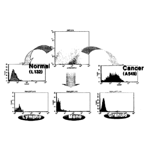
An examination was made to see whether the antibody binds to peripheral blood.
For this, PBMC (peripheral blood mononuclear cells from the Korean Red Cross
Blood
Services) was incubated with 100 gl of a hybridoma supernatant at 4 C for 30
min, and
centrifuged, together with 3 ml of PBS, at 1,500 rpm for 3 min to wash off
unbound
antibodies. A 200-fold dilution of the secondary antibody goat anti-mouse Ig-
FITC
(DINONA INC, Korea) was used to detect the bound antibodies. After reaction
for 15 min
at 4 C, the reaction mixture was washed with 3 ml of PBS in the same manner.
The
26
CA 03043692 2019-05-13
antibody titer was measured using flow cytometry, and the results are shown
(Fig. 1). Fig.
1 shows titers of the lung adenocarcinoma-specific 27B6 monoclonal antibody in
the
peripheral blood, as measured via flow cytometry.
In this manner, the antibody that was positive to the lung cancer cell line
A549 and
negative to the normal lung cell line L132 and all of granulocytes,
lymphocytes and
monocytes of the peripheral blood were selected and designated "27B6".
Finally, during a
limiting dilution procedure, 27B6 hybridoma cells were diluted and selected
for single
colony growth.
The 27B6 hybridoma cell line was deposited on February 14, 2012, with the
Korean Cell Line Bank, located at 28, Yongon-Dong, Chongno-gu, Seoul, Korea,
and
received Accession No. KCLRF-BP-00280 on February 20, 2012.
1-4: Determination of Isotype for 27B6 monoclonal antibody
The 2786 monoclonal antibody prepared in Example 1-3 was analyzed for isotype,
using a mouse immunoglobulin isotyping ELISA kit (BD Biosciences, USA).
Briefly,
isotyping was performed with anti-murine isotype specific antisera (IgGl,
IgG2a, IgG2b,
IgG3, IgM, IgA, Kappa, Lambda) while peroxidase-labeled goat anti-mouse IgG
served as
a secondary antibody. Color development was induced with ortho-
phenylenediamine
(OPD) and a hydrogen peroxide substrate. Absorbance at 450 nm was read.
As a result, the 27B6 monoclonal antibody was identified as mouse IgGlikappa
light chain (results not shown).
1-5: CDR Sequences of 27B6 Antibody
An antibody cloning procedure is illustrated in Fig. 1. Specifically, the 27B6
antibody gene was cloned using Mouse Ig-Primer Set (Millipore, Cat. #: 69831).
PCR was
performed using the mouse Ig-primer set from the RNA isolated from the 27B6
hybridoma, inserted into a pGem-T vector (Promega, Cat. #: A3600), and
sequenced to
confirm the DNA sequence. The mouse antibody gene was identified through the
IMGT
site (www.imgt.org). Heavy and light chain sequences including the CDR
sequences of the
27B6 Ab are represented by SEQ ID NOs: 12 and 13, respectively. CDR1 to CDR3
of the
27
CA 03043692 2019-05-13
heavy chain variable region are shown in SEQ ID NOs: 6 to 8, respectively, and
CDR1 to
CDR3 of the light chain variable region are shown in SEQ ID NOs: 9 to 11,
respectively
(see the Table 2).
EXAMPLE 2: Production of 27B6 Chimeric Antibody
When a monoclonal antibody of mouse origin is administered to the human body,
the human immune system recognizes the monoclonal antibody as a foreign
antigen and
thus produces a human anti-mouse antibody (HAMA) to eliminate the mouse
antibody
from the blood. In addition, the Fe domain of the mouse antibody cannot exert
its
io effective biological functions in the human body. Therefore, not only
does the therapeutic
effect sharply decrease, but also side effects such as severe allergic
reactions and renal
dysfunction may be induced. In order to reduce the immunogenicity of the 27B6
antibody
upon administration to the human body, a chimeric antibody in which the mouse
antibody,
except for the variable region, was substituted with the Fe of the human
antibody was
constructed. The chimeric antibody was observed to be similar in antigen
specificity and
affinity to the original mouse 27B6 antibody.
To construct a chimeric antibody, the 27B6-HuIgFc DNA prepared in the above-
mentioned manner was transfected into the DHFR DG44 cell line derived from CHO
cells,
followed by a selective culturing procedure in a selective medium to establish
a stable cell
line producing a 27B6 recombinant antibody. Details are described as follows.
First, three hours before transfection, the DG44 cell line (Invitrogen, Cat
No.
A1100001) was inoculated at a density of lx106cells/m1 into 6-well plates and
incubated
with 1 ml of GIBCO CD DG44 Medium (Invitrogen, USA) at 37 C in a 5% CO2
atmosphere for 3 hours. Then, the 27B6-HulgFc DNA was transfected into the
competent
DG 44 cells using an Effectene transfection reagent kit (Q1AGEN, Hilden,
Germany).
On three days post transfection, the supernatant was taken and added to A549
cells
which were then incubated at 4 C for 30 min. Unbound antibodies were removed
by
centrifugation, together with 3 ml of PBS, at 1500 rpm for 3 min. The bound
antibodies
were detected by incubation with a 150-fold dilution of the secondary antibody
goat anti-
Mouse lg-FITC (DINONA INC, Korea) at 4 C for 15 min, followed by washing with
3 ml
28
CA 03043692 2019-05-13
of PBS in the same manner. Thereafter, the antibody titer to A549 cells was
measured
using flow cytometry. Subsequently, a stable cell line was established. For
this, the
medium was exchanged with a Power CHO medium (LONZA, Switzerland) supplemented
with 30nM MTX (Sigma, USA) and 200 jig/m1 G418 (Invitrogen, USA), after which
clone
selection was started. Concentrations of MTX and G418 in the selection medium
were
increased with the repetition of clone selection rounds. Each round was set to
be three
weeks. The final round of clone selection was performed in a PowerCHO medium
supplemented with 1000 nM MTX and 400 jig/m1 G418. Thereafter, the final cell
line was
established as a single colony through limiting dilution.
The 27B6 chimeric antibody established in this manner was found to have
antigen
specificity and affinity to those of the original mouse 27B6 antibody, as
measured by flow
cytometry (Fig. 2). Fig. 2 shows the antigen specificity and affinity of the
27B6 chimeric
antibody.
EXAMPLE 3: Production of Monoclonal Antibody for CA XII (4B4)
3-1: 27B6 Pairing antibody
To develop another antibody which recognizes the same antigen but binds to a
different epitope, 27B6 pairing antibody was developed.
Firstly to explore the possibility of development of 27B6 paring antibody,
sandwich ELISA using chimeric 27B6 and mouse serum was established. In the
same
manner of Example 1-2, balb/c female mice 6 weeks old were each IP
(intraperitoneal
cavity)-injected with the A549 cell line (1 x 10⁷ cells) at regular intervals
of three weeks,
followed by taking sera from the veins.
Specifically, chimeric 27B6 purified antibody was added to a microplate at a
concentration of 100 ng / mL and coated at 37 C for 1 hour. Blocking buffer
(Sigma) was
added to 200 n1 / well of 27B6-coated microplate and blocked at 37 C for 1
hour. A549
cells were lysed at 1x10⁷ cells/ml using 1% NP40 lysis buffer. The prepared
A549 lysate
was added to the microplate at 50 uL/well, reacted at 37 C for 1 hour, and
then washed
three times with PBS. 100 I/well of A549 immunized mouse serum 1000-fold
dilution
was added to the microplate and incubated at 37 C for 1 hour and then washed
three
29
CA 03043692 2019-05-13
times with PBS. Finally, a secondary antibody goat anti-mouse IgG-HRP
(Jackson) 2000
dilution was added at 100 uL / well and incubated at 37 C for 30 minutes,
followed by 3
washes with PBS. TMB (3,3 ', 5,5'-tetramethylbenzidine) was added at 50 1iL /
well,
followed by reaction at room temperature for 10 minutes to induce color
development, and
2N H2SO4 (Sigma) was added in the same amount. The absorbance was then
measured at
a wavelength of 450 nm.
As was expected, the positive reaction was observed in sandwich EL1SA using
chimeric 27B6 and mouse serum (data not shown).
3-2: Production of Monoclonal Antibody
Preparation of hybridoma cells from splenocytes of the immunized mice was
carried out in the same manner as in Example 1-3.
As a result, the antibody that was positive to the lung cancer cell line A549
and
negative to the normal lung cell line L132 and to all of the granulocytes,
lymphocytes and
monocytes in peripheral blood, like 27B6, was selected and designated "4B4".
Finally,
during a limiting dilution procedure, 4B4 hybridoma cells were diluted and
selected for
single colony growth (Fig. 3). Fig. 3 shows titers of the 4B4 monoclonal
antibody in the
peripheral blood, as measured by flow cytometry. The 4B4 hybridoma cell line
was
deposited on February 14, 2012, with the Korean Cell Line Bank, located at 28
Yongon-
Dong, Chongno-gu, Seoul, Korea, and received Accession No. KCLRF-BP-00279 on
February 20, 2012.
3-3: Analysis of 4B4 Antibody
The amino acid sequences of the antibody were analyzed according to the
substantially same method of Example 1-5. Heavy chain sequences and light
chain
sequences including the CDR sequences of the 4B4 Ab obtained in Example 3-2
are
represented by SEQ ID NOs: 20 and 21, respectively., and CDR1 to 3 of heavy
chain are
shown in SEQ ID NO: 14 to 16, and CDR1 to 3 of light chain are shown in SEQ ID
NO:
17 to 19 (see Table 2).
30
CA 03043692 2019-05-13
EXAMPLE 4: Production of 4B4 Chimeric Antibody
In order to reduce the immunogenicity of the 4B4 antibody upon administration
to
the human body, a chimeric antibody in which the mouse antibody, except for
the variable
region, was substituted with the Fc of the human antibody was constructed
according to
Example 2. The chimeric antibody was observed to be similar in antigen
specificity and
affinity to the original mouse 4B4 antibody.
The production method of 4B4 chimeric antibody was carried out according to to
the same method of <Example 2>. As a result, the prepared antibody was found
to have
antigen specificity and affinity similar to those of the original mouse 4B4
antibody, as
measured by flow cytometry (Fig. 5). Fig. 5 shows the antigen specificity and
affinity of
the 4B4 chimeric antibody.
EXAMPLE 5: Analysis of Antibody Expression in Various Cell Lines
5-1: Antibody Expression in Various Cell Lines
27B6 chimeric antibody obtain in Example 2 and 4B4 chimeric antibody obtain in
Example 4 were analyzed for binding to various cell lines obtained from KCLB
(Korean
Cell Line Bank) and SNU (Seoul National University) using flow cytometry.
Specifically, various cell lines were obtained from KCLB (Korean Cell Line
Bank)
and SNU (Seoul National University). At 37 C under a 5% CO2 atmosphere, L-132,
SW-
900, DU145, LNCap, MCF-7, Huh7, and Hs-578T were cultured in Dulbecco's MEM
(GIBCO, Invitrogen) supplemented with 10% heat-inactivated fetal bovine serum
(FBS;
GIBCO, Invitrogen) and A549, NCI-H460, NCI-H417, DLD-1, HCT116, HT-29, SW-480,
SW-620, LS174T, PC-3, SNU I. SNU638, SNU719, MKN1, MKN28, MKN45, MKN74,
NCI-N87, SK-BR3, MDA-MB231, and MDA-MB453 were cultured in RPM1 1640
(GIBCO, Invitrogen) supplemented with 10% heat-inactivated FBS. In
addition,
incubation was carried out at 37 C under a 5% CO2 atmosphere in Eagle's MEM
(GIBCO,
Invitrogen) supplemented with 10% heat-inactivated fetal bovine serum (FBS;
GIBCO,
Invitrogen) for Calu-3, Hep3B, SK-HEP-1, C3A, Hep G2, PLC/PRF/5, and BT-20, in
IMDM (GIBCO, Invitrogen) supplemented with 20% heat-inactivated fetal bovine
serum
(FBS; GIBCO, Invitrogen) for KATO III, and in Leibovitz's L-15 medium
supplemented
31
CA 03043692 2019-05-13
with 10% heat-inactivated fetal bovine serum (FBS; GIBCO, Invitrogen) for
SW480 and
MDA-MB468.
The cultured cancer cell lines were incubated with the 27B6 or the 4B4
monoclonal
antibody of the present invention at 4 C for 30 min, washed with PBS, and
treated with
FITC-conjugated goat anti-mouse IgG (DINONA INC, Korea) at 4 C for 15 min. The
cell
lines were washed again with PBS before analysis by FACScaliber (Becton
Dickinson,
USA). The results are summarized in Table 5, below. Also, titers of the 27136
and the
4B4 antibody were measured in various solid tumor cell lines.
The following Table 5 shows the expression pattern of the carbonic anhydrase
12
antigen in various solid cancer cell lines. In Table 16, the percentage of
positive cells for
27B6 and 4B4 monoclonal antibodies among the 5000 cells to be tested was
analyzed by
FACS analysis, and ¨ indicates 10% or less of the number of positive cells, +
indicates the
range from 10 to 30% of the number of positive cells, ++ refers to 30 ¨ 70% of
the number
of positive cells, and +++ refers to 70 ¨ 100% of the number of positive
cells. When the
number of positive cells is less than 10%, it is regarded as negative, and
when the number of
positive cells is 10% or higher, it is regarded as positive.
[TABLE 5]
Origin Cell line 27B6 4B4
A549 +++ +++
Lung
NCI-H460 ++ ++
HCT116
Colon HT-29
LS174T +++ +++
Prostate LNCap
SNU 719 ++
Stomach
MKN 45 +++
Huh-7 ++
Liver Hep3B
PLC/PRF/5 +++ +++
Breast MCF-7
32
CA 03043692 2019-05-13
SK-BR3 +++ +++
MDAMB231 +++ +++
MDAMB453 +++ ++
BT20
Lymphocyte
PBMC Monocyte
Granulocyte
Cell binding profiling of 4B4 chimeric antibody performed by flow cytometry.
-: less than 10% of number of positive cells
+: 10-30%
++: 30-70%
+++ : higher than 70%.
As shown in Table 5, the 27B6 and 4B4 monoclonal antibodies of the present
invention showed a positive reaction, although the degree of binding affinity
was different
in various types of lung cancer, colorectal cancer, stomach cancer, liver
cancer and breast
cancer cell line. In contrast, peripheral blood lymphocytes, mononuclear
cells, and
granulocytes showed all negative results. This shows that the 27B6 and 4B4
antibodies of
the present invention can be used as therapeutic agents against solid tumors
expressing CA
XI! antigen.
5-2: Expression Pattern in Breast Cancer Cell
27B6 and 484 were observed to have positive responses to ER-, PR-, and HER2-
positive breast cancer cells. Accordingly, both antibodies can be used as
therapeutic
agents for various breast cancer subtypes including triple-negative breast
cancer.
The binding of the 2786 and the 4B4 monoclonal antibody to three different
phenotype breast cancer cell lines was examined via flow cytometry. Cell
culturing was
carried out at 37 C under a 5% CO2 atmosphere for MCF-7 cells (Breast cancer
cell, ER
positive) in Dulbecco's MEM (GIBCO, Invitrogen) supplemented with 10% heat-
33
CA 03043692 2019-05-13
inactivated fetal bovine serum (FBS; GIBCO, Invitrogen) and for MDA-MB231
(cell lines
derived from malignant breast cancer) and SK-BR-3 cells (human breast cancer
cell line
that overexpresses the Her2 (Neu/ErbB-2) gene product)in RPMI 1640 (GIBCO,
Invitrogen) supplemented with 10% heat-inactivated FBS.
The cultured cancer cell lines were incubated at 4 C for 30 min with the 27B6
or
the 4B4 monoclonal antibody of the present invention, washed with PBS, and
treated at
4 C for 15 min with FITC-conjugated goat anti-mouse IgG (DINONA INC, Korea).
The
cell lines were washed again with PBS before analysis by FACS caliber (Becton
Dickinson, USA). The results are summarized in Table 6.
[Table 6]
4B4
Origin Cell line Subtype Immunoprofile
Expression
MCF-7 L um inal ER+, PR+, HER2- +++
SK-BR3 HER2 ER-, PR-, HER2+ ++
MDAMB453 Basal ER-,PR-HER2- +++
Breast MDAMB231 Basal ER-,PR-HER2- ++
MDAMB 468 Basal ER-,PR-HER2-
HS578T Basal ER-,PR-HER2-
BT20 Basal ER-,PR-HER2-
Therefore, the antibodies 27B6 and 4B4 according to the present invention can
be
used not only for triple negative breast cancer but also for various types of
breast cancer,
because they show the positive reaction in both ER and PR as well as HER2-
positive
breast cancer cells.
5-3: IHC (Immunohistochemistry)
Antigens to which 27B6 and 4B4 monoclonal antibodies bind were analyzed for
distribution in normal tissues of the human body by immunohistochemistry
(IHC).
Normal thymus and tonsil tissues of the human body were obtained from the
Chungbuk
National University Hospital and prepared into cryosections in the department
of
pathology in the Chungbuk National University Hospital.
34
The prepared cryosections were subjected to immunohistochemical staining with
27B6 and 4B4 monoclonal antibodies of the present invention as follows. Thymus
and
tonsil cryosections stored at -20 C or lower were dried at room temperature
for 30 - 60
min, and immersed in lx PBS for 60 min. Then, the tissues were treated at room
.. temperature for 10 min with 3% H202 to suppress the activity of endogenous
peroxidase,
washed with flowing water, and blocked at room temperature for 30 min with a
goat
immunoglobulin-containing serum to exclude non-specific staining with mouse
antibodies.
Then, the tissues were incubated at room temperature for 60 min with the
primary
antibody (27B6, 4B4). Each antibody was used at a concentration of 10 miml.
Thereafter, the tissues were washed three times with lx PBS for 5 min,
incubated at room
temperature for 30 min with an HRP-conjugated goat anti-mouse antibody (Dako,
Denmark), and then washed three times with lx PB ST (0.05% TweenTm-20, lx PBS)
for 5
min. The color was developed with diaminobenzidine (DAB), followed by washing
for 5
min with flowing water. The tissues were counterstained with hematoxylin and
then
washed for 7 min with flowing water. After staining, the slides were
dehydrated and
sealed. The staining results were analyzed by microscopy below.
[Table 7]
Tissue 27B6 4B4
Cortex
Thymus
Medulla
Inter follicular T cell
B cell
Tonsil
Germinal center
Basal layer Basal layer +
As shown in Table 7, the antigens that the 27B6 and the 4B4 monoclonal
antibody
of the present invention recognize are distributed neither in normal thymus
nor in normal
tonsil tissues. Particularly, nowhere are the antigens expressed in normal
mature or
immature T cells or B cells. The 27B6 antibody was weakly stained in the basal
layer of
the tonsil, which, however, seemed to result from non-specific binding.
Date Recue/Date Received 2020-12-03
CA 03043692 2019-05-13
EXAMPLE 6: Analysis of Antigen for Monoclonal Antibody
6-1: Isolation and Purification of 4B4 and 27B6 Monoclonal Antibodies
The lung adenocarcinoma cell line A549 that had been used to develop the 4B4
and
the 27B6 monoclonal antibody was cultured. Then, 1x10⁸ cells were suspended in
50 ml
of a lysis buffer (1% Nonidet P-40; NP-40 in 50mM Tris-HC1, pH 7.4, 50mM EDTA,
and
1mM phenyl-methyl-sulfonyl-fluoride; PMSF) and lysed for 15 min. After
centrifugation,
the cell debris was removed, and a cell lysate was obtained as a supernatant.
The cell
lysates was used to separate antigens that were recognized by 4B4 or 27B6
antibodies.
Five mg of each of purified 4B4 and 27B6 monoclonal antibodies were dialyzed
against a binding buffer (0.2 M sodium bicarbonate, 0.5M sodium chloride, pH
8.3) to
afford two different antibody solutions. A 5-ml column packed with 2 ml of NHS-
activated sepharose 4 Fast Flow resin (GE Healthcare) was washed with 20 ml of
1 mM
HC1 and then with 20 ml of a binding buffer (20 mM sodium bicarbonate, 0.5 M
sodium
chloride, pH 8.3) so as to allow the prepared antibodies to bind to the
column. The
column was blocked at the outlet thereof, loaded with either of the two
different antibody
solutions, and blocked at the inlet thereof. Incubation was performed at room
temperature
for 4 hrs. Then, 20 ml of a washing buffer (20mM Sodium acetate, 0.5M sodium
chloride,
pH 5.4) was made to flow through the column so as to remove excess antibodies
that were
not bound to the resin. Again, the column was washed with 50 ml of a blocking
buffer
(0.1 M ethanolamine, 0.5 M sodium chloride, pH 8.3) to remove remaining
reaction
groups. The two columns were washed with 20 ml of a stock buffer (20 mM Tris-
HCl,
150 mM NaC1, 0.02 % sodium azide, pH 8.0), and refrigerated until use.
The prepared columns were applied to FPLC (Acta FPLC) so that the antibodies
bound to the resin could recognize antigens and thus could allow for the
separation of the
antigens. The lung adenocarcinomic A549 cell line lysates was loaded to the
column
coupled to FPLC and used as an antigenic source that was recognized by 4B4 and
27B6
monoclonal antibodies. Antigen separation was performed in a four-step
process:
equilibrium; sample loading; washing and second washing; elution. An
equilibrium buffer
and a wash buffer have the same composition: 0.5% Tween-80, 20 mM Sodium
36
CA 03043692 2019-05-13
phosphate, 150 mM sodium chloride, pH 7.4. This buffer was used in an amount
of 10 ml
for equilibrium and in an amount of 20 ml for washing. An elution buffer
contained 0.3 M
Glycine, 0.1 M sucrose, 0.1 M Mannitol, 1.0 M urea, and 0.5% Tween-80, had a
pH of
3.0, and was used in an amount of 20 ml for washing. For the second washing, a
mixture
in which the elution buffer was mixed at a ratio of 25 % with the washing
buffer was
employed. 5 ml of TCA was added to 20 ml of the eluted solution obtained
during the
antigen separation and stored for 30 min in a refrigerator. After
centrifugation, the pellet
was further washed twice with acetone. The finally obtained pellet was
suspended in lx
SDS-PAGE sample buffer, subjected to electrophoresis, and stained with
Coomassie blue.
As described above, antigens that were isolated and purified through the
columns
respectively fabricated with 484 and 27B6 antibodies are shown in Fig. 6. Fig.
6 shows
electrophoretograms of antigens isolated and purified from the lung
adenocarcinoma A549
cell line through columns fabricated with 4B4 and 27B6 monoclonal antibodies.
6-2: Identification of Antigen for 4B4 and 27B6 Monoclonal Antibodies
The antigens isolated and purified from the resin coupled with the 4B4 and the
27B6 monoclonal antibody were visualized as shown in Fig. 7. The two main
protein
bands indicated by the arrows at about 58 kDa were analyzed in Seoul Pharma
Laboratories. For identification, peptides were prepared via in-gel digestion
and analyzed
using LC-MS/MS, followed by processing the MS/MS spectra with PLGS (Waters)
and
MASCOT (Matrix Science). A series of analyses was conducted as follows.
Gel pieces containing proteins were dehydrated using 100 A CAN (acetonitrile)
and completely dried in a Speed-vac. The proteins in the dried gel pieces were
digested
for 15 min with trypsin. The tryptic peptides were extracted with 60% CAN and
0.1%
TFA. The pooled extracts were dried in a Speed-vac. The samples were dissolved
in 5%
CAN, 0.2% TFA (Trifluoroacetic acid) 20 pi prior to LC-MS/MS analysis.
Peptides were
eluted from the LC column nanoACQUITY UPLC BEH C18 (1.7pm, 300A, 2.1mm x 150
mm ID.), with a gradient of a mobile phase buffer A (0.1% TFA in 100% DW) to a
mobile phase buffer B (0.1% TFA in 100% ACN) in a LC-MS/MS analysis. The
separated
peptides were analyzed online in a positive survey scan mode on a nano-ES1-Q-
TOF
37
CA 03043692 2019-05-13
instrument. Subsequently, the spectral data were processed with PLGS and
MASCOT.
As a result of the above-described series of analyzes, the final
identification results
are obtained as shown in the following table, and they are shown in Table 8
below.
[Table 8]
Antibody No. Description mW(Da)
pI(pH)
4B4 1 Actin cytoplasmic 1 41739 5.15
4B4 2 Carbonic anhydrase 12 39426 6.79
4B4 3 Keratin type I cytoskeletal 9 62026 4.96
4B4 4 Serum albumin 68647 5.68
4B4 5 Trypsin 24393 6.91
4B4 6 Actin 3 41813 5.01
4B4 7 Actin 1 41648 5.24
27B6 1 Carbonic anhydrasel2 39426 6.79
27B6 2 Pyruvate kinase isozymes Ml/M2 58470 7.96
27B6 3 Actin cytoskeletal 1 41814 5.24
27B6 4 Retinal dehydrogenase 1 42327 5.08
27B6 5 Hemoglobin subunit beta 1 15830 7.5
Synaptic vesicle membrane protein VAT-1
27B6 6 42122 5.88
homolog
27B6 7 Protein disulfide isomerase 57146 5.98
27B6 8 Serum albumin 68647 5.68
27B6 9 Trypsin 24393 6.91
27B6 10 Actin gamma 41580 5.33
As expected, carbonic anhydrase 12 of the analysis results was identified in
common from the antigens purified by both the 4B4 and the 27B6 monoclonal
antibody,
and was found to exist on cell surfaces. The other proteins cannot be antigens
for the 4B4
and the 27B6 monoclonal antibody, because they are intracellular proteins.
Thus, they
seemed to be impurities included due
to imperfect separation and purification.
Four peptides were separated by 27B6: QFLLTNNGHSVK (SEQ ID NO: 22),
WTYFGPDGENSWSK (SEQ ID NO: 23), GQEAFVPGFNIEELLPER (SEQ ID NO: 24),
38
CA 03043692 2019-05-13
and YKGQEAFVPGFNIEELLPER (SEQ ID NO: 25). Three peptides were separated by
4B4: QFLLTNNGHSVK (SEQ ID NO: 22), EMINNFR (SEQ ID NO: 26). and
GVIYKPATK (SEQ ID NO: 27). Of them, the sequence QFLLTNNGHSVK was
analyzed in common in both 4B4 and 27B6. Fig.8 shows the amino acid sequence
of
carbonic anhydrase 12 precursor isoform 1, with the analyzed peptide sequence
expressed
in bold. Fig. 8 lists up the proteins identified by LC-MS/MS analysis from
purified
antigens. Table 9 shows the amino acid sequence of carbonic anhydrase 12
isoform 1
according to Example 6 and antigenic peptide fragment of the antigens
recognized by
27B6 and 4B4 antibody, detected by LC-MS/MS. Such results show that the
antigenic
peptide fragment of the antigens recognized by 27B6 and 4B4 antibody is
carbonic
anhydrase 12 isoform 1.
[Table 9]
CA12 --17MPRRSLHAAAVILLVILKEQPSSPAPVNGSK;:fFY
isoform 1 WSKKYPSCGGLLQSPIDLHSDILQYDASLTPLEFQGY NLSANKQ
' FL LTN NGH SVKLN LPSDMHIQGLQSRYSATQLHLHWGNPND
PHG SEHTVSGQHFAAELHNHYNSDLYPDASTASNKSEGLAVL
AVLIEMGSFNPSYDKIFSHLQHVK Y E N: E 1.P 11
TAEYYRYRGSLTTPPCN PTVLWTVFRNPVQISQEQLLALETALYC
THMDDPSPREMINNFRQVQKFDERLVYTSFSQVQVCTAAGLS
LGIILSLALAGILGICTVVVVSIWLFRRKSIKKGDNKGVIYKF'ATKM
ETEAHA
Peptide fragment QFLLTNNGHSVK EMINNFR
of antibody recog
1.nized by 4134 GVIYKPATK
QFLLTNNGHSVK WTYFGPDGENSWSK
Peptide fragment
of antibody recog YKGQEAFVPGFNIEELLPER
Inized by 27B6 GQEAFVPGFNIEELLPER
6-3: Assay of Antigen for 4B4 and 27B6 Monoclonal Antibodies (ELISA)
To evaluate the antigen identification results obtained by LC-MS/MS, the
reactivity
of the 4B4 and the 27B6 monoclonal antibody to the recombinant protein
carbonic
anhydrase 12 (R&D Systems) were examined by ELISA and Western blotting assay.
The recombinant protein CA I 2 was plated at a density of 100 ng/well into
Maxisrop ELISA plates and incubated at 37 C for 1 hr. To each of the antigen-
coated
39
CA 03043692 2019-05-13
wells, 200 1.11 of a lx blocking buffer (Sigma) was added, followed by
incubation at 37 C
for 1 hr for blocking. 4B4. 27B6, and an anti-CA12 monoclonal antibody (R&D
Systems)
were plated, together with 100 1 of PBS, into the plates. After incubation
for 1 hr at
37 C, the plates were washed with PBS to remove unbound antibodies.
Subsequently, a
dilution of goat anti-mouse IgG-HRP (Jackson) was added to the wells, reacted
for 30 min,
and washed with PBS. Color development was accomplished for 10 min with 50 I
of
TMB in each well, and stopped with 50 1 of sulfuric acid. Absorbance at 450
nm was
read. Although the reactivity of the 27136 monoclonal antibody to the
recombinant
carbonic anhydrase 12 was low, reactivity signals of 4B4, 27B6, and anti-CA12
monoclonal antibody (R&D Systems) against the recombinant antigen are shown in
Fig. 7.
Fig. 7 shows the identification of carbonic anhydrase 12 as an antigen for the
4B4 and the
27B6 monoclonal antibody, as analyzed by ELISA assay.
6-4: Assay of Antigen for 4B4 and 27B6 Monoclonal Antibodies (Western
blotting)
The recognition of carbonic anhydrase 12 as an antigen by the 4B4 and the 27B6
monoclonal antibody, proven in the previous experiment, was confirmed by
Western
blotting. The recombinant carbonic anhydrase 12 was boiled for 3 min. loaded
into an 8%
separating sodium dodecyl sulfate-polyacrylamide gel, and run by
electrophoresis. The
separated proteins were transferred to a nitrocellulose membrane which was
then blocked
with 5% skim milk (Sigma) and treated with the 484, 27B6, or anti-CA12
monoclonal
antibody (R&D Systems) (27B6: lanes I and 2, 4B4: lanes 3 and 4, anti-CA12
monoclonal
antibody: lanes 5 and 6). After three rounds of washing with a wash buffer
(0.1% Tween-
20 in PBS), the antibody was coupled with peroxidase-conjugated goat anti-
mouse IgG
(Sigma. Saint Louis, USA). After the nitrocellulose membrane was washed with a
wash
buffer, bands were visualized using an enhanced chemiluminescence detection
system
(ECL, Amersham, Sweden). The results are shown in Fig. 8. Fig. 8 shows the
identification of carbonic anhydrase 12 as an antigen for the 4B4 and the 27B6
monoclonal antibody, as analyzed by Western blotting assay. The recombinant
CA12 was
detected at 40 kDa by all of the 4B4, 27B6, and anti-CA12 monoclonal
antibodies (R&D
CA 03043692 2019-05-13
Systems).
6-5: Assay of Antigen for 4B4 and 27B6 Monoclonal Antibodies (Sandwich
ELISA)
ELISA and WB assays demonstrated that 4B4 and 27B6 monoclonal antibodies
recognize carbonic anhydrase 12 as an antigen, but the detection signal of the
27B6
monoclonal antibody was relatively low. To compensate for the relatively low
signal,
Sandwich ELISA was conducted as follows. The chimeric 494 or 27B6 monoclonal
antibody was plated at a concentration of 100 ng/well into Maxisrop ELISA
plates and
incubated at 37 C for 1 hr. To each of the antigen-coated wells, 200 ul of lx
blocking
buffer (Sigma) was added, followed by incubation at 37 C for 1 hr for
blocking. Two-fold
serial dilutions of the recombinant carbonic anhydrase 12 staring from 100
ng/ml were
added to wells, incubated at 37 C for 1 hr, and washed with PBS to remove
unbound
antigens. Subsequently, the 4B4 monoclonal antibody and the 27B6 monoclonal
antibody
were added at a concentration of 100 ng/well to chimeric 27B6-coated wells and
chimeric
4B4-coated wells, respectively. Following 1 hr of incubation at 37 C, the
wells were
washed with PBS to remove unbound antibodies. In addition, the bound
antibodies were
incubated with a dilution of goat anti-Mouse IgG-HRP (Jackson) for 30 min and
washed
with PBS. Color development was accomplished for 10 min with 50 1.11 of TMB in
each
well and stopped with 50 1.1.1 of sulfuric acid. The absorbance at 450 nm was
detected.
When chimeric 27B6 and 4B4 were used as a capture antibody and a detector
antibody,
respectively, high reaction signals were read, as shown in Table 10. Both of
the 4B4 and
2786 monoclonal antibodies were therefore proven to recognize carbonic
anhydrase 12 as
an antigen.
/5 Table 10
shows the concurrent recognition of carbonic anhydrase 12 by the 27B6
and 4134 monoclonal antibodies, as measured by sandwich ELISA assay using the
2786
and 4B4 monoclonal antibodies as capture/detector antibodies.
[Table 10]
Capture antibody Chimeric 27B6 Chimeric 4B4
Detector antibody 4B4 mAb 27B6 mAb
41
CA 03043692 2019-05-13
CA12 10Ong/m1 1.653 0.021
CA12 5Ong/m1 1.349 0.016
CA12 25ng/m1 0.954 0.016
CA12 12.5ng/m1 0.634 0.011
CA12 6.25ng/m1 0.351 0.009
CA12 3.13ng/m1 0.193 0.008
blank 0.064 0.007
blank 0.055 0.008
EXAMPLE 7: Epitope Mapping
To analyze an epitope, as shown in Fig. 13, recombinant antigen, with or
without
the epitope, were constructed, and analyzed for immune reactions of mouse
monoclonal
antibodies of 27B6 and 4B4 in Example 1 and 3.
7-1: Construction and Expression of CA12 Mutant Recombinant Gene
The recombinant vector pSec-Tag-CA 12 full-hFc was digested with Baml ii and
HindlII to prepare CA12 mutant recombinant genes. A recombinant gene in which
a full
base sequence of CA12 antigen was fused to hFc was inserted into pSec-Tag
which was
then allowed to express a recombinant fusion protein containing the full
length of CA12
plus hFc. As seen in Fig. 13, deletion mutant-hFc constructs having various
lengths within
a range from the N terminus to amino acid 300 were prepared.
Respective pSee-Tag vectors carrying the CA12 full-hFc and five different
deletion
.. mutant-hFc constructs were introduced into CHO cells with the aid of
ViaFect (Promega).
Briefly, one day before transfection, CHO cells were plated and incubated.
After
the medium was exchanged with a fresh one, a complex of the vector and ViaFect
was
applied to the CHO cells and incubated for 48 hours. Two days after
transfection, the
culture supernatant was collected and analyzed for the expression of the gene
by detecting
.. human Fe (hFc) through sandwich ELISA.
7-2: Assay of epitope of monoclonal antibody
In order to examine a CA12 epitope recognized by the monoclonal antibodies of
42
CA 03043692 2019-05-13
the present invention, 50 ng of an anti-human Ig antibody (Jackson Laboratory)
was added
to each well and incubated at 37 C for 1 hr. The antibody fixed to the well,
which would
serve as a capture antibody, was blocked via incubation with 200 I of a lx
blocking
buffer (Sigma) at 37 C for 1 hr in each well. Each of the respective cultures
containing
the CA12 full-hFc and the five different deletion mutant-hFc constructs was
added at a
concentration of 100 pl/well to the plates. Following 1 hr of incubation at 37
C, the wells
were washed with PBS to remove unbound antibodies. Subsequently, a dilution of
anti-
mouse Ig, Fc specific-HRP (Jackson Laboratory) was added to the wells, reacted
for 30
min, and washed with PBS. Color development was accomplished for 10 min with
50 1.11
of TMB in each well, and stopped with 50 i.t1 of sulfuric acid. The absorbance
at 450 nm
was read. The presence of CA12 mutant-hFc proteins in the culture supernatants
was
examined using Capture & Detect Sandwich ELISA, with an anti-human Ig antibody
serving as a control. The results are given in Fig. 9.
As can be seen in Fig. 9, the epitopes were located in a site from a.a. 25 to
a.a. 57,
which is a non-catalytic domain. Hence, the antibodies of the present
invention do not
bind to the catalytic domain of CA-XII, so they do not inhibit the enzymatic
activity of
CA-XII.
In detail, the epitope specific for the 27B6 antibody was found to have the
amino
acid sequence APVNGSKWTYFGPD of SEQ ID NO: 2 (the span from 25th amino acid.
to 38th amino acid on SEQ ID NO: 5), as analyzed by the deletion method. A
three-
dimensional crystal structure of CA12 confined the epitope into 7 consecutive
amino acids
WTYFGPD of SEQ ID NO: 1 (corresponding to 32 to 38 of SEQ ID NO: 5) on the
amino
acid sequence of SEQ ID NO: 2. Further, the epitope specific for the 4B4
antibody was
found to have the amino acid sequence GENSWSKKYPSCGGLLQSP of SEQ ID NO: 4
(the span from 39th to 57th amino acids in SEQ ID NO: 5) while a three-
dimensional
crystal structure of CA12 confined the epitope into 14 consecutive amino acid
sequence
GENSWSKKYPSCGG of SEQ ID NO: 3 (corresponding to 39th to 52th amino acids of
SEQ ID NO: 5) on the amino acid sequence of SEQ ID NO: 4.
EXAMPLE 8: Therapeutic Effect of Chimeric Antibody on Solid Tumor
43
CA 03043692 2019-05-13
(CDC)
8-1: CDC Effect in Lung Adenocarcinomic Cell Line
The lung adenocarcinomic cell line A549 cells were plated at a density of
5x10³
cells/well into 96-well plates and cultured for 20-24 hours in a 37 C, CO2
incubator. After
removal of the culture medium from each well, an RPMI medium, free of fetal
bovine
serum, was mixed with 10 % human serum and the chimeric 27B6 antibody was
added at a
final concentration of 10 pg/m1 to a mixture. This solution was plated at a
concentration
of 100 p,l/well into the plates.
The 4B4 chimeric antibody was also treated in the same manner. Following 3
hours of incubation in a 37 C CO2 incubator, Ez-CyTox agent (DOGEN, KOREA) was
added in an amount of 10 l.t1 to each well. Incubation for 3.5 hours in a 37
C, CO2
incubator was followed by measuring absorbance at 450 nm on a plate reader.
The results
are given in Fig 10. Fig 10 shows the complement-dependent cytotoxic effects
of the 27B6
antibody.
As can be seen in Fig. 10, the 27B6 and 4B4 chimeric antibodies obtained in
Example 2 and 4 exhibit complement-dependent cytotoxicity.
8-2: CDC effect in triple-negative breast cancer
According to the same method of <Example 8-1>, the therapeutic effect was
evaluated, except that MDAMB-231 of triple-negative breast cancer cell line
was used as a
target cell instead of A549 cell lines, and absorbance at 450 nm was measured
and shown
in Table II. Table 11 shows the results of confirming that the 4B4 chimeric
antibody
according to Example 8 exhibits complement dependent cytotoxicity in triple
negative
breast cancer.
[Table 11]
Sample Cytotoxicity (%)
No treat 0.0
5% HS (12.7)
5% NHS+4B4 ing/m1 76.5
44
CA 03043692 2019-05-13
5% NHS+4B4 long/ml 86.3
5% NHS+4B4 10Ong/m1 83.4
5% NHS+4B4 1000ng/m1 75.7
5% NHS+4B4 10000ng/m1 83.7
4B4+5% NHS+CVF 50ug 83.9
4B4+5% NHS+CVF 10Oug 80.7
As shown in Table 11, the 27B6 monoclonal antibody of the present invention
exhibited complement-dependent cytotoxicity against lung adenocarcinomic
tumors.
EXAMPLE 9: Therapeutic Effect of Chimeric Antibody in Solid Tumor
(ADCC)
9-1: Assay for antibody-dependent cell-mediated cytotoxicity (ADCC- LDH assay)
In order to prepare effector cells, Ficoll was added to a human blood sample
(blood : Ficoll =1:2), followed by centrifugation at 2000 rpm for 20 min to
obtain PBMCs
(Peripheral Blood Mononuclear Cells). The PBMCs were stored at 37 C in a 5%
FBS-
supplemented RPMI medium. The antibody-dependent cell-mediated cytotoxicity
assay
was conducted in conjunction with an LDH assay or a Luciferase assay.
As targets, various solid tumor cell lines - HT29 (colorectal cancer), A549
(lung
adenocarcinoma), NCI-H460 (lung adenocarcinoma), and MCF7 (breast cancer) ¨
were
each plated at a density of lx 104 cells/well into 96-well plates and cultured
for 18-20
hours in a 37 C, CO2 incubator. After removal of the culture medium from each
well, the
27B6 chimeric antibody was added at a concentration of 0 i.tg/mL, 0.1 ug/mL,
or 3 1.1g/mL
to a culture medium supplemented with 5% FBS, and then plated at a
concentration of 100
ill/well into the plates, followed by incubation for 30 min in a 37 C CO2
incubator.
Thereafter, the effector cells prepared above were plated at a density of
5x10⁵ cells/well
(50 times as many as the target cells), and cultured for 24 hours in a 37 C
CO2 incubator.
For a positive control, a lysis buffer was added before incubation at 37 C for
24 hours.
Following 24 hours of incubation, the cell culture was centrifuged at 2500 rpm
for 5 min.
The supernatant thus obtained was measured for LDH (lactate dehydrogenase)
activity to
CA 03043692 2019-05-13
calculate the cell lysis (Promega assay kit). As shown in Fig. 11, the 27B6
monoclonal
antibody of the present invention exhibited antibody-dependent cell-mediated
cytotoxicity
in various solid tumors (Fig. 11). Fig. 11 shows the antibody-dependent cell-
mediated
cytotoxic effects of the 27B6 antibody.
9-2: Antibody-dependent cell-mediated cytotoxicity assay in triple-negative
breast
cancer (ADCC- LDI I assay)
Effector cells were prepared in the same manner as in Example 9-1, and tested
for
antibody-dependent cell-mediated cytotoxicity.
In addition, the 27B6 antibody was found to exhibit high antibody-dependent
cell-
mediated cytotoxicity in triple-negative breast cancer cell lines (MDAMB-231,
MDAMB-
468, MDAMB-453, BT-20) for which no therapeutic agents had yet been developed
(Fig.
12). Fig. 12 shows the antibody-dependent cell-mediated cytotoxic effects of
the 27136
antibody on triple-negative breast cancer cell lines.
Example 10: Therapeutic effect of defucosylated antibody in solid tumor
(ADCC)
10-1: Assay for ADCC of Defucosylated Chimeric 27B6 Antibody- Colon, Lung,
Breast Cancer
In order to induce the defucosylation of antibody proteins, the 27136 antibody
cell
lines of Example 2 and the 484 chimeric antibody cell lines of Example 4 were
incubated
with 100 ng/ml kifunensine which induces the defucosylation of antibody, and
the
defucosylated antibodies were separated and compared to corresponding
fucosylated
antibodies. Assay for ADCC of Kifunensine treated, ADCC-Enhanced, Chimeric
27B6
Antibody showed the ADCC effect of defucosylated antibody in Colon cancer,
Lung
cancer and Breast cancer.
As can be seen in Fig. 13, the antibodies defucosylated by kifunensine were
increased in antibody-dependent cell-mediated cytotoxicity against various
solid tumor
cell lines. Fig. 13 shows the antibody-dependent cell-mediated cytotoxicity of
the
defucosylated 27B6 chimeric antibody.
46
CA 03043692 2019-05-13
10-2: Assay for ADCC of defucosylated chimeric 27B6 antibody ¨ triple-negative
breast cancer
By using a luciferase ADCC assay, antibody-dependent cell-mediated
cytotoxicity
against the triple-negative breast cancer cell line MDAMB231 and the HER2
receptor-
positive breast cancer cell line SK-BR3 was analyzed. The antibodies after
being
defucosylated by treatment with kifunensine exerted higher antibody-dependent
cell-
mediated cytotoxicity on MDAMB231 and SK-BR-3 than corresponding fucosylated
antibodies (Fig. 14).
Fig. 14 shows the antibody-dependent cell-mediated cytotoxicity of
defucosylated
4B4 and 27B6 chimeric antibodies, as measured by a luciferase assay.
Specifically, as target cells, the breast cancer cell lines MDAMB231 and SK-
BR3
were each plated at a density of 1.25x10⁴ cells/well into 96-well plates and
cultured for 20-
24 hours in a 37 C CO? incubator. After removal of the culture medium from
each well.
25 I of an RPMI medium containing 4% low IgG FBS was added to each well in
which
the cells were plated. 27B6 and 4B4 antibodies were 3-fold diluted in serial
from 10
g/m1 to 1.2 ng/ml in an RPMI medium containing 4 % low IgG FBS. The serial
antibody
dilutions were each added in an amount of 25 l/well, and the plates were
covered with
respective lids and left on a clean bench. ADCC reporter cells (ADCC Reporter
Bioassay,
Promega) were harvested from the cell culture and suspended at a concentration
of 3x10⁶
cells/m1 in an RPMI medium containing 4 % low IgG FBS. To each well was added
25 1
of the suspension of ADCC reporter cells, followed by 24 hours of incubation
in a 37 C
CO2 incubator. Before the plates were withdrawn, a frozen luciferase substrate
was
thawed in a water bath. The plates were removed from the clean bench and left
at room
temperature for 15 min. The luciferase substrate was added at a concentration
of 75
l/well to the plates and reacted for 30 mm in a dark condition, followed by
measuring
luminescence with a luminometer. The result of antibody-dependent cytotoxicity
test of
defucosylated 27B6 and 4B4 chimeric antibody using Luciferase assay is shown
in FIG. 14.
As seen in Fig. 14, 27B6 and 4B4 antibodies, after being defucosylated by
treatment with kifunensine, exerted greater antibody-dependent cell-mediated
cytotoxicity
47
CA 03043692 2019-05-13
on MDAMB231 and SK-BR-3 than did corresponding fucosylated antibodies.
EXAMPLE 11: Therapeutic Effect of 27B6 and 4B4 Antibodies in Mouse
Models
11-1: Cell line establishment
Animal models with human breast cancer were established using the triple-
negative
breast cancer cell lines MDA-MB-231 and MDA-MB-453. First, MDA-MB-231 or
MDA-MB-453 was subcutaneously injected at a dose of 1.5x10⁸ cells (in RPMI:
Matrigel
mixture) into the right flank of mice. The injected mice were randomly
classified into test
and control groups.
Fig. 15 further shows the binding of 27B6 chimeric antibody in Example 2 and
4B4
chimeric antibody in Example 4 to the surface of MDA-MB231 cells utilized in
the animal
experiment, and Fig. 16 shows the results of the animal experiment using the
antibodies,
demonstrating that the antibodies suppress the growth and size of MDA-MB231-
induced
tumor.
As test materials, the 27B6 fucosylated chimeric antibody, 27B6 defucosylated
chimeric antibody, 4B4 fucosylated chimeric antibody, and 4B4 defucosylated
chimeric
antibody were inoculated into breast cancer cells. Three days later, the cells
were
intraperitoneally injected at a dose of 12 mg/kg to each mouse. Injection was
conducted
twice a week for three weeks. Tumor sizes were measured just before injection.
The
inhibitory activity of the anti-CA12 antibodies against breast cancer was
expressed as the
tumor volume calculated according to the following formula: (a x b2) / 2 (a is
the short
diameter and b is the long diameter). The volume calculation equation is the
same as the
volume calculation formula of Example 20-1.
[Equation]
Volume calculation equation (volume = (a x b) / 2, where a is the short
diameter and
b is the long diameter)
11-2: Inhibitory activity of anti CA12 antibodies against triple-negative
breast
cancer
48
CA 03043692 2019-05-13
Targeting a CA12 epitope specifically expressed on triple-negative breast
cancer,
anti-CA12 chimeric antibody 4B4 was assayed for inhibitory activity against
triple-
negative breast cancer (Fig. 17).
Breast tumors were decreased in volume by 4B4, and the defucosylated antibody
was superior in inhibitory activity against tumor growth to the corresponding
fucosylated
antibody. The inhibitory activity of the 4B4 fucosylated antibody against the
growth of
breast cancer tumors was found in both MDA-MB-231 and MDA-MB-453.
Particularly, complete remission was observed in the MDA-MB-453 model as the
tumor did not grow further after day 21 (Fig. 17). Fig. 17 shows the
inhibitory activity of
the 484 antibodies against triple-negative breast cancer.
EXAMPLE 12: Effect of Antibody on Cell Survival
The effect of the chimeric 4B4 antibody of Example 4 on cell viability was
confirmed. When the antibodies were applied to CA12-positive cancer cells, the
effects of
the antibodies on cell growth and survival were examined. To this end, cells
were plated
at a density of 3x10⁴ cells/well into 96-well flat bottom plates one day
before application
(10% RPM!). After 24 hours, the RPMI was removed, and fresh 5% RPM! containing
the
antibody was added in an amount of 100 I to each well.
After 24 hours, a CytoTox 96 Non-Radioactive Cytotoxicity Assay kit (Promega,
Cat.# G1780) was plated at a concentration of 50 l/well and incubated for 30
min at room
temperature. Cell viability was measured using a spectrophotometer. Twenty
four hours
after the antibody was applied to MDA-MB231 cells, the cell viability was
measured.
The results of the measurements are shown in Fig. 18. The administration of
the
antibodies neither promoted nor degraded cell viability. The antibodies did
not inhibit
CA12 enzymatic activity, and had no influences on tumor cell growth.
Therefore, the
antibodies according to the present invention were found to exhibit anti-tumor
activity via
ADCC and CDC through the immune system.
Cell viability was measured 24 hours, 48 hours and 72 hours after the
administration of the antibody to A549. No significant changes in cell
viability were
observed compared to the cells to which no antibodies were administered. Figs.
19 and 20
49
CA 03043692 2019-05-13
show that the binding of the 4B4 antibody alone to tumor cells does not affect
the growth
of the tumor cells.
The 4B4 antibody, as an anti-CA12 antibody, had no influence on cell growth
only
when the antibody was bound to cells. This seems to be attributable to the
fact that the
4B4 antibody does not affect the enzymatic activity of CA12 because it binds
to an N-
terminal non-enzymatic region of the CA12 antigen.
EXAMPLE 13: Evaluation of Therapeutic effect by Combination of Antibody
Therapy and Radiotherapy
An examination was made to see whether or not a combination of the antibody of
the present invention and radiotherapy could bring about an increased
anticancer effect.
Briefly, the 27B6 chimeric antibody of Example 2 was used in combination with
5
ug/m1 cisplatin, 2 Gy radiation, or 4 Gy radiation, and A549 cells were
analyzed for CA12
expression via flow cytometry. As a result, both cisplatin and radiation were
found to
increase the expression of CA12 on cell surfaces, with the maximum expression
level
induced by 4 Gy radiation. This indicates that a combination of the anti-CA12
antibody of
the present invention with radiotherapy is able to affect the growth of tumor
cells (upper
diagram in Fig. 20).
To assay the effect of the combined therapy on the growth of tumor cells, as
shown
in the lower diagram of Fig. 20, the viability of the cancer cell line A549
was measured via
an MTT assay after it was treated with a combination of the 27B6 antibody and
radiotherapy. In Fig. 20, the lower graph shows the effects of a combination
of the 27B6
antibody and radiotherapy on cell viability. As can be seen in Fig. 20, a
combination of
2786 and radiotherapy induced cell death at higher rates, compared to the
antibody alone
or a combination of an isotype control antibody and radiotherapy.
Example 14: Humanization of anti-CA-XII chimeric antibody
14-1: Construction of humanized variable domains (DNP004 scFv form selection)
CA 03043692 2019-05-13
The 4B4 chimeric antibody in the full IgG form obtained in Example 4 was
transformed into the scFv form for antibody screening by using the phage
display
technology possessed by the D.H. lap. For this, the variable region of
chimeric 4B4 antibody
was changed to E. coli codon and the scFv gene was constructed by linking VL
and VH with
PCR. The order of VL and VH sequence and linker length were combined to
construct
various constructs and ligated to phagemid vector. The ligated plasmid vector
was analyzed
by binding assay with antigen CA XII, and VH-long linker-VL form of DNP004
scFv was
selected and used as a mutagenesis template.
14-2: Sub-library construction and screening
11 random mutation sub-libraries or position specific mutation sub-libraries
were
prepared for all or part of the variable region through the know-how of D.H.
Lab. The types
of sub-libraries produced are as follows:
(1) Random mutation sub-library: 1 kind of VL, I kind of VH, 2 kinds of VH&VL,
and
(2) Position specific mutation sub-library: three kinds of LCDR, three kinds
of
HCDR, and 1 kind of VH&VL.
Bio-panning of CA XII was performed on the eleven (11) sub-libraries. From the
first screening, 552 clones to have high signal in the VL region were
selected. Secondary
screening was performed to select 78 clones to have higher than the signal of
the mother
clone. A total of 32 clones with different polynucleotide sequences were
selected after
confirming the polynucleotide sequence.
The selected 32 clones were reaffirmed their binding affinity to CA XII by
ELISA,
and 10 clones (clone ID 1, 2, 4, 5, 8, 15, 24, 25, 26, 28) with an ELISA
signal greater than
1.5 times of the 4B4 chimeric antibody were selected. As a result of sequence
analysis of
their CDRs, it was confirmed that there were mutations in the LCDR1 and LCDR2
regions
(FIG. 22).
Then, the LCDR1 and LCDR2 portions are judged as hot-spot, and the 10 clones
(clone ID: 1, 2, 4, 5, 8, 15, 24, 25, 26, 28) and additional 4 clones having
mutations in
LCDR2 portion (clone ID: 11, 22, 19, 30) were selected and then conversed to
be full IgG
51
CA 03043692 2019-05-13
(LK sun, P Curtis, Chimeric antibody with human constant regions and mouse
variable
regions directed against carcinoma-associated antigen 17-1A, Proc Natl Acad
Sci US A.
1987 Jan; 84 (1): 214-8] (FIGS. 21 and 22). However, clone IDs: 4, 5, 24, and
28 among 14
clones were excluded from further material testing because of low expression.
The mutation
.. sites of the ten selected clones are summarized in Table 12 below.
[Table 12]
Clone ID Sub-library Mutation position
random selected
#1 0 VL-CDR1, VL-CDR2
#2 0 VL-CDR2
#4 0 VL-FR1
#5 0 VL-CDR1, VL-CDR2
#8 0 VL-CDR2
#15 0 VL-CDR2
#24 0 VL-CDR2
#25 0 VL-CDR2
#26 0 VL-CDR1, VL-F R1
#28 0 VL-FR1
14-3: Transfection of humanized library and culture and measurement of IgG
CHO cells were inoculated into 96-well plates and transfected with mini-
preparative
.. DNA of humanized clones. The detailed experimental method is as follows.
First, before 12 hours of transfection, CHO cell line was inoculated into a 6-
well
plate at a concentration of 1x10⁵ cells / ml, and 3 ml of DMEM containing 5%
fetal calf
serum was added and cultured at 37 C in 5% CO2 for 12 hours. Ten kinds of
Full IgG
DNA were transformed into prepared CHO cells using a ViaFect reagent kit
(Promega,
.. USA). Cell culture supernatants were collected 48 hours after transfection
and the reactivity
of each humanized clone and control IgG antibody were confirmed by ELISA.
100 ng of recombinant protein CA XII per well was added to the Maxisrop ELISA
plate and reacted at 37 C for one hour to coat the antigen. Then, 1 x
blocking solution
(Sigma) was added to 200 ill per well and blocked for reaction at 37 C for
one hour. 4B4,
52
CA 03043692 2019-05-13
27B6, anti-CA XII monoclonal antibody (R&D Systems) and PBS 100u1 were added
to the
prepared plate, incubated at 37 C for 1 hour and washed with PBS to remove
the unbound
antibodies. Then, the reaction was added with diluted Goat anti-Human lgG-
HRP(Jackson),
incubated for 30 minutes, washed with PBS, reacted with TMB solution at an
amount of
50u1 per well for 10 minutes, and the reaction was quenched with addition of
50u1 of sulfuric
acid. The absorbance of product was measured at 450nm.
14-4: Antigen affinity test for candidate humanized antibody using ELISA
The parallel test on the binding of humanized clones to antigen CA-X11 was
carried
out by using the protocols and antigens provided in the D.H Lab. Activity
(affinity) was
calculated for each clone and compared to the activity of the positive control
(chimeric clone
4B4) on the same plate. The antigen binding affinity of 10 variants (clone ID
1, 2, 4, 5, 8,
15, 24, 25, 26, 28) was not significantly higher than that of the mother clone
(4B4 chimeric).
The expected affinity of the mother clone analyzed by ELISA was about KD 10-
10M, which
is quite high. Thus, it is considered that the binding force of 10 candidate
antibodies is not
low even if the antigen binding affinities of the candidate clones do not
reach the mother
clone (FIGs. 23 and 24) .
Example 15: CDR sequence / antibody sequence of Humanized antibody
15-1: Selection of gene sequence of anti-DNP004 humanized antibody
The DNP004 humanized antibody gene was prepared by using a light chain
variable
region gene and heavy chain variable region gene of mouse monoclonal antibody
4B4
(hybridoma deposited as an accession number: KCLRF-BP-00279) specifically
binding to
CA-XII as a template to prepare a humanized antibody, 10 random mutation sub-
libraries or
position specific mutation sub-libraries were prepared for all or part of the
variable region
through the in silico know-how of D.H. Lab
552 clones were screened by phage display technique using the constructed
library,
and 78 clones showing higher signal than mother clone were screened. 32
different clones
were screened according to the nucleotide sequence analysis. As a result of
analysis for 32
clones for the binding affinity to CA XII, 10 clones were selected, when ELISA
signal was
53
CA 03043692 2019-05-13
more than 1.5 times as high as that of mother clone, and the mutation sites
were largely in
LCDR I and LCDR2 regions. Therefore. LCDRI and 2 were selected as hot-spot,
and 4
kinds (Clone ID: 11, 22, 19, 30) with mutation in LCDR2 region were selected
to perform
full IgG1 conversion. Among the prepared 14 IgGs, clone IDs 4, 5, 24, and 28
were
excluded for low expression, and remnant 10 clones were expressed and
purified, and finally
EL1SA was performed to select the final 10 species.
Finally, humanized antibody # 8 candidate that was mostly similar to mouse
monoclonal antibody 4B4 were selected. The humanized antibody genes for each
candidate
antibody were identified by sequencing. The heavy chain variable region
sequences and
light chain variable region sequences of the analyzed DNP004 antibody are as
follows
(Tables 13 and 14). As shown in Table 13 below, the CDR sequences of the heavy
chain
variable region may be the same or partly different, but the CDR sequences of
the light
chain variable region may be different. The amino acids expressed in bold in
SEQ ID NOs:
15, 16, 28, and 32 to 42 below are modified.
[Table 13]
Clone ID no. CDR1 SEQ CDR2 SEQ CDR3 SEQ
ID ID ID NO
NO NO
VH-chlmenc GYSYTDYN 14 IDPANGDT 15 ARPIYYGAYWYF 16
DV
VH-hurnamzed GYSYTDYN 14 IDPANGDT 15 SRPIYYGAYWYED 28
V
VL-chimeric KSLLHSNGNTY, 17 RMS 18 MQHLEYPFT 19
VL-humanued ASSX1VTY 29 NiTSX3LX4Xs 30 QQWSSNPLT 31
General formula
#1-VL AS SPVTY 32 ATS SLAP 34 QQWSSNPLT 31
#2-VL ASSSVTY 33 ATSSLVS 35 QQWSSNPLT 31
#8-VL ASSSVTY 33 GTSRLVS 36 QQWSSNPLT 31
#11-VL ASSSVTY 33 ATSHLVS 37 QQWSSNPLT 31
#15-VL ASSSVTY 33 GTSQLVS 38 QQWSSNPLT 31
#19-VL ASSSVTY 33 RTSDLIS 39 QQWSSNPLT 31
#22-VL ASSSVTY -33 ATSELMS 40 QQWSSNPLT 31
54
CA 03043692 2019-05-13
#25 -VL ASSSVTY 33 GTSMLAS 41 QQWSSNPLT 31
#26-V1 ASSPVTY 32
ATSSLAS 42 QQWSSNPLT 31
#30-VL ASSSVTY 33
ATSSLVS 35 QQWSSNPLT 31
[Table 14]
Frame work #1 Of VH-humanized EVQLVESGGGLVQPGGSLRLSCAAS 43
Frame work #2 of V1-1-hurnalized
1YWVRQAPGKGLEWVGY 44
Frame work #3 of V11 zed
TYNQKFKGRATISVDKSKNTAYLQMNSLR 45
AEDTAVYYC
Frame work #4 of VH-humathzed WGQGTLVTVSS 46
Frame work #1 of VL-humanized DIQMTQSPSSLSASVGDRVTITCR 47
Frame work #3 of VL-6.,6,e6
GVPSRFSGSGSGTDFTLTISSLQPEDFATYY 51
Frame work #4 of VL-hurnanized FGQGTKVEIK 52
Frame work #2 of VI__ General formula MHWYX6QKPGKAPX7PWIY 48
humanIzed (X6= Q or H; H7=R or K)
#26-VL MHWYQQKPGKAPRPW1Y 49
#30-VL MHWYHQKPGKAPKPW1Y 50
In Table 15 below, the heavy chain variable region sequence and light chain
variable region sequence of the selected clones are shown, all the heavy chain
variable
regions are identical (SEQ ID NO: 53), but the light chain variable regions
are different
(SEQ ID NOS: 54 to 63). The underlined part of sequence corresponds to the CDR
sequence.
[Table 15]
SEQ
Clone ID no. Amino acid sequence
ID NO
VH
EVQLVESGGGLVQPGGSLRLSCAASGYSYTDYNIYWVRQAPGKGLEW 53
VGYIDPANGDTTYNQKFKGRAT1SVDKSKNTAYLQMNSLRAEDTAVYY
CSRPIYYGAYWYFDVWGQGTLVTVSS
#1-VL DIQMTQSPSSLSASVGDRVTITCRASSPVTYMHWYQQKPGKAPKPWIYA 54
TSSLAPGVPSRFSGSGSGTDFTLTISSLQPEDFATYYCQQWSSNPLTFGQ
GTKVEIK
CA 03043692 2019-05-13
#2-VL DIQMTQSPSSLSASVGDRVTITCRASSSVTYMHWYQQKPGKAPKPWIYA 55
TSSLVSGVPSRFSGSGSGTDFTLTISSLQPEDFATYYCQQWSSNPLTFGQ
GTKVEIK
#8-VL DIQMTQSPSSLSASVGDRVTITCRASSSVTYMHWYQQKPGKAPKPWIYG 56
TSRLVSGVPSRFSGSGSGTDFTLTISSLQPEDFATYYCQQWSSNPLTFGQ
GTKVEIK
#11-VL DIQMTQSPSSLSASVGDRVTITCRASSSVTYMHWYQQKPGKAPKPWIYA 57
TSHLVSGVPSRFSGSGSGTDFTLTISSLQPEDFATYYCOQWSSNPLTFGQ
GTKVEIK
#15-VL DIQMTQSPSSLSASVGDRVTITCRASSSVTYMHWYQQKPGKAPKPWIYG 58
TSQLVSGVPSRFSGSGSGTDFTLTISSLQPEDFATYYCQOWSSNPLITGQ
GTKVEIK
#19-VL DIQMTQSPSSLSASVGDRVTITCRASSSVTYMHWYQQKPGKAPKPWIYR 59
TSDLISGVPSRFSGSGSGTDFTLTISSLQPEDFATYYCQQWSSNPLTFGQG
TKVEIK
422-VL DIQMTQSPSSLSASVGDRVTITCRASSSVTYMHWYQQKPGKAPKPWIYA 60
TSELMSGVPSRFSGSGSGTDFTLTISSLQPEDFATYYCOOWSSNPLTFGQ
GTKVEIK
#25-VL DIQMTQSPSSLSASVGDRVTITCRASSSVTYMHWYQQKPGKAPKPWIYG 61
TSMLASGVPSRFSGSGSGTDFTLTISSLQPEDFATYYCQQWSSNPLTFGQ
GTKVEIK
#26-VL DIQMTQSPSSLSASVGDRVTITCRASSPVTYMHWYQQKPGKAPRPWIYA 62
TSSLASGVPSRFSGSGSGTDFTLTISSLQPEDFATYYCQQWSSNPLTFGQ
GTKVEIK
#30-VL DIQMTQSPSSLSASVGDRVTITCRASSSVTYMHWYHQKPGKAPKPWIYA 63
SSLVSGVPSRFSGSGSGTDFTLTISSLQPEDFATYYCQQWSSNPLTFGQ
GTKVEIK
15-2: Preparation of humanized antibody
Based on the amino acid sequence of the prepared anti-CA-XII humanized
antibody
DNP004, a humanized antibody was prepared.
For the expression of anti-CA-XII humanized antibodies, a plasmid for
expression
of heavy chain and a plasmid for light chain expression were respectively
prepared.
56
CA 03043692 2019-05-13
PcDNA3.4 (Invitrogen) vector was used as the heavy chain expression plasmid,
and
pOptiVEC (Invitrogen) vector was used as the light chain expression plasmid.
In order to express the variable region coding cDNA and the constant region
coding
cDNA of each antibody as a continuous amino acid sequence without inserting
additional
amino acid, the coding sequence of the cloned variable region and the known
human IgG1
constant region (heavy chain) and the kappa constant region (light chain)
coding sequences
were synthesized (Bioneer). The synthesized heavy chain genes and light chain
genes were
digested with restriction enzymes Xho I and Sal I, and the heavy chain gene
fragment was
ligated to the pcDNA3.4 vector and the light chain gene fragment was ligated
to the
pOptiVec vector to construct a complete antibody expression plasmid (pcDNA3.4-
4B4
heavy chain expression plasmid and pOptiVEC-4B4 light chain expression
plasmid).
The prepared pcDNA3.4-464 heavy chain expression plasmid and the pOptiVEC-
4B4 light chain expression plasmid were transfected into CHO cell-derived DG44
cells
(Invitrogen) to perform transformation.
Three days prior to transfection, DG44 cells in suspension were adapted to
MEMa
medium containing 5% FBS to convert them into adherent cells and to improve
transfection
efficiency. Transfection was performed on a 6-well plate using the ViaFeet
transfection
regent (Promega, Cat. #: E4981). On the day before the transfection, DG44
cells adapted to
the adhered state were prepared by sub-culturing at a concentration of 1 x 10⁵
cells / well.
The amount of DNA used for transfection was determined by using 3 1.tg of the
pOptiVEC-
4B4 light chain expression plasmid and 1 ug of the pcDNA3.4-4B4 heavy chain
expression
plasmid at 3: 1 mixing ratio. Transfection was carried out for 48 hours. Flow
cytometry was
used to analyze the transfected cell population.
Fig. 25 shows the binding of the humanized antibody DNP004 in the CA-MI
positive triple negative breast cancer cell line MDAMB-231.
Example 16: Evaluation of physical properties of antibody candidate group
SDS-PAGE analysis, Size exclusion chromatography, Melting temperature, ANS
reactivity, etc. were evaluated and compared in order to compare the
expression and
physical properties of humanized antibody variants.
57
CA 03043692 2019-05-13
16-1: Antibody candidate group - Analysis of expression level
In order to confirm the expression level and the occurrence of precipitation
by
protein A purification, transient transfection was induced by introducing a
combination
vector of 8 mutants into HEK293F.
300 mL of each culture was injected into protein A (GE Helthcare, Cat. No.! 1 -
0034-93) and purified using elution buffer (20 mM citric buffer, pH 3.0). The
obtained
antibody was dialyzed with phosphate buffered saline and quantified before and
after
dialysis to confirm the loss. The expression levels were very different in the
range of 1.0 -
10.0 pig / mL, and high expression rates were observed in variant number of 25
and 26
(Table 16).
[Table 16]
Clone Number The expression amount before The expression amount
dialysis (mg) after dialysis (mg)
[expression amount per unit
volume]
Humanized antibody variant #1 0.6 [2.0 lig/mL] 0.6
Humanized antibody variant #2 2.5 [8.33 i.tglmL] 2.5
Humanized antibody variant #8 1.5 [5.0 pg/mL] 1.2
Humanized antibody variant #11 1.5 [5.0 pg/mL] 1.3
Humanized antibody variant #15 0.3 [1.0 p.g/mL] 0.3
Humanized antibody variant #19 1.2 [4.0 vg/mL] 1.0
Humanized antibody variant #22 0.5 [1.67 1g,/mL] 0.5
Humanized antibody variant #25 2.7 [9.0 p.g/mL] 2.7
Humanized antibody variant #26 3.0 [10.0 1g/mL] 3.0
Humanized antibody variant #30 1.5 [5.0 pg/mL] 1.5
16-2: SDS-PAGE analysis of antibody candidates
SDS-PAGE analysis was performed to evaluate the completeness of heavy / light
chain binding of the humanized antibody variants, unity of the heavy chain and
light chain.
In the non-reducing assay, 5 lug of the antibody was reduced, 10 lug of the
antibody
was mixed in 2 x Laemmli sample buffer (Bioread, Cat. No. 161-0737), boiled at
100 C
58
CA 03043692 2019-05-13
for 5 minutes and ii-PROTEAN TGX gel Bio-Rad, Cat. No. 456-1083).
Electrophoresis
was carried out at 150 V for 1 hour and stained with SimplyBlue Safestain
(Invitrogen, Cat.
No: LC6060) for 2 hours and desalted with distilled water. Among the eight
humanized
antibody variants, light chain staining was unclear for the clone # 15
variants, but none of
.. the other 7 variants were found to be abnormal (FIG. 26).
Fig. 26 is a photograph showing the result of analyzing the physical
properties of
the candidate antibody group using SDS-PAGE. Among 15 variants of the
humanized
antibody variants, light chain staining was unclear for the clone # 15
variants, but none in
the other 7 variants (Fig. 26).
16-3: SE-HPLC analysis of antibody candidates
Size exclusion-chromatography analysis was performed for purity evaluation.
Each antibody was diluted with phosphate-buffered saline to prepare 1.0 mg /
mL
and injected 20 IL into TSKgel G3000SWXL (TOSOH) equilibrated with
equilibrium
buffer (0.1 M sodium phosphate, 0.1 M sodium chloride pH 7.0). The equilibrium
buffer
was flowed at a flow rate of 0.5 mL / mm for 40 minutes, and the eluted
protein was
detected at a wavelength of 280 nm. The detected peaks were integrated by
automatic
analysis to calculate the area for each peak, and the area ratio of the main
peak was
described as a percentage.
The major peak area ratio of most variants was 95% or more than of purity, but
variants # 11 and # 26 were 94.3% and 94.1%, respectively, which are somewhat
lower
(Table 17).
[Table 17]
Clone number Retention time(min) Main peak Area ratio (%)
The chimeric antibody (4B4) 15.99 98.3402
Humanized antibody variant # 1 16.366 95.325
Humanized antibody variant # 2 16.326 95.0894
Humanized antibody variant # 8 16.332 95.1814
Humanized antibody variant # 11 16.409 94.3005
Humanized antibody variant # 15 16.307 96.8589
59
CA 03043692 2019-05-13
Humanized antibody variant # 19 16.263 97.4621
Humanized antibody variant # 22 16.158 96.7872
Humanized antibody variant # 25 16.329 95.3279
Humanized antibody variant # 26 16.361 94.1417
Humanized antibody variant # 30 16.326 95.735
16-4: Antibody candidate group - Melting temperature (Tm) analysis
For comparative evaluation of the robustness, the Melting temperature of
humanized antibody variants was measured.
Protein thermal shift dye and buffer (Invitrogen, Cat. No. 4462263) were added
to
0.44 1.1g of antibody variant according to the manufacturer's manual to
prepare 20 I.J.L of the
mixed solution, which was injected into Real time PCR equipped with Protein
Thermal Shift
TM Software v1.0 . Fluorescence values detected by binding between the protein
thermal
shift dye and the antibody were detected while increasing the temperature
continuously from
25 C to 95 C at a rate of 0.05 C / sec. After completion of the test,
the Boltzmann fitting
was implemented with ViiA (TM) 7 Software and the melting temperature (Tm) for
each
antibody was calculated. For mutant 8, Tm was the most robust antibody at
71.41 C. In
order to confirm the correlation between Tm and purity, each mutant was
allowed to stand at
62 C for 3 hours at high temperature and then subjected to size exclusion
chromatography
analysis to calculate the area ratio of the main peak.
The variant 8 showed the highest peak-to-peak ratio at the high temperature of
62
C and was the most robust variant with the same Tm analysis (Table 18).
[Table 18]
Clone number Melting temperature ( C) Main peak Area ratio
(%)
Humanized antibody variant # 69.96 89.534
2
Humanized antibody variant # 71.41 94.684
8
Humanized antibody variant # 66.93 83.662
CA 03043692 2019-05-13
19
Humanized antibody variant 14 68.59 NA
22
Humanized antibody variant # 70.01 88.852
16-5: Evaluation of the binding strength of CA XII positive cell line
MDAMB231 breast cancer cell line expressing CA XII antigen was cultured in
RPMI 1640 (GIBCO, Invitrogen) supplemented with 10% heat inactivated FBS,
desorbed
5 with trypsine-EDTA (Invitrogen), washed with phosphate buffered saline, and
poured to
tube at an amount of 2x10⁶ cells / tube.
Humanized antibody variants were added to each tube to a concentration of 1.0
[ig /
mL and reacted for 30 min at refrigeration. FITC-labeled secondary antibody
Goat anti-
Mouse IgG (HL) -FITC (DINONA INC, Korea) was added and the cells were reacted
for 20
10 minutes in the refrigerator. The cells were centrifuged once more with
phosphate-buffered
saline, the cells were suspended in phosphate-buffered saline containing 1%
formaldehyde,
and the fluorescence was analyzed with a flow cytometer (Stratedigm,
S1000EXi).
The binding strength of the mutants based on the Mean Fluorescence Intensity
(MFI) of the 4B4 chimeric antibody bound to the MDAMB231 cell line is shown in
FIG. 28
15 as a percentage. All variants showed a relative binding strength of 90% or
more, and
variants 8, 11, 15, and 26 showed 99% of relative binding strength, which
there is no little
difference from the chimeric antibody (FIG. 27).
Example 17 Analysis of Humanized Antibody Expression in Various Cell Lines
20 17-1: Antibody expression in various cell lines
The binding of humanized antibody (DNP004) in various cancer cell lines
obtained
from KCLB (Korean Cell Line Bank) and SNU (Seoul National University) was
confirmed
by flow cytometry. LNCap, MCF-7, Huh7 and Hs-578T were cultured in Dulbecco's
MEM
(GIBCO, Invitrogen) supplemented with 10% heat-inactivated fetal bovine serum
(FBS;
25 GIBCO, Invitrogen) and A549, NCI-H460, DLD-1. HT-29, LS174T, PC-3, SNU638,
61
CA 03043692 2019-05-13
SNU719, MKN45, NCI-H87,SK-BR3, MDA-MB231, MDA-MB453 were cultured in RPM!
1640 (GIBCO, Invitrogen) medium supplemented with 10% heat-inactivated FBS at
37 C
in a 5% CO2 incubator. In addition, Hep3B.1-7 and PLC / PRF / 5 were cultured
in Eagle's
MEM (GIBCO, Invitrogen) medium supplemented with 10% heat inactivated fetal
bovine
serum (FBS; GIBCO, Invitrogen) and KATO III was were cultured in IMDM (GIBCO,
Invitrogen) medium supplemented with 20% heat inactivated fetal bovine serum
(GIBCO,
Invitrogen) at 37 C in a 5% CO2 incubator.
After incubation with DNP004 at 4 C for 30 minutes, the cancer cells were
washed
with PBS. and FITC-conjugated goat anti-Huma IgG (DINONA INC, Korea) was added
to
to the cultured cancer cells. Min. After washing with PBS, the cells were
analyzed with FACS
caliber (Becton Dickinson, USA) and the results are shown below (Table 19).
Table 19
below shows the expression patterns of the carbonic anhydrase 12 antigen in
various solid
cancer cell lines. In Table 19, the percentage of DNP004-positive cells was
analyzed by
FACS analysis. The number of cells that bind to DNP004 antibody among the 5000
cells to
be tested was calculated as %; - refers to less than 10% of the number of
positive cells, +
refers to 10 to 30 % of the number of positive cells, ++ refers to 3010 70% of
the number of
positive cells, and +++ refers to 70 to 100% of the number of positive cell.
[Table 19]
Origin Cell line 4B4 chimeric DNP004
Lung A549 TA +++ +++
NC1-H460 ++ ++
Colon DLD-1
HT-29 +++
LS174T +++ +++
Stomach SNU 638
SNU719 ++ ++
KATO HI +++
MKN 45 +++ +++
Liver Huh-7 ++ +++
Hep3B.1-7
PLC/PRF/5 +++ +++
Prostate LNCap
62
CA 03043692 2019-05-13
Kidney 786-0 +++ +++
Breast MCF-7 ++
SK-BR3 +++ ++
MDAMB231 +++ +++
MDAMB453 ++ +++
Hs-578T ++
PBMC Lymphocyte
Monocyte
Granulocyte
As shown in Table 19, even though the cells having the same origin have
different
tissue sources used in the cell line construction, the degree of positive
reaction may vary
depending on the type of the cells. However, the humanized antibody DNP004 of
the
present invention showed the positive reaction to a variety of types of
adenocarcinoma of
the lung, colon cancer, stomach cancer, liver cancer, and breast cancer cell
line, even though
the degree of positive reaction is somewhat different, but showed negative
reaction in
prostate cancer. On the other hand, peripheral blood lymphocytes, mononuclear
cells and
granulocytes were found to be negative. These results indicate that the
humanized antibody
DNP004 of the present invention can be used as a therapeutic agent for
indications of solid
tumors exhibiting a positive reaction.
17-2: Expression patterns in various types of breast cancer cells
DNP004 antibody is positive in both ER and PR as well as in HER2 positive
breast
cancer cells. Thus, the antibody of present invention can be used for the
triple negative
breast cancer, as well as various types of breast cancer treatment. The
binding of DNP004
humanized antibodies in three different phenotypic breast cancer cell lines
was confirmed by
flow cytometry and the differences in the degree of binding with chimeric 4B4
were
compared.
Specifically, MDAMB-231 and MDAMB453, MCF-7 and SK-BR-3 cell lines were
cultured in the same manner as in Example 5-2. DNP004 humanized antibody was
added
to each cultured cancer cell lines. The cells were incubated at 4 C for 30
minutes, washed
with PBS, and incubated at 4 C for 15 minutes with FITC-conjugated goat anti-
Human
63
CA 03043692 2019-05-13
IgG (DINONA INC, Korea). The cells were washed again with PBS and analyzed
with
FACS caliber (Becton Dickinson, USA), and the results are shown in FIGs. 28
and 29.
Figs. 28 and 29 show the results of binding tests of DNP004 humanized
antibodies
against the carbonic anhydrase 12 antigen in various types of breast cancer
cells.
Therefore, the DNP004 humanized antibody according to the present invention
can be
used not only for triple negative breast cancer but also for various types of
breast cancers,
since it is positive in both ER and PR as well as HER2-positive breast cancer
cells.
Example 18: Therapeutic effect (ADCC) of humanized antibodies against
various solid tumors
Antibody-dependent cytotoxicity (ADCC) was evaluated by Luciferase assay in
various solid tumors. MDAMB-231 and MDAMB-453. MCF7, SK-BR3, lung cancer cell
line A549, liver cancer cell line Huh7 and HEP3B, and gastric cancer cell
lines KATO III,
SNU719 and MKN-45 were inoculated at 1.25 x 10⁴ cells / well, and incubated at
37 C in a
CO2 incubator for 20-24 hours. After removing the culture medium from each
well, 25 I of
RPM! containing 4% low IgG FBS were added to the plated wells. DNP004
humanized
antibody of Example 15 was diluted with RPM] containing 4% low IgG FBS, at 3-
fold to
range from a maximum concentration of 10 g/m1 to 1.2ng/ml. Each prepared
antibody
sample was added to the corresponding wells at the respective concentrations,
and the lid of
the plate was closed and kept in a clean bench. The cultured ADCC reporter
cell (ADCC
Reporter Bi 5 assay, Promega) was harvested and suspended in RPM! containing
4% of low
IgG FBS at 3 x 10 6 cells / ml. 25 1 of ADCC reporter cell suspension was
added to each
well and cultured in at 37 C in CO 2 incubator for 24 hours.
The luciferase substrate, which has been frozen in advance, was prepared by
melting it in a hot water bath. The plate was left for 15 minutes at room
temperature. After
adding 75 pi of luciferase substrate to each well, the reaction was occurred
for 30 minutes in
a dark room and the luminous intensity was measured using a luminometer.
FIG. 30 shows the result of antibody-dependent cytotoxic effect of DNP004
humanized antibody in breast cancer cell line, FIG. 31 shows the result of
antibody-
dependent cytotoxic effect of DNP004 humanized antibody in lung cancer cell
line A549,
64
CA 03043692 2019-05-13
FIG. 32 shows DNP004 humanized antibody-dependent cytotoxic effect in Huh7 and
HEP3B, and FIG. 33 shows the results of confirming antibody-dependent
cytotoxic effect of
DNP004 humanized antibody in gastric cancer cell lines KATO 111, SNU719 and
MKN-45.
Thus, the DNP004 humanized antibody of the present invention was proven to
have
a cell apoptotic mechanism by antibody-dependent cytotoxic effect in various
types of solid
cancer cells expressing antigen of DNP004 (CA XII, Carbonic anhydrase XII).
Example 19 Evaluation of therapeutic effect of humanized antibody using
mouse experimental model
19-1: Breast cancer animal model / antibody-administered group with single
concentration
Human breast cancer animal models were established with the MDA-MB-453 cell
line of a triple negative breast cancer cell line. First, 1.5 x 10⁷ cells of
MDA-MB-453 were
subcutaneously inoculated into the right flank of the mouse, tumor formation
and growth
were observed, and the tumor size was calculated by the following equation.
(Volume = (a x b) / 2, a is a short diameter, and b is a long diameter)
When the tumor size reached 100 mm3+20, mice were randomly divided into a
control group (5 rats) and a treatment group (5 rats), and a DNP004 humanized
antibody
was administered to mouse tail vein at a dose of 10 mg / kg. Then, the tumors
were
measured twice a week at intervals of 3-4 days during the experiment period,
and tumor
growth curves were taken from the start of antibody administration to the end
of the
experiment, and the average values of the results are shown in FIG 34a. Thus,
the group
treated with humanized antibody showed the inhibitory effect on the most of
tumor growth
(FIG. 34).
19-2: Breast cancer animal model / antibody-administered group with multiple
concentrations
Experiments were conducted to administer DNP004 humanized antibody at various
concentrations using the same breast cancer animal model as in Example 19-1.
CA 03043692 2019-05-13
Specifically, the mice were divided into four groups (4 mg / kg, 8 mg / kg, 16
mg /
kg and 32 mg / kg) in which 5 mice were allocated to each group. The antibody-
treated
group with 16 mg / kg dose showed partial tumor suppression and did not show
any tumor
suppression effect in a dose-depending manner, but the result of the complete
remission, in
which tumor growth was completely inhibited, had a tendency to increase, as
the
concentrations were increases (Fig. 35, Table 20).
[Table 20]
MDAMB453( Triple negative breast cancer) Xenograft model
Dose groups of administered
mouse# Mass size( mm3)
DNP004
191 333
211 429
1 (Control) 190 91
197 211
198 143
236 161
210 155
2(4 mg/kg of DNP004) 227 53
192 0 (complete remission)
215 0
201 213
214 182
3 (8 mg/kg of DNP004) 189 0 (complete remission)
196 0 (complete remission)
228 0 (complete remission)
224 333
217 486
4 (16 mg/kg DNP004) 195 0 (complete remission)
206 135
205 0 (complete remission)
5 (32 mg/kg of DNP004) 222 0 (complete remission)
66
CA 03043692 2019-05-13
234 175
209 0 (complete remission)
200 0 (complete remission)
188 0 (complete remission)
19-3: kidney cancer Animal model / antibody-administered group with single
concentration
786-0 cells which are renal cell cancer cell line highly expressing the target
antigen
(CA-XII) of the humanized antibody (DNP004), were used to confirm mouse animal
models
and antitumor effects (FIG. 36).
Specifically, nude mice were subcutaneously injected with a 786-0 cell line of
1.5
X 107 cells. However, tumor formation was very late in this experimental set,
unlike in the
breast cancer cell line, and tumor formation was confirmed after about 70
days. DNP004
humanized antibody was administered in a mouse tail vein at a dose of 32 mg /
kg to the
treatment groups in which the tumor formation was confirmed. The tumors were
measured
twice a week at 3-4 day intervals during the experiment. The tumor growth
curves were
obtained from the beginning of the antibody administration to the end of the
experiment, and
the average tumor growth curve of 8 mice in DNP004 humanized antibody-
administered
group, compared to 3 mice of control group was obtained and shown in Fig. 36.
Therefore, the anti-cancer effect of DNP004 humanized antibody was confirmed
in
not only breast cancer but also kidney cancer.
67