Note: Descriptions are shown in the official language in which they were submitted.
CA 03044196 2019-05-16
DESCRIPTION
TITLE OF THE INVENTION: LIGHT RADIATING PROBE FOR
PHOTODYNAMIC THERAPY EMPLOYING ENDOSCOPE
TECHNICAL FIELD
[0001)
The present invention relates to a light radiating
probe for photodynamic therapy (PDT) employing an endoscope,
a method of manufacturing the light radiating probe, and a
photodynamic therapy apparatus including the light
radiating probe for photodynamic therapy.
BACKGROUND ART
[0002]
The photodynamic therapy (PDT) is a therapy to be
applied to a proliferative disease such as a cancer, by
using a photosensitizing action that a photosensitive
substance, that is, a photosensitizer (PS) has. The
principle of the PDT has been well known for one hundred
years or more after the principle was spotlighted as the
subject of the Nobel Prize for Medicine in 1903. However,
in spite of the fact that the principle of PDT therapy
(photodynamic therapy) is extremely excellent, a clinical
achievement has been considered extremely insufficient with
respect to skin disease (skin tuberculosis or the like)
which has been considered as a target of the PDT or a
superficial cancer and the like which have become widely
1
CA 03044196 2019-05-16
known over the recent twenty to thirty years.
[0003]
The PDT therapy has two main drawbacks. One drawback
is that a photosensitizer (PS) which has been used
conventionally in the PDT therapy is a low molecular weight
substance. Accordingly, the PS uniformly spreads in the
entire body of a patient including an affected part and a
normal part after intravenous injection, and a skin damage
(photodermatosis) occurs in the normal part to which light
is radiated. For example, see Patent Literature 1 which is
a co-pending patent application filed by the same inventors
of the present invention.
[0004]
The other drawback is brought about by a situation
where light having a relatively large wavelength region
(for example, a HeNe laser having a peak wavelength of 633
nm) is usually used or light having a wavelength within a
near infrared region is used on a trial basis to make light
used in the PDT therapy easily arrive at a deep portion of
a living body. That is, an optimum excitation wavelength
of a photosensitizer (PS) such as Laserphyrin or Photofolin
(registered trademark) is 400 to 460 nm so that the optimum
excitation wavelength of the PS does not agree with the
peak wavelength of the light source.
[0005]
2
CA 03044196 2019-05-16
The inventors of the present invention carried out an
experiment and confirmed that when a nano-particle type
photosensitizer (PS) containing Zn protoporphyrin (ZnPP) is
used (see Patent Document 1 above), the photosensitizer was
cumulated only in a tumor part due to an enhanced
permeability and retention effect (EPR effect) after a
lapse of several hours from intravenous injection (IV)
conducted one time (Non-patent Documents 1 to 4). The
inventors of the present invention also confirmed that
breast cancers and colon cancers of mice and rats were
completely cured by just radiating an arbitrary light
source containing a wavelength region of 400 to 460 nm one
to five times to the tumor part (see Patent Document 1 and
Non-patent Documents 1 and 2 above).
[0006]
The conventional PDT therapy has been applied mainly
to a superficial cancer (a skin cancer, a breast cancer or
the like), an endothelial target cancer (bronchial lung
cancer) or the like. In the latter case, an endoscopic
fiberscope is introduced into an affected part (bronchial
lung cancer) through an air duct, and a helium-neon (HeNe)
laser beam is radiated to the affected part from the
endoscopic fiberscope. However, a peak wavelength of the
helium-neon laser beam is 633 nm and largely differs from
an optimum excitation wavelength of a photosensitizer such
3
CA 03044196 2019-05-16
as Laserphyrin or Photofolin (registered trademark).
Accordingly, there is no possibility that the
photosensitizer absorbs light energy and performs
fluorescent light emission and hence, a singlet oxygen
which kills the affected part is not also generated.
Accordingly, the inventors of the present invention
understand that such a therapy is not a photodynamic
therapy (PDT therapy) in the true meaning of the term.
[0007]
On the other hand, a generally-used endoscope is
formed of three parts consisting of an operation part, an
insertion part, and a connection part which connects the
operation part and the insertion part to each other. As
shown in Fig. 2, a distal end portion of the insertion part
includes, an imaging element formed of an object lens, a
CCD and the like, an optical fiber through which light from
a light source apparatus propagates, an illumination lens
which focuses a propagated light to an affected part,
forceps openings through which treatment jigs are inserted
or removed and which also function as suction openings, and
a nozzle which feeds water and air. That is, the endoscope
is configured such that light which passes through the
illumination lens is radiated to a frontward direction (an
axial direction), and the imaging element observes an
affected part disposed in the frontward direction in the
4
CA 03044196 2019-05-16
same manner through the object lens. In the case where an
affected part does not exist in the frontward direction,
the endoscope is operated such that the insertion portion
per se is bent so that the distal end portion is disposed
in the frontward direction of the affected part. In both
cases, light from the endoscope is radiated in the
frontward direction from the distal end portion of the
insertion part. Further, the generally-used endoscope is
designed to observe an affected part disposed in the
frontward direction of the distal end portion of the
insertion part, and is not designed to make use of PDT
therapy.
[0008]
In Patent Document 2, a laser probe which is used in
PDT therapy is described. However, an optical fiber to be
used in working is a plastic cladded quartz core optical
fiber or an all quartz optical fiber where both a core and
a cladding are made of quartz (see paragraph[0030] and Fig.
5). Accordingly, a therapy target part is limited to, for
example, a tubular organ such as a nasal cavity, a throat
part or an uterine cervix. That is, the optical fiber
having the quartz core is hard and is easily broken and
hence, it is extremely difficult and, further, dangerous to
insert the laser probe into a deep portion of a hollow
organ such as the colon. Accordingly, it is strongly
=
CA 03044196 2019-05-16
requested to provide a flexible light radiating probe
having flexibility which can radiate light to a cancer
affected part which exists in a deep part of a hollow organ.
[0009]
On the other hand, as shown in Fig. 5, with respect
to many cancers such as a colon cancer or a bladder cancer,
for example, a cancer does not exist only at one place but
exists in a wide area in a scattered manner simultaneously
along a hollow organ. Accordingly, it is desirable to
provide a light radiating probe which uniformly radiate
light at all azimuth angles of 360 degrees from a side
surface within a substantial length range so as to enable
the simultaneous radiation of light to cancers which spread
at a plurality of places in a wide region. However, the
laser probe according to Patent Document 2 merely protrudes
frontward from a hand piece by a slight distance (see Fig.
of Patent Document 2). Accordingly, the laser probe
according to Patent Document 2 cannot simultaneously
radiate light to cancers at a plurality of places
scattering in a wide region along a hollow organ.
[0010]
The inventors of the present invention have submitted
a large number of papers besides Patent Document 1 and Non-
patent Documents 1 to 4, which are previously mentioned
(Non-patent Documents 5 to 11).
6
CA 03044196 2019-05-16
PRIOR ART DOCUMENTS
PATENT DOCUMENTS
[0011]
Patent Document 1: WO 2013/035750
Patent Document 2: JP 2005-087531 A
NON-PATENT DOCUMENTS
[0012]
Non-patent Document 1: Journal of Japanese Society
for Molecular Imaging No.9, 3-10 (2015), "Large expectation
on innovative PDT by a nano probe having an EPR effect"
(Hiroshi Maeda, J Fang, Hideaki Nakamura)
Non-patent Document 2: Future Science OA (2015),
"Photodynamic therapy baised on tumor-targeted polymer-
conjugated zinc protoporphyrin and irradiation with xenon
light", (J. Fang, L. Liao, H. Yin, H. Nakamura, V. Subr, K.
Ulbrich, H. Maeda)
http://www.future-
science.com/doi/pdf/10.4155/fso.15.2, published online
(2015)
Non-patent Document 3: Cancer Science 104, 779-789
(2013), "Tumor vasculature, free radicals, and drug
delivery to tumors via the EPR effect", (H. Maeda)
Non-patent Document 4: Microcirculation 23,173-182
(2016), "A retrospective 30 years after discovery of the
EPR effect of solid tumors: treatment, imaging, and next-
generation PDT - problems, solutions, prospects", (H. Maeda.
7
CA 03044196 2019-05-16
K. Tsukigawa, J. Fang)
Non-patent Document 5: Cancer Science 100, 2426-2430
(2009), "Enhanced delivery of macromolecular antitumor
drugs to tumors by nitroglycerin application", (T. Seki J.
Fang, H. Maeda)
Non-patent Document 6: Advanced Drug Delivery Review,
65, 71-79 (2013), "The EPR effect for macromolecular drug
delivery to solid tumors: improved tumor uptake, less
systemic toxicity, and improved tumor imaging in vivo", (H.
Maeda, H. Nakamura, J. Fang)
Non-patent Document 7: Journal Controlled Release 165,
191-198 (2013), "Micelles of zinc protoporphyrin conjugated
to N-(2-hydroxypropyl)methacrylamide (HPMA) copolymer for
imaging and light-induced antitumor effects in vivo", (H.
Nakamura, L. Liao, Y. Hitaka, K. Tsukigawa, V. Subr, J.
Fang, K. Ulbrich, H. Maeda)
Non-patent Document 8: Therapeutic Delivery (Future
Science) 5 (6), 627-630 (2014), "Emergence of EPR effect
theory and development of clinical applications for cancer
therapy", (H. Maeda)
Non-patent Document 9: European Journal
Pharmaceutical Biopharmaceutics, 81, 540-547 (2012), "HSP32
(H0-1) inhibitor, copoly (styrene-maleic acid)-zinc
protoporphyrin IX, a water-soluble micelle as anticancer
agent: In vitro and in vivo anticancer effect", (J. Fang, K.
8
CA 03044196 2019-05-16
Greish, H. Qin, H. Nakamura, M. Takeya, and H. Maeda)
Non-patent Document 10: Expert Opinion on Drug
Delivery 12 (1), 53-64 (2015), "Development of next-
generation macromolecular drugs based on the EPR effect:
challenges and pitfalls", (H. Nakamura, J. Fang and H.
Maeda)
Non-patent Document 11: European Journal
Pharmaceutical Biopharmaceutics, 89, 259-270 (2015),
"Effect of different chemical bonds in pegylation of zinc
protoporphyrin that affects drug release, intracellular
uptake, and therapeutic effect in the tumor", (K. Tsukigawa,
H. Nakamura, J. Fang, M. Otagiri, H. Maeda)
SUMMARY OF THE INVENTION
PROBLEMS TO BE SOLVED BY THE INVENTION
[0013]
The present invention has been made in view of the
above-mentioned drawbacks, and according to an aspect of
the present invention, there is provided a light radiating
probe which is flexible and uniformly radiates light from a
side surface of a substantial length range (for example, 20
cm to 30 cm) at all azimuth angles of 360 degrees so as to
enable the simultaneous radiation of light to cancers
disposed at a plurality of places scattered in a wide
region.
MEANS FOR SOLVING THE PROBLEMS
9
CA 03044196 2019-05-16
[0014]
According to an aspect of the present invention,
there is provided a light radiating probe for photodynamic
therapy. The light radiating probe for photodynamic
therapy includes an optical fiber which extends in an axial
direction and through which light from a light source
propagates, in which the optical fiber has a light guide
portion which is formed by forming thin film cladding on a
side surface of a flexible core, and a light scattering and
radiating portion which is configured to scatter, with
uniform intensity, light propagating through the light
guide portion to a periphery of the light scattering and
radiating portion in all azimuth angles with respect to an
axial direction of the flexible core.
[0015]
According to one embodiment of the present invention,
the light scattering and radiating portion has a length
which corresponds to a length of 1 cm or more of an
affected part therapy target portion in an axial direction
and is configured to radiate the light propagating from the
light guide portion to an entire area near the affected
part therapy target portion in all azimuth angles of 360
degrees, and a peak wavelength of the light from the light
source is included in an optimum excitation wavelength
region of a desired photosensitizer used in photodynamic
CA 03044196 2019-05-16
therapy.
[0016]
According to another embodiment of the present
invention, the light scattering and radiating portion
further includes a spirally wound rod.
[0017]
Preferably, the rod is a rod-shaped endoscopic
fiberscope.
[0018]
According to still another embodiment of the present
invention, the light radiating probe for photodynamic
therapy further includes a covering part which covers the
light scattering and radiating portion and the rod.
[0019]
According to another aspect of the present invention,
there is provided a photodynamic therapy apparatus
including a light source which radiates light. The
photodynamic therapy apparatus includes: a first optical
fiber through which light from the light source propagates
and including an emitting end surface which has a first
cross-sectional area; an optical condenser adapter having a
condenser incident end surface through which light from the
first optical fiber propagates and which is substantially
adapted to the emitting end surface of the first optical
fiber, and a condenser emitting end surface which is
11
CA 03044196 2019-05-16
smaller than the emitting end surface of the first optical
fiber and is substantially adapted to an incident end
surface of a second optical fiber; and the second optical
fiber through which the light from the optical condenser
adapter propagates and including an incident end surface
substantially adapted to a second cross-sectional area of
the emitting end surface of the optical condenser adapter,
in which the second optical fiber has: a light guide
portion which is formed by forming a thin film cladding on
a side surface of a flexible core; and a light scattering
and radiating portion configured to scatter, with uniform
intensity, light which propagates through the light guide
portion to a periphery of the light scattering and
radiating portion in all azimuth angles with respect to an
axial direction of a flexible core.
[0020]
According to one embodiment of the present invention,
the light scattering and radiating portion has a length
which corresponds to a length of 1 cm or more of an
affected part therapy target portion in an axial direction
and is configured to radiate the light propagating from the
light guide portion to an entire area near the affected
part therapy target portion in all azimuth angles of 360
degrees, and a peak wavelength of the light from the light
source is included in an optimum excitation wavelength
12
CA 03044196 2019-05-16
region of a desired photosensitizer used in photodynamic
therapy.
[0021]
According to another embodiment of the present
invention, the first and second optical fibers are formed
of a plastic optical fiber having flexibility, and the
optical condenser adapter has: a glass core whose cross-
sectional area is continuously decreased between an
incident end surface having a first cross-sectional area
and an emitting end surface having a second cross-sectional
area; and a thin film cladding which is formed on a side
surface of the glass core.
[0022]
Preferably, the light scattering and radiating
portion of the second optical fiber further includes a
spirally wound rod.
[0023]
According to still another embodiment of the present
invention, the rod is a rod-shaped endoscopic fiberscope.
[0024]
According to yet another embodiment of the present
invention, the photodynamic therapy apparatus further
includes a covering part which covers the light scattering
and radiating portion and the rod.
[0025]
13
CA 03044196 2019-05-16
According to still another aspect of the present
invention, there is provided a method of manufacturing a
light radiating probe for photodynamic therapy. The method
includes the steps of: providing an optical fiber which is
formed by forming a thin film cladding on a side surface of
a flexible core; forming a light scattering and radiating
portion by processing a side surface of a distal end
portion of the optical fiber so as to scatter, with uniform
intensity, light which propagates to the optical fiber at a
distal end portion of the optical fiber in all azimuth
angles; and winding the light scattering and radiating
portion around a rod.
[0026]
According to one embodiment of the present invention,
the light scattering and radiating portion has a length
which corresponds to a length of 1 cm or more of an
affected part therapy target portion in an axial direction
and is configured to radiate the light propagating from the
light guide portion to an entire area near the affected
part therapy target portion in all azimuth angles of 360
degrees, and a peak wavelength of the light from the light
source is included in an optimum excitation wavelength
region of a desired photosensitizer used in photodynamic
therapy.
[0027]
14
CA 03044196 2019-05-16
According to another embodiment of the present
invention, the step of forming the light scattering and
radiating portion by processing the side surface of the
distal end portion of the optical fiber includes any one of
the steps of: exposing the flexible core by removing the
thin film cladding disposed on the side surface of the
distal end portion of the optical fiber and roughening a
surface of the flexible core; making the side surface of
the thin film cladding disposed on the side surface of the
distal end portion of the optical fiber opaque using a
solvent,; and adhering fine powder on the side surface of
the flexible core exposed by removing the thin film
cladding disposed on the side surface of the distal end
portion of the optical fiber.
[0028]
According to still another embodiment of the present
invention, the method further includes a step of covering
the light scattering and radiating portion and the rod with
a resin material.
EFFECTS OF THE INVENTION
[0029]
According to the aspect of the present invention, it
is possible to provide a flexible light radiating probe
which can uniformly radiate light from the light scattering
and radiating portion having a length of 1 cm or more in an
CA 03044196 2019-05-16
axial direction in all azimuth angles of 360 degrees so as
to enable simultaneous radiation of light to cancers at a
plurality of places which spread in a wide region.
BRIEF DESCRIPTION OF THE DRAWINGS
[0030]
Fig. 1 is a schematic view of a photodynamic therapy
apparatus including a light radiating probe for
photodynamic therapy according to one embodiment of the
present invention.
Fig. 2 is a schematic view showing a distal end
portion of a light radiating probe according to the prior
art.
Fig. 3 is a cross-sectional view of the light
radiating probe for photodynamic therapy according to one
embodiment of the present invention.
Fig. 4 is a schematic view showing a light scattering
and radiating portion of the light radiating probe for
photodynamic therapy shown in Fig. 3.
Fig. 5 is a conceptual view of a state where the
light radiating probe for photodynamic therapy shown in Fig.
3 is inserted into a colon and the probe radiates light to
a superficial cancer tissue and a lower layer cancer tissue
in the colon.
Fig. 6 is a schematic view of an optical condenser
adapter for condensing light by connecting first and second
16
CA 03044196 2019-05-16
optical fibers to the optical condenser adapter.
Fig. 7 (a) is a schematic view showing a light
radiating probe for photodynamic therapy according to a
modification in a state where a light scattering and
radiating portion is wound around a rod, Fig. 7(b) is a
schematic view of the light radiating probe for
photodynamic therapy in a state where the light radiating
probe and the light scattering and radiating portion shown
in Fig. 7(a) are covered by a thermosetting plastic sheath,
and Fig. 7(c) is a schematic view showing a state where
the thermosetting plastic sheath shown in Fig. 7(b) is
shrunken by heating.
Fig. 8 is a conceptual view substantially equal to
Fig. 5 when the light radiating probe for photodynamic
therapy according to the modification is inserted into the
colon.
Fig. 9 is a schematic view showing the light
radiating probe in an ON state (an upper portion of the
drawing), a state where the light scattering and radiating
portion in an OFF state is inserted into the colon through
the anus of a mouse (an intermediate portion of the
drawing) and a state where the light scattering and
radiating portion inserted into the colon through the anus
of the mouse is brought into an ON state (a lower portion
of the drawing).
17
CA 03044196 2019-05-16
EMBODIMENTS OF THE INVENTION
[0031]
With reference to attached drawings, a photodynamic
therapy apparatus including a light radiating probe for
photodynamic therapy according to one embodiment of the
present invention is described in detail hereinafter. The
photodynamic therapy apparatus 1 according to the present
invention roughly includes: as shown in Fig. 1, a light
source apparatus 10, a flexible fiber optics (first optical
fiber) 20, a joint jig 30, and a light radiating probe for
photodynamic therapy (second optical fiber) 40. The light
radiating probe for photodynamic therapy 40 is used for
curing an affected part such as cancer cells of mainly
hollow organs (esophagus, enteron, stomach, bladder or
uterus or the like), but not limited thereto.
[0032]
The photodynamic therapy apparatus 1 of the present
invention is configured such that light emitted from the
light source apparatus 10 propagates through the fiber
optics 20, and propagates to the light radiating probe for
photodynamic therapy 40 through the joint jig 30. Although
it is optional, as described later in detail, the joint jig
30 may have an optical condenser adapter 32 which increases
photo intensity per unit area between the fiber optics 20
and the light radiating probe for photodynamic therapy 40.
18
CA 03044196 2019-05-16
[0033]
The light source apparatus 10 may be a light source
apparatus which has a xenon arc lamp, a tungsten lamp, or a
multicolor LED light source. It is preferable to use a
light source apparatus which emits light in a wide
wavelength region ranging from a near ultraviolet ray to a
near infrared ray. It is more preferable to use a light
source apparatus where a peak wavelength is included in an
optimum excitation wavelength region of a photosensitive
substance, that is, a photosensitizer (PS) used in a
photodynamic therapy (PDT). Photodynamic therapy (PDT) is
a therapy where singlet oxygen is generated by radiating
light having an optimum excitation wavelength region to a
photosensitive substance, that is, a photosensitizer (PS)
by making use of a photosensitizing action which the
photosensitizer has, and an affected part such as cancer
cells or the like is cured (killed) by singlet oxygen. In
this manner, with the use of the light source which
generates light of a wide wavelength region including 400nm
to 460nm which is an optimum excitation wavelength region
of a desired photosensitizer (PS) used in photodynamic
therapy (for example, Laserphyrin and Photofolin), singlet
oxygen is efficiently generated in the photosensitizer so
that an affected part such as cancer cells can be
effectively cured. Although the light source apparatus 10
19
CA 03044196 2019-05-16
is not limited to such a light source apparatus, for
example, a light source apparatus (EVIS CLV-U20D,
registered trademark) made by OLYMPUS Corporation may be
used.
[0034]
Optionally, the light source apparatus 10 may be used
in such a manner that an excitation light having Gauss
distribution intensity within a range of 400 nm to 460 nm
is emitted by combining a blue LED or an ultraviolet LED
with a fluorescent substance. In this manner, by selecting
the LED light source apparatus 10 in conformity with a
desired photosensitizer used in a photodynamic therapy, a
more compact, light-weighted and inexpensive photodynamic
therapy apparatus 1 can be realized, and a curing effect of
a photosensitizer can be optimized.
[0035]
A conventional light radiating probe 100 is described
with reference to Fig. 2. Fig. 2 is a perspective view
showing a distal end portion of a light radiating probe 100.
The light radiating probe 100 basically has an illumination
lens 102 which radiates light to an affected part, an
object lens (including a CCD element) 104, forceps openings
106 which are used for inserting and removing treatment
jigs and functioning as suction openings, and a nozzle 108
which feeds water or air. That is, the conventional
CA 03044196 2019-05-16
optical light radiating probe 100 is formed of the
illumination lens 102 which radiates light to an affected
part. Accordingly, light can be radiated only in a
longitudinal direction (a frontward direction) of the light
radiating probe 100 and hence, light cannot be radiated to
an entire area near an affected part therapy target portion
such as a cancer affected part at all azimuth angles of 360
degrees whereby the conventional light radiating probe 100
is not suitable for being used in the photodynamic therapy.
(0036)
Next, the light radiating probe 40 according to the
present invention (hereinafter simply referred to as "light
radiating probe") is described with reference to Fig. 3 and
Fig. 4. In general, it is preferable to form the light
radiating probe 40 using an arbitrary constitutional
material having flexibility. As shown in Fig. 3 which is a
cross-sectional view, for example, the light radiating
probe 40 may be an optical fiber cable which is formed by
covering a core member 42 made of an acrylic resin or the
like by a cladding 44 made of a transparent fluoro-resin
layer or the like (thin film cladding). A refractive index
(12) of the cladding 44 is designed to be smaller than a
refractive index (ill) of the core member 42 Oil > fl2).
Accordingly, light which propagates to the core member 42
is confined in the core member 42 due to the total
21
CA 03044196 2019-05-16
reflection of the light on a boundary surface between the
core member 42 and the cladding 44 and hence, propagates
toward a distal end portion in an axial direction while
repeating the total reflection on the boundary surface
between the core member 42 and the cladding 44. However,
it is not always necessary for the light radiating probe 40
to have flexibility depending on a usage, and the core
member 42 may be formed using glass or the like. Further,
the light radiating probe 40 according to the present
invention may adopt a more inexpensive step index multi-
mode optical fiber, but not limited thereto.
[0037]
As shown in Fig. 4, the light radiating probe 40
according to the present invention includes: a light guide
portion 46 which receives light from the optical condenser
adapter 32 and where side surface of the core member 42 is
covered by a cladding 44 at a distal end portion of the
light guide portion 46; and a light scattering and
radiating portion 48 which scatters, with uniform intensity,
light which propagates through the light guide portion 46
to a periphery of the light scattering and radiating
portion 48 over all azimuth angles with respect to an axial
direction of the light radiating probe 40. The light
scattering and radiating portion 48 has a length of 1 cm or
more (may be also 20 cm to 30 cm) in a longitudinal
22
CA 03044196 2019-05-16
direction from the distal end portion of the light
radiating probe 40. The light scattering and radiating
portion 48 preferably has a length which corresponds to a
length of an affected part therapy target portion in an
axial direction. The light scattering and radiating
portion 48 is configured to radiate light which propagates
from the light guide portion 46 to an entire area near the
affected part therapy target portion such as a cancer
affected part in all azimuth angles of 360 degrees.
[0038]
To be more specific, the light radiating probe 40
according to the present invention includes the light guide
portion 46 and the light scattering and radiating portion
48. It is sufficient that a diameter of the light
radiating probe 40 be 0.1 mm or more. However, the
diameter of the light radiating probe 40 is not limited to
such a value. As shown in Fig. 4(a) to Fig. 4 (c), the
diameter of the light radiating probe 40 may be 2 mm, 3 mm
or 5 mm.
[0039]
The light scattering and radiating portion 48 of the
light radiating probe 40 can be manufactured using various
techniques. For example, the light scattering and
radiating portion 48 may be manufactured by forming
scratches by grinding or rubbing the cladding 44 disposed
23
CA 03044196 2019-05-16
on the distal end portion of the light radiating probe 40
in a random direction using, for example, a sand paper
(coarseness of grit being, for example, a coarse grit of
#100, a middle grit of #200 or a fine grit of #400) or a
rasp or the like.
[0040]
Additionally or selectively, the core member 42 is
immersed in a soluble solvent (for example, acetone or
chloroform or the like) in which a resin which forms the
cladding 44 disposed on the distal end portion of the light
radiating probe 40 is dissolved and, thereafter, the
cladding 44 is immersed in a non-soluble solvent for a
short time, and is dried naturally so that a surface of the
cladding is made opaque thus forming the light scattering
and radiating portion 48. In such an operation, a resin
which forms the cladding 44 of the light scattering and
radiating portion 48 is partially removed. Accordingly,
due to a change in physical characteristics including the
reduction of a refractive index, a light confining effect
is decreased and hence, it is possible to scatter, with
uniform intensity, light from the entire light scattering
and radiating portion 48 to a periphery of the light
scattering and radiating portion 48 in all azimuth angles.
[0041]
Additionally or alternatively, the cladding 44 which
24
CA 03044196 2019-05-16
is formed on the distal end portion of the light radiating
probe 40 is wholly or partially removed and, thereafter,
particles (including fine particles) of alumina, copper,
silver, iron, an alloy of these metals or other arbitrary
metal; ceramic; titanium dioxide; celite; white soil
powder; or the like may be suspended or dispersed at an
appropriate concentration in an acrylic resin or the like
which forms a side surface of the core member 42. By
applying such a treatment, light which propagates from the
core member 42 of the light scattering and radiating
portion 48 is subjected to diffused reflection by the
above-mentioned particles (including fine particles) so
that it is possible to scatter light from the entire light
scattering and radiating portion 48 to a periphery in all
azimuth angles with uniform intensity by diffused
reflection on the above-mentioned particles (including fine
particles).
[0042]
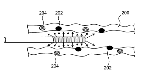
Fig. 5 is a schematic view of a state where, for
example, the light radiating probe 40 according to the
present invention shown in Fig. 4(c) is inserted into the
colon 200, a photosensitizer (PS) is injected, and light
including an optimum excitation wavelength region is
radiated to a superficial cancer tissue 202 and a lower
layer cancer tissue 204 in the colon 200. In general, the
CA 03044196 2019-05-16
lower layer cancer tissue 204 is a tumor nodule which is
difficult to recognize with naked eye. However, the
inventors of the present invention have confirmed that not
only the lower layer cancer tissue 204 of the colon 200 but
also lower layer cancer tissues 204 of an esophageal, a
stomach, an intestinal tract, a bladder cavity, a
peritoneum, an uterus, an abdominal cavity and other body
cavities can be detected by phosphorous detection due to an
EPR effect of the photosensitizer. In this manner,
according to the light radiating probe 40 of the present
invention, it is possible to properly capture a lower layer
cancer tissue of a body cavity which cannot be easily
recognized with the naked eye, and the lower layer cancer
tissue 204 can be killed more efficiently.
[0043]
According to the embodiment of the present invention,
as described previously, the joint jig 30 may have the
optical condenser adapter 32 between the fiber optics 20
and the light radiating probe for photodynamic therapy 40.
The optical condenser adapter 32 is provided for increasing
light intensity per unit area. The optical condenser
adapter 32 may be formed of, for example, a glass core
formed using a hard material such as glass, and a clad thin
film having a smaller refractive index than the glass core.
Further, as shown in Figs. 6(a) to 6(d), the optical
26
CA 03044196 2019-05-16
condenser adapter 32 includes a cylindrical large-diameter
portion 34, a small-diameter portion 36, and a neck portion
35 disposed between the cylindrical large-diameter portion
34 and the small-diameter portion 36, and has an incident
end surface 37 disposed on a left side in the drawing, and
an emitting end surface 38 disposed on a right side in the
drawing. The incident end surface 37 of the large-diameter
portion 34 of the optical condenser adapter 32 has the same
size and shape as an emitting end surface (not shown in the
drawing) of the fiber optics (first optical fiber) 20 and
is configured to be joined (coupled) to the emitting end
surface such that the incident end surface 37 is
substantially adapted to (coupled to) the light radiating
surface. On the other hand, the emitting end surface 38 of
the small-diameter portion 36 of the optical condenser
adapter 32 has the same size and shape as an incident end
surface (not shown in the drawing) of the light radiating
probe (second optical fiber) 40 and is configured to be
joined (coupled) to the incident end surface such that the
emitting end surface 38 is substantially adapted to the
incident end surface.
[0044]
The optical condenser adapter 32 can be easily
manufactured by melting a portion of Pyrex glass using a
burner, and by pulling the portion in directions in which
27
CA 03044196 2019-05-16
the large-diameter portion 34 and the small-diameter
portion 36 are separated from each other, the portion
having a diameter of 10 mm, for example, and corresponding
to the neck portion 35. The optical condenser adapter 32
according to the present invention can be manufactured by
softening by heating a polymer resin having high
transparency such as Lucite (registered trademark),
polypropylene, polyethylene, polyvinyl alcohol or
polystyrene besides glass.
[0045]
Accordingly, the optical condenser adapter 32 is
formed such that light intensity per unit area of light
which propagates to the incident end surface 37 of the
large-diameter portion 34 is increased along with the
reduction of a cross-sectional area in a path ranging from
the large-diameter portion 34 to the small-diameter portion
36 by way of the neck portion 35. With such a
configuration, it is possible to radiate stronger light
from the light scattering and radiating portion 48 to an
entire area near an affected part therapy target portion
such as a cancer affected part in all azimuth angles of 360
degrees. In Fig. 6, the optical condenser adapters 32
having various end surface diameters and various axial
lengths are illustrated. However, provided that the
optical condenser adapter 32 is configured such that light
28
CA 03044196 2019-05-16
intensity per unit area is increased, the optical condenser
adapter 32 having an arbitrary end surface diameter and an
arbitrary axial direction length may be used.
[0046]
Modifications of the above-mentioned embodiment are
described with reference to Fig. 7 and Fig. 8. The light
radiating probes 40 shown in Fig. 7 are formed such that
the light scattering and radiating portion 48 of the light
radiating probe 40 according to the above-mentioned
embodiment is wound around a rod (or a simple bar-like
member) 60 in a spiral shape or in a coil shape. It is
preferable that the rod 60 be made of a material harder
than a material for forming the light radiating probe 40
having flexibility. For example, the rod 60 may be formed
of endoscope fiber optics. Fig. 8 is a schematic view
showing a state where the light radiating probe 40
according to the modification shown in Fig. 7(a) is
inserted into the colon, a photosensitizer (PS) is injected,
and light including an optimum excitation wavelength region
is radiated to the superficial cancer tissue 202 and the
lower layer cancer tissue 204 of the colon.
[0047]
As shown in Fig. 7(a), the light scattering and
radiating portion 48 is wound around the rod 60 in a coil
shape. Accordingly, light which propagates from the light
29
CA 03044196 2019-05-16
guide portion 46 can be radiated to an entire area near an
affected part therapy target portion such as a cancer
affected part in all azimuth angles of 360 degrees along a
desired length in a longitudinal direction. Although it
also depends on winding density, compared to the light
scattering and radiating portion 48 according to the above-
mentioned embodiment shown in Fig. 5, the light scattering
and radiating portion 48 according to the modification
shown in Fig. 7(a) can radiate stronger (more concentrated)
light to an affected part therapy target portion and hence,
the light scattering and radiating portion 48 according to
the modification shown in Fig. 7(a) can expect a higher
therapeutic effect. Further, the light radiating probe 40
is formed by winding the light scattering and radiating
portion 48 around the rod 60 according to the previously-
mentioned embodiment in a coil shape. Accordingly, a
length of the light scattering and radiating portion 48 can
be easily adjusted corresponding to a length of the
affected part therapy target portion in the longitudinal
direction. Accordingly, it is possible to extremely easily
manufacture the light scattering and radiating portion 48
suitable for a length of an affected part therapy target
portion to be radiated by light.
(0048]
As shown Fig. 7(b), the light radiating probe 40 is
CA 03044196 2019-05-16
formed such that a light scattering and radiating portion
48 is wound around a rod 60 in a coil shape and, then, the
rod 60 and the light scattering and radiating portion 48
are covered by a sheath (cover film) 62 made of a
thermosetting resin (for example, a polyvinyl-based resin),
and the sheath 62 is thermally shrunken by applying heat to
the sheath 62 by a hot air generating device (for example,
a dryer or the like). As shown Fig. 7(c), a light
radiating probe 40 may be formed such that a rod 60 and a
light scattering and radiating portion 48 are integrally
fixed to each other by a protective film (sheath) 62. In
such a configuration, it is preferable that a coating
adhesive agent be applied to outer surfaces of the rod 60
and to the light scattering and radiating portion 48 or to
an inner surface of the sheath 62 in advance so as to
acquire the adhesion between the rod 60, the light
scattering and radiating portion 48 and the sheath 62 with
certainty. With such a configuration, when the light
scattering and radiating portion 48 shown in Fig. 7(c) is
inserted into a body cavity such as the colon, it is
possible to prevent the removal of the light scattering and
radiating portion 48 from the rod 60 and hence, it is
possible to make the light scattering and radiating portion
48 reach an affected part therapy target portion which
forms a target with certainty.
31
CA 03044196 2019-05-16
[0049]
A protective film 62 substantially equal to the
protective film shown in Fig. 7(c) can be easily
manufactured by winding the light scattering and radiating
portion 48 around the rod 60 in a coil shape and,
thereafter, by immersing the rod 60 and the light
scattering and radiating portion 48 into a melted solution
of a polyvinyl alcohol-based resin or an acrylic resin or
into a tacky solution made of a synthetic resin and, then,
by drying.
[0050]
As has been described heretofore, the present
invention has the following advantageous effects.
It is important for a general-use optical fiber to
transmit light frontward without loss. On the other hand,
in the present invention, light from the distal end portion
of the light radiating probe 40 is radiated to a peripheral
portion of a hollow organ part (for example, an oral cavity,
an esophageal, a stomach, an intestinal tract, an abdominal
cavity, a bladder cavity, a peritoneum, a diaphragm, an
uterus, a thoracic cavity, a bronchial tube, upper and
lower air ducts, a pharynx, a liver surface and the like)
of 360 . By performing fluorescent light emission by
exciting photosensitizer (PS) molecules of a nano-size
selectively accumulated in a local cancer tissue by an EPR
32
CA 03044196 2019-05-16
effect, the position of the lower layer cancer tissue 204
which cannot be easily recognized with naked eye can be
easily specified and, at the same time, singlet oxygen
which is one of active oxygens is generated from the
photosensitizer (PS) molecules so that the light radiating
probe 40 can exhibit an anti-cancer effect.
[0051]
Accordingly, in the photodynamic therapy (PDT), it is
necessary to radiate light in all azimuth angles of 360
toward a tube wall (an intestinal tract, an abdominal wall,
a chest wall) which forms a peripheral portion behind an
affected part rather than advancing the light straight in
an area near the affected part. Accordingly, it is
preferable that the light radiating probe 40 according to
the present invention be formed using a wire-like (string-
like) optical fiber having high flexibility (having
resiliency). In the present invention, the diameter of the
light radiating probe 40 may be, but not limited thereto, a
diameter (0.3 mm) narrower than the diameter illustrated
in Fig. 4(a), or may be substantially a value which falls
between 0.3 mm and 5 mm. The core member 42 of the light
radiating probe 40 may be made of, besides the above-
mentioned acrylic resin, polyethylene, polypropylene,
silicon, polyvinylchloride(PVC), Teflon, polyvinyl alcohol,
polyvinyl butyral, polyimide, polyurethane, nylon, various
33
CA 03044196 2019-05-16
polyesters, polyethylene naphthalate, polyethylene
terephthalate or the like. However, a material for forming
the core member 42 is not limited to these materials.
[0052]
The light radiating probe 40 according to the present
invention is formed of an optical fiber having high
flexibility and hence, the invasiveness of the light
radiating probe 40 when the light radiating probe 40 is
inserted into a body cavity of a patient is low.
Accordingly, a burden applied to a patient can be
suppressed as much as possible. Shearing, breaking by
bending or the like minimally occurs even when the light
radiating probe 40 is used plural times and hence, the
light radiating probe 40 can be used for a long period.
Further, the light radiating probe 40 can be easily
manufactured by applying roughening treatment or the like
to the distal end portion as described previously.
[0053]
Further, in the photodynamic therapy apparatus
according to the present invention, in the case where a
xenon light source or a tungsten light source having a
broad spectrum distribution is used, unlike laser light
source or a multicolor LED light source where an 'output
wavelength region is limited, with the use of an arbitrary
band pass filter, light which includes a wavelength region
34
CA 03044196 2019-05-16
in which an arbitrary optimum excitation wavelength region
of a photosensitizer (PS) is included at a peak can be
selectively outputted. That is, light which corresponds to
the photosensitizer (PS) can be outputted. Accordingly,
the present invention is applicable to the PS molecular
probe described in Patent Document 1 above.
EXAMPLE 1
[0054]
Using the photodynamic therapy apparatus 1 having the
light radiating probe 40 according to the present invention,
curing was applied to a colon/rectum cancer of a mouse in
which a colon/rectum cancer was generated in the form of an
autologous carcinogenesis (a cancer closest to a natural
cancer) by photodynamic therapy (PDT).
[0055]
Fig. 9(a) shows the light radiating probe 40
according to the present invention extending from the joint
jig 30. Fig. 9(a) shows a state where light from the xenon
light source apparatus 10 propagated, and was radiated in
all azimuth angles of 360 from the entire side surface of
the light scattering and radiating portion 48 in a
longitudinal direction. Fig. 9(b) shows a state where the
light scattering and radiating portion 48 was inserted into
the colon through an anus of a mouse into which a
photosensitizer (PS) was injected by intravenous injection
CA 03044196 2019-05-16
in advance. At this stage of the operation, the xenon
light source apparatus 10 was not operated so that the
light scattering and radiating portion 48 did not radiate
light. Fig. 9(c) shows a state where the xenon light
source apparatus 10 was operated from the state shown in
Fig. 9(b) so that light was radiated to the colon/rectum
cancer of the mouse from the light scattering and radiating
portion 48. At this stage of the operation, it was
confirmed that light was radiated to the entire abdomen of
the mouse.
When photodynamic therapy (PDT) was continuously
applied to the colon/rectum cancer for 10 min to 20 min
once a week, it was confirmed three weeks later that the
colon/rectum cancer of the mouse had substantially
disappeared.
DESCRIPTION OF REFERENCE SYMBOLS
[0056]
LIGHT SOURCE APPARATUS
FLEXIBLE FIBER OPTICS (FIRST OPTICAL FIBER)
JOINT JIG
32 OPTICAL CONDENSER ADAPTER
34 LARGE-DIAMETER PORTION
NECK PORTION
36 SMALL-DIAMETER PORTION
37 INCIDENT END SURFACE
36
CA 03044196 2019-05-16
38 EMITTING END SURFACE
40 LIGHT RADIATING PROBE FOR PHOTODYNAMIC THERAPY
(SECOND OPTICAL FIBER)
42 CORE MEMBER
44 CLADDING
46 LIGHT GUIDE PORTION
48 LIGHT SCATTERING AND RADIATING PORTION
60 ROD
62 SHEATH (COVER FILM)
100 CONVENTIONAL LIGHT RADIATING PROBE
102 ILLUMINATION LENS
104 OBJECT LENS (INCLUDING A CCD ELEMENT)
106 FORCEPS OPENING
108 NOZZLE
200 COLON
202 SUPERFICIAL CANCER TISSUE
204 LOWER LAYER CANCER TISSUE
37