Note: Descriptions are shown in the official language in which they were submitted.
CA 03044251 2019-05-16
WO 2018/094235 PCT/US2017/062344
MICROFLUIDIC SYSTEM AND METHOD OF USE THEREOF
CROSS-REFERENCE TO RELATED APPLICATIONS
[0001] This application claims benefit of priority under 35 U.S.C. 119(e)
of U.S. Serial
No. 62/424,208, filed November 18, 2016, the entire contents of which is
incorporated herein
by reference in its entirety.
BACKGROUND
FIELD OF THE INVENTION
[0002] The invention relates generally to the field of microfluidic devices
and more
specifically to a microfluidic system and method for reprogramming, expanding,
storing and
optionally differentiating cells.
BACKGROUND INFORMATION
[0003] Microfluidic systems are important in medical diagnostics and
biotechnology
research. Components of such systems include networks of very small wells and
channels,
through which liquids can deliver and combine precisely-controlled amounts of
chemicals,
cells, and molecules. The systems are used for a variety of tasks including
mixing reagents,
isolation and study of biomolecules, and sequestering and sorting living
cells. To accomplish
the precise transfer, mixing, and accurate metering that is required,
microfluidic chips and
substrates require complex control machinery such as micro-valves and pumps
built into the
chip, as well as pneumatic actuators, electronic solenoids, pneumatic
actuators, robotic
controllers, and the complex computer programs and systems that are required
to control
those devices.
[0004] The use of microfluidic devices provides many advantages over
classical benchtop
methods, including for example, an unrivaled economy of scale, as well as a
high degree of
parallelization and integration. As technology advances, microfluidic devices
are becoming
increasingly small in size and increasingly capable of performing multiple
tasks. For
example, microfluidic approaches have independently been proposed for cell
segregation and
isolation, cell culture, cellular differentiation, as well as screening for
cellular reprogramming
factors.
[0005] Stem cells are unspecialized cells that self-renew for long periods
through cell
division, and can be induced to differentiate into cells with specialized
functions, i.e.,
differentiated cells. These qualities give stem cells great promise for use in
therapeutic
applications to replace damaged cells and tissue in various medical
conditions. Embryonic
1
CA 03044251 2019-05-16
WO 2018/094235 PCT/US2017/062344
stem (ES) cells are derived from the blastocyst of an early stage embryo and
have the
potential to develop into endoderm, ectoderm, and mesoderm (the three germ
layers) (i.e.,
they are "pluripotent"). In vitro, ES cells tend to spontaneously
differentiate into various
types of tissues, and the control of their direction of differentiation can be
challenging. There
are unresolved ethical concerns that are associated with the destruction of
embryos in order to
harvest human ES cells. These problems limit their availability for research
and therapeutic
applications.
[0006]
Adult stem (AS) cells are found among differentiated tissues. Stem cells
obtained
from adult tissues typically have the potential to form a more limited
spectrum of cells (i.e.,
"multipotent"), and typically only differentiate into the cell types of the
tissues in which they
are found, though recent reports have shown some plasticity in certain types
of AS cells.
They also generally have a limited proliferation potential.
[0007]
Induced pluripotent stem cells (iPSC or iPSCs) are produced by laboratory
methods from differentiated adult cells. iPSCs are widely recognized as
important tools, e.g.,
for conducting medical research. Heretofore, the technology for producing
iPSCs has been
time-consuming and labor-intensive.
Differentiated adult cells, e.g., fibroblasts, are
reprogrammed, cultured, and allowed to form individual colonies which
represent unique
clones. Previously, identifying these types of cells has been difficult
because the majority of
the cells are not fully-reprogrammed iPSC clones. The standard is for iPSC
clones to be
selected based on the morphology of the cells, with desirable colonies
possessing sharply
demarcated borders containing cells with a high nuclear-to-cytoplasmic ratio.
When clones
are identified, they are manually-picked by micro-thin glass tools and
cultured on "feeder"
layers of cells typically, Murine Embryonic Fibroblasts (MEF). This step is
performed
typically at 14 ¨ 21 days post-infection with a reprograming vector. Then the
clones are
expanded for another 14 ¨ 21 days or more, prior to undergoing molecular
characterization.
[0008]
Others have focused on developing techniques to rapidly and more accurately
identify and characterize fully-reprogrammed adult fibroblasts and their
downstream
differentiation potential (Bock et at., 2011, Cell 144: 439-452; Boulting et
at., 2011, Nat
Biotechnol 29: 279-286). Also see, for example, co-owned U.S. Ser. No.
13/159,030, filed
on June 13, 2011, describing the use of Fluorescence Activated Cell Sorting
(FACS) to
identify and live sort unique subpopulations of s as defined by unique
expression patterns of
surface proteins.
[0009]
Thus, stem cells are an attractive source of cells for therapeutic
applications,
medical research, pharmaceutical testing, and the like. The use of patient-
specific stem cells
2
CA 03044251 2019-05-16
WO 2018/094235 PCT/US2017/062344
and reprogrammed somatic cells makes immunologically compatible cell
replacement
strategies extremely desirable for several medical treatments such as
treatment of cancer and
neuronal diseases to name a few. However, there remains a longstanding need in
the art for
improved microfluidic devices and methods for processing patient specific-
cells utilizing an
integrated approach in which multiple tasks are performed using an automated
and rapid
approach to go from initially processing a patient's blood to individualized
treatment of the
same patient using patient-specific stem cells and reprogrammed somatic cells.
SUMMARY OF THE INVENTION
[0010] The present invention provides a microfluidic based system and
methods utilizing
the system to process biological samples to provide a time and cost efficient
workflow to the
laboratory and/or medical based environment. The overall workflow is capable
of providing
patient-specific treatments.
[0011] Accordingly, in one aspect, a microfluidic system for processing a
biological
sample is provided. The system includes one or more microfluidic units
operable to perform
a number of sample processing steps such that cells from the sample, or cells
derived from
the sample may be stored and catalogued and eventually utilized to treat a
patient from which
the sample was taken. The microfluidic units are operable to isolate cells
from the sample,
expand the isolated cells and reprogram the cells. The system also includes
microfluidic
functionality to differentiate the reprogrammed cells to a desired cell type
for use in treating
the patient. At any point in the process the system includes functionality for
storing and
cataloging cells. Additionally, the system is operable to perform analysis of
the cells at any
stage of processing to make qualitative and quantitative assessments of cells.
[0012] In embodiments, the system includes one or more computer memory
modules
containing instructions for controlling the processing functions along with
one or more
computer processor modules configured to execute the instructions.
[0013] In another aspect, the invention provides a method for processing a
biological
sample utilizing the microfluidic bases system of the disclosure. The method
includes
applying the sample to the system and performing the processing steps on the
sample to
produce reprogrammed cells and/or cells of a desired cell type derived from
the
reprogrammed cells.
[0014] In another aspect, the invention provides a method of treating a
disease or disorder
in a subject utilizing the microfluidic based system of the disclosure. The
method includes:
a) obtaining a sample from the subject; b) applying the sample to the system;
c) processing
3
CA 03044251 2019-05-16
WO 2018/094235 PCT/US2017/062344
the sample with the system; and d) administering processed cells to the
subject, thereby
treating the disease or disorder in the subject.
[0015] In
still another aspect, the invention provides a pharmaceutical composition
including cells processed by the microfluidic system, or a cellular fraction
thereof, and
optionally containing a pharmaceutically acceptable excipient.
[0016] In
yet another aspect, the invention provides a cell bank. The cell bank includes
one or more populations of cells which are processed by the system of the
disclosure. In
embodiments, each cell population is catalogued and stored at an appropriate
temperature for
future use.
BRIEF DESCRIPTION OF THE FIGURES
[0017]
Figure 1 is a schematic diagram of a microfluidic system in one embodiment of
the invention.
[0018]
Figure 2 is a schematic diagram of a microfluidic system in one embodiment of
the invention.
[0019]
Figure 3 shows steps for acquiring a fibroblast cell bank in one embodiment of
the
invention.
[0020]
Figure 4 shows steps for obtaining a stem cell array from a fibroblast bank in
one
embodiment of the invention.
[0021]
Figure 5 is a flowchart showing steps in a system for producing iPSCs in one
embodiment of the invention.
[0022]
Figures 6A-6C show examples of a flow of patient samples through multi-well
tissue culture plates during an automated reprogramming process in one
embodiment of the
invention.
[0023]
Figures 7A-7C show an example of an equipment configuration to accomplish the
workflow in one embodiment of the invention.
DETAILED DESCRIPTION
[0024] The
present invention provides an integrated microfluidic system that utilizes
microfluidic chip technology to process a patient sample and generate patient-
specific
reprogrammed cells and optionally differentiated cells of a specific cell type
from the
reprogrammed cells. The
invention system greatly improves the efficiency and
reproducibility of making standardized iPSC lines. Typically, researchers
generate iPSCs by
hand, which limits the cells utility due to researcher variability and an
inability to generate
large numbers of cells. The system circumvents these problems with a
completely automated
system from receipt of the tissue or cell sample to banking of stocks of well-
defined iPSC
4
CA 03044251 2019-05-16
WO 2018/094235 PCT/US2017/062344
lines. The system allows for consistency and invariability for generation of
large numbers of
cells from many donors, which will facilitate the use of iPSC technology.
[0025] The system utilizes one or more microfluidic units, which may be in
the form of
one or more individual microfluidic chips, to process a sample, generate
reprogrammed cells
and maintain the cells in a microfluidic chip (hoteling) format for later
retrieval and
differentiation. Differentiation and expansion is then performed to generate
cells of a desired
cell type. Overall the workflow methodology and system is ideal as a
laboratory or hospital
based system that will allow the generation of pluripotent cells from every
patient for
downstream diagnostic or therapeutic use.
[0026] Before the present compositions and methods are described, it is to
be understood
that this invention is not limited to particular compositions, methods, and
experimental
conditions described, as such compositions, methods, and conditions may vary.
It is also to
be understood that the terminology used herein is for purposes of describing
particular
embodiments only, and is not intended to be limiting, since the scope of the
present invention
will be limited only in the appended claims.
[0027] As used in this specification and the appended claims, the singular
forms "a", "an",
and "the" include plural references unless the context clearly dictates
otherwise. Thus, for
example, references to "the method" includes one or more methods, and/or steps
of the type
described herein which will become apparent to those persons skilled in the
art upon reading
this disclosure and so forth.
[0028] Unless defined otherwise, all technical and scientific terms used
herein have the
same meaning as commonly understood by one of ordinary skill in the art to
which this
invention belongs. Although any methods and materials similar or equivalent to
those
described herein can be used in the practice or testing of the invention, the
preferred methods
and materials are now described.
[0029] The present disclosure provides a microfluidic system for processing
a biological
sample. The system includes one or more microfluidic units operable to perform
a number of
sample processing steps such that cells from the sample, or cells derived from
the sample,
such as iPSCs, may be stored and catalogued and eventually utilized to treat a
patient from
which the sample was taken. In embodiments, iPSCs generated from adult cells
isolated from
a patient sample are differentiated into a desired cell type suitable for use
to treat the patient.
[0030] As used herein "adult" means post-fetal, i.e., an organism from the
neonate stage
through the end of life, and includes, for example, cells obtained from
delivered placenta
tissue, amniotic fluid and/or cord blood.
CA 03044251 2019-05-16
WO 2018/094235 PCT/US2017/062344
[0031] As used herein, the term "adult differentiated cell" encompasses a
wide range of
differentiated cell types obtained from an adult organism, that are amenable
to producing
iPSCs using the instantly described automation system. Preferably, the adult
differentiated
cell is a "fibroblast." Fibroblasts, also referred to as "fibrocytes" in their
less active form, are
derived from mesenchyme. Their function includes secreting the precursors of
extracellular
matrix components including, e.g., collagen. Histologically, fibroblasts are
highly branched
cells, but fibrocytes are generally smaller and are often described as spindle-
shaped.
Fibroblasts and fibrocytes derived from any tissue may be employed as a
starting material for
the automated workflow system on the invention.
[0032] As used herein, the term, "induced pluripotent stem cells" or,
iPSCs, means that
the stem cells are produced from differentiated adult cells that have been
induced or changed,
i.e., reprogrammed into cells capable of differentiating into tissues of all
three germ or dermal
layers: mesoderm, endoderm, and ectoderm. The iPSCs produced do not refer to
cells as they
are found in nature.
[0033] Mammalian somatic cells useful in the present invention include, by
way of
example, adult stem cells, sertoli cells, endothelial cells, granulosa
epithelial cells, neurons,
pancreatic islet cells, epidermal cells, epithelial cells, hepatocytes, hair
follicle cells,
keratinocytes, hematopoietic cells, melanocytes, chondrocytes, lymphocytes (B
and T
lymphocytes), erythrocytes, macrophages, monocytes, mononuclear cells,
fibroblasts, cardiac
muscle cells, other known muscle cells, and generally any live somatic cells.
In particular
embodiments, fibroblasts are used. The term somatic cell, as used herein, is
also intended to
include adult stem cells. An adult stem cell is a cell that is capable of
giving rise to all cell
types of a particular tissue. Exemplary adult stem cells include hematopoietic
stem cells,
neural stem cells, and mesenchymal stem cells.
[0034] One advantage of the present invention is that it provides an
essentially limitless
supply of isogenic or synegenic human cells suitable for transplantation, use
in drug
discovery assays, or for disease modeling. The iPSCs are tailored specifically
to the patient,
avoiding immune rejection. Therefore, it will obviate the significant problem
associated with
current transplantation methods, such as, rejection of the transplanted
tissue, which may
occur because of host versus graft or graft versus host rejection. When
utilized for drug
discovery the cells demonstrate each person's response to chemicals when used
in drug
discovery or their individual manifestation of diseases in disease models.
Several kinds of
iPSCs or fully differentiated somatic cells prepared from iPSCs derived from
somatic cells
derived from humans can be stored in an iPSC bank as a library of cells, and
one kind or
6
CA 03044251 2019-05-16
WO 2018/094235 PCT/US2017/062344
more kinds of the iPSCs in the library can be used for preparation of somatic
cells, tissues, or
organs that are free of rejection by a patient to be subjected to stem cell
therapy.
[0035] The iPSCs of the present invention may be differentiated into a
number of different
cell types to treat a variety of disorders by methods known in the art. For
example, iPSCs
may be induced to differentiate into hematopoetic stem cells, muscle cells,
cardiac muscle
cells, liver cells, cartilage cells, epithelial cells, urinary tract cells,
neuronal cells, and the like.
The differentiated cells may then be transplanted back into the patient's body
to prevent or
treat a condition or used to advance medical research or in to develop drug
discovery assays.
Thus, the methods of the present invention may be used to as a treatment or to
develop a
treatment for a subject having a myocardial infarction, congestive heart
failure, stroke,
ischemia, peripheral vascular disease, alcoholic liver disease, cirrhosis,
Parkinson's disease,
Alzheimer's disease, diabetes, cancer, arthritis, wound healing,
immunodeficiency, aplastic
anemia, anemia, Huntington's disease, amyotrophic lateral sclerosis (ALS),
lysosomal storage
diseases, multiple sclerosis, spinal cord injuries, genetic disorders, and
similar diseases,
where an increase or replacement of a particular cell type/tissue or cellular
de-differentiation
is desirable.
[0036] The term "totipotency" refers to a cell with a developmental
potential to make all
of the cells in the adult body as well as the extra-embryonic tissues,
including the placenta.
The fertilized egg (zygote) is totipotent, as are the cells (blastomeres) of
the morula (up to the
16-cell stage following fertilization).
[0037] The term "pluripotent" as used herein refers to a cell with the
developmental
potential, under different conditions, to differentiate to cell types
characteristic of all three
germ cell layers, i.e., endoderm (e.g., gut tissue), mesoderm (including
blood, muscle, and
vessels), and ectoderm (such as skin and nerve). A pluripotent cell has a
lower developmental
potential than a totipotent cell. The ability of a cell to differentiate to
all three germ layers can
be determined using, for example, a nude mouse teratoma formation assay. In
some
embodiments, pluripotency can also evidenced by the expression of embryonic
stem (ES) cell
markers, although the preferred test for pluripotency of a cell or population
of cells generated
using the compositions and methods described herein is the demonstration that
a cell has the
developmental potential to differentiate into cells of each of the three germ
layers. In some
embodiments, a pluripotent cell is termed an "undifferentiated cell."
Accordingly, the terms
"pluripotency" or a "pluripotent state" as used herein refer to the
developmental potential of a
cell that provides the ability for the cell to differentiate into all three
embryonic germ layers
(endoderm, mesoderm and ectoderm). Those of skill in the art are aware of the
embryonic
7
CA 03044251 2019-05-16
WO 2018/094235 PCT/US2017/062344
germ layer or lineage that gives rise to a given cell type. A cell in a
pluripotent state typically
has the potential to divide in vitro for a long period of time, e.g., greater
than one year or
more than 30 passages.
[0038] The term "multipotent" when used in reference to a "multipotent
cell" refers to a
cell that has the developmental potential to differentiate into cells of one
or more germ layers,
but not all three. Thus, a multipotent cell can also be termed a "partially
differentiated cell."
Multipotent cells are well known in the art, and examples of multipotent cells
include adult
stem cells, such as for example, hematopoietic stem cells and neural stem
cells. "Multipotent"
indicates that a cell may form many types of cells in a given lineage, but not
cells of other
lineages. For example, a multipotent hematopoietic cell can form the many
different types of
blood cells (red, white, platelets, etc.), but it cannot form neurons.
Accordingly, the term
"multipotency" refers to a state of a cell with a degree of developmental
potential that is less
than totipotent and pluripotent.
[0039] The terms "stem cell" or "undifferentiated cell" as used herein,
refer to a cell in an
undifferentiated or partially differentiated state that has the property of
self-renewal and has
the developmental potential to differentiate into multiple cell types, without
a specific
implied meaning regarding developmental potential (i.e., totipotent,
pluripotent, multipotent,
etc.). A stem cell is capable of proliferation and giving rise to more such
stem cells while
maintaining its developmental potential. In theory, self-renewal can occur by
either of two
major mechanisms. Stem cells can divide asymmetrically, which is known as
obligatory
asymmetrical differentiation, with one daughter cell retaining the
developmental potential of
the parent stem cell and the other daughter cell expressing some distinct
other specific
function, phenotype and/or developmental potential from the parent cell. The
daughter cells
themselves can be induced to proliferate and produce progeny that subsequently
differentiate
into one or more mature cell types, while also retaining one or more cells
with parental
developmental potential. A differentiated cell may derive from a multipotent
cell, which itself
is derived from a multipotent cell, and so on. While each of these multipotent
cells may be
considered stem cells, the range of cell types each such stem cell can give
rise to, i.e., their
developmental potential, can vary considerably. Alternatively, some of the
stem cells in a
population can divide symmetrically into two stem cells, known as stochastic
differentiation,
thus maintaining some stem cells in the population as a whole, while other
cells in the
population give rise to differentiated progeny only. Accordingly, the term
"stem cell" refers
to any subset of cells that have the developmental potential, under particular
circumstances,
to differentiate to a more specialized or differentiated phenotype, and which
retain the
8
CA 03044251 2019-05-16
WO 2018/094235 PCT/US2017/062344
capacity, under certain circumstances, to proliferate without substantially
differentiating. In
some embodiments, the term stem cell refers generally to a naturally occurring
parent cell
whose descendants (progeny cells) specialize, often in different directions,
by differentiation,
e.g., by acquiring completely individual characters, as occurs in progressive
diversification of
embryonic cells and tissues. Some differentiated cells also have the capacity
to give rise to
cells of greater developmental potential. Such capacity may be natural or may
be induced
artificially upon treatment with various factors. Cells that begin as stem
cells might proceed
toward a differentiated phenotype, but then can be induced to "reverse" and re-
express the
stem cell phenotype, a term often referred to as "dedifferentiation" or
"reprogramming" or
"retrodifferentiation" by persons of ordinary skill in the art.
[0040] The term "embryonic stem cell" as used herein refers to naturally
occurring
pluripotent stem cells of the inner cell mass of the embryonic blastocyst
(see, for e.g., U.S.
Pat. Nos. 5,843,780; 6,200,806; 7,029,913; 7,584,479, which are incorporated
herein by
reference). Such cells can similarly be obtained from the inner cell mass of
blastocysts
derived from somatic cell nuclear transfer (see, for example, U.S. Pat. Nos.
5,945,577,
5,994,619, 6,235,970, which are incorporated herein by reference). Embryonic
stem cells are
pluripotent and give rise during development to all derivatives of the three
primary germ
layers: ectoderm, endoderm and mesoderm. In other words, they can develop into
each of the
more than 200 cell types of the adult body when given sufficient and necessary
stimulation
for a specific cell type. They do not contribute to the extra-embryonic
membranes or the
placenta, i.e., are not totipotent.
[0041] As used herein, the distinguishing characteristics of an embryonic
stem cell define
an "embryonic stem cell phenotype." Accordingly, a cell has the phenotype of
an embryonic
stem cell if it possesses one or more of the unique characteristics of an
embryonic stem cell,
such that that cell can be distinguished from other cells not having the
embryonic stem cell
phenotype. Exemplary distinguishing embryonic stem cell phenotype
characteristics include,
without limitation, expression of specific cell-surface or intracellular
markers, including
protein and microRNAs, gene expression profiles, methylation profiles,
deacetylation
profiles, proliferative capacity, differentiation capacity, karyotype,
responsiveness to
particular culture conditions, and the like. In some embodiments, the
determination of
whether a cell has an "embryonic stem cell phenotype" is made by comparing one
or more
characteristics of the cell to one or more characteristics of an embryonic
stem cell line
cultured within the same laboratory.
9
CA 03044251 2019-05-16
WO 2018/094235 PCT/US2017/062344
[0042] The term "somatic stem cell" is used herein to refer to any
pluripotent or
multipotent stem cell derived from non-embryonic tissue, including fetal,
juvenile, and adult
tissue. Natural somatic stem cells have been isolated from a wide variety of
adult tissues
including blood, bone marrow, brain, olfactory epithelium, skin, pancreas,
skeletal muscle,
and cardiac muscle. Each of these somatic stem cells can be characterized
based on gene
expression, factor responsiveness, and morphology in culture. Exemplary
naturally occurring
somatic stem cells include, but are not limited to, neural stem cells, neural
crest stem cells,
mesenchymal stem cells, hematopoietic stem cells, and pancreatic stem cells.
In some aspects
described herein, a "somatic pluripotent cell" refers to a somatic cell, or a
progeny cell of the
somatic cell, that has had its developmental potential altered, i.e.,
increased, to that of a
pluripotent state by contacting with, or the introduction of, one or more
reprogramming
factors using the compositions and methods described herein.
[0043] The term "progenitor cell" is used herein to refer to cells that
have greater
developmental potential, i.e., a cellular phenotype that is more primitive
(e.g., is at an earlier
step along a developmental pathway or progression) relative to a cell which it
can give rise to
by differentiation. Often, progenitor cells have significant or very high
proliferative potential.
Progenitor cells can give rise to multiple distinct cells having lower
developmental potential,
i.e., differentiated cell types, or to a single differentiated cell type,
depending on the
developmental pathway and on the environment in which the cells develop and
differentiate.
[0044] As used herein, the term "somatic cell" refers to any cell other
than a germ cell, a
cell present in or obtained from a pre-implantation embryo, or a cell
resulting from
proliferation of such a cell in vitro. Stated another way, a somatic cell
refers to any cell
forming the body of an organism, as opposed to a germline cell. In mammals,
germline cells
(also known as "gametes") are the spermatozoa and ova which fuse during
fertilization to
produce a cell called a zygote, from which the entire mammalian embryo
develops. Every
other cell type in the mammalian body--apart from the sperm and ova, the cells
from which
they are made (gametocytes) and undifferentiated, pluripotent, embryonic stem
cells--is a
somatic cell: internal organs, skin, bones, blood, and connective tissue are
all made up of
somatic cells. In some embodiments the somatic cell is a "non-embryonic
somatic cell," by
which is meant a somatic cell that is not present in or obtained from an
embryo and does not
result from proliferation of such a cell in vitro. In some embodiments the
somatic cell is an
"adult somatic cell," by which is meant a cell that is present in or obtained
from an organism
other than an embryo or a fetus or results from proliferation of such a cell
in vitro. Unless
otherwise indicated, the compositions and methods for reprogramming a somatic
cell
CA 03044251 2019-05-16
WO 2018/094235 PCT/US2017/062344
described herein can be performed both in vivo and in vitro (where in vivo is
practiced when
a somatic cell is present within a subject, and where in vitro is practiced
using an isolated
somatic cell maintained in culture).
[0045] The term "differentiated cell" encompasses any somatic cell that is
not, in its
native form, pluripotent, as that term is defined herein. Thus, the term a
"differentiated cell"
also encompasses cells that are partially differentiated, such as multipotent
cells, or cells that
are stable, non-pluripotent partially reprogrammed, or partially
differentiated cells, generated
using any of the compositions and methods described herein. In some
embodiments, a
differentiated cell is a cell that is a stable intermediate cell, such as a
non-pluripotent,
partially reprogrammed cell. It should be noted that placing many primary
cells in culture can
lead to some loss of fully differentiated characteristics. Thus, simply
culturing such
differentiated or somatic cells does not render these cells non-differentiated
cells (e.g.
undifferentiated cells) or pluripotent cells. The transition of a
differentiated cell (including
stable, non-pluripotent partially reprogrammed cell intermediates) to
pluripotency requires a
reprogramming stimulus beyond the stimuli that lead to partial loss of
differentiated character
upon placement in culture. Reprogrammed and, in some embodiments, partially
reprogrammed cells, also have the characteristic of having the capacity to
undergo extended
passaging without loss of growth potential, relative to parental cells having
lower
developmental potential, which generally have capacity for only a limited
number of
divisions in culture. In some embodiments, the term "differentiated cell" also
refers to a cell
of a more specialized cell type (i.e., decreased developmental potential)
derived from a cell of
a less specialized cell type (i.e., increased developmental potential) (e.g.,
from an
undifferentiated cell or a reprogrammed cell) where the cell has undergone a
cellular
differentiation process.
[0046] In various embodiments, the system is configured to perform a series
of processes
in a directional workflow. The processes performed by the system include
isolating cells,
expanding isolated cells, reprogramming expanded cells, differentiating
reprogrammed cells
to a desired cell type, and storing cells.
[0047] In various embodiments, the system is configured to isolate cells
from a biological
sample. This includes separation and isolation of specific cells types. In
embodiments, a
biological sample may include pre-isolated cells in which case it is not
necessary to perform
the isolation step. Isolation and/or separation techniques performed in a
microfluidic
capacity are known in the art and may be utilized in the practice of the
invention. Such
techniques include, but are not limited to cell capture and separation
methodologies.
11
CA 03044251 2019-05-16
WO 2018/094235 PCT/US2017/062344
[0048] A "biological sample" is a sample of biological material taken from
a patient or
subject that includes intact cells. Biological samples include samples taken
from bodily
fluids and tissues (e.g., from a biopsy) or tissue preparations (e.g., tissue
sections,
homogenates, etc.). A "bodily fluid" is any fluid obtained or derived from a
subject suitable
for use in accordance with the invention. Such fluids include whole blood,
blood fractions
such as serum and plasma, urine, sweat, lymph, feces, ascites, seminal fluid,
sputum, nipple
aspirate, post-operative seroma, wound drainage fluid, saliva, synovial fluid,
ascites fluid,
bone marrow aspirate, cerebrospinal fluid, nasal secretions, amniotic fluid,
bronchoalveolar
lavage fluid, pleural effusion, peripheral blood mononuclear cells, total
white blood cells,
lymph node cells, spleen cells, and tonsil cells. In embodiments the sample
includes white
blood cells or is a sample of isolated white blood cells.
[0049] The term "isolated cell" as used herein refers to a cell that has
been removed from
an organism in which it was originally found, or a descendant of such a cell.
Optionally the
cell has been cultured in vitro, e.g., in the presence of other cells.
Optionally, the cell is later
introduced into a second organism or re-introduced into the organism from
which it (or the
cell or population of cells from which it descended) was isolated.
[0050] The term "isolated population" with respect to an isolated
population of cells as
used herein refers to a population of cells that has been removed and
separated from a mixed
or heterogeneous population of cells. In some embodiments, an isolated
population is a
"substantially pure" population of cells as compared to the heterogeneous
population from
which the cells were isolated or enriched. In some embodiments, the isolated
population is an
isolated population of pluripotent cells which comprise a substantially pure
population of
pluripotent cells as compared to a heterogeneous population of somatic cells
from which the
pluripotent cells were derived.
[0051] In various embodiments, the system is also configured to perform a
cell expansion
step utilizing cells isolated from the sample. This is to ensure that there
are a sufficient
number of cells to perform downstream processes.
[0052] Once cells are expanded, the system includes functionality to
reprogram the
expanded cell, for example to generate iPSCs. The term "reprogramming" as used
herein
refers to a process that reverses the developmental potential of a cell or
population of cells
(e.g., a somatic cell). Stated another way, reprogramming refers to a process
of driving a cell
to a state with higher developmental potential, i.e., backwards to a less
differentiated state.
The cell to be reprogrammed can be either partially or terminally
differentiated prior to
reprogramming. In some embodiments of the aspects described herein,
reprogramming
12
CA 03044251 2019-05-16
WO 2018/094235 PCT/US2017/062344
encompasses a complete or partial reversion of the differentiation state,
i.e., an increase in the
developmental potential of a cell, to that of a cell having a pluripotent
state. In some
embodiments, reprogramming encompasses driving a somatic cell to a pluripotent
state, such
that the cell has the developmental potential of an embryonic stem cell, i.e.,
an embryonic
stem cell phenotype. In some embodiments, reprogramming also encompasses a
partial
reversion of the differentiation state or a partial increase of the
developmental potential of a
cell, such as a somatic cell or a unipotent cell, to a multipotent state.
Reprogramming also
encompasses partial reversion of the differentiation state of a cell to a
state that renders the
cell more susceptible to complete reprogramming to a pluripotent state when
subjected to
additional manipulations, such as those described herein. Such manipulations
can result in
endogenous expression of particular genes by the cells, or by the progeny of
the cells, the
expression of which contributes to or maintains the reprogramming. In certain
embodiments,
reprogramming of a cell using the synthetic, modified RNAs and methods thereof
described
herein causes the cell to assume a multipotent state (e.g., is a multipotent
cell). In some
embodiments, reprogramming of a cell (e.g. a somatic cell) using the
synthetic, modified
RNAs and methods thereof described herein causes the cell to assume a
pluripotent-like state
or an embryonic stem cell phenotype. The resulting cells are referred to
herein as
"reprogrammed cells," "somatic pluripotent cells," and "RNA-induced somatic
pluripotent
cells." The term "partially reprogrammed somatic cell" as referred to herein
refers to a cell
which has been reprogrammed from a cell with lower developmental potential by
the
methods as disclosed herein, such that the partially reprogrammed cell has not
been
completely reprogrammed to a pluripotent state but rather to a non-
pluripotent, stable
intermediate state. Such a partially reprogrammed cell can have a
developmental potential
lower that a pluripotent cell, but higher than a multipotent cell, as those
terms are defined
herein. A partially reprogrammed cell can, for example, differentiate into one
or two of the
three germ layers, but cannot differentiate into all three of the germ layers.
[0053] The term a "reprogramming factor," as used herein, refers to a
developmental
potential altering factor, as that term is defined herein, such as a gene,
protein, RNA, DNA,
or small molecule, the expression of which contributes to the reprogramming of
a cell, e.g. a
somatic cell, to a less differentiated or undifferentiated state, e.g. to a
cell of a pluripotent
state or partially pluripotent state. A reprogramming factor can be, for
example, transcription
factors that can reprogram cells to a pluripotent state, such as 50X2, OCT3/4,
KLF4,
NANOG, LIN-28, c-MYC, and the like, including as any gene, protein, RNA or
small
molecule, that can substitute for one or more of these in a method of
reprogramming cells in
13
CA 03044251 2019-05-16
WO 2018/094235 PCT/US2017/062344
vitro. In some embodiments, exogenous expression of a reprogramming factor,
using the
synthetic modified RNAs and methods thereof described herein, induces
endogenous
expression of one or more reprogramming factors, such that exogenous
expression of one or
more reprogramming factors is no longer required for stable maintenance of the
cell in the
reprogrammed or partially reprogrammed state. "Reprogramming to a pluripotent
state in
vitro" is used herein to refer to in vitro reprogramming methods that do not
require and/or do
not include nuclear or cytoplasmic transfer or cell fusion, e.g., with
oocytes, embryos, germ
cells, or pluripotent cells. A reprogramming factor can also be termed a "de-
differentiation
factor," which refers to a developmental potential altering factor, as that
term is defined
herein, such as a protein or RNA, that induces a cell to de-differentiate to a
less differentiated
phenotype, that is a de-differentiation factor increases the developmental
potential of a cell.
[0054]
Methods for transfecting and transforming or reprogramming adult cells to form
iPSC lines are generally known, e.g., Takahashi et al., 2007 Cell, 131: 861-
872, 2007, Yu et
al., 2007, Science, vol. 318, pp. 1917-1920. iPSC are induced from somatic
cells with
reprogramming factors.
Reprogramming factors are contemplated to include, e.g.,
transcription factors. The method for reprogramming adult cells includes,
e.g., introducing
and expressing a combination of specific transcription factors, e.g., a
combination of 0ct3/4,
Sox2, Klf4 and c-Myc genes. Others have demonstrated that other transcription
factors may
be employed in transforming or reprogramming adult cells. These other
transcription factors
include, e.g., Lin28, Nanog, hTert and SV40 large T antigen as described, for
example, by
Takahashi et al., 2006 Cell, 126: 663-676 and Huiqun Yin, et al. 2009, Front.
Agric. China
3(2): 199-208, incorporated by reference herein.
[0055]
iPSCs can also be generated using direct introduction of RNAs into a cell,
which,
when translated, provide a desired protein or proteins. Higher eukaryotic
cells have evolved
cellular defenses against foreign, "non-self," RNA that ultimately result in
the global
inhibition of cellular protein synthesis, resulting in cellular toxicity. This
response involves,
in part, the production of Type I or Type II interferons, and is generally
referred to as the
"interferon response" or the "cellular innate immune response." The cellular
defenses
normally recognize synthetic RNAs as foreign, and induce this cellular innate
immune
response. In certain aspects where the ability to achieve sustained or
repeated expression of
an exogenously directed protein using RNA is hampered by the induction of this
innate
immune response, it is desirable to use synthetic RNAs that are modified in a
manner that
avoids or reduces the response. Avoidance or reduction of the innate immune
response permit
sustained expression from exogenously introduced RNA necessary, for example,
to modify
14
CA 03044251 2019-05-16
WO 2018/094235 PCT/US2017/062344
the developmental phenotype of a cell. In one aspect, sustained expression is
achieved by
repeated introduction of synthetic, modified RNAs into a target cell or its
progeny. The
inventive methods include natural or synthetic RNAs.
[0056] The natural, modified, or synthetic RNAs in one aspect, can be
introduced to a cell
in order to induce exogenous expression of a protein of interest in a cell.
The ability to direct
exogenous expression of a protein of interest using the modified, synthetic
RNAs described
herein is useful, for example, in the treatment of disorders caused by an
endogenous genetic
defect in a cell or organism that impairs or prevents the ability of that cell
or organism to
produce the protein of interest. Accordingly, in some embodiments,
compositions and
methods comprising the RNAs described herein can be used for the purposes of
gene therapy.
[0057] The RNAs described can advantageously be used in the alteration of
cellular fates
and/or developmental potential. The ability to express a protein from an
exogenous RNA
permits either the alteration or reversal of the developmental potential of a
cell, i.e., the
reprogramming of the cell, and the directed differentiation of a cell to a
more differentiated
phenotype. A critical aspect in altering the developmental potential of a cell
is the
requirement for sustained and prolonged expression of one or more
developmental potential
altering factors in the cell or its immediate progeny. Traditionally, such
sustained expression
has been achieved by introducing DNA or viral vectors to a cell. These
approaches have
limited therapeutic utility due to the potential for insertional mutagenesis.
[0058] One of the areas that can most benefit from the ability to express a
desired protein
or proteins over a sustained period of time from exogenous RNAs as described
herein is the
generation of pluripotent or multipotent cells from cells initially having a
more differentiated
phenotype. In this aspect, RNAs encoding a reprogramming factor or factors are
used to
reprogram cells to a less differentiated phenotype, i.e., having a greater
developmental
potential.
[0059] In some embodiments of this aspect and all such aspects described
herein, the
synthetic, modified RNA molecule comprises at least two modified nucleosides.
In one such
embodiment, the two modified nucleosides are selected from the group
consisting of 5-
methylcytidine (5mC), N6-methyladenosine (m6A), 3,2'-0-dimethyluridine (m4U),
2-
thiouridine (s2U), 2' fluorouridine, pseudouridine, 2'-0-methyluridine (Um),
2' deoxy uridine
(2' dU), 4-thiouridine (s4U), 5-methyluridine (m5U), 2'-0-methyladenosine
(m6A), N6,2'-0-
dimethyladenosine (m6Am), N6,N6,2'-0-trimethyladenosine (m62Am), 2'-0-
methylcytidine
(Cm), 7-methylguanosine (m7G), 2'-0-methylguanosine (Gm), N2,7-
dimethylguanosine
(m2,7G), N2,N2,7-trimethylguanosine (m2,2,7G), and inosine (I). In one such
embodiment of
CA 03044251 2019-05-16
WO 2018/094235 PCT/US2017/062344
this aspect and all such aspects described herein, the at least two modified
nucleosides are 5-
methylcytidine (5mC) and pseudouridine. (see e.g., Rossi US 2012/0046346,
herein
incorporated by reference).
[0060]
Genes, proteins or RNA used in the methods of the invention include but are
not
limited to OCT4, SOX1, SOX 2, SOX 3, SOX15, SOX 18, NANOG, KLF1, KLF 2, KLF 4,
KLF 5, NR5A2, c-MYC, 1-MYC, n-MYC, REM2, TERT, and LIN28.
[0061] It
has also been shown that a single transcription factor may be employed in
reprogramming adult fibroblasts to iPSCs with the addition of certain small
molecule
pathway inhibitors. Such pathway inhibitors include e.g., the transforming
growth factor-beta
(TGFb) pathway inhibitors, SB431542 (444-(1,3-benzodioxo1-5-y1)-5-(2-
pyridiny1)-1H-
imi dazol-2-yl] -b enzami de), and A-
83-01 [3 -(6-Methy1-2-pyri diny1)-N-pheny1-4-(4-
quinoliny1)-1H-pyrazole-l-carbothioamide], the extracellular signal-regulated
kinases (ERK)
and microtubule-associated protein kinase (MAPK/ERK) pathway inhibitor
PD0325901 (N-
[(2R)-2,3-dihydroxypropoxy]-3,4-difluoro-2-[(2-fluoro-4-iodophenyl)amino]-
benzamide),
the GSK3 inhibitor CHIR99021 [6-((2-((4-(2,4-Dichloropheny1)-5-(4-methyl-1H-
imidazol-2-
yl)pyrimidin-2-yl)amino)ethyl)amino)nicotinonitrile] which activates Wnt
signaling by
stabilizing beta-catenin, the lysine-specific demethylasel Parnate (a/k/a
tranylcypromine), the
small molecule activator of 3'-phosphoinositide-dependent kinase-1 (PDK1) P548
[(2Z)-5-
(4-Chloropheny1)-3-pheny1-2-pentenoic acid], the hi stone deacetylase (HDAC)
inhibitors
sodium butyrate and valproic acid, small molecules that modulate mitochondrial
oxidation
(e.g., 2,4-dinitrophenol), glycolytic metabolism (fructose 2,6-bisphosphate
and oxalate), HIF
pathway activation (N-oxaloylglycine and Quercetin) Zhu et al., 2010, Cell
Stem Cell 7: 651-
655, incorporated by reference herein it its entirety. Zhu et al showed that
0ct4 combined
with Parnate and CHIR99021 was sufficient to reprogram adult human epidermal
keratinocytes.
[0062]
Although individual protocols differ, a general reprogramming protocol
consists of
expanding differentiated adult cells from tissue samples, e.g., skin biopsies
and contacting
them with reprogramming factors as discussed above, e.g., infecting them,
i.e., transfecting,
with e.g., expression vectors, such as viral constructs containing transcripts
for pluripotent
transcription factors. The
fibroblasts are obtained by art-known methods, e.g., by
mechanically disrupting the tissue followed by enzymatic dissociation to
release the
fibroblasts, and culturing the fibroblasts by art-known methods, e.g., as
described by Dimos
et. al., 2008, Science Vol. 321 (5893): 1218-1221.
16
CA 03044251 2019-05-16
WO 2018/094235 PCT/US2017/062344
[0063]
While illustrative aspects of the invention use vectors, e.g., viral vectors,
plasmid
vectors, in some aspects vectors are not required for transfection techniques,
including those
transferring mRNA molecules to cells.
[0064]
Transfection of the fibroblasts with an expression vector is carried out
according to
instructions provided with the desired vector. After a time (e.g., ranging
from about 2 to
about 10 days post-transfection), the cells are dissociated and contacted with
fluorescent
tagged antibodies raised against the CD13NEG, SSEA4PGs and Tra-1-60PGs surface
markers.
The dissociated and antibody-labeled cells are then resuspended in a phosphate
buffered
saline solution and moved to an automated sorting and isolation of iPSC
clones. Surface
marker positive cells are sorted by tag color or absence thereof directly into
sterile tubes
containing tissue culture media or multiwell (6-96 well) tissue culture plates
coated with
MEFs or cell free biological matrices and cultured until formation of visible
colonies occurs.
[0065]
Colonies are then further confirmed as iPSC by light microscopic inspection of
the
resulting clones or optionally by microscopic fluorescence inspection of
clones labeled with
fluorescent tagged antibodies. Optionally, in certain embodiments, one or more
of the
vectors also insert a green fluorescence protein (GFP) expression marker, for
convenience in
sorting and identification.
Several individual colonies possessing morphological
characteristics consistent with pluripotent ES cell lines are plucked from
cultures and
expanded individually to form monoclonal cultures.
[0066] In
one preferred embodiment of the inventive system, the treated cells are
subjected to genetic analysis to provide early confirmation and identification
of iPSCs.
Preferably, the genetic analysis is conducted by Southern blot, but other art-
known methods
may be employed which include but are not limited to MicroArray, NanoString,
quantitative
real time PCR (qPCR), whole genome sequencing, immunofluorescence microscopy,
flow
cytometry. Detection of enzymatic activity of alkaline phosphatase, positive
expression of
the cell membrane surface markers SSEA3, SSEA4, Tra-1-60, Tra-1-81 and the
expression of
the KLF4, 0ct3/4, Nanog, 5ox2 transcription factors in reprogrammed human
fibroblasts
confirms that a clone is an iPSC. Preferably, all of the markers are present.
[0067] Any
art-known transfection vector may be employed as a reprogramming factor,
including, e.g., an RNA such as mRNA, microRNA, siRNA, antisense RNA and
combinations thereof. Other expression vectors that may be employed include,
e.g., a
retrovirus, a lentivirus, an adenovirus, an adeno associated virus, a herpes
virus, a Sindbis
virus, a pox virus, a bacula virus, a bacterial phage, a Sendai virus and
combinations thereof
Preferably, an employed vector is a non-replicative vector such as, e.g.,
Sendai virus vectors
17
CA 03044251 2019-05-16
WO 2018/094235 PCT/US2017/062344
engineered to be nonreplicative. The preferred Sendai virus vector, while
incapable of
replication, remains capable of productive expression of nucleic acids
encoding protein(s)
carried by the vector, thereby preventing any potential uncontrolled spread to
other cells or
within the body of a vaccinee. This type of Sendai vector is commercially
available as a
CytoTuneTm-iPSC Sendai viral vector kit (DNAVEC, DV-0301).
[0068] Any art-known transfection method may be employed to insert such
vectors into
the adult fibroblasts, including, e.g., electroporation, gene gun, and the
like. Chemical
transfection is optionally conducted by means of a transfecting agent e.g., a
polymer, calcium
phosphate, a cationic lipid, e.g., for lipofection, and the like. Cell
penetrating peptides are
also optionally employed to carry vectors or other agents into the adult
fibroblast cells. In
brief, cell-penetrating peptides include those derived from proteins, e.g.,
protein transduction
domains and/or amphipathic peptides that can carry vectors or other agents
into the cell
include peptides. The subject of cell-penetrating peptides has been reviewed,
e.g., by Heitz et
al., 2009 British Journal of Pharmacology, 157: 195-206, incorporated by
reference herein in
its entirety. Other cell penetrating peptides are art-known, and are disclosed
by Heitz, Id.
Other cell-penetrating technologies including, e.g., liposomes and
nanoparticles, are also
contemplated to be employed in the methods of the present invention. Liposomes
and
nanoparticles are also described by Heitz, Id.
[0069] Antibodies can be employed in order to identify the transformed
cells. Four
antibodies against stem cell specific surface proteins are commonly used to
identify and
characterize human pluripotent stem cell populations; SSEA3, SSEA4, Tra-1-60
and Tra-1-
81. The Stage Specific Embryonic Antigens 3 and 4 (SSEA3 and SSEA4) are two
monoclonal antibodies which recognize sequential regions of a ganglioside
present on human
2102Ep cells (Henderson et al., 2002 Stem Cells 20: 329-337; Kannagi et al.,
1983, Embo J
2: 2355-2361). The Tra-1-60 and Tra-1-81 antibodies were originally raised
against human
embryonal carcinoma (EC) cells (PW et al., 1984, Hybridoma 3: 347-361) and
have been
shown to specifically recognize a carbohydrate epitope on a keratan sulfated
glycoprotein
identified as podocalyxin, a member of the CD34-related family of sialomucins
(Badcock et
al., 1999, Cancer Research 59: 4715-4719; Nielsen et al., 2007, PLoS ONE 2:
e237;
Schopperle and DeWolf, 2007, Stem Cells 25: 723-730). Several other surface
markers have
been shown to be expressed on ES cells and include CD326 or EpCam (Sundberg et
al.,
2009, Stem Cell Res 2: 113-124), CD24 (Heat Stable Antigen) and CD133 (Barraud
et al.,
2007, Journal of Neuroscience Research 85, 250-259) (Gang et al., 2007, Blood
109: 1743-
1751). Chan et al., 2009, Id. reported that the identification of bona fide
IPSc from fibroblasts
18
CA 03044251 2019-05-16
WO 2018/094235 PCT/US2017/062344
undergoing reprogramming via four factor retro viral transduction can be
achieved via live
cell imaging and by the observation, over time, that fibroblasts lose
expression of the cell
surface markers CD13 and D7Fib, and gain expression of the pluripotent stem
cell markers
SSEA4 and Tra-1-60 (Chan et al., 2009, Id.).
[0070] Thus, the invention further provides iPSCs produced using the
methods described
herein, as well as populations of such cells. The reprogrammed cells of the
present invention,
capable of differentiation into a variety of cell types, have a variety of
applications and
therapeutic uses. The basic properties of stem cells, the capability to
infinitely self-renew and
the ability to differentiate into every cell type in the body make them ideal
for therapeutic
uses.
[0071] In various embodiments, the system further includes functionality to
differentiate
reprogrammed cells to a desired cell type. A major goal of stem cell
technology is to make
the stem cell differentiate into a desired cell type, i.e., directed
differentiation or produce cells
via transdifferentiation. Not only are the compositions and methods described
herein useful
for reprogramming cells, they are also applicable to this directed
differentiation and
transdifferentiation of cells to a desired phenotype. That is, the same
technology described
herein for reprogramming is directly applicable to the differentiation of the
reprogrammed
cell, or any other stem cell or precursor cell, for that matter, to a desired
cell type.
[0072] A wide variety of additional cell types may be generated with
differentiation,
transdifferentiation and dedifferentiation. In the context of cell ontogeny,
the term
"differentiate", or "differentiating" is a relative term that refers to a
developmental process by
which a cell has progressed further down a developmental pathway than its
immediate
precursor cell. Thus in some embodiments, a reprogrammed cell as the term is
defined herein,
can differentiate to a lineage-restricted precursor cell (such as a mesodermal
stem cell), which
in turn can differentiate into other types of precursor cells further down the
pathway (such as
a tissue specific precursor, for example, a cardiomyocyte precursor), and then
to an end-stage
differentiated cell, which plays a characteristic role in a certain tissue
type, and may or may
not retain the capacity to proliferate further.
[0073] Differentiation is typically performed by contacting an iPSC with
one or more
differentiation factors. As used herein, the term "differentiation factor"
refers to a
developmental potential altering factor, as that term is defined herein, such
as a protein,
RNA, or small molecule, that induces a cell to differentiate to a desired cell-
type, i.e., a
differentiation factor reduces the developmental potential of a cell. In some
embodiments, a
differentiation factor can be a cell-type specific polypeptide, however this
is not required.
19
CA 03044251 2019-05-16
WO 2018/094235 PCT/US2017/062344
Differentiation to a specific cell type can require simultaneous and/or
successive expression
of more than one differentiation factor. In some aspects described herein, the
developmental
potential of a cell or population of cells is first increased via
reprogramming or partial
reprogramming using synthetic, modified RNAs, as described herein, and then
the cell or
progeny cells thereof produced by such reprogramming are induced to undergo
differentiation by contacting with, or introducing, one or more synthetic,
modified RNAs
encoding differentiation factors, such that the cell or progeny cells thereof
have decreased
developmental potential.
[0074] As used herein, the term "without the formation of a pluripotent
intermediate cell"
refers to the transdifferentiation of one cell type to another cell type,
preferably, in one step;
thus a method that modifies the differentiated phenotype or developmental
potential of a cell
without the formation of a pluripotent intermediate cell does not require that
the cell be first
dedifferentiated (or reprogrammed) and then differentiated to another cell
type. Instead, the
cell type is merely "switched" from one cell type to another without going
through a less
differentiated phenotype. Accordingly, transdifferentiation refers to a change
in the
developmental potential of a cell whereby the cell is induced to become a
different cell
having a similar developmental potential, e.g., a liver cell to a pancreatic
cell, a pancreatic
alpha cell into a pancreatic beta cell, etc. The system and methods of the
invention are well
suited for transdifferentiation of cells.
[0075] In various aspects, illustrative genes encoding differentiation
factors useful for
differentiating, dedifferentiating, or transdifferentiating a cell include
OCT4, NANOG, SOX2,
SOX17, HNF4, GATA4, HHEX, CEBPfl, CEBP6, PRDM16, MY0D1, NKX2.5, MEF2c,
MYOCARDIN, RUNX2, PDX, NGN, SALL4 or SOX9, or combination thereof. The
transcription factors encoded include 0ct4, NANOG, Sox2, Sox9, Sox17, HNF4a2,
HNF4a4,
HNF4a7, HNF4a8,HNF4y, GATA4, Hhex, CEBPO, CEB136, PRDM16, MyoD1, Nkx2.5,
Mef2c, Myocardin, Runx2-I, Pdxl, Ngn3, Sall4 or Runx2-II. For example,
differentiation of
mesoderm or fibroblasts to adipocytes, chondrocytes, osteocytes and myocytes
may be
performed using chimeric proteins including the following transcription
factors:
CEBPWCEBP6 (adipocytes), Sox9 (chondrocytes), Runx2 (osteocytes) and MyoD1
(myocytes).
[0076] Cell differentiation techniques performed in a microfluidic capacity
are known in
the art and may be utilized in the practice of the invention. Such techniques
include those
described in WO 2013/188748 which is incorporated herein by reference. WO
2013/188748
describes a microfluidic device for transdifferentiating cells from one cell
type to another.
CA 03044251 2019-05-16
WO 2018/094235 PCT/US2017/062344
The cells are cultured with one or more vector-free gene regulator
oligonucleotides
concurrently or in succession, and then harvested when cell markers or the
morphology of the
culture shows that transdifferentiation is complete. Suitable gene regular
oligonucleotides
include microRNAs and messenger RNAs that encode a differentiation factor.
Conditions for
transdifferentiation are optimized by dividing cells into different culture
chambers of a
microfluidic device. Cells are cultured with different additives in each
chamber, and then
compared.
[0077] Once differentiation is completed, the system has functionality for
expanding the
differentiated cells to generate sufficient numbers of a desired cell type for
downstream use.
[0078] At any stage of processing, the system of the present invention has
functionality
for storing cells. For example, isolated cells may be stored, expanded cells
may be stored,
reprogrammed cells may be stored, differentiated cells may be stored. Storage
may be under
any suitable conditions for prolonging cell life, for example, by freezing
cells at about -80 C
or below.
[0079] Furthermore, the system includes functionality for analyzing cells
at any stage of
processing to make qualitative and quantitative assessments of cells. Analysis
may include
any type of cellular analysis known in the art such as, by way of
illustration, image analysis,
cell number analysis, cell morphology analysis, polymerase chain reaction
(PCR) analysis,
sequence analysis, DNA analysis, RNA analysis, gene expression profiling,
proteome
analysis, metabolome analysis, immunoassays, nuclear exclusion analysis, or a
combination
thereof.
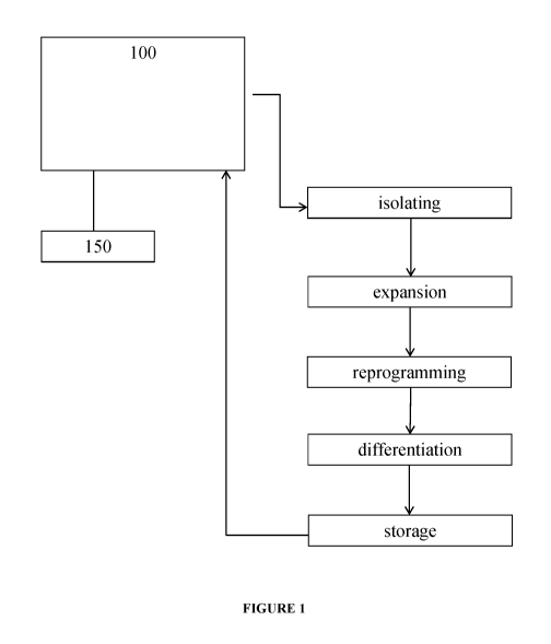
[0080] Figure 1 illustrates an embodiment of the system which includes a
single
microfluidic unit 100 configured to perform each of the processing steps. The
system is
shown as also including a single computer module 140 which includes a computer
memory
module containing instructions for controlling the processing steps and a
computer processor
module configured to execute the instructions.
[0081] It will be understood that the processing steps may be performed via
one or more
microfluidic units. For example, Figure 2 shows an embodiment of a system
having a first
microfluidic unit 100 and a second microfluidic unit 200. Unit 100 is operable
to perform
cell isolation, cell expansion, cell reprogramming and optionally cell
storage. Unit 200 is
operable to perform cell differentiation and storage. Each unit is controlled
by a single
computer module 150.
[0082] It is envisioned that any number of processing steps may be
performed by a single
microfluidic unit. For example, each processing step may be performed by
different
21
CA 03044251 2019-05-16
WO 2018/094235 PCT/US2017/062344
microfluidic units. In the context of the present invention, a microfluidic
unit may be
formatted as a microfluidic chip which is designed such that a specific task
may be performed
on the chip, e.g., cell isolation, expansion, reprogramming and
differentiation. It is also
envisioned that a single microfluidic chip may include multiple microfluidic
units, with
different units arranged in different locations on the chip. In embodiments,
the chip may be
severable so that the microfluidic units may be separated at some stage during
processing.
For example, reprogramming may be performed by a microfluidic unit disposed on
a first
region of a chip and the storage of the reprogrammed cells may be performed by
a
microfluidic storage unit disposed on a second region of the chip. The two
regions may be
severable from one another such that the reprogramming region can be separated
from the
storage zone and only the storage zone be frozen.
[0083] To accomplish specific processing tasks, microfluidic units are
designed to include
a number of channels through which fluid flow is directed, the channels being
formed in a
nonporous substrate. The term "nonporous substrate" means a solid support
material or
matrix on top of which a microfluidic unit of the invention is created using
photolithography
or other suitable process. The material is typically poly dimethyl siloxane
(PDMS) or poly
methyl methacrylate (PMMA) or other suitable materials known in the art.
[0084] In various embodiments, the width of the flow channels can be from
about 5 um to
about 1000 um and, for larger width flow channels, can be about 100 um, at or
between about
100 um and about 150 um, at or between about 150 um and 200 um, at or between
about 200
um and 250 um, at or between about 250 um and about 300 um, at or between
about 300 um
and about 350 um, at or between about 350 um and about 400 um, at or between
about 400
um and about 450 um, at or between about 450 um and about 500 um, at or
between about
500 um and about 550 um, at or between about 550 um and 600 um, at or between
about 600
um and about 650 um, at or between about 650 um and about 700 um, at or
between about
700 um and about 750 um, at or between about 750 um and 800 um, at or between
about 800
um and about 850 um, at or between about 850 um and about 900 um, at or
between about
900 um and about 950 um, at or between 950 um and 1000 um. In many
applications, a range
of flow channel widths from about 75 um to about 125 um will be preferred.
However, in
certain instances, channel widths could exceed 1000 um. For narrower channels,
the widths
can be about 5 um or greater and about 100 um or smaller. Channel widths can
be from about
um to about 75 um, from about 15 um to about 50 um, and from about 20 um to
about 40
um. In some embodiments the channel width is about 5, 10, 15, 20, 25, 30, 35,
40, 45, 50, 55,
60, 65, 70, or 75 um. The height can be from about 5 um to about 100 um, from
about 10 um
22
CA 03044251 2019-05-16
WO 2018/094235 PCT/US2017/062344
to about 75 um, from about 15 um to about 50 um, and from about 20 to about 40
um. In
some embodiments the channel height is about 5, 10, 15, 20, 25, 30, 35, 40,
45, 50, 55, 60,
65, 70, or 75 um. The cross sectional area can be from about 20 to about 13000
um2, from
about 50 to about 10000 um2, from about 200 to about 8000 um2, from about 250
to about
5000 um2, from about 500 to about 3000 um2, and in many embodiments, it is
preferred to be
from about 1400 to about 1600 um2. In some embodiments the cross sectional
area is about
500, 600, 700, 800, 900, 1000, 1100, 1200, 1300, 1400, 1500, 1600, 1700, 1800,
1900, or
about 2000 um2. The shape of the cross section of the individual channels of
the matrix
devices of this invention can be the same or different and can take different
shapes such as
square, rectangular, other polygonal, circular, elliptical, semicircular,
semielliptical, and the
like. The cross sectional shapes and areas can vary within the same channel
and can be
prepared by fabrication techniques described earlier and known in the art.
Square or
rectangular channel geometries are generally favored.
[0085] The present invention is described partly in terms of functional
components and
various processing steps. Such functional components and processing steps may
be realized
by any number of components, operations and techniques configured to perform
the specified
functions and achieve the various results. For example, the present invention
may employ
various biological samples, biomarkers, elements, materials, computers, data
sources, storage
systems and media, information gathering techniques and processes, data
processing criteria,
statistical analyses, regression analyses and the like, which may carry out a
variety of
functions. In addition, although the invention is described in the medical
diagnosis context,
the present invention may be practiced in conjunction with any number of
applications,
environments and data analyses; the systems described are merely exemplary
applications for
the invention.
[0086] Methods for processing according to various aspects of the present
invention may
be implemented in any suitable manner, for example using a computer program
operating on
the computer system. An exemplary system according to various aspects of the
present
invention is implemented in conjunction with a computer system, for example a
conventional
computer system comprising a processor and a random access memory, such as a
remotely-
accessible application server, network server, personal computer or
workstation. The
computer system also suitably includes additional memory devices or
information storage
systems, such as a mass storage system and a user interface, for example a
conventional
monitor, keyboard and tracking device. The computer system may, however,
comprise any
suitable computer system and associated equipment and may be configured in any
suitable
23
CA 03044251 2019-05-16
WO 2018/094235 PCT/US2017/062344
manner. In one embodiment, the computer system comprises a stand-alone system.
In another
embodiment, the computer system is part of a network of computers including a
server and a
database.
[0087] The software required for receiving, processing, and analyzing
information may be
implemented in a single device or implemented in a plurality of devices. The
software may be
accessible via a network such that storage and processing of information takes
place remotely
with respect to users. The system according to various aspects of the present
invention and its
various elements provide functions and operations to facilitate biomarker
analysis, such as
data gathering, processing, analysis, reporting and/or diagnosis. The present
system maintains
information relating to samples and may also facilitate analysis and/or
diagnosis. For
example, in the present embodiment, the computer system executes the computer
program,
which may receive, store, search, analyze, and report information relating to
analysis of cells.
The computer program may comprise multiple modules performing various
functions or
operations, such as a processing module for processing raw data and generating
supplemental
data and an analysis module for analyzing raw data and supplemental data to
cause the
system to perform specific tasks.
[0088] The system may also provide various additional modules and/or
individual
functions. For example, the system may also include a reporting function, for
example to
provide information relating to the processing and analysis functions. The
system may also
provide various administrative and management functions, such as controlling
access and
performing other administrative functions.
[0089] It will be understood that all, or any portion of the process
required to generate
iPSCs or differentiated cells therefrom, may be performed using a microfluidic
unit, or a
similarly automated process in operable connection to a microfluidic unit of
the system of the
invention.
[0090] In various embodiments, the system of the disclosure may utilize, or
be in operable
communication with, one or more systems (Systems 1-8) described in the
following
workflow system as disclosed in U.S. Patent Application Publication No.
2013/0345094,
which is incorporated herein by reference in its entirety.
[0091] The Workflow System
[0092] The workflow system is broken down into four independently-operated
units:
(1) Quarantine Somatic Cell Isolation and Growth (System 1);
(2) Quarantine Assay (System 2);
(3) Thawing, Infection and Identification (Systems 3, 4, and 5); and
24
CA 03044251 2019-05-16
WO 2018/094235 PCT/US2017/062344
(4) Maintenance, QC, Expansion, and Freezing. (Systems 6, 7, and 8)
[0093] Additionally, an automated -80 storage and retrieval system for
storing fibroblasts
and final clones in 1.4mL Matrix screw cap tubes, is part of the system. The
systems, and the
steps and operations that each unit will perform, will be described below.
[0094] System 1, Part A: Quarantine Somatic Cell Isolation and Growth
Workflow,
Biopsy Processing Pre-Mycoplasma Test
1. Technician will plate 40 biopsies per week in 6-well dishes;
2. 6-well plates will be maintained in quarantine incubator with 200-plate
capacity;
3. Periodic confluency checks are performed on an integrated Cyntellect
Celigo
Cytometer.
[0095] The system components that may be used to perform these automated
steps include
by way of example, STARlet Manual Load, a Modular Arm for 4/8/12 ch./MPH, 8
channels
with 1000 1 Pipetting Channels and an iSWAP Plate Handler, all available from
Hamilton
Science Robotics. If centerfuging is needed or desired, an Agilent VSpin
Microplate
Centerfuge can be used. The software may be Celigo API Software. The incubator
may be a
Cytomat Incubator. For plate handling a Cytomat 24 Barcode Reader, Cytomat
23mm
Stackers, and a Cytomat 400mm transfer station may be used. For plate tilting,
one may use
a MultiFlex Tilt Module. The system controller may be a Dell PG with a Windows
XP
operating system. The carrier package may be a Q Growth Carrier Package.
[0096] System 1, Part B: Quarantine Growth Workflow, Mycoplasma Test
1. Retrieve from incubator to deck of Quarantine Growth STARlet, remove
media from wells to plate for ELISA based mycoplasma test.
2. Manually transfer 96-well assay plates to Quarantine Assay STARlet.
[0097] System 1, Part C: Quarantine Growth Workflow, After Passing
Mycoplasma
Testing
1. Expanded fibroblasts distributed into multiple cryovials, capped,
transferred to
SAM - 80 C.
[0098] The system components that may be used to perform these automated
steps may be
selected from the same components used in the Quarantine Growth Workflow,
except a
STARlet Auto Load may be used. A Spectramax L Reader may be used as a spectral
acquisition device.
[0099] System 2: Quarantine Assay Workflow
1. Test using glow luminescence method, Lonza MycoAlert.
CA 03044251 2019-05-16
WO 2018/094235 PCT/US2017/062344
2. Perform luminescence plate read on spectral acquisition device.
[00100] The system components that may be used to perform these automated
steps include
STARlet Manual Load, a Modular Arm for 4/8/12 ch./MPH, 8 channels with 1000 1
Pipetting Channels and an iSWAP Plate Handler, all available from Hamilton
Science
Robotics. For luminescence assays the BioTek Synergy HT Reader may be used.
The
system controller may be a Dell PG with a Windows XP operating system. The
carrier
package may be a Q Growth Carrier Package.
[00101] Systems 3, 4, and 5: Thawing, Infection and Identification
[00102] Thawing Module & Infection Module
1. Retrieve cryotubes from SAM-80 C (61, 190)
2. Thaw on warming block (122)
3. Decap (Hamilton Capper Decapper) (126)
4. Add media to dilute cryoprotectants (122)
5. Spin (128)
6. Resuspend in plating data (122)
7. Plate one sample per well of 6-well (62, 122)
8. Move to incubator (130, 132)
9. Fibroblasts recover for about 3 -4 days
10. Confluence check on Cyntellect Celigo Cytometer (124)
11. Fibroblast passaging of all wells on the same day for reprogramming
(122)
12. In batches, tryspin passage wells (122)
13. Count cells on Cyntellect Celigo Cytometer (124)
14. Plate a defined number per well on one-to-three wells of a 24-well
plate
consolidating samples onto as few as 24-well plates as possible (64, 122)
15. Return plates to the incubator overnight (130, 132)
16. Retrieve plates and thaw virus in tube format and add to each well of
the
fibroblasts in the 24-well plates (130, 122)
17. Daily partial media exchanges (122)
[00103] Magnetic Sorting Module
18. Harvest cultures with accutase to single-cell suspension (134)
19. Dilute in staining buffer (134)
20. Stain with magnetic beads against fibroblast surface marker (134)
21. Wash step (134)
26
CA 03044251 2019-05-16
WO 2018/094235 PCT/US2017/062344
22. Apply to magnet (for Dynal beads) or column (for Miltenyi system) (134,
136)
23. Retrieve non-magnetic fraction to new wells (134)
24. Count cells on Cyntellect Celigo Cytometer (124)
25. Dilute to appropriate cell density for delivering 1 ¨ 10 cells per well
to 96-well
plate in passaging media (66, 134)
26. Retrieve new Matrigel or matrix-coated 96-well plate from 4 C incubator
(142)
27. Distribute cells to 96-well matrix plates, number based on cell count
for
example, two per plates per infection (66, 134)
28. Return plates to incubator (132)
29. Daily partial media exchanges (122)
[00104] Colony Identification Module
30. Retrieve 96-well plates from incubator to Colony identification liquid
handler
(66, 132, 138)
31. Perform live cell stain with pluripotency surface marker (138)
32. Image on Cyntellect Celigo Cytometer (140)
33. Identify wells with a single-marker positive colony that has a sharp
colony
border (140)
34. Techs review hits and select 6 per original sample for passage and
retrieve
plate and positive well IDs.
35. Cherry-pick wells with single positive colonies (138)
36. Retrieve new Matrigel or matrix coated 96-well plate from 4 C incubator
(68,
142)
37. Harvest selected wells and passage to new 96-well matrix plate
consolidating
clones onto as few plates as possible and plating each in passaging media (68,
138)
38. Daily partial media exchanges (122)
[00105] The system components that may be used to perform these automated
steps may be
selected from the same components used in the Quarantine Growth Workflow with
the
addition of one or more CORE 96 PROBEHEAD II 1000 1 model probe heads.
Systems 6, 7, and 8: Maintenance, QC, Expansion, and Freezing
[00106] Maintenance Module
39. Will serially-passage clones 1:1 into new 96-well matrix-coated plates
until
27
CA 03044251 2019-05-16
WO 2018/094235 PCT/US2017/062344
colony density is high enough (68-72, 160)
40. Daily feeding of all plates with ¨75% media exchange with 96-tip head
(160)
41. Periodic monitoring of colony density and growth rates on Cyntellect
Celigo
Cytometer (166)
42. Plate replication to produce plates for QC of clones (74-86, 160)
43. Goal is to expand clones onto multiple plates for use in several QC
assays to
eliminate poorly-performing clones until left with two-to-three high-quality
clones per original sample
44. Will also cherry-pick and re-array clones that pass QC steps as the
poor clones
are eliminated to consolidate clones onto as few plates as possible (80, 86,
160)
45. Daily feeding throughout this process (160)
[00107] QC Module
46. Harvest cells (74, 150)
47. Count cells (164)
48. Plate a defined cell number in V-bottom plates (range of 5000-10000
cells/well) in 2-6 replicates per line (84, 150)
49. Return to incubator ¨ (1g aggregation) (172)
50. Media exchange after two days (150)
51. Incubate for additional 12 days in incubator (172)
52. Partial media exchange every two days (150)
53. Transfer to nucleic acid prep station to remove media from wells
leaving
embryoid bodies in the well (84, 192)
54. Resuspend in RNA lysis buffer and combine and mix replicates for each
sample and make plates available for analysis in Nanostring nCounter assay
(84, 192)
[00108] Freezing Module
55. Begins with a 96-well plate after an expansion passage (88)
56. Incubate 6 days in incubator (172)
57. Partial media exchange every day (154)
58. Remove plate from incubator (88, 162)
59. Remove media (needs to be complete) (154)
60. Add cool Pre-freeze media (diluted matrigel in growth media) (154)
61. Incubate in incubator for lh (172)
28
CA 03044251 2019-05-16
WO 2018/094235 PCT/US2017/062344
62. Remove media (needs to be complete) (154)
63. Addition of cold freezing media ¨ low volume (154)
64. Seal plate (88, 164
65. Samples taken off-line to -80 C storage to freeze (190)
66. Store in vapor phase Liquid Nitrogen
[00109] Cryovial Storage
67. Begins with a 96-well plate after an expansion passage (90)
68. Incubate 6 days (172)
69. Daily partial media exchanges (154)
70. Passage wells 1:1 to a 24-well plate (92, 154)
71. Incubate 6 days (172)
72. Daily partial media exchanges (154)
73. Passage wells 1:1 to a 6-well plate (94, 154)
74. Incubate 4-6 days (172)
75. Daily partial media exchanges (154)
76. Remove plate from incubator (162)
77. Partial media exchange with pre-freeze media (154)
78. Incubate in incubator for lh (172)
79. Harvest cells for freezing as for normal passage (154)
80. Move to matrix tubes, two-to-three tubes per well (96, 154)
81. Spin and remove media (168, 154)
82. Addition of cold freezing media (154)
83. Cap tubes (170)
84. Samples taken off-line to -80 C storage (190)
[00110] Figure 3 shows the steps performed by System 1, including plating of a
biopsy (2),
outgrowth and passaging (4) (rolling production on liquid handling robot), QC
(6) (automated
testing for mycoplasma), and (8) automated freezing on liquid handling robot.
[00111] Figure 4 shows the steps performed by Systems 2, 3, and 4. Fibroblasts
are plated
by the automated system (10), reprogramming factors are introduced by the
automated
system (12), iPSCs are isolated by automated sorting and isolation (14),
desired clones are
selected and expanded by the automated system (16), automated quality checks
(QC) for
pluripotent status by marker assays and embryoid body assays (18), followed by
automated
freezing and storage of desired cells (20).
[00112] Figure 5 is a flowchart showing the step (22) through (60) involved in
System 1.
29
CA 03044251 2019-05-16
WO 2018/094235 PCT/US2017/062344
[00113] Figure 5 illustrates an example of the workflow and decision tree for
production of
fibroblasts from biopsies. The workflow is divided into Quarantine (58) and
Clean phases
(60). As biopsies enter the facility, a technician plates biopsies in 6-well
plates (22) and logs
the plates into the automated incubator (24). After biopsies are given time to
attach to the
plate, the liquid handling robot retrieves the plates from the automated
incubator to feed and
check confluency of the outgrowths on an automated microscope (26). The plates
are
returned to the incubator and allowed to outgrow (28). The liquid handler
removes the plate
from the incubator and exchanges the media for antibiotic and antimycotic free
media (30).
The robot moves the plate to the incubator for another five days (32). The
robot then
removes the plate and retrieves media to daughter plates for mycoplasma test
(34). The
daughter plates are moved to the Quarantine Assay system for mycoplasma
testing (36). A
choice is then made based on a positive signal from the assay (38). If all
wells of a 6-well
plate fail with a positive mycoplasma assay result (40) they are discarded. If
all wells of a 6-
well plate are negative and free of mycoplasma, they are transferred out of
quarantine into the
clean growth system (46). If some wells are positive and some wells are
negative, the
negative wells are maintained in quarantine (42). The negative wells are
passaged (44) to
new plates, transferred to the incubator, and the source plates containing
positive wells are
discarded. These cultures proceed through steps to retest for mycoplasma (24,
26, 28, 30, 32,
34, 36, 38). Clean cultures are monitored for growth (50), passaged (52) and
frozen in
cryovials (54, 56).
[00114] Figures 6A, 6B1, 6B2, and 6C illustrate an example of the flow of
patient samples
through multi-well tissue culture plates during the automated reprogramming
process. At the
top of each diagram, a flowchart describes the flow of procedures performed at
each step of
the workflow (70, 88, 98). At the bottom of each diagram, multi-well cell
culture plates are
shown with platemaps for example samples represented by shaded wells or groups
of wells
marked with sample labels (61-68, 72-86, 88-96). Transfer of a sample from
plate-to-plate or
well-to-well through the procedure is shown from left to right as indicated by
arrows. As
shown in Figure 6A, the automated iPSC derivation process begins when patient
samples and
control fibroblast samples (61) are plated in individual wells of a 6-well
plate (62). These are
passaged at defined cell number into individual wells of a 24-well plate (64)
for infection
using viruses encoding reprogramming factors or other means of introducing
reprogramming
factors to the cells. In the next step, reprogrammed samples are depleted of
non-
reprogrammed cells by cell sorting or, as is preferred, using magnetic bead
based enrichment
and plated at clonal density in multiple wells in 96-well plates (66). Two
such plates are
CA 03044251 2019-05-16
WO 2018/094235 PCT/US2017/062344
shown in this example. In this example, 6 wells, as indicated by wells with a
dot in the
middle (66) are identified containing a single clone positive for a
pluripotency surface marker
as assayed by immunofluorescent analysis on automated imager. These clones are
passaged
and cherry picked to reformat the clones into a minimum number of 96- well
plates (68). The
example figure shows six clones per individual starting sample and indicates
that clones from
16 starting sample can be arrayed onto a 96-well plate. To facilitate plate
processing, this
cherry picking step can be performed over multiple passages to consolidate the
clones onto a
minimum number of plates. As show in FIGs. 6B1 and 6B2, these clones are
serially
passaged until confluence of stem cell colonies within a well is achieved for
each starting
sample (72). Each plates' samples are then replicated onto duplicate plates
(74-86), to allow
for the quality control (6) and selection of clones that demonstrate
appropriate stem cell
characteristics. To begin the QC process, one plate is generated by the system
for a
Pluripotency quality control assay needed to determine pluripotent status of
the individual
clones (74) and one plate is generated for carrying forward in subsequent
passages (76). The
plate that is carried forward is passaged again into three plates (78, 80, 82)
for further quality
control and expansion. One plate is harvested for QC assays to characterize
Karyotype and
genetic diversity (78). A second plate (82) is passaged onto v-bottom plates
to form
embryoid bodies (84) for a QC assay that assesses differentiation capability
of the iPS clones.
The final plate (80) is carried forward for further expansion. Individual
clones that do not
pass quality control from previous pluripotency QC assays are not carried
forward as shown
by the "X" in the wells indicated in Figure 6. In the example shown in Figure
6B2, the
consolidated plate (86) will contain iPS lines (or differentiated lines) from
up to 32
individuals represented by 3 iPS clones per individual on a single 96 well
plate or up to 96
individuals if represented by a single clone each. Remaining clones are
consolidated onto as
few plates as possible until one to three clones remain (86-92). As shown in
Figure 6C, these
are expanded for cryopreservation while attached to the plate (88) or further
expanded (92-
94) and cryopreserved in cryovials (96). Any or all information from the
pluripotency
marker screen shown in Figure 6A (70), and the quality control assays shown in
Figure 6B1
can be used alone or in combination to decide which clones to select for
consolidation and
arraying in the automated process.
[00115] Figures 7A, 7B, 7C illustrate an example of the equipment
configuration needed to
accomplish the workflow in one embodiment of the invention. Figure 7A shows a
system
configuration for the automated expansion and quality control of a fibroblast
bank. Figure
7B shows a system configuration for the automated thawing of patient samples,
such as
31
CA 03044251 2019-05-16
WO 2018/094235 PCT/US2017/062344
fibroblasts, automated introduction of reprogramming factors with the patient
samples, such
as fibroblasts, automated cell sorting with MultiMACS, and automated colony
identification
and reformatting. Figure 7C shows a system configuration for the automated
expansion of
iPS clones, automated Embryoid Body production, and automated freezing.
[00116] As discussed herein, cells processed utilizing the system of the
disclosure may be
stored for downstream use. For example, processed cells of the invention may
be utilized to
treat a subject. For example, reprogrammed or differentiated cells may be
utilized to treat a
disease or disorder in subject. As such, the invention provides a method of
treating a disease
or disorder in a subject utilizing the microfluidic based system of the
disclosure. The method
includes: a) obtaining a sample from the subject; b) applying the sample to
the system; c)
processing the sample with the system; and d) administering processed cells to
the subject.
[00117] In embodiments, the subject is healthy when the sample is obtained.
The sample is
processed to produce reprogrammed cells and the reprogrammed cells catalogued
and stored.
Once the subject is diagnosed with a disease and in need of medical treatment,
the
reprogrammed cells may be further processed to produce differentiated cells of
a desired cell
type which are then used to treat the subject. For some treatments, the
reprogrammed cells
may be used to treat the subject. Appropriate differentiated cells (of
ectodermal, mesodermal
or endodermal lineage) may be derived from iPSCs produced by the inventive
methods. The
mode of administration can be determined by a person of skill in the art
depending on the
type of organ/injury to be treated. For example, iPSCs or differentiated cells
derived
therefrom, may be administered by injection (as a suspension) or implanted on
a
biodegradable matrix.
[00118] The term "healthy", "normal" or "clinically normal" means the subject
has no
known or apparent or presently detectable disease or dysfunction correlated
with a disease.
[00119] In another embodiment, the subject from which the sample is obtained
has been
diagnosed with, or as risk of having, a disease or disorder. The sample is
processed to
produce reprogrammed cells and the reprogrammed cells optionally stored. Once
the subject
medical treatment has been determined, the reprogrammed cells may be further
processed to
produce differentiated cells of a desired cell type which are then used to
treat the subject. For
some treatments, the reprogrammed cells may be used to treat the subject.
[00120] A "subject" is a member of any animal species, preferably a mammalian
species,
optionally a human. Thus, the methods and compositions described herein are
applicable to
both human and veterinary disease. Further, while a subject is preferably a
living organism,
the invention described herein may be used in post-mortem analysis as well.
Preferred
32
CA 03044251 2019-05-16
WO 2018/094235 PCT/US2017/062344
subjects are humans, and most preferably "patients," which as used herein
refers to living
humans that are receiving medical care for a disease or condition. This
includes persons with
no defined illness who are being investigated for signs of pathology. The
subject can be an
apparently healthy individual, an individual suffering from a disease, or an
individual being
treated for a disease.
[00121] Also contemplated to be within the scope of the invention are
compositions
comprising iPSCs or differentiated cells, e.g., compositions employed as
research tools, or as
pharmaceutical compositions, comprising effective amounts of cells prepared by
the system.
[00122] In addition, the invention relates to methods of testing
pharmaceuticals by
contacting iPSCs, transdifferentiated, or differentiated cells derived
therefrom, for example,
with one or more pharmaceutical agents of interest, and then detecting the
effect of the
applied pharmaceutical agent(s) on the contacted cells. For efficiency,
pharmaceutical
agent(s) are applied to a battery of iPSCs, or differentiated cells derived
therefrom. The cells
can vary in tissue source, in differentiated cell type, or allelic source, to
allow identification
of cells or tissue types that react favorably or unfavorably to one or more
pharmaceutical
agents of interest.
[00123] Further, the iPSCs produced by the inventive automated system may be
used as a
vehicle for introducing genes to correct genetic defects, such as osteogenesis
imperfecta,
diabetes mellitus, neurodegenerative diseases such as, for instance,
Alzheimer's disease,
Parkinson's disease, the various motor neuron diseases (MIND), e.g.,
amyotrophic lateral
sclerosis (ALS), primary lateral sclerosis (PLS), progressive muscular atrophy
(PMA) and the
like.
[00124] iPSCs produced by the inventive automated system may also be employed
to
provide specific cell types for biomedical research, as well as directly, or
as precursors, to
produce specific cell types for cell-based assays, e.g., for cell toxicity
studies (to determine
the effect of test compounds on cell toxicity), to determine teratogenic or
carcinogenic effects
of test compounds by treating the cells with the compound and observing and/or
recording
the compound's effects on the cells, e.g. effect on cellular differentiation.
[00125] Although the invention has been described with reference to the above
example, it
will be understood that modifications and variations are encompassed within
the spirit and
scope of the invention. Accordingly, the invention is limited only by the
following claims.
33