Note: Descriptions are shown in the official language in which they were submitted.
CA Application
Blakes Ref: 11871/00280
1 ABERRANT MITOCHONDRIAL DNA, ASSOCIATED FUSION TRANSCRIPTS AND
2 HYBRIDIZATION PROBES THEREFOR
3 FIELD OF THE INVENTION
4 [0001] The present invention relates to the field of mitochondria!
genomics. In one aspect, the
invention relates to the identification and use of mitochondrial genome fusion
transcripts and probes
6 .. that hybridize thereto.
7 BACKGROUND OF THE INVENTION
8 [0002] Mitochondrial Genome
9 [0003] The mitochondrial genome is a compact yet critical sequence
of nucleic acids.
Mitochondria! DNA, or "mtDNA", comprises a small genome of 16,569 nucleic acid
base pairs (bp)
11 (Anderson et al., 1981; Andrews et al., 1999) in contrast to the immense
nuclear genome of 3.3
12 billion bp (haploid). Its genetic complement is substantially smaller
than that of its nuclear cell mate
13 .. (0.0005%). However, individual cells carry anywhere from 103 to 104
mitochondria depending on
14 .. specific cellular functions (Singh and Modica-Napolitano 2002).
Communication or chemical
signalling routinely occurs between the nuclear and mitochondria! genomes
(Sherratt et at., 1997).
16 .. Moreover, specific nuclear components are responsible for the
maintenance and integrity of
17 mitochondria! sequences (Croteau et at., 1999). All mtDNA genonnes in a
given individual are
18 .. identical due to the clonal expansion of mitochondria within the ovum,
once fertilization has
19 .. occurred. However mutagenic events can induce sequence diversity
reflected as somatic mutations.
These mutations may accumulate in different tissues throughout the body in a
condition known as
21 .. heteroplasnny.
22 [0004] Mitochondrial Proteome
23 [0005] About 3,000 nuclear genes are required to construct,
operate and maintain mitochondria,
24 with only thirty-seven of these coded by the mitochondrial genome,
indicating heavy mitochondrial
dependence on nuclear loci. The mitochondrial genome codes for a complement of
24 genes,
26 including 2 rRNAs and 22 tRNAs that ensure correct translation of the
remaining 13 genes which are
27 vital to electron transport (see Figure 1). The mitochondrial genome is
dependent on seventy
28 nuclear encoded proteins to accomplish the oxidation and reduction
reactions necessary for this vital
29 .. function, in addition to the thirteen polypeptides supplied by the
mitochondria! genome. Both nuclear
.. and mitochondrial proteins form complexes spanning the inner mitochondrial
membrane and
31 collectively generate 80-90% of the chemical fuel adenosine
triphosphate, or ATP, required for
32 cellular metabolism. In addition to energy production, mitochondria play
a central role in other
23634990.1 1
CA 3044262 2019-05-27
CA Application
Blakes Ref: 11871/00280
1 metabolic pathways as well. A critical function of the mitochondria is
mediation of cell death, or
2 apoptosis (see Green and Kroemer, 2005). Essentially, there are signal
pathways which
3 permeabilize the outer mitochondrial membrane, or in addition, the inner
mitochondrial membrane as
4 well. When particular mitochondrial proteins are released into the
cytosol, non-reversible cell death
is set in motion. This process highlights the multi-functional role that some
mitochondrial proteins
6 have. These multi-tasking proteins suggest that there are other
mitochondrial proteins as well which
7 .. may have alternate functions.
8 [0006] Mitochondria! Fusion Transcriptome
9 [0007] The mitochondrial genome is unusual in that it is a
circular, intron-less DNA molecule.
The genome is interspersed with repeat motifs which flank specific lengths of
sequences.
11 Sequences between these repeats are prone to deletion under
circumstances which are not well
12 .. understood. Given the number of repeats in the mitochondrial genome,
there are many possible
13 deletions. The best known example is the 4977 "common deletion." This
deletion has been
14 associated with several purported conditions and diseases and is thought
to increase in frequency
with aging (Dai et al., 2004; Ro et al., 2003; Barron et al., 2001; Lewis et
al., 2000; Muller-Hocker,
16 1998; Porteous et al., 1998) (Figure 4). The current thinking in the
field of mitochondrial genomics is
17 that mitochondrial deletions are merely deleterious by-products of
damage to the mitochondria!
18 genome by such agents as reactive oxygen species and UVR. (Krishnan et
al 2008, Nature
19 Genetics). Further, though it is recognized that high levels of mtDNA
deletions can have severe
.. consequences on the cell's ability to produce energy in the form of ATP as
a result of missing gene
21 sequences necessary for cellular respiration, it is not anticipated that
these deleted mitochondria!
22 molecules may be a component of downstream pathways, have an intended
functional role, and
23 possibly may be more aptly viewed as alternate natural forms of the
recognized genes of the
24 mitochondria as has been anticipated by the Applicant.
[0008] The sequence dynamics of mtDNA are important diagnostic tools.
Mutations in mtDNA
26 are often preliminary indicators of developing disease. For example, it
has been demonstrated that
27 .. point mutations in the mitochondrial genome are characteristic of tumour
foci in the prostate. This
28 trend also extends to normal appearing tissue both adjacent to and
distant from tumour tissue (Parr
29 et al. 2006). This suggests that mitochondrial mutations occur early in
the malignant transformation
pathway.
31 [0009] For example, the frequency of a 3.4kb mitochondrial
deletion has excellent utility in
32 discriminating between benign and malignant prostate tissues (Maki et
al. 2008).
23634990.1 2
CA 3044262 2019-05-27
CA Application
Blakes Ref: 11871/00280
1 [0010] Mitochondrial fusion transcripts have been reported previously
in the literature, first in
2 soybeans (Morgens et al. 1984) and then later in two patients with Kearns-
Sayre Syndrome, a rare
3 neuromuscular disorder (Nakase et al 1990). Importantly, these
transcripts were not found to have
4 (or investigated regarding) association with any human cancers.
SUMMARY OF THE INVENTION
6 [0011] An object of the present invention to provide aberrant
mitochondrial DNA, associated
7 fusion transcripts and hybridization probes therefor.
8 [0012] In accordance with an aspect of the invention, there is
provided an isolated mitochondrial
9 fusion transcript associated with cancer.
[0013] In accordance with an aspect of the invention, there is provided a
mitochondrial fusion
11 protein corresponding to the above fusion transcript, having a sequence
as set forth in any one of
12 SEQ ID NOs: 34 to 49 and 52.
13 [0014] In accordance with another aspect of the invention, there
is provided an isolated mtDNA
14 encoding a fusion transcript of the invention.
[0015] In accordance with another aspect of the invention, there is
provided a hybridization
16 probe having a nucleic acid sequence complementary to at least a portion
of a mitochondria! fusion
17 transcript or an mtDNA of the invention.
18 [0016] In accordance with another aspect of the invention, there
is provided a method of
19 detecting a cancer in a mammal, the method comprising assaying a tissue
sample from the mammal
for the presence of at least one mitochondrial fusion transcript associated
with cancer by hybridizing
21 the sample with at least one hybridization probe having a nucleic acid
sequence complementary to
22 at least a portion of a mitochondrial fusion transcript according to the
invention.
23 [0017] In accordance with another aspect of the invention, there
is provided a method of
24 detecting a cancer in a mammal, the method comprising assaying a tissue
sample from the mammal
for the presence of at least one aberrant mtDNA associated with cancer by
hybridizing the sample
26 with at least one hybridization probe having a nucleic acid sequence
complementary to at least a
27 portion of an mtDNA according to the invention.
28 [0018] In accordance with another aspect of the invention, there
is provided a kit for conducting
29 an assay for detecting the presence of a cancer in a mammal, said kit
comprising at least one
hybridization probe complementary to at least a portion of a fusion transcript
or an mtDNA of the
31 invention.
23634990.1 3
CA 3044262 2019-05-27
CA Application
Blakes Ref: 11871/00280
1 [0019] In accordance with another aspect of the invention, there
is provided a screening tool
2 comprised of a microarray having 10's, 100's, or 1000's of mitochondrial
fusion transcripts for
3 identification of those associated with cancer.
4 [0020] In accordance with another aspect of the invention, there
is provided a screening tool
comprised of a microarray having 10's, 100's, or 1000's of mitochondrial DNAs
corresponding to
6 mitochondrial fusion transcripts for identification of those associated
with cancer.
7 [0021] In accordance with another aspect of the invention, there
is provided a screening tool
8 comprised of a multiplexed branched DNA assay having 10's, 100's, or
1000's of mitochondria!
9 fusion transcripts for identification of those associated with cancer.
[0022] __ In accordance with another aspect of the invention, there is
provided a screening tool
11 comprised of a multiplexed branched DNA assay having 10's, 100's, or
1000's of mitochondria!
12 DNAs corresponding to mitochondrial fusion transcripts for
identification of those associated with
13 cancer.
14 BRIEF DESCRIPTION OF THE DRAWINGS
[0023] The embodiments of the invention will now be described by way of
example only with
16 reference to the appended drawings wherein:
17 [0024] Figure us an illustration showing mitochondrial coding
genes.
18 [0025] Figure 2 shows polyadenalated fusion transcripts in
prostate samples invoked by the loss
19 of the 3.4kb deletion.
[0026] Figure 3 shows polyadenalated fusion transcripts in prostate samples
invoked by the loss
21 of the 4977kb common deletion.
22 [0027] Figure 4 shows polyadenalated fusion transcripts in breast
samples invoked by the loss
23 of the 3.4 kb segment from the mtgenome.
24 [0028] Figures 5a and 5b show an example of a mitochondrial DNA
region before and after
splicing of genes.
26 [0029] Figures 6a to 6g illustrate the results for transcripts 2,
3, 8, 9, 10, 11 and 12 of the
27 invention in the identification of colorectal cancer tumours.
28 [0030] Figures 7a to 7d illustrate the results for transcripts 6,
8, 10 and 20 of the invention in the
29 identification of lung cancer tumours.
23634990.1 4
CA 3044262 2019-05-27
CA Application
Blakes Ref: 11871/00280
1 [0031] Figures 8a to 8g illustrate the results for transcripts 6,
10, 11, 14, 15, 16 and 20 of the
2 invention in the identification of melanomas.
3 [0032] Figures 9a to 9h illustrate the results for transcripts 1,
2, 3, 6, 11, 12, 15 and 20 of the
4 invention in the identification of ovarian cancer.
[0033] Figures 10 to 18 illustrate the results for transcripts 2, 3, 4, 11,
12, 13, 15, 16 and 20 of
6 the invention in the identification of testicular cancer.
7 DETAILED DESCRIPTION OF THE INVENTION
8 [0034] The present invention provides novel mitochondrial fusion
transcripts and the parent
9 mutated mtDNA molecules that are useful for predicting, diagnosing and/or
monitoring cancer. The
invention further provides hybridization probes for the detection of fusion
transcripts and associated
11 mtDNA molecules and the use of such probes.
12 [0035] Definitions
13 [0036] __ Unless defined otherwise, all technical and scientific
terms used herein have the same
14 meaning as commonly understood by one of ordinary skill in the art to
which this invention belongs.
[0037] As used herein, "aberration" or "mutation" encompasses any
modification in the wild type
16 mitochondria! DNA sequence that results in a fusion transcript and
includes, without limitation,
17 insertions, translocations, deletions, duplications, recombinations,
rearrangements or combinations
18 thereof.
19 [0038] As defined herein, "biological sample" refers to a tissue
or bodily fluid containing cells
from which a molecule of interest can be obtained. For example, the biological
sample can be
21 derived from tissue such as prostate, breast, colorectal, lung and skin,
or from blood, saliva, cerebral
22 spinal fluid, sputa, urine, mucous, synovial fluid, peritoneal fluid,
amniotic fluid and the like. The
23 biological sample may be a surgical specimen or a biopsy specimen. The
biological sample can be
24 used either directly as obtained from the source or following a pre-
treatment to modify the character
of the sample. Thus, the biological sample can be pre-treated prior to use by,
for example,
26 preparing plasma or serum from blood, disrupting cells, preparing
liquids from solid materials,
27 diluting viscous fluids, filtering liquids, distilling liquids,
concentrating liquids, inactivating interfering
28 components, adding reagents, and the like.
29 [0039] A "continuous" transcript is a fusion transcript that keeps
the reading frame from the
beginning to the end of both spliced genes. An "end" transcript is a fusion
transcript that results in a
31 premature termination codon before the original termination codon of a
second spliced gene.
23634990.1 5
CA 3044262 2019-05-27
CA Application
Blakes Ref: 11871/00280
1 [0040] As used herein, "mitochondria! DNA" or "mtDNA" is DNA
present in mitochondria.
2 [0041] As used herein, the expression "mitochondrial fusion
transcript" or "fusion transcript"
3 refers to an RNA transcription product produced as a result of the
transcription of a mutated
4 mitochondria! DNA sequence wherein such mutations may comprise
mitochondrial deletions and
other large-scale mitochondria! DNA rearrangements.
6 [0042] Computer Analysis and Sequence Targeting
7 [0043] As discussed above, mitochondrial fusion transcripts have
been reported in soybeans
8 (Morgens et al. 1984) and in humans suffering from a rare neuromuscular
disorder (Nakase et al
9 1990). Fusion transcripts associated with human cancer have not, however,
been described.
[0044] Using the knowledge gained from mapping the large-scale deletions of
the human
11 mitochondrial genome associated with cancer, the observation of high
frequencies of these
12 deletions, and the evidence in another organism and another disease type
of trancriptionally active
13 mutated mtDNA molecules, Applicant hypothesized that such deletions may
have importance
14 beyond the DNA molecule and the damage and repair processes as it
relates to cancer. To test this
hypothesis computer analysis of the mitochondrial genome was conducted,
specific for repeat
16 elements, which suggested many potential deletion sites. Following this
initial step identifying
17 unique repeats in the mitochondrial sequence having non-adjacent or non-
tandem locations, a filter
18 was then applied to identify those repeats that upon initiating a
deletion event in the DNA molecule
19 would then likely reclose or religate to produce a fused DNA sequence
having an open reading
frame (ORF). A subset of 18 molecules were then selected for targeting to
investigate whether: 1)
21 they existed in the natural biological state of humans and 2) they had
relevance to malignancy.
22 Results from these investigations are described hereinafter.
23 [0045] Genomic Mutations
24 [0046] Mitochondrial DNA (mtDNA) dynamics are an important
diagnostic tool. Mutations in
mtDNA are often preliminary indicators of developing disease and behave as
biomarkers indicative
26 of risk factors associated with disease onset. According to the present
invention, large-scale
27 rearrangement mutations in the mitochondrial genome result in the
generation of fusion transcripts
28 associated with cancer. Thus, the use of mtDNA encoding such transcripts
and probes directed
29 thereto for the detection, diagnosis and monitoring of cancer is
provided.
[0047] One of skill in the art will appreciate that the mtDNA molecules for
use in the methods of
31 the present invention may be derived through the isolation of naturally-
occurring mutants or may be
32 based on the complementary sequence of any of the fusion transcripts
described herein. Exemplary
23634990.1 6
CA 3044262 2019-05-27
CA Application
Blakes Ref: 11871/00280
1 mtDNA sequences and fusion transcripts are disclosed in Applicant's U.S.
priority application no.
2 61/040,616.
3 [0048] Detection of Mutant Genomic Sequences
4 [0049] Mutant mtDNA sequences according to the present invention
may comprise any
modification that results in the generation of a fusion transcript. Non-
limiting examples of such
6 modifications include insertions, translocations, deletions,
duplications, recombinations,
7 rearrangements or combinations thereof. While the modification or change
can vary greatly in size
8 from only a few bases to several kilobases, preferably the modification
results in a substantive
9 deletion or other large-scale genomic aberration.
[0050] Extraction of DNA to detect the presence of such mutations may take
place using art-
11 recognized methods, followed by amplification of all or a region of the
mitochondrial genome, and
12 may include sequencing of the mitochondrial genome, as described in
Current Protocols in
13 Molecular Biology. Alternatively, crude tissue homogenates may be used
as well as techniques not
14 requiring amplification of specific fragments of interest.
[0051] The step of detecting the mutations can be selected from any
technique as is known to
16 those skilled in the art. For example, analyzing mtDNA can comprise
selection of targets by
17 branching DNA, sequencing the mtDNA, amplifying mtDNA by PCR, Southern,
Northern, Western
18 South-Western blot hybridizations, denaturing HPLC, hybridization to
microarrays, biochips or gene
19 chips, molecular marker analysis, biosensors, melting temperature
profiling or a combination of any
of the above.
21 [0052] Any suitable means to sequence mitochondria! DNA may be
used. Preferably, mtDNA is
22 amplified by PCR prior to sequencing. The method of PCR is well known in
the art and may be
23 performed as described in Mullis and Faloona, 1987, Methods Enzymol.,
155: 335. PCR products
24 can be sequenced directly or cloned into a vector which is then placed
into a bacterial host.
Examples of DNA sequencing methods are found in Brumley, R. L. Jr. and Smith,
L.M., 1991, Rapid
26 DNA sequencing by horizontal ultrathin gel electrophoresis, Nucleic
Acids Res. 19:4121-4126 and
27 Luckey, J.A., et al, 1993, High speed DNA sequencing by capillary gel
electrophoresis, Methods
28 Enzymol. 218: 154-172. The combined use of PCR and sequencing of mtDNA
is described in
29 Hopgood, R., et al, 1992, Strategies for automated sequencing of human
mtDNA directly from PCR
products, Biotechniques 13:82-92 and Tanaka, M. et al, 1996, Automated
sequencing of mtDNA,
31 Methods Enzymol. 264: 407-421.
23634990.1 7
CA 3044262 2019-05-27
CA Application
Blakes Ref: 11871/00280
1 [0053] Methods of selecting appropriate sequences for preparing
various primers are also
2 known in the art. For example, the primer can be prepared using
conventional solid-phase synthesis
3 using commercially available equipment, such as that available from
Applied Biosystems USA Inc.
4 .. (Foster City, California), DuPont, (Wilmington, Del.), or Milligen
(Bedford, Mass.).
[0054] According to an aspect of the invention, to determine candidate
genomic sequences, a
6 junction point of a sequence deletion is first identified. Sequence
deletions are primarily identified by
7 direct and indirect repetitive elements which flank the sequence to be
deleted at the 5' and 3' end.
8 .. The removal of a section of the nucleotides from the genome followed by
the ligation of the genome
9 results in the creation of a novel junction point.
[0055] Upon identification of the junction point, the nucleotides of the
genes flanking the junction
11 .. point are determined in order to identify a spliced gene. Typically the
spliced gene comprises the
12 initiation codon from the first gene and the termination codon of the
second gene, and may be
13 expressed as a continuous transcript, i.e. one that keeps the reading
frame from the beginning to the
14 end of both spliced genes. It is also possible that alternate initiation
or termination codons contained
.. within the gene sequences may be used as is evidenced by SEQ ID No:2 and
SEQ ID No: 17
16 disclosed herein. Some known mitochondrial deletions discovered to have
an open reading frame
17 (ORF) when the rearranged sequences are rejoined at the splice site are
provided in Table 1.
18 [0056] Exemplary mtDNA molecules for use in the methods of the
present invention, which have
19 been verified to exist in the lab, are provided below. These mtDNAs are
based on modifications of
the known mitochondrial genome (SEQ ID NO: 1) and have been assigned a fusion
or "FUS"
21 designation, wherein A:B represents the junction point between the last
mitochondrial nucleotide of
22 the first spliced gene and the first mitochondrial nucleotide of the
second spliced gene. The
23 identification of the spliced genes is provided in parentheses followed
by the corresponding
24 sequence identifier. Where provided below, (AltMet) and (OrigMet) refer
to alternate and original
translation start sites, respectively.
26 FUS 8469:13447 (AltMet) (ATP synthase FO subunit 8 to NADH dehydrogenase
subunit)
27 (SEQ ID No: 2)
28 FUS 10744:14124 (NADH dehydrogenase subunit 4L (ND4L) to NADH
dehydrogenase
29 subunit 5 (ND5)) (SEQ ID No: 3)
FUS 7974:15496 (Cytochrome c oxidase subunit II (C011) to Cytochrome b (Cytb))
(SEQ ID
31 No: 4)
23634990.1 8
CA 3044262 2019-05-27
CA Application
Blakes Ref: 11871/00280
1 FUS 7992:15730 (Cytochrome c oxidase subunit II (C011) to Cytochrome b
(Cytb)) (SEQ ID
2 No: 5)
3 FUS 8210:15339 (Cytochrome c oxidase subunit II (C011) to Cytochrome b
(Cytb)) (SEQ ID
4 No: 6)
FUS 8828:14896 (ATP synthase FO subunit 6 (ATPase6) to Cytochrome b (Cytb))
(SEQ ID
6 No: 7)
7 FUS 10665:14856 (NADH dehydrogenase subunit 4L (ND4L) to Cytochrome b
(Cytb)) (SEQ
8 ID No: 8)
9 FUS 6075:13799 (Cytochrome c oxidase subunit I (COI) to NADH de
hydrogenase subunit 5
(ND5)) (SEQ ID No: 9)
11 FUS 6325:13989 (Cytochrome c oxidase subunit I (COI) to NADH
dehydrogenase subunit 5
12 (ND5)) (SEQ ID No: 10)
13 FUS 7438:13476 (Cytochrome c oxidase subunit I (COI) to NADH
dehydrogenase subunit 5
14 (ND5)) (SEQ ID No: 11)
FUS 7775:13532 (Cytochrome c oxidase subunit II (C011) to NADH dehydrogenase
subunit 5
16 (ND5)) (SEQ ID No: 12)
17 FUS 8213:13991 (Cytochrome c oxidase subunit II (C011) to NADH
dehydrogenase subunit 5
18 (ND5)) (SEQ ID No: 13)
19 FUS 9191:12909 (ATP synthase FO subunit 6 (ATPase6) to NADH
dehydrogenase subunit 5
(ND5)) (SEQ ID No: 14)
21 FUS 9574:12972 (Cytochrome c oxidase subunit III (C0111) to NADH
dehydrogenase subunit
22 5 (ND5)) (SEQ ID No: 15)
23 FUS 10367:12829 (NADH dehydrogenase subunit 3 (ND3) to NADH
dehydrogenase subunit
24 5 (ND5)) (SEQ ID No: 16)
FUS 8469:13447 (OrigMet) (ATP synthase FO subunit 8 to NADH dehydrogenase
subunit)
26 (SEQ ID No: 17)
27 FUS 9144:13816 ((ATP synthase FO subunit 6 (ATPase6) to NADH
dehydrogenase subunit
28 5 (ND5)) (SEQ ID No: 51)
29 [0057] The present invention also provides the use of variants or
fragments of these sequences
for predicting, diagnosing and/or monitoring cancer.
23634990.1 9
CA 3044262 2019-05-27
CA Application
Blakes Ref: 11871/00280
1 [0058] "Variant", as used herein, refers to a nucleic acid
differing from a mtDNA sequence of the
2 present invention, but retaining essential properties thereof. Generally,
variants are overall closely
3 similar, and, in many regions, identical to a select mtDNA sequence.
Specifically, the variants of the
4 present invention comprise at least one of the nucleotides of the
junction point of the spliced genes,
and may further comprise one or more nucleotides adjacent thereto. In one
embodiment of the
6 invention, the variant sequence is at least 80%, 85%, 90%, 95%, 96%, 97%,
98% or 99% identical to
7 any one of the mtDNA sequences of the invention, or the complementary
strand thereto.
8 [0059] In the present invention, "fragment" refers to a short
nucleic acid sequence which is a
9 portion of that contained in the disclosed genomic sequences, or the
complementary strand thereto.
This portion includes at least one of the nucleotides comprising the junction
point of the spliced
11 genes, and may further comprise one or more nucleotides adjacent
thereto. The fragments of the
12 invention are preferably at least about 15 nt, and more preferably at
least about 20 nt, still more
13 preferably at least about 30 nt, and even more preferably, at least
about 40 nt, at least about 50 nt,
14 at least about 75 nt, or at least about 150 nt in length. A fragment "at
least 20 nt in length," for
example, is intended to include 20 or more contiguous bases of any one of the
mtDNA sequences
16 listed above. In this context "about" includes the particularly recited
value, a value larger or smaller
17 by several (5, 4, 3, 2, or 1) nucleotides, at either terminus or at both
termini. These fragments have
18 uses that include, but are not limited to, as diagnostic probes and
primers as discussed herein. Of
19 course, larger fragments (e.g., 50, 150, 500, 600, 2000 nucleotides) are
also contemplated.
[0060] Thus, in specific embodiments of the invention, the mtDNA sequences
are selected from
21 the group consisting of:
22 SEQ ID NO: 2 (FUS 8469:13447; AltMet)
23 SEQ ID NO: 3 (FUS 10744:14124)
24 SEQ ID NO: 4 (FUS 7974:15496)
SEQ ID NO: 5 (FUS 7992:15730)
26 SEQ ID NO: 6 (FUS 8210:15339)
27 SEQ ID NO: 7 (FUS 8828:14896)
28 SEQ ID NO: 8 (FUS 10665:14856)
29 SEQ ID NO: 9 (FUS 6075:13799)
SEQ ID NO: 10 (FUS 6325:13989)
23634990.1 10
CA 3044262 2019-05-27
CA Application
Blakes Ref: 11871/00280
1 SEQ ID NO: 11 (FUS 7438:13476)
2 SEQ ID NO: 12 (FUS 7775:13532)
3 SEQ ID NO: 13 (FUS 8213:13991)
4 SEQ ID NO: 14 (FUS 9191:12909)
SEQ ID NO: 15 (FUS 9574:12972)
6 SEQ ID NO: 16 (FUS 10367:12829)
7 SEQ ID NO: 17(FUS 8469:13447; OrigMet)
8 SEQ ID NO: 51 (FUS 9144:13816), and
9 fragments or variants thereof.
[0061] Probes
11 [0062] Another aspect of the invention is to provide a
hybridization probe capable of recognizing
12 an aberrant mtDNA sequence of the invention. As used herein, the term
"probe" refers to an
13 oligonucleotide which forms a duplex structure with a sequence in the
target nucleic acid, due to
14 complementarity of at least one sequence in the probe with a sequence in
the target region. The
probe may be labeled, according to methods known in the art.
16 [0063] Once aberrant mtDNA associated with a particular disease is
identified, hybridization of
17 mtDNA to, for example, an array of oligonucleotides can be used to
identify particular mutations,
18 however, any known method of hybridization may be used.
19 [0064] As with the primers of the present invention, probes may be
generated directly against
exemplary mtDNA fusion molecules of the invention, or to a fragment or variant
thereof. For
21 instance, the sequences set forth in SEQ ID NOs: 2-17 and 51 and those
disclosed in Table 1 can
22 be used to design primers or probes that will detect a nucleic acid
sequence comprising a fusion
23 sequence of interest. As would be understood by those of skill in the
art, primers or probes which
24 hybridize to these nucleic acid molecules may do so under highly
stringent hybridization conditions
or lower stringency conditions, such conditions known to those skilled in the
art and found, for
26 example, in Current Protocols in Molecular Biology (John Wiley & Sons,
New York (1989)), 6.3.1-
27 6.3.6.
28 [0065] In specific embodiments of the invention, the probes of the
invention contain a sequence
29 complementary to at least a portion of the aberrant mtDNA comprising the
junction point of the
spliced genes. This portion includes at least one of the nucleotides involved
in the junction point A:B,
23634990.1 11
CA 3044262 2019-05-27
CA Application
Blakes Ref: 11871/00280
1 and may further comprise one or more nucleotides adjacent thereto. In
this regard, the present
2 invention encompasses any suitable targeting mechanism that will select
an mtDNA molecule using
3 the nucleotides involved and/or adjacent to the junction point A:B.
4 [0066] Various types of probes known in the art are contemplated
by the present invention. For
example, the probe may be a hybridization probe, the binding of which to a
target nucleotide
6 sequence can be detected using a general DNA binding dye such as ethidium
bromide, SYBRO
7 Green, SYBR Gold and the like. Alternatively, the probe can incorporate
one or more detectable
8 labels. Detectable labels are molecules or moieties a property or
characteristic of which can be
9 detected directly or indirectly and are chosen such that the ability of
the probe to hybridize with its
target sequence is not affected. Methods of labelling nucleic acid sequences
are well-known in the
11 art (see, for example, Ausubel et al., (1997 & updates) Current
Protocols in Molecular Biology, Wiley
12 & Sons, New York).
13 [0067] Labels suitable for use with the probes of the present
invention include those that can be
14 directly detected, such as radioisotopes, fluorophores,
chemiluminophores, enzymes, colloidal
particles, fluorescent nnicroparticles, and the like. One skilled in the art
will understand that directly
16 detectable labels may require additional components, such as substrates,
triggering reagents, light,
17 .. and the like to enable detection of the label. The present invention
also contemplates the use of
18 labels that are detected indirectly.
19 [0068] The probes of the invention are preferably at least about
15 nt, and more preferably at
least about 20 nt, still more preferably at least about 30 nt, and even more
preferably, at least about
21 40 nt, at least about 50 nt, at least about 75 nt, or at least about 150
nt in length. A probe of "at least
22 20 nt in length," for example, is intended to include 20 or more
contiguous bases that are
23 complementary to an mtDNA sequence of the invention. Of course, larger
probes (e.g., 50, 150, 500,
24 600, 2000 nucleotides) may be preferable.
[0069] The probes of the invention will also hybridize to nucleic acid
molecules in biological
26 samples, thereby enabling the methods of the invention. Accordingly, in
one aspect of the invention,
27 there is provided a hybridization probe for use in the detection of
cancer, wherein the probe is
28 complementary to at least a portion of an aberrant mtDNA molecule. In
another aspect the present
29 invention provides probes and a use of (or a method of using) such
probes for the detection of
colorectal cancer, lung cancer, breast cancer, ovarian cancer, testicular,
cancer, prostate cancer
31 and/or melanoma skin cancer.
32 [0070] Assays
23634990.1 12
CA 3044262 2019-05-27
CA Application
Blakes Ref: 11871/00280
1 [0071] Measuring the level of aberrant mtDNA in a biological
sample can determine the
2 presence of one or more cancers in a subject. The present invention,
therefore, encompasses
3 methods for predicting, diagnosing or monitoring cancer, comprising
obtaining one or more biological
4 samples, extracting mtDNA from the samples, and assaying the samples for
aberrant mtDNA by:
quantifying the amount of one or more aberrant mtDNA sequences in the sample
and comparing the
6 quantity detected with a reference value. As would be understood by those
of skill in the art, the
7 reference value is based on whether the method seeks to predict, diagnose
or monitor cancer.
8 Accordingly, the reference value may relate to mtDNA data collected from
one or more known non-
9 cancerous biological samples, from one or more known cancerous biological
samples, and/or from
one or more biological samples taken overtime.
11 [0072] In one aspect, the invention provides a method of detecting
cancer in a mammal, the
12 method comprising assaying a tissue sample from the mammal for the
presence of an aberrant
13 mitochondria! DNA described above. The present invention also provides
for methods comprising
14 assaying a tissue sample from the mammal by hybridizing the sample with
at least one hybridization
probe. The probe may be generated against a mutant mitochondria! DNA sequence
of the invention
16 as described herein.
17 [0073] In another aspect, the invention provides a method as
above, wherein the assay
18 comprises:
19 a) conducting a hybridization reaction using at least one of the probes
to allow the at least
one probe to hybridize to a complementary aberrant mitochondria! DNA sequence;
21 b) quantifying the amount of the at least one aberrant mitochondrial DNA
sequence in the
22 sample by quantifying the amount of the mitochondria! DNA hybridized to
the at least one probe;
23 and,
24 c) comparing the amount of the mitochondria! DNA in the sample to at
least one known
reference value.
26 [0074] Also included in the present invention are methods for
predicting, diagnosing or
27 monitoring cancer comprising diagnostic imaging assays as described
below. The diagnostic assays
28 of the invention can be readily adapted for high-throughput. High-
throughput assays provide the
29 advantage of processing many samples simultaneously and significantly
decrease the time required
to screen a large number of samples. The present invention, therefore,
contemplates the use of the
31 nucleotides of the present invention in high-throughput screening or
assays to detect and/or
32 quantitate target nucleotide sequences in a plurality of test samples.
23634990.1 13
CA 3044262 2019-05-27
CA Application
Blakes Ref: 11871/00280
1 [0075] Fusion Transcripts
2 [0076] The present invention further provides the identification
of fusion transcripts and
3 associated hybridization probes useful in methods for predicting,
diagnosing and/or monitoring
4 cancer. One of skill in the art will appreciate that such molecules may
be derived through the
isolation of naturally-occurring transcripts or, alternatively, by the
recombinant expression of mtDNAs
6 isolated according to the methods of the invention. As discussed, such
mtDNAs typically comprise a
7 spliced gene having the initiation codon from the first gene and the
termination codon of the second
8 gene. Accordingly, fusion transcripts derived therefrom comprise a
junction point associated with the
9 spliced genes.
[0077] Detection of Fusion Transcripts
11 [0078] Naturally occurring fusion transcripts can be extracted
from a biological sample and
12 identified according to any suitable method known in the art, or may be
conducted according to the
13 methods described in the examples. In one embodiment of the invention,
stable polyadenylated
14 fusion transcripts are identified using Oligo(dT) primers that target
transcripts with poly-A tails,
followed by RT-PCR using primer pairs designed against the target transcript.
16 [0079] The following exemplary fusion transcripts were detected
using such methods and found
17 useful in predicting, diagnosing and/or monitoring cancer as indicated
in the examples. Likewise,
18 fusion transcripts derived from the ORF sequences identified in Table 1
may be useful in predicting,
19 diagnosing and/or monitoring cancer according to the assays and methods
of the present invention.
SEQ ID NO: 18 (Transcripts 1;8469:13447; AltMet)
21 SEQ ID NO: 19 (Transcript 2;10744:14124)
22 SEQ ID NO: 20 (Transcript 3;7974:15496)
23 SEQ ID NO: 21 (Transcript 4;7992:15730)
24 SEQ ID NO: 22 (Transcript 5;8210:15339)
SEQ ID NO: 23 (Transcript 6;8828:14896)
26 SEQ ID NO: 24 (Transcript 7;10665:14856)
27 SEQ ID NO: 25 (Transcript 8;6075:13799)
28 SEQ ID NO: 26 (Transcript 9;6325:13989)
29 SEQ ID NO: 27 (Transcript 10;7438:13476)
23634990.1 14
CA 3044262 2019-05-27
CA Application
Blakes Ref: 11871/00280
1 SEQ ID NO: 28 (Transcript 11;7775:13532)
2 SEQ ID NO: 29 (Transcript 12;8213:13991)
3 SEQ ID NO: 30 (Transcript 14;9191:12909)
4 SEQ ID NO: 31 (Transcript 15;9574:12972)
SEQ ID NO: 32 (Transcript 16;10367:12829)
6 SEQ ID NO: 33 (Transcript 20;8469:13447; OrigMet)
7 SEQ ID NO: 50 (Transcript 13; 9144:13816)
8 [0080] Further, fusion transcripts of like character to those
described herein are contemplated
9 for use in the field of clinical oncology.
[0081] Fusion transcripts can also be produced by recombinant techniques
known in the art.
11 Typically this involves transformation (including transfection,
transduction, or infection) of a suitable
12 host cell with an expression vector comprising an mtDNA sequence of
interest.
13 [0082] Variants or fragments of the fusion transcripts identified
herein are also provided. Such
14 sequences may adhere to the size limitations and percent identities
described above with respect to
genomic variants and fragments, or as determined suitable by a skilled
technician.
16 [0083] In addition, putative protein sequences corresponding to
transcripts 1-16 and 20 are
17 listed below. These sequences, which encode hypothetical fusion
proteins, are provided as a further
18 embodiment of the present invention.
19 SEQ ID NO: 34 (Transcripts 1)
SEQ ID NO: 35 (Transcript 2)
21 SEQ ID NO: 36 (Transcript 3)
22 SEQ ID NO: 37 (Transcript 4)
23 SEQ ID NO: 38 (Transcript 5)
24 SEQ ID NO: 39 (Transcript 6)
SEQ ID NO: 40 (Transcript 7)
26 SEQ ID NO: 41 (Transcript 8)
27 SEQ ID NO: 42 (Transcript 9)
28 SEQ ID NO: 43 (Transcript 10)
23634990.1 15
CA 3044262 2019-05-27
CA Application
Blakes Ref: 11871/00280
1 SEQ ID NO: 44 (Transcript 11)
2 SEQ ID NO: 45 (Transcript 12)
3 SEQ ID NO: 46 (Transcript 14)
4 SEQ ID NO: 47 (Transcript 15)
SEQ ID NO: 48 (Transcript 16)
6 SEQ ID NO: 49 (Transcripts 20)
7 SEQ ID NO: 52 (Transcript 13)
8 [0084] Probes
9 [0085] Once a fusion transcript has been characterized, primers or
probes can be developed to
target the transcript in a biological sample. Such primers and probes may be
prepared using any
11 known method (as described above) or as set out in the examples provided
below. A probe may, for
12 example, be generated for the fusion transcript, and detection
technologies, such as QuantiGene
13 2.0TM by Panomics TM, used to detect the presence of the transcript in a
sample. Primers and
14 probes may be generated directly against exemplary fusion transcripts of
the invention, or to a
fragment or variant thereof. For instance, the sequences set forth in SEQ ID
NOs: 18-33 and 50 as
16 well as those disclosed in Table 1 can be used to design probes that
will detect a nucleic acid
17 sequence comprising a fusion sequence of interest.
18 [0086] As would be understood by those skilled in the art, probes
designed to hybridize to the
19 fusion transcripts of the invention contain a sequence complementary to
at least a portion of the
transcript expressing the junction point of the spliced genes. This portion
includes at least one of the
21 nucleotides complementary to the expressed junction point, and may
further comprise one or more
22 complementary nucleotides adjacent thereto. In this regard, the present
invention encompasses any
23 suitable targeting mechanism that will select a fusion transcript that
uses the nucleotides involved
24 and adjacent to the junction point of the spliced genes.
[0087] Various types of probes and methods of labelling known in the art
are contemplated for
26 the preparation of transcript probes. Such types and methods have been
described above with
27 respect to the detection of genomic sequences. The transcript probes of
the invention are preferably
28 at least about 15 nt, and more preferably at least about 20 nt, still
more preferably at least about 30
29 nt, and even more preferably, at least about 40 nt, at least about 50
nt, at least about 75 nt, or at
least about 150 nt in length. A probe of "at least 20 nt in length," for
example, is intended to include
23634990.1 16
CA 3044262 2019-05-27
CA Application
Blakes Ref: 11871/00280
1 20 or more contiguous bases that are complementary to an mtDNA sequence
of the invention. Of
2 course, larger probes (e.g., 50, 150, 500, 600, 2000 nucleotides) may be
preferable.
3 [0088] In one aspect, the invention provides a hybridization probe
for use in the detection of
4 cancer, wherein the probe is complementary to at least a portion of a
mitochondrial fusion transcript
provided above.
6 [0089] In another aspect, the present invention provides probes
and a use of (or a method of
7 using) such probes for the detection of colorectal cancer, lung cancer,
breast cancer, ovarian
8 cancer, testicular cancer, prostate cancer or melanoma skin cancer.
9 [0090] Assays
[0091] Measuring the level of mitochondrial fusion transcripts in a
biological sample can
11 determine the presence of one or more cancers in a subject. The present
invention, therefore,
12 provides methods for predicting, diagnosing or monitoring cancer,
comprising obtaining one or more
13 biological samples, extracting mitochondria! RNA from the samples, and
assaying the samples for
14 fusion transcripts by: quantifying the amount of one or more fusion
transcripts in the sample and
comparing the quantity detected with a reference value. As would be understood
by those of skill in
16 the art, the reference value is based on whether the method seeks to
predict, diagnose or monitor
17 cancer. Accordingly, the reference value may relate to transcript data
collected from one or more
18 known non-cancerous biological samples, from one or more known cancerous
biological samples,
19 and/or from one or more biological samples taken over time.
[0092] In one aspect, the invention provides a method of detecting a cancer
in a mammal, the
21 method comprising assaying a tissue sample from said mammal for the
presence of at least one
22 fusion transcript of the invention by hybridizing said sample with at
least one hybridization probe
23 having a nucleic acid sequence complementary to at least a portion of
the mitochondrial fusion
24 transcript.
[0093] In another aspect, the invention provides a method as above, wherein
the assay
26 comprises:
27 a) conducting a hybridization reaction using at least one of the above-
noted probes to allow
28 the at least one probe to hybridize to a complementary mitochondrial
fusion transcript;
29 b) quantifying the amount of the at least one mitochondrial fusion
transcript in the sample by
quantifying the amount of the transcript hybridized to the at least one probe;
and,
23634990.1 17
CA 3044262 2019-05-27
CA Application
Blakes Ref: 11871/00280
1 c) comparing the amount of the mitochondrial fusion transcript in the
sample to at least one
2 known reference value.
3 [0094] As discussed above, the diagnostic assays of the invention
may also comprise
4 diagnostic methods and screening tools as described herein and can be
readily adapted for high-
throughput. The present invention, therefore, contemplates the use of the
fusion transcripts and
6 associated probes of the present invention in high-throughput screening
or assays to detect and/or
7 quantitate target nucleotide sequences in a plurality of test samples.
8 [0095] Diagnostic Methods and Screening Tools
9 [0096] Methods and screening tools for diagnosing specific
diseases or identifying specific
mitochondrial mutations are also herein contemplated. Any known method of
hybridization may be
11 used to carry out such methods including, without limitation,
probe/primer based technologies such
12 as branched DNA and qPCR, both single-plex and multi-plex. Array
technology, which has
13 oligonucleotide probes matching the wild type or mutated region, and a
control probe, may also be
14 used. Commercially available arrays such as microarrays or gene chips
are suitable. These arrays
contain thousands of matched and control pairs of probes on a slide or
microchip, and are capable
16 of sequencing the entire genome very quickly. Review articles describing
the use of microarrays in
17 genome and DNA sequence analysis are available on-line.
18 [0097] Screening tools designed to identify targets which are
relevant to a given biological
19 condition may include specific arrangements of nucleic acids associated
with a particular disease or
disorder. Thus, in accordance with one embodiment of the invention, there is
provided a screening
21 tool comprised of a microarray having 10's, 100's, or 1000's of
mitochondrial fusion transcripts for
22 identification of those associated with one or more cancers. In
accordance with another
23 embodiment, there is provided a screening tool comprised of a microarray
having 10's, 100's, or
24 1000's of mitochondria! DNAs corresponding to mitochondrial fusion
transcripts for identification of
those associated with one or more cancers. In a further embodiment, there is
provided a screening
26 tool comprised of a multiplexed branched DNA assay having 10's, 100's,
or 1000's of mitochondria!
27 fusion transcripts for identification of those associated with one or
more cancers. In yet another
28 embodiment of the invention, there is provided a screening tool
comprised of a multiplexed branched
29 DNA assay having 10's, 100's, or 1000's of mitochondrial DNAs
corresponding to mitochondrial
fusion transcripts for identification of those associated with one or more
cancers.
31 [0098] Approaches useful in the field of clinical oncology are
also herein contemplated and may
32 include such diagnostic imaging techniques as Positron Emission
Tomography (PET), contrast
23634990.1 18
CA 3044262 2019-05-27
CA Application
Blakes Ref: 11871/00280
1 Magnetic Resonance Imaging (MRI) or the like. These diagnostic methods
are well known to those
2 of skill in the art and are useful in the diagnosis and prognosis of
cancer.
3 [0099] Diagnostic Monitoring
4 [00100] The methods of the present invention may further comprise
the step of recommending a
monitoring regime or course of therapy based on the outcome of one or more
assays. This allows
6 clinicians to practice personalized medicine; e.g. cancer therapy, by
monitoring the progression of
7 the patient's cancer (such as by recognizing when an initial or
subsequent mutation occurs) or
8 treatment (such as by recognizing when a mutation is stabilized).
9 [00101] With knowledge of the boundaries of the sequence variation
in hand, the information can
be used to diagnose a pre-cancerous condition or existing cancer condition.
Further, by quantitating
11 the amount of aberrant mtDNA in successive samples over time, the
progression of a cancer
12 condition can be monitored. For example, data provided by assaying the
patient's tissues at one
13 point in time to detect a first set of mutations from wild-type could be
compared against data
14 provided from a subsequent assay, to determine if changes in the
aberration have occurred.
[00102] Where a mutation is found in an individual who has not yet
developed symptoms of
16 cancer, the mutation may be indicative of a genetic susceptibility to
develop a cancer condition. A
17 determination of susceptibility to disease or diagnosis of its presence
can further be evaluated on a
18 qualitative basis based on information concerning the prevalence, if
any, of the cancer condition in
19 the patient's family history and the presence of other risk factors,
such as exposure to environmental
factors and whether the patient's cells also carry a mutation of another sort.
21 [00103] Biological Sample
22 [00104] The present invention provides for diagnostic tests which
involve obtaining or collecting
23 one or more biological samples. In the context of the present invention,
"biological sample" refers to
24 a tissue or bodily fluid containing cells from which mtDNA and mtRNA can
be obtained. For
' example, the biological sample can be derived from tissue including, but not
limited to, skin, lung,
26 breast, prostate, nervous, muscle, heart, stomach, colon, rectal tissue
and the like; or from blood,
27 saliva, cerebral spinal fluid, sputa, urine, mucous, synovial fluid,
peritoneal fluid, amniotic fluid and
28 the like. The biological sample may be obtained from a cancerous or non-
cancerous tissue and may
29 be, but is not limited to, a surgical specimen or a biopsy specimen.
[00105] The biological sample can be used either directly as obtained from
the source or
31 following a pre-treatment to modify the character of the sample. Thus,
the biological sample can be
32 pre-treated prior to use by, for example, preparing plasma or serum from
blood, disrupting cells,
23634990.1 19
CA 3044262 2019-05-27
CA Application
Blakes Ref: 11871/00280
1 preparing liquids from solid materials, diluting viscous fluids,
filtering liquids, distilling liquids,
2 concentrating liquids, inactivating interfering components, adding
reagents, and the like.
3 [00106] One skilled in the art will understand that more than one
sample type may be assayed at
4 a single time (i.e. for the detection of more than one cancer).
Furthermore, where a course of
collections are required, for example, for the monitoring of cancer over time,
a given sample may be
6 diagnosed alone or together with other samples taken throughout a test
period. In this regard,
7 biological samples may be taken once only, or at regular intervals such
as biweekly, monthly, semi-
8 annually or annually.
9 [00107] Kits
[00108] The present invention provides diagnostic/screening kits for
detecting cancer in a clinical
11 environment. Such kits may include one or more sampling means, in
combination with one or more
12 probes according to the present invention.
13 [00109] The kits can optionally include reagents required to
conduct a diagnostic assay, such as
14 buffers, salts, detection reagents, and the like. Other components, such
as buffers and solutions for
the isolation and/or treatment of a biological sample, may also be included in
the kit. One or more of
16 the components of the kit may be lyophilised and the kit may further
comprise reagents suitable for
17 the reconstitution of the lyophilised components.
18 [00110] Where appropriate, the kit may also contain reaction
vessels, mixing vessels and other
19 components that facilitate the preparation of the test sample. The kit
may also optionally include
instructions for use, which may be provided in paper form or in computer-
readable form, such as a
21 disc, CD, DVD or the like.
22 [00111] In one embodiment of the invention there is provided a kit
for diagnosing cancer
23 comprising sampling means and a hybridization probe of the invention.
24 [00112] Various aspects of the invention will be described by
illustration using the following
examples. The examples provided herein serve only to illustrate certain
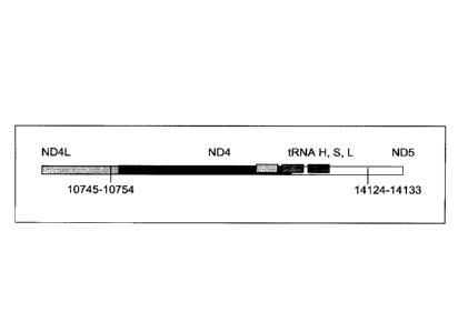
specific embodiments of the
26 invention and are not intended to limit the scope of the invention in
any way.
27 EXAMPLES
28 [00113] Example 1: Detection of Mitochondrial Fusion Transcripts
29 [00114] The mitochondrial 4977 "common deletion" and a 3.4kb
deletion previously identified by
the present Applicant in PCT application no. PCT/CA2007/001711 result in
unique open reading
31 frames having active transcripts as identified by oligo-dT selection in
prostate tissue (Figures 2 and
23634990.1 20
CA 3044262 2019-05-27
CA Application
Blakes Ref: 11871/00280
1 3). Examination of breast tissue samples also reveals the presence of a
stable polyadenylated
2 fusion transcript resulting from the 3.4kb deletion (Figure 4).
3 [00115] Reverse transcriptase-PCR protocol for deletion transcript
detection
4 [00116] RNA isolation cDNA synthesis
[00117] Total RNA was isolated from snap frozen prostate and breast tissue
samples (both
6 malignant and normal samples adjacent to tumours) using the AurumTM Total
RNA Fatty and
7 Fibrous Tissue kit (Bio-Rad, Hercules, CA) following the manufacturer's
instructions. Since in this
8 experiment, genomic DNA contamination was to be avoided, a DNase
!treatment step was included,
9 using methods as commonly known in the art. RNA quantity and quality were
determined with an
ND-1000 spectrophotometer (NanoDrop technologies). From a starting material
of about 100g,
11 total RNA concentrations varied from 100¨ 1000ng/ulwith a 260/280 ratio
between 1.89¨ 2.10.
12 RNA concentrations were adjusted to 10Ong/u1 and 2u1 of each template
were used for first strand
13 DNA synthesis with SuperScriptTM First-Strand Synthesis System for RT-
PCR (Invitrogen) following
14 the manufacturer's instructions. In order to identify stable
polyadenylated fusion transcripts,
Oligo(dT) primers that target transcripts with poly-A tails were used.
16 [00118] PCR
17 [00119] Real time PCR was performed using 5u1 of each cDNA
template with the iQTM SYBRO
18 Green Supernnix (Bio-Rad, Hercules, CA) on DNA Engine Opticon 2
Continuous Fluorescence
19 Detection System (Bio-Rad, Hercules, CA). The primer pairs targeting the
4977bp deletion are;
8416F 5'- CCTTACACTATTCCTCATCAC- 3', 13637R 5'- TGACCTGTTAGGGTGAGAAG -3', and
21 those for the 3.4 kb deletion are; ND4LF 5'- TCGCTCACACCTCATATCCTC -3',
ND5R 5'-
22 TGTGATTAGGAGTAGGGTTAGG -3'. The reaction cocktail included: 2X SYBRO
Green Supermix
23 (100mM KCL, 40mM Tris-HCI, pH 8.4, 0.4mM of each dNTP [dATP, dCTP, dGTP,
and dTTP],
24 iTaq TM DNA polymerase, 50 units/ml, 6mM MgC12, SYBRO Green 1, 20nM
flourescein, and
stabilizers), 250nM each of primers, and ddH20. PCR cycling parameters were as
follows; (1) 95 C
26 for 2 min, (2) 95 C for 30 sec, (3) 55 C (for the 4977bp deletion) and
63 C (for the 3.4 kb deletion)
27 for 30 sec, (4) 72 C for 45 sec, (5) plate read, followed by 39 cycles
of steps 3 to 5, and final
28 incubation at 4 C. Apart from cycling threshold and melting curve
analysis, samples were run on
29 agarose gels for specific visualization of amplification products (see
Figures 2 to 4).
[00120] Figure 2 is an agarose gel showing polyadenalated fusion
transcripts in prostate samples
31 invoked by the loss of 3.4kb from the mitochondria! genome. Legend for
Figure 2: B-blank, Lanes 1-
23634990.1 21
CA 3044262 2019-05-27
CA Application
Blakes Ref: 11871/00280
1 6 transcripts detected in cDNA; lanes 7-12 no reverse transcriptase (RT)
controls for samples in
2 lanes 1-6.
3 [00121] Figure 3 shows polyadenalated fusion transcripts in
prostate samples invoked by the loss
4 of the 4977kb common deletion. Legend for Figure 3: B-blank, Lanes 1-6
transcripts detected in
cDNA; lanes 7-12 no RT controls for samples in lanes 1-6.
6 [00122] Figure 4 shows polyadenalated fusion transcripts in breast
samples invoked by the loss
7 of 3.4kb from the mtgenome. Legend for Figure 4: Lanes 2-8 transcripts
from breast cDNAs; lane 9
8 negative (water) control; lanes 10 and 11, negative, no RT, controls for
samples in lanes 2 and 3.
9 [00123] These results demonstrate the existence of stable
mitochondrial fusion transcripts.
[00124] Example 2: Identification and Targeting of Fusion Products
11 [00125] Various hybridization probes were designed to detect, and
further demonstrate the
12 presence of novel transcripts resulting from mutated mitochondrial
genomes, such as the 3.4kb
13 deletion. For this purpose, a single-plex branched DNA platform for
quantitative gene expression
14 analysis (QuantiGene 2.OTM, PanomicsTM) was utilized. The specific
deletions and sequences listed
in this example are based on their relative positions with the entire mtDNA
genome, which is recited
16 in SEQ ID NO: 1. The nucleic acid sequences of the four transcript to
which the probes were
17 designed in this example are identified herein as follows: Transcript 1
(SEQ ID NO: 18), Transcript 2
18 (SEQ ID NO: 19), Transcript 3 (SEQ ID NO: 20) and Transcript 4 (SEQ ID
NO: 21).
19 [00126] An example of a continuous transcript from the 3.4kb
mitochondrial genome deletion
occurs with the genes ND4L (NADH dehydrogenase subunit 4L) and ND5 (NADH
dehydrogenase
21 subunit 5). A probe having a complementary sequence to SEQ ID NO: 19,
was used to detect
22 transcript 2. The repetitive elements occur at positions 10745-10754 in
ND4L and 14124-14133 in
23 ND5.
24 [00127] The 3.4kb deletion results in the removal of the 3' end of
ND4L, the full ND4 gene, tRNA
histidine, tRNA serine2, tRNA leucine2, and the majority of the 5' end of ND5
(see Figure 5a),
26 resulting in a gene splice of ND4L and ND5 with a junction point of
10744(ND4L):14124(ND5)
27 (Figure 5b). SEQ ID NO: 3 is the complementary DNA sequence to the RNA
transcript (SEQ ID NO:
28 19) detected in the manner described above.
29 [00128] Similarly, transcript 1 is a fusion transcript between
ATPase 8 and ND5 associated with
positions 8469:13447 (SEQ ID NO: 18). Transcripts 3 and 4 (SEQ ID NO: 20 and
SEQ ID NO: 21,
31 respectively) are fusion transcripts between C011 and Cytb associated
with nucleotide positions
32 7974:15496 and 7992:15730 respectively. Table 3 provides a summary of
the relationships between
23634990.1 22
CA 3044262 2019-05-27
CA Application
Blakes Ref: 11871/00280
1 the various sequences used in this example. Table 3 includes the detected
fusion transcript and the
2 DNA sequence complementary to the fusion transcript detected.
3 [00129] Example 3: Application to Prostate Cancer
4 [00130] Using the four fusion transcripts, i.e. transcripts 1 to
4, discussed above, two prostate
tissue samples from one patient were analyzed to assess the quantitative
difference of the novel
6 predicted fusion transcripts. The results of the experiment are provided
in Table 2 below, wherein
7 "Homog 1" refers to the homogenate of frozen prostate tumour tissue from
a patient and "Homog 2"
8 refers to the homogenate of frozen normal prostate tissue adjacent to the
tumour of the patient.
9 These samples were processed according to the manufacturer's protocol
(QuantiGene Sample
Processing Kit for Fresh or Frozen Animal Tissues; and QuantiGene 2.0 Reagent
System User
11 Manual) starting with 25.8 mg of Homog 1 and 28.9 mg of Homog 2 (the
assay setup is shown in
12 Tables 5a and 5b).
13 [00131] Clearly demonstrated is an increased presence of
mitochondrial fusion transcripts in
14 prostate cancer tissue compared to normal adjacent prostate tissue. The
fusion transcript is present
in the normal tissue, although at much lower levels. The relative luminescence
units (RLU)
16 generated by hybridization of a probe to a target transcript are
directly proportional to the abundance
17 of each transcript. Table 2 also indicates the coefficients of
variation, CV, expressed as a
18 percentage, of the readings taken for the samples. The CV comprises the
Standard deviation
19 divided by the average of the values. The significance of such stably
transcribed mitochondria! gene
products in cancer tissue has implications in disease evolution and
progression.
21 [00132] Example 4: Application to Breast Cancer
22 [00133] Using the same protocol from Example 3 but focusing only
on Transcript 2, the novel
23 fusion transcript associated with the 3.4kb mtgenome deletion, analyses
were conducted on two
24 samples of breast tumour tissue and two samples of tumour-free tissues
adjacent to those tumours,
as well as three samples of prostate tumour tissue, one sample comprising
adjacent tumour-free
26 tissue. Results for this example are provided in Table 4. The prostate
tumour tissue sample having
27 a corresponding normal tissue section demonstrated a similar pattern to
the prostate sample
28 analyzed in Example 3 in that the tumour tissue had approximately 2
times the amount of the fusion
29 transcript than did the normal adjacent tissue. The breast tumour
samples demonstrated a marked
increase in the fusion transcript levels when compared to the adjacent non-
tumour tissues. A 1:100
31 dilution of the homogenate was used for this analysis as it performed
most reproducibly in the
32 experiment cited in Example 3.
23634990.1 23
CA 3044262 2019-05-27
CA Application
Blakes Ref: 11871/00280
1 [00134] Thus, the above discussed results illustrate the
application of the transcripts of the
2 invention in the detection of tumours of both prostate and breast tissue.
3 [00135] Example 5: Application to Colorectal Cancer
4 [00136] This study sought to determine the effectiveness of
several transcripts of the invention in
detecting colorectal cancer. A total of 19 samples were prepared comprising
nine control (benign)
6 tissue samples (samples 1 to 9) and ten tumour (malignant) tissue samples
(samples 10 to 19). The
7 samples were homogenized according to the manufacturer's recommendations
(Quantigene
8 Sample Processing Kit for Fresh or Frozen Animal Tissues; and Quantigene
2.0 Reagent System
9 User Manual). Seven target transcripts and one housekeeper transcript
were prepared in the
manner as outlined above in previous examples. The characteristics of the
transcripts are
11 summarized as follows:
12 [00137] Table 7: Characteristics of Breast Cancer Transcripts
Transcript ID Junction Site Gene Junction
2 10744:14124 ND4L:ND5
3 7974:15496 C011:Cytb
10 7438:13476 COI:ND5
11 7775:13532 C011:ND5
12 8213:13991 C011:ND5
Peptidylpropyl isomerase B (PPIB) N/A N/A
("housekeeper")
13
14 [00138] It is noted that transcripts 2 and 3 are the same as those
discussed above with respect to
Examples 3 and 4.
16 [00139] Homogenates were prepared using approximately 25mg of
tissue from OCT blocks and
17 diluted 1:1 for transcripts 2 and 4, and 1:8 for transcripts 10 and 11.
The quantity of the transcripts
18 was measured in Relative Luminenscence Units RLU on a GlomaxTm Multi
Detection System
19 (Promega). All samples were assayed in triplicate for each transcript.
Background measurements
(no template) were done in triplicate as well. The analysis accounted for
background by subtracting
21 the lower limit from the RLU values for the samples. Input RNA was
accounted for by using the
22 formula 1og2 a RLU ¨ 10g2 h RLU where a is the target fusion transcript
and h is the housekeeper
23 transcript.
24 [00140] The analysis of the data comprised the following steps:
a) Establish CV's (coefficients of variation) for triplicate assays;
acceptable if 15%.
23634990.1 24
CA 3044262 2019-05-27
CA Application
Blakes Ref: 11871/00280
1 b) Establish average RLU value for triplicate assays of target fusion
transcript(a) and
2 housekeeper transcript (h).
3 c) Establish lower limit from triplicate value of background RLU (I).
4 d) Subtract lower limit (I) from (a).
e) Calculate 1og2 a RLU ¨ 1og2 h RLU.
6 [00141] Summary of Results:
7 [00142] The results of the above analysis are illustrated in
Figures 6a to 6g, which comprise plots
8 of the 10g2 a RLU ¨ 10g2 h RLU against sample number. Also illustrated
are the respective ROC
9 (Receiver Operating Characteristic) curves determined from the results
for each transcript.
[00143] Transcript 2:
-- There exists a statistically significant difference between the means
11 (p<0.10) of the normal and malignant groups (p>0.09), using a cutoff
value of 3.6129 as
12 demonstrated by the ROC curve results in a sensitivity of 60% and
specificity of 89% and the area
13 under the curve is 0.73 indicating fair test accuracy. The threshold
value chosen may be adjusted to
14 increase either the specificity or sensitivity of the test for a
particular application.
[00144] Transcript 3:
There exists a statistically significant difference between the means
16 (p<0.05) of the normal and malignant groups (p=0.03), using a cutoff
value of 4.0813 as
17 demonstrated by the ROC curve results in a sensitivity of 60% and
specificity of 78% and the area
18 under the curve is 0.79 indicating fair to good test accuracy. The
threshold value chosen may be
19 adjusted to increase either the specificity or sensitivity of the test
for a particular application.
[00145] Transcript 8:
There exists a statistically significant difference between the means
21 (p<0.1) of the normal and malignant groups (p=0.06). Using a cutoff
value of -6.0975 as
22 demonstrated by the ROC curve results in a sensitivity of 60% and
specificity of 89% and the area
23 under the curve is 0.76 indicating fair test accuracy. The threshold
value chosen may be adjusted
24 to increase either the specificity or sensitivity of the test for a
particular application.
[00146] Transcript 9:
-- There exists a statistically significant difference between the means
26 (p<0.1) of the normal and malignant groups (p=0.06). Using a cutoff
value of -7.5555 as
27 demonstrated by the ROC curve results in a sensitivity of 60% and
specificity of 89% and the area
28 under the curve is 0.76 indicating fair to good test accuracy. The
threshold value chosen may be
29 adjusted to increase either the specificity or sensitivity of the test
for a particular application.
[00147] Transcript 10: There is a statistically significant
difference between the means
31 (1350.01) of the normal and malignant groups (p=0.01). Using a cutoff
value of -3.8272as
23634990.1 25
CA 3044262 2019-05-27
CA Application
Blakes Ref: 11871/00280
1 demonstrated by the ROC curve results in a sensitivity of 90% and
specificity of 67% and the area
2 under the curve is 0.84, indicating good test accuracy. The threshold
value chosen may be adjusted
3 to increase either the specificity or sensitivity of the test for a
particular application.
4 [00148] Transcript 11: There exists a statistically
significant difference between the means
.. (p<0.1) of the normal and malignant groups (p=0.06), using a cutoff value
of 3.1753 as
6 demonstrated by the ROC curve results in a sensitivity of 70% and
specificity of 78% and the area
7 under the curve is 0.76 indicating fair to good test accuracy. The
threshold value chosen may be
8 adjusted to increase either the specificity or sensitivity of the test
for a particular application.
9 [00149] Transcript 12: There exists a statistically
significant difference between the means
.. (p<0.1) of the normal and malignant groups (p=0.06), using a cut-off value
of 3.2626 as
11 demonstrated by the ROC curve results in a sensitivity of 70% and
specificity of 78% and the area
12 under the curve is 0.76 indicating fair to good test accuracy. The
threshold value chosen may be
13 adjusted to increase either the specificity or sensitivity of the test
for a particular application.
14 [00150] Conclusions:
[00151] The above results illustrate the utility of transcripts 2, 3, 8, 9,
10, 11, and 12 in the
16 detection of colorectal cancer and in distinguishing malignant from
normal colorectal tissue. As
17 indicated above, transcripts 2 and 3 were also found to have utility in
the detection of prostate
18 .. cancer. Transcript 2 was also found to have utility in the detection of
breast cancer. Transcript 11
19 was also found to have utility in the detection of melanoma skin cancer.
Transcript 10 was also
.. found to have utility in the detection of lung cancer and melanoma.
Transcript 8 was also found to
21 have utility in the detection of lung cancer. Any of the 7 transcripts
listed may be used individually or
22 .. in combination as a tool for the detection of characterization of
colorectal cancer in a clinical setting.
23 [00152] Example 6: Application to Luna Cancer
24 [00153] This study sought to determine the effectiveness of
several transcripts of the invention in
the detection of lung cancer. As in Example 5, nine control (benign) tissue
samples (samples 1 to 9)
26 .. and ten tumour (malignant) tissue samples (samples 10 to 19) were
homogenized according to the
27 manufacturer's recommendations (Quantigene Sample Processing Kit for
Fresh or Frozen Animal
28 .. Tissues; and Quantigene 2.0 Reagent System User Manual). Homogenates
were diluted 1:8 and
29 the quantity of 4 target transcripts and 1 housekeeper transcript was
measured in Relative
Luminenscence Units RLU on a GlomaxTM Multi Detection System (Promega). All
samples were
31 assayed in triplicate for each transcript. Background measurements (no
template) were done in
32 triplicate as well.
23634990.1 26
CA 3044262 2019-05-27
CA Application
Blakes Ref: 11871/00280
1 [00154] The following transcripts were prepared for this example:
2 [00155] Table 8: Characteristics of Lung Cancer Transcripts
Transcript ID Junction Site Gene Junction
6 8828:14896 ATPase6:Cytb
8 6075:13799 COI:ND5
7438:13476 COI:ND5
8469:13447 ATPase8:ND5
Peptidylpropyl isomerase B (PPIB) N/A N/A
("housekeeper")
3
4 [00156] The tissue samples used in this example had the following
characteristics:
5 [00157] Table 9: Characteristics of Lung Cancer Samples
Sample Malignant Comments (source of tissue)
1 NO interstitial lung disease
2 NO emphysema
3 NO aneurysm
4 NO bronchopneumonia, COPD
5 NO malignant neoplasm in liver, origin unknown, calcified
granulomas in lung
6 NO 12 hours post mortem, mild emphysema
12 hours post mortem, large B cell lymphoma, pulmonary edema,
7 NO pneumonia
8 NO pneumonia, edema, alveolar damage
9 NO congestion and edema
10 YES adenocarcinoma, non-small cell
11 YES small cell
12 YES squamous cell carcinoma, NSC, emphysema
13 YES adenocarcinoma, lung cancer, nsc, metastatic
14 YES squamous cell carcinoma, non-small cell
15 YES mixed squamous and adenocarcinoma
16 YES non-small cell carcinoma, squamous
17 YES small cell carcinoma
18 YES adenocarcinoma, lung cancer, nsc
19 YES adenocarcinoma, lung cancer, nsc, metastatic
6
7 [00158] The analysis of data was performed according to the method
described in Example 5.
8 The results are illustrated in figures 7a, 7b, 7c and 7d.
9 [00159] Summary of Results:
10 [00160] Transcript 6: There exists a statistically
significant difference between the means
11 (p<0.1) of the normal (benign) and malignant groups (p=0.06), using a
cutoff value of -6.5691as
12 demonstrated by the ROC curve results in a sensitivity of 80% and
specificity of 71% and the area
23634990.1 27
CA 3044262 2019-05-27
CA Application
Blakes Ref: 11871/00280
1 under the curve is 0.77, indicating fair test accuracy. The threshold
value chosen may be adjusted
2 to increase either the specificity or sensitivity of the test for a
particular application.
3 [00161] Transcript 8: The difference between the means of
the normal and malignant
4 groups is statistically significant, p<0.05 (p=0.02). Using a cutoff
value of -9.6166 as demonstrated
by the ROC curve results in a sensitivity of 90% and specificity of 86% and
the area under the curve
6 is 0.86 indicating good test accuracy. The threshold value chosen may be
adjusted to increase
7 either the specificity or sensitivity of the test for a particular
application.
8 [00162] Transcript 10: The difference between the means of
the normal and malignant
9 groups is statistically significant, 00.01 (p=0.01). Using a cutoff value
of -10.6717 as demonstrated
by the ROC curve results in a sensitivity of 90% and specificity of 86% and
the area under the curve
11 is 0.89 indicating good test accuracy. The threshold value chosen may be
adjusted to increase
12 either the specificity or sensitivity of the test for a particular
application.
13 [00163] Transcript 20: The difference between the means of
the normal and malignant
14 groups is statistically significant, 00.1 (p=0.1). Using a cutoff value
of 2.5071 as demonstrated by
the ROC curve results in a sensitivity of 70% and specificity of 71% and the
area under the curve is
16 0.74 indicating fair test accuracy. The threshold value chosen may be
adjusted to increase either
17 the specificity or sensitivity of the test for a particular application.
18 [00164] Conclusions:
19 [00165] The results from example 6 illustrate the utility of
transcripts 6,8, 10, and 20 of the
invention in the detection of lung cancer tumours and the distinction between
malignant and normal
21 lung tissues. Any of these three transcripts may be used for the
detection or characterization of lung
22 cancer in a clinical setting.
23 [00166] Example 7: Application to Melanoma
24 [00167] This study sought to determine the effectiveness of
several transcripts of the invention in
the detection of melanomas. In this study a total of 14 samples were used,
comprising five control
26 (benign) tissue samples and nine malignant tissue samples. All samples
were formalin fixed,
27 paraffin embedded (FFPE). The FFPE tissue samples were sectioned into
tubes and homogenized
28 according to the manufacturer's recommendations (Quantigene 2.0 Sample
Processing Kit for
29 FFPE Samples; and Quantigene 2.0 Reagent System User Manual) such that
each sample
approximated 20 microns prior to homogenization. Homogenates were diluted 1:4
and the quantity
31 of 7 target transcripts and 1 housekeeper transcript was measured in
Relative Luminenscence Units
23634990.1 28
CA 3044262 2019-05-27
CA Application
Blakes Ref: 11871/00280
1 RLU on a Glomax TM Multi Detection System (Promega). All samples were
assayed in triplicate for
2 each transcript. Background measurements (no template) were done in
triplicate as well.
3 [00168] The 14 tissue samples used in this example had the
following characteristics:
4 [00169] Table 10: Characteristics of Melanoma Cancer Samples
Sample Malignant Comments (source of tissue)
1 NO breast reduction tissue (skin)
2 NO breast reduction tissue (skin)
3 NO breast reduction tissue (skin)
4 NO breast reduction tissue (skin)
NO breast reduction tissue (skin)
6 YES lentigo maligna, (melanoma in situ) invasive melanoma not present
7 YES invasive malignant melanoma
8 YES nodular melanoma, pT3b, associated features of lentigo maligna
9 YES residual superficial spreading invasive malignant melanoma,
Clark's level 11
YES superficial spreading malignant melanoma, Clark's Level 11
11 YES nodular malignant melanoma, Clark's level IV
12 YES superficial spreading malignant melanoma in situ, no evidence of
invasion
superficial spreading malignant melanoma, Clark's level II, focally present
13 YES vertical phase
14 YES superficial spreading malignant melanoma in situ, Clark's level I
5
6 [00170] The following transcripts were prepared for this example:
7 [00171] Table 11: Characteristics of Melanoma Cancer Transcripts
Transcript ID Junction Site GeneJunction
6 8828:4896 ATPase6:Cytb
10 7438:13476 COI:ND5
11 7775:13532 C011:ND5
14 9191:12909 ATPase6:ND5
9574:12972 C0111:ND5
16 10367:12829 ND3:ND5
8469:13447 ATPase8:ND5
Peptidylpropyl isomerase B (PPIB) N/A N/A
("housekeeper")
8
9 [00172] As indicated, transcripts 10 and 11 were also used in
Example 5. The analysis of data
10 was performed according to the method described in Example 5. The
results are illustrated in
11 figures 8a -8g.
12 [00173] Summary of Results:
23634990.1 29
CA 3044262 2019-05-27
CA Application
Blakes Ref: 11871/00280
1 [00174] Transcript 6: There exists a statistically
significant difference between the means
2 (pa.01) of the normal and malignant groups (p=0.01). Further, using a
cutoff value of -5.9531 as
3 demonstrated by the ROC curve results in a sensitivity of 89% and
specificity of 80% and the area
4 under the curve is 0.96, indicating very good test accuracy. The
threshold value chosen may be
adjusted to increase either the specificity or sensitivity of the test for a
particular application.
6 [00175] Transcript 10: There exists a statistically
significant difference between the means
7 (135Ø05) of the normal and malignant groups (p=0.05), using a cutoff
value of -4.7572as
8 demonstrated by the ROC curve results in a sensitivity of 89% and
specificity of 40% and the area
9 under the curve is 0.82, indicating good test accuracy. The threshold
value chosen may be adjusted
to increase either the specificity or sensitivity of the test for a particular
application.
11 [00176] Transcript 11: There exists a statistically
significant difference between the means
12 (p<0.05) of the normal and malignant groups (p=0.02). Further, using a
cutoff value of 1.6762 as
13 demonstrated by the ROC curve results in a sensitivity of 78% and
specificity of 100% and the area
14 under the curve is 0.89, indicating good test accuracy. The threshold
value chosen may be adjusted
to increase either the specificity or sensitivity of the test for a particular
application.
16 [00177] Transcript 14: There exists a statistically
significant difference between the means
17 (130.05) of the normal and malignant groups (p=0.05). Further, using a
cutoff value of -4.9118 as
18 demonstrated by the ROC curve results in a sensitivity of 89% and
specificity of 60% and the area
19 under the curve is 0.82, indicating good test accuracy. The threshold
value chosen may be adjusted
to increase either the specificity or sensitivity of the test for a particular
application.
21 [00178] Transcript 15: There exists a statistically
significant difference between the means
22 (p<0.1) of the normal and malignant groups (p=0.07), using a cutoff
value of -7.3107as
23 demonstrated by the ROC curve results in a sensitivity of 100% and
specificity of 67% and the area
24 under the curve is 0.80, indicating good test accuracy. The threshold
value chosen may be adjusted
to increase either the specificity or sensitivity of the test for a particular
application.
26 [00179] Transcript 16: There exists a statistically
significant difference between the means
27 (p<0.05) of the normal and malignant groups (p=0.03). Further, using a
cutoff value of -10.5963as
28 demonstrated by the ROC curve results in a sensitivity of 89% and
specificity of 80% and the area
29 under the curve is 0.878, indicating good test accuracy. The threshold
value chosen may be
adjusted to increase either the specificity or sensitivity of the test for a
particular application.
31 [00180] Transcript 20: There exists a statistically
significant difference between the means
32 (p<0.05) of the normal and malignant groups (p=0.04). Further, using a
cutoff value of -8.3543as
23634990.1 30
CA 3044262 2019-05-27
CA Application
Blakes Ref: 11871/00280
1 demonstrated by the ROC curve results in a sensitivity of 100% and
specificity of 80% and the area
2 under the curve is 0.89, indicating good test accuracy. The threshold
value chosen may be adjusted
3 to increase either the specificity or sensitivity of the test for a
particular application.
4 [00181] Conclusions:
[00182] The results from example 7 illustrate the utility of transcripts 6,
10, 11, 14, 15, 16 and 20
6 of the invention in the detection of malignant melanomas. As indicated
above, transcripts 10
7 and11were also found have utility in detecting colorectal cancer while
transcript 6 has utility in the
8 detection of lung cancer. A transcript summary by disease is provided at
Table 6.
9 [00183] Example 8: Application to Ovarian Cancer
[00184] This study sought to determine the effectiveness of several
transcripts of the invention in
11 detecting ovarian cancer. A total of 20 samples were prepared comprising
ten control (benign)
12 tissue samples (samples 1 to 10) and ten tumour (malignant) tissue
samples (samples 11 to 20).
13 The samples were homogenized according to the manufacturer's
recommendations (Quantigene
14 Sample Processing Kit for Fresh or Frozen Animal Tissues; and Quantigene
2.0 Reagent System
User Manual). Eight target transcripts and one housekeeper transcript were
prepared in the manner
16 as outlined above in previous examples.
17 [00185] The 20 tissue samples used in this example had the
following characteristics:
18 [00186] Table 12: Characteristics of Ovarian Cancer Samples
Sample Diagnosis Comments
1 Normal follicular cyst
2 Normal fibroma
3 Normal No pathological change in ovaries
4 Normal follicular cysts
5 Normal cellular fibroma
6 Normal benign follicular and simple cysts
7 Normal leiomyomata, corpora albicantia
8 Normal copora albicantia and an epithelial inclusions cysts
9 Normal corpora albicantia
10 Normal corpora albicantia, surface inclusion cysts,
follicullar cysts
11 Malignant high grade poorly differentiated papillary serous
carcinoma involving
omentum
12 Malignant endometrioid adenocarcinoma, well to moderately
differentiated with
focal
serous differentiation
13 Malignant papillary serous carcinoma
14 Malignant mixed epithelial carcinoma predominantly papillary
serous carcinoma
15 Malignant High grade: serous carcinoma, papillary and solid
growth patterns
23634990.1 31
CA 3044262 2019-05-27
CA Application
Blakes Ref: 11871/00280
16 Malignant High Grade (3/3) Papillary serous carcinoma
17 Malignant papillary serous carcinoma, high nuclear grade
18 Malignant Papillary serous cystadenocarcinomas Grade:III
19 Malignant poorly differentiated papillary serous carcinoma
20 Malignant Well-differentiated adnocarcinoma, Endometrioid
type, Grade 1
1
2 [00187] The characteristics of the transcripts are summarized as
follows:
3 [00188] Table 13: Characteristics of Ovarian Cancer Transcripts
Transcript ID Junction Site Gene Junction
1 8469:13447 ATPase8:ND5
2 10744:14124 ND4L:ND5
3 7974:15496 C011:Cytb
6 8828:14896 ATPase6:Cytb
11 7775:13532 C011:ND5
12 8213:13991 C011:ND5
15 9574:12972 C0111:ND5
20 8469:13447 ATPase8:ND5
Ribosomal Protein Large PO (LRP) N/A N/A
Housekeeper
4
[00189] It is noted that transcripts 1, 2, 3, 6, 11, 12, 15 and 20 are the
same as those discussed
6 above with respect to Examples 3-7.
7 [00190] Homogenates were prepared using approximately 25mg of
frozen tissue and diluted 1:4.
8 The quantity of the transcripts was measured in Relative Luminenscence
Units RLU on a Glomax TM
9 Multi Detection System (Promega). All samples were assayed in triplicate
for each transcript.
Background measurements (no template) were done in triplicate as well. The
analysis accounted for
11 background by subtracting the lower limit from the RLU values for the
samples. Input RNA was
12 accounted for by using the formula 10g2 a RLU ¨ log2 h RLU where a is
the target fusion transcript
13 and h is the housekeeper transcript.
14 [00191] The analysis of the data comprised the following steps:
a) Establish CV's (coefficients of variation) for triplicate assays;
acceptable if 15%.
16 b) Establish average RLU value for triplicate assays of target fusion
transcript(a) and
17 housekeeper transcript (h).
18 c) Establish lower limit from triplicate value of background RLU (I).
19 d) Subtract lower limit (I) from (a).
e) Calculate 10g2 a RLU ¨ log2 h RLU.
23634990.1 32
CA 3044262 2019-05-27
CA Application
Blakes Ref: 11871/00280
1 [00192] Summary of Results:
2 [00193] The results of the above analysis are illustrated in
Figures 9a to 9h, which comprise plots
3 of the 10g2 a RLU ¨ 1og2 h RLU against sample number. Also illustrated
are the respective ROC
4 (Receiver Operating Characteristic) curves determined from the results
for each transcript.
[00194] Transcript 1: There exists a statistically significant
difference between the means
6 (p<0.05) of the normal and malignant groups (p=0.002). Using a cutoff
value of -11.1503 as
7 demonstrated by the ROC curve results in a sensitivity of 90% and
specificity of 80% and the area
8 under the curve is 0.91 indicating very good test accuracy. The threshold
value chosen may be
9 adjusted to increase either the specificity or sensitivity of the test
for a particular application.
[00195] Transcript 2: There exists a statistically significant
difference between the means
11 (p<0.01) of the normal and malignant groups (p=0.001). Using a cutoff
value of 0.6962 as
12 demonstrated by the ROC curve results in a sensitivity of 90% and
specificity of 100% and the area
13 under the curve is 0.96 indicating very good test accuracy. The
threshold value chosen may be
14 adjusted to increase either the specificity or sensitivity of the test
for a particular application.
[00196] Transcript 3: There exists a statistically significant
difference between the means
16 (p<0.01) of the normal and malignant groups (p=0.000). Using a cutoff
value of 0.6754 as
17 demonstrated by the ROC curve results in a sensitivity of 100% and
specificity of 100% and the area
18 under the curve is 1.00 indicating excellent test accuracy. The
threshold value chosen may be
19 adjusted to increase either the specificity or sensitivity of the test
for a particular application.
[00197] Transcript 6: There exists a statistically significant
difference between the means
21 (p<0.01) of the normal and malignant groups (p=0.007). Using a cutoff
value of -9.6479 as
22 demonstrated by the ROC curve results in a sensitivity of 90% and
specificity of 70% and the area
23 under the curve is 0.86 indicating good test accuracy. The threshold
value chosen may be adjusted
24 to increase either the specificity or sensitivity of the test for a
particular application.
[00198] Transcript 11: There is a statistically significant
difference between the means
26 (p<0.01) of the normal and malignant groups (p=0.000). Using a cutoff
value of -1.3794
27 demonstrated by the ROC curve results in a sensitivity of 100% and
specificity of 90% and the area
28 under the curve is 0.99, indicating excellent test accuracy. The
threshold value chosen may be
29 adjusted to increase either the specificity or sensitivity of the test
for a particular application.
[00199] Transcript 12: There exists a statistically significant
difference between the means
31 (p<0.01) of the normal and malignant groups (p=0.001). Using a cutoff
value of -1.2379 as
32 demonstrated by the ROC curve results in a sensitivity of 90% and
specificity of 100% and the area
23634990.1 33
CA 3044262 2019-05-27
CA Application
Slakes Ref: 11871/00280
1 under the curve is 0.96 indicating excellent test accuracy. The threshold
value chosen may be
2 adjusted to increase either the specificity or sensitivity of the test
for a particular application.
3 [00200] Transcript 15: There exists a statistically
significant difference between the means
4 (p<0.05) of the normal and malignant groups (p=0.023). Using a cut-off
value of -8.6926 as
demonstrated by the ROC curve results in a sensitivity of 70% and specificity
of 80% and the area
6 under the curve is 0.80 indicating good test accuracy. The threshold
value chosen may be adjusted
7 to increase either the specificity or sensitivity of the test for a
particular application.
8 [00201] Transcript 20: There exists a statistically
significant difference between the means
9 (p<0.01) of the normal and malignant groups (p=0.000). Using a cut-off
value of 0.6521 as
demonstrated by the ROC curve results in a sensitivity of 100% and specificity
of 100% and the area
11 under the curve is 0.76 indicating fair to good test accuracy. The
threshold value chosen may be
12 adjusted to increase either the specificity or sensitivity of the test
for a particular application.
13 [00202] Conclusions:
14 [00203] The above results illustrate the utility of transcripts 1,
2, 3, 6, 11, 12, 15, and 20 in the
detection of ovarian cancer and in distinguishing malignant from normal
ovarian tissue. Transcripts
16 1, 2 and 3 were also found to have utility in the detection of prostate
cancer. Transcript 6 was also
17 found to have utility in the detection of melanoma and lung cancer.
Transcript 11 was also found to
18 have utility in the detection of melanoma skin cancer, colorectal cancer
and testicular cancer.
19 Transcript 12 was also found to have utility in the detection of
colorectal cancer and testicular
cancer. Transcript 15 was also found to have utility in the detection of
melanoma and testicular
21 cancer. Transcript 20 was also found to have utility in the detection of
colorectal cancer, melanoma,
22 and testicular cancer. Any of the 8 transcripts listed may be used
individually or in combination as a
23 tool for the detection or characterization of ovarian cancer in a
clinical setting.
24 [00204] Example 9: Application to Testicular Cancer
[00205] This study sought to determine the effectiveness of several
transcripts of the invention in
26 detecting testicular cancer. A total of 17 samples were prepared
comprising eight control (benign)
27 tissue samples (samples 1 to 8) and 9 tumour (malignant) tissue samples
(samples 9 to 17), 5 of
28 the malignant samples were non-seminomas (samples 9-13)and 4 were
seminomas (samples 14-
29 17). The samples were homogenized according to the manufacturer's
recommendations
(QuantigeneO Sample Processing Kit for Fresh or Frozen Animal Tissues; and
Quantigene 2.0
31 Reagent System User Manual). 10 target transcripts and one housekeeper
transcript were prepared
32 in the manner as outlined above in previous examples.
23634990.1 34
CA 3044262 2019-05-27
CA Application
Blakes Ref: 11871/00280
1 [00206] The 17 tissue samples used in this example had the
following characteristics:
2 [00207] Table 14: Characteristics of Testicular Cancer Samples
Sample General Stratified
Diagnosis Malignant
Diagnosis
1 Benign Benign
2 Benign Benign
3 Benign Benign
4 Benign Benign
Benign Benign
6 Benign Benign
7 Benign Benign
8 Benign Benign
9 Malignant Non-
Seminoma
Malignant Non-
Seminoma
11 Malignant Non-
Seminoma
12 Malignant Non-
Seminoma
13 Malignant Non-
Seminoma
14 Malignant Seminoma
Malignant Seminoma
16 Malignant Seminoma
17 Malignant Seminoma
3 [00208] The characteristics of the transcripts are summarized as
follows:
4 [00209] Table 15: Characteristics of Testicular Cancer Transcripts
Transcript ID Junction Site Gene Junction
2 10744:14124 ND4L:ND5
3 7974:15496 C011:Cytb
4 7992:15730 C011:Cytb
11 7775:13532 C011:ND5
12 8213:13991 C011:ND5
13 9144:13816 ATPase6:ND5
15 9574:12972 C0111:ND5
16 10367:12829 ND3:ND5
8469:13447 ATPase8:ND5
Peptidylpropyl isomerase B (PPIB) N/A N/A
23634990.1 35
CA 3044262 2019-05-27
CA Application
Blakes Ref: 11871/00280
1
2 [00210] It is noted that transcripts 2, 3, 4, 7, 11, 12, 15, 16
and 20 are the same as those
3 discussed above with respect to Examples 3-8.
4 [00211] Homogenates were prepared using approximately 25mg of
frozen tissue and diluted 1:4.
The quantity of the transcripts was measured in Relative Luminenscence Units
RLU on a Glomax TM
6 Multi Detection System (Promega). All samples were assayed in triplicate
for each transcript.
7 Background measurements (no template) were done in triplicate as well.
The analysis accounted for
8 background by subtracting the lower limit from the RLU values for the
samples. Input RNA was
9 accounted for by using the formula 1og2 a RLU ¨ log2 h RLU where a is the
target fusion transcript
and h is the housekeeper transcript.
11 [00212] The analysis of the data comprised the following steps:
12 a) Establish CV's (coefficients of variation) for triplicate assays;
acceptable if 15%.
13 b) Establish average RLU value for triplicate assays of target fusion
transcript(a) and
14 housekeeper transcript (h).
c) Establish lower limit from triplicate value of background RLU (I).
16 d) Subtract lower limit (I) from (a).
17 e) Calculate 10g2 a RLU ¨ 10g2 h RLU.
18 [00213] Summary of Results:
19 [00214] The results of the above analysis are illustrated in
Figures 10 to 18, which comprise plots
of the log2 a RLU ¨ 1og2 h RLU against sample number. Also illustrated are the
respective ROC
21 (Receiver Operating Characteristic) curves determined from the results
for each transcript.
22 [00215] While some transcripts distinguish between benign and
malignant testicular tissue,
23 others demonstrate distinction between the tumour subtypes of seminoma
and non-seminoma
24 and/or benign testicular tissue. It is therefore anticipated that
combining transcripts from each class
will facilitate not only detection of testicular cancer but also
classification into subtype of seminoma
26 or non-seminomas.
27 [00216] Transcript 2: There exists a statistically
significant difference between the means
28 (p<0.05) of the normal group and the malignant seminomas (p=0.02). Using
a cutoff value of 1.5621
29 as demonstrated by the ROC curve results in a sensitivity of 100% and
specificity of 100% and the
area under the curve is 1.00 indicating excellent test accuracy. There also
exists a statistically
31 significant difference between the means (p<0.05) of the malignant
seminomas and the malignant
32 non-seminomas (p=0.024). Using a cutoff value of 2.1006 as demonstrated
by the ROC curve
23634990.1 36
CA 3044262 2019-05-27
CA Application
Blakes Ref: 11871/00280
1 .. results in a sensitivity of 100% and specificity of 80% and the area
under the curve is 0.90 indicating
2 excellent test accuracy. The threshold value chosen may be adjusted to
increase either the
3 specificity or sensitivity of the test for a particular application.
4 [00217] Transcript 3: There exists a statistically
significant difference between the means
(p<0.05) of the normal group and the malignant seminomas (p=0.018). Using a
cutoff value of 0.969
6 .. as demonstrated by the ROC curve results in a sensitivity of 100% and
specificity of 87.5% and the
7 .. area under the curve is 0.969 indicating excellent accuracy. There also
exists a statistically
8 significant difference between the means (p<0.05) of the malignant
seminomas and the malignant
9 non-seminomas (p=0.017). Using a cutoff value of 1.8181 as demonstrated
by the ROC curve
results in a sensitivity of 100% and specificity of 80% and the area under the
curve is 0.9 indicating
11 excellent test accuracy. The threshold value chosen may be adjusted to
increase either the
12 specificity or sensitivity of the test for a particular application.
13 [00218] Transcript 4: There exists a statistically
significant difference between the means
14 (p<0.05) of the normal and malignant groups (p=0.034). Using a cutoff
value of -9.7628 as
demonstrated by the ROC curve results in a sensitivity of 67% and specificity
of 100% and the area
16 under the curve is 0.833 indicating good test accuracy. The threshold
value chosen may be
17 adjusted to increase either the specificity or sensitivity of the test
for a particular application.
18 [00219] Transcript 11: There exists a statistically
significant difference between the means
19 (p<0.05) of the normal group and the malignant seminomas (p=0.016).
Using a cutoff value of 0.732
as demonstrated by the ROC curve results in a sensitivity of 100% and
specificity of 100% and the
21 area under the curve is 1.00 indicating excellent test accuracy. There
also exists a statistically
22 significant difference between the means (p<0.05) of the malignant
seminomas and the malignant
23 .. non-seminomas (p=0.016). Using a cutoff value of 0.9884 as demonstrated
by the ROC curve
24 results in a sensitivity of 100% and specificity of 80% and the area
under the curve is 0.90 indicating
.. excellent test accuracy. The threshold value chosen may be adjusted to
increase either the
26 specificity or sensitivity of the test for a particular application.
27 [00220] Transcript 12: There exists a statistically
significant difference between the means
28 (p<0.1) of the normal group and the malignant seminomas (p=0.056). Using
a cutoff value of 1.5361
29 .. as demonstrated by the ROC curve results in a sensitivity of 100% and
specificity of 87.5% and the
.. area under the curve is 0.969 indicating excellent test accuracy. There
also exists a statistically
31 significant difference between the means (p<0.05) of the malignant
seminomas and the malignant
32 non-seminomas (p=0.044). Using a cutoff value of 1.6039 as demonstrated
by the ROC curve
33 results in a sensitivity of 100% and specificity of 80% and the area
under the curve is 0.9 indicating
23634990.1 37
CA 3044262 2019-05-27
CA Application
Blakes Ref: 11871/00280
1 excellent test accuracy. The threshold value chosen may be adjusted to
increase either the
2 specificity or sensitivity of the test for a particular application.
3 [00221] Transcript 13: There exists a statistically
significant difference between the means
4 (p<0.05) of the normal group and the malignant group (p=0.019). Using a
cutoff value of -9.8751 as
demonstrated by the ROC curve results in a sensitivity of 87.5% and
specificity of 78% and the area
6 under the curve is 0.875 indicating very good test accuracy. There also
exists a statistically
7 .. significant difference between the means (p<0.01) of the malignant non-
seminomas and the benign
8 group (p=0.000). Using a cutoff value of -13.9519 as demonstrated by the
ROC curve results in a
9 sensitivity of 100% and specificity of 87.5% and the area under the curve
is 0.975 indicating
excellent test accuracy. There also exists a statistically significant
difference between the means
11 (p<0.01) of the malignant seminomas and the malignant non-seminomas
(p=0.001). Using a cutoff
12 value of -15.8501 as demonstrated by the ROC curve results in a
sensitivity of 100% and specificity
13 of 100% and the area under the curve is 1.00 indicating excellent test
accuracy. The threshold value
14 .. chosen may be adjusted to increase either the specificity or sensitivity
of the test for a particular
application.
16 [00222] Transcript 15: There exists a statistically
significant difference between the means
17 (p<0.1) of the normal and malignant groups (p=0.065). Using a cut-off
value of -5.4916 as
18 demonstrated by the ROC curve results in a sensitivity of 75% and
specificity of 89% and the area
19 under the curve is 0.833 indicating good test accuracy. The threshold
value chosen may be
adjusted to increase either the specificity or sensitivity of the test for a
particular application.
21 [00223] Transcript 16: There exists a statistically
significant difference between the means
22 (p<0.05) of the normal and malignant groups including both seminomas and
non-
23 seminomas(p=0.037). Using a cut-off value of -6.448 as demonstrated by
the ROC curve results in
24 a sensitivity of 89% and specificity of 75% and the area under the curve
is 0.806 indicating good test
accuracy. There also exists a statistically significant difference between the
means (p<0.05) of the
26 normal and malignant seminomas (p=0.037). Using a cut-off value of -
7.4575 as demonstrated by
27 the ROC curve results in a sensitivity of 100% and specificity of 87.5%
and the area under the curve
28 is 0.938 indicating excellent test accuracy. The threshold value chosen
may be adjusted to increase
29 either the specificity or sensitivity of the test for a particular
application.
[00224] Transcript 20: There exists a statistically significant
difference between the means
31 (p<0.01) of the normal group and the malignant seminomas (p=0.006).
Using a cutoff value of
32 1.8364 as demonstrated by the ROC curve results in a sensitivity of 100%
and specificity of 100%
33 and the area under the curve is 1.00 indicating excellent test accuracy.
There also exists a
23634990.1 38
CA 3044262 2019-05-27
CA Application
Blakes Ref: 11871/00280
1 statistically significant difference between the means (p<0.01) of the
malignant seminomas and the
2 malignant non-seminomas (p=0.004). Using a cutoff value of 1.6065 as
demonstrated by the ROC
3 curve results in a sensitivity of 100% and specificity of 100% and the
area under the curve is 1.00
4 .. indicating excellent test accuracy. The threshold value chosen may be
adjusted to increase either
the specificity or sensitivity of the test for a particular application.
6 [00225] Conclusions:
7 [00226] The above results illustrate the utility of transcripts 2,
3, 4, 11, 12, 13, 15, 16, and 20 in
8 the detection of testicular cancer, and testicular cancer subtypes, and
in distinguishing malignant
9 from normal testicular tissue. Transcript 2 was also found to have
utility in the detection of prostate,
.. breast, colorectal and ovarian cancer. Transcript 3 was also found to have
utility in the detection of
11 prostate, breast, melanoma, colorectal, and ovarian cancers. Transcript
4 was also found to have
12 utility in the detection of prostate and colorectal cancers. Transcript
11 was also found to have utility
13 in the detection of colorectal, melanoma, and ovarian cancers.
Transcript 12 was also found to have
14 utility in the detection of colorectal and ovarian cancers. Transcript
15 was also found to have utility
in the detection of melanoma and ovarian cancers. Transcript 16 was also found
to have utility in
16 the detection of melanoma skin cancer. Transcript 20 was also found to
have utility in the detection
17 of colorectal cancer, melanoma, and ovarian cancer. Any of the 9
transcripts listed may be used
18 individually or in combination as a tool for the detection or
characterization of testicular cancer in a
19 clinical setting.
[00227] In one aspect, the invention provides a kit for conducting an assay
for determining the
21 presence of cancer in a tissue sample. The kit includes the required
reagents for conducting the
22 assay as described above. In particular, the kit includes one or more
containers containing one or
23 .. more hybridization probes corresponding to transcripts 1 to 17, and 20
described above. As will be
24 understood, the reagents for conducting the assay may include any
necessary buffers, salts,
.. detection reagents etc. Further, the kit may include any necessary sample
collection devices,
26 containers etc. for obtaining the needed tissue samples, reagents or
materials to prepare the tissue
27 .. samples for example by homogenization or nucleic acid extraction, and
for conducting the subject
28 assay or assays. The kit may also include control tissues or samples to
establish or validate
29 acceptable values for diseased or non-diseased tissues.
[00228] Although the invention has been described with reference to certain
specific
31 embodiments, various modifications thereof will be apparent to those
skilled in the art without
32 departing from the scope of the invention as outlined in the claims
appended hereto.
33 [00229] Bibliography
23634990.1 39
CA 3044262 2019-05-27
CA Application
Blakes Ref: 11871/00280
1 [00230] The following references, amongst others, were cited in the
foregoing description.
Author Journal Title Volume
Date
Anderson et al Nature Sequence and Organization of the Human
290(5806):457- 1981
Mitochondrial Genome 65
Andrews et al Nat Genet Reanalysis and revision of the Cambridge
23(2):147 1999
reference sequence for human
mitochondrial DNA.
Modica- Expert Rev Mitochondria as targets for detection and
4:1-19 2002
Napolitano et al Mol Med treatment of cancer
Sherratt et al Clin Sci (Lond) Mitochondria! DNA defects: a widening
92(3):225-35 1997
clinical spectrum of disorders.
Croteau et al Mutat Res Mitochondrial DNA repair pathways.
434(3):137-48 1999
Green and J Clin Invest Pharmacological
manipulation of cell death: 115(10): 2610¨ 2005
Kroemer clinical applications in sight? 2617
Dai et al Acta Correlation of cochlear blood supply with
24(2):130-6 2004
Otolaryngol mitochondria! DNA common deletion in
presbyacusis.
Ro et al Muscle Nerve Deleted 4977-bp mitochondria! DNA
28(6):737-43 2003
mutation is associated with sporadic
amyotrophic lateral sclerosis: a hospital-
based case-control study.
Barron et al Invest Mitochondrial abnormalities in ageing
42(12):3016-22 2001
Ophthalmol macular photoreceptors.
Vis Sci
Lewis et at J Pathol Detection of damage to the mitochondrial
191(3):274-81 2000
genome in the oncocytic cells of Warthin's
tumour.
Muller-Hocker Mod Pathol The common 4977 base pair deletion of
11(3):295-301. 1998
et al mitochondrial DNA preferentially
accumulates in the cardiac conduction
system of patients with Kearns-Sayre
syndrome.
Porteous et al Eur J Biochem Bioenergetic consequences of accumulating
257(1):192-201 1998
the common 4977-bp mitochondria! DNA
deletion.
Parr et al J Mol Diagn Somatic mitochondrial DNA mutations in
8(3):312-9. 2006
prostate cancer and normal appearing
adjacent glands in comparison to age-
matched prostate samples without
malignant histology.
Maki et al Am J Clin Mitochondrial genome deletion aids in the
129(1):57-66 2008
Pathol identification of false- and true-negative
prostate needle core biopsy specimens.
Nakase et al Am J Hum Transcription and translation of deleted
46(3):418-27. 1990
Genet mitochondrial genomes in Kearns-Sayre
syndrome: implications for pathogenesis.
Libura et al Blood Therapy-related acute myeloid leukemia-
105(5):2124-31 2005
like MLL rearrangements are induced by
etoposide in primary human CD34+ cells
and remain stable after clonal expansion.
23634990.1 40
CA 3044262 2019-05-27
CA Application
Blakes Ref: 11871/00280
Meyer et al Proc Natl Diagnostic tool for the identification of MLL
102(2):449-54 2005
Acad Sci U S rearrangements including unknown partner
A genes.
Eguchi et al Genes MLL chimeric protein activation renders
45(8):754-60 2006
Chromosomes cells vulnerable to chromosomal damage:
Cancer an explanation for the very short latency of
infant leukemia.
Hayashi et al Proc Natl Introduction of disease-related 88: 10614-
1991
Acad Sci U S mitochondrial DNA deletions into HeLa cells
10618
A lacking mitochondrial DNA results in
mitochondrial dysfunction
1
2
23634990.1 41
CA 3044262 2019-05-27
CA Application
Blakes Ref: 11871/00280
Table 1: Known mitochondrial deletions having an ORF
Deletion Junction (rit:nt) Deletion Size (bp)Repeat Location intintl Number of
Repeats References
COX I - ND5
=Mita, S., Rizzuto, R., Moraes, C.T., Shanske, S., Arnaud , E., Fabrizi,
60Th 13799 -7723 6076-6084/13799-
D, 9/9 G.M. Koga, V. DiMauro, S. Schon, E.A. (1990) "Recombination via
to 13807 flanking direct
repeats is a major cause of large-scale deletions of
o human
mitochondria' DNA" Nucleic Acids Research 18(3):561 -567
01
6235-6238114099- =Blok, R.B.,
Thorburn, DR., Thompson, G.N., Dahl, H.H. (1995) "A
6238:14103 -7864 D, 414
topoisomerase II cleavage site is associated with a novel mitochondria'
14102
DNA deletion" Human Genetics 95 (1): 75-81
=Larsson, N.G., Holme, E., Kristiansson, B., Oldfors, A., Tulinius, M.
(1990) "Progressive increase of the mutated mitochondria' DNA
fraction in Kearns-Sayre syndrome " Pediatric Research 28 (2): 131-
6326-6341/13889- 136 =Larsson,
N.G., Holme, E. (1992) "Multiple short direct repeats
632513989 -7663 D, 16/17
14004 associated with
single rntDNA deletions " Biochimica et Biophysica
Acta 1139 (4): 311-314
=Mita, S., Rizzuto, R. hAoraes, CT., Shanske, S., Arnaudo, E., Fabrizi,
6331 -6341 /13994- G.M., Koga, Y.,
DiMauro, S., Schon, E.A. (1990) "Recombination via
6330:13994 -7663 D, 11t11
14004 flanking direct
repeats is a major cause of large-scale deletions of
human mitochondria' DNA" Nucleic Acids Research 18(3):561-567
COX II - ND5
Bet, L., Moggio, M., Comi, G.P., Mariani, C., Prelle, A., Checcarelli, N.,
7824-7829/14129- Bordoni, A.,
Bresolin, N., Scarpini, E., Scarlato, G. (1994) "Multiple
7829:14135 -6305 D, 6/6
14134 sclerosis arid
mitochondrial myopathy: an unusual combination of
diseases" Journal of Neurology 241 (8): 511-516
=Hinokio, Y., Suzuki, S., Komatu, K., Ohtomo, M., Onoda, M.,
Matsumoto, M., Hirai, S., Sato, Y., Akai, H., Abe, K., Toyota, T. (1995)
8214-8220113991-
8213:13991 -5777 D, 717 "A new
mitochondrial DNA deletion associated with diabetic
13997
amyotrophy, diabetic myoatrophy and diabetic fatty liver" Muscle and
Nerve 3 (9): S142-149
ATPase - ND5
=Zhang, C., Baurner, A., Mackay, I.R., Linnane, A.VV., Nagley, P. (1995)
8625-8631/13506- "Unusual pattern
of mitochondria' DNA deletions in skeletal muscle of
863113513 -4881 D, 717
13512 an aduit human
with chronic fatigue syndrome" Human Molecular
Genetics 4 (4): 751-754
=Ota , Y., Tanaka, M., Sato, W., Ohno, K., Yamamoto, T., Maehara, M.,
9137-9144/13808- Negoro, T.,
Watanabe, K., Awaya, S., Ozawa, T. (1991) "Detection of
9144:13816 -4671 D, 8/8
1381 5 platelet
mitochondria' DNA deletions in Kearns-Sayre syndrome"
Investigative Ophthalmology and Visual Science 32 (10): 2667-2675
=Tanaka, M., Sato, W., Ohno, K., Yamamoto, T., Ozawa, T. (1989)
9189-9191/12906- "Direct
sequencing of mitochondria' DNA in myopathic patients"
9191:12909 -3717 D, 313
12908 Biochemical and
Biophysical Research Communications 164 (): 15E-
163
COX III - ND5
23634990.1 42
CA Application
Blakes Ref: 11871/00280
0 -Rutio, A ,
tiourgeron, F., Chrehen, D Rustin, , Muni-m..11, A (1995)
Spectrurri ot mitochondrial DNA rearrangements in the Poitersan
K.) 101 F11171'1:176.71 -3652 11:11 Ni -101
gElt13753.
r). 878 marresv-pancreem syndrome" Human Molecular Cilenefic, 4 (0)., 1227-
-Pie4ig, A , Cnrmier, V., krill,
, Mixes, C E., .qairdishroy, ,
1 371301
Veerman, A , Peomon. H. A , Munnieh, A. (11391) "Site-specific:
K.)
ot the mitonhondriel generne in Peer:son rrierrow-pencrees
K) ..sYnctrohne"
Qenerniss 101i2T 502-.504
-11.en..!;e, R., Thompoion, C3.N Thorburn, DII, Dahl, NH, Morruld, $.,
0 ! 10J65-1 warn 2426-
F.Jyrne, E , (1M4.1 "A novel rtitDNA deletion In on
intont with
.
law:Jr = 2829 -2451 D mr3
12625 = Pearson
yncircimo'= Jourroi o Inharllad Noteeolic r..)MeGiee 1 7 (s) 421-
NID4L - NOS 526
. . .
0
=Corraier-Daire, V , Ponnesrant, , Ruston P., Mesurays, C., ringler, H
1(174414124
,
1974 G-107 G4114124- D 0 ';'chtni17, Rioour,
Sendubray, unnich, , Roby, A. (1594)
-3379 . SA
K.) 1 41 a3 "IVilteerendriel
DNA reerrengernentz with en.set ors ehronle illorrheo
withi atrOrirre
drilrfrial of rmriotrio..7., i 24 C1I 3-70
ND4 -1.405
.P011O. A., Cormier, Y., Koll, F., Mize, C. E., Seudt..ibray, J.-M., Yeerrnen,
A.. Persoin, H. A., 14.innich, A. (1991) "Site-;:pecifle dieletiunv of the
reltochunctrial cienorne In Pearecm marrow-pancreas indrorne"
Genortile....; 19 (2): 502-504 =Ratig, A., Cormier, V., Elleriche, S.,
Eormeford, J.P., Ledeist, F., Romero, N., Sc:hrriitx, J., Ruskin, P.,
Fischer, A., Sakidsibray, J.tvl. (1990) ''Pearsorrw marrow-pancreas.'
11 224-11242A 3991
syndrome A ini.illi-.vµtem Mitnchortdrusl disorder in infancy" Journal of
IF 23".2.1:-.19tX1 -2747 - 9,9 Clinical
investigation 09 0: 16'01-16011 =Cormier, V Ratio, Al. , Quartinn,
1 39199
Porn, =Cerene,
P., Meier, M., Sauclubrery, Munnieh, A.
(199C1) 'WelespreadrillAtliliSIAC deletion., of the mitorhondnal yenerms
in Peoton morrow-ponereo." m,,ndrome" ..100tnei ot PeAtiatries 117 (4).
599-I1C.12 ..kwertn, I,,roatSLIIIIC40. T., PenIMOtO, Y., Matsuda, A,
T 3050, T. 3e.3) "..lespetnee0 emse ot dlahelesmoluitUl eind
leetnass with ini.rtertions in nytoehondriel tP.NALeuf,I.JIJR) gene pottorr
onnot .341 FRFInsl. -
1247
23634990.1 43
CA Application
Blakes Ref: 11871/00280
n
(,..)
0
0. Table 2: Prostate Cancer Detection with Novel Mitochondrial
Fusion Transcripts
.o.
n)
01
iv
RNA Homog Homog RNA Homog Homog RNA
Homog Homog RNA Homog Homog
iv 1 2 1 2
1 2 1 2
0
1-. Transcript
Transcript Transcript Transcript
Transcript Transcript Transcript Transcript Transcript Transcript Transcript
Transcript Transcript
to 1 1 1 2 2 2
, 3 3 3 - 4 4 4
I 1 2 3 4 5 6
7 8 9 10 11 12
0
cri .
I No dilution A 2957 353 233 144838 75374
17192 348424 333189 213844 509 565 207
iv
...1 Replicate A B 3174 475 298 202793 100062
31750 320877 278137 210265 401 676 250
1:10 dilution C 1041 262 114 106195 98403
36191 238467 248677 123497 181 486 168
Replicate C D 1040 272 176 120308 116930
50323 239231 262520 129778 153 467 149
1:100 dilution E 318 170 110 25155 64823
27725 100345 164606 85287 72 265 119
Replicate E F 287 150 109 23500 50524
24629 100856 178527 84731 83 251 120
,
1:1000 dilution G 100 76 123 3002 12960
252 29203 102309 137 31 143 66
Replicate G H 94 83 91 1263 5796
285 29092 ' 97257 96 45 110 94
, ,
. .
-
ACV A 5.0 20.9 17.3 23.6 19.9 _
42.1 5.8 12.7 1.2 16.9 12.7 13.3
%CV C 0.1 2.5 30.1 8.8 12.2
23.1 0.2 3.8 3.5 12.0 2.8 8.3
%CV E 7.1 9.0 0.6 4.8 17.5
8.4 0.4 5.7 _ 0.5 9.8 3.8 0.6
%CV G 4.7 6.0 20.8 57.7 54.0
8.8 0.3 3.6 25.0 27.0 18.2 24.9
* unit results in table are RLU (relative luminescence units); Data read on
GlorunnerTM.
%CV = Coefficient of variation (as %).
Legend: Homog = homogenate.
Homog 1: Prostate tumour tissue sample from patient;
Homog 2: Histologically normal tissue adjacent to tumour from patient.
RNA: Control: Total RNA from prostate tissue (Ambion p/n 7988).
Shading: Background measurement.
23634990.1 44
CA Application
Blakes Ref: 11871/00280
0
Table 3: Deletion/Transcript/DNA Complement
n.)
n.)
n.) Deletion RNA transcript DNA
sequence with Transcript No.
0
deletion complementary
to RNA transcript
ATP synthase FO subunit 8 to NADH SEQ ID NO: 18 SEQ ID NO: 2
1
cri
dehydrogenase subunit mitochondrial
n.)
positions 8366-14148 (with reference to
SEQ ID NO: 1).
NADH dehydrogenase subunit 4L SEQ ID NO: 19 SEQ ID NO: 3
2
(ND4L) to NADH dehydrogenase subunit
(ND5); Mitochondrial positions 10470-
14148 (with reference to SEQ ID NO: 1)
Cytochrome c oxidase subunit II (C011) to SEQ ID NO: 20 SEQ ID NO:4
3
Cytochrome b (Cytb); Mitochondria!
positions 7586-15887 (with reference to
SEQ ID NO: 1)
Cytochrome c oxidase subunit II (C011) to SEQ ID NO: 21 SEQ ID NO:5
4
Cytochrome b (Cytb); Mitochondria!
positions 7586-15887 (with reference to
SEQ ID NO:1)
23634990.1 45
CA Application
0
Blakes Ref: 11871/00280
u..)
0
.o. Table 4: Breast and Prostate Cancer Detection
.o.
i..)
ch
i..)
i..)
0
1-. Breast Normal
Breast Normal Prostate Prostate Prostate Normal
to
1 Tumour adjacent
Tumour Adjacent Tumour Tumour Tumour Adjacent
o
cri 1 Breast 2 to
3 4 5 to
1
n.) Tumour
Breast Prostate
,.1 1
Tumour Tumour
2
5
,
1 - 2 3 4 - ' 5 6 7 8
1:100 dilution E 68920 2971 49108
1245 46723 56679 99836 35504
_
1:100 dilution replicate F 92409 3017 60637
1512 53940 56155 100582 44221
G 420 3 i 31 6
26 25 44 23
H 518 - 3 4 5
5 3 4 2
-
%CV 20.6 1.1 14.9
13.7 10.1 0.7 0.5 15.5
- unit results in table are RLU (relative luminescence units)
- background G1, H1
- empty well G2-G8, H2- H8
23634990.1 46
CA Application
Blakes Ref: 11871/00280
n
u..)
0
0. Table 5a: Assay Conditions
i..)
ch
i..) Template for the assay
_
i..) RNA Homogen 1 Homogen 2 RNA
Homogen 1 Homogen 2 RNA Homogen 1 Homogen 2 RNA Homogen 1 Homogen
2
0
i-. Transcript 1 Transcript 1 Transcript 1
Transcript 2 Transcript 2 Transcript 2 Transcript 3
Transcript 3 Transcript 3 Transcript 4 Transcript 4 Transcript 4
to
o 1 2 3 4 5 6 - 7
8 9 10 11 12
Cri ,
I A RNA Homog 1 Homog 2 RNA
Homog 1 Homog 2 RNA Homog 1 Homog 2 RNA Homog 1
Homog 2
i..)
-.1 B Dil 1 Dil 1 Dil 1 Dil 1 Dil 1 Dil 1 Dil
1 Dil 1 Dil 1 Oil 1 Dil 1 Dil 1
C RNA Homog 1 Homog 2 RNA Homog 1 -
Homog 2 RNA Homog 1 Homog 2 RNA Homog 1 Homog 2
D Dil 2 Dil 2 Dil 2 Dil 2 DV 2 Dil
2 Dil 2 Dil 2 - Dil 2 Dil 2 - DU 2 Dil 2
_
E RNA Homog 1 Homog 2 RNA Homog 1
Homog 2 RNA Homog 1 Homog 2 RNA Homog 1 Homog 2
F Dil 3 Dil 3 Dil 3 Dil 3 Dil 3 Dil
3 Dil 3 Di! 3 Dil 3 Dil 3 Dil 3 Dil 3
_
G RNA Homog 1 _ Transcript 1 RNA Homog 1
Transcript 1 RNA Homog 1 Transcript 1 RNA Homog 1
Transcript 1
H Dil 4 Dil 4 Background Dil 4 Dil
4 Background Dil 4 Dil 4 Background Dil 4 Dil 4
Background
Homogenate1- Used 26 mg of tissue to homogenize in 700u1 H soln with
Proteinase K (PK). Used Qiagen TissueRuptor. Used 40u1 homogenate
supernatant, 20, 10 and 5 ul for dilution
Homogenate1= Tumour tissue from the tumorous Prostate
Homogenate2- Used 29 mg of tissue to homogenize in 700u1H soln with PK. Used
Qiagen TissueRuptor. Used 40u1 homogenate supernatant, 20,
and 5 ul for dilution
Homogenate2= Normal tissue from the tumorous Prostate
RNA dilution was made as below. RNA was from Prostate Normal from Ambion.
Assay was done in duplicates.
Table 5b: RNA dilution
RNA Dilution ng/ul
Oil 1 3000
1:3 dil Dil 2 1000
Serial dil Dil 3 333
D114 111
23634990.1 47
CA Application
Blakes Ref: 11871/00280
0
Table 6: Transcript Summary by Disease
Probe Prostate Breast Colorectal Melanoma Lung Ovarian Testicular
0
Cancer Cancer Cancer Skin Cancer Cancer
Cancer
Cancer
0
01 = 1 =
2 = = = = =
=
3 = =
=
=
4 =
= 6 = =
7
8 = =
9 =
= 10 = =
11 = = =
12 = =
=
13
=
= 14
= = =
16 =
=
17
= = = =
23634990.1 48