Note: Descriptions are shown in the official language in which they were submitted.
CA 03044605 2019-05-21
WO 2018/098295 PCT/US2017/063036
1
PERSONALIZED CELLULAR BIOMANUFACTURING WITH A CLOSED, MINIATURE
CELL CULTURE SYSTEM
CROSS-REFERENCE TO RELATED APPLICATION
[0001] This application claims priority to U.S. Provisional Patent Application
No.
62/425,141, filed November 22, 2016, which is incorporated by reference herein
in its entirety.
BACKGROUND OF THE DISCLOSURE
[0002] The present disclosure relates generally to personalized culturing,
reprogramming, expanding, differentiating, and/or downstream processing of
cells, such as
primary human cells, primary human tumor cells, human pluripotent stem cells
(hPSCs)
(including human induced pluripotent stem cells (hESCs) and human embryonic
stem cells
(iPSCs)) and their derivatives (i.e. cells differentiated from hPSCs) in a
closed, miniature cell
culture system. More particularly, the present disclosure relates to a closed
device for
manufacturing, expanding, differentiating and/or reprogramming cells for
personalized medicine,
such to allow medical procedures at the point-of-care (i.e., at the time and
place of patient care).
[0003] Autologous cells refer to cells from the patient, and thus are
attractive for use in
cellular therapies as they induce minimal or no immune rejection after
transplanting to the
patient. Autologous cells include primary cells isolated from the patient,
such as T cells,
chondrocytes, and mesenchymal stem cells. These cells can be used to treat
many human
diseases that cannot be treated, or their progression cannot be altered by
current treatments.
[0004] Autologous cells also include patient specific human induced
pluripotent stem
cells (iPSCs). By delivering a few reprogramming factors into the cells, adult
cells from the
patient (e.g., fibroblasts) can be reprogrammed into iPSCs within about one
month. iPSCs can be
cultured for long durations and expanded into large numbers under completely
defined
conditions. They can be further differentiated into presumably all the cell
types of the human
body.
[0005] Autologous cells also include primary tumor cells from the patient,
such as
glioblastoma cells. These cells can be used to screen drugs that can
specifically and efficiently
kill the patient's tumor cells.
CA 03044605 2019-05-21
WO 2018/098295 PCT/US2017/063036
2
[0006] Autologous cell-based personalized medicine, however, cannot benefit
the large
patient population until they become affordable. The expense to biomanufacture
personalized
cells with current technologies and bioprocess are extremely high. For
instance, to make patient
specific iPSC-based autologous cells with the current bioprocessing, patient
cells are collected
and cultured for a few days; then, reprogramming factors are delivered to
these cells to
reprogram them into iPSCs (which takes approximately one month). Next, high
quality iPSC
clones are selected, expanded and characterized for their pluripotency and
genome integrity with
a variety of assays (which takes approximately one to two months); then, iPSCs
are expanded
and differentiated into the desired cells. Finally, the produced cells are
purified, characterized for
their identities, purity, and potency, and formulated for transplantation. The
whole bioprocessing
takes a few months and is mainly done using 2D, open culture systems (e.g., 2D
cell culture
flasks) through manual operations ¨ a processing which leads to low
reproducibility, high risk of
contamination, and requirement for highly skilled technicians. In addition, 2D
culture systems
have low yield. For instance, only ¨2x10⁵ cells can be produced per cm2
surface area, meaning
that it would require ¨85 six-well plates to produce the cells (-1 x 10⁹
cells) sufficient for one
patient. Maintaining these plates requires large incubators and cGMP facility
space, labor, and
reagents.
[0007] If large numbers of patients need iPSC-based personalized cell
therapies, the
cell production can only be done in large cell biomanufacturing centers (i.e.
centralized cellular
biomanufacturing). Patient cells are sent to the center, and the produced
cells are sent back to the
point-of-care for transplantation. This centralized biomanufacturing has
additional
disadvantages, including: (i) cross-contamination and (ii) high costs and
risks associated with the
transportation, logistics, tracking, and recording. In summary, the cost for
biomanufacturing
personalized iPSCs and their derivatives with current technologies is not
affordable for the
majority of patients.
[0008] One method to significantly reduce the biomanufacturing cost is to
automate the
bioprocessing in individualized, closed, computer controlled miniature cell
culture devices to
biomanufacture the cells at the point-of-care (i.e. cGMP-in-a-box production).
Using closed
culture devices avoids contamination risk and eliminates the requirement for
cGMP processing.
Automation of all key operations avoids output variations and reduces the need
for highly skilled
operators. Biomanufacturing at the point-of-care reduces the cost and risk
related to the logistics
and transportation. Miniaturizing the culture system makes it possible to
simultaneously
CA 03044605 2019-05-21
WO 2018/098295 PCT/US2017/063036
3
biomanufacture cells for large numbers of patients at the point-of-care (i.e.
high throughput
biomanufacturing).
[0009] Based on the foregoing, there is a need in the art for a closed,
miniature device
for manufacturing, expanding, differentiating and reprogramming cells,
particularly on a scale
such that can be used at the point-of-care for personalized medicine. It would
further be
advantageous if the closed device could be made to be disposable to limit
cross-contamination.
BRIEF DESCRIPTION OF THE DISCLOSURE
[0010] The present disclosure is generally directed to culturing,
reprogramming,
expanding, differentiating and downstream processing cells in a closed culture
system. More
particularly, the present disclosure is directed to a closed culturing system
and device including a
closed housing that can be used for manufacture, expansion, differentiation of
cells, and then
further, for concentration, purification and transportation of the cultured
cells.
[0011] In one aspect, the present disclosure is directed to a device for
culturing cells for
personalized medicine, the device comprising: a closed housing comprising a
three-dimensional
(3D) hydrogel scaffold; an inlet for introducing a cell culture medium into
the housing; and an
outlet for exhausting cell culture medium from the housing.
[0012] In another aspect, the present disclosure is directed to a method of
expanding
cells using the device, the method comprising: suspending a cell solution
including cells in the
3D hydrogel scaffold of the closed housing; introducing a cell culture medium
into the closed
housing from the inlet; and culturing the cells.
[0013] In yet another aspect, the present disclosure is directed to a method
of
differentiating cells using the device, the method comprising: suspending a
cell solution
including cells in the 3D hydrogel scaffold of the closed housing; introducing
a cell
differentiation medium into the closed housing from the inlet; and culturing
the cells.
[0014] In another aspect, the present disclosure is directed to a method of
reprogramming cells using the device, the method comprising: suspending a cell
solution
including adult cells in the 3D hydrogel scaffold of the closed housing;
introducing a cell culture
medium into the closed housing from the inlet; and reprogramming the cells.
CA 03044605 2019-05-21
WO 2018/098295 PCT/US2017/063036
4
[0015] In accordance with the present disclosure, methods have been discovered
that
surprisingly allow for culturing, manufacturing, expanding, differentiating
and reprogramming
cells in a closed, miniature culture system. The methods and devices of the
present disclosure will
have significant impact on personalized medicine as they allow for sufficient,
high quality and
affordable cells that can be used at the point-of-care. Further, the devices
and methods provide an
advantageous impact on the biopharmaceutical industry by providing more
affordable methods for
manufacturing, expanding, differentiating and reprogramming cells in a manner
that limits
contamination and cross-contamination.
BRIEF DESCRIPTION OF THE DRAWINGS
[0016] The disclosure will be better understood, and features, aspects and
advantages
other than those set forth above will become apparent when consideration is
given to the
following detailed description thereof. Such detailed description makes
reference to the
following drawings, wherein:
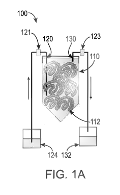
[0017] FIGS. 1A-1C depict a closed, miniature cell culture device for
personalized
cellular biomanufacturing as described in the present disclosure. FIG. 1A
depicts a schematic
illustration of the device. FIG. 1B is a picture of the cell culture device
with the inlet and outlet
identified. FIG. 1C is a picture of the cell mass in hydrogel fibers within
the cell culture device.
[0018] FIGS. 2A-2C depict personalized iPSC expansion and differentiation into
neural
stem cells (NSCs) in a closed, miniature cell culture device. FIG. 2A
illustrates the methods of
the bioprocessing as described in the present disclosure. FIG. 2B depicts the
miniature cell
culture device 210 including a pump 212 for medium perfusion, an oxygen-
permeable plastic
bag 214 for stocking medium and a closed 15-ml conical tube 216. Further,
fibrous hydrogel
fibers with cells are shown suspended in the tube. FIG. 2C depicts mixing
single iPSCs with a
10% PNIPAAm-PEG solution at 4 C on day 0 and injected into room temperature
cell culture
medium in a 15-mL conical tube to instantly form hydrogel fibers with cells;
culturing the cells
in E8 medium for 5 days; culturing the cells for an additional 7 days in
neural induction medium
in the conical tube to differentiate the cells (medium was continuously
perfused); liquefying the
hydrogel scaffolds by placing the cell culture tube on ice for 5 minutes;
pelleting the cell
spheroids by spinning the tube at 100 g for 3 minutes (medium was removed);
purifying the
cells; adding magnetic beads coated with anti-SSEA4 antibodies into the tube
to pull down the
undifferentiated SSEA4+ iPSCs with a magnetic cell separator; transferring the
purified cells in
CA 03044605 2019-05-21
WO 2018/098295 PCT/US2017/063036
the supernatant into a new, closed tube, and transporting the closed tube to
the surgical room;
and transplanting the NSCs to rat brain with a stereotactic injector.
Specifically, as shown in the
purifying step, cell spheroids were incubated in Accutase at 37 C for 10
minutes. The reagents
were removed from the tube and new reagents were added to the tube with a
sterile syringe
through the septum cap.
[0019] FIGS. 3A-3E depict cells in the miniature bioprocessing method of the
present
disclosure. FIG. 3A are phase images of the hydrogel fibers and cells on day
0, 5 and 12 of the
bioprocessing. FIG. 3B depict Live/dead staining of cells on day 12. FIG. 3C
show that ¨97% of
the purified cell products expressed NSC markers, PAX6 and Nestin. FIG. 3D
show that cells
pulled down by the magnetic anti-SSEA4 beads were positive for 0ct4 and Nanog.
FIG. 3E
show that HuNu+ (human nuclear antigen) NSCs survived well in the rat brain 7
days post-
transplantation.
[0020] FIGS. 4A-4E depict culturing cells in alginate hollow fibers as
described in the
present disclosure. FIG. 4A is a schematic showing a hyaluronic acid (HA)
solution containing
single cells 320 and alginate solution 322 pumped into the central 324 and
side channels 326 of a
home-made micro-extruder, respectively, to form a coaxial core-shell flow that
is extruded into a
CaCl2 buffer 328 (100 mM), which instantly crosslinks the alginates to form
hydrogel shells to
make hollow fibers. Subsequently, CaCl2 buffer was replaced by cell culture
medium and cells
were suspended and grown in the core microspace of the hollow fibers. FIG. 4B
shows that,
within the first 24 hours, the single cells associated to form small clusters
(i.e., initial clustering
phase). Subsequently these small clusters expanded as spheroids (FIG. 4C) that
eventually merge
to form cylindrical cell masses (FIG. 4D) (i.e., cell growth phase). FIG. 4E
depict a cylindrical
cell mass in one hollow fiber on day 9.
[0021] FIGS. 5A & 5B depict personalized iPSC expansion and differentiation
into
NSCs in a closed, miniature cell culture device using alginate hydrogel hollow
fibers as
described in the present disclosure. FIG. 5A depicts a schematic illustration
of the bioprocess. As
shown in FIG. 5B, iPSCs and hydrogel fibers were extruded into a closed 15-ml
tube; iPSCs in
the hollow fibers were expanded for 5 days in the expansion medium with
automated medium
perfusion. iPSCs were then differentiated into NSCs in the differentiation
medium for 7 days.
Fibers were dissolved by adding 0.5 mM EDTA, and cell spheroids were harvested
by gravity.
Spheroids were then dissociated into single cells with Accutase.
Undifferentiated iPSCs were
CA 03044605 2019-05-21
WO 2018/098295 PCT/US2017/063036
6
depleted with magnetic anti-SSEA-4 beads. The cell products were transferred
to a new tube and
concentrated by centrifugation. Cells were transported to the surgery room and
transplanted.
[0022] FIGS. 6A-6J depict iPSC expansion and differentiation into NSCs in a
miniature
bioprocess using alginate hydrogel hollow fibers as described in the present
disclosure. FIG. 6A
are phase images of cells growing in hydrogel fibers on day 0 (single iPSCs),
day 5 (iPSC
spheroids) and day 5+7 (NSC aggregates). On day 5+7, 400-fold of expansion
(FIG. 6B), yield
of 4.1 x 10⁸ cells/ml (FIG. 6C), >95% cell viability were achieved (FIG. 6D).
98% of cells were
SSEA negative (FIG. 6E); and very few dead cells (via live/dead cell staining)
were detected
(FIG. 6F). FIGS. 6G & 6H show that >99% of the cells pulled down by the anti-
SSEA4
antibody-coated magnetic beads were Nanog+/0ct4+ undifferentiated iPSCs. FIG.
61 shows that
>99% of the purified cell products were PAX6+/Nestin+ NSCs. FIG. 6J shows that
purified
NSCs survived well in mouse brain 7 days after transplantation. HuNu: human
nuclear antigen.
[0023] FIG. 7 depicts iPSC colonies formed in the 3D hydrogels used in the
devices of
the present disclosure after 3 weeks of reprogramming.
[0024] While the disclosure is susceptible to various modifications and
alternative
forms, specific embodiments thereof have been shown by way of example in the
drawings and
are herein described below in detail. It should be understood, however, that
the description of
specific embodiments is not intended to limit the disclosure to cover all
modifications,
equivalents and alternatives falling within the spirit and scope of the
disclosure as defined by the
appended claims.
DETAILED DESCRIPTION OF THE DISCLOSURE
[0025] Unless defined otherwise, all technical and scientific terms used
herein have the
same meaning as commonly understood by one of ordinary skill in the art to
which the
disclosure belongs. Although any methods and materials similar to or
equivalent to those
described herein can be used in the practice or testing of the present
disclosure, the preferred
methods and materials are described below.
[0026] In accordance with the present disclosure, devices and methods have
been
discovered that surprisingly allow for the culturing, reprogramming,
expanding, differentiating
and downstream processing of cells in a closed, miniature system such to allow
for limited
contamination, lower costs, high cell yield and purity, and ease of providing
personalized
CA 03044605 2019-05-21
WO 2018/098295 PCT/US2017/063036
7
medicine. Particularly, the present disclosure provides a closed, miniature
device and methods
of using the device for manufacturing, expanding, differentiating and
reprogramming cells in a
closed, miniature system using 3D hydrogel scaffolds.
Device for Culturing/Manufacturing/Expanding/Differentiating/Reprogramming
Cells
[0027] Advantageously, the device of the present disclosure allows for
biomanufacturing sufficient and affordable personalized cells at the point of
care. Further, the
device provides high cell yields and purity while limiting contamination.
Generally, the device
includes a closed, miniature housing including hydrogel scaffolds with cells;
an inlet with filter
for flowing cell culture medium into the housing; and an outlet with filter
for flowing out of the
housing the exhausted medium. As used herein, "miniature" refers to the device
including a
housing having a capacity of less than 10 L, including from about 1 ml to less
than 10 L,
including from about 1 ml to about 1000 ml in capacity.
[0028] More particularly, as shown in FIG. 1A, the device 100 includes a
closed
housing 110; an inlet 120 and an outlet 130. As used herein, "closed" as
referred to in "closed
device", "closed system", and/or "closed housing" refers to the device,
system, and/or housing
that is sealed such that the exchange of matter with its surroundings can only
be done through the
inlet and outlet with filters, 121, 123. The filters 121, 123 can prevent the
virus and bacteria in
the environment from entering the cell culture device. More particularly, the
closed device,
system, and/or housing suitably prevents at least 70% of surrounding matter
from entry into the
device, system, and/or housing; more suitably, at least 75%; even more
suitably, at least 80%;
even more suitably, at least 90%; even more suitably, at least 95%, including
96%, 97%, 98%,
99%, and even 100% of surrounding matter from entry into the device, system
and/or housing.
[0029] The closed housing 110 as shown in FIG. 1A is a closed 50-ml conical
tube;
however, it should be understood by one skilled in the art that any closed
culture system known
in the art, for example larger conical tubes or small volume plastic bags.
Typically, when a
conical tube is used, the tube is sized to a capacity of from 1 ml to about 10
L, including from
about 1 ml to about 1 L, and including from about 5 ml to about 50 ml. When
plastic bags are
used, the bags have a capacity of from about 1 ml to about 10 L and including
from about 1 ml to
about 1 L.
CA 03044605 2019-05-21
WO 2018/098295 PCT/US2017/063036
8
[0030] The closed housing 110 includes a three-dimensional (3D) hydrogel
scaffold
112. The 3D hydrogel scaffold is prepared by extruding the hydrogel precursor
solution with
cells through the septum cap 122 (FIG. 1B) of the cell culture device into a
buffer containing
crosslinking reagents in the cell culture device that can quickly crosslink
the hydrogel precursor
solution into hydrogels.
[0031] Typically, the 3D hydrogel scaffold 112 is prepared using any polymers
as
known in the hydrogel art for culturing, manufacturing, expanding,
differentiating and/or
reprogramming cells. For example, in suitable embodiments, the 3D hydrogel
scaffold is
prepared as a thermoreversible hydrogel scaffold using polymers such as for
example
poly(ethylene glycol)-(N-isopropylacrylamide) and the like. In yet other
suitable embodiments,
the 3D hydrogel scaffold is prepared from alginate polymers. Suitable alginate
polymers include
any commercially available or home-purified alginate polymer, such as alginate
acid or sodium
alginate from Sigma (+W201502), and modified alginate polymers, such as
methacrylate
modified alginate.
[0032] Generally, the 3D hydrogel scaffold for use in the closed housings of
the
devices of the present disclosure are in any form as known in the art,
including, by way of
example, sheets, fibers, hollow fibers, spheres, and combinations thereof.
[0033] Generally, cells are encapsulated in the hydrogel scaffold. In some
suitable
embodiments, cells are suspended in the hollow space created by the hydrogel
hollow fibers.
Cells include primary cells isolated from humans, such as T cells,
chondrocytes, mesenchymal
stem cells. Cells also include human induced pluripotent stem cells, human
embryonic stem cells
and their derivatives (i.e. cell differentiated from them). Cells also include
primary human tumor
cells. While described herein in the context of human cells, it should be
understood by one
skilled in the art that the device of the present disclosure can be used with
any other animal cells
without departing from the scope of the present disclosure.
[0034] Further, in one embodiment, the cells are autologous cells in that they
are cells
from the same patient desired to be treated. In another embodiment, the cells
are allogenic cells
(e.g., formed in another location and transported).
[0035] The device of the present disclosure further includes an inlet 120 and
an outlet
130. The inlet 120 allows for entry of a cell culture medium into the closed
housing 110, and the
CA 03044605 2019-05-21
WO 2018/098295 PCT/US2017/063036
9
outlet 130 allows for exit of the cell culture medium from the closed housing
110. In particular
embodiments, it is advantageous to include a pump (not shown) in flow
communication with the
inlet 120 to thereby pump cell culture medium from a medium reservoir 124 to
the closed
housing 110. While described in communication with a pump, it should be
understood by one
skilled in the art that any means of flowing the cell culture medium from
medium reservoir 124
to the closed housing 110 can be used in the device 100 of the present
disclosure without
departing from the scope of the present disclosure.
[0036] Once used for cell culturing, the cell culture medium is automatically
perfused
through the closed housing 110 and exhausted from the closed housing 110 via
the outlet 130 to
an exhausted medium reservoir 132.
[0037] The cell culture medium can be any medium known in the cell culture art
that is
suitable for supporting cell survival, growth, expansion, and differentiation.
Typically, the cell
culture medium will include, but is not limited to, a carbon source, a
nitrogen source, and growth
factors. The specific cell culture medium for use in culturing the cells will
depend on the cell
type to be cultured. Cell culture conditions will also vary depending on the
type of cell, the
amount of cell expansion, and the number of cells desired.
Methods of Culturing/Manufacturing/Expanding/Differentiating/Reprogramming
Cells
[0038] The methods of the present disclosure may be used to culture cells on a
personalized scale. As used herein, "culturing cells" or "culture cells" or
the like refers to
manufacturing, expanding, differentiating, and/or reprograming cells within
the device of the
present disclosure. "Reprogramming" or "reprogram" refers to the conversion of
adult cells back
to iPSCs, or from one adult cell type to another cell type. The methods of the
present disclosure
provide at least the following advantages over conventional cell culture
methods: (1) allow for
biomanufacturing cells at high volumetric yield. At least 2 x 10⁷ cells can be
produced per ml of
hydrogel scaffold. In general, 5.0 x 10⁸ cells can be produced per ml of
hydrogel scaffold; (2)
allow for personalized medicine with miniature device at the point-of-care;
(3) allow for limited
contamination and/or cross-contamination as the closed culturing and point-of-
care procedure
removes the risk of contamination during cell culture transportation; and (4)
allow for low batch-
to-batch variation. Further, the methods of using the hydrogel scaffold for
expanding and
differentiating cells provide the additional benefits of: (1) providing 3D
spaces for cell growth;
and (2) providing physical barriers to prevent cell agglomeration and isolate
shear force, major
CA 03044605 2019-05-21
WO 2018/098295 PCT/US2017/063036
factors of which lead to low cell growth and volumetric yield of cells in the
conventional 3D
suspension culture technologies. The methods of using the device for
reprogramming cells
provide the additional benefit of allowing only the successfully reprogrammed
cells to grow in
the 3D hydrogel scaffold, thus generating cells at high purity.
[0039] Non-limiting examples of such cells that can be cultured, manufactured,
expanded, differentiated, and/or reprogrammed using the methods and devices
described herein
include primary cells isolated from human (i.e., human primary cells) such as
T cells,
chondrocytes, and mesenchymal stem cells. Cells also include human induced
pluripotent stem
cells, human embryonic stem cells and their derivatives (i.e. cell
differentiated from them). Cells
also include primary human tumor cells. Cells can also be animal cells, for
instance pig induced
pluripotent stem cells or primary pig cells. While described more fully using
iPSCs, it should be
recognized that the methods and devices described herein can be used with any
of the above-
listed types of cells without departing from the scope of the present
disclosure.
[0040] In general, the method of culturing cells includes: encapsulating cells
in the
hydrogel scaffolds or suspending cells in the hollow space created by the
hydrogel hollow fibers
of the closed housing; introducing a cell culture medium into the closed
housing including the
cells suspended in the hydrogel scaffolds to allow expansion, differentiation
or reprogramming
of the cells; and culturing the cells.
[0041] Cells are encapsulated or suspended in hydrogel scaffolds at
concentrations
varying from 1 to a few billion cells per milliliter and can be expanded to up
to 6.0 X 10⁸ cells
per milliliter.
[0042] In suitable embodiments, cells are encapsulated in the hydrogel
scaffold. In
other suitable embodiments, cells are suspended in the hollow space created by
the hydrogel
hollow fibers.
[0043] Cell culture medium is then introduced into the closed housing for
culturing the
cells. The cell culture medium can be any medium known in the cell culture art
that is suitable
for supporting cell survival, growth, expansion, differentiation and
reprogramming. Typically,
the cell culture medium will include, but is not limited to, a carbon source,
a nitrogen source, and
growth factors. The specific cell culture medium for use in culturing the
cells will depend on the
cell type to be cultured.
CA 03044605 2019-05-21
WO 2018/098295 PCT/US2017/063036
11
[0044] Cell culture conditions will vary depending on the type of cell, the
amount of
cell expansion/differentiation/reprogramming, and the number of cells desired.
Once sufficient
cell expansion/differentiation/reprogramming and desired numbers of cells are
reached, the cells
are released from the 3D hydrogel scaffold by dissolving the 3D hydrogel
scaffold chemically or
physically within the housing. In one aspect, the scaffold is dissolved using
a chemical
dissolvent such as ethylenediaminetetraacetic acid (EDTA), ethylene glycol
tetraacetic acid
(EGTA), or an alginate lyase solution (available from Sigma-Aldrich). In
another aspect, the
hydrogel scaffold is dissolved using a physical method, such as lowering the
temperature to
below 4 C. The duration of the cells within the 3D hydrogel scaffold can
typically vary from
days to months.
[0045] The cells are useful in personalized medicine and can be used at the
point-of-
care. By way of example, the cells can be used in a procedure at the bedside
of a patient. Cells
can be efficiently and effectively prepared in size and number for use in
degenerative disease and
injury treatment, drug screening, for expressing proteins and the like.
Additionally, the cells can
be used to manufacture proteins and vaccines. In yet other aspects, the cells
can be used for
tissue engineering.
[0046] The disclosure will be more fully understood upon consideration of the
following non-limiting Examples.
EXAMPLES
[0047] Unless otherwise indicated, the hollow fibers were prepared as
described above.
EXAMPLE 1
[0048] In this Example, expansion and growth of neural stem cells (NSCs) from
induced pluripotent stem cells (iPSCs) were analyzed.
Methods
[0049] Miniature bioprocessing: With a syringe, 4 C PNIPAAm-PEG solution
containing iPSCs were injected into room temperature E8 medium in a 15-ml
conical tube.
Fibrous hydrogels were formed instantly. A Variable-Speed Peristaltic Tubing
Pump (Control
Company, USA) was used to continuously perfuse the culture medium into the
tube through
septum cap. Medium was stocked in a sealed and oxygen-permeable plastic bag.
Medium in the
CA 03044605 2019-05-21
WO 2018/098295 PCT/US2017/063036
12
bag was changed daily. The cell culture tube, pump and medium bag were placed
in a cell
culture incubator at 37 C. E8 medium and neural induction medium was used for
days 1 to 5,
and days 6 to 12, respectively. On day 12, the cell culture tube was placed on
ice for 5 minutes to
liquefy the hydrogel and release the spheroids. Cells were collected by
spinning the tube at 100 g
for 5 minutes. The cell pellet was treated with Accutase at 37 C for 10
minutes and dissociated
into single cells. Single cells were collected by spinning at 300 g for 5
minutes. Cells were
resuspended with 80 pl PBS buffer and 20 pl of anti-SSEA-4 microbeads
(Miltenyi Biotec) were
added and incubated at 4 C for 15 minutes. The SSEA4+ iPSCs were pulled down
with a magnet
and NSCs in the supernatant were transferred into a new tube. Cells were
pelleted by spinning at
300 g for 5 minutes and transported to the surgery room for transplantation.
[0050] Cell transplantation: The animal experiments were carried out following
the
protocols approved by the University of Nebraska¨Lincoln Animal Care and Use
Committee.
Sprague Dawley female rats were obtained from Charles River. Animals received
intraperitoneal
cyclosporine A (10 mg/kg, LC Laboratories) injection starting 1 day before
transplantation. For
transplantation, animals were anesthetized with 2-4% isoflurane. 2X10⁵ cells
suspended in 4 ul
DMEM medium were injected into striatum (AP+0.5 mm; ML 3.0 mm; DV-6 mm) at 0.5
ul/min using a 10 ul Hamilton syringe (Hamilton Company, USA) with a
stereotaxic frame
(RWD Life Science Inc). On day 7, rats were anesthetized with
ketamine/xylazine and perfused
with PBS followed by 4% paraformaldehyde. After fixation, the brain was
serially sectioned (40
um in thickness) with a Leica cryo-section machine, and free-floating sections
were stained with
antibodies.
[0051] To stain the brain sections, samples were then incubated with PBS +
0.25%
Triton X-100 + 5% goat serum + primary antibodies at 4 C for 48 hours. After
extensive wash,
secondary antibodies in 2% BSA were added and incubated at 4 C for 4 hours.
Results
[0052] Taking advantage of the high cell yield in the PNIPAAm-PEG hydrogels, a
prototype device of the present disclosure was built to make NSCs from hPSCs
for personalized
cell therapies (FIGS. 2A-2K and 3A-3E). On day 0, single iPSCs were mixed with
10%
PNIPAAm-PEG solution at 4 C. With a syringe, the mixture was injected into
room temperature
E8 medium contained in a closed and sterile 15-ml conical tube with a septum
cap (FIG. 2C).
Fibrous hydrogels (with diameter ¨1 mm) were instantly formed with single
iPSCs uniformly
CA 03044605 2019-05-21
WO 2018/098295 PCT/US2017/063036
13
distributed in the hydrogels. The cells were cultured in a cell culture
incubator at 37 C and 5%
CO2. Medium stocked in a gas-permeable bag was continuously perfused into the
cell culture
tube (FIG. 2B). E8 medium was supplied for 5 days (FIG. 2C), followed by an
additional 7 days
of neural induction medium (FIG. 2C). On day 7, hydrogel scaffolds were
liquefied by placing
the cell culture tube on ice for 5 minutes (FIG. 2C). Cell spheroids were
pelleted by spinning the
tube at 100 g for 3 minutes (FIG. 2C). Medium was removed. Cell spheroids were
incubated in
Accutase at 37 C for 10 minutes (FIG. 2C). Removing reagents from the tube and
adding
reagents to the tube were done with a sterile syringe through the septum.
Magnetic beads coated
with anti-SSEA4 antibodies were added into the tube to pull down the
undifferentiated SSEA4+
iPSCs with a magnetic cell separator (FIG. 2C). Purified cells in the
supernatant were transferred
into a new, close tube (FIG. 2C) and transported to the surgical room. NSCs
were transplanted to
the brain of SCID mouse with a stereotactic injector (FIG. 2C).
[0053] Single iPSCs in hydrogel fibers grew into iPSC spheroids on day 5, and
then
became NSC spheroids on day 12 (FIG. 3A). With initial seeding density at
1x10⁶ cells/ml, 25-
fold expansion and 2.5x10⁷ cells/ml hydrogel were achieved on day 7. A total
of 1.0x10⁸ cells
were produced in 4 ml of hydrogel in a 15-ml conical tube. Cell viability was
>95% on day 7.
2% of the day 7 cells were SSEA4+. LIVE/DEADO cell staining showed no or
undetectable
dead cells (FIG. 3B). After magnetic separation, the produced cells expressed
PAX6 and Nestin
(FIG. 3C) and 0ct4+/Nanog+ cells were not detectable. Cells pulled down by the
magnetic beads
expressed both 0ct4 and Nanog (FIG. 3D). 7 days after transplantation, large
numbers of the
human nuclear antigen positive (HuNu+) cells were found in the mouse brain
(FIG. 3E).
EXAMPLE 2
[0054] In this Example, expansion and growth of neural stem cells (NSCs) from
induced pluripotent stem cells (iPSCs) were analyzed.
Methods
[0055] Miniature bioprocessing: a home-made micro-extruder was used to process
alginate hollow fibers. A hyaluronic acid (HA) solution containing single
cells and an alginate
solution was pumped into the central and side channel of the home-made micro-
extruder,
respectively, and extruded into a CaCl2 buffer (100 mM) in a closed 15-mL
conical tube to make
hollow fibers (FIGS. 4A, 5A & 5B). Subsequently, CaCl2 buffer was replaced by
cell culture
CA 03044605 2019-05-21
WO 2018/098295 PCT/US2017/063036
14
medium. A Variable-Speed Peristaltic Tubing Pump (Control Company, USA) was
used to
continuously perfuse the culture medium into the tube through septum cap.
Medium was stocked
in a sealed and oxygen-permeable plastic bag. Medium in the bag was changed
daily. The cell
culture tube, pump and medium bag were placed in a cell culture incubator at
37 C. E8 medium
and neural induction medium was used for days 1 to 5, and days 6 to 12,
respectively. On day 12,
0.5 mM EDTA was pumped into the tube. The alginate hollow fibers were
dissolved within 5
minutes. Cells were collected by spinning the tube at 100 g for 5 minutes. The
cell pellet was
treated with Accutase at 37 C for 10 minutes and dissociated into single
cells. Single cells were
collected by spinning at 300 g for 5 minutes. Cells were resuspended with 80
pl PBS buffer and
20 pl of anti-SSEA-4 microbeads (Miltenyi Biotec) were added and incubated at
4 C for 15
minutes. The SSEA4+ iPSCs were pulled down with a magnet and NSCs in the
supernatant were
transferred into a new tube. Cells were pelleted by spinning at 300 g for 5
minutes and
transported to the surgery room for transplantation.
[0056] Cell transplantation: The animal experiments were carried out following
the
protocols approved by the University of Nebraska¨Lincoln Animal Care and Use
Committee.
Sprague Dawley female rats were obtained from Charles River. Animals received
intraperitoneal
cyclosporine A (10 mg/kg, LC Laboratories) injection starting 1 day before
transplantation. For
transplantation, animals were anesthetized with 2-4% isoflurane. 2X10⁵ cells
suspended in 4 ul
DMEM medium were injected into striatum (AP+0.5 mm; ML 3.0 mm; DV-6 mm) at 0.5
ul/min using a 10 ul Hamilton syringe (Hamilton Company, USA) with a
stereotaxic frame
(RWD Life Science Inc). On day 7, rats were anesthetized with
ketamine/xylazine and perfused
with PBS followed by 4% paraformaldehyde. After fixation, the brain was
serially sectioned (40
um in thickness) with a Leica cryo-section machine, and free-floating sections
were stained with
antibodies.
[0057] To stain the brain sections, samples were then incubated with PBS +
0.25%
Triton X-100 + 5% goat serum + primary antibodies at 4 C for 48 hours. After
extensive wash,
secondary antibodies in 2% BSA were added and incubated at 4 C for 4 hours.
Results
[0058] Taking advantage of the high cell yield in the alginate hollow fibers,
a prototype
device of the present disclosure was built to make NSCs from hPSCs for
personalized cell
therapies (FIGS. 4A-4E, 5A & 5B). On day 0, single iPSCs were mixed with 1% HA
solution.
CA 03044605 2019-05-21
WO 2018/098295 PCT/US2017/063036
With a micro-extruder, the HA solution containing single cells 320 and an
alginate solution 322
were pumped into the central 324 and side channel 326 of the micro-extruder,
respectively, and
extruded into a CaCl2 buffer 328 (100 mM) in a closed 15-mL conical tube to
make hollow
fibers (FIG. 5B). The cells were cultured in a cell culture incubator at 37 C
and 5% CO2.
Medium stocked in a gas-permeable bag was continuously perfused into the cell
culture tube
(FIG. 5B). E8 medium was supplied for 5 days (FIG. 5B), followed by an
additional 7 days of
neural induction medium (FIG. 5B). On day 7, hydrogel scaffolds were liquefied
by placing the
cell culture tube on ice for 5 minutes (FIG. 5B). Cell spheroids were pelleted
by spinning the
tube at 100 g for 3 minutes (FIG. 5B). Medium was removed. Cell spheroids were
incubated in
Accutase at 37 C for 10 minutes (FIG. 5B). Removing reagents from the tube and
adding
reagents to the tube were done with a sterile syringe through the septum.
Magnetic beads coated
with anti-SSEA4 antibodies were added into the tube to pull down the
undifferentiated SSEA4+
iPSCs with a magnetic cell separator (FIG. 5B). Purified cells in the
supernatant were transferred
into a new, close tube (FIG. 5B) and transported to the surgical room. NSCs
were transplanted to
the brain of SCID mouse with a stereotactic injector (FIG. 5B).
[0059] Single iPSCs in hydrogel fibers grew into iPSC spheroids on day 5, and
then
became NSC spheroids on day 12 (FIG. 6A). With initial seeding density at
1x10⁶ cells/ml, 400-
fold expansion and 4.0x10⁸ cells/ml hydrogel were achieved on day 7. A total
of 1.6x10⁹ cells
were produced in 4 ml of hydrogel in a 15-ml conical tube. Cell viability was
>95% on day 7.
2% of the day 7 cells were SSEA4+. LIVE/DEADO cell staining showed no or
undetectable
dead cells (FIG. 6F). After magnetic separation, the produced cells expressed
PAX6 and Nestin
and 0ct4+/Nanog+ cells were not detectable. Cells pulled down by the magnetic
beads expressed
both 0ct4 and Nanog (FIG. 6G). 7 days after transplantation, large numbers of
the human
nuclear antigen positive (HuNu+) cells were found in the mouse brain (FIG.
6J).
EXAMPLE 3
[0060] In this Example, human skin fibroblasts were reprogrammed into iPSCs
using
the methods and devices of the present disclosure.
[0061] Fibroblasts transfected with Episomal reprogramming vectors (e.g.
EpiSTM
Episomal iPSC Reprogramming Kit, ThemoFisher, Catalog number: A15960) were
encapsulated
and cultured in 3D thermoreversible PNIPAAm-PEG hydrogels prepared as
described in
Example 1 in E8 medium.
CA 03044605 2019-05-21
WO 2018/098295 PCT/US2017/063036
16
[0062] As shown in FIG. 7, pure iPSCs were produced within approximately 3
weeks.
[0063] These results demonstrated that the methods and devices of the present
disclosure can be used to culture and manufacture cells. It is contemplated
that the methods may
be useful in both research laboratories, industries, and at the point-of-care
for preparing
sufficient and high quality cells for disease and injury treatments, screening
libraries for drugs,
and manufacturing proteins and vaccines.