Note: Descriptions are shown in the official language in which they were submitted.
MASS SPECTROMETRY ASSAY METHOD FOR DETECTION AND
QUANTITATION OF KIDNEY FUNCTION METABOLITES
[0001]
BACKGROUND
[0002] The following information to describe the background of the
invention
is provided to assist the understanding of the invention and is not admitted
to
constitute or describe prior art to the invention.
[0003] There is a significant unmet clinical need for a sensitive, accurate
and
woven ient test to assess The excretory -function of the kidneys (glomerular
filtration
rate, GFR). The most accurate measurement of renal function is the measured
glomerular filtration rate (mGFR), which requires the use of filtration
markers (e.g.,
inulin, iothalamate, iohexol). Due to its complexity, this measurement is
expensive,
difficult to perform in routine clinical practice, and is typically only used
in research
studies or for potential kidney donors. Other current assessments of kidney
function
(e.g., BUN, urine albumin measurements; glomerular filtration rate estimates
(eGFR)
based on the levels of serum creatinine, cystatin C) are not sufficiently
sensitive
and/or accurate to detect compromised kidney function at an early stage of
kidney
injury or early kidney disease or to monitor disease progression, especially
at the
earliest stages of chronic kidney disease (CKD) when individuals are
asymptomatic.
Consequently, alternative measures of kidney function based on the measured
levels
of combinations of one or more and up to seventeen metabolite biomarkers
selected
from the group consisting of pseudouridine, N-acetylthreonine,
phenylacetylglutamine, tryptophan, N,N,N-Trimethyl-L-Alanyl-L-Proline (TMAP),
creatinine, meso-erythritol, arabitol, myo-inositol, N-acetylserine, N-
acetylalanine, 3-
methylhistidine, trans-4-hydroxyproline, kynurenine, urea, C-
glycosyltryptophan
1
Date Recue/Date Received 2023-09-05
CA 03045022 2019-05-24
WO
2018/118630 PCT/US2017/066364
(also referred to as 2-mannopyranosyl-tryptophan, 2-(a-D-Mannopyranosyl)-L-
tryptophan, Manno-L-tryptophan, or 2-MT), and 3-indoxylsulfate have been
developed. Combinations of these analytes are used in complex equations to
derive an
estimated GFR (eGFR) that is more precise than the eGFR estimates based on the
levels of serum creatinine and/or cystatin C. The advantage of this approach
is its
ease of use in routine clinical practice for more precise assessment of kidney
function.
The improved precision in assessing kidney function allows appropriate
treatment
intervention and monitoring of kidney function, which enables better treatment
outcomes.
[0004] Described herein are methods for the detection and quantitation of
up
to seventeen analytes in a biological sample. The seventeen analytes may
include a
panel comprised of one or more analytes selected from pseudouridine, N-
acetylthreonine, phenylacetylglutamine, tryptophan, TMAP, creatinine, meso-
erythritol, arabitol, myo-inositol, N-acetylserine, N-acetylalanine, 3-
methylhistidine,
trans-4-hydroxyproline, kynurenine, urea, C-glycosyltryptophan, and 3-
indoxylsulfate. Advantageously, the metabolite assays require a small sample
size, do
not require derivatization and can be performed using mass spectrometry
analysis
methods.
SUMMARY
[0005] In a first aspect of the invention, a method comprises
detecting and
determining the amount of a panel of analytes comprised of one or more
analytes
selected from the group consisting of pseudouridine, N-acetylthreonine,
phenylacetylglutamine, tryptophan, TMAP, creatinine, meso-erythritol,
arabitol, myo-
inositol, N-acetylserine, N-acetylalanine, 3-methylhistidine, trans-4-
hydroxyproline,
kynurenine, urea, C-glycosyltryptophan, 3-indoxylsulfate and combinations
thereof in
a sample by mass spectrometry. In one embodiment, the method comprises
subjecting
the sample to an ionization source under conditions suitable to produce one or
more
ions detectable by mass spectrometry from each of the one or more analytes. In
another embodiment, the analytes are not derivatized prior to ionization.
Methods to
extract the analytes from biological samples and to chromatographically
separate the
analytes prior to detection by mass spectrometry are also provided.
[0006] In another aspect, a method comprises detecting and
determining the
2
CA 03045022 2019-05-24
WO
2018/118630 PCT/US2017/066364
amount of a panel of analytes comprised of one or more analytes selected from
the
group consisting of pseudouridine, N-acetylthreonine, phenylacetylglutamine,
tryptophan, TMAP, creatinine, meso-erythritol, arabitol, myo-inositol, N-
acetylserine,
N-acetylalanine, 3:methylhistidine, trans-4-hydroxyproline, kynurenine, urea,
C-
glycosyltryptophan, 3-indoxylsulfate and combinations thereof in a sample by
mass
spectrometry wherein, if the one or more assayed analytes is only one analyte,
the one
analyte is not creatinine.
100071 In an embodiment, the mass spectrometry is tandem mass
spectrometry.
[0008] In an embodiment wherein the one or more analytes comprises N-
acetylthreonine, the one or more ions from N-acetylthreonine may comprise one
or
more ions selected from the group consisting of ions with a mass to charge
ratio (m/z)
of about 162.0+0.5, 74.1+0.5, 144.0+0.5, 126.1+0.5, 119.9+0.5, 116.1+0.5,
102.0+0.5,
97.9+0.5, 84.0+0.5, 70.0+0.5, 57.0+0.5, 56.0+0.5, 43.0+0.5, 28.1+0.5, 159.9
0.5,
73.9 0.5, 118.1 0.5, 115.8 0.5, 97.9 0.5, 71.9 0.5, 70.9 0.5, 70.1 0.5, 56.1
0.5,
54.0 0.5, 42.0 0.5, 40.9 0.5, 26.0 0.5, and 159.9-1-Q5,
[0009] In an embodiment wherein the one or more analytes comprises
phenylacetylglutamine, the one or more ions from phenylacetylglutamine may
comprise one or more ions selected from the group consisting of ions with a
mass to
charge ratio (m/z) of about 265.0+0.5, 91.0+0.5, 248.1+0.5, 219.1+0.5,
147.1+0.5,
136.0+0.5, 130.0+0.5, 129.1+0.5, 101.1+0.5, 84.0+0.5, 83.0+0.5, 65.0+0.5,
56.0+0.5,
50.9+0.5, 44.0+0.5, 40.9+0.5, 39.1+0.5, 28.0+0.5, 262.9 0.5, and 42.0 0.5.
[0010] In an embodiment wherein the one or more analytes comprises
creatinine, the one or more ions from creatinine may comprise one or more ions
selected from the group consisting of ions with a mass to charge ratio (m/z)
of about
113.9+0.5, 43.0+0.5, 86.0+0.5, 72.0+0.5, 44.1+0.5, 42.0+0.5, 28.1+0.5, 111.9
0.5,
and 67.9 0.5.
[0011] In an embodiment wherein the one or more analytes comprises
tryptophan, the one or more ions from tryptophan may comprise one or more ions
selected from the group consisting of ions with a mass to charge ratio (m/z)
of about
205.0+0.5, 146.0+0.5, 191-193+0.5, 173-174+0.5, 163-164+0.5, 144.8-151.2+0.5,
117.1-122.1+0.5, 102.9-110.1+0.5, 89.9-96.0+0.5, 74.1-81.1+0.5, 60.9-68.9+0.5,
50.1-54.1+0.5, 38.0-43.1+0.5, 28.0-29.0 +0.5, 202.9 0.5, 115.9 0.5, 185.9 0.5,
3
CA 03045022 2019-05-24
WO
2018/118630 PCT/US2017/066364
158.9 0.5, 141.9 0.5, 130.0 0.5, 74.1 0.5, 72.2 0.5, 59.0 0.5, 44.9 0.5.
[0012] In an embodiment wherein the one or more analytes comprises
pseudouridine, the one or more ions from pseudouridine may comprise one or
more
ions selected from the group consisting of ions with a mass to charge ratio
(m/z) of
about 244.9 0.5, 191.0 0.5, 209.0 0.5, 179.010.5, 167.010.5, 163.010.5,
154.810.5,
151.0 0.5, 148.010.5, 139.010.5, 125.010.5, 120.010.5, 111.810.5, 109.810.5,
107.8 0.5, 96.010.5, 92.010.5, 84.0-10.5, 82.010.5, 80.010.5, 68.010.5,
65.210.5,
55.010.5, 54.010.5, 43.010.5, 41.010.5, 39.010.5, 242.9 0.5, 153.0 0.5, 182.8
0.5,
151.9 0.5, 139.9 0.5, 138.9 0.5, 124.0 0.5, 110.8 0.5, 109.9 0.5, 96.0 0.5,
82.0 0.5, 55.0 0.5, 42.0 0.5, and 41.0 0.5.
[0013] In an embodiment wherein the one or more analytes comprises
meso-
erythritol, the one or more ions from meso-erythritol may comprise one or more
ions
selected from the group consisting of ions with a mass to charge ratio (m/z)
of about
120.9 0.5, 88.9 0.5, 120.0 0.5, 119.0 0.5, 105.9 0.5, 103.0 0.5, 100.9 0.5,
93.9 0.5, 92.8 0.5, 79.9 0.5, 77.0 0.5, 70.9 0.5, 67.9 0.5, 65.8 0.5, 65.0
0.5,
58.9-10.5, 52.0 0.5, 43.210.5, and 40.010.5.
[0014] In an embodiment wherein the one or more analytes comprises
arabitol, the one or more ions from arabitol may comprise one or more ions
selected
from the group consisting of ions with a mass to charge ratio (m/z) of about
150.9 0.5, 88.9 0.5, 149.1 0.5, 136.0 0.5, 133.0 0.5, 131.1 0.5, 119.0 0.5,
112.8 0.5, 108.2 0.5, 103.1 0.5, 100.9 0.5, 96.8 0.5, 91.8 0.5, 84.9 0.5, 83.0
0.5,
81.9 0.5, 78.8 0.5, 77.0 0.5, 73.0 0.5, 70.9 0.5, 68.9 0.5, 66.9 0.5, 59.0
0.5,
57.0 0.5, 55.0 0.5, 45.0 0.5, 42.9 0.5, and 41.2 0.5.
[0015] In an embodiment wherein the one or more analytes comprise
myo-
inositol, the one or more ions from myo-inositol may comprise one or more ions
selected from the group consisting of ions with a mass to charge ratio (m/z)
of about
178.9 0.5, 87.0 0.5, 177.2 0.5, 161.0 0.5, 159.0 0.5, 146.8 0.5, 141.0 0.5,
134.9 0.5, 128.8 0.5, 125.0 0.5, 122.7 0.5, 117.0 0.5, 112.8 0.5, 110.9 0.5,
100.9 0.5, 98.9 0.5, 97.0 0.5, 95.0 0.5, 90.8 0.5, 89.0 0.5, 85.0 0.5, 82.9
0.5,
81.0 0.5, 78.8 0.5, 74.8 0.5, 73.1 0.5, 70.9 0.5, 68.9 0.5, 59.0 0.5, 56.9
0.5,
55.0 0.5, 45.1 0.5, 43.0 0.5, and 41.0 0.5.
[0016] In an embodiment wherein the one or more analytes comprise N-
4
CA 03045022 2019-05-24
WO 2018/118630
PCT/US2017/066364
acetylserine, the one or more ions from N-acetylserine may comprise one or
more
ions selected from the group consisting of ions with a mass to charge ratio
(m/z) of
about 145.9 0.5, 74.0 0.5, 119.0 0.5, 116.0 0.5, 104.9 0.5, 103.9 0.5, 103.0
0.5,
97.9 0.5, 84.0 0.5, 81.0 0.5, 72.0 0.5, 70.0 0.5, 60.8 0.5, 57.0 0.5,
42.0 0.5, and 40.9 0.5.
[0017] In an embodiment wherein the one or more analytes comprise N-
acetylalanine, the one or more ions from N-acetylalanine may comprise one or
more
ions selected from the group consisting of ions with a mass to charge ratio
(m/z) of
about 131.9 0.5, 89.9 0.5, 114.1 0.5, 86.1 0.5, and 44.0 0.5.
[0018] In an embodiment wherein the one or more analytes comprise 3-
methylhistidine, the one or more ions from 3-methylhistidine may comprise one
or
more ions selected from the group consisting of ions with a mass to charge
ratio (m/z)
of about 170.0 0.5, 94.9 0.5, 109.1 0.5, 97.0 0.5, 96.0 0.5, 92.9 0.5, 83.0
0.5,
81.0 0.5, 80.1 0.5, 70.2 0.5, 67.9 0.5, 67.0 0.5, 55.0 0.5, 54.0 0.5, 42.0
0.5, and
41.0 0.5.
[0019] In an embodiment whetein the one or MOre analytes comprise
trans-4-
hydroxyproline, the one or more ions from trans-4-hydroxyproline may comprise
one
or more ions selected from the group consisting of ions with a mass to charge
ratio
(m/z) of about 131.9 0.5, 68.0 0.5, 114.2 0.5, 86.0 0.5, 58.0 0.5, and 41.0
0.5.
[0020] In an embodiment wherein the one or more analytes comprise
kynurenine, the one or more ions from kynurenine may comprise one or more ions
selected from the group consisting of ions with a mass to charge ratio (m/z)
of about
209.0 0.5, 94.0 0.5, 192.1 0.5, 191.2 0.5, 174.0 0.5, 164.1 0.5, 163.1 0.5,
150.0 0.5, 146.1 0.5, 136.0 0.5, 119.9 0.5, 118.1 0.5, 98.9 0.5, 88.0 0.5, and
73.9 0.5.
[0021] In an embodiment wherein the one or more analytes comprise
urea, the
one or more ions from urea may comprise one or more ions selected from the
group
consisting of ions with a mass to charge ratio (m/z) of about 60.9 0.5, 29.2
0.5,
44.0 0.5, 43.0 0.5, 42.1 0.5, 28.0 0.5, and 27.1 0.5.
[0022] In an embodiment wherein the one or more analytes comprise 3-
indoxylsulfate, the one or more ions from 3-indoxylsulfate may comprise one or
more
ions selected from the group consisting of ions with a mass to charge ratio
(m/z) of
5
CA 03045022 2019-05-24
WO 2018/118630
PCT/US2017/066364
about 211.8 0.5, 79.9 0.5, 132.0 0.5, 104.0 0.5, 80.9 0.5, and 77.0 0.5.
[0023] In an embodiment wherein the one or more analytes comprise
TMAP,
the one or more ions from TMAP may comprise one or more ions selected from the
group consisting of ions with a mass to charge ratio (m/z) of about 229.1 0.5,
170.1 0.5, 142.2 0.5, 126.0 0.5, 124.0 0.5, 116.0 0.5, 114.0 0.5,98.0 0.5,
96.0 0.5, 70.0 0.5, 68.0 0.5, 60.0 0.5, 59.1 0.5, 58.1 0.5, 54.9 0.5, 227.0
0.5,
181.0 0.5, 159.0 0.5, 133.2 0.5, 114.8 0.5, 112.9 0.5, 105.8 0.5, 89.1 0.5,
71.0 0.5, 69.0 0.5, and 45.1 0.5.
[0024] In an embodiment wherein the one or more analytes comprise C-
glycosyltryptophan, the one or more ions from C-glycosyltryptophan may
comprise
one or more ions selected from the group consisting of ions with a mass to
charge
ratio (m/z) of about 365.2 0.5, 245.0 0.5, 130.0 0.5, 142.0 0.5, 156.0 0.5,
and
116.0 0.5.
[0025] In an embodiment, the method includes determining the amount
of a
plurality of analytes, such as, for example, the amount of two or more
analytes
selected from the group consisting of tryptophan and 3-indoxylsulfate in a
sample by
mass spectrometry using a single injection.
[0026] In an embodiment, the method includes determining the amount
of a
plurality of analytes, such as, for example, the amount of two or more or
three
analytes selected from the group consisting of tryptophan, 3-indoxylsulfate,
and C-
glycosyltryptophan in a sample by mass spectrometry using a single injection.
[0027] In an embodiment, the method includes determining the amount
of a
plurality of analytes, such as, for example, the amount of two or more, three
or more,
four or more, or five analytes selected from the group consisting of
pseudouridine, N-
.. acetylthreonine, phenylacetylglutamine, tryptophan, and creatinine, in a
sample by
mass spectrometry using a single injection. In another embodiment, the method
includes determining the amount of N-acetylthreonine, pseudouridine,
phenylacetylglutamine, and tryptophan.
[0028] In an exemplary embodiment, the levels of two or more analytes
are
determined wherein the two or more analytes comprise pseudouridine and N-
acetylthreonine.
[0029] In an exemplary embodiment, the levels of two or more analytes
are
determined wherein the two or more analytes comprise pseudouridine and
6
CA 03045022 2019-05-24
WO 2018/118630
PCT/US2017/066364
phenylacetylglutamine.
[0030] In an exemplary embodiment, the levels of two or more analytes
are
determined wherein the two or more analytes comprise pseudouridine and
tryptophan.
[0031] In an exemplary embodiment, the levels of two or more analytes
are
determined wherein the two or more analytes comprise pseudouridine and
creatinine.
[0032] In an exemplary embodiment, the levels of two or more analytes
are
determined wherein the two or more analytes comprise N-acetylthreonine and
phenylacetylglutamine.
[0033] In an exemplary embodiment, the levels of two or more analytes
are
determined wherein the two or more analytes comprise N-acetylthreonine and
tryptophan.
[0034] In an exemplary embodiment, the levels of two or more analytes
are
determined wherein the two or more analytes comprise N-acetylthreonine and
creatinine.
[0035] In an exemplary embodiment, the levels of two or more analytes are
determined wherein the two or more analytes comprise phenylacetylglutamine and
tryptophan.
[0036] In an exemplary embodiment, the levels of two or more analytes
are
determined wherein the two or more analytes comprise phenylacetylglutamine and
creatinine.
[0037] In an exemplary embodiment, the levels of two or more analytes
are
determined wherein the two or more analytes comprise tryptophan and
creatinine.
[0038] In an exemplary embodiment, the levels of two or more analytes
are
determined wherein the two or more analytes comprise TMAP and pseudouridine.
[0039] In an exemplary embodiment, the levels of two or more analytes are
determined wherein the two or more analytes comprise TMAP and N-
acetylthreonine.
[0040] In an exemplary embodiment, the levels of two or more analytes
are
determined wherein the two or more analytes comprise TMAP and
phenylacetylglutamine.
[0041] In an exemplary embodiment, the levels of two or more analytes are
determined wherein the two or more analytes comprise TMAP and tryptophan.
[0042] In an exemplary embodiment, the levels of two or more analytes
are
determined wherein the two or more analytes comprise TMAP and creatinine.
7
CA 03045022 2019-05-24
WO 2018/118630
PCT/US2017/066364
[0043] In an exemplary embodiment, the levels of two or more analytes
are
determined wherein the two or more analytes comprise C-glycosyltryptophan and
pseudouridine.
[0044] In an exemplary embodiment, the levels of two or more analytes
are
determined wherein the two or more analytes comprise C-glycosyltryptophan and
N-
acetylthreonine.
[0045] In an exemplary embodiment, the levels of two or more analytes
are
determined wherein the two or more analytes comprise C-glycosyltryptophan and
phenylacetylglutamine.
[0046] In an exemplary embodiment, the levels of two or more analytes are
determined wherein the two or more analytes comprise C-glycosyltryptophan and
tryptophan.
[0047] In an exemplary embodiment, the levels of two or more analytes
are
determined wherein the two or more analytes comprise C-glycosyltryptophan and
creatinine.
[0048] In an exemplary embodiment, the levels of two or more analytes
arc
determined wherein the two or more analytes comprise C-glycosyltryptophan and
TMAP.
[0049] In an embodiment, the method includes determining the amount
of a
plurality of analytes, such as, for example, the amount of two or more, three
or more,
four or more, or five analytes selected from the group consisting of N-
acetylthreonine,
arabitol, phenylacetylglutamine, creatinine, and pseudouridine, in a sample by
mass
spectrometry using a single injection.
[0050] In an embodiment, the method includes determining the amount
of a
plurality of analytes, such as, for example, the amount of two or more, three
or more,
four or more, five or more, or six analytes selected from the group consisting
of N-
acetylthreonine, pseudouridine, meso-erythritol, arabitol, myo-inositol, and N-
acetylserine, in a sample by mass spectrometry using a single injection.
[0051] In an embodiment, the method includes determining the amount
of a
plurality of analytes, such as, for example, the amount of two or more, three
or more,
four or more, five or more or six analytes selected from the group consisting
of N-
acetylthreonine, pseudouridine, phenylacetylglutamine, tryptophan, TMAP, and
creatinine, in a sample by mass spectrometry using a single injection.
8
CA. 03045022 2019-05-24
WO 2018/118630
PCT/US2017/066364
[0052] In an embodiment, the method includes determining the amount
of a
plurality of analytes, such as, for example, the amount of two or more, three
or more,
four or more, five or more, or six analytes selected from the group consisting
of N-
acetylthreonine, myo-inositol, tryptophan, phenylacetylglutamine, creatinine,
and
pseudouridine, in a sample by mass spectrometry using a single injection.
[0053] In an embodiment, the method includes determining the amount
of a
plurality of analytes, such as, for example, the amount of two or more, three
or more,
four or more, five or more, six or more, seven or more, eight or more, or nine
analytes
selected from the group consisting of N-acetylthreonine,
phenylacetylglutamine,
tryptophan, creatinine, N-acetylalanine, 3-methylhistidine, trans-4-
hydroxyproline,
kynurenine, urea, and combinations thereof in a sample by mass spectrometry
using a
single injection. Exemplary combinations of analytes are shown in Table A,
provided
as Appendix A.
[0054] In an embodiment, the method includes determining the amount
of a
plurality of analytes, such as, for example, the amount of two or more, three
or more,
four or more, five or more, six or more, seven or more, eight or more, nine or
more, or
ten analytes selected from the group consisting of N-acetylthreonine, meso-
erythritol,
arabitol, myo-inositol, 3-indoxyl sulfate, tryptophan, phenylacetylglutamine,
creatinine, pseudouridine, and N-acetylserine, and combinations thereof in a
sample
by mass spectrometry using a single injection.
[0055] In one embodiment, the run time may be 7 minutes or less. In
another
embodiment, the run time may be less than 4 minutes.
[0056] In embodiments, the sample may be a plasma sample or a serum
sample. The sample volume may be 10 I to 200 1. For example, the sample volume
may be 100, 15, 20, 25, 30, 40, 50 I, 60, 70, 80, 90, 100, 120, 140, 160, 180
or 200
1 or any other volume between 10 and 200 I.
BRIEF DESCRIPTION OF THE DRAWINGS
[0057] FIGS. 1A-F show example chromatograms of
phenylacetylglutamine,
pseudouridine, tryptophan, N-acetylthreonine, and creatinine, in a single
chromatogram with internal standards (IA) and the chromatogram for each
analyte
individually (1B-F), respectively, generated using Chromatography Method 1.
[0058] FIGS. 2A-H show example chromatograms of meso-erythritol, N-
acetylserine, arabitol, N-acetylthreonine, myo-inositol, and pseudouridine, in
a single
9
CA 03045022 2019-05-24
WO 2018/118630
PCT/US2017/066364
chromatogram including internal standards from serum (2A) or calibration
standards
in BSA (2B) and the chromatogram for each analyte individually (2C-H),
respectively, generated using Chromatography Method 2.
[0059] FIGS. 3A-K show chromatograms of urea, creatinine, trans-4-
hydroxypro line, N-acetylalanine, N-acetylthreonine, 3-methylhistidine,
tryptophan,
kynurenine, and phenylacetylglutamine, in a single chromatogram from serum
(3A) or
plasma (3B) and the chromatogram for each analyte individually (3C-K),
respectively,
generated using Chromatography Method 3.
[0060] FIGS. 4A-H show chromatograms of C-glycosyltryptophan,
tryptophan and 3-indoxylsulfate, in a single chromatogram from serum (4A) or
plasma (4E) and the chromatogram for each analyte individually from serum (4B-
D)
and plasma (4F-H), generated using Chromatography Method 4.
[0061] FIG. 5 shows an exemplary chromatogram of
phenylacetylglutamine,
creatinine, N-acetylthreonine, tryptophan, pseudouridine, and TMAP in a single
chromatogram, generated using Chromatography Method 5. Internal standards were
included for phenylacetylglutamine, creatinine? N-acetylthreonine, tryptophan.
and
pseudouridine; TMAP is endogenous.
[0062] FIG. 6 shows an exemplary chromatogram of N-acetylthreonine,
meso-
erythritol, arabitol, myo-inositol, 3-indoxyl sulfate, tryptophan,
phenylacetylglutamine, creatinine, pseudouridine, and N-acetylserine in a
single
chromatogram, including internal standards, generated using Chromatography
Method 6.
[0063] FIG. 7 shows an exemplary chromatogram of arabitol,
phenylacetylglutamine, creatinine, pseudouridine, and N-acetylthreonine in a
single
chromatogram, including internal standards, generated using Chromatography
Method 7.
[0064] FIG. 8 shows an exemplary chromatogram of myo-inositol,
tryptophan, phenylacetylglutamine, creatinine, pseudouridine and N-
acetylthreonine
in a single chromatogram, including internal standards, generated using
Chromatography Method 8.
[0065] FIG. 9 shows exemplary parent and daughter ion peaks generated
from
tandem mass spectrometric fragmentation of N-acetylthreonine.
[0066] FIG. 10 shows exemplary parent and daughter ion peaks
generated
CA 03045022 2019-05-24
WO 2018/118630
PCT/US2017/066364
from tandem mass spectrometric fragmentation of phenylacetylglutamine.
[0067] FIG. 11 shows exemplary parent and daughter ion peaks
generated
from tandem mass spectrometric fragmentation of creatinine.
[0068] FIG. 12 shows exemplary parent and daughter ion peaks
generated
from tandem mass spectrometric fragmentation of tryptophan.
[0069] FIG. 13 shows exemplary parent and daughter ion peaks
generated
from tandem mass spectrometric fragmentation of pseudouridine.
[0070] FIGS. 14A-B show exemplary parent and daughter ion peaks
generated
from tandem mass spectrometric fragmentation of TMAP in positive ionization
mode
(A) and negative ionization mode (B).
DETAILED DESCRIPTION
[0071] Methods are described for measuring the amount of one or more
analytes selected from the group of metabolites consisting of: N-
acetylthreonine,
pseudouridine, phenylacetylglutamine, tryptophan, TMAP, meso-erythritol,
arabitol,
myo-inositol, N-acetylserine, N-acetylalanine, 3-methylhistidine, trans-4-
hydroxyproline, kynurcninc, urca, C-glycosyltryptophan, 3-indoxylsultate and
creatinine in a sample wherein, if the one or more assayed analytes is only
one
analyte, the one analyte is not creatinine. Mass spectrometric methods are
described
for quantifying single and multiple analytes' in a sample using a single
injection
method. The methods may use a liquid chromatography step such as UPLC to
perform a separation (purification, enrichment) of selected analytes combined
with
methods of mass spectrometry, thereby providing a high-throughput assay system
for
quantifying a plurality of analytes in a sample that is amenable to
automation.
[0072] The methods presented herein provide advantages over current
methods to measure these analytes. The ability to measure, in a single
injection, a
plurality of analytes in various combinations, reduces the time required to
obtain
analysis results, uses fewer resources in terms of laboratory disposables
(e.g., tubes,
pipette tips, reagents), laboratory instruments and human resources. These
improvements lead to savings by decreasing the costs of the assays and
increasing the
instrument and laboratory capacity for sample analysis.
10073] Prior to describing this invention in further detail, the
following terms
are defined.
11
CA 03045022 2019-05-24
WO
2018/118630 PCT/US2017/066364
Definitions:
[0074] The term "solid phase extraction" refers to a sample
preparation
process where components of complex mixture (i.e., mobile phase) are separated
according to their physical and chemical properties using solid particle
chromatographic packing material (i.e. solid phase or stationary phase). The
solid
particle packing material may be contained in a cartridge type device (e.g. a
column).
[0075] The term "separation" refers to the process of separating a
complex
mixture into its component molecules or metabolites. Common, exemplary
laboratory
separation techniques include electrophoresis and chromatography.
[0076] The term "chromatography" refers to a physical method of
separation
in which the components (i.e., chemical constituents) to be separated are
distributed
between two phases, one of which is stationary (stationary phase) while the
other (the
mobile phase) moves in a definite direction. The mobile phase may be gas ("gas
chromatography", "GC") or liquid ("liquid chromatography", "LC").
Chromatographic output data may be used in embodiments of the method described
herein.
[0077] The term "liquid chromatography" or "LC" refers to a process
of
selective inhibition of one or more components of a fluid solution as the
fluid
uniformly moves through a column of a finely divided substance or through
capillary
passageways. The inhibition results from the distribution of the components of
the
mixture between one or more stationary phases and the mobile phase(s) as the
mobile
phase(s) move relative to the stationary phase(s). Examples of "liquid
chromatography" include "Reverse phase liquid chromatography" or "RPLC", "high
performance liquid chromatography" or "HPLC", "ultra-high performance liquid
chromatography" or "UPLC" or "UHPLC".
[0078] The term "retention time" refers to the elapsed time in a
chromatography process since the introduction of the sample into the
separation
device. The retention time of a constituent of a sample refers to the elapsed
time in a
chromatography process between the time of injection of the sample into the
separation device and the time that the constituent of the sample elutes
(e.g., exits
from) the portion of the separation device that contains the stationary phase.
[0079] The term "retention index" of a sample component refers to a
number,
obtained by interpolation (usually logarithmic), relating the retention time
or the
12
CA 03045022 2019-05-24
WO
2018/118630 PCT/US2017/066364
retention factor of the sample component to the retention times of standards
eluted
before and after the peak of the sample component, a mechanism that uses the
separation characteristics of known standards to remove systematic error.
[00801 The term "separation index" refers to a metric associated
with
chemical constituents separated by a separation technique. For chromatographic
separation techniques, the separation index may be retention time or retention
index.
For non-chromatographic separation techniques, the separation index may be
physical
distance traveled by the chemical constituent.
[00811 As used herein, the terms "separation information" and
"separation
data" refer to data that indicates the presence or absence of chemical
constituents with
respect to the separation index. For example, separation data may indicate the
presence of a chemical constituent having a particular mass eluting at a
particular
time. The separation data may indicate that the amount of the chemical
constituent
eluting over time rises, peaks, and then falls. A graph of the presence of the
chemical
constituent plotted over the separation index (e.g., time) may display a
graphical peak.
Thus, within the context of separation data, the terms "peak information" and
"peak
data" are synonymous with the terms "separation information" and "separation
data".
[0082] The term "Mass Spectrometry" (MS) refers to a technique for
measuring and analyzing molecules that involves ionizing or ionizing and
fragmenting a target molecule, then analyzing the ions, based on their
mass/charge
ratios, to produce a mass spectrum that serves as a "molecular fingerprint".
Determining the mass/charge ratio of an object may be done through means of
determining the wavelengths at which electromagnetic energy is absorbed by
that
object. There are several commonly used methods to determine the mass to
charge
ratio of an ion, some measuring the interaction of the ion trajectory with
electromagnetic waves, others measuring the time an ion takes to travel a
given
distance, or a combination of both. The data from these fragment mass
measurements
can be searched against databases to obtain identifications of target
molecules.
[00831 The terms "operating in negative mode" or "operating in
negative
MRM mode" or "operating in negative ionization mode" refer to those mass
spectrometry methods where negative ions are generated and detected. The terms
"operating in positive mode" or "operating in positive MRIV1 mode" or
"operating in
positive ionization mode" refer to those mass spectrometry methods where
positive
13
CA 03045022 2019-05-24
WO 2018/118630
PCT/US2017/066364
ions are generated and detected.
[0084] The term "mass analyzer" refers to a device in a mass
spectrometer that
separates a mixture of ions by their mass-to-charge ("m/z") ratios.
[0085] The term "m/z" refers to the dimensionless quantity formed by
dividing the mass number of an ion by its charge number. It has long been
called the
"mass-to-charge" ratio.
[0086] As used herein, the term "source" refers to a device in a
mass
spectrometer that ionizes a sample to be analyzed. Examples of ion sources
include
electrospray ionization (ES!), atmospheric pressure chemical ionization
(APCI),
heated electrospray ionization (HESI), atmospheric pressure photoionization
(APP!),
flame ionization detector (FID), matrix-assisted laser desorption ionization
(MALDI),
etc.
[0087] As used herein, the term "detector" refers to a device in a
mass
spectrometer that detects ions.
[0088] The term "ion" refers to any object containing a charge, which can
be
formed for example by adding electrons to or removing electrons from the
object.
[0089] The term "mass spectrum" refers to a plot of data produced by
a mass
spectrometer, typically containing m/z values on x-axis and intensity values
on y-axis.
[0090] The term "scan" refers to a mass spectrum that is associated
with a
particular separation index. For example, systems that use a chromatographic
separation technique may generate multiple scans, each scan at a different
retention
time.
[0091] The term "run time", refers to the time from sample injection
to
generation of the instrument data. The total run time includes chromatography
and
mass spectrometry for the sample.
[0092] The term "tandem MS" refers to an operation in which a first
MS step,
called the "primary MS", is performed, followed by performance of one or more
of a
subsequent MS step, generically referred to as "secondary MS". In the primary
MS,
an ion, representing one (and possibly more than one) chemical constituent, is
detected and recorded during the creation of the primary mass spectrum. The
substance represented by the ion is subjected to a secondary MS, in which the
substance of interest undergoes fragmentation in order to cause the substance
to break
into sub-components, which are detected and recorded as a secondary mass
spectrum.
14
CA 03045022 2019-05-24
WO 2018/118630
PCT/US2017/066364
In a true tandem MS, there is an unambiguous relationship between the ion of
interest
in the primary MS and the resulting peaks created during the secondary MS. The
ion
of interest in the primary MS corresponds to a "parent" or precursor ion,
while the
ions created during the secondary MS correspond to sub-components of the
parent ion
and are herein referred to as "daughter" or "product" ions.
[0093] Thus, tandem MS allows the creation of data structures that
represent the
parent-daughter relationship of chemical constituents in a complex mixture.
This
relationship may be represented by a tree-like structure illustrating the
relationship of
- the parent and daughter ions to each other, where the daughter ions
represent sub-
components of the parent ion. Tandem MS may be repeated on daughter ions to
determine "grand-daughter" ions, for example. Thus, tandem MS is not limited
to
two-levels of fragmentation, but is used generically to refer to multi-level
MS, also
referred to as "MS"". The term "MS/MS" is a synonym for "MS2". For simplicity,
the
term "daughter ion" hereinafter refers to any ion created by a secondary or
higher-
order (i.e., not the primary) MS.
[0094] The "level" of one or more biomarkers means the absolute or
relative amount
or concentration of the biomarker measured in the sample.
[0095] "Sample" or "biological sample" means biological material
isolated from a
subject. The biological sample may contain any biological material suitable
for detecting
the desired biomarkers, and may comprise cellular and/or non-cellular material
from the
subject. The sample can be isolated from any suitable biological fluid or
tissue such as,
for example, blood, blood plasma (plasma), blood serum (serum), urine,
cerebral spinal
fluid (CSF), or tissue.
[0096] "Subject" means any animal, but is preferably a mammal, such as,
for
example, a human, monkey, mouse, rabbit or rat.
[0097] C-glycosyltryptophan is also referred to as 2-mannopyranosyl-
tryptophan,
2-(a-D-Mannopyranosyl)-L-tryptophan, Manno-L-tryptophan, 2-MT. Accordingly,
these terms are used interchangeably herein.
I. Sample Preparation and Quality Control (QC)
[0098] Sample extracts containing analytes are prepared by isolating the
analytes
away from the macromolecules (e.g., proteins, nucleic acids, lipids) that may
be
present in the sample. Some or all analytes in a sample may be bound to
proteins.
Various methods may be used to disrupt the interaction between analyte(s) and
CA 03045022 2019-05-24
WO
2018/118630 PCT/US2017/066364
protein prior to MS analysis. For example, the analytes may be extracted from
a
sample to produce a liquid extract, while the proteins that may be present are
precipitated and removed. Proteins may be precipitated using, for example, a
solution
of ethyl acetate or methanol. To precipitate the proteins in the sample, an
ethyl acetate
or methanol solution is added to the sample, then the mixture may be spun in a
centrifuge to separate the liquid supernatant, which contains the extracted
analytes,
from the precipitated proteins
[0099] In other embodiments, analytes may be released from protein
without
precipitating the protein. For example, a formic acid solution may be added to
the
sample to disrupt the interaction between protein and analyte. Alternatively,
ammonium sulfate, a solution of formic acid in ethanol, or a solution of
formic acid in
methanol may be added to the sample to disrupt ionic interactions between
protein
and analyte without precipitating the protein. In one example, a solution of
acetonitrile, methanol, water, and formic acid may be used to extract analytes
from
the sample.
[00100] In some embodiments the extract may be subjected to various methods
including liquid chromatography, electrophoresis, filtration, centrifugation,
and
affinity separation as described herein to purify or enrich the amount of the
selected
analyte relative to one or more other components in the sample.
[00101] To assess, for example, precision, accuracy, calibration range, or
analytical
sensitivity of methods of detecting and quantifying analytes, quality control
(QC)
samples may be used. The concentration of a given analyte(s) to be used in a
QC
sample may be determined based on lower limit of quantitation (LLOQ) or upper
limit of quantitation (ULOQ) of the given analyte(s), as detected in a sample.
In one
example, the LLOQ may be represented by the concentration of a standard (e.g.,
Standard A), and the LTLOQ may be represented by the concentration of a second
standard (e.g., Standard H). The Low QC value may be set at a concentration of
about 3 X LLOQ, the Mid QC value may be at a concentration of about 25-50% of
High QC, and the High QC value may be at a concentration of about 80% of the
ULOQ. The QC target concentration levels may be chosen based on a combination
of
the Analytical Measurement Range (AMR) and the frequency of sample results as
measured in a set of representative samples.
16
CA 03045022 2019-05-24
WO
2018/118630 PCT/US2017/066364
H. Chromatography
[00102] Prior to mass spectrometry, the analyte extract may be subjected to
one or
more separation methods such as electrophoresis, filtration, centrifugation,
affinity
separation, or chromatography. In one embodiment the separation method may
comprise liquid chromatography (LC), including, for example, ultra high
performance
LC (UHPLC).
[00103] In some embodiments, UHPLC may be conducted using a reversed phase
column chromatographic system, hydrophilic interaction chromatography (HILIC),
or
a mixed phase column chromatographic system.
[00104] The column heater (or column manager) for LC may be set at a
temperature of from about 25 C to about 80 C. For example, the column heater
may
be set at about 30 C, 40 C, 50 C, 60 C, 70 C, etc.
[00105] In an example, UHPLC may be conducted using HILIC system. In another
example, UHPLC may be conducted using a reversed phase column chromatographic
system. The system may comprise two or more mobile phases. Mobile phases may
be
referred to as, for example, mobile phase A, mobile phase B, mobile phase A',
and
mobile phase B'.
[00106] In an exemplary embodiment using two mobile phases, A and B, mobile
phase A may comprise ammonium formate, formic acid, and water, and mobile
phase
B may comprise acetonitrile. The concentration of ammonium formate in mobile
phase A may range from 0.1mM to 100mM and the concentration of formic acid may
range from 0.001% to 5%. Further, the concentration of acetonitrile may range
from
0% to 100%. In one example, mobile phase A may comprise 20mM ammonium
formate+1% formic acid in water and mobile phase B may comprise 100%
acetonitrile. In another example, mobile phase A may comprise 50mM ammonium
formate+1% formic acid in water and mobile phase B may comprise 100%
acetonitrile.
[00107] In one example, linear gradient elution may be used for
chromatography.
The starting conditions for linear gradient elution may include the
concentration of a
mobile phase (e.g., mobile phase A) and/or the flow rate of a mobile phase
through
the column (e.g., mobile phase A). The starting conditions may be optimized
for the
separation and/or retention of one or more analytes. The gradient conditions
may also
be optimized for the separation and/or retention of analytes and may vary
depending
17
CA 03045022 2019-05-24
WO
2018/118630 PCT/US2017/066364
on the flow rate selected. For example, with initial conditions of 12% mobile
phase A
and 550 IlL/min flow rate, mobile phase A may be increased to 22% at 1.9 min,
to
30% at 2.5 min, then to 42% at 2.7 min. Mobile phase B may revert to 12% at
3.4
min where it may be maintained for 0.3 min for equilibration for next sample
injection. In another example, initial conditions may be 12% mobile phase A
and a
500 4/min flow rate. Mobile phase A may be increased to 22% at 1.9 min, to 30%
at
2.5 min, to 35% at 3.1 min, to 38% at 3.7 min, and to 45% at 5.0 min where it
may be
maintained for 0.5 min. Mobile phase A may revert to 12% at 5.7 min where it
may
be maintained for 1.3 min for equilibration before the next sample injection.
In
another example, initial conditions may be 12% mobile phase A and 550 'AL/min
flow
rate. Mobile phase A may be increased to 22% at 1.9 min, to 30% at 2.5 min,
and
42% at 2.7 min. Then, mobile phase A may revert to 12% at 3.4 min where it may
be
maintained for 0.3 min for equilibration before the next sample injection.
1001081 In another example, mobile phase A may comprise ammonium
acetate,
ammonium hydroxide, and water, and mobile phase B may comprise acetonitrile.
The concentration of ammonium acetate may range from about 5mM to about
200mM. For example, the concentration of ammonium acetate may be about 50mM
or about 100mM. The concentration of ammonium hydroxide may range from about
0.001% to about 1%. For example, the concentration of ammonium hydroxide may
be
about 0.1% or about 0.2%. In a further example, mobile phase A may be 50 mM
ammonium acetate + 0.1% ammonium hydroxide in water and mobile phase B may
be 100% acetonitrile. Linear gradient elution may be used for chromatography
and
may be carried out with an initial condition of 7% mobile phase A and a flow
rate of
450 4/min. The proportion of mobile phase A may then be increased to 20% at
1.5
min. The proportion of mobile phase A may be increased to 30% at 4.7 min, to
35%
at 5.0 min then back to 7% at 5.1 min where it may be maintained for 1.9 min
for
equilibration before the next sample injection. The total run time may be 7
minutes or
less. In another example, mobile phase A may be 100 mM ammonium acetate + 0.2%
ammonium hydroxide in water and mobile phase B may be 100% acetonitrile.
Linear
gradient elution may be used for chromatography and may be carried out with an
initial condition of 7% mobile phase A and a flow rate of 500 L/min. Mobile
phase
A may be increased to 20% at 1.5 min, to 30% at 4.7 min, and to 35% at 5.0
min.
Then, mobile phase A may revert to 7% at 5.1 min where it may be maintained
for 1.9
18
CA 03045022 2019-05-24
WO
2018/118630 PCT/US2017/066364
min for equilibration before the next sample injection. In another example,
linear
gradient elution may be carried out with an initial condition of 7% mobile
phase A
and a flow rate of 800 L/min. Mobile phase A may be increased to 20% at 0.9
min,
to 25% at 1.9 min, and to 30% at 2.1 min. Then, mobile phase A may revert to
7% at
2.2 min where it may be maintained for 0.5 min for equilibration before the
next
sample injection. In yet another example, using an initial condition of 7%
mobile
phase A and a flow rate of 800 L/min for linear gradient elution, mobile
phase A
may be increased to 22% at 0.9 min, to 30% at 2.5 min, and to 35% at 2.7 min.
Then,
mobile phase A may revert to 7% at 2.8 min where it may be maintained for 0.4
min
for equilibration before the next sample injection.
[00109] In yet other embodiments, mobile phase A may comprise formic acid and
water, and mobile phase B may comprise formic acid and acetonitrile. In an
exemplary embodiment, mobile phase A may contain from about 0.001 to about
1.0%
formic acid, and mobile phase B may contain formic acid and acetonitrile from
0-
100%. In an example, the concentration of mobile phase A may be about 0.1%
formic
acid in water and the concentration of mobile phase B may be about 0.1% formic
acid
in acetonitrile. Linear gradient elution may be used for chromatography and
may be
carried out with initial conditions of 2% mobile phase B and a flow rate was
700
L/min. Mobile phase B may be increased to 90% at 2.5 mm, maintained at 90% for
0.3 min, and may then be decreased to 2% at 2.9 min where it may be maintained
for
0.4 min for equilibration before the next sample injection. The total run time
may be
less than 4 minutes.
III. Mass Spectrometry and Quantitation
[00110] One or more analytes may be ionized by any method known to the skilled
artisan, including, for example, mass spectrometry. Mass spectrometry is
performed
using a mass spectrometer that includes an ion source for ionizing the
fractionated
sample and creating charged molecules for further analysis. Ionization of the
sample
may be performed by, for example, electrospray ionization (ESI). Other ion
sources
may include, for example, atmospheric pressure chemical ionization (APCI),
heated
electrospray ionization (RESI), atmospheric pressure photoionization (APPI),
flame
ionization detector (FID), or matrix-assisted laser desorption ionization
(MALDI).
The choice of ionization method may be determined based on a number of
19
CA 03045022 2019-05-24
WO
2018/118630 PCT/US2017/066364
considerations. Exemplary considerations include the analyte to be measured,
type of
sample, type of detector, and the choice of positive or negative mode.
[00111] The one or more analytes may be ionized in positive or negative mode
to
create one or more ions. For example, the analytes N-acetylthreonine,
pseudouridine,
phenylacetylglutamine, tryptophan, TMAP, creatinine, N-acetylalanine, 3-
methylhistidine, trans-4-hydroxyproline, kynurenine, and urea may be ionized
in
positive mode. In yet another example, the analytes N-acetylthreonine, TMAP,
pseudouridine, meso-erythritol, arabito I, myo-inositol, N-acetylserine,
tryptophan, C-
glycosyltryptophan, and 3-indoxyl sulfate may be ionized in negative mode. In
yet
another example, the analytes N-acetylthreonine, meso-erythritol, arabitol,
myo-
inositol, 3-indoxyl sulfate, tryptophan, phenylacetylglutamine, creatinine,
pseudouridine, and N-acetylserine may be ionized in negative mode. In some
examples, analytes may be ionized in positive mode and negative mode in a
single
injection.
[00112] Mass spectrometer instrument settings may be optimized for the
given
method and/or for the particular mass spectrometer used. The instrument may
use
various gases, for example, nitrogen, helium, argon, or zero air. Mass
spectrometry
may be performed using AB Sciex QTrap 5500 mass spectrometers. In one example,
the mass spectrometer may be operated in positive multiple reaction monitoring
(1VIRM) mode. The ionspray voltage setting may range from about 0.5kV to about
5.0kV; in one embodiment the voltage may be set at 4.0 kV. The source
temperature
may range from about 350 C to about 600 C; in one embodiment the source
temperature may be set at 550 C. The curtain gas may range from about 10 to
about
55 psi; in one embodiment the curtain gas is set at 20 psi. The nebulizer and
desolvation gas flow rates may range from about 0 to about 90 psi. In one
embodiment the flow rates may be set at 75. The CAD gas setting may range from
high to low; in one embodiment the collisionally activated dissociation (CAD)
gas is
set at medium. Declustering potential may range from less than 15V to more
than
170V. The collision energy (CE) may range from less than 12 eV to more than
100
eV. The entrance potential (EP) setting may range from less than about 10V to
more
than 10V. The collision cell exit potential (CXP) setting may range from less
than 8V
to more than 14V.
[0OW] In another example, the instrument may be operated in negative MEM
CA 03045022 2019-05-24
WO
2018/118630 PCT/US2017/066364
mode. Ionspray voltage settings may range from -0.5kV to -5.5kV; in one
embodiment the voltage may be set at -4.0 kV. In one embodiment, the voltage
may
be set at -4.5 kV. The source temperature may range from about 350 C to 600
C; in
one embodiment the source temperature may be set at 550 C. The curtain gas
may
range from 10 to 30 or another appropriate value; in one embodiment the
curtain gas
may be set at 20. The nebulizer and desolvation gas flow rates may range from
40 to
80 or another appropriate value. In one embodiment the flow rates may be set
at 70;
in another embodiment, the flow rates may be set at 50. In another example the
nebulizer gas flow rate may be set at 60 and the desolvation gas flow rate may
be set
at 65. The CAD gas may range from low to high. In one example the CAD may be
set, for example, at medium. In another example, the CAD may be set at high.
[00114] After a sample has been ionized, the positively or negatively
charged ions
may be analyzed to determine a mass-to-charge ratio. Exemplary suitable
analyzers
for determining mass-to-charge ratios include quadrupole analyzers, ion trap
analyzers, and time of flight analyzers. The ions may be detected using, for
example,
a selective mode or a scanning mode. Exemplary scanning modes include MRM and
selected reaction monitoring (SRM).
[00115] Analysis results may include data produced by tandem MS. In exemplary
embodiments, tandem MS may be accurate-mass tandem MS. For example, the
accurate-mass tandem mass spectrometry may use a quadrupole time-of-flight (Q-
TOF) analyzer. Tandem MS allows the creation of data structures that represent
the
parent-daughter relationship of chemical constituents in a complex mixture.
This
relationship may be represented by a tree-like structure illustrating the
relationship of
the parent and daughter ions to each other, where the daughter ions represent
sub-
components of the parent ion.
[00116] For example, a primary mass spectrum may contain five distinct ions,
which may be represented as five graphical peaks. Each ion in the primary MS
may
be a parent ion. Each parent ion may be subjected to a secondary MS that
produces a
mass spectrum showing the daughter ions for that particular parent ion.
[00117] The parent/daughter relationship may be extended to describe the
relationship between separated components (e.g., components eluting from the
chromatography state) and ions detected in the primary MS, and to the
relationship
between the sample to be analyzed and the separated components.
21
CA 03045022 2019-05-24
WO
2018/118630 PCT/US2017/066364
[00118] The mass spectrometer typically provides the user with an ion
scan (i.e., a
relative abundance of each ion with a particular mass/charge over a given
range).
Mass spectrometry data may be related to the amount of the analyte in the
original
sample by a number of methods. In one example, a calibration standard is used
to
generate a standard curve (calibration curve) so that the relative abundance
of a given
ion may be converted into an absolute amount of the original analyte. In
another
example, the calibration standard may be an external standard and a standard
curve
may be generated based on ions generated from those standards to calculate the
quantity of one more analytes. In a further example, the external standard may
be an
unlabeled analyte.
[00119] Internal standards may be added to calibration standards and/or
test
samples. An internal standard may be used to account for loss of analytes
during
sample processing in order to get a more accurate value of a measured analyte
in the
sample. The ratio of analyte peak area to internal standard peak area in the
levels of
the calibration standards may be used to generate a calibration curve and
quantitate
samples. One or more isotopically labeled analogs of analytes, for example, N-
acetyl-
d3-DL-threonine-d2, phenylacetylglutam ine-d5, creatinine-d3, L-tryptophan-d5,
pseudouridine-13C,15N2, Erythrito1-13C4, D-Arabinito1-13C5, myo-Inositol-do,
Acetylserine-d3, N- N-Acetyl-L-alan ine-da, 3-Methyl-L-histidine-d3, trans-4-
Hydroxy-
L-proline-d3, Kynurenine-d6,Urea-13C,I5N2, 2-(a-D-Mannopyranosyl)-L-tryptophan-
d4, 3-indoxylsulfate-d4, and N,N,N-Trimethyl-L-Alanyl-L-Proline-13C3, may be
used
as internal standards.
[00120] The analysis data may be sent to a computer and processed using
computer
software. In one example, peak area ratios of analyte to internal standard are
fitted
against the concentrations of the calibration standards using a statistical
regression
method for quantitation. In another example, the statistical regression is
weighted
linear least squares regression. The slope and intercept calculated using the
calibration curve may be used to calculate the unknown concentrations of
analytes in
experimental samples.
[00121] After obtaining the concentration of the one or more kidney panel
analytes,
the concentration values are entered into a multivariate algorithm to generate
an
estimated GFR (Glomerular Filtration Rate) score. For example, the
concentrations of
two analytes, three analytes, four analytes, five analytes, or six analytes
selected from
22
CA 03045022 2019-05-24
WO
2018/118630 PCT/US2017/066364
N-acetylthreonine, phenylacetylglutamine, tryptophan, TMAP, pseudouridine, and
creatinine may be determined. In one example, clinical parameters (e.g., BUN,
SCr,
urine albumin measurements), markers of kidney function (e.g., f3-2
microglobulin,p-
TRACE, 2-mannopyranosyl tryptophan (2-MPT)), and/or patient information (e.g.,
age, family history of CKD, other risk factors) may be used in combination
with the
concentration values of analytes obtained using the methods described herein.
IV. Kit
[00122] A kit for assaying one or more of the kidney panel analytes selected
from
the group consisting of N-acetylthreonine, phenylacetylglutamine, tryptophan,
TMAP, pseudouridine, creatinine, meso-erythritol, arabitol, myo-inositol, N-
acetylserine, N-acetylalanine, 3-methylhistidine, trans-4-hydroxyproline,
kynurenine,
urea, C-glycosyltryptophan, 3-indoxylsulfate, and combinations thereof,
wherein, if
the one or more assayed analytes is only one analyte, the one analyte is not
creatinine,
is described herein. For example, a kit may include packaging material and
measured
amounts of one or more analyte standards or internal standards in amounts
sufficient
for one or more assays. In exemplary embodiments, the internal standards may
be
isotopically labeled, the kit may comprise pre-made mobile phase solutions,
and/or
the kit may comprise mobile phase reagents and instructions to prepare the
mobile
phase solutions. Kits may also comprise instructions recorded in tangible form
(e.g.
on paper such as, for example, an instruction booklet or an electronic medium)
for
using the reagents to measure the one or more analytes.
EXAMPLES
I. Sample Preparation
A. Reagents and Instruments
[00123] Mass spectrometric grade (98%) formic acid and ammonium formate
(>98%) were obtained from Sigma-Aldrich; HPLC grade methanol and acetonitrile
were obtained from JT Baker; and Hydrochloric acid, 6N (Certified) was
obtained
from Fisher Scientific. A Multi-Tube Vortexer from VWR Scientific was used for
mixing. Centrifugation of plates was carried out in a Sorvall ST 40R
centrifuge from
Thermo Scientific with a 3617 bucket rotor. Human plasma (lithium heparin) and
serum were obtained from Bioreclamation. Bovine serum albumin (fatty acid
free)
was obtained from GenDepot. Phenylacetyl L-Glutamine, N-Acetyl-L-alanine, Beta-
Pseudouridine-13C, 15N2, L-Tryptophan-d5, D-Arabinito1-13C5, Erythrito1-13C4,
2-
23
CA 03045022 2019-05-24
WO
2018/118630 PCT/US2017/066364
(sa-D-Mannopyranosyl)-L-tryptophan-da, and 3-Indoxylsulfate-d4 potassium salt
were
obtained from Toronto Research Chemicals; Creatinine Hydrochloride, L-
Tryptophan, N-Acetyl-DL-serine, L-Kynurenine, trans-4-hydroxy-L-proline, 3-
Methyl-L-histidine, D-(+)-Arabitol, meso-Erythritol, myo-Inositol, 3-Indoxyl
sulfate
potassium salt, and urea were obtained from Sigma-Aldrich; Beta-pseudouridine
was
obtained from MP Biomedicals; Acetyl-L-threonine was obtained from Santa Cruz
Biotechnology; and Na-(Phenyl-d5-acetyl)-L-glutamine, Creatinine-d3, N-Acetyl-
d3-
L-threonine-2,3-d2, N-Acetyl-L-alanine-2,3,3,3-d4,N-Acetyl-L-serine-2,3,3-d3,
trans-
4-Hydroxy-L-proline-2,5,5-d3, NT-Methyl-d3-L-histidine, myo-Inosito1-
1,2,3,4,5,6-d6
were obtained from CDN Isotopes; L-Kynurenine sulfate (Ring-d4, 3,3-d2) and
Urea
(13C, 15N2) were obtained from Cambridge Isotope Laboratories. N,N,N-Trimethyl-
L-
Alanyl-L-Proline-13C3 (13C3-L,L-TMAP) was obtained from Albany Molecular
Research.
B. Sample Preparation
1001241 Sample preparation was carried out in a polypropylene 96-well
plate.
Study samples. QC samples, and calihratinn standards were thawed on ice and
vortexed. To extract the analytes from the study samples and QC samples, 175
p.L of
a working internal standard (WIS) solution of
acetonitrile/methanol/water/formic acid
mixture (88/10/2/0.2) containing the appropriate internal standard(s) was
added to
each well. The WIS solution may be comprised of one or more internal standards
and
may comprise one or more internal standards for each of the seventeen analytes
described herein. The sample blanks were extracted by adding 175 p.L of
acetonitrile/methanol/water/formic acid mixture (88/10/2/0.2) without internal
standards. The WIS concentrations for sixteen analytes are shown in Table 1.
All
WIS solutions were prepared in a solution of
acetonitrile/methanol/water/formic acid
(88/10/2/0.2). The determination of WIS concentration may be based on, for
example, the concentrations of the analyte in the calibration range. For
example, the
concentration of the WIS for TMAP may be about the concentration of TMAP
calibration standards C and D.
24
CA 03045022 2019-05-24
WO 2018/118630 PCT/US2017/066364
Table 1. Working Internal Standard (WIS) Solutions
Concentration
Internal Standard Name
(pg/mL)
N-Acetyl-L-alanine-2,3,3,3-d4 0.0400
Creatinine-d3 0.100
Na-(Phenyl-ds-acety1)-L-glutamine 0.0500
N-Acetyl-L-serine-2,3,3-d3 0.0400
N-Acetyl-d3-L-threonine-2,3-d2 0.300
N'-Methyl-d3-L-histidine 0.0800
L-Tryptophan-ds 0.500
L-Kynurenine sulfate (Ring-d4, 3,3-d2) 1.00
trans-4-Hydroxy-L-proline-2,5,5-d3 0.200
D-Arabinitol-13C5 0.200
Erythritol-'3C4 0.100
3-Indoxyl sulfate-d4 potassium salt 0.200
Urea (13C, I5N2) 50.0
myo-Inosito1-1,2,3,4,5,6-d6 1.00
13-Pseudouridine-13C, 15N2 0.500
2-(a-D-Mannopyranosyl)-L-tryptophan-d4 0.200
[001251 The calibration range of each analyte was determined. For
each
analyte, the LLOQ represents the low end of the calibration range, and the
high end of
the calibration range is represented by the ULOQ. One of ordinary skill in the
art
would understand how to determine the calibration range for each analyte
without
undue experimentation. Eight calibrators (standards A-H) were used to cover
the
calibration ranges. The final analyte concentrations in each calibrator are
listed in
Table 2. Calibration spiking solutions were prepared at 20-fold of the
corresponding
calibration concentrations.
Table 2. Calibration Ranges for Analytes
Actual Concentration of Calibration Range in Assay
Analyte (p.g/mL)
A B C D EF GH
N-acetylthreonine 0.02 0.04 0.08 0.2 0.6 1 1.8
2
Phenylacetylglutami
0.1 0.2 0.4 1 3 7.5 18 20
ne
Creatinine 2 4 8 20 60 100 180 200
Tryptophan 1 2 4 10 30 50 90 100
Pseudouridine 0.4 0.8 1.6 4 12 20 36 40
N-acetylalanine 0.0075 0.015 0.03 0.06 0.24 0.6 1.5 3
00 4,00
Urea 10 20 40
80 320 800 2,0 0
CA 03045022 2019-05-24
WO 2018/118630 PCT/US2017/066364
Kynurenine 0.025 0.05 0.1 0.2 0.8 2 5 10
3-Methylhistidine 0.04 0.08 0.16 0.32 1.28 3.2 8 16
trans-4-
0.05 0.1 0.2 0.4 1.6 4 10 20
hydroxyproline
N-Acetylserine 0.015 0.03 0.06 0.12 0.48 1.2 3 6
meso-Erythritol 0.03 0.06 0.12 0.24 0.96 2.4 6 12
Arabitol 0.05 0.1 0.2 0.4 1.6 4 10 20
myo-Inositol 0.1 0.2 0.4 0.8 3.2 8 20 40
3-Indoxyl sulfate 0.03 0.06 0.12 0.24 0.96 2.4 6
12
0.0050 0.010 0.020 0.040 0.16 0.40
Manno-L-tryptophan 1.00 2.00
0 0 0 0 0 0
[00126] QC levels were determined based on LLOQ and ULOQ. Low, mid,
and high level QC samples were prepared from combination of human plasma or
serum pools of appropriate analyte concentrations with fortification of
analytes as
necessary. LLOQ samples were prepared in a fatty-acid free BSA solution (7.5%
in
PBS) at the same concentrations as standard A in Table 2 for all analytes. QC
samples were stored at -80 C.
1001271 For study samples, QC samples, calibration standards, and
blanks, 25
'IL of the extracted sample was transferred to the appropriate wells of the
plate. The
plate was sealed and mixed on a plate shaker at high speed for approximately 2
minutes. The plate was centrifuged at 4 C for 10 minutes at 4,000 rpm; and an
aliquot of 150 L of the supernatant was transferred to a new plate for LC-
MS/MS
analysis. To assess sample recovery, medium QC samples were spiked with a
concentration equivalent to calibration standard E as presented in Table 2.
The
calibration values for standard E are presented in the column headed "E".
Stock
solutions, calibration spiking solutions, and internal standard solutions were
stored at
4 C.
Example 1: Chromatographic Purification and Separation of Analytes from
Samples
[00128] Chromatographic methods were developed using UHPLC to analyze
one or more and up to ten analytes from a single injection. For each
chromatographic
method a single fixed aliquot of 1.0 L of the final extraction solution was
injected
onto the UPLC column for each sample analyzed. For Chromatography Methods 1,
3,
5, 6, 7 and 8 an Agilent 1290 Infinity UHPLC system equipped with a binary
solvent
26
CA 03045022 2019-05-24
WO
2018/118630 PCT/US2017/066364
pump unit, a refrigerated autosampler (set at 4 C), and a column heater (set
at 60 C)
was used for liquid chromatography with a HILIC column (Waters ACQUITY
UPLC BEH Amide, 1.7 gm, 2.1x150 mm). A Waters Acquity UPLC system
equipped with a binary solvent pump unit, a refrigerated autosampler (set at 4
C),
and a thermostatted column manager (set at 60 C) was used for liquid
chromatography with a HILIC column (Waters ACQUITY UPLC BEH Amide, 1.7
gm, 2.1x150 mm) for Chromatography Method 2 and with a reversed phase column
(Waters ACQUITY UPLC BEH C18, 1.7 gm, 2.1x1 00 mm) for Chromatography
Method 4. The details of each chromatography method (i.e., mobile phase
buffers,
elution gradients, flow rates, run time) are exemplified below.
A. Chromatography Method 1 (5 Analytes: N-acetylthreonine,
phenylacetylglutamine, pseudouridine, tryptophan, creatinine)
[00129] In one example, a liquid chromatography method was developed
for
the purification and separation in the same injection of one or more, two or
more, and
up to all five analytes selected from the group consisting of N-
acetylthreonine,
phenylacetylglutamine, pseudouridine, tryptophan, creatinine and combinations
thereof, wherein, if the one or more assayed analytes is only one analyte, the
one
analyte is not creatinine.
[00130] Mobile phase A was 20 mM ammonium formate +1% formic acid in
water and mobile phase B was 100% acetonitrile. Linear gradient elution, was
carried
out with an initial condition of 12% mobile phase A (88% mobile phase B) and
550
gLimin flow rate unless otherwise indicated. Mobile phase A was increased from
the
initial 12% to 22% (78% mobile phase B) at 1.9 min, from 22% to 30% (70%
mobile
phase B) at 2.5 min, and from 30% to 42% (58% mobile phase B) at 2.7 min.
Then,
mobile phase A reverted to 12% (88% mobile phase B) at 3.4 min where it was
maintained for 0.3 min for equilibration before the next sample was injected.
The
total run time was 3.70 min.
[00131] Chromatography Method 1 separated a plurality of up to five
analytes
with good peak shapes. Exemplary chromatograms of the resulting separated
analytes
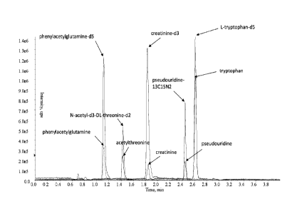
are shown in Figures 1A-F. The approximate retention time for the peak of
interest
for each analyte is indicated. Approximate retention times (in minutes) were
1.11,
2.45, 2.61, 1.43, and 1.83 for phenylacetylglutamine, pseudouridine,
tryptophan, N-
acetylthreonine, and creatinine, respectively.
27
CA 03045022 2019-05-24
WO
2018/118630 PCT/US2017/066364
B. Chromatography Method 2 (6 Analytes: pseudouridine, N-acetylthreonine,
meso-erythritol, arabitol, myo-inositol, N-acetylserine)
[00132] In another example, a liquid chromatography method was
developed
for the purification and separation in the same injection of one or more, two
or more,
and up to all six analytes selected from the group consisting of
pseudouridine, N-
acetylthreonine, meso-erythritol, arabitol, myo-inositol, N-acetylserine and
combinations thereof.
[00133] Mobile phase A was 50 mM ammonium acetate + 0.1% ammonium
hydroxide in water and mobile phase B was 100% acetonitrile. Linear gradient
elution, was carried out with an initial condition of 7% mobile phase A (93%
mobile
phase B) and 450 Lim in flow rate unless otherwise indicated. Mobile phase A
was
increased from the initial 7% to 20% (80% mobile phase B) at 1.5 min, from 20%
to
30% (70% mobile phase B) at 4.7 min, and from 30% to 35% (65% mobile phase B)
at 5.0 min. Then, mobile phase A reverted to 7% (93% mobile phase B) at 5.1
min
where it was maintained for 1.9 min for equilibration before the next sample
was
injected. The trital nin time was 7.0 min.
[00134] Chromatography Method 2 separated a plurality of up to six
analytes
with good peak shapes. Exemplary chromatograms of the resulting separated
analytes
are shown in Figures 2A-H. Approximate retention times (in minutes) were 2.21,
3.30, 2,72, 2.99, 4.59, and 2.89 for meso-erythritol, N-acetylserine,
arabitol, N-
acetylthreonine, myo-inositol, and pseudouridine, respectively.
C. Chromatography Method 3 (9 Analytes: N-acetylthreonine,
phenylacetylglutamine, tryptophan, creatinine, N-acetylalanine, 3-
methylhistidine,
trans-4-hydroxyproline, kynurenine, urea)
[00135] In another example, a liquid chromatography method was developed
for the purification and separation in the same injection of one or more, two
or more,
and up to all nine analytes selected from the group consisting of N-
acetylthreonine,
phenylacetylglutamine, tryptophan, creatinine, N-acetylalanine, 3-
methylhistidine,
trans-4-hydroxyproline, kynurenine, urea and combinations thereof, wherein, if
the
one or more assayed analytes is only one analyte, the one analyte is not
creatinine.
[00136] Mobile phase A was 20 mM ammonium formate +1% formic acid in
water and mobile phase B was 100% acetonitrile. Linear gradient elution, was
carried
out with an initial condition of 12% mobile phase A (88% mobile phase B) and
500
28
CA 03045022 2019-05-24
WO
2018/118630 PCT/US2017/066364
p.L/min flow rate unless otherwise indicated. Mobile phase A was increased
from the
initial 12% to 22% (78% mobile phase B) at 1.9 min, from 22% to 30% (70%
mobile
phase B) at 2.5 min, from 30% to 35% (65% mobile phase B) at 3.1 min, from 35%
to
38% (62% mobile phase B) at 3.7 min, and from 38% to 45% (55% mobile phase B)
at 5.0 min where it was maintained for 0.5 min. Then, mobile phase A reverted
to
12% (88% mobile phase B) at 5.7 min where it was maintained for 1.3 min for
equilibration before the next sample was injected. The total run time was 7.0
min.
[00137] Chromatography Method 3 separated a plurality of up to nine
analytes
with good peak shapes. Exemplary chromatograms of the resulting separated
analytes
are shown in Figures 3A-I. Approximate retention times (in minutes) were 1.36,
1.94,
3.74, 1.17, and 1.69 for urea, creatinine, trans-4-hydroxyproline, N-
acetylalanine, N-
acety lthreonine, 3-methy lhistidine, tryptophan, kynurenine, and
phenylacetylglutamine, respectively.
D. Chromatography Method 4 (tryptophan, 3-indoxyl sulfate, C-
glycosyltryptophan)
100138] In another example, a liquid chromatography method was
developed
for the purification and separation in the same injection of one or more, two
or more,
and up to all three analytes selected from the group consisting of tryptophan,
3-
indoxyl sulfate, and C-glycosyltryptophan, and combinations thereof.
[00139] Mobile phase A was 0.1% Formic Acid in water and mobile phase B
was 0.1% Formic Acid in Acetonitrile. Linear gradient elution, was carried out
with
an initial condition of 2% mobile phase B (98% mobile phase A) and a flow rate
of
7001.1L/min. Mobile phase B was increased from the initial 2% to 90% (10%
mobile
phase A) at 2.5 min and was maintained at 90% for 0.3 min. Then, mobile phase
B
reverted to 2% (98% mobile phase A) at 2.9 min where it was maintained for 0.4
min
for equilibration before the next sample was injected. The total run time was
3.30
min.
1001401 Chromatography Method 4 separated a plurality of up to three
analytes
with good peak shapes. Exemplary chromatograms of the resulting separated
analytes
are shown in Figures 4A-H. Approximate retention times (in minutes) were 0.91
and
0.95 for C-glycosyltryptophan, 1.32 and 1.33 for tryptophan and 1.45 for 3-
indoxylsulfate in serum and plasma, respectively.
E. Chromatography Method 5 (6 Analytes: N-acetylthreonine,
29
CA 03045022 2019-05-24
WO
2018/118630 PCT/US2017/066364
phenylacetylglutamine, pseudouridine, tryptophan, TMAP, creatinine)
[00141] In another example, a liquid chromatography method was
developed
for the purification and separation in the same injection of one or more, two
or more,
and up to all six analytes selected from the group consisting of N-
acetylthreonine,
phenylacetylglutamine, pseudouridine, tryptophan, TMAP, creatinine, and
combinations thereof. If the one or more assayed analytes is only one analyte,
the one
analyte is not creatinine.
1001421 Mobile phase A was 20 mM ammonium formate +1% formic acid in
water and mobile phase B was 100% acetonitrile. Linear gradient elution, was
carried
out with an initial condition of 12% mobile phase A (88% mobile phase B) and
550
4/min flow rate. Mobile phase A was increased from the initial 12% to 22% (78%
mobile phase B) at 1.9 min, from 22% to 30% (70% mobile phase B) at 2.5 min,
and
from 30% to 42% (58% mobile phase B) at 2.7 min. Then, mobile phase A reverted
to
12% (88% mobile phase B) at 3.4 min where it was maintained for 0.3 min for
equilibration before the next sample was injected. The total run time was 3.70
min.
[00143] Chromatography Method 5 separated a plurality of up to six
analytes
with good peak shapes. Exemplary chromatograms of the resulting separated
analytes
are shown in Figure 5. Approximate retention times (in minutes) were 1.40,
1.86,
2.14, 2.61, 2.71, and 3.16 for phenylacetylglutamine, creatinine, N-
acetylthreonine,
tryptophan, pseudouridine, and 'TMAP, respectively.
F. Chromatography Method 6 (10: N-acetylthreonine, meso-erythritol, arabitol,
myo-inositol, 3-indoxyl sulfate, tryptophan, phenylacetylglutamine,
creatinine,
pseudouridine, N-acetylserine)
1001441 In another example, a liquid chromatography method was
developed
for the purification and separation in the same injection of one or more, two
or more,
and up to all ten analytes selected from the group consisting of N-
acetylthreonine,
meso-erythritol, arabitol, myo-inositol, 3-indoxyl sulfate, tryptophan,
phenylacetylglutamine, creatinine, pseudouridine, and N-acetylserine, and
combinations thereof, wherein, if the one or more assayed analytes is only one
analyte, the one analyte is not creatinine.
[00145] Mobile phase A was 100 mM ammonium acetate + 0.2% ammonium
hydroxide in water and mobile phase B was 100% acetonitrile. Linear gradient
elution, was carried out with an initial condition of 7% mobile phase A (93%
mobile
CA 03045022 2019-05-24
WO
2018/118630 PCT/US2017/066364
phase B) and 500 pLimin flow rate. Mobile phase A was increased from the
initial
7% to 20% (80% mobile phase B) at 1.5 min, from 20% to 30% (70% mobile phase
B) at 4.7 min, and from 30% to 35% (65% mobile phase B) at 5.0 min. Then,
mobile
phase A reverted to 7% (93% mobile phase B) at 5.1 min where it was maintained
for
1.9 min for equilibration before the next sample was injected. The total run
time was
7.0 min.
[00146] Chromatography Method 6 separated a plurality of up to ten
analytes
with good peak shapes. An exemplary chromatogram of the resulting separated
analytes is shown in Figure 6. Approximate retention times (in minutes) were
2.35,
2.87, 4.85, 0.78, 3.20, 2.82, 2.40, 3.00, 3.30, and 3.69 for meso-erythritol,
arabitol,
myo-inositol, 3-indoxyl sulfate, tryptophan, phenylacetylglutamine,
creatinine,
pseudouridine, N-acetylthreonine, and N-acetylserine, respectively.
G. Chromatography Method 7 (5: arabitol, phenylacetylglutamine, creatinine,
pseudouridine, N-acetylthreonine)
[00147] In another example, a liquid chromatography method was developed
for the purification and separation in the same injection of one or more, two
or more,
and up to all five analytes selected from the group consisting of arabitol,
phenylacetylglutamine, creatinine, pseudouridine, N-acetylthreonine, and
combinations thereof, wherein, if the one or more assayed analytes is only one
analyte, the one analyte is not creatinine.
[00148] Mobile phase A was 100 mM ammonium acetate + 0.2% ammonium
hydroxide in water and mobile phase B was 100% acetonitrile. Linear gradient
elution, was carried out with an initial condition of 7% mobile phase A (93%
mobile
phase B) and 800 L/min flow rate. Mobile phase A was increased from the
initial
7% to 20% (80% mobile phase B) at 0.9 min, from 20% to 25% (75% mobile phase
B) at 1.9 min, and from 25% to 30% (70% mobile phase B) at 2.1 min. Then,
mobile
phase A reverted to 7% (93% mobile phase 13) at 2.2 min where it was
maintained for
0.5 min for equilibration before the next sample was injected. The total run
time was
2.7 min.
[00149] Chromatography Method 7 separated a plurality of up to five
analytes
with good peak shapes. An exemplary chromatogram of the resulting separated
analytes is shown in Figure 7. Approximate retention times (in minutes) were
1.74,
1.74, 1.48, 1.84, and 1.98 for arabitol, phenylacetylglutamine, creatinine,
31
CA 03045022 2019-05-24
WO
2018/118630 PCT/US2017/066364
pseudouridine, and N-acetylthreonine, respectively.
H. Chromatography Method 8 (6: myo-inositol, tryptophan,
phenylacetylglutamine,
creatinine, pseudouridine, N-acetylthreonine)
[00150] In another example, a liquid chromatography method was
developed
for the purification and separation in the same injection of one or more, two
or more,
and up to all six analytes selected from the group consisting of myo-inositol,
tryptophan, phenylacetylglutamine, creatinine, pseudouridine, N-
acetylthreonine, and
combinations thereof, wherein, if the one or more assayed analytes is only one
analyte, the one analyte is not creatinine.
[00151] Mobile phase A was 100 mM ammonium acetate + 0.2% ammonium
hydroxide in water and mobile phase B was 100% acetonitrile. Linear gradient
elution, was carried out with an initial condition of 7% mobile phase A (93%
mobile
phase B) and 800 }IL/min flow rate. Mobile phase A was increased from the
initial
7% to 22% (78% mobile phase B) at 0.9 min, from 22% to 30% (70% mobile phase
B) at 2.5 min, and from 30% to 35% (65% mobile phase B) at 2.7 min. Then,
mobile
phase A reverted to 7% (93% mobile phase B) at 2.8 min where it was maintained
for
0.4 min for equilibration before the next sample was injected. The total run
time was
3.2 min.
[00152] Chromatography Method 8 separated a plurality of up to six
analytes
with good peak shapes. An exemplary chromatogram of the resulting separated
analytes is shown in Figure 8. Approximate retention times (in minutes) were
2.64,
1.83, 1.64, 1.40, 1.74, and 1.85 for myo-inositol, tryptophan,
phenylacetylglutamine,
creatinine, pseudouridine, and N-acetylthreonine, respectively.
Example 2: MS/MS measurement of analytes
[00153] Mass spectrometry was performed on the sample extracts as described
in the methods below using an AB Sciex QTrap 5500 mass spectrometer with Turbo
V source (ESI). Raw data were acquired from the instrument and processed using
Analyst 1.6.2 software (AB Sciex). For quantitation, peak area ratios of
analyte to
internal standard were fitted against the concentrations of the calibration
standards by
weighted (1/x2) linear least squares regression. The resulting slope and
intercept of
the calibration curve were used to calculate the unknown concentrations in
experimental samples.
32
CA 03045022 2019-05-24
WO
2018/118630 PCT/US2017/066364
A. MS/MS Method 1
[00154] A method was developed to detect in the same injection the
levels of
one or more, two or more, and up to all five analytes selected from the group
consisting of pseudouridine, N-acetylthreonine, phenylacetylglutamine,
tryptophan,
creatinine and combinations thereof, wherein, if the one or more assayed
analytes is
only one analyte, the one analyte is not creatinine. The same MS/MS method was
used to detect in the same injection the levels of one or more, two or more,
and up to
all six analytes selected from the group consisting of pseudouridine, N-
acetylthreonine, phenylacetylglutamine, tryptophan, TMAP, creatinine, and
combinations thereof.
[00155] The eluent from the chromatography column described in
Example 1,
Chromatography Method 1, was directly and automatically introduced into the
electrospray source of a mass spectrometer. In another example, the eluent
from the
chromatography column described in Example 1, Chromatography Method 5, was
directly and automatically introduced into the electrospray source of a mass
spectrometer. Acetonitrile:Water (50:50) was used for needle wash. The
instrument
was operated in positive multiple reaction monitoring (MRM) mode. Ionspray
voltage was set at 4.0 kV, source temperature at 550 C, curtain gas (e.g.,
nitrogen) at
psi, and nebulizer and desolvation gas (e.g., nitrogen) flow rates at 75 psi,
20 collisionally activated dissociation (CAD) gas (e.g., nitrogen) at
medium.
[00156] Raw data were acquired from the instrument and processed
using
Analyst 1.6.2 software (AB Sciex). For quantitation, peak area ratios of
analyte to
internal standard were fitted against the concentrations of the calibration
standards by
weighted (1/x2) linear least squares regression. The resulting slope and
intercept of
the calibration curve were used to calculate the unknown concentrations in
experimental samples. Exemplary ions that were generated for the quantitation
of
pseudouridine, N-acetylthreonine, phenylacetylglutamine, tryptophan, and
creatinine,
and TMAP are listed in Table 3. The parent ions are listed under the column
headed
"Parent ion (m/z)", and the daughter ions used for quantitation in this
example are
listed in the column labeled "Daughter ion for quantitation (m/z)". The choice
of
daughter ion for quantitation in this example was optimized for sensitivity
across the
analytical measurement range; however, additional daughter ions may be
selected to
replace or augment the daughter ions used for quantitation in the examples.
33
CA 03045022 2019-05-24
WO 2018/118630
PCT/US2017/066364
Table 3. Parent and Daughter Ion Mass to Charge Ratios (m/z) of Analytes as
measured in positive ionization mode
Daughter
Parent ion ion for Additional daughter
Analyte
(m/z) quantitation ions (m/z) (all 0.5)
(m/z)
144.0, 126.1, 119.9,
116.1, 102.0, 97.9, 84.0,
N-acetyl-DL-threonine 162.0 0.5 74.1+0.5
70.0, 57.0, 56.0, 43.0,
28.1
149.1, 131.9, 129.8,
125.9, 122.8, 121.0,
N-acetyl-d3-DL-
167.0 0.5 77.1 0.5 104.0, 91.0, 86.1, 76.0,
threonine-d2
59.0, 58.1, 45.9, 43.0,
31.1, 29.2, 28.0
248.1, 219.1, 147.1,
136.0, 130.0, 129.1,
phenylacetylglutamine 265.0 0.5 91.0 0.5 101.1, 84.0, 83.0,
65.0,
56.0, 50.9, 44.0, 40.9,
39.1,28.0
253.3, 224.3, 147.0,
phenylaoetylglutaminc- 270.0 0.5 96.3 0.5 141.3, 130.0,
100.8, 84.0,
d5 69.0, 68.1, 56.0, 41.1,
28.1
creatinine 113.9 0.5 43.0 0.5 86.0, 72.0, 44.1,
42.0,
28.1
creatinine-d3 116.9 0.5 47.0+0.5 89.2, 43.1, 29.1,
28.0
188.1, 170.0, 159.1,
144.0, 143.0, 142.0,
140.0, 132.0, 130.1,
128.1, 126.9, 117.9,
L-tryptophan 205.0 0.5 146.0 0.5 116.9, 114.9,
103.0, 91.0,
89.9, 89.0, 77.0, 74.9,
74.0, 64.9, 63.0, 62.0,
61.0, 50.9, 49.9, 39.2,
28.0
191-193, 173-174, 163-
164, 144.8-151.2, 117.1-
122.1, 102.9-110.1, 89.9-
L-tryptophan-d5 210.0 0.5 150.1 0.5
96.0, 74.1-81.1, 60.9-
68.9, 50.1-54.1, 38.0-
43.1, 28.0-29.0
209.0, 179.0, 167.0,
163.0, 154.8, 151.0,
pseudouridine 244.9 0.5 191.0 0.5
148.0, 139.0, 125.0,
120.0, 111.8, 109.8,
34
CA 03045022 2019-05-24
WO 2018/118630
PCT/US2017/066364
107.8, 96.0, 92.0, 84.0,
82.0, 80.0, 68.0, 65.2,
55.0, 54.0, 43.0, 41.0,
39.0
230.2, 212.0, 206.9,
194.1, 182.2, 169.9,
166.0, 157.9, 154.0,
148.9, 141.9, 128.1,
120.9,
pseudouridine-13C15N2 247.9+0.5 194.0+0.5 13 96.8,
91.0, 85.3, 83.1,
81.2, 78.8, 77.0, 69.1,
65.0, 56.9, 55.0, 53.3,
51.3, 44.9, 43.0, 40.9,
39.0
170.1, 126.0, 124.0,
TMAF 229.1+0.5 142.2+0.5 or 116.0 114.0, 98.0,
96.0,
70.0+0.5 68.0, 60.0, 59.1, 58.1,
54.9
170.1, 126.0, 124.0,
116.0, 114.0, 98.0, 97.1,
96.0, 93.9, 81.0, 80.0,
142.1+0.5 or 79.0, 77.0, 74.0, 72.1,
13C3-1,,L-TMAP 232.2+0.5
70.1+0.5 71.1, 69.0, 68.0, 67.1,
63.0, 62.1, 61.1, 60.0,
59.0, 58.1, 57.1, 56.1,
55.0, 54.0, 53.1
[001571 Figures 9-14 show mass spectra resulting from fragmentation
of the
parent ions indicated in Table 3.
[00158] MRM transitions that were generated for the quantitation of N-
acetylthreonine in positive ionization mode include those produced by
fragmenting a
parent ion having a m/z of about 162.0+0.5 to produce daughter ions having m/z
of
about 74.1+0.5, 144.0+0.5, 126.1+0.5, 119.9+0.5, 116.1+0.5, 102.0+0.5,
97.9+0.5,
84.0+0.5, 70.0+0.5, 57.0+0.5, 56.0+0.5, 43.0+0.5, and 28.1+0.5. These parent
and
daughter ion peaks generated from tandem mass spectrometric fragmentation of N-
acetylthreonine are illustrated in Figure 9. Any of the daughter ions may be
selected
for quantitation. In this example, the daughter ion used for quantitation of N-
acetylthreonine has a m/z of about 74.1+0.5. The calibration range for N-
acetylthreonine was determined to be 0.0200 to 2.00 pg/mL.
[00159] MRM transitions that were generated for the quantitation of
phenylacetylglutarnine in positive ionization mode include those produced by
fragmenting a parent ion having m/z of about 265.0+0.5 to produce daughter
ions
CA 03045022 2019-05-24
WO
2018/118630 PCT/US2017/066364
having m/z of about 91.0+0.5, 248.1+0.5, 219.1+0.5, 147.1+0.5, 136.0+0.5,
130.0+0.5, 129.1+0.5, 101.1+0.5, 84.0+0.5, 83.0+0.5, 65.0+0.5, 56.0+0.5,
50.9+0.5,
44.0+0.5, 40.9+0.5, 39.1+0.5, and 28.0+0.5. These parent and daughter ion
peaks
generated from tandem mass spectrometric fragmentation of
phenylacetylglutamine
are illustrated in Figure 10. Any of the daughter ions may be selected for
quantitation.
In this example, the daughter ion used for quantitation of
phenylacetylglutamine has
m/z of about 91.0+0.5. The calibration range for phenylacetylglutamine was
determined to be 0.100 to 20.0 iig/mL.
1001601 MRM transitions that were generated for the quantitation of
creatinine in positive ionization mode include those produced by fragmenting a
parent
ion having m/z of about 113.9+0.5 to produce daughter ions having a m/z of
about
43.0+0.5, 86.0+0.5, 72.0+0.5, 44.1+0.5, 42.0+0.5, and 28.1+0.5. These parent
and
daughter ion peaks generated from tandem mass spectrometric fragmentation of
creatinine are illustrated in Figure 11. Any of the daughter ions may be
selected for
quantitation. In this example, the daughter ion used for quantitation of
creatinine has a
m/z of about 43.0+0.5. The calibration range for creatinine was determined to
be 2.00
to 200 lig/mL.
[001611 MRM transitions that were generated for the quantitation of
tryptophan in positive ionization mode include those produced by fragmenting a
parent ion having a m/z of about 205.0+0.5 to produce daughter ions having m/z
of
about 146.0+0.5, 191-193+0.5, 173-174+0.5, 163-164+0.5, 144.8-151.2+0.5, 117.1-
122.1+0.5, 102.9-110.1+0.5, 89.9-96.0+0.5, 74.1-81.1+0.5, 60.9-68.9+0.5, 50.1-
54.1+0.5, 38.0-43.1 0.5, and 28.0-29.0 +0.5. These parent and daughter ion
peaks
generated from tandem mass spectrometric fragmentation of tryptophan are
illustrated
in Figure 12. Any of the daughter ions may be selected for quantitation. In
this
example, the daughter ion used for quantitation of tryptophan has a m/z of
about
146.0+0.5. The calibration range for tryptophan was determined to be 1.00 to
100
1.tg/mL.
[001621 MRM transitions that were generated for the quantitation of
pseudouridine in positive ionization mode include those produced by
fragmenting a
parent ion having a m/z of about 244.9 0.5 to produce daughter ions having m/z
of
about 191.0+0.5, 209.0+0.5, 179.0+0.5, 167.0+0.5, 163.0+0.5, 154.8+0.5,
151.0+0.5,
148.0+0.5, 139.0+0.5, 125.0+0.5, 120.0+0.5, 111.8+0.5, 109.8+0.5, 107.8+0.5,
36
CA 03045022 2019-05-24
WO
2018/118630 PCT/US2017/066364
96.0 0.5, 92.0 0.5, 84.0 0.5, 82.0 0.5, 80.0+0.5, 68.0 0.5, 65.2 0.5, 55.0
0.5,
54.0 0.5, 43.0 0.5, 41.0 0.5, and 39.0 0.5. These parent and daughter ion
peaks
generated from tandem mass spectrometric fragmentation of pseudouridine are
illustrated in Figure 13. Any of the daughter ions may be selected for
quantitation. In
this example, the daughter ion used for quantitation of pseudouridine has a
m/z of
about 191.0 0.5. The calibration range for pseudouridine was determined to be
10.0
to 4001.1g/mL.
[00163] MRIVI transitions that were generated for the quantitation of
TMAP in
positive ionization mode include those produced by fragmenting a parent ion
having a
m/z of about 229.1 0.5 to produce daughter ions having m/z of about 170.1 0.5,
142.2 0.5, 126.0 0.5, 124.0 0.5, 116.0 0.5, 114.0 0.5, 98.0 0.5, 96.0 0.5,
70.0 0.5,
68.0 0.5, 60.0 0.5, 59.1 0.5, 58.1 0.5, 54.9 0.5, 227.0 0.5, 181.0 0.5, 159.0
0.5,
133.2 0.5, 114.8 0.5, 112.9 0.5, 105.8 0.5, 89.1 0.5, 71.0 0.5, 69.0 0.5, and
45.1 0.5. Any of the daughter ions may be selected for quantitation. For
example,
daughter ions having a m/z of about 58.1 0.5, 70.0 0.5, 114.0 0.5, or 142.2
0.5 may
be used fur the quantitation of TMAP. These parent and daughter ion peaks
generated
from tandem mass spectrometric fragmentation of TMAP are illustrated in Figure
14.
B. MS/MS Method 2
[00164] In another example, a method was developed to detect in the
same
injection the levels of one or more, two or more, and up to all six analytes
selected
from the group consisting of pseudouridine, N-acetylthreonine, meso-
erythritol,
arabitol, myo-inositol, N-acetylserine and combinations thereof. The eluent
from the
chromatography column described in Example 1, Chromatography Method 2, was
directly and automatically introduced into the electrospray source of a mass
spectrometer. Water:acetonitrile (90:10) was used for the strong/seal wash;
acetonitrile:water (90:10) was used for the weak wash.
[00165] The instruments were operated in negative MRM mode. Ionspray
voltage was set at -4.0 kV, source temperature at 550 C, and curtain gas at
20 psi;
nebulizer and desolvation gas flow rates were set at 70 psi, and CAD gas at
medium.
[00166] Exemplary ions that were generated for the quantitation of
pseudouridine, N-acetylthreonine, meso-erythritol, arabitol, myo-inositol, and
N-
acetylserine are shown in Table 4. The parent ions are listed under the column
headed
37
CA 03045022 2019-05-24
WO 2018/118630 PCT/US2017/066364
"Parent ion (m/z)", and the daughter ions used for quantitation in this
example are
listed in the column labeled "Daughter ion for quantitation (m/z)". The choice
of
daughter ion for quantitation in this example was optimized for sensitivity
across the
analytical measurement range; however, any of the daughter ions may be
selected to
replace or augment the daughter ions used for quantitation in the examples.
Table 4. Ions useful for quantitation of analytes
Daughter Additional daughter
Parent ion for ions (m/z) (all 0.5)
Analyte
ion (m/z) quantitation
(m/z)
182.8, 151.9, 139.9,
138.9, 124.0, 110.8,
pseudouridine 242.9 0.5 153.0 0.5
109,9, 96.0, 82.0, 55.0,
42.0, 41.0
Pseudouridine-13C,15N2 245.9 0.5 156.0 0.5 1141.11.0 97i0
44.1
118.1, 115.8, 97.9, 71.9,
Acetyl-L-threonine 159.9 0.5 73.9 0.5 70.9, 70.1, 56.1, 54.0,
42.0, 40.9. 26.0
N-Acetyl-dl-L-threonine-d2 165.0+0 5 102,0 0.5 120.0,103.0, 82.2,
77.0,
76.0, 74.1
120.0, 119.0, 105.9,
103.0, 100.9, 93.9, 92.8,
meso-Erythritol 120.9 0.5 88.9 0.5 79.9, 77.0, 70.9,
67.9,
65.8, 65.0, 58.9, 52.0,
43.2, 40.0
123.0, 110.0, 107.0,
105.0, 98.0, 95.2, 91.9,
87.0 82.1 81.1 76.1,
Erythrito1-13C4 124.9 0.5 73.9 0.5 , , ,
67.0, 65.9, 6LO,
58.0, 44.8, 41.2, 39,9,
25.8
149.1, 136.0, 133.0,
131.1, 119,0, 112.8,
108.2, 103.1, 100.9,
96.8 91.8 84.9 83.0
D-Arabitol 150.9 0.5 88.9 0.5 , , , ,
81.9, 78,8, 77.0, 73.0,
70.9, 68.9, 66.9, 59.0,
57.0, 55.0, 45.0, 42.9,
41.2
154.1, 138.0, 136.0,
123.1, 118.1, 107.0,
D-Arabinito1-13C5 155.9 0.5 91.9 0.5 104.9, 102.0, 88.9,
86.9,
76.0, 74.0, 73.2, 61.0,
59.9, 58.0, 45.0, 43.1
myo-Inositol 178.9 0.5 87.0 0.5 177.2, 161.0,
159.0,
38
CA 03045022 2019-05-24
WO 2018/118630
PCT/US2017/066364
146.8, 141.0, 134.9,
128.8, 125.0, 122.7,
117.0, 112.8, 110.9,
100.9, 98.9, 97.0,95.0,
90.8, 89.0, 85.0, 82.9,
81.0, 78.8, 74.8, 73.1,
70.9, 68.9, 59.0, 56.9,
55.0, 45.1, 43.0, 41.0
181.8, 167.1, 164.1,
158.1, 144.9, 140.0,
131.9, 130.1, 128.9,
119.9, 118.9, 117.2,
104.1, 103.1, 102.0,
myo-Inositoi-d6 184.9 0.5 88.9 0.5 100.9, 97.9,
93.1, 90.0,
88.0, 85.9, 82.0, 80.8,
76.9, 74.1, 72.9, 72.1,
69.8, 62.1, 61.0, 60.1,
58.1, 57.0, 46.0, 45.0,
42.1
119.0, 116.0, 104.9,
103.9, 103.0, 97.9, 84.0,
N-acetylserine 145.9 0.5 74.0 0.5
81.0, 72.0, 70.0, 64.9,
60.8, 57.0, 42.0, 40.9
121.9, 106.9, 98.9, 87.0,-
74.9 73.0 70.9
Acetylserine-d3 148.9 0.5 117.0 0.5 86.0, , ,
,
60.8, 59.0, 58.0, 45.0,
41.2, 42.0
C. MS/MS Method 3
[4:101671 In another
example, a method was developed to detect in the same
injection the levels of one or more, two or more, and up to all nine analytes
selected
from the group consisting of N-acetylthreonine, phenylacetylglutamine,
tryptophan,
creatinine, N-acetylalanine, 3-methylhistidine, trans-4-hydroxyproline,
kynurenine,
urea and combinations thereof, wherein, if the one or more assayed analytes is
only
one analyte, the one analyte is not creatinine. The eluent from the
chromatography
column described in Example 3, Chromatography Method 3, was directly and
automatically introduced into the electrospray source of a mass spectrometer.
Water:acetonitrile (90:10) was used for the strong/seal wash;
acetonitrile:water
(90:10) was used for the weak wash. The instrument was operated in positive
multiple reaction monitoring (MRM) mode. lonspray voltage was set at 4.0 kV,
source temperature at 550 C, curtain gas (e.g., nitrogen) at 20 psi, and
nebulizer and
desolvation gas (e.g., nitrogen) flow rates at 75 psi, collisionally activated
dissociation
39
CA 03045022 2019-05-24
WO 2018/118630
PCT/US2017/066364
(CAD) gas (e.g., nitrogen) at medium.
[00168] Exemplary ions that were generated for the quantitation of N-
acetylthreonine, phenylacetylglutamine, tryptophan, creatinine, N-
acetylalanine, 3-
methylhistidine, trans-4-hydroxyproline, kynurenine, and urea are shown in
Table 5.
The parent ions are listed under the column headed "Parent ion (m/z)", and the
daughter ions used for quantitation in this example are listed in the column
labeled
"Daughter ion for quantitation (m/z)". The choice of daughter ion for
quantitation in
this example was optimized for sensitivity across the analytical measurement
range;
however, any of the daughter ions may be selected to replace or augment the
daughter
ions used for quantitation in the examples.
Table 5. Ions useful for quantitation of analytes
Daughter
Parent ion ion for Additional daughter ions
Analyte
(m/z) quantitation (m/z) (all +0.5)
(m/z)
144.0, 126.1, 119.9,116.1,
Acetyl-L-threonine 162.0-10.5 74.1 0.5 102.0, 97.9,
84.0, 70.0,
57.0, 56 0, 43 0, 28.1
149.1, 131.9, 129.8, 125.9,
N-Acetyl-d3-L- 167 . 0 0.5 77.1 0.5 122.8, 121.0,
104.0, 91.0,
threonine-d2 86.1, 76.0, 59.0, 58.1, 45.9,
43.0, 31.1, 29.2, 28.0
248.1,219.1, 147.1, 136.0,
Phenylacetyl-L- 265 . 0 0 . 5 91 . 0 0 . 5 130.0,
129.1, 101.1, 84.0,
glutamine 83.0, 65.0, 56.0, 50.9, 44.0,
40.9, 39.1, 28.0
253.3, 224.3, 147.0, 141.3,
Phenylacetylglutamine-
270.0+0.5 96.3+0.5 130.0, 100.8, 84.0, 69.0,
d5
68.1, 56.0, 41.1, 28.1
Creatinine 113.9 0.5 43.0 0.5 86.0, 72.0, 44.1,
42.0, 28.1
Creatinine-d3 116.9 0.5 47.0 0.5 89.2, 43.1, 29.1,
28.0
188.1, 170.0, 159.1, 144.0,
143.0, 142.0, 140.0, 132.0,
130.1, 128.1, 126.9, 117.9,
L-Tryptophan 205.0 0.5 146.0 0.5 116.9, 114.9,
103.0, 91.0,
89.9, 89.0, 77.0, 74.9, 74.0,
64.9, 63.0, 62.0, 61.0, 50.9,
49.9, 39.2, 28.0
191-193, 173-174, 163-164,
144.8-151.2, 117.1-122.1,
L-Tryptophan-d5 210.0 0.5 150.1 0.5 102.9-110.1,
89.9-96.0,
74.1-81.1, 60.9-68.9, 50.1-
54.1, 38.0-43.1, 28.0-29.0
CA 03045022 2019-05-24
WO 2018/118630
PCT/US2017/066364
N-Acetyl-L-alanine 131.9 0.5 89.9 0.5 114.1, 86.1, 44.0
N-Acetyl-L-alanine-d4 136.0 0.5 94.0 0.5 118.1, 90.0, 48.0
109.1, 97.0, 96.0, 92.9,
3-Methyl-L-histidine 170.0 0.5 94.9 0.5 83.0, 81.0, 80.1,
70.2, 67.9,
67.0, 55.0, 54.0, 42.0, 41.0
3-Methyl-L-histidine- 173.0 0.5 127.00.5 129.1, 112.0,
100.1, 83.0
d3
trans-4-Hydroxy-L- 131.9 0.5 68.0 0.5 114.2, 86.0, 58.0,
41.0
pro line
trans-4-Hydroxy-L- 134.9 0.5 71.1 0.5 117.1, 89.0, 70.0,
61.0
proline-d3
192.1, 191.2, 174.0, 164.1,
163.1, 150.0, 146.1
L-Kynurenine 209.0 0.5 94.0 0.5 ,
136.0,
119.9, 118.1, 98.9, 88.0,
73.9
198.2, 197.2, 179.1, 170.1,
169.2, 155.1, 154.1, 151.0,
Kynurenine-d6 215.0 0.5 98.0 0.5
142.0, 141.0, 125.0, 118.0,
99.9, 99.1, 97.0, 89.9, 74.2
Urea 60.9 0.5 29.2 0.5 44.0, 43.0, 42.1,
28.0, 27.1
Urea-13C, 15N2 63.9 0.5 29.1 0.5 46.0, 45.0, 44.1,
30.0
D. MS/MS Method 4
[001691 In another example, a method was developed to detect in the
same
injection the levels of one or more, two or more, and up to all three analytes
selected
from the group consisting of tryptophan, C-glycosyltryptophan, and 3-indoxyl
sulfate.
The eluent from the chromatography column described in Example 1,
Chromatography Method 4, was directly and automatically introduced into the
electrospray source of a mass spectrometer. Methanol was used for needle wash.
The
instrument was operated in negative MRM mode. Ionspray voltage was set at -4.5
kV, source temperature at 550 C, and curtain gas at 20 psi; nebulizer and
desolvation
gas flow rates were set at 60 psi and 65 psi, respectively, and CAD gas at
high.
[00170] Exemplary ion pairs that may be used for the quantitation of
tryptophan, C-glycosyltryptophan, and 3-indoxylsulfate are shown in Table 6.
The
parent ions are listed under the column headed "Parent ion (m/z)", and the
daughter
ions used for quantitation in this example are listed in the column labeled
"Daughter
ion for quantitation (m/z)". The choice of daughter ion for quantitation in
this
example was optimized for sensitivity across the analytical measurement range;
however, any of the daughter ions may be selected to replace or augment the
daughter
41
CA 03045022 2019-05-24
WO 2018/118630
PCT/US2017/066364
ions used for quantitation in the examples.
Table 6. Ions useful for quantitation of analytes
Daughter ion
Parent ion for Additional daughter
Analyte
(m/z) quantitation ions (m/z) (all *0.5)
(m/z)
185.9, 158.9, 141.9,
L-Tryptophan 202.9 0.5 115.9 0.5 130.0, 74.1, 72.2,
59.0,
44.9
190.2, 164.2, 162.2,
146.0, 134.1,
L-Tryptophan-d5 208.0 0.5 119.9 0.5 147.1,
121.0, 120.0, 76.0, 75.0,
73.9, 72.8, 72.0
3-indoxylsulfate 211.8 0.5 79.9 0.5 132.0, 104.0, 80.9,
77.0
3-indoxylsulfate-d4 215.9 0.5 80.9 0.5 136.0, 79.9
Manno-L-tryptophan 365.2 0.5 245.0 0.5 130.0, 142.0,
156.0,
116.0
Manno-L-tryptophan- 369.2 0.5
249.0 0.5
D. MS/MS Method 5
11101711 In another example, a method was developed to detect in the same
injection the levels of one or more, two or more, and up to all ten analytes
selected
from the group consisting of meso-erythritol, D-arabitol, inositol, 3-indoxyl
sulfate,
L-tryptophan, phenylacetylglutamine, creatinine, pseudouridine, N-
acetylthreonine,
and N-acetylserine, and combinations thereof. The eluent from the
chromatography
column described in Example 1, Chromatography Method 6, was directly and
automatically introduced into the electrospray source of a mass spectrometer.
1001721 In another example, the eluent from the chromatography column
described in Example 1, Chromatography Method 7, was directly and
automatically
introduced into the electruspray source of a mass spectrometer. The method
detected,
in the same injection, the levels of one or more, two or more, and up to all
five
analytes selected from the group consisting of D-arabitol,
phenylacetylglutamine,
creatinine, pseudouridine, N-acetylthreonine, and combinations thereof,
wherein, if
the one or more assayed analytes is only one analyte, the one analyte is not
creatinine.
1001731 In another example, the eluent from the chromatography
column
described in Example 1, Chromatography Method 8, was directly and
automatically
introduced into the electrospray source of a mass spectrometer. The method
detected,
42
CA 03045022 2019-05-24
WO 2018/118630 PCT/US2017/066364
in the same injection, the levels of one or more, two or more, and up to all
six analytes
selected from the group consisting of inositol, L-tryptophan,
phenylacetylglutamine,
creatinine, pseudouridine, N-acetylthreonine, and combinations thereof,
wherein, if
the one or more assayed analytes is only one analyte, the one analyte is not
creatinine.
Acetonitrile:Water (50:50) was used for the needle wash. The instrument was
operated in negative MRM mode. Ionspray voltage was set at -4.5 kV, source
temperature at 550 C, and curtain gas at 20 psi; nebulizer and desolvation
gas flow
rates were set at 50 psi, and CAD gas at medium.
[00174] Exemplary ion pairs that may be used for the quantitation of
meso-
erythritol, D-arabitol, inositol, 3-indoxyl sulfate, L-tryptophan,
phenylacetylglutamine, creatinine, pseudouridine, N-acetylthreonine, and N-
acetylserine are shown in Table 7. The parent ions are listed under the column
headed
"Parent ion (m/z)", and the daughter ions used for quantitation in this
example are
listed in the column labeled "Daughter ion for quantitation (m/z)". The choice
of
daughter ion for quantitation in this example was optimized for sensitivity
across the
analytical measurement range; however, any of the daughter ions may be
selected to
replace or augment the daughter ions used for quantitation in the examples.
Table 7. Ions useful for quantitation of analytes
Parent Daughter Parent Daughter
ion ion for ion ion for
Analyte Analyte
+0.5 quantitation +0.5 quantitation
(m/z) 0.5 (m/z) (m/z) 0.5
(m/z)
Meso-erythritol 120.9 89
Phenylacetylgluta262.9 42
mine
Meso- Phenylacetylgluta
124.9 74' 268 42
erythrito1-13C4 mine-d5
D-Arabitol 150.9 88.9 Creatinine 111.9 67.9
D:Arabitol-
155.9 91.9 Creatinine-d3 114.9 68
I3C5
Inositol 178.9 87 Pseudouridine 242.9 42
Inositol-d6 184.9 88.9 Pseudouridine-
13C,I5N2 245.9 156
3-Indoxyl N-Acetyl-DL-
211.8 103.9 159.9 73.9
sulfate threonine
3-Indoxyl N-Acetyl-d3-DL-
215.9 107.9 164.9 102
sulfate-chi threonine-d2
L-Tryptophan 202.9 115.9 N-Acetylserine 145.9 74
43
CA 03045022 2019-05-24
WO 2018/118630 PCT/US2017/066364
1 L-Tryptophan-
207.9 1 119.9 1 N-Acetylserine-d3 1 148.9 1 117
I
d5
Example 3: Method Validation
A. Chromatography Method 1 and MS/MS Method I
[00175] The analytical performance of the combination of
Chromatography
Method 1 and MS/MS Method 1 resulted in the quantitation of a plurality of up
to five
analytes in a single injection with a run time of 3.7 minutes.
[00176] The precision of the method for measuring a plurality of five
analytes
' was evaluated at three QC levels (low, mid, and high) in plasma and serum.
Three
replicates per QC level per matrix were analyzed in two runs per day over 20
days for
a total of 40 runs. A total of 120 replicates per QC level were included in
the inter-day
CV calculations for each analyte per matrix. The inter-day precision was less
than
6.3% at each QC level in plasma and less than 7.1% at each QC level in serum.
The
results are presented in Table 8. Linear responses (R2>0.98) were observed
over a
100 fold range for N-acetylthreonine, creatinine, pseudouridine, and
tryptophan, and
over a 2004old tange fur plienylacetylglutarnine. Calibration ranges were
selected
based on analysis of over 1,000 plasma and serum samples.
Table 8. Inter-day Precision for a Plurality of Analytes in Plasma and Serum.
SERUM PLASMA
QC Mean Mean
Analyte %CV %CV
Level (1.tgimL) ( g/mL)
Low 0.0772 5.9 0.0758 6.0
Acetyfthreonine
Mid 0.889 5.9 0.891 5.3
(n=120)
High 1.78 4.8 1.64 5.4
Low 7.42 7.1 6.70 5.3
Creatinine
Mid 88.9 6.6 87.0 4.9
(n=120)
High 172 5.5 _ 158 5.4
Low 0.328 6.4 0.327 6.3
Phenylacetylglutamine
Mid 8.15 5.1 8.19 4.2
(n=120)
High 16.0 3.8 14.7 3.8
Low 1.27 6.1 1.23 5.1
..
Pseudouridine
Mid 16.4 5.1 16.4 4.8
(n=120)
High 32.1 3.7 29.8 4.4
Tryptophan Low 4.07 6.9 3.26 6.1
(n=120) Mid _ 44.5 5.4 43.4 4.9
44
CA 03045022 2019-05-24
WO 2018/118630
PCT/US2017/066364
I High I 88.0 I 4.3 I 80.6 I 4.8 I
[00177] Accuracy
and precision of dilution QCs in serum and plasma were
evaluated to accurately measure samples with analyte values above the ULOQ
(i.e.,
outside the calibration range). The dilution QCs were prepared by diluting the
high
QC matrix 5-fold with fatty acid-free BSA solution. Three replicates of
dilution QCs
per matrix were analyzed in two runs per day over five days for a total of 10
runs. A
total of 30 replicates were included in the inter-day accuracy and precision
calculations. The inter-day accuracy (as compared to the measured high QC
value) in
serum was greater than 94.5% and the inter-day precision was less than 7.6%;
the
inter-day accuracy in plasma was greater than 94.7% and the inter-day
precision was
less than 4.4%. The results are presented in Table 9.
Table 9. Inter-Day Accuracy and Precision of the 5X Dilution QC
SERUM
N- Phenylacetyl Pseudouri
acetylthreo Creatinine glutamine dine Tryptophan
nine (n=30) (n=30) (n=30) (n=30) (n=30)
Mean
(mg/mL) 1.80 177 17.0 33.6 83.3
%CV 7.6 5.8 7.0 6.0 6.8
% Accuracy 97.9 101 94.5 95.0 107
PLASMA
N- Phenylacetyl Pseudouri
acetylthreo Creatinine glutamine dine Tryptophan
nine (n=30) (n=30) (n=30) (n=30) (n=30)
Mean
(mg/mL) 1.66 163 15.5 30.3 76.3
%CV 3.7 4.0 3.2 3.9 4.4
% Accuracy 100 101 94.7 99.6 107
[00178] The precision at the LLOQ was evaluated. The signal-to-noise
ratio
for every analyte was greater than 5:1. Three replicates of the LLOQ samples
were
analyzed in two runs per day over 15 days for a total of 30 runs. A total of
90
replicates of the LLOQ were included in the inter-day %CV calculations for
each
analyte. All intra- and inter-day precision was less than 2.8% CV; the data
are shown
in Table 10. These results indicated that quantitation of the plurality of
analytes at the
lower limit was highly precise.
Table 10. Intra- and Inter-Day Precision at the LLOQ.
CA 03045022 2019-05-24
WO 2018/118630
PCT/US2017/066364
LLOQ Concentration (u,g/mL)
0.0200 2.00 0.100 0.400 1.00
N-
acetylthr Phenylacetyl Pseudouri
eonine Creatinine glutamine dine Tryptophan
Day (%RSD) (%RSD) (%RSD) (%RSD) (%RSD)
1 (n=6) 5.2 5.8 11.2 4.7 9.6
2(n=6) 7.6 6.3 10.0 5.4 10.9
3 (n=6) 5.7 3.3 6.5 4.8 7.0
4(n6) 9.9 2.6 4.7 3.2 5.3
(n=6) 4.6 4.2 4.2 3.7 3.5
6(n=6) 6.1 7.6 3.7 7.6 8.1
7 (n=6) 10.1 8.1 8.1 9.8 6.2
8 (n=6) 4.3 8.8 5.5 ' 4.1 6.8
9(n6) 3.1 5.5 6.6 10.1 9.2
10(n=6) 8.9 3.1 = 7.1 6.0 5.7
11 (n=6) 5.1 5.3 5.7 4.6 6.5
12(n=6) 7.1 9.9 8.6 5.7 6.1
..
13 (n=6) 4.9 3.7 3.6 8.4 12.8
14 (n=6) 5.0 , 6.6 12.3 5.9 7.2
15(n=6) 5.0 6.5 4.6 5.9 5.3
_
Intei-Day
(n=90) 6.5 6.4 8.6 7.0 9.3
[00179] In order to assess the recovery of analytes during the
extraction, QC
samples in serum and plasma were fortified with known concentrations of
analytes.
Six replicates of the spiked QC samples were extracted and analyzed along with
5 regular QC samples in triplicate. Recovery of the spiked amount was
calculated after
subtraction of the amount in the spiked QC samples. The recoveries for the
five
analytes were determined to be 97.4% to 113% in serum and 103% to 110% in
plasma
for the five analytes. The data are presented in Table 11.
Table 11. Recovery of Analytes
SERUM
N-
acetylthreo Phenylacetyl
Pseudouri
nine Creatinine glutamine dine Tryptophan
% Recovery 103 97.4 98.5 103 113
PLASMA
N-
acetylthreo Phenylacetyl
Pseudouri
nine Creatinine glutamine dine Tryptophan
% Recovery 105 110 105 103 109
46
CA 03045022 2019-05-24
WO 2018/118630
PCT/US2017/066364
[00180] To evaluate the interference of the sample type on
quantitation of
analytes, a post column infusion experiment with an internal standard solution
was
performed concurrent with analysis of ten individual lots of plasma and serum
samples extracted without internal standards. Internal standard transitions
were
monitored and the level of suppression/enhancement at the retention times of
the
analytes was observed. Tryptophan demonstrated a matrix effect in the
retention area
and eluted just prior to a suppression that appeared to exceed 25% of the
unaffected
signal. However, since the co-eluting internal standards in this assay are
isotopically-
labeled, any mild sample type effect should occur similarly for the analyte
and
internal standard. By using the peak area ratio of the analyte to internal
standard for
quantitation, the sample type effect is thus compensated for in the final
calculation.
[00181] Interference was also evaluated for icterus, lipidemia, known
isomers
of the analytes, and pharmaceuticals including statins, NSAIDs, pain
relievers,
antihistamines, and anti-diabetics. Chromatography Method 1 and MS/MS Method 1
was determined to be free of interference from the tested interference
conditions.
B. Chromatography Method 2 and MS/MS Method 2
[00182] The analytical performance of the combination of
Chromatography
Method 2 and MS/MS Method 2 resulted in the quantitation of a plurality of up
to six
analytes selected from the group consisting of pseudouridine, N-
acetylthreonine,
meso-erythritol, arabitol, myo-inositol, and N-acetylserine in a single
injection with a
run time of 7.0 minutes.
[00183] The precision of the method for measuring a plurality of six
analytes
was evaluated in a representative lot of plasma and serum. Twelve replicates
for
plasma and six replicates for serum were analyzed over three runs. The inter-
run
precision was less than 7.3% in plasma and less than 20% in serum. The results
are
presented in Table 12.
Table 12. Inter-run Precision for a Plurality of Analytes in Plasma and Serum.
Plasma Serum
Inter-Run Inter-Run
Inter-Run Inter-Run
Mean Mean
Analyte (YORSD %RSD
(pg/mL) (pg/mL)
(n=36) (n=18)
(n=36) (n=18)
47
CA 03045022 2019-05-24
WO 2018/118630 PCT/US2017/066364
pseudouridine 0.645 2.6 0.778 20
N-acetylthreonine 0.0657 4.5 0.0721 10
meso-erythritol 0.459 7.3 0.749 5.3
arabitol 0.530 2.8 0.522 3.6
myo-inositol 3.58 2.5 5.08 2.8
N-acetylserine 0.100 6 0.102 4.3
[00184] The
precision and accuracy at the LLOQ were evaluated. The signal-
to-noise ratio for every analyte was greater than 5:1. Six replicates of the
LLOQ
samples were analyzed in three runs. A total of 18 replicates of the LLOQ were
included in the inter-day %RSD and accuracy calculations for each analyte for
each
matrix. Inter-run precision was less than 18.0% for plasma and less than 13%
for
serum. Inter-run accuracy was between 98.7-104% for plasma and between 95.7-
101.1% for serum. The data are shown in Table 13.
Table 13. Inter-Run Precision and Accuracy at the LLOQ.
Plasma (n=18) Serum (n=18)
Inter- Inter- Inter- Inter-
Inter- Inter-
Run Run Run Min
Analyte. Run Run
Mean %RSD %RSD Accuracy Mean
Accuracy
Vo
( g/mL) (%) 0.tgimL) (%)
pseudouridine 0.0252 4.7 101 0.0249 13
100.4
N-acetylthreonine 0.00744 11.9 99.1 0.0075 9.4
100.67
meso-erythritol 0.0311 18 104 0.0304 9.0
98.5
arabitol 0.049 7.2 98.7 0.0495 8.7
101.1
myo-inositol 0.1 6.8 100 0.1043 7.8
95.7
N-acetylserine 0.015 4.8 99 0.0150 5.9
100
[00185] In order
to assess the recovery of analytes during the extraction, QC
samples in serum and plasma were fortified with known concentrations of
analytes.
Six replicates of the spiked QC samples were extracted and analyzed along with
regular QC samples in triplicate. Recovery of the spiked amount was calculated
after
subtraction of the amount in the spiked QC samples. The recoveries for the six
analytes were determined to be 80.4% to 97.5% in plasma and 75.6% to 96.0% in
serum for the six analytes. The data are presented in Table 14.
Table 14. Recovery of Analytes
Plasma Serum
Average A) Average %
Analyte
Recovery Recovery
48
CA 03045022 2019-05-24
WO 2018/118630 PCT/US2017/066364
pseudouridine 94.4 75.6
N-acetylthreonine 90.7 89.4
meso-erythritol 97.5 93.4
arabitol 91.4 96.0
myo-inositol 87.2 93.5
N-acetylserine 80.4 79.9
C. Chromatography Method 3 and MS/MS Method 3
[00186] The analytical performance of the combination of
Chromatography
Method 3 and MS/MS Method 3 resulted in the quantitation of a plurality of up
to
nine analytes selected from the group consisting of N-acetylthreonine,
phenylacetylglutamine, tryptophan, creatinine, N-acetylalanine, 3-
methylhistidine,
trans-4-hydroxyproline, kynurenine, and urea in a single injection with a run
time of
7.0 minutes.
[00187] The precision of the method for measuring a plurality of nine
analytes
was evaluated in a representative lot of plasma and serum. Twelve replicates
for
plasma and six replicates for serum weic analyzed over Three runs. 1 he inter-
run
precision was less than 6.2% in plasma and less than 6.0% in serum. The
results are
presented in Table 15.
Table 15. Inter-run Precision for a Plurality of Analytes in Plasma and Serum.
Plasma Serum
Inter-Run Inter-Run
Inter-Run Inter-Run
Mean Mean
Analyte %RSD %R SD
(pg/mL) (pg/mL)
(n=36) (n=18)
(n=36) (n=18)
N-acetylthreonine 0.0717 4.9 0.0847 4.4
phenylacetylglutamine 0.252 4.5 0.64 2.2
tryptophan 11.0 4.2 15.04 4.1
creatinine 7.880 2.8 9.72 3.3
N-acetylalanine 0.160 6.2 0.20 6.0
3-methylhistidine 0.891 5.7 1.13 4.1
trans-4-hydroxyproline 1.97 4.8 2.08 4.2
kynurenine 0.228 3.9 0.437 4.1
urea 209 3.1 481 2.2
[00188] The precision and accuracy at the LLOQ were evaluated. The
signal-
49
CA 03045022 2019-05-24
WO 2018/118630
PCT/US2017/066364
to-noise ratio for every analyte was greater than 5:1. Six replicates of the
LLOQ
samples were analyzed in three runs. A total of 18 replicates of the LLOQ were
included in the inter-day %RSD and accuracy calculations for each analyte for
each
matrix. Inter-run precision was less than 14.4% for plasma and less than 9.5%
for
serum. Inter-run accuracy was between 91.7-102% for plasma and between 92.6-
101.9% for serum. The data are shown in Table 16.
Table 16. Inter-Run Precision and Accuracy at the LLOQ.
Plasma (n=18) Serum (n=18)
Inter- Inter-
Inter- Inter- Inter-
Inter-
Run Run
Run Run Run Run
Analyte Mean Mean
%RS Accurac %RS Accurac
(p.g/mL D (pg/mL
y 0/0) y (%)
N-acetylthreonine 0.00754 8.4 101 0.008 7.9 95.5
phenylacetylglutamine 0.0124 4.8 98.9 0.0127 8.0
98.2
tryptophan 0.2 14.4 92.8 0.206 8.0 96.9
creatinine 0.176 4 101 0.188 7.6 92.6
N-acetylalanine 0.007 7.2 98.4 0.0078 9.1 96.0
3-methylhistidine 0.0409 13.5 102 0.081 4.9 99.13
trans-4-
0.0493 5.5 98.6
hydroxyproline 0.050 9.5 100.17
kynurenine 0.0229 6.2 91.7 0.025 7.3 101.9
urea 9 7.6 93.4 10.5 7.4 94.6
[00189] In order to assess the recovery of analytes during the
extraction, QC
samples in serum and plasma were fortified with known concentrations of
analytes.
Six replicates of the spiked QC samples were extracted and analyzed along with
regular QC samples in triplicate. Recovery of the spiked amount was calculated
after
subtraction of the amount in the spiked QC samples. The recoveries for the
nine
analytes were determined to be 86.1% to 96.4% in plasma and 91.5% to 98.3% in
serum for the nine analytes. The data are presented in Table 17.
Table 17. Recovery of Analytes
Plasma Serum
Average Average
Analyte
Recovery Recovery
N-acetylthrconine 89.9 94.3
phenylacetylglutamine 91.8 94.8
CA 03045022 2019-05-24
WO 2018/118630
PCT/US2017/066364
tryptophan 96.4 91.5
creatinine 93.6 93.7
N-acetylalanine 89.5 95.4
3-methylhistidine 86.1 98.3
trans-4-hydroxyproline 86.3 93.7
kynurenine 86.6 92.7
urea 96.1 93.2
D. Chromatography Method 4 and MS/MS Method 4
[00190] The analytical performance of the combination of
Chromatography
Method 4 and MS/MS Method 4 resulted in the quantitation of a plurality of up
to
three analytes selected from the group consisting of tryptophan, C-
glycosyltryptophan, and 3-indoxyl sulfate in a single injection with a run
time of 3.30
minutes.
[00191] The precision of the method for measuring tryptophan and/or 3-
indoxyl sulfate was evaluated in a representative lot of plasma and serum.
Twelve
replicates for plasma and six replicates for serum were analyzed over three
runs. The
inter-run precision was less than 4.4% in plasma and less than 5.8% in serum.
The
results are presented in Table 18.
Table 18. Inter-run Precision for a Plurality of Analytes in Plasma and Serum.
Plasma Serum
Inter- Inter-
Inter- Inter-
Run Run Run
Run
Analyte Mean Mean
%RSD Mean
(ug/mL) (ug/mL)
(n=36) (n=18)
(n=36) (n=18)
tryptophan 10.8 3.2 14.5 5.8
3-indoxyl sulfate 0.6 4.4 0.865 5.8
[00192] The precision and accuracy at the LLOQ were evaluated. The
signal-
to-noise ratio for every analyte was greater than 5:1. Six replicates of the
LLOQ
samples were analyzed in three runs. A total of 18 replicates of the LLOQ were
included in the inter-day %RSD and accuracy calculations for each analyte for
each
matrix. Inter-run precision was less than 7.8% for plasma and less than 8.3%
for
serum. Inter-run accuracy was between 106-107% for plasma and between 93.5-
94.2% for serum. The data are shown in Table 19.
51
CA 03045022 2019-05-24
WO 2018/118630 PCT/US2017/066364
Table 19. Inter-Run Precision and Accuracy at the LLOQ.
Plasma (n=18) Serum (n=18)
Inter- Inter- Inter- Inter-
Inter- Inter-
Run Run Run Run
Analyte Run Run
Mean %RSD %RSD Accuracy Mean
Accuracy
(pg/mL) (%) (pg/mL) (%)
tryptophan 0.212 7.8 106 0.212 8.3 94.2
3-indoxyl sulfate 0.0 4.3 107 0.0319 7.7 93.5
[00193] In order
to assess the recovery of analytes during the extraction, QC
samples in serum and plasma were fortified with known concentrations of
analytes.
Six replicates of the spiked QC samples were extracted and analyzed along with
regular QC samples in triplicate. Recovery of the spiked amount was calculated
after
subtraction of the amount in the spiked QC samples. The recovery for
tryptophan was
determined to be 97.7% in plasma and 90.9% in serum; the recovery for 3-
indoxylsulfate was determined to be 94.3% in plasma and 96.3% in serum.
E. Chromatography Method 6 and MS/MS Method 5
[00194] The analytical performance of the combination of Chromatography
Method 6 and MS/MS I'victliud 5 iesulled In The quantitation of a plurality of
up to ten
analytes selected from the group consisting of meso-erythritol, D-arabitol,
inositol, 3-
indoxyl sulfate, L-tryptophan, phenylacetylglutamine, creatinine,
pseudouridine, N-
acetylthreonine, and N-acetylserine in a single injection with a run time of
7.0
minutes.
[00195] The
precision of the method for measuring a plurality of ten analytes
was evaluated in representative lots of plasma and serum. Twelve replicates
for
plasma and serum were analyzed over three runs. The results are presented in
Table
20.
Table 20. Inter-run Precision for a Plurality of Analytes in Plasma and Serum.
Plasma Serum
Inter-Run Inter-Run
Inter-Run Inter-Run
Mean Mean
Analyte %RSD %RSD
(pg/mL) (pg/mL)
(n=36) (n=36)
(n=36) (n=36)
meso-erythritol 0.542 9.8 0.687 7.6
Arabitol 0.424 10.1 0.463 8.1
52
CA 03045022 2019-05-24
WO 2018/118630
PCT/US2017/066364
Inositol 3.59 6.2 3.57 6
3-indoxyl sulfate 1.19 31.7 1 26.1
tryptophan 11.5 3.4 13.6 3.2
phenylacetylglutamine 0.234 9.3 0.196 12.4
creatinine 8.37 4.7 8.42 6.3
pseudouridine 0.671 7.2 0.693 6.9
N-acetylthreonine 0.06655 5.6 0.0647 7.3
N-acetylserine 0.118 8.6 0.125 11.3
[00196] In order to assess the recovery of analytes during the
extraction, QC
samples in serum and plasma were fortified with known concentrations of
analytes.
Six replicates of the spiked QC samples were extracted and analyzed along with
six
regular QC replicate samples in plasma and serum. Recovery of the spiked
amount
was calculated after subtraction of the amount in the spiked QC samples. The
recovery for the ten analytes was determined to be greater than 90% in plasma
and
serum.
F. Chromatography Method 7 and MS/MS 1VIothod 5
[00197] The analytical performance of the combination of
Chromatography
Method 7 and MS/MS Method 5 resulted in the quantitation of a plurality of up
to five
analytes selected from the group consisting of arabitol,
phenylacetylglutamine,
creatinine, pseudouridine, N-acetylthreonine in a single injection with a run
time of
2.7 minutes.
[00198] The precision of the method for measuring a plurality of five
analytes
was evaluated in representative lots of plasma and serum. Twelve replicates
for
plasma and serum were analyzed over three runs. The results are presented in
Table
21.
Table 21. Inter-run Precision for a Plurality of Analytes in Plasma and Serum.
Plasma Serum
Inter- Inter-
Inter- Inter-
Run Run
Run Run
Analyte Mean Mean
'YoRSD (YoRSD
(pg/mL) (a.g/mL)
(n=36) (n=36)
(n=36) (n=36)
Arabitol 0.488 12.2 0.527 10
53
CA 03045022 2019-05-24
WO 2018/118630
PCT/US2017/066364
phenylacetylglutamine 0.249 8 0.199 10.8
creatinine 8.32 3.7 8.4 3.2
pseudouridine 0.662 8.3 0.697 7.5
N-acetylthreonine 0.0653 7.2 0.0621 6
[00199] In order to assess the recovery of analytes during the
extraction, QC
samples in serum and plasma were fortified with known concentrations of
analytes.
Six replicates of the spiked QC samples were extracted and analyzed along with
six
regular QC replicate samples in plasma and serum. Recovery of the spiked
amount
was calculated after subtraction of the amount in the spiked QC samples. The
recovery of the analytes phenylacetylglutamine, creatinine, and pseudouridine
was
determined to be greater than 90% in plasma and serum; the recovery of the
analytes
arabitol and N-acetylthreonine were determined to be greater than 85%.
G. Chromatography Method 8 and MS/MS Method 5
[00200] The analytical performance of the combination of
Chromatography
Method 8 and MS/MS Method 5 resulted in the quantitation of a plurality of up
to six
analytes selected from the group consisting of myo-inositol, tryptophan,
phenylacetylglutamine, creatinine, pseudouridine, N-acetylthreonine in a
single
injection with a run time of 3.2 minutes.
[00201] The precision of the method for measuring a plurality of six
analytes
was evaluated in representative lots of plasma and serum. Twelve replicates
for
plasma and serum were analyzed over three runs. The results are presented in
Table
22.
Table 22. Inter-run Precision for a Plurality of Analytes in Plasma and Serum.
Plasma Serum
Inter- Inter-
Inter- Inter-
Run Run
Run Run
Analyte Mean Mean
%RSD %RSD
(pg/mL) (pg/mL)
(n=36) (n=36)
(n=36) (n=36)
Inositol 3.53 7.6 3.59 6.6
tryptophan 11.6 2.1 13.3 3.5
phenylacetylglutamine 0.257 10.5 0.208 9.7
creatinine 8.33 3.9 8.42 4
pseudouridine 0.658 7.5 0.698 6.6
N-acetylthreonine 0.066 8 0.0633 8.2
54
CA 03045022 2019-05-24
WO 2018/118630
PCT/US2017/066364
[00202] In order to assess the recovery of analytes during the
extraction, QC
samples in serum and plasma were fortified with known concentrations of
analytes.
Six replicates of the spiked QC samples were extracted and analyzed along with
six
regular QC replicate samples in plasma and serum. Recovery of the spiked
amount
was calculated after subtraction of the amount in the spiked QC samples. The
recovery of the six analytes was determined to be greater than 90% in plasma
and
serum.
CA 03045022 2019-05-24
WO 2018/118630
PCT/US2017/066364
APPENDIX A
=
56
CA 03045022 2019-05-24
WO 2018/118630 PCT/US2017/066364
TABLE A
Combinations of 2, 3, and 4 analtyes are shown for Method 1
,
Combinations of 2, 3, 4, and 5 analytes are shown for Method 5
Combinations of 2 analytes are shown for Methods 2, 3, 4, 6, 7, and 8
Method 8 Method 8
Method 7 Method 7 I I 7 I 7 FT1
Method 6 Method 6 M
Method 5 Method I
I I-,
, Method 4 I 4 4
Method 3 Method 3 Method 3
Method 2 2 I Method 2 _
,
Method 1 Method 1
;9 '7&.$ 3 2, il R tf" 4 ,?e7'
2 E a 9. 2 g; 2,'. `4. g .(1 (5, g
ca. ,.'' `,5-.6. 5* a `i
cn cir cir F .,< .5
rt) `-=
S= et. -, = = ¨ 4
'5 = CD = = ..<
CD a . ..u.
n 0 or
fq: 'a. g
co .
Combination 1 X X .. . ..... ....
Combination 2 X , , X ,
Combination 3 X X
Combination 4 X X
Combination 5 X X . Combination 6 X X
Combination 7 X X
Combination 8 , X , X , ,
,
Combination 9 X X
Combination 10 X X .
Combination 11 X X X ' -
Combination 12 X X X .
Combination 13 X X X
Combination 15 X X X
Combination 16 X X X .
Combination 14 X X X
Combination 17 , , X , X X , Combination 18 X X X
Combination 19 X X X
Combination 20 X X X
Combination 21 X X X X
Combination 22 X X X X
-
Combination 23 X X X X
Combination 24 X X X X
57
CA 03045022 2019-05-24
WO 2018/118630 PCT/US2017/066364
Combination 25 X X X X
Combination 26 X , , , X
Combination 27 X X
Combination 28 X X
Combination 29 X X
Combination 30 X X ,
Combination 31 X X .
Combination 32 , , X X
, ,
Combination 33 X X
Combination 34 X X
Combination 35 X X
Combination 36 X X
Combination 37 X X
Combination 38 , , , , , , X X ,
Combination 39 X X
Combination 40 X X .
Combination 41 X , , X
, ,
Combination 42 X X
Combination 43 X X
Combination 44 X , X ,
Combination 45 , X X
Combination 46 X X
Combination 47 X X ,
Combination 48 X , X
, -
Combination 49 X X
Combination 50 X X
Combination 51 X , X , , , . Combination 52 X X
,
Combination 53 X X
,
Combination 54 X , X , , -
Combination 55 X X
Combination 56 X X
Combination 57 , X . , X
Combination 58 , X X
,
Combination 59 X X
Combination 60 , , , , , , , , , , X , X
Combination 61 X X
Combination 62 X X
Combination 63 X , , X
,
Combination 64 X X
Combination 65 X X
Combination 66 X X
.
Combination 67 X X
,
Combination 68 X X ,
Combination 69 X X
Combination 70 , X X
,
Combination 71 X X
58
CA 03045022 2019-05-24
WO 2018/118630
PCT/US2017/066364
Combination 72 X X ,
Combination 73 X X
Combination 74 X X
Combination 75 X X
Combination 76 X X ,
Combination 77 X X
,
Combination 78 X X
Combination 79 X , X
Combination 80 X , X
Combination 81 X X
,Combination 82 X X .
Combination 83 X X
Combination 84 X X
Combination 85 X X
Combination 86 X X
. '
Combination 87 X X
Combination 88 X X X . .
Combination 89 X X X
Combination 90 X X X
Combination 91 X , X , X
Combination 92 X X X
Combination 93 X X - X
Combination 94 X X ' , X ,
_ '
Combination 95 X X X
Combination 96 X X X
Combination 97 X X X ,
Combination 98 X X X
Combination 99 X X X
Combination 100 , , X X , X ,
Combination 101 X X X
'
Combination 102 X X X
Combination 103 X X , X
Combination 104 X X X ,
Combination 105 X X X
Combination 106 , , X X , X
Combination 107 X X X
Combination 108 X X X X
Combination 109 X X X X ,
Combination 110 X X X X
-
Combination 111 X X X X
Combination 112 X , X X X
Combination 113 X X X X
Combination 114 X X X X
Combination 115 X , X , X X
Combination 116 X X X X
Combination 117 X X X X
Combination 118 X X X X _ _
59
CA 03045022 2019-05-24
WO 2018/118630 PCIUUS2017/066364
Combination 119 X X X X
Combination 120 X X X X
Combination 121 5 X , X ' X , , 5 X
Combination 122 X X X X
Combination 123 X X X X X
Combination 124 X X X X , X
Combination 125 X X X = X X
Combination 126 X X X X X
Combination 127 X X X X X
Combination 128 , X X X , X X
Combination 129 X X
Combination 130 X X
Combination 131 X X
Combination 132 X X
Combination 133 X X
Combination 134 X X ,
Combination 135 X X
Combination 136 X X
Combination 137 X X
Combination 138 , X X _
Combination 139 X X
Combination 140 X X
Combination 141 , X , X
Combination 142 X X
Combination 143 X X
Combination 144 5 X , X ,
Combination 145 X X
Combination 146 X X
Combination 147 X X
, ,
Combination 148 , X X
Combination 149 X X
Combination 150 X X
Combination 151 , X X
Combination 152 X X
Combination 153 X X
Combination 154 X X
Combination 155 X X
Combination 156 X X
Combination 157 X 5 X
Combination 158 , X X
Combination 159 X X
Combination 160 X X
Combination 161 , 5 X , X
Combination 162 X X
Combination 163 X X
Combination 164 _ X X _
Combination 165 X X
CA 03045022 2019-05-24
WO 2018/118630
PCT/US2017/066364
Combination 166 X X
Combination 167 X X
Combination 168 X X _
Combination 169 X X
Combination 170 , , X X
Combination 171 X X
Combination 172 X X
Combination 173 X X
Combination 174 X X
Combination 175 X X
Combination 176 X X
Combination 177 X X
- Combination 178 X X
Combination 179 X X
Combination 180 X X
Combination 181 X X
Combination 182 X _ X _
Combination 183 X X
Combination 184 X X
Combination 185 X X ,
Combination 186 X X
Combination 187_ X X
Combination 188 X X
Combination 189 X X
Combination 190 X X
Combination 191 X X ,
Combination 192 X X
Combination 193 X X
Combination 194 X , X ,
Combination 195 X X ,
Combination 196 X , X
Combination 197 X X
Combination 198 X X
61