Note: Descriptions are shown in the official language in which they were submitted.
CA 03045182 2019-05-28
WO 2018/098597
PCT/CA2017/051460
GENERATING ATRIAL AND VENTRICULAR CARDIOMYOCYTE LINEAGES FROM HUMAN
PLURIPOTENT
STEM CELLS
Cross Reference to Related Applications
[0001] This application claims priority under 35 U.S.C. 119 to United
States
Provisional Application Serial No. 62/429,823 filed December 4, 2016 and to
United
States Provisional Application Serial No. 62/430,815 field December 6, 2016.
The
entire contents of these earlier-filed patent applications are hereby
expressly incor-
porated herein by reference including, without limitation, each of the
specification,
claims, and abstract, as well as any figures, tables, or drawings thereof.
Field
[0002] The disclosure provides methods for producing and compositions
com-
prising enriched populations of atrial cardiomyocytes, ventricular
cardiomyocytes,
and use of same for therapeutic treatment, disease modeling, drug discover, as
well
as biomarkers and methods for identifying these enriched subpopulations.
Background
[0003] The goals of heart disease research are to understand in
greater detail
what happens in heart disease and why, and to find ways to prevent damage or
to
repair or replace damaged heart tissue. Existing therapies are aimed at
slowing pro-
gression of heart failure rather than restoring lost contractile function. At
present, the
only available therapeutic option to replace the lost contractile function is
whole or-
gan transplantation, but because demand greatly exceeds supply, there has been
considerable interest in stem cell-based therapies as an alternative approach.
Of
particular use would be the ability to utilize cardiomyocytes differentiated
from stem
cells for purposes of transplantation. Various studies have demonstrated that
use of
human embryonic stem cell (hESC) derived cardiomyocytes, once transplanted,
can
remuscularize injured hearts and mediate improvements in contractile function
(see
for example Shiba et al. (2012). One of the challenges however has been the
mixed
nature of the stemcell-derived cardiomyocyte populations, which may be
responsible
for problems such as e.g. graft-related ventricular tachyarrhythmias. What is
needed
is the ability to further differentiate stem cells to allow for the formation
of enriched
1
CA 03045182 2019-05-28
WO 2018/098597
PCT/CA2017/051460
populations of particular subtypes of cardiomyocytes, such as ventricular
cardiomyo-
cytes and atrial cardiomyocytes and to allow these enriched populations of
cardiomy-
ocytes to be used for purposes of treatment.
Summary
[0004] In an aspect, there is provided a method of producing a
population of
cardiomyocytes enriched for atrial cardiomyocytes, the steps comprising: i.
incubat-
ing pluripotent stem cells in a mesoderm induction medium said mesoderm
induction
medium comprising at least a BMP component, optionally BMP4, and an effective
amount of an activin component, optionally Activin A, to generate atrial
mesoderm.
In this aspect, the method comprises further adding a retinoic acid component
to the
cells, said addition of retinoic acid added during the mesoderm induction or
cardio-
vascular specification stage, and culturing said cells so that a population of
cardio-
myocytes enriched for atrial cardiomyocytes is generated.
[0005] In one aspect that atrial mesoderm may be characterized by said
cells
being one or more of RALDH2 positive 0D235 negative, and CYP26A1 negative In
an embodiment the BMP component to the activin component is provided in a
ratio
of 3:2. In another embodiment the activin component is present in an amount of
about 0.001ng/m1 to 6ng/m1 and said BMP component is present in an amount of
from about 3ng/mIto about 100 ng/ml.
[0006] In an aspect, there is provided a method of producing a
population of
cardiomyocytes enriched for ventricular cardiomyocytes, the steps comprising:
incu-
bating the pluripotent stem cells in a mesoderm induction medium comprising a
BMP component, optionally BMP4, and an effective amount of an activin
component,
optionally Activin A, sufficient to generate ventricular mesoderm and
thereafter, cul-
turing said cells in a medium(s) suitable to generate a population of
cardiomyocytes
enriched for ventricular cardiomyocytes. In an embodiment the amount of
activin
component effective to generate ventricular mesoderm is characterized by said
ven-
tricular mesoderm being one or more of RALDH2 negative, CD235a positive, and
CYP26A1 positive. In another embodiment the concentration of the activin compo-
nent is greater than the concentration of the BMP component. In an embodiment
ac-
tivin component is present in an amount of about 6ng/mIto 20ng/mland said BMP
is
present in an amount of from about 3ng/mIto about 20 ng/ml.
2
CA 03045182 2019-05-28
WO 2018/098597
PCT/CA2017/051460
[0007] In an aspect, there is provided a population of cardiomyocytes
enriched
for ventricular cardiomyocytes, wherein said population is essentially free of
pace-
maker cells. In another aspect the population is devoid of pacemaker cells. In
an-
other aspect there is provided an isolated population of cardiomyocytes:
enriched
for ventricular cardiomyocytes comprising at least or about 50% of ventricular
cardio-
myocytes, at least or about 60% of ventricular cardiomyocytes, at least or
about 70%
of ventricular cardiomyocytes, at least or about 80% of ventricular
cardiomyocytes, at
least or about 90% of ventricular cardiomyocytes, at least about 95% of
ventricular
cardiomyocytes, or at least about 99% ventricular cardiomyocytes, preferably
ob-
tained according to the method described herein. In an aspect of the
invention, the
isolated population is essentially free or pacemaker cells (less than 5% of
total cells).
In a preferred embodiment the population includes less than 1% pacemaker
cells,
less than 0.5% pacemaker cells, less than 0.1% pacemaker cells, less than
0.01%
pacemaker cells, less than 0.001% pacemaker cells, 0.0001% pacemaker cells, or
is
completely devoid of pacemaker cells. While not wishing to be bound by any
theory
it is postulated that the presence of pacemaker cells may induce independent
and
separate contraction of muscle when introduced to a patient. In a preferred
embodi-
ment, pacemaker cells are not detectable in the isolated population of
ventricular
cardiomyocytes using currently available techniques.
[0008] In an aspect, there is provided an isolated population of
cardiomyo-
cytes enriched for atrial cardiomyocytes comprising at least or about 50% of
atrial
cardiomyocytes, at least or about 60% of atrial cardiomyocytes, at least or
about
70% of atrial cardiomyocytes, at least or about 80% of atrial cardiomyocytes,
or at
least or about 90% of atrial cardiomyocytes, or at least or about 95% atrial
cardiomy-
ocytes, or at least or about 99 atrial cardiomyocytes, preferably obtained
according
to the method described herein.
[0009] In an aspect, there is provided a method of treating a subject
in need of
cardiac repair, such as, for example, a subject with heart failure, or a
subject at risk
of heart failure, comprising administering to the subject the population of
ventricular
cardiomyocytes described herein.
[0010] In an aspect, there is provided the population of ventricular
cardiomyo-
cytes described herein, for use in the treatment of a subject in need of
cardiac repair,
such as, for example, a subject with heart failure or a subject at risk of
heart failure.
3
CA 03045182 2019-05-28
WO 2018/098597
PCT/CA2017/051460
[0011] In an aspect, there is provided use of the population of ventricular
car-
diomyocytes described herein, in the preparation of a medicament for the
treatment
of a subject in need of cardiac repair, such as, for example, a subject with
heart fail-
ure or a subject at risk of heart failure.
[0012] In an aspect, there is provided a process for detecting atrial
mesoderm
in a population of cells, comprising detecting ALDH, preferably RALDH2,
wherein a
presence of ALDH, preferably RALDH2, is indicative of atrial mesoderm.
[0013] In an aspect, there is provided a process for detecting
ventricular mes-
oderm in a population of cells, comprising detecting one or more of CD235a,
CD235b, and CYP26A1, wherein a presence of CD235a, CD235b, and/or CYP26A1
is indicative of ventricular mesoderm.
[0014] Other features and advantages of the present disclosure will
become
apparent from the following detailed description. It should be understood,
however,
that the detailed description and the specific examples while indicating
preferred em-
bodiments of the disclosure are given by way of illustration only, since
various
changes and modifications within the spirit and scope of the disclosure will
become
apparent to those skilled in the art from this detailed description.
Brief description of the drawings
[0015] An embodiment of the present disclosure will now be described
in rela-
tion to the drawings.
[0016] Figure 1. RA signaling Promotes Atrial-like Cardiomyocyte Develop-
ment. (A) Schematic of the hPSC cardiomyocyte differentiation protocol
indicating
developmental stages and timing of RA addition. (B and C) qRT-PCR analysis of
the
expression levels of (B) a pan-cardiomyocyte gene and (C) ventricular-specific
(MYL2), and atrial-specific (KCNJ3) genes in NKX2-5+SIRPa+CD90- cells isolated
from day 20 EB populations induced with 10 ng/mL BMP4 and 6 ng/mL Activin A
(106/6A) and treated with RA at the indicated time points (n = 3) and in fetal
tissue
controls (n = 6) (t test, *p < 0.05 and **p < 0.01 versus DMSO control and p
<0.01
F-V versus F-A). (D) Heatmap comparing the gene expression profiles of NKX2-
5+SIRPa+CD90- cells isolated from day 20 EBs (106/6A induced) and treated with
RA or DMSO (control) between days 3 and 5 (n = 5). Values represent logio of
ex-
4
CA 03045182 2019-05-28
WO 2018/098597
PCT/CA2017/051460
pression levels relative to the housekeeping gene TBP. (E) Representative flow
cy-
tometric analyses of the proportion of NKX2-5+/CTNT+ and MLC2V+/CTNT+ cells in
day 20 EB populations induced with 106/6A and treated between days 3 and 5
with
RA or DMSO (control). (F) Bar graph showing the average proportion of
MLC2V+CTNT+ cells in day 20 EBs treated as indicated (t test, **p <0.01 versus
DMSO control; n = 4). (G and H) Photomicrograph showing immunostaining of (G)
MLC2V and (H) COUPTFII in day 20 EBs (106/6A induced) treated with either
DMSO (control) or RA between days 3 and 5. Cells were co-stained with CTNT to
identify all cardiomyocytes and DAPI to visualize all cells. Scale bars
represent 100
mm. For all PCR analyses, expression values were normalized to the
housekeeping
gene TBP. Error bars represent SEM. F-V, fetal ventricular tissue; F-A, fetal
atrial tis-
sue. See also Figure 8.
[0017] Figure 2. Induction of ALDH + Cardiogenic Mesoderm (A)
Representa-
tive flow cytometric analyses of ALDH activity in PDGFRalpha+ mesoderm on
10B/6A-induced EBs. ALDH inhibitor (DEAB)-treated cells were used as a
control.
(B and C) Representative flow cytometric analyses of day 4 ALDH activity and
PDGRalpha expression (left columns) and corresponding day 20 CTNT expression
following manipulation (days 1-3) of (B) Activin A concentrations (0,110 ng/mL
in the
presence of 10 ng/mL BMP4 or (C) BMP4 concentrations (1-10ng/mL in the pres-
ence of 2 ng/mL Activin A. (D) Representative flow cytometric analyses of ALDH
ac-
tivity and PDGFRalpha expression in EBs induced with 36/2A. (E)qRT-PCR anal-
yses of the expression levels of ALDH1A2 and CYP26A1 in 106/6A- and 3B/2A-in-
duced EB populations (t test, *p < 0.05 and **p < 0.01 versus 10B/6A-induced
EBs at
corresponding differentiation days; n = 4). For all PCR analyses, expression
values
were normalized to the housekeeping gene TBP. Error bars represent SEM. See
also Figure 9.
[0018] Figure 3. Retinol specifies AF+ mesoderm to an Atrial Fate (A)
Sche-
matic of the strategy used for the isolation and analyses of the cardiogenic
potential
of the ALDH + PDGFRa+(fraction I) and ALDH- PDGFRa+ (fraction II) fractions
isolated
from day 4 EBs induced with 36/2A. (B) Representative flow cytometric plot
showing
the cell-sorting strategy used to isolate the ALDH+ PDGFRa+ (fraction I) and
ALDH-
PDGFRa+ (fraction II) fractions. (C) qRT-PCR analyses of ALDH1A2 expression
within the isolated populations indicated above (t test, **p < 0.01; n = 3).
(D and E)
5
CA 03045182 2019-05-28
WO 2018/098597
PCT/CA2017/051460
Flow cytometric analyses of the proportion of (D) CTNT+ and (E) MLC2V+ cells
in
day 20 populations generated from ROH-, RA-, or DMSO (control)- treated day 4
ALDH+ PDGFRa+ and ALDH- PDGFRa+ fractions (t test, *p < 0.05 and **p <0.01
versus DMSO control; n = 6). (F and G) qRT-PCR analysis of the expression
levels
of (F) ventricular and (G) atrial genes in the day 20 populations of indicated
treat-
ment groups (n = 6) (t test, *p < 0.05 and **p < 0.01 versus DMSO control).
For all
FOR analyses, expression values were normalized to the housekeeping gene TBP.
Error bars represent SEM. WNTi, WNT inhibition; ROH, retinol. See also Figure
10.
[0019] Figure 4. CD235a Expression Marks Mesoderm with Ventricular
Poten-
tial (A) Representative flow cytometric analyses of CD235a expression and ALDH
activity in EBs induced with either 106/6A (top) or 36/2A (bottom). (B)
Representa-
tive flow cytometric plot showing the cell-sorting strategy used for isolating
the
CD235a+ (fraction III, ventricular potential) and ALDH + (fraction IV, atrial
potential)
fractions from 5B/4A-induced EBs at day 4. (C and D) Flow cytometric analyses
of
the proportion of (C) CTNT+ and (D) MLC2V+ cells in day 20 populations
generated
from the day 4 ALDH + and CD235a+ fractions treated for 24 hr with ROH, RA, or
DMSO (control) (t test, *p < 0.05 and **p < 0.01 versus DMSO control and /"Ip
< 0.01
versus indicated sample; n = 5). (E and F) qRT-PCR analyses of the expression
lev-
els of (E) ventricular and (F) atrial genes in day 20 populations generated
from the
day 4 ALDH + and CD235a+ fractions treated as indicated (n = 5) (t test, *p <
0.05 and
**p < 0.01 versus DMSO control, #p <0.05 and #410 < 0.01 versus indicated
sample).
For all FOR analyses, expression values were normalized to the housekeeping
gene
TBP. Error bars represent SEM. See also Figure 11
[0020] Figure 5 Optimization of CD235a+ Cardiogenic Mesoderm Induction
(A
and B) Representative flow cytometric analyses of day 4 ALDH activity and
CD235a
expression (left columns) and corresponding day 20 MLC2V and CTNT expression
(right columns) following the manipulation (days 1-3) of (A) Activin A
concentrations
(2-20 ng/mL) in the presence of 10 ng/mL BMP4 or (B) BMP4 concentrations (3-20
ng/mL) in the presence of 12 ng/mL Activin A. (C) Representative flow
cytometric
plots showing the proportion of ALDH activity and CD235a expression in day 4
56/12A- (top) and 3B/2A-induced EBs (bottom). (D and E) Flow cytometric
analyses
of the proportion of (D) CTNT+ and (E) MLC2V+ cells in day 20 EB populations
from
56/12A- or 3B/2A-induced EBs treated with ROH, RA, or DMSO (control) for 48 hr
6
CA 03045182 2019-05-28
WO 2018/098597
PCT/CA2017/051460
(days 3-5) (t test, *p < 0.05 and **p < 0.01 versus DMSO control; n = 4). (F
and G)
qRT-PCR analyses of the expression levels of (F) ventricular and (G) atrial
genes in
day 20 EB populations generated with the indicated treatments (n = 4) (t test,
*p <
0.05 and **p < 0.01 versus DMSO control). (H) Representative flow cytometric
anal-
yses of the proportion of NKX2-5-CTNT+ cells in day 20 EB populations induced
with
56/12A or 36/2A. (I) Quantification of spontaneous beating rates of day 20 EBs
in-
duced with 56/12A or 36/2A (n = 17) (t test, **p < 0.01). (J) Bar graph
showing the
average proportion of NKX2-5-CTNT+ cells in day 20 EB populations induced with
56/12A, 106/6A, or 36/2A (days 1-3) in the presence or absence of RA (0.5 mM,
days 3-5) (t test, *p < 0.05 versus indicated sample; n = 5). For all FOR
analyses,
expression values were normalized to the house- keeping gene TBP. Error bars
rep-
resent SEM. See also Figure 12
[0021]
Figure 6 Comparison of Cardiomyocytes Derived from Different Meso-
derm Populations (A and B) qRT-PCR analysis of the expression levels of (A)
pan-cardio-
myocyte and (B) ventricular genes in NKX2-5+SIRPa+CD90- cells isolated from
day 20 EBs
induced under ventricular induction (VI), mixed induction (MI also referred to
as MM), and
atrial induction (Al) conditions (n = 5) and in fetal tissue controls (n = 6)
(t test, *p < 0.05 and
**p <0.01 versus indicated sample, #44p < 0.01 F-V versus F-A). (C) qRT-
PCR anal-
yses of the expression levels of atrial genes in NKX2-5+SIRPa+0D90- cells
isolated
from day 20 non-treated or RA-treated EBs (days 3-5) induced as indicated (n =
4) (t
test, *p < 0.05 and **p < 0.01 VI versus VI + RA. (D) Photomicrograph showing
im-
munostaining of COUPTFII in NKX2-5+SIRPa+CD90- cells isolated from day 20 EBs
induced with VI + RA or Al + RA. Cells were co-stained with CTNT to identify
all car-
diomyocytes and with DAPI to visualize all cells. Scale bars represent 100 mm.
(E¨
G) AP measurements in NKX2-5+SIRPa+CD90- cardiomyocytes isolated from day 20
EBs induced as indicated. (E) Representative recordings of spontaneous APs in
indi-
vidual cardiomyocytes isolated from the indicated groups. (F) Quantification
of AP
duration at 30%/90% repolarization (APD30/90) in cardiomyocytes isolated from
VI
(n = 18), VI + RA (n = 18), and Al + RA (n = 20) EBs (t test, *p < 0.05 and
**p <0.01
versus indicated sample). (G) Bar graph showing the proportion of atrial
(APD30/90
<0.3), ventricular (APD30/90 R 0.3), and immature (maximal upstroke velocity
[dv/dtmax] < 10 and cycle length [CL] R 1) cardiomyocytes in each group based
on
analyses of recorded APs. (H¨J) Analysis of acetylcholine-activated inward
rectifier
potassium current densities (I KACh) in cardiomyocytes isolated from EBs
induced as
7
CA 03045182 2019-05-28
WO 2018/098597
PCT/CA2017/051460
indicated. (H) Representative recording showing the carbachol (CCh)-sensitive
cur-
rent (Imoil) in a cardiomyocyte isolated from Al + RA-induced EBs, quantified
as the
difference between the current measured after (CCh) and before (control)
application
of 10 mM CCh (inset: voltage protocol). (I) Current-voltage relationship for I
KACh cur-
rent densities in ventricular cardiomyocytes (validated ventricular-like AP
shape) iso-
lated from VI EBs and in atrial cardiomyocytes (validated atrial-like AP
shape) iso-
lated from VI + RA and Al + RA EBs. (J) Quantification of maximum I KACh
current
densities recorded at -120 mV in each group (t test, *p < 0.05 and **p <0.01
versus
indicated sample). For all PCR analyses, expression values were normalized to
the
housekeeping gene TBP. Error bars represent SEM. F-V, fetal ventricular
tissue; F-
A, fetal atrial tissue; n.s., not significant. See also Figure 13.
[0022] Figure 7. Generation of Ventricular and Atrial Cardiomyocytes
from
Other hPSC Lines (A) Representative flow cytometric analyses of ALDH activity
and
CD235a expression in day 4 HES2-derived EBs induced under ventricular (5I3/6A,
top) or atrial (5I3/2A, bottom) conditions. (B) Representative flow cytometric
anal-
yses of CTNT and MLC2V expression in corresponding day 20 EB populations gen-
erated under ventricular or atrial conditions and subjected to ROH, RA, or
DMSO
(control) treatment from days 3 to 5. (C and D) qRT-PCR analyses of the
expression
levels of (C) ventricular and (D) atrial genes in SIRPa+CD90- cells isolated
from day
20 EBs induced under the indicated conditions (t test, *p <0.05 versus DMSO
con-
trol, #p <0.05 and /"Ip < 0.01 versus indicated sample; n = 5). (E)
Representative
flow cytometric analyses of ALDH activity and CD235a expression in day 4 MSC-
iPS1-derived EBs induced under ventricular (4I3/4A, top) or atrial (4I3/1A +
SB, bot-
tom) conditions. (F) Representative flow cytometric analyses of CTNT and MLC2V
expression in corresponding day 20 EB populations generated in ventricular or
atrial
conditions and subjected to ROH, RA, or DMSO (control) treatment from days 3
to 5.
(G and H) qRT-PCR analyses of the expression levels of (G) ventricular and (H)
atrial genes in SIRPa+CD90- cells isolated from day 20 EBs induced as
indicated (t
test, *p < 0.05 and **p < 0.01 versus DMSO control, ##p < 0.01 versus
indicated
sample; n = 5). (I) Model of human atrial and ventricular cardiomyocyte
development
from hPSCs. In this model, distinct mesoderm populations defined by CD235a and
CYP26A1 expression or RALDH2 expression and ALDH activity are induced by dif-
ferent concentrations of Activin A and BMP4. The RALDH2+ALDH+, but not the
8
CA 03045182 2019-05-28
WO 2018/098597
PCT/CA2017/051460
CD235a+CYP26A1+, mesoderm can respond to ROH to generate atrial-like cardio-
myocytes. RA can specify both mesoderm populations to an atrial fate. However,
specification from the CD235a+ mesoderm is less efficient than from the
RALDH2+
mesoderm and the resulting atrial phenotype is suboptimal. In the absence of
retin-
oid signaling (ROH, RA), the RALDH2+ mesoderm can give rise to ventricular
cardi-
omyocytes with low efficiency. For all FOR analyses, expression values were
nor-
malized to the housekeeping gene TBP. Error bars represent SEM. SB, SB-431542
(Nodal/Activin A/TGF-b inhibitor); WNTi, WNT inhibition. See also Figure 14.
[0023] Figure 8. Related to Figure 1. Generation of atrial-like
cardiomyocytes
from hPSCs. (A) Representative flow cytometric plot showing the cell sorting
strat-
egy used for the isolation of SIRPalpha+ NKX2-5+CD90- cardiomyocytes at day 20
of
differentiation. (B-E) Graphs of the QRT-PCR analysis represented as a heat
map in
Figure 1D showing the expression levels of: (B) pan-cardiomyocyte, (C)
ventricular
cardiomyocyte, (D) atrial cardiomyocyte, (E) cardiac ion channel and connexin
chan-
nel genes in SIRPalpha+NKX2-5+CD90- cardiomyocytes isolated from EBs induced
with 106/6A and treated with either RA or DMSO (control) between days 3 and 5
of
differentiation (n=5). t-test: *P<0.05, **P<0.01 vs. DMSO-control, P < 0 . 0 1
F-V vs. F-
A. (F) QRT-PCR analyses of the expression levels of retinoic acid receptor
isoforms
(RARA, RARB, and RARG) in whole EB populations induced with 106/6A at the indi-
cated days of differentiation (n=3). (G) Flow cytometric analyses of the
proportion of
CTNT+ cells in day 20 EBs treated between days 3-5 with either DMSO (Control),
RA
or the receptor-specific agonists (n=3). (H) QRT-PCR analyses of the
expression
levels of ventricular-specific gene MYL2 in day 20 EBs treated between days 3
and 5
with the indicated treatments (n=3). t-test: **P<0.01 vs. DMSO-control. For
all FOR
analyses, expression values were normalized to housekeeping gene TBP. Error
bars
represent SEM. F-V: fetal ventricular tissue, F-A: fetal atrial tissue, RA:
retinoic acid,
AM580: RARalpha-agonist, A055649: RAR8-agonist, 0D437: RARy-agonist.
[0024] Figure 9. Related to Figure 2. Developmental kinetics of 106/6A-
and 36/2A-
induced mesoderm. (A) Bar graph showing the average number of cells generated
per well
of a 6-well plate of EBs (day 20) induced with 36/2A and 106/6A (n=5). (B) QRT-
PCR anal-
yses of the expression kinetics of ALDH1A1 and ALDH1A3 aldehyde dehydrogenase
isoforms in EBs at the indicated days of differentiation following the
induction (days 1-3) with
either 36/2A or 106/6A (n=3). (C) QRT-PCR analyses of the expression kinetics
of the prim-
itive streak marker T (Brachyury) and cardiogenic mesoderm marker MESP1 in EBs
at the
9
CA 03045182 2019-05-28
WO 2018/098597
PCT/CA2017/051460
indicated days of differentiation following the induction (days 1-3) with
either 36/2A or
106/6A (n=4). For all PCR analyses, expression values were normalized to
housekeeping
gene TBP. Error bars represent SEM.
[0025] Figure 10. ALDH activity in 3B/2A-induced mesoderm populations.
Representative flow cytometric analyses of ALDH activity following 24 hour
culture
as aggregates of ALDH + PDGFRalpha+ (fraction 1) and ALDH- PDGFRalpha+ (frac-
tion II) cells isolated from day 4 EBs induced with 36/2A.
[0026] Figure 11. Analysis of GYPA expression in unsorted and sorted
meso-
derm populations. (A)QRT-PCR analyses of the expression levels of GYPA in
36/2A
and 10B/6A-induced EBs at the indicated days of differentiation. t-test:
**P<0.01 vs.
.. indicated sample (n=4). (B) QRT-PCR analyses of the expression levels of
ALDH1A2, CYP26A1, and GYPA in ALDH + (fraction IV) and CD235a+(fraction111)
fractions isolated from day 4 EBs induced with 56/4A. For all FOR analyses,
expres-
sion values were normalized to housekeeping gene TBP. t-test: **P<0.01 (n=3).
Er-
ror bars represent SEM.
[0027] Figure 12. Optimization of ventricular differentiation through
manipula-
tion of mesoderm induction.
[0028] (A and B) Flow cytometric analyses of the proportion of CD235a+
cells
in day 4 EBs (left) and resulting CTNT+MLC2V+ cells in day 20 EBs (right)
following
the manipulation (days 1-3) of: (A) Activin A concentrations (2-20ng/m1) in
the pres-
ence of 1Ong/m1 BMP4 or (B) BMP4 concentrations (3-20ng/m1) in the presence of
12ng/mIActivin A (n=6). t-test: *P<0.05, **P<0.01 vs. indicated sample. (C)
Bar
graph showing the average number of cells generated per well of a 6-well plate
of
EBs (day 20) induced with either 56/12A or 106/6A (n=4). t-test: P>0.05 =
n.s., not
significant. (D) Flow cytometric analyses of the proportion of CTNT+MLC2V+
cells at
.. day 20 and 40 of culture in EB populations induced as indicated (n=3). t-
test: P>0.05
= n.s., not significant. Error bars represent SEM.
[0029] Figure 13. Characterization of atrial and ventricular
cardiomyocytes
derived from different mesoderm populations. (A) Flow cytometric analysis of
the pro-
portion of MLC2V+ cells in day 20 EBs induced under ventricular induction
(VI), mixed induc-
tion (MI) and atrial induction (Al) conditions. t-test: **P<0.01 vs. indicated
sample. (B) Photo-
micrograph showing immunostaining of MLC2V in day 20 EB populations generated
from Al
and VI. Cells were co-stained with CTNT to identify all cardiomyocytes and
DAPI to visualize
CA 03045182 2019-05-28
WO 2018/098597
PCT/CA2017/051460
all cells. Scale bars represent 100pm. (C and D) QRT-PCR analyses of the
expression lev-
els of (C) atrial and (D) pacemaker genes in NKX2- 5 SIRPalpha+CD90- cells
isolated from
day 20 EB populations induced as indicated (n=4) and in fetal tissue controls
(n=6). t-test:
*P<0.05, **P<0.01 VI vs. VI+RA; Al vs. Al+RA and vs. indicated samples,
##P<0.01 F-V vs.
F-A. (E) Flow cytometric analyses of the proportion of NKX2-5 SIRPalpha+ cells
in day 20
EBs induced under either VI or Al conditions and treated with the indicated
concentrations of
RA (0.125-4pM) between days 3 and 5 (n=3). (F-H) QRT-PCR analyses of the
expression
levels of (F) the atrial gene KCN5A (G) the ventricular genes MYL2, IRX4 and
(H) the atrial
genes KCNJ3, CACNA1D and NR2F2 in NKX2-5 SIRPalpha+CD90- cells isolated from
the
different day 20 populations. t-test: *P<0.05, **P<0.01 vs. VI sample at the
respective RA
concentration (n=4). For all PCR analyses, expression values were normalized
to house-
keeping gene TBP. Error bars represent SEM. F-V: fetal ventricular tissue, F-
A: fetal atrial
tissue, n.s.: not significant.
[0030] Figure 14 Characterization of atrial and ventricular
cardiomyocytes de-
rived from HES2 and MSC-iPS1 hPSCs. (A)Representative flow cytometric analysis
of ALDH activity and CD235a expression in MSC-iPS1-derived EBs induced with
413/1A and subsequently treated with or without SB-431542 (SB) (days 3-5). (B-
D)
QRT-PCR analyses of the expression levels of (B) pan-cardiomyocyte, (C)
ventricu-
lar and (D) atrial genes in the SIRPalpha+CD90- cells isolated from day 20
HES2-de-
rived EB populations induced under ventricular (513/6A) or atrial (513/2A)
conditions
(days 1-3) and treated between days 3 and 5 with either ROH, RA or DMSO (Con-
trol). t-test: *P<0.05, **P<0.01 vs. DMSO-control, 4P<0.05, #41p<0.01 vs.
indicated
sample (n=5). (E-G) QRT-PCR analyses of the expression levels of (E) pan-
cardio-
myocyte, (F) ventricular and (G) atrial genes in SIRPalpha+CD90- cells
isolated from
day 20 MSC-iPS1- derived EBs induced under ventricular (413/4A) or atrial
(413/1A+SB) conditions (days 1-3) and treated between days 3 and 5 with either
ROH, RA or DMSO (Control). t-test: *P<0.05, **P<0.01 vs. DMSO-control,
#41p<0.01
vs. indicated sample (n=5). For all PCR analyses, expression values were
normal-
ized to housekeeping gene TBP. Error bars represent SEM. SB: SB-431542
(Nodal/Activin A/TG93 inhibitor)
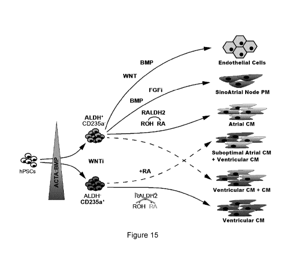
[0031] Figure 15. A schematic depicting various differentiation pathways
for
cardiac cells.
Detailed Description
[0032] Definitions
11
CA 03045182 2019-05-28
WO 2018/098597
PCT/CA2017/051460
[0033] The term "ventricular cardiomyocytes" as used herein refers to a
popu-
lation of cells enriched for ventricular cells, or enriched for cells which
have ventricu-
locyte properties. These include cardiomyocytes expressing ventricular
specific
markers such as MYL2, IRX4, and/or elevated levels of NKX2-5 and/or display
elec-
trophysical properties of a ventricular cell (e.g. action potential).
[0034] The term "atrial cardiomyocytes" as used herein refers to a
population
of cells enriched for atrial cells or enriched for cells which have atrial
cell like proper-
ties. These include cardiomyocytes expressing atrial specific markers such as
the
atrial ion channel gene KCNJ3, NPPA, GJA5 and/or MYL7 and/or display electro-
physical properties of an atrial cell (e.g. action potential).
[0035] The terms "cardiovascular mesoderm cells" and "cardiovascular meso-
derm" as used herein refer to a population of mesoderm cells enriched for
mesoderm
cells having increased potential for differentiation into cardiovascular cells
relative to
other mesoderm cells.
[0036] The terms "ventricular mesoderm cells" and "ventricular
mesoderm" as
used herein refer to a population comprising mesoderm cells enriched for
mesoderm
cells having increased potential for differentiation into ventricular
cardiomyocytes rel-
ative to other mesoderm cells. These include mesoderm cells that are one or
more of
ALDH-, RALDH2- CD235a+, CD235b+, and CYP26A1+.
[0037] The terms "atrial mesoderm cells" and "atrial mesoderm" as used
herein refer to a population comprising mesoderm cells enriched for mesoderm
cells
having increased potential for differentiation into atrial cardiomyocytes
relative to
other mesoderm cells. These include mesoderm cells that are one or more of
ALDH+, RALDH2+, CD235a-, CD235b-, and CYP26A1-.
[0038] The term "cardiomyocyte" as used herein is a cardiac lineage
cell. Car-
diac lineage cells typically express the pan cardiac specific marker cTNT.
[0039] The term "pacemaker cell" as used herein refers to a
cardiomyocyte,
which has pacemaker activity and expresses sinoatrial nodal (SAN) cell
specific
markers. Pacemaker cells generally have faster beating rates than ventricular
cardi-
omyocytes. Pacemaker cells do not express NKX2-5.
12
CA 03045182 2019-05-28
WO 2018/098597
PCT/CA2017/051460
[0040] The term "NKX2-5" as used herein refers to the cardiac homeobox pro-
tein NKX2-5 encoded in humans by the NKX2-5 gene. The gene is involved in car-
diac differentiation and is expressed in cardiomyocyte subtypes such as
ventricular
cardiomyocytes. Expression of NKX2-5 can be measured using for example an anti-
body specific to NKX2-5 or for example by using a NKX2-5 reporter construct.
[0041] The term "BMP component" as used herein means any molecule, op-
tionally any BMP or growth and differentiation factor (GDF), or small
molecule, that
activates the receptor for BMP4, including for example BMP4 and/or BMP2.
[0042] The term "BMP4" (for example Gene ID: 652) as used herein
refers to
Bone Morphogenetic Protein 4, for example human BMP4, as well as active
.. conjugates and/or fragments thereof, that can for example activate BMP4
receptor
signlaing.
[0043] The term "essentially free of pacemaker cells" as used herein
refers to
ted a population of card iomyocytes wherein pacemaker cells comprise less than
5%
of total cells, less than 1% pacemaker cells, less than 0.5% pacemaker cells,
less than
0.1% pacemaker cells, less than 0.01% pacemaker cells, less than 0.001% pace-
maker cells, or less than 0.0001% pacemaker cells, is completely devoid of
pacemaker
cells, or wherein pacemaker cells are not detectable in the population of
cardiomyo-
cytes via currently available methods of detection. While not wishing to be
bound by
any theory it is postulated that the presence of pacemaker cells in a
population of
ventricular cells may induce independent and separate contraction of muscle
when
introduced to a patient.
[0044] The term "activin component" as used herein means one or more
com-
ponents, or a composition comprising said component(s), that activates nodal
signal
transduction, optionally which has Activin A activity such as Activin A and/or
nodal.
[0045] The term "activin" or "ActA" as used herein refers to "Activin A",
(e.g.
Gene ID: 3624), for example human Activin A, as well as active conjugates and
fragments thereof or small molecules, that can activate nodal signal
transduction.
[0046] The term "retinoic acid" or "RA" signifies retinoic acid.
[0047] The term "retinoic acid component" includes compounds that
mediate
the function of vitamin A, and includes for example all-trans RA (e.g. Sigma
R2625),
9-cis RA (e.g. Sigma R4643), and retinal (e.g. Sigma R7632) as well as RA
analogs (e.g.
13
CA 03045182 2019-05-28
WO 2018/098597
PCT/CA2017/051460
RAR agonists), such as AM580, a selective RARalpha agonist (Tocris 0760),
A055649, a selective RAR8 agonist (Tocris 2436), and 0D437, a selective RARy
ag-
onist (Tocris 1549)
[0048] The term "embryoid body medium" as used herein is a culture
medium
that supports formation of aggregates (e.g. floating aggregates of PSCs having
the
potential to differentiate into cells of all three germ layers) or embryoid
bodies of
PSCs, and comprises a minimal media such as StemPro 34 (ThermoFisher),
MesoFateTM (Stemgent), RPM! (ThermoFisher and other companies), HES-media
(DMEM/F12 with KnockOut Serum Replacement, ThermoFisher and other compa-
nies) and for example a BMP component, optionally BMP4, and further optionally
comprising a Rho-associated protein kinase (ROCK) inhibitor.
[0049] The term "embryoid body aggregation phase" as used herein means
the time period non-aggregated hPSCs are cultured for example with an embryoid
body medium described herein and are treated with BMP component and as well as
optionally ROCK inhibitor and/or other components that result in aggregates,
such as
embryoid bodies (e.g., aggregates of PSCs that can be differentiated into
cells of all
three germ layers). The component treatments can be simultaneous, overlapping
or
distinct. For example, a first component can be comprised in the medium and a
sec-
ond component can be added to the medium during the embryoid body aggregation
phase.
[0050] The term "mesoderm induction medium" can include a culture medium
that supports the formation of cardiovascular mesoderm cells and comprises a
mini-
mal media such as StemPro 34 (ThermoFisher), MesoFateTM (Stemgent), RPM!
(ThermoFisher and other companies). Mesoderm induction medium can include ad-
ditional components such as a BMP component, optionally BMP4, an activin compo-
nent, optionally Activin A, and may include other components such as bFGF. De-
pending upon the desired fate of the cardiomyocyte cells produced from the
meso-
derm, different concentrations of each of the BMP component and activin
component
may be adjusted as taught herein.
[0051] The term "mesoderm induction phase" can describe the time
period in
which PSCs are cultured with mesoderm induction medium, including treatment
with
BMP component and an activin component as well as optionally an FGF component
14
CA 03045182 2019-05-28
WO 2018/098597
PCT/CA2017/051460
and/or other components, such that PSCs differentiate into mesoderm cells. The
BMP and activin component treatments can be simultaneous, overlapping or
distinct.
For example, a first component can be included in the medium at the outset of
meso-
derm induction and a second component can be added to the medium during the
mesoderm induction phase.
[0052] The term "cardiac induction medium" can include a culture medium
that
supports induction of cardiac progenitor cells from mesoderm cells, such as
for ex-
ample StemPro-34 minimal media comprising for example a WNT inhibitor, option-
ally IWP2, VEGF and/or an optionally activin/nodal inhibitor, optionally SB-
431542.
Depending on the desired cell type, the cardiac induction medium may also
comprise
a BMP component, retinoic acid, a FGF inhibitor or a FGF component. One embodi-
ment of a cardiac induction medium (also referred to as standard cardiac
induction
media) is StemPro-34 minimal media containing 0.5 pM IWP2, 10 ng/ml VEGF, and
optionally 5.4 pM SB-431542. Other minimal media that can be used include
MesoFateTM (Stemgent) and RPM! (ThermoFisher and other companies).
[0053] The term "cardiac induction phase" can be used to describe the time
period in which mesoderm cells are induced to differentiate into cardiac
progenitor
cells when cultured with cardiac induction medium and are treated for example
with
BMP component and RA as well as optionally a FGF inhibitor or FGF component
and/or other components that result in cardiovascular progenitor cells. The
treat-
ments can be simultaneous, overlapping or distinct. For example, a first
component
can be comprised in the medium and a second component can be added to the me-
dium during the cardiac induction phase.
[0054] The term "basic medium" can include a culture medium that
supports
growth of cardiovascular progenitor cells and cardiomyocytes comprising a
minimal
media such as StemPro 34 (ThermoFisher), MesoFateTM (Stemgent), RPM! (Ther-
moFisher and other companies), and for example VEGF. An example of a basic me-
dium is provided in Example 1.
[0055] The term "basic phase" can be used to refer to the time period
cardio-
vascular progenitor cells are cultured with basic medium and are treated with
VEGF
and/or other components that result in cardiomyocytes. The treatments can be
simul-
taneous, overlapping or distinct.
CA 03045182 2019-05-28
WO 2018/098597
PCT/CA2017/051460
[0056] The term "incubating" can include any in vitro method of maintaining
and/or propagating a population of cells, including monolayer, bead, flask, or
3D cul-
tures, optionally where ambient conditions are controlled as in an incubator
and op-
tionally involving passaging of cells. In steps that involve incubating the
cells with
one or more components, the components can be added simultaneously, at
different
times, for overlapping periods or for distinct periods. A factor can be added
to the
medium after the cells have started incubating in for example an induction
medium
or the factor can be added to the medium before the medium is added to the
cells.
Further, cells may be washed between incubations, for example to reduce the
level
of a component from a previous incubation.
[0057] The term "culturing" can include any in vitro method of maintaining
and
propagating a population of cells at least through one cell division,
including mono-
layer, bead, flask, or 3D cultures, optionally where ambient conditions are
controlled
as in an incubator.
[0058] The term "enriched for" as used herein means comprising at
least 50%,
at least 60%, or at least 70% up to 100% of the cell type which is enriched.
In one
embodiment, enrichment is measured in a day 20 culture using a method as de-
scribed herein.
[0059] The term "subject" as used herein includes all members of the
animal
kingdom including mammals, and suitably refers to humans.
[0060] The terms "treat", "treating", "treatment", etc., as applied to a
cell, in-
clude subjecting the cell to any kind of process or condition or performing
any kind of
manipulation or procedure on the cell.
[0061] The term "treatment" as used herein as applied to a subject,
refers to
an approach aimed at obtaining beneficial or desired results, including
clinical results
and includes medical procedures and applications including pharmaceutical or
other
product interventions. In one embodiment treatment refers to administration of
a
product for the purposes of engraftment. Beneficial or desired clinical
results can in-
clude, but are not limited to, alleviation or amelioration of one or more
symptoms or
conditions, diminishment of extent of disease, stabilized (i.e. not worsening)
state of
disease, preventing spread of disease, delay or slowing of disease
progression,
16
CA 03045182 2019-05-28
WO 2018/098597
PCT/CA2017/051460
amelioration or palliation of the disease state, and remission (whether
partial or to-
tal), whether detectable or undetectable. "Treatment" can also mean prolonging
sur-
vival as compared to expected survival if not receiving treatment.
[0062] As used herein, the term "heart failure" refers to a condition
in which a
subject's heart is unable to pump sufficiently to maintain suitable blood flow
in the
subject's body. A subject "at risk of heart failure" refers to a subject
having one or
more characteristics known to precede heart failure. For example, a subject at
risk of
heart failure may have or have had coronary artery disease, previous
myocardial in-
farction (heart attack), high blood pressure, atrial fibrillation, valvular
heart dis-
ease, excess alcohol use, tobacco use, obesity, sleep apnea, infection (viral
and/or
bacterial), cardiomyopathy, myocarditis, congenital heart defects,
arrhythmias,
and/or other diseases such as, but not limited to, diabetes, hyperthyroidism,
hypothy-
roidism, hemochromatosis and/or amyloidosis.
[0063] As used herein, the terms "myocardial infarction" and "Ml",
refers to an
event in which blood flow decreases or stops to a part of the heart, thereby
causing
death to cardiomyocytes, due to lack of oxygen supply (ischemia), resulting in
dam-
age to the heart muscle.
[0064] As used herein, the terms "administering", "introducing" and
"trans-
planting" and are used interchangeably in the context of delivering cells into
a sub-
ject, by a method or route which results in at least partial localization of
the intro-
duced cells at a desired site.
[0065] The term "pluripotent stem cell" or "PSC" as used herein refers
to a cell
with the capacity, under different conditions, to differentiate into any one
of the cell
types characteristic of the three germ cell layers, and includes embryonic
stem cells
and induced pluripotent stem cells. Pluripotent cells are characterized by
their ability
to differentiate to more than one cell type using, for example, a nude mouse
tera-
toma formation assay. Pluripotency is also evidenced by the expression of
embry-
onic stem (ES) cell markers. As used herein, pluripotent stems can include
induced
pluripotent stem cells (iPSC) and embryonic stem cells (ESC).
[0066] In an embodiment, the term "embryonic stem cells" excludes stem
cells
involving destruction of an embryo such as a human embryo.
17
CA 03045182 2019-05-28
WO 2018/098597
PCT/CA2017/051460
[0067] As used herein, the terms "iPSC" and "induced pluripotent stem cell"
are used interchangeably and refer to a pluripotent stem cell artificially
derived (e.g.,
induced or by complete reversal) from a non-pluripotent cell, typically an
adult so-
matic cell, for example, by inducing expression of one or more genes
(including, for
example, POU4F1/00T4 (Gene ID; 5460) in combination with, but not restricted
to,
SOX2 (Gene ID; 6657), KLF4 (Gene ID; 9314), cMYC (Gene ID; 4609), NANOG
(Gene ID; 79923), LIN28/ LIN28A (Gene ID; 79727)).
[0068] Cardiomyocytes prepared, enriched, or isolated by a method of
the in-
vention are derived from pluripotent stem cells. For example, a patient's
cells may
be genetically modified prior to use through introduction of genes that may
control
their state of differentiation prior to, during or after their exposure to
differentiation
factors described herein. Pluripotent stem cells suitable for use in methods
described
herein, which are derived from a patient's own tissue enhances compatibility
of differ-
entiated tissue grafts derived from the stem cells with the patient.
[0069] The term "embryonic stem cell" is used to refer to the
pluripotent stem
cells of the inner cell mass of the embryonic blastocyst (see, for example,
U.S. Pat.
Nos. 5,843,780, 6,200,806). Such cells can also be obtained from the inner
cell
mass of blastocysts derived from somatic cell nuclear transfer (see, for
example,
U.S. Pat. Nos. 5,945,577, 5,994,619, 6,235,970). The distinguishing
characteristics
of an embryonic stem cell define an embryonic stem cell phenotype.
Accordingly, a
cell has the phenotype of an embryonic stem cell if it possesses one or more
of the
unique characteristics of an embryonic stem cell such that that cell can be
distin-
guished from other cells. Exemplary distinguishing embryonic stem cell
characteris-
tics include, without limitation, gene expression profile, proliferative
capacity, differ-
entiation capacity, responsiveness to particular culture conditions, and the
like.
[0070] Pluripotent stem cells, as used herein, may also be genetically modi-
fied through introduction of vectors expressing a selectable marker under the
control
of a stem cell specific promoter, such as Oct-4, or of genes that may be
upregulated
to induce cardiomyocyte differentiation. The stem cells may be genetically
modified
at any stage with markers or genes so that the markers or genes are carried
through
to any stage of culturing. The markers may be used to purify or enrich the
differenti-
ated or undifferentiated stem cell populations at any stage of culture.
18
CA 03045182 2019-05-28
WO 2018/098597
PCT/CA2017/051460
[0071] The term "pharmaceutically acceptable carrier" as used herein
includes
essentially chemically inert and nontoxic compositions that do not interfere
with the
effectiveness of the biological activity of the pharmaceutical composition.
Examples
of suitable pharmaceutical carriers include, but are not limited to, water,
saline solu-
tions, glycerol solutions, ethanol, N-(1(2,3-dioleyloxy) propyl) N,N,N-
trimethylammo-
nium chloride (DOTMA), diolesylphosphotidyl-ethanolamine (DOPE), and
liposomes.
Such compositions should contain a therapeutically effective amount of the com-
pound(s), together with a suitable amount of carrier so as to provide the form
for di-
rect administration to the subject.
[0072] In understanding the scope of the present disclosure, the term
"con-
centration" as used herein means a final concentration of a substance such as
for
example BMP4, Activin A, retinoic acid in a medium. Unless indicated
otherwise, the
concentration is based on a weight/volume ratio.
[0073] Terms of degree such as "substantially", "about" and
"approximately"
as used herein mean a reasonable amount of deviation of the modified term such
that the end result is not significantly changed. These terms of degree should
be
construed as including a deviation of at least 5% of the modified term if
this devia-
tion would not negate the meaning of the word it modifies.
[0074] The recitation of numerical ranges by endpoints herein includes
all
numbers and fractions subsumed within that range (e.g. 1 to 5 includes 1, 1.5,
2,
2.75, 3, 3.90, 4, and 5). It is also to be understood that all numbers and
fractions
thereof are presumed to be modified by the term "about." Further, it is to be
under-
stood that "a," "an," and "the" include plural referents unless the content
clearly dic-
tates otherwise. The term "about" means plus or minus 0.1 to 50%, 5-50%, or 10-
40%, preferably 10-20%, more preferably 10% or 15%, of the number to which
refer-
ence is being made.
[0075] Further, the definitions and embodiments described in
particular sec-
tions are intended to be applicable to other embodiments herein described for
which
they are suitable as would be understood by a person skilled in the art. For
example,
in the following passages, different aspects of the invention are defined in
more de-
tail. Each aspect so defined may be combined with any other aspect or aspects
un-
less clearly indicated to the contrary. In particular, any feature indicated
as being
19
CA 03045182 2019-05-28
WO 2018/098597
PCT/CA2017/051460
preferred or advantageous may be combined with any other feature or features
indi-
cated as being preferred or advantageous.
[0076] Aspects and Embodiments
[0077] In an aspect, there is provided a method of producing a
population of
cardiomyocytes enriched for atrial cardiomyocytes, the steps comprising: i.
incubat-
ing pluripotent stem cells in a medium suitable to generate aggregates and/or
embry-
oid bodies, ii. further incubating the stem cells in a medium suitable for
mesoderm in-
duction, wherein said medium at least includes a BMP component, optionally
BMP4,
and an activin component, optionally Activin A, wherein the BMP component to
the
activin component is provided in a ratio of 3:2; iii. further adding a
retinoic acid com-
ponent to the cells, said addition of retinoic acid added during the mesoderm
induc-
tion or cardiovascular specification stage; iv. Continue growth of said cells
in suitable
medium(s) to generate a population of cardiomyocytes, wherein said population
of
cardiomyocytes is enriched for atrial cardiomyocytes. In some embodiments the
ra-
tio of BMP to activin is 1.5:1.0 (or 3:2).
[0078] In some embodiments, said BMP component is BMP4, the activin com-
ponent is Activin A, the concentration of BMP4 is 3ng/mland the concentration
of
Activin A is 2ng/ml. In some embodiments, said retinoic acid component is
trans
retinoic acid and is added in a concentration of between 50nm and 5pM. In some
embodiments, said retinoic acid component is added at a concentration of
500nM.
[0079] In some embodiments, the BMP component and the Activin component
are added at day 1 of the process. In some embodiments, the retinoic acid
compo-
nent is added at day 3 of the process. In some embodiments, additional BMP com-
ponent is not added to the medium at day 3 of the process.
[0080] In some embodiments, an FGF inhibitor is excluded from the
medium
at day 3 of the process. In some embodiments, the cells produced by the
process
are utilized in an in vitro assay to screen for cardiac texicity that may be
caused by
potential therapeutic compounds.
[0081] In an aspect, there is provided an isolated population of
cardiomyo-
cytes enriched for atrial cardiomyocytes comprising at least or about 50% of
atrial
cardiomyocytes, at least or about 60% of atrial cardiomyocytes, at least or
about
70% of atrial cardiomyocytes, at least or about 80% of atrial cardiomyocytes,
or at
CA 03045182 2019-05-28
WO 2018/098597
PCT/CA2017/051460
least or about 90% of atrial cardiomyocytes, preferably obtained according to
the
method described herein. In an aspect, there is provided a method of producing
a
population of cardiomyocytes enriched for ventricular cardiomyocytes, the
steps
comprising: i. incubating pluripotent stem cells in a medium suitable to
generate ag-
gregates (embryoid bodies), ii. incubating the aggregated stem cells in a
medium
suitable for mesoderm induction, wherein said medium at least includes a BMP
com-
ponent, optionally BMP4, and an activin component, optionally Activin A,
wherein the
concentration of the activin component is greater than the concentration of
the BMP
component; iii. continue growth of said cells in suitable medium(s) to
generate a pop-
ulation of cardiomyocytes, wherein said population of cardiomyocytes is
enriched for
ventricular cardiomyocytes. In some embodiments that ratio of BMP to activin
is
about 0.3:1.0, about 0.5:1.0 (or 1:2) or about 0.8:1Ø
[0082] In some embodiments, the concentration of the BMP component
and/or the Activin component are determined by measuring for the level of
CD235a
and comparing this to the level of RALDH2.
[0083] In some embodiments, the concentration of the Activin component is
chosen on the basis of the concentration which preferentially results in more
CD235a
expressing mesoderm cells as compared with RALDH2 expressing mesoderm cells,
and the BMP component is added to achieve a lower concentration than the
concen-
tration of the Activin component. In some embodiments, the BMP component is
added to achieve optimal cardiogenesis from the induced mesoderm.
[0084] In some embodiments, said BMP component is BMP4, the activin
com-
ponent is Activin A, the concentration of BMP4 is between 3-20ng/ml, the
concentra-
tion of the Activin A is between 4 - 20ng/ml, and the concentration of the
Activin A is
greater than the concentration of the BMP4. In some embodiments, the concentra-
tion of BMP4 is 1Ong/m1 and the concentration of Activin A is 12ng/ml.
[0085] In an aspect, there is provided an isolated population of
cardiomyo-
cytes: enriched for ventricular cardiomyocytes comprising at least or about
30% of
ventricular cardiomyocytes, at least or about 40% of ventricular
cardiomyocytes, at
least or about 50% of ventricular cardiomyocytes, at least or about 60% of
ventricular
cardiomyocytes, at least or about 70% of ventricular cardiomyocytes, at least
or
21
CA 03045182 2019-05-28
WO 2018/098597
PCT/CA2017/051460
about 80% of ventricular cardiomyocytes, or at least or about 90% of
ventricular car-
diomyocytes, preferably obtained according to the method described herein. In
an
embodiment, the isolated population of cardiomyocytes enriched for ventricular
car-
diomyocytes is essentially free of pacemaker cells. In a preferred embodiment,
the
isolated population of cardiomyocytes enriched for ventricular cardiomyocytes
is de-
void of pacemaker cells.
[0086] An isolated population of cardiomyocytes according to the
invention
may be used in a method for screening for potential cardiac toxicity of
potential ther-
apeutic active agents for use in treating cardiovascular and any other
disorders. For
example, they provide a source of cells that can be used in drug screens for
cardio-
vascular applications; they provide a source of cells that can be used for
therapeutic
purposes¨to restore cardiac function; to repair the ischemic heart and/or to
regener-
ate the coronary vasculature, they can be used for tissue engineering purposes
where components of the heart or the coronary vasculature are required; and
they
may serve as a research tool for the study of cardiovascular development and
dis-
ease. . An isolated population of cardiomyocytes used for the screening of
active
agents, according to methods of the invention may, for example, include
cardiomyo-
cyte populations enriched for ventricular cardiomyocytes. Such ventricular
cardiomy-
ocyte populations include, optionally, populations which are essentially free
of pace-
maker cells, or devoid of pacemaker cells. An isolated population of
cardiomyocytes
used to scree active agents, according to methods of the invention, may also
include
a population enriched for atrial cardiomyocytes. Such methods for screening or
eval-
uating the potential cardiac toxicity of a test compound or agent, involve
exposing a
population of cardiomyocytes according to the present invention to a compound
to
be tested for cardiotoxicity. Effects to evaluated include changes in the
viability, con-
tractility, membrane electric potentials and/or other functionalities of the
cells.
[0087]
[0088] Cardiomyocyte and cardiomyocyte progenitor cell populations pro-
duced using methods of the invention that may be used for transplantation,
cell ther-
apy or gene therapy. For example, the invention provides differentiated cells
pro-
duced using methods of the invention that may be used for therapeutic
purposes,
22
CA 03045182 2019-05-28
WO 2018/098597
PCT/CA2017/051460
such as in methods of treating a subject in need of cardiac repair. For
example,
therapeutic repair may involve restoring, in full or in part, cardiac function
in a subject
in need of cardiac repair, such as a subject suffering from a heart disease or
condi-
tion.
[0089] Another aspect of the invention is a method of treating or
preventing a
cardiac disease or condition. Cardiac disease is typically associated with
decreased
cardiac function and includes conditions such as, but not limited to,
myocardial in-
farction, cardiac hypertrophy and cardiac arrhythmia. In this aspect of the
invention,
the method includes introducing into a subject in need of cardiac repair,
isolated dif-
ferentiated ventricular cardiomyocyte cells of the invention and/or cells
capable of
differentiating into ventricular cardiomyocyte cells. The isolated
cardiomyocyte cells
may be transplanted into damaged cardiac tissue of a subject. Ideally, the
method
results in the restoration of some or all cardiac function in a patient.
[0090] In an aspect, there is provided a method of treating a subject
with heart
failure, comprising administering to the subject the population of ventricular
cardio-
myocytes described herein. In some embodiments, said subject is suffering from
a
myocardial infarction. In some embodiments, the myocardial infarction is in
the ven-
tricle of the patient and the population is as described herein.
[0091] In an aspect, there is provided the population of ventricular
cardiomyo-
cytes described herein, for use in the treatment of a subject with heart
failure or at
risk of heart failure. In an aspect, there is provided use of the population
of ventricu-
lar cardiomyocytes described herein, in the preparation of a medicament for
the
treatment of a subject with heart failure or at risk for heart failure.
[0092] In yet another aspect of the invention there is provided a
method of re-
pairing cardiac tissue, the method including introducing an isolated
ventricular cardi-
omyocyte or cardiac progenitor cell of the invention and/or a cell capable of
differen-
tiating into a ventricular cardiomyocyte cell when treated using a method of
the in-
vention into damaged cardiac tissue of a patient.
[0093] The patient may be suffering from a cardiac disease or
condition. In the
method of repairing cardiac tissue of the present invention, the isolated
cardiomyo-
cyte cell may be transplanted into damaged cardiac tissue of a patient.
Ideally, the
method results in the restoration of at least some cardiac function in a
patient.
23
CA 03045182 2019-05-28
WO 2018/098597
PCT/CA2017/051460
[0094] In one embodiment, ventricular cardiomyocytes disclosed herein are
administered to a subject during the acute phase after myocardial infarction
or during
the chronic stage of heart failure. Cells are administered to the site of
damage in the
ventricle either by direct injection or catheter-based delivery. Cells may be
formu-
lated together with pharmaceutically acceptable carriers, hydrogels or
scaffolds, for
example, to aid in placement, survival and/or engraftment of the cells in the
tis-
sue. Cell dosage ranges may include, for example, from about 0.5 billion to 2
billion
cells per dose. The cells may be administered to the subject in single or
multiple
doses, at one or more point in time in order to treat the subject.
[0095] The present invention preferably provides a myocardial model for
test-
ing the ability of stems cells that have differentiated into cardiomyocytes or
cardiac
progenitors using methods of the invention to restore cardiac function. In
order to
test the effectiveness of cardiomyocyte transplantation in vivo, it is
important to have
a reproducible animal model with a measurable parameter of cardiac function.
The
parameters used should clearly distinguish control and experimental animals
[see for
example in Pa!men et al. (2001), Cardiovasc. Res. 50, 516-524] so that the
effects of
transplantation can be adequately determined. PV relationships are a measure
of the
pumping capacity of the heart and may be used as a read-out of altered cardiac
function following transplantation.
[0096] In an aspect, there is provided a process for detecting atrial
mesoderm
in a population of cells, comprising detecting RALDH2, wherein a presence of
RALDH2 is indicative of atrial mesoderm. In an aspect, there is provided a
process
for detecting ventricular mesoderm in a population of cells, comprising
detecting
CD235a and/or CYP26A1, wherein a presence of CD235a and/or CYP26A1 is indic-
ative of ventricular mesoderm.
[0097] Methods of the invention for identifying atrial or ventricular
mesoderm
on the basis of ALDH, preferably RALDH2, and/orCD235a and/or CD235b, and/or
CYP26A1 expression, respectively are provided. More particularly, they can be
used
for identification of secreted factors produced by the mesodermal cell which
influ-
ence cardiomyocyte proliferation, survival, function and differentiation of
atrial or ven-
tricular cell populations. For example, methods of the invention for
identifying atrial or
24
CA 03045182 2019-05-28
WO 2018/098597
PCT/CA2017/051460
ventricular cardiomyocyte populations provide systems to both understand
atrial and
ventricular mesoderm differentiation at the molecular level and to identify
new drug
targets(e.g., signaling pathways) that modulate differentiation.
[0098] According to one or more of the embodiments disclosed herein
Retin-
oic acid (RA) specifies atrial cardiomyocytes within a specific developmental
time
window and the application of RA to mesoderm from day 3-5 specifies atrial
cardio-
myocytes. RA concentration range: 50nM ¨ 5uM. RA sources: all-trans RA,
retinoic
receptor (RAR) agonists (AM580 for - alpha, A055649 for -8, 0D437 for-y)
Agonist
concentrations: 3-300nM for AM580, 0.025-2.5uM A055649; 0.05-5uM 0D437.
[0099] RALDH2 (Retinaldehydrogenase, or Aldefluor) is a marker for
atrial
mesoderm. The proportion of RALDH2 + cells is monitored by using the aldefluor
as-
say for optimizing atrial differentiation. Days of analysis: day 2-6.
[00100] The early mesoderm inductions using Activin A and BMP4 at day 1
de-
termine the proportion of RALDH2 + mesodermal cells at day 4. Induction
conditions
are low BMP (1-5 ng/ml BMP) and low Activin A (0.1-4 ng/ml), most commonly
used
3ng/mIBMP/2ng/m1Activin A (3I3/2A).
[00101] The functionality of RALDH2 is shown by the treatment with
retinol
(ROH) at day3-5, which is sufficient to induce an atrial phenotype. (Retinol
is con-
verted by RALDH2 into RA, RA than specifies the atrial phenotype). Glycophorin
A
(CD235a) is a marker for ventricular mesoderm. CD235a is expressed exclusively
on the ventricular mesoderm and absent on the RALDH2 + atrial mesoderm. The
CD235a + cells do not express RALDH2. The CD235a + cells express CYP26A1, an
enzyme that degrades RA, to antagonize RA signaling and assure the
establishment
of a ventricular phenotype. Days of analysis: day 2-6.
[00102] The early mesoderm inductions using Activin A and BMP4 at day1
de-
termine the proportion of CD235a + mesodermal cells at day 4. Induction
conditions
are high BMP (5-20 ng/ml BMP), and high Activin (6-20 ng/ml), most commonly
used
lOng/m1 BMP/12ng/m1 Activin A (106/12A). Treatment of the CD235a + cells with
ret-
inol (ROH) at day3-5 is NOT sufficient to induce an atrial phenotype. (These
cells are
not able to convert retinol into RA, therefore the cells develop into a
ventricular phe-
notype). The CD235a + cells are giving rise to populations highly enriched in
MLC2V+
ventricular cardiomyocytes.
CA 03045182 2019-05-28
WO 2018/098597
PCT/CA2017/051460
[00103] Ventricular and atrial cardiomyocytes are derived from two distinct
mesodermal subpopulations. The ventricular differentiation is monitored by the
emergence of day4 CD235a+ cells and day20 MLC2V+/ CTNT+ cells. The atrial dif-
ferentiation is monitored by the emergence of day4 AF+ cells and day20 MLC2v-
/CTNT+ cells. The day20 population derived from the ventricular mesoderm
(106/12A) contains a higher proportion of MLC2v+ ventricular cardiomyocytes
than
those derived from the atrial mesoderm (3I3/2A).
[00104] Gene expression analysis and single cell patch clamp analysis
showed
that the day20 population generated by RA treatment from the atrial mesoderm
(3I3/2A+RA) contains a higher proportion of atrial cardiomyocytes than the
day20
population generated by RA treatment from the ventricular mesoderm
(106/12A+RA)
[00105] The proper mesoderm subpopulations need to be specified to
enrich
for the desired cardiomyocyte subtypes. Improved protocol for the
specification of
ventricular cardiomyocytes for cell replacement therapy after myocardial
infarction.
The CD235a+ ventricular mesoderm (106/12A) is giving rise to populations
highly
enriched for MLC2v+ ventricular cardiomyocytes devoid of pacemaker cells. This
re-
sults in lower spontaneous beating rates compared to other heterogeneous
cardio-
myocyte populations.
[00106] These are desirable characteristics for cell replacement
therapies after
myocardial infarction because mixed cell populations that contain
contaminating
pacemaker cells and have fast spontaneous beating rates can cause life
threatening
arrhythmias. We propose that our new protocol for specification of ventricular
cardi-
omyocytes is superior to previous protocols that generated mixed populations
of ven-
tricular and pacemaker cells.
EXAMPLES
[00107] Methodologies and Results
[00108] Human pluripotent stem cell lines can be cultured as previously
de-
scribed (e.g. Kennedy et al., 2007). For differentiation into the cardiac
lineage, an es-
tablished protocol such as that described in Kattman et al. , 2011) can be
used. Var-
ious modifications to the procedures are possible including those as described
W02016131137. In one embodiment 80% confluent hPSCs cultures can be dissoci-
ated into single cells, suspended in StemPro-34 Media containing 1 ng/ml BMP4
and
26
CA 03045182 2019-05-28
WO 2018/098597
PCT/CA2017/051460
10 pM ROCK inhibitor and incubated for 18 hours on an orbital shaker to
generate
embryoid bodies (EBs). The next day (day 1 of differentiation) the EBs can be
trans-
ferred to mesoderm induction media: Stem Pro-34 containing a set concentration
of
BMP4, and a set concentration of Activin A as further described herein, as
well 5
ng/ml bFGF. At day 3 of differentiation the EBs can be washed once using IMDM
and suspended in cardiac induction media: in one embodiment cardiac induction
me-
dia can include StemPro-34 containing 0.5 pM IWP2, 10 ng/ml VEGF, and
optionally
5.4 pM SB-431542 (SB, Activin/NodaITTGF8 inhibitor). Cardiac induction media
can
also optionally include retinoic acid (RA), or an RA component as further
described
herein.
[00109] Retinoic acid signaling specifies atrial-like cardiomyocytes from
hESCs
[00110] To determine if retinoic acid signaling can specify an atrial
fate in
hPSC-derived cardiogenic populations generated with our embryoid-body (EB)-
based protocol, all trans retinoic acid (RA) was added to the differentiation
cultures
at 4 different time points that represent the following developmental stages:
meso-
derm induction (day 3), cardiovascular specification (day 5), cardiac
progenitor de-
velopment (day 7) and emergence of contracting cardiomyocytes (day 9) (Kattman
et
al., 2011) (Figure 1A). The HES3 NKX2-5: GFP reporter hESC line was used for
these experiments to allow us to monitor and quantify cardiovascular
development
and to isolate GFP+ cardiomyocytes. At day 20 of culture, GFP+SIRPA+CD90- car-
diomyocytes were isolated from the differentiated populations and analyzed by
RT-
qPCR for expression of genes indicative of atrial and ventricular development.
(Fig-
ures 1B-D and 8B-E).
[00111] None of the RA treatments significantly altered the levels of
expression
of the pan-cardiomyocyte marker CTNT, indicating comparable card iomyocyte con-
tent in the different populations (Figure 1B). Addition of RA at days 3 and 5
resulted
in a significant reduction in expression of the ventricular-specific gene MYL2
and an
upregulation of the atrial ion channel gene KCNJ3 (Figure 1C) suggesting a
change
in cardiomyocyte fate in the day 20 populations. Interestingly, addition of RA
at later
stages (days 7 and 12) had no effect on expression of these genes. Analyses of
ad-
ditional chamber-specific markers showed that cardiomyocytes generated from
day 3
RA-treated mesoderm also expressed lower levels of the ventricular markers
IRX4
and MYH7 than the non-treated group, whereas the reverse pattern was observed
27
CA 03045182 2019-05-28
WO 2018/098597
PCT/CA2017/051460
for the atrial markers NR2F2, TBX5, NPPA, and MYL7 and atrial-specific ion
chan-
nels CACNA1D, KCNA5, and GJA5 (Figures 1D and 80-E). Analyses of control fetal
tissues verified the atrial and ventricle expression patterns of these
different genes.
Flow cytometric and immunostaining analyses of cardiomyocyte populations gener-
ated from day 3 RA-treated mesoderm confirmed the qRT-PCR expression patterns,
and they showed a dramatic reduction in the proportion of MLC2V+ cells and a
much
higher frequency of COUPTFII+ cells in the population generated from day 3 RA-
treated mesoderm comparted to the one generated from the non-treated control
mesoderm (Figures 1E-H).
[00112] Taken together, these findings strongly suggest that RA
signaling in-
duces a fate change in hPSC cardiogenesis, promoting the development of atrial
cardiomyocytes at the expense of the ventricular lineage. Additionally, they
show
that this effect of RA is restricted to an early developmental window, between
days 3
and 5 of differentiation, corresponding to the mesoderm state of
differentiation.
[00113] To further characterize the RA response, we next analyzed
populations
between days 2 and 6 of differentiation for expression of the 3 RA receptor
(RAR)
isoforms, RAR-alpha, -13, -y (RARA, RARB, and RARG in Fig. 8F, respectively).
All
three were expressed during the responsive stage, suggesting that the RA
response
may be mediated through all of them (Figure 8F). To test this, we added the
recep-
tor-specific agonists AM580 for alpha, A055649 for 13 or 0D437 for y in place
of RA
to the day 3 cultures. Addition of each of the agonists led to a reduction of
MYL2
expression in day 20 CTNT+ populations, suggesting that signaling through all
recep-
tor isoforms can mediate the change in fate (Figures 8G and 8H).
[00114] In some embodiments RA can be added in a concentration of about
0.05 pM to a concentration of about 5 pM. In one embodiment the concentration
of
RA is 500nM (0.5pM). In one embodiment the concentration of RA added is be-
tween 0.05pM and 0.01 pM. In one embodiment the concentration of RA added is
between 0.01 pM and 0.1 pM. In some embodiments an RA component is added.
In some embodiments the RA component is a retinoic acid receptor (RAR)
agonist.
In some embodiments the RAR agonist is an agonist against the alpha receptor.
In
some embodiments the RAR agonist is AM580. In some embodiments the AM580
RAR agonist is added in a concentration of about 3nM to about 300nM. In some
28
CA 03045182 2019-05-28
WO 2018/098597
PCT/CA2017/051460
embodiments the RAR agonist is an agonist against the beta receptor. In some
em-
bodiments the RAR agonist is A055649. In some embodiments the A055649 is
added in a concentration of about 0.025pM to 2.5 pM. In some embodiments the
RAR agonist is an agonist against the gamma receptor. In some embodiments the
RAR agonist is 0D437. In some embodiments the 0D437 RAR agonist is added in a
concentration of about 0.05pM to about 5pM.
[00115] RALDH2 and CYP26A1 expression identifies mesoderm subpopula-
tions
[00116] If specification of atrial fate is mediated via autocrine RA
signaling, the
mesoderm population that gives rise to these cardiomyocytes should display
RALDH
activity. To test this we analyzed PDGFRalpha mesoderm induced with our
standard
conditions (10 ng/mL BMP4 and 6 ng/mL Activin A (106/6A) on different days,
using
the adefluor assay that detects the activity of all aldehyde dehydrogenases,
(ALDHs), including the three retinaldehyde dehydrogenases, RALDH1, -2, and -3
(Jones et. al.., 1995). These analyses revealed the presence of a small ALDH+
PDGFRalpha+ population at days 4 and 5 of differentiation (Figure 2A),
suggesting
that a subpopulation of mesoderm at these stages may have the capacity to
synthe-
size RA. In an attempt to increase the size of the ALDH+ PDGFRalpha+
population,
we tested the effect of varying the concentrations of Activin A and BMP4
during the
mesoderm induction step. Reducing the amount of Activin A in the presence of a
constant concentration of BMP4 (10 ng/mL) led to a substantial increase in the
size
of the ALDH+ PDGFRalpha+ population at day 4 of differentiation (Figure 2B).
How-
ever, this increase was associated with a decrease in the proportion of CTNT+
cardi-
omyocytes generated, suggesting that these changes promoted a non-cardiogenic
fate. As we have previously shown that the ratio of Activin A and BMP4
signaling is
important for maintaining optimal cardiogenic potential (Kattman et al.,
2011), we next
varied the concentration of BMP4 in the presence of the amount of Activin A (2
ng/mL) that induced the largest ALDH+ PDGFRalpha+ population. Reducing the
BMP4 concentration from 10 to 3 ng/mL (3I3/2A) did not influence the size of
the
ALDH+ PDGFRalpha+ population, but it did increase the frequency of CTNT+ cells
generated at day 20 (Figure 20). Comparable cell numbers were obtained from
the
36/2A and 106/6A cultures, indicating that the manipulations did not
significantly im-
pact total cardiomyocyte output (Figure 9A).
29
CA 03045182 2019-05-28
WO 2018/098597
PCT/CA2017/051460
[00117] Analyses of cultures induced with 36/2A revealed the emergence of a
large PDGFRalpha+ mesoderm population at day 3 of differentiation, followed by
the
development of an ALDH+ PDGFRalpha+ population at day 4 (Figure 2D). The size
of
the ALDH+ PDGFRalpha+ population increased until day 5 and then started to de-
crease at day 6. Molecular analyses showed that expression of RALDH2 (ALDH1A2)
increased sharply between days 2 and 3 of differentiation, and then it
declined over
the next 7 days in the group induced with 36/2A (Figure 2E). The 10B/6A-
induced
cells ex- pressed significantly lower levels of ALDH1A2 at days 3 and 4,
consistent
with the smaller proportion of ALDH+ cells in this group. The expression
levels of
other RALDH isoforms (ALDH1A1 and ALDH1A3) were markedly lower than those of
ALDH1A2, and they did not differ between the two populations (Figure 9B). T
(BRACHYURY) and MESP1 showed similar temporal expression patterns in both the
106/6A- and 313/2A- induced populations, indicating that the kinetics of
mesoderm in-
duction were not dramatically different between the two groups (Figure 90). In
the
developing embryo, the boundaries of RA activity and the duration of signaling
are estab-
lished by a balance between localized agonist synthesis and degradation
(Cunningham and
Duester, 2015; Rydeen and Waxman, 2014). To determine if this balance is at
play in the
hPSC differentiation cultures, we next analyzed the two populations for
expression of
CYP26A1, a member of cytochrome P450 family enzyme responsible for RA
degradation.
These analyses revealed a striking difference between the two groups, with the
day 3
10B/6A-induced cells showing dramatically higher expression levels than any
other 106/6A-
or 3B/2A-induced population (Fig. 2E). Collectively, these findings support
the interpretation
that combinations of 36/2A and 106/6A induce different mesoderm populations
distin-
guished by expression of ALDH1A2 and CYP26A1.
[00118] Retinol specifies ALDH+ mesoderm to an atrial fate.
[00119] To determine if the ALDH+ cells can synthesize RA, the ALDH+ PDG-
FRalpha+ and ALDH- PDGFRalpha+ fractions were isolated from the day 4 36/2A-
induced population, and the cells were cultured as aggregates in retinol
(ROH), RA,
or DMSO (control) for 24 hr (Figures 3A and 3B). ALDH1A2 expression segregated
to
the ALDH+ fraction, confirming the validity of aldefluor-based sorting
strategy for iso-
lating RALDH2-expressing cells (Figure 30). Following an additional 15 days of
cul-
ture, all groups contained a high proportion of CTNT+ cells, demonstrating
efficient
cardio- myocyte differentiation (Figure 3D). The untreated controls generated
cardio-
CA 03045182 2019-05-28
WO 2018/098597
PCT/CA2017/051460
myocyte populations that contained ML02V+ cells and expressed IRX4, demonstrat-
ing that, in the absence of RA signaling, the 3B/2A-induced mesoderm has some
ventricular cardiogenic potential (Figures 3E and 3F). Following treatment
with ROH,
the ALDH+ mesoderm generated an atrial-like cardiomyocyte population that had
a
lower frequency of MLC2V+ cells, lower levels of IRX4 expression, and elevated
1ev-
els of KCNJ3 expression compared to the untreated control (Figures 3E-3G). The
ex-
pression patterns in the ROH- and RA- treated ALDH+ PDGFRalpha+-derived popu-
lations were similar, strongly suggesting that the ALDH+ cells were able to
synthe-
size RA from ROH.
[00120] Surprisingly, we observed a similar response to ROH in the ALDH-
cells (Fig. 3E-3F) despite their lack of ALDH1A2 expression at the time of
isolation
(Fig. 3B and 30). This response was likely due to the fact that the majority
of the
ALDH- --derived population became ALDH+ during the 24-hour aggregation culture
(Figure 10A), enabling the cells to respond to ROH. Interestingly, we observed
a de-
crease in aldefluor staining in the ALDH+ -derived population over the same 24-
hour
period, highlighting the dynamic natures of the ALDH activity (RALDH2
expression)
within the mesoderm population.
[00121] Together, these findings demonstrate that 36/2A induces ALDH+
PDG-
FRalpha+ (RALDH2+) mesoderm that can respond to ROH and generate atrial-like
cardiomyocytes, supporting the notion that specification of this fate is
mediated via
autocrine RA signaling.
[00122] 0D235a expression marks mesoderm that gives rise to ventricular
car-
diomyocytes. It is contemplated herein that 0D235b may replace 0D235a as a
marker of mesoderm that gives rise to ventricular mesoderm, at least due to
the
amino acid sequence similarity and/or identity of the N-terminal region of
Glyco-
phorin B and Glycophorin A.
[00123] To be able to monitor the development of AYP26A1-expressing
meso-
derm (VM) that gives rise to ventricular cardiomyocytes, we initiated a search
for sur-
face markers on this population that would allow us to distinguish it from the
ALDH+
mesoderm. Through a previous screen on an anti-CD antibody array
(http://www.ocigc.ca/antibody/), we found that glycophorin A (CD235a) was ex-
pressed on a subset of day 5 cardiogenic PDGFRalpha+ cells induced with106/6A
31
CA 03045182 2019-05-28
WO 2018/098597
PCT/CA2017/051460
(data not shown). Analyses of 106/6A- and 3B/2A-induced populations revealed
that
CD235a+ population increased within the next 24 hr (> 60%) and then declined
over
the following 48hr. The small proportion of ALDH+ cells detected at day 5 were
CD235a-, indicating that the ALDH+ and CD235a+ populations are mutually exclu-
sive. Only a few CD235a+ cells were detected at day 4 in the 36/2A- induced
popula-
tions. The qRT-PCR analyses revealed an upregulation of GYPA (glycophorin A)
ex-
pression onday 3 of differentiation in the group induced with 106/6A. Figure
11A).
The expression levels declined sharply over the next 24 hours and remained low
for
the duration of the analyses. Only low levels of expression were detected in
the
3B/2A-induced populations. Based on these findings, we hypothesize that glyco-
phorin A is expressed on mesoderm that contributes to the ventricular
cardiomyocyte
lineage.
[00124] To test the utility of CD235a for the isolation of ventricular
pro- geni-
tors, we generated a day 4 population that contained both CD235a+ and ALDH+
subpopulations using an induction strategy with intermediate concentrations of
BMP4 and Activin A (5 ng/mL BMP4 and 4 ng/mL Activin A [5B/4A]) (Figure 4B).
Both the CD235a+ALDH- and CD235a-ALDH+ fractions were isolated and the
cells cultured as aggregates. The qRT-PCR analyses of the sorted fractions
showed that ALDH1A2 was expressed at higher levels in the CD235a-ALDH+
cells than in the CD235a+ALDH- cells (Figure 11B). The levels of GYPA and
CYP26A 1 expression were not significantly different between the two, likely
due to
the fact that the fractions were isolated at day 4, a day beyond the peak
expres-
sion of these genes. In the absence of ROH and RA, both fractions generated
ventricular-like cells (Figures 40- 4E). However, the proportion of MLC2V+
cells
and the expression of IRX4 were higher in the population generated from the
CD235a+ALDH- mesoderm than in the CD235a-ALDH+ derivatives. The reverse
pattern was observed for the atrial genes KCNJ3 and NR2F2 (Figure 4F). When
cultured in the presence of ROH, the CD235a-ALDH+ gave rise to an atrial-like
car-
dio- myocyte population characterized by a low frequency of MLC2V+ cells; low
levels of IRX4 expression; and elevated levels of NPPA, KCNJ3, and NR2F2
expres-
sion (Figures 4D-4F). The CD235a+ALDH-cells by contrast showed no response to
ROH, demonstrating an inability to synthesize RA in the absence of ALDH+
cells. As
expected, both mesoderm populations responded to RA and generated MLC2V-
32
CA 03045182 2019-05-28
WO 2018/098597
PCT/CA2017/051460
cells. Taken together, these findings demonstrate that CD235a expression marks
a mesoderm population with ventricular cardiomyocyte potential that is unable
to
respond to ROH to generate atrial cells, a characteristic that distinguishes
it from
the CD235a-ALDH+ mesoderm. These findings also suggest that the CD235a+
and ALDH+ mesoderm populations are already patterned to their respective
fates, as indicated by the differential expression of the ventricular and
atrial genes
in the cardiomyocyte populations generated in the absence of RA signaling.
[00125] Optimization of ventricular cardiomyocyte differentiation
[00126] Although induction with 106/6A favors the development of
ventricular
cardiomyocytes, the mesoderm generated under these conditions often contains a
small proportion of ALDH+ cells and gives rise to CTNT+ populations that
contain
variable proportions (40% to 60% of MLC2V+ cells. To further optimize
ventricular
cardiomyocyte development, we monitored the size of the CD235a+ fraction in
day 4
EB populations induced with different concentrations of Activin A and BMP4 and
compared this to the frequency of MLC2V+CTNT+ cells at day 20 (Figures 5A and
5B). Increasing the concentration of Activin A from 2ng/mIto 12ng/m1 in the
pres-
ence of a constant amount of BMP4 (10 ng/mL) led to an increase in the size of
the
day 4 CD235a+ population, the elimination of the ALDH++ population, and an in-
crease in the proportion of MLC2V+ CTNT+ cells (Figure 5A).
[00127] Next, the concentration of BMP4 (3-20 ng/mL) was varied against
the
amount of Activin A (12 ng/mL) that generated the highest frequency of
MLC2V+CTNT+ cells. Changes in BMP4 concentration had little impact on the size
of
the CD235a+ population, but they did influence ventricular specification. Day
20 pop-
ulations generated from EBs induced with the highest concentration (20 ng/mL)
of
BMP4 had the lowest frequency of MLC2V+CTNT+ cells, whereas EBs induced with
a low concentration of BMP4 (5 ng/mL [513/12A]) generated the highest
frequency of
these cardiomyocytes (80% 5%) (Figures 5B and 13B). The 513/12A- and 106/6A-
induced cultures yielded comparable cell numbers, indicating that the
enrichment of
MLC2V+CTNT+ cells was obtained without compromising the total cell output
(Figure
12C).
33
CA 03045182 2019-05-28
WO 2018/098597
PCT/CA2017/051460
[00128] It is worth noting that the optimal concentration of Activin A and
BMP4
is dependent on the activity of the particular lot of cytokine. Given this,
these titra-
tions need to be repeated with each new lot of cytokine to determine the
optimal con-
centration. To determine if time in culture could influence the MLC2V+ content
of the
hPSC-derived cardiomyocyte populations as has been reported (Burridge et al.,
2014), we compared day 20 and 40 populations generated from EBs induced with
36/2A, 106/6A, and 5I3/12A. As shown in Figure 13B, similar proportions of
MLC2V+CTNT+ cardiomyocytes were detected at both time points in each of the
populations, demonstrating that extended time in culture did not influence
their ven-
tricular content under these conditions. Taken together, these findings
indicate that
induction of a day 4 CD235a+ population is a pre- requisite for the generation
of pop-
ulations highly enriched in MLC2V+CTNT+ cardiomyocytes. However, they also
show that the size of this population is not necessarily predictive of the
percentage of
MLC2V+ cells at day 20 of culture. The EB population induced with 5I3/12A con-
tained a high proportion of CD235a+ cells and no ALDH+ cells (Figure 50)
whereas
the one induced with 36/2A had a high frequency of ALDH+ cells and few CD235a+
cells. When specified in the absence or presence of ROH or RA (days 3-5) and
cul-
tured for an additional 15 days, both populations displayed similar
cardiogenic poten-
tial as measured by the frequency of CTNT+ cells generated (Figure 5D).
[00129] The 3B/2A-induced EBs responded to ROH and generated an atrial-
like cardiomyocyte population, characterized by a loss of MLC2V+ cells, a
reduction
in IRX4 expression, and an upregulation of KCNJ3 and NR2F2 expression (Figures
5E-5G). In contrast, the 5B/12A-induced EBs did not respond to ROH, consistent
with a complete absence of ALDH+ cells. As expected, RA treatment was able to
in-
duce an atrial-like cardiomyocyte phenotype from this mesoderm.
[00130] To determine if the conditions used to optimize ventricular
differentia-
tion impacted the proportion of NKX2.5- sinoatrial node pacemaker-like cells
(Birket et
al., 2015; Protze et al., 2017) normally detected in these cultures, we
analyzed the pop-
ulation for the presence of NKX2-5-GFP- cells. As shown in Figure 5H, the
population
generated from the optimized 5B/12A-induced EBs contained significantly fewer
NKX2-5-GFP-CTNT+ cells than those derived from 10B/6A-induced (Figure 1E) and
36/2A- induced EBs, indicating a reduced sinoatrial node-like pace- maker cell
34
CA 03045182 2019-05-28
WO 2018/098597
PCT/CA2017/051460
(SANPLC) content. This decrease in pacemaker content was associated with a sig-
nificant decrease in spontaneous beating rates of the 5B/12A-induced EBs com-
pared to 3B/2A-induced EBs (Figure 51). Consistent with our previous findings
(Protze
et al., 2017), RA treatment did not influence the proportion of NKX2-5-GFP-
cells in
the derivative populations (Figure 5J).
[00131] Taken together, these findings show that 5I3/12A specifies a
subpopu-
lation of mesoderm that contains a high proportion of CD235a + cells and gives
rise to
populations highly enriched in ventricular cardiomyocytes and devoid of atrial
cardio-
myocytes and SANPLCs. This subpopulation may also be referred to as
ventricular
mesoderm.
[00132] In some embodiments, optimization of the ventricular
differentiation re-
sults in enrichment of at least 50%, at least 60%, at least 70%, at least 80%,
at least
90%, at least 95% of ventricular cardiomyocytes when measured in a day 20
culture
using a method as described herein. In some embodiments, the population is es-
sentially free of pacemaker cells. In some embodiments, the population is
devoid of
pacemaker cells.
[00133] In some embodiments, methods of optimizing ventricular
differentiation
optimize the generation of the ventricular mesoderm by addition of optimized
con-
centrations of a BMP component and an activin component. In some embodiments
the BMP component is BMP4 and the activin component is Activin A. In some em-
bodiments, the BMP4 is added in a concentration of 3ng/mIto 20 ng/ml. In some
embodiments the Activin A is added in a concentration of 4ng/mIto 20ng/ml. In
a
preferred embodiment, the Activin A is added at a higher concentration than
the
BMP4. In some embodiments the BMP4 is added at a concentration of 1Ong/m1 and
the Activin A is added at a concentration of 12ng/ml. In some embodiments the
BMP
component and the Activin component are added at day 1 of the process. In some
embodiments, the concentration of the BMP component and/or the Activin compo-
nent are determined by measuring for the presence or quantity of CD235a. In
some
embodiments the concentration of the BMP component and/or the Activin compo-
nent are determined by measuring for the presence or quantity of RALDH2. In
some
embodiment the concentration of the BMP component and/or the Activin component
are determined by measuring for the level of CD235a and comparing this to the
level
of RALDH2. In some embodiments the concentration of the Activin component is
CA 03045182 2019-05-28
WO 2018/098597
PCT/CA2017/051460
chosen on the basis of the concentration which preferentially results in more
CD235a
expressing mesoderm cells as compared with RALDH2 expressing cells, and the
BMP component is added at a lower concentration than the concentration of the
Ac-
tivin component. In some embodiments the concentration of the BMP component is
chosen on the basis of the concentration which preferentially results in more
CD235a
as compared with RALDH2, and the Activin component is added at a higher concen-
tration than the concentration of the BMP component.
Characterization of cardiomyocytes generated from different mesoderm
[00134] To further investigate the cardiogenic potential of the
different meso-
derm populations, we isolated day 20 NKX2-5+SIRPalpha+CD90- cardiomyocytes
generated from EBs induced with our original cytokine combination (10B/6A,
mixed
induced MI) or with combinations optimized for ventricular (5B/12A,
ventricular in-
duced VI) or atrial (3B/2A, atrial induced Al) fates. As expected, the
expression lev-
els of CTNT were similar in the sorted populations (Figure 6A). Cardiomyocytes
gen-
erated from the VI EBs expressed significantly higher levels of genes
associated with
ventricular myocytes including MYL2, IRX4 and MYH7, than cardiomyocytes derive
from MI or Al EBs (Figure 6B). Populations derived from VI Ebs had the highest
fre-
quency of MCL2V+ cardiomyocytes. (80% 2% from VI EBs, 56% 4% from MI
EBs, and 25% 5% from Al EBs), suggesting that the improved ventricular
expres-
sion profile is due, in part, to the enriched frequency of ventricular-like
cardiomyo-
cytes (Figure 13A). lmmunostaining analyses confirmed the differences in MLC2V
content of the cardiomyocyte populations (Figure 13B).
[00135] Cardiomyocytes generated from RA-treated VI and Al EBs (VI + RA
and Al + RA, respectively) showed elevated levels of expression of all the
atrial
genes analyzed compared to those isolated from the non-treated EBs (Figures 6C
and 13C). The levels of expression of KCNA5, KCNJ3, NR2F2 and CACNA1D in the
cells from the Al + RA were as high as or higher than those in the fetal
atrial tissue
(Figure 6C). Notably, their expression levels were also significantly higher
than those
detected in the myocytes generated from the VI + RA EBs. In contrast, other
atrial
genes, such as GJA5, NPPA, and MYL7, were expressed at comparable levels in
the two RA-treated cardiomyocyte populations but at significantly lower levels
than
36
CA 03045182 2019-05-28
WO 2018/098597
PCT/CA2017/051460
those detected in the fetal atrial tissue (Figure 140). The levels of the
pacemaker
gene TBX3 were comparable in the two RA-treated groups, indicating that the ob-
served differences in KCNA5, KCNJ3, CACNA1D, and NR2F2 expression were not
due to contaminating pacemaker cells in the atrial population (Figure 13D).
[00136] Given that CD235a+ mesoderm expresses CYP26A1 that can degrade
RA, it is possible that the differences in expression of the atrial genes are
due to dif-
ferences in the final concentration of active ligand that reaches the nuclear
recep-
tors. To test this, we varied the concentration of RA used for atrial
specification and
analyzed isolated NKX2-5+SIRPa+ cells (day 20) generated from each EB
induction
condition (Figure 13E). Increasing the concentration of RA from 0.5 to 1-2 mM
did
increase the expression level of KCNA5 in the cardiomyocytes from the VI EBs
to
levels comparable to the cells from the Al EBs (Figure 13F). These
concentrations of
RA were also sufficient to completely suppress the expression of the
ventricular
genes MYL2 and IRX4 in the VI population (Figure 13G). In contrast, addition
of RA
at concentrations of up to 2 mM failed to increase the expression of KCNJ3,
CACNA1D, and NR2F2 in VI cardiomyocytes to the levels observed in Al cells
(Fig-
ure 14H). Comparable expression levels of these genes were only detected in
cardi-
omyocytes generated from EBs treated with 4 mM RA, a concentration that
resulted
in a dramatic reduction in the frequency of NKX2-5+ SIRPa+ cells in the day 20
pop-
ulations (Figure 14E). These data further demonstrate that the VI and Al
mesoderm
populations do not have the same potential. Additionally, they highlight the
im-
portance of using appropriate early-stage induction strategies for the
efficient specifi-
cation of ventricular and atrial cardiomyocytes. To assess whether the above
popula-
tions differed functionally, we tested the electrophysiological
characteristics of the NKX2-
5+SIRPa+CD90-cardiomyocytes derived from VI and Al RA EBs using patch-clamp
experi-
ments.
[00137] Table 1 (Related to Figure 6) Elerophysiological
characteristics of the cardio-
myocytes derived from VI and Al EBs
37
CA 03045182 2019-05-28
WO 2018/098597
PCT/CA2017/051460
Al+RA
spontaneous (ri = 151 stimulated (n = 3) (n = 18) (n = 20)
AP characteristics
DNIP (my) -57.0 2.3 -70.0 4.2* -54.3 1.9 -
54.6 17
ARA (mV) 91.5 4.1 81.7 4.1 80.0 5.2 84.5
5.2
dy'cit,,aõ (Ws) 55.8 6.4 48.9 3.5 67.1 7.6 67.7
12.2
APD30 (ms) 172 18 133 23 55 20" 130 4.8
APD90 320 32 227 30 258 25 189
C;Ll 3.7 0.7 n 0 V'
Classification into AP types
ventricular (11(0) 100 0 (18 cells) 38 56 cells)" 6
6(2 cells
atrial (%) 0 0 62 5 (.12 cells) 86
9(17 cellsr
pacemaker (%) S C S C 0 0
immature (%) 0 0 0 0 8 8 (.1 cell)
[00138]
[00139]
APA, action potential amplitude; APD30/90, action potential duration at
30%/90% of repolarization; CL, cycle length; DMP, diastolic membrane
potential; dv/dtmax ,
maximum action potential upstroke velocity; t-test: *P<0.05, **P<0.01 vs VI
spontaneous and
#P<0.05, #c#P<0.01 vs VI+RA. % of AP type SEM was quantified from cell
batches patched
of n = 5 (VI, VI+RA) and n = 6 (Al+RA) independent differentiation. Details on
the parameters
used for the classification into AP types are specified in the methods
section.
[00140] As flow cytometric analysis for MLC2V had already demonstrated
a low effi-
ciency of specification of ventricular cardiomyocytes from Al EBs in the
absence of RA,
these cardiomyocytes were not further analyzed in the patch-clamp experiments.
VI EB-de-
rived cardiomyocytes (in the absence of RA) showed typical ventricular action
potentials
(APs) with fast upstroke velocities (>10 V/s) and long AP durations (APD30 >
50 ms) (Fig-
ures 6E and 6F). Importantly, 100% of the analyzed cells showed this
ventricular phenotype
(Figure 6G). Card iomyocytes that were specified from VI or Al EBs in the
presence of RA
displayed significantly faster beating rates and shorter APD30s compared to VI
EB-derived
cardiomyocytes, indicative of an atrial AP phenotype (Figures 6E and 6F). How-
ever, the
APD30 and APD90 of VI + RA EB-derived cardiomyocytes were significantly longer
than
found in Al + RA EB-derived cardiomyocytes (APD30, 55 20 ms versus 13.0
4.8 ms;
APD90, 258 25 ms versus 189 18 ms). Classification of the observed AP
types revealed
striking differences in the proportion of atrial and ventricular-like APs re-
corded in the cells
from the two groups (Figure 6G). Only 62% 5% of the cells analyzed from the
VI + RA
EBs showed an atrial pattern, with the remaining 38% 5% displaying a
ventricular pheno-
type (APD30/90 > 0.3). In contrast, the majority (86% 9%) of the cells in
the Al + RA EBs
showed an atrial pattern with only 6% 6% displaying a ventricular AP. One
cell of 20 rec-
orded from the Al + RA EBs had a slow upstroke velocity (<10 V/s) and slow
beating rate (50
38
CA 03045182 2019-05-28
WO 2018/098597
PCT/CA2017/051460
bpm), indicative of an immature cardiomyocyte. To further characterize the
atrial cells gen-
erated from the two EB populations, we next measured acetylcholine-activated
potassium
current densities (IKACh), focusing only on cells that dis- played an atrial
AP phenotype (up-
stroke velocity > 10 V/s, APD30/90 < 0.3). As expected, control VI EB-derived
ventricular
cells (-RA) displayed significantly smaller IKACh current density than the
atrial cells gener-
ated from both populations (Figures 6H-6J). Comparison of the two atrial
cardiomyocyte
populations revealed interesting differences, as those derived from Al + RA
EBs showed sig-
nificantly higher IKACh current densities than those derived from VI + RA EBs
(2.8 0.4
pA/pF versus 1.6 0.4 pA/pF). Taken together with the above observations,
these findings
indicate that the efficiency of generating atrial cells and the quality of
these cells is depend-
ent on generating the appropriate mesoderm population.
[00141] Generation of atrial and ventricular cardiomyocytes from other
hPSC
lines.
[00142] To determine if the approach for optimizing atrial and
ventricular differ-
entiation based on ALDH activity and CD235a expression is broadly applicable,
we
next used it to generate these cardiomyocyte populations from the HES2 human
em-
bryonic stem cell and the MSC-iPS1 induced pluripotent stem cell lines.
Titration
studies identified 56/2A and 56/6A as optimal for atrial and ventricular
inductions, re-
spectively, for HES2 cells and 46/4A as optimal for ventricular induction for
MSC-
iPSC1 cells (Figures 7A, 7B, 7E, and 7F, Mendeley http://dx.doi.org/10.17632/
7z7d5v2c3w.1). Optimization of atrial induction from the MSC- iPSC1 cells was
more
challenging, as all cytokine combinations promoted the development of a
substantial
CD235a+ population. One interpretation of these patterns is that the MSC-iPS1
cells
have a high level of endogenous Nodal/Activin A signaling, resulting in the
develop-
ment of some CD235a+ cells under all conditions. To test this, we added the
Nodal/Activin A/transforming growth factor beta (TGF-beta) inhibitor SB-431542
(SB)
from days 3 to 5 to cells induced with 46/1A. SB addition did lead to a
reduction in
CD235a+ cells and an increase in the size of the ALDH+ population without
affecting
the CTNT+MLC2V- cardiogenic potential of the day 4 mesoderm (Figures 7E, 7F,
and 14A), supporting the interpretation that the MSC-iPS1 cells have higher
levels of
endogenous Nodal/Activin A signaling than the other lines.
39
CA 03045182 2019-05-28
WO 2018/098597
PCT/CA2017/051460
[00143] EBs optimized for CD235a+ mesoderm development from both lines
generated day 20 populations that contained high proportions of MLC2V+CTNT+
cardiomyocytes that expressed IRX4 (Figures 7B, 70, 7F, and 7G). Neither
0D235a+ mesoderm population responded to ROH. As expected, both responded to
RA, and they generated cardiomyocyte populations that showed reduced MLC2V
content, a downregulation of MYL2 and IRX4 expression, and an upregulation of
KCNJ3 and NR2F2 compared to the untreated controls (Figures 7B-7D, 7F-7H, and
14B-14G). The EBs optimized for ALDH+ mesoderm development responded to
both ROH and RA, and they generated cardiomyocyte populations that displayed
ex-
pression profiles indicative of the atrial linage (Figures 7B-7D, 7F-7H, and
14B-
14G). Taken together, these findings demonstrate that ALDH+ and 0D235a+ meso-
derm populations generated from the different hPSC lines display atrial and
ventricu-
lar potential, respectively, similar to the populations generated from the
HES3-NKX2-
5eGFP/w line. We used the hPSC differentiation system to model the earliest
stages
of human cardiac development, with the goal of mapping the emergence and segre-
gation of the atrial and ventricular cardiomyocyte lineages. The findings from
this
work support a scheme of human cardiac development in which atrial and
ventricular
cardiomyocytes derive from distinct mesoderm populations that are specified by
dif-
ferent levels of Activin A and BMP4 signaling and can be identified based on
ALDH
activity (RALDH2) or CD235a/CYP26A1 expression, respectively (Figure 71). We
pro-
pose that atrial cardiogenesis is induced via autocrine RA signaling within a
subpop-
ulation of RALDH2+ mesoderm, whereas inhibition of the pathway in 0D235a+ meso-
derm through expression of CYP26A1 is required for ventricular cardiomyocyte
de-
velopment. Although the RALDH2+ and 0D235a+ populations can give rise to both
types of cardiomyocytes, the efficient generation of atrial and ventricular
cells is de-
pendent upon induction of the appropriate mesoderm. Collectively, these new in-
sights provide a framework for accessing the earliest stages of human cardiac
devel-
opment and a platform for designing optimal protocols for the efficient
generation of
specific cardiomyocyte subtypes.
[00144] Our observation that atrial specification is mediated by RA
signaling
during the mesoderm stage of development is consistent with previous reports
on
atrial differentiation from hPSCs (Devalla et al., 2015; Zhang et al., 2011)
as well as with
the time-restricted effect of RA on cardiogenesis described in the early
embryo (Moss
CA 03045182 2019-05-28
WO 2018/098597
PCT/CA2017/051460
et al., 1998; Xavier-Neto et al., 2000). In the embryo, this stage correlates
with the
emergence of a population of RA-responsive and RALDH2-expressing cells in the
lateral plate mesoderm that is thought to contribute to the posterior region
of the
heart tube and ultimately gives rise to atrial cardiomyocytes (Hochgreb et
al., 2003;
Moss et al., 1998). The highly overlapping patterns of RA responsiveness and
RALDH2 expression suggest that this mesoderm can both synthesize and respond
to RA. The concept that a subpopulation of cardiac mesoderm in vivo can
synthesize
RA is supported by the study of Lescroart et al. (2014), which showed that the
migrat-
ing Mesp1+ mesoderm (E7.25) that contributes to atria development ex- presses
sig-
nificantly higher levels of ALDH1A2 (RALDH2) than the early migrating
ventricular
progenitors (E6.25-6.75). The findings from our cell-sorting experiments
clearly
demonstrate that 31312A-induced mesoderm with atrial potential does express
RALDH2 and is able to respond to ROH, providing compelling evidence that human
atrial specification is mediated through autocrine RA signaling.
[00145] The finding that CD235a+CYP26A1+ALDH- mesoderm efficiently
gener-
ates ventricular cardiomyocytes but is unable to respond to ROH to generate
atrial
cells provides strong evidence that these cardiomyocyte subtypes derive from
differ-
ent mesoderm populations. The differential expression of CYP26A1 and RALDH2 in
the CD235a+ and ALDH+ mesoderm indicates that these hPSC-derived progenitors
have established the balance between RA synthesis and degradation similar to
the
RA- signaling boundaries found along the anterior-posterior axis of the
cardiovascu-
lar progenitor field in developing embryos (Cunningham and Duester, 2015;
Rydeen and
Waxman, 2014). Currently, it is not known if the CD235a mesoderm generates
left or
right ventricular cardiomyocytes or a mixture of both. Until we are able to
achieve
better resolution of these populations in vitro, it is difficult to
incorporate our find-
ings into the first and second heart field model that proposes that different
progeni-
tors contribute to the left ventricle and the right ventricle oufflow tract
(Buckingham et
al., 2005; Meilhac et al., 2004; Spaeter et al., 2013). Our findings are,
however, in line
with those of Bardot et al. (2017), who used a lineage-tracing strategy to
show that
expression of FOXA2 in the mouse marks progenitors that give rise to left and
right
ventricular, but not atrial, cardiomyocytes.
41
CA 03045182 2019-05-28
WO 2018/098597
PCT/CA2017/051460
[00146] The ability to monitor ventricular and atrial progenitor
development
quantitatively through CD235a expression and ALDH activity enabled us to
investi-
gate the pathways that regulate the specification of these two populations and
to
demonstrate that gradients of BMP4 and Activin A signaling play a pivotal role
in
these early decisions. Our analyses of different hPSC lines revealed that
specifica-
tion of the ventricular lineage is dependent on a higher ratio of Activin A to
BMP4 sig-
naling than is required for the generation of the atrial lineage. These
differences may
reflect the different signaling environments that these progenitors are
exposed to in
the early embryo. Evidence in support of this is provided by the study of
Lescroart et
al. (2014), which showed that transcripts for Nodal and its downstream target
genes
PITX2, LEFT1, FGF8, GSC, and MIX/ (Lee et al., 2011) are enriched in the early
mi-
grating left ventricular progenitors compared to the later developing atrial
progeni-
tors.
[00147] The observation that optimal ventricular and atrial development
is de-
pendent on the efficient specification of the appropriate mesoderm underscores
the
importance of understanding the earliest stages of development in the hPSC
differ-
entiation cultures. Our findings show that both the efficiency of lineage
development
and, in the case of atrial cardiomyocytes, the quality of the cells generated
are influ-
enced by the early induction steps. The precise control of lineage development
in the
differentiation cultures has important implications for translating the
potential of
.. hPSCs to therapeutic applications for cardiovascular disease. For instance,
the
highly enriched ventricular cardiomyocytes, devoid of contaminating pacemaker
and
atrial cells, would be an ideal candidate population for developing cell-based
thera-
pies aimed at remuscularization of the ventricular wall in patients suffering
from a
myocardial infarction. Elimination of the non-ventricular cells may reduce the
arrhyth-
mias observed in animal models following transplantation of mixed populations
of
hPSC-derived cardiomyocytes (Chong et al., 2014; Shiba et al., 2016). Access
to en-
riched populations of cardiomyocyte subtypes is also important for modeling
dis-
eases that affect specific regions of the heart, such as atrial fibrillation,
hypertrophic
cardiomyopathy, and other chamber-specific congenital heart defects. The
ability to
generate different cardiac populations will not only provide the appropriate
target
cells for such studies but will also enable analyses of potential off-target
effects of
therapeutic strategies on the other cardiomyocyte subtypes. These
comprehensive
42
CA 03045182 2019-05-28
WO 2018/098597
PCT/CA2017/051460
analyses will provide insights into human cardiovascular disease that are not
possi-
ble with the use of poorly characterized, mixed populations.
[00148] METHODS DETAILS
[00149] Directed Differentiation of Human ESC/iPSC Lines
[00150] For cardiac differentiation, we used a modified version of our
embryoid
body (EB)-based protocol (Kattman et al., 2011). hPSC populations at 80%-90%
con-
fluence were dissociated into single cells (TrypLE, ThermoFisher) and re-
aggregated
to form EBs in StemPro-34 media (ThermoFisher) containing
penicillin/streptomycin
(1%, ThermoFisher), L-glutamine (2mM, ThermoFisher), trans- ferrin (150 pg/ml,
ROCHE), ascorbic acid (50 pg/ml, Sigma), and monothioglycerol (50 pg/ml,
Sigma),
ROCK inhibitor Y-27632 (10 pM, TOCRIS) and rhBMP4 (lng/ml, R&D) for 18h on an
orbital shaker. At day 1, the EBs were transferred to mesoderm induction media
con-
sisting of StemPro-34 with above supplements (-ROCK inhibitor Y-27632) and
.. rhBMP4, rhActivinA (R&D) and rhbFGF (5ng/ml, R&D) at the indicated
concentrations.
At day 3, the EBs were harvested, washed with IMDM and transferred to cardiac
mes-
oderm specification media consisting of StemPro-34, the Wnt inhibitor IWP2 (1
pM,
TOCRIS) and rhVEGF (10ng/mL, R&D). At day 5, the EBs were transferred to
Stem Pro-34 with rhVEGF (5ng/m1) for another 7 days and then to Stem Pro-34
media
without additional cytokines for further 8 days. At day 20, HES3-NKX2-5gfp/w-
derived
cardiomyocytes were analyzed and isolated based on the expression of NKX2-
5:GFP
and SIRPa and a lack of CD90. Cardiomyocytes generated from non-transgenic
hPSC
lines were analyzed and isolated as SIRPa+CD90- populations. Media was changed
every 3 days. Cultures were incubated in a low oxygen environment (5% CO2, 5%
02,
90% N2) for first 12 days and a normoxic environment (5% CO2) for the
following 8
days in total of 20 days. The EBs were cultured in ultra-low attachment 6-well
dishes
(Corning) throughout the differentiation for maintaining suspension cultures.
[00151] Optimization of Atrial and Ventricular Inductive Conditions
43
CA 03045182 2019-05-28
WO 2018/098597
PCT/CA2017/051460
[00152] For determining the optimal atrial inductive conditions, the
selection of
Activin A and BMP4 concentrations was based on identification of a mesoderm
popu-
lation with the highest proportion of ALDH+CD235a- cells at day 4 that showed
the
greatest potential to generate CTNT+MLC2V- cardiomyocytes at day 20. Following
optimization, either ATRA (0.5 pM, Sigma) or retinol (2 pM, Sigma) was
included in
the cardiac mesoderm specification media from days 3-5 for the generation of
atrial
cardiomyocytes.
[00153] For determining the optimal ventricular inductive conditions,
the selec-
tion of Activin A and BMP4 concentrations was based on identification of a
mesoderm
population that contained a high proportion of CD235a+ cells, no ALDH+ cells
and
generated a high pro- portion of CTNT+MLC2V+ at day 20.
[00154] Flow Cytometry and Cell Sorting
[00155] Day 2-6 EBs were dissociated with TrypLE for 2-4 min at room
temper-
ature (RT). Day 20 EBs were dissociated by incubation in Collagenase type 2
(0.5mg/ml, Worthington) in HANKs buffer overnight at RT followed by TrypLE
treat-
ment as described above. The following antibodies were used for staining: anti-
PDG-
FRa-PE (R&D Systems, 3:50), anti-CD235a-APC (BD PharMingen,1:100), anti-
SIRPa-PeCy7 (Biolegend,1:1000), anti-CD90-APC (BD PharMingen, 1:1000), anti-
cardiac isoform of CTNT (ThermoFisher Scientific, 1:2000), or anti-myosin
light chain
2 (Abcam,1:1000). For unconjugated primary antibodies, the following secondary
an-
tibodies were used for detection: goat anti-mouse IgG-APC (BD Pharmigen,
1:250),
or donkey anti-rabbit IgG-PE (Jackson ImmunoResearch, 1:250). Detailed
antibody
information is described in Table 2.
[00156] Table 2 Experimental Resources.
44
CA 0 3 0 45182 2 019-05-2 8
WO 2018/098597
PCT/CA2017/051460
REAGENT or RESOURCE SOURCE IDENTIFIER
Antibodies
Mouse monoclonal to PDGFRx (clone /R1), PE conjugated BD PharMingen Oath
556002; RRID: A13_396286
Mouse monoclonal to GD235a (clone 1-11P2), ARC conjugated BD PharMingen
Cat.# 551336; RRID: AB_398499
Mouse monoclonal to SIRPI (clone SE5A5), PeCy7 Biolegend Oath
323807; RRID: AB_1236443
conjugated
Mouse monoclonal to GD90 (clone 5E10), ARC conjugated BD PharMingen
Cat.# 559869; RRID: AB_398677
Mouse monoclonal to CTNT (clone 13-11) ThermoFisher Oath MA5-12960;
RR1D: A13_11000742
Rabbit polyclonal to MLC2V Abcarn Gat.# 79935; RRID,
AB_1952220
Goat ant-mouse IgG (H+L), ARC conjugated BD PharMingen Cat.# 556826;
RRID: AB_398465
Donkey anti-rabbit 190 (H+L), PE conjugated Jackson
ImmunoResearch Oath 711-116-152; RRID, AB_2340599
Mouse monoclonal to COUP-TFII (clone 1-17147) R&D Gat.# PP-H7147-
00; RRID, A13_2155627
Rabbit monoclonal to CTNT Genway Biotech Cat.# GWB-25E5E5
Donkey anti-rabbit IgG (H+L), AlexaFluor555 conjugated Thermohsher Oath
A31572; RRID, A13_192543
Donkey anti-mouse IgG (1-1,L), AlexaFluor647 conjugated Thermohsher
Gat.# A31571; RRIO, A13_192542
Biological Samples
Human fetal heart tissues Provided by R.Uarnilton N/A
(SickKids Hospital, Canada)
Chemicals, Peptides, and Recombinant Proteins
Penicillin/streptomycin ThermoFisher Cat.# 15070063
L-glutamine ThermoFisher Cat.# 25030081
non-essential amino acids ThermoFisher Cat.# 11140-050
Transfernn ROCHE Cat.# 10652202
Ascorbic acid Sigma Cat.# A-45440
Monothioglycerol Sigma Gat.# M-6145
13-Mercaptoethanol Thermohsher Oath 21985-023
ROCK inhibitor Y-27832 Toms Oath 1254
Recombinant human BMP4 R&D Gat.# 314-BP
Recombinant human ActivinA R&D Cat.# 338-AC
Recombinant human bFGF R&D Cat.# 223-FB
IVVP2 (Wet inhibitor) Tocns Gat.# 3533
Recombinant human VEGF R&D Cat.# 293-VE
All trans RA Sigma Oath R2625
Retied Sigma Oath R7632
SB-431542 (TGFri inhibitor) Sigma Cat.# 54317-5MG
Col lagenase type 2 Worthington Oath 4176
AM580 (RAIR/ agonist) Tocris Cat.# 0730
A055849 (RAR6 agonist) Tocris Oath 2436
CD437 (RARy agonist) Tocris Cat.# 1549
Fetal calf serum (FCS) W sent Cat.# 088-150
Bovine serum albumin (BSA) Sigma Oath A2153
Mafrigel, growth factor reduced Coming Cat.# 356230
Glycine Sigma Cat.# 02289
SlowFade gold antifade with DAP1 ThermoFisher Cat.# S36939
Critical Commercial Assays
Aldefluor assay kit STEM CELL Technologies Gat.# 1700
[00157] RNAqueous-micro kit with RN ase-free DNase treatment Am bion
Cat.# AM1931
CA 03045182 2019-05-28
WO 2018/098597
PCT/CA2017/051460
Continued
REAGENT or RESOURCE SOURCE IDENTIFIER
TR Vol Thernichisher Cat.# 15596026
Superscript III Reverse Transcriptase kit Thermo Fisher Cat.#18080044
Quantfast SYBR Green PCR kit GLAGEN Cat.lf 204145
Deposited Data
Optimization data of HES2 hESO and MSC-IPS1 hiPSC lines This paper;
Mendeley Data hi11p.-cx.coi.cro.10 17632:2z7c5v2e3w.1
Experimental Models: Cell Lines
Human ESC: HEM line Gift from Dm. E. Stanley and N/A
A. Elefanty, Monash University,
AU (Elliott et td 2011)
Human ESC: HES2 line VViCell Cat.# ES02
Human iPSC: MSC-iPSC1 line Gift from D. G. Daley, Harvard
N/A
Medical School, US (ark
2003)
Oligonucleotdes
See-fable S2 for PCP primer sequences This paper Table. S2
Software and Algorithms
pCLAMP Molecular Devices
ierns,,,entional patch clamp.
pclartip 10 SOttk WT.
Flowslo Tree Star tilltys://www.f
owic.corri
FV10-ASW Olympus elltys://www.olympu5
lift.cience. COM
MultExpenment Viewer MV tatp://rnevirttifirgi
GraphPad Prism 6 GraphPad Software http ..www
graphp. cDru.scientac
eDftware..pnsur
Other
SterePro-34 meda TherrnoFisher Cat.lf 10640019
DMEM/F12 Collgro Cat.# 10-092-CV
KnockOut serum replacement Thermohsher Cat.# 10828028
[00158] TrypLE TherrnoFisher Cat.lf 12605010
[00159] For cell-surface marker analyses, cells were stained for 30 min
at 4 de-
grees C in FACS buffer consisting of PBS with 5% fetal calf serum (FCS)
(Wisent) and
0.02% sodium azide. For intracellular staining, cells were fixed for 15 min at
4 degrees
C with 4% PFA in PBS followed by permeabilization using 90% methanol for 20
min
at 4 degrees C. Cells were washed with PBS containing 0.5% BSA (Sigma) and
stained with unconjugated primary antibodies in FACS buffer overnight at 4
degrees
C. Stained cells were washed with PBS with 0.5% BSA and stained with secondary
antibodies in FACS buffer for lh at 4 degrees C.
[00160] Stained cells were analyzed using the LSR II Flow cytometer
(BD). For
cell sorting, stained cells were kept in IMDM with 0.5% FCS and sorted using
Influx
(BD), FACSAriall (BD), MoFlo-XDP (BD) and FACSAria Fusion (BD) at the
Sickids/UHN flow cytometry facility. Data were analyzed using FlowJo software
(Tree
Star).
[00161] Aldefluor Assay
[00162] The aldefluorTM assay (STEMCELL Technologies) was performed ac-
cording to the instruction provided by the manufacturer. Briefly, day2-6 EBs
were dis-
sociated as described above. Cells were stained at a concentration of 2x10⁶
cells/ml
46
CA 03045182 2019-05-28
WO 2018/098597
PCT/CA2017/051460
in the aldefluor assay buffer containing 0.1% BSA and BAAA substrate (0.12
mg/ml)
for 60 min at 37 degrees C. The aldehyde dehydrogenase inhibitor DEAB (0.75nM)
was added to the negative control sample. Cells were washed with cold media
con-
sisting of IMDM with 5% FCS and 10% aldefluor assay buffer. Cells were then
stained
with antibodies to cell surface markers at the concentrations indicated above
in cold
wash media for additional 20 min at 4 degrees C. Stained cells were analyzed
as
described above. During analyses, the cells were kept in cold wash media. For
cell
sorting, FCS was replaced with KnockOutTM serum replacement (ThermoFisher) to
avoid any impact of serum-contained cy- tokines on the cell differentiations.
Cells were
maintained in StemPro-34 containing 10% aldefluor assay buffer throughout the
sort-
ing procedure. The sorted cells were collected and re-aggregated in StemPro-34
con-
taining ROCK inhibitor (10 pM), IWP2 (0.5 pM) and rhVEGF (5ng/m1).
[00163] lmmunohistochemistry
[00164] Day20 EBs were dissociated as described above and the cells
plated
onto 12mm cover glasses (VVVR) pre-coated with matrigel (25% v/v, BD). Cells
were
cultured for 3-5 days to enable the formation of adherent cell monolayers.
Cells were
fixed with
[00165] 4% PFA in PBS for 10 min at room temperature and permeabilized
with
PBS containing 0.3% TritonX, 200mM Glycine (Sigma) for 20 min at RT. Cells
were
blocked with PBS containing 10% FCS, 0.1% TritonX, and 2% BSA. The following
antibodies were used for staining: mouse anti-cardiac isoform of CTNT
(ThermoFisher
Scientific, 1:200), rabbit anti-human/rodent myosin light chain 2 (Abcam,
1:200),
mouse anti-human COUPTF-II (R&D, 1:1000), or rabbit anti-human CTNT (Genway
Biotech Inc., 1:1000). For detecting unconjugated primary antibodies, the
following
secondary antibodies were used: donkey anti-mouse IgG-A647 (ThermoFisher,
1:1000), or donkey anti-rabbit IgG-A555 (ThermoFisher, 1:1000). Detailed
antibody
information is described in the Key Resources Table. Cells were stained with
primary
antibodies in staining buffer consisting of PBS with 0.1% TritonX, and 0.1%
BSA over-
night at 4 degrees C. The stained cells were washed with staining buffer for
15 min at
RT on an orbital shaker. The cells were then stained with secondary antibodies
in
staining buffer for lh at RT followed by a wash step as described above. The
samples
47
CA 03045182 2019-05-28
WO 2018/098597
PCT/CA2017/051460
were mounted using SlowFade Gold Antifade reagent with DAPI (ThermoFisher).
Stained cells were analyzed using an Olympus FluoView 1000 Laser Scanning Con-
focal Microscope. FV10-ASW software was used for image acquisition.
[00166] Quantitative Real-Time FOR
[00167] Total RNA from hPSC-derived populations was isolated using RNAque-
ous-micro Kit including RNase-free DNase treatment (Ambion). RNA from
dissected
ventricular and atrial tissue of human fetal hearts was isolated using the
TRIzol method
(ThermoFisher) and treated with DNase (Ambion). Between 10Ong and 1 mg of iso-
lated RNA was reverse transcribed into cDNA using oligo (dT) primers and
random
hexamers and Superscript III Reverse Transcriptase (ThermoFisher). QRT-PCR was
performed on an EP Real- Flex MasterCycler (Eppendorf) using QuantiFast SYBR
Green FOR kit (QIAGEN). All experiments were prepared in duplicates and
included
a 10-fold dilution series of sonicated human genomic DNA standards ranging
from
25ng/mIto 2.5pg/mlfor evaluating the efficiency of FOR reaction and the copy
number
of each gene relative to the house keeping gene TBP. Heatmaps of gene
expression
data were generated using the MultiExperiment Viewer (MeV) open source
software.
[00168] Patch Clamp
[00169] For electrophysiological characterization using patch clamp,
EBs were
dissociated and NKX2-5+SIRPa+0D90- cardiomyocytes were isolated by FACS as
described above. Isolated cells were suspended in Stem Pro-34 media
supplemented
with ROCK inhibitor (10mM) at 1.25-5x10⁵ cells/ml and filtered through a 70mm
filter.
Drops of 40u1 of this cell suspension were applied to glass coverslips (3x5mm)
that
were pre-coated with Matrigel (10% v/v) in 30mm dishes. The cells were
incubated in
the 40 mL volume for 16-18h to facilitate cell attachment. The dishes were
then flooded
with 2m1 of StemPro-34 media. The media was changed every 4 days. Cul tures
were
used for patch clamp recordings between 7 to 14 days following plating. APs
and
membrane currents were measured using standard patch- clamp techniques in cur-
rent- and voltage-clamp modes, respectively (Axopatch 200B, Molecular
Devices).
Voltages and currents were recorded with 5KHz sampling rate (DigiData,
Molecular
Devices) and analyzed with pCLAMP software (Molecular Devices). Borosilicate
glass
48
CA 03045182 2019-05-28
WO 2018/098597
PCT/CA2017/051460
microelectrodes were used with tip resistances of 2-5MU when filled with
pipette so-
lution. Series resistance were compensated by rv70%. APs and membrane currents
were recorded at RT using the whole-cell ruptured patch method with the
following
bath solution (mM): NaCI 140, KCI 5.4, CaCl2 1.2, MgCl2 1, glucose 10, and
HEPES
(pH 7.4, adjusted with NaOH). The pipette solution consisted of (mM):
potassium
10 aspartate 120, KCI 20, NaCI 5, MgATP 5 and HEPES 10 (pH 7.2, adjusted with
KOH).
[00170] In quiescent cardiomyocytes APs were elicited by 1-3 ms-long
depolar-
izing current pulses of 5-15 pA at a frequency of 1 Hz. Spon- taneous and
stimulated
APs were classified based on the following parameters; pacemaker-like:
dv/dtmax <
10 V/s, atrial-like: dv/dtmax R 10 V/s and APD30/90 < 0.3, ventricular-like:
dv/dtmax
R 10 V/s and APD30/90 R 0.3. The acetylcholine activated potassium current
(IKACh)
was characterized as a CCh-sensitive current (activated by CCh). Currents were
measured before and after addition of carbachol (CCH, 10mM) in response to a
350m5
voltage ramp protocol ranging from 20mV to -120mV from a holding potential of -
40mV
(see voltage protocol inset in respective original current trace). IKACh was
quantified
by subtraction of the current recorded without CCh from the current recorded
in the
presence of CCh.
[00171] QUANTIFICATION AND STATISTICAL ANALYSIS
[00172] All data are represented as mean standard error of mean (SEM).
Indi-
cated sample sizes (n) represent biological replicates including independent
cell cul-
ture replicates and individual tissue samples. For single cell data (beating
rate quanti-
fication and patch-clamp data) samples size (n) represents the number of cells
ana-
lyzed from R three independent experiments. No statistical method was used to
pre-
determine the samples size. Due to the nature of the experiments,
randomization was
not performed and the investigators were not blinded. Statistical significance
was de-
termined by using Student's t test (unpaired, two-tailed) in GraphPad Prism 6
soft-
ware. Results were considered to be significant at p < 0.05 (*/#) and very
significant
at p < 0.01 (**/##). All statistical parameters are reported in the respective
figures and
figure legends.
49
CA 03045182 2019-05-28
WO 2018/098597
PCT/CA2017/051460
[00173] Although preferred embodiments of the invention have been described
herein, it will be understood by those skilled in the art that variations may
be made
thereto without departing from the spirit of the invention or the scope of the
ap-
pended claims. All documents disclosed herein, including those in the
following refer-
ence list, are incorporated by reference.
50