Note: Descriptions are shown in the official language in which they were submitted.
CA 03045882 2019-05-31
WO 2018/106696
D3 17L fi,4, 7õ2A WO 1
CORE-SHELL MICRONEEDLE DEVICES AND
USES THEREOF
CROSS REFERENCE TO RELATED APPLICATIONS
This application claims the benefit of U.S. Provisional Patent Application
Serial No.
62/430,260, filed December 5, 2016, the disclosure of which is expressly
incorporated herein by
reference.
FIELD
The present disclosure relates to microneedle devices and methods for treating
a disease
(for example, diabetes) using a degradable cross-linked gel for self-regulated
delivery of a
therapeutic agent (for example, insulin).
BACKGROUND
Diabetes mellitus, a chronic disease affecting 422 million people worldwide in
2016, is
characterized by a deficit of endogenously-produced insulin and elevated blood
glucose levels
(BGLs). In the absence of proper control, chronically elevated BGLs can lead
to limb amputation,
blindness, kidney failure and cardiovascular disease. To prevent these
diabetic complications,
patients with type 1 and advanced type 2 diabetes use injected or infused
insulin generally fail to
reach targets and with the aim to achieve normoglycemia. However, open-loop
exogenous insulin
injections or infusion generally fail to reach targets and carries the
additional risk of hypoglycemia
when insulin levels exceed that needed; these hypoglycemic episodes can be
severe and even
lethal. Therefore, there is an urgent need for a bio-inspired "artificial 13-
cell" system that can
intelligently "secrete" desirable amounts of insulin in response to elevated
BGLs while
maintaining basal insulin release kinetics at normoglycemia.
To this end, closed-loop device-based systems have been developed and
integrate patient-
calibrated continuous glucose-monitoring sensor and an external insulin
infusion pump. However,
such systems remain challenged in terms of algorithm accuracy and sensor
reliability. Therefore,
chemically-engineered formulations or devices that can swell, degrade, or
dissociate in response
to ambient elevated BGLs have attracted increasing attention as an alternate
solution. These
systems typically employ one of three different materials and corresponding
mechanisms of
actions, including glucose oxidase (G0x), phenylboronic acid (PBA), and
glucose binding proteins
1
CA 03045882 2019-05-31
WO 2018/106696
DEKT/3 17,Lt4, 7,2A W 1
(GBP). GOx catalyzes the oxidation of D-glucose to D-gluconolactone, which can
hydrolyze to
gluconic acid, and generate hydrogen peroxide in the presence of oxygen:
GOx
Glucose + 02 + H20 ¨> Gluconic acid + H202
Accordingly, acidity-sensitive systems entrapping GOx can create a local
acidic
environment in response to elevated glucose levels to trigger the release of
insulin. However, it is
highly challenging to rapidly switch the physiological pH in vivo to achieve
fast response. A
hypoxia-sensitive formulation to achieve fast response was developed based on
the enzymatic
consumption of local oxygen level. However, this formulation is limited by the
hydrogen peroxide
that remains in this system raising concerns over long-term biocompatibility.
Moreover, the
simultaneous release of GOx with insulin has the potential to cause systemic
toxicity. Moving
forward, the next generation of smart insulin delivery should be developed to
prioritize rapid
responsiveness, ease of preparation and administration, and excellent
biocompatibility.
The compositions, devices, microneedle patches, and methods disclosed herein
address
these and other concerns.
SUMMARY
Disclosed herein is a bio-inspired glucose-responsive therapeutic agent (for
example,
insulin) delivery system for self-regulation of blood glucose levels. The
compositions and methods
disclosed herein are desirable for improving health and quality of life
outcomes for type 1 and
advanced type 2 diabetic patients. In some embodiments, disclosed herein is a
painless core-shell
microneedle array patch consisting of degradable crosslinked gel for smart
delivery of a
therapeutic agent with rapid responsiveness and excellent biocompatibility.
This gel-based device
can partially dissociate and subsequently release the therapeutic agent (for
example, insulin) when
triggered by H202 generated during the oxidation of glucose by a glucose-
specific enzyme
embedded inside the gel. Importantly, the H202-responsive microneedles are
coated with a thin
layer embedding H202-scavenging enzyme, thus mimicking the complementary
function of
enzymes in peroxisomes to protect normal tissues from injury caused by
oxidative stress. Utilizing
a chemically-induced type 1 diabetic mouse model, this smart insulin patch
with a bio-responsive
core and protective shell is shown to effectively regulate blood glucose
levels within a normal
range with negligible long-term side effects.
In some aspects, disclosed herein is a microneedle patch, comprising:
a plurality of microneedles each having a base end and a tip; and
a substrate to which the base ends of the microneedles are attached;
2
CA 03045882 2019-05-31
WO 2018/106696
DE,C,17,3 17,Lt4, 7õ2A WO 1
wherein the microneedles comprise:
a shell, comprising:
a first poly(vinyl alcohol) (PVA) polymer cross-linked with a first peroxide-
sensitive
linker; and
a peroxide scavenging enzyme encapsulated within a first nanogel, wherein the
first
nanogel is embedded in the first PVA polymer;
and
a core, comprising:
a second poly(vinyl alcohol) (PVA) polymer cross-linked with a second peroxide-
sensitive linker;
a glucose-responsive agent encapsulated within a second nanogel, wherein the
second
nanogel is embedded in the second PVA polymer; and
a therapeutic agent, wherein the therapeutic agent is covalently attached to
the second
PVA polymer with a third peroxide-sensitive linker.
In some embodiments, the first peroxide-sensitive linker comprises a boronic
ester. In some
embodiments, the first peroxide-sensitive linker detaches from the first PVA
polymer upon
exposure to peroxide. In some embodiments, the first peroxide-sensitive linker
is N1-(4-
boronobenzy1)-N3-(4-boronopheny1)-N1,N1,N3,N3 -tetram ethylprop an e- 1,3 -di
aminium
(T SPBA).
In some embodiments, the second peroxide-sensitive linker comprises a boronic
ester. In
some embodiments, the second peroxide-sensitive linker detaches from the
second PVA polymer
upon exposure to peroxide. In some embodiments, the second peroxide-sensitive
linker is N1-(4-
boronobenzy1)-N3-(4-boronopheny1)-N1,N1,N3,N3 -tetram ethylprop an e- 1,3 -di
aminium
(T SPBA).
In some embodiments, the third peroxide-sensitive linker comprises a boronic
ester. In
some embodiments, the third peroxide-sensitive linker detaches from the second
PVA polymer
upon exposure to peroxide. In some embodiments, the third peroxide-sensitive
linker is 4-
nitrophenyl-(4,4, 5,5 -tetram ethyl- 1,3 ,2-di oxab orol an-2-yl)b enzyl
carbonate (NBC).
In some embodiments, the glucose-responsive agent comprises glucose oxidase.
In some
embodiments, the peroxide scavenging enzyme is catalase. In some embodiments,
the therapeutic
agent is insulin. In some embodiments, the microneedles further comprise
hyaluronic acid (HA).
In some aspects, disclosed herein is a method of delivering a therapeutic
agent to a subject,
comprising:
administering to the subject a microneedle patch as disclosed herein; and
3
CA 03045882 2019-05-31
WO 2018/106696
DE,C-E3 17,Lt4, 7õ2A WO 1
releasing the therapeutic agent from the microneedle patch in the presence of
hyperglycemic levels of glucose.
In some embodiments, the subject has hyperglycemia.
In some embodiments, the glucose-responsive agent produces a peroxide when
exposed to
hyperglycemic levels of glucose. In some embodiments, the method further
comprises detaching
the first peroxide-sensitive linker from the first PVA polymer upon exposure
to the peroxide.
In some embodiments, the method further comprises detaching the third peroxide-
sensitive
linker from the second PVA polymer upon exposure to the peroxide. In some
embodiments, the
detaching of the third peroxide-sensitive linker from the second PVA polymer
releases the
therapeutic agent from the microneedle patch.
In some embodiments, the method further comprises reducing blood glucose
levels. In
some embodiments, the therapeutic agent comprises insulin. In some
embodiments, the method
further comprises terminating release of the therapeutic agent prior to
causing hypoglycemia.
The details of one or more embodiments of the invention are set forth in the
accompanying
drawings and the description below. Other features, objects, and advantages of
the invention will
be apparent from the description and drawings, and from the claims.
DESCRIPTION OF DRAWINGS
The accompanying figures, which are incorporated in and constitute a part of
this
specification, illustrate several aspects described below.
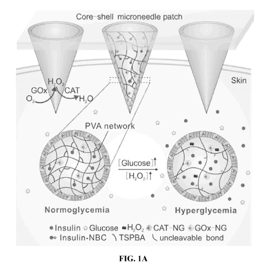
FIGS. 1A-1C. Schematic representation of the glucose-responsive insulin
delivery system
using H202 responsive poly(vinyl alcohol) - N1 -(4-b oronob enzy1)-N3 -(4-b
oronoph eny1)-
N1 ,N1 ,N3 ,N3 -tetram ethyl prop an e- 1,3 -di aminium (PVA-TSPBA) gel. FIG.
1A, Insulin release
was triggered by the hyperglycemic state from PVA-TSPBA microneedle patch and
local
inflammation was greatly reduced by the catalase (CAT) embedded PVA-TSPBA
shell. FIG. 1B,
Insulin modified with
4-nitrophenyl-(4,4, 5,5 -tetram ethyl- 1,3 ,2-di oxab orol an-2-yl)b enzyl
carbonate (NBC) and the mechanism of H202 responsive release. FIG. 1C,
Fabrication and the
H202 responsiveness of PVA-TSPBA gels.
FIGS. 2A-2F. In vitro glucose-responsive insulin release from PVA-TSPBA gels.
FIG. 2A,
Insulin release from PVA-TSPBA gels in PBS with 10 mM H202 at pH 7.4 and 3.5.
FIG. 2B,
Glucose concentration dependent H202 generation in PBS 7.4 in the presence of
glucose oxidase
(GOx). GOx was directly added to solution to 0.2 mg/mL. FIG. 2C, Glucose
concentration
dependent insulin release from gels in PBS 7.4 in the presence of GOx. GOx was
directly added
to solution to 0.2 mg/mL. The glucose concentration was set as 0, 100 and 400
mg/dL. FIG. 2D,
4
CA 03045882 2019-05-31
WO 2018/106696
DE,C-E3 17,Lt4, 7õ2A WO 1
the glucose dependent change of gels in PBS 7.4 with GOx (0.2 mg/mL). FIG. 2E,
Self-regulated
insulin release profile as a function of glucose concentration. FIG. 2F,
Pulsatile insulin release
profile as a function of glucose concentration.
FIGS. 3A-3G. Characterization of a microneedle (MN) array patch of PVA-TSPBA.
FIG.
3A, Representative fluorescence microscopy images of insulin loaded MN arrays
with hyaluronic
acid (HA) base. Rhodamine B labeled insulin was loaded in PVA-TSPBA gel at the
top of
microneedles (red) while the HA base (green, labeled by FITC-insulin) was
mainly located at the
bottom. FIG. 3B, Representative fluorescence image of MN array patch loaded
with insulin-FITC.
Scale bars, 300 p.m. FIG. 3C, Representative scanning electron microscopy
image of microneedle
patch. scale bar, 300 p.m. FIG. 3D, Mechanical strength of MNs; FIG. 3E,
Representative images
of bottom view of hollow CAT loaded MNs. These images were obtained using
confocal laser
scanning microscopy and the interals at z-direction were set as 100 p.m. Scale
bar, 300 p.m. FIG.
3F, Representative images of cross-section of core-shell MN using cryosection:
rhodamine B
labeled CAT shell (red), FITC labeled insulin (green) and their overlap. The
shell was 10 p.m thick
as analyzed using imagek FIG. 3G, The time dependent release of GOx or GOx-
nanogel (G0x-
NG) from PVA methylacrylate gel.
FIGS. 4A-4H. In vivo studies of MN array patches for type 1 diabetes
treatment. FIG. 4A,
Mice treated with a MN array patch (left), and the skin inserted by MN array
patch was excised
and stained using trypan blue (right). Scale bar, 600 p.m. FIG. 4B,
Representative images of core-
shell MNs inserted into skins: the shell embedding rhodamine B labeled CAT
(red), the core
labeled by insulin-FITC (green) and their overlap. Scale bar, 100 p.m. FIG.
4C, Blood glucose
levels of type 1 diabetic mice treated with different kinds of microneedle
array patches. FIG. 4D,
In vivo glucose tolerance test toward diabetic mice at one hour post-treatment
of MN-CAT or
subcutaneously injected with insulin. Healthy mice were used as the control.
FIG. 4E,
Responsiveness was calculated based on the area under the curve (AUC) in 120
min, with the
baseline set at the 0-min blood glucose reading. FIG. 4F, Blood glucose levels
change of healthy
mice treated with MN array patch or subcutaneously injected insulin. The
treatment was given at
0 min. FIG. 4G, Quantification of the hypoglycemia index, identified as the
difference between
the initial and nadir blood glucose readings divided by the time at which
nadir was reached. FIG.
4H, Blood glucose levels change of diabetic mice treated with multiple MN-
array patchs. The
administration of MN-CAT was indiated by blue arrows. Each time, there were
two microneedles
on mice except the first one, and the last two microneedles were removed as
indiated by red arrows.
Student'st-test: **12.< 0.01. Data points represent mean SD (n=5). Error bars
indicate SD.
5
CA 03045882 2019-05-31
WO 2018/106696
DE,C-E3 17,Lt4, 7õ2A WO 1
FIGS. 5A-5C. In vivo biocompatibility studies of MN-CAT arrays for diabetes
treatment.
FIG. 5A, Reprensentative images of skins at the treated site of mice and their
corresponding H&E
staining results. Mice were treated with MN-Gel, MN-Gel(G+I), MN-Gel(G+C+I),
and MN-CAT.
Scale bar, 1 mm or 300 p.m for mice skin images and H&E staining respectively.
FIG. 5B,
Statistical analysis of the thickness of epidermis and skin treated by MNs.
The epidermis and skin
treated by MN-CAT showed significantly less swelling than that treated with MN-
Gel(G+I)
("P<0.01). FIG. 5C, Immunohistology stain with TUNEL assay (green) and Hoechst
(blue) of
skins treated with MN-Gel(G+I), MN-Gel(G+C+I) and MN-CAT. Scale bars, 150 p.m.
FIG. 6. Schematic illustration of H202 generation by glucose oxidase nanogel
(G0x-NG)
and elimination by catalase nanogel (CAT-NG).
FIGS. 7A-7B. H1 -NMR (300 MHz, in D20) of TSPBA before and after oxidization
in
PBS. FIG. 7A) before oxidation; FIG. 7B) after oxidation in 10 mM H202 in 1 h.
FIG. 8. MALDI-TOF mass spectrum of the purified insulin-NBC.
FIGS. 9A-9B. Dynamic rheological behavior of PVA before and after gelation at
25 C
measured using a TA Instruments AR-2000 stress controlled rheometer with 25 mm
aluminum
cross-hatched parallel plates. All experiments were conducted in the linear
viscoelastic regime
with a 500 p.m gap between the plates. FIG. 9A) Frequency spectra of the
elastic (G') and viscous
(G") moduli of PVA and PVA-TSPBA samples, with the former exhibiting solution-
like
characteristics and the latter a gel-like behavior. FIG. 9B) Evolution of G'
and G" as a function of
time of the PVA-TSPBA sample showing sol-gel transition. Experiments were at a
constant
frequency of 5 rad/s. Measurements were started after pre-shear the sample for
10 s at a shear rate
of 10 s-1.
FIG. 10. CD spectra of native insulin solution and insulin released from the
gels incubated
with 400 mg/dL glucose.
FIGS. 11A-11B. Characterization of CAT-NG. FIG. 11A) The size distribution of
CAT and
CAT-NG measured by dynamic laser scattering. FIG. 11B) The representative TEM
images of
CAT-NG.
FIGS. 12A-12B. Characterization of G0x-NG. FIG. 12A) The size distribution of
GOx
and G0x-NG measured by dynamic laser scattering. FIG. 12B) The representative
TEM image of
G0x-NG.
FIGS. 13A-13B. Representative images of hollow CAT loaded MNs: side view (FIG.
13A)
and overhead view (FIG. 13B). The intervals for b was 80 p.m at direction from
bottom to top of
microneedle.
6
CA 03045882 2019-05-31
WO 2018/106696
DE,C,17,3 17,Lt4, 7õ2A WO 1
FIG. 14. The H202 dependent release of insulin from insulin-NBC loaded in PVA
methacrylate gel. Native insulin was used as control.
FIG. 15. Skin puncture marks at 0 min, 5 min and 120 imn post-treatment of
MNs. Scale
bar, 0.5 cm.
FIG. 16. Blood glucose level of type 1 diabetic mice treated by MN-Gel (G+I).
FIG. 17. Blood glucose levels in streptozotocin (STZ)-induced diabetic mice
after
treatment with insulin-NBC loaded PVA-TSPBA gel with or without GOx.
FIG. 18. The H202 generation rate through oxidation of glucose by GOx in the
presence of
CAT of different ratio in glucose solution (100 or 400 mg/dL) in PBS with an
initial pH at 7.4. The
concentration of GOx was set as 0.2 mg/mL.
FIG. 19. The plasma human insulin levels in mice treated with MN-CAT, MN-
Gel(I), or
MN-Gel(G+I).
FIG. 20. Skin bubbling induced by subcutaneously injected Gel-(G+I). left: the
site of gel
inoculation; right: skin swelling observed 1 h post-inoculation. Scale bar, 1
cm.
DETAILED DESCRIPTION
Disclosed herein is an innovative core-shell microneedle (MN) array patch
consisting of
degradable crosslinked poly(vinyl alcohol) (PVA) gel for self-regulated
delivery of a therapeutic
agent (for example insulin) with rapid responsiveness to elevated blood
glucose levels (BGLs). As
shown in Figure la, a core component of this device contains glucose oxidase
(GOx) that generates
H202 to stimulate release of the therapeutic agent, such as insulin, while the
shell component is
embedded with catalase (CAT) that serves as an active strainer to scavenge
excessive H202, thus
minimizing the risk of inflammation caused by H202 (Figure 6).
To achieve H202-responsive insulin release, insulin is chemically modified
with 4-
nitrophenyl 4-(4,4,5,5-tetramethy1-1,3,2-dioxaborolan-2-y1) benzyl carbonate
(designated insulin-
NBC, Figure lb) and subsequently anchored to the water-soluble PVA matrix.[17]
Of note, to
further facilitate transport of free insulin in the polymeric matrix and
promote responsiveness
speed, PVA is also gelated by a H202-labile linker: N1-(4-boronobenzy1)-N3-(4-
boronopheny1)-
N1,N1,N3,N3 -tetram ethyl prop an e-1,3 -di aminium (TSPBA) (Figure 1c). Both
insulin-NBC and
TSPBA are oxidized and hydrolyzed when exposed to local elevated levels of
H202 generated by
GOx in high glucose concentrations [18, 19], leading to the quick release of
free insulin (Figure
la).
To limit the potentially harmful release of GOx itself [16], GOx is
encapsulated into the
acrylated nanogel (GOx-NG) to acquire a large size[8] and get immobilized with
covalent linkage
7
CA 03045882 2019-05-31
WO 2018/106696
DE,C,17,3 17,Lt4, 7õ2AW01
to PVA methacrylate during radical polymerization, which forms a partially
uncleavable network
of PVA to further prevent the leakage of G0x-NG while maintaining ease of
insulin release. The
shell component is designed to mimic the complementary function in
peroxisome[20] where
catalase nanogel (CAT-NG)[8] is formed and embedded inside a crosslinked PVA
layer covering
the surface of PVA-TSPBA microneedle core.
Collectively, the design of the core and shell components offers: 1)
sufficient catalysis with
GOx to perform the glucose-responsive action in the core; and 2) efficient
elimination of H2 0 2 to
alleviate inflammation affecting the surrounding tissues and mitigate systemic
toxicity.
Additionally, the direct conjugation method that is utilized to load insulin
onto the MN scaffold
enhances both efficiency and capacity of the insulin-loading process. Upon
painless
transcutaneous administration, this bio-responsive MN patch partially
dissolves when exposed to
high interstitial fluid glucose concentration in the capillary networks, [9]
thereby releasing insulin
for quick uptake through the regional capillary vessels and lymph networks to
subsequently
regulate BGLs. [9]
A number of embodiments of the invention have been described. Nevertheless, it
will be
understood that various modifications may be made without departing from the
spirit and scope of
the invention. Accordingly, other embodiments are within the scope of the
following claims.
Unless defined otherwise, all technical and scientific terms used herein have
the same
meaning as commonly understood to one of ordinary skill in the art to which
this invention
belongs. The following definitions are provided for the full understanding of
terms used in this
specification.
Terminology
Unless defined otherwise, all technical and scientific terms used herein have
the same
meaning as commonly understood to one of ordinary skill in the art to which
this disclosure
belongs. The term "comprising" and variations thereof as used herein is used
synonymously with
the terms "including," "containing," and variations thereof and are open, non-
limiting terms.
Although the terms "comprising," "including," and "containing" have been used
herein to describe
various embodiments, the terms "consisting essentially of' and "consisting of'
can be used in place
of "comprising," "including," and "containing" to provide for more specific
embodiments and are
also disclosed.
Disclosed are the components to be used to prepare the disclosed compositions,
devices,
and patches, as well as the compositions, devices, and patches themselves to
be used within the
methods disclosed herein. These and other materials are disclosed herein, and
it is understood that
8
CA 03045882 2019-05-31
WO 2018/106696
DE,C,17,3 17,Lt4, 7õ2AW01
when combinations, subsets, interactions, groups, etc. of these materials are
disclosed that while
specific reference of each various individual and collective combination and
permutation of these
compounds may not be explicitly disclosed, each is specifically contemplated
and described
herein. For example, if a particular composition or device is disclosed and
discussed and a number
of modifications that can be made are discussed, specifically contemplated is
each and every
combination and permutation and the modifications that are possible unless
specifically indicated
to the contrary. Thus, if a class of components A, B, and C are disclosed as
well as a class of
components D, E, and F and an example of a combination, or, for example, a
combination
comprising A-D is disclosed, then even if each is not individually recited
each is individually and
collectively contemplated meaning combinations, A-E, A-F, B-D, B-E, B-F, C-D,
C-E, and C-F
are considered disclosed. Likewise, any subset or combination of these is also
disclosed. Thus,
for example, the sub-group of A-E, B-F, and C-E would be considered disclosed.
This concept
applies to all aspects of this application including, but not limited to,
steps in methods of making
and using the disclosed compositions, devices, and patches. Thus, if there are
a variety of
additional steps that can be performed it is understood that each of these
additional steps can be
performed with any specific embodiment or combination of embodiments of the
disclosed
methods.
It is understood that the components, compositions, devices, and patches
disclosed herein
have certain functions. Disclosed herein are certain structural requirements
for performing the
disclosed functions, and it is understood that there are a variety of
structures which can perform
the same function which are related to the disclosed structures, and that
these structures will
ultimately achieve the same result.
Unless otherwise expressly stated, it is in no way intended that any method
set forth herein
be construed as requiring that its steps be performed in a specific order.
Accordingly, where a
method claim does not actually recite an order to be followed by its steps or
it is not otherwise
specifically stated in the claims or descriptions that the steps are to be
limited to a specific order,
it is no way intended that an order be inferred, in any respect. This holds
for any possible non-
express basis for interpretation, including: matters of logic with respect to
arrangement of steps or
operational flow; plain meaning derived from grammatical organization or
punctuation; and the
number or type of embodiments described in the specification.
As used in the specification and claims, the singular form "a," "an," and
"the" include plural
references unless the context clearly dictates otherwise. For example, the
term "a cell" includes a
plurality of cells, including mixtures thereof.
9
CA 03045882 2019-05-31
WO 2018/106696
DE,C,17,3 17,Lt4, 7õ2AW01
As used herein, the terms "may," "optionally," and "may optionally" are used
interchangeably and are meant to include cases in which the condition occurs
as well as cases in
which the condition does not occur. Thus, for example, the statement that a
formulation "may
include an excipient" is meant to include cases in which the formulation
includes an excipient as
well as cases in which the formulation does not include an excipient.
The terms "about" and "approximately" are defined as being "close to" as
understood by
one of ordinary skill in the art. In one non-limiting embodiment the terms are
defined to be within
10%. In another non-limiting embodiment, the terms are defined to be within
5%. In still another
non-limiting embodiment, the terms are defined to be within 1%.
"Activities" of a protein, including those relating to "bioactivity," include,
for example,
transcription, translation, intracellular translocation, secretion,
phosphorylation by kinases,
cleavage by proteases, and/or homophilic and heterophilic binding to other
proteins.
The term "administering" refers to an administration to a subject that is
oral, topical,
intravenous, subcutaneous, transcutaneous, transdermal, intramuscular, intra-
joint, parenteral,
intra-arteriole, intradermal, intraventricular, intracranial, intraperitoneal,
intralesional, intranasal,
rectal, vaginal, by inhalation or via an implanted reservoir. Administering
can be performed using
transdermal microneedle-array patches. The term "parenteral" includes
subcutaneous, intravenous,
intramuscular, intra-articular, intra-synovial, intrasternal, intrathecal,
intrahepatic, intralesional,
and intracranial injections or infusion techniques.
"Biocompatible" generally refers to a material and any metabolites or
degradation products
thereof that are generally non-toxic to the recipient and do not cause any
significant adverse effects
to the subject.
As used herein, the term "comprising" is intended to mean that the
compositions and
methods include the recited elements, but not excluding others. "Consisting
essentially of' when
used to define compositions and methods, shall mean excluding other elements
of any essential
significance to the combination. Thus, a composition consisting essentially of
the elements as
defined herein would not exclude trace contaminants from the isolation and
purification method
and pharmaceutically acceptable carriers, such as phosphate buffered saline,
preservatives, and the
like. "Consisting of' shall mean excluding more than trace elements of other
ingredients and
substantial method steps for administering the compositions of this invention.
Embodiments
defined by each of these transition terms are within the scope of this
invention.
A "control" is an alternative subject or sample used in an experiment for
comparison
purpose. A control can be "positive" or "negative."
As used herein, "conjugated" refers to a non-reversible binding interaction.
CA 03045882 2019-05-31
WO 2018/106696
DE,C,17,3 17,Lt4, 7õ2AW01
A "linker" as used herein refers to a molecule that joins adjacent molecules.
Generally, a
linker has no specific biological activity other than to join the adjacent
molecules or to preserve
some minimum distance or other spatial relationship between them. In some
cases, the linker can
be selected to influence or stabilize some property of the adjacent molecules,
such as the folding,
net charge, or hydrophobicity of the molecule. In some embodiments, the linker
can be detached
(e.g. chemically cleaved) upon exposure to a peroxide, such as hydrogen
peroxide. In other
embodiments, the linker can remain intact upon exposure to a peroxide, such as
hydrogen peroxide.
The terms "peptide," "protein," and "polypeptide" are used interchangeably to
refer to a
natural or synthetic molecule comprising two or more amino acids linked by the
carboxyl group
of one amino acid to the alpha amino group of another.
The term "carrier" or "pharmaceutically acceptable carrier" means a carrier or
excipient
that is useful in preparing a pharmaceutical or therapeutic composition that
is generally safe and
non-toxic, and includes a carrier that is acceptable for veterinary and/or
human pharmaceutical or
therapeutic use. As used herein, the terms "carrier" or "pharmaceutically
acceptable carrier"
encompasses can include phosphate buffered saline solution, water, emulsions
(such as an
oil/water or water/oil emulsion) and/or various types of wetting agents. As
used herein, the term
"carrier" encompasses any excipient, diluent, filler, salt, buffer,
stabilizer, solubilizer, lipid,
stabilizer, or other material well known in the art for use in pharmaceutical
formulations and as
described further below.
As used herein, the term "polymer" refers to a relatively high molecular
weight organic
compound, natural or synthetic, whose structure can be represented by a
repeated small unit, the
monomer (e.g., polyethylene, rubber, cellulose). Synthetic polymers are
typically formed by
addition or condensation polymerization of monomers. As used herein, the term
"copolymer"
refers to a polymer formed from two or more different repeating units (monomer
residues). By
way of example and without limitation, a copolymer can be an alternating
copolymer, a random
copolymer, a block copolymer, or a graft copolymer. It is also contemplated
that, in certain
aspects, various block segments of a block copolymer can themselves comprise
copolymers.
Ranges can be expressed herein as from "about" one particular value, and/or to
"about"
another particular value. When such a range is expressed, another embodiment
includes from the
one particular value and/or to the other particular value. Similarly, when
values are expressed as
approximations, by use of the antecedent "about," it will be understood that
the particular value
forms another embodiment. It will be further understood that the endpoints of
each of the ranges
are significant both in relation to the other endpoint, and independently of
the other endpoint. It
is also understood that there are a number of values disclosed herein, and
that each value is also
11
CA 03045882 2019-05-31
WO 2018/106696
DE,C,17,3 17,Lt4, 7õ2AW01
herein disclosed as "about" that particular value in addition to the value
itself For example, if the
value "10" is disclosed, then "about 10" is also disclosed.
The terms "treat," "treating," "treatment," and grammatical variations thereof
as used
herein, include partially or completely delaying, alleviating, mitigating or
reducing the intensity
of one or more attendant symptoms of a disorder or condition and/or
alleviating, mitigating or
impeding one or more causes of a disorder or condition. Treatments according
to the invention
may be applied preventively, prophylactically, pallatively or remedially.
Prophylactic treatments
are administered to a subject prior to onset (e.g., before obvious signs of
cancer), during early
onset (e.g., upon initial signs and symptoms of cancer), or after an
established development of
cancer. Prophylactic administration can occur for several days to years prior
to the manifestation
of symptoms of an infection.
By the term "effective amount" of a therapeutic agent is meant a nontoxic but
sufficient
amount of a beneficial agent to provide the desired effect. The amount of
beneficial agent that is
"effective" will vary from subject to subject, depending on the age and
general condition of the
subject, the particular beneficial agent or agents, and the like. Thus, it is
not always possible to
specify an exact "effective amount." However, an appropriate "effective"
amount in any subject
case may be determined by one of ordinary skill in the art using routine
experimentation. Also, as
used herein, and unless specifically stated otherwise, an "effective amount"
of a beneficial can
also refer to an amount covering both therapeutically effective amounts and
prophylactically
effective amounts.
An "effective amount" of a drug necessary to achieve a therapeutic effect may
vary
according to factors such as the age, sex, and weight of the subject. Dosage
regimens can be
adjusted to provide the optimum therapeutic response. For example, several
divided doses may be
administered daily or the dose may be proportionally reduced as indicated by
the exigencies of the
therapeutic situation.
As used herein, a "therapeutically effective amount" of a therapeutic agent
refers to an
amount that is effective to achieve a desired therapeutic result, and a
"prophylactically effective
amount" of a therapeutic agent refers to an amount that is effective to
prevent an unwanted
physiological condition. Therapeutically effective and prophylactically
effective amounts of a
given therapeutic agent will typically vary with respect to factors such as
the type and severity of
the disorder or disease being treated and the age, gender, and weight of the
subject.
The term "therapeutically effective amount" can also refer to an amount of a
therapeutic
agent, or a rate of delivery of a therapeutic agent (e.g., amount over time),
effective to facilitate a
desired therapeutic effect. The precise desired therapeutic effect will vary
according to the
12
CA 03045882 2019-05-31
WO 2018/106696
DE,C,17,3 17,Lt4, 7õ2AW01
condition to be treated, the tolerance of the subject, the drug and/or drug
formulation to be
administered (e.g., the potency of the therapeutic agent (drug), the
concentration of drug in the
formulation, and the like), and a variety of other factors that are
appreciated by those of ordinary
skill in the art.
As used herein, the term "pharmaceutically acceptable" component can refer to
a
component that is not biologically or otherwise undesirable, i.e., the
component may be
incorporated into a pharmaceutical formulation of the invention and
administered to a subject as
described herein without causing any significant undesirable biological
effects or interacting in a
deleterious manner with any of the other components of the formulation in
which it is contained.
When the term "pharmaceutically acceptable" is used to refer to an excipient,
it is generally
implied that the component has met the required standards of toxicological and
manufacturing
testing or that it is included on the Inactive Ingredient Guide prepared by
the U.S. Food and Drug
Administration.
Also, as used herein, the term "pharmacologically active" (or simply
"active"), as in a
"pharmacologically active" derivative or analog, can refer to a derivative or
analog (e.g., a salt,
ester, amide, conjugate, metabolite, isomer, fragment, etc.) having the same
type of
pharmacological activity as the parent compound and approximately equivalent
in degree.
As used herein, the term "subject" can refer to living organisms such as
mammals,
including, but not limited to humans, livestock, dogs, cats, and other
mammals. Administration of
the therapeutic agents can be carried out at dosages and for periods of time
effective for treatment
of a subject. In some embodiments, the subject is a human.
Microneedle Devices (Patches)
Disclosed herein is an innovative self-regulated microneedle (MN) patch for
the delivery
of a therapeutic agent (for example, insulin).
In some aspects, disclosed herein is a microneedle patch, comprising:
a plurality of microneedles each having a base end and a tip; and
a substrate to which the base ends of the microneedles are attached;
wherein the microneedles comprise:
a shell, comprising:
a first poly(vinyl alcohol) (PVA) polymer cross-linked with a first peroxide-
sensitive
linker; and
a peroxide scavenging enzyme encapsulated within a first nanogel, wherein the
first
nanogel is embedded in the first PVA polymer;
13
CA 03045882 2019-05-31
WO 2018/106696
DE,C,173 17,Lt4, 7õ2A WO 1
and
a core, comprising:
a second poly(vinyl alcohol) (PVA) polymer cross-linked with a second peroxide-
sensitive linker;
a glucose-responsive agent encapsulated within a second nanogel, wherein the
second
nanogel is embedded in the second PVA polymer; and
a therapeutic agent, wherein the therapeutic agent is covalently attached to
the second
PVA polymer with a third peroxide-sensitive linker.
In some embodiments, the first peroxide-sensitive linker comprises a boronic
ester. In some
embodiments, the first peroxide-sensitive linker detaches from the first PVA
polymer upon
exposure to peroxide. In some embodiments, the first peroxide-sensitive linker
is N1-(4-
boronobenzy1)-N3-(4-boronopheny1)-N1,N1,N3,N3 -tetram ethylprop ane- 1,3 -di
aminium
(T SPBA).
In some embodiments, the second peroxide-sensitive linker comprises a boronic
ester. In
some embodiments, the second peroxide-sensitive linker detaches from the
second PVA polymer
upon exposure to peroxide. In some embodiments, the second peroxide-sensitive
linker is N1-(4-
boronobenzy1)-N3-(4-boronopheny1)-N1,N1,N3,N3 -tetram ethylprop ane- 1,3 -di
aminium
(T SPBA).
In some embodiments, the third peroxide-sensitive linker comprises a boronic
ester. In
some embodiments, the third peroxide-sensitive linker detaches from the second
PVA polymer
upon exposure to peroxide. In some embodiments, the third peroxide-sensitive
linker is 4-
nitrophenyl-(4,4, 5,5 -tetram ethyl- 1,3 ,2-di oxab orol an-2-yl)b enzyl
carbonate (NBC).
In some embodiments, the peroxide-sensitive linker comprises 4-nitrophenyl-
(4,4,5,5-
tetramethyl- 1,3 ,2-di ox ab orol an-2-yl)b enzyl carbonate (NBC) or N1 -(4-b
oronob enzy1)-N3 -(4-
boronopheny1)-N1,N1,N3,N3 -tetram ethylprop ane- 1,3 -di aminium (TSPBA).
In some embodiments, the glucose-responsive agent comprises glucose oxidase.
In some
embodiments, the peroxide scavenging enzyme is catalase. In some embodiments,
the glucose-
responsive agent is encapsulated within a nanogel. In some embodiments, the
glucose-responsive
agent is covalently attached to the nanogel. In some embodiments, the peroxide
scavenging
enzyme is encapsulated within a nanogel. In some embodiments, the peroxide
scavenging enzyme
is covalently attached to the nanogel. In some embodiments, the microneedles
are coated with the
peroxide scavenging enzyme.
Examples of peroxide (H202) scavenging enzymes include, but are not limited to
catalase,
phenolic acid, 3,4,5-trihydroxybenzoic (gallic) acid and 1,2,3 -
trihydroxybenzene (pyrogallol).
14
CA 03045882 2019-05-31
WO 2018/106696
DE,C,173 17,Lt4, 7õ2A WO 1
The H202 scavenging enzymes can be incorporated into the microneedle by any
means known in
the art, including incorporation of the H202 scavenging enzyme in a nanogel
(for example a
peroxisome catalase nanogel).
In some embodiments, the first nanogel comprises a methacrylate nanogel. In
some
embodiments, the first nanogel comprises poly(vinyl alcohol) (PVA)
methacrylate.
In some embodiments, the second nanogel comprises a methacrylate nanogel. In
some
embodiments, the second nanogel comprises poly(vinyl alcohol) (PVA)
methacrylate.
In some embodiments, the nanogels disclosed herein are embedded within a
crosslinked
PVA polymer. In some embodiments, the nanogels disclosed herein are covalently
linked to a
crosslinked PVA polymer. In some embodiments, the first nanogel is embedded in
the first PVA
polymer. In some embodiments, the first nanogel is covalently attached to the
first PVA polymer.
In some embodiments, the second nanogel is embedded in the second PVA polymer.
In some
embodiments, the second nanogel is covalently attached to the second PVA
polymer.
In some embodiments, the covalent attachment is via a non-cleavable covalent
bond.
In some embodiments, the therapeutic agent is insulin. In some embodiments,
the
microneedles further comprise hyaluronic acid (HA).
In some aspects, disclosed herein is a device for transport of a material
across a biological
barrier of a subject comprising:
a plurality of microneedles each having a base end and a tip;
a substrate to which the base ends of the microneedles are attached or
integrated; and
wherein the microneedles comprise:
a shell, comprising:
a first poly(vinyl alcohol) (PVA) polymer cross-linked with a first peroxide-
sensitive
linker; and
a peroxide scavenging enzyme encapsulated within a first nanogel, wherein the
first
nanogel is embedded in the first PVA polymer;
and
a core, comprising:
a second poly(vinyl alcohol) (PVA) polymer cross-linked with a second peroxide-
sensitive linker;
a glucose-responsive agent encapsulated within a second nanogel, wherein the
second
nanogel is embedded in the second PVA polymer; and
a therapeutic agent, wherein the therapeutic agent is covalently attached to
the second
PVA polymer with a third peroxide-sensitive linker.
CA 03045882 2019-05-31
WO 2018/106696
DE,C,17,3 17,Lt4, 7õ2AW01
In further aspects, also disclosed herein is a kit of parts for delivering a
therapeutic agent
(for example, insulin) across a biological barrier comprising:
a plurality of microneedles each having a base end and a tip;
a substrate to which the base ends of the microneedles are attached or
integrated; and
wherein the microneedles comprise:
a shell, comprising:
a first poly(vinyl alcohol) (PVA) polymer cross-linked with a first peroxide-
sensitive
linker; and
a peroxide scavenging enzyme encapsulated within a first nanogel, wherein the
first
nanogel is embedded in the first PVA polymer;
and
a core, comprising:
a second poly(vinyl alcohol) (PVA) polymer cross-linked with a second peroxide-
sensitive linker;
a glucose-responsive agent encapsulated within a second nanogel, wherein the
second
nanogel is embedded in the second PVA polymer; and
a therapeutic agent, wherein the therapeutic agent is covalently attached to
the second
PVA polymer with a third peroxide-sensitive linker.
In addition to a therapeutic agent such as insulin, the agent to be delivered
to the recipient
can also be a prophylactic agent or diagnostic agent. For example, the agent
can be selected from
the group consisting of peptides, proteins, carbohydrates, nucleic acid
molecules, lipids, organic
molecules, biologically active inorganic molecules, and combinations thereof.
For example, a wide
range of drugs may be formulated for delivery with the present microneedle
devices and methods.
As used herein, the terms "drug" or "drug formulation" are used broadly to
refer to any
prophylactic, therapeutic, or diagnostic agent, or other substance that which
may be suitable for
introduction to biological tissues, including pharmaceutical excipients and
substances for
tattooing, cosmetics, and the like. The drug can be a substance having
biological activity. The drug
formulation may include various forms, such as liquid solutions, gels, solid
particles (e.g.,
microparticles, nanoparticles), or combinations thereof The drug may comprise
small molecules,
large (i.e., macro-) molecules, or a combination thereof. In representative,
not non-limiting,
embodiments, the drug can be selected from among immunologic adjuvants (for
example,
monophosphoryl lipid A (MPLA) , aluminum salt (Alum), CpG
oliogodeoxynucleotides
(ODN)), amino acids, vaccines, antiviral agents, gene delivery vectors,
interleukin inhibitors,
immunomodulators, neurotropic factors, neuroprotective agents, antineoplastic
agents,
16
CA 03045882 2019-05-31
WO 2018/106696
DE,C,17,3 17,Lt4, 7õ2AW01
chemotherapeutic agents, polysaccharides, anti-coagulants, antibiotics,
analgesic agents,
anesthetics, antihistamines, anti-inflammatory agents, and viruses. The drug
may be selected from
suitable proteins, peptides and fragments thereof, which can be naturally
occurring, synthesized or
recombinantly produced.
The compositions and/or drug formulation may further include one or more
pharmaceutically acceptable excipients, including pH modifiers, viscosity
modifiers, diluents, etc.,
which are known in the art.
In one embodiment, the microneedles comprise hyaluronic acid. In addition to
hyaluronic
acid, the microneedles may also comprise a variety of materials, including
metals, ceramics,
semiconductors, organics, polymers, composites, or a combination thereof.
Typical materials of
construction include pharmaceutical grade stainless steel, gold, titanium,
nickel, iron, tin,
chromium, copper, palladium, platinum, alloys of these or other metals,
silicon, silicon dioxide,
and polymers. Representative biodegradable polymers include polymers of
hydroxy acids such as
lactic acid and glycolic acid polylactide, polyglycolide, polylactide-co-
glycolide, and copolymers
with PEG, polyanhydrides, poly(ortho)esters, polyurethanes, poly(butyric
acid), poly(valeric
acid), and poly(lactide-co-caprolactone).
The microneedles should have the mechanical strength to remain intact while
being
inserted into the biological barrier, while remaining in place for up to a
number of days, and while
being removed. In some embodiments, the microneedle must remain intact at
least long enough
for the microneedle to serve its intended purpose (e.g., delivery of the
therapeutic agent).
The microneedles can have straight or tapered shafts. In one embodiment, the
diameter of
the microneedle is greatest at the base end of the microneedle and tapers to a
point at the end distal
the base. The microneedle can also be fabricated to have a shaft that includes
both a straight
(untapered) portion and a tapered portion. The needles may also not have a
tapered end at all, i.e.
they may simply be cylinders with blunt or flat tips.
The microneedles can be oriented perpendicular or at an angle to the
substrate. In one
embodiment, the microneedles are oriented perpendicular to the substrate so
that a larger density
of microneedles per unit area of substrate can be provided. An array of
microneedles can include
a mixture of microneedle orientations, heights, or other parameters.
The microneedles can be formed with shafts that have a circular cross-section
in the
perpendicular, or the cross-section can be non-circular. For example, the
cross-section of the
microneedle can be polygonal (e.g. star-shaped, square, triangular), oblong,
or another shape. The
cross-sectional dimensions can be between about 1 1.tm and 1000 jim, such that
the base can be
17
CA 03045882 2019-05-31
WO 2018/106696
DE,C-E3 17,Lt4, 7õ2A WO 1
about 100-500 um, and the tip can be between 1 and 20 um. In one embodiment,
the microneedle
can be approximately 300 um at the base, and approximately 5 um at the tip.
The length of the microneedles typically is between about 10 um and 1 mm,
preferably
between 400 um and 1 mm. In one embodiment, the length (or height) of the
microneedle is about
600 um. The length is selected for the particular application, accounting for
both an inserted and
uninserted portion. An array of microneedles can include a mixture of
microneedles having, for
example, various lengths, outer diameters, inner diameters, cross-sectional
shapes, and spacings
between the microneedles. In one embodiment, the microneedles are arranged in
a 15 by 15 array
with 600 um tip-to-tip spacing. In one embodiment, the microneedles are
arranged in a 20 by 20
array with 600 um tip-to-tip spacing.
The shell of the microneedle can be considered as the outside portion of the
microneedle
that comes into contact with the subject. The core or the microneedle can be
considered as the
portion of the microneedle located toward the center of each microneedle and
is separated from
contacting the subject's skin by the shell portion of the microneedle.
In one embodiment, the glucose-responsive agent is glucose oxidase (G0x).
Glucose
oxidase converts blood glucose to gluconic acid. This leads to production of a
peroxide (hydrogen
peroxide), and a decrease in the pH.
Methods of Treatment
In some aspects, disclosed herein is a method of delivering a therapeutic
agent to a subject,
comprising:
administering to a subject in need thereof a microneedle patch, wherein the
microneedle
patch comprises:
a plurality of microneedles each having a base end and a tip; and
a substrate to which the base ends of the microneedles are attached;
wherein the microneedles comprise:
a shell, comprising:
a first poly(vinyl alcohol) (PVA) polymer cross-linked with a first peroxide-
sensitive
linker; and
a peroxide scavenging enzyme encapsulated within a first nanogel, wherein the
first
nanogel is embedded in the first PVA polymer;
and
a core, comprising:
18
CA 03045882 2019-05-31
WO 2018/106696
DE,C,17,3 17,Lt4, 7õ2AW01
a second poly(vinyl alcohol) (PVA) polymer cross-linked with a second peroxide-
sensitive linker;
a glucose-responsive agent encapsulated within a second nanogel, wherein the
second
nanogel is embedded in the second PVA polymer; and
a therapeutic agent, wherein the therapeutic agent is covalently attached to
the second
PVA polymer with a third peroxide-sensitive linker;
and
releasing the therapeutic agent from the microneedle patch in the presence of
hyperglycemic levels of glucose.
In some aspects, also disclosed herein is a method for treating a disease in a
subject in need
thereof, comprising:
providing a microneedle patch to a subject, wherein the microneedle patch
comprises:
a plurality of microneedles each having a base end and a tip; and
a substrate to which the base ends of the microneedles are attached;
wherein the microneedles comprise:
a shell, comprising:
a first poly(vinyl alcohol) (PVA) polymer cross-linked with a first peroxide-
sensitive
linker; and
a peroxide scavenging enzyme encapsulated within a first nanogel, wherein the
first
nanogel is embedded in the first PVA polymer;
and
a core, comprising:
a second poly(vinyl alcohol) (PVA) polymer cross-linked with a second peroxide-
sensitive linker;
a glucose-responsive agent encapsulated within a second nanogel, wherein the
second
nanogel is embedded in the second PVA polymer; and
a therapeutic agent, wherein the therapeutic agent is covalently attached to
the second
PVA polymer with a third peroxide-sensitive linker.
In some embodiments, the method further comprises releasing the therapeutic
agent from
the microneedle patch in the presence of hyperglycemic levels of glucose.
As used herein, "hyperglycemic levels of glucose" refer to concentrations of
glucose which
cause, or are at risk of causing, clinical hyperglycemia. Strict cutoff values
for hyper-, normo-, and
hypoglycemia can vary between subjects, particularly between subjects with
varying forms or
degrees of severity of diabetes. In some embodiments, a hyperglycemic level of
glucose comprises
19
CA 03045882 2019-05-31
WO 2018/106696
DE,C,173 17,Lt4, 7õ2A WO 1
greater than 100 mg/dL glucose. In some embodiments, a hyperglycemic level of
glucose
comprises 125 mg/dL or greater, 150 mg/dL or greater, 175 mg/dL or greater, or
200 mg/dL
glucose or greater. Conversely, "normoglycemic levels of glucose" refer to
concentrations of
glucose which are typical/normal and are not usually known to relate to
clinical conditions (or
severe clinical conditions) of glycemic imbalance. In some embodiments, a
normoglycemic level
of glucose comprises from about 70 mg/dL glucose to less than 200 mg/dL
glucose. In some
embodiments, a normoglycemic level of glucose comprises from about 70 mg/dL
glucose to about
175 mg/dL glucose, from about 70 mg/dL glucose to about 150 mg/dL glucose,
from about 70
mg/dL glucose to about 125 mg/dL glucose, or from about 70 mg/dL glucose to
about 100 mg/dL
glucose. A "hypoglycemic level of glucose" refers to a concentration of
glucose which causes, or
is at risk of causing, clinical hypoglycemia. In some embodiments, a
hypoglycemic level of
glucose comprises 70 mg/dL glucose or less. In some embodiments, a
hypoglycemic level of
glucose comprises 60 mg/dL or less, 50 mg/dL or less, 40 mg/dL or less, or 30
mg/dL glucose or
less. In some embodiments, a hyperglycemic level of glucose comprises 200
mg/dL or more
glucose, a normoglycemic level of glucose comprises from about 70 mg/dL
glucose to less than
200 mg/dL glucose, and a hypoglycemic level of glucose comprises less than
about 70 mg/dL
glucose.
In some embodiments, the subject has hyperglycemia. In some embodiments, the
subject
has diabetes or some other glucose regulation disease. In some embodiments,
the subject has
diabetes. In some embodiments, the subject has Type I diabetes. In some
embodiments, the subject
has Type II diabetes.
In some embodiments, the glucose-responsive agent produces a peroxide when
exposed to
hyperglycemic levels of glucose. In some embodiments, the method further
comprises detaching
the first peroxide-sensitive linker from the first PVA polymer upon exposure
to the peroxide.
In some embodiments, the method further comprises detaching the third peroxide-
sensitive
linker from the second PVA polymer upon exposure to the peroxide. In some
embodiments, the
detaching of the third peroxide-sensitive linker from the second PVA polymer
releases the
therapeutic agent from the microneedle patch.
In some embodiments, the method further comprises reducing blood glucose
levels. In
some embodiments, the blood glucose levels are reduced to no lower than
normoglycemic levels.
In some embodiments, the therapeutic agent comprises insulin. In some
embodiments, the method
further comprises terminating release of the therapeutic agent prior to
causing hypoglycemia.
In some embodiments, the disease is diabetes. In some embodiments, the disease
is Type I
diabetes. In some embodiments, the disease is Type II diabetes.
CA 03045882 2019-05-31
WO 2018/106696
DE,C,17,3 17,Lt4, 7õ2A WO 1
In some embodiments, the first peroxide-sensitive linker comprises a boronic
ester. In some
embodiments, the first peroxide-sensitive linker detaches from the first PVA
polymer upon
exposure to peroxide. In some embodiments, the first peroxide-sensitive linker
is N1-(4-
boronobenzy1)-N3-(4-boronopheny1)-N1,N1,N3,N3 -tetram ethylprop an e- 1,3 -di
aminium
(T SPBA).
In some embodiments, the second peroxide-sensitive linker comprises a boronic
ester. In
some embodiments, the second peroxide-sensitive linker detaches from the
second PVA polymer
upon exposure to peroxide. In some embodiments, the second peroxide-sensitive
linker is N1-(4-
boronobenzy1)-N3-(4-boronopheny1)-N1,N1,N3,N3 -tetram ethylprop an e- 1,3 -di
aminium
(T SPBA).
In some embodiments, the third peroxide-sensitive linker comprises a boronic
ester. In
some embodiments, the third peroxide-sensitive linker detaches from the second
PVA polymer
upon exposure to peroxide. In some embodiments, the third peroxide-sensitive
linker is 4-
nitrophenyl-(4,4, 5,5 -tetram ethyl- 1,3 ,2-di oxab orol an-2-yl)b enzyl
carbonate (NBC).
In some embodiments, the glucose-responsive agent comprises glucose oxidase.
In some
embodiments, the peroxide scavenging enzyme is catalase. In some embodiments,
the therapeutic
agent is insulin. In some embodiments, the microneedles further comprise
hyaluronic acid (HA).
In further aspects, also disclosed herein is a method for treating a disease
in a subject in
need thereof, comprising:
providing a microneedle patch to a subject, wherein the microneedle patch
comprises:
a plurality of microneedles each having a base end and a tip; and
a substrate to which the base ends of the microneedles are attached;
wherein the microneedles comprise:
a shell, comprising:
a first poly(vinyl alcohol) (PVA) polymer cross-linked with a first peroxide-
sensitive
linker; and
a peroxide scavenging enzyme encapsulated within a first nanogel, wherein the
first
nanogel is embedded in the first PVA polymer;
and
a core, comprising:
a second poly(vinyl alcohol) (PVA) polymer cross-linked with a second peroxide-
sensitive linker;
a glucose-responsive agent encapsulated within a second nanogel, wherein the
second
nanogel is embedded in the second PVA polymer; and
21
CA 03045882 2019-05-31
WO 2018/106696
DE,C,17,3 17,Lt4, 7õ2A WO 1
a therapeutic agent, wherein the therapeutic agent is covalently attached to
the second
PVA polymer with a third peroxide-sensitive linker; and
inserting the microneedles into a biological barrier, wherein the presence of
hyperglycemic levels
of glucose releases the therapeutic agent from the microneedle patch.
In another aspect, disclosed herein is a method for treating hyperglycemia in
a subject in
need thereof, comprising administering to the subject the microneedle patch of
preceding aspect
or embodiment. In some embodiments, the hyperglycemia is a symptom of
diabetes.
Due to the innovative design with quick responsiveness to a hyperglycemic
state, the core-
shell patch is able to effectively control blood glucose levels in a normal
range. Moreover, the
disclosed microneedle patch can avoid the risk of hypoglycemia compared to the
native insulin.
Current glucose oxidase (G0x)-based glucose-responsive insulin delivery
systems mainly utilize
matrices consisting of pH-sensitive materials, the response speed of which is
extremely slow (as
it is hard to reduce pH level efficiently in a physiologically buffered
system) and therefore remain
challenging for clinical translation. In the present disclosure, enzymatically
generated H202 is
directly applied as a trigger for self-regulating insulin release, based on
both dissociation of matrix
and detachment of insulin. This leads to a remarkably faster and sharper
responsiveness upon
glucose level changes in both in vitro and in vivo studies.
The microneedle patches disclosed herein also have excellent biocompatibility.
In some
embodiments, the base of the microneedle patches and the matrix of
microneedles were made from
hyaluronic acid (HA) and poly (vinyl alcohol) (PVA) respectively, which are
highly biocompatible
and can be further tailored. Both HA and PVA have been widely applied in
numerous FDA-
approved therapeutic formulations or medical devices due to excellent
biocompatibility and
biodegradability. In addition, previously applied hypoxia-responsive systems
to enhance the
response speed caused severe inflammation for long-term usage due to H202. In
the present
disclosure, the excessive H202 generated by oxidation of glucose was
restricted within the space
of microneedle by a surface coated membrane, which can protect normal tissues
from the damage
of H202.
The microneedle patches disclosed herein also can be conveniently
administered.
Integration of the crosslinked gel with a microneedle array patch provides a
painless and
disposable administration modality. Additionally, the insulin dose and
response speed can be
readily adjusted upon personalized requirement. This platform is also much
more convenient
compared to an insulin pump with implanted needles/electrodes or other needle-
injection systems.
22
CA 03045882 2019-05-31
WO 2018/106696
DE,C-E3 17,Lt4, 7õ2AW01
EXAMPLES
The following examples are set forth below to illustrate the compositions,
devices,
methods, and results according to the disclosed subject matter. These examples
are not intended
to be inclusive of all aspects of the subject matter disclosed herein, but
rather to illustrate
representative methods and results. These examples are not intended to exclude
equivalents and
variations of the present invention which are apparent to one skilled in the
art.
Example 1: Core-Shell Microneedle Gel for Self-Regulated Insulin Delivery.
Results
TSPBA was synthesized via quaternization reaction of Ni, Ni, N3, N3 -
1 0
tetramethylpropane-1,3 -diamine with an excess of 4-
(bromomethyl)phenylboronic acid (Scheme
1). The quaternary ammonium groups on TSPBA greatly enhanced its water
solubility (-100
mg/mL), which facilitating the gel formation with PVA aqueous solution without
organic solvents.
Upon oxidation in the presence of 10 mM H202, 70% of TSPBA released p-quinone
methide (p¨
hydroxylmethylenephenol) and became tertiary amines in two hours as
demonstrated by H1 -NMR
(Figure 7).[i8] Insulin-NBC was prepared in the presence of a slight excess of
NBC in a mixed
solvent composed of DMSO and 10 mMNaHCO3 aqueous solution. [19] The product
was purified
using preparative scale high performance liquid chromatography (HPLC),[13] and
was confirmed
by molecular weight to be a conjugate of one insulin modified by one NBC by
MALDI-TOF mass
spectroscopy (Figure 8). Importantly, insulin-NBC had much higher aqueous
solubility (>100
mg/mL) at pH 7.4 than native insulin, which was critical to prepare MN gels
with a high loading
capacity of insulin. The phenylboronic ester of insulin-NBC can be hydrolyzed
in an aqueous
solution; this reaction is facilitated in the presence of diols.[19] The
insulin-NBC could then be
conjugated to PVA chains via the kinetic ester bond between the phenylboronic
acid and the cis-
1,3-diols in PVA. [21] Addition of TSPBA to this reaction solution led to a
rapid increase in the
elastic (G') modulus and formation of a network between the PVA chains in 60 s
(Figure 9).[22]
The gelation of PVA by TSPBA is critical for maintaining the integrity of the
shell structure, which
is composed of water-sensitive materials, and specifically prevents its
dissolution to aqueous
solution when preparing the core.
23
CA 03045882 2019-05-31
WO 2018/106696
DE,C¨E3 17,Lt4, 7õ2AWO 1
HO, ,OH
B.
I
n- 1 I
LI)
+
,
,
Br HO, Br ,NI
HO \f TSPBA
Scheme 1
The H202-sensitive insulin release was evaluated in the presence of 10 mM H202
in PBS
at pH 7.4. Insulin was released from a formed gel with the addition of H202 at
a steady rate, and
more than half of insulin was released within two hours (Figure 2a). Although
the esters between
phenylboronic acids and diols are unstable in acidic solution,[23] the gel
formed between PVA
and phenylboronic acid is stable in acidic environment.[21] At pH 3.5, the gel
showed high
stability and insulin was released at a rather slow rate as compared to pH
7.4.
Next, the release rate of insulin was assessed in the presence of GOx in PBS
at initial pH
7.4 at 3 different glucose concentrations, including a typical hyperglycemic
level (400 mg/dL), a
normoglycemic level (100 mg/dL), and a control level (0 mg/dL). The H202
generation rate was
measured using a fluorometric hydrogen peroxide assay kit. [24] At the
hyperglycemic glucose
concentration of 400 mg/dL, H202 generation was efficient and reached as high
as 6 mM within
30 min (Figure 2b). Compared to the hyperglycemic solution, H202 was generated
at a much
slower rate in the normoglycemic solution of 100 mg/dL glucose. The insulin
release corresponded
to the H202 release such that the insulin release rate was dramatically
promoted under a glucose
concentration of 400 mg/dL compared to that of 100 mg/dL, whereas negligible
insulin release
was observed when the gel was incubated in the control solution (Figure 2c),
consistent with the
morphology change of gels (Figure 2d).
Moreover, the release rate of insulin from PVA-TSPBA gels was steadily
enhanced when
gradually increasing the glucose concentrations of the tested solutions from
normoglycemic to
hyperglycemic conditions, where a maximum of a 15-fold difference in insulin
release rate was
achieved in two hours when the glucose concentration was increased from 100 to
400 mg/dL
(Figure 2e). The limited release of insulin at normoglycemia is a significant
safety feature for the
in vivo application. Additionally, a pulsatile kinetic release profile of
insulin was monitored for
several cycles by alternately varying glucose concentrations between
normoglycemic and
hyperglycemic conditions, and the pulsatile release profile of insulin was
achieved when the gels
24
CA 03045882 2019-05-31
WO 2018/106696
DE,C-E3 17,Lt4, 7õ2A WO 1
were alternatively exposed to the normal and hyperglycemic levels (Figure 2f).
In sum, these
findings suggest that the dissociation of crosslinked gels only takes place in
hyperglycemic
conditions and the PVA-TSPBA gels release insulin in a glucose concentration-
dependent
behavior. Finally, the far-UV circular dichroism (CD) spectra of the native
and released insulin
from gels (1 mg/mL) were similar, suggesting that released insulin retains a-
helical secondary
structure and bioactivity (Figure 10).
To fabricate the core-shell MN gel, PVA-TSPBA gels were integrated into MN
array patch
using a micromolding approach. [9] A "solution-gelation" method was developed
to conveniently
load the crosslinked gel into MNs and form a core-shell structure. Briefly,
diluted aqueous
solutions of PVA, TSPBA and CAT-NG (Figure 11) with low viscosity were
prepared, combined,
and deposited in a silicone mold. The mixed solution was kept in the mold
under vacuum for 30
minutes and then centrifuged at 500 rpm for one hour to form a "shell" on the
mold. Another round
of diluted aqueous solutions of PVA, TSPBA and G0x-NG (Figure 12) were loaded
into the mold
and this procedure was repeated for several times until a predetermined amount
of insulin-loaded
gel was achieved. During this process, acrylated PVA and radical initiator was
added to the native
PVA aqueous solution to form a partially non-degradable network. Finally,
hyaluronic acid (HA)
aqueous solution was cast and dried under vacuum to provide a base for the
mechanical support. [9]
The resulting device was arranged in a 20x20 MN array on a 10 x10 mm2 patch.
The needle had a
conical shape with a base diameter of 300 p.m, 5 p.m at the tip, and a height
of 600 p.m (Figure 3a).
The structure of MNs was confirmed with SEM and fluorescence microscopy
(Figure 3b, 3c). The
mechanical strength of MN was determined as 2 N/needle (Figure 3d), which
sufficiently allows
for skin insertion without breaking.[25] To validate the feasibility of
coating a CAT layer on MN
arrays, a hollow MN array patch constructed by rhodamine B labeled CAT-NG
loaded PVA-
TSPBA gel shell was prepared. These hollow MNs showed a complete shell
structure in a bottom-
view, side view or overhead view (Figure 3e and 13). In addition, it was found
found that the
integrity of this CAT-NG-loaded shell was not affected when preparing the core
part (Figure 3f).
Collectively, these results demonstrated the feasibility of preparing a core-
shell MN array, with a
core loaded with insulin and G0x-NG, in addition to a shell embedded with CAT-
NG. The
methacrylated PVA formed gel was shown to selectively release insulin over G0x-
NG (Figures
3g and 14). Release of G0x-NG was likely prevented due to its much larger size
(-12 nm) than
insulin and the covalent bond, thereby reducing the potential local and
systemic toxicity of
G0x. [16]
The in vivo performance of the core-shell MN array patches was evaluated in a
mouse
model of type 1 diabetes induced by streptozotocin (STZ). The mice were
divided into six groups:
CA 03045882 2019-05-31
WO 2018/106696
DE,C-E3 17,Lt4, 7õ2A WO 1
1) treatment with CAT-NG shelled MN array patch of GOx-NG and insulin-NBC
loaded gels
(MN-CAT); 2) treatment with subcutaneous injection of human recombinant
insulin; 3) treatment
with MN array patch of GOx-NG and insulin-NBC loaded gels (MN-Gel(G+I))
without shell; 4)
treatment with only microneedle array patch loaded blank gel (MN-Gel); 5)
treatment with an MN
array patch of insulin-NBC loaded gels (MN-Gel(I)); 6) group treated by MN
array patch of GOx
and insulin and CAT-NG loaded gels (MN-Gel(G+C+I)). The insulin dosage was set
as 50 mg/kg
for all MN based insulin treatments. Staining by trypan blue indicated
successful penetration of
MNs in excised skin (Figure 4a, b).[26] In addition, the temporal
microchannels on the skin caused
by MNs recovered quickly within two hours post-treatment (Figure 15,
Supporting Information).
BGLs of the mice were monitored over time following administration. A rapid
decrease of
BGLs of mice treated by MN-Gel(G+I) (Figure 16) and MN-CAT (Figure 4c) was
observed in 30
min post-administration, and BGLs then slowly decreased to around 100 mg/dL
and maintained
near 200 mg/dL for almost six hours, much longer than subcutaneously injected
insulin (Figure
4c). This was attributed to the quick local generation of H202 through the
oxidation of glucose in
the presence of GOx, as well as the high sensitivity of gel to H202. In
contrast, no obvious BGLs
reduction was observed for the mice treated with MN-Gel(I), MN-Gel (G+C+I) and
MN-Gel.
These results were consistent to that observed in diabetic mice subcutaneously
injected with PVA-
TSPBA gel with or without GOx (Figure 17). Taken together, these observations
confirmed the
essential role of H202 in releasing insulin, as well as the high stability of
insulin in PVA-TSPBA
in the physiological environment. In vitro, CAT can preferentially decompose
H202 efficiently at
normoglycemia (100 mg/dL) (Figure 18). However, the capability of MN-
Gel(G+C+I) to reduce
BGLs was significantly limited (Figure 4c), indicating the necessity to
separate CAT in a shell
layer to establish a robust level of H202 locally. Additionally, the plasma
human insulin levels in
mice treated with MN-CAT and MN-Gel(G+I) were significantly higher than those
treated with
MN-Gel(I) (Figure 19).
Intraperitoneal glucose tolerance tests (IPGTTs) were further carried out one-
hour post-
administration of MNs or insulin. A spike in BGLs was observed for all groups
after the IPGTT
(Figure 4d). However, only healthy mice and MN-CAT could restore blood glucose
levels to a
normoglycemic level within a short period of time, and the mice treated with
MG-CAT showed
significantly enhanced glucose tolerance to the glucose challenge (Figure 4e).
In order to assess
the risk of hypoglycemia associated with treatment by MN-CAT, the BGLs of
healthy mice treated
with MN array patch were observed. The BGLs of mice treated with insulin
showed a remarkable
decrease, while the BGLs of mice treated with MN-CAT showed only a slight
decrease, consistent
with the slow release of insulin from gels at the normoglycemic state (Figure
4f). Additionally, the
26
CA 03045882 2019-05-31
WO 2018/106696
DE,C-E3 17,Lt4, 7õ2AW01
MN-CAT treated group showed significantly lower hypoglycemia index than
insulin (Figure 4g),
suggesting that the MN-CAT is safe for clinical application. Furthermore, a 40-
hour long
consecutive MN administration was performed to evaluate the in vivo glucose
control capability
of MN (Figure 4h). During this time, BGLs were maintained in a narrow range
between 100 to
250 mg/dL, and critically, no hypoglycemia was recorded.
The reaction between the phenylboronic acid and H202 alleviates the harm of
generated
H202 to surrounding tissues. However, any excessive unreacted H202 still
carries potential to be
harmful. Compared to the skin treated with MN-Gel (Figure 5a), obvious
inflammation was
observed for mice treated with MN-Gel(G+I). Similar phenomenon was observed
for skins treated
with directly subcutaneously injected gel (Figure 20). In a sharp contrast,
almost no visible
inflammation was observed on skins of mice treated with MN-CAT and MN-
Gel(G+C+I) (Figure
5a). These findings were reinforced in hematoxylin and eosin (H&E) staining
results. Compared
with skin treated with MN-Gel, skin samples treated with MN-Gel(G+I) showed
obvious
neutrophil infiltration, indicating a pathophysiological response and tissue
damage induced by the
generated H202.[27] However, greatly reduced neutrophil infiltration was
observed in skin of mice
treated with MN-Gel(G+C+I) and MN-CAT. The epidermal thickness and skin
thickness for mice
treated with both CAT-MN and MN-Gel(G+C+I) was comparable to that of skin
treated with blank
MN, but significantly thinner than they were for mice treated with MN-Gel(G+I)
(Figure 5b).[28]
Moreover, the skin tissue stained with the in situ TUNEL assay clearly
demonstrated the cell
apoptosis in the skin sample treated with MN-Gel(G+I), whereas negligible cell
death was
observed in the skin tissue treated with CAT-loaded or coated MNs (Figure Sc).
In summary, a core-shell gelated MN-array patch for glucose-responsive smart
insulin
delivery is disclosed. The MN-Gels were prepared via "solution-gelation"
method involving layer-
by-layer deposition of diluted solution. In vitro experiments showed that this
crosslinked gel could
rapidly release insulin when triggered by G0x-generated H202 in hyperglycemic
conditions.
Elevated local levels of H202 promotes both detachment of insulin from the gel
matrix and
degradation of matrix itself, which contributes to the effective glucose
responsiveness. In vivo
experiments indicated that the MN-CAT was highly effective in regulating BGLs
and maintaining
normoglycemia, while avoiding the critical risk of hypoglycemia. Importantly,
utilization of CAT
coating shows promise to significantly eliminate the inflammation caused by
the generated H202.
This bio-responsive core-shell MN array patch offers a broad platform for
transdermal drug
delivery with a physiological factor-controlled manner and enhanced
biocompatibility.
27
CA 03045882 2019-05-31
WO 2018/106696
DE,CM.3 17,Lt4, 7,2A WO 1
Materials
4-(Bromomethyl) phenylboronic acid and 4-(hydroxylmethyl) phenylboronic acid
were
purchased from Boron Molecular. N,N,N',N'-tetramethy1-1,3-propanediamine
(TMPA), 4-
nitrobenzoyl chloride, triethylamine, PVA (89-98KDa, 99% hydrolysis) were
purchased from
Sigma-Aldrich. 4-nitrophenyl-(4,4,5,5 -tetram ethyl-1,3 ,2-di oxab orol an-2 -
yl)b enzyl carbonate
(NBC) was synthesized according to literature[29]. TSPBA was synthesized from
TMPA and 4-
(Bromomethyl) phenylboronic acid. PVA methacrylate was synthesized from the
esterification
reaction between PVA and methacrylic anhydride. Insulin-NBC was synthesized
from insulin and
NBC in a mixed solvent of DMSO and NaHCO3 aqueous solution.
Methods
Synthesis of TSPBA. 4-(Bromomethyl) phenylboronic acid (1 g, 4.6 mmol) and N,
N, N',
N'-tetramethy1-1,3-propanediamine (0.2 g, 1.5 mmol) were mixed in DMF (40 mL)
and stirred at
60 oC for 24 h. The mixture was poured into THF (100 mL), filtrated, and
washed by THF (3 x20
mL). After dried under vacuum overnight, pure TSPBA (0.6 g, yield 70%) was
obtained. 1H-NMR
(300 MHz, D20, 6): 7.677 (d, 4H), 7.395 (d, 4H), 4.409 (s, 4H), 3.232 (t, 4H),
2.936 (s, 6H), 2.81
(m, 2H).
Synthesis of insulin-NBC. Insulin (100 mg) was dissolved in 0.1 M NaHCO3
buffer
solution (5 mL, pH=8.5) under stirring. To the above solution, 5 mL DMSO
solution containing
mg NBC was added. The reaction mixtures were then stirred at room temperature
overnight,
20 followed by pH adjustment to precipitate insulin-NBC. The product was
purified preparative using
high scale performance liquid chromatography (HPLC, Agilent).
Synthesis of PVA methacrylate PVA (1 g) and methacrylic anhydride (1 g) were
dissolved
in DMSO (20 mL) containing triethylamine (1 g). The solution was stirred
overnight at room
temperature. PVA methacrylate was precipitated upon addition of THF and washed
three times.
The product was dried under vacuum.
Rhodamine B or FITC labeled insulin or CAT Rhodamine B isothiocyanate (0.5 mg)
dissolved in DMSO (1 mL) was added to insulin (20 mg) dissolved in NaHCO3 (10
mM, 1 mL).
The mixture was stirred overnight and dialysis against DI H20 (3 x2 L). The
result solution was
lyophilized to obtain rhodamine B labeled insulin. Other fluorescence labeled
proteins were
obtained with the same methods. The fluorescently labeled insulin or CAT were
used in the same
way as the one not labeled and the fluorescence images were taken on a
fluorescence microscopy
(Olympus, IX71).
Insulin release from PVA methacrylate gel. Insulin or insulin-NBC was
dissolved in H20
containing PVA methacrylate, and photo initiator (Irgacure 2959; 5% wt/vol)
was added. This
28
CA 03045882 2019-05-31
WO 2018/106696
DE,C,173 17,Lt4, 7õ2A WO 1
solution was assigned to three tubes, and exposed to UV light (360 nm) for 30
s for gelation. After
the 30 s gelation, another 1 mL PBS and predetermined amount of H202 was
added. At
predetermined time intervals, solution (10
each tube) was withdrawn and stained with
Coomassie blue (200 L). The absorbance at 595 nm was detected on an Infinite
200 PRO
multimode plate reader (TecanGroup Ltd.). The insulin content was calibrated
by a standard curve.
GOx-NG release from PVA methacrylate gel. Native GOx or GOx-NG was dissolved
in
H20 containing PVA methacrylate, and initiator (Irgacure 2959; 5% wt/vol) was
added. This
solution was assigned to one of three tubes, and exposed to UV light (360 nm)
for 30 s. Then,
another 1 mL PBS was added. At predetermined time intervals, solution (10 tL
each tube) was
withdrawn, and added to Coomassie blue (200 L). The absorbance at 595 nm was
detected on an
Infinite 200 PRO multimode plate reader (TecanGroup Ltd.). The GOx
concentration was
calibrated by a standard curve.
H202 generation rate assay in glucose solution in the presence of GOx. The
H202
concentration was determined using a fluorometric hydrogen peroxide assay kit
according to the
manufacturer's protocol (Sigma-Aldrich). Glucose solutions (100 or 400 mg/dL)
containing GOx
(0.2 mg/mL) were incubated at 37 C. Samples (10 tL each tube) were withdrawn
and diluted at
timed intervals and the fluorescence intensity was detected.
Preparation of insulin-NBC loaded PVA-TSPBA gel. PVA-TSPBA gel was prepared by
mixing PVA and TSPBA together. PVA (10 wt% in H20, 100 L) and insulin-NBC (10
wt% in
H20, 30 L) were mixed first, followed by the addition of TSPBA (10 wt% in
H20, 30 ilL) to
fabricate a tough gel. During in vitro insulin release experiment, this gel
was cut into pieces and
incubated under different conditions.
In vitro insulin release from PVA-TSPBA gels. Insulin-NBC loaded PVA-TSPBA
gels were
equally divided to centrifuge tubes containing 1 mL 10 mM PBS at pH 7.4.
Various amounts of
glucose (0, 100 or 400 mg/dL final concentration) and GOx (0.2 mg/mL) were
added to the
solution. At predetermined time intervals, solution (10
each tube) was withdrawn, stained with
Coomassie blue (200 L) and the absorbance at 595 nm was detected on an
Infinite 200 PRO
multimode plate reader (Tecan Group Ltd.). The insulin concentration was
calibrated by a standard
curve.
Fabrication of microneedle array patch (with MN-CAT as an example). All of the
MNs in
this study were fabricated using five uniform silicone molds from Blueacre
Technology Ltd. Each
MN had a round base of 300 p.m in diameter, which tapers over a height of 600
p.m to a tip radius
of around 5 p.m. The MNs were arranged in a 20x20 array with 600 pm tip-tip
spacing. First,
diluted aqueous solutions of PVA (3.5 wt% in H20, 450 L), TSPBA (3.5 wt% in
H20, 150 L)
29
CA 03045882 2019-05-31
WO 2018/106696
DE,C-E3 17,Lt4, 7õ2AW01
and CAT-NG (1 mg in 400 tL H20) were prepared and mixed together. After
deposition in a
silicone mold, the solution was kept under reduced vacuum for 30 minutes and
then transferred to
a Hettich Universal 32R centrifuge for 20 min at 500 rpm to compact gel
solution into MN cavities
to form a membrane on the mold. Then, diluted aqueous solutions of PVA,
methacrylated PVA,
TSPBA, G0x-NG and photoinitiator (Irgacure 2959; 5% wt/vol) were loaded into
mold and this
procedure was repeated for several times until predetermined amount of insulin-
NBC gel were
loaded. Finally, 2 mL HA (4 wt% in H20) was filled in each micromold
surrounded by silver
adhesive tape and dried under vacuum for 2 days. After the desiccation, the MN
arrays were
carefully separated from the silicone mold and the MNs underwent cross-linking
polymerization
via UV irradiation (wavelength of 365 nm) for a short period. The morphology
of the MNs was
characterized on an FEI Verios 460L field-emission scanning electron
microscope.
The mechanical strength test. The mechanical strength of microneedles with a
stress-strain
gauge was determined by pressing a stainless steel plate against microneedles
on an MTS 30G
tensile testing machine. The initial gauge was 2.00 mm between the tips of
microneedle and the
plate, with 10.00 N as the load cell capacity. The speed of the plate
approaching microneedles was
set as 0.1 mm/s. The failure force of microneedles was recorded as the force
at which the needle
began to buckle.
In vitro skin penetration test. To evaluate the in vitro skin penetrating
ability of MNs, the
MNs were inserted into the skin of the mouse for 10 min. The skin was stained
with trypan blue
for 10 min before imaging by optical microscopy (Leica EZ4 D
stereomicroscope).
The sample size calculated by power analysis: G*power 3./. The experiments
were not use
a method of randomization. The investigators were not blinded to allocation
during experiments
and outcome assessment.
In vivo studies using streptozotocin-induced diabetic mice. The in vivo
efficacy of both
MN-array patches and gels for diabetes treatment was evaluated on
streptozotocin-induced adult
diabetic mice (male C57B6, age 8 wk; Jackson Laboratory). The animal study
protocol was
approved by the Institutional Animal Care and Use Committee at North Carolina
State University
and the University of North Carolina at Chapel Hill. The plasma glucose was
measured from tail
vein blood samples (-3 [IL) of mice using the Clarity GL2Plus glucose meter
(Clarity
Diagnostics). Mouse glucose levels were monitored for two days before drug
administration. Five
mice for each group were selected to be treated using MN or native insulin.
The glucose level of
each mouse was monitored until stabilization. For mice treated with insulin-
NBC loaded gels, PVA
(10 wt% in H20), insulin-NBC (50 i.tg in 10 !IL H20) and G0x-NG ((3 ig in 5
!IL H20), TSPBA
(10 wt% in H20) were consecutively injected subcutaneously to form the gel in
situ.
CA 03045882 2019-05-31
WO 2018/106696
Dc3.OZWO1
Statistical analysis. Differences in blood glucose levels between the treated
groups and
controlled groups were determined by unpaired student's t-test. The results
were considered
statistically significant if the two-tailed P-values were less than 0.05. The
statistical approach
remained consistent throughout all analyses.
References
[1] a) R. Mo, T. Jiang, J. Di, W. Tai, Z. Gu, Chem Soc Rev 2014, 43, 3595;
b) 0. Veiseh,
B. C. Tang, K. A. Whitehead, D. G. Anderson, R. Langer, Nat Rev Drug Discov
2015, 14, 45.
[2] Y Ohkubo, H. Kishikawa, E. Araki, T. Miyata, S. Isami, S. Motoyoshi, Y
Kojima,
N. Furuyoshi, M. Shichiri, Diabetes Res Clin Pract 1995, 28, 103.
[3] D. R. Owens, B. Zinman, G. B. Bolli, Lancet 2001, 358, 739.
[4] E. Cengiz, J. L. Sherr, S. A. Weinzimer, W. V. Tamborlane, Expert Rev
Med Devices
2011, 8,449.
[5] B. W. Bequette, Diabetes Technol Ther 2005, 7, 28.
[6] a) K. M. Bratlie, R. L. York, M. A. Invernale, R. Langer, D. G.
Anderson, Adv
Healthc Mater 2012, 1, 267; b) C. R. Gordijo, K. Koulajian, A. J. Shuhendler,
L. D. Bonifacio, H.
Y Huang, S. Chiang, G. A. Ozin, A. Giacca, X. Y. Wu, Adv Funct Mater 2011, 21,
73; c) C. M.
Hassan, F. J. Doyle, N. A. Peppas, Macromolecules 1997, 30, 6166.
[7] Z. Gu, A. A. Aimetti, Q. Wang, T. T. Dang, Y Zhang, 0. Veiseh, H.
Cheng, R. S.
Langer, D. G. Anderson, ACS Nano 2013, 7, 4194.
[8] Z. Gu, T. T. Dang, M. Ma, B. C. Tang, H. Cheng, S. Jiang, Y Dong, Y
Zhang, D.
G. Anderson, ACS Nano 2013, 7, 6758.
[9] J. Yu, Y. Zhang, Y Ye, R. DiSanto, W. Sun, D. Ranson, F. S. Ligler, J.
B. Buse, Z.
Gu, Proc Natl Acad Sci U S A 2015, 112, 8260.
[10] W. Tai, R. Mo, J. Di, V. Subramanian, X. Gu, J. B. Buse, Z. Gu,
Biomacromolecules
2014, 15, 3495.
[11] a) K. Podual, F. J. Doyle Iii, N. A. Peppas, J Control Release 2000,
67, 9; b) K.
Zhang, X. Y. Wu, J Control Release 2002, 80, 169; c) K. Podual, F. J. Doyle,
N. A. Peppas, Polymer
2000, 41, 3975.
[12] a) Y. Dong, W. Wang, 0. Veiseh, E. A. Appel, K. Xue, M. J. Webber, B. C.
Tang,
X.-W. Yang, G. C. Weir, R. Langer, D. G. Anderson, Langmuir 2016, 32, 8743; b)
A. Matsumoto,
T. Kurata, D. Shiino, K. Kataoka, Macromolecules 2004, 37, 1502; c) D. Shiino,
Y Murata, A.
Kubo, Y J. Kim, K. Kataoka, Y Koyama, A. Kikuchi, M. Yokoyama, Y Sakurai, T.
Okano, J
Control Release 1995, 37, 269; d) A. Matsumoto, R. Yoshida, K. Kataoka,
Biomacromolecules
31
CA 03045882 2019-05-31
WO 2018/106696
Dc3.OZWO1
2004, 5, 1038; e) K. Kataoka, H. Miyazaki, M. Bunya, T. Okano, Y. Sakurai, J
Am Chem Soc
1998, 120, 12694.
[13] D. H. Chou, M. J. Webber, B. C. Tang, A. B. Lin, L. S. Thapa, D. Deng, J.
V. Truong,
A. B. Cortinas, R. Langer, D. G. Anderson, Proc Nat! Acad Sci US A2015, 112,
2401.
[14] a) F. Liu, S. C. Song, D. Mix, M. Baudyg, S. W. Kim, Bioconjug Chem 1997,
8,
664; b) S. Joel, K. B. Turner, S. Daunert, ACS Chem Biol 2014, 9, 1595.
[15] G. Saravanakumar, J. Kim, W. J. Kim, Adv Sci 2016.
[16] W. A. Broom, C. E. Coulthard, M. R. Gurd, M. E. Sharpe, Br J Pharmacol
Chemother 1946, 1, 225.
[17] S. Kitano, Y. Koyama, K. Kataoka, T. Okano, Y. Sakurai, J Control Release
1992,
19, 161.
[18] X. Liu, J. Xiang, D. Zhu, L. Jiang, Z. Zhou, J. Tang, X. Liu, Y. Huang,
Y. Shen, Adv
Mater 2016, 28, 1743.
[19] M. Wang, S. Sun, C. I. Neufeld, B. Perez-Ramirez, Q. Xu, Angew Chem Int
Ed
Engl 2014, 53, 13444.
[20] C. De Duve, P. Baudhuin, Physiol Rev 1966, 46, 323.
[21] M. Piest, X. L. Zhang, J. Trinidad, J. F. J. Engbersen, Soft Matter
2011,7, 11111.
[22] N. A. Burns, M. A. Naclerio, S. A. Khan, A. Shojaei, S. R. Raghavan, J
Rheol 2014,
58, 1599.
[23] G. Springsteen, B. Wang, Tetrahedron 2002, 58, 5291.
[24] Y Yamamoto, H. Koma, T. Yagami, NeuroToxicology 2015, 49, 86.
[25] a) 0. Olatunji, D. B. Das, M. J. Garland, L. Belaid, R. F. Donnelly,
Journal Of
Pharmaceutical Sciences 2013, 102, 1209; b) S. P. Davis, B. J. Landis, Z. H.
Adams, M. G. Allen,
M. R. Prausnitz, J Biomech 2004, 37, 1155.
[26] I. C. Lee, J.-S. He, M.-T. Tsai, K.-C. Lin, J Mater Chem B 2015, 3,
276.
[27] Y Liu, J. Du, M. Yan, M. Y. Lau, J. Hu, H. Han, 0. 0. Yang, S. Liang, W.
Wei, H.
Wang, J. Li, X. Zhu, L. Shi, W. Chen, C. Ji, Y. Lu, Nat Nano 2013, 8, 187.
[28] X. J. Jin, E. J. Kim, I. K. Oh, Y K. Kim, C. H. Park, J. H. Chung, J
Korean Med
Sci 2010, 25, 930.
[29] J. L. Major Jourden, S. M. Cohen, Angew Chem Int Ed Engl 2010, 49, 6795.
32
CA 03045882 2019-05-31
WO 2018/106696
D3 17L fi,4, 7õ2AW01
Unless defined otherwise, all technical and scientific terms used herein have
the same
meanings as commonly understood by one of skill in the art to which the
disclosed invention
belongs. Publications cited herein and the materials for which they are cited
are specifically
incorporated by reference.
Those skilled in the art will recognize, or be able to ascertain using no more
than routine
experimentation, many equivalents to the specific embodiments of the invention
described herein.
Such equivalents are intended to be encompassed by the following claims.
33