Note: Descriptions are shown in the official language in which they were submitted.
CA 03046006 2019-06-03
WO 2018/106858
PCT/US2017/065004
METHODS AND DEVICES FOR TREATING AN EYE USING A FILTER
CROSS-REFERENCE TO RELATED APPLICATIONS
[0001] This application claims the benefit under 35 U.S.0 119(e) of U.S.
Provisional Application No. 62/431,807, entitled "Filter Element," filed
December 8,
2016, the entirety of which is incorporated by reference herein.
FIELD
[0002] Disclosed herein is one or more filter apparatuses configured for
deployment in one or more vascular structures providing blood flow to or
around the
eye, e.g., the internal carotid artery (ICA) and the ophthalmic artery (OA).
The
present disclosure relates to treating eye diseases and conditions.
BACKGROUND
[0003] Diseases of the eye, specifically age-related macular degeneration
(AMD), glaucoma and diabetic retinopathy affect a large percentage of the
population. In the example of AMD, currently approved treatments include
surgically
implanting a miniature lens (e.g., a VisionCare lens), monthly injections of
the anti-
cancer drug Avastin into the eye, injecting a therapeutic antibody into the
eye (e.g.,
Macugen, pegaptanib), and/or photo or laser treatment to destroy or treat
"abnormal"
blood vessels. However, these therapies are deficient in one or more aspects,
necessitating improved approaches. In part, most of the diseases of the eye
are
treated by treating one or more symptoms, but failing to address the
underlying
cause(s) of the disease or condition.
[0004] In a general sense, the pathogenesis of some of these eye diseases
and conditions is similar if not the same as those seen for cardiac diseases
and for
abdominal aorta conditions. However, the anatomy of the vasculature behind the
eye is smaller, includes more branches, and includes more odd angles in the
blood
flow pathway, e.g., the angle where one artery meets or joins another is
sometimes
quite severe, sharp, etc. That is, the anatomy of the vasculature behind the
eye
includes a more tortuous blood flow pathway than the anatomy of the
vasculature of
other portions of the cardiac system, including around the abdominal aorta.
1
CA 03046006 2019-06-03
WO 2018/106858
PCT/US2017/065004
[0005] While not intending to be restricted to any particular theory of
operation, function, or causal connection, the inventors believe any condition
that
leads to lowered oxygen delivery (or other such nutrient) to the tissue in and
around
the eye mediates and/or causes any of a variety of eye diseases, including but
not
limited to AMD. Possible conditions include but are not limited to one or more
of the
following: blockage in the internal carotid artery; blockage in the ophthalmic
artery;
reduced blood flow anywhere in the fluid flow path between the ICA and eye
tissue;
reduced blood flow rate anywhere in the fluid flow path between the ICA and
eye
tissue; decreased hemoglobin amount or delivery to one or more eye tissues;
and
blockage or reduced flow in any of the junctions or ostia between any of the
vasculature between the ICA and one or more eye tissues.
[0006] The general anatomical area of interest is all of the vasculature
that is
in the fluid flow path to and from the eye, the rear of the eye, portions of
the eye, or
regions near the eye. The primary areas of the anatomy include, but are not
limited
to the Internal Carotid Artery (ICA), the Ophthalmic Artery (OA) and the
junction
between the ICA and the OA, which is referred to in this disclosure as the
ostium.
Secondary areas of the anatomy include the vascular system commonly referred
to
as the terminal branches. These areas include, but are not limited to the
Supra
Orbital Artery (SOA), the Supra Trochlear Artery (STA), the Dorsal Nasal
Artery
(DNA), and the Facial Arteries (FA).
[0007] Medically and therapeutically, there are also zones of interest:
Zone 1
includes the ICA above and below the OA ostium (including the ostium itself);
Zone 2
includes the OA from the ostium to the annulus of Zinn; and Zone 3 includes
the
annulus of Zinn to the terminal OA arteries (e.g., SOA, STA, DNA, and FA).
SUMMARY
[0008] The present disclosure addresses some or all of the problems found
in
current therapies, for example, by improving oxygen delivery to and around the
eye.
The inventors believe that decreased oxygen, regardless of the cause and even
to
the point of hypoxia, may be involved or implicated in many eye diseases or
conditions.
[0009] The present disclosure includes, in certain aspects, methods and
devices for restoring or increasing the amount of oxygen that reaches the eye
or eye
2
CA 03046006 2019-06-03
WO 2018/106858
PCT/US2017/065004
area. Restoring or increasing refers to, for example, removing or opening a
blockage (or partial blockage) in one or more vascular systems that support
the eye.
Opening a blockage or partial blockage refers to, for example, increasing or
restoring
blood flow to or around the eye. As used herein, increasing blood flow
includes but
is not limited to increasing the blood flow rate.
[0010] The present disclosure, in certain aspects, includes methods for
percutaneous access and treatment of vascular structures at the rear of the
eye,
intended to provide devices and treatment methods for diseases of the eye
related to
compromised vascular flow. These methods include, but are not limited to,
treatment for the symptoms related to Age Related Macular Degeneration (AMD),
Glaucoma and Diabetic Retinopathy by placement of a stent in the ICA/OA ostium
to
provide treatment to stenosis in Ophthalmic/Internal Carotid Artery (ICA/0A)
ostium,
thereby restoring normal or near normal, or improving blood flow to the rear
of the
eye, including the retina, choroid and/or associated structures
[0011] Embodiments of the present disclosure may include delivery of one or
more stents positioned in the vasculature supplying blood to the eye, and a
stent that
is specifically designed for placement in the Internal Carotid Artery (ICA)
will reduce
the likelihood of thrombotic events due to ICA plaque disruption, places
specific
support in the ICA/Ophthalmic Artery (OA) ostium to provide patency, and may
be
designed with radiopaque features to guide in accurate placement.
[0012] In accordance with the present disclosure, diseases and conditions
of
the eye may be directly mediated by compromised blood flow to the vasculature
of
the posterior eye.
[0013] The present disclosure, in certain aspects, is also directed to one
or
more intravascular medical devices and methods intended to sufficiently
unblock or
partially restore blood flow in a blocked or partially blocked artery such
that oxygen
content is increased distal to the blockage. An embodiment of the disclosure
is
directed to devices and methods for restoring blood flow through the ostium.
An
embodiment of the disclosure includes using these devices and methods to
restore
or increase blood flow to the eye or a portion thereof. An embodiment of the
present
disclosure includes restoring or increasing oxygen levels in the eye or a
portion
3
CA 03046006 2019-06-03
WO 2018/106858
PCT/US2017/065004
thereof. Restoring or increasing oxygen flow may include using these devices
and
methods, or equivalent devices and methods, but is not to be limited thereby.
[0014] The use of catheter delivery systems for positioning and deploying
therapeutic devices, such as balloons, stents and embolic devices, in the
vasculature
of the human body has become a standard procedure for treating endovascular
diseases. It has been found that such devices are particularly useful in
treating
areas where traditional operational procedures are impossible or pose a great
risk to
the patient. Advancements in catheter deployment systems have provided an
alternative treatment in such cases. Some of the advantages of catheter
delivery
systems are that they provide methods for treating blood vessels by an
approach
that has been found to reduce the risk of trauma to the surrounding tissue,
and they
also allow for treatment of blood vessels that in the past would have been
considered inoperable.
[0015] A disease target is, for example, Age-Related Macular Degeneration
(AMD). In AMD, a lack of blood flow to the posterior eye vasculature may
directly
reduce healthy levels of 02 as supplied by blood to the choroid. This lack of
02
initiates a cascade of events which begins with thinning of choroidal tissue
and ends
with symptomatic AMD. While there are some cases of AMD which are genetically
related, compromised blood flow acts to initiate and advance the disease in
many
non-genetic cases and may have a causative role in genetic AMD. It is
postulated
that the cause of both wet and dry AMD may be linked to reduced blood flow to
the
back of the eye. There is a literature precedent which establishes a link
between
coronary artery disease (CAD) and AMD. While this link is well established in
modern medical literature, until now, a direct link between supply of oxygen
to the
posterior ophthalmic vasculature and AMD has not been studied or established.
[0016] Human blood vessels often become occluded or blocked to the extent
that the blood carrying capacity of the vessel is reduced. Should the blockage
occur
at a critical place in the circulatory system, serious and permanent injury
can occur.
To prevent this, some form of medical intervention is usually performed when
significant occlusion is detected.
[0017] Several procedures are now used to open these stenosed or occluded
blood vessels in a patient caused by the deposit of plaque or other material
on the
4
CA 03046006 2019-06-03
WO 2018/106858
PCT/US2017/065004
walls of the blood vessels. Angioplasty, for example, is a widely known
procedure
wherein an inflatable balloon is introduced into the occluded region. The
balloon is
inflated, dilating the occlusion, and thereby increasing the intraluminal
diameter.
[0018] Another procedure is atherectomy. During atherectomy, a catheter is
inserted into a narrowed artery to remove the matter occluding or narrowing
the
artery, e.g., fatty material. The catheter may include a rotating blade or
cutter
disposed in the tip thereof. Also located at the tip may be an aperture and a
balloon
disposed on the opposite side of the catheter tip from the aperture. As the
tip is
placed in close proximity to the fatty material, the balloon is inflated to
force the
aperture into contact with the fatty material. When the blade is rotated,
portions of
the fatty material are shaved off and retained within the interior lumen of
the
catheter. This process is repeated until a sufficient amount of fatty material
is
removed and substantially normal blood flow is resumed.
[0019] In another procedure, stenosis within arteries and other blood
vessels
is treated by permanently or temporarily introducing a stent into the stenosed
region
to open the lumen of the vessel. The stent typically comprises a substantially
cylindrical tube or mesh sleeve made from such materials as stainless steel or
nitinol. The design of the material permits the diameter of the stent to be
radially
expanded, while still providing sufficient rigidity such that the stent
maintains its
shape once it has been enlarged to a desired size.
[0020] Embodiments herein relate to methods for percutaneous access and
treatment of vascular structures at the rear of the eye, intended to provide
devices
and treatment methods for diseases of the eye related to compromised vascular
flow. These methods include, but are not limited to, treatment for the
symptoms
related to Age Related Macular Degeneration, Glaucoma and Diabetic Retinopathy
(and other vascular related eye diseases) by use of a specially designed
vascular
filter during stent placement, or with other methods, used to provide
interventional
treatment to the Ophthalmic/Internal Carotid Artery (0A/ICA) ostium. This
filter
device is designed to reduce the likelihood of stroke due to dislodgement of
vascular
material during a procedure. This specially designed filter is an integral
part of the
treatment methodology for treating any of the vasculature behind the eye.
CA 03046006 2019-06-03
WO 2018/106858
PCT/US2017/065004
[0021] In one example, a method for treating at least one of an ophthalmic
artery or an ostium between the ophthalmic artery and an internal carotid
artery of a
subject may include delivering a microcatheter to a location within
vasculature of the
subject. The method may further include delivering a filter to a location
within at
least one of the ophthalmic artery or the ostium and transitioning the filter
between a
first delivery configuration and a second deployed configuration. Further, the
method
may include deploying a stent to a location within the internal carotid
artery.
[0022] Examples of the method may include any one or more of the following
features. The method may further include withdrawing the filter toward the
stent.
The method may further include aligning an opening of the stent with the
ostium.
The deploying the stent may include delivering a distal portion of the stent,
confirming maintained alignment of the opening of the stent with the ostium,
and
delivering a proximal portion of the stent after confirming maintained
alignment of the
opening of the stent with the ostium. The aligning may include observing one
or
more radiopaque markers of the stent. The transitioning the filter may include
one or
both of rotating the filter or withdrawing the filter. The method may further
include
delivering the filter via a central catheter positioned radially within the
stent. Prior to
the deploying the stent, the stent may be compressed about an external surface
of
the central catheter. The method may further include withdrawing the central
catheter from the stent. The method may further include removing debris from
within
the at least one of the ophthalmic artery or the ostium.
[0023] In another example, a method for treating at least one of an
ophthalmic
artery or an ostium between the ophthalmic artery and an internal carotid
artery of a
subject may include extending a filter to a location within the ophthalmic
artery and
transitioning the filter between a first delivery configuration and a second
deployed
configuration. The method may further include deploying a stent to a location
within
the internal carotid artery and removing debris from within the ophthalmic
artery.
[0024] Examples of the method may include any one or more of the following
features. The treating the at least one of the ophthalmic artery or the ostium
between the ophthalmic artery and the internal carotid artery may include
treating an
eye disease, disorder, or condition by restoring or increasing the amount of
oxygen
available to the eye, or a portion of the eye, or a structure associated with
the eye or
6
CA 03046006 2019-06-03
WO 2018/106858
PCT/US2017/065004
a portion thereof. The deploying the stent may include aligning an opening of
the
stent with the ostium. The method may further include restoring or maintaining
blood
flow through the ophthalmic artery and/or the internal carotid artery. The
method
may further include increasing an oxygen content of blood flowing to the eye.
The
deploying the stent may include expanding the stent into contact with a wall
of the
internal carotid artery. A diameter of the filter in the first delivery
configuration may
be smaller than a diameter of the filter in the a second deployed
configuration.
[0025] In a further example, a system for treating at least one of an
ophthalmic
artery or the ostium between the ophthalmic artery and an internal carotid
artery of a
subject may include a stent having a proximal portion, a distal portion, and a
side-
wall opening positioned between the proximal portion and the distal portion.
The
opening may be configured for alignment with the ostium. The system also may
include a central catheter removably positioned within a lumen of the stent.
In a first
configuration of the stent, the stent may be compressed against a surface of
the
central catheter, and in a second configuration of the stent, the stent may be
expanded away from the surface of the central catheter. Additionally, the
system
may include a filter wire terminating in a filter moveable relative to the
central
catheter and capable of transitioning between a first arrangement and a second
arrangement.
[0026] Examples of the system may include any one or more of the following
features. The stent may include one or more radiopaque markers. The stent may
have a cross-sectional dimension of between about 2.5 mm to about 5.5 mm and a
length ranging between 15 mm to 40 mm.
[0027] Both the foregoing general description and the following detailed
description are exemplary and explanatory only and are not restrictive of the
features, as claimed. As used herein, the terms "comprises," "comprising,"
"having,"
"including," or other variations thereof, are intended to cover a non-
exclusive
inclusion such that a process, method, article, or apparatus that comprises a
list of
elements does not include only those elements, but may include other elements
not
expressly listed or inherent to such a process, method, article, or apparatus.
Additionally, the term "exemplary" is used herein in the sense of "example,"
rather
7
CA 03046006 2019-06-03
WO 2018/106858
PCT/US2017/065004
than "ideal." As used herein, the terms "about," "substantially," and
"approximately,"
indicate a range of values within +/- 5% of the stated value unless otherwise
stated.
BRIEF DESCRIPTION OF THE FIGURES
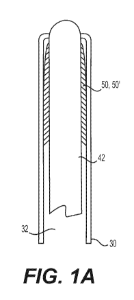
[0028] Figure 1A shows an interventional device of the present disclosure,
showing a guidewire with a compressed filter element;
[0029] Figure 1B shows the guidewire and filter of Figure 1A deployed in
the
ophthalmic artery;
[0030] Figure 10 shows a close-up view of an exemplary ophthalmic artery
filter of the present disclosure;
[0031] Figure 2A shows the guidewire placement in the ICA in relation to
the
OA,
[0032] Figure 2B shows the filter wire in the ICA and placement of a stent
near
the junction with the OA,
[0033] Figure 20 shows deployment of the stent and deployment of a filter
element in the ICA;
[0034] Figure 2D shows the stent expanded in the ICA after removal of the
filter element from the ICA;
[0035] Figure 3A shows the filter wire in a delivery position; and
[0036] Figure 3B shows the filter wire in a deployed position.
DETAILED DESCRIPTION
[0037] In at least certain embodiments, the present disclosure is directed
to
restoring and/or increasing the amount of oxygen that is available to one or
more
parts of the eye or to the eye area. Devices and methods are described.
[0038] Restoring and/or increasing the amount of oxygen is used herein to
refer to any device, method, therapy, or combination that changes the oxygen
content in or near the eye. Examples of such include, but are not limited to,
8
CA 03046006 2019-06-03
WO 2018/106858
PCT/US2017/065004
increasing the blood flow anywhere in the vasculature leading to the eye or a
portion
of the eye; removing or opening an obstruction in the fluid flow path in the
vasculature leading to the eye; delivering and deploying a stent in the fluid
flow path
in the vasculature leading to the eye; using atherectomy or similar devices to
physically remove portions of any obstructions in the vasculature leading to
the eye
or portion of the eye; and localized drug and/or an oxygen device for
increasing flow
or amount of oxygen in one or more eye tissues. In some embodiments, a device
or
method of the present disclosure may be combined with a known or new drug or
oxygen device in order to treat one or more eye diseases or conditions.
[0039] The present disclosure provides for an apparatus for deployment of a
detachable diagnostic or therapeutic implant device such as a stent 10,
embolic coil,
or other vascular occlusion device using a catheter, whereby placement of a
stent 10
or the like in a portion of the carotid artery changes the diameter of the
internal
carotid artery (ICA) 2 and/or the ophthalmic artery (OA) 4, which in turn
increases
blood flow between the ICA 2 and the eye.
[0040] The present disclosure, in at least certain aspects, is directed to
restoring and/or increasing the amount of oxygen that is available to one or
more
parts of the eye or to the eye area, specifically by removing or partially
opening a
blockage in one or more of the arteries that supplies blood flow to the eye.
In
embodiments of the disclosure, a blockage is removed or opened in the ICA 2,
the
OA 4, the ostium 6 (as used herein, referring to the junction between the ICA
2 and
the OA 4), or combinations thereof. In embodiments, the devices and methods of
the present disclosure involve increasing the blood flow and/or blood flow
rate to or
near the eye. To or near the eye, as used herein, refers to the vasculature
system
that supplies blood to the various structures of the eye.
[0041] The present disclosure includes methods, devices, and systems for
removing a blockage in the ostium, wherein removing the blockage comprises
opening a channel or access through the ostium 6 sufficient to provide a
therapeutically beneficial amount of oxygen to the eye, the rear of the eye,
or
portions thereof. The present disclosure also includes restoring and/or
improving
blood flow anywhere in the vascular pathway to or within the eye.
9
CA 03046006 2019-06-03
WO 2018/106858
PCT/US2017/065004
[0042] Another embodiment of the present disclosure includes reducing
and/or removing any blockage in the oxygen pathway to the eye. In this and
other
embodiments of the present disclosure, reducing blockage includes but is not
limited
to piercing or penetrating the blockage. In embodiments of the present
disclosure,
piercing and penetrating the blockage refers to obtaining sufficient blood
and/or fluid
flow through or around the blocked vascular area sufficient to provide a
therapeutically beneficial amount of oxygen to the eye or a portion of the
eye.
[0043] Another embodiment of the present disclosure further includes
supplying oxygen to the eye or near the eye, wherein, in this embodiment, the
source of the oxygen is external.
[0044] Another embodiment of the present disclosure includes one or more
medical devices, such as a catheter 30 or the like, and its use to clear or
penetrate a
blockage in the vascular system that provides oxygen to the eye. In
embodiments of
the present disclosure, the blockage in the vascular system is a blockage in
the
junction or ostium 6 between the ICA 2 and the OA 4.
[0045] Another embodiment of the present disclosure includes a medical
device, such as a stent 10 or the like, that is configured for and may be used
to
open, clear, or improve vascular flow to or around the eye, wherein vascular
flow
mediates the amount of oxygen that is delivered to the eye.
[0046] Typically, these procedures involve inserting the distal end of a
delivery
catheter 30 into the vasculature of a patient and guiding it through the
vasculature to
a predetermined delivery site. A vascular occlusion device may be attached to
the
end of a delivery member which pushes the occlusion device through the
catheter 30
and out of the distal end of the catheter 30 into the delivery site.
[0047] For some of these embodiments, one or more layers of the implant
device may be configured to anchor or fix the implant device in a clinically
beneficial
position. For some embodiments, the implant device may be disposed in whole or
in
part within the vascular defect in order to anchor or fix the device with
respect to the
vascular structure or defect. The one or more layers of the implant device may
be
configured to span an opening, neck or other portion of a vascular defect in
order to
isolate the vascular defect, or a portion thereof, from the patient's nominal
vascular
CA 03046006 2019-06-03
WO 2018/106858
PCT/US2017/065004
system in order to allow the defect to heal or to otherwise minimize the risk
of the
defect to the patient's health.
[0048] The present disclosure also includes a delivery system configured or
adapted to position and/or orient the stent 10 in the ostium 6.
[0049] An embodiment of the present disclosure includes methods and
devices for treating a non-human animal. Some embodiments of the present
disclosure include treating a dog, including but not limited to treating
central serous
retinopathy.
[0050] Some embodiments of a delivery system for deployment of an implant
device to treat a patient's vasculature include a microcatheter (or delivery
catheter)
30 having an inner lumen 32 extending the length thereof. The inner lumen 32
provides a passageway for an implant or other diagnostic or therapeutic device
(e.g., stent 10 and/or filter 50) to treat a patient's vasculature. Some
implant or
therapeutic device embodiments may include one or more self-expanding
resilient
layers of thin coupled filaments, the layers defining a longitudinal axis
between a
proximal end and a distal end. Such embodiments can assume a radially-
constrained, axially-elongated state configured for delivery through
microcatheter 30,
with the thin woven filaments extending longitudinally from the proximal end
to the
distal end being radially adjacent to each other, as shown in FIG. 1A. The
delivery
system further includes an elongated delivery apparatus having a proximal end
and
a distal end releasably secured to a proximal portion (e.g., a hub or the
like) of the
implant or therapeutic device.
[0051] Access to a variety of blood vessels of a patient may be
established,
including arteries such as the femoral artery, the radial artery, and the
like, in order
to achieve percutaneous access to a vascular defect. In general, the patient
may be
prepared for surgery, the access artery may be exposed (e.g., via a small
surgical
incision), and access to the lumen is gained using the Seldinger technique
where an
introducing needle is used to place a wire over which a dilator, or a series
of dilators,
may dilate a vessel allowing an access sheath to be inserted into the vessel.
This
would allow the device to be used percutaneously. With an access sheath in
place,
a guiding catheter (e.g., catheter 30) is used to provide a safe passageway
from the
entry site to a region near a treatment site. Exemplary guidewires for
vascular use
CA 03046006 2019-06-03
WO 2018/106858
PCT/US2017/065004
may include the Synchro2 made by Boston Scientific and the Glidewire Gold
Neuro
made by MicroVention Terumo. Typical guidewire sizes may include about 0.014
inches (0.36 mm) and about 0.018 inches (0.46 mm). Once the distal end of the
microcatheter 30 is positioned at the site, often by locating its distal end
through the
use of radiopaque marker material and fluoroscopy, the microcatheter 30 is
cleared.
For example, if a guidewire has been used to position the microcatheter, it
may be
withdrawn from the microcatheter 30, and then the delivery apparatus may be
advanced through the microcatheter 30.
[0052] Once the implant or therapeutic device (e.g., stent 10, filter 50,
etc.) is
deployed at a desired treatment site, the microcatheter 30 may then be
withdrawn.
Characteristics of the implant or therapeutic device (e.g., stent 10, filter
50, etc.) and
delivery apparatus discussed herein generally allow for retraction of the
implant or
therapeutic device after initial deployment into the vascular defect, but in
the case of
a permanent implant, before detachment of the implant device. Therefore, it
may
also be possible and desirable to withdraw or retrieve an initially deployed
implant
device after the fit within the vascular defect has been evaluated in favor of
a
differently-sized implant device. The tip of a catheter, such as the
microcatheter 30,
may be advanced into or adjacent to the vascular site or vascular defect. An
example of a suitable microcatheter having an inner lumen diameter of about
0.51
mm to about 0.56 mm is the Rapid Transit manufactured by Cordis Corporation.
Examples of some suitable microcatheters 30 may include microcatheters 30
having
an inner lumen 32 diameter of about 0.66 mm to about 0.71 mm, such as the
Reber
by Ev3 Company, the Renegade Hi-Flow by Boston Scientific Corporation, and
the
Mass Transit by Cordis Corporation. Suitable microcatheters 30 having an
inner
lumen 32 diameter of about 0.79 mm to about 0.84 mm may include the Marksmen
by Chestnut Medical Technologies, Inc. and the Vasco 28 by Bait Extrusion. A
suitable microcatheter 30 having an inner lumen 32 diameter of about 1.0 mm to
about 1.04 mm includes the Vasco 35 by Bait Extrusion. These microcatheters
are
listed as exemplary embodiments only, and other suitable microcatheters may
also
be used with any of the embodiments discussed herein.
[0053] It is understood that the present disclosure is not limited solely
to
changing vascular flow in order to improve or restore the amount of oxygen
that is
delivered to the eye. For example, in some embodiments of the present
disclosure,
12
CA 03046006 2019-06-03
WO 2018/106858
PCT/US2017/065004
the vascular flow may be unaffected for the most part, but the amount or
concentration of hemoglobin may be increased, thereby increasing the amount of
oxygen that may be delivered to the eye. One skilled in the art may recognize,
with
the teaching of the present disclosure, that there are other biological
systems or
capabilities that may be used to increase the amount of oxygen that is
delivered to
the eye.
[0054] In accordance with the present disclosure, any process, device, or
agent that increases the availability of oxygen to the eye or eye region is
included
within the scope of the present disclosure. These processes, devices, and
agents
include, but are not limited to internal sources of oxygen, e.g., through the
vascular
system. These processes, devices, and agents include, but are not limited to
external sources of oxygen, e.g., an injection into the eye or eye region with
one or
more substances that carry oxygen, a substance that captures or concentrates
oxygen, a device that manufactures oxygen, and/or one of more substances that
result in an increase in the amount of oxygen.
[0055] In some embodiments of the present disclosure, a stent 10, is
adapted
and configured to be delivered to any predetermined area in the vascular
system that
supplies oxygen to the eye, e.g., ICA 2. In some embodiments of the present
disclosure, the stent 10 (Figs. 2A-2D) is adapted and configured for placement
in the
ICA/ophthalmic artery ostium 6.
[0056] Stent 10 of the present disclosure may be configured for placement
in
the vasculature supplying blood to the eye. Exemplary blood vessels include
but are
not limited to the ICA 2, and the OA 4. Stent 10 may also be configured or
adapted
for treating an obstruction of the Ophthalmic/Internal Carotid Artery ostium
6,
comprising: stent 10 ranging in diameter from about 2.5 mm to about 5.5 mm,
with
an overall length ranging between 15 mm to 40 mm. The stent 10 may have a
tapered diameter to facilitate placement within the vasculature. The stent 10
may be
self-expanding, non-expanding, or expandable. In embodiments of the present
disclosure in which the stent 10 is expandable, the stent 10 may be expanded
using
any known expanding element, e.g., a balloon or the like. In some embodiments
of
the present disclosure, the stent 10 is percutaneously delivered.
13
CA 03046006 2019-06-03
WO 2018/106858
PCT/US2017/065004
[0057] The present disclosure is also directed to a system comprising stent
10
and an appropriate delivery apparatus, e.g., microcatheter 30; said system may
be
used for increasing the amount of oxygenated blood in the eye area.
[0058] A system of the present disclosure includes stent 10 configured for
placement and function in the ostium 6; microcatheter 30 for delivering the
stent 10
to the ostium 6 or near the ostium 6, and any of a number of already known
structures and devices typically delivered by microcatheter 30.
[0059] A stent 10 of the present disclosure may be constructed from
materials
commonly used in the design and manufacture of self-expanding stents. These
materials include, but are not limited to, Nitinol, chromium cobalt, stainless
steel,
polymers, and bioresorbable and/or other materials commonly used in the
coronary
vasculature.
[0060] The stent 10 may also include a cover (not shown). The cover could
be on the inner diameter, the outer diameter, some combination of location
specific
(strut or struts). It could be a fabric like covering, liquid, and/or a
degrading material.
[0061] In some embodiments of the present disclosure, the cover may
function to trap particulate in and around the area of the stent 10. In this
embodiment of the present disclosure, the cover is believed to reduce the
potential
for inducing thrombosis. In other embodiments of the present disclosure, the
stent
may include one or more anti-stenosis agents. In other embodiments, the stent
may include both functions.
[0062] The cover may be formed from PTFE, ePTFE, or other commonly used
materials designed to be affixed to the outer and/or inner diameter of the
stent 10
with the purpose of providing a method of retaining plaque (or stenotic
material) as
the stent is expanded against the artery. This cover material is designed to
expand
with the stent 10 and trap material potentially loosened by the dilatation
effect of the
stent 10 between the cover and the vascular wall.
[0063] The stent 10 or the cover may also include one of more markers,
typically radiopaque markers. The stent or cover may be coated or impregnated
with
one or more radiopaque markers 13 to aid in the proper placement of the stent
within
the target anatomy, e.g., the ostium 6 of the ICA 2 and the OA 4. Target
anatomy,
14
CA 03046006 2019-06-03
WO 2018/106858
PCT/US2017/065004
as used herein, refers to any place in the vascular system supplying blood to
the
eye, including but not limited to the ostium of the ICA 2 and OA 4.
In some embodiments, the stent 10 or its associated covering is designed to
provide
an opening 11 for accommodation of the ostium 6 such that the material does
not
block access to the ostium 6 (e.g., the opening 11 is dimensionally compatible
with
the opening of ostium 6). In some embodiments of the present disclosure, the
opening 11 is an area of the stent 10 that is free of stent struts and is
unobscured by
the stent cover. An exemplary opening 11 is shown in the Figs. 2B-2D. As
shown,
the opening of the stent 10 (and any associated stent cover) is configured to
correspond or align with complementary markers integrated into the
microcatheter
30. These markers are designed to facilitate proper placement of the stent 10
within
the anatomy such that the ostium 6 is not blocked by the stent/stent cover
material.
[0064] In another embodiment, the stent 10 is disposed within a delivery
microcatheter 30 and delivery sheath, said microcatheter 30 having a means of
providing a single radiopaque marker or plurality of radiopaque markers to aid
in the
positioning the stent 10 in the appropriate anatomical location within the
target
anatomy.
[0065] In another embodiment, the stent 10 is designed to deploy (e.g.,
via
self-expansion) such that the distal portion of the stent 10 deploys first and
aids in
anchoring the stent 10 prior to deployment of the proximal section of the
stent 10.
This may enable the physician to accurately place the stent 10 within the
target
anatomy. The stent 10 is first placed in the desired location, and then fully
delivered.
[0066] In another embodiment, the stent 10 is designed with an
asymmetrical
feature that exerts additional diametric force in the area of the ostium 6.
[0067] The stent 10 of the present disclosure may be delivered using any
medically appropriate route and/or technique. Suitable routes include but are
not
limited to subclavian, brachial, and/or direct common carotid access. In an
embodiment, the device and system is configured for percutaneous access of the
ICA 2 via a femoral approach, as well as other typical percutaneous access
locations.
CA 03046006 2019-06-03
WO 2018/106858
PCT/US2017/065004
[0068] In another embodiment, the system is configured to be used with
commonly available coronary guide wire products in styles and size ranges.
[0069] A stent 10 or stent cover of the present disclosure may be
configured
to be visible using non-invasive imaging techniques (e.g., fluoroscopy, etc.).
In this
embodiment of the present disclosure, the stent 10 and/or cover may include
one of
more elements to assist in positioning and deploying the stent 10.
[0070] In use, the stent 10 is mounted on a central catheter 34 within
microcatheter 30 by means of an outer sheath 33 that compresses and holds the
stent 10 against a portion of the central catheter 34 to aid in the delivery
of the stent
to the desired anatomy. Controlled removal of the sheath 33 may provide for
the
ability to deliver the stent 10 to the desired anatomical location. The sheath
33 may
include a mechanical element to allow for controlled advancement and/or
retraction
of the stent 10. The sheath also may have radiopaque markings to aid in the
positioning and delivery of the stent 10.
[0071] As shown in FIG. 2A, a guidewire 40 may be delivered via any
appropriate means to a target location within the vasculature. Once so
positioned,
sheath 33, central catheter 34, stent 10, and microcatheter 30 (not shown in
FIGS.
2A-2D)may be advanced over guidewire 40 (e.g., via a lumen of central catheter
34),
as shown in FIG. 2B. Alternatively, microcatheter 30 first may be delivered to
the
site over the guidewire 40, followed by sheath 33 and central catheter 34
carrying
stent 10. Once proper placement is achieved, the guidewire 40 may be removed
and
replaced with a filter wire 42, as shown in FIG. 2B. The filter wire 42 may be
deployed such that an optional filtering capability (e.g., via filter 50) is
placed distal to
the ostium 6 and outside of the field of stent 10 deployment. Once in the
proper
position, the filter 50 may be deployed such that filtering capability is
provided, as
shown in FIG. 2C. The stent 10 is then manipulated with the aid of the
radiopaque
markings such that the ostium 6 will not be obscured by the stent 10 (e.g.,
such that
opening 11 is aligned with ostium 6). The stent 10 is then deployed by slowly
retracting the sheath 33 overlying stent 1030, as shown in FIG. 2C. Retracting
the
sheath 33 may be aided by radiopaque markings on the sheath as well as
markings
on the stent 10. The distal portion of the stent 10 is delivered first to
ensure the
ostium 6 will not be blocked. Once distal portion of the stent 10 is in place
and/or
16
CA 03046006 2019-06-03
WO 2018/106858
PCT/US2017/065004
delivered to a desired location, observation of a non-blocked ostium 6 is
confirmed
and the proximal portion of the stent 10 is delivered. Next, the filter wire
42, filter 50,
and any captured debris is withdrawn into the microcatheter 30 and removed.
FIG.
2D shows the stent 10 positioned in the ICA 2 with the opening 11 aligned with
the
ostium 6 between the ICA 2 and the OA 4.
[0072] The present disclosure is also directed to a system comprising one
or
more medical devices, (e.g., a stent 10) and its delivery apparatus; said
system is
used for increasing the amount of oxygenated blood in the eye area, or for
increasing the amount of oxygen that is or can be delivered to the eye. The
present
disclosure may also include this system, device, or method in combination with
one
or more agents or devices for improving vascular blood flow between the common
carotid artery and a central artery of the retina; and/or one or more agents
for
improving vascular blood flow at the ostium 6 and within the OA 4.
[0073] The present disclosure further includes the use of one of more
diagnostic devices or agents that allow a person to monitor oxygen content in
the
eye.
[0074] In another embodiment, a medical device or agent is capable of
delivering drugs to the ostium 6 for the purpose of improving vascular blood
flow at
the ostium 6 and within the OA 4. These drugs may include (but are not limited
to)
low dose Viagra (or equivalent RPE inhibitor), Lucentis, Avastin, Taxol,
Rapamyacin
or other pharmaceuticals used to improve vascular blood flow.
[0075] In an embodiment of the filter 50, the device provides distal emboli
protection as part of the stent delivery system (but not limited to stents).
Indeed, the
filter wire 42 (which also may serve as a guidewire 42), as shown in FIG. 2B,
is
designed with an overall length intended to facilitate the appropriate
anatomical
approach, e.g., femoral access would be about 180 cm in overall length. Other
access points would use a guidewire/filter wire 42 with an overall length
appropriate
for their respective access locations. The diameter of a distalmost segment of
the
wire 42 may range from about 0.008" to about 0.014". Filter wire 42 may
include
Nitinol material, or the like. The filter wire 42 may have a filter 50 element
attached
(or monolithically and integrally formed therewith) at a distal end thereof,
which may
be composed of expanded polyester thread, suture material, or equivalent. The
filter
17
CA 03046006 2019-06-03
WO 2018/106858
PCT/US2017/065004
50 may continue alongside the filter wire 42 (e.g., in a generally parallel
fashion) (as
shown in FIG. 3A) except for a proximalmost portion of a delivery system
(e.g.,
microcatheter 30) nearest the user. A tip (e.g., a distalmost end of filter
wire 42
coupled to filter 50) of the guidewire/filter wire 42 is positioned distal to
the delivery
system (e.g., microcatheter 30) such that it will not interfere with the stent
10
delivery, but will be in close enough approximation so as to effectively
provide debris
capture capability. Once in the desired location, the guidewire 42 is slightly
withdrawn while simultaneously rotated so as to place the filter 50 in a
random coiled
circular pattern (e.g., a bunched, longitudinally shortened configuration)
within the
vasculature, as shown in FIG. 20 and 3B. This arrangement serves to provide
filtering capability for any potentially dislodged material during stent
deployment.
The filter 50 may be treated with a platelet aggregation compound, such as
nitric
oxide, to reduce the likelihood of platelet aggregation (clotting or thrombus
formation)
and may be imparted with a specific electrical charge to facilitate attraction
of debris
to the filter 50 and/or filter wire 42. Removal of the filter 50 (and any
trapped
material) is accomplished by slight advancement of the delivery catheter
(e.g.,
microcatheter 30) and/or withdrawing the filter 50 into the delivery catheter
(e.g.,
microcatheter 30). It is understood that the direction of filter 50 in FIGS.
3A and 3B
is reversed relative to the direction of filter 50 in FIGS. 2B and 20. That
is, a distal
end of filter 50 is positioned to the left in FIGS. 3A and 3B while a distal
end of filter
50 is positioned to the right in FIGS. 2B and 20.
[0076] In another embodiment, the filter wire 42 is used in conjunction
with
several other components, including a delivery sheath 33 with mounted stent 10
on a
central catheter 34. The central catheter 34 may incorporate a through lumen
intended to facilitate the use of a common guidewire to aid in positioning the
device
within the target vasculature. Once proper placement is achieved, the common
guidewire is removed and replaced with a filter wire 42, as described above in
connection with FIGS. 2A-20. The filter wire 42 is deployed such that the
filtering
capability is placed distal to the ostium 6 and outside of the field of stent
10
deployment. Once in the proper position, the filter 50 is deployed such that
filtering
capability is provided. The stent 10 is then manipulated and deployed. Once
the
stent 10 is in place, the filter wire 42 and any captured debris is withdrawn
into the
sheath 33 or microcatheter 30 and the system removed.
18
CA 03046006 2019-06-03
WO 2018/106858
PCT/US2017/065004
[0077] An embodiment of a device and system of the present disclosure
includes a filter element 50' configured and adapted for deployment in the OA
4. An
exemplary configuration is shown in FIGS. 1A-10. FIG. 1A shows the filter 50'
compressed around a guidewire/filter wire 42, and positioned within a lumen 32
of
microcatheter 30. FIG. 1B shows an example of a suitable deployment of the
filter
50' in the OA 4. Microcatheter 30 may be positioned in the ICA 2 (as shown) or
may
be extended into the ostium 6 and/or further into the OA 4. The filter 50' may
be
deployed in the OA 4 (as shown) or may be deployed at any position between the
ostium 6 and further into the OA 4.
[0078] Figure 10 shows a close-up of an exemplary filter 50' configured for
use and deployment in the OA. As illustrated, the filter 50' may include one
or more
micropores 52, 54, typically for capturing, collecting, and removing debris.
In the
illustrated embodiment, some of the micropores 52 capture debris or allow
debris to
enter the filter; other micropores 54 allow blood to pass by and through the
filter 50'.
[0079] In one embodiment, the ophthalmological disease or disorder treated
or prevented by any of the methods or compositions described herein is age-
related
macular degeneration. Vision changes that can be associated with macular
degeneration include distortions and/or blind spots (scotoma) detected using
an
Amsler grid, changes in dark adaptation (diagnostic of rod cell health),
changes in
color interpretation (diagnostic of cone cell health), or a decrease in visual
acuity.
Examples of age-related macular degeneration are normeovascular (also known as
"dry") and neovascular (also known as "wet" or "exudative") macular
degeneration.
[0080] In one embodiment, the dry age-related macular degeneration is
associated with the formation of drusen. In one embodiment, treating or
preventing
dry macular degeneration encompasses treating or preventing an abnormality of
the
retinal pigment epithelium and/or underlying vasculature, known as
choriocapilaries.
Examples of abnormalities of the retinal pigment epithelium include geographic
atrophy, non-geographic atrophy, focal hypopigmentation, and focal
hyperpigmentation. In another embodiment, treating or preventing wet age-
related
macular degeneration encompasses treating or preventing choroidal
neovascularization or pigment epithelial detachment.
19
CA 03046006 2019-06-03
WO 2018/106858
PCT/US2017/065004
[0081] In some embodiments, wet age-related macular degeneration is
classified according to the appearance of its choroidal neovascularization
(CNV), into
classic, occult or mixed (classic and occult) CNV types, as determined by an
angiography, known as fluorescence angiography. Classic, occult or mixed
(classic
and occult) CNV classification can be based on the time, intensity and level
of
definition of dye appearance, and leakage from the CNV, as assessed by the
fluorescein angiography. In some embodiments, the subject has classic CNV
(e.g.,
pure classic) or mixed CNV (predominantly or minimally classic CNV). In some
embodiments, the subject has occult CNV (e.g., pure occult CNV).
[0082] In certain embodiments, the ophthalmological disease or disorder is
a
cataract (e.g., age-related cataract), diabetic macula edema, macular
telangiectasia
(e.g., type 1 or 2 macular telangiectasia), atrophic macular degeneration,
chorioretinopathy (e.g., central serous chorioretinopathy), retinal
inflammatory
vasculopathy, pathological retinal angiogenesis, age-related maculopathy,
retinoblastoma, Pseudoxanthoma elasticum, a vitreoretinal disease, choroidal
sub-
retinal neovascularization, central serous chorioretinopathy, ischemic
retinopathy,
hypertensive retinopathy or diabetic retinopathy (e.g., nonproliferative or
proliferative
diabetic retinopathy, such as macular edema or macular ischemia), retinopathy
of
prematurity (e.g., associated with abnormal growth of blood vessels in the
vascular
bed supporting the developing retina), venous occlusive disease (e.g., a
retinal vein
occlusion, branch retinal vein occlusion or central retinal vein occlusion),
arterial
occlusive disease (e.g., branch retinal artery occlusion (BRAO), central
retinal artery
occlusion or ocular ischemic syndrome), central serous chorioretinopathy
(CSC),
cystoid macular edema (CME) (e.g., affecting the central retina or macula, or
after
cataract surgery), retinal telangiectasia (e.g., characterized by dilation and
tortuosity
of retinal vessels and formation of multiple aneurysms, idiopathic JXT,
Leber's
miliary aneurysms, or Coats' disease), arterial macroaneurysm, retinal
angiomatosis,
radiation-induced retinopathy (RIRP), or rubeosis iridis (e.g., associated
with the
formation of neovascular glaucoma, diabetic retinopathy, central retinal vein
occlusion, ocular ischemic syndrome, or chronic retinal detachment).
[0083] Embodiments of the present disclosure and the various components or
elements thereof can be used interchangeably so that features and functions of
one
exemplary embodiment of a filter device can be used with other embodiments of
the
CA 03046006 2019-06-03
WO 2018/106858
PCT/US2017/065004
filter device. Illustratively, the restraining members or mechanisms of the
described
embodiments of the present disclosure can be used with multiple different
configurations of the filter 50, 50' device. Further, exemplary capture
catheters 30
can be used interchangeably such that any capture catheter can be used with
any of
the described filter 50, 50' devices and such other that may be known to those
skilled
in the art in light of the teaching contained herein. Additionally, methods of
using
one embodiment of the present disclosure can be used with other embodiments of
the present disclosure. Therefore, embodiments of the present disclosure
provide
filter 50, 50' devices that have small or low profiles, few parts and
components, are
simple to manufacture and use, are able to be easily inserted into a patient,
be
steerable through the tortuous anatomy of a patient, provide filtering
capabilities,
provide exchange capability so other medical devices can be advanced over or
along the filter device, and be capable of removing captured material without
allowing such material to escape during filter retrieval.
EXAMPLES
[0084] Example 1
[0085] Compromised blood flow to the vasculature of the posterior eye may
directly contribute to diseases of the eye. This lack of normal blood flow may
originate in the ICA 2, the OA 4, branches of the OA 4, and/or combinations
thereof,
and be directly caused by a blockage in one or more of these vessels. This
lack of
sufficient blood flow may directly contribute to inadequate oxygen levels seen
in
tissues such as the choroid, retina, optic nerve and other ophthalmic anatomy.
This
blockage may manifest as stenosis, lesions or other physiology within the
ophthalmic
related vasculature and compromise normal blood flow such that the posterior
eye
vasculature does not receive an adequate oxygen supply for maintenance of
normal
function. As a result of this reduction of oxygen, it is possible for a
cascade of
events to begin which may result in various diseases of the eye.
[0086] Blood flow was measured for healthy controls and diseased patients
(with confirmed AMD diagnosis). Flow rates were measured for the Left
Ophthalmic
Artery (LOA), Right Ophthalmic Artery (ROA), Left Internal Carotid Artery
(LICA) and
21
CA 03046006 2019-06-03
WO 2018/106858
PCT/US2017/065004
Right Internal Carotid Artery (RICA) using a Phased Contrast Magnetic
Resonance
Imaging (PCMRI) technique. These flow rates were measured in cm/sec. The
average size of the ICA was 4.66 mm and the average size of the OA was 1.00
mm.
[0087] Specific flow rates were compared, and the OA flow data showed a
medically or clinically observable difference between the flow rates for
healthy
controls compared to diseased patients. Specific flow rates were compared, and
the
ICA flow data showed a medically or clinically observable difference between
the
flow rates for healthy controls compared to diseased patients. In every case,
the
blood flow rate for the diseased patients appears to be lower than the blood
flow rate
for the healthy controls.
[0088] Example 2
[0089] Cadaveric tissue samples were obtained with confirmed diagnosis of
CAD with no diagnosis of AMD. Visual confirmation of the presence of stenosis
in
the ophthalmic/internal carotid ostium of the samples was performed. One
sample
had extensive stenosis that appeared to completely block the OA in both the
left and
right ICA/OC ostiums. It should be noted that the left OA, as observed
branching off
the ICA, was much smaller in diameter than that of a typical OA, almost to the
point
of being non-existent. This sample was diagnosed with CAD, CHF, PAD, HTN and
4x bypass Sx.
[0090] A different sample had what appeared to be early stage stenosis
accumulation in both the left and right ICA/OA ostiums as confirmed by visual
observation. None of these stenosis appeared to cause blockage in the OA of
either
ostium. This sample was diagnosed with CAD, chronic anemia, Buerger's disease,
thromboembolic disease and extensive DVT.
[0091] Example 3
[0092] In another sample the right ICA was removed and the ostium was
visually examined. A blockage of the OA at the ostium was confirmed and
appeared
to be complete. Once the section of left ICA was removed, internal access to
the
OA ostium was gained, and a micro PTCA balloon catheter was inserted. This
test
was performed to visually observe the effect of placing and inflating a
balloon
catheter in the OA. This (non-compliant) balloon catheter has a maximum
diameter
22
CA 03046006 2019-06-03
WO 2018/106858
PCT/US2017/065004
of 0.85 mm at 16 atms, with a crossing profile of 0.74 mm and a working length
of
approximately 5 mm. The balloon was inflated several times to approximately 12
atms max, and the balloon was observed through the vessel. The vessel appeared
to tolerate the inflations without obvious damage.
23