Note: Descriptions are shown in the official language in which they were submitted.
CA 03046087 2019-06-04
WO 2018/107123 PCT/US2017/065469
SYSTEMS, DEVICES, AND METHODS FOR THE ACCURATE DEPLOYMENT OF AN
IMPLANT IN THE PROSTATIC URETHRA
CROSS-REFERENCE TO RELATED APPLICATIONS
[0001] This application claims priority to and the benefit of U.S.
Provisional Application
Serial No. 62/432,542, filed December 9, 2016, which is incorporated by
reference herein in its
entirety for all purposes.
FIELD
[0002] The subject matter described herein relates to systems, devices, and
methods for
delivery or deployment of an implant into the prostatic urethra, more
specifically, delivery in an
atraumatic and minimally-invasive manner through the tortuous bends of the
male urethra.
BACKGROUND
[0003] There are numerous clinical reasons for placement of an implant into
the prostatic
urethra, such as for treatment of urinary retention associated with benign
prostatic hyperplasia
(BPH), blockages from prostate cancer, bladder cancer, urinary tract injury,
prostatitis, bladder
sphincter dyssynergia, benign or malignant urethral stricture, and other
conditions for which
treatment is desired. Due to the naturally complex and tortuous anatomical
geometry, patient-to-
patient geometric and tissue variability, and anatomical restrictions
associated with those
conditions, accurate and consistent placement of an implant into the prostatic
urethral lumen has
proven challenging. Furthermore, complex challenges are presented in the
design and/or
fabrication of systems with sufficient flexibility to deliver such an implant
in a minimally-
invasive manner. For these and other reasons, needs exist for improved
systems, devices, and
methods of implant delivery to the prostatic urethra.
SUMMARY
[0004] Provided herein are a number of example embodiments of delivery
systems for
delivering or deploying implants within the prosthetic urethra or other parts
of the body, and
methods related thereto. Embodiments of the delivery system can include a
delivery device
insertable into the prosthetic urethra and a proximal control device coupled
with the delivery
device and configured to control deployment of one or more implants from the
delivery device.
In some embodiments, the delivery device can include multiple tubular
components each having
- 1 -
CA 03046087 2019-06-04
WO 2018/107123 PCT/US2017/065469
various functions described in more detail herein. Multiple embodiments of
implants for use
with the delivery systems are also described.
[0005] Other systems, devices, methods, features and advantages of the
subject matter
described herein will be or will become apparent to one with skill in the art
upon examination of
the following figures and detailed description. It is intended that all such
additional systems,
methods, features and advantages be included within this description, be
within the scope of the
subject matter described herein, and be protected by the accompanying claims.
In no way should
the features of the example embodiments be construed as limiting the appended
claims, absent
express recitation of those features in the claims.
BRIEF DESCRIPTION OF THE FIGURES
[0006] The details of the subject matter set forth herein, both as to its
structure and operation,
may be apparent by study of the accompanying figures, in which like reference
numerals refer to
like parts. The components in the figures are not necessarily to scale,
emphasis instead being
placed upon illustrating the principles of the subject matter. Moreover, all
illustrations are
intended to convey concepts, where relative sizes, shapes and other detailed
attributes may be
illustrated schematically rather than literally or precisely.
[0007] FIG. 1A is a block diagram depicting an example embodiment of a
delivery system.
[0008] FIGs. 1B, 1C, and 1D are side, end, and perspective views,
respectively, depicting an
example embodiment of an implant.
[0009] FIGs. 2A-2H are perspective views depicting example embodiments of a
delivery
system in different stages of deployment of an implant.
[0010] FIGs. 3A-3C are perspective views depicting example embodiments of a
grasper
component in use within a delivery system.
[0011] FIGs. 4A-4J are partial cross-sectional views depicting example
embodiments of
anchor delivery members of a delivery system.
[0012] FIGs. 5A-5B are side views depicting an example embodiment of a
delivery system
in various stages of deployment of an implant.
[0013] FIGs. 6A and 6B are interior side and interior perspective views,
respectively,
depicting an example embodiment of a proximal control device.
[0014] FIG. 6C is a perspective view depicting an example embodiment of a
gear for use
with the delivery system.
- 2 -
CA 03046087 2019-06-04
WO 2018/107123 PCT/US2017/065469
[0015] FIG. 7A is an interior top down view depicting an example embodiment
of
components of a proximal control device.
[0016] FIG. 7B is a perspective view depicting an example embodiment of a
cam.
[0017] FIG. 8 is an interior side view depicting an example embodiment of a
gear assembly.
[0018] FIGs. 9A-9F are interior perspective views depicting an example
embodiment of
components of a proximal control device.
[0019] FIG. 10A is a flowchart depicting an example embodiment of a method
for delivering
an implant.
[0020] FIG. 10B is a timing diagram depicting an example embodiment of a
sequence of
steps for deploying an implant.
DETAILED DESCRIPTION
[0021] Before the present subject matter is described in detail, it is to
be understood that this
disclosure is not limited to the particular embodiments described, as such
may, of course, vary.
It is also to be understood that the terminology used herein is for the
purpose of describing
particular embodiments only, and is not intended to be limiting, since the
scope of the present
disclosure will be limited only by the appended claims.
[0022] The subject matter presented herein is described in the context of
delivery or
deployment of one or more implants within the prostatic urethra. The purpose
for deployment of
the implant(s) in the prostatic urethra can vary. The embodiments described
herein are
particularly suited for treatment of BPH, but they are not limited to such.
Other conditions for
which these embodiments can be used include, but are not limited to, treatment
of blockages
from prostate cancer, bladder cancer, urinary tract injury, prostatitis,
bladder sphincter
dyssynergia, and/or benign or malignant urethral stricture. Further, these
embodiments can have
applicability for deployment of one or more implants in other locations of the
urinary tract or in
the bladder, as well as other biological lumens, cavities, or spaces, such as
the human
vasculature, cardiac system, pulmonary system, or gastro-intestinal tract,
including locations
within the heart, stomach, intestines, liver, spleen, pancreas, and kidney.
[0023] FIG. 1A is a block diagram depicting an example embodiment of
delivery system 100
having an elongate delivery device 103 coupled with a proximal control device
200. A distal end
region 104 is adapted to be inserted into the patient's urethra (or other
lumen or body cavity of
- 3 -
CA 03046087 2019-06-04
WO 2018/107123 PCT/US2017/065469
the patient) through the urethral orifice. Distal end region 104 preferably
has an atraumatic
configuration (e.g., relatively soft and rounded) to minimize irritation or
trauma to the patient.
Elongate delivery device 103 carries or houses one or more implants 102 (not
shown) to be
delivered or deployed within or adjacent to the prostatic urethra. A proximal
end region 105 of
delivery device 103 is coupled with proximal control device 200, which remains
outside of the
patient's body and is configured to be used by the physician or other
healthcare professional to
control the delivery of one or more implants 102.
Example Embodiments of Delivery Devices and Related Methods
[0024] FIGs. 1B, 1C, and 1D are side, end, and perspective views,
respectively, depicting an
example embodiment of implant 102 in an at-rest configuration. Implantable
device 102 is
biased towards the at-rest configuration depicted here and is deformable
between the at-rest
configuration and a relatively more elongate housed (or delivery)
configuration (e.g., see FIG.
3A) for housing implant 102 within delivery device 103. The housed
configuration can be a
straight or lineated state with minimal curvature. The at-rest configuration
has a relatively
greater lateral width, and a relatively shorter longitudinal length than the
housed configuration.
Upon exiting an open end of delivery device 103, implant 102 is free to
transition its shape back
towards that of the at-rest configuration although restraints imparted by the
patient's urethral
wall may prevent implant 102 from fully reaching the at-rest configuration.
Because implant
102 is biased towards the at-rest configuration, implant 102 is configured to
automatically
expand when freed from the restraint of delivery device 103 and can be
referred to as "self-
expanding." The shape of implant 102 in its deployed state within, e.g., the
patient's urethra, can
be referred to as the deployed configuration, and will often be a shape that
is deformed from the
at-rest configuration by the surrounding tissue, although the deployed
configuration can be the
same as the at-rest configuration.
[0025] Implant 102 can be configured in numerous different ways, including
any and all of
those implant configurations described in U.S. Patent Publ. No. 20150257908
and/or Int'l Publ.
No. WO 2017/184887, both of which are incorporated by reference herein for all
purposes.
[0026] Implant 102 can be formed from one or more discrete bodies (e.g.,
wires, ribbons,
tubular members) of varying geometries. Referring to the embodiment of FIGs.
1B-1D, implant
102 has a main body formed of only one single wire member set in a
predetermined shape.
- 4 -
CA 03046087 2019-06-04
WO 2018/107123 PCT/US2017/065469
Implant 102 can have two or more ring-shaped structures 111 (in this
embodiment there are four:
111a, 111b, 111c, and 111d) with one or more interconnections 112 extending
between each pair
of adjacent ring-shaped structures 111 (in this embodiment there is one
interconnection between
each adjacent pair, for a total of three: 112a, 112b, and 112c). Each
interconnection 112 extends
from one ring-shaped structure 111 to an immediately adjacent ring-shaped
structure 111. Each
interconnection 112 can have a relatively straight shape (not shown) or a
curved (e.g., semi-
circular or semi-elliptical) shape as shown in FIGs. 1B-1D.
[0027] Ring-shaped structures 111 are configured to maintain the urethra in
a fully or
partially open state when expanded from the housed configuration. Device 100
can be
manufactured in various sizes as desired, such that the width (e.g., diameter)
of each ring-shaped
structure 111 is slightly larger than the width of the urethra, and the length
of each
interconnection 112 determines the spacing between ring-shaped structures 111.
Ring-shaped
structures 111 can have the same or different widths. For example, in the
embodiment depicted
here, ring-shaped structure 111a has a relatively smaller width than
structures 111b-111d, which
have the same width. This can accommodate prostatic urethras that converge to
a smaller
geometry before the bladder neck.
[0028] Each ring-shaped structure 111 can be located or lie in a single
plane, and in some
embodiments that single plane can be oriented with a normal axis perpendicular
to a central
access 124 of implant 102 (as depicted in FIG. 1B). In other embodiments, ring-
shaped
structures 111 can be located in multiple planes. Ring-shaped structures 111
can extend around
central axis 126 to form a complete circle (e.g., a 360-degree revolution) or
can form less than a
complete circle (e.g., less than 360 degrees) as shown here. Although not
limited to such, in
many embodiments ring-shaped structures 111 extend between 270 and 360
degrees.
[0029] As can be seen from FIGs. 1B-1D, the geometry of implant 102 can
have a
cylindrical or substantially cylindrical outline shape with a circular or
elliptical cross-section. In
other embodiments, implant 102 can have a prismatic or substantially prismatic
shape with
triangular or substantially triangular cross-section, or otherwise.
[0030] Implant 102 can also include a distal engagement member 114 and a
proximal
engagement member 115 that are each configured to engage with elements of
delivery device
103. Engagement with delivery device 103 can serve one or more purposes such
as allowing
control of the release of implant 102, allowing movement of the ends of
implant 102 relative to
- 5 -
CA 03046087 2019-06-04
WO 2018/107123 PCT/US2017/065469
each other, and/or allowing retrieval of implant 102 after deployment, e.g.,
in an instance where
the physician desires to recapture implant 102 and redeploy implant 102 in a
different position.
In this embodiment, distal engagement member 114 is a wire-like extension from
ring-shaped
structure 111a that has a curved (e.g., S-like) shape for positioning an
atraumatic end 116 (e.g.,
rounded, spherical, ballized) in a location suitable for engagement with
delivery device 103 and
thereby allow control of the distal end region of implant 102. Likewise,
proximal engagement
member 115 has a curved shape for positioning another atraumatic end 117 in a
location suitable
for engagement with delivery device 103 and thereby allow control of the
proximal end region of
implant 102. In other embodiments, distal engagement member 114 and proximal
engagement
member 115 can be omitted, and delivery device 103 can couple with implant 102
at one or more
other distal and/or proximal locations, such as on a ring-shaped structure 111
or interconnect
112.
[0031] Delivery device 103 can include one or more elongate flexible
members (e.g., 120,
130, 140, and 150 as described below), each having one or more inner lumens.
One or more
elongate flexible members of delivery device 103 can be a solid or a non-
hollow member with no
inner lumen. FIG. 2A is a perspective view depicting an example embodiment of
distal end
region 104 of a delivery device 103. In this embodiment, delivery device 103
includes a first
elongate tubular member 120, a second elongate tubular member 130, a third
elongate tubular
member 140, and a fourth elongate tubular member 150. Delivery device 103 can
vary and in
other embodiments can include more or less tubular members.
[0032] In this embodiment, first elongate tubular member 120 is the
outermost tubular
member and is flexible yet provides support for members contained therein.
First tubular
member 120 is referred to herein as outer shaft 120 and can have one or more
inner lumens. In
this embodiment, outer shaft 120 includes a first inner lumen 121 housing
second elongate
tubular member 130, which is referred to herein as inner shaft 130. Outer
shaft 120 and inner
shaft 130 are each controllable independent of the other. Inner shaft 130 can
slide distally and
proximally within lumen 121 and is shown here partially extending from an open
distal terminus
of outer shaft 120.
[0033] In this embodiment, outer shaft 120 includes three additional lumens
122, 123, and
124. An illumination device (not shown) and an imaging device (not shown) can
be housed in
either of lumens 122 and 123. The imaging device can utilize any desired type
of imaging
- 6 -
CA 03046087 2019-06-04
WO 2018/107123 PCT/US2017/065469
modality, such as optical or ultrasound imaging. In one example embodiment the
imaging
device utilizes a forward (distal) looking CMOS imager. The illumination
device can be
configured to provide adequate illumination for optical imaging, and in one
embodiment
includes one or more light emitting diodes (LEDs). In embodiments where
illumination is not
required, such as for ultrasound imaging, the illumination device and its
respective lumen 122 or
123 can be omitted. The illumination device and/or the imaging device can each
be fixedly
secured at the distal terminuses of lumens 122 and 123, or each can be
slidable within lumens
122 and 123 to allow advancement further distally from outer shaft 120 and/or
retraction into
outer shaft 120. In one example embodiment, the illumination device and the
imaging device are
mounted together and only a single lumen 122 or 123 is present for that
purpose. Lumen 124
can be configured as an irrigation or flush port from which fluid such as
saline can be introduced
to the urethra to flush the region and provide adequate fluid through which
implant 102 and the
surrounding prostatic urethra wall can be imaged.
[0034] Outer shaft 120 has a proximal end (not shown) coupled with proximal
control device
200. Delivery device 103 can be configured to be steerable to navigate
tortuous anatomy.
Steerability can be unidirectional (e.g., using a single pull wire) or
multidirectional (e.g., using
two or more pull wires arranged at different radial locations about device
103) depending on the
needs of the application. In some embodiments, the structures (e.g., pull
wires) for steerability
extend from distal end region 104 of delivery device 103 (e.g., where the
distal ends of the pull
wires are secured to a plate or other structure within distal end region 104)
to proximal control
device 200, where they can be manipulated by the user to steer delivery device
103. The steering
structures can be located in one or more lumens of outer shaft 120 or can be
coupled to or
embedded within a sidewall of outer shaft 120. Delivery device 103 can be
biased to deflect in a
particular lateral direction (e.g., bend) such that device 103 automatically
deflects in that manner
and forces imparted to steer delivery device 103 are in opposition to this
biased deflection.
Other mechanisms for steering delivery device 103 can also be used. The
steering mechanism
may also be locked or adjusted during deployment of implant 102 to control the
position of
implant 102 within the anatomy (e.g., steering anteriorly during deployment
may help place
implant 102 in a more desirable anterior position).
[0035] Inner shaft 130 can include one or more inner lumens for housing one
or more
implants 102 and/or other components. In this embodiment, inner shaft 130
includes a first
- 7 -
CA 03046087 2019-06-04
WO 2018/107123 PCT/US2017/065469
lumen 131 in which one or more implants 102 can be housed, and a second lumen
132 in which
third elongate tubular member 140 can be housed. In this embodiment, third
elongate tubular
member 140 is configured to releasably couple with the distal end region of
implant 102 and is
referred to as a distal control member or tether 140. Distal control member
140 can be slidably
advanced and/or retracted with respect to inner shaft 130. Distal control
member 140 can
include an inner lumen 141 that houses fourth elongate tubular member 150,
which is shown
here extending from an open distal terminus of distal control member 140.
Fourth elongate
tubular member 150 is configured to anchor delivery device 103 with respect to
the patient's
anatomy, e.g., to keep components of delivery device 103 stationary with
respect to the anatomy
during deployment of implant 102 and is referred to as anchor delivery member
150.
[0036] In the configuration depicted in FIG. 2A, anchor delivery member 150
is extended
from lumen 141 of distal control member 140, and distal control member 140
along with inner
shaft 130 are shown extended from lumen 121 of outer shaft 120. When delivery
device 130 is
advanced through the urethra, anchor delivery member 150 is preferably housed
entirely within
distal control member 140, and distal control member 140 along with inner
shaft 130 are
retracted from the positions shown in FIG. 2A such that they reside within
lumen 121 of outer
shaft 120 and do not extend from the open distal terminus of lumen 120. In
other words, in some
embodiments the open distal terminus of outer shaft 120 forms the distalmost
structure of device
103 upon initial advancement through the urethra. This facilitates steering of
delivery device
103 by outer shaft 120. The physician can advance distal end region 104 of
delivery device 103
to be in proximity with the desired implantation site, or entirely into the
patient's bladder.
Anchor delivery member 150 can be exposed from the open distal terminus of
distal control
member 140, either by distally advancing anchor delivery member 150 further
into the bladder,
or if already present within the bladder, then by proximally retracting the
other components of
delivery device 103. At this point the anchor from anchor delivery member 150
can be deployed
in the bladder.
[0037] The placement of these components within system 100 is not limited
to the
embodiments described with respect to FIG. 2A. In some embodiments, outer
shaft 120 can be
omitted altogether. In such embodiments, visualization of the deployment
procedure can be
accomplished with external imaging such as fluoroscopy, where implant 102 and
delivery device
103 can be radiopaque or can include radiopaque markers, and where the imaging
and
- 8 -
CA 03046087 2019-06-04
WO 2018/107123 PCT/US2017/065469
illumination lumens 122 and 123 (and the imaging and illumination devices), as
well as the
irrigation lumen are omitted. In some embodiments, instead of distal control
member 140 being
slidably received within inner shaft 130, distal control member 140 can be
slidable within a
lumen of outer shaft 120 (either the same lumen receiving inner shaft 130 or a
different lumen).
Similarly, instead of anchor delivery member 150 being slidably received
within distal control
member 140, anchor delivery member 150 can be slidable within a lumen of outer
shaft 120
(either the same lumen receiving inner shaft 130 and/or anchor delivery member
150 or a
different lumen) or a lumen of inner shaft 130 (either the same lumen
receiving distal control
member 140 or a different lumen). In some embodiments, outer shaft 130 has a
separate and
distinct lumen for each of members 130, 140, and 150, and can be configured to
deploy implant
102 around members 140 and 150.
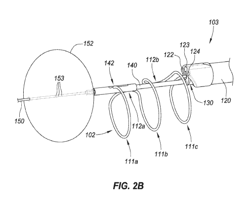
[0038] FIG. 2B is a perspective view depicting distal end region 104 of
delivery device 103
with the various components deployed. In this embodiment, anchor delivery
member 150
includes an anchor 152 in the form of an inflatable member or balloon. Other
embodiments of
anchors 152 are described with respect to FIGs. 4A-4G. Anchor 152 expands (or
otherwise
transitions) to a size greater than that of the bladder neck such that anchor
152 resists proximal
retraction (e.g., a relatively light tension). In embodiments where anchor 152
is a balloon, that
balloon can be elastic or inelastic and inflatable with an inflation medium
(e.g., air or liquid such
as saline) introduced into balloon 152 through one or more inflation ports
153. Here three
inflation ports 153 are located on the shaft of anchor delivery member 150 and
communicate
with an inflation lumen that extends proximally back to proximal control
device 200, which can
include a port for inflation with a syringe. Upon deployment of anchor 152,
the physician can
proximally retract delivery system 100 until anchor 152 is in contact with the
bladder neck
and/or wall (if not already).
[0039] The physician can use the imaging device of outer shaft 120 to move
delivery device
103 proximally away from anchor 152 until the physician is in the desired
position within the
urethra to begin deployment of implant 102. A retainer 142 on distal control
member 140 is
releasably coupled with distal engagement member 114 of implant 102. The
physician can
position retainer 142 in a location along the length of the urethra where the
physician desires the
distal end of implant 102 to deploy. This can involve moving distal control
member 140 and
inner shaft 130, together, proximally and/or distally with respect to anchor
delivery member 150.
- 9 -
CA 03046087 2019-06-04
WO 2018/107123 PCT/US2017/065469
In another embodiment, the position of retainer 142 is fixed with respect to
anchor 152 such that
the longitudinal position of implant 102 within the anatomy is set by the
system independently of
any manipulation by the physician. The coupling of distal engagement member
114 with
retainer 142 also permits the physician to manipulate the radial orientation
of implant 102 by
rotating distal control member 140 and inner shaft 130 together. Active or
passive shaping of
distal control member 140 may allow for a more desirable placement of implant
102. For
example, member 140 may have a curvature that places the implant in a more
anterior
anatomical position. This curvature may be inherently set in member 150 or
actively applied by
the physician though a separate entity such as a control wire. Once in the
desired location and
orientation, the physician can proximally retract inner shaft 130 with respect
to distal control
member 140 to initiate deployment of implant 102.
[0040] Distal engagement member 114 is held in place with respect to distal
control member
140 by retainer 142, and proximal retraction of inner shaft 130 with respect
to distal control
member 140 causes ring-shaped structures 111 to begin to deploy in sequence
(111a, then 111b,
then 111c, then 111d (not shown)). Distal control member 140 can remain
stationary or be
moved longitudinally with respect to the urethra during deployment. In some
embodiments,
distal control member 140 is steerable to allow for angulation of implant 102
to accommodate
relatively tortuous anatomy. Mechanisms for accomplishing steerability are
discussed elsewhere
herein and can likewise be applied to distal control member 140. In these or
other embodiments,
distal control member 140 can be significantly flexible to passively
accommodate tortuous
anatomy. In some embodiments, distal control member 140 has a predefined curve
to assist in
navigation.
[0041] To assist in deployment, inner shaft 130 can rotate clockwise and
counterclockwise
(as depicted by arrow 134) about distal control member 140. Referring back to
FIGs. 1B-1C,
implant 102 has a non-constant direction of winding that, when viewed as
commencing at distal
engagement member 114, proceeds clockwise along ring-shaped structure 111a,
then reverses
along interconnect 112a to a counterclockwise direction for ring-shaped
structure 111b, then
reverses along interconnect 112b to a clockwise direction for ring-shaped
structure 111c, and
then reverses along interconnect 112c to a counterclockwise direction for ring-
shaped structure
111d, until ending at proximal engagement member 115. Depending on the
direction of winding
of the portion of implant 102 about to exit the open distal terminus of lumen
131, the transition
- 10 -
CA 03046087 2019-06-04
WO 2018/107123 PCT/US2017/065469
of implant 102 towards the at-rest configuration can impart a torque on shaft
130 if shaft 130 is
not actively rotated as implant 102 is deployed. That torque can cause shaft
130 to passively
rotate (without user intervention) either clockwise or counterclockwise
accordingly. In certain
embodiments described elsewhere herein, shaft 130 is actively rotated during
deployment.
Rotation of inner shaft 130 with respect to distal control member 140 thus
allows delivery device
103 to rotate and follow the direction of winding of implant 102. In some
embodiments, all ring-
shaped structures 111 are wound in the same direction, clockwise or
counterclockwise (e.g., as in
the case of a fully spiral or helical implant), or do not have a set direction
of winding.
[0042] In this or other embodiments, the distal end region of inner shaft
130 is configured to
be relatively more flexible than the more proximal portion of inner shaft 130,
which can permit
avoidance of excessive motion of the rest of device 103 during deployment,
resulting in better
visualization and less tissue contact by device 103. Such a configuration can
also reduce the
stress imparted on implant 102 by device 103 during delivery. For example, the
portion of inner
shaft 130 extending from outer shaft 120 during deployment can be relatively
more flexible than
the portion of inner shaft 130 that remains within outer shaft 120, thus
allowing inner shaft 130
to flex more readily as implant 102 exits inner lumen 131. This in turn can
stabilize delivery
device 103 and allow the physician to obtain stable images of the appointment
process.
[0043] FIG. 2B depicts implant 102 after three ring-shaped structures 111a,
111b, and 111c
have been deployed. Proximal retraction of shaft 130 continues until the
entirety of implant 102,
or at least all of ring-shaped structures 111, have exited lumen 131. If the
physician is satisfied
with the deployed position of implant 102 and the deployed shape of implant
102, then implant
102 can be released from delivery device 103.
[0044] Release of the distal end of implant 102 can be accomplished by
releasing retainer
142. Retainer 142 can be a cylindrical structure or other sleeve that linearly
or rotationally
actuates over a cavity or recess in which a portion of implant 102 is housed.
In the embodiment
of FIG. 2B, retainer 142 includes an opening or slot that allows distal
engagement member 114
to pass therethrough. Retainer 142 can rotate with respect to the cavity or
recess in which distal
engagement member 114 (not shown) is housed until the opening or slot is
positioned over
member 114, at which point member 114 is free to release from distal control
member 130.
Rotation of retainer 142 can be accomplished by rotation of a rotatable shaft,
rod or other
member coupled with retainer 142 (and accessible at proximal control device
200).
- 11 -
CA 03046087 2019-06-04
WO 2018/107123 PCT/US2017/065469
[0045] FIGs. 2C and 2D are perspective views depicting another example
embodiment of
system 100 with a different embodiment of retainer 142 shown in more detail.
Here, retainer 142
slides distally and/or proximally with respect to distal control member 140.
Distal engagement
member 114 of implant 102 can be received within a corresponding recess of
distal control
member 140. Retainer 142 can slide over distal engagement member 114 while
received within
this recess until retainer 142 abuts a stepped portion of member 140. A
control wire 146 extends
within the length of control member 140, either in the same lumen as anchor
delivery member
150 or in a different lumen. Control wire 146 couples with retainer 142 with
an enlarged portion
147 from which control wire 146 can be routed into member 140 through an
opening 148.
[0046] Engagement member 114 can be placed within the recess and retainer
142 can be
advanced over engagement 114 to secure the distal end of implant 102 to
control member 140.
Upon satisfactory deployment of implant 102 within the urethra, e.g., in the
state of FIG. 2C,
retainer 142 can be proximally retracted with control wire 146 to expose
engagement member
114 and permit its release from member 140. FIGs. 2E and 2F are perspective
views depicting
another embodiment of system 100 with another configuration for retainer 142
that operates in
similar fashion to that described with respect to FIGs. 2C and 2D. Here,
implant 102 is not
shown and recess 143 in which distal engagement member 114 can be received is
shown in more
detail.
[0047] FIGs. 2G and 2H are side and perspective views, respectively, of
another example
embodiment of system 100. In this embodiment, inner shaft 130 includes a
flexible distal
extension 160 in which inner lumen 131 (not shown) is located. In this
configuration, the open
distal terminus of lumen 131 is located distal to the open distal terminus of
lumen 132 (not
shown) from which distal control member 140 extends. Lumens 122, 123, and 124
(not shown)
are located on outer shaft 120 opposite to distal extension 160. Flexible
distal extension 160
contributes to the flexibility to stabilize the delivery system, as well as to
stabilize the image.
Flexible extension 160 helps align ring-shaped structures 111 in a planar
manner, and helps
vector implant 102 (e.g., point radially) toward the urethral wall during
deployment.
[0048] Release of the proximal end of implant 102 is also controllable.
FIG. 3A is a partial
cross-sectional view depicting an example embodiment of system 100 with a
portion of implant
102 shown within inner lumen 131 of inner shaft 130. Here, implant 102 is in
the lineated state
prior to deployment with proximal engagement member 115 coupled with a grasper
136 that is
- 12 -
CA 03046087 2019-06-04
WO 2018/107123 PCT/US2017/065469
slidable distally and/or proximally within lumen 131. Grasper 136 can include
a distal end
region 137 on or coupled with a shaft 138. Grasper 136 is preferably
controllable to rotate and
longitudinally translate (e.g., push and pull) implant 102 with respect to
inner shaft 130.
[0049] FIGs. 3B and 3C are perspective views depicting an example
embodiment of distal
end region 137 of grasper 136 without implant 102 and with implant 102,
respectively. Grasper
136 includes a recess (also referred to as a cavity or pocket) 139 for
receiving and holding
proximal engagement member 115. Here, the enlarged portion 117 is retained
within recess 139
by a distal necked down region having a relatively smaller width. While within
inner lumen 131,
the sidewalls of inner shaft 130 maintain proximal engagement member 115
within recess 139.
When distal end region 137 exits inner lumen 131 (either by retracting inner
shaft 130 with
respect to grasper 136 or by advancing grasper 136 with respect to inner shaft
130), the restraint
imparted by the inner shaft sidewalls is no longer present and engagement
member 115 is free to
release from grasper 136. Thus, when the physician is satisfied with placement
of the deployed
implant 102, distal engagement member 114 can be released by moving retainer
142 and
permitting distal engagement member 114 to decouple from control member 140,
and proximal
engagement member 115 can be released by exposing grasper 136 from within
inner shaft 130
and permitting proximal engagement member 115 to decouple from grasper 136.
[0050] Grasper 136 can also assist in loading implant 102. In some
embodiments,
application of a tensile force on implant 102 with grasper 136 (while the
opposite end of implant
102 is secured, for example, by retainer 142) facilitates the transition of
implant 102 from the at-
rest configuration to a lineated configuration suitable for insertion of
implant 102 into inner shaft
130.
[0051] Anchor delivery member 150 can have multiple different
configurations and
geometries (e.g., including those that extend in one direction across the
bladder wall, two
directions across the bladder wall (e.g., left and right), or three or more
directions across the
bladder wall). FIGs. 4A-4B are cross-sectional views depicting an example
embodiment of
anchor delivery member 150 in various stages of deployment within a patient's
body. In FIG.
4A, anchor delivery member 150 has been advanced through urethra 401 until
open distal end
151 is past the bladder neck and within bladder 402, although in this and
other embodiments end
401 can be stopped prior to entering bladder 402. Here, two anchoring arms
408a and 408b are
housed within an inner lumen of anchor delivery member 150. In other
embodiments, anchoring
- 13 -
CA 03046087 2019-06-04
WO 2018/107123 PCT/US2017/065469
arms 408 can each be housed in a separate lumen within member 150. Anchoring
arms 408 can
be distally advanced with respect to anchor delivery member 150 (or anchor
delivery member
150 can be advanced into bladder 402 and proximally retracted with respect to
anchoring arms
408) such that upon exiting open distal end 151, deflectable portions 410a and
410b transition
laterally into contact with the bladder wall forming anchor 152 as depicted in
FIG. 4B.
[0052] Anchoring arms 408 can be formed of a shape retentive material that
is biased
towards the at-rest configuration of FIG. 4B. The distal ends of anchoring
arms 408 can each
have an atraumatic terminus as depicted here (e.g., rounded, spherical,
ballized) and, or
alternatively, the distal ends of arms 408 can curve away from the bladder
wall for added
atraumatic effect. In other embodiments, only one anchoring arm 408 is used.
FIG. 4C is a
cross-sectional view depicting another example embodiment of anchor delivery
member 150.
Here, deflectable portions 410a and 410b have a generally straight or lineated
shape and deflect
from a shared shaft 412 that is slidable distally and/or proximally with
respect to anchor delivery
member 150. In all of the anchoring embodiments described herein, the one or
more deflectable
portions can deflect from a shared shaft (such as depicted here) or from
separate shafts (such as
depicted in FIGs. 4A-4B).
[0053] FIGs. 4D-4E are partial cross-sectional views depicting another
example embodiment
of anchor delivery member 150. FIG. 4D depicts this embodiments with anchor
152 in a state of
partial deployment from open distal end 151 of anchor delivery member 150.
FIG. 4E depicts
anchor 152 after full deployment within bladder 402. Here, anchor 152 includes
laterally
deflectable struts 420a, 420b, 421a, and 421b connected by hinges 422a, 422b,
and 422c.
specifically, laterally deflectable struts 420a and 421a are connected by
hinge 422a, laterally
deflectable struts 420b and 421b are connected by hinge 422b, and struts 421a
and 421b are
connected by hinge 422c. Again, anchor 152 is biased towards the at-rest
configuration depicted
in FIG. 4E and automatically transitions towards this configuration once
exposed from within the
inner lumen of anchor delivery member 150. Hinges 422 can each be implemented
as a living
hinge such as depicted in FIG. 4E, e.g., defined by a reduced with or
relatively more flexible
section of the device. Other hinge configurations can also be utilized.
[0054] In another embodiment, a pull wire or other member 424 is attached
to one or more of
struts 421 and/or hinge 422c and extends proximally to proximal control device
200. In FIG. 4E,
pull member 424 is shown with a dashed line to indicate that it is optional.
Proximal retraction
- 14 -
CA 03046087 2019-06-04
WO 2018/107123 PCT/US2017/065469
of pull member 424 at proximal control device 200 causes the structural
arrangement to laterally
deflect into the configuration depicted in FIG. 4E. This arrangement provides
a significant
locking force while tension is maintained on pull member 424.
[0055] FIG. 4F is a partial cross-sectional view depicting another example
embodiment of
anchor delivery member 150. Here, a shape retentive element 430 has been
advanced from
within the inner lumen of anchor delivery member 150 where it was in a
relatively straight or
lineated shape. Upon exiting open distal end 151, the distal portion of
element 430 automatically
transitions towards a laterally expanded shape 432, which in this embodiment
is in the shape of a
coil or spiral. FIG. 4G depicts another example embodiment where the laterally
expanded shape
432 has multiple loops and resembles a numeral "8" or a bowtie. Many different
shapes can be
utilized for laterally expanded shape 432 in addition to those depicted here.
In all of the
anchoring embodiments, the distal termini of the wires or elements exposed to
the body tissue
can have a rounded or enlarged atraumatic end (as depicted in FIGs. 4F and
4G).
[0056] Upon completion of the implant deployment procedure, anchor 152 can
be collapsed
or retracted to permit removal of delivery device 103. For instance, in
embodiments where
anchor 152 is a balloon, that balloon is deflated and optionally retracted
back into a lumen of
device 103, and subsequently withdrawn from the bladder and urethra. In
embodiments where
anchor 152 is a wire form or other expandable member (such as those described
with respect to
FIGs. 4A-4G), anchor 152 is retracted back into the lumen of device 103 from
which it was
deployed, and device 103 can subsequently be withdrawn from the bladder and
urethra.
Retraction can be accomplished using fluid or pneumatic actuation, a screw
type mechanism, or
others.
[0057] In FIG. 2B, anchor 152 is a generally spherical balloon with anchor
delivery member
150 extending through the center. In other embodiments, balloon anchor 152 can
be laterally
offset, or positioned on only one side of anchor delivery member 150. FIG. 4H
is a partial cross-
sectional view depicting an example embodiment having a laterally offset
balloon 152. Here the
laterally offset balloon 152 exerts force on the side of bladder neck 403, and
forces anchor
delivery member 150 (and delivery device 103) in direction 450.
[0058] In other embodiments device 103 can include two or more balloons
that can
independently inflate in different lateral directions. Independent inflation
of one or more
balloons while maintaining the one or more remaining balloons in a deflated
state can allow the
- 15 -
CA 03046087 2019-06-04
WO 2018/107123 PCT/US2017/065469
user to change the angle of the delivery catheter relative to the anatomy, and
thus allow for
deployment of the implant in anatomy with significant curvatures. FIG. 41
depicts another
example embodiment where a first anchor balloon 152a is inflated to a larger
size than a second
anchor balloon 152b located on the opposite side of member 150. As a result of
the forces
exerted on the bladder wall, member 150 is tilted away from the smaller
balloon 152b in
direction 451. Selection of the appropriate balloon or balloons for inflation
can be performed by
the physician and the process of inflation and deflation can be repeated until
the physician
achieves a desirable angular orientation of device 103 within the anatomy, at
which point the rest
of the delivery procedure can be performed. Delivery member 150 can be a
flexible or rigid
shaft pre-shaped in a manner which will not impede the ability of implant 102
to be placed in a
desirable anatomical position. For example, curvature in member 150 just
proximal to the
balloon mount location may allow implant 102 to be placed more anteriorly
without constraint
from the bladder neck.
[0059] In some embodiments, a shaped balloon or substantially elastic
balloon can be
inflated at the same location as the bladder neck. FIG. 4J depicts an example
embodiment where
balloon 152 is inflated at bladder neck 403. Here, balloon 152 includes a
first lobe 155 formed
in bladder 402 and a second lobe 156 formed in urethra 401. This configuration
can be used to
anchor member 150 directly over bladder neck 403.
Example Embodiments of Proximal Control Devices and Related Methods
[0060] FIG. 5A is a side view depicting an example embodiment of delivery
system 100
prior to deployment of implant 102, and FIG. 5B is a side view depicting this
embodiment with
implant 102 in a deployed configuration (anchor delivery member 150 and distal
control member
140 are not shown). In this embodiment proximal control device 200 is a
handheld device
having a handle 201, a user actuator 202 (configured in this example as a
trigger), and a main
body 203. A longitudinal axis of delivery device 103 is indicated by dashed
line 204. Proximal
control device 200 can include mechanisms that are manually powered by
actuation of actuator
202 to cause relative motions of the components of device 103. In other
embodiments, proximal
control device 200 can utilize electrically powered mechanisms instead.
[0061] FIG. 6A is an interior view of proximal control device 200 that
depicts various
mechanical assemblies or subassemblies within a main housing 203 of control
device 200. In
- 16 -
CA 03046087 2019-06-04
WO 2018/107123 PCT/US2017/065469
this embodiment, proximal control device 200 is configured to perform three
types of motion on
implant 102, namely, distal advancement of implant 102 along axis 204 (e.g.,
pushing), proximal
retraction of implant 102 and/or inner shaft 130 along axis 204 (e.g.,
pulling), and rotation of
inner shaft 130 about axis 204 (e.g., rotation). In other embodiments,
depending on the delivery
functions desired, proximal control device 200 can be configured to perform
any subset of one or
two of the aforementioned types of motion, to perform these types of motion
but imparted on
different components, or to perform other types of motion not mentioned here.
[0062] In this embodiment, proximal control device 200 includes a
longitudinally
translatable member 601 that, in this embodiment, is configured as a yoke.
Yoke 601 is coupled
with trigger 202 such that depression of trigger 202 causes proximal
longitudinal translation of
yoke 601. Yoke 601 is coupled with two proximally-located ratchet members 602
and 603 that,
in this embodiment, are configured as pawls. Pawl 602 has a set of teeth that
oppose
corresponding teeth on pawl 603, and the teeth of each pawl 602 and 603 can
interface or engage
with complementary teeth on a gear 605 (see FIG. 6B), referred to herein as a
pinion gear, that is
part of a first gear assembly 600.
[0063] A switch 604 is accessible to the user and can be shifted between
two positions,
where each position is responsible for bringing only one of pawls 602 and 603
into engagement
with pinion gear 605. Each of pawls 602 and 603 are deflectable and biased
(e.g., with the
spring) towards engagement with pinion gear 605. In this embodiment, placement
of switch 604
in a downward position moves pawl 602 out of engagement with pinion gear 605
and moves
pawl 603 into engagement with pinion gear 605. The proximal movement of yoke
601 and pawl
603 causes pinion gear 605 to rotate counterclockwise. Placement of switch 604
in an upward
position reverses the engagement and places pawl 602 into engagement with
pinion gear 605 and
the proximal movement of yoke 601 and pawl 602 causes pinion gear 605 to
rotate clockwise.
[0064] In this embodiment, first gear assembly 600 includes pinion gear
605, a second gear
610, a third gear 612, and a fourth gear 614. In other embodiments, first gear
assembly 600 can
be implemented to achieve the same or similar functionality with more or less
gears than those
described here.
[0065] Pinion gear 605 is engaged with second gear 610, which is oriented
perpendicular to
pinion gear 605. Pinion gear 605 has teeth that project from the radial edge
of gear 605 while the
second gear 610 has teeth that project from both distal face and a proximal
face of the gear 610,
- 17 -
CA 03046087 2019-06-04
WO 2018/107123 PCT/US2017/065469
which is referred to herein as face gear 610. Counterclockwise rotation of
pinion gear 605 will
cause rotation of face gear 610 in a first direction and clockwise rotation of
pinion gear 605 will
cause rotation of face gear 610 in a second, opposite direction. The direction
of rotation of face
gear 610 in turn determines whether implant 102 is proximally retracted or
distally advanced
with respect to housing 203.
[0066] FIG. 6B is a perspective view depicting the interior of this
embodiment of proximal
control device 200 in more detail. The proximally facing teeth on face gear
610 engage with
teeth on gear 612, referred to as an input gear. The teeth of input gear 612
are engaged with
teeth of gear 614. Gear 614 is coupled with, or integrated with, a reel 616
that is configured to
house or hold grasper shaft 138. As can be seen in the embodiment of FIGs. 9A-
9B, reel 616 can
include an optional groove or channel 617 in which grasper shaft 138 can be
received. Rotation
of reel 616 causes grasper shaft 138 to be wound onto reel 616 or unwound from
reel 616
depending on the direction of rotation. Winding of grasper shaft 138 onto reel
616 corresponds
to proximal retraction of implant 102 (e.g., into inner shaft lumen 131),
while unwinding of
grasper shaft 138 from reel 616 corresponds to distal advancement of implant
102 (e.g., out of
inner shaft lumen 131). In the embodiment of FIGs. 9A-9B, channel 617 is a
helical channel that
extends about the circumference of reel 616 multiple times. In the embodiment
depicted in FIG.
6B, channel 617 is omitted.
[0067] In some embodiments, input gear 612 can be configured as an
interrupted gear, where
one or more teeth are not present such that rotation of input gear 612 will
not cause
corresponding rotation of another gear at all times. An example of such an
input gear 612 is
depicted in the perspective view of FIG. 6C. From the perspective depicted
here, input gear 612
has teeth 620 spaced at regular intervals on the left side 621 of the radial
edge of the gear. Teeth
620 are also present at regular intervals on the right side 622 of the radial
edge of the gear except
for a region 623 where no teeth are present. A smooth surface hub 624 is
present adjacent to this
interrupted region 623. The right side 622 of input gear 612 is configured to
engage with reel
gear 614. Placement of interrupted region 623 is predetermined such that
continuous user
depression of trigger 202 (and thus continuous rotation of pinion gear 605,
face gear 610, and
input gear 612) does not translate into continuous rotation of reel gear 614.
Instead, reel gear
614 will only be turned when engaged with the portion of input gear 612 having
teeth 620 and
will not be turned while interrupted region 623 is traversing reel gear 614.
Placement of
- 18 -
CA 03046087 2019-06-04
WO 2018/107123 PCT/US2017/065469
interrupted region 623 allows for a pause in longitudinal translation (e.g.,
distal and/or proximal)
of grasper shaft 138. Interrupted region 623 is specifically placed such that
longitudinal
translation only occurs during certain parts of the delivery sequence.
[0068] In this embodiment, placement of switch 604 in the down position
translates user
depression of trigger 202 into pushing of implant 102, while placement of
switch 604 in the up
position translates user depression of trigger 202 into pulling of implant 102
and/or inner shaft
130. In other embodiments, these switch positions can be reversed to cause the
opposite
motions.
[0069] FIG. 7A is a top down view depicting a cam assembly 702 of proximal
control device
200. Cam assembly 702 includes an outer slotted tube or cam 703, an inner
slotted tube 704, and
a guide member 706. Cam assembly can be positioned within yoke 601. FIG. 7B is
a
perspective view depicting this embodiment of cam 703. Cam 703 is coupled with
face gear 610
such that rotation of face gear 610 also rotates cam 703. Inner slotted tube
704 is mounted
within proximal control device 200 such that it does not rotate when cam 703
rotates. Guide
member 706 can be configured as an arm or strut member that is located within
and follows both
a slot 710 in cam 703 and a slot 714 in inner tube 704. Guide member 706 is
coupled with a hub
802 (FIG. 8) located within inner slotted tube 704 that is in turn coupled
with inner shaft 130.
Rotation of face gear 610 causes rotation of cam 703 which in turn causes
guide member 706 to
follow the path or route of slot 710 in cam 703. Because guide member 706
extends through slot
714 in inner tube 704, which is not rotatable, rotation of cam 703 causes
guide member 706 to
move only in a longitudinal direction and not a radial direction.
[0070] Slot 710 can have one or more sloped slot portions and/or one or
more radial slot
portions. In the embodiment depicted here, slot 710 has multiple sloped
portions (e.g., slot
portions 717a, 717b, and 717c) and multiple radial portions (e.g., slot
portions 719a, 719b, 719c,
and 719d). Other shapes can be used as well and linked together to form the
desired path.
Sloped slot portions 717 can have a constant or variable slope, and in some
embodiments these
sloped slot portions can vary such that the slope reverses from positive to
negative (like a "V").
[0071] A sloped slot portion 717 can be an opening or groove in cam 703
with a non-
perpendicular and non-parallel angle (with respect to longitudinal axis 204)
that moves guide
member 706 along longitudinal axis 204 during rotation. A radial slot portion
719, in most
embodiments, is parallel to longitudinal axis 204 such that rotation of cam
703 moves radial slot
- 19 -
CA 03046087 2019-06-04
WO 2018/107123 PCT/US2017/065469
portion 719 with respect to guide member 706 while guide member 706 does not
move in the
longitudinal direction (proximally or distally). Radial slot portion 719 can
correspond to a pause
in the delivery sequence where trigger 202 is continuing to be depressed and
other components
of delivery device 103 are moving but inner shaft 130 remains in the same
relative position.
[0072] In FIG. 7A, guide member 706 is located at the distal most terminus
within radial slot
portion 719a (FIG. 7B). For retraction of inner shaft 130, cam 703 is rotated
in counterclockwise
direction 720. While cam 703 rotates radial slot portion 719a past guide
member 706 there is no
longitudinal movement of inner shaft 130. When guide member 706 reaches sloped
slot portion
717a, it begins to proximally retract along with inner shaft 130. This process
repeats as guide
member 706 moves through the succession of radial slot portions 719 (e.g.,
pauses in shaft 130
retraction) and sloped slot portions 717 (e.g., retraction of shaft 130). In
some embodiments,
guide member 706 can be selectively coupled with outer shaft 120 to cause
longitudinal
movement of that component. For example, proximally retracting inner shaft
130, outer shaft
120 can be proximally retracted as well, for example to allow the physician to
continue imaging
the deployment process. Similar embodiments utilizing a cam assembly, that can
be used with
the embodiments described here, are described in the incorporated Int'l Publ.
No. WO
2017/184887.
[0073] Proximal control device 200 can also be configured to rotate inner
shaft 130 with
respect to distal control member 140 during extrusion of implant 102 from
within inner lumen
131. FIG. 8 is a side view depicting an example embodiment of a second gear
assembly 800
configured to translate rotation of face gear 610 into rotation of hub 707,
which is in turn coupled
with inner shaft 130. Gear assembly 800 is located distal to cam assembly 702
(see FIGs. 6A
and 7A). Gear assembly 800 can include a first gear 802 coupled with cam 703
such that
rotation of cam 703 causes rotation of gear 802. In this embodiment, gear 802
has an annular or
ring-like shape with a first set of radially inwardly projecting teeth 804 and
an interrupted region
806. Gear 802 can have a second set of radially inwardly projecting teeth (not
shown) with an
interrupted region that are located in a plane different from teeth 804.
[0074] Gear assembly 800 can also include translation gears 810, 812, and
814, which can
also be referred to as planetary gears, which translate rotation of gear 802
to a centrally located
gear 816. In this example, the first set of teeth 804 engages with gear 810,
which in turn engages
with and rotates central gear 816 in a first direction. Central gear 816 has
an aperture in which
- 20 -
CA 03046087 2019-06-04
WO 2018/107123 PCT/US2017/065469
hub 707 is rotationally secured but free to slide longitudinally. Thus,
rotation of gear 802 is
translated to rotation of hub 707, which in turn rotates inner shaft 130. The
second set of teeth of
gear 802 (not shown) engages with gear 812, which in turn is engaged with gear
814, which in
turn is engaged with central gear 816 and causes rotation of central gear 816
in the opposite
direction. Depending on the positions of the first and second sets of teeth,
and the interrupted
regions in the various planes, constant rotation of annular gear 802 in one
direction can translate
into timed rotation of central gear 816 in the same direction, in the opposite
direction, or no
rotation of central gear 816 at all.
[0075] The delivery sequence of the three stages can be described relative
to corresponding
features of implant 102. Each ring-shaped structure 111 and interconnect 112
is subjected to
pushing by grasper 136. In some embodiments, implant 102 can be rotated by
grasper 136 as
well. In some embodiments, the total longitudinal push distance traveled by
grasper 136
(provided by reel 616) in an implant delivery is roughly equivalent to the
additive
circumferences of all ring-shaped structures 111 of the embodiment of implant
102. The
combined movement of pushing and rotating can ensure that, despite lateral
forces impinged on
the prostatic urethra, ring-shaped structures 111 of implant 102 are laid down
in plane to provide
sufficient radial force to open the cavity. Each interconnect 112 of implant
102 is subjected to
the pulling stage (without rotation) by the hub and cam. Thus, the total axial
pull distance
traveled by the hub inside the cam is roughly equivalent to the total
longitudinal length of
implant 102. The pulling stage and pushing/rotation stage do not occur at the
same time during
the delivery sequence; they are mutually exclusive.
[0076] Proximal control device 200 can be configured so that, after all of
ring-shaped
structures 111 have been deployed from inner lumen 131 but prior to
advancement of proximal
engagement feature 115 and recess 139 from within lumen 131, further
deployment of implant
102 is automatically prevented. This provides the physician with an
opportunity to verify that
implant 102 has been properly deployed and placed prior to releasing implant
102 from delivery
device 103.
[0077] FIGs. 9A-9F are interior perspective views depicting an example
embodiment of
proximal control device 200 with a lock or locking mechanism 900 for
preventing premature
release of implant 102. Locking mechanism 900 interfaces with a groove or
channel 902 in the
proximally facing surface of face gear 610 as shown in FIGs. 9A-9B. A
longitudinally, laterally,
-21 -
CA 03046087 2019-06-04
WO 2018/107123 PCT/US2017/065469
and radially inwardly movable tracking mechanism 904 has a head portion with a
projection 905
and is biased distally such that projection 905 presses into and tracks within
groove 902. As face
gear 610 is rotated by pinion gear 605 (not shown), tracking mechanism 904
follows the spiral
groove 902 and moves radially inwardly. This movement continues until implant
102 is almost
fully deployed, but proximal engagement member 115 is still retained by
grasper 136 within
inner lumen 131. At this point, projection 905 enters a relatively deeper
portion 906 of groove
902 (e.g., a cavity), which securely captures tracking mechanism 904. Further
rotation of face
gear 610 causes tracking mechanism 904 to move laterally or swivel in a
semicircular arc to the
position depicted in FIGs. 9C-9D, where an arm 907 of tracking mechanism 904
is prevented
from further lateral motion by a fixed body 915. Further rotation of face gear
610 is prevented,
which in turn prevents rotation of all gears and prevents the user from
continuing to pull trigger
202.
[0078] If the physician is satisfied with placement of implant 102, then an
unlock actuator or
tab 910, which is accessible to the user outside of housing 203, is pulled
proximally. Unlock tab
910 is coupled, directly or indirectly, to the control wire 146 responsible
for releasing retainer
142 as described with respect to FIGs. 2C and 2D. Thus, the proximal movement
of unlock tab
910 causes retainer 142 to move proximally and allows release of distal
engagement member 114
of implant 102 from delivery device 103. Unlock tab 910 can also be coupled
with tracking
mechanism 904 such that proximal retraction of tab 910 withdraws projection
905 from within
groove 902. This action unlocks device 200 and the user is free to continue
depression of trigger
202, which in turn feeds reel 616 forward to further unwind grasper shaft 138
and cause
proximal engagement member 115 of implant 102 and recess 139 to exit inner
lumen 131 of
shaft 130. At this stage both distal engagement member 114 and proximal
engagement member
115 of implant 102 are exposed and implant 102 is free to disengage or release
from device 103.
[0079] Proximal control device 200 can be configured to rotate distal
control member 140
with respect to the other components of delivery device 103 to facilitate the
removal of distal
engagement member 114 from distal control device 140. In the embodiment
depicted in FIG.
9E, a second cam 940 is rotatable within body 941. Distal control member 140
(not shown) is
secured to cam 940 (e.g., with a set screw) such that rotation of cam 940
causes rotation of distal
control member 140. Cam 940 has two sloped surfaces 944a and 944b that are in
contact with
two rigid members (e.g., pins) 946a and 946b, respectively, that are fixed to
body 941 and
- 22 -
CA 03046087 2019-06-04
WO 2018/107123 PCT/US2017/065469
located on opposite sides of cam 940. Cam 940 is rotatable but longitudinally
fixed with respect
to body 941. Pulling unlock tab 910 moves body 941 and members 946a and 946b
proximally.
Cam 940 cannot move proximally so the contact of members 946 on sloped
surfaces 944 cause
cam 940 to rotate, which in turn rotates distal control member 140. Thus, the
retraction of tab
910 releases retainer 142 and rotates distal control member 140, which
uncovers distal
engagement member 114 of implant 102 (implant 102 is now expanded in contact
with the
urethra). The rotation assists in withdrawing distal engagement member 114
from recess 143 of
member 140 and can ensure complete disengagement.
[0080] In some embodiments, distal control member 140 has a preset bend
(not shown)
proximal to retainer 142. Distal control member 140 is deformed from this
preset bent shape
when attached to distal engagement member 114 (e.g., as depicted in FIGs. 2B,
2G, and 2H), and
thus is biased to return to this preset bent shape, which can also assist in
the disengagement of
member 140 from implant 102 (either instead of, or in addition to, embodiments
where device
200 rotates member 140).
[0081] A stop surface 912 is present on tracking mechanism 904 that opposes
another stop
surface 914 on fixed body 915. In the position of tracking mechanism 904 shown
in FIG. 9B,
these opposing stop surfaces 912 and 914 prevent unlock tab 910 from being
proximally
retracted since body 915 is a separate component held in a static position
(e.g., by housing 203).
Lateral movement of tracking mechanism 904, e.g., in the semicircular arc,
continues until stop
surface 912 ceases and passes stop surface 914 as shown in FIG. 9D. This
feature prevents
premature unlocking of implant 102 by proximally retracting unlock tab 910
before implant 102
is sufficiently deployed.
[0082] Proximal control device 200 can also include an emergency release
mechanism that
permits removal of a partially deployed implant 102 from the patient. Unlock
tab 910 can be
decoupled from tracking mechanism 904 by disengaging a notch of a deflectable
arm 920 from a
detent 922 on the base of tracking mechanism 904. In other embodiments the
notch and detent
features can be reversed. An emergency release button 924 having a ramped
surface 925 is
positioned underneath arm 920 (see FIGs. 9A-9B). Actuation, e.g., by pushing,
release button
924, causes the ramped surface 925 to deflect arm 920 upwards and decouple the
notch from
detent 922 as depicted in FIG. 9E. In this state, unlock tab 910 is decoupled
from tracking
mechanism 904 and is free to be proximally retracted even while stop surfaces
912 and 914 are
- 23 -
CA 03046087 2019-06-04
WO 2018/107123 PCT/US2017/065469
in opposing positions. Proximal retraction of unlock tab 910 retracts control
wire 146 and
releases distal engagement member 114 of implant 102 from distal control
member 140. At this
point, the partially deployed implant 102 is still attached to grasper 136,
which can be proximally
retracted into outer shaft 120 and then completely removed from the patient.
Example Embodiments of Delivery Methods
[0083] FIG. 10A is a flow diagram depicting an example embodiment of a
method 1000 of
delivering implant 102 using system 100. Distal end region of outer shaft 120
is inserted into the
urethra, preferably with inner shaft 130, distal control member 140, and
anchor delivery member
150 in retracted states fully contained within outer shaft 120 such that no
part is extending from
the open distal terminus of outer shaft 120. After advancement into the
urethra, at step 1002
anchor delivery member 150 is advanced distally with respect to the remainder
of delivery
device 103 (e.g., members 120, 130, and 140) and used to deploy anchor 152
within the bladder.
In some embodiments, deployment of anchor 152 can be the inflation of one or
more balloons
(e.g., as depicting in FIGs. 2B, and 4H-4J) by the introduction of an
inflation medium through an
injection (e.g., luer taper) port. FIG. 6A depicts tubing 650 for balloon
inflation. In other
embodiments deployment of anchor 152 can be the advancement of one or more
wire-form
members from anchor delivery member 150 such that they deflect into a position
that opposes
the bladder wall (e.g., FIGs. 4A-4G). The longitudinal positioning (e.g.,
advancement and
retraction) of anchor delivery member 150 and/or any wire-form members can be
accomplished
manually by the user manipulating a proximal end of anchor delivery member 150
and/or any
wire-form members either directly or with proximal control device 200.
[0084] At step 1004, anchor 152 can be held in tension against the bladder
wall by exertion
of a proximally directed force on device 200. Anchor 152 can therefore provide
an ordinate for
system 100 from which to deploy implant 102 in an accurate location. This
feature can ensure
the implant is not placed too close to the bladder neck.
[0085] At 1006, distal control member 140 and inner shaft 130 can then be
distally advanced
from within outer shaft 120 if they have not already (for example, step 1006
can occur prior to
steps 1002 and/or 1004). The user can manipulate the position of proximal
control device 200
with the aid of imaging (as described herein) until implant 102 is in the
desired position. Once
implant 102 is in the desired position, the implant deployment procedure can
begin. The steps
- 24 -
CA 03046087 2019-06-04
WO 2018/107123 PCT/US2017/065469
for implant deployment can be performed automatically by user actuation of
proximal control
device 200 (e.g., actuation of trigger 202, selection of a position for switch
604, etc.), or the steps
can be performed directly by hand manipulation of each component of delivery
device 103, or by
a combination of the two as desired for the particular implementation.
[0086] In some embodiments, deployment of implant 102 from within lumen 131
is fully
accomplished by (1) distally advancing grasper 136 with respect to inner shaft
130, while inner
shaft 130 is not moved; while in other embodiments, deployment of implant 102
from within
inner lumen 131 is fully accomplished by (2) proximally retracting inner shaft
130 with respect
to grasper 136 while grasper 136 is not moved. In some embodiments, deployment
of implant
102 is fully accomplished by (3) a combination of both movements. In still
other embodiments,
deployment of implant 102 is fully accomplished by (1), (2), or (3) in
combination with one or
more rotations of inner shaft 130, in one or more directions (e.g., clockwise
or counterclockwise)
with respect to distal control member 140.
[0087] An example embodiment of a sequence of steps 1008, 1010, and 1012
for deploying
implant 102 is described with reference to FIG. 10A and the timing diagram of
FIG. 10B. First
with reference to FIG. 10A, at step 1008 a first ring-shaped structure 111a is
caused to exit
lumen 131 of inner shaft 130, at step 1010 an interconnect 112 is caused to
exit lumen 131, and
at step 1012 a second ring-shaped structure 111b is caused to exit lumen 131.
Steps 1010 and
1012 can be repeated for each additional interconnect 112 and ring-shaped
structure 111 present
on implant 102.
[0088] In FIG. 10B, step 1008 begins at the far left of the timing diagram
at TO. Deployment
of ring-shaped structure 111a corresponds to the duration of time marked 1008,
deployment of
interconnect 123 corresponds to time span 1010, and deployment of ring-shaped
structure 111b
corresponds to time span 1012. Those of ordinary skill in the art will
recognize that the
differentiations between deployment of a ring-shaped structure 111 and
deployment of an
interconnect 112 are approximations as the transitions between those portions
of implant 102 can
be gradual and do not have to have precise demarcations.
[0089] The embodiment described with respect to FIG. 10B is for an implant
with ring-
shaped structures 111 having opposite directions of winding (e.g., clockwise,
then
counterclockwise, then clockwise, etc.). Three different motions are indicated
in FIG. 10B. At
top is rotational motion of inner shaft 130 in one direction (e.g.,
clockwise), in the middle is
- 25 -
CA 03046087 2019-06-04
WO 2018/107123 PCT/US2017/065469
longitudinal motion (e.g., proximal or distal) of one or more components of
delivery device 103,
and at bottom is rotational motion inner shaft 130 in the direction opposite
(e.g.,
counterclockwise) to that indicated at top. In embodiments where ring-shaped
structures 111 of
implant 102 are all wound in the same one direction, rotation of inner shaft
130 will also be in
only one direction.
[0090] From time TO to Ti, deployment of implant 102 is accomplished by
rotating inner
shaft 130, as indicated in region 1031. At the same time, in region 1032,
grasper 136, and thus
implant 102, is distally advanced without moving outer shaft 120
longitudinally (neither distally
nor proximally) nor rotationally, and also without longitudinally moving inner
shaft 130 (neither
distally nor proximally). By way of example, within proximal control device
200 the rotational
movement of inner shaft 130 without corresponding longitudinal movement of
both inner shaft
130 and outer shaft 120 is accomplished by the user depression of trigger 202
being translated
(through the yoke and pawl) into the rotation of pinion gear 605 and face gear
610. Rotation of
face gear 610 also rotates cam 703 of cam assembly 702 (FIGs. 7A-7B) while
guide member 706
is in a radial slot portion (e.g., 719a), and thus neither of shafts 120 and
130 move longitudinally.
Rotation of cam 703 also causes second gear assembly 800 (FIG. 8) to rotate
inner shaft 130.
The advancement of grasper 136 is caused by face gear 610 rotating input gear
612, which in
turn rotates reel gear 614 (FIGs. 6A-6B) and causes reel 616 to rotate and
unwind grasper shaft
138 distally.
[0091] From time Ti to T2, rotation of inner shaft 130 is stopped but
distal advancement of
grasper 136 continues while shafts 120 and 130 do not move longitudinally. By
way of example,
within proximal control device 200, user depression of trigger 202 continues
and cam 703
continues to rotate with guide member 706 in a radial slot portion (e.g.,
719a). Rotation of cam
703 continues to rotate annular gear 802 of second gear assembly 800, but at
this point an
interrupted portion (without teeth) of annular gear 802 is reached none of
planetary gears 810,
812, and 814 are rotated, and thus rotation of central gear 816 and inner
shaft 130 is stopped. In
this embodiment, deployment of first ring-shaped structure 111a is complete at
time T2.
[0092] From time T2 to T4, deployment of a first interconnect 112 takes
place. In region
1033, from time T2 to T4, no distal advancement of grasper 136 (and implant
102) occurs.
Deployment of interconnect 112 is accomplished by proximal retraction of both
outer shaft 120
and inner shaft 130 while holding grasper 136 in place. This causes
interconnect 112 to exit
- 26 -
CA 03046087 2019-06-04
WO 2018/107123 PCT/US2017/065469
inner lumen 131 of shaft 130. By way of example, in proximal control device
200, user
depression of trigger 202 continues and face gear 610 continues to rotate, as
do both cam 703
and input gear 612. Interrupted portion 623 in input gear 612 is reached and
rotation of input
gear 612 no longer causes rotation of reel gear 614, and thus distal
advancement of grasper shaft
138 is stopped. Within cam assembly 702, guide member 706 transitions from a
radial slot
portion (e.g., 719a) to a sloped slot portion (e.g., 717a), and rotation of
cam 703 causes guide
member 706 to move proximally. With guide member 706 coupled with shafts 120
and 130,
these shafts 120 and 130 also move proximally.
[0093] With respect to rotation of inner shaft 130, from time T2 to T3 no
rotation of inner
shaft 130 occurs. Within proximal control device 200 the interrupted portion
of annular gear 802
continues and there is no rotation of shaft 130 by central gear 816.
[0094] In embodiments where interconnect 112 is straight, it can be
desirable to refrain from
rotating shaft 130 while interconnect 112 is deployed from time T2 to T4. For
embodiments
where interconnect 112 is curved, such as the embodiment of FIGs. 1B-1D, it
can be desirable to
initiate rotation of inner shaft 130 during interconnect deployment. FIG. 10B
depicts
deployment for a curved interconnect 112, and from T3-T4 inner shaft 130 is
rotated in the
opposite direction as indicated by region 1034. By way of example, within
proximal control
device 200, user depression of trigger 202 continues and this motion is
translated to annular gear
802, which has a region with teeth that come into engagement with the
planetary gears
responsible for motion of central gear 816 in the opposite direction. Rotation
of central gear 816
in the opposite direction therefore begins and inner shaft 130 is likewise
rotated in the opposite
direction from that of times TO to Ti, which facilitates deployment of
interconnect 112 and
begins rotation of inner shaft in the direction appropriate for the oppositely
wound second ring-
shaped structure 111b.
[0095] At T4, deployment of interconnect 112 is complete and deployment of
second ring-
shaped structure 111b begins. Proximal retraction of shafts 120 and 130 is
stopped as indicated
by the cessation of region 1033. Distal advancement of grasper shaft 138 is
restarted in region
1035 at T4, while outer shaft 120 is not moved rotationally nor
longitudinally. Rotation of inner
shaft 130 continues as indicated in region 1034, but inner shaft 130 is not
moved longitudinally.
By way of example, within proximal control device 200, the user continues to
depress trigger
202. Rotation of cam 703 continues but guide member 706 reaches a second
radial slot portion
- 27 -
CA 03046087 2019-06-04
WO 2018/107123 PCT/US2017/065469
(e.g., 719b) and proximal movement of guide member 706 stops (as does
retraction of shafts 120
and 130). Rotation of central gear 816 continues. Interrupted portion 623 of
input gear 612
ceases and teeth 620 reengage with reel gear 614 causing rotation of both reel
gear 614 and reel
616 to begin again, and thus distal advancement of grasper shaft 138 begins as
well.
[0096] These motions continue until time T5, at which point rotation of
inner shaft 130 is
stopped. Within proximal control device 200, an interrupted portion of annular
gear 802 is
reached and gear 802 disengages from the planetary gears and rotation of
central gear 816 is
stopped. User depression of trigger 202 continues from time T5-T6, the
components operate
with similar motions as described from time Ti to T2. If another interconnect
112 and ring-
shaped structure 111 are present, then the sequence beginning at time T6 can
be the same as that
described beginning at time T2 and continuing to time T6. This process can
repeat as needed
until all ring-shaped structures 111 of implant 102 are deployed. In some
embodiments, further
depression of trigger 202 can be stopped by lock mechanism 900 (FIGs. 9A-9B)
to prevent
premature deployment and release of proximal engagement portion 115.
[0097] In many embodiments described here, deployment of all of ring-shaped
structures 111
can occur with a single continuous depression of trigger 202. In all of these
embodiments,
proximal control device 200 can instead be configured such that repeated pulls
of trigger 202 are
required to deploy all of ring-shaped structures 111 of implant 102.
[0098] During deployment, e.g., after time TO up until completed deployment
of the
proximal-most ring-shaped structure 112, if the physician wishes to recapture
implant 102, then
depression of trigger 202 can be stopped. Trigger 202 can be spring-loaded or
otherwise biased
to return to the outermost position. The physician can adjust switch 604 from
the position
corresponding to deployment to a different position corresponding to
recapture. This adjustment
of switch 604 will disengage pawl 603 and engage pawl 602. The physician can
again depress
trigger 202 and that depression will translate into the reverse motion of face
gear 610, which in
turn translates into reverse motion of the remainder of first gear assembly
600, cam 703, and
second gear assembly 800. For example, if switch 604 is adjusted at any time
between times TO
and T6, then the next depression of trigger 202 will cause the sequence of
events to be reversed
going from right to left in FIG. 10B. Because these motions are merely a
reversal of that already
described, they will not be repeated here.
- 28 -
CA 03046087 2019-06-04
WO 2018/107123 PCT/US2017/065469
[0099] If the physician is satisfied with deployment, then at 1014 distal
engagement portion
114 and proximal engagement portion 115 of implant 102 can be released from
distal control
member 140 and grasper 136, respectively. By way of example, in proximal
control device 200
the physician can pull tab 910 to permit trigger 202 to be depressed the rest
of the way, which in
turn can deploy proximal engagement portion 115 of implant 102, either by
distal advancement
of grasper 136, proximal retraction of shafts 120 and 130, or both. Tab 910
can be coupled with
control wire 146 and the pulling of tab 910 can pull wire 146 and remove
retainer 142 from distal
engagement portion 114.
[0100] Anchor 152 can then be recaptured (e.g., deflation of the balloon or
retraction of the
wire-form members) and withdrawn into anchor delivery member 150 if desired.
Anchor
delivery member 150, distal control member 140, and inner shaft 130 can be
retracted into outer
shaft 120 and then withdrawn from the urethra.
[0101] The embodiments described herein are restated and expanded upon in
the following
paragraphs without explicit reference to the figures. In many example
embodiments, a system for
delivering an implantable device is provided, where the system includes a
delivery device
including: an outer tubular member; an inner tubular member having a first
inner lumen and a
second inner lumen, the inner tubular member being slidable within the outer
tubular member,
where the first inner lumen is adapted to house an elongate grasper member
configured to
releasably couple with a proximal portion of an implant; and a distal control
member slidable
within the second inner lumen, where the distal control member includes a
retainer configured to
releasably couple with a distal portion of the implant.
[0102] In some embodiments, the implant is configured to maintain a
prostatic urethra in an
at least partially open state. In some embodiments, the implant has a body
including first and
second ring-shaped structures and an interconnect that extends between the
first and second ring-
shaped structures. The body of the implant can be only a single wire. The
implant can include a
distal engagement member configured to releasably couple with the retainer
and/or a proximal
engagement member configured to releasably couple with the elongate grasper
member. In some
embodiments, the implant includes a wire-like distal engagement member that
extends
proximally away from a distal-most portion of the implant and/or a wire-like
proximal
engagement member. In some embodiments, the first ring-shaped structure can be
the distal-
- 29 -
CA 03046087 2019-06-04
WO 2018/107123 PCT/US2017/065469
most ring-shaped structure of the implant and has a relatively smaller width
than the second ring-
shaped structure.
[0103] In some embodiments, the inner tubular member is slidable and
rotatable with respect
to the distal control member while the retainer is releasably coupled with the
distal portion of the
implant. The system can further include an elongate member coupled with the
retainer and
having a proximal end that is manipulatable by a user to permit release of the
distal portion of the
implant from the retainer. In some embodiments, the retainer is tubular and
adapted to slide
along the distal control member. The distal control member can include a
recess adapted to
receive the distal portion of the implant and the retainer can be movable to
uncover the recess
while the distal portion of the implant is received within the recess. In some
embodiments the
retainer includes a slot through which the implant can pass.
[0104] In some embodiments, the system includes an elongate anchor member.
The elongate
anchor member can include an anchor configured to contact a bladder wall. The
anchor can be
an inflatable balloon or multiple inflatable balloons. In some embodiments,
the elongate anchor
member includes a wire-form member having a portion configured to
automatically deflect when
deployed.
[0105] In some embodiments, the elongate grasper member includes a recess
configured to
releasably couple with the proximal portion of an implant. In some
embodiments, the system is
configured such that the proximal portion of the implant is free to release
from the recess of the
elongate grasper member when the recess is unconstrained by the first inner
lumen.
[0106] In some embodiments, a proximal control device is included and
coupled with a
proximal end region of the delivery device. The proximal control device can be
manipulatable
by a user to control deployment of the implant from the delivery device. In
some embodiments,
the proximal control device includes a housing and is configured to distally
advance the elongate
grasper member with respect to the housing and the inner tubular member,
and/or is configured
to proximally retract and rotate the inner tubular member with respect to the
housing and the
distal control member, and/or is configured to proximally retract the outer
tubular member with
respect to the housing.
[0107] In some embodiments, the proximal control device includes: a user
actuator; a first
gear assembly coupled with the user actuator; a cam assembly coupled with the
first gear
assembly; and a second gear assembly coupled with the cam assembly. In some
embodiments,
- 30 -
CA 03046087 2019-06-04
WO 2018/107123 PCT/US2017/065469
the first gear assembly is configured to control longitudinal movement of the
elongate grasper
member, the cam assembly is configured to control longitudinal movement of the
inner tubular
member, and/or the second gear assembly is configured to control rotation of
the inner tubular
member.
[0108] In many embodiments, a system for delivering an implantable device
is provided,
where the system includes: a delivery device including a first elongate member
having an inner
lumen, an elongate grasper member slidable within the inner lumen and
configured to hold a
proximal portion of an implant, and a distal control member configured to hold
a distal portion of
the implant; and a proximal control device coupled with a proximal end region
of the delivery
device, the proximal control device including a user actuator and a housing.
[0109] In some embodiments, the proximal control device includes a first
gear assembly
within the housing, the proximal control device being configured to translate
movement of the
user actuator into movement in the first gear assembly. In some embodiments,
the proximal
control device includes a switch that selects between movement of the first
gear assembly in a
first direction and movement of the first gear assembly in a second direction.
In some
embodiments, the user actuator is coupled with a yoke that is coupled with a
first pawl and a
second pawl. The switch selectively can engage either the first pawl or the
second pawl with a
pinion gear. The proximal control device can be configured such that rotation
of the pinion gear
causes rotation of a face gear. The proximal control device can be configured
such that rotation
of the face gear causes rotation of a reel coupled with the elongate grasper
member.
[0110] In some embodiments, the system further includes an input gear
engaged with the
face gear and a reel gear engaged with the input gear, the reel gear being
coupled with or
integrated with the reel. In some embodiments, the input gear is an
interrupted gear, and rotation
of the reel gear by the input gear causes rotation of the reel and
longitudinal movement of the
elongate grasper member. In some embodiments, movement of the first gear
assembly in the
first direction causes distal movement of the elongate grasper member, and
movement of the first
gear assembly in the second direction causes proximal movement of the elongate
grasper
member.
[0111] In some embodiments, the proximal control device includes a cam
assembly within
the housing, the proximal control device being configured to translate
movement of the user
actuator into movement in the cam assembly. The cam assembly can be coupled
with the first
- 31 -
CA 03046087 2019-06-04
WO 2018/107123 PCT/US2017/065469
elongate member and can be configured to move the first elongate member
proximally with
respect to the housing. In some embodiments, the cam assembly includes a
rotatable cam having
a slot, the first elongate member being coupled with a guide member received
within the slot. In
some embodiments, the slot includes a sloped slot portion and a radial slot
portion. The cam
assembly can include an inner tube having a longitudinal slot with the guide
member received in
the longitudinal slot.
[0112] In some embodiments, the first gear assembly includes a face gear
having a first set of
teeth that engage with teeth of another gear in the first gear assembly, where
the face gear is
coupled with the cam assembly such that movement of the face gear causes
movement in the
cam assembly.
[0113] In some embodiments, the proximal control device includes a second
gear assembly
and movement in the cam assembly can cause movement in the second gear
assembly. The
second gear assembly can be coupled with the first elongate member and can be
configured to
rotate the first elongate member with respect to the housing. The second gear
assembly can
include a central gear having an aperture configured to receive the first
elongate member such
that rotation of the central gear causes rotation of the first elongate
member. In some
embodiments, the second gear assembly includes an annular gear coupled with
the cam assembly
and coupled with the central gear by way of a planetary gear assembly. The
annular gear can
engage the planetary gear assembly such that rotation of the annular gear in a
first direction
causes first directional rotation of the central gear and rotation of the
annular gear in a second
direction causes second directional rotation of the central gear, the first
directional rotation of the
central gear being opposite to the second directional rotation.
[0114] In some embodiments, the proximal control device includes a
releasable lock
mechanism that prevents the proximal portion of the implant held by the
elongate grasper
member from exiting the inner lumen. In some embodiments, the lock mechanism
includes a
movable tracking mechanism that interfaces with a groove in a face gear of the
first gear
assembly, the proximal control device configured such that movement of the
face gear moves the
tracking mechanism as the implant exits the inner lumen. The proximal control
device can be
configured such that the tracking mechanism is prevented from further motion
prior to the
proximal portion of the implant exiting the inner lumen.
- 32 -
CA 03046087 2019-06-04
WO 2018/107123 PCT/US2017/065469
[0115] In some embodiments, the proximal control device includes a release
structure
configured to be actuated by a user, where the release structure is configured
to disengage the
tracking mechanism from the face gear to allow the proximal portion of the
implant to exit the
inner lumen. The release structure can be a pull tab and can be coupled with
the elongate grasper
member.
[0116] In many embodiments, a method of delivering an implant is provided
that includes:
advancing a delivery device within a body lumen of a patient, where the
delivery device includes
as first tubular member housing an implant, a distal control member slidable
within the first
tubular member and releasably coupled with a distal portion of the implant,
and an elongate
grasper member slidable within the first tubular member and releasably coupled
with a proximal
portion of the implant; causing relative motion between the elongate grasper
member and the
first tubular member to expose at least a portion of the implant from within
the first tubular
member; and releasing the distal portion of the implant from the distal
control member and the
proximal portion of the implant from the elongate grasper member.
[0117] In some embodiments, the body lumen is a prostatic urethra of a
human. In some
embodiments, upon release of the distal portion and the proximal portion, the
implant is released
from the delivery device in a state adapted to maintain the prostatic urethra
in an at least partially
open state.
[0118] In some embodiments, the implant has a body including first and
second ring-shaped
structures and an interconnect that extends between the first and second ring-
shaped structures
and causing relative motion can include distally advancing the elongate
grasper member. In
some embodiments, the method further includes rotating the first tubular
member in a first
direction with respect to the distal control member during exposure of the
first ring-shaped
structure from the first tubular member. In some embodiments, the method
further includes
rotating the first tubular member in a second direction with respect to the
distal control member
during exposure of the second ring-shaped structure from the first tubular
member, the second
direction being opposite the first direction. Rotation of the first tubular
member in the first and
second directions can occur while the distal control member is releasably
coupled with the distal
portion of the implant.
[0119] In some embodiments, the method further includes proximally
retracting the first
tubular member with respect to the elongate grasper member and the distal
control member to
- 33 -
CA 03046087 2019-06-04
WO 2018/107123 PCT/US2017/065469
expose the interconnect from the first tubular member. In some embodiments,
the method
further includes rotating the first tubular member while proximally retracting
the first tubular
member. In these embodiments, the interconnect can be curved.
[0120] In some embodiments, a retainer couples the distal portion of the
implant to the distal
control member, and the method includes releasing the retainer to release the
distal portion of the
implant from the distal control member.
[0121] In some embodiments, the method further includes exposing the
proximal portion of
the implant from within the first tubular member to release the proximal
portion of the implant
from the elongate grasper member.
[0122] In some embodiments, the method further includes anchoring the
delivery device
against a wall of a bladder before causing relative motion between the
elongate grasper member
and the first tubular member. In some embodiments, anchoring the delivery
device includes
inflating a balloon in the bladder.
[0123] In some embodiments, a proximal control device is coupled with a
proximal end
region of the delivery device, and the method includes moving a user actuator
of the proximal
control device by the user, where moving the user actuator causes motion in a
first gear assembly
of the proximal control device. In some embodiments, the first gear assembly
causes the
elongate grasper member to distally advance with respect to the first tubular
member. In some
embodiments, the first gear assembly causes movement in a cam assembly and a
second gear
assembly. In some embodiments, movement in the cam assembly causes
intermittent retraction
of the first tubular member with respect to the distal control member. In some
embodiments,
movement in the second gear assembly causes intermittent rotation of the first
tubular member
with respect to the distal control member.
[0124] In some embodiments, the user actuator is a first user actuator, and
the method
includes actuating a second user actuator of the proximal control device. In
some embodiments,
actuating the second user actuator unlocks a lock mechanism and permits
release of the distal
portion of the implant from the distal control member and the proximal portion
of the implant
from the elongate grasper member. In some embodiments, actuating the second
user actuator
removes a retainer from the distal portion of the implant and rotates the
distal control member to
cause the distal portion of the implant to disengage from the distal control
member.
- 34 -
CA 03046087 2019-06-04
WO 2018/107123 PCT/US2017/065469
[0125] In some embodiments, the first tubular member is an inner tubular
member slidably
received within an outer tubular member of the delivery device.
[0126] All features, elements, components, functions, and steps described
with respect to any
embodiment provided herein are intended to be freely combinable and
substitutable with those
from any other embodiment. If a certain feature, element, component, function,
or step is
described with respect to only one embodiment, then it should be understood
that that feature,
element, component, function, or step can be used with every other embodiment
described herein
unless explicitly stated otherwise. This paragraph therefore serves as
antecedent basis and
written support for the introduction of claims, at any time, that combine
features, elements,
components, functions, and steps from different embodiments, or that
substitute features,
elements, components, functions, and steps from one embodiment with those of
another, even if
the following description does not explicitly state, in a particular instance,
that such
combinations or substitutions are possible. It is explicitly acknowledged that
express recitation
of every possible combination and substitution is overly burdensome,
especially given that the
permissibility of each and every such combination and substitution will be
readily recognized by
those of ordinary skill in the art.
[0127] As used herein and in the appended claims, the singular forms "a,"
"an," and "the"
include plural referents unless the context clearly dictates otherwise
[0128] While the embodiments are susceptible to various modifications and
alternative
forms, specific examples thereof have been shown in the drawings and are
herein described in
detail. It should be understood, however, that these embodiments are not to be
limited to the
particular form disclosed, but to the contrary, these embodiments are to cover
all modifications,
equivalents, and alternatives falling within the spirit of the disclosure.
Furthermore, any
features, functions, steps, or elements of the embodiments may be recited in
or added to the
claims, as well as negative limitations that define the inventive scope of the
claims by features,
functions, steps, or elements that are not within that scope.
- 35 -