Note: Descriptions are shown in the official language in which they were submitted.
METHODS AND SYSTEMS FOR DRIVE PRESSURE SPONTANEOUS
VENTILATION
Background
Medical ventilator systems have long been used to provide ventilatory and
supplemental oxygen support to patients. These ventilators typically comprise
a source of
pressurized gas, such air or oxygen, which is fluidly connected to the patient
through a
conduit or tubing. As each patient may require a different ventilation
strategy, modern
ventilators can be customized for the particular needs of an individual
patient. For example,
several different ventilator modes or settings have been created to provide
better ventilation
for patients in various different scenarios.
Summary
This disclosure describes systems and methods for providing drive pressure
ventilation of a patient. The disclosure describes a novel breath type that
provides
spontaneous ventilation that allows for the calculation of drive pressure that
does not require
invasive monitoring. To accomplish this goal, the drive pressure (DP) breath
type (also
referred to herein as drive pressure ventilation) briefly interrupts and
smoothly transitions
from a base spontaneous breath subtype, into a temporary breath subtype in
response to the
detection of a condition. As such, ventilator systems and methods utilizing
the DP breath
type as disclosed herein may adjust ventilator parameters and/or perform other
actions based
on a monitored dynamic drive pressure.
The base spontaneous breath subtype does not include a proportional assist
(PA) breath
subtype.
In one embodiment, there is provided a ventilator system for delivering drive
pressure
ventilation to a patient. The ventilator system includes a pressure generating
system that
generates a flow of breathing gas, and a ventilation tubing system including a
patient
interface for connecting the pressure generating system to the patient. The
ventilator system
further includes one or more non-invasive sensors operatively coupled to at
least one of the
pressure generating system or the ventilation tubing system, wherein the one
or more non-
1
Date recu/Date Received 2020-07-07
invasive sensors generate output indicative of at least one of flow, volume or
pressure. The
ventilator system further includes a controller that collects and analyzes the
output of the
sensors to determine a condition. The controller is configured to, in response
to the
condition, temporarily switch the ventilator system from a spontaneous breath
subtype into a
proportional assist (PA) breath subtype for at least one breath, estimate a
respiratory system
compliance of the patient during the PA breath subtype based on the output
collected during
the PA breath subtype, after the at least one breath, switch the ventilator
system from the PA
breath subtype back to the spontaneous breath subtype, after a return to the
spontaneous
breath subtype, and calculate a drive pressure of the patient based on the
respiratory system
compliance and the output after the return, the drive pressure being a
pressure represented in
cmH20 that is applied within the patient's lungs to cause inflation. The
system further
includes a display for displaying the drive pressure.
The controller may compare the drive pressure to a threshold to form a
comparison.
The controller may determine that the drive pressure breaches the threshold
based on the
comparison to form a determination. In response to the determination, the
controller may
provide an alert.
In further response to the determination, the controller may adjust a
ventilation
parameter for the ventilator system.
The ventilation parameter may be at least one of oxygen percentage, rise time,
trigger
sensitivity, peak flow rate, peak inspiratory pressure, tidal volume, PEEP, or
a target setting.
The controller may utilize a predetermined percent support setting for the PA
breath
subtype.
These and various other features as well as advantages which characterize the
systems
and methods described herein will be apparent from a reading of the following
detailed
description and a review of the associated drawings. Additional features are
set forth in the
description which follows, and in part will be apparent from the description,
or may be
learned by practice of the technology. The benefits and features of the
technology will be
realized from a reading of the disclosure and the appended drawings.
It is to be understood that both the foregoing general description and the
following
detailed description are exemplary and explanatory.
2
Date recu/Date Received 2020-07-07
Brief Description of the Drawings
The following drawing figures, which form a part of this application, are
illustrative
of embodiments of systems and methods described below and are not meant to
limit the
scope of the disclosure in any manner.
FIG. I is a schematic diagram illustrating an example of a ventilator in
accordance
with aspects of the disclosure.
FIG. 2 is flow a diagram illustrating an example of a method for ventilating a
patient
on a ventilator in a drive pressure breath type, in accordance with aspects of
the invention.
FIG. 3 is a chart illustrating an example of a normalized respiratory
mechanics plane
in accordance with aspects of the disclosure.
FIG. 4 is a chart illustrating an example of a normalized respiratory plane
with
provided patient trend line in accordance with aspects of the disclosure.
FIG. 5 is a chart illustrating an example of a normalized respiratory plane
with
provided boundaries in accordance with aspects of the disclosure.
3
Date recu/Date Received 2020-07-07
Detailed Description
Although the techniques introduced above and discussed in detail below may be
implemented for a variety of medical devices, the present disclosure will
discuss the
implementation of these techniques in the context of a medical ventilator for
use in providing
ventilation support to a human patient. A person of skill in the art will
understand that the
technology described in the context of a medical ventilator for human patients
could be
adapted for use with other systems such as ventilators for non-human patients
and general
gas transport systems.
Medical ventilators are used to provide a breathing gas to a patient who may
otherwise be unable to breathe sufficiently. In modern medical facilities,
pressurized air and
oxygen sources are often available from wall outlets. Accordingly, ventilators
may
4
Date recu/Date Received 2020-07-07
CA 03046571 2019-06-07
WO 2019/099185
PCT/US2018/058226
provide pressure regulating valves (or regulators) connected to centralized
sources of
pressurized air and pressurized oxygen. The regulating valves function to
regulate flow so
that respiratory gas having a desired concentration of oxygen is supplied to
the patient at
desired pressures and rates. Ventilators capable of operating independently of
external
sources of pressurized air are also available.
While operating a ventilator, it is desirable to control the percentage of
oxygen in
the gas supplied by the ventilator to the patient. Further, as each patient
may require a
different ventilation strategy, modern ventilators can be customized for the
particular
needs of an individual patient.
For the purposes of this disclosure, a -breath" refers to a single cycle of
inspiration
and exhalation delivered with the assistance of a ventilator. The term "breath
type" refers
to some specific definition or set of rules dictating how the pressure and
flow of
respiratory gas is controlled by the ventilator during a breath.
A ventilation "mode", on the other hand, is a set of rules controlling how
multiple
subsequent breaths should be delivered. Modes may be mandatory, that is
controlled by
the ventilator, Or spontaneous, that is that allow a breath to be delivered or
controlled upon
detection of a patient's effort to inhale, exhale or both. For example, a
simple mandatory
mode of ventilation is to deliver one breath of a specified mandatory breath
type at a
clinician-selected respiratory rate (e.g., one breath every 6 seconds). Until
the mode is
changed, ventilators will continue to provide breaths of the specified breath
type as
dictated by the rules defining the mode. For example, breath types may be
mandatory
mode breath types (that is, the initiation and termination of the breath is
made by the
ventilator) or spontaneous mode breath types (which refers to breath types in
which the
breath is initiated and terminated by the patient). Examples of breath types
utilized in the
spontaneous mode of ventilation include proportional assist (PA) breath type,
volume
support (VS) breath type, pressure support (PS) breath type, and etc. Examples
of
mandatory breath types include a volume control breath type, a pressure
control breath
type, and etc.
Positive pressure delivery during mechanical ventilation can be injurious to
the
lung. Therefore, measurements and methods that would allow for minimizing the
lung
injury have been utilized by mechanical ventilators to reduce lung injuries.
Previously,
studies showed that utilizing low tidal volume was likely to prevent
ventilator-induced
lung injury (VILI). However, newer studies have shown that low tidal volumes
only
increase the chance of patient survival (or reduce the likelihood VILI) if
this low tidal
CA 03046571 2019-06-07
WO 2019/099185 PCT/US2018/058226
volume is associated with decreases in patient drive pressure. Further,
studies have shown
that increases in patient drive pressure, particularly above 15 cm of H20, are
strongly
associated with decreased patient survival rates. As such, patient drive
pressure may be a
better mechanical ventilation parameter than tidal volume for survival
prediction and/or
ventilation control.
Patient drive is the pressure that is applied 'inside the lungs' causing them
to
inflate. This 'driving pressure' is what the lungs are exposed to in order to
inflate them
against the compliance of the lung. For a mechanically ventilated patient, the
patient drive
pressure can be calculated as the pressure above baseline pressure applied by
the ventilator
at the patient wye (i.e., Pwye ¨ Pend exp), minus the pressure to overcome the
artificial
airway (i.e., RTUBE*QLUNG), minus the pressure created by the respiratory
muscles
(i.e., Pmus). Accordingly, the equation for calculating drive pressure is
listed below:
Pdrive =Pwye - Pend exp - RTUBE QLUNG ¨ Pmus, (EQ #1)
where:
Pdrive is patient drive pressure;
Pwye is pressure at the wye;
Pend exp is pressure at the end of exhalation;
RTUBE is the resistance of the endotracheal tube or tracheostomy tube;
QLUNG is lung flow; and
Pmus, is muscle pressure.
During mandatory modes of ventilation, the patient is sedated. As such, during
mandatory
modes of ventilation, the muscle pressure of the patient is zero since the
patient is passive.
Accordingly, if an inspiratory pause is applied to the patient during the
mandatory mode of
ventilation, such that the pressure on either side of the artificial airway
(endotracheal tube
or tracheostomy tube) is the same, the lung flow (QLUNG) will be zero and the
above
Equation #1 simplifies to:
Pdrive =Pwye - Pend exp, (EQ # 2).
However, in order for the above equation to work, the patient must be
ventilated utilizing a
mandatory mode of ventilation and the patient must be passive (such as
sedated). As such,
.. several ventilators are capable of calculating and displaying drive
pressure during
mandatory modes of ventilation on a passive patient with use of an inspiratory
pause.
However, if the patient is not passive, then the ventilator, even during a
mandatory mode
of ventilation, is not capable of calculating patient drive pressure. During a
spontaneous
mode of ventilation, the patient is not passive so the patient's muscle
pressure varies
6
CA 03046571 2019-06-07
WO 2019/099185
PCT/US2018/058226
throughout each breath and patient drive pressure is, therefore, a much more
difficult
calculation. Currently, the only ventilators that are capable of calculating
drive pressure
during a spontaneous mode of ventilation or during any mode of ventilation
where the
patient is not passive, requires invasive monitoring techniques.
Accordingly, the current disclosure describes a drive pressure (DP) breath
type for
ventilating a patient. The DP breath type (also referred to herein as drive
pressure
ventilation) is a spontaneous breath type that allows for the calculation of
drive pressure
that does not require invasive monitoring. To accomplish this goal, the DP
breath type
briefly interrupts and smoothly transitions from a base spontaneous breath
subtype into a
temporary proportional assist (PA) breath subtype for a predetermined period
in response
to a condition and then smoothly transitions back into the base spontaneous
breath
subtype. In some aspects, the DP breath type accomplishes the smooth
transition by
determining a percent support setting for the PA breath subtype based on the
target
settings of the base spontaneous breath subtype and/or based on non-invasively
monitored/measured parameters. In other aspects, a predetermined percent
support setting
is utilized for the transition by the DP breath type. As such, ventilator
systems and
methods utilizing the DP breath type may adjust ventilator parameters and/or
perform
other actions based on a monitored drive pressure.
FIG. I is a schematic diagram illustrating an example of a ventilator 100
connected
to a human patient 150. Ventilator 100 includes a pneumatic system 102 (also
referred to
as a pressure generating system 102) for circulating breathing gases to and
from patient
150 via the ventilation tubing system 130, which couples the patient 150 to
the pneumatic
system 102 via an invasive (e.g., endotracheal tube, as shown) or a non-
invasive (e.g.,
nasal mask) patient interface 180.
Ventilation tubing system 130 (or patient circuit 130) may be a two-limb
(shown)
or a one-limb circuit for carrying gases to and from the patient 150. In a two-
limb
embodiment, a fitting, typically referred to as a "wye-fitting" 170, may be
provided to
couple a patient interface 180 (as shown, an endotracheal tube) to an
inspiratory limb 132
and an expiratory limb 134 of the ventilation tubing system 130.
Pneumatic system 102 may be configured in a variety of ways. In the present
example, pneumatic system 102 includes an expiratory module 108 coupled with
the
expiratory limb 134 and an inspiratory module 104 coupled with the inspiratory
limb 132.
Compressor 106 or other source(s) of pressurized gases (e.g., air, oxygen,
and/or helium)
7
CA 03046571 2019-06-07
WO 2019/099185
PCT/US2018/058226
is coupled with inspiratory module 104 and the expiratory module 108 to
provide a gas
source for ventilatory support via inspiratory limb 132.
The inspiratory module 104 is configured to deliver gases to the patient 150
according to prescribed ventilatory settings. In some embodiments, inspiratory
module
104 is configured to provide ventilation according to various breath types,
e.g.. via a DP
breath type, or via any other suitable breath types.
The expiratory module 108 is configured to release gases from the patient's
lungs
according to prescribed ventilatory settings. Specifically, expiratory module
108 is
associated with and/or controls an expiratory valve for releasing gases from
the patient
.. 150.
The ventilator 100 may also include one or more non-invasive sensors 107
communicatively coupled to ventilator 100. Sensors are referred to herein as
non-invasive
when the sensors are located externally to patient. For example, sensors
located in the
patient wye 170, in the expiratory module 108, in the inspiratory module 104,
or on the
patient's finger are all external to the patient and are non-invasive. Sensors
are referred to
herein as invasive when the sensors are located within the patient or placed
inside the
patient's body, such as sensors located in an endotracheal tube, near a
patient diaphragm,
or on an esophageal balloon. While invasive sensors can provide great patient
data or
measurements, these sensors can often be hard to maintain or keep properly
positioned.
For example, an esophageal balloon can easily be knocked out of proper
position in
response to patient movement. Once moved, all of the data recorded from the
sensors on
the balloon are inaccurate. Further, if condensation or material corrupts the
sensor and
interferes with accurate measurements, the invasive sensor has to be removed
from the
body to service and/or clean it. Further, because invasive sensors are located
within the
patient, they usually require the patient to be sedated or undergo a surgical
procedure for
implantation or positioning adjustment. As such, invasive sensors are
burdensome to the
patient, hard to implant, hard to maintain, and hard to keep positioned when
compared to
non-invasive sensors. The embodiment of FIG. 1 illustrates a sensor 107 in
pneumatic
system 102.
Sensors 107 may communicate with various components of ventilator 100, e.g.,
pneumatic system 102, other sensors 107, processor 116, condition module 117,
drive
pressure module 118, treatment module 119, and/or any other suitable
components and/or
modules. In one embodiment, sensors 107 generate output and send this output
to
pneumatic system 102, other sensors 107, processor 116, condition module 117,
drive
8
CA 03046571 2019-06-07
WO 2019/099185
PCT/US2018/058226
pressure module 118, treatment module 119 and any other suitable components
and/or
modules. Sensors 107 may employ any suitable sensory or derivative technique
for
monitoring one or more patient parameters or ventilator parameters associated
with the
ventilation of a patient 150. Sensors 107 may detect changes in patient
parameters
indicative of patient triggering, for example. Sensors 107 may be placed in
any suitable
non-invasive location, e.g., within the ventilatory circuitry (excluding an
endotracheal
tube) or other devices communicatively coupled to the ventilator 100. Further,
sensors
107 may be placed within the ventilatory circuitry or within components or
modules of
ventilator 100. For example, sensors 107 may be coupled to the inspiratory
and/or
expiratory modules for detecting changes in circuit pressure and/or flow. In
other
examples, sensors 107 may be affixed to the ventilatory tubing or may be
embedded in the
tubing itself Additionally or alternatively, sensors 107 may be affixed or
embedded in or
near wye-fitting 170 and/or in a non-invasive patient interface Indeed, any
non-invasive
sensory device useful for monitoring changes in measurable parameters during
ventilatory
treatment may be employed in accordance with embodiments described herein. In
some
aspects, the ventilator 100 does not utilize any invasive sensors or sensory
devices.
As should be appreciated, with reference to the Equation of Motion,
ventilatory
parameters are highly interrelated and, according to embodiments, may be
either directly
or indirectly monitored. That is, parameters may be directly monitored by one
or more
sensors 107, as described above, or may be indirectly monitored or
estimated/calculated
using a model, such as a model derived from the Equation of Motion:
Pmus = Pwye -Pend exp ¨ (RTUBE + Rrs)QLUNG NLUNGdtEQ #3
Crs
where:
Rrs is respiratory system resistance;
Crs is respiratory system compliance; and
fQLUNGdt is lung flow integrated over time.
The pneumatic system 102 may include a variety of other components, including
mixing modules, valves, tubing, accumulators, filters, etc. Controller 110 is
operatively
coupled with pneumatic system 102, signal measurement and acquisition systems,
and an
operator interface 120 that may enable an operator to interact with the
ventilator 100 (e.g.,
change ventilator settings, select operational modes, view monitored
parameters, etc.).
In one embodiment, the operator interface 120 of the ventilator 100 includes a
display 122 communicatively coupled to ventilator 100. Display 122 provides
various
9
CA 03046571 2019-06-07
WO 2019/099185 PCT/US2018/058226
input screens, for receiving clinician input, and various display screens, for
presenting
useful information to the clinician. In one embodiment, the display 122 is
configured to
include a graphical user interface (GUI). The GUI may be an interactive
display, e.g., a
touch-sensitive screen or otherwise, and may provide various windows and
elements for
receiving input and interface command operations. Alternatively, other
suitable means of
communication with the ventilator 100 may be provided, for instance by a
wheel,
keyboard, mouse, or other suitable interactive device. Thus, operator
interface 120 may
accept commands and input through display 122. Display 122 may also provide
useful
information in the form of various ventilatory data regarding the physical
condition of a
patient 150. The useful information may be derived by the ventilator 100,
based on data
collected by a processor 116, and the useful information may be displayed to
the clinician
in the form of graphs, wave representations, pie graphs, text, or other
suitable forms of
graphic display. For example, patient data may be displayed on the GUI andlor
display
122. Additionally or alternatively, patient data may be communicated to a
remote
monitoring system coupled via any suitable means to the ventilator 100. In one
embodiment, the display 122 may display one or more of an alert, a current
drive pressure,
a past drive pressure, a drive pressure graph, a recommendation, a drive
pressure breach of
a threshold, a ventilation parameter change, a current patient effort, a
diaphragmatic
pressure, a patient respiratory compliance, a patient respiratory resistance,
a desired drive
pressure range, a trigger sensitivity, a condition, a tidal volume, a flow, a
pressure, a target
setting, a breath type, a ventilation mode, and/or etc.
Controller 110 is a command and control computing devices and may include
memory 112, one or more processors 116, storage 114, and/or other components
of the
type commonly found in command and control computing devices. Controller 110
may
further include a condition module 117, a drive pressure module 118, and/or a
treatment
module 119 as illustrated in FIG. 1. A module as used herein may also refer to
a
command and control computing device. A module as used herein may refer to
memory,
one or more processors, storage, and/or other components of the type commonly
found in
command and control computing devices. In alternative embodiments, the
condition
module 117, the drive pressure module 118, and the treatment module 119 may be
located
in other components of the ventilator 100, such as the pressure generating
system 102
(also known as the pneumatic system 102).
The memory 112 includes non-transitory, computer-readable storage medium that
stores software that is executed by the processor 116 and which controls the
operation of
CA 03046571 2019-06-07
WO 2019/099185
PCT/US2018/058226
the ventilator 100. In an embodiment, the memory 112 includes one or more
solid-state
storage devices such as flash memory chips. In an alternative embodiment, the
memory
112 may be mass storage connected to the processor 116 through a mass storage
controller
(not shown) and a communications bus (not shown). Although the description of
computer-readable media contained herein refers to a solid-state storage, it
should be
appreciated by those skilled in the art that computer-readable storage media
can be any
available non-transitory medium that can be accessed by the processor 116.
That is,
computer-readable storage media includes non-transitory, volatile and non-
volatile,
removable and non-removable media implemented in any method or technology for
.. storage of information such as computer-readable instructions, data
structures, program
modules or other data. For example, computer-readable storage media includes
RAM,
ROM, EPROM, EEPROM, flash memory or other solid state memory technology, CD-
ROM, DVD, or other optical storage, magnetic cassettes, magnetic tape,
magnetic disk
storage or other magnetic storage devices, or any other medium which can be
used to store
the desired information and which can be accessed by the computer.
The inspiratory module 104 receives a selected DP breath type from the
controller
110. The DP breath type utilizes a mix of two different breath types (referred
to herein as
breath subtypes) and smoothly transitions between the two different breath
types. The
two different breath types utilized within the DP breath type are referred to
herein as a
base breath subtype and a temporary breath subtype that is triggered upon the
detection or
occurrence of a condition. The base breath subtype is any spontaneous breath
type other
than the PA breath type, such as a PS or VS breath type. In some aspects, the
base
spontaneous breath subtype is predetermined for the DP breath type. In other
aspects, the
base spontaneous breath subtype is selected by the clinician. Depending upon
the base
spontaneous breath subtype, other inputs, such as a target setting, may be
required from
the clinician for operating the DP breath type. A target setting as utilized
herein refers to
a setting that has to be input for a breath type or breath subtype to function
or work. For
example, if the base spontaneous breath subtype is a PS breath type, the
ventilator 100
may require a target pressure input from the clinician. For example, if the
base
spontaneous breath subtype is a VS breath type, ventilator 100 may require a
target tidal
volume input from the clinician. However, other inputs, such as patient
interface type,
ventilation tubing system size, PEEP levels, and/or etc. may also be required
from the
clinician for operating the DP breath type depending upon the type of
ventilator and/or the
base spontaneous breath subtype. The temporary breath subtype is a PA breath
type.
11
CA 03046571 2019-06-07
WO 2019/099185
PCT/US2018/058226
When the PA breath type is being utilized as the temporary breath subtype
during a DP
breath type, the PA breath type is referred to herein a PA breath subtype. As
such, while
the use of different breath types, such as PA, PS, VS are discussed herein,
these breath
types are not being implemented, but instead are being utilized as breath
subtype or
portion within the DP breath type. During the DP breath type, the controller
110 sends
instructions to the inspiratory module 104 and/or the expiratory module 108
for delivering
the base spontaneous breath subtype while the condition module 117 of the
controller 110
monitors for a condition.
Initiation and execution of a DP breath type requires detection of an
inspiratory
trigger. In some aspects, a patient trigger is calculated based on a measured
or monitored
patient inspiration flow. Any suitable type of triggering detection for
determining a
patient trigger may be utilized by the ventilator 100, such as nasal
detection, diaphragm
detection, andlor brain signal detection. Further, the ventilator 100 may
detect patient
triggering via a pressure-monitoring method, a flow-monitoring method, direct
or indirect
measurement of neuromuscular signals, or any other suitable method. Sensors
107
suitable for this detection may include any suitable sensing device as known
by a person
of skill in the art for a ventilator.
According to an embodiment, a pressure-triggering method may involve the
ventilator 100 monitoring the circuit pressure, and detecting a slight drop in
circuit
pressure. The slight drop in circuit pressure may indicate that the patient's
respiratory
muscles are creating a slight negative pressure that in turn generates a
pressure gradient
between the patient's lungs and the airway opening in an effort to inspire.
The ventilator
100 may interpret the slight drop in circuit pressure as a patient trigger and
may
consequently initiate inspiration by delivering respiratory gases.
Alternatively, the ventilator 100 may detect a flow-triggered event.
Specifically,
the ventilator 100 may monitor the circuit flow, as described above. If the
ventilator 100
detects a slight drop in the base flow through the exhalation module during
exhalation,
this may indicate, again, that the patient 150 is attempting to inspire. In
this case, the
ventilator 100 is detecting a drop in bias flow (or baseline flow)
attributable to a slight
redirection of gases into the patient's lungs (in response to a slightly
negative pressure
gradient as discussed above). Bias flow refers to a constant flow existing in
the circuit
during exhalation that enables the ventilator 100 to detect expiratory flow
changes and
patient triggering.
12
CA 03046571 2019-06-07
WO 2019/099185 PCT/US2018/058226
In response to a detection of a patient trigger, the controller 110 sends
instruction
to the inspiratory module 104 to deliver breathing gas to the patient based on
the
parameters of DP breath type.
During ventilation with the base spontaneous breath subtype, the condition
module
.. 117 monitors input to determine the occurrence of one or more conditions.
In some
aspects, the condition module 117 monitors the measurements from the non-
invasive
sensors. In other aspects, the condition module 117 monitors other received
ventilator
data or calculations to determine the occurrence of the condition. In some
aspects, the
condition may be any event that is indicative of a change in patient
respiratory system
compliance and/or patient respiratory system resistance, such as a
predetelmined pressure
differential, volume differential, a tidal volume differential, a specific
flow waveform
shape, a specific volume waveform shape, a specific pressure waveform shape, a
predetermined change in pressure, a predetermined change in flow, a
predetermined
change in tidal volume and/or etc. For example, the condition may be a change
in non-
invasively monitored flow, pressure, and/or of volume of at least 25%. In
other aspects,
the condition is an expiration of a set period or predetermined number of
breaths, since the
last PA breath subtype switch or since the start of the last PA breath
subtype. For
example, the condition may be the expiration of 30, 60, 90, or 120 minutes or
the
occurrence of 400, 300, or 200 breaths since the last temporary switch into
the PA breath
.. subtype or the start of the last PA breath subtype. In other examples, the
condition module
117 monitors for the following condition to occur: 1) expiration of 1 hour
since the last
PA breath subtype; or 2) a 25% change in one of non-invasively measured
pressure, flow,
or tidal volume during the base spontaneous breath subtype. If the DP breath
type was just
initialized, the conditions discussed above may be monitored from the start of
ventilation
or the start of the DP breath type instead of since the last temporary switch
into the PA
breath subtype or the start of the last PA breath subtype_ If the condition
module 117
detects a condition, the condition module 117 of the controller 110 determines
a percent
support setting and sends instructions to the pressure generating system 102
to provide a
short temporary switch into a PA breath subtype utilizing the determined
percent support
setting.
In some aspects, the condition module 117 determines a percent support setting
by
utilizing a predetermined or preset percent support setting. In other aspects,
the condition
module 117 determines a percent support setting based on a target setting for
the base
spontaneous breath subtype. For example, if the target pressure for the PS
breath type is
13
CA 03046571 2019-06-07
WO 2019/099185
PCT/US2018/058226
cm HO, then the condition module 117 will determine a percent supporting
setting to
achieve approximately the same pressure level. In another example, if the
target volume
for a VS breath type is 400 ml, then the condition module 117 will determine a
percent
support setting to achieve approximately the same volume level. In other
aspects, the
5 percent setting is determined by the condition module 117 based on
outputs from the non-
invasive sensor. For example, if inspiratory pressure measurement is 9.8 cm I-
120 from
inspiratory pressure sensor, then the condition module 117 will determine a
percent
support setting to achieve approximately the same pressure level. In further
aspects, the
condition module 117 may utilize additional ventilator parameters or inputs to
the target
10 setting and/or the outputs from the non-invasive sensor to determine a
percent support
setting, such as mask type, patient circuit diameter, and etc.
The PA breath subtype is an effort-based breath type that dynamically
determines
the amount of ventilatory support to deliver based on a continuous
estimation/calculation
of patient effort and respiratory characteristics. Patient effort as discussed
in the PA
breath type is not a muscle pressure (Pmus). In contrast, the patient effort
during the PA
breath type refers to resistive and elastic pressure drops. The resulting
dynamically
generated profile is computed in real- or quasi-real-time and used by the
ventilator as a set
of points for control of applicable parameters.
Initiation and execution of an effort-based breath type, such as PA breath
type or
PA breath subtype, has two operation prerequisites: (I) detection of an
inspiratory trigger;
and (2) detection and measurement of an appreciable amount of patient
respiratory effort
to constitute a sufficient reference above a ventilator's control signal error
deadband.
Advanced, sophisticated triggering technologies detect initiation of
inspiratory efforts
efficiently. Patient effort is calculated based on measured patient
inspiration flow. Patient
effort is utilized to calculate a target airway pressure for the inspiration.
The delivered
airway pressure as used herein is the airway pressure measured at the
ventilator-patient
interface. The target airway pressure is resistive pressure (Presistive) plus
elastic pressure
(Pelastic) plus positive end exhalation pressure (PEEP), where Presistive and
Pelastic are
scaled by the percent support setting.
A PA breath type or subtype refers to a type of ventilation in which the
ventilator
acts as an inspiratory amplifier that provides pressure support based on the
patient's effort.
Usually, the degree of amplification (the "percent support setting") during a
PA breath
type is set by an operator or clinician, for example as a percentage based on
the patient's
14
CA 03046571 2019-06-07
WO 2019/099185 PCT/US2018/058226
effort. However, during the DP breath type, the condition module 117
determines the
percent support setting provided during the PA breath subtype.
In one implementation of a PA breath subtype, the ventilator may continuously
monitor the patient's instantaneous inspiratory flow and instantaneous net
lung volume,
which are indicators of the patient's inspiratory effort. These signals,
together with
ongoing estimates of the patient's lung compliance and lung/airway resistance
and the
Equation of Motion (Pmus = Pwye -Pend exp ¨ (RT(JBE
Rrs)QLUNG fQLUNGdt crs ), allow the ventilator to estimate/calculate a patient
effort and
derive therefrom a target airway pressure to provide the support that assists
the patient's
inspiratory muscles to the degree selected by the operator as the percent
support setting. In
this equation, the patient effort is inspiratory muscle pressure and is
negative. The percent
support setting as determined by the condition module 117 divides the total
work of
breathing calculated between the patient and the ventilator.
Unlike other spontaneous breath subtypes, the PA breath subtype can calculate
compliance and resistance without having to utilize an invasive sensor. As
such, the PA
breath subtype is a spontaneous breath type that is able to calculate dynamic
respiratory
system compliance and respiratory system resistance. In other spontaneous
breath
subtypes, an invasive sensor located in an esophageal balloon is needed.
However, as
discussed above, an esophageal balloon can easily become dislodged if the
patient moves
affecting sensor accuracy, is highly invasive to implant, and/or is
uncomfortable for a
spontaneously breathing patient. Due to the disruptive nature of the
esophageal balloon,
the esophageal balloon is rarely utilized during a spontaneous breath subtype.
Due to the unique configuration of the PA breath subtype, the PA breath
subtype
is capable of determining a patient respiratory system compliance and/or
resistance in an
end exhalation hold of 300 ms or 0.3 seconds, which will usually go unnoticed
by a
spontaneously breathing patient. In a typical PA breath type, this 300 ms end
expiratory
hold is provided intermittently at random. During the DP breath type, the 300
ms end
expiratory hold is provided in the first, second, third, or fourth breath of
the temporary PA
breath subtype portion of the DP breath type. Any additional 300 ms holds are
provided
_______ after a predetei mined number of breaths or after a set time period
during the PA breath
subtype. In other words, the PA breath subtype does not provide the 300 ms end
expiratory hold at random but instead at predetermined intervals. As such, the
DP breath
type is able to calculate patient respiratory compliance and patient
respiratory system
CA 03046571 2019-06-07
WO 2019/099185
PCT/US2018/058226
resistance without having to utilize an invasive sensor measurement. The DP
breath type
utilizes the following equation to determine patient respiratory system
compliance:
CRAW = (VLITNG/ Pressure delta).
The DP breath type utilizes the following equation to determine patient
respiratory
system resistance:
RRAw ¨ RRAW+ET RET,
where:
RRAW is patient respiratory system resistance;
Riziki,v+Er is the combined resistance of the patient respiratory system and
the
endotracheal tube/tracheostomy tube resistance; and
RE r is endotracheal tube/tracheostomy tube resistance.
RRAw-4; r is the difference in lung pressure and Avye pressure divided by the
estimated lung
flow. The lung pressure is based upon the lung pressure at the beginning of
exhalation
minus exhaled volume times the elastance. Wye pressure is estimated as the
measured
pressure inside the ventilator compensated for inspiratory limb resistance.
During the PA breath subtype, the drive pressure module 118 calculates patient
respiratory resistance and/or compliance based on non-invasive sensor output.
The
condition module 117 provides the PA breath subtype for at least one breath.
In some
aspects, the condition module 117 provides the PA breath subtype for at least
three
breaths. In some aspects, the condition module 117 provides the PA breath
subtype until a
predetermined number of patient respiratory compliance and/or resistance
measurements
have been made by the ventilator 100. In some aspects, the condition module
117
provides the PA breath subtype until at least two or three patient respiratory
compliance
and/or resistance measurements have been made by the ventilator 100. In other
aspects,
the condition module 117 provides the PA breath subtype until at least one,
two, three,
four, or five patient respiratory compliance and/or resistance measurements
have been
made by the ventilator 100. The predetermined number of patient respiratory
compliance
and/or resistance measurements can be completed in 1 breath, 2 breaths, 3
breaths, 5
breaths, 7 breaths, 8 breaths, 10 breaths, 12 breaths, 15 breaths, 20 breaths,
25 breaths or
30 breaths. In other aspects, a predetermined number of patient respiratory
compliance
and/or resistance measurements can be completed by the condition module 117 in
4 to 12
breaths.
After the temporary PA breath subtype portion has been completed (e.g., the
predetermined number of patient respiratory compliance and/or resistance
measurements
16
CA 03046571 2019-06-07
WO 2019/099185
PCT/US2018/058226
have been made by the ventilator 100), the condition module 117 switches the
ventilation
of the patient back to the previously utilized base spontaneous breath
subtype.
After the return to the previously utilized base spontaneous breath subtype,
the
drive pressure module 118 monitors respiratory data of the patient, such as
the non-
invasive sensor output. In some aspects, the drive pressure module 118
estimates a
dynamic drive pressure wavefolin of the patient during the spontaneous breath
subtype
based on the respiratory data and the respiratory system compliance and/or
compliance.
Next, the drive pressure module 118 calculates a drive pressure of the patient
during the
spontaneous breath subtype utilizing the respiratory system compliance and/or
the
respiratory system resistance, and the respiratory data. The drive pressure
calculated by
the drive pressure module 118 can be dynamic and/or static.
In some aspects, equations (1) and (3) can be combined to get the following
drive
pressure equation:
Pdrive = Rrs QLUNG 1/CrsfQLUNGdt, EQ 44,
If equation 44 above is evaluated at the end of the inspiratory phase, and
QLUNG is
assumed to be zero (e.g., at the transition point between inspiration and
exhalation), the
integral of QLUNG is the tidal volume, Vt. Based on these assumptions, a
static drive
pressure is calculated by the drive pressure module 118 of control 110 by
utilizing the
following equation:
Pdrive = 1/Crs Vt = Vt/Crs, EQ # 5.
In further aspects, a dynamic drive pressure is calculated by the drive
pressure module 118
of control 110 by utilizing the following equation:
Pmus = Pwye ¨Pend exp - (RTUBE Rrs) QLUNG ¨ I/CrsfQLUNGdt, EQ 4 6
where:
Pmus = respiratory muscle pressure;
Pwye = pressure at the patient wye;
Pend exp = pressure at the end of the expiratory phase;
RTUBE = resistance of the artificial airway;
Rrs = patient respiratory resistance;
QLUNG =lung flow; and
Crs = compliance of the respiratory system.
As can be seen from the above equations, at the end of the inspiratory phase
where
QLUNG = 0 and fQLUNGdt= tidal volume, dynamic and static drive pressure are
the
same. However, when the lung flow is non-zero, the driving pressure includes a
17
CA 03046571 2019-06-07
WO 2019/099185 PCT/US2018/058226
component related to the resistance of the patient respiratory system. Under
some
conditions, this can result in the maximum driving pressure being higher than
the driving
pressure at the end of the inspiratory phase. In these situations, the use of
the driving
pressure at the end of inspiration (or static drive pressure) may not fully
represent the
impact of the ventilator 100 on lung injury. As such, the dynamic drive
pressure
measurement is a better or more accurate measurement for determining and/or
preventing
lung injury than the static drive pressure measurement.
The drive pressure module 118 measures the drive pressure repeatedly
throughout
a breath. In some aspects, the drive pressure module 118 measures drive
pressure every
servo cycle, such as every 2 milliseconds, 5 millisecond, or 10 milliseconds.
The servo
cycle is the amount of time required by the processor 116 or controller 110 of
the
ventilator 100 to perform a calculation in response to a received measured
pressure or
flow. In some aspects, the sensors 107 send output or measurements every servo
cycle.
The drive pressure module 118 communicates the drive pressure to other
modules,
such as the treatment module 119 and condition module 117, controller 110, the
pneumatic
system 102, and/or the display 122.
The treatment module 119 performs an action in response to receiving the drive
pressure. The action may include generating a display of the drive pressure,
evaluating the
drive pressure, generating an alert based on the drive pressure, providing a
recommendation based on the drive pressure, and/or changing ventilator
parameters based
on the drive pressure. For example, the treatment module 119 may send
instruction to the
display to display 122 a determined drive pressure. In other aspects, the
treatment module
may generate a graph of the drive pressure, such as a waveform or bar graph of
the drive
pressure. For instance, the treatment module 119 may generate a graph or
waveform of
drive pressure versus time.
In some aspects, the treatment module 119 evaluates the drive pressure by
comparing the drive pressure to a threshold. If the treatment module 119
determines that
the drive pressure breaches the threshold, the treatment module 119 performs
an action in
response to this determination. As discussed above, the action may include a
display of
the drive pressure and/or the breach, generating an alert based on the breach,
providing a
recommendation based on the breach, and/or changing ventilator parameters
based on the
breach. If the treatment module 119 determines that the drive pressure does
not breach the
threshold, the treatment module 119 continues to evaluate the received drive
pressures
from the drive pressure module 118. In further aspects, if the treatment
module 119
18
CA 03046571 2019-06-07
WO 2019/099185
PCT/US2018/058226
determines that the drive pressure does not breach the threshold, the
treatment module 119
may also provide a recommendation to the clinician based on the drive pressure
meeting
the threshold.
The drive pressure threshold may be a drive pressure of 15 cm of 1420 or less,
a
drive pressure of 10 cm of H20 or less, or a drive pressure of 5 cm of RA) to
15 cm of
H20. This list is exemplary and is not meant to be limiting. Any suitable
drive pressure
range for optimal patient ventilation may be utilized by the treatment module
119,
controller 110, and/or ventilator 100. The threshold may be predetermined,
selected by
the ventilator based on other patient information, or selected or input by a
clinician.
In response to a drive pressure or a breach of a threshold by the drive
pressure, the
treatment module 119 may generate an alert. The alert may be a visual, audio,
or any
other type of sensory notification that notifies a clinician that the
patient's drive pressure
has breached a predetermined threshold. In response to a drive pressure
meeting a
threshold, or a breach of a threshold, the treatment module 119 may provide a
recommendation. The recommendation may be changes to ventilator parameters,
such as
target settings, other ventilator settings, changes in breath type, changes in
breath subtype,
and/or changes in ventilator mode. For example, if the drive pressure exceeds
a threshold,
such as is greater than 15 cm of I-120, the treatment module 119 may recommend
a
decrease in tidal volume, a decrease in flow, a decrease in pressure, an
increase in PEEP,
and/or a decrease in PEEP to try and bring the drive pressure within the
desired levels.
For example, if the drive pressure exceeds a threshold, such as is less than 2
cm of H70,
the treatment module 119 may recommend an increase in tidal volume, an
increase in
flow, an increase in pressure, and/or a increase in PEEP because such changes
may be
beneficial for the patient and have no or very low risk of causing lung
injury.
Alternatively, the treatment module 119 may automatically modify the
ventilation
parameters listed above based on drive pressure or the result of a comparison
of drive
pressure to a threshold. The ventilation parameter may include a target
setting, oxygen
percentage, rise time, trigger sensitivity, peak flow rate, peak inspiratory
pressure, tidal
volume, and/or PEEP. In some aspects, the treatment module 119 may adjust
ventilation
parameters to maintain the drive pressure within a target range, such as the
threshold. An
automatic change in ventilation parameter may be sent by treatment module 119
to the
display 122 or other modules to notify the clinician of the change.
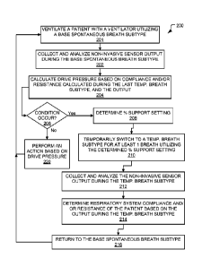
As discussed above, method 200 illustrates a method for drive pressure
ventilation
of a patient with a ventilator. Accordingly, method 200 ventilates a patient
with a DP
19
CA 03046571 2019-06-07
WO 2019/099185
PCT/US2018/058226
breath type. Method 200 provides a spontaneous breath type that allows for the
calculation of dynamic drive pressure and does not require invasive
monitoring. To
accomplish this goal, the method 200 briefly interrupts and smoothly
transitions from a
base spontaneous breath subtype, other than a PA breath subtype, into the PA
breath
subtype in response to a condition and then smoothly transitions back into the
base
spontaneous breath subtype when a patient respiratory system compliance and/or
resistance has been calculated. Method 200 accomplishes the smooth transition
by
determining a percent support setting for the PA breath subtype. As such,
method 200
may adjust ventilator parameters and/or perform other actions based on a
monitored
dynamic drive pressure.
As illustrated, method 200 includes a spontaneous ventilation operation 201.
During the spontaneous ventilation operation 201, the ventilator ventilates
the patient
utilizing a spontaneous breath subtype. The spontaneous breath subtype is any
spontaneous breath type other than a PA breath type.
As illustrated, method 200 includes a spontaneous collection operation 202.
During the spontaneous collection operation 202, the ventilator collects and
analyzes non-
invasive sensor output during the spontaneous breath subtype. In other words,
during
spontaneous collection operation 202, the ventilator non-invasively monitors
respiratory
data of the patient. Non-invasive sensor output or respiratory data refers to
the output or
measurements generated by non-invasive sensors. As such, in some aspects,
during
spontaneous collection operation 202, the ventilator collects flow rate, tidal
volume,
and/or pressure measurements from non-invasive sensors located in the
ventilator 100
and/or ventilation tubing system 130. In some aspects during spontaneous
collection
operation 202, the ventilator 100 estimates a pressure or flow at the wye 170
based on an
analysis of the non-invasive sensor output. In other aspects, other parameters
are derived
by the ventilator 100 during spontaneous collection operation 202 based on
analysis of the
of the non-invasive sensor output.
During operations 201 and 202, the ventilator analyzes the non-invasive sensor
output or respiratory data to detect a patient effort. During operations 201
and 202, the
ventilator delivers inspiratory gas to the patient with the ventilator in
response to a
detected patient effort. The inspiratory gas is delivered according to the
spontaneous
breath subtype.
At DP operation 204, a drive pressure of the patient is calculated or
estimated
during the spontaneous breath subtype utilizing a calculated or estimated
compliance
CA 03046571 2019-06-07
WO 2019/099185
PCT/US2018/058226
measurement andior resistance measurement determined during the last PA breath
subtype
and the output from the sensors during the spontaneous breath subtype. The
calculation
and/or estimation of the compliance measurement andlor resistance measurement
is
discussed in more detail below and performed during- operations 212 and 214.
In some
aspects, the ventilator during DP operation 204 may calculate or estimate the
muscle
pressure of the patient during the spontaneous breath subtype based on the
compliance
measurement and/or resistance measurement. During DP operation 204, the
ventilator
calculates or estimates a dynamic drive pressure. For example, as discussed
above, the
ventilator during DP operation 204 may calculate or estimate the dynamic drive
pressure
by utilizing Equation 4 6 listed above. In some aspects, the ventilator during
DP operation
204 is also capable of calculating or estimating static drive pressure by
utilizing Equation
4 5 listed above.
Method 200 also includes a determination operation 206. At determination
operation 206, the ventilator determines if a condition occurred. In some
aspects, the
ventilator during determination operation 206 monitors the non-invasive sensor
output to
determine if the condition has occurred. In other aspects, the ventilator
during
determination operation 206 monitors the number of delivered breath or the
passage of
time to determine if a condition has occurred. If the ventilator determines
that the
condition occurred at determination operation 206, the ventilator selects to
perform
support setting operation 208. If the ventilator determines that the condition
did not occur
during determination operation 206, the ventilator selects to perform action
operation 220.
The condition may be the expiration of a predetermined amount of time, the
delivery of a
predetermined number of breaths, and/or a change in one or more monitored
parameters
that indicates that a change in patient respiratory system compliance and/or
resistance has
occurred. In some aspects, the condition is a change in monitored pressure,
monitored
tidal volume, or monitored flow of at least 25%. In other aspects, the
condition is
expiration of I hour from the last use of the PA breath subtype without a
change in
monitored pressure, monitored tidal volume, or monitored flow of at least 25%
since the
last PA breath subtype. In further aspects, the condition is the delivery of
200 breaths
from the last use of the PA breath subtype without a change in monitored
pressure,
monitored tidal volume, or monitored flow of at least 25% since the last PA
breath
subtype.
As illustrated, method 200 includes support setting operation 208. At support
setting operation 208 the ventilator determines a percent support setting for
a PA breath
21
CA 03046571 2019-06-07
WO 2019/099185
PCT/US2018/058226
subtype. In some aspects, at support setting operation 208, the ventilator
utilizes a
predetermined support setting. In other aspects, at support setting operation
208 the
ventilator selects a support setting based on at least one of a target setting
from the
spontaneous breath subtype or the non-invasively measured respiratory data
collected
during the spontaneous breath subtype. In further aspects, the ventilator
during support
setting operation 208 determines other settings for the PA breath subtype. For
example, a
PEEP level for the PA breath subtype may be set based on a PEEP level utilized
in the
spontaneous breath subtype.
Next, switch operation 210 is performed by the ventilator. At switch operation
210 the ventilator automatically and temporarily switches from the spontaneous
breath
subtype into the PA breath subtype for at least one breath utilizing the
determined or
calculated percent support setting. In some aspects, at switch operation 210
the ventilator
automatically and temporarily switches from the spontaneous breath subtype
into the PA
breath subtype for at least three breaths utilizing the determined or
calculated percent
support setting. The PA breath subtype is performed for at least one breath,
at least two
breaths, or at least three breaths. In some aspects, the PA breath subtype is
delivered by
the ventilator during switch operation 210 until at least one patient
respiratory system
compliance and/or resistance measurement has been obtained. In some aspects,
the PA
breath subtype is delivered by the ventilator during switch operation 210
until at least two
different patient respiratory system compliance and/or resistance measurements
have been
obtained. In some aspects, the PA breath subtype is delivered by the
ventilator during the
switch operation 210 until 5, 4, 3, or 2 patient respiratory system compliance
and/or
resistance measurements have been obtained. As such, the ventilator may
deliver
ventilation utilizing the PA breath subtype for at most 4 breaths, 8 breaths,
10 breaths, 12
breaths, 15 breaths, 20 breaths, 30 breaths, 40 breaths, or 50 breaths.
Accordingly, method 200 also includes PA collect and analyze operation 212.
The
ventilator during the PA collect and analyze operation 212, collects and
analyzes the non-
invasively measured respiratory data during the PA breath subtype. Next, a
compliance
operation 214 is performed by the ventilator. During the compliance operation
214, the
ventilator calculates or estimates the patient respiratory system compliance
and/or
resistance based on the non-invasively measured respiratory data taken during
the PA
breath subtype during the PA collect and analyze operation 212. If multiple
patient
respiratory system compliance and/or resistance measurements are taken by the
ventilator
during compliance operation 214, the ventilator determines a compliance
measurement
22
CA 03046571 2019-06-07
WO 2019/099185 PCT/US2018/058226
and/or a resistance measurement based on these multiple measurements. For
example, if
multiple patient respiratory system compliance measurements are taken, the
ventilator
may average the measurements or select the middle or last obtained measurement
to be
utilized as the PA breath subtype calculated compliance measurement for use
during DP
operation 204.
Method 200 also includes a return operation 216. At return operation 216 the
ventilator switches from the PA breath subtype back to the previously utilized
spontaneous breath subtype. As discussed above, the ventilator returns the
spontaneous
breath subtype after a predetermined number of patient respiratory system
compliance or
resistance measurements have been obtained during the PA breath subtype, after
a
predetermined number of breaths, or after a predetermined amount of time.
Next,
spontaneous ventilation operation 201 is performed again.
Method 200 also includes action operation 220. At action operation 220, the
ventilator performs an action based on drive pressure. The action may include
generating
a display of the drive pressure, evaluating the drive pressure, generating an
alert based on
the drive pressure, providing a recommendation based on the drive pressure,
and/or
changing ventilator parameters based on the drive pressure. In some aspects,
the ventilator
may generate a graph of the drive pressure for display during action operation
220, such as
a waveform or bar graph of the drive pressure. In some aspects, the ventilator
evaluates
the drive pressure by comparing the drive pressure to threshold during action
operation
220. If the ventilator determines that the drive pressure breaches the
threshold during
action operation 220, ventilator performs an action in response to this
determination. As
discussed above the action may include a display of the drive pressure and/or
the breach,
generating an alert based on the breach, providing a recommendation based on
the breach,
and/or changing ventilator parameters based on the breach. If the ventilator
determines
that the drive pressure does not breach the threshold during action operation
220, the
ventilator continues to evaluate the calculated or estimated drive pressure.
In further
aspects, if the ventilator during action operation 220 determines that the
drive pressure
does not breach the threshold, the ventilator may also provide a
recommendation to the
.. clinician based on the drive pressure meeting the threshold.
In response to a drive pressure or a breach of a threshold by the drive
pressure, the
ventilator may generate an alert during action operation 220. In response to a
drive
pressure meeting a threshold, or a breach of a threshold, the ventilator may
provide a
recommendation. Alternatively, the ventilator during action operation 220 may
23
CA 03046571 2019-06-07
WO 2019/099185 PCT/US2018/058226
automatically modify the ventilation parameters listed above based on drive
pressure or
the result of a comparison of drive pressure to a threshold.
In some embodiments, a microprocessor-based ventilator that accesses a
computer-
readable medium having computer-executable instructions for performing the
method of
ventilating a patient with a medical ventilator is disclosed. This method
includes
repeatedly performing the steps disclosed in method 200 above and/or as
illustrated in
FIG. 2. In some aspects, method 200 is performed by the ventilator system 100
described
above with reference to FIG. I.
In another example, FIG. 3 is a chart illustrating a normalized respiratory
mechanics plane (R-M Plane). FIG. 3 depicts the relationship between tidal
volume (ml)
and distending pressure (AP in cmH20). Distending pressure is calculated by
subtracting
the Positive End Expiratory Pressure (PEEP) from Plateau Pressure (PpLA1)., as
illustrated
by the X-axis of FIG 3. In the context of patient ventilation, the following
equation
would operationalize the relationship: VT = AP*CL, where CL represents the
compliance
(elasticity) of the patient lung-thorax system. The units of CL for FIGS. 3
and 4 are
volume/pressure or rtil/cmH2O. Thus, if CL is known, the volume (m1) is found
by
multiplying- CL by AP. An examination of the equation VT = AP*Cr reveals that
CL
becomes a constant with the units of VT/AP. i.e., Cr is visualized as the
positive slope of a
line originating at 0,0, rising linearly up and to the right (should a
separate slide be made).
With a simple transformation of the units for the Y-axis, volume/predicted
body weight
(PBW) (the volume units for lung protective ventilation (ml/kg) and likewise
expressing
as Cr/kg provides the chart illustrated in FIG. 3. FIG. 3 assumes the
following:
1) The term ml/kg applied to all patients is valid and
2) The term Cr/kg applied to all patients is also valid.
As such, the following can be stated (where VL is lung volume):
1) If VI/kg and AP are known, Cr/kg = (VI/kg)/ AP;
2) If VL/kg and CL/kg are known, AP = (VL/kg)/(CL/kg); and
3) If AP and CL/kg are known, Vilkg = AP * Cr/kg.
Accordingly, any matched pair of coordinates for mag and AP on FIG. 3 locates
a unique
point on the R-M Plane and that point lies on a line whose slope is Cr/kg.
Furthermore,
all such matched coordinates whose ratio is equivalent will
also lie on that CL/kg slope.
Recognizing that valid estimates for AP and Vrikg are available, the
intersection of
orthogonal projections of these two values identifies a probable estimate of
the patient's
24
CA 03046571 2019-06-07
WO 2019/099185
PCT/US2018/058226
current CL/kg. A current estimate of a patient's actual CL is found by
multiplying the
normalized value by the patient's estimated PBW.
Given the structure of the R-M Plane, it's now possible to indicate how the
patient's status can be monitored and identified, either by a software
algorithm or by using
boundary conditions set by the clinician. If the clinician were interested in
maintaining
lung-protective ventilation, upper and lower, horizontal boundaries would
alert when
V-rikg were too low or too high. Ventilator notifications could identify key
changes and
suggest corrections. A patient with ARDS might be decompensating with ever
worsening
compliance. Boundary violations could notify the clinician of this occurring.
In another aspect, a feature of the recurring points could be utilized with
FIG. 3, to
indicate the trajectory the patient's change as illustrated in FIG. 4. FIG. 4
is a chart
illustrating a normalized respiratory mechanics plane with provided patient
temporal
status. The connection between sequential points would indicate rate of change
and a
notification could be provided by the ventilator to the clinician based on
this rate of
change. In FIG. 4 the repeated values for VT/kg, AP and CL/kg are captured and
processed
every 5 minutes or so. At the end of each interval, software analyzes the
patient's sensor
data and indicates the patient's location on the R-M Plane. Identical sets of
values would
produce equivalent points. However, as shown in FIG. 4, if a new point
differed by X
from the last one, a new point whose structure/identity would differ from the
last one is
plotted on the chart. In some aspects, each point is time stamped on the
chart. The three
vertical array points, illustrated in FIG. 4, indicate that the insufflation
pressure remained
constant but the patient's CL was increasing coincident with increasing VL.
Given that the
sequential values for VT/kg, AP and CL/k2 could change in any of several
logical
trajectories, a temporal indicator on the R-M plane can apprise a clinician of
the patient's
status.
FIG. 5 is a chart illustrating a normalized respiratory mechanics plane with
provided boundaries. Similar to FIG. 3, FIG. 5 depicts the relationship
between tidal
volume (ml) and distending pressure (AP in cmH20) and provides boundaries that
show
better and worse ventilation areas on the chart. hi some aspects, FIG. 5 could
be displayed
at each start-up on request. FIG. 5 reinforces in the clinician's mind the
areas of better or
worse ventilation. In some aspects, once the patient's PBW is known, the
depiction of
FIG. 5 is converted to the given patient or defaulted to the normalized
patient as shown in
FIG. 3.
CA 03046571 2019-06-07
WO 2019/099185 PCT/US2018/058226
In some embodiments, the ventilator system includes: means for ventilating a
patient with the ventilator in a spontaneous breath subtype; means for non-
invasively
monitoring respiratory data of the patient with at least one of a pressure
sensor and a flow
sensor operatively coupled to at least one of a patient circuit or a pressure
generating
system; means for analyzing the respiratory data to detect a patient effort;
means for
delivering inspiratory gas to the patient with the ventilator in response to a
detected patient
effort; means for determining an occurrence of a condition by the ventilator
based on
information gathered by the ventilator; in response to the condition, means
for determining
a percent support setting for a PA breath subtype based on a target setting or
the
respiratory data from the spontaneous breath subtype; means for automatically
and
temporarily switching from the spontaneous breath subtype into the PA breath
subtype for
at least one breath in response to calculating the percent support setting;
means for
estimating a respiratory system compliance and/or respiratory system
resistance of the
patient during the PA breath subtype based on the respiratory data; means for
returning to
the spontaneous breath subtype after the at least three breaths; means for
calculating a
drive pressure of the patient during the spontaneous breath subtype utilizing
the
respiratory system compliance and/or the respiratory system resistance and the
respiratory
data; and means for performing an action based on the drive pressure. The
spontaneous
breath subtype does not include a proportional assist (PA) breath type.
Those skilled in the art will recognize that the methods and systems of the
present
disclosure may be implemented in many manners and as such are not to be
limited by the
foregoing exemplary embodiments and examples. In other words, functional
elements
being performed by a single or multiple components, in various combinations of
hardware
and software or firmware, and individual functions, can be distributed among
software
applications at either the client or server level or both. In this regard, any
number of the
features of the different embodiments described herein may be combined into
single or
multiple embodiments, and alternate embodiments having fewer than or more than
all of
the features herein described are possible. Functionality may also be, in
whole or in part,
distributed among multiple components, in manners now known or to become
known.
Thus, myriad software/hardware/firmware combinations are possible in achieving
the
functions, features, interfaces and preferences described herein. Moreover,
the scope of
the present disclosure covers conventionally known manners for carrying out
the described
features and functions and interfaces, and those variations and modifications
that may be
26
CA 03046571 2019-06-07
made to the hardware or software firmware components described herein as would
be understood
by those skilled in the art now and hereafter.
While specific embodiments have been described and illustrated, such
embodiments
should be considered illustrative of the subject matter described herein and
not as limiting the
claims as construed in accordance with the relevant jurisprudence.
27