Note: Descriptions are shown in the official language in which they were submitted.
CA 03046827 2019-06-11
WO 2018/111765
PCT/US2017/065600
METHODS AND SYSTEMS FOR SCREENING USING
MICROCAPILLARY ARRAYS
CROSS REFERENCE TO RELATED APPLICATIONS
[0001] This application claims the benefit of U.S. Provisional Application No.
62/433,210, filed on December 12, 2016, all of which is expressly incorporated
herein by reference in its entirety.
BACKGROUND OF THE INVENTION
[0002] The analysis of biological samples, including the identification,
characterization, and re-engineering of proteins, nucleic acids,
carbohydrates, and
other important biomolecules, has benefited greatly from the scaling up of
sample
numbers and the scaling down of sample sizes. For example, the two-dimensional
microarrays of biological materials, such as DNA microarrays, have enabled the
development of high-throughput screening methods involving multiplexed
approaches for processing samples and detecting results.
[0003] The above approaches have, in some cases, benefited from their
combination with optical sensing technology to identify specimens of interest
using fluorescent or other corresponding specific and sensitive labeling
approaches.
[0004] While such techniques provide analytical information about a particular
sample, for example the presence and potentially the amount of a particular
biomolecule in a solution or the sequence of a particular nucleic acid or
polypeptide, they typically do not allow for the recovery of a biological
sample
identified by the assay without inactivating or otherwise damaging the sample
of
interest.
[0005] There is therefore a continuing need to develop improved microscale
screening and analysis methods and systems with high throughput capabilities,
and
particularly methods and systems that enable recovery of samples identified in
the
screening and analysis.
1
CA 03046827 2019-06-11
WO 2018/111765
PCT/US2017/065600
SUMMARY OF THE INVENTION
[0006] The present disclosure addresses these and other needs by providing in
one aspect methods of screening a population of variant proteins comprising
the
steps of:
providing a microcapillary array comprising a plurality of microcapillaries,
each microcapillary comprising a variant protein, an immobilized target
molecule,
and a reporter element, wherein the variant protein associates with the
immobilized
target molecule with a particular affinity; and
measuring a signal from at least one reporter element that indicates
association of at least one variant protein with at least one immobilized
target
molecule to identify at least one microcapillary of interest.
[0007] In some embodiments, the methods further comprise the step of isolating
the contents of the microcapillary of interest.
[0008] In another aspect are provided systems for screening a population of
variant proteins comprising:
an array comprising a plurality of microcapillaries, each microcapillary
comprising a variant protein, an immobilized target molecule, and a reporter
element, wherein the variant protein associates with the immobilized target
molecule with a particular affinity.
[0009] In some embodiments, the systems further comprise a microscope.
[0010] In some embodiments, the systems further comprise an optical source and
a detector.
[0011] In some embodiments, the systems further comprise an extraction device.
[0012] In some embodiments, the systems further comprise a two-stage sample
recovery element.
[0013] In some embodiments, the present invention provides a method of
screening a population of variant proteins comprising the steps of:
providing a microcapillary array comprising a plurality of microcapillaries,
each microcapillary comprising a variant protein, an immobilized target
molecule,
and a reporter element, wherein the variant protein associates with the
immobilized
target molecule with a particular affinity; and
measuring a signal from at least one reporter element that indicates
2
CA 03046827 2019-06-11
WO 2018/111765
PCT/US2017/065600
association of at least one variant protein with at least one immobilized
target
molecule to identify at least one microcapillary of interest.
[0014] In some embodiments, the variant protein is expressed by an expression
system.
[0015] In some embodiments, the expression system is a cell-free expression
system.
[0016] In some embodiments, the expression system is a cellular expression
system.
[0017] In some embodiments, the cellular expression system is an animal
system,
a fungal system, a bacterial system, an insect system, or a plant system.
[0018] In some embodiments, the cellular expression system is a yeast system.
[0019] In some embodiments, the variant protein is a soluble protein.
[0020] In some embodiments, the target molecule is a target protein or
polypeptide, a target nucleic acid, a target carbohydrate, or a combination of
each.
[0021] In some embodiments, the target molecule is immobilized on a surface.
[0022] In some embodiments, the surface is a surface of a cell.
[0023] In some embodiments, the target molecule is a native protein.
[0024] In some embodiments, the surface is a surface of a bead.
[0025] In some embodiments, the surface is a surface of a microcapillary wall.
[0026] In some embodiments, the surface is a surface configured to settle in
the
microcapillary by gravitational sedimentation.
[0027] In some embodiments, the reporter element is a labeled antibody or
other
binding molecule.
[0028] In some embodiments, the labeled antibody or other binding molecule is
a
fluorescently-labeled antibody or other binding molecule.
[0029] In some embodiments, the labeled antibody is a primary or a secondary
antibody.
[0030] In some embodiments, the labeled antibody or other binding molecule is
an enzyme-linked antibody or other binding molecule.
[0031] In some embodiments, the reporter element is activated within a cell,
and
the target molecule is immobilized on a surface of the cell.
3
CA 03046827 2019-06-11
WO 2018/111765
PCT/US2017/065600
[0032] In some embodiments, the reporter element comprises a green fluorescent
protein or variant.
[0033] In some embodiments, the signal is a fluorescent signal, an absorbance
signal, a bright-field signal, or a dark-field signal.
[0034] In some embodiments, each microcapillary in the microcapillary array
comprises 0 to 5 variant proteins from the population of variant proteins.
[0035] In some embodiments, the microcapillary array comprises at least
100,000, at least 300,000, at least 1,000,000, at least 3,000,000, or at least
10,000,000 microcapillaries.
[0036] In some embodiments, each microcapillary further comprises an agent to
improve viability of the cellular expression system.
[0037] In some embodiments, the agent is methylcellulose, dextran pluronic F-
68, polyethylene glycol, or polyvinyl alcohol.
[0038] In some embodiments, the agent is a growth medium.
[0039] In some embodiments, the signal is measured by an optical detector.
[0040] In some embodiments, the signal is measured by a microscope.
[0041] In some embodiments, the further comprises the step of isolating the
contents of the microcapillary of interest.
[0042] In some embodiments, the contents of the microcapillary of interest are
isolated by pulsing the microcapillary of interest with a laser.
[0043] In some embodiments, the laser is a diode-pumped Q-switched laser.
[0044] In some embodiments, the laser is directed at the water-glass interface
between the microcapillary wall and the sample contained in the
microcapillary.
[0045] In some embodiments, the contents of the microcapillary of interest are
isolated using a two-stage sample recovery element.
[0046] In some embodiments, the microcapillary does not comprise a
microparticle capable of inhibiting the transmission of electromagnetic
radiation, a
magnetic microparticle, a magnetic bead, or an electromagnetic radiation
absorbent
material.
[0047] The present invention also provides a system for screening a population
of variant proteins comprising:
an array comprising a plurality of microcapillaries, each microcapillary
4
CA 03046827 2019-06-11
WO 2018/111765
PCT/US2017/065600
comprising a variant protein, an immobilized target molecule, and a reporter
element, wherein the variant protein associates with the immobilized target
molecule with a particular affinity.
[0048] In some embodiments, the variant protein is expressed by an expression
system.
[0049] In some embodiments, the expression system is a cell-free expression
system.
[0050] In some embodiments, the expression system is a cellular expression
system.
[0051] In some embodiments, the cellular expression system is an animal
system,
a fungal system, a bacterial system, an insect system, or a plant system.
[0052] In some embodiments, the cellular expression system is a yeast system.
[0053] In some embodiments, the variant protein is a soluble protein.
[0054] In some embodiments, the target molecule is a target protein or
polypeptide, a target nucleic acid, a target carbohydrate, or a combination of
each.
[0055] In some embodiments, the target molecule is immobilized on a surface.
[0056] In some embodiments, the surface is a surface of a cell.
[0057] In some embodiments, the target molecule is a native protein.
[0058] In some embodiments, the surface is a surface of a bead.
[0059] In some embodiments, the surface is a surface of a microcapillary wall.
[0060] In some embodiments, the surface is a surface configured to settle in
the
microcapillary by gravitational sedimentation.
[0061] In some embodiments, the reporter element is a labeled antibody or
other
binding molecule.
[0062] In some embodiments, the labeled antibody or other binding molecule is
a
fluorescently-labeled antibody or other binding molecule.
[0063] In some embodiments, the labeled antibody is a primary or a secondary
antibody.
[0064] In some embodiments, the labeled antibody or other binding molecule is
an enzyme-linked antibody or other binding molecule.
[0065] In some embodiments, the reporter element is activated within a cell,
and
the target molecule is immobilized on a surface of the cell.
CA 03046827 2019-06-11
WO 2018/111765
PCT/US2017/065600
[0066] In some embodiments, the reporter element comprises a green fluorescent
protein or variant.
[0067] In some embodiments, the signal is a fluorescent signal, an absorbance
signal, a bright-field signal, or a dark-field signal.
[0068] In some embodiments, each microcapillary in the microcapillary array
comprises 0 to 5 variant proteins from the population of variant proteins.
[0069] In some embodiments, the microcapillary array comprises at least
100,000, at least 300,000, at least 1,000,000, at least 3,000,000, or at least
10,000,000 microcapillaries.
[0070] In some embodiments, each microcapillary further comprises an agent to
improve viability of the cellular expression system.
[0071] In some embodiments, the agent is methylcellulose, dextran pluronic F-
68, polyethylene glycol, or polyvinyl alcohol.
[0072] In some embodiments, the agent is a growth medium.
[0073] In some embodiments, the system further comprises an optical source and
a detector.
[0074] In some embodiments, the system further comprises a microscope.
[0075] In some embodiments, n the system further comprises an extraction
device.
[0076] In some embodiments, the extraction device comprises a diode-pumped
Q-switched laser.
[0077] In some embodiments, the system further comprises a two-stage sample
recovery element.
[0078] In some embodiments, the microcapillary does not comprise a
microparticle capable of inhibiting the transmission of electromagnetic
radiation, a
magnetic microparticle, a magnetic bead, or an electromagnetic radiation
absorbent
material.
BRIEF DESCRIPTION OF THE DRAWINGS
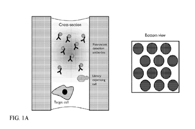
[0079] FIG. 1A- FIG. 1C schematically illustrate the steps of an exemplary
microcapillary screening assay. The illustration on the left in each panel is
a cross-
sectional view from the side of a single microcapillary. The illustration on
the
6
CA 03046827 2019-06-11
WO 2018/111765
PCT/US2017/065600
right in each panel is a bottom view of a subsection of the array of
microcapillaries. The shading in each case is intended to illustrate an
electromagnetic signal, such as fluorescence.
[0080] FIG. 2A-FIG. 2C show the bottom view of a subsection of a
microcapillary array illustrating hybridoma screening against mammalian cells,
where the cells are imaged using either bright-field (FIG. 2A), LiveGreen
(FIG.
2B), or a fluorescent anti-mouse secondary antibody (FIG. 2C).
[0081] FIG. 3 shows images of a microcapillary containing both an A431 target
cell and a hybridoma cell over the course of a 4 hour incubation.
[0082] FIG. 4A - FIG. 4B show images of a subsection of a microcapillary array
highlighting expressing and non-expressing yeast cells against mammalian
cells,
where the cells are imaged using either bright-field (FIG. 4A) or a
fluorescent
antibody (FIG. 4B).
[0083] FIG. 5A- FIG. 5G illustrate the growth of an immortalized human cell in
a microcapillary array over the course of 6 days.
[0084] FIG. 6A- FIG. 6E are different views of a microscope system designed to
carry out the screening methods of the instant disclosure.
[0085] FIG. 7 shows an exemplary embodiment of cells (including mammalian or
yeast) expression and binding to mammalian cells using the present invention.
Each one of the four panels represents one microcapillary microcavity over
time.
[0086] FIG. 8A ¨ FIG. 8B shows an exemplary embodiment of cells (including
mammalian or yeast) expression and binding to 2 or more mammalian cell types
using the present invention. 8A) Each one of the four panels represents one
microcapillary microcavity over time. 8B) Provides potential readouts from the
exemplary assay embodiment.
[0087] FIG. 9 shows exemplary fluorescence and bright-field data generated by
the exemplary assay described in Example 6.
[0088] FIG. 10 shows an exemplary embodiment of cells (including mammalian
or yeast) expression and binding to immobilized targets on a solid support
(such as
a bead) using the present invention. Each one of the four panels represents
one
microcapillary microcavity over time.
7
CA 03046827 2019-06-11
WO 2018/111765
PCT/US2017/065600
[0089] FIG. 11 shows an exemplary embodiment of cells (including mammalian
or yeast) expression and functional reporter response using the present
invention.
Each one of the four panels represents one microcapillary microcavity over
time.
Reporters can include any detectable reporter, including for example GFP, YFP,
and/or RFP, as well as any fluorophores described herein or known in the art.
[0090] FIG. 12 provides examples of IgGl, IgG2, IgG3, and IgG4 sequences.
[0091] FIG. 13A ¨ FIG. 13B. Data provides A) image of beads from Example 7
titration experiment. B) Graph showing the optimum range of signal was around
1:500-1:5000 (lower range of manufacturer recommended ranges).
DETAILED DESCRIPTION OF THE INVENTION
[0092] Microcapillary arrays have recently been employed in approaches for
high-throughput analysis and protein engineering with large numbers of
biological
samples, for example in an approach that has been termed "microcapillary
single-
cell analysis and laser extraction" or "u.SCALE". See Chen etal. (2016) Nature
Chem. Biol. 12:76-81; DOI: 10.1038/NCHEMBI0.1978. This approach relies on
the spatial segregation of single cells within a microcapillary array, and
thus
enables repeated imaging, cell growth, and protein expression of the separate
samples within each microcapillary of the microcapillary array. Accordingly,
the
technique enables massively parallel, quantitative biochemical and biophysical
measurements on millions or multi-millions of samples within a microcapillary
array, for example, in the analysis of millions or multi-millions of protein
variants
expressed from yeast, bacteria, or other suitable cells distributed throughout
the
array. Advantageously, the approach has allowed the simultaneous time-resolved
kinetic analysis of the multiplexed samples, as well as the sorting of those
cells
based on targeted phenotypic features.
[0093] The development of SCALE methods and apparatus for the quantitative
biochemical and biophysical analysis of populations of biological variants has
also
been reported in U.S. Patent Application Publication No. 2016/0244749 Al,
which
is incorporated by reference herein in its entirety. Extraction of the
contents of a
desired microcapillary according to the SCALE approach requires, however, the
inclusion of a radiation-absorbing material in each sample and the directing
of
8
CA 03046827 2019-06-11
WO 2018/111765
PCT/US2017/065600
electromagnetic radiation from a pulsed laser into this material, thus adding
complexity to the extraction methods. In addition, earlier methods of
screening of
biological variants in arrays of microcavities relied on the addition of
microparticles to the arrayed samples to partially or completely inhibit the
transmission of electromagnetic radiation into and out of the sample in order
to
minimize signal emitted from microcavities lacking a desired binding activity.
See
U.S. Patent Application Publication No. U.S. 2014/0011690 Al. In some aspects
of the instant disclosure, the screening methods do not rely on these
additional
sample components or manipulations, thus simplifying and improving the
efficiency of the screening techniques.
[0094] In specific applications of these approaches, and as will be disclosed
in
more detail herein, the target molecule can be immobilized on a surface, such
as
the surface of a particle (e.g., a magnetic particle), a cell, or a
microcapillary wall.
The interaction between a variant protein and a target molecule in these
approaches
can then be measured by several methods, including methods utilizing
detectable
antibodies and methods of measuring detectable signals generated within the
target
cells. It will be understood that such methods can be used in high-throughput
screens to discover protein variants that bind to target molecules, for
example a
target molecule on a cell or other surface.
Methods of Screening
[0095] Accordingly, in some aspects, the instant disclosure provides methods
of
screening a population of variant proteins comprising the steps of:
providing a microcapillary array comprising a plurality of
microcapillaries, each microcapillary comprising a variant protein, an
immobilized
target molecule, and a reporter element, wherein the variant protein
associates with
the immobilized target molecule with a particular affinity; and
measuring a signal from at least one reporter element that indicates
association of at least one variant protein with at least one immobilized
target
molecule to identify at least one microcapillary of interest.
[0096] In these methods, the microcapillary arrays preferably comprise a
plurality of longitudinally fused capillaries, for example fused silica
capillaries,
although any other suitable material may be utilized in the arrays. See, e.g.,
PCT
9
CA 03046827 2019-06-11
WO 2018/111765
PCT/US2017/065600
International Patent Publication Nos. W02012/007537 and W02014/008056, the
disclosures of which are incorporated by reference herein in their entireties.
Such
arrays can be fabricated, for example, by bundling millions or billions of
silica
capillaries and fusing them together through a thermal process, although other
suitable methods of fabrication may also be employed. The fusing process may
comprise, for example, the steps of i) heating a capillary single draw glass
that is
drawn under tension into a single clad fiber; ii) creating a capillary multi
draw
single capillary from the single draw glass by bundling, heating, and drawing;
iii)
creating a capillary multi-multi draw multi capillary from the multi draw
single
capillary by additional bundling, heating, and drawing; iv) creating a block
assembly of drawn glass from the multi-multi draw multi capillary by stacking
in a
pressing block; v) creating a block pressing block from the block assembly by
treating with heat and pressure; and vi) creating a block forming block by
cutting
the block pressing block at a precise length (e.g., 1 mm).
[0097] In some embodiments, the fabrication method further comprises slicing
the silica capillaries, thereby forming very high-density glass microcapillary
arrays. In some embodiments, the microcapillary arrays may be cut to
approximately 1 millimeter in height, but even shorter microcapillary arrays
are
contemplated, including arrays of 10 p.m in height or even shorter. In some
embodiments, even longer microcapillary arrays are contemplated, including
arrays of 10 mm or even longer.
[0098] Such processes form very high-density microcapillary arrays that are
suitable for use in the present methods. In an exemplary array, each
microcapillary
has an approximate 5 p.m diameter and approximately 66% open space (i.e.,
representing the lumen of each microcapillary). In some arrays, the proportion
of
the array that is open ranges between about 50% and about 90%, for example
about
60 to 75%, such as a microcapillary array provided by Hamamatsu that has an
open
area of about 67%. In one particular example, a 10x1 0 cm array having 5 p.m
diameter microcapillaries and approximately 66% open space has about 330
million total microcapillaries.
[0099] In various embodiments, the internal diameter of each microcapillary in
the array ranges from between approximately 1 p.m and 500 p.m. In some arrays,
CA 03046827 2019-06-11
WO 2018/111765
PCT/US2017/065600
each microcapillary can have an internal diameter in the range between
approximately 1 p.m and 300 p.m; optionally between approximately 1 p.m and
100
p.m; further optionally between approximately 1 p.m and 75 p.m; still further
optionally between approximately 1 p.m and 50 p.m; and still further
optionally
between approximately 5 p.m and 50 p.m.
[0100] In some microcapillary arrays, the open area of the array comprises up
to
90% of the open area (OA), so that, when the pore diameter varies between 1
p.m
and 500 p.m, the number of microcapillaries per cm of the array varies between
approximately 460 and over 11 million. In some microcapillary arrays, the open
area of the array comprises about 67% of the open area, so that, when the pore
size
varies between 1 p.m and 500 p.m, the number of microcapillaries per square cm
of
the array varies between approximately 340 and over 800,000.
In some embodiments, the pore size is 1 p.m, 5 p.m, 10 p.m 50 p.m, 100 p.m,
250
p.m 350 or 500 p.m. In some embodiments, the pore size is between 5 p.m and
500
p.m. In some embodiments, the pore size is between 10 p.m and 450 p.m. In some
embodiments, the pore size is between 50 p.m and 500 p.m. In some embodiments,
the pore size is between 100 p.m and 500 p.m. In some embodiments, the pore
size
is between 250 p.m and 500 p.m. In some embodiments, the pore size is between
350 p.m and 500 p.m. In some embodiments, the pore size is between 100 p.m and
450 p.m. In some embodiments, the pore size is between 250 p.m and 450 p.m.
In some embodiments, the number of microcapillaries per square cm of the array
is
approximately 400; 500; 1000; 2,000; 3,000; 4,000; 5,000; 6,000; 7,000; 8,000;
9,000; 10,000; 20,000; 50,000, 100,000; 200,000; 300,000; 400,000; 500,000;
600,
000; 700,000; or 800,000. In some embodiments, the number of microcapillaries
per square cm of the array varies between approximately 500 and 800,000. In
some embodiments, the number of microcapillaries per square cm of the array
varies between approximately 1000 and 700,000. In some embodiments, the
number of microcapillaries per square cm of the array varies between
approximately 2000 and 600,000. In some embodiments, the number of
microcapillaries per square cm of the array varies between approximately
10,000
and 800,000. In some embodiments, the number of microcapillaries per square cm
of the array varies between approximately 10,000 and 700,000. In some
11
CA 03046827 2019-06-11
WO 2018/111765
PCT/US2017/065600
embodiments, the number of microcapillaries per square cm of the array varies
between approximately 50,000 and 800,000. In some embodiments, the number of
microcapillaries per square cm of the array varies between approximately
50,000
and 700,000. In some embodiments, the number of microcapillaries per square cm
of the array varies between approximately 100,000 and 700,000. In some
embodiments, the number of microcapillaries per square cm of the array varies
between approximately 100,000 and 600,000. In some embodiments, the number
of microcapillaries per square cm of the array varies between approximately
100,000 and 500,000. In some embodiments, the number of microcapillaries per
square cm of the array varies between approximately 500,000 and 800,000.
[0101] In one particular embodiment, a microcapillary array can be
manufactured by bonding billions of silica capillaries and then fusing them
together through a thermal process. After that slices (0.5 mm or more) are cut
out
to form a very high aspect ratio glass microcapillary array. Arrays are also
commercially available, such as from Hamamatsu Photonics K. K. (Japan), Incom,
Inc. (Massachusetts), Photonis Technologies, S.A.S. (France) Inc., and others.
In
some embodiments, the microcapillaries of the array are closed at one end with
a
solid substrate attached to the array.
[0102] The microcapillary arrays of the instant screening methods can comprise
any number of microcapillaries within the array. In some embodiments, the
microcapillary array comprises at least 100,000, at least 300,000, at least
1,000,000, at least 3,000,000, at least 10,000,000, or even more
microcapillaries.
In some embodiments, the array comprises at least 100,000, at least 200,000,
at
least 300,000, at least 400,000, at least 500,000, at least 600,000, at least
700,000,
at least 800,000, at least 1,000,000, at least 1,500,000, at least 2,000,000,
at least
2,500,000, or at least 3,000,000 or more microcapillaries. The number of
microcapillaries within an array is preferably chosen in view of the size of
the
variant protein library to be screened.
[0103] As described above, each capillary in the microcapillary arrays used in
the instant screening methods comprises a variant protein, an immobilized
target
molecule, and a reporter element, where the variant protein is one of the
population
of variant proteins that is being subjected to the screening method. The
population
12
CA 03046827 2019-06-11
WO 2018/111765
PCT/US2017/065600
of variant proteins can be any population of proteins that can be suitably
distributed within a microcapillary array. Ideally, the population of variant
proteins is distributed in the microcapillary array so that each
microcapillary
comprises a small number of different variant proteins, preferably just a
single
different variant protein per microcapillary. Importantly, the population of
variant
proteins is chosen in combination with the immobilized target molecule, such
that
at least some of the proteins in the population can associate with the
immobilized
target molecule with a particular affinity, such that the association is
detectable by
measuring a signal from a reporter element.
[0104] The term "protein", as used herein, refers both to full-length proteins
or
polypeptide sequences and to fragments thereof Such fragments may include
fragments that retain a functional activity, such as, for example, a binding
activity.
The terms "protein" and "polypeptide" are used interchangeably throughout the
disclosure and include chains of amino acids covalently linked through peptide
bonds, where each amino acid in the polypeptide may be referred to as an
"amino
acid residue". Use of the terms "protein" or "polypeptide" should not be
considered limited to any particular length of polypeptide, e.g., any
particular
number of amino acid residues. The subject proteins may include proteins
having
non-peptidic modifications, such as post-translational modifications,
including
glycosylation, acetylation, phosphorylation, sulfation, or the like, or other
chemical
modifications, such as alkylation, acetylation, esterification, PEGylation, or
the
like. Additional modifications, such as the inclusion of non-natural amino
acids
within a polypeptide sequence or non-peptide bonds between amino acid residues
should also be considered within the scope of the definition of the term
"protein"
or "polypeptide".
[0105] The population of variant proteins is preferably a population of
proteins
having minor variations, for example a population of proteins where each
protein
has a slightly different amino acid sequence. The screening assays can,
therefore,
identify variant protein sequences having desirable properties. Because the
screens
can be performed in such large numbers at microscopic scale, huge numbers of
variant proteins can be assayed in relatively short times.
13
CA 03046827 2019-06-11
WO 2018/111765
PCT/US2017/065600
[0106] Variant proteins and/or variant polypeptides can include but are not
limited to secreted proteins. In some embodiments, the secreted proteins are
from
a recombinant protein and/or polypeptide library. In some embodiments, the
secreted proteins are from a recombinant protein and/or polypeptide library.
In
some embodiments, the secreted proteins are from a recombinant protein and/or
polypeptide library from a mammalian cell line. In some embodiments, the
recombinant protein and/or polypeptide library comprises full length mammalian
antibodies. In some embodiments, the recombinant protein and/or polypeptide
library comprises full length mammalian antibodies, including IgGl, IgG2, and
IgG4 antibodies and variants thereof In some embodiments, the recombinant
protein and/or polypeptide library comprises full length human antibodies. In
some embodiments, the recombinant protein and/or polypeptide library comprises
full length human antibodies, including IgGl, IgG2, and IgG4 antibodies. In
some
embodiments, the recombinant protein and/or polypeptide library comprises full
length mouse antibodies. In some embodiments, the recombinant protein and/or
polypeptide library comprises full length mouse antibodies, including IgGl,
IgG2,
and IgG4 antibodies. In some embodiments, the recombinant protein and/or
polypeptide library comprises full length rat antibodies. In some embodiments,
the
recombinant protein and/or polypeptide library comprises full length rat
antibodies,
including IgGl, IgG2, and IgG4 antibodies. In some embodiments, the
recombinant protein and/or polypeptide library comprises antibody fragments
(Fab). In some embodiments, the recombinant protein and/or polypeptide library
comprises single chain variable fragments (scFv). In some embodiments, the
recombinant protein and/or polypeptide library comprises natural protein
ligands.
In some embodiments, the recombinant protein and/or polypeptide library
comprises natural protein ligands to a defined target protein and/or
polypeptide.
[0107] In some embodiments, each microcapillary in the microcapillary array
comprises 0 to 5 different variant proteins from the population of variant
proteins.
In specific embodiments, each microcapillary in the microcapillary array
comprises 0 to 4, 0 to 3, 0 to 2, or even 0 to 1 different variant proteins
from the
population of variant proteins. It should be understood that the different
variant
proteins in the population of variant proteins differ in their molecular
structure,
14
CA 03046827 2019-06-11
WO 2018/111765
PCT/US2017/065600
whether the difference is in their amino acid sequence or in some other
chemical
modification of the protein.
[0108] It should be understood that each microcapillary will typically
comprise
many multiple copies of the same variant protein, depending on the source and
expression level of the particular variant protein (see below). In some
embodiments, each microcapillary will comprise thousands, tens of thousands,
hundreds of thousands, millions, billions, or even more molecules of a
particular
variant protein, depending on how the variant protein is delivered to or
expressed
within the microcapillary. In some embodiments, the variant protein can bind
to
one, two, three, or four or more target molecules. In some embodiments, the
variant protein can bind to one target molecule. In some embodiments, the
variant
protein can bind to two target molecules. In some embodiments, the variant
protein can bind to three target molecules. In some embodiments, the variant
protein can bind to four target molecules. In some embodiments, the variant
protein can bind to more than four target molecules. In some embodiments, this
assay can alternatively be used to screen for antibodies that bind both a
mouse and
human (or other combination of animals) variant of a target protein, i.e.
finding
"cross-reactive" antibodies. For example, the presence of stained cells and
the
presence of stained beads within a microcapillary indicates the presence of an
antibody which binds to the "target protein" (for example, a mouse target) and
which also binds to the "target protein analog" (for example, a human target),
in
order to identify antibodies which bind to both a mouse and human target. For
example, the presence of stained cells and the presence of stained beads
within a
microcapillary indicates the presence of an antibody which binds to the
"target
protein" (for example, a cynomolgus target) and which also binds to the
"target
protein analog" (for example, a human target), in order to identify antibodies
which bind to both a cynomolgus and human target.
[0109] The population of variant proteins is typically generated using a
genetic
library in a biological expression system, for example in an in vitro (i.e.,
cell-free)
expression system or in an in vivo or cellular expression system. Exemplary
cellular expression systems include, for example, animal systems (e.g.,
mammalian
systems), fungal systems (e.g., yeast systems), bacterial systems, insect
systems, or
CA 03046827 2019-06-11
WO 2018/111765
PCT/US2017/065600
plant systems. In specific embodiments, the expression system is a mammalian
system or a yeast system. The expression system, whether cellular or cell-
free,
typically comprises a library of genetic material encoding the population of
variant
proteins. Cellular expression systems offer the advantage that cells with a
desirable phenotype, for example cells that express a particular variant
protein of
interest, such as a variant protein capable of associating with an immobilized
target
molecule with high affinity, can be grown and multiplied, thus facilitating
and
simplifying the identification and characterization of the proteins of
interest
expressed by the cells. In some embodiments, the biological expression system
comprises a mammalian cell line. In some embodiments, the mammalian cell line
is selected from the group consisting of CHO-K1, CHO-S, HEK293T, and/or any
derivatives of these cell types. In some embodiments, the mammalian cell line
is
CHO-Kl. In some embodiments, the mammalian cell line is CHO-S. In some
embodiments, the mammalian cell line is HEK293T. In some embodiments, the
mammalian cell line is selected from the group consisting of human, mouse,
and/or
rat hybridoma cell lines. In some embodiments, the mammalian cell line is a
human hybridoma cell line. In some embodiments, the mammalian cell line is a
mouse hybridoma cell line. In some embodiments, the mammalian cell line is a
rat
hybridoma cell line.
[0110] Genetic libraries encoding large populations of variant proteins are
well
known in the art of bioengineering. Such libraries are often utilized in
systems
relying on the process of directed evolution to identify proteins with
advantageous
properties, such as high-affinity binding to target molecules, stability, high
expression, or particular spectroscopic, e.g., fluorescence, or enzymatic
activities.
Often the libraries include genetic fusions with sequences from the host
expression
system, for example fragments of proteins directing subcellular localization,
where
the expressed population of variant fusion proteins are directed by the
targeting
fragment to a particular location of the cell or virus particle for purposes
of activity
screening of the variant protein population. Large numbers of variant proteins
(e.g., 106 variants, 108 variants, 1010 variants, 1012 variants, or even more
variants)
can be generated using routine bioengineering techniques, as is well known in
the
art. Such libraries can include any of the variant proteins described herein,
16
CA 03046827 2019-06-11
WO 2018/111765
PCT/US2017/065600
including antibodies, antibody fragments, single chain variable fragments, or
natural protein ligands.
[0111] Accordingly, in some embodiments, the variant proteins are soluble
proteins, for example soluble proteins that are secreted by a cellular
expression
system. Exemplary soluble variant proteins include antibodies and antibody
fragments, alternative protein scaffolds, such as disulfide-bonded peptide
scaffolds,
extracellular domains of cell-surface receptor proteins, receptor ligands,
such as,
for example, G-protein coupled receptor ligands, other peptide hormones,
lectins,
and the like. Advantageously, the variant proteins screened for binding
activity in
the instant methods do not need to be covalently attached to the cell or virus
that
expresses them in order to be identified following a screening assay, since a
variant
protein with a desired binding activity and the cell that expressed it remain
co-
localized within the same microcapillary throughout the assay. Isolation of
the
contents of the desired microcapillary, followed by propagation of the cell or
virus
clone responsible for expression of the desired variant protein, thereby
enables the
identification and characterization of that protein. Unlike screening assays
where a
variant protein of interest is displayed by fusion of the protein to a
molecule on the
surface of a cell or virus particle, the variant proteins identified in the
instant
screening methods need not be altered in any way following their
identification.
The observed activities of the variant proteins in the screens are thus more
likely to
represent the actual activities of those proteins in their subsequent
applications.
[0112] In other embodiments, however, it may be desirable for the variant
proteins to be membrane-associated proteins, for example proteins remaining
associated with the surface of a cell or a viral particle in an expression
system.
Screening of cell-associated variant proteins may be desirable where the
variant
protein and its target molecule mediate interactions between two cells within
a
biological tissue. The ability to screen against cell-associated variant
proteins may
also be desirable in screening for interactions with traditionally "non-
druggable"
protein targets, such as, for example, G-protein coupled receptors or ion
channels.
[0113] In addition to a variant protein, each microcapillary in the
microcapillary
arrays of the instant screening methods also comprises an immobilized target
molecule. The immobilized target molecule serves as the potential binding
partner
17
CA 03046827 2019-06-11
WO 2018/111765
PCT/US2017/065600
for the variant protein of the screening assay. Unlike the population of
variant
proteins, where each microcapillary ideally contains a variant protein of
slightly
different sequence, the immobilized target molecules ideally have the same
molecular structure in each microcapillary of the array. In some embodiments,
there is no binding or other interaction between the variant protein and
another
agent or molecule (e.g., the target molecule) prior to the addition of the
variant
protein to the microcapillary. In some embodiments, the interaction between
the
variant protein and the target molecule occurs within the microcapillary
and/or
microcavity.
[0114] In some embodiments, the target molecule is a target protein or
polypeptide, a target nucleic acid, a target carbohydrate, a target lipid, or
a
combination of two or more of these target molecules. For example, in some
embodiments the target molecule can be a lipid-modified or glycosylated
protein.
In some embodiments, the target molecule is immobilized on a surface. In more
specific embodiments, the target molecule is immobilized on the surface of a
cell,
such as a target cell, the surface of a bead, the surface of a microcapillary
wall, or
another suitable surface. In other more specific embodiments, the target
molecule
is a native protein, for example a native protein immobilized on the surface
of a
cell. In still other more specific embodiments, the target molecule is
immobilized
on a surface configured to settle in the microcapillary by gravitational
sedimentation. In some embodiments, one, two, three, or four, or more target
molecules are employed, in order to identify variants that bind to one, two,
three,
or four, or more target molecules. In some embodiments, the target molecules
are
contained separately in separate and different microcapillaries. In some
embodiments, the target molecules are contained separately in separate and
different microcapillaries within a single array. In some embodiments, the
target
molecules are contained separately in separate and different microcapillaries
within
one or more arrays. In some embodiments, the target molecules are contained
together in a single microcapillary. In some embodiments, the target molecules
are
contained together in a single microcapillary within a single array. In some
embodiments, the one, two, three, or four, or more target molecules to which
the
variant binds are derivatives or variants of an original target molecule,
including
18
CA 03046827 2019-06-11
WO 2018/111765
PCT/US2017/065600
chemical modifications, secondary post-translational modifications, or
sequence
identity variants (including, for example, variants with 70%, 75%, 80%, 85%,
90%, 95%, or 99% sequence identity to an original nucleic acid or amino acid
target sequence).
[0115] As previously noted, in the methods of the instant disclosure, the
variant
protein associates with the immobilized target molecule with a particular
affinity
within a microcapillary. Importantly, such affinities should be sufficiently
strong
for variant proteins of interest that the association can be measured by a
signal
from a reporter element. Binding affinities are typically assessed by a
dissociation
constant (Kd), as is well understood by those of ordinary skill in the art,
where the
lower the dissociation constant, the higher the affinity. In some embodiments,
the
association between the variant protein of interest and the immobilized target
molecule displays a dissociation constant in the millimolar to micromolar
range.
In specific embodiments, the association displays a dissociation constant from
micromolar to high nanomolar (i.e., 106 M to 10-8 M). In more specific
embodiments, the association displays a dissociation constant from lower
nanomolar to high picomolar (i.e., 108 M to 10-10 M). In even more specific
embodiments, the association displays a dissociation constant in the picomolar
range (i.e., 1010 M to 10-12 M), or even lower. In some embodiments, a first
cell
expresses and secretes the variant protein or polypeptide and a second cell
comprises the target, such that the first cells binds to the second cell. In
some
embodiments, the second cell expresses the target. In some embodiments, the
second cell is labeled with the target. In some embodiments, the first cell
binds to
the second cell in the microcapillary. In some embodiments, the first cell
binds to
the second cell in the microcapillary and/or microcavity.
[0116] In addition to a variant protein and an immobilized target molecule,
each
microcapillary in the microcapillary array of the instant screening methods
also
comprises a reporter element. Importantly, the reporter element provides a
measureable signal indicative of the association of a variant protein with an
immobilized target molecule and thus serves to identify a microcapillary
containing variant proteins of interest.
19
CA 03046827 2019-06-11
WO 2018/111765
PCT/US2017/065600
[0117] In some embodiments, the reporter element is a labeled antibody or
other
molecule capable of binding to each variant protein in the population of
variant
proteins. More specifically, the reporter element is a fluorescently-labeled
antibody or other binding molecule.
[0118] In some embodiments, the labeled antibody is a labeled primary antibody
or a labeled secondary antibody. For purposes of this disclosure, a primary
antibody is typically considered to be an antibody that binds directly to an
antigen
of interest, whereas a secondary antibody is typically considered to be an
antibody
that binds to a constant region on a primary antibody for purposes of labeling
the
primary antibody. Accordingly, secondary antibodies are frequently labeled
with
fluorophores or other detectable labels or are labeled with enzymes that are
capable
of generating detectable signals. They are generally specific for a primary
antibody from a different species. For example, a goat or other animal species
may
be used to generate secondary antibodies against a mouse, chicken, rabbit, or
nearly any primary antibody other than an antibody from that animal species,
as is
understood by those of ordinary skill in the art. In specific embodiments, the
labeled antibody is a fluorescent antibody or an enzyme-linked antibody.
[0119] In some of the method embodiments, for example in the screening
methods illustrated in FIGs. 1A-1C, the variant protein mediates the
association of
a reporter element with a target molecule, in this example, a target molecule
on the
surface of a target cell. As shown in FIG. 1B, where the variant protein (here
designated as a "secreted protein") has sufficient affinity for its target
molecule on
the target cell that the variant proteins associate with the target cell under
the
conditions of the microcapillary solution. The reporter element (here
designated as
"fluorescent detection antibodies") binds to the variant protein, ideally at
an
epitope that does not affect the affinity of the variant protein for the
target
molecule, as shown in FIG. 1C.
[0120] As would be understood by those of ordinary skill in the art, when a
soluble reporter element, such as a fluorescent antibody, is used in the
instant
screening methods, the signal emitted by any excess reporter element remaining
free in solution (i.e., either not bound to a variant protein or bound to a
variant
protein that is not bound to a target molecule) within the microcapillary
should not
CA 03046827 2019-06-11
WO 2018/111765
PCT/US2017/065600
be so high that it overwhelms the signal of reporter elements associated with
a
target molecule via a variant protein (see, e.g., the unassociated fluorescent
detection antibodies illustrated in FIG. 1C). Such background signals can be
minimized, however, by limiting the concentration of labeled antibody or other
reporter element within the microcapillary solution. In addition, where
signals
from the screening methods are measured using a fluorescent microscope,
configuring the microscope to image a relatively narrow depth of field
bracketing
the location of the target molecules (e.g., the bottom of the microcapillaries
when
target cells have settled there by gravitational sedimentation) can minimize
the
background signal from reporter elements not associated with the target
molecule.
[0121] In other embodiments, the reporter element is an intracellular reporter
element that generates a detectable signal in connection with a binding event,
such
as, for example, the association of a variant protein with an immobilized
target
molecule, for example, a receptor or other target molecule on the surface of
the
cell. In these embodiments, the reporter element may comprise an entire
cellular
pathway, such as, for example, an intracellular signaling pathway. Such a
pathway
should include, or be engineered to include, a detectable signal as the
downstream
readout of the pathway. In contrast to the assays illustrated in FIGs. 1A-1C,
where
the detectable signal is bound to the outer surface of the target cell, the
detectable
signal in these embodiments would typically be generated inside the target
cell.
[0122] Many intracellular signaling pathways have been developed for use in
high throughput screening assays, in particular in drug discovery screens, and
can
be adapted for use in the instant assays. See, e.g., Michelini etal.
(2010)Anal.
Bioanal. Chem. 398:227-38. In particular, any cellular assay where a binding
event with a target molecule on the surface of a cell results in the
generation of a
measurable signal, in particular a fluorescent signal, can be used as a
reporter
element in the instant assays. Preferably, the cells can be engineered to
express a
target molecule of interest on their surface, so that the binding of a
particular
variant protein to the target molecule and the consequent activation of the
intracellular signaling pathway result in the production of a detectable
signal from
the reporter element, thus enabling the identification of the microcapillary
as a
positive hit. The expression of a green fluorescent protein (GFP), or any of a
wide
21
CA 03046827 2019-06-11
WO 2018/111765
PCT/US2017/065600
variety of variant fluorescent proteins, is often used as a readout in such
cellular
assays and can serve as the reporter element endpoint in the instant methods.
Alternatively, the signaling readout can be provided by luciferase or other
related
enzymes that produce bioluminescent signals, as is well understood by those of
ordinary skill in the art. See, e.g., Kelkar etal. (2012) Curr. Opin.
Pharmacol.
12:592-600. Reporter elements can also include RFP (red fluorescent protein)
as
well as YFP (yellow fluorescent protein), and variants thereof Other well-
known
enzymatic reporters from bacterial and plant systems include 0-galactosidase,
chloramphenicol acetyltransferase, 0-glucuronidase (GUS), and the like, which
can
be adapted for use in the instant screening assays with suitable colorogenic
substrates. Transcriptional reporters using firefly luciferase and GFP have
been
used extensively to study the function and regulation of transcription
factors. They
can likewise be adapted for use in the instant screening assays. Exemplary
intracellular signaling systems are available commercially, for example the
CignalTM Reporter Assay kits from Qiagen (see, e.g.,
www.sabiosciences.com/reporterassays.php), which are available with either
luciferase or GFP readouts. Such systems can be suitably re-engineered for use
in
the instant screening methods.
[0123] It should be understood that a variant protein expression system, in
particular where the expression system is a cellular expression system, can be
combined with the immobilized target molecule and the reporter element (or
suitable components, such as cellular components, responsible for generating
the
immobilized target molecule and/or reporter element) prior to the expression
of the
variant proteins and/or prior to delivery of an assay mixture into the array
of
microcapillaries. Such approaches advantageously allow for flexibility and
control
in the timing of interactions between the components compared to prior art
microcapillary screening systems, where all of the components of the screening
assays are typically mixed and loaded into the microcapillaries in static
form. In
contrast, the instant methods enable some or all of the components of a
binding
assay to be generated in situ within the microcapillaries, either by allowing
for the
growth of cellular components, the expression of genetic components, or both.
In
some embodiments, cell culture media and/or growth agents are employed in
order
22
CA 03046827 2019-06-11
WO 2018/111765
PCT/US2017/065600
to maintain the health of the cells during the assay process. In some
embodiments,
components are included that facilitate the metabolic health of the cells.
[0124] It should also be understood that the concentrations of each component
of
the screening assay within a microcapillary, including the concentration of
the
variant protein, the concentration of the immobilized target molecule, and the
concentration of the reporter element, can be modulated as desired in an assay
in
order to achieve an optimal outcome. In particular, it may be desirable to
modulate
the concentration of variant protein and/or immobilized target molecule to
achieve
the desired level of association between these components. The level of
association will also depend on the particular affinity between these
components,
wherein a higher affinity results in a higher level of association for a given
concentration of the components, and a lower affinity results in a lower level
of
association of the components for a given concentration. Concentration of the
reporter element may likewise be modulated in order to achieve optimum levels
of
signal output, as would be understood by those of ordinary skill in the art.
In some
embodiments, the reporter element employed includes a secondary antibody,
including those commercially available. In some embodiments the dilution range
is 1:200-1:2000. In some embodiments the dilution range is 1:300-1:2000. In
some embodiments the dilution range is 1:300-1:1500. In some embodiments the
dilution range is 1:400-1:1500. In some embodiments the dilution range is
1:500-
1:1500. In some embodiments the dilution range is 1:200-1:1000. In some
embodiments the dilution range is 1:500-1:1000. In some embodiments the
dilution range is 1:1000-1:2000. In some embodiments the dilution range is
1:1500-1:2000. In some embodiments, the dilution is 1:200, 1:300, 1:400,
1:500,
1:600, 1:700, 1:800, 1:900, 1:1000, 1:1500, or 1:2000. In some embodiments,
the
fluorophore can include but is not limited to AlexaFluor 3, AlexaFluor 5,
AlexaFluor 350, AlexaFluor 405, AlexaFluor 430, AlexaFluor 488, AlexaFluor
500, AlexaFluor 514, AlexaFluor 532, AlexaFluor 546, AlexaFluor 555,
AlexaFluor 568, AlexaFluor 594, AlexaFluor 610, AlexaFluor 633, AlexaFluor
647, AlexaFluor 660, AlexaFluor 680, AlexaFluor 700, and AlexaFluor 750
(Molecular Probes AlexaFluor dyes, available from Life Technologies, Inc.
(USA)). In some embodiments, the fluorophore can include but is not limited to
23
CA 03046827 2019-06-11
WO 2018/111765
PCT/US2017/065600
Cy dyes, including Cy2, Cy3, Cy3B, Cy3.5, Cy5, Cy5.5 and Cy7 (available from
GE Life Sciences or Lumiprobes). In some embodiments the fluorophore can
include but is not limited to DyLight 350, DyLight 405, DyLight 488, DyLight
550, DyLight 594, DyLight 633, DyLight 650, DyLight 680, DyLight 750 and
DyLight 800 (available from Thermo Scientific (USA)). In some embodiments,
the fluorophore can include but is not limited to a FluoProbes 390, FluoProbes
488,
FluoProbes 532, FluoProbes 547H, FluoProbes 594, FluoProbes 647H, FluoProbes
682, FluoProbes 752 and FluoProbes 782, AMCA, DEAC (7-
Diethylaminocoumarin-3-carboxylic acid); 7-Hydroxy-4-methylcoumarin-3; 7-
Hydroxycoumarin-3; MCA (7-Methoxycoumarin-4-acetic acid); 7-
Methoxycoumarin-3; AMF (4'-(Aminomethyl)fluorescein); 5-DTAF (5-(4,6-
Dichlorotriazinyl)aminofluorescein); 6-DTAF (6-(4,6-
Dichlorotriazinyl)aminofluorescein); 6-FAM (6-Carboxyfluorescein), 5(6)-FAM
cadaverine; 5-FAM cadaverine; 5(6)-FAM ethylenediamme; 5-FAM
ethylenediamme; 5-FITC (FITC Isomer I; fluorescein-5-isothiocyanate); 5-FITC
cadaverin; Fluorescein-5-maleimide; 5-IAF (5-Iodoacetamidofluorescein); 6-JOE
(6-Carboxy-4',5'-dichloro-2',7'-dimethoxyfluorescein); 5-CR110 (5-
Carboxyrhodamine 110); 6-CR110 (6-Carboxyrhodamine 110); 5-CR6G (5-
Carboxyrhodamine 6G); 6-CR6G (6-Carboxyrhodamine 6G); 5(6)-
Carboxyrhodamine 6G cadaverine; 5(6)-Caroxyrhodamine 6G ethylenediamme; 5-
ROX (5-Carboxy-X-rhodamine); 6-ROX (6-Carboxy-X-rhodamine); 5-TAMRA
(5-Carboxytetramethylrhodamine); 6-TAMRA (6-Carboxytetramethylrhodamine);
5-TAMRA cadaverine; 6-TAMRA cadaverine; 5-TAMRA ethylenediamme; 6-
TAMRA ethylenediamme; 5-TMR C6 maleimide; 6-TMR C6 maleimide; TR C2
maleimide; TR cadaverine; 5-TRITC; G isomer (Tetramethylrhodamine-5-
isothiocyanate); 6-TRITC; R isomer (Tetramethylrhodamine-6-isothiocyanate);
Dansyl cadaverine (5-Dimethylaminonaphthalene-1-(N-(5-
aminopenty1))sulfonamide); EDANS C2 maleimide; fluorescamine; NBD; and
pyrromethene and derivatives thereof In some embodiments, the reporter element
used can be a donkey anti-goat IgG secondary antibody labeled with AlexaFluor
633.
24
CA 03046827 2019-06-11
WO 2018/111765
PCT/US2017/065600
[0125] In some embodiments, each microcapillary in the microcapillary arrays
of
the instant screening methods further comprises an agent or agents to improve
viability of the cellular expression system. Specifically, the agent or agents
is
included to prevent cell damage during the step of isolating the contents of
the
microcapillary of interest, for example by a laser pulse (see below). In
preferred
embodiments, the agent is methylcellulose (for example at 0.001 to 10 wt %),
dextran (for example at 0.5 to 10 wt %), pluronic F-68 (for example at 0.01 to
10
wt %), polyethylene glycol ("PEG") (for example at 0.01 to 10 wt %), polyvinyl
alcohol ("PVA") (for example at 0.01 to 10 wt %), or the like. Alternatively,
or in
addition, each microcapillary in the microcapillary arrays of the instant
screening
methods can further comprise a growth additive, such as, for example, 50%
conditioned growth media, 25% standard growth media, or 25% serum. In some
embodiments, the conditioned growth media is conditioned for 24 hours. In some
embodiments, the added agent is insulin, transferrin, ethanolamine, selenium,
an
insulin-like growth factor, or a combination of these agents or any of the
agents
recited above.
[0126] The screening methods of the instant disclosure preferably include the
further step of measuring a signal from at least one reporter element that
indicates
association of at least one variant protein with at least one immobilized
target
molecule to identify at least one microcapillary of interest. In some
embodiments,
the signal measured is a fluorescent signal, an absorbance signal, a bright-
field
signal, a dark-field signal, a phase contrast signal, or the like.
Accordingly, the
measuring step can be performed by an appropriate detector device, for example
a
device capable of detecting electromagnetic radiation or any other suitable
signal.
In specific embodiments, the measuring step is performed by a microscope, such
as
a fluorescence microscope or any other microscope configured to detect the
above-
mentioned signals.
[0127] It should be understood that in preferred embodiments, the
microcapillaries utilized in the instant screening methods do not comprise
microparticles capable of inhibiting the transmission of electromagnetic
radiation.
In other words, the microcapillaries are preferably fully transparent to
electromagnetic radiation incident on the microcapillary array, in particular
along
CA 03046827 2019-06-11
WO 2018/111765
PCT/US2017/065600
the longitudinal axes of the microcapillaries. In other preferred embodiments,
the
microcapillaries of the instant screening methods do not comprise magnetic
microparticles or beads. In still other preferred embodiments, the
microcapillaries
of the instant screening methods do not comprise microparticles capable of
inhibiting the transmission of electromagnetic radiation, magnetic
microparticles,
or magnetic beads.
[0128] In other preferred embodiments, the microcapillaries utilized in the
instant screening methods do not comprise an electromagnetic radiation
absorbent
material. It should be understood, however, that the component of a reporter
element responsible generating a measurable signal in the screening method,
for
example the fluorophore on a fluorescent antibody, should not be considered an
electromagnetic radiation absorbent material for purposes of this aspect of
the
invention.
[0129] In some embodiments, the instant screening methods further comprise the
step of isolating the contents of the microcapillary of interest. In specific
embodiments, the contents of the microcapillary of interest are isolated by
pulsing
the microcapillary of interest with a laser. More specifically, the laser can
be a
diode laser or a diode-pumped Q-switched Nd:YLF laser. In some embodiments,
the laser can be directed at the water-glass interface between the
microcapillary
wall and the sample contained in the microcapillary. Without intending to be
bound by theory, it is believed that firing a UV laser at this interface can
break the
meniscus/water surface tension that normally holds a sample in the
microcapillary,
thus allowing the sample to fall out of the array via the force of gravity. In
other
embodiments, the contents of the microcapillary of interest are isolated by
laser-
triggered vapor force expansion. In some embodiments, the contents of the
microcapillary are isolated by breaking the glass of the microcapillary itself
[0130] In some embodiments, the microcapillary screening methods of the
instant invention allow for screening reactions and/or interactions (including
binding interactions) that occur between the variant protein and the target
molecule
within minutes of the addition of the components to the microcapillary. In
some
embodiments, the reactions and/or interactions between the variant protein and
the
target molecule occur and/or are detectable within about 1 minute to about 10
26
CA 03046827 2019-06-11
WO 2018/111765
PCT/US2017/065600
minutes. In some embodiments, the reactions and/or interactions between the
variant protein and the target molecule occur and/or are detectable within
about 1
hour to about 6 hours. In some embodiments, the reactions and/or interactions
occur within about 1 hour, about 2 hours, about 4 hours, about 6, about 10
hours,
about 12 hours, about 16 hours, about 24 hours, about 36 hours, or about 48
hours.
hours minute to about 10 minutes. In some embodiments, the reactions and/or
interactions between the variant protein and the target molecule occur and/or
are
detectable within a period of time such that the cells within the
microcapillary are
alive and healthy. In some embodiments, the reactions and/or interactions
between
the variant protein and the target molecule occur and/or are detectable within
a
period of time such that the cells within the microcapillary are viable. In
some
embodiments, the cells can be grown after removal from the microcapillary
and/or
microcavity. In some embodiments, the cells are viable after removal from the
microcapillary and/or microcavity. In some embodiments, the reactions and/or
interactions the between the variant protein and the target molecule occur
within
the microcapillary.
Systems for Screening
[0131] According to another aspect of the invention are provided systems for
screening a population of variant proteins comprising:
an array comprising a plurality of microcapillaries, each
microcapillary comprising a variant protein, an immobilized target molecule,
and a
reporter element, wherein the variant protein associates with the immobilized
target molecule with a particular affinity. The components of these screening
devices are described in detail above and any of those can be incorporated
into a
system for screening.
[0132] In some embodiments, the screening systems further comprise an optical
source and a detector. The optical source and detector are chosen according to
the
particular reporter element used in the screening system. For example, where
the
reporter element generates a fluorescent signal, the optical source provides
excitation light of an appropriate wavelength to excite the fluorescent probe.
Likewise, the detector is chosen to be sensitive to the wavelength of light
emitted
by the fluorescent probe. The optical source and the detector may, for
example, be
27
CA 03046827 2019-06-11
WO 2018/111765
PCT/US2017/065600
components of a microscope, such as a fluorescent microscope, or they may be
separate devices, as would be understood by those of ordinary skill in the
art.
[0133] In some embodiments, the screening system further comprises an
extraction device, for example a diode laser, a diode-pumped Q-switched laser,
or
other appropriate component for isolating the contents of a microcapillary of
interest.
[0134] An exemplary microscope for screening populations of variant proteins
according to the instant methods is illustrated in the drawings of FIGs. 6A-
6E.
FIG. 6A shows a perspective view from above the microscope, illustrating both
the
array-holding stage and the sample-recovery stage. FIG. 6B shows a front view
of
the device. FIG. 6C shows a view from the right side. A magnified view of the
right side of the device is provided in FIG. 6D, which illustrates in detail
the
relationship between the array-holding stage and the sample-recovery stage.
FIG.
6E provides an exploded view of various components of a multi-stage sample-
recovery microscope suitable for use in the instant screening systems.
[0135] It will be readily apparent to one of ordinary skill in the relevant
arts that
other suitable modifications and adaptations to the methods and applications
described herein can be made without departing from the scope of the invention
or
any embodiment thereof Having now described the present invention in detail,
the
same will be more clearly understood by reference to the following Examples,
which are included herewith for purposes of illustration only and are not
intended
to be limiting of the invention. In some embodiments, any aspects disclosed
under
the methods section above are also readily applicable to the systems of the
instant
invention. The systems of the invention can be employed with any of the
methods
described herein.
EXEMPLARY EMBODIMENTS:
[0136] The present application provides a method of screening a population of
variant proteins comprising the steps of:
providing a microcapillary array comprising a plurality of microcapillaries,
each microcapillary comprising a variant protein, an immobilized target
molecule,
and a reporter element, wherein the variant protein associates with the
immobilized
28
CA 03046827 2019-06-11
WO 2018/111765
PCT/US2017/065600
target molecule with a particular affinity; and
measuring a signal from at least one reporter element that indicates
association of at least one variant protein with at least one immobilized
target
molecule to identify at least one microcapillary of interest.
[0137] In some embodiments, the variant protein is expressed by an expression
system.
[0138] In some embodiments, the expression system is a cell-free expression
system.
[0139] In some embodiments, the expression system is a cellular expression
system.
[0140] In some embodiments, the cellular expression system is an animal
system,
a fungal system, a bacterial system, an insect system, or a plant system.
[0141] In some embodiments, the cellular expression system is a yeast system.
[0142] In some embodiments, the variant protein is a soluble protein.
[0143] In some embodiments, the target molecule is a target protein or
polypeptide, a target nucleic acid, a target carbohydrate, or a combination of
each.
[0144] In some embodiments, the target molecule is immobilized on a surface.
[0145] In some embodiments, the surface is a surface of a cell.
[0146] In some embodiments, the target molecule is a native protein.
[0147] In some embodiments, the surface is a surface of a bead.
[0148] In some embodiments, the surface is a surface of a microcapillary wall.
[0149] In some embodiments, the surface is a surface configured to settle in
the
microcapillary by gravitational sedimentation.
[0150] In some embodiments, the reporter element is a labeled antibody or
other
binding molecule.
[0151] In some embodiments, the labeled antibody or other binding molecule is
a
fluorescently-labeled antibody or other binding molecule.
[0152] In some embodiments, the labeled antibody is a primary or a secondary
antibody.
[0153] In some embodiments, the labeled antibody or other binding molecule is
an enzyme-linked antibody or other binding molecule.
29
CA 03046827 2019-06-11
WO 2018/111765
PCT/US2017/065600
[0154] In some embodiments, the reporter element is activated within a cell,
and
the target molecule is immobilized on a surface of the cell.
[0155] In some embodiments, the reporter element comprises a green fluorescent
protein or variant.
[0156] In some embodiments, the signal is a fluorescent signal, an absorbance
signal, a bright-field signal, or a dark-field signal.
[0157] In some embodiments, each microcapillary in the microcapillary array
comprises 0 to 5 variant proteins from the population of variant proteins.
[0158] In some embodiments, microcapillary array comprises at least 100,000,
at
least 300,000, at least 1,000,000, at least 3,000,000, or at least 10,000,000
microcapillaries.
[0159] In some embodiments, each microcapillary further comprises an agent to
improve viability of the cellular expression system.
[0160] In some embodiments, the agent is methylcellulose, dextran pluronic F-
68, polyethylene glycol, or polyvinyl alcohol.
[0161] In some embodiments, the agent is a growth medium.
[0162] In some embodiments, the signal is measured by an optical detector.
[0163] In some embodiments, the signal is measured by a microscope.
[0164] In some embodiments, the method further comprises the step of isolating
the contents of the microcapillary of interest.
[0165] In some embodiments, the contents of the microcapillary of interest are
isolated by pulsing the microcapillary of interest with a laser.
EXAMPLES
Example 1. Screenin2 for a Secreted EGFR-bindin2 Protein
[0166] FIG. 1A-FIG. 1C illustrate an exemplary screening method for a soluble
protein capable of associating with a cell-surface protein (e.g., the
epidermal
growth factor receptor ("EGFR")) as the immobilized target molecule, in this
case
an immobilized target protein. FIG. 1A (left panel) shows the target cell,
which
expresses EGFR on its surface. Also shown is a "library expressing cell",
which
expresses a population of variant proteins, and a number "fluorescent
detection
CA 03046827 2019-06-11
WO 2018/111765 PCT/US2017/065600
antibodies" in the microcapillary solution. A bottom view of the
microcapillary
array is illustrated in the right panel.
[0167] Components of each microcapillary according to this screening assay:
1. Cells secreting the variant protein of interest (the "library expressing
cell").
The variant protein of interest is preferably a member of a population of
variant proteins, i.e., a protein library.
2. Target protein immobilized on a surface of a "target cell". In this
example,
the target protein is a native, cell-surface receptor (i.e., EGFR).
Alternatively, however, the target protein could be immobilized on another
surface, such as a bead surface or a surface of the microcapillary itself
3. Reporter element
a. In this example, the reporter element corresponds to a fluorescently-
labeled antibody specific for the secreted protein (i.e., the
"fluorescent detection antibodies"). The antibody specifically
localizes to an epitope on the secreted protein but ideally does not
interfere with the binding of the secreted protein to the target
protein on the target cell.
b. Alternatively, the reporter element can be a signaling pathway
within the cells that express the target protein. If a secreted variant
protein binds the target protein on the cell surface and activates the
signaling pathway within the target cell, the binding interaction will
generate a fluorescent signal within the cell (not shown).
4. Reaction buffer:
a. Can be media for the library-expressing cells or for the target cells
(for example, such media could be hybridoma medium: such as one
commercially available, including for example from
ThermoFIshcer, see, the World Wide Web at
thermofisher.com/order/catalog/product/11279023; CD Hybridoma
Medium).
b. Can be a mammalian imaging solution (for example, such an
imaging solution can be optically clear, physiological solution
buffered with HEPES at pH 7.4).
31
CA 03046827 2019-06-11
WO 2018/111765
PCT/US2017/065600
Illustration of method:
[0168] Step 1: Add all components into microcapillary (see FIG. 1A).
[0169] Step 2: A specific "secreted protein" is expressed by the library-
expressing cell into the microcapillary. Secreted protein variants capable of
binding to the target protein are localized to the target cell surface as
shown (see
FIG. 1B).
[0170] Step 3: Fluorescent detection antibodies associated with the bound
secreted protein variants are observed in association with target cells in
specific
microcapillaries (see FIG. 1C).
Detailed description and sample data:
[0171] To demonstrate this method, a yeast vector library expressing a protein
designed to bind to EGFR on human cancer cells was created. In this library,
some
yeast variants were capable of expressing the protein, while other variants
were not
able to express the protein. Yeast cells, cancer cells, and a fluorescent
antibody
against the expressed protein were added to the microcapillary. After 18
hours, the
microcapillary array was imaged. Further details and results of the screen are
provided in Example 3 below.
Example 2. Hybridoma Screenin2 A2ainst Mammalian Cells
General back2round
[0172] Current methods to screen binding interactions between proteins or
other
target molecules typically rely on the use of "display" methods, e.g., phage
display,
bacterial display, yeast display, mammalian display, or virus display. In the
display methods, a library of genes encoding protein variants is expressed at
the
surface of the cell or phage. The protein variants are incubated with a
soluble
version of the target molecule in order to identify protein variants capable
of
binding to the target. The library can be screened by panning or by
fluorescence-
activated cell sorting ("FACS"). Such assays have two primary limitations: 1)
the
engineered protein is typically tethered to the display platform; and 2) it is
usually
advantageous for a soluble form of the target molecule to exist. Therefore, it
can
be difficult to develop reliable assays for variant proteins that bind to many
target
molecules, in particular membrane proteins, such as G-protein coupled
receptors
and other such receptors.
32
CA 03046827 2019-06-11
WO 2018/111765
PCT/US2017/065600
Hybridoma screenin2 a2ainst mammalian cells
[0173] To identify antibody variants with specific binding to a target
molecule,
hybridomas (which secrete antibody variants) were added to a cancer cell line
that
expressed high levels of EGFR as the target molecule. Labeled antibodies
specific
for the secreted antibodies were then added.
Materials:
[0174] Cells:
[0175] Mouse hybridoma
[0176] A431 target cells (human cancer cell line expressing high levels of
EGFR)
Detection antibodies:
[0177] Anti-mouse secondary antibody labeled with Alexa488 (a fluorophore)
Media for cell culture:
[0178] DMEM-10% fetal bovine serum
[0179] DMEM-10% horse serum
[0180] Cell line growth and preparation. Mouse hybridoma cells were cultured
in complete media (Dulbecco's Modified Eagle's Medium with 10% horse serum).
The hybridoma cells were washed twice with PBSA and suspended in complete
media at 600 cells /uL. The A431 cells were cultured in complete media
(Dulbecco's Modified Eagle's Medium with 10% fetal bovine serum). The A431
cells were washed twice with PBSA and stained with a LiveGreen fluorescent
signal. The A431 cells were then suspended in the complete media containing
hybridoma at a final concentration of 1800 cells /uL.
[0181] Assay setup. Following mixing of the two cell types, detection
antibodies
were added to the reaction mixture: 1:100 dilution of secondary (anti-mouse
Alexa488). This reaction mixture was then loaded into an ethanol-sterilized,
corona-treated microcapillary array (40 p.m diameter, 1 mm thick). A 2 mm slab
of 1% weight/volume agarose was placed on the array to help prevent
evaporation.
After each hour, the sample was imaged under fluorescence and bright-field
microscopy.
33
CA 03046827 2019-06-11
WO 2018/111765
PCT/US2017/065600
Sample data:
[0182] FIGs. 2A-2C show images of a subsection of the microcapillary array
showing either all of the cells (FIG. 2A, bright-field signal), A431 target
cells
(FIG. 2B, LiveGreen signal), or cells labeled with the fluorescent anti-mouse
secondary antibody (FIG. 2C, Ab-a555 signal). Microcapillaries containing
hybridoma cells that express antibodies specific for EGFR are indicated with
two
arrows in each image.
[0183] FIG. 3 shows images of a microcapillary containing both an A431 target
cell and a hybridoma cell over the course of a 4 hour incubation, where the
antibody binding signal to the A431 target cells increased during the time
course of
the assay as mouse antibodies specific to EGFR are produced (middle column).
LiveGreen staining of the A431 target cells declined over the same time period
(right column).
Example 3. Yeast Library Screenin2 A2ainst Mammalian Cells
[0184] To determine the best secretion yeast plasmid vectors, a yeast vector
library expressing scaffold proteins designed to bind to EGFR on a cancer cell
surface was created. This library contained yeast cells with various soluble
expression levels of a scaffold protein. Using the described assay, the
variant
expression library was screened to recover the plasmid vectors with high
expression of the desired scaffold protein. In this experiment, the secreted
scaffold
has a c-Myc tag, which can be labeled with fluorescently-labeled antibodies.
Materials:
[0185] Cells:
[0186] Yeast secretion library of scaffold proteins
A431 cells (human cancer cell line expressing high levels of EGFR)
[0187] Detection antibodies:
[0188] Chicken anti-c-Myc
[0189] Anti-chicken secondary antibody labeled with Alexa488
[0190] Media for cell culture:
[0191] DMEM-10% FBS
[0192] SD-CAA minimal yeast media
34
CA 03046827 2019-06-11
WO 2018/111765
PCT/US2017/065600
[0193] Reaction buffer:
[0194] SD-CAA minimal yeast media
Methods:
[0195] Cell line growth and preparation. The yeast library was grown in SD-
CAA minimal yeast media (20 g dextrose; 6.7 g Difco yeast nitrogen base; 5 g
Bacto casamino acids; 5.4 g Na2HPO4; 8.56 g NaH2PO4=H20; dissolved in
deionized H20 to a volume of 1 liter). After growth, the yeast cells were
washed
twice with PBSA (phosphate-buffered saline + 1 mg/ml BSA) and suspended in
SD-CAA at a final concentration of 2,400 cells/uL.
[0196] The A431 cells were cultured in complete media (Dulbecco's Modified
Eagle's Medium with 10% fetal bovine serum). The A431 cells were washed twice
with PBSA and suspended in the SD-CAA containing yeast cells at a final
concentration of 600 cells /uL.
[0197] Assay setup. Following mixing of the two cell types, two antibodies
were
added to the reaction mixture: 1:250 dilution of an unlabeled primary antibody
(chicken anti-c-Myc) and 1:200 dilution of a labeled secondary antibody (anti-
chicken Alexa488). This reaction mixture was then loaded into an ethanol-
sterilized, corona-treated microcapillary array (40 p.m diameter, 1 mm thick).
A 2
mm slab of 1% weight/volume agarose was placed on the array to help prevent
evaporation. After 18 hours of growth, the sample was imaged under
fluorescence
and bright-field microscopy.
[0198] Microcapillary array extraction. A Triton UV laser was used to extract
the contents of desired capillaries. The laser operates for 18 2 ms (n = 5
measurements), delivering a train of pulses at 2.5 kHz with a total energy of
approximately 100 IA The microcapillary contents were extracted onto a glass
coverslip, which was then placed in yeast growth media (liquid medium or agar
plates) to propagate the extracted cells.
Sample data:
[0199] FIGs. 4A and 4B show images of a subsection of the microcapillary array
that identifies microcapillaries with expressing and non-expressing cells
using
bright-field imaging (FIG. 4A) and fluorescence imaging (FIG. 4B).
CA 03046827 2019-06-11
WO 2018/111765
PCT/US2017/065600
Example 4. Growth of Cultured Human Cells in a Microcapillary Array
[0200] FIGs. 5A-5G demonstrate the growth of K562 cells (a human
immortalized myelogenous leukemia cell-line) in growth media over the course
of
6 days within an array of microcapillaries. A bright-field image of the same
section of the array was taken every 24 hours. FIG. 5A: Day 0; FIG. 5B: Day 1;
FIG. 5C: Day 2; FIG. 5D: Day 3; FIG. 5E: Day 4; FIG. 5F: Day 5; and FIG. 5G:
Day 6. A 40 [tm scale bar is shown in each image.
Example 5. Hybridoma Screening Against Mammalian Reporter Cells
[0201] To identify antibody variants that activate specific signaling
pathways,
hybridomas secreting different antibody variants were added into a
microcapillary
array with a reporter cell. For example, the reporter cell can be from Qiagen
(see
http ://www. sabiosciences. com/reporter as s ay_product/HTML/C C S -013L.
html).
If a protein variant binds the reporter cell and activates the signaling
pathway, the
reporter cell expresses a fluorescent protein. The signal fluorescence of
activated
cells is observed in microcapillaries that contain desirable protein variants
and used
to isolate the contents of those microcapillaries.
Example 6. Sample Data for Multiple Target Binding
Study goal:
[0202] Identify hybridoma secreting antibody that would specifically bind
target
protein, but not bind to a similarly structured protein. See, for example,
FIG. 9.
This method allowed for and will continue to allow for screening of antibody
libraries.
Materials:
[0203] A hybridoma library.
[0204] The target protein would be displayed on a cancer cell type. See, for
example, FIG. 9.
[0205] The target protein analog is immobilized on beads. See, for example,
FIG. 9.
Protocol:
1. Cultured cells that displayed target protein A on surface.
36
CA 03046827 2019-06-11
WO 2018/111765
PCT/US2017/065600
2. Labeled protein Dynabeads Biotin Binder with target protein B following
manufacturing directions.
3. Cultured rat hybridoma library.
4. Diluted cells, beads, and hybridoma so that there was an average of 1
hybridoma per microcapillary, ¨2 cells of target protein A cells, and ¨10
beads covered of target protein B.
5. Added anti-rat secondary antibody (reporter element) to prepared cell
sample.
6. Loaded sample into array.
7. Incubated 1 hours before imaging.
8. Recovered cells that bind cells with target protein A but not beads with
target protein B.
Sample result:
[0206] Identified microcapillaries that have properly stained cells but no
stained
beads. The presence of stained cells and the absence of stained beads within a
microcapillary indicated the presence of an antibody which bound to the target
protein but did not bind to the target protein analog.
[0207] In some embodiments, the assay tested in this example can alternatively
be adapted used to screen for antibodies that bind both a mouse and human (or
other combination of animals) variant of a target protein, i.e. finding "cross-
reactive" antibodies. For example, the presence of stained cells and the
presence
of stained beads within a microcapillary indicates the presence of an antibody
which binds to the "target protein" (for example, a mouse target) and which
also
binds to the "target protein analog" (for example, a human target), in order
to
identify antibodies which bind to both a mouse and human target. For example,
the presence of stained cells and the presence of stained beads within a
microcapillary indicates the presence of an antibody which binds to the
"target
protein" (for example, a cynomolgus target) and which also binds to the
"target
protein analog" (for example, a human target), in order to identify antibodies
which bind to both a cynomolgus and human target.
37
CA 03046827 2019-06-11
WO 2018/111765
PCT/US2017/065600
Example 7. Titration of reporter element for optimum si2na1 output
[0208] A frequent reporter element used is a fluorescently labeled secondary
antibody. In this assay format, the secondary antibody was added in the
beginning
of the assay, and over time this antibody bound to the secreted antibody (the
variant protein), which was bound to the target protein.
[0209] A key consideration was the amount of secondary antibody used. If there
was too much secondary antibody, the background noise would be too high. If
too
little secondary antibody was used, the signal would be too low. In this
experiment, beads were labeled with the primary antibody and then titrated
with
various levels of the secondary antibody to determine the optimal signal to
noise
ratio.
[0210] Reporter element used was Donkey anti-Goat IgG Secondary Antibody
labeled with Alexa Fluor 633.
[0211] Manufacturer recommended range of usage: 1:200-1:2000 dilution range.
[0212] Dilution series tested was 1:100, 1:200, 1:500, 1:1000, 1:2000, 1:5000.
Image of beads from the experiment is shown in FIG. 13.
[0213] All patents, patent publications, and other published references
mentioned
herein are hereby incorporated by reference in their entireties as if each had
been
individually and specifically incorporated by reference herein.
[0214] While specific examples have been provided, the above description is
illustrative and not restrictive. Any one or more of the features of the
previously
described embodiments can be combined in any manner with one or more features
of any other embodiments in the present invention. Furthermore, many
variations
of the invention will become apparent to those skilled in the art upon review
of the
specification. The scope of the invention should, therefore, be determined by
reference to the appended claims, along with their full scope of equivalents.
[0215] The examples set forth above are provided to give those of ordinary
skill
in the art a complete disclosure and description of how to make and use the
embodiments of the compositions, systems and methods of the invention, and are
not intended to limit the scope of what the inventors regard as their
invention.
38
CA 03046827 2019-06-11
WO 2018/111765
PCT/US2017/065600
Modifications of the above-described modes for carrying out the invention that
are
obvious to persons of skill in the art are intended to be within the scope of
the
following claims. All patents and publications mentioned in the specification
are
indicative of the levels of skill of those skilled in the art to which the
invention
pertains. All references cited in this disclosure are incorporated by
reference to the
same extent as if each reference had been incorporated by reference in its
entirety
individually.
[0216] All headings and section designations are used for clarity and
reference
purposes only and are not to be considered limiting in any way. For example,
those
of skill in the art will appreciate the usefulness of combining various
aspects from
different headings and sections as appropriate according to the spirit and
scope of
the invention described herein.
[0217] All references cited herein are hereby incorporated by reference herein
in
their entireties and for all purposes to the same extent as if each individual
publication or patent or patent application was specifically and individually
indicated to be incorporated by reference in its entirety for all purposes.
[0218] Many modifications and variations of this application can be made
without departing from its spirit and scope, as will be apparent to those
skilled in
the art. The specific embodiments and examples described herein are offered by
way of example only, and the application is to be limited only by the terms of
the
appended claims, along with the full scope of equivalents to which the claims
are
entitled.
39