Note: Descriptions are shown in the official language in which they were submitted.
CA 03046884 2019-06-12
WO 2018/109540 PCT/IB2017/000413
1
METHODS OF TREATING DISEASES ASSOCIATED WITH ILC2 CELLS
BACKGROUND
Group 2 innate lymphoid cells (ILC2s) are abundant at mucosal barriers and act
as
key initiators of type 2 inflammation and tissue repair1'2. ILC2s are
activated by cell-extrinsic
cytokines, including IL-25, IL-33 and thymic stromal lymphopoietin1'2.
Previous reports
indicated that discrete lymphocyte subsets and haematopoietic progenitors are
controlled by
dietary signals and neuroregulators23'5-9, suggesting that ILC2s may exert
their function in the
context of neuro-immune cell units.
SUMMARY
As shown herein, the neuropeptide Neuromedin U has been determined to be a
uniquely potent regulator of type 2 innate immunity in the context of a novel
neuron-ILC2
unit. More specifically, it was determined that ILC2s express the Neuromedin U
receptor 1
(Nmurl) while Neuromedin U is expressed by enteric neurons. Activation of
ILC2s with
Neuromedin U resulted in prompt and strong production of the type 2 cytokines
interleukin 5
(IL-5), IL-13 and Amphiregulin in a NMUR1-dependent manner. Neuromedin U
controlled
ILC2 downstream of ERK activation and calcium-influx-dependent activation of
Calcineurin
cytokines and NFAT. Moreover, Neuromedin U treatment in vivo resulted in
immediate type
2 responses. Accordingly, ablation of Nmurl led to impaired type 2 responses
and poor
worm infection control. Strikingly, mucosal neurons were found adjacent to
ILC2s and
directly sensed worm products to control Neuromedin U expression and innate
type 2
cytokines. This work reveals novel neuro-immune interactions at the core of
mucosal
homeostasis indicating that neuron-ILC2 cell units are poised to confer
immediate protection
via coordinated neuro-immune sensory responses.
According to one aspect, methods for increasing activity or proliferation of
Group 2
innate lymphoid cells (ILC2s) are provided. The methods include contacting
ILC2s with an
agonist of neuromedin U receptor 1 (NMUR1) in an amount effective to increase
activity of
the ILC2s. In some embodiments, the agonist of NMUR1 is neuromedin U (NMU) or
an
analog thereof, or an antibody that specifically binds and activates NMUR1 or
an antigen-
binding fragment thereof. In some embodiments, the NMU or analog thereof is
NMU25,
NMU precursor protein, NMU23, or NMU8.
CA 03046884 2019-06-12
WO 2018/109540 PCT/IB2017/000413
2
In some embodiments, the contacting is in vitro. In some embodiments, the
ILC2s are
contacted in an ILC2 expansion protocol.
In other embodiments, the contacting is in vivo. In some embodiments, the
agonist of
neuromedin U receptor 1 (NMUR1) is administered to a subject. In some
embodiments, the
subject is a human. In some embodiments, the subject is not otherwise in need
of treatment
with the agonist of NMUR1.
According to another aspect, methods for treating a disease associated with
Group 2
innate lymphoid cells (ILC2s) are provided. In some embodiments, the methods
include
administering to a subject in need of such treatment an agonist of neuromedin
U receptor 1
(NMUR1) in an amount effective to treat the disease. In some embodiments, the
agonist of
NMUR1 is neuromedin U (NMU) or an analog thereof, or an antibody that
specifically binds
and activates NMUR1 or an antigen-binding fragment thereof. In some
embodiments, the
NMU or analog thereof is NMU25, NMU precursor protein, NMU23, or NMU8. In some
embodiments, the subject is a human.
In some embodiments, the disease is infection, tissue repair, wound healing,
obesity,
treatable by increasing induction of type 2 immune responses, treatable by
metabolic
regulation, treatable by increasing eosinophils, or treatable by increasing
mast cells. In some
embodiments, the subject is not otherwise in need of treatment with the
agonist of NMUR1.
In some embodiments, the agonist of NMUR1 is administered intravenously,
orally,
nasally, rectally or through skin absorption.
According to another aspect, agonists of neuromedin U receptor 1 (NMUR1) are
provided for use in treating a disease associated with Group 2 innate lymphoid
cells (ILC2s)
including administering to a subject in need of such treatment the agonist of
NMUR1 in an
amount effective to treat the disease. In some embodiments, the agonist of
NMUR1 is
neuromedin U (NMU) or an analog thereof, or an antibody that specifically
binds and
activates NMUR1 or an antigen-binding fragment thereof. In some embodiments,
the NMU
or analog thereof is NMU25, NMU precursor protein, NMU23, or NMU8. In some
embodiments, the subject is a human.
In some embodiments, the disease is infection, tissue repair, wound healing,
obesity,
.. treatable by increasing induction of type 2 immune responses, treatable by
metabolic
regulation, treatable by increasing eosinophils, or treatable by increasing
mast cells. In some
embodiments, the subject is not otherwise in need of treatment with the
agonist of NMUR1.
CA 03046884 2019-06-12
WO 2018/109540 PCT/IB2017/000413
3
In some embodiments, the agonist of NMUR1 is administered intravenously,
orally,
nasally, rectally or through skin absorption.
According to another aspect, methods for treating a disease associated with
Group 2
innate lymphoid cells (ILC2s) are provided. The methods include administering
to a subject
.. in need of such treatment a composition comprising activated ILC2s in an
amount effective to
treat the disease. In some embodiments, the composition further comprises an
agonist of
neuromedin U receptor 1 (NMUR1). In some embodiments, the agonist of NMUR1 is
neuromedin U (NMU) or an analog thereof, or an antibody that specifically
binds and
activates NMUR1 or an antigen-binding fragment thereof. In some embodiments,
the NMU
or analog thereof is NMU25, NMU precursor protein, NMU23, or NMU8. In some
embodiments, the subject is a human.
In some embodiments, the disease is infection, tissue repair, wound healing,
obesity,
treatable by increasing induction of type 2 immune responses, treatable by
metabolic
regulation, treatable by increasing eosinophils, or treatable by increasing
mast cells. In some
embodiments, the subject is not otherwise in need of treatment with the
activated ILC2s or
the agonist of NMUR1.
In some embodiments, the activated ILC2s or the agonist of NMUR1 is
administered
intravenously, orally, nasally, rectally or through skin absorption.
According to another aspect, compositions are provided that include activated
Group
2 innate lymphoid cells (ILC2s) for use in treating a disease associated with
ILC2s including
administering to a subject in need of such treatment the composition
comprising activated
ILC2s in an amount effective to treat the disease. In some embodiments, the
composition
further comprises an agonist of neuromedin U receptor 1 (NMUR1). In some
embodiments,
the agonist of NMUR1 is neuromedin U (NMU) or an analog thereof, or an
antibody that
specifically binds and activates NMUR1 or an antigen-binding fragment thereof.
In some
embodiments, the NMU or analog thereof is NMU25, NMU precursor protein, NMU23,
or
NMU8. In some embodiments, the subject is a human.
In some embodiments, the disease is infection, tissue repair, wound healing,
obesity,
treatable by increasing induction of type 2 immune responses, treatable by
metabolic
regulation, treatable by increasing eosinophils, or treatable by increasing
mast cells. In some
embodiments, the subject is not otherwise in need of treatment with the
activated ILC2s or
the agonist of NMUR1.
CA 03046884 2019-06-12
WO 2018/109540 PCT/IB2017/000413
4
In some embodiments, the activated ILC2s or the activated ILC2s and the
agonist of
NMUR1 is administered intravenously, orally, nasally, rectally or through skin
absorption.
According to another aspect, methods for decreasing activity or proliferation
of Group
2 innate lymphoid cells (ILC2s) are provided. The methods include contacting
ILC2s with an
antagonist of neuromedin U receptor 1 (NMUR1) or neuromedin U (NMU) in an
amount
effective to decrease activity of the ILC2s. In some embodiments, the
antagonist of NMUR1
or NMU is an antibody that specifically binds and inhibits NMUR1 or NMU,
respectively, or
an antigen-binding fragment thereof. In some embodiments, the antagonist of
NMUR1 or
NMU is an inhibitory nucleic acid molecule that reduces that reduces
expression,
transcription or translation of NMUR1 or NMU. In some embodiments, the
inhibitory
nucleic acid is a sRNA, shRNA, or antisense nucleic acid molecule.
In some embodiments, the contacting is in vitro.
In other embodiments, the contacting is in vivo. In some embodiments, the
antagonist
of NMUR1 or NMU is administered to a subject. In some embodiments, the subject
is a
human. In some embodiments, the subject is not otherwise in need of treatment
with the
antagonist of NMUR or NMU 1.
According to another aspect, methods for treating a disease associated with
Group 2
innate lymphoid cells (ILC2s) are provided. The methods include administering
to a subject
in need of such treatment an antagonist of neuromedin U receptor 1 (NMUR1) or
neuromedin
U (NMU) in an amount effective to treat the disease. In some embodiments, the
antagonist of
NMUR1 or NMU is an antibody that specifically binds and inhibits NMUR1 or NMU,
respectively, or an antigen-binding fragment thereof. In some embodiments, the
antagonist of
NMUR1 or NMU is an inhibitory nucleic acid molecule that reduces that reduces
expression,
transcription or translation of NMUR1 or NMU. In some embodiments, the
inhibitory
.. nucleic acid is a sRNA, shRNA, or antisense nucleic acid molecule. In some
embodiments,
the subject is a human.
In some embodiments, the disease is allergy, allergic asthma, food allergy,
eosinophilic esophagitis, atopic dermatitis, fibrosis, allergic rhinitis,
allergic rhinosinusitis,
chronic obstructive pulmonary disease (COPD), cystic fibrosis, treatable by
reducing type 2
immune responses, treatable by reducing eosinophils, or treatable by reducing
mast cells. In
some embodiments, the subject is not otherwise in need of treatment with the
agonist of
NMUR1 or NMU.
CA 03046884 2019-06-12
WO 2018/109540 PCT/IB2017/000413
In some embodiments, the antagonist of NMUR1 is administered intravenously,
orally, nasally, rectally or through skin absorption.
According to another aspect, antagonists of neuromedin U receptor 1 (NMUR1) or
neuromedin U (NMU) are provided for use in treating a disease associated with
Group 2
5 innate lymphoid cells (ILC2s) comprising administering to a subject in
need of such
treatment the antagonist of NMUR1 or NMU in an amount effective to treat the
disease. In
some embodiments, the antagonist of NMUR1 or NMU is an antibody that
specifically binds
and inhibits NMUR1 or NMU, respectively, or an antigen-binding fragment
thereof. In some
embodiments, the antagonist of NMUR1 or NMU is an inhibitory nucleic acid
molecule that
reduces that reduces expression, transcription or translation of NMUR1 or NMU.
In some
embodiments, the inhibitory nucleic acid is a sRNA, shRNA, or antisense
nucleic acid
molecule. In some embodiments, the subject is a human.
In some embodiments, the disease is allergy, allergic asthma, food allergy,
eosinophilic esophagitis, atopic dermatitis, fibrosis, allergic rhinitis,
allergic rhinosinusitis,
chronic obstructive pulmonary disease (COPD), cystic fibrosis, treatable by
reducing type 2
immune responses, treatable by reducing eosinophils, or treatable by reducing
mast cells. In
some embodiments, the subject is not otherwise in need of treatment with the
agonist of
NMUR1 or NMU.
In some embodiments, the antagonist of NMUR1 or NMU is administered
intravenously, orally, nasally, rectally or through skin absorption.
The invention is not limited in its application to the details of construction
and the
arrangement of components set forth in the following description or
illustrated in the
drawings. The invention is capable of other embodiments and of being practiced
or of being
carried out in various ways. Also, the phraseology and terminology used herein
is for the
purpose of description and should not be regarded as limiting. The use of
"including,"
"comprising," or "having," "containing," "involving," and variations thereof
herein, is meant
to encompass the items listed thereafter and equivalents thereof as well as
additional items.
BRIEF DESCRIPTION OF DRAWINGS
The accompanying drawings are not intended to be drawn to scale. In the
drawings,
each identical or nearly identical component that is illustrated in various
figures is
CA 03046884 2019-06-12
WO 2018/109540 PCT/IB2017/000413
6
represented by a like numeral. For purposes of clarity, not every component
may be labeled
in every drawing. In the drawings:
Figures la-le. ILC2s express neuromedin U receptor 1 and closely locate with
Neuromedin U-expressing neurons. Fig. la, Heat map for 40 neuronal-related
mRNA
transcripts in CD4 T cells, ILC1s, ILC2s, NCR- (CD4 + and CD4-) and NCR +
ILC3s subsets10
.
Fig. lb, Comparison of ILC2 gene expression with ILC1, ILC3 NCR + and CD4 T
cellsm, by
volcano plots. Nmurl is highlighted in red. Fig. lc, Nmurl quantitative RT-PCR
analysis in
intestinal lamina propria cells unless stated otherwise. Common lymphoid
progenitor (CLP);
Common helper innate lymphoid progenitor (CHILP); Bone marrow ILC2 progenitor
(ILC2P); Eosinophils (Eo); Mast cells (Mast); Macrophages (MO); Neutrophils
(Neu);
Dendritic cells (DC); T cells (T); B cells (B); Lamina propria glial cells (G)
and neurons (N);
Epithelial cells (Ep). n=6. Fig. id, Nmu quantitative RT-PCR analysis in
intestinal
populations. n=6. Fig. le, Confocal analysis of intestinal lamina propria.
Green: neurons
(RetGFP); Red: KLRG1; Cyan: CD3. Cyan arrows: T cells (CD3+). Red arrows:
ILC2s.
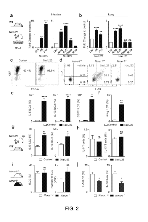
Figures 2a-2j. Neuromedin U is a uniquely potent regulator of innate type 2
cytokines, via NMUR1 activation. Figs. 2a-2f, ILC2-intrinsic activation with
NmU23. Fig.
2a, Type 2 cytokine gene expression in intestinal ILC2s. n=6. Fig. 2b, Type 2
cytokine gene
expression in lung ILC2s. n=6. Fig. 2c, Ki67 expression in intestinal ILC2s.
Fig. 2d, IL-5 and
IL-13 expression in Nmurl competent and deficient ILC2s. Fig. 2e, Innate
inflammatory type
2 cytokines at the protein level. n=6. Fig. 2f, Innate tissue-repair cytokine
AREG. n=6. Figs.
2g,2h, in vivo administration of NmU23. Fig. 2g, ILC2-derived type 2
cytokines. n=6. Fig.
2h, T cell-derived type 2 cytokines. n=6. Figs. 2i,2j, in vivo ablation of
Nmurl . Fig. 2i,
Intestinal ILC2s from Nmur14- and their Nmurl l WT littermate controls. WT
n=6; Nmurl-l-
n=9. Fig. 2j, ILC2-derived type 2 cytokines in bone marrow chimeras of Nmurl-l-
and in their
Nmurl l WT littermate control origin. WT n=6; Nmurl-l- n=3. Error bars show
s.e.m.
*P<0.05; **P<0.01; ***P<0.001; ****P<0.0001; ns not significant.
Figures 3a-3e. Neuromedin U regulates ILC2-derived cytokines via ERK1/2 and
a Ca2 /Calcineurin/NFAT cascade. Figs. 3a-e, Intestinal ILC2 activation by
Neuromedin U.
.. Fig. 3a, Percentage of pERK cells n=4. Mean fluorescence intensity (MFI) of
pERK
expression. n=4. Fig. 3b, 115, Ill 3 and Csf2 expression in ILC2s cultured
with medium
(control) (n=3), NmU23 (n=3) or NmU23 and ERK inhibitor PD98059 (n=3). Fig.
3c, Left
CA 03046884 2019-06-12
WO 2018/109540 PCT/IB2017/000413
7
and centre: Ca2+ influx, represented by Fluo-4 AM intensity. NmU23 was added
60 seconds
after ILC2 baseline acquisition (arrow). Right: Mean intensity of Ca2+ influx.
n=3. Fig. 3d,
115, 1113 and Csf2 expression in ILC2s cultured with medium (control) (n=6),
NmU23 (n=6)
or NmU23 and Calcineurin inhibitor FK506 (n=6). Fig. 3e, 115, 1113 and Csf2
expression in
ILC2s cultured with medium (control) (n=3), NmU23 (n=3) or NmU23 and NFAT
inhibitor
11R-VIVIT (VIVIT) (n=3). Error bars show s.e.m. *P<0.05; **P<0.01; ***P<0.001;
****P<0.0001; ns not significant.
Figures 4a-4h. The neuroregulatory axis NmU-NMUR1 confers protection
against worm infection. Mice were infected with N. brasiliensis larvae and
lungs analysed at
48 hours. Fig. 4a, Nmu expression in total lung from infected mice compared to
non-infected
controls. n=3. Fig. 4b, Pulmonary inflammatory cell infiltrates 48 hours after
infection.
NmU23 treated and control sections are displayed. Hematoxylin and eosin. Fig.
4c,
Myeloperoxidase- (granulocytes) and Luna-stained (eosinophils) lung sections.
Fig. 4d,
Granulocyte and eosinophil cell counts (cells/mm2). Control n=8; NmU23 n=8.
Fig. 4e,
Nmurl-l- and their WT littermate controls were infected with N. brasiliensis.
Hematoxylin
and eosin. Fig. 4f, Myeloperoxidase- (granulocytes) and Luna-stained
(eosinophils) lung
sections. Fig. 4g, Granulocyte and eosinophilic cell counts (cells/mm2). WT
n=8; Nmurl-'n=8. Fig. 4h, N. brasiliensis infection burden at 48 hours in the
lung. WT n=3; Nmur14- n=3.
Scale bars: 50m. Error bars show s.e.m. *P<0.05; **P<0.01; ***P<0.001;
****P<0.0001;
ns not significant.
Figures 5a-5c. Genome-wide ILC2 transcriptional profiling and neuron-ILC2
interactions. Fig. 5a, Weighted Unifrac PCoA analysis of ILC2s, CD4 T cells,
ILC1s and
ILC3s. Fig. 5b, Levels of Nmurl expression in ILC2s, CD4 T cell, ILC1 and ILC3
populations. Fig. 5c, Separate channels of confocal analysis in Fig.le right.
Green: neurons
(RetGFP); Red: KLRG1; Cyan: CD3.
Figures 6a-6f. Neuromedin U is potent regulator of lung innate type 2
cytokines,
via NMUR1 activation. Figs. 6a,6b, ILC2-intrinsic activation with NmU23. Fig.
6a, IL-5
and IL-13 expression in lung ILC2s. Fig. 6b, Innate type 2 cytokines at the
protein level. n=3.
Figs. 6c,6d, in vivo administration of NmU23. Fig. 6c, ILC2-derived type 2
cytokines in the
lung. n=3. Fig. 6d, T cell-derived type 2 cytokines in the lung. n=3. Figs.
6e,6f, in vivo
ablation of Nmurl . Fig. 6e, Lung ILC2s in Nmurl-l- and in their Nmur1+4 WT
littermate
controls. WT n=6; Nmurl-l- n=9. Fig. 6f, Intestinal T cell-derived type 2
cytokines in Nmurl-l-
CA 03046884 2019-06-12
WO 2018/109540 PCT/IB2017/000413
8
and in their Nmur1+4 WT littermate controls. WT n=6; Nmur14- n=6. Error bars
show s.e.m.
*P<0.05; **P<0.01; ***P<0.001; ****P<0.0001; ns not significant.
Figures 7a-7c. Nmurl is dispensable for ILC2 development. Figs. 7a,7c,
Competitive bone marrow chimeras. Fig. 7a, 106 cells of each genotype (CD45.2)
were
injected intravenously in direct competition with a third-party WT competitor
(CD45.1/CD45.2), in a 1:1 ratio, into non-lethally irradiated (150 Rad) NSG
mice (CD45.1).
Fig. 7b, Percentage and number of donor ILC2s in the intestine. WT n=12;
Nmur14- n=12.
Fig. 7c, Percentage and number of donor ILC2s in the lung. WT n=12; Nmurl-l-
n=12. Error
bars show s.e.m. *P<0.05; **P<0.01; ***P<0.001; ****P<0.0001; ns not
significant.
Figure 8. A novel neuron-ILC2 unit orchestrated by neuromedin U. Neuron-
derived Neuromedin U directly activates ILC2s in a NMUR1 dependent manner,
resulting in
a potent production of inflammatory and tissue repair type 2 cytokines that
confer protection
to worm infection. Neuromedin U activates NMUR1 with induces type 2 cytokine
expression
downstream of ERK phosphorylation and activation of a Ca2 /Calcineurin/NFAT
cascade.
This model suggests that neuron-ILC2 cell units are poised to uniquely ensure
potent and
immediate type 2 responses in a neuromedin U-dependent manner.
Figures 9a-9i: Fig. 9a, Nmurl quantitative RT-PCR analysis in the lungs at day
6
post Nippostrongylus brasiliensis (NB) - infection in lung. Eosinophils (Eo);
Mast cells
(Mast); Macrophages (MO); Neutrophils (Neu); naive T cells (T); Innate
lymphoid cells type
2 (ILC2). Fig. 9b, NMUR1 expression in human adaptive (CD4 T cells) and innate
type 2
lymphocytes ILC2 from blood. Fig9c, Type 2 cytokine gene expression in human
ILC2 and
Th2 after in vitro stimulation with the peptide NmU25. Fig. 9d, Nmurl
expression in lung
ILC2 before and after infection (at day 6). Fig. 9e, Nmurl expression in
steady state in
Common lymphoid progenitor (CLP); Common helper innate lymphoid progenitor
(CHILP),
Bone marrow ILC2 progenitor (ILC2P) and Eo, Mast, Mo, Neu, Dendritic cells
(DC); naive
T cells (T); T-helper 2 cells (Th2); memory T cells, B cells (B), Lamina
Propria glial cells
(G) and neurons (N). n=3-6. Fig. 9f, Type 2 cytokine gene expression in
intestinal ILC2 and
Th2 after in vitro stimulation with the peptide NmU23 (10Ong/mL). n=3-6. Fig.
9g, Confocal
analysis of intestinal lamina propria. Green: neurons (RetGFP); Cyan: KLRG1;
red: CD3.
Cyan: KLRG1. Fig.9h, Neurosphere-derived neurons. Red: TUJ1. Blue: DAPI.
Fig.9i,
Activation of neurosphere-derived neurons with alarmins, TLR-ligands and N.
brasiliensis
CA 03046884 2019-06-12
WO 2018/109540 PCT/IB2017/000413
9
excretory/secretory proteins (NES). *P<0.05; **P<0.01; ***P<0.001;
****P<0.0001; ns not
significant.
Figures 10a-10d: Fig. 10a, Ki67 expression in intestinal ILC2s after an
overnight in
vitro stimulation with NmU23 alone (10Ong/mL, Phoenix Pharmaceutical) or NmU23
together with the survival cytokines Interleukin (IL)-2 and/or IL-7 (lOng/mL).
Fig. 10b, Ki67
expression in intestine ILC2 after in vivo administration of NmU23 (4i.tg/day
during 2 days).
n=5. Fig. 10c, ILC2-derived type 2 cytokines (IL-5, IL-13 and Amphiregulin
(Areg)) in
sorted intestine ILC2 after an overnight stimulation with NmU23, mouse
recombinant IL-25
or IL-33 (R&D) (10, 50 and 10Ong/mL). Negative control: unstimulated ILC2,
Positive
control: ILC2 activated with phorbol 12-myristate 13-acetate (PMA, 50ng/m1)
plus
ionomycin (500ng/m1). n=3. Fig. 10d, Dot plots representative of the cytokine
production
with increasing dose of NmU23, rIL-25 and rIL-33. *P<0.05; **P<0.01;
***P<0.001;
****P<0.0001; ns not significant.
Figures lla-11b: ILC2 were FBS deprived for 2 hours prior to treatment with
either
(Fig. 11a) 11R-VIVIT (inhibits NFAT activation) (10 11M) or (Fig. 11b)
cyclosporin A (CsA,
100pM). Expression of type 2 cytokines were measured by quantitative RT-PCR
(Figs.
11a,11b). n=3-6. Fig. 11c, Deprivated ILC2 from Lamina Propria were stimulated
90' with
NmU23 (10Ong/mL), fixed, permeabilized and stained with anti-NFAT2 monoclonal
antibody (abcam). Cells were analyzed by confocal microscopy. *P<0.05;
**P<0.01;
***P<0.001; ****P<0.0001; ns not significant.
Figures 12a-12f: (Figs. 12a-12c) Mice were infected with N. brasiliensis
larvae and
treated with NmU23 peptide (8i.tg/day) or PBS (control). Lungs were analysed
at day 2 post-
infection. Fig. 12a, ILC2 response in lungs from NmU23 treated mice (n=5)
compared to
control (n=5). Fig. 12b, Burden of infection in lungs of infected mice treated
with PBS (n=5)
or NmU23 (n=5). Fig. 12c, Pulmonary hemorrhage in lung of infected mice
treated with
NmU23 compared to control. (Figs. 12d-121) Mice were infected with N.
brasiliensis larvae
and treated with NmU23 peptide (8i.tg/day) or PBS (control). Lungs and small
intestine were
analysed at day 6 post-infection. Fig. 12d, Neutrophils and eosinophils
infiltrate in broncho-
alveolar lavage (BAL) in infected mice treated with NmU23 versus PBS. Control
n=5;
NmU23 n=5. Fig. 12e, Mastocytes and Macrophages infiltrate in broncho-alveolar
lavage
(BAL) in infected mice treated with NmU23 versus PBS. Control n=5; NmU23 n=5.
Fig.
12f, Burden of infection in small intestine of infected mice treated with PBS
(n=5) or NmU23
CA 03046884 2019-06-12
WO 2018/109540 PCT/IB2017/000413
(n=5). Error bars show s.e.m. *P<0.05; **P<0.01; ***P<0.001; ****P<0.0001; ns
not
significant.
Figures 13a-13c: Nmur14- and their WT littermate controls were infected with
N.
brasiliensis and analyzed at day 6 post-infection. Fig. 13a, ILC2 response in
lungs of infected
5 Ninurl-l- and their WT littermate controls D6 post-infection. WT n=6;
Nmur14- n=8. Fig.
13b, Neutrophils (Neu) and eosinophils (Eos) infiltrate in broncho-alveolar
lavage (BAL) in
infected Ninurl-l- and their WT littermate controls. WT n=6; Nmur14- n=7. Fig.
13c,
Mastocytes and Macrophages infiltrate in broncho-alveolar lavage (BAL) in
infected Nmur14-
and their WT littermate controls. WT n=6; Nmur14- n=7. Error bars show s.e.m.
*P<0.05;
10 **P<0.01; ***P<0.001; ****P<0.0001; ns not significant.
Figures 14a-14c: Competitive bone marrow chimeras treated with NmU23. Fig.
14a,
106 cells of each genotype (CD45.2) were injected intravenously in direct
competition with a
third-party WT competitor (CD45.1/CD45.2), in a 1:1 ratio, into non-lethally
irradiated (150
Rad) NSG mice (CD45.1). The mice received one injection of PBS or NmU23 (20
jig). Fig.
14b, Percentage and number of donor ILC2s in the lungs. WT n=5; Ninurl-l- n=5.
Fig. 14c,
Percentage and number of donor T cells in the lungs. WT n=5; Nmur14- n=5.
Error bars show
s.e.m. *P<0.05; **P<0.01; ***P<0.001; ****P<0.0001; ns not significant.
DETAILED DESCRIPTION
Group 2 innate lymphoid cells (ILC2s) are major regulators of inflammation,
tissue
repair and metabolic homeostasis1'2. ILC2 activation has been shown by host-
derived
cytokines and alarmins1'2, but, how ILC2s respond to neuronal-derived signals
remains
unclear.
As described herein, it was determined that ILC2s express the Neuromedin U
receptor
1 (Nmurl) and that the neuropeptide Neuromedin U is a potent activator of
ILC2s.
Neuromedin U resulted in prompt and strong production of the type 2 cytokines
interleukin 5
(IL-5), IL-13 and Amphiregulin in a NMUR1-dependent manner. Neuromedin U
controlled
ILC2 downstream of ERK activation and calcium-influx-dependent activation of
Calcineurin
cytokines and NFAT. When used in vivo, Neuromedin U treatment resulted in
immediate
type 2 responses. It also was shown that ablation of Nmurl led to impaired
type 2 responses
and poor worm infection control.
CA 03046884 2019-06-12
WO 2018/109540 PCT/IB2017/000413
11
Increasing activity of ILC2s
The methods disclosed herein include methods for increasing activity or
proliferation
of Group 2 innate lymphoid cells (ILC2s) by contacting ILC2s with an agonist
of neuromedin
U receptor 1 (NMUR1) in an amount effective to increase activity of the ILC2s.
The methods disclosed herein also include methods for treating a disease
associated
with Group 2 innate lymphoid cells (ILC2s) by administering to a subject in
need of such
treatment an agonist of neuromedin U receptor 1 (NMUR1) in an amount effective
to treat the
disease.
Other methods for treating disease include administering to a subject in need
of such
treatment a composition comprising activated ILC2s in an amount effective to
treat the
disease. In some of these methods, the composition comprising activated ILC2s
also includes
an agonist of neuromedin U receptor 1 (NMUR1). Alternatively, an agonist of
NMUR1 can
be administered separately from the composition comprising activated ILC2s.
Also provided herein are agonists of NMUR1 for use in treating a disease
associated
with ILC2s, and compositions comprising ILC2s (and optionally an agonist of
NMUR1) for
use in treating a disease associated with ILC2s.
As used herein, neuromedin U receptor 1 (NMUR1) is a 7 transmembrane receptor
of
the rhodopsin family, and is also known as FM3, FM-3, GPC-R, G-protein coupled
receptor
66 (GPR66), and NMU1R. As described elsewhere herein, an agonist of NMUR1
includes a
neuromedin U (NMU) or an analog thereof, an antibody that specifically binds
and activates
NMUR1 or an antigen-binding fragment thereof, or a small molecule hg and of
NMUR1.
Contacting ILC2s with an agonist of NMUR1 can be performed in vitro, such as
in an
ILC2 expansion protocol performed to produce ILC2s, or can be performed in
vivo. In some
embodiments of methods in which the contacting of ILC2s with an agonist of
NMUR1 is
performed in vivo, the agonist of NMUR1 is administered to a subject, such as
a human. In
some of these methods, the subject is not otherwise in need of treatment with
the agonist of
NMUR1.
In the disclosed methods, the subject can be a human. In some of these
methods, the
subject is not otherwise in need of treatment with the agonist of NMUR1 and/or
treatment
with the activated ILC2s.
Diseases treatable by the disclosed methods include infection, tissue repair,
wound
healing, obesity, diseases treatable by increasing induction of type 2 immune
responses,
CA 03046884 2019-06-12
WO 2018/109540 PCT/IB2017/000413
12
diseases treatable by metabolic regulation, diseases treatable by increasing
eosinophils, and
diseases treatable by increasing mast cells.
The agonist of NMUR1 and/or the activated ILC2s can be administered by any
suitable route of administration or delivery method. Suitable routes of
administration include
intravenous, oral, nasal, rectal or through skin absorption.
The agonist of NMUR1 and/or the activated ILC2s can be administered at any
suitable interval, including daily, twice daily, three times per day, four
times per day, every
other day, weekly, every two weeks, every four weeks, continuously (e.g., by
infusion, patch,
or pump), and so on.
Decreasing activity of ILC2s
Additional methods disclosed herein include methods for decreasing activity or
proliferation of Group 2 innate lymphoid cells (ILC2s) by contacting ILC2s
with an
antagonist of neuromedin U receptor 1 (NMUR1) or an antagonist of NMU (or
both) in an
amount effective to decrease activity of the ILC2s.
The methods disclosed herein also include methods for treating a disease
associated
with Group 2 innate lymphoid cells (ILC2s) by administering to a subject in
need of such
treatment an antagonist of neuromedin U receptor 1 (NMUR1) in an amount
effective to treat
the disease.
Also provided herein are antagonists of NMUR1 for use in treating a disease
associated with ILC2s.
As described elsewhere herein, an antagonist of NMUR1 includes an inhibitory
nucleic acid molecule that reduces that reduces expression, transcription or
translation of
NMUR1, such as a sRNA, shRNA, or antisense nucleic acid molecule; an antibody
that
specifically binds and inhibits NMUR1 or an antigen-binding fragment thereof,
or a small
molecule antagonist of NMUR1.
Contacting ILC2s with an antagonist of NMUR1 can be performed in vitro, or can
be
performed in vivo. In some embodiments of methods in which the contacting of
ILC2s with
an antagonist of NMUR1 is performed in vivo, the antagonist of NMUR1 is
administered to a
subject, such as a human. In some of these methods, the subject is not
otherwise in need of
treatment with the antagonist of NMUR1.
CA 03046884 2019-06-12
WO 2018/109540 PCT/IB2017/000413
13
In the disclosed methods, the subject can be a human. In some of these
methods, the
subject is not otherwise in need of treatment with the antagonist of NMUR1.
In the methods disclosed herein for treating disease by administering an
antagonist of
NMUR1, the disease can be allergy, allergic asthma, food allergy, eosinophilic
esophagitis,
atopic dermatitis, fibrosis, allergic rhinitis, allergic rhinosinusitis,
chronic obstructive
pulmonary disease (COPD), cystic fibrosis, diseases treatable by reducing type
2 immune
responses, diseases treatable by reducing eosinophils, or diseases treatable
by reducing mast
cells.
The antagonist of NMUR1 can be administered by any suitable route of
administration or delivery method. Suitable routes of administration include
intravenous,
oral, nasal, rectal or through skin absorption.
The antagonist of NMUR1 can be administered at any suitable interval,
including
daily, twice daily, three times per day, four times per day, every other day,
weekly, every two
weeks, every four weeks, continuously (e.g., by infusion, patch, or pump), and
so on.
Agonists of neuromedin U receptor 1 (NMUR1)
Agonists of NMUR1 include peptide agonists (including modified peptides and
conjugates), activating antibody molecules, and small molecules. Peptide
agonists include
neuromedin U (also known as and referred to herein as NMU or NmU) or analogs
thereof.
The NMUR1 agonists may be entirely specific for NMUR1, may agonize NMUR1
preferentially (as compared to neuromedin U receptor 2, NMUR2), or may agonize
both
NMUR1 and NMUR2. Such agonists may be useful even if NMUR1 is agonized less
than
NMUR2, but it is preferred that the agonists used in the methods described
herein agonize
NMUR1 to a greater extent than NMUR2. As used herein agonizing NMUR1
preferentially
(as compared to neuromedin U receptor 2, NMUR2) means that the agonist
agonizes
NMUR1 at least 10%, 25%, 50%, 100%, 200%, 300%, 400%, 500%, 600%, 700%, 800%,
900%, 1000%, or more than NMUR2.
Neuromedin U (also referred to herein as NMU) is a neuropeptide conserved in
many
species, which was isolated as a peptide consisting of 25 amino acid residues
(NMU-25) or as
a peptide consisting of 8 amino acid residues (NMU-8), from pig small
intestine. NMU-8
consists of the C-terminal 8 residues of porcine NMU25. NMU-25 also is present
in humans,
and is preferred for use in humans. The C-terminal 8 amino acid residues of
human NMU-25
CA 03046884 2019-06-12
WO 2018/109540 PCT/IB2017/000413
14
(also referred to as NMU-8) are the same as that of the C-terminal 8 amino
acid residues of
porcine NMU-8. The 8 amino acids at the C terminus of NMU-25 are the most
highly
conserved and this peptide has been shown to have similar activity as NMU-25.
Rat NMU
consists of 23 amino acid residues, and is known as NMU-23. The amino acid
sequence of
the C-terminal 8 residues of rat NMU-23 differs from that of the C-terminal 8
residues of
porcine NMU-8 by one amino acid residue. NMU precursor protein (and its
cleaved
peptides) also can be used in the methods described herein. NMU precursor
protein is a 174
amino acid long protein.
Amino acid sequences of the NMU precursor protein and NMU are provided as
follows:
NMU precursor protein
(P48645INMU HUMAN Neuromedin-U OS=Homo sapiens GN=NMU PE=1 SV=1)
MLRTESCRPRSPAGQVAAASPLLLLLLLLAWCAGACRGAPILPQGLQPEQQLQLWNE
IDDTCSSFLSIDSQPQASNALEELCFMIMGMLPKPQEQDEKDNTKRFLFHYSKTQKLG
KSNVVSSVVHPLLQLVPHLHERRMKRFRVDEEFQSPFASQSRGYFLFRPRNGRRSAG
FT (SEQ ID NO: 1)
NMU25
FRVDEEFQSPFASQSRGYFLFRPRN (SEQ ID NO: 2)
NMU23
FKAEYQSPSVGQSKGYFLFRPRN (SEQ ID NO: 3)
NMU8
YFLFRPRN (SEQ ID NO: 4)
Agonists of NMUR1 include NMU analogs, derivatives, and conjugates, such as
NMU analogs having variations in amino acid sequence relative to natural NMU
sequences
but which retain function of binding to and activating NMUR1. Other examples
of analogs,
derivatives, and conjugates of NMU include: the modified peptides of Takayama
et al. (ACS
Med Chem Lett. 2015 Mar 12; 6(3): 302-307); the NMU-8 analogs of Inooka et al.
(Bioorg
CA 03046884 2019-06-12
WO 2018/109540 PCT/IB2017/000413
Med Chem. 2017 Feb 21. pii: S0968-0896(17)30108-6); the PEGylated derivatives
of NMU
of Ingallinella et al. (Bioorg Med Chem. 2012 Aug 1;20(15):4751-9); the human
serum
albumin (HSA)-NMU conjugate of Neuner et al. (J Pept Sci. 2014 Jan;20(1):7-
19); the
truncated/lipid-conjugated NMU analogs of Micewicz (Eur J Med Chem. 2015 Aug
5 28;101:616-26); and the lipidated NMU analogs of Dalboge et al. (J Pept
Sci. 2015
Feb;21(2):85-94).
As described in US 2011/0294735 and WO 2007/109135 (each incorporated herein
by reference for the specific recitation of the following compounds),
additional NMUR1
agonists comprise the general formula (I)
10 Z1-peptide-Z2 (I)
wherein the peptide has the amino acid sequence X1 X2 X3 X4 X5 X6 X7
x8 x9 x10 x11 x12 x13 x14 x15 x16 x17 x18 x19 x20 x21 x22 x23
X'¨X5, -µ A,25,
wherein amino acids 1 to 17 can be any amino acid or absent, wherein amino
acid
X18 is absent, Y, W, F, a des-amino acid or an acyl group; amino acid X19 is
A, W, Y, F or an
15 .. aliphatic amino acid; amino acid X2 is absent, L, G, sarcosine (Sar), D-
Leu, NMe-Leu, D-
Ala or A; amino acid X21 is F, NMe-Phe, an aliphatic amino acid, an aromatic
amino acid, A
or W; X22 is R, K, A or L; amino acid X23 is P, Sar, A or L; amino acid X24 is
R, Harg or K;
and amino acid X25 is N, any D- or L-amino acid, Nle or D-Nle, A; and Z1 is an
optionally
present protecting group that, if present, is joined to the N-terminal amino
group; and Z2 is
NH2 or an optionally present protecting group that, if present, is joined to
the C-terminal
carboxy group, and pharmaceutically acceptable salts thereof.
As described in US 2012/0094898 (incorporated herein by reference for the
specific
recitation of the following compounds), additional NMUR1 agonists include
peptide
derivatives selected from the group consisting of
PEG20k(AL)-0-Ala-Tyr-Nal(1)-Leu-Phe-Arg-Pro-Arg-Asn-NH2,
PEG20k(AL)-0-Ala-Tyr-Nal(2)-Leu-Phe-Arg-Pro-Arg-Asn-NH2,
PEG20k(AL)-NpipAc-Tyr-Nal(2)-Leu-Phe-Arg-Pro-Arg-Asn-NH2,
PEG20k(AL)-NpipAc-Tyr-Nal(2)-Leu-Phe-Arg-Ala-Arg-Asn-NH2.
PEG20k(AL)-PEG(2)-Tyr-Nal(2)-Leu-Phe-Arg-NMeAla-Arg-Asn-NH2,
PEG20k(AL)-Pic(4)-Tyr-Nal(2)-Leu-Phe-Arg-NMeAla-Arg-Asn-NH2,
PEG20k(AL)-Acp-Tyr-Nal(2)-Leu-Phe-Arg-NMeAla-Arg-Asn-NH2, and
CA 03046884 2019-06-12
WO 2018/109540 PCT/IB2017/000413
16
PEG20k(AL)-0-A1a-Tyr-Na1(2)-Leu-Pya(4)-Arg-Pro-Arg-Asn-NH2, or a salt of any
of the
peptide derivatives.
As described in WO 2011/005611 (incorporated herein by reference for the
specific
recitation of the following compounds), additional NMUR1 agonists include
compositions
comprising the formula
Z1-peptide-Z2
wherein the peptide has the amino acid sequence X1 x2 x3 x4 x5 x6 x7 x8
x9 x10 x11 x12 x13 x14 x15 x16 x17 x18 x19 x20 x21 x22 x23
X'¨X5, wherein amino acids 1 to 17 can be any amino acid or absent; wherein
amino acid
.. X18 is absent, Tyr or D-Tyr, Leu, Phe, Val, Gln, Nle, Glu or D-Glu, Asp,
Ala, D-Lys, an
aromatic amino acid, a des-amino acid or an acyl group; amino acid X19 is Ala,
Trp, Tyr, Phe,
Glu, Nva, Nle or an aromatic amino acid; amino acid X2 is absent, Leu, Gly,
sarcosine (Sar),
D-Leu, NMe-Leu, D-Ala or Ala, or any D- or L-amino acid; amino acid X21 is
Phe, NMe-
Phe, an aliphatic amino acid, an aromatic amino acid, Ala or Trp; X22 is Arg,
Lys, Harg, Ala,
or Leu; amino acid X23 is Pro, Ser, Sar, Ala or Leu; amino acid X24 is Arg,
Harg or Lys; and
amino acid X25 is Asn, any D- or L-amino acid, Nle or D¨Nle, D-Ala or Ala; Z1
is
optionally a protecting group that, if present, is joined to the N-terminus
amino group; and Z2
is NH2 or an optionally present protecting group that, if present, is joined
to the C-terminal
carboxy group, and pharmaceutically acceptable salts thereof.
As described in WO 2010/138343 (incorporated herein by reference for the
specific
recitation of the following compounds), additional NMUR1 agonists include
compositions
comprising a neuromedin U receptor agonist in which neuromedin U or an analog
thereof is
conjugated to cysteine residue 34 of human serum albumin by a non-maleimido or
non-
succinimidyl linkage or a pharmaceutically acceptable salt thereof.
As described in WO 2009/042053 (incorporated herein by reference for the
specific
recitation of the following compounds), additional NMUR1 agonists include a
neuromedin U
receptor agonist represented by the following formula:
Z1-peptide-Z2
wherein the peptide has the amino acid sequence
ILQRGSGTAAVDFTKKDHTATWGRPFFLFRPRN (SEQ ID NO: 5), wherein the peptide
can have one or more insertions or substitutions of the amino acid sequence
with an
alternative amino acid and wherein the peptide can have one or more deletions
of the amino
CA 03046884 2019-06-12
WO 2018/109540 PCT/IB2017/000413
17
acid sequence; Z1 is an optionally present protecting group that, if present,
is joined to the N-
terminal amino group; and Z2 is NH2 or an optionally present protecting group
that, if
present, is joined to the C-terminal carboxy group; and pharmaceutically
acceptable salts
thereof.
As described in WO 2009/044918 (incorporated herein by reference for the
specific
recitation of the following compounds), additional NMUR1 agonists include
neuromedin U
derivatives selected from polypeptides consisting of an amino acid sequence
which is bound
with a methoxypolyethylene glycol(s) via a linker, wherein the amino acid
sequence contains
at least 8 amino acids of the C-terminus of an amino acid sequence of
neuromedin U, and is
the same or substantially the same as the amino acid sequence of neuromedin U.
Antagonists of neuromedin U receptor 1 (NMUR1) or Neuromedin U (NMU)
Antagonists of NMUR1 include peptide antagonists (including modified peptides
and
conjugates), inhibitory antibody molecules, inhibitory nucleic acid molecules,
and small
molecules. The NMUR1 antagonists may be entirely specific for NMUR1, may
antagonize
NMUR1 preferentially (as compared to neuromedin U receptor 2, NMUR2), or may
antagonize both NMUR1 and NMUR2. Such antagonists may be useful even if NMUR1
is
antagonized less than NMUR2, but it is preferred that the antagonists used in
the methods
described herein antagonize NMUR1 to a greater extent than NMUR2. As used
herein,
.. antagonizing NMUR1 preferentially (as compared to neuromedin U receptor 2,
NMUR2)
means that the antagonist antagonizes NMUR1 at least 10%, 25%, 50%, 100%,
200%, 300%,
400%, 500%, 600%, 700%, 800%, 900%, 1000%, or more than NMUR2.
As described in US 2011/0165144 (incorporated herein by reference for the
specific
recitation of the following compounds), additional NMU and NMUR1 antagonists
include
(i) a neuromedin U (NMU)-specific inhibitory nucleic acid, e.g., an siRNA,
antisense,
aptamer, or ribozyme targeted specifically to NMU;
(ii) a neuromedin U (NMU) inhibitory peptide, e.g., a peptide comprising the
sequence Phe-Arg-Pro-Arg-Asn (SEQ ID NO: 6); or
(iii) an antibody or antigen binding fragment thereof that binds to an NMU-R,
e.g.,
NMU-R1, and inhibits NMU signalling, e.g., inhibits binding of NMU to the NMU-
R1.
Suitable NMUR1 antagonists also can include:
CA 03046884 2019-06-12
WO 2018/109540 PCT/IB2017/000413
18
(i) a neuromedin U receptor 1 (NMUR1)-specific inhibitory nucleic acid, e.g.,
an
siRNA, antisense, aptamer, or ribozyme targeted specifically to NMUR1; or
Suitable NMU antagonists also can include:
(i) a soluble NMUR1 molecule that binds NMU, such as an extracellular portion
of
NMUR1 (e.g., amino acids 1 ¨ 65 of UniProtKB - Q9HB89) optionally linked or
fused to
another polypeptide sequence for stability or other functions, such as an
immunoglobulin Fc
region; and
(ii) an antibody or antigen binding fragment thereof that binds to an NMU,
e.g.,
NMU-8, NMU-23, or NMU-25, and inhibits NMU signalling, e.g., inhibits binding
of NMU
to the NMU-Rl.
A subject shall mean a human or vertebrate mammal including but not limited to
a
dog, cat, horse, goat and non-human primate, e.g., monkey. Preferably the
subject is a
human. In some embodiments the subject is one who is not otherwise in need of
treatment
with an NMUR1 agonist or NMUR1 antagonist. Therefore the subject, in
specifically
identified embodiments, may be one who has not been previously diagnosed with
a disorder
for which an NMUR1 agonist or NMUR1 antagonist is an identified form of
treatment.
The subject can be first identified as a subject in need of treatment, such as
one having
a disease that is treatable by the methods disclosed herein, and then treated
with an NMUR1
.. agonist (and/or activated ILC2s) or NMUR1 antagonist. The skilled artisan
is aware of
methods for identifying a subject as having a disease that is treatable by the
methods
disclosed herein.
As used herein, the terms "treat," "treated," or "treating" refers to a
treatment of a
disease that ameliorates the disease (disease modification), ameliorates
symptoms of the
disease, prevents the disease from becoming worse, or slows the progression of
the disease
compared to in the absence of the therapy.
A "disease associated with Group 2 innate lymphoid cells (ILC2s)" as used
herein is a
disease or disorder in which ILC2s play some role in the development,
maintenance or
worsening of the disease or disorder.
In some of the methods disclosed herein, such diseases can be effectively
treated by
increasing activity or proliferation of ILC2s, such as by contacting ILC2s
with an agonist of
neuromedin U receptor 1 (NMUR1) in an amount effective to increase activity of
the ILC2s;
CA 03046884 2019-06-12
WO 2018/109540 PCT/IB2017/000413
19
by administering to a subject in need of such treatment an agonist of NMUR1 in
an amount
effective to treat the disease; or by administering activated ILC2s (and
optionally an agonist
of NMUR1) in an amount effective to treat the disease.
Diseases treatable by such methods include: infection, tissue repair, wound
healing,
obesity, diseases treatable by increasing induction of type 2 immune
responses, diseases
treatable by metabolic regulation, diseases treatable by increasing
eosinophils, and diseases
treatable by increasing mast cells
In other of the method disclosed herein, the diseases can be effectively
treated by
decreasing activity or proliferation of ILC2s, such as by contacting ILC2s
with an antagonist
of neuromedin U receptor 1 (NMUR1) in an amount effective to decrease activity
of the
ILC2s; or by administering to a subject in need of such treatment an
antagonist of NMUR1 in
an amount effective to treat the disease.
Diseases treatable by such methods include: allergy, allergic asthma, food
allergy,
eosinophilic esophagitis, atopic dermatitis, fibrosis, allergic rhinitis,
allergic rhinosinusitis,
chronic obstructive pulmonary disease (COPD), cystic fibrosis, diseases
treatable by reducing
type 2 immune responses, diseases treatable by reducing eosinophils, or
diseases treatable by
reducing mast cells.
Toxicity and efficacy of the methods of the present invention can be
determined by
standard pharmaceutical procedures in cell cultures or experimental animals,
e.g., for
determining the LD50 (the dose lethal to 50% of the population) or TD50 (the
dose toxic to
50% of the population) and the ED50 (the dose therapeutically effective in 50%
of the
population). The dose ratio between toxic and therapeutic effects is the
therapeutic index and
it can be expressed as the ratio LD50/ED50 or TD50/ED50. Therapeutic agents
that exhibit
large therapeutic indices are preferred. While therapeutic agents that exhibit
toxic side
effects may be used, in such cases it is preferred to use a delivery system
that targets such
agents to the site of affected tissue in order to minimize potential damage to
other cells or
tissues and, thereby, reduce side effects.
The data obtained from the cell culture assays and/or animal studies can be
used in
formulating a range of dosage of the therapeutic agents for use in humans. The
dosage of
such agents lies preferably within a range of circulating concentrations that
include the ED50
with little or no toxicity. The dosage may vary within this range depending
upon the dosage
form employed and the route of administration utilized. For any agent used in
the method of
CA 03046884 2019-06-12
WO 2018/109540 PCT/IB2017/000413
the invention, the therapeutically effective dose can be estimated initially
from cell culture
assays. A dose may be formulated in animal models to achieve a circulating
plasma
concentration range that includes the IC50 (i.e., the concentration of the
test compound that
achieves a half-maximal inhibition of symptoms) as determined in cell culture.
Such
5 information can be used to more accurately determine useful doses in
humans.
In certain embodiments, pharmaceutical compositions may comprise, for example,
at
least about 0.1% of an active compound. In other embodiments, the an active
compound may
comprise between about 2% to about 75% of the weight of the unit, or between
about 25% to
about 60%, for example, and any range derivable therein. Other, higher
percentages of an
10 active compound also can be used.
The pharmaceutical compositions may also be, and preferably are, sterile in
some
embodiments. In other embodiments the compounds may be isolated. As used
herein, the
term "isolated" means that the referenced material is removed from its native
environment,
e.g. , a cell. Thus, an isolated biological material can be free of some or
all cellular
15 components, i.e., components of the cells in which the native material
is occurs naturally
(e.g., cytoplasmic or membrane components). In the case of nucleic acid
molecules, an
isolated nucleic acid includes a PCR product, an isolated RNA, a synthetically
(e.g.,
chemically) produced RNA, such as an siRNA, an antisense nucleic acid, an
aptamer, etc.
Isolated nucleic acid molecules include sequences inserted into plasmids,
cosmids, or other
20 vectors to form part of a chimeric recombinant nucleic acid construct,
or produced by
expression of a nucleic acid encoding it. Thus, in a specific embodiment, a
recombinant
nucleic acid is an isolated nucleic acid. An isolated protein may be
associated with other
proteins or nucleic acids, or both, with which it associates in the cell, or
with cellular
membranes if it is a membrane-associated protein, or may be synthetically
(e.g., chemically)
produced, or produced by expression of a nucleic acid encoding it. An isolated
cell, such as
an ILC2 cell, can be removed from the anatomical site in which it is found in
an organism, or
may be produced by in vitro expansion of an isolated cell or cell population.
An isolated
material may be, but need not be, purified.
The term "purified" in reference to a protein, a nucleic acid, or a cell or
cell
population, refers to the separation of the desired substance from
contaminants to a degree
sufficient to allow the practitioner to use the purified substance for the
desired purpose.
Preferably this means at least one order of magnitude of purification is
achieved, more
CA 03046884 2019-06-12
WO 2018/109540 PCT/IB2017/000413
21
preferably two or three orders of magnitude, most preferably four or five
orders of magnitude
of purification of the starting material or of the natural material. In
specific embodiments, a
purified agonist of NMUR1 or antagonist of NMUR1 or ILC2 population is at
least 60%, at
least 80%, or at least 90% of total protein or nucleic acid or cell
population, as the case may
be, by weight. In a specific embodiment, a purified agonist of NMUR1 or
antagonist of
NMUR1 or ILC2 population is purified to homogeneity as assayed by standard,
relevant
laboratory protocols.
In some embodiments a purified and or isolated molecule is a synthetic
molecule.
Subject doses of the compounds described herein typically range from about 0.1
vg to
10,000 mg, more typically from about 1 vg/day to 8000 mg, and most typically
from about
10 vg to 100 [lg. Stated in terms of subject body weight, typical dosages
range from about 1
microgram/kg/body weight, about 5 microgram/kg/body weight, about 10
microgram/kg/body weight, about 50 microgram/kg/body weight, about 100
microgram/kg/body weight, about 200 microgram/kg/body weight, about 350
microgram/kg/body weight, about 500 microgram/kg/body weight, about 1
milligram/kg/body weight, about 5 milligram/kg/body weight, about 10
milligram/kg/body
weight, about 50 milligram/kg/body weight, about 100 milligram/kg/body weight,
about 200
milligram/kg/body weight, about 350 milligram/kg/body weight, about 500
milligram/kg/body weight, to about 1000 mg/kg/body weight or more per
administration, and
any range derivable therein. In non-limiting examples of a derivable range
from the numbers
listed herein, a range of about 1 mg/kg/body weight to about 100 mg/kg/body
weight, about 5
microgram/kg/body weight to about 500 milligram/kg/body weight, etc., can be
administered,
based on the numbers described above. The absolute amount will depend upon a
variety of
factors including the concurrent treatment, the number of doses and the
individual patient
.. parameters including age, physical condition, size and weight. These are
factors well known
to those of ordinary skill in the art and can be addressed with no more than
routine
experimentation. It is preferred generally that a maximum dose be used, that
is, the highest
safe dose according to sound medical judgment. Multiple doses of the molecules
of the
invention are also contemplated.
The compounds and/or cells described herein may be used alone without other
active
therapeutics or may be combined with other therapeutic compounds for the
treatment of the
diseases described herein.
CA 03046884 2019-06-12
WO 2018/109540 PCT/IB2017/000413
22
When used in combination with the compounds and cells described herein, the
dosages of known therapies may be reduced in some instances, to avoid side
effects. In some
instances, when the compounds and/or cells described herein are administered
with another
therapeutic, a sub-therapeutic dosage of either the compounds and/or cells
described herein or
.. the known therapies, or a sub-therapeutic dosage of both, is used in the
treatment of a subject.
A "sub-therapeutic dose" as used herein refers to a dosage which is less than
that dosage
which would produce a therapeutic result in the subject if administered in the
absence of the
other agent. Thus, the sub-therapeutic dose of a known therapy is one which
would not
produce the desired therapeutic result in the subject in the absence of the
administration of the
.. compounds and cells described herein. Existing therapies for the diseases
described herein
are well known in the field of medicine, and may be described in references
such as
Remington's Pharmaceutical Sciences; as well as many other medical references
relied upon
by the medical profession as guidance for treatment.
When the compounds and/or cells described herein are administered in
combination
.. with other therapeutic agents, such administration may be simultaneous or
sequential. When
the other therapeutic agents are administered simultaneously they can be
administered in the
same or separate formulations, but are administered at the same time. The
administration of
the other therapeutic agent and the compounds and/or cells described herein
can also be
temporally separated, meaning that the other therapeutic agents are
administered at a different
time, either before or after, the administration of the compounds and cells
described herein.
The separation in time between the administration of these compounds may be a
matter of
minutes or it may be longer.
The active agents of the invention (e.g., the compounds and cells described
herein) are
administered to the subject in an effective amount for treating disease.
According to some
aspects of the invention, an effective amount is that amount, depending on the
disease being
treated, of a NMUR1 agonist (and/or activated ILC2s) or NMUR1 antagonist alone
or in
combination with another medicament, which when combined or co-administered or
administered alone, results in a therapeutic response to the disease. The
biological effect may
be the amelioration and or absolute elimination of disease, or of symptoms
resulting from the
disease. In another embodiment, the biological effect is the complete
abrogation of the
disease, as evidenced for example, by the absence of a symptom of the disease.
CA 03046884 2019-06-12
WO 2018/109540 PCT/IB2017/000413
23
The effective amount of a compound (i.e., any of the agonists, antagonists, or
ILC2s)
used in methods of the invention in the treatment of a disease described
herein may vary
depending upon the specific compound used, the mode of delivery of the
compound, and
whether it is used alone or in combination. The effective amount for any
particular
application can also vary depending on such factors as the disease being
treated, the particular
compound being administered, the size of the subject, or the severity of the
disease or
condition. One of ordinary skill in the art can empirically determine the
effective amount of a
particular molecule of the invention using routine and accepted methods known
in the art,
without necessitating undue experimentation. Combined with the teachings
provided herein,
.. by choosing among the various active compounds and weighing factors such as
potency,
relative bioavailability, patient body weight, severity of adverse side-
effects and preferred
mode of administration, an effective therapeutic treatment regimen can be
planned which
does not cause substantial toxicity and yet is effective to treat the
particular subject.
Pharmaceutical compositions of the present invention comprise an effective
amount
of one or more agents, dissolved or dispersed in a pharmaceutically acceptable
carrier. The
phrases "pharmaceutical or pharmacologically acceptable" refers to molecular
entities and
compositions that do not produce an adverse, allergic or other untoward
reaction when
administered to an animal, such as, for example, a human, as appropriate.
Moreover, for
animal (e.g., human) administration, it will be understood that preparations
should meet
sterility, pyrogenicity, general safety and purity standards as required by
relevant government
regulatory agencies. The compounds are generally suitable for administration
to humans.
This term requires that a compound or composition be nontoxic and sufficiently
pure so that
no further manipulation of the compound or composition is needed prior to
administration to
humans.
As used herein, "pharmaceutically acceptable carrier" includes any and all
solvents,
dispersion media, coatings, surfactants, antioxidants, preservatives (e.g.,
antibacterial agents,
antifungal agents), isotonic agents, absorption delaying agents, salts,
preservatives, drugs,
drug stabilizers, gels, binders, excipients, disintegration agents,
lubricants, sweetening agents,
flavoring agents, dyes, such like materials and combinations thereof, as would
be known to
.. one of ordinary skill in the art (see, for example, Remington's
Pharmaceutical Sciences
(1990), incorporated herein by reference). Except insofar as any conventional
carrier is
CA 03046884 2019-06-12
WO 2018/109540 PCT/IB2017/000413
24
incompatible with the active ingredient, its use in the therapeutic or
pharmaceutical
compositions is contemplated.
The therapeutic compositions used as described herein may comprise different
types
of carriers depending on whether it is to be administered in solid, liquid or
aerosol form, and
whether it need to be sterile for such routes of administration as injection.
The compounds
and/or cells described herein can be administered intravenously,
intradermally, intraarterially,
intralesionally, intracranially, intraarticularly, intranasally,
intravitreally, intravaginally,
intrarectally, topically, intramuscularly, intraperitoneally, subcutaneously,
intravesicularlly,
mucosally, orally, locally, by inhalation (e.g., aerosol inhalation), by
injection, by infusion
.. including by continuous infusion, by localized perfusion, via a catheter,
via a lavage, in
cremes, in lipid compositions (e.g., liposomes), or by other method or any
combination of the
foregoing as would be known to one of ordinary skill in the art (see, for
example,
Remington's Pharmaceutical Sciences) and as is appropriate for the disease
being treated.
In any case, the composition may comprise various antioxidants to retard
oxidation of
one or more components. Additionally, the prevention of the action of
microorganisms can
be brought about by preservatives such as various antibacterial and antifungal
agents,
including but not limited to parabens (e.g., methylparabens, propylparabens),
chlorobutanol,
phenol, sorbic acid, thimerosal or combinations thereof.
The compounds described herein may be formulated into a composition in a free
base,
neutral or salt form. Pharmaceutically acceptable salts, include the acid
addition salts, e.g.,
those formed with the free amino groups of a proteinaceous composition, or
which are
formed with inorganic acids such as for example, hydrochloric or phosphoric
acids, or such
organic acids as acetic, oxalic, tartaric or mandelic acid. Salts formed with
the free carboxyl
groups also can be derived from inorganic bases such as for example, sodium,
potassium,
ammonium, calcium or ferric hydroxides; or such organic bases as
isopropylamine,
trimethylamine, histidine or procaine.
In embodiments where the compounds and/or cells described herein is in a
liquid
form, a carrier can be a solvent or dispersion medium comprising but not
limited to, water,
ethanol, polyol (e.g., glycerol, propylene glycol, liquid polyethylene glycol,
etc.), lipids (e.g.,
.. triglycerides, vegetable oils, liposomes) and combinations thereof. The
proper fluidity can be
maintained, for example, by the use of a coating, such as lecithin; by the
maintenance of the
required particle size by dispersion in carriers such as, for example liquid
polyol or lipids; by
CA 03046884 2019-06-12
WO 2018/109540 PCT/IB2017/000413
the use of surfactants such as, for example hydroxypropylcellulose; or
combinations thereof
such methods. In many cases, it will be preferable to include isotonic agents,
such as, for
example, sugars, sodium chloride or combinations thereof.
The compounds and/or cells described herein can be administered in various
ways and
5 to different classes of recipients. In some instances the administration
is chronic. Chronic
administration refers to long term administration of a drug to treat a
disease. The chronic
administration may be on an as needed basis or it may be at regularly
scheduled intervals.
For instance, the compounds and/or cells described herein may be administered
twice daily,
three times per day, four times per day, every other day, weekly, every two
weeks, every four
10 weeks, continuously (e.g., by infusion, patch, or pump), and so on.
The compounds and/or cells described herein may be administered directly to a
tissue.
Direct tissue administration may be achieved by direct injection. The
compounds may be
administered once, or alternatively they may be administered in a plurality of
administrations.
If administered multiple times, the compounds may be administered via
different routes. For
15 example, the first (or the first few) administrations may be made
directly into the affected
tissue while later administrations may be systemic.
The compounds and/or cells described herein are administered in
pharmaceutically
acceptable solutions, which may routinely contain pharmaceutically acceptable
concentrations of salt, buffering agents, preservatives, compatible carriers,
adjuvants, and
20 optionally other therapeutic ingredients.
According to the methods described herein, the compounds and/or cells
described
herein may be administered in a pharmaceutical composition. In general, a
pharmaceutical
composition comprises the compound of the invention and a pharmaceutically-
acceptable
carrier. Pharmaceutically-acceptable carriers useful with compounds and/or
cells described
25 herein are well-known to those of ordinary skill in the art. As used
herein, a
pharmaceutically-acceptable carrier means a non-toxic material that does not
interfere with
the effectiveness of the biological activity of the compounds and/or cells
described herein.
Pharmaceutically acceptable carriers include diluents, fillers, salts,
buffers, stabilizers,
solubilizers and other materials which are well-known in the art. Exemplary
pharmaceutically acceptable carriers for peptides in particular are described
in U.S. Patent
No. 5,211,657. Such preparations may routinely contain salt, buffering agents,
preservatives,
compatible carriers, and optionally other therapeutic agents. When used in
medicine, the
CA 03046884 2019-06-12
WO 2018/109540 PCT/IB2017/000413
26
salts should be pharmaceutically acceptable, but non-pharmaceutically
acceptable salts may
conveniently be used to prepare pharmaceutically-acceptable salts thereof and
are not
excluded from the scope of the invention. Such pharmacologically and
pharmaceutically-
acceptable salts include, but are not limited to, those prepared from the
following acids:
hydrochloric, hydrobromic, sulfuric, nitric, phosphoric, maleic, acetic,
salicylic, citric,
formic, malonic, succinic, and the like. Also, pharmaceutically-acceptable
salts can be
prepared as alkaline metal or alkaline earth salts, such as sodium, potassium
or calcium salts.
The compounds and/or cells described herein may be formulated into
preparations in
solid, semi-solid, liquid or gaseous forms such as tablets, capsules, powders,
granules,
ointments, solutions, depositories, inhalants and injections, and usual ways
for oral,
parenteral or surgical administration. The invention also embraces
pharmaceutical
compositions which are formulated for local administration, such as by
implants.
Compositions suitable for oral administration may be presented as discrete
units, such
as capsules, tablets, lozenges, each containing a predetermined amount of the
active agent.
Other compositions include suspensions in aqueous liquids or non-aqueous
liquids, such as a
syrup, an elixir or an emulsion.
For oral administration, the compounds can be formulated readily by combining
the
active compounds with pharmaceutically acceptable carriers well known in the
art. Such
carriers enable the compounds of the invention to be formulated as tablets,
pills, dragees,
capsules, liquids, gels, syrups, slurries, suspensions and the like, for oral
ingestion by a
subject to be treated. Pharmaceutical preparations for oral use can be
obtained as solid
excipient, optionally grinding a resulting mixture, and processing the mixture
of granules,
after adding suitable auxiliaries, if desired, to obtain tablets or dragee
cores. Suitable
excipients are, in particular, fillers such as sugars, including lactose,
sucrose, mannitol, or
sorbitol; cellulose preparations such as, for example, maize starch, wheat
starch, rice starch,
potato starch, gelatin, gum tragacanth, methyl cellulose, hydroxypropylmethyl-
cellulose,
sodium carboxymethylcellulose, and/or polyvinylpyrrolidone (PVP). If desired,
disintegrating agents may be added, such as the cross-linked polyvinyl
pyrrolidone, agar, or
alginic acid or a salt thereof such as sodium alginate. Optionally the oral
formulations may
also be formulated in saline or buffers for neutralizing internal acid
conditions or may be
administered without any carriers.
Dragee cores are provided with suitable coatings. For this purpose,
concentrated
CA 03046884 2019-06-12
WO 2018/109540 PCT/IB2017/000413
27
sugar solutions may be used, which may optionally contain gum arabic, talc,
polyvinyl
pyrrolidone, carbopol gel, polyethylene glycol, and/or titanium dioxide,
lacquer solutions,
and suitable organic solvents or solvent mixtures. Dyestuffs or pigments may
be added to the
tablets or dragee coatings for identification or to characterize different
combinations of active
compound doses.
Pharmaceutical preparations which can be used orally include push-fit capsules
made
of gelatin, as well as soft, sealed capsules made of gelatin and a
plasticizer, such as glycerol
or sorbitol. The push-fit capsules can contain the active ingredients in
admixture with filler
such as lactose, binders such as starches, and/or lubricants such as talc or
magnesium stearate
and, optionally, stabilizers. In soft capsules, the active compounds may be
dissolved or
suspended in suitable liquids, such as fatty oils, liquid paraffin, or liquid
polyethylene
glycols. In addition, stabilizers may be added. Microspheres formulated for
oral
administration may also be used. Such microspheres have been well defined in
the art. All
formulations for oral administration should be in dosages suitable for such
administration.
For buccal administration, the compositions may take the form of tablets or
lozenges
formulated in conventional manner.
For administration by inhalation, the compounds and/or cells described herein
may be
conveniently delivered in the form of an aerosol spray presentation from
pressurized packs or
a nebulizer, with the use of a suitable propellant, e.g.,
dichlorodifluoromethane,
trichlorofluoromethane, dichlorotetrafluoroethane, carbon dioxide or other
suitable gas. In
the case of a pressurized aerosol the dosage unit may be determined by
providing a valve to
deliver a metered amount. Capsules and cartridges of e.g. gelatin for use in
an inhaler or
insufflator may be formulated containing a powder mix of the compound and a
suitable
powder base such as lactose or starch. Techniques for preparing aerosol
delivery systems are
well known to those of skill in the art. Generally, such systems should
utilize components
which will not significantly impair the biological properties of the active
agent (see, for
example, Remington's Pharmaceutical Sciences). Those of skill in the art can
readily
determine the various parameters and conditions for producing aerosols without
resort to
undue experimentation.
The compounds, when it is desirable to deliver them systemically, may be
formulated
for parenteral administration by injection, e.g., by bolus injection or
continuous infusion.
Formulations for injection may be presented in unit dosage form, e.g., in
ampoules or in
CA 03046884 2019-06-12
WO 2018/109540 PCT/IB2017/000413
28
multi-dose containers, with an added preservative. The compositions may take
such forms as
suspensions, solutions or emulsions in oily or aqueous vehicles, and may
contain formulatory
agents such as suspending, stabilizing and/or dispersing agents.
Preparations for parenteral administration include sterile aqueous or non-
aqueous
solutions, suspensions, and emulsions. Examples of non-aqueous solvents are
propylene
glycol, polyethylene glycol, vegetable oils such as olive oil, and injectable
organic esters such
as ethyl oleate. Aqueous carriers include water, alcoholic/aqueous solutions,
emulsions or
suspensions, including saline and buffered media. Parenteral vehicles include
sodium
chloride solution, Ringer's dextrose, dextrose and sodium chloride, lactated
Ringer's, or fixed
oils. Intravenous vehicles include fluid and nutrient replenishers,
electrolyte replenishers
(such as those based on Ringer's dextrose), and the like. Preservatives and
other additives
may also be present such as, for example, antimicrobials, anti-oxidants,
chelating agents, and
inert gases and the like. Lower doses will result from other forms of
administration, such as
intravenous administration. In the event that a response in a subject is
insufficient at the
initial doses applied, higher doses (or effectively higher doses by a
different, more localized
delivery route) may be employed to the extent that patient tolerance permits.
Multiple doses
per day are contemplated to achieve appropriate systemic levels of compounds.
In yet other embodiments, vehicle for the compounds and/or cells described
herein is
a biocompatible microparticle or implant that is suitable for implantation
into a mammalian
recipient. Exemplary bioerodible implants are known in the art. The implant
may be a
polymeric matrix in the form of a microparticle such as a microsphere (wherein
the agent is
dispersed throughout a solid polymeric matrix) or a microcapsule (wherein the
agent is stored
in the core of a polymeric shell). Other forms of the polymeric matrix for
containing the
agent include films, coatings, gels, implants, and stents. The size and
composition of the
polymeric matrix device is selected to result in favorable release kinetics in
the tissue into
which the matrix device is implanted. The size of the polymeric matrix device
further is
selected according to the method of delivery which is to be used, typically
injection into a
tissue or administration of a suspension by aerosol into the nasal and/or
pulmonary areas.
The polymeric matrix composition can be selected to have both favorable
degradation rates
and also to be formed of a material which is bioadhesive, to further increase
the effectiveness
of transfer when the device is administered to a vascular, pulmonary, or other
surface. The
matrix composition also can be selected not to degrade, but rather, to release
by diffusion
CA 03046884 2019-06-12
WO 2018/109540 PCT/IB2017/000413
29
over an extended period of time.
Both non-biodegradable and biodegradable polymeric matrices can be used to
deliver
the compounds and/or cells described herein to the subject. Biodegradable
matrices are
preferred. Such polymers may be natural or synthetic polymers. The polymer is
selected
based on the period of time over which release is desired, generally in the
order of a few
hours to a year or longer. Typically, release over a period ranging from
between a few hours
and three to twelve months is most desirable. The polymer optionally is in the
form of a
hydrogel that can absorb up to about 90% of its weight in water and further,
optionally is
cross-linked with multivalent ions or other polymers.
In general, the compounds and/or cells described herein may be delivered using
the
bioerodible implant by way of diffusion, or more preferably, by degradation of
the polymeric
matrix. Exemplary synthetic polymers which can be used to form the
biodegradable delivery
system include: polyamides, polycarbonates, polyalkylenes, polyalkylene
glycols,
polyalkylene oxides, polyalkylene terepthalates, polyvinyl alcohols, polyvinyl
ethers,
polyvinyl esters, poly-vinyl halides, polyvinylpyrrolidone, polyglycolides,
polysiloxanes,
polyurethanes and co-polymers thereof, alkyl cellulose, hydroxyalkyl
celluloses, cellulose
ethers, cellulose esters, nitro celluloses, polymers of acrylic and
methacrylic esters, methyl
cellulose, ethyl cellulose, hydroxypropyl cellulose, hydroxy-propyl methyl
cellulose,
hydroxybutyl methyl cellulose, cellulose acetate, cellulose propionate,
cellulose acetate
.. butyrate, cellulose acetate phthalate, carboxylethyl cellulose, cellulose
triacetate, cellulose
sulphate sodium salt, poly(methyl methacrylate), poly(ethyl methacrylate),
poly(butylmethacrylate), poly(isobutyl methacrylate), poly(hexylmethacrylate),
poly(isodecyl
methacrylate), poly(lauryl methacrylate), poly(phenyl methacrylate),
poly(methyl acrylate),
poly(isopropyl acrylate), poly(isobutyl acrylate), poly(octadecyl acrylate),
polyethylene,
polypropylene, poly(ethylene glycol), poly(ethylene oxide), poly(ethylene
terephthalate),
poly(vinyl alcohols), polyvinyl acetate, poly vinyl chloride, polystyrene and
polyvinylpyrrolidone.
Examples of non-biodegradable polymers include ethylene vinyl acetate,
poly(meth)acrylic acid, polyamides, copolymers and mixtures thereof.
Other delivery systems can include time-release, delayed release or sustained
release
delivery systems. Such systems can avoid repeated administrations of the
compound,
increasing convenience to the subject and the physician. Many types of release
delivery
CA 03046884 2019-06-12
WO 2018/109540 PCT/IB2017/000413
systems are available and known to those of ordinary skill in the art. They
include polymer
base systems such as poly(lactide-glycolide), copolyoxalates,
polycaprolactones,
polyesteramides, polyorthoesters, polyhydroxybutyric acid, and polyanhydrides.
Such
delivery systems also include non-polymer systems such as lipids including
sterols such as
5 cholesterol, cholesterol esters and fatty acids or neutral fats such as
mono- di- and tri-
glycerides; hydrogel release systems; silastic systems; peptide based systems;
wax coatings;
compressed tablets using conventional binders and excipients; partially fused
implants; and
the like. In addition, pump-based hardware delivery systems can be used, some
of which are
adapted for implantation.
10 Use of a long-term sustained release implant may be particularly
suitable for
treatment of chronic diseases. Long-term release, as used herein, means that
the implant is
constructed and arranged to delivery therapeutic levels of the active
ingredient for at least 30
days, and preferably at least 60 days. Long-term sustained release implants
are well-known
to those of ordinary skill in the art and include some of the systems
described above.
15 Thus the compounds and/or cells described herein described herein may,
in some
embodiments, be assembled into pharmaceutical or research kits to facilitate
their use in
therapeutic or research applications. A kit may include one or more containers
housing the
components of the invention and instructions for use. Specifically, such kits
may include one
or more compounds and/or cells described herein, along with instructions
describing the
20 intended therapeutic application and the proper administration of these
agents. In certain
embodiments the compounds and/or cells described herein in a kit may be in a
pharmaceutical formulation and dosage suitable for a particular application
and for a method
of administration of the agents.
The kit may have a variety of forms, such as a blister pouch, a shrink wrapped
pouch,
25 a vacuum sealable pouch, a sealable thermoformed tray, or a similar
pouch or tray form, with
the accessories loosely packed within the pouch, one or more tubes,
containers, a box or a
bag. The kit may be sterilized after the accessories are added, thereby
allowing the individual
accessories in the container to be otherwise unwrapped. The kits can be
sterilized using any
appropriate sterilization techniques, such as radiation sterilization, heat
sterilization, or other
30 sterilization methods known in the art. The kit may also include other
components,
depending on the specific application, for example, containers, cell media,
salts, buffers,
CA 03046884 2019-06-12
WO 2018/109540 PCT/IB2017/000413
31
reagents, syringes, needles, a fabric, such as gauze, for applying or removing
a disinfecting
agent, disposable gloves, a support for the agents prior to administration
etc.
The present invention also encompasses a finished packaged and labeled
pharmaceutical product. This article of manufacture includes the appropriate
unit dosage
form in an appropriate vessel or container such as a glass vial or other
container that is
hermetically sealed. In the case of dosage forms suitable for parenteral
administration the
active ingredient is sterile and suitable for administration as a particulate
free solution. In
other words, the invention encompasses both parenteral solutions and
lyophilized powders,
each being sterile, and the latter being suitable for reconstitution prior to
injection.
Alternatively, the unit dosage form may be a solid suitable for oral,
transdermal, topical or
mucosal delivery.
The following examples are provided to illustrate specific instances of the
practice of
the present invention and are not intended to limit the scope of the
invention. As will be
apparent to one of ordinary skill in the art, the present invention will find
application in a
variety of compositions and methods.
EXAMPLES
Materials and Methods
Mice: C57BL/6J (B6) mice were purchased from Charles River. Nod/Scid/Gamma
(NSG) mice were bought from the Jackson Laboratory. Sperm from the strain
C57BL/6N-
Nmurltml 1(KOMP)V1cg, which contains a Nmurl deletion, was obtained from the
KOMP
Repository, located at the University of California Davis and Children's
Hospital Oakland
Research Institute, US. Nmur14- mice were generated by in vitro fertilization
at the
Champalimaud Centre for the Unknown, Portugal. RetGFP 16 mice were on a
C57B1/6J
background. Mice were bred and maintained at the iMM Lisboa animal facility
under specific
pathogen free conditions. Mice were systematically compared with co-housed
littermate
controls. Both males and females were used in this study. All animal
experiments were
approved by national and institutional ethical committees, respectively
Direcao Geral de
Veterinaria and iMM Lisboa ethical committee. Randomization and blinding were
not used
unless stated otherwise. Power analysis was performed to estimate the number
of
experimental mice.
CA 03046884 2019-06-12
WO 2018/109540 PCT/IB2017/000413
32
Analysis of gene expression microarray data: The expression profile of 239
genes
related to neural pathways was performed in mouse lymphoid cells based on the
Affymetrix
Mouse Gene 1.0 ST Array dataset (GEO accession number G5E37448)10.
Preprocessing of
microarray data (including background correction and normalization) was
performed
applying the robust multiarray (RMA) method30, included in the Bioconductor
package affy31
for the statistical software environment R32. Linear models and the B
(empirical Bayes)
statistic were employed in differential gene expression analysis, using
Bioconductor package
1imma33 . Plots associated with the microarray data analyses were generated in
R.
Bone marrow transplantation: Bone marrow cells were flushed out from femurs
and tibiae of Nmurl-l- and WT littermate controls. Bone marrow cells were CD3-
depleted
using Dynabeads Biotin Binder (Thermo Fisher Scientific) according to the
manufacturer's
instructions. 106 cells of each genotype (CD45.2) were injected intravenously
in direct
competition with a third-party WT competitor (CD45.1/CD45.2), in a 1:1 ratio,
into non-
lethally irradiated (150 Rad) NSG mice (CD45.1). Mice were analysed at 8 weeks
after
transplantation.
In vitro and in vivo Neuromedin U activation: For in vitro experiments,
purified
lung and small intestine lamina propria ILC2s were FBS starved for 2 hours
prior to
stimulation and cultured in complete RPMI (supplemented with 10% foetal bovine
serum
(FBS), 1% hepes, sodium pyruvate, glutamine, streptomycin and penicillin) at
37 C. For
mRNA analysis, ILC2s were stimulated overnight with recombinant mouse
Neuromedin U
23 peptide (NmU23, 100ng/mL; Phoenix pharmaceuticals). Both NmU23-stimulated
and
control ILC2s were cultured in the presence of IL-2 and IL-7 (lOng/mL;
Peprotech). ILC2s
were lysed using RLT buffer (Qiagen). For cytokine protein analysis, ILC2s
were incubated
exclusively with brefeldin A (eBioscience) for 12 hours prior to intracellular
staining. For in
vivo experiments, B6 mice were injected intraperitoneal with NmU23 peptide
(2i.tg/day)
during Nippostrongylus brasiliensis infection or with a single dose of NmU23
(20 jig) and
analyzed after 8 hours. Control mice were treated with PBS alone.
Parasite infection: Nippostrongylus brasiliensis was maintained by monthly
passages
in Lewis rats as previously described34. Infective (iL3) worms were kindly
provided by
Nicola Harris (Lausanne, Switzerland). iL3 larvae were treated for 15 minutes
with
penicillin/streptomycin (300U/mL; Thermo Fisher Scientific), gentamicin
(1.5mg/mL;
Sigma) and tetracyclin (30m/mL; Sigma), washed with PBS and counted under a
CA 03046884 2019-06-12
WO 2018/109540 PCT/IB2017/000413
33
stereomicroscope. Mice were injected subcutaneously with 500 iL3 in 200pL of
sterile PBS
using a 21G needle. Mice were sacrificed at day 2 post-infection and lungs
were collected
and analysed.
Burden of infection: Lung parasite burden was quantified in finely minced
lungs and
as previously described34. Lung were placed on sterile cheesecloth and
suspended in a 50 mL
tube containing PBS at 37 C for at least 4 hours. Viable worms that migrate
out into the
bottom of the tube were counted under a stereomicroscope (steREO Lumar V12;
Zeiss).
Cell isolation: Lungs were perfused with a solution of cold PBS and 2% heparin
through the right ventricle of the heart and were subsequently finely minced
and digested in
complete RPMI supplemented with collagenase D (0.1mg/mL; Roche) and DNase I
(20U/ mL; Affymetrix) for lh at 37 C under gentle agitation. For isolation of
small intestine
lamina propria cells, intestines were thoroughly rinsed with PBS, cut in lcm
pieces, and
shaken for 30 minutes in PBS containing 2% FBS, 1% hepes and 5mM EDTA to
remove
intraepithelial and epithelial cells. Intestines were then digested with
collagenase D
(0.5mg/mL; Roche) and DNase I (20U/ mL; Affymetrix) in complete RPMI for 30
minutes at
37 C, under gentle agitation. Enteric neurons and glial cells were isolated as
previously
described4'35. Briefly, isolated tissues were digested with Liberase TM
(7,5m/mL; Roche)
and DNase I (20U/ mL; Affymetrix) in complete RPMI for 30 minutes at 37 C,
under gentle
agitation. Digested organs were disrupted by passage through a 1001.tm cell
strainer (BD
Biosciences). A 40-80% percoll gradient centrifugation (2,400 rpm, 30 minutes
at room
temperature) was used for additional leukocyte purification from lung and
small intestine cell
suspensions. Erythrocytes from lung, small intestine and bone marrow
preparations were
lysed with RBC lysis buffer (eBioscience).
Flow cytometry and cell sorting: Intracellular staining was performed using IC
fixation/permeabilization kit (eBioscience). Flow cytometry analysis and cell
sorting were
performed using BD LSRFORTESSA and BD FACSAria flow cytometers (BD
Biosciences).
Data analysis was done using FlowJo software (Tristar). Sorted populations
were >95% pure.
Cell suspensions were stained with anti-CD45 (30-F11), anti-TER119 (TER-119),
TCRf3
(H57-597), anti-CD3E (eBio500A2), anti-CD19 (eBiolD3), anti-NK1.1 (PK136),
anti-CD11c
(N418), anti-Grl (RB6-8C5), anti-CD1lb (Mi/70), anti-CCR6 (29-2L17), anti-
CD127 (IL-
7Ra; A7R34), anti-a4137 (DATK32), anti-F1t3 (A2F10), anti-CD25 (PC61.5), anti-
cKit
(2B8), anti-Thy1.2 (53-2.1), anti-CD49b (DX5), anti-CD49a (HMal), anti-TCR6
(GL3),
CA 03046884 2019-06-12
WO 2018/109540 PCT/IB2017/000413
34
anti-Nkp46 (29A1.4), anti-CD4 (GK1.5), anti-CD31 (390), anti-IL-13 (eBiol3A),
anti-IL-4
(AAB11), anti-CSF2 (MP1-22E9), anti-F4/80 (BM8), anti-FccR1 (MAR-1), 7AAD
viability
dye, Anti-Mouse CD16/CD32 (Fc block) all from eBioscience; anti-CD8a (53-6.7),
anti-
KLRG1 (2F1), anti-scal (D7), anti-CCR3 (J073E), anti-MHC-II (M5/114.15.2) from
biolegend, anti-IL-5 (MH9A3) from BD Biosciences, anti-amphiregulin (R&D).
LIVE/DEAD Fixable Aqua Dead Cell Stain Kit was purchased from Invitrogen. Cell
populations were defined as: ILC2 - CD45 Lin-Thy1.2 KLRG1 Scal ; ILC3 - CD45
Lin-
Thy1.2h1L7Ra+RORyt ; for ILC3 subsets additional markers were employed: LTi -
CCR6 Nkp46-; ILC3 NCR- - CCR6-Nkp46-; ILC3 NCR + - CCR6-Nkp46 ; NK cells -
CD45 Lin-NKp46 NK1.1 CD49b+CD49a-CD127-; Lineage was composed by CD3c, CD8a,
TCRP, TCRy6, CD19, Grl, CD11c and TER119; enteric glial cells - CD45-CD31-
TER119-
CD49b ; T cells - CD45 CD3+ TCRf3+; B cells - CD45 CD19 ; enteric neurons -
CD45-
CD31-TER119-RET ; Eosinophils - MHC-II-CCR3h'GR1'; Neutrophils - MHC-II-CCR3-
GR1hi; Macrophages - CD3-MHC-II F4/80 ; Mastocytes/Basophils - CD3-FccR1+;
Common
Lymphoid Progenitor (CLP) - Lin-CD127 Flt3 Scal"cKit'; Common Helper Innate
Lymphoid Progenitor (CHILP) - Lin-CD127 a4f37 Flt3-CD25-; ILC2 precursor
(ILC2P) -
Lin-CD127 a4f37 Flt3-CD25 .
Quantitative RT-PCR: Total RNA was extracted using RNeasy micro kit (Qiagen)
according to the manufacturer's protocol. RNA concentration was determined
using
Nanodrop Spectrophotometer (Nanodrop Technologies). Quantitative real-time
RT¨PCR was
performed as previously described5'8. Hprtl , Gapdh and Eeflal were used as
housekeeping
genes. For TaqMan assays (Applied Biosystems) RNA was retro-transcribed using
a High
Capacity RNA-to-cDNA Kit (Applied Biosystems), followed by a pre-amplification
PCR
using TaqMan PreAmp Master Mix (Applied Biosystems). TaqMan Gene Expression
Master
Mix (Applied Biosystems) was used in real-time PCR. TaqMan Gene Expression
Assays
(Applied Biosystems) were the following: Hprtl Mm00446968 ml; Gapdh
Mm99999915 gl; Eefl al Mm01973893 gl; 115 Mm00439646 ml; 1113 Mm00434204 ml;
Areg Mm01354339 ml; 114 Mm00445259 ml; Csf2 Mm01290062 ml; Gata3
Mm00484683 ml; Rora Mm01173766 ml; Nmu Mm00479868 ml; Nmurl
Mm04207994 ml. Real-time PCR analysis was performed using StepOne Real-Time
PCR
system (Applied Biosystems).
CA 03046884 2019-06-12
WO 2018/109540 PCT/IB2017/000413
Cell signalling: Purified ILC2s from small intestine and lung were FBS starved
for 2
hours before in vitro activation with NmU23 at 37 C. To test for ERK
phosphorylation (Cell
Signaling Technology), purified ILC2s were activated with NmU23 (10Ong/mL;
Phoenix
pharmaceuticals) in the presence of IL-2 and IL-7 (lOng/mL; Peprotech) for 10
minutes prior
5 .. to intracellular staining. To test ERK, calcineurin and NFAT activation,
ILC2s were cultured
for 1 hour with their respective inhibitor and then stimulated with NmU23
overnight before
mRNA expression analysis. ERK inhibitor - PD98059 (Sigma); Calcineurin
inhibitor -
FK506 (Tocris Bioscience); NFAT inhibitor - 11R-VIVIT (Tocris Bioscience).
Calcium signaling: Purified ILC2s from the small intestine were cultured with
IL-2
10 and IL-7 (lOng/mL) and FBS deprived for 6 hours prior to calcium
signaling experiments.
ILC2s were stained with Fluo-4 Direct Calcium Assay Kit (Thermo Fisher
Scientific)
according to manufacturer's protocol. Calcium (Ca2 ) influx, represented by
the Fluo-4 AM,
was recorded over time on a BD Accuri C6 (BD Biosciences) flow cytometer as
previously
reported36. The recombinant mouse NmU23 was added 60 seconds after ILC2
baseline
15 recording. Data was represented by the mean values of Ca2+ influx
kinetics between the ILC2
baseline response and the peak of response after recombinant mouse NmU23
addition.
Histopathology analysis: Mice were sacrificed by cervical dislocation, and
caudal
lobe of the right lung was harvested, fixed in 10% neutral buffered formalin
and processed
for paraffin embedding. Serial 41.tm sections were stained for hematoxylin and
eosin (H&E),
20 Luna stain, and immunohistochemistry for myeloperoxidase (MPO) was
performed. Briefly,
using standard protocols, antigen heat-retrieval was performed at low pH37 in
Dako PT
module, followed by incubation with the primary antibody (polyclonal rabbit
anti-human
Myeloperoxidase, Dako Corp). Incubation with ENVISION kit (Peroxidase/DAB
detection
system, Dako Corp) was followed by Harri's hematoxylin counterstaining (Bio
Otica).
25 Negative control included the absence of primary antibodies. Slides were
analyzed by a
pathologist blinded to experimental groups and images were acquired in a Leica
DM2500
microscope, coupled with a Leica MC170 HD microscope camera. Quantification of
inflammatory cell infiltration of the lung was performed in MPO-stained
sections by manual
counting of MPO-positive cells at 20x original magnification, corresponding to
0.2mm2 per
30 .. field. Quantification of pulmonary eosinophils was performed in Luna-
stained slides by
manual counting the number of granulocytes with eosinophilic granular
cytoplasm in low
power fields (1mm2 per field).
CA 03046884 2019-06-12
WO 2018/109540 PCT/IB2017/000413
36
Microscopy: Analysis of thick gut sections intestines were fixed with 4% PFA
at 4 C
overnight and were then included in 4% low-melting temperature agarose
(Invitrogen).
Sections of 10011m were obtained with a Leica VT1200/VT1200 S vibratome.
Sections were
incubated overnight or for 1-2 days respectively at 4 C using the following
antibodies:
mouse monoclonal anti-KLRG1 (2F1/KLRG1; Biolegend); anti-CD3 (17A2;
Biolegend).
A647 goat anti-hamster and A568 goat anti-rat were purchased from Invitrogen.
After several
washing steps with PBS samples were incubated with antibodies during 3 hour at
room
temperature and then mounted in Mowio15. Samples were acquired on a Zeiss
LSM710
confocal microscope using EC Plan-Neofluar 10x/0.30 M27, Plan Apochromat
20x/0.8 M27
and EC Plan-Neofluar 40x/1.30 objectives.
Statistics: Results are shown as mean SEM. Statistical analysis was
performed with
GraphPad Prism software (GraphPad Software, La Jolla, Calif). Student's t-test
was
performed on homocedastic populations. Unpaired t-test was applied on samples
with
different variances. Results were considered significant at *P<0.05, **P<0.01,
***P<0.0(J1,
****P<0.0001.
Example 1: Expressions of Neuromedin U Receptor 1 (Nmur 1) in ILC2s
Group 2 innate lymphoid cells (ILC2s) are abundant at mucosal barriers and act
as
key initiators of type 2 inflammation and tissue repair1'2. ILC2s are
activated by cell-extrinsic
cytokines, including IL-25, IL-33 and thymic stromal lymphopoietin1'2.
Previous reports
indicated that discrete lymphocyte subsets and haematopoietic progenitors are
controlled by
dietary signals and neuroregulators23'5-9, suggesting that ILC2s may exert
their function in the
context of neuro-immune cell units.
To interrogate whether ILC2s directly and selectively perceive neuronal-
derived
molecules, genome-wide transcriptional profiling of ILC2s versus their
adaptive (T helper
lymphocytes) and innate (ILC1 and ILC3) counterpartsl was employed (Fig. la-
lb). This
analysis identified the gene neuromedin U receptor 1 (Nmurl) as being
selectively enriched
in ILC2s when compared to ILC1s, ILC3s and T helper 2 cells (Figs. la-lb and
Figs. 5a,5b).
This finding was confirmed by independent quantitative expression assays in
multiple subsets
of immune cells, including ILC1s, ILC3s, NK cells, eosinophils, mast cells,
macrophages,
neutrophils, dendritic cells, T cells and B cells (Fig. lc).
CA 03046884 2019-06-12
WO 2018/109540 PCT/IB2017/000413
37
Nmurl encodes for a transmembrane receptor for Neuromedin U. The latter is a
secreted neuropeptide found in the brain and highly expressed in the
gastrointestinal tract11-14.
As such, Neuromedin U (NMU) acts as a neuronal-derived regulator in diverse
physiologic
processes14. Neuromedin U was shown to be produced by enteric neurons, which
also express
the neurotrophic factor receptor RET11-13,15. In agreement, neurons in the
lamina propria were
main expressers of the Neuromedin U gene (Nmu), while these transcripts were
not detectable
in enteric neuroglia and epithelial cells (Fig. 1d). Similarly, all analysed
immune cell subsets,
including dendritic cells, macrophages and B cells, had no significant Nmu
expression (Fig.
1d). Strikingly, reporter mice for enteric neurons (RetGFP)16 revealed that
lamina propria CD3-
.. KLRG1+ candidate ILC2s are adjacent to the intestinal lamina propria RetGFP
neuronal
network (Fig. le and Fig. 5c). Taken together these data suggest a paracrine
neuron-ILC2
crosstalk orchestrated by NMU-NMUR1 interactions.
Example 2: Activation Of ILC2s With Neuromedin U
To explore this hypothesis, intestine and lung-derived ILC2s were purified and
activated with Neuromedin U (NmU23 neuropeptide) (Figs. 2a-2f). Astonishingly,
cell-
autonomous activation of ILC2s with NmU23 resulted in prompt and very potent
expression
of the pro-inflammatory and tissue-protective type 2 cytokines genes 115,
1113, Areg and Csf2,
which was paralleled by increased expression of the master type 2
transcription factor Gata3
(Figs.2a,2b). Similar finding were obtained with human ILC2 (Fig.9b,c). NmU23-
dependent
activation of ILC2s increased ILC2 proliferation as measured by Ki67 (Fig. 2c;
Fig. 10a,
10b). NmU was shown to bind with similar affinity to two orphan class A G-
protein-coupled
receptors, NMUR1 and NMUR214.
Formal definition that NMUR1 activation is the molecular link between NMU-
dependent ILC2 activation and type cytokine production was provided by genetic
ablation of
Nmurl. Activation of purified ILC2s with NmU23 led to potent expression of the
type 2
cytokine proteins IL-5 and IL-13 in a NMUR1-dependent manner (Fig. 2d-2f and
Fig. 6a,6b).
Importantly, in vivo administration of the neuropeptide NmU23 resulted in
immediate
and selective type 2 cytokine production from ILC2s, while their adaptive T
helper cell-
derived counterparts were unperturbed (Fig. 2g,2h and Fig. 6c,6d). In
agreement, Nmurl
deficient mice had an intact ILC2 compartment, but reduced innate IL-5 and IL-
13 expression
when compared to their wild-type (WT) littermate controls (Fig. 2i,2j; Fig.
6e,6f; and Fig. 7a-
CA 03046884 2019-06-12
WO 2018/109540 PCT/IB2017/000413
38
7c). It was noteworthy that T helper cell-derived cytokines were unperturbed
in Nmurl
knockout mice (Fig. 6f).
These data indicate that the neuropeptide Neuromedin U is a potent regulator
of innate
type 2 inflammatory and tissue repair cytokines, via NMUR1 activation.
Example 3: Signalling By Activated NMUR1 In ILC2s
To further examine how Neuromedin U controls innate type 2 responses the
signalling
cues provided by activated NMUR1 in ILC2s were investigated. In neurons
activation of
Neuromedin U receptors leads to increased Calcium (Ca2 ) influx and ERK1/2
activation,
while NFAT activity is required for type 2 cytokine production17-20.
Neuromedin U-induced
activation of ILC2s led to immediate and efficient ERK1/2 activation, while
inhibition of
ERK activity upon NmU23-induced ILC2 activation resulted in impaired type 2
cytokine
gene expression (Fig. 3a,3b).
Analysis of Neuromedin U-induced activation of ILC2s also led to immediate and
robust Ca2+ influx, suggesting a role of the calcium dependent
serine/threonine protein
phosphatase Calcineurin in NmU23-induced type 2 responses (Fig. 3c). In
agreement,
inhibition of Calcineurin upon NmU23 activation led to impaired innate 115,
1113 and Csf2
expression (Fig. 3d).
Finally, inhibition of NFAT activity upon NmU23-induced NMUR1 activation led
to
similarly decreased 115, 1113 and Csf2 (Fig. 3e). Thus, it was concluded that
the neuronal-
derived peptide Neuromedin U can operate in an ILC2-intrinsic manner by
activating
NMUR1, which regulates innate type 2 cytokines downstream of a Ca2
/Calcineurin/NFAT
cascade and ERK1/2 phosphorylation.
Example 4: Regulation of Mucosal Defence By ILC2 Cells
To interrogate whether neuronal peptides regulate mucosal defence, how varying
degrees of NMUR1 signals can control mucosal aggression shortly after
infection with the
helminthic parasite Nippostrongylus brasiliensis21, and before adaptive T cell
responses are
established22 was tested. Strikingly, infection of WT mice with N.
brasiliensis resulted in
strongly increased Nmu expression in the lung (Fig. 4a), suggesting that
Neuromedin U may
regulate in vivo responses to worm infection. Accordingly, administration of
the neuropeptide
NmU23 in N. brasiliensis infected mice resulted in very robust and immediate
innate type 2
CA 03046884 2019-06-12
WO 2018/109540 PCT/IB2017/000413
39
responses characterised by increased eosinophil infiltrates in the lung when
compared to their
vehicle (PBS) treated counterparts (Fig. 4b-4d).
To further explore the role of NMUR1 in innate type 2 responses, Nmurl
deficient
mice and their littermate controls were infected with N. brasiliensis (Fig. 4e-
4i). Strikingly,
when compared to their WT littermate counterparts, Nmurl knockout mice had
decreased
type 2 responses, notably markedly reduced eosinophil and granulocyte
infiltrates (Fig. 4e-
4g). In line with these findings, Nmurl deficient mice had increased N.
brasiliensis infection
burden (Fig. 4i). Altogether, these data indicate that the neuropeptide
Neuromedin U provides
critical cues that regulate type 2 responses in vivo, thus increasing
immediate mucosal
protection against worm infections.
Example 5: Signal Integration by ILC2 Cells
Deciphering the mechanisms by which ILC2s perceive, integrate and respond to
environmental signals is critical to understand tissue and organ homeostasis.
The results
reported herein establish unexpected relationships between ILC2s and their
environment. A
novel neuron-ILC2 cell unit orchestrated by Neuromedin U was deciphered (Fig.
8). This
neuropeptide directly activates ILC2s in a NMUR1 dependent manner, resulting
in a potent
innate type 2 cytokine production downstream of ERK phosphorylation and
activation of a
Ca2 /Calcineurin/NFAT cascade (Fig. 8).
While it is well-established that ILC2s integrate cytokine signals, including
IL-25, IL-
33 and thymic stromal lymphopoietin1'2'23, the results reported herein
demonstrate that ILC2s
can more broadly integrate signals from different germ-layer-derived tissues
to
simultaneously regulate inflammatory and tissue repair type 2 responses and
organ defence.
Thus, it is proposed that neuron-ILC2 cell units are poised to uniquely ensure
potent and
immediate type 2 responses in a neuromedin U-dependent manner (Fig. 8).
Previous studies demonstrated that ILC2s contribute to multiple homeostatic
processes, including nutrient sensing, metabolism, tissue repair and infection
contro11,2,21,23-27.
Here it has been shown that neuromedin U is the molecular link between
neuronal activity,
innate type 2 responses and mucosal protection. Thus, coupling neuronal
activity and ILC2-
dependent immune regulation may have ensured potent, efficient and integrated
multi-tissue
responses to environmental challenges throughout evolution. Notably,
coordinated,
neuromedin U-dependent smooth muscle contraction14 and type 2 innate immunity
may have
CA 03046884 2019-06-12
WO 2018/109540 PCT/IB2017/000413
coevolved to control worms that have been intimate evolution partners of
mammals. In line
with this hypothesis, neuromedin U is highly conserved across mammalian,
amphibian, avian
and fish species14. Finally, the current data and other independent studies
indicate that the
mucosal nervous system partners with ILCs and macrophages to ensure local
tissue
5 regulation3 '4 '28 '29 ; thus it is tempting to speculate the existence
of neuroimmune sensory units
that regulate physiology and homeostasis at an organismic level.
Example 6: Selective Expression of Nmurl and Activation of ILC2s
Transcriptional analysis identified the gene neuromedin U receptor 1 (Nmurl)
as
10 being selectively enriched in ILC2s when compared to ILC1s, ILC3s and T
helper 2 cells
(Figs. 1 a, lb and Figs. 5a,5b). This finding was confirmed by independent
quantitative
expression assays in multiple subsets of immune cells, including ILC1s, ILC3s,
NK cells,
eosinophils, mast cells, macrophages, neutrophils, dendritic cells, T cells
and B cells (Fig.
9e). In line with this finding, activation of ILC2 with NMU23, resulted in
immediate innate
15 .. 115 and 1113 upregulation, while their adaptive T cell counterparts were
unperturbed (Fig. 9f).
Noteworthy, after infection with Nippostrongylus brasiliensis Nmurl expression
was
selectively increased in ILC2 (Figs. 9a,9d).
Neurons in the lamina propria were found to be the main expressers of the
Neuromedin U gene (Nmu), while these transcripts were not detectable in
enteric neuroglia
20 and epithelial cells (Fig. 1d). Similarly, all analysed immune cell
subsets, including
eosinophils, dendritic cells, macrophages, B cells and T cells, had no
significant Nmu
expression (Fig. 1d). Strikingly, reporter mice for enteric neurons (RetGFP)
revealed that
lamina propria CD3-KLRG1+ candidate ILC2s found at 4.716pm 0.656 from adjacent
neurons while their adaptive T cell counterparts are found at a significantly
bigger distance
25 (8.623pm 1.447) (Fig. le; Fig. 5c; and Fig. 9g). Astonishingly,
neurospherere-derived
neurons stimulated with N. brasiliensis /secretory proteins (NES) rapidly up-
regulated Nmu
expression (Fig.9h,i), indicating that neurons can directly sense worm
products to regulate
NMU production.
30 Example 7: Type 2 Cytokines Expressed Upon Activation of ILC2s
Intestine and lung-derived ILC2s were purified and activated with Neuromedin U
(NmU23 neuropeptide) (Figs. 2a-2f). Astonishingly, cell-autonomous activation
of ILC2s
CA 03046884 2019-06-12
WO 2018/109540 PCT/IB2017/000413
41
with NmU23 resulted in prompt and very potent expression of the pro-
inflammatory and
tissue-protective type 2 cytokines genes 115, 111 3, Areg and Csf2, which was
paralleled by
increased expression of the master type 2 transcription factor Gata3 (Figs.
2a,2b). NmU23-
dependent activation of ILC2s increased ILC2 proliferation as measured by Ki67
in vitro and
in vivo (Fig. 2c and Figs. 10a,10b).
Sequentially, the response of ILC2 to NMU, IL-33 and IL-25, in a dose
dependent
manner, was compared. Strikingly, when compared to their cytokine
counterparts, IL-33 and
IL-25, NMU activation of ILC2 led to a prompt, and very robust expression of
innate IL-5
and IL-13. This immediate up-regulation of NMU-induced innate type 2 cytokines
was
comparable to the effects observed with PMA-ionomycin activation, indicating
that
Neuromedin U is a uniquely potent regulator of ILC2-derived type 2 cytokines
(Figs.
10c,10d).
Example 8: Effect of NmU23-induced Cell Activation on NFAT
Neuromedin U-induced activation of ILC2s led to immediate and efficient ERK1/2
activation, while inhibition of ERK activity upon NmU23-induced ILC2
activation resulted in
impaired type 2 cytokine gene expression (Figs. 3a,3b). Analysis of Neuromedin
U-induced
activation of ILC2s also led to immediate and robust Ca2+ influx, suggesting a
role of the
calcium dependent serine/threonine protein phosphatase Calcineurin in NmU23-
induced type
2 responses (Fig. 3c).
In agreement, inhibition of Calcineurin or its interactions with NFAT, upon
NmU23
activation led to impaired innate 115, 1113 and Csf2 expression (Fig. 3d and
Figs. 11a,11b). In
agreement, NmU23-induced cell activation led to efficient translocation of
NFAT from the
cytoplasm to the nucleus of ILC2 (Fig. 11c). Finally, inhibition of NFAT
activity upon
NmU23-induced NMUR1 activation led to similarly decreased 115, Ill 3 and Csf2
(Fig. 3e).
Thus, it was concluded that the neuronal-derived peptide Neuromedin U, can
operate in an
ILC2-intrinsic manner by activating NMUR1, which regulates innate type 2
cytokine.
Example 9: Effects of NmU23 Treatment in N. brasiliensis Infected Mice
To interrogate whether neuronal peptides regulate mucosal defence, how varying
degrees of NMUR1 signals can control mucosal aggression shortly after
infection with the
helminthic parasite Nippostrongylus brasiliensis, and before adaptive T cell
responses are
CA 03046884 2019-06-12
WO 2018/109540 PCT/IB2017/000413
42
established, was tested. Strikingly, infection of WT mice with N. brasiliensis
resulted in
strongly increased Nmu expression in the lung (Fig. 4a), suggesting that
Neuromedin U may
regulate in vivo responses to worm infection. Accordingly, administration of
the neuropeptide
NmU23 in N. brasiliensis infected mice resulted in a very robust and immediate
innate type 2
.. responses characterised by increased ILC2-derived IL-5, IL-13 and
Amphiregulin, and
increased eosinophil in the lung when compared to their vehicle (PBS) treated
counterparts
(Figs. 4b-4d and Fig. 12a). Accordingly, NmU23 treatment in N. brasiliensis
infected mice
led to reduced lung haemorrhage and decreased lung and intestinal parasite
burden (Figs.
12b,12f).
Example 10: Confirmation of the Role of NMUR1 Using Nmurl Deficient Mice
To further explore the role of NMUR1 in innate type 2 responses, Nmurl
deficient
mice and their littermate controls were infected with N. brasiliensis (Figs.
4e-4i). Strikingly,
when compared to their WT littermate counterparts, Nmurl knockout mice had
decreased
.. type 2 responses, notably markedly reduced ILC2-derived IL-5, IL-13 and
Amphiregulin,
reduced eosinophil and granulocyte infiltrates (Figs. 4e-4g and Figs. 13a-
13c). In line with
these findings, Nmurl deficient mice had increased N. brasiliensis infection
burden in the
lung and intestine (Fig. 4i and Fig. 13d). Altogether, these data indicate
that the neuropeptide
Neuromedin U provides critical cues that regulate type 2 responses in vivo,
thus increasing
.. immediate mucosal protection against worm infections.
Example 11: Activation of ILC2 leads to innate t ipe 2 cytokine production in
vivo
To formally establish the link between ILC2-autonomous activation via NMUR1
and
innate type 2 cytokine production in vivo, bone-marrow (BM) mixed chimeras
with NIMLIR1
sufficient and deficient BM cells were performed. It was found that after
NMI.'
administration, Nmurl deficient ILC2 had reduced innate IL-5 and IL-13
expression when
compared to their wild-type competitive counterparts (Figs. 14a,14b).
Noteworthy, Nmurl
deficient or competent T cells had unperturbed expression of these type 2
cytokines (Fig.
14c). Thus. NMU-NMUR1 operate in an ILC2-intrinsic manner to control type 2
cytokine
expression in vivo.
CA 03046884 2019-06-12
WO 2018/109540 PCT/IB2017/000413
43
References
1 Cording, S., Medvedovic, J., Aychek, T. & Eberl, G. Innate lymphoid
cells in defense,
immunopathology and immunotherapy. Nat Immunol 17, 755-757 (2016).
2 Klose, C. S. & Artis, D. Innate lymphoid cells as regulators of
immunity,
inflammation and tissue homeostasis. Nat Immunol 17, 765-774 (2016).
3 Veiga-Fernandes, H. & Mucida, D. Neuro-Immune Interactions at
Barrier Surfaces.
Cell 165, 801-811 (2016).
4 Ibiza, S. et al. Glial-cell-derived neuroregulators control type 3
innate lymphoid cells
and gut defence. Nature 535, 440-443 (2016).
5 van de Pavert, S. A. et al. Maternal retinoids control type 3 innate
lymphoid cells and
set the offspring immunity. Nature 508, 123-127 (2014).
6 Patel, A. et al. Differential RET signaling pathways drive
development of the enteric
lymphoid and nervous systems. Sci Signal 5, ra55 (2012).
7 Veiga-Fernandes, H. et al. Tyrosine kinase receptor RET is a key
regulator of Peyer's
Patch organogenesis. Nature 446, 547-551 (2007).
8 Fonseca-Pereira, D. et al. The neurotrophic factor receptor RET
drives haematopoietic
stem cell survival and function. Nature 514, 98-101 (2014).
9 Veldhoen, M. & Veiga-Fernandes, H. Feeding immunity: skepticism,
delicacies and
delights. Nat Immunol 16, 215-219 (2015).
10 Robinette, M. L. et al. Transcriptional programs define molecular
characteristics of
innate lymphoid cell classes and subsets. Nat Immunol 16, 306-317 (2015).
11 Augood, S. J., Keast, J. R. & Emson, P. C. Distribution and
characterisation of
neuromedin U-like immunoreactivity in rat brain and intestine and in guinea
pig
intestine. Regul Pept 20, 281-292 (1988).
12 Ballesta, J. et al. Occurrence and developmental pattern of neuromedin U-
immunoreactive nerves in the gastrointestinal tract and brain of the rat.
Neuroscience
25, 797-816 (1988).
13 Honzawa, M., Sudoh, T., Minamino, N., Kangawa, K. & Matsuo, H.
Neuromedin U-
like immunoreactivity in rat intestine: regional distribution and
immunohistochemical
study. Neuropeptides 15, 1-9 (1990).
14 Martinez, V. G. & O'Driscoll, L. Neuromedin U: a multifunctional
neuropeptide with
pleiotropic roles. Clin Chem 61, 471-482 (2015).
CA 03046884 2019-06-12
WO 2018/109540 PCT/IB2017/000413
44
15 Mulligan, L. M. RET revisited: expanding the oncogenic portfolio.
Nat Rev Cancer
14, 173-186 (2014).
16 Hoshi, M., Batourina, E., Mendelsohn, C. & Jain, S. Novel mechanisms
of early
upper and lower urinary tract patterning regulated by RetY1015 docking
tyrosine in
mice. Development 139, 2405-2415 (2012).
17 Hermann-Kleiter, N. & Baier, G. NFAT pulls the strings during CD4+ T
helper cell
effector functions. Blood 115, 2989-2997 (2010).
18 Howard, A. D. et al. Identification of receptors for neuromedin U
and its role in
feeding. Nature 406, 70-74 (2000).
19 Raddatz, R. et al. Identification and characterization of two neuromedin
U receptors
differentially expressed in peripheral tissues and the central nervous system.
J Biol
Chem 275, 32452-32459 (2000).
Shan, L. et al. Identification of a novel neuromedin U receptor subtype
expressed in
the central nervous system. J Biol Chem 275, 39482-39486 (2000).
15 21 Fallon, P. G. et al. Identification of an interleukin (IL)-25-
dependent cell population
that provides IL-4, IL-5, and IL-13 at the onset of helminth expulsion. The
Journal of
experimental medicine 203, 1105-1116 (2006).
22 Van Dyken, S. J. et al. A tissue checkpoint regulates type 2
immunity. Nat Immunol
17, 1381-1387 (2016).
20 23 Spencer, S. P. et al. Adaptation of innate lymphoid cells to a
micronutrient deficiency
promotes type 2 barrier immunity. Science 343, 432-437 (2014).
24 Monticelli, L. A. et al. Arginase 1 is an innate lymphoid-cell-
intrinsic metabolic
checkpoint controlling type 2 inflammation. Nat Immunol 17, 656-665 (2016).
Monticelli, L. A. et al. Innate lymphoid cells promote lung-tissue homeostasis
after
25 infection with influenza virus. Nat Immunol 12, 1045-1054 (2011).
26 Brestoff, J. R. et al. Group 2 innate lymphoid cells promote beiging
of white adipose
tissue and limit obesity. Nature 519, 242-246 (2015).
27 Nussbaum, J. C. et al. Type 2 innate lymphoid cells control
eosinophil homeostasis.
Nature 502, 245-248 (2013).
28 Gabanyi, I. et al. Neuro-immune Interactions Drive Tissue Programming in
Intestinal
Macrophages. Cell 164, 378-391 (2016).
CA 03046884 2019-06-12
WO 2018/109540 PCT/IB2017/000413
29 Muller, P. A. et al. Crosstalk between muscularis macrophages and
enteric neurons
regulates gastrointestinal motility. Cell 158, 300-313 (2014).
30 Irizarry, R. A. et al. Exploration, normalization, and summaries of
high density
oligonucleotide array probe level data. Biostatistics 4, 249-264 (2003).
5 31 Gautier, L., Cope, L., Bolstad, B. M. & Irizarry, R. A. affy--
analysis of Affymetrix
GeneChip data at the probe level. Bioinformatics 20, 307-315 (2004).
32 Huber, W. et al. Orchestrating high-throughput genomic analysis with
Bioconductor.
Nature methods 12, 115-121 (2015).
33 Ritchie, M. E. et al. limma powers differential expression analyses
for RNA-
10 sequencing and microarray studies. Nucleic Acids Res 43, e47 (2015).
34 Bouchery, T. et al. ILC2s and T cells cooperate to ensure
maintenance of M2
macrophages for lung immunity against hookworms. Nat Commun 6, 6970 (2015).
35 Joseph, N. M. et al. Enteric glia are multipotent in culture but
primarily form glia in
the adult rodent gut. The Journal of clinical investigation 121, 3398-
3411(2011).
15 36 Doherty, T. A. et al. Lung type 2 innate lymphoid cells express
cysteinyl leukotriene
receptor 1, which regulates TH2 cytokine production. J Allergy Clin Immunol
132,
205-213 (2013).
37 Zhu, J. D. Myeloid cell-lineage and premylocytic-stage-specific-
expression of
themouse myeloperoxidase gene is controlled at initiation as well as
elongation levels
20 of transcription. Cell Res 9, 107-134 (1999).
Having thus described several aspects of at least one embodiment of this
invention, it
is to be appreciated various alterations, modifications, and improvements will
readily occur to
those skilled in the art. Such alterations, modifications, and improvements
are intended to be
25 part of this disclosure, and are intended to be within the spirit and
scope of the invention.
Accordingly, the foregoing description and drawings are by way of example
only.
What is claimed is: