Note: Descriptions are shown in the official language in which they were submitted.
CA 03047059 2019-06-13
WO 2018/115003 PCT/EP2017/083632
1
Novel TNFR agonists and uses thereof
The present invention relates to a new class of Tumour Necrosis Factor
Receptor Super Family
(TNFR) agonists comprising multiple binding portions to at least two different
portions of the
TNFR. The present invention also relates to methods of activating components
of the immune
system in a patient via the administration of the TNFR agonist according to
the present
invention as well as the use of such materials for therapeutic and other
purposes.
Introduction
Immunotherapy has become a major focus of innovation in the development of
anti-cancer
therapies, as when successful patients have long-lasting anti-tumour immune
responses that
not only eradicate primary tumours but also metastatic lesions and can lead to
the
establishment of a protective anti-tumour memory immune response.
Investigators have
focused and had great success with therapies which offset checkpoint
inhibitors, such as CTLA-
4 and PD-1 that remove in vivo inhibition of anti-tumor T cell responses
through antibody-
mediated antagonism of these receptors. It is increasingly clear however that
removing the
effects of one or more checkpoint inhibitor is not sufficient to promote tumor
regression in a
majority of patients. Generating a robust therapeutic immune response requires
not only
removing inhibitory pathways but also activating stimulatory pathways.
Within a tumour the presence of checkpoint inhibitors, can inhibit T cell
function to suppress
anti-tumor immune responses. Checkpoint inhibitors, such as CTLA-4 and PD-1,
attenuate T
cell proliferation and cytokine production. CD8 T cell responses also requires
T cell receptor
activation plus co-stimulation, which can be provided through ligation of
tumor necrosis factor
receptor family members, including 0X40 (CD134) and 4-1BB (CD137). 0X40 is of
particular
interest as treatment with an activating (agonist) anti-0X40 mAb augments T
cell
differentiation and cytolytic function leading to enhanced anti-tumor immunity
against a
variety of tumors. When used as single agents, these drugs can induce potent
clinical and
immunologic responses in patients with metastatic disease. However, each of
these agents
only benefits a subset of patients, highlighting the critical need for more
effective
combinatorial therapeutic strategies acting via more pathways/components of
the immune
system.
CA 03047059 2019-06-13
WO 2018/115003 PCT/EP2017/083632
2
The members of the tumour necrosis factor (TNF)/tumour necrosis factor
receptor (TNFR)
superfamily are critically involved in the maintenance of homeostasis in the
immune system.
The biological functions of the immune system encompass beneficial and
protective effects
in inflammation and host defence as well as a crucial role in organogenesis.
Members of the TNFR super family are listed in Table 1 below.
TNFR super family member Synonyms Gene Ligand(s)
Tumor necrosis factor CD120a TNFRSF1A TNF-alpha (cachectin)
receptor 1
Tumor necrosis factor CD120b TNFRSF18
receptor 2
Lymphotoxin beta receptor CD18 LTBR Lymphotoxin
beta (TNF-C)
0X40 CD134 TNFRSF4 OX4OL
CD40 Bp50 CD40 CD154
Fas receptor Apo-1, CD95 FAS FasL
Decoy receptor 3 TR6, M68 TNFRSF68 FasL, LIGHT, TL1A
CD27 S152, Tp55 CD27 CD70, Siva
CD30 Ki-1 TNFRSF8 CD153
4-1BB CD137 TNFRSF9 4-1BB ligand
Death receptor 4 TRAILR1, Apo-2, TNFRSF10A TRAIL
CD261
Death receptor 5 TRAILR2, CD262 TNFRSF108
Decoy receptor 1 TRAILR3, LIT, TRID, TNFRSF10C
CD263
Decoy receptor 2 TRAILR4, TRUNDD, TNFRSF1OD
CD264
RANK CD265 TNFRSF11A RANKL
Osteoprotegerin OCIF, TR1 TNFRSF118
TWEAK receptor Fn14, CD266 TNFRSF12A TWEAK
CA 03047059 2019-06-13
WO 2018/115003 PCT/EP2017/083632
3
TACI IGAD2, CD267 TNFRSF138 APRIL, BAFF, CAMLG
BAFF receptor CD268 TNFRSF13C BAFF
Herpesvirus entry mediator ATAR, TR2, CD270 TNFRSF14 LIGHT
Nerve growth factor p75NTR, CD271 NGFR
NGF, BDNF, NT-3, NT-
receptor) 4
B-cell maturation antigen TNFRSF13A, CD269 TNFRSF17 BAFF
Glucocorticoid-induced AITR, CD357 TNFRSF18 GITR ligand
TN FR-related
TROY TAJ, TRADE TNFRSF19 unknown
Death receptor 6 CD358 TNFRSF21
Death receptor 3 Apo-3, TRAMP, TNFRSF25 TL1A
LARD, WS-1
Ectodysplasin A2 receptor XEDAR EDA2R EDA-A2
Table 1
0X40 (CD134; TNFRSF4) is a member of the TNFR super-family and was originally
characterized as a receptor that was primarily expressed by rat CD4 T cells
from the thymus
and lymph nodes following stimulation with concanavalin A. Subsequent research
demonstrated that in both mice and humans, 0X40 is expressed by CD4 and CD8 T
cells during
antigen-specific priming and that 0X40 expression is induced following TCR/CD3
cross-linking,
and by the presence of inflammatory cytokines, including IL-1, IL-2, and TNF-
a. The expression
of 0X40 following antigen encounter is largely transient for both CD4 and CD8
T cells (24-72
h), with the duration of 0X40 expression by CD8 T cells reported to be shorter
than for CD4 T
cells. In the absence of activating signals, relatively few mature T cell
subsets have been shown
to express 0X40 at biologically relevant levels. However, the constitutive
expression of 0X40
by follicular helper CD4 T cells (Tfh) has been described in both mice and
humans. Within
germinal centers, the CD4+/CXCR5+/CCR7¨ subpopulation of Tfh cells have been
shown to
have the highest level of 0X40 expression and are thought to be important
regulators of
antibody production. In mice, 0X40 is also constitutively expressed on FoxP3+
regulatory T
cells (Treg cells), in contrast to human Treg cells where its expression is
inducible. In contrast,
antigen-specific activation can induce 0X40 expression by numerous subsets of
differentiated
CD4 and CD8 T cells. In a murine model system (0T-11), Th1 and Th17 cells were
both capable
CA 03047059 2019-06-13
WO 2018/115003 PCT/EP2017/083632
4
of a similarly robust induction of 0X40 in response to peptide-activation. In
humans, a
substantial proportion of tumor-infiltrating CD4 T cells express 0X40,
presumably due to
recognition of tumor antigens, and the frequency of 0X40+ CD4 T cells may be
prognostic for
patient outcomes. Similarly, activated peripheral CD8 T cells have also been
shown to express
0X40 in mice and humans.
Ligation of 0X40 on CD8 and conventional (non-regulatory) CD4 T cells, using
either its natural
ligand (0X4OL) or agonist antibodies, promotes their survival and expansion.
Evidence of this
comes from studies using 0X40- and OX40L-deficient mice, which are discussed
in detail in several
recent reviews. These studies demonstrated that 0X40- or OX40L-knockout mice
had reduced
expansion of both CD4 and CD8 T cells, combined with defective memory
responses following
antigen challenge, indicating the importance of endogenous 0X40 expression in
regulating T cell
expansion. Furthermore, treatment with agonist anti-0X40 monoclonal antibodies
(mAbs) along
with TCR stimulation in wild-type animals induced expansion, differentiation,
and increased
survival of CD4 and CD8 T cells. Likewise, depletion of CD8 or CD4 T cells
eliminated the ability of
anti-0X40 mAbs to induce tumor regression in several tumor models. One study
demonstrated
that anti-0X40 administration was sufficient to overcome CD8 T cell tolerance
to a self-antigen
and restored their cytotoxic activity, highlighting the therapeutic potential
for 0X40 agonists. This
is of particular importance for patients with cancer, as T cell tolerance to
the tumor is a major
obstacle for therapeutic modalities.
Another group has demonstrated that enhanced CD8 T cell function following
anti-0X40
treatment was mediated by the induction of CD4OL expression on effector T
cells thereby
promoting DC maturation, because CD40¨/¨ mice have significantly fewer CD11c+
dendritic cells
that migrate into the draining lymph nodes following anti-0X40 mAb. In fact,
CD40¨/¨ mice
treated with anti-0X40 mAbs all succumb to their tumors in contrast to wild-
type mice, which
have a 60% survival rate, suggesting the importance of CD40 expression
following 0X40
stimulation. Collectively, these data suggest that exogenous manipulation of
0X40 signaling can
boost stagnant T cell responses. Several investigators have conducted studies
to determine the
mechanism by which 0X40 promotes T cell survival. It has been demonstrated
that following
activation, 0X40-deficient CD4 T cells failed to sustain expression of the
anti-apoptotic proteins
BcI-xL and BcI-2. Moreover, the survival of activated CD4 T cells was rescued
by retroviral
CA 03047059 2019-06-13
WO 2018/115003 PCT/EP2017/083632
transduction of BcI-xL or BcI-2. Sustained expression of BcI-xL was also
necessary for the survival
of tumor-reactive CD8 T cells following 0X40 co-stimulation. Subsequent
studies demonstrated
that 0X40 signaling in T cells induced expression of Survivin, and this was
required to regulate and
sustain T cell division over time. Survivin expression was maintained via the
sustained activation
of PI3K and PKB by 0X40 signaling. However, Survivin expression does not
supersede the
requirement for BcI-xL and BcI-2 following 0X40 signaling in order to inhibit
T cell apoptosis.
Enhanced expression of Survivin and BcI-2 family members is mediated via
activation of IkB kinase
and NF-01 following 0X40 signaling. Other investigators have shown that TRAF2
is required
following 0X40 signaling in antigen-specific CD4 T cells, as the expression of
a dominant negative
TRAF2 in CD4 T cells inhibited their expansion, survival, and cytokine
production. One of the
functions of TRAF2 appears to be to prevent CTLA-4 expression following T cell
co-stimulation
through 0X40, as CTLA-4 blockade at the time of T cell priming with antigen
and anti-0X40 mAbs
partially restored defective expansion in mice expressing a dominant negative
TRAF2 protein. It
remains unknown whether the same TRAF adaptors and NF-KB pathways are
activated in T cells
following ligand binding by other TNFR family members, such as CD27 and GITR.
Similarities and differences in the signaling pathways activated by T cell co-
stimulatory receptors,
including both TNFR family members, like 0X40 and CD27, and immunoglobulin
super-family
members, like CD28 and B7 families, has been reviewed extensively elsewhere.
The activation of
multiple pathways by both co-stimulatory receptor super-families results in
enhanced cell growth
and effector function, and improves survival. Numerous investigators are
currently testing the
modulation of these receptors for various clinical applications and
immunotherapies. Preclinical
studies demonstrated that treatment of tumorbearing hosts with 0X40 agonists,
including both
anti-0X40 mAb and OX40L-Fc fusion proteins, resulted in tumor regression in
several preclinical
models. Recent studies have investigated the mechanisms by which these
agonists function. In
addition to promoting effector T cell expansion, since 0X40 is constitutively
expressed on Treg
cells, 0X40 agonists have the ability to directly regulate Treg cells. There
are conflicting reports
on whether these agonists promote or diminish Treg cell responses. Some have
observed that
anti-0X40 mAbs blocked the suppressive function of Treg cells in vivo, while
others have observed
Treg cell expansion. These studies suggest that anti-0X40 can push Treg cells
in both directions,
depending upon the context of stimulation and the cytokine milieu. Indeed, the
importance of
the 0X40 co-stimulatory pathway in regulating immunity is exemplified by the
presence of
CA 03047059 2019-06-13
WO 2018/115003 PCT/EP2017/083632
6
autoimmune-like disease in mice with constitutive expression of OX4OL. 0X40
signaling has also
been shown to inhibit the production of IL-10 by and suppressive function of
Treg cells. Supporting
these data, administration of anti-0X40 mAbs prior to tumor engraftment
rendered Treg cells
functionally inactive through inhibition of IL-10 production and elimination
of Treg cell-mediated
suppression of CD8 T cell responses. One recent report observed that cells
expressing activating
FcyR were required for the selective depletion of Treg cells from tumors,
while there was no
change in Treg cells in the draining lymph nodes at day 5 following anti-0X40
therapy. Other
studies confirm that even at later time points following anti-0X40 treatment,
there is no change
in the frequency of Treg cells in the draining lymph nodes, so this effect may
be localized to the
tumor. In fact, this effect may be transient, as another report showed that at
day 7 there was no
difference in Treg cell frequency in the tumor between control-treated and
anti-0X40-treated
mice using the same CT26 colon cancer model. This study in particular also
suggests that the
immunological effects of anti-0X40 therapy can vary based on the tumor model
examined; thus,
one must be cautious of making generalizations regarding the precise mechanism
of 0X40
agonists. Other studies report that anti-0X40 mAbs reduce the suppressive
activity of Treg cells
in vitro and in vivo. Whether anti-0X40 functions via Treg cell suppression,
deletion, or both,
treatment with these agonists should diminish the inhibitory effects mediated
by Treg cells and
thereby promote antitumor CD8 T cell responses necessary to maintain long-term
antitumor
immune responses. It is likely that multiple mechanisms are important for the
anti-tumour activity
of 0X40 agonists.
Given the complexity and plasticity of the human immune system and the further
complexity
of dealing with the immune system of a cancer patient which is being
purposively disrupted
by tumour cells in order to evade eliciting a curative immune response,
combination therapies
modulating different immune system receptors/cell populations are increasingly
being
proposed and validated both preclinically and in patients. For instance
workers in the field
have shown that the sequencing of PD-1 antagonistic antibodies and 0X40
agonistic
antibodies is critical with concurrent administration leading to a negation of
the effects of the
0X40 agonist (Shrimali et al., Cancer Immunol Res; 5(9); 1-12) and
Messenhiemer et., Clin
Cancer Res. 2017 Oct 15;23(20):6165-6177).
CA 03047059 2019-06-13
WO 2018/115003 PCT/EP2017/083632
7
This complexity also means that as the field of immune-oncology develops
further and the
understanding of the optimal ways to elicit a therapeutic immune response
using
immunomodulatory agents increases, it is going to be essential to generate
pharmacologically
active substances against as broad a range of relevant targets as possible,
TNFRs represent
perhaps the most important class of immuno-oncology target and the generation
of
pharmacologically active agonists has proven difficult to date.
Summary of the invention
The present invention relates to TNFR agonists comprising binding portions to
at least two
different parts of a TNFR.
The inventors have surprisingly found that agonists comprising binding
portions which bind to
at least two different parts or epitopes of a TNFR show levels of agonism
better than the effect
of the binding portions when not comprised in the same agonist and in
comparison to the
native ligand of the TNFR and other previously known agonists of the TNFR.
In accordance with the present invention the TNFR is selected from the group
shown in Table
1 or any other member of the TNFR superfamily.
Preferably the TNFR is involved in costimulation of T cell responses.
Preferably the TNFR is selected from the group comprising: CD27, 4-1BB
(CD137), 0X40
(CD134), HVEM, CD30, and GITR and most preferably is 0X40.
In accordance with the present invention the term 'two different parts of the
TNF receptor'
shall mean two portions of the TNFR which can be simultaneously bound by the
one of each
of the binding portions, meaning that they can bind simultaneously on the same
TNFR or
bridge between two identical TNFRs by binding to these simultaneously.
In particular the present invention relates to binding portions from protein
based target
specific binding molecules such as antibodies, DARPins, Fynomers, Affimers,
variable
lymphocyte receptors, anticalin, nanofitin, variable new antigen receptor
(VNAR), but is not
limited to these.
CA 03047059 2019-06-13
WO 2018/115003 PCT/EP2017/083632
8
In particular the TNFR comprises binding portions taken or derived from an
antibody such as
a Fab, Fab', Fab'-SH, Fd, Fv, dAb, F(ab')2, scFv, Fcabs, bispecific single
chain Fv dimers,
diabodies, triabodies. In preferred embodiments the agonist comprises binding
portions taken
or derived from Fab, ScFy and dAb.
In accordance with another aspect of the present invention the binding
portions comprised
with the agonist are of different types, a preferred embodiment combines Fab
and scFy or Fab
and dAb binding portions in the same agonist.
Method are known to transform Fab binding portions into other types of binding
portions such
as scFvs, dAbs, scFabs and similarly to transform such binding portions into
Fabs
interchangeably.
In particular the binding portions maybe genetically fused to a scaffold
comprising the same
or a different antibody Fc or a portion thereof. In accordance with this
aspect of the present
invention, a first full length antibody such as an IgG may form the basis of
an agonist according
ot the present invention and a second set of binding portions may be grated
onto the starting
antibody in accordance with the present invention.
Alternatively the binding portions maybe genetically fused to a scaffold other
than one
derived from the Fc of an immunoglobulin, such as those based upon the SH3
domain of Fyn
as used in fynomers and those based upon the human protease inhibitor Stefin A
used in
Affimers.
According to the present invention the binding portions which bind to
different portions of
the TNFR are disposed at the C and N terminus of the scaffold comprised within
the TNFR
agonist respectively.
In accordance with another aspect of the present invention the binding
portions are disposed
at either the C or N terminus and are concatenated.
Preferably the binding portions which bind to the same portion of the TNFR are
disposed at
the same terminus of the agonist. In accordance with the present invention,
the binding
portions to a first part of the TNFR are disposed at the C or N terminus and
the binding portions
CA 03047059 2019-06-13
WO 2018/115003 PCT/EP2017/083632
9
to a second part of the TNFR are disposed at the opposite terminus. The
inventors have found
that the binding portions to the same part of the target TNFR should be
preferentially disposed
on the same terminus of the agonist.
In accordance with another aspect of the present invention the binding portion
may be
nucleotide based such as an aptamer.
Preferably the agonist comprises more than two binding portions.
More preferably the agonist comprises four or more binding portions.
Preferably the agonist comprises at least two binding portions that bind to
the same
part/epitope of the TNFR.
Most preferably the agonist comprises at least two sets of two identical
binding portions. The
inventors have found that TNFR agonists comprising two binding portions to
each of the
parts/epitopes of the TNFR and which are disposed at either end of the agonist
show
consistently high levels of agonism.
In particular the inventors have found that agonists which comprise binding
portions that bind
to different cysteine-rich domains (CRD) of the same TNFR, meaning that they
comprise
membrane proximal and membrane distal binding portions from different cysteine-
rich
domains (CRD) of the TNFR.
Preferably the agonist binds to a membrane proximal and membrane distal
epitope.
In accordance with a further aspect of the present invention relates to an
0X40 receptor
(0X40) agonist which comprises multiple 0X40 binding portions to two different
parts/epitopes of 0X40.
In accordance with the present invention the 0X40 agonist binds to epitopes in
cysteine-rich
domain (CRD) 1 and CRD 3 of 0X40. Alternatively the 0X40 agonist binds to CRD
1 and CRD 4.
In accordance with a further aspect of the present invention the 0X40 binding
portion is
selected from a sequence selected from the group comprising: SEQ. ID NO: 2, 3,
12, 13, 14, 15,
CA 03047059 2019-06-13
WO 2018/115003 PCT/EP2017/083632
16, 17, 18, 18, 19, 20, 21, 22, 23, 24, 25, 26, 27, 28, 29, 30, 31, 32, 35,
36, 37, 38, 39, 40, 41,
42, 43, 44, 45, 46, 47, 48, 49, or isolated polypeptides having an amino acid
sequence that is
at least 50%, 60%, 65%, 70%, 75%, 80%, 85%, 90%, 91%, 92%, 93%, 94%, 95%, 96%,
97%, 98%,
or 99% identical thereto. The present invention also relates to a construct
comprising any of
the other 0X40 binding portions comprised in the specification and sequence
listing.
In accordance with a preferred embodiment of the present invention the 0X40
agonist is
encoded by SEQ. ID Nos: 45 and 16 or isolated polypeptides having an amino
acid sequence
that is at least 50%, 60%, 65%, 70%, 75%, 80%, 85%, 90%, 91%, 92%, 93%, 94%,
95%, 96%,
97%, 98%, or 99% thereto.
The present invention also relates to methods of activating components of the
immune
system in a patient via the administration of the 0X40 agonist according to
the present
invention.
The present invention also relates to the use of the 0X40 agonist according to
the present
invention as a medicament.
The present invention also relates to the use of the 0X40 agonist according to
the present
invention as a medicament for the treatment of cancer, an immunological
disorder or other
disease characterised or exasperated by under activation of the patient's
immune system.
The present invention also relates to a method of treating a patient suffering
from cancer,
involving administering to the patient an effective amount of the 0X40
agonist.
The present invention also relates to a method of treating a patient suffering
from cancer,
involving administering to the patient an effective amount of the 0X40 agonist
and one or
more other agents, such as small molecule or biological medicines to further
modulate the
immune system of the patient. Examples of such agents include anti-PD-1
antibodies and
antineoplastic small molecules such as multikinase inhibitors.
Further the present invention relates to the co-administration of the 0X40
agonist according
to the present invention and another medicament to a patient, wherein the
other
medicament has a synergistic or additive effect.
CA 03047059 2019-06-13
WO 2018/115003 PCT/EP2017/083632
11
In accordance with a further aspect of the present invention relates to a CD40
receptor (CD40)
agonist which comprises multiple CD40 binding portions.
Preferably the agonist comprises more than two binding portions.
More preferably the agonist comprises four binding portions
Preferably the agonist comprises at least two identical binding portions.
Preferably the agonist comprises at least two sets of two identical binding
portions.
Alternatively the agonist comprises at four binding portions which bind to the
same epitope.
The present invention also relates to methods of activating components of the
immune
system in a patient via the administration of the CD40 agonist according to
the present
invention.
Use of the CD40 agonist according to the present invention as a medicament.
In accordance with another aspect of the present invention the TNFR agonist
comprises two
monoclonal antibodies which recognise and bind to two different portions of
the same TNFR
and with can be coadministered to a patient in need thereof.
Further the present invention relates to the co-administration of the TNFR
agonist according
to the present invention and another medicament to a patient, wherein the
other
medicament has a synergistic or additive effect.
A non-exhaustive list of medicaments include T cell redirecting multispecific
antibodies,
checkpoint inhibitors, immunomodulatory agents.
The present invention also relates to the use of such materials for further
therapeutic and
other uses.
Unless otherwise defined, scientific and technical terms used in connection
with the present
invention shall have the meanings that are commonly understood by those of
ordinary skill in
the art. Further, unless otherwise required by context, singular terms shall
include pluralities
CA 03047059 2019-06-13
WO 2018/115003 PCT/EP2017/083632
12
and plural terms shall include the singular. Generally, nomenclatures utilized
in connection
with, and techniques of, cell and tissue culture, molecular biology, and
protein and oligo- or
polynucleotide chemistry and hybridization described herein are those well-
known and
commonly used in the art. Standard techniques are used for recombinant DNA,
oligonucleotide synthesis, and tissue culture and transformation (e.g.,
electroporation,
lipofection). Enzymatic reactions and purification techniques are performed
according to
manufacturer's specifications or as commonly accomplished in the art or as
described herein.
The foregoing techniques and procedures are generally performed according to
conventional
methods well known in the art and as described in various general and more
specific
references that are cited and discussed throughout the present specification.
See e.g.,
Sambrook et al. Molecular Cloning: A Laboratory Manual (2d ed., Cold Spring
Harbor
Laboratory Press, Cold Spring Harbor, N.Y. (1989)). The nomenclatures utilized
in connection
with, and the laboratory procedures and techniques of, analytical chemistry,
synthetic organic
chemistry, and medicinal and pharmaceutical chemistry described herein are
those well-
known and commonly used in the art. Standard techniques are used for chemical
syntheses,
chemical analyses, pharmaceutical preparation, formulation, and delivery, and
treatment of
patients.
The basic antibody structural unit is known to comprise a tetramer. Each
tetramer is
composed of two identical pairs of polypeptide chains, each pair having one
"light" (about 25
kDa) and one "heavy" chain (about 50-70 kDa). The amino-terminal portion of
each chain
includes a variable region of about 100 to 110 or more amino acids primarily
responsible for
antigen recognition. The carboxy-terminal portion of each chain defines a
constant region
primarily responsible for effector function. In general, antibody molecules
obtained from
humans relate to any of the classes IgG, IgM, IgA, IgE and IgD, which differ
from one another
by the nature of the heavy chain present in the molecule. Certain classes have
subclasses (also
known as isotypes) as well, such as IgG1, IgG2, and others. Furthermore, in
humans, the light
chain may be a kappa chain or a lambda chain.
The term "monoclonal antibody" (MAb) or "monoclonal antibody composition", as
used
herein, refers to a population of antibody molecules that contain only one
molecular species
of antibody molecule consisting of a unique light chain gene product and a
unique heavy chain
CA 03047059 2019-06-13
WO 2018/115003 PCT/EP2017/083632
13
gene product. In particular, the complementarity determining regions (CDRs) of
the
monoclonal antibody are identical in all the molecules of the population. MAbs
contain an
antigen binding site capable of immunoreacting with a particular epitope of
the antigen
characterized by a unique binding affinity for it.
The term "antigen-binding site" or "binding portion" refers to the part of the
immunoglobulin
molecule that participates in antigen binding. The antigen binding site is
formed by amino acid
residues of the N-terminal variable ("V") regions of the heavy ("H") and light
("L") chains.
Three highly divergent stretches within the V regions of the heavy and light
chains, referred
to as "hypervariable regions," are interposed between more conserved flanking
stretches
known as "framework regions," or "FRs". Thus, the term "FR" refers to amino
acid sequences
which are naturally found between, and adjacent to, hypervariable regions in
immunoglobulins. In an antibody molecule, the three hypervariable regions of a
light chain
and the three hypervariable regions of a heavy chain are disposed relative to
each other in
three-dimensional space to form an antigen-binding surface. The antigen-
binding surface is
complementary to the three-dimensional surface of a bound antigen, and the
three
hypervariable regions of each of the heavy and light chains are referred to
as"complementarity-determining regions," or"CDRs." The assignment of amino
acids to each
domain is in accordance with the definitions of Kabat Sequences of Proteins of
Immunological
Interest (National Institutes of Health, Bethesda, Md. (1987 and 1991)), or
Chothia 84 Lesk J.
Mol. Bio1.196:901-917 (1987), Chothia et al. Nature 342:878-883 (1989).
The single domain antibody (sdAb) fragments portions of the fusion proteins of
the present
disclosure are referred to interchangeably herein as targeting polypeptides
herein.
As used herein, the term"epitope" includes any protein determinant capable of
specific
binding to/by an immunoglobulin or fragment thereof, or a T-cell receptor. The
term"epitope"
includes any protein determinant capable of specific binding to/by an
immunoglobulin or T-
cell receptor. Epitopic determinants usually consist of chemically active
surface groupings of
molecules such as amino acids or sugar side chains and usually have specific
three dimensional
structural characteristics, as well as specific charge characteristics. An
antibody is said to
specifically bind an antigen when the dissociation constant is 1 mM, for
example, in some
embodiments, 1 uM; e.g., 100 nM, 10 nM or. 1 nM.
CA 03047059 2019-06-13
WO 2018/115003 PCT/EP2017/083632
14
As used herein, the terms "immunological binding," and "immunological binding
properties"
refer to the non-covalent interactions of the type which occur between an
immunoglobulin
molecule and an antigen for which the immunoglobulin is specific. The
strength, or affinity of
immunological binding interactions can be expressed in terms of the
dissociation constant
(Kd) of the interaction, wherein a smaller Kd represents a greater affinity.
Immunological
binding properties of selected polypeptides can be quantified using methods
well known in
the art. One such method entails measuring the rates of antigen- binding
site/antigen complex
formation and dissociation, wherein those rates depend on the concentrations
of the complex
partners, the affinity of the interaction, and geometric parameters that
equally influence the
rate in both directions. Thus, both the"on rate constant" (kon) and the"off
rate constant"
(koff) can be determined by calculation of the concentrations and the actual
rates of
association and dissociation. (See Nature 361:186-87 (1993)). The ratio of
koff /kon enables
the cancellation of all parameters not related to affinity, and is equal to
the dissociation
constant Kd. (See, generally, Davies et al. (1990) Annual Rev Biochem 59:439-
473). An
antibody of the present disclosure is said to specifically bind to an antigen,
when the
equilibrium binding constant (Kd) is 1 mM, in some embodiments, uM, 100 nM, 10
nM,
or. 100 pM to about 1 pM, as measured by assays such as radioligand binding
assays, surface
plasmon resonance (SPR), flow cytometry binding assay, or similar assays known
to those
skilled in the art.
The term"isolated protein" referred to herein means a protein of cDNA,
recombinant RNA, or
synthetic origin or some combination thereof, which by virtue of its origin,
or source of
derivation, the"isolated protein" (1) is not associated with proteins found in
nature, (2) is free
of other proteins from the same source, e.g., free of marine proteins, (3) is
expressed by a cell
from a different species, or (4) does not occur in nature.
The term "polypeptide" is used herein as a generic term to refer to native
protein, fragments,
or analogs of a polypeptide sequence. Hence, native protein fragments, and
analogs are
species of the polypeptide genus.
The term "naturally-occurring" as used herein as applied to an object refers
to the fact that
an object can be found in nature. For example, a polypeptide or polynucleotide
sequence that
is present in an organism (including viruses) that can be isolated from a
source in nature and
CA 03047059 2019-06-13
WO 2018/115003 PCT/EP2017/083632
which has not been intentionally modified by man in the laboratory or
otherwise is naturally-
occurring.
The term "sequence identity" means that two polynucleotide or amino acid
sequences are
identical (i.e., on a nucleotide-by-nucleotide or residue-by-residue basis)
over the comparison
window. The term "percentage of sequence identity" is calculated by comparing
two optimally
aligned sequences over the window of comparison, determining the number of
positions at
which the identical nucleic acid base (e.g., A, T, C, G, U or I) or residue
occurs in both sequences
to yield the number of matched positions, dividing the number of matched
positions by the
total number of positions in the comparison window (i.e., the window size),
and multiplying
the result by 100 to yield the percentage of sequence identity. The terms
"substantial identity"
as used herein denotes a characteristic of a polynucleotide or amino acid
sequence, wherein
the polynucleotide or amino acid comprises a sequence that has at least 85
percent sequence
identity, for example, at least 90 to 95 percent sequence identity, more
usually at least 99
percent sequence identity as compared to a reference sequence over a
comparison window
of at least 18 nucleotide (6 amino acid) positions, frequently over a window
of at least 24-48
nucleotide (8-16 amino acid) positions, wherein the percentage of sequence
identity is
calculated by comparing the reference sequence to the sequence which may
include deletions
or additions which total 20 percent or less of the reference sequence over the
comparison
window. The reference sequence may be a subset of a larger sequence.
As used herein, the twenty conventional amino acids and their abbreviations
follow
conventional usage. See Immunology - A Synthesis (2nd Edition, E.S. Golub and
D.R. Gren, Eds.,
Sinauer Associates, 5under1and7 Mass. (1991)). Stereoisomers (e.g., D- amino
acids) of the
twenty conventional amino acids, unnatural amino acids such as a-, a -
disubstituted amino
acids, N-alkyl amino acids, lactic acid, and other unconventional amino acids
may also be
suitable components for polypeptides of the present disclosure. Examples of
unconventional
amino acids include: 4 hydroxyproline, y-carboxyglutamate, E-N,N,N-
trimethyllysine, -N-
acetyllysine, 0-phosphoserine, N- acetylserine, N-formylmethionine, 3-
methylhistidine, 5-
hydroxylysine, a-N-methylarginine, and other similar amino acids and imino
acids (e.g., 4-
hydroxyproline). In the polypeptide notation used herein, the left-hand
direction is the amino
CA 03047059 2019-06-13
WO 2018/115003 PCT/EP2017/083632
16
terminal direction and the right-hand direction is the carboxy-terminal
direction, in
accordance with standard usage and convention.
Similarly, unless specified otherwise, the left-hand end of single-stranded
polynucleotide
sequences is the 5' end the left-hand direction of double-stranded
polynucleotide sequences
is referred to as the 5' direction. The direction of 5' to 3' addition of
nascent RNA transcripts
is referred to as the transcription direction sequence regions on the DNA
strand having the
same sequence as the RNA and which are 5' to the 5' end of the RNA transcript
are referred
to as "upstream sequences", sequence regions on the DNA strand having the same
sequence
as the RNA and which are 3' to the 3' end of the RNA transcript are referred
to as "downstream
sequences".
As applied to polypeptides, the term "substantial identity" means that two
peptide sequences,
when optimally aligned, such as by the programs GAP or BESTFIT using default
gap weights,
share at least 80 percent sequence identity, for example, at least 90 percent
sequence
identity, at least 95 percent sequence identity, or at least 99 percent
sequence identity.
In some embodiments, residue positions which are not identical differ by
conservative amino
acid substitutions.
Conservative amino acid substitutions refer to the interchangeability of
residues having similar
side chains. For example, a group of amino acids having aliphatic side chains
is glycine, alanine,
valine, leucine, and isoleucine; a group of amino acids having aliphatic-
hydroxyl side chains is
serine and threonine; a group of amino acids having amide- containing side
chains is
asparagine and glutamine; a group of amino acids having aromatic side chains
is
phenylalanine, tyrosine, and tryptophan; a group of amino acids having basic
side chains is
lysine, arginine, and histidine; and a group of amino acids having sulfur-
containing side chains
is cysteine and methionine. Suitable conservative amino acids substitution
groups are: valine-
leucine-isoleucine, phenylalanine-tyrosine, lysine-arginine, alanine valine,
glutamic- aspartic,
and asparagine-glutamine.
As discussed herein, minor variations in the amino acid sequences of
antibodies or
immunoglobulin molecules are contemplated as being encompassed by the present
CA 03047059 2019-06-13
WO 2018/115003 PCT/EP2017/083632
17
disclosure, providing that the variations in the amino acid sequence maintain
at least 75%, for
example, at least 80%, 90%, 95%, or 99%. In particular, conservative amino
acid replacements
are contemplated. Conservative replacements are those that take place within a
family of
amino acids that are related in their side chains. Genetically encoded amino
acids are generally
divided into families: (1) acidic amino acids are aspartate, glutamate; (2)
basic amino acids are
lysine, arginine, histidine; (3) non-polar amino acids are alanine, valine,
leucine, isoleucine,
proline, phenylalanine, methionine, tryptophan, and (4) uncharged polar amino
acids are
glycine, asparagine, glutamine, cysteine, serine, threonine, tyrosine. The
hydrophilic amino
acids include arginine, asparagine, aspartate, glutamine, glutamate,
histidine, lysine, serine,
and threonine. The hydrophobic amino acids include alanine, cysteine,
isoleucine, leucine,
methionine, phenylalanine, proline, tryptophan, tyrosine and valine. Other
families of amino
acids include (i) serine and threonine, which are the aliphatic-hydroxy
family; (ii) asparagine
and glutamine, which are the amide containing family; (iii) alanine, valine,
leucine and
isoleucine, which are the aliphatic family; and (iv) phenylalanine,
tryptophan, and tyrosine,
which are the aromatic family. For example, it is reasonable to expect that an
isolated
replacement of a leucine with an isoleucine or valine, an aspartate with a
glutamate, a
threonine with a serine, or a similar replacement of an amino acid with a
structurally related
amino acid will not have a major effect on the binding or properties of the
resulting molecule,
especially if the replacement does not involve an amino acid within a
framework site. Whether
an amino acid change results in a functional peptide can readily be determined
by assaying
the specific activity of the polypeptide derivative. Assays are described in
detail herein.
Fragments or analogs of antibodies or immunoglobulin molecules can be readily
prepared by
those of ordinary skill in the art. Suitable amino- and carboxy-termini of
fragments or analogs
occur near boundaries of functional domains. Structural and functional domains
can be
identified by comparison of the nucleotide and/or amino acid sequence data to
public or
proprietary sequence databases. In some embodiments, computerized comparison
methods
are used to identify sequence motifs or predicted protein conformation domains
that occur in
other proteins of known structure and/or function. Methods to identify protein
sequences
that fold into a known three-dimensional structure are known. Bowie et al.
Science 253:164
(1991). Thus, the foregoing examples demonstrate that those of skill in the
art can recognize
sequence motifs and structural conformations that may be used to define
structural and
functional domains in accordance with the invention.
CA 03047059 2019-06-13
WO 2018/115003 PCT/EP2017/083632
18
Suitable amino acid substitutions are those which: (1) reduce susceptibility
to proteolysis, (2)
reduce susceptibility to oxidation, (3) alter binding affinity for forming
protein complexes, (4)
alter binding affinities, and (4) confer or modify other physicochemical or
functional
properties of such analogs. Analogs can include various muteins of a sequence
other than the
naturally-occurring peptide sequence. For example, single or multiple amino
acid
substitutions (for example, conservative amino acid substitutions) may be made
in the
naturally- occurring sequence (for example, in the portion of the polypeptide
outside the
domain(s) forming intermolecular contacts. A conservative amino acid
substitution should not
substantially change the structural characteristics of the parent sequence
(e.g., a replacement
amino acid should not tend to break a helix that occurs in the parent
sequence, or disrupt
other types of secondary structure that characterizes the parent sequence).
Examples of art-
recognized polypeptide secondary and tertiary structures are described in
Proteins, Structures
and Molecular Principles (Creighton, Ed., W. H. Freeman and Company, New York
(1984));
Introduction to Protein Structure (C. Branden and J. Tooze, eds., Garland
Publishing, New York,
N.Y. (1991)); and Thornton et al. Nature 354:105 (1991).
The term "polypeptide fragment" as used herein refers to a polypeptide that
has an amino
terminal and/or carboxy-terminal deletion, but where the remaining amino acid
sequence is
identical to the corresponding positions in the naturally-occurring sequence
deduced, for
example, from a full length cDNA sequence. Fragments typically are at least 5,
6, 8 or 10 amino
acids long, for example, at least 14 amino acids long, at least 20 amino acids
long, at least 50
amino acids long, or at least 70 amino acids long. The term "analog" as used
herein refers to
polypeptides which are comprised of a segment of at least 25 amino acids that
has substantial
identity to a portion of a deduced amino acid sequence and which has specific
binding to
CD47, under suitable binding conditions. Typically, polypeptide analogs
comprise a
conservative amino acid substitution (or addition or deletion) with respect to
the naturally-
occurring sequence. Analogs typically are at least 20 amino acids long, for
example, at least
50 amino acids long or longer, and can often be as long as a full- length
naturally-occurring
polypeptide.
Peptide analogs are commonly used in the pharmaceutical industry as non-
peptide drugs with
properties analogous to those of the template peptide. These types of non-
peptide compound
CA 03047059 2019-06-13
WO 2018/115003 PCT/EP2017/083632
19
are termed "peptide mimetics" or "peptidomimetics". Fauchere, J. Adv. Drug
Res.15:29
(1986), Veber and Freidinger TINS p.392 (1985); and Evans et al. J. Med.
Chem.30:1229 (1987).
Such compounds are often developed with the aid of computerized molecular
modeling.
Peptide mimetics that are structurally similar to therapeutically useful
peptides may be used
to produce an equivalent therapeutic or prophylactic effect. Generally,
peptidomimetics are
structurally similar to a paradigm polypeptide (i.e., a polypeptide that has a
biochemical
property or pharmacological activity), such as human antibody, but have one or
more peptide
linkages optionally replaced by a linkage selected from the group consisting
of: -- CH2NH--, --
CH2S-, --CH2- CH2--, --CH=CH--(cis and trans), --COCH2--, CH(OH)CH2--, and -
CH2S0--, by
methods well known in the art. Systematic substitution of one or more amino
acids of a
consensus sequence with a D-amino acid of the same type (e.g., D-lysine in
place of L-lysine)
may be used to generate more stable peptides. In addition, constrained
peptides comprising
a consensus sequence or a substantially identical consensus sequence variation
may be
generated by methods known in the art (Rizo and Gierasch Ann. Rev.
Biochem.61:387 (1992));
for example, by adding internal cysteine residues capable of forming
intramolecular disulfide
bridges which cyclize the peptide.
The term "agent" is used herein to denote a chemical compound, a mixture of
chemical
compounds, a biological macromolecule, and/or an extract made from biological
materials.
As used herein, the terms "label" or "labeled" refers to incorporation of a
detectable marker,
e.g., by incorporation of a radiolabeled amino acid or attachment to a
polypeptide of biotinyl
moieties that can be detected by marked avidin (e.g., streptavidin containing
a fluorescent
marker or enzymatic activity that can be detected by optical or calorimetric
methods). In
certain situations, the label or marker can also be therapeutic. Various
methods of labeling
polypeptides and glycoproteins are known in the art and may be used. Examples
of labels for
polypeptides include, but are not limited to, the following: radioisotopes or
radionuclides
(e.g., 3H, 14C, 15N, 355, 90Y, 99Tc, 111In, 1251, 1311), fluorescent labels
(e.g., FITC, rhodamine,
lanthanide phosphors), enzymatic labels (e.g., horseradish peroxidase, (3-
galactosidase,
luciferase, alkaline phosphatase), chemiluminescent, biotinyl groups,
predetermined
polypeptide epitopes recognized by a secondary reporter (e.g., leucine zipper
pair sequences,
binding sites for secondary antibodies, metal binding domains, epitope tags).
In some
CA 03047059 2019-06-13
WO 2018/115003 PCT/EP2017/083632
embodiments, labels are attached by spacer arms of various lengths to reduce
potential steric
hindrance. The term"pharmaceutical agent or drug" as used herein refers to a
chemical
compound or composition capable of inducing a desired therapeutic effect when
properly
administered to a patient.
The term "antineoplastic agent" is used herein to refer to agents that have
the functional
property of inhibiting a development or progression of a neoplasm in a human,
particularly a
malignant (cancerous) lesion, such as a carcinoma, sarcoma, lymphoma, or
leukemia.
Inhibition of metastasis is frequently a property of antineoplastic agents.
As used herein, the terms "treat," treating," "treatment," and the like refer
to reducing and/or
ameliorating a disorder and/or symptoms associated therewith. By "alleviate"
and/or
"alleviating" is meant decrease, suppress, attenuate, diminish, arrest, and/or
stabilize the
development or progression of a disease such as, for example, a cancer. It
will be appreciated
that, although not precluded, treating a disorder or condition does not
require that the
disorder, condition or symptoms associated therewith be completely eliminated.
Other chemistry terms herein are used according to conventional usage in the
art, as
exemplified by The McGraw-Hill Dictionary of Chemical Terms (Parker, S., Ed.,
McGraw-Hill,
San Francisco (1985)).
As used herein, "substantially pure" means an object species is the
predominant species
present (i.e., on a molar basis it is more abundant than any other individual
species in the
composition), and in some embodiments, a substantially purified fraction is a
composition
wherein the object species comprises at least about 50 percent (on a molar
basis) of all
macromolecular species present.
Generally, a substantially pure composition will comprise more than about 80
percent of all
macromolecular species present in the composition, for example, more than
about 85%, 90%,
95%, and 99%. In some embodiments, the object species is purified to essential
homogeneity
(contaminant species cannot be detected in the composition by conventional
detection
methods) wherein the composition consists essentially of a single
macromolecular species.
CA 03047059 2019-06-13
WO 2018/115003 PCT/EP2017/083632
21
In this disclosure, "comprises," "comprising," "containing," "having," and the
like can have the
meaning ascribed to them in U.S. and/or European Patent law and can mean
"includes,"
"including," and the like; the terms "consisting essentially of" or "consists
essentially" likewise
have the meaning ascribed in U.S. Patent law and these terms are open-ended,
allowing for
the presence of more than that which is recited so long as basic or novel
characteristics of that
which is recited are not changed by the presence of more than that which is
recited, but
excludes prior art embodiments.
By "effective amount" is meant the amount required to ameliorate the symptoms
of a disease
relative to an untreated patient. The effective amount of active compound(s)
used to practice
the present invention for therapeutic treatment of a disease varies depending
upon the
manner of administration, the age, body weight, and general health of the
subject. Ultimately,
the attending physician or veterinarian will decide the appropriate amount and
dosage
regimen. Such amount is referred to as an "effective" amount.
By "subject" is meant a mammal, including, but not limited to, a human or non-
human
mammal, such as a bovine, equine, canine, rodent, ovine, primate, camelid, or
feline. [00152]
The term "administering," as used herein, refers to any mode of transferring,
delivering,
introducing, or transporting a therapeutic agent to a subject in need of
treatment with such
an agent. Such modes include, but are not limited to, oral, topical,
intravenous,
intraperitoneal, intramuscular, intradermal, intranasal, and subcutaneous
administration.
Brief Description of the Figures
Figure 1: SPR sensorgram of the binding of 2H6 scFv-Fc (upper line) and 2H6
M108L scFv-Fc
(lower line) at fixed concentration (200 nM) to human OX4OR captured on CMS
chip with a
ligand density of 600 RU at 25 C.
Figure 2: The graph shows the results of normalized 3H-thymidine incorporation
from 4
independent MLR experiments with the mean SD. Each data point is the mean of
triplicate
values of an individual allogeneic combinations. The dotted line represents
the level of the
allogeneic reaction (No antibody). All the combinations were not significantly
different (ns).
CA 03047059 2019-06-13
WO 2018/115003 PCT/EP2017/083632
22
Figure 3: PBMCs were incubated in the presence of the SEB with or without
antibodies for 7
days; supernatants were harvested on day 5. The graphs show the mean SD of
normalized
absolute counts of CD4 CD25+ per well (A) and normalized IL-2 concentration
(B) from 5
independent experiments. Each data point is the mean of triplicate values and
represents an
independent PBMC donor. The dotted lines represent the level of the condition
in which
PBMCs were incubated only with SEB (No antibody). ns, not significant; *, p
<0.05; ***, p <
0.001 were obtained using the one-tailed non-parametric Mann-Whitney test.
Figure 4: PBMCs were incubated in presence of PHA with or without antibodies
for 5 days. The
graph shows the results of normalized 3H-thymidine incorporation from 3
independent
experiments with the mean SD. Each data point is the mean of triplicate
values and
represents an independent PBMC donor. The dotted line represents the level of
the condition
in which PBMCs were incubated only with PHA (No antibody). ns stands for not
significant.
Figure 5: SDS-PAGE analysis of Tetra-1 and Tetra-8. A photograph of a
Coomassie blue stain
SDS-PAGE gel under non-reducing conditions of Tetra-1 and Tetra-8 obtained
after protein A
purification. (MW) molecular weight markers as indicated.
Figure 6: Analytical size exclusion chromatography of Tetra-1 and Tetra-8.
Figures 6A and 6B
are a series of graphs depicting the elution profile from a size exclusion
chromatography (SEC)
column for Tetra-1 (Fig. 6A) and Tetra-8 (Fig. 6B). The peak area percentage
(%) which
indicates the % of the total 'detectable' peaks in the sample chromatogram
(taken as 100%)
was calculated for each peaks depending on their retention time and indicated
in tables for
Tetra-1 (figure 6C) and Tetra-8 (figure 6D).
Figure 7: Cation exchange purification of Tetra-8. Figure 7A shows a graph
depicting the
elution profile of Tetra-8 (dotted line) from a cation exchange HiTrap SP HP
column. The
sodium acetate gradient used for protein separation is indicated by a black
line. Figure 7B is a
photograph of a Coomassie blue stain SDS-PAGE gel under non-reducing
conditions of the
different fractions collected from the cation exchange purification
chromatography of Tetra-
8.
CA 03047059 2019-06-13
WO 2018/115003 PCT/EP2017/083632
23
Figure 8: Thermal stability assessment of Tetra-1 and Tetra-8 by Differential
Scanning
Calorimetry. Figures 8A and 8B are graphs representing thermo-stability
measurements of
Tetra-1 and Tetra-8, respectively, using differential scanning calorimetry
(DSC). Data are
expressed as excess molar heat capacity (abbreviated Cp [kcal/mol/ C.]; Y
axis) vs.
temperature ( C.; X axis). Unfolding events corresponding to the scFv, Fab,
CH2 and CH3
domains are indicated.
Figure 9: Structure of the extracellular domain of 0X40. Ribbon representation
of the
extracellular domain of human 0X40 (RCSB: 2HEV). The cysteine-rich domains
(CRD) are
highlighted using grey or black colors, alternatively. Disulfide bonds are
depicted by spheres.
Figure 10: Alignment of human, cynomolgus monkey and rat 0X40 extracellular
domains.
Multiple sequence alignment of human (SEQ. ID NO: 1), cynomolgus monkey (SEQ.
ID NO: 122)
(abbreviated cyno) and rat 0X40 (SEQ. ID NO: 121) extracellular domains
prepared with T-
coffee. CRDs are indicated by boxes of white or black colors. Disulfide bond
pairings are
indicate by arrows. Residues which are strictly conserved between species are
shaded in black,
residues with 70 % conservation are shaded in grey.
Figure 11. A dose-response of various antibodies was incubated on recombinant
Human 0X40
receptor, then detected with anti-human Fab fragment specific coupled with
Horseradish
Peroxidase enzyme. The graphs show the nonlinear sigmoidal regression binding
curves
(Absorbance at 450 nM) for each treatment. The following treatments were
tested: Tetra-8
(0), 7H11_v8 IgG1 (0), Tetra-22 (7). Each data point is the mean SD of
duplicate values.
Figure 12. A dose-response of various antibodies was incubated on JURKAT-NFkB-
0X40 cells,
then detected with an anti-human Fc fragment specific coupled with
Phycoerythrin. The graph
shows the nonlinear sigmoidal regression binding curves (Geometric Mean of
Intensity) for
each treatment. The following treatments were tested: Tetra-8 (0), 7H11_v8
IgG1 (0), 2H6
IgG1 (A), Control IgG (7).
Figure 13. A dose-response of various antibodies was incubated on various
receptors,
members of Tumor Necrosis Factor Receptor family, then detected with
Streptavidin coupled
with Horseradish Peroxidase enzyme. The same treatments were tested on all
receptors:
CA 03047059 2019-06-13
WO 2018/115003 PCT/EP2017/083632
24
Tetra-8 (0), 7H11 IgG1 (0), 2H6 IgG1 (A), Control IgG (7), respective
commercial positive
control ( X ). The graphs show the nonlinear sigmoidal regression binding
curves (Absorbance
at 450 nM) for each treatment. Each data point is the mean of duplicate values
except for
control curves that were performed in simplicate.
Figure 14. A dose-response of antibodies was incubated on recombinant
cynomolgus 0X40,
then detected with anti-Human Fab fragment specific coupled with Horseradish
Peroxidase
enzyme. The graphs show the nonlinear sigmoidal regression binding curves
(Absorbance at
450 nM) for each condition. The following treatments were tested: Tetra-8 (0),
7H11 IgG1
(0), Tetra-22(A). Each data point is the mean SD of duplicate values.
Figure 15. Antibodies were incubated on Human and Cynomolgous PBMC, then
detected with
anti-Human Fc fragment specific coupled with Phycoerythrin. The graphs
represent an overlay
of multiple histograms (Geometric Mean of Fluorescence) for each antibody on
either Human
or Rhesus CD4+ T cells.
Figure 16. JURKAT-NFkB-0X40 cells were transferred to OKT3 pre-coated (5
ug/mL; overnight)
or regular luminescence plates. Subsequently, a dose-response of antibodies or
controls was
incubated on JURKAT-NFkB-0X40 cells. After 5h of incubation, Luciferase
substrate was added
to the wells and luminescence was measured using a microplate reader (read
tape ¨ endpoint;
integration time ¨ 1 minute; emission ¨ hole; optics position ¨ top; gain 135;
read height ¨
1.00 mm). The graph shows the nonlinear sigmoidal regression binding curves
(Luminescence)
for each condition. The following treatments were tested: Tetra-8 (0), 7H11
IgG1 LALA (0),
2H6 IgG1 LALA (A), Control IgG (7), OX4OL ( X ). Each data point is the mean
SD of duplicate
values.
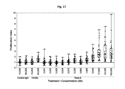
Figure 17. Dendritic cells (DC) were differentiated for 6 days then co-
cultured with freshly
isolated CD4+ T cells. Dose-response of antibodies or controls were incubated
on such cells,
then after 6 days of incubation, tritiated-thymidine was added for 18 to 20
additional hours
of incubation. Proliferation Index was calculated with the following method:
thymidine
incorporation background induced in autologous condition (CD4+ T cells only)
was subtracted
for each sample (specific for each CD4+ T cell donor), then this results was
divided by the
thymidine incorporation induced in allogeneic condition. The graphs show the
proliferation
CA 03047059 2019-06-13
WO 2018/115003 PCT/EP2017/083632
index for each treatment. Each data point is the mean of triplicate values
obtained for each
DC-CD4+ T cells combination. N = 36 combinations. The line Y = 1 represents
the normalized
allogenic response.
Figure 18. PBMC were isolated from filters and incubated with Staphylococcal
enterotoxin B
superantigen (SEB) in the presence of antibodies or controls. After 5 days of
incubation,
supernatants were harvested and quantified on Luminex for IL-2 release.
Normalized IL-2
release was calculated with the following method: IL-2 quantification induced
in non-
stimulated cells (PBMC without SEB) was subtracted for each sample (specific
for each PBMC
donor), then this results was divided by the IL-2 release induced in SEB-
stimulated cells (No
treatment). Filled heavy line represents the response threshold. The graphs
show the
normalized IL-2 release for each treatment. Each data point is the mean of
triplicate values
obtained for each PBMC donor. N = 17 PBMC donors. The line Y = 1 represents
the normalized
SEB only induced-response.
Figure 19. PBMC were isolated from filters and incubated with Staphylococcal
enterotoxin B
superantigen (SEB) in the presence of antibodies or controls at 80 and 10 nM.
After 5 days of
incubation, supernatants were harvested and quantified on Luminex for IL-2
release.
Normalized IL-2 release was calculated with the following method: IL-2
quantification induced
in non-stimulated cells (PBMC without SEB) was subtracted for each sample
(specific for each
PBMC donor), then this results was divided by the IL-2 release induced in SEB-
stimulated cells
(No treatment). Filled heavy line represents the response threshold. The
graphs show the
normalized IL-2 release for each treatment. Each data point is the mean of
triplicate values
obtained for each PBMC donor. The line Y = 1 represents the normalized SEB
only induced-
response.
Figure 20: Schematic representation of molecules based on 7H11 and 2H6 binding
units having
different valences and architectures.
Figure 21: Analysis of 7H11 and 2H6 binding to 0X40 when fused in C-terminus
as Fab or scFv
format. Surface Plasmon Resonance (SPR) measurements of proteolytically
cleaved
tetravalent molecules near their hinge regions (the Fc-2H6 Fab/2H6 Fab, Fc-2H6
Fab/7H11
scFv, Fc-7H11 Fab/7H11 Fab and Fc-7H11 Fab/2H6 scFv, as indicated) for the
chimeric 0X40
CA 03047059 2019-06-13
WO 2018/115003 PCT/EP2017/083632
26
molecules chi0X40R-Fc HHRH (Fig. 21A) or chi0X40R-Fc RRHH (Fig 21B). Data are
expressed
as number of response units (abbreviated RU; Y axis) vs. time (X axis). Fig
21C shows a
schematic representation of the agonists used in the analysis.
Figure 22: Determination of 0X40 co-engagement by 7H11 Fab and 2H6 scFy when
fused in C-
terminus. Co-engagement measurements by SPR of the Fc-7H11 Fab/2H6 scFy
fragment with
chimeric 0X40 molecules chi0X40R-Fc HHRH (Fig. 22A) or chi0X40R-Fc RRHH (Fig
22B)
immobilized on the CHIP and human 0X40 (HHHH), chi0X40R-Fc (HHRH) and chi0X40R-
Fc
(RRHH) sequentially injected. Data are expressed as number of response units
(abbreviated
RU; Y axis) vs. time (X axis). Fig 22C shows a schematic representation of the
agonists used in
the analysis.
Figure 23: Determination of 0X40 co-engagement by 7H11 scFy and 2H6 Fab when
fused in C-
terminus. Co-engagement measurements by SPR of the Fc-2H6 Fab/7H11 scFy
fragment with
chimeric 0X40 molecules chi0X40R-Fc HHRH (Fig. 23A) or chi0X40R-Fc RRHH (Fig
23B)
immobilized on the CHIP and human 0X40 (HHHH), chi0X40R-Fc (HHRH) and chi0X40R-
Fc
(RRHH) sequentially injected. Data are expressed as number of response units
(abbreviated
RU; Y axis) vs. time (X axis). Fig 23C shows a schematic representation of the
agonists used in
the analysis.
Figure 24: PBMC were isolated from filters and incubated with Staphylococcal
enterotoxin B
superantigen (SEB) in the presence of antibodies or controls at 80 and 10 nM.
After 5 days of
incubation, supernatants were harvested and quantified on Luminex for IL-2
release.
Normalized IL-2 release was calculated with the following method: IL-2
quantification induced
in non-stimulated cells (PBMC without SEB) was subtracted for each sample
(specific for each
PBMC donor), then this results was divided by the IL-2 release induced in SEB-
stimulated cells
(No treatment). Filled heavy line represents the response threshold. The
graphs show the
normalized IL-2 release for each treatment. Each data point is the mean of
triplicate values
obtained for each PBMC donor. The line Y = 1 represents the normalized SEB
only induced-
response.
Figure 25: PBMC were isolated from filters and incubated with Staphylococcal
enterotoxin B
superantigen (SEB) in the presence of antibodies or controls at 80 and 10 nM.
After 5 days of
CA 03047059 2019-06-13
WO 2018/115003 PCT/EP2017/083632
27
incubation, supernatants were harvested and quantified on Luminex for IL-2
release.
Normalized IL-2 release was calculated with the following method: IL-2
quantification induced
in non-stimulated cells (PBMC without SEB) was subtracted for each sample
(specific for each
PBMC donor), then this results was divided by the IL-2 release induced in SEB-
stimulated cells
(No treatment). Filled heavy line represents the response threshold. The
graphs show the
normalized IL-2 release for each treatment. Each data point is the mean of
triplicate values
obtained for each PBMC donor. The line Y = 1 represents the normalized SEB
only induced-
response.
Figure 26: Overlay of analytical gel filtration chromatograms. Chromatograms
for Tetra-8
alone, h0X40 alone and antibody-h0X40 complexes at 1:4 ratio were overlaid.
The arrows
indicating expected molecular weights correspond to the peaks of the
calibration run and are
Ferritin (440 kDa), Aldolase (158 kDa) and Carbonic anhydrase (29 kDa). Note
the differences
between Tetra-8 and reversed Tetra-8 (indicated by arrows) ¨ Tetra-8 has a
shoulder in VO
and the second peak is shifted to higher molecular weight compared to that of
reversed Tetra-
8.
Figure 27: Tetra-8-h0X40 crystalline-like lattice. One possibility of a large,
2-dimensional
lattice structure is shown. Two h0X40 per TETRA-8 were used to build an, in
theory, infinitively
large structure.
Figure 28. Time lapse of 0X40-GFP on Jurkat 0X40-GFP cell line following
treatment with
Tetra-8. Jurkat expressing 0X40 eGFP cells were incubated overnight at 37 C
and 5% CO2 on
Fluorodish (WPI) cell culture dishes (20000ce11s/cm2) pre-coated with
fibronectin (1 g/cm2
in PBS). Tetra-8 was then added to the cell medium at 80 nM final
concentration for various
time intervals (ranging from 2.5 to 27.5min) and cells were imaged using a
Zeiss Inverted
microscope Z1 equipped with a confocal module LSM 800 at 63x magnification.
Figure 29. Confocal images of 0X40 clusters induced by Tetra-8 and other 0X40-
targeting
molecules. Jurkat 0X40-GFP cells were treated for either 5, 10, or 20 minutes
with various
molecules targeting 0X40 (Tetra-8, 1A7, OX4OL and Tetra-14), used at either at
20nM (A) or
80nM (B).
CA 03047059 2019-06-13
WO 2018/115003 PCT/EP2017/083632
28
Figure 30. Quantitative analysis of 0X40 clustering induced by various anti-
0X40 molecules
on Jurkat-0X40 GFP cell line. Confocal images of 0X40 clusters induced by
Tetra-8 and other
0X40-targeting molecules on Jurkat 0X40-GFP cells were analyzed using the
Kurtosis
method, as described in the example.
Figure 31. DC activation assay. Dendritic cells (DC) were isolated from PBMC
(3 donors from
filters and one donor from whole blood) and differentiated for 6 days then
cultured for two
additional days in the presence of antibodies or controls. After incubation,
cells were
harvested and stained with anti-CD1c-APC, anti-CD8O-PE, anti-CD86-PerCP-eF710
for Panel 1
or anti-CD1c-APC, anti-CD83-FITC, anti-HLA-DR-PerCP5.5 for Panel 2. The graph
shows the
percentage of overexpressing cells for CD83 and CD86 markers, compared to No
treated DC,
that are also expressing some of these markers constitutively. Each data point
is the value
for one DC donor. N = 4 donors.
Figure 32. A dose-response of antibodies or controls were incubated on thaw-
and-use NFkB-
Luc2P/U2OS cells. After 4h of incubation, luciferase substrate was added to
the wells and
luminescence was measured using a microplate reader (read tape ¨ endpoint;
integration
time ¨ 1 minute; emission ¨ hole; optics position ¨ top; gain 135; read height
¨ 1.00 mm).
The graph shows the nonlinear sigmoidal regression binding curves
(Luminescence) for each
condition. The following treatments were tested: Selicrelumab IgG (0), ADC-
1013 IgG1 (0),
3h56 IgG1 LALA (A), Selicrelumab_3h56 (0), ADC-1013_3h56 (M), CD4OL ( X). Each
data
point is the mean SD of duplicate values.
Example 1:
Generation and screening of mouse anti-human 0X40 antibodies
To produce the recombinant human 0X40-his protein, the extracellular region
(amino acids 1-
214 as set forth in SEQ ID NO: 1) of human TNFRSF4 was amplified by PCR adding
a 3' GSG-
6xHis linker and restriction sites for cloning. The PCR product was
subsequently cloned in the
modified pcDNA3.1(-) plasmid described above. This recombinant plasmid allowed
for the
expression of the human 0X40-his protein in mammalian cells with secretion
into the cell
culture media driven by the native signal peptide of the human TNFRSF4. For
protein
production, the recombinant vector was transfected into suspension-adapted HEK
293 cells
CA 03047059 2019-06-13
WO 2018/115003 PCT/EP2017/083632
29
(ATCC number CRL 1573) using jetPElTM transfection reagent (Polyplus-
transfection S.A.,
Strasbourg, France; distributor: Brunschwig, Basel, Switzerland). The cell
culture supernatant
was collected five days after transfection and purified using a Ni2+-NTA
affinity purification
column (HiTrap Ni2+-NTA sepharose column; GE Healthcare Europe GmbH,
Glattbrugg,
Switzerland) operated on an AKTA FPLC system (GE Healthcare Europe GmbH,
Glattbrugg,
Switzerland).
Recombinant human 0X40-Fc and 0X40-his proteins were found to be 95% pure as
judged by
SDS-PAGE, and further buffered exchanged into phosphate buffer saline (PBS)
prior use.
To produce the recombinant human OX40L-Fc protein, a cDNA for the human TNFSF4
was
purchased from imaGenes (clone name: 10H46203, Berlin, Germany) and the
extracellular
portion (amino acids 51-183) of human TNFSF4 ligand (numbering according to
the Uniprot
Q6FGS4 sequence) was amplified with flanking restriction sites for subsequent
cloning into a
modified mammalian expression vector based on the pcDNA3.1(-) plasmid from
Invitrogen
(Invitrogen AG, Basel, Switzerland, Cat. No. V795-20), containing the human Fc
region of a
human IgG1 (EU positions 223-451), the human CMV promoter with the Ig donor
acceptor
fragment (first intron) described in US Patent 5924939, the OriP sequence
(Koons et al. 2001,
J Virol. 75 (22):10582-92.), the 5V40 enhancer, and the 5V40 polyA fused to
the gastrin
terminator as described by Kim et al. (2003, Biotechnol Prog. 19 (5), p. 1620-
2). This
recombinant plasmid allowed for expression of the human TNFSF4 extracellular
domain ¨ Fc
fusion protein in mammalian cells with secretion into the cell culture medium
driven by the
VJ2C leader peptide. For recombinant protein production, the aforementioned
recombinant
vector was transfected into suspension-adapted HEK 293 cells (ATCC number CRL
1573) using
cationic polymers. The cell culture supernatant was collected after five days
and further
purified in batch using CaptivATM primAB affinity beads (Repligen, Waltham,
Massachussets,
USA) and further buffer-exchanged to phosphate buffer saline (PBS) prior to
use.
To produce the recombinant macaca 0X40 ¨Fc protein, a synthetic gene
corresponding to the
extracellular portion of macaca 0X40 (amino acids 29-214 of NCB! sequence
XP_001090870.1)
was generated (GeneArt, ThermoFisher Scientific, Waltham, Massachusetts) with
restriction
sites for subsequent cloning into a modified mammalian expression vector based
on the
pcDNA3.1(-) plasmid from Invitrogen (Invitrogen AG, Basel, Switzerland, Cat.
No. V795-20),
CA 03047059 2019-06-13
WO 2018/115003 PCT/EP2017/083632
containing the human Fc region of a human IgG1 (EU positions 223-451), the
human CMV
promoter with the Ig donor acceptor fragment (first intron) described in US
Patent 5924939,
the OriP sequence (Koons et al. 2001, J Virol. 75 (22):10582-92.), the SV40
enhancer, and the
SV40 polyA fused to the gastrin terminator as described by Kim et al. (2003,
Biotechnol Prog.
19 (5), p. 1620-2). This recombinant plasmid allowed for expression of the
macaca 0X40
extracellular domain ¨ Fc fusion protein in mammalian cells with secretion
into the cell culture
medium driven by the VJ2C leader peptide. For recombinant protein production,
the
aforementioned recombinant vector was transfected into suspension-adapted HEK
293 cells
(ATCC number CRL 1573) using cationic polymers. The cell culture supernatant
was collected
after five days and further purified in batch using CaptivATM primAB affinity
beads (Repligen,
Waltham, Massachussets, USA) and further buffer-exchanged to phosphate buffer
saline (PBS)
prior to use. To produce the recombinant human 0X40-Fc protein, a cDNA for the
human
TNFRSF4 was purchased from imaGenes (clone number: RZPDB737H0329D; Berlin,
Germany).
This cDNA was used as a template to PCR-amplify the DNA coding region of the
human
TNFRSF4 extracellular domain (amino acids 1-214 as set forth in SEQ. ID NO:
1). In a separate
PCR reaction, the Fc region of a human IgG1 (EU positions 223-451) was
amplified. The two
resulting products were then fused using overlap extension PCR with flanking
primers, adding
restriction sites for subsequent cloning into a modified mammalian expression
vector based
on the pcDNA3.1(-) plasmid from Invitrogen (Invitrogen AG, Basel, Switzerland,
Cat. No. V795-
20), containing the human CMV promoter with the Ig donor acceptor fragment
(first intron)
described in US Patent 5924939, the OriP sequence (Koons et al. 2001, J Virol.
75 (22):10582-
92.), the 5V40 enhancer, and the 5V40 polyA fused to the gastrin terminator as
described by
Kim et al. (2003, Biotechnol Prog. 19 (5), p. 1620-2). This recombinant
plasmid allowed for
expression of the human TNFRSF4 extracellular domain ¨ Fc fusion protein in
mammalian cells
with secretion into the cell culture medium driven by the native signal
peptide of the human
TNFRSF4 protein. For recombinant protein production, the aforementioned
recombinant
vector was transfected into suspension-adapted HEK 293 cells (ATCC number CRL
1573) using
jetPElTM transfection reagent (Polyplus-transfection S.A., Strasbourg, France;
distributor:
Brunschwig, Basel, Switzerland). The cell culture supernatant was collected
after five days and
further purified using a Protein A affinity purification column (HiTrap
Protein A sepharose
column; GE Healthcare Europe GmbH, Glattbrugg, Switzerland) operated on an
AKTA FPLC
system (GE Healthcare Europe GmbH, Glattbrugg, Switzerland).
CA 03047059 2019-06-13
WO 2018/115003 PCT/EP2017/083632
31
Recombinant human 0X40-Fc protein dissolved in PBS was mixed with an equal
volume of
Stimune adjuvant (Prionics, Switzerland, ref: 7925000) and an emulsion was
prepared. The
emulsion was transferred to 0.5 mL insulin syringes (BD Pharmingen, Allschwil,
Switzerland)
and BALB/c animals (Harlan, Netherlands) were immunized sub-cutaneously in the
back
footpads, the base of the tail and the neck with 50 lig of the emulsified
protein. The
immunization was repeated two weeks later with the same amount of antigen and
the same
route of injection.
The presence of circulating anti-human 0X40 antibodies in the immunized mouse
sera was
evaluated by direct [LISA using plates coated with the recombinant human 0X40-
his protein.
A serial dilution (from 1:109 to 1:109) of the different mouse sera was added
to the plates and
the bound antibodies were detected using a goat anti-mouse H+L whole molecule-
HRP
(Sigma-Aldrich Chemie GmbH, Buchs, Switzerland).
A final sub-cutaneous boost with 50 lig of antigen without adjuvant was
performed in animals
displaying the best anti-human 0X40 IgG serum titer 3 days before sacrifice.
Animals were euthanized and the inguinal, axillary, brachial, popliteal and
sciatic lymph nodes
were collected to prepare a single cell suspension by disturbing the lymph
node architecture
with two 25G needles in a DNAse (Roche Diagnostics (Schweiz) AG, Rotkreuz,
Switzerland) and
collagenase (Roche Diagnostics (Schweiz) AG, Rotkreuz, Switzerland) solution.
Single cell
suspensions were fused to a myeloma cell line X63AG8.653 (mouse BALB/c myeloma
cell line;
ATCC accession number: CRL 1580; J Immunol 1979, 123:1548-1550)) at a ratio of
7:1 with
polyethylene glycol 1500 (Roche Diagnostics (Schweiz) AG, Rotkreuz,
Switzerland). The fused
cells were plated into 96 well flat bottom plates containing mouse macrophages
in DMEM-10
medium (Invitrogen AG, Basel, Switzerland) supplemented with 10% fetal bovine
serum (FBS,
PAA Laboratories, Pasching, Austria), 2mM L-glutamine, 100U/m1 (Biochrom AG,
Germany)
penicillin, 100 ug/m1 streptomycin (Biochrom AG, Germany), 10mM HEPES
(Invitrogen AG,
Basel, Switzerland), 50 uM (3-mercaptoethanol (Sigma-Aldrich Chemie GmbH,
Buchs,
Switzerland), HAT (Sigma-Aldrich Chemie GmbH, Buchs, Switzerland) and 1%
Growth factor
(Hybridokine, Interchim/Uptima, Montlugon, France).
CA 03047059 2019-06-13
WO 2018/115003 PCT/EP2017/083632
32
Approximatively 800 hundred wells from the fusions were screened by [LISA for
the presence
of mouse IgG that recognized human 0X40. Positive wells were expanded and
subjected to
two rounds of subcloning. Cells were collected and the heavy and light chains
were cloned and
sequenced.
Example 2:
Cloning and sequencing of the VH and VL chains of the anti-0X40 antibodies
from hybridoma
cells
For each positively selected hybridoma, total RNA was prepared, reverse-
transcribed into
cDNA and VH and VL genes were respectively amplified by PCR. These PCR
products were
ligated into a rescue-vector (pDrive vector; QIAGEN AG, Hombrechtikon,
Switzerland; Cat. No.
231124), allowing for the DNA sequencing of individual PCR products and the
determination
of mono- or poly-clonality of the selected hybridomas. This vector allowed for
blue/white
selection on LB-agar plates containing IPTG and X-gal (colonies with no insert
were blue
because of the degradation of X-gal by the LacZ a -peptide). Recombinant
plasmids from
positive (white) bacterial clones were prepared and sequenced using standard
DNA
sequencing primers specific for the vector backbone (M13rev, M13fwd, T7 or
5P6). DNA
sequences were finally subcloned into an expression vector for recombinant
expression of the
antibody of interest in mammalian cells.
RNA isolation
Total RNA was isolated from 2-10x106 cells using the RNeasy Mini Kit from
QIAGEN (QIAGEN
AG, Hombrechtikon, Switzerland; Cat. No. 74106) according to the
manufacturer's protocol;
samples were quantified using a NanoDrop ND-1000 spectrophotometer (WITEC AG,
Littau,
Switzerland).
One step RT-PCR
The total RNA preparations described above were further reverse-transcribed
into cDNA, and
the VH and VL fragments were amplified by PCR using two different mixtures of
degenerated
primers, each one allowing the recovery of all the different subfamilies of
mouse
CA 03047059 2019-06-13
WO 2018/115003 PCT/EP2017/083632
33
immunoglobulin heavy chain variable fragments and variable heavy chain
junction regions or
the recovery of all mouse immunoglobulin light chain kappa variable fragments
and variable
light chain kappa junction regions. The primers used for reverse transcription
and
amplification were synthetized by Microsynth (Balgach, Switzerland), and were
HPLC purified
(Tables 1-4). Both reverse-transcription and PCR amplification were performed
simultaneously using the QIAGEN one step RT-PCR kit (QIAGEN AG, Hombrechtikon,
Switzerland; Cat. No. 210212). Since the technique used specific primers, each
mRNA sample
was then treated in duplicate allowing for the individual reverse-
transcription and
amplification of either the VH or the VL fragments. 2ug of total RNA dissolved
into RNase-free
water to a final volume of 30111 were mixed with: 10111 of a 5x stock solution
of QIAGEN
OneStep RT-PCR Buffer, 2111 of a dNTPs mix at a concentration of 10mM, 30 of
primer mix at
a concentration of 10 M and 20 of QIAGEN OneStep RT-PCR Enzyme Mix. The final
mixture
was then placed in a PCR tube, and cycled in a PCR-themocycler (BioRad iCycler
version 4.006,
Bio-rad Laboratories AG, Reinach, Switzerland) using the following settings:
30 min at 50 C
15 min at 95 C
40 cycles: 30 sec at 94 C
30 sec at 55 C
1 min at 72 C
min at 72 C
Hold at 4 C
pDrive cloning
PCR products were run onto 2% agarose gels. Following DNA electrophoresis, the
fragments
of interest (-450bp) were excised from the agarose gels, and further extracted
using the
Macherey-Nagel NucloSpin Extract ll kit 250 (Macherey-Nage1,0ensingen,
Switzerland; Cat.
No. 740609.250). For DNA sequencing, the extracted PCR products were cloned
into the
CA 03047059 2019-06-13
WO 2018/115003 PCT/EP2017/083632
34
rescue-vector described above (pDrive vector, QIAGEN AG, Hombrechtikon,
Switzerland; Cat.
No. 231124) and transformed into the E. coliTOP10 strain (Invitrogen AG,
Basel, Switzerland;
Cat. No. C404006)
Miniprep extraction
Positive colonies were cultured overnight at 37 C (shaking 250 RPM) in 1.5m1
of Luria Bertani
(LB) medium supplemented with 100 g/mlampicillin seeded in Macherey-Nagel
Square-well
Block plates (Macherey-Nagel, Oensingen, Switzerland; Cat. No. 740488.24). The
next day
DNA miniprep extractions were performed using the NucleoSpin Multi-8 Plasmid
kit
(Macherey-Nagel, Oensingen, Switzerland; Cat. No. 740620.5).
Sequencing
Samples were sent for DNA sequencing to the DNA sequencing service company
Fastens (Plan-
les-Ouates, Switzerland). The standard primers: M13rev, M13fwd, T7, 5P6 were
used (Table
5).
Sequence analysis
The Clone Manager 9 Professional Edition (Scientific 84 Educational Software,
NC, USA) and
the BioEdit Sequence Alignment Editor (Hall, T.A. 1999. BioEdit: a user-
friendly biological
sequence alignment editor and analysis program for Windows 95/98/NT. Nucl.
Acids. Symp.
Ser. 41:95-98) were used for the analysis of DNA sequences.
Cloning of expression vector for recombinant chimeric antibody expression
For recombinant expression in mammalian cells, the isolated murine VH and VL
fragments
were formatted as chimeric immunoglobulins using assembly-based PCR methods.
These
chimeric antibodies consist of a heavy chain where the murine heavy chain
variable domain is
fused to the human IgG1 heavy chain constant domains (y1, hinge, y2, and y3
regions) and a
light chain where the murine light chain variable domain is fused to a human
kappa constant
domain (CK). PCR- assembled murine variable and human constant parts were
subsequently
cloned into a modified mammalian expression vector based on the modified
pcDNA3.1(-)
CA 03047059 2019-06-13
WO 2018/115003 PCT/EP2017/083632
vector from Invitrogen mentioned in Example 1 with the difference that a human
immunoglobulin light chain kappa leader peptide was employed to drive protein
secretion.
For protein production of the immunoglobulin candidates, equal quantities of
heavy and light
chain vector DNA were co-transfected into suspension-adapted HEK-293 (ATCC
number: CRL-
1573). The cell culture supernatant was collected after five days and purified
using a Protein
A affinity purification column (HiTrap Protein A sepharose column; GE
Healthcare Europe
GmbH, Glattbrugg, Switzerland) operated on an AKTA FPLC system (GE Healthcare
Europe
GmbH, Glattbrugg, Switzerland).
Table 2: primer Mix VH ¨ back (SEQ ID NO: 50- 68)
GTGATC GCC ATG GCG TCG ACC GAK GTR MAG CU CAG GAG TC
GTGATC GCC ATG GCG TCG ACC GAG GTB CAG CTB CAG CAG TC
GTGATC GCC ATG GCG TCG ACC CAG GTG CAG CTG AAG SAR TC
GTGATC GCC ATG GCG TCG ACC GAG GTC CAR CTG CAA CAR TC
GTGATC GCC ATG GCG TCG ACC CAG GTY CAG CTB CAG CAR TC
GTGATC GCC ATG GCG TCG ACC CAG GTY CAR CTG CAG CAR TC
GTGATC GCC ATG GCG TCG ACC CAG GTC CAC GTG AAG CAR TC
GTGATC GCC ATG GCG TCG ACC GAG GTG AAS STG GTG GAR TC
GTGATC GCC ATG GCG TCG ACC GAV GTG AWG STG GTG GAG TC
GTGATC GCC ATG GCG TCG ACC GAG GTG CAG STG GTG GAR TC
GTGATC GCC ATG GCG TCG ACC GAK GTG CAM CTG GTG GAR TC
GTGATC GCC ATG GCG TCG ACC GAG GTG AAG CTG ATG GAR TC
GTGATC GCC ATG GCG TCG ACC GAG GTG CAR CU GU GAR TC
GTGATC GCC ATG GCG TCG ACC GAR GTR AAG CTT CTC GAR TC
GTGATC GCC ATG GCG TCG ACC GAA GTG AAR STT GAG GAR TC
GTGATC GCC ATG GCG TCG ACC CAG GTT ACT CTR AAA SAR TC
GTGATC GCC ATG GCG TCG ACC CAG GTC CAA CTV CAG CAR CC
GTGATC GCC ATG GCG TCG ACC GAT GTG AAC TTG GAA SAR TC
GTGATC GCC ATG GCG TCG ACC GAG GTG AAG GTC ATC GAR TC
Table 3: primer Mix VH ¨ FOR (SEQ ID NO: 69-72)
CA 03047059 2019-06-13
WO 2018/115003
PCT/EP2017/083632
36
CCTCCACCACTCGAGCC CGA GGA AAC GGT GAC CGT GGT
CCTCCACCACTCGAGCC CGA GGA GAC TGT GAG AGT GGT
CCTCCACCACTCGAGCC CGC AGA GAC AGT GAC CAG AGT
CCTCCACCACTCGAGCC CGA GGA GAC GGT GAC TGA GGT
Table 4: primer Mix VL ¨ BACK (SEQ ID NO: 73-92)
GGCGGTGGC GCT AGC GAY ATC CAG CTG ACT CAG CC
GGCGGTGGC GCT AGC CAA ATT GU CTC ACC CAG TC
GGCGGTGGCGCT AGC GAY ATT GTG MTM ACT CAG TC
GGCGGTGGC GCT AGC GAY ATT GTG YTR ACA CAG TC
GGCGGTGGC GCT AGC GAY ATT GTR ATG ACM CAG TC
GGCGGTGGC GCT AGC GAY ATT MAG ATR AMC CAG TC
GGCGGTGGC GCT AGC GAY ATT CAG ATG AYD CAG TC
GGCGGTGGCGCT AGC GAY ATY CAG ATG ACA CAG AC
GGCGGTGGC GCT AGC GAY ATT GTT CTC AWC CAG TC
GGCGGTGGCGCT AGC GAY ATT GWG CTS ACC CAA TC
GGCGGTGGC GCT AGC GAY ATT STR ATG ACC CAR TC
GGCGGTGGC GCT AGC GAY RU KTG ATG ACC CAR AC
GGCGGTGGCGCT AGC GAY ATT GTG ATG ACB CAG KC
GGCGGTGGC GCT AGC GAY ATT GTG ATA ACY CAG GA
GGCGGTGGC GCT AGC GAY ATT GTG ATG ACC CAG WT
GGCGGTGGC GCT AGC GAY ATT GTG ATG ACA CAA CC
GGCGGTGGCGCT AGC GAY ATT TTG CTG ACT CAG TC
GGCGGTGGC GCT AGC GAA ACA ACT GTG ACC CAG TC
GGCGGTGGCGCT AGC GAA AAT GTK CTS ACC CAG TC
GGCGGTGGCGCT AGC CAG GCT GU GTG ACT CAG GAA TC
Table 5: primer Mix VL ¨ FOR (SEQ ID NO: 93-96)
ATGCTGAC GC GGC CGC ACG TTT KAT TTC CAG CTT GG
ATGCTGAC GC GGC CGC ACG TTT TAT TTC CAA CU TG
ATGCTGAC GC GGC CGC ACG TTT CAG CTC CAG CTT GG
ATGCTGAC GC GGC CGC ACC TAG GAC AGT CAG TTT GG
CA 03047059 2019-06-13
WO 2018/115003 PCT/EP2017/083632
37
Table 6: sequencing primers (SEQ ID NO: 97-100)
M13-Fwd GTAAAACGACGGCCAGT
M13-Rev AACAGCTATGACCATG
T7 TAATACGACTCACTATAGG
SP6 GATTTAGGTGACACTATAG
Example 3:
Biological characterization of anti-human 0X40 antibodies
0X40-specific Antibody Detection ELISA
Antibody titers, specificity and production by hybridomas and recombinant
antibody
candidates were determined by a direct [LISA. In brief, 96 well-microtiter
plates (Costar USA,
distributor VWR AG, Nyon, Switzerland) were coated with 100 ul of recombinant
human 0X40-
his at 2 ug/m1 in PBS (see example 1 for the generation of the 0X40-his
protein). Plates were
incubated overnight at 4 C and were then blocked with PBS 2% BSA (Bovine
Serum Albumine,
PAA Laboratories, Pasching, Austria) at room temperature (RT) for one hour.
The blocking
solution was removed and the hybridoma supernatants or purified antibodies
were added.
The plates were incubated at RT for 30 minutes, then washed nine times with
PBS 0.01%
Tween-20 (Sigma-Aldrich Chemie GmbH, Buchs, Switzerland) and a Horseradish
Peroxidase
(HRP) labeled-Goat anti-mouse H+L-detection antibody (Sigma-Aldrich Chemie
GmbH, Buchs,
Switzerland) was added at a dilution of 1:1000. To detect recombinant chimeric
antibodies
(see example 2) that possess a human Fc, a HRP-labeled rabbit anti human IgG
antibody
(Sigma-Aldrich Chemie GmbH, Buchs, Switzerland) at a dilution of 1:1000 was
used as the
detection antibody. Plates were incubated for 30 minutes at RT, washed nine
times with PBS
0.01% Tween-20 and the TMB substrate (Bio-rad Laboratories AG, Reinach,
Switzerland) was
added to the plates and the reaction stopped after six minutes by adding
H2504. Absorbance
was then read at 450 nm by a microplate reader (Biotek, USA; distributor:
WITTEC AG, Littau,
Switzerland). Amongst positive clones, hybridoma 7H11 and 2H6 were selected,
coding DNA
CA 03047059 2019-06-13
WO 2018/115003 PCT/EP2017/083632
38
sequences of their variable domains were obtained and mouse-human IgG1
chimeras were
prepared as described in example 2.
Example 4: Humanization and optimization of mouse 7H11 antibody
Humanizing the anti-human 0X40 mouse antibody 7H11 including selection of
human
acceptor frameworks, back mutations, and mutations that substantially retain
and/or improve
the binding and properties of human CDR-grafted acceptor frameworks while
removing
potential post-translational modifications is described herein. The mouse 7H11
antibody has
variable heavy chain domain sequence set forth in SEQ. ID NO: 2 and variable
light chain
domain sequence set forth in SEQ. ID NO: 3.
Methods
Recombinant production of antibodies
Coding DNA sequences (cDNAs) for the different VH and VL domains were
synthesized in a
scFy format by GENEART AG (Regensburg, Germany) thereby allowing for a single
DNA
sequence to encompass both variable domains. Individual variable domain cDNAs
were
retrieved from this scFy construct by PCR, and further assembled upstream of
their respective
constant domain cDNA sequence(s) using PCR assembly techniques. Finally, the
complete
heavy and light chain cDNAs were ligated in independent vectors that are based
on a modified
pcDNA3.1 vector (Invitrogen, CA, USA) carrying the CMV promoter and the Bovine
Growth
Hormone poly-adenylation signal. The light chain specific vector allowed
expression of kappa
isotype light chains by ligation of the light chain variable domain cDNA of
interest in front of
the kappa light chain constant domain cDNA using BamHI and BsiWI restriction
enzyme sites;
while the heavy chain specific vector was engineered to allow ligation of the
heavy chain
variable domain cDNA of interest in front of the cDNA sequence encoding the
IGHG1 CH1,
IGHG1 hinge region, IGHG1 CH2, and IGHG1 CH3 constant domains using BamHI and
Sall
restriction enzyme sites. In both heavy and light chain expression vectors,
secretion was
driven by the mouse VJ2C leader peptide containing the BamHI site. The BsiWI
restriction
enzyme site is located in the kappa constant domain; whereas the Sall
restriction enzyme site
is found in the IGHG1 CH1 domain.
CA 03047059 2019-06-13
WO 2018/115003 PCT/EP2017/083632
39
Antibodies were transiently produced by co-transfecting equal quantities of
heavy and light
chains vectors into suspension-adapted HEK293-EBNA1 cells (ATCC catalogue
number: CRL-
10852) using polyethylenimine (PEI, Sigma, Buchs, Switzerland). Typically, 100
ml of cells in
suspension at a density of 0.8-1.2 million cells per ml is transfected with a
DNA-PEI mixture
containing 50 lig of expression vector encoding the heavy chain and 50 lig of
expression vector
encoding the light chain. When recombinant expression vectors encoding
antibody genes are
introduced into the host cells, antibodies are produced by further culturing
the cells for a
period of 4 to 5 days to allow for secretion into the culture medium (EX-CELL
293, HEK293-
serum-free medium; Sigma, Buchs, Switzerland), supplemented with 0.1% pluronic
acid, 4 mM
glutamine, and 0.25 ug/mIgeneticin).
The humanized antibodies were purified from cell-free supernatant using
recombinant
protein-A streamline media (GE Healthcare Europe GmbH, Glattbrugg,
Switzerland), and
buffered exchanged into phosphate buffer saline prior to assays.
Affinity measurements on HPB-ALL cells by FACS
HPB-ALL cells (DSMZ, Braunschweig, Germany, Cat. No: ACC483) were used as a
human 0X40
positive cell line for FACS staining. HPB-ALL were maintained in RPM! 1640
supplemented with
10% FCS and 100 Wm! Penicillin and 100 ug/m1 streptomycin. 4 x10e5 HPB-ALL
cells in FACS
buffer (PBS supplemented with 1% BSA and 0.1% sodium azide) were incubated for
45 min on
ice with the anti-0X40 antibody of interest kept at a concentration of 10
ug/m1 An irrelevant
human IgG1 was used as an isotype control; the cells were incubated with a
1/200 dilution of
anti-Human Fc-PE (EBioscience, Vienna, Austria) for 45 min on ice. Cells were
then washed
again and resuspended in 200 ul FACS buffer. The relative mean fluorescence of
each sample
was measured on a FACSCalibur instrument (BD Biosciences, Allschwil,
Switzerland).
Affinity measurements by SPR
SPR analysis was used to measure the association and dissociation rate
constants for the
binding kinetics of the anti-0X40 antibodies. The binding kinetics were
measured on a BlAcore
2000 (BlAcore-GE Healthcare Europe GmbH, Glattbrugg, Switzerland) at room
temperature
and analyzed with the BiaEvaluation software (v4.1, GE Healthcare Europe
GmbH).
CA 03047059 2019-06-13
WO 2018/115003 PCT/EP2017/083632
Measurements were performed on CM5 sensor chips (Biacore 2000, GE Healthcare
Europe
GmbH, Cat. No: BR-1000-14) individually coupled with Protein A (Sigma, Buchs,
Switzerland,
Cat. No: P7837) using a commercial amine coupling kit (GE Healthcare Europe
GmbH, Cat. No:
BR-1000-50). 200-600 RUs of humanized antibody were captured. Dilution series
of 0X40-his
were injected at a flow rate of 10 ul/min in HBS-EP buffer (GE Healthcare
Europe GmbH, Cat.
No: BR1001-88). After each binding event, the surface was regenerated with 10
ul of glycine
buffer pH 1.5. Experimental data were processed using a 1:1 Langmuir model
with local Rmax.
The dissociation time was about 7 min. Measurements were performed in
duplicates or
triplicates and included zero-concentration samples for referencing. Both Chi2
and residual
values were used to evaluate the quality of a fit between the experimental
data and individual
binding models.
Thermostability assessment by differential scanning calorimetry
The thermal stabilities of the humanized antibodies were measured using
differential scanning
calorimetry (DSC). Monoclonal antibodies melting profiles are characteristic
of their isotypes
(Garber and Demarest (2007), BBRC 355:751-7), however the mid-point melting
temperature
of the FAB fragment can be easily identified even in the context of a full-
length IgG. Such mid-
point melting of FAB portion was used to monitor monoclonal stability of the
humanized
candidates.
Calorimetric measurements were carried out on a VP-DSC differential scanning
microcalorimeter (Malvern Instruments Ltd, Malvern, UK). The cell volume was
0.128 ml, the
heating rate was 200 C/h, and the excess pressure was kept at 65 p.s.i. All
antibodies were
used at a concentration of 1 mg/ml in PBS (pH 7.4). The molar heat capacity of
antibody was
estimated by comparison with duplicate samples containing identical buffer
from which the
antibody had been omitted. The partial molar heat capacities and melting
curves were
analyzed using standard procedures. Thermograms were baseline corrected and
concentration normalized before being further analyzed using a Non-Two State
model in the
software Origin v7Ø
Results
CA 03047059 2019-06-13
WO 2018/115003 PCT/EP2017/083632
41
Design of the reshaped variable regions
Homology matching was used to select the human acceptor frameworks of the CDRs
of the
mouse 7H11 antibody. Databases (e.g. a database of germline variable genes
from the
immunoglobulin loci of human and mouse, the IMGT database (the international
ImMunoGeneTics information system ; Lefranc MP et al., Nucleic Acids Res,
27(1):209-12
(1999); Ruiz M et al., Nucleic Acids Res, 28(1):219-21 (2000); Lefranc MP,
Nucleic Acids Res,
29(1):207-9 (2001); Lefranc MP, Nucleic Acids Res, 31(1):307-10 (2003);
Lefranc MP et al., Dev
Comp lmmunol, 29(3):185-203 (2005); Kaas Q et al., Briefings in Functional
Genomics &
Proteomics, 6(4):253-64 (2007)) or the VBASE2 (Retter I. et al, 2005, Nucleic
Acids Res., 33,
Database issue D671-D674), or the Kabat database (Johnson G. et al, 2000,
Nucleic Acids Res.,
28, p214-218)) or publications (e.g., Kabat et al, Sequences of Proteins of
Immunological
Interest, 1992) ) may be used to identify the human subfamilies to which the
murine heavy
and light chain V regions belong and determine the best-fit human germlime
framework to
use as the acceptor molecule for the mouse CDRs. Selection of heavy and light
chain variable
sequences (VH and VL) within these subfamilies to be used as acceptor may be
based upon
sequence homology and/or a match of structure of the CDR1 and CDR2 regions to
help
preserve the appropriate relative presentation of the six CDRs after grafting.
For example, use of the IMGT database indicates good homology between the 7H11
heavy
chain variable domain framework and the members of the human heavy chain
variable
domain subfamily 1. Highest homologies and identities of both CDRs and
framework
sequences were observed for germline sequences: IGHV1-3*01 (SEQ. ID NO: 4),
IGHV1-2*02
(SEQ. ID NO: 5), and IGHV1-46*01(SEQ ID NO: 6), all of which having sequence
identity above
68 % for the whole sequence up to CDR3. IGHV1-8*01 (SEQ. ID NO: 7)had a lower
sequence
identity (66.3%).
Using the same approach, 7H11 light chain variable domain sequence showed good
homology
to the members of the human light chain variable domain kappa subfamily 3 and
4. Highest
homologies and identities of both CDRs and framework sequences were observed
for germline
sequences: IGKV4-1*01 (SEQ. ID NO: 8) (81.2% homology), IGKV3D-7*01 (SEQ. ID
NO: 9) (67.3%
homology), IGKV3D-15*01 (SEQ. ID NO: 10) (67.3% homology), and IGKV3-20*01
(SEQ. ID NO:
11) (65.3% homology).
CA 03047059 2019-06-13
WO 2018/115003 PCT/EP2017/083632
42
Best matching JH and JK segment sequences to the human acceptor framework were
identified from the I MGT searches mentioned above.
As starting point to the humanization process, the four variable heavy and
light chain domains
stated above were selected as acceptors to the mouse 7H11 CDRs. A first set of
16 humanized
antibodies of human gamma one isotype were prepared. These first humanized
candidates
were assessed for transient expression in HEK293E cells and binding to HB-ALL
cell by flow
cytometry (Table 7).
VL VL1 VL2 VL3 VL4
VH IGKV4-1*01 IGKV3D-7*01 IGKV3D-15*01 IGKV3-20*01
VH1 Yield 41 45 3 2
MFI 119.7 60 79.2 69.7
IGHV1-3*01
VH2 Yield 51 7 2 26
IGHV1-2*02 MFI 120 67.8 82.5 185.5
VH3 Yield 31 17 2 35
MFI 121.3 19.7 44.9 34.3
IGHV1-46*01
VH4 Yield 34 8 1.5 20
MFI 34.1 17.8 30.3 20.2
IGHV1-8*01
_
CA 03047059 2019-06-13
WO 2018/115003 PCT/EP2017/083632
43
Isotype control MFI 16.9
Chimeric 7H11 MFI 132.2
IgG1
Table 7: Characterization of the first humanized 7H11 antibody candidates
(IgG1). FACS
staining of anti-0X40 antibodies on HPB-ALL cell line. MFI values correspond
to antibodies
mid-point fluorescence measured by flow-cytometry using 10 ug/m1 of antibody
candidate.
Transient expression yields are reported in mg per L of culture. Original
human germline
frameworks are indicated.
Best humanized candidates were antibodies VH1/VL1, VH2/VL1, and VH3/VL1. These
antibodies exhibited FACS staining levels close to the level observed for the
parental mouse
antibody with expression yields above the remainder of the candidates.
The three candidates were further assayed by SPR for affinity ranking (Table
8). Surprisingly,
the humanized VH2/VL1 IgG1 antibody was found to have superior affinity (i.e.
lower KD)
compared to the chimeric 7H11 antibody. In addition, expression yield,
apparent affinity for
HBP-ALL cells, and Fab stability were comparable to the other two variants.
Chimeric Humanized Humanized Humanized VH3-
7H11 IgG1 VH2-VL1 IgG1 VL1 IgG1
VH1-VL1 IgG1
KD (nM) 50 35 21 80
ka 1.16xe4 1.67xe4 5.85xe4 6.25xe4
(1/Ms)
kd (1/s) 6.6xe-4 5.8xe-4 1.3xe-3 5.5xe-3
Fab Tm 68 76.9 77 77.4
( C)
Table 8: Characterization of the best first-graft humanized antibodies.
Affinity constants
measured by SPR and Fab mid-point denaturation temperatures measured by DSC
are shown.
CA 03047059 2019-06-13
WO 2018/115003 PCT/EP2017/083632
44
Based on its good binding, expression and Fab stability, the VH2/VL1 antibody
was selected
for further affinity improvement via the process known as back mutagenesis
wherein amino
acids from the mouse antibody sequence are introduced in the humanized
antibody sequence.
It was thought that affinity could be further improved by the process
regardless of the fact
that the VH2/VL1 antibody had better affinity than its parental mouse
antibody.
Back mutations of grafted human frameworks
The process of back mutation necessitates the identification and the selection
of critical
framework residues from the mouse antibody that need to be retained in order
to preserve
or improve affinity while at the same time minimizing potential immunogenicity
in the
humanized antibody. To identify residues that may impact the most CDR
conformation and/or
inter-variable domain packing, a 3D model for the VH2/VL1 pair of variable
domains was
calculated using the structure homology-modelling server SWISS-MODEL (Arnold K
et al.,
(2006) Bioinformatics 22(2):195-201; http://swissmodel.expasy.org) set in
automated mode.
Model analysis allowed the selection of a subset of positions based on their
putative influence
on CDR regions and/or heavy chain-light chain variable domain packing. This
subset of
positions was selected out of the 26 possible back mutations found in the
variable heavy chain,
and consisted of positions: 37, 58, 60, 61, 85, 89, and 91 (Kabat numbering)
(Table 9).
VH Kabat number Antibody Amino acid Spatial location in VH/VL
interface
37 7H11 M Middle interface
VH2 V
58 7H11 K Middle interface
VH2 N
60 7H11 N Bottom interface
VH2 A
CA 03047059 2019-06-13
WO 2018/115003 PCT/EP2017/083632
61 7H11 E Bottom interface
VH2 Q
85 7H11 E Bottom interface
VH2 D
89 7H11 I Middle / bottom interface
VH2 V
91 7H11 F Middle interface
VH2 Y
Table 9: Details of the positions selected for back mutation between the
humanized VH2/VL1
candidate and the mouse 7H11 antibody.
Further humanized candidates based on these single back mutations were
prepared in the
context of the VH2/VL1 antibody sequences using standard PCR mutagenesis and
the methods
described above. Humanized antibody candidates were then assayed for their
binding affinity
by SPR and Fab thermal stability by DSC. Production yields, binding
affinities, and Fab mid-
points of thermal unfolding are shown in Table 10. Out of the seven antibodies
tested, the
N58K back mutation significantly improved affinity while maintaining good Fab
thermal
stability and expression. Consequently, the humanized VH2-N58K/VL1 antibody
was selected
for futher optimization.
Antibody Expression KD (nM) Fab Tm ( C)
(mg/L)
7H11 (chimeric) Not measured 50 68.0
VH2 V37M/VL1 12 2.87 70.3
VH2 N58K/VL1 24 0.77 72.0
CA 03047059 2019-06-13
WO 2018/115003 PCT/EP2017/083632
46
VH2 A6ON/VL1 14 ND 72.5
VH2 Q61E/VL1 23 ND 71.3
VH2 D85E/VL1 26 ND 71.5
VH2 V89I/VL1 21 ND 70.8
VH2 Y91F/VL1 9 ND 72.0
Table 10: Characterization of the humanized VH2/VL1 back mutated antibodies.
Removal of a potential isomerisation site.
Sequence analysis of 7H11 VH2 N58K highlighted the presence of a putative
aspartate
isomerization site (DG) in 7H11 CDRH2 at position 54 and 55 (Kabat numbering).
To abrogate
this isomerization site, site-directed mutagenesis was performed to replace
7H11 aspartate
54 residue by negatively charged or neutral polar amino acids like glutamate,
serine and
threonine and 7H11 glycine 55 by alanine. Using PCR assembly techniques,
mutations D54E,
D545, D54T and G55A were introduced in the cDNA of 7H11 VH before ligation in
a vector
based on a modified pcDNA3.1 vector (Invitrogen, CA, USA) carrying the CMV
promoter and a
Bovine Growth Hormone poly-adenylation signal.
Using these approach, several vectors were generating encoding humanized 7H11
VH D54E
(Humanized 7H11-VH2 N58K-D54E), 7H11 VH D545 (Humanized 7H11-VH2 N58K-D545),
7H11
VH D54T (Humanized 7H11-VH2 N58K-D54T) and 7H11 VH G55A (Humanized 7H11-VH2
N58K-
G55A). The parental sequence and variants of humanized 7H11 VH were co-
transfected with
7H11 light chain in HEK293-EBNA1 cells. Cell supernatant were then collected 4
days after
transfection for further purification using protein A. Tested mutations did
not change 7H11
expression in mammalian cell as compared to the parental antibody (table 10).
To determine if these mutations could have changed antibody thermal stability,
differential
scanning fluorimetry was performed. Antibodies in PBS were first mixed with a
10X
concentrated solution of SYPRO orange (Thermo Fisher Scientific, Ecublens,
Switzerland) at a
concentration of 250 ug/m1 in a final volume of 20 ul. To record protein
unfolding, samples
CA 03047059 2019-06-13
WO 2018/115003 PCT/EP2017/083632
47
were then exposed to an incremental increase of the temperature in a Rotor-
Gene Q 2p1ex
HRM (QIAGEN, Hi!den, Germany), thermal unfolding was followed by the presence
of the
SYPRO orange dye, whose fluorescence is quenched in polar environments but
strongly emits
a fluorescent signal when exposed to hydrophobic surroundings like the
hydrophobic core of
proteins upon unfolding. Recorded fluorescence signals were similar for both
the parental and
mutated forms of 7H11 indicating that mutations introduced in humanized 7H11
VH did not
change antibody thermal stability (table 11).
Finally, Surface Plasmon Resonance analyses were applied to control antibody
variant
affinities as described earlier. Results in table 11 show that mutations
introduced in 7H11 did
not change antibody affinity.
Antibody Expression KD (nM) Fab Tm ( C)
(mg/L)
Humanized 7H11- 39 16 74.2
VH2 N58K
Humanized 7H11- 44 9 73.5
VH2 N58K-D54E
Humanized 7H11- 37 49 73.5
VH2 N58K-D545
Humanized 7H11- 44 32 73.5
VH2 N58K-D54T
Humanized 7H11- 44 ND 74
VH2 N58K-G55A
Table 11: Summary of the humanized 7H11 variants after isomerization site
removal
Example 5: Humanization and optimization of mouse 2H6 antibody
Humanization of mouse monoclonal 2H6
CA 03047059 2019-06-13
WO 2018/115003 PCT/EP2017/083632
48
Humanization of the anti-human 0X40 mouse antibody 2H6 including selection of
human
acceptor frameworks and mutations that substantially retain the binding
properties of human
CDR-grafted acceptor frameworks while removing potential post-translational
modifications
is described herein.
The human acceptor frameworks chosen to graft 2H6 CDRs were selected to confer
maximum
expression and/or stability to the humanized version of 2H6. Selection of
human heavy and
light chain variable sequences (VH and VL) to be used as acceptor may be based
upon
germlines with good biophysical properties (as documented in Ewert S et al.,
(2003) J.Mol.Biol,
325, 531-553) and/or pairing as found in natural antibody repertoire (as
documented in
Glanville J et al., (1999) Proc Natl Acad Sci U S A, 106(48):20216-21; DeKosky
BJ et al., (2015)
Nat Med, 21(1):86-91). Framework sequences known in the field for good paring
and/or
stability are the human IGHV3-23*01 (SED ID NO: 33) and IGKV1-16*01(SED ID NO:
34) which
were used as acceptor frameworks for the 2H6 humanization.
A first humanized antibody of human gamma one isotype was prepared. The
antibody
encompassed a human-mouse hybrid heavy chain variable domain and a human-mouse
hybrid light chain variable domain. The hybrid heavy chain variable domain was
based on the
human heavy chain variable domain IGHV3-23*01 wherein germline CDRH1 and H2
where
respectively replaced for 2H6 CDRH1 and CDRH2. Best matching JH segment
sequence to the
human acceptor framework was identified from the IMGT database using homology
search.
The resulting human-mouse hybrid heavy chain variable sequence having human
IGHV3-
23*01 framework regions, 2H6 mouse CDRs, and best matching JH to human
acceptor is
referred herein as heavy chain variable domain VH1 with SEQ. ID NO: 31.
Similarly, the human-mouse hybrid light chain variable domain used for this
first humanized
antibody candidate had human IGKV1-16*01 framework regions, 2H6 mouse CDRs,
and best
matching JK to human acceptor, and is refereed herein as light chain variable
domain VL1 with
SEQ. ID NO: 32. The first humanized antibody encompassing VH1 and VL1 is
abbreviated herein
2H6 VH1/VL1 antibody.
Production of the humanized 2H6 scFv-Fc
CA 03047059 2019-06-13
WO 2018/115003 PCT/EP2017/083632
49
Coding DNA sequences (cDNAs) for VH1 and VL1 were synthesized in a scFy format
by
GENEART AG (Regensburg, Germany) thereby allowing for a single DNA sequence to
encompass both variable domains (SEQ. ID NO: 35). The scFy cDNA was ligated in
a vector
based on a modified pcDNA3.1 vector (Invitrogen, CA, USA) described earlier.
The scFv-Fc
specific vector was engineered to allow ligation of the scFy cDNA of interest
in front of the
cDNA sequence encoding the human IGHG1 hinge region, IGHG1 CH2 and IGHG1 CH3
constant
domains using BamHI and Kpnl restriction enzyme sites. Secretion was driven by
the mouse
VJ2C leader peptide containing the BamHI site. An artificial Glycine-Threonine
linker was
introduced at the C-ter part of the scFy which contains the Kpnl site.
The scFv-Fc was transiently produced by transfecting scFv-Fc vector into
suspension-adapted
HEK293-EBNA1 cells (ATCC catalogue number: CRL-10852) as described earlier.
Then, the
scFv-Fc was purified from cell-free supernatant using recombinant protein-A
streamline media
(GE Healthcare Europe GmbH, Glattbrugg, Switzerland), and buffered exchanged
into
phosphate buffer saline prior to assays. Binding to human and cynomolgus
monkey 0X40 was
measured by surface plasmon resonance as described below. The 2H6 humanized
scFy
encompassing VH1 and VL1 is abbreviated herein 2H6 scFv1.
Kinetic binding affinity constants of the humanized 2H6 scFv1-Fc for human and
cynomolgus
monkey 0X40 receptor extracellular domain by surface plasmon resonance (SPR)
Kinetic binding affinity constants (KD) were measured using recombinant
histidine tagged
human 0X40 receptor extracellular domain and recombinant Fc fused cynomolgus
monkey
0X40 receptor extracellular domain captured on a CMS chip and 2H6 scFv1-Fc and
mouse
chimeric 2H6 scFv-Fc as analytes. Measurements were conducted on a BlAcore
T200 (GE
Healthcare -BlAcore, Uppsala, Sweden) at room temperature, and analyzed with
the Biacore
T200 Evaluation software. A CMS research grade sensor chip (GE Healthcare
Europe GmbH,
Glattbrugg, Switzerland; BR100530) was activated by injecting 35 ul of a 1:1 N-
hydroxysulfosuccinimide (NHS)/ 1-Ethyl-3-[3-dimethylaminopropyl]
carbodiimide
Hydrochloride (EDC) solution (v/v; 5 ul/min flow-rate; on flow paths 1, 2, 3
and 4).
Cynomolgus monkey OX40R-Fc was diluted to a final concentration of 25nM in
acetate buffer
pH 4.0 (GE, BR-1003-49) and subsequently immobilized on the previously
activated CMS
CA 03047059 2019-06-13
WO 2018/115003 PCT/EP2017/083632
sensor chip by injecting 10 ul on the flow path 2 (10 ul/min) which
corresponds approximately
to 600 response units (RUs). Human OX40R-His was diluted to a final
concentration of 25nM
in acetate buffer pH 4.0 (GE, BR-1003-49) and subsequently immobilized on the
previously
activated CM5 sensor chip by injecting 45 ul on the flow path 4 (10 ul/min)
which corresponds
approximately to 400 response units (RUs). The OX40R-CM5 sensor chip was then
deactivated
by injecting 35 ul of ethanolamine solution (5 I/min). Finally, two injections
of 10 ul of glycine
solution (GE, ref. BR-1003-54; 10 mM; pH 1.5) were performed to release non-
crosslinked
(human and cynomolgus monkey) OX4OR molecules.
The 2H6 scFv-Fc was injected at different concentrations (0.78nM to 0.2 M) on
the 4 flow-
paths (flow-path 1 and 3 being used as references) at a 30 ul/min flow rate.
After each binding
event, surface was regenerated with glycine buffer pH 1.5 injected for 30
seconds (10 ul/min).
Measurements (sensorgram: fc2-fc1 and fc4-fc3) were best fitted with a 2:1
bivalent analyte
model with mass transfer. Dissociation times were of at least 300-600 seconds.
The Chi2 value
represents the sum of squared differences between the experimental data and
reference data
at each point; while the plots of residuals indicate the difference between
the experimental
and reference data for each point in the fit. Both Chi2 and residual values
were used to
evaluate the quality of a fit between the experimental data and individual
binding models.
Results shown in table 12 indicates that humanized 2H6 scFv1 has a similar
affinity to human
and cyno 0X40 than the parental mouse 2H6 scFy
mouse
humanized mouse chimeric humanized 2H6
chimeric 2H6
2H6 scFv1-Fc 2H6 scFv-Fc scFv1-Fc
scFv-Fc
Analyte Human 0X40 Cynomogus monkey 0X40
KD (nM) 62 27 39 18
ka (1/Ms) 6.8xe5 1.55xe5 5.79xe5 2.06xe5
CA 03047059 2019-06-13
WO 2018/115003 PCT/EP2017/083632
51
kd (1/s) 2.2xe-2 7.41xe-4 3.19xe-4 5.86xe-4
Table 12: Characterization of the humanized 2H6-scFy by SPR
2H6 Met removed from JH
Sequence analysis of 2H6 scFy highlighted the presence of a putative oxydation
site
(Methionine 108) in the 2H6 VH JH region. To abrogate this potential oxydation
site, site-
directed mutagenesis was performed to replace 2H6 VH methionine 108 residue by
a leucine
amino acid. Using PCR assembly technique, VH mutation M108L (kabbat numbering)
was
introduced in the cDNA of 2H6 scFy before ligation in a vector based on a
modified pcDNA3.1
vector as described earlier. Using this approach, a vector encoding humanized
2H6 scFy M108L
(abbreviated 2H6 scFv2) was generated. The parental and mutated forms of 2H6
scFv-Fc were
transfected in HEK293-EBNA1 cells as described earlier. Cell supernatant were
then collected
4 days after transfection for further purification using protein A. Tested
mutations did not
change 2H6 scFv-Fc expression in mammalian cell as compared to the parental
antibody (table
13). To determine if M108L mutation could have changed scFy thermal stability,
differential
scanning fluorimetry was performed as described earlier. Recorded fluorescence
signals were
similar for both the parental and mutated forms of 2H6 indicating that M108L
mutation did
not change scFy thermal stability (table 12). Finally, Surface Plasmon
Resonance analyses were
applied to control binding properties of 2H6 scFv2 variant as described
earlier. Results in figure
1 show that mutations introduced in 2H6 scFy did not change its binding.
Antibody Expression scFy Tm ( C)
(mg/L)
2H6 scFv1-Fc 40 63
2H6 scFv2-Fc 40 62.7
Table 13: Characterization of the impact of M108L mutation on humanized 2H6-
scFy
Example 7: Engineering and production of tetravalent anti-human 0X40
antibodies
CA 03047059 2019-06-13
WO 2018/115003 PCT/EP2017/083632
52
First tetravalent molecules
The tetravalent format used is a whole IgG to which scFvs were connected via
(Gly4Thr) linker
at the C-terminus of the heavy chain. For the generation of a tetravalent
antibody having 7H11
IgG1 fused to 2H6 scFv, coding DNA sequences (cDNAs) for humanized 7H11-VH2
N58K (SEQ.
ID NO: 21), VL1 (SEQ. ID NO: 16) and 2H6 scFv1 were PCR amplified before
digestion and
ligation in vectors based on a modified pcDNA3.1 vector (Invitrogen, CA, USA)
described
earlier. The light chain specific vector was engineered to allow ligation of
the VL cDNA of
interest in front of the cDNA sequence encoding the human kappa constant
domain using
BamHI and BsiWI restriction enzyme sites. The heavy chain specific vector was
engineered to
allow ligation of the VH cDNA of interest in front of the cDNA encoding the
IGHG1 hinge region,
a modified IGHG1 CH2 domain with the L234A/L235A double mutation (LALA, Eu
numbering,
Hezareh M et al., (2001) J Virol, 75:12161-8) which reduces Fc-FcyRs
interactions and a
modified IGHG1 CH3 constant domain having a (Gly4Thr) linker in its C-terminal
part using
BamHI and Sall restriction enzyme sites. Then, scFy cDNA of interest was
ligated after the
IGHG1 CH3 constant domain and the (Gly4Thr) linker of the heavy chain specific
vector using
Kpnl and Notl restriction enzyme sites. In both heavy and light chain
expression vectors,
secretion was driven by the mouse VJ2C leader peptide containing the BamHI
site. The BsiWI
restriction enzyme site is located in the kappa constant domain; whereas the
Sall restriction
enzyme site is found in the IGHG1 CH1 domain. The glycine-threonine linker
contains the Kpnl
site while the Notl site is present before the Bovine Growth Hormone poly-
adenylation signal
found in the modified pcDNA3.1 vector encoding the heavy chain.
This tetravalent antibody (abbreviated Tetra-1) was transiently produced by co-
transfecting
equal quantities of the 7H11VL1 light chain and Tetra-1 heavy chain vectors
into suspension-
adapted HEK293-EBNA1 cells as described previously. The tetravalent antibody
was purified
from cell-free supernatant using recombinant protein-A streamline media (GE
Healthcare
Europe GmbH, Glattbrugg, Switzerland), and buffered exchanged into phosphate
buffer saline
prior to assays.
Optimized 7H11x2H6 tetravalent antibodies:
CA 03047059 2019-06-13
WO 2018/115003 PCT/EP2017/083632
53
In the tetravalent molecule described above, scFy is fused in C-terminus of
the IgG. Therefore,
the last C-terminal residue of the antibody is a Lysine naturally present in
the JK region. To
avoid C-ter lysine clipping of the tetravalent molecule in the circulation
which could have an
impact on the biology of the antibody, site directed was used to replace the C-
ter Lysine of
2H6 scFy by a leucine residue. Using PCR assembly technique, VL mutation K107L
(Kabbat
numbering) was introduced in the cDNA of 2H6 scFy before ligation in the
vector coding for
the tetravalent antibody described above. The parental and mutated forms of
tetravalent
antibodies were transfected in HEK293-EBNA1 cells as described earlier. Cell
supernatant
were then collected 4 days after transfection for further purification using
protein A. Tested
mutation did not change tetravalent antibody expression in mammalian cell as
compared to
the parental antibody (table 14). To determine if K107L mutation could have
changed C-
terminal scFy thermal stability, differential scanning fluorimetry was
performed as described
earlier. Recorded fluorescence signals were similar for both the parental and
mutated forms
of 2H6 scFy indicating that K107L mutation did not change scFy thermal
stability (table 13).
Antibody Expression C-ter 2H6 scFy Tm ( C)
(mg/L)
Tetra-1 33 60.5
Tetra-1-K107L 32 59.7
Table 14: Characterization of the impact of K107L mutation on humanized Tetra-
1 antibody
To finalize the optimization of the tetravalent antibody, 7H11 mutation D54E,
2H6 VH
mutation M108L and VL mutation K107L were introduced in the vector coding for
the
tetravalent molecule heavy chain (abbreviated Tetra-6) by site-directed
mutagenesis as
previously described. Tetra-6 was produced by co-transfecting 7H11 VL1 light
chain and Tetra-
6 heavy chain vectors as shown before and purified from cell-free supernatant
using
recombinant protein-A streamline media.
Example 8: in vitro characterization of tetravalent anti-human 0X40 antibodies
CA 03047059 2019-06-13
WO 2018/115003 PCT/EP2017/083632
54
Example 8.1 Tetra-1 and Tetra-6 display a proliferative effect in a mixed
lymphocyte reaction
(MLR) assay
PBMCs were isolated from citrated whole blood of healthy donors using ficoll
density gradient.
Monocytes were isolated from PBMCs using Monocyte isolation kit (Miltenyi) and
cultured
with GM-CSF at 50 ng/mL (R&D) and rhl L-4 at 20 ng/mL (R&D) for 7 days to
differentiate them
into dendritic cells (DC). The phenotype of dendritic cells was verified by
flow cytometry using
CD1c APC (eBioScience). On day 7, CD4 T cells (from an allogeneic donor) were
isolated from
PBMCs using the EasySep kit (StemCell Technologies). CD4 T cells (40'000 cells
/ well) and DC
(8'000 cells / well) were co-cultured with antibodies at 80 nM for 6 days in
complete media in
a 96-well round-bottom plate in triplicate. On day 13, 3H-thymidine was added
(Perkin Elmer,
0.5 uCi per well). Twenty hours after pulsing, cells were harvested and
incorporated
radioactivity was quantified on a Wallac beta counter. A normalized
stimulation index (SI) was
determined using this formula:
(Sample ¨ Resp only) / (Allo ¨ Resp only)
"Sample" corresponds to the counts of the conditions in which DC + CD4 T cells
+ tested
antibody were co-cultured. "Resp only" corresponds to the counts of the
condition in which
only responder cells (CD4 T cells) were added. "Allo" corresponds to the
condition in which
DC (stimulator cells) and allogeneic CD4 T cells (responder cells) were co-
incubated. Data were
analyzed using Graphpad Prism 7 software; Statistical analysis was performed
with a Mann-
Whitney test (non-parametric test) or a Wilcoxon matched-pairs test. P<0.05
was considered
as statistically significant.
The OX40L-Fc is a potent agonistic molecule that can efficiently engage and
crosslink 0X40 on
surface of T cells (Muller FEBS J. 2008 May;275(9):2296-304). In agreement
with this feature,
OX40L-Fc was able to increase a mixed-lymphocyte reaction. In this assay, both
Tetra-1 and
Tetra-6 enhanced the allogeneic response to a similar level as OX40L-Fc
(differences between
these three molecules not statistically significant; Figure 1). A Wilcoxon
matched-pairs test
comparing SI of Tetra-1 and Tetra-6 tested in the same experiment showed that
these two
tetravalent molecules improved similarly the proliferation (data not shown).
Therefore these
CA 03047059 2019-06-13
WO 2018/115003 PCT/EP2017/083632
results highlight that targeting 0X40 with Tetra-1 and Tetra-6 provides a
relevant
immunostimulatory potential.
Example 8.2 Tetra-1 and Tetra-6 induce a strong immunostimulatory effect in a
Staphylococcal enterotoxin B stimulation assay
Peripheral blood mononuclear cells (PMBCs) were harvested from blood filters
obtained from
La Chaux-de-Fonds Transfusion Center using ficoll density gradient isolation.
PBMCs (105)
were distributed in a 96-well round-bottom plate in triplicate. Staphylococcal
enterotoxin B
(SEB) at a final concentration of 50 or 100 ng/mL (suboptimal concentrations)
and antibodies
at 80 nM final concentration were added to the wells. Plates were incubated
for 7 days at 37 C
in a CO2 incubator. IL-2 production in the culture supernatants was measured
with Luminex
using a ProcartaPlex kit (eBiosciences) on day 5. On day 7, cells were
harvested and labeled
with anti-human CD4 [CD and anti-human CD25 Pacific Blue (eBioSciences).
Stained cells were
resuspended in 100 uL of FACS buffer and analyzed by flow cytometry on
CytoFLEX S
(Beckman). Flow cytometry data were analyzed using CytExpert (Beckman). The
gating
strategy consisted in gating on living cells (based on FSC and SSC plots)
cells, CD4 positive cells
and subsequently on CD4+ CD25+. The total number of cells per well within this
subset of
interest was calculated using this formula:
(Volume used to resuspend cells * Number of events in the gate) / Sample
volume acquired
Each data point was normalized to the condition in which PBMCs were incubated
with SEB
only (No antibody). Data were analyzed using Graphpad Prism 7 software and
statistical
analysis was performed with a Mann-Whitney test (non-parametric test). P<0.05
was
considered as statistically significant.
In this assay, Tetra-1 was able to significantly increase the number of
CD4+CD25+ cells (91%
of the donors tested reached a 1.2 fold induction compared to SEB only;
threshold arbitrarily
defined) while OX40L-Fc and Tetra-6 did so to a lesser extent (67% and 88%
respectively). The
expression of CD25 defines activated T cells. The increase of CD25 expressing
CD4 T cells can
be due to an increase in the activation of T cells and/or an increase of
proliferation of activated
T cells. Similarly, the addition of Tetra-1, Tetra-6 and OX40L-Fc also
substantially enhanced IL-
CA 03047059 2019-06-13
WO 2018/115003 PCT/EP2017/083632
56
2 production compared to SEB only. IL-2 production also indicates T cell
activation and is linked
directly to T cell proliferation. There was no statistical difference between
the three molecules
with this readout and at this stage of the experiment (day 5).
Overall, targeting 0X40 in this superantigen-mediated PBMC stimulation
strikingly improved
T cell responses as visualized by enhanced cytokine production (IL-2) or
enhanced T cell
activation (CD25).
Example 8.3 Tetra-1 displays a strong immunostimulatory effect in a PHA
stimulation assay
PBMCs were prepared the same way as for the SEB assay. PBMCs (105) were
distributed in a
96-well round-bottom plate in triplicate. PHA at 2 or 1 ug/mL final
concentration and
antibodies at 80 nM final concentration were added. Plates were incubated for
7 days at 37 C
in a CO2 incubator. Six days after the start of the assay, cells were pulsed
with 0.5 uCi per well
of 3H-thymidine (Perkin Elmer). Twenty hours after pulsing, cells were
harvested and
incorporated radioactivity was quantified on a Wallac beta counter. A
stimulation index was
determined using this formula:
Sample! PHA only
"Sample" corresponds to the counts of the condition PBMCs + PHA + tested
antibody. "PHA
only" corresponds to the counts of the condition in which PBMCs were cultured
with PHA only
(no antibody). Each data point was normalized to the condition in which PBMCs
were
incubated only with PHA (No antibody). Data were analyzed using Graphpad Prism
7 software.
Statistical analysis was performed with a Mann-Whitney test (non-parametric
test). P<0.05
was considered as statistically significant.
In this assay, Tetra-1 and Tetra-6 increased cell proliferation in response to
a suboptimal
concentration of PHA. No significant difference were detected between Tetra-1,
Tetra-6 and
OX40L-Fc.
Example 9: Generation of disulfide bond stabilized Tetra-8 molecule
CA 03047059 2019-06-13
WO 2018/115003 PCT/EP2017/083632
57
In order to further enhance the Tetra-6 molecule, the 2H6 scFy bearing VH
mutation M108L
and VL mutation K107L was engineered to increase its stability by introducing
a disulphide
bond between the VH and VL domains (Reiter Y et al., Nat Biotechnol.,
14(10):1239-45, Oct
1996). Using PCR assembly technique, VH G44C and VL Q100C mutations were
introduced in
the cDNA of the mutated 2H6 scFy before ligation in the vector coding for a
new tetravalent
molecule heavy chain (abbreviated Tetra-8). Tetra-8 was then produced by co-
transfecting
7H11 VL1 light chain (SEQ. ID NO: 16) and Tetra-8 heavy chain (SEQ. ID NO: 45)
vectors as
previously described. After protein A purification, molecule was analysed by
non-reduced SDS-
PAGE (figure 5) and SEC-HPLC (figure 6). Both analytical methods showed that
disulfide bond
engineering of the tetravalent molecule induces the formation of covalent
multimers. To
separate multimers from the monomer, an additional cation exchange
purification step was
carried it out. Briefly, a HiTrap SP HP column (GE Healthcare Europe GmbH,
Glattbrugg,
Switzerland) was first equilibrated using 50mM Sodium Acetate pH5.5 buffer.
Then, protein
sample was loaded and molecules were separated using a gradient of 50mM Sodium
Acetate
+ 1M NaCI pH 5.5 from 5% to 20% and 100% (figure 7). Fractions containing
monomeric form
of Tetra-8 were then pooled and submitted to buffer exchange against PBS
(Gibco,
ThermoFischer scientific, MA, USA). Finally, thermal stability of the
monomeric Tetra-8 was
assessed by Differential Scanning Calorimetry (DSC) and compared to the Tetra-
1 (figure 8). A
clear increase of thermal stability (3 C) of the mutated 2H6 scFy domain in
Tetra-8 was
measured indicating that disulfide-bond engineering was efficiently enhancing
2H6 scFy
melting temperature.
Biophysical characterization of Tetra-8 antibody
The affinity of the 2H6 scFy binder in Tetra-8 was determined by Biacore. To
allow precise
measurement, Tetra-8 molecule was digested with the FabALACTICA protease
(Genovis AB,
Lund, Sweden) to remove the 7H11 Fab. Briefly, Tetra-8 molecule was first
submitted to buffer
exchange in 150nM Sodium Phosphate pH7.0 before addition of 1 FabALACTICA unit
per lig of
antibody. The antibody/protease mixture was incubated over night at 37 C.
Then, this
material was further purified using CaptureSelectTM FcXL Affinity Matrix resin
(ThermoFischer
scientific, MA, USA) to remove the 7H11 Fabs and the protease from the mixture
while
capturing the Fc-2H6 scFy fragments. The resin was washed with PBS and the
specific Fc-2H6
CA 03047059 2019-06-13
WO 2018/115003 PCT/EP2017/083632
58
fragment was then eluted with 0.1M Glycine pH3.0 and finally formulated in PBS
pH7.4. Using
anti-human IgG Fc immobilized on a CM5 chip, kinetic was measured by capturing
600 RU's of
Fc-2H6 fragment and by injecting dilution series of 0X40-CRD-Avi-his (SEQ. ID
NO: 159) at a
flow rate of 30 ul/min in HBS-EP buffer (GE Healthcare Europe GmbH, Cat. No:
BR1001-88).
After each binding event, the surface was regenerated with 10 ul of MgCl2 (3M)
buffer.
Experimental data were processed using a 1:1 Langmuir model with local Rmax.
The
dissociation time was about 7 min. The measured affinity of the 2H6 scFy
(VH/M108L-
VL/K107L-disulfide engineered) fused in C-terminus of the Tetra-8 molecule is
60 nM which
only represents a 2-fold loss compared to humanized 2H6 scFyl-Fc (table 6) and
indicates that
the 2H6 binding arms are functional.
Anti-0X40 antibodies
To assess Tetra-8 agonist activity, several known agonist anti-0X40 were
produced. The 11D4
IgG2 (US 2012/0225086 Al), the 9812 IgG1 (0X40mab24, US 2016/0137740 Al), the
106-222
IgG1 (US 2016/0068604 Al), the 1A7 IgG1 (WO 2015/153513 Al) and the pab1949
(US
2016/0347847 Al) heavy and light chain sequences were retrieved from their
respective
patent applications and were gene synthesized as cDNA by Geneart AG
(ThermoFischer
scientific, MA, USA). The heavy and light chain sequences were then ligated in
independent
vectors which are based on the modified pcDNA3.1 vector previously described.
The vectors
coding for the respective heavy and light chains of each antibody were co-
transfected in
HEK293-EBNA1 cells as described earlier (table 15). Cell supernatant were then
collected 4
days after transfection for further purification using protein A. The domain
antibody (dAb)
sequences of Tetra-hzG3V9, Tetra-hz1D10v1, Hexa-hzG3V9 and Hexa-hz1D10v1 (WO
2017
/123673 A2) were also gene synthesized by Geneart AG before cloning in frame
of a mutated
IgG1 Fc sequence (LALA) into the modified pcDNA3.1 vector. Vectors coding for
respective
molecules were then transfected alone in HEK293-EBNA1 cells and the
supernatants were
collected before protein A purification. Finally, the 7H11-VH2 N58K-D54E was
cloned in frame
of the human IgG1 or IgG1 LALA sequences and were combined to 7H11 VL1 for the
production
of 7H11 IgG1 or IgG1 LALA (table x). The 2H6 scFv1 was cloned in frame of the
IgGl-Fc LALA
domain for production of 2H6 scFv-Fc LALA. The 2H6 VH1 was cloned in frame of
the human
CA 03047059 2019-06-13
WO 2018/115003 PCT/EP2017/083632
59
IgG1 or IgG1 LALA sequences. These sequences were co-transfected with 2H6 LC
to produce
2H6 IgG1 and IgG1 LALA (Table 15).
Heavy and light chains used for antibody production
IgG / dAb-Fc Heavy chain Light chain
11D4 IgG2 11D4 IgG2 HC (SEQ. ID: 101) 11D4 LC (SEQ. ID:102)
9812 IgG1 9812 IgG1 HC (SEQ. ID:103) 9812 LC (SEQ. ID:104)
106-222 IgG1 106-222 IgG1 HC (SEQ. ID:105) 106-222 LC (SEQ. ID:106)
1A7 1A7 IgG1 HC (SEQ. ID: 107) 1A7 LC (SEQ. ID:108)
pab1949 pab1949 IgG1 HC (SEQ. ID:109) pab1949 LC (SEQ. ID:110)
Tetra-hz1D10v1 HC (SEQ.
Tetra-hz1D10v1
ID :111)
Tetra-hzG3V9 Tetra-hzG3V9 HC (SEQ. ID:112) -
Hexa-hz1D10v1 HC (SEQ.
Hexa-hz1D10v1
ID :113)
Hexa-hzG3V9 Hexa-hzG3V9 HC (SEQ. ID:114) -
2H6 IgG1 2H6 IgG1 HC (SEQ. ID:115) 2H6 LC (SEQ. ID:116)
2H6 IgG1 LALA 2H6 IgG1 LALA HC (SEQ. ID:117) 2H6 LC (SEQ. ID:116)
2H6 scFv-Fc 2H6 scFv-Fc LALA HC (SEQ.
LALA ID:118)
7H11 IgG1 7H11_v8 IgG1 HC (SEQ. ID: 119) Humanized 7H11-VL1 (SEQ.
ID:16)
7H11 IgG1 LALA HC (SEQ. ID:
7H11 IgG1 LALA Humanized 7H11-VL1 (SEQ. ID:16)
120)
Table 15: Combination of heavy and light chains for antibody production.
Chimeric 0X40 molecules for antibody epitope mapping
CA 03047059 2019-06-13
WO 2018/115003 PCT/EP2017/083632
0X40 is a member of the TNFR superfamily which is characterized by the
presence of four
domains defined as cysteine-rich domain (CRD) in its extra-cellular part
(Figure 9). To
determine the domains targeted by antagonist antibodies, 0X40 chimeras must be
designed
and expressed to be used as tools for epitope mapping. The sequences of the
extracellular
domains of human, rat and cynomolgus monkey 0X40 were retrieved from the
Uniprot
database (SEQ. ID NOs: 1, 121, 122, respectively), gene synthesised by Geneart
before cloning
as Fc-fusion proteins (SEQ. ID NOs: 123, 124, 125, respectively). These
constructs were
expressed in HEK293-EBNA1 cells and purified using protein A. Then, human and
rat 0X40-Fc
were tested in [LISA and Biacore to determine antibody cross-reactivity. For
the [LISA, 96
well-microtiter plates (Costar USA, distributor VWR AG, Nyon, Switzerland)
were coated with
100 ul of recombinant human or rat 0X40-Fc at 2 ug/m1 in PBS. Plates were
incubated
overnight at 4 C and were then blocked with PBS 2% BSA (Bovine Serum
Albumine, PAA
Laboratories, Pasching, Austria) at room temperature (RT) for one hour. The
blocking solution
was removed and the purified antibodies were added at 10 ug/m1 in PBS 2% BSA.
The plates
were incubated at RT for 1 hour, then washed 5 times with PBS 0.01% Tween-20
(Sigma-
Aldrich Chemie GmbH, Buchs, Switzerland). To detect recombinant antibodies
that possess a
human Fab, a peroxidase-conjugated Goat Anti-Human IgG, Fab Fragment Specific
(Jackson
ImmunoResearch, 109-035-006) diluted of 1:2000 in PBS 2% BSA was used as the
detection
antibody. Plates were incubated for 1 hour at RT, washed 5 times with PBS
0.01% Tween-20
and the TMB substrate (Bio-rad Laboratories AG, Reinach, Switzerland) was
added to the
plates and the reaction stopped after 5 minutes by adding H2504. Absorbance
was then read
at 450 nm by a microplate reader (Biotek, USA; distributor: WITTEC AG, Littau,
Switzerland).
Biacore experiments were specifically conducted with Hexa-hzG3V9 and Hexa-
hz1D10v1
which are dAb-Fc fusion proteins. Briefly, 5 ul of Tetra-hz1D10v1 (5 g/mL) was
immobilized
on the previously activated CMS sensor chip by injecting to flow path 2 at a
flow rate of 10
ul/min which corresponds approximately to 206 RU. Similarly, 15 ul of Tetra-
hzG3V9 (5 g/mL)
was injected to flow path 4 at a flow rate of 10 ul/min which corresponds
approximately to
251 RU. Binding of human and cyno 0X40-Fc molecules was determined by
successively
injecting these proteins on the 4 flow-paths (flow-path 1 and 3 being used as
references) at a
concentration of 200nM and a flow rate of 30uL/min for 240 seconds.
Regeneration between
CA 03047059 2019-06-13
WO 2018/115003 PCT/EP2017/083632
61
the two injections was done using 3M MgC12, at 30uL/min for 60 seconds. Using
these two
approaches, human/rat 0X40 binding was assessed (table 16).
Cross-reactivity of a nti-0X40 antibodies
Human 0X40-Fc binding Rat 0X40-Fc binding
2H6 Yes No
7H11 Yes No
91312 Yes No
11D4 Yes No
106-222 Yes No
1A7 Yes Yes
pa b1949 Yes No
Tetra-hzG3V9 Yes No
Tetra-hz1D10v1 Yes No
Table 16: Characterization of cross-reactivity of anti-0X40 antibodies
These experiments clearly demonstrated that most of these antibodies (at the
exception of
1A7) were lacking cross-reactivity with rat 0X40 indicating that this protein
could be used to
generate chimeras with human 0X40 sequence for epitope mapping purposes.
Epitope mapping
Sequence alignment of human (SEQ. ID NO: 1), cynomolgus monkey (SEQ. ID
NO:122), and rat
(SEQ. ID NO:121) 0X40 extracellular domains was carried out with T-coffee
(Notredame C. et
al. J Mol Biol, 302 (205-217) 2000) (figure 10). CRDs were identified based on
disulfide bond
patterns in the sequence of 0X40. The theoretical sequences of human/rat 0X40
chimeras
were established by mixing CRDs as follows: human CRD1, CRD2, CRD3 and rat
CRD4 (HHHR)
CA 03047059 2019-06-13
WO 2018/115003 PCT/EP2017/083632
62
(SEQ. ID NO: 126); human CRD1, CRD2, rat CRD3, human CRD4 (HHRH) (SEQ. ID NO:
127);
human CRD1, CRD2, rat CRD3, CRD4 (HHRR) (SEQ. ID NO: 128); human CRD1, rat
CRD2, CRD3,
CRD4 (HRRR) (SEQ. ID NO: 129); rat CRD1, CRD2, human CRD3, CRD4 (RRHH) (SEQ.
ID NO: 130);
rat CRD1, human CRD2, rat CRD3, CRD4 (RHRR) (SEQ. ID NO: 131). Designed
sequences were
then gene synthesized by Geneart AG and obtained cDNA were cloned in frame of
a human
IgG1-Fc region into a modified pcDNA3.1 vector. Proteins were then produced
and purified as
previously described. These molecules were then used in the previously
described ELISA and
Biacore experiments to determine the 0X40 domains targeted by these
antibodies. Results
are summarized in table 17.
Domain specificity of anti-0X40 antibodies
CRD1 CRD2 CRD3 CRD4
2H6 X
7H11 X
91312 X
11D4 X
106-222 X
pa b1949 X
Tetra-hzG3V9 X
Tetra -hz1D 10y1 X
Table 17: Characterization of anti-0X40 antibody domain specificity
Using 0X40 chimeras, we found that 7H11 and 11D4 are mainly binding to 0X40
CRD1. 9812
and 106-222 are mainly binding to 0X40 CRD2. 2H6 and pAB1949 are mainly
binding to CRD3.
The hzG3V9 and 1D10v1 dAbs are mainly binding to CRD4. As 1A7 cross-reacts
with rat 0X40,
the epitope of this antibody was not characterized.
CA 03047059 2019-06-13
WO 2018/115003 PCT/EP2017/083632
63
Example 10: in vitro biological characterization of tetra-8
10.1 Tetra-8 binds specifically to 0X40
Binding activity of Tetra-8 on soluble 0X40 was assessed by direct [LISA,
following the method
detailed above in example 3. Briefly, Tetra-8 was tested at various
concentrations (ranging
from 10 to 0.00017 g/m1) in 96 well-microtiter plates pre-coated overnight
with recombinant
human 0X40 His protein diluted at 2 ug/m1 in PBS (see example 1 for the
generation of the
0X40-his protein). In order to test the two binding units of Tetra-8
individually, 7H11_v8 IgG1
and Tetra-22 molecules were included in the same assay. Tetra-22, as described
later, is a
control molecule which is composed of an irrelevant IgG1 LALA where the 2H6
ScFvs have
been fused to the C-terminus. Results from figure 11 show that Tetra-8,
7H11_v8 IgG1 and
Tetra-22 molecules recognize recombinant human 0X40 protein with equivalent
binding
profiles.
Binding of Tetra-8 on membrane-bound 0X40 was evaluated by flow cytometry
using
GloResponseTM NFkB 1uc2/0X40-Jurkat cell line (Promega). In brief, cells were
harvested,
counted, and plated at 100'000 cells/well in a 96-well round-bottom plate. The
plate was
centrifuged at 350g for 3 minutes and the cells were resuspended in 50'11 of
FACS buffer
(PBS-i-10% versene+2%FBS) containing various concentrations (ranging from 100
to 0.00056
g/m1) of either Tetra-8, 7H11_v8 IgG1 or 2H6 IgG1 antibody. Stained cells were
incubated for
20 minutes at 4 C, washed twice with FACS buffer at 350g for 3 min and
resuspended in 100 I
of an anti-human IgG PE secondary antibody (Thermofischer) diluted in FACS
buffer. Cells were
then washed twice, and resuspended in 200 I of FACS buffer and samples were
aquired on a
FACSCalibur instrument (BD Biosciences, Allschwil, Switzerland). The cells
were gated based
on size on FSC vs SSC and analyzed for PE-geometric mean fluorescence
intensity using FlowJo
software. As depicted in figure 12, Tetra-8, 7H11_v8 IgG1 and 2H6 IgG1
antibodies recognize
membrane-bound 0X40 expressed on transfected Jurkat cells. The 3 molecules
were also
directly labeled with AF647 dye as per manufacturer's instructions
(Thermofischer) and
subsequently evaluated for binding to other cells expressing various levels of
0X40. The KD
values for all the tested molecules and cell lines are summarized on table 18.
CA 03047059 2019-06-13
WO 2018/115003 PCT/EP2017/083632
64
Activated Activated Activated
HPB- JURKAT
T cells T cells T cells
ALL OX40
Donor 1 Donor 2 Donor 3
2H6 IgG1 15,97 17,65 3,12 8,45 8,89
7H11 IgG1 3,57 4,47 3,58 5,40 4,80
OX40L-Fc 5,30 5,46 9,27 19,32 13,54
Tetra-8 2,47 3,62 2,03 2,28 3,40
Table 18
In order to further demonstrate the selective binding of Tetra-8 to 0X40, a
direct [LISA was
performed against other TNFR members. The experiment was conducted following
the same
protocol than previously described. In this assay, a serial dilution of Tetra-
8 (ranging from 10
to 0.0001 g/m1) was tested against recombinant BAFF, CD40, DR3, DR6, GITR, and
TWEAK
molecules (R&D). These molecules were all coated at 2 ug/m1 in PBS overnight
at 4 C. Results
from figure 13 show that Tetra-8 binds selectively to 0X40 molecule, and does
not recognize
other members of TNFR family, including those which display up to 40% of
identity in their
amino-acid sequence.
10.2 Tetra-8 binds to cynomolgus 0X40 via its 2H6 portion
To assess the cross-reactivity of Tetra-8 to cynomolgus 0X40, a direct [LISA
was performed
following the protocol described previously. In this assay, Tetra-8 and
Rituximab-2H6 (which
is a control molecule equivalent to Tetra-22, in which the irrelevant IgG1
LALA portion is from
Rituximab) bind to recombinant cynomolgus 0X40 protein, but not 7H11_v8 IgG1
(figure 14).
These data demonstrate that Tetra-8 recognizes cynomolgus 0X40 through its 2H6
portion.
Results from binding activities to human and cynomolgus 0X40 and EC50 values
are
summarized in table 19.
cynomolgus Cynomolgus
Human 0X40 Human 0X40
OX40 OX40
Unit u.g/mL nM
Tetra-8 0,041 0,237 0,207 1,185
CA 03047059 2019-06-13
WO 2018/115003 PCT/EP2017/083632
7H11 IgG1 0,014 NA 0,093 NA
Tetra-24 0,015 0,076 0,097 0,509
Table 19
The cross-reactivity of Tetra-8 with cynomolgus 0X40 was further demonstrated
by flow
cytometry. In this procedure, cynomolgus PBMCs from a commercial source
(Silabe) were
diluted in cRPMI and plated in T-25 flask at 1x10⁶cells/m1 in the presence of
5 g/m1 of PHA at
37 C in a CO2 incubator. The same experiment was conducted using human PBMCs
isolated as
described earlier. Two days later, activated cells were harvested and labeled
with anti-human
CD4 APC (Thermofischer). A subsequent staining with either Tetra-8, 7H11_v8
IgG1 or 2H6
IgG1 antibody was performed, followed by a detection with an anti-human IgG PE
(Thermofischer). Stained cells were washed and resuspended in 100111 of FACS
buffer, and
analyzed by flow cytometry using CytExpert (Beckman). The gating strategy
consisted in gating
on living cells (based on FSC and SSC plots), and CD4 positive cells. Results
from figure 15 show
that Tetra-8, 2H6 IgG1, but not 7H11_v8 IgG1 bind to membrane-bound cynomolgus
0X40
expressed on CD4 positive cells. These data confirm that the cynomolgus cross-
reactive
activity of Tetra-8 is mediated by its 2H6 portion.
10.3 Tetra-8 induces a significant activation of the 0X40-NFkB luciferase
reporter cell line in
an FcyR-independent manner
In order to evaluate the potential of Tetra-8 to activate 0X40-signaling, a
luciferase assay was
performed using the GloResponseTM NFkB 1uc2/0X40-Jurkat cell line expressing
0X40,
following manufacturer's instructions (Promega). Briefly, Jurkat NFkB were
harvested,
counted, and resuspended at 2 x 10⁶ cells/ml in complete RPM! medium (RPM!
1640 + 10%
FBS + 1% NEAA + 1% NaPyr + hygromycin 500 g/m1+ G418 800 g/m1). Fifty ul of
cells were
distributed in a 96-well luminescence plate and incubated at 37 C, 5% CO2 with
250 of either
Tetra-8, 7H11IgG1 LALA or 2H6 IgG1 LALA, serially diluted in the assay medium
(RPM! 1640+
%FBS). In parallel, these molecules were tested in conditions where a TCR
stimulation was
applied, by immobilizing 5 g/m1 of anti-CD3 antibody (OKT3 clone) on 96-well
luminescence
plates. Five hours later, 75111 of Bio-Glo solution was added to the wells and
signals for all the
plates were read in a luminescence microplate reader (Synergy HT2-
Spectrophotometer,
CA 03047059 2019-06-13
WO 2018/115003 PCT/EP2017/083632
66
Biotek). The luciferase assay was performed without any crosslinking
conditions, that is in the
absence of secondary antibody or cells expressing FcyRs. As depicted in figure
16, Tetra-8
induces a dose-dependent activation of the 0X40 NFkB luciferase reporter
Jurkat cell line in
two different experimental settings (with or without TCR stimulation), and
promotes a higher
0X40 signaling than its individual binding units (7H11 IgG1 LALA and 2H6 IgG1
LALA).
Importantly, Tetra-8 was further tested in an ADCC assay and did not show, as
expected, a
significant activity (data not shown). These data highligth important
differences between
Tetra-8 and other monoclonal 0X40 agonist antibodies, which are described to
exhibit their
activity through Fc R-mediated crosslinking.
10.4 Tetra-8 increases T-cell allogeneic activity in MLR assay
MLR assay is commonly used to evaluate the potential of immunomodulators
targeting co-
stimulatory molecules, such as 0X40, to enhance allogeneic T-cell responses
(Keli L. Hippen et
al. Blood 2008). In order to evaluate the potential of Tetra-8 to enhance T-
cell responses, an
allogeneic MLR assay was performed following the protocol described in example
3. In this
assay, Tetra-8 was tested at 6 different concentrations (ranging from 160 to
0.001 nM) and
OX4OL was tested at 80 and 10nM. Results depicted in figure 17 show that Tetra-
8 strongly
and significantly enhances (by 2 to 3 fold) alloreactive T cell proliferation
in the MLR assay.
This effect is dose-dependent and even more potent than OX4OL as summarized in
table 20.
Mean
% donor
of
Molecule ID responder (fold increase Number
p-value Significancy
proliferation vs tested donors
(>2)
control)
OX40 L-Fc 14% 1,54 37 <,0001 ***
Tetra-8 71% 2,99 38 <,0001 ***
Tetra-8 compared to
/ / / <,0001 ***
OX4OL-Fc
Table 20
CA 03047059 2019-06-13
WO 2018/115003 PCT/EP2017/083632
67
10.5 Tetra-8 induces a strong immunostimulatory effect in a Staphylococcal
enterotoxin B
stimulation assay
The SEB stimulation assay has also been widely used to evaluate the potential
of
immunomodulators targeting 0X40 and other members of TNFR family to enhance T-
cell
responses. This assay, which protocol is described in example 8.2, was used to
evaluate the
functional activity of Tetra-8. Tetra-8 was tested at various concentrations
ranging from 80 to
0.01M. As shown in figure 18, incubation of human PBMCs with Tetra-8 results
in a
substantial increase in the proliferative activity of T-cells in response to
SEB antigen. Tetra-8
was also tested in comparison with other monoclonal anti-0X40 agonists, used
at 80 and
10nM. Results from figure 19 demonstrate that 7H11_v8 IgG1, 2H6 IgG1 and other
anti-0X40
monoclonal agonistic antibodies tested in the same SEB assay enhance SEB-
induced
proliferation of T-cells, compared to the isotype control. However, Tetra-8
displays a
significantly higher level of agonism compared to all the tested monospecific
bivalent anti-
0X40 molecules. Overall, results from SEB and MLR assays show that Tetra-8
enhances T-cell
responses and displays higher potency than other agonistic anti-0X40
molecules. In contrast
to monoclonal agonistic anti-0X40 antibodies which are described to work
essentially via
FcyR-mediated crosslinking, activity of Tetra-8 is FcyR-independent, as shown
previously.
Furthermore, testing of 9812 in SEB assays results in an increase in IL-2
levels but decreased
numbers of activated CD4+T-cells (CD4+CD25+). In contrast, Tetra-8 promotes
expansion of
this T-cell subset while inducing high levels of IL-2 (data not shown). This
strong FcyR-
independent agonistic activity of Tetra-8 is probably related to the higher
valency and/or its
architecture. This hypothesis was further explored in examples 12 and 14.
Example 11: Generation of alternative Tetra-8 architectures
Molecular design and expression
To monitor the effect of FcyR engagement on Tetra-8 biological activity, a
modified version of
Tetra-8 having wt IgG1 Fc (Tetra-13) was cloned and produced as previously
described. Then,
to determine the repercussion of Tetra-8 architecture on its agonist
properties, several
constructs were produced having different binder combination, orientation and
valences
(Figure 20).
CA 03047059 2019-06-13
WO 2018/115003 PCT/EP2017/083632
68
The Tetra-14 molecule is a tetravalent antibody wherein the 7H11-VH2 N58K-D54E
and 7H11
VL1 sequences were formatted as scFy engineered with disulfide bond (gene
synthesised by
Geneart AG) and further cloned, in the modified pcDNA3.1 vector, in 3' of the
2H6 IgG1 LALA
heavy chain sequence, as previously described for Tetra-8. The 2H6 VL was
cloned in frame of
the human Kappa constant region (2H6-LC) in the modified pcDNA3.1 vector. The
Tetra-14
was produced by co-transfecting vectors coding Tetra-14 HC and 2H6 LC in
HEK293-EBNA1
cells (table 21). Cell supernatant was then collected and molecules were
purified using protein
A affinity purification column as previously described. Then, an additional
cation exchange
purification step was carried it out to remove covalent multimers
contaminants, as previously
described for Tetra-8. Therefore, this molecule is composed of the same
binders used in Tetra-
8 but in reversed orientation.
Tetravalent molecules having the same 4 binders were also designed and gene
synthesized by
Geneart AG. The Tetra-15 antibody is composed of 2H6 binders while Tetra-16 is
composed
of 7H11 binders. In this format, 2H6 and 7H11 scFy sequences, engineered with
disulfide bond,
were fused to 2H6 and 7H11 IgG1 LALA heavy chain sequences, respectively.
Tetra-15 and
Tetra-16 were produced by transfecting vectors coding for Tetra-15 and Tetra-
16 heavy chain
with vectors coding 2H6 LC and 7H11 VL1, respectively. Proteins were purified
using Protein
A chromatography and further polished by cation exchange, as previously
described.
Therefore, Tetra-15 and Tetra-16 have a format which is similar to Tetra-8
molecule while
having 4 related binding arms. Furthermore, the Tetra-17 and Tetra-18
antibodies were
designed to have 4 identical binding arms. In the Tetra-17 and 18 format, Fabs
are fused in C-
terminus of IgG1 heavy chain. The Tetra-17 heavy chain sequence is composed of
the 7H11-
VH2 N58K-D54E IgG1 heavy chain bearing the LALA mutation where the 7H11-VH2
N58K-
D54E-IgG1 CH1 sequence was fused to IgG1 CH3 domain through a short Gly4Thr
(G4T) linker.
Similarly, the Tetra-18 heavy chain is made of the 2H6 IgG1 LALA sequence
linked to a
sequence coding for 2H6 Fab. Using the same methodology described earlier,
vectors coding
Tetra-17 and Tetra-18 heavy chains were co-transfected with vectors coding for
7H11 VL1 and
2H6 LC, respectively, in HEK293-EBNA1 cells to produce Tetra-17 and Tetra-18
antibodies
(table 21).
CA 03047059 2019-06-13
WO 2018/115003 PCT/EP2017/083632
69
Other tetravalent antibodies were designed to combine three identical binders
with one
unrelated binder and were gene synthesized by Geneart AG. The Tetra-19
antibody is a
combination of three 7H11 Fabs with one 2H6 scFv. In this format, heavy chain
heterodimerization is required. Therefore, the BEAT technology (Skegro D., et
al., J Biol Chem.,
292(23):9745-59, Jun 2017) was used to produce and purify the Fc heterodimer.
Two different
heavy chains were built and cloned in two different vectors. The first heavy
chain (Tetra-19
HC1) comprises, from N to C-terminus, the domain sequences of 7H11-VH2 N58K-
D54E, IgG1
CH1, IgG1 hinge, IgG1 CH2 containing the LALA mutation, IgG3 CH3 BEAT (A), G4T
linker and
2H6 scFv engineered with disulfide bond. The second chain (Tetra-19 HC2) is
made, from N to
C-terminus, of the domain sequences of 7H11-VH2 N58K-D54E, IgG1 CH1, IgG1
hinge, IgG1
CH2 containing the LALA mutation, IgG1 CH3 BEAT (B), G4T linker, 7H11-VH2 N58K-
D54E and
IgG1 CH1. The vectors coding for these two different heavy chains were
transfected at
equimolar ratio with the vector coding for 7H11 VL1 light chain in HEK293-
EBNA1 cells using
the same protocol that was previously described (table 21). The BEAT
technology induces
preferential heterodimerization of BEAT(A) and BEAT(B) containing chains.
However, some
homodimer impurities can still be produced. Nevertheless, the BEAT technology
was
engineered for heavy chain asymmetric protein A binding which allows efficient
purification
of the heterodimer from the homodimers contaminants present in the supernatant
of
expressing cells. Briefly, the clarified supernatant of transfected cells was
loaded onto a
HiTrapTM MabSelect SuReTM Protein A column pre-equilibrated in 0.2 M
citrate/phosphate
buffer, pH 6, and operated on an AKTATM purifier chromatography system (both
from GE
Healthcare Europe GmbH) at a flow rate of 1 ml/min. Running buffer was 0.2 M
citrate/phosphate buffer, pH 6. Wash buffer was 0.2 M citrate/phosphate
buffer, pH 5.
Heterodimer elution was performed using 20 mM sodium acetate buffer, pH 4.1.
Elution was
followed by absorbance reading at 280 nm; relevant fractions containing the
heterodimer,
Tetra-19, were pooled and neutralized with 0.1 volume of 1 M TrisHCI, pH 8. An
additional
cation exchange purification step was then performed to remove covalent
multimers. The
Tetra-20 antibody, which is a combination of three 2H6 Fabs with one 7H11
scFv, was
produced and purified using the same protocol. The Tetra-20 HC1 comprises,
from N to C-
terminus, the domain sequences of 2H6 VH, IgG1 CH1, IgG1 hinge, IgG1 CH2
containing the
LALA mutation, IgG3 CH3 BEAT (A), G4T linker and 7H11 scFv engineered with
disulfide bond.
The Tetra-20 HC2 is made, from N to C-terminus, of the domain sequences of 2H6
VH, IgG1
CA 03047059 2019-06-13
WO 2018/115003 PCT/EP2017/083632
CH1, IgG1 hinge, IgG1 CH2 containing the LALA mutation, IgG1 CH3 BEAT (B), G4T
linker, 2H6
VH and IgG1 CH1. These two chains were co-expressed with 2H6 light chain to
produce Tetra-
20 (table 21) which was purified using differential protein A and cation
exchange
chromatography.
The Tetra-21 antibody was designed to contain four antibody binding domains
with one 2H6
scFy and one 7H11 Fab fused in N-terminus of the Fc-region and one 2H6 scFy
and one 7H11
Fab fused in C-terminus of the same Fc-region. This heterodimer was produced,
as previously
described, by co-expressing Tetra-19 HC1 with Tetra-21 HC2 and 7H11 VL1 light
chain (table
21). The Tetra-21 HC2 was built to contain, from N to C-terminus, the 2H6 scFy
linked to the
IgG1 CH2 containing the LALA mutation followed by the IgG1 CH3 BEAT (B), the
G4T linker, the
7H11-VH2 N58K-D54E and the IgG1 CH1. This chain was synthesized by Geneart AG
and cloned
into the modified pcDNA3.1 vector. Tetra-21 was then purified using
differential protein A
chromatography followed by an additional cation exchange purification step, as
previously
described.
The Tetra-22 was designed to combine trastuzumab Fab with 2H6 scFy fused in C-
terminus of
the Fc region as a control of 2H6 scFy agonist activity alone. The Tetra-22 HC
was built to
contain, from N to C-terminus, the trastuzumab VH, the IgG1 CH1, the IgG1
hinge region, the
IgG1 CH2 containing the LALA mutation , the IgG1 CH3, the G4T linker and the
2H6 scFy
disulfide bond engineered. This heavy chain was gene synthesized by Geneart AG
as well as
the trastuzumab VL (Tetra-22 LC), both chains were then cloned into modified
pcDNA3.1
vector. Tetra-22 was then produced and purified as previously described for
Tetra-8 (table 21).
Similarly to the C-terminal fusion of 2H6 scFy to trastuzumab IgG1 LALA, the
rituximab was
used as an irrelevant binder and a tetravalent molecule rituximab-2H6 was
produced using
Tetra-8 and Tetra-22 architecture as templates.
Finally, the Tri-8 molecule was generated to produce a trivalent molecule
having two 7H11
Fab and ony one 2H6 scFy fused in C-terminus. This heterodimer was made by
combining the
Tetra-19 HC1 with the Tri-8 HC2 and the 7H11 VL1 light chain (table 21). The
Tri-8 HC2 is
composed, from N to C-terminus, of the 7H11-VH2 N58K-D54E, the IgG1 CH1, the
IgG1 CH2
containing the LALA mutation followed by the IgG1 CH3 BEAT (B). This chain was
gene
CA 03047059 2019-06-13
WO 2018/115003 PCT/EP2017/083632
71
synthesized by Geneart AG and cloned into the modified pcDNA3.1 vector. Tri-8
was then
produced and purified as previously described for Tetra-19, 20 and 21.
Heavy and light chains used for tetravalent antibody production
Tetravalent Heavy chain 1 Heavy chain 2 Light chain
Humanized 7H11-VL1 (SEQ. ID:
Tetra-8 Tetra-8 HC (SEQ ID: 45) -
16)
Tetra-13 HC (SEQ. ID: Humanized 7H11-VL1 (SEQ.
ID:
Tetra-13
132) 16)
Tetra-14 HC (SEQ. ID:
Tetra-14 2H6 LC (SEQ. ID: 116)
133)
Tetra-15 HC (SEQ. ID:
Tetra-15 2H6 LC (SEQ. ID: 116)
134)
Tetra-16 HC (SEQ. ID: Humanized 7H11-VL1 (SEQ.
ID:
Tetra-16
135) 16)
Tetra-17 HC (SEQ. ID: Humanized 7H11-VL1 (SEQ.
ID:
Tetra-17
136) 16)
Tetra-18 HC (SEQ. ID:
Tetra-18 2H6 LC (SEQ. ID: 116)
137)
Tetra-19 HC1 (SEQ. ID: Tetra-19 HC2 (SEQ. ID: Humanized 7H11-VL1 (SEQ. ID:
Tetra-19
138) 139) 16)
Tetra-20 HC1 (SEQ. ID: Tetra-20 HC2 (SEQ. ID:
Tetra-20 2H6 LC (SEQ. ID: 116)
140) 141)
Tetra-19 HC1 (SEQ. ID: Tetra-21 HC2 (SEQ. ID: Humanized 7H11-VL1 (SEQ. ID:
Tetra-21
138) 142) 16)
Tetra-22 HC (SEQ. ID:
Tetra-22 Tetra-22 LC (SEQ. ID: 144)
143)
Tetra-19 HC1 (SEQ. ID: Humanized 7H11-VL1 (SEQ.
ID:
Tri-8 Tri-8 HC2 (SEQ. ID: 145)
138) 16)
CA 03047059 2019-06-13
WO 2018/115003 PCT/EP2017/083632
72
Table 21: Combination of heavy and light chains for the production of
tetravalent and trivalent
antibody.
Characterization of antibodies having alternative Tetra-8 architecture
Molecules were then further characterized by Biacore using the method
described earlier.
Proteins were digested using FabALACTICA and the Fc-fused C-terminal binders
obtained after
proteolysis were purified using CaptureSelectTM FcXL. Obtained material were
then used to
study C-terminal binders potency by Biacore.
The affinity of the 7H11 scFy fused in C-terminus of the Tetra-14 molecule for
0X40 was
measured using the same approach described for the measurement of 2H6 scFy
affinity for
0X40 in Tetra-8. An affinity of 19 nM was determined (table 22), indicating
that Tetra-14
binding arms fused in C-terminus are functional although a 2-fold decrease of
affinity of 7H11
was measured
Affinity of 2H6 and 7H11 to human 0X40 depending on their orientation
N-ter C-ter
2H6 27 nM 60 nM
7H11-VH2 N58K-D54E 9 nM 19 nM
Table 22: Characterization of 7H11 and 2H6 affinity when fused in N or C-
terminus.
Tetra-17, Tetra-18, Tetra-19 and Tetra-20 were also digested to be studied by
Biacore using
0X40 chimeras. chi0X4OR HHRH-Fc and chi0X4OR RRHH-Fc were immobilized on the
previously activated CM5 sensor chip (3000 RU) by injecting them to flow path
2 and 4
respectively, to reach 3000 RUs for both molecules. Then, the purified
digestion products of
Tetra-17, Tetra-18, Tetra-19 and Tetra-20 were used as analytes and injected
on the 4 flow-
paths (flow-path 1 and 3 being used as references) at a concentration of 200nM
and a flow
rate of 30uL/min for 240 seconds. A second injection was then performed using
HBS-EP buffer
for 3 min followed by 5 min of dissociation. Regeneration between the
injections was done
CA 03047059 2019-06-13
WO 2018/115003 PCT/EP2017/083632
73
using glycine pH1.5 buffer for 1 min (figure 21 and table 23). 7H11 binder is
specific of 0X40
CRD1, therefore it can only bind to chi0X40R HHRH-Fc but not chi0X40R RRHH-Fc
(table 23).
In these settings, we observed that the binding of 7H11 to chi0X40R HHRH-Fc
when it is fused
in C-terminus as a Fab is approximately 4-fold better than in scFv format
(comparison of Fc-
7H11Fab/2H6 scFv with Fc-7H11 scFv/2H6 fab, 450 response units (RU) versus 100
RUs,
respectively) which indicates that 7H11 is functional regardless of its
format. We also
determined that, in bivalent format, the binding of 7H11 Fab to 0X40 CRD1 was
2-fold better
than in monovalent format (comparison of Fc-7H11Fab/7H11 Fab with Fc-7H11
Fab/2H6 scFv,
870 RUs versus 450 RUs, respectively), suggesting that the two Fabs fused in C-
terminus are
both functional and could potentially co-engage two 0X40 molecules. 2H6 binder
is specific
of 0X40 CRD3, therefore it can only bind to chi0X40R RRHH-Fc but not chi0X40R
HHRH-Fc.
We also observed that the binding to chi0X40R RRHH-Fc of 2H6 fused in C-
terminus as a Fab
is better than in scFv format (comparison of Fc-7H11Fab/2H6 scFv with Fc-7H11
scFv/2H6 fab)
confirming that 2H6 is functional when formatted as Fab or scFv. We also
observed that, in
bivalent format, the binding of 2H6 Fab to 0X40 CRD3 was better than in
monovalent format
(comparison of Fc-2H6 Fab/2H6 Fab with Fc-7H11 scFv/2H6 Fab), suggesting that
the 2H6
binding units fused in C-terminus are both functional and could potentially co-
engage two
0X40 molecules.
Binding of C-terminal 2H6 and 7H11 to CRD1 and CRD3 depending on their valence
and format
expressed as number of response units
chi0X40R HHRH-Fc chi0X40R RRHH-Fc
Fc-7H11 Fab/7H11 Fab 870 RU 0 RU
Fc-7H11 Fab/2H6 scFv 450 RU 320 RU
Fc-7H11 scFv/2H6 Fab 100 RU 380 RU
Fc-2H6 Fab/2H6 fab 0 RU 450 RU
Table 23: Characterization of 7H11 and 2H6 binding when fused in C-terminus
with different
valence and/or format.
CA 03047059 2019-06-13
WO 2018/115003 PCT/EP2017/083632
74
To clearly demonstrate co-binding events, we slightly modified the Biacore set-
up described
earlier by only using the Fc-7H11 Fab/2H6-scFy or the Fc-7H11 scFv/2H6 Fab as
analytes for
the first injection and by injecting the buffer, human 0X40-Fc, chi0X4OR HHRH-
Fc (7H11
specific) and chi0X4OR RRHH-Fc (2H6 specific) chimeras at 400nM for 3 min
followed by 5 min
of dissociation for the second injection step. Regeneration between the
injections was
performed using glycine pH1.5 buffer for 1 min (figure 22 and 23). Using these
approaches co-
binding events could be monitored. Effectively, the Fc-7H11 Fab/2H6 scFy
portion, when it is
captured on the CHIP through the binding of 7H11 to 0X40 CRD1, can still
interact with human
0X40-Fc but also to chi0X4OR RRHH-Fc (2H6 specific) (figure 22a). Similarly,
Fc-7H11 Fab/2H6
scFy portion which is captured on the CHIP via 2H6 binding to 0X40 CRD3 can
interact human
0X40 and chi0X4OR 0X40 HHRH-Fc (7H11 specific) (figure 22b). Similar results
were obtained
with Fc-7H11 scFv/2H6 Fab (Figure 23a and 23b). Taken together, these data
indicate that
7H11 or 2H6 binding units fused in C-terminus are functional, regardless of
their Fab or scFy
formats, and that they can co-engage two different 0X40 units at the same
time.
Example 13: the agonistic activity of tetra-8 is related to its architecture
and valency
Tetra-8, which exhibits four binding units, displays a higher agonistic
activity compared to
monoclonal bivalent 7H11_v8 IgG1 or 2H6 IgG1, as shown in example 10. In order
to evaluate
the contribution of the architecture of Tetra-8 in its biological functions,
several variants of
Tetra-8 displaying different architectures and valencies were generated and
tested in an SEB
assay. These molecules are listed in figure 20. As shown in figure 24, Tetra-
8, which is
composed of 4 binding portions derived from 2 different clones, triggers a
higher agonistic
activity than molecules composed of i) 2 binding portions derived from one
clone (either 7H11
or 2H6) ii) 3 binding portions derived from 2 different clones (Tri-8) iii)
quadrivalent molecules
composed of 4 similar binding portions (Tetra-15 and Tetra-16). Two other
quadrivalent anti-
0X40 variants molecules with different architectures than Tetra-8 were tested
in the same
assay: Tetra-21 and Tetra-14. These two molecules are composed of the same
0X40 binding
portions than Tetra-8 (derived from 7H11 and 2H6 clones) but with different
orientations. As
shown in figure 24, both quadrivalent molecules exhibit weak agonistic
potentials compared
to Tetra-8 antibody in SEB assays. Also, Tetra-8 induces higher IL-2 levels
than the combination
of 7H11 and 2H6 (7H11 IgG1 LALA+ Tetra-22), as summarized in table 24, which
shows that
CA 03047059 2019-06-13
WO 2018/115003 PCT/EP2017/083632
the presence of the four 0X40-binding units in the same molecule is key in
driving Tetra-8
activity. Taken together, results from figure 24 demonstrate that the strong
agonistic property
of Tetra-8 is related to its quadrivalency and to its architecture.
Is Is
% donor Number treatment treatment
Molecule ID responder Mean of testedX differentSignificancy X
differentSignificancy
(>2) donors from No from
Treatment Tetra-8
Control IgG 5% 0,88 85 0,0263 * <,0001 ***
OX4OL 83% 4,26 90 <,0001 *** 0,0013 **
Tetra-8 68% 3,61 84 <,0001 *** / /
7H11IgG1 LALA +
0% 1,02 10 0,9098 NS 0,0004 ***
Tetra-22
2H6 IgG1 LALA 25% 1,45 16 0,0239 * 0,0003 ***
7H11IgG1 LALA 6% 0,81 16 0,4646 NS 0,0001 ***
Tetra-13 64% 2,60 11 0,0079 ** 0,0473 *
Tetra-14 40% 1,65 10 0,0373 * 0,0064 **
Tetra-15 0% 0,93 6 0,406 NS 0,0083 **
Tetra-16 33% 1,72 6 0,0932 NS 0,0026 **
Tetra-17 8% 1,05 12 0,8024 NS 0,0088 **
Tetra-18 0% 0,88 12 0,187 NS 0,0124 *
Tetra-19 0% 1,00 12 0,9546 NS 0,0006 ***
Tetra-20 0% 1,14 12 0,2581 NS 0,0009 ***
Tetra-21 8% 1,32 12 0,0349 * 0,001 ***
Tetra-22 0% 1,00 15 0,987 NS <,0001 ***
Tetra-23 33% 1,66 12 0,0287 * <,0001 ***
Tri-8 0% 0,96 6 0,6304 NS 0,0045 **
106-222 IgG1 6% 0,86 17 0,3101 NS 0,0036 **
11D4 IgG2 18% 1,87 11 0,1372 NS 0,0261 *
1A7 IgG1 36% 1,93 11 0,0178 * 0,0911 NS
CA 03047059 2019-06-13
WO 2018/115003 PCT/EP2017/083632
76
9812 IgG1 50% 2,36 12 0,0005 *** 0,0198 *
pa b1949 IgG1 80% 4,74 5 0,0884 NS 0,6301 NS
Tetra-hz1D10v1 50% 1,89 12 0,0022 ** 0,0114 *
Tetra-hzG3V9 50% 1,93 12 0,006 ** 0,0068 **
Hexa-hz1D10v1 0% 1,14 12 0,4894 NS 0,0025 **
Hexa-hzG3V9 33% 1,82 12 0,0033 ** 0,0053 **
106-222_1949 9% 0,84 11 0,3347 NS 0,0068 **
106-222 2H6 8 9% 1,30 11 0,1412 NS 0,0002 ***
106-222_hzG3v9 0% 0,51 11 0,0018 ** 0,0115 *
11D4_1949 100% 11,86 11 0,0164 * 0,0275 *
11D4_hzG3v9 100% 22,65 11 0,0557 NS 0,063 NS
1A7_1949 9% 1,43 11 0,2819 NS 0,0054 **
1A7 2H6 8 0% 0,79 11 0,0281 * 0,0002 ***
2H6_hzG3v9_8 100% 48,39 11 0,087 NS 0,0935 NS
7H11 1949 8 100% 11,89 11 0,024 * 0,0504 NS
7H11 9812 8 73% 6,47 11 0,0368 * 0,0844 NS
7H11_hzG3v9_8 100% 30,34 11 0,0798 NS 0,0894 NS
9B12_1949 18% 1,60 11 0,119 NS 0,0081 **
9B12_hzG3v9 91% 12,42 11 0,0154 * 0,0169 *
Table 24
Example 13 Tetravalent antibody targeting 0X40 domains
The Tetra-8 agonist antibody engages 0X40 CRD1 and CRD3 through its 7h11 and
2H6 binding
units, respectively. The Tetra-8 tetravalent architecture, consisting in
disulfide engineered
scFy fused to the C-terminal part of an IgG1 LALA heavy chain, seems to be
optimum for its
agonist activity. In addition, the data obtained with antibodies sharing
similar binding units
with Tetra-8 but having different architectures also suggest that the N-ter
Fab portion has to
target membrane distal 0X40 domain while the C-terminal scFy should interact
with
membrane proximal 0X40 domain. The 2H6, 7H11, 9812, 11D4, 1A7, 106-222,
pab1949 and
hzG3V9 0X40 binding units were used to determine whether other 0X40 binders
combined in
CA 03047059 2019-06-13
WO 2018/115003 PCT/EP2017/083632
77
a tetravalent format could agonize 0X40. Anti-0X40 antibody sequences were
assembled
using the Tetra-8 specific format as template (i.e. composed of VH, IgG1 CH1,
IgG1-hinge, IgG1
CH2 LALA, IgG1 CH3, linker, disulfide engineered scFy or dAb from N to C
terminus). In N-
terminus, the VHs of 7H11, 11D4, 1A7, 9B12, 106-222 and 2H6 were selected
while in C-
terminus, the scFvs of 9B12, 2H6, pab1949 and the hzG3v9 dAb were chosen.
Binder
combination were designed to explore different 0X40 epitope engagement by
tetravalent
molecules (table 25). Then, cDNA encoding the designed heavy chains,
1A7_2H6_8, 106-
222 2H6 8 7H11 1949 8
11D4_1949, 1A7_1949, 9B12_1949, 106-222 1949
_ ,
7H11_9B12_8, 7H11_hzG3v9_8, 11D4_hzG3v9, 106-222_hzG3v9, 9B12_hzG3v9 and
2H6_hzG3v9_8 were gene synthesized by GeneArt before cloning in a modified
pCDNA3.1
vector, as previously described. For the production of these tetravalent
antibodies, heavy
chains were co-transfected with their respective light chains (table 26) in
HEK293-EBNA1 cells
and the supernatants were collected before protein A purification. Then, an
additional cation
exchange purification step was used to remove covalent multimers formed with
tetravalent
molecules having disulfide engineered scFy fused in C-terminus.
0X40 Binders combined in tetravalent format
CRD2 CRD3 CRD4 C-ter
7H11/9B12 7H11 /pab1949 7H11/hzG3v9
CRD1
11D4/pab1949 11D4/hzG3v9
106-222/2H6 106-222/
hzG3v9
106-
CRD2
222/pab1949 9B12/hzG3v9
9B12/pab1949
CRD3 2H6/hzG3v9
CA 03047059 2019-06-13
WO 2018/115003 PCT/EP2017/083632
78
N-ter
Table 25: Combination of 0X40 binders in tetravalent antibody format based on
their epitope.
Heavy and light chains used for tetravalent antibody production
Tetravalent Heavy chain Light chain
1A7 2H6 8 1A7 2H6 8 HC (SEQ. ID: 146) 1A7 LC (SEQ. ID: 108)
106-222 2H6 8 HC (SEQ. ID:
106-222 2H6 8 106-222 LC (SEQ. ID: 106)
147)
Humanized 7H11-VL1 (SEQ. ID:
7H11 1949 8 7H11 1949 8 HC (SEQ ID: 148)
16)
11D4 1949 11D4 1949 HC (SEQ. ID: 149) 11D4 LC (SEQ. ID:152)
1A7 1949 1A7 1949 HC (SEQ. ID: 150) 1A7 LC (SEQ. ID:108)
9B12 1949 9B12 1949 HC (SEQ. ID: 1511) 9B12 LC (SEQ. ID:104)
106-222 1949 HC (SEQ. ID:
106-222 1949 106-222 LC (SEQ. ID:106)
152)
7H11 9B12 8 7H11 9B12 8 HC (SEQ. ID: 153) Humanized 7H11-VL1 (SEQ. ID:16)
7H11_hzG3v9_ 7H11_hzG3v9_8 HC (SEQ. ID:
Humanized 7H11-VL1 (SEQ. ID:16)
8 15554444)
11D4_hzG3v9 HC (SEQ. ID:
11D4_hzG3v9 11D4 LC (SEQ ID:102)
1555)
106- 106-222_hzG3v9 HC (SEQ. ID:
106-222 LC (SEQ. ID:106)
222_hzG3v9 156)
9B12_hzG3v9 9B12_hzG3v9 HC (SEQ. ID: 157) 9B12 LC (SEQ. ID:104)
2H6_hzG3v9_8 HC (SEQ. ID:
2H6_hzG3v9_8 2H6 LC (SEQ. ID:116)
158)
Table 26: Combination of heavy and light chains for tetravalent antibody
production
Characterization of pab1949 affinity when fused in N or C-terminus.
CA 03047059 2019-06-13
WO 2018/115003 PCT/EP2017/083632
79
The affinity of the pab1949 scFy fused in C-terminus was measured using the
same approach
described for the measurement of 2H6 scFy affinity for 0X40 in Tetra-8. An
affinity of 460 nM
was determined (table 27). To determine the affinity of pab1949 IgG1 for 0X40,
approximately
600 RUs of this antibody was captured on CM5 chip where anti-human IgG Fc was
previously
immobilized. Dilution series of hs0X40 _ CRD _ Avi _His (SED ID NO: 159) were
then injected. In
this format, an affinity of 304 nM was measured for pab1949.
Affinity of pab1949 to human 0X40
IgG1 scFy fused in C-terminus
pab1949 304 nM 460 nM
Table 27: Affinity of pab1949 as IgG1 or scFy fused in C-terminus.
Example 14: engagement of multiple epitopes of 0X40 increases its agonistic
potential
The functional differences observed between Tetra-8 and Tetra-16 or between
Tetra-8 and
Tetra-15 strongly suggest that molecules composed of multiple binding units
targeting
different epitopes of 0X40 show a higher agonistic potential than multivalent
monospecific
molecules. In order to validate this hypothesis, molecules composed of binding
units
recognizing different 0X40 epitopes were generated and tested in an SEB assay.
As depicted
in figure 25, Tetra-8, 7H11_1949_8 and 11D4_1949 molecules, which are both
quadrivalent
molecules composed of binding units specific for domains 1 and 3 of 0X40,
exhibit higher
agonism than Tetra-15 or Tetra-16 quadrivalent monospecific antibodies.
Similarly,
quadrivalent 7H11_hzG3v9_8 and 11D4_hzG3v9, which target domains 1 and 4 of
0X40,
trigger more potent agonistic activity than their bivalent counterparts. These
data show that
Tetra-8 exhibits a potent 0X40 agonistic activity via a multivalent and bi-
epitopic targeting of
membrane distal and proximal domains of 0X40. Furthermore, the combination of
7H11 IgG1
LALA+Tetra-22 induces higher IL-2 levels compared to individual molecules but
does not
recapitulate the levels of IL-2 induced by Tetra-8, as shown on Table 24. This
demonstrates
that bi-epitopic targeting of 0X40 mediated by quadrivalent Tetra-8 antibody
triggers higher
CA 03047059 2019-06-13
WO 2018/115003 PCT/EP2017/083632
agonistic activity than bi-epitopic targeting mediated by the combination of
two bivalent
molecules.
Example 15 Multimerization of TETRA-8 + h0X40 correlates with in vitro
activity
In order to determine the stoichiometry of the Tetra-8 + h0X40 complex,
analytical gel
filtration chromatography was performed on a Superdex 200 10/300 GL increase
column
connected to an AktaPurifier system (both GE Healthcare Europe GmbH,
Glattbrugg,
Switzerland). Running buffer was PBS pH 7.4 (Gibco, Thermo Fisher Scientific,
Reinach,
Switzerland) and the flowrate was 0.5 ml/min. Injected sample volumes did not
exceed 2% of
the column volume and were generally 0.3-0.5 ml. In a first run, 2500 pmol (1
part) of antibody
were injected. In a second run, 5000 pmol (2 parts) of h0X40 (SEQ. ID NO: 160)
were injected.
For a third run, 2500 pmol of antibody (1 part) were mixed with 10000 pmol of
h0X40 (4 parts)
and incubated for 10 minutes at room temperature before injection. A
calibration run was
performed before using high and low molecular weight calibration kits (28-4038-
41, 28-4038-
42, GE Healthcare). Chromatograms for Tetra-8 alone, h0X40 alone and Tetra-
8/h0X40 at 1:4
ratio are shown in Fig. 26. Rather than observing the peak for the complex
minimally shifted
towards earlier elution volume in the shape of a single peak, a number of new
peaks were
observed at significantly higher molecular weight than expected, considering
the binding of a
16 kDa ligand. The peaks for the assembly were found eluting earlier than the
440 kDa
calibration marker, suggesting complexes were formed that consisted of more
than one
antibody. The excess of unbound h0X40 of around 5000 pmol (derived from the
area under
the curve) and the fact that 10'000 pmol were added in the complex mixture,
implied that two
h0X40 molecules were bound per Tetra-8. Considering the theoretical molecular
weights of
198 kDa for Tetra-8 and 16 kDa for h0X40, it could be inferred that the second
peak contained
2-3 Tetra-8 molecules in complex with h0X40 and the first peak and its
shoulder contained
multimers of higher order (complexes composed of more than two antibodies and
a number
of h0X40). We postulate, that due to its biparatopic nature, Tetra-8 can
multimerize with
h0X40 to form large crystalline-like lattices (Fig. 27). One h0X40 per Tetra-8
would suffice to
create an, in theory, infinitively large lattice, though as mentioned above,
two receptors
appear to be bound per antibody. A number of control molecules were tested in
the same
experiment in order to correlate multimerization with in vitro activity.
Control molecules
CA 03047059 2019-06-13
WO 2018/115003 PCT/EP2017/083632
81
containing 7H11 domains only, showed a peak at ¨440kDa suggesting the
formation of
unspecific dimers induced by h0X40 binding (Fig. 26, peaks for control
antibodies alone are
not shown for simplicity). No higher order multimers were observed. In line
with this result,
no in vitro activity could be observed for these molecules. Tri-8 showed a
peak at ¨440kDa
suggesting dimer formation, but no significant amounts of higher order
multimers could be
observed. Consistent with this result, Tri-8 showed no in vitro activity.
Tetra-14 showed two
peaks, a first that likely contained higher order multimers and a second peak,
eluting before
the 440 kDa marker, which may have contained dimers or a single antibody in
complex with a
number of h0X40 molecules. Tetra-14 showed no activity in vitro, which we
hypothesize is
the result of its lower propensity for multimerization compared to Tetra-8.
Tetra-8 showed no
peak at or before the 440 kDa marker but only peaks for higher order
multimers. Furthermore,
Tetra-8/h0X40 showed a shoulder eluting in the void volume (VO), an
observation that could
not be made Tetra-14. 7H11_1949_8 showed the highest activity in vitro and at
the same time
showed the highest magnitude of multimerization in analytical gel filtration
compared to any
other molecules tested, with most of the protein eluting in VO. 7H11_v8 IgG1
was also
included in the experiment and showed a peak at ¨440kDa, which potentially is
the result of
unspecific dimer formation. As expected, no peaks for higher order
multimerization could be
observed for this molecule. Taken together, we propose that the combination of
epitope and
antibody architecture determines the propensity for multimerization, and
higher order
multimerization correlates with in vitro activity.
Example 16: Tetra-8 induces local 0X40 clustering on cell surface
16.1 Generation of a stable Jurkat cell line expressing human 0X40-eGFP
Jurkat E6.1 cells (ECACC 88042803) were transfected with pT1-hs0X40-
eGFP Jusion_IRES_Puromycin plasmid (GSY935a) using electroporation (Neon
Transfection
System), hs0X40-eGFP being a fusion protein with eGFP fused at the C-terminus
part of
hs0X40. A limiting dilution was done the day after in growth medium containing
puromycin
(RPM! 1640 with Glutamine + 10% FBS + 0.25ug/mL puromycin). After 2 weeks of
incubation
at 37 C and 5% CO2, single pools were analysed for expression of eGFP using
Guava easyCyte
flow cytometer and 19 pools were selected. Five days later those pools were
analysed for the
expression of hs0X40 using FACS and 7 homogenous pools with different
expression levels of
CA 03047059 2019-06-13
WO 2018/115003 PCT/EP2017/083632
82
hs0X40-eGFP fusion protein were kept. They were amplify and frozen in 90% FBS
containing
10% DMSO. Expression of 0X40-eGFP fusion protein on cell surface allowed a
direct
visualization of 0X40 aggregation upon cellular activation.
16.2 Tetra-8 induces clustering of membrane-bound 0X40 on Jurkat 0X40-eGFP
cell line
16.2.1 Time lapse confocal microscopy on 0X40-GFP Jurkat cells treated with
Tetra-8.
As for other members of TNFRSFs, the activation of downstream signaling
depends on 0X40
multimerization. The fact that Tetra-8 triggers 0X40 signaling in an FcyR-
independent way
strongly suggests a direct effect of the antibody on 0X40 multimerization. In
order to
interogate the ability of Tetra-8 to trigger membrane-bound 0X40 clustering, a
time lapse
confocal microscopy experiment was conducted on Jurkat expressing 0X40 eGFP
cells pre-
incubated with Tetra-8. In brief, Fluorodish (WPI) cell culture dishes were
pre-coated with 1mL
of fibronectin (at 1 g/cm2 in PBS) for 45 min at room temperature. Dishes were
then washed
2 times with PBS and 3 mL of cell suspension in RPM! and puromycin (20000
cells/cm2) were
poured in the dishes. Cells were incubated overnight at 37 C and 5% CO2. The
dishes were
placed under the microscope, the focus was set on a typical cell and pictures
taken repeatedly
every 30 seconds. At time 1.5 min, a solution containing Tetra-8 was added to
the medium at
80 nM final concentration. Cells were imaged using a Zeiss Inverted microscope
Z1 equipped
with a confocal module LSM 800 at 63x magnification. As shown in figure 28,
following
treatment of Jurkat 0X40-eGFP cells with Tetra-8, the fluorescence pattern of
0X40-GFP
switches overtime from a uniform staining of the plasma membrane to a clear
aggregation in
discrete patches at the plasma membrane.
16.2.2 Testing the 0X40-clustering induced by Tetra-8 in comparison with other
0X40-
specific molecules
As demonstrated in previous sections, Tetra-8 displays a more potent FcyR-
independent
agonistic activity than many other monoclonal 0X40 agonists. These differences
were further
investigated in 0X40-clustering experiments using fluorescence confocal
microscopy. In these
experiments, the effect of Tetra-8 in inducing local clustering of membrane-
bound 0X40 was
tested in comparison with Tetra-14, 1A7 and OX4OL molecules. In brief, these
molecules were
CA 03047059 2019-06-13
WO 2018/115003 PCT/EP2017/083632
83
tested at two concentrations, 20 and 80nM, on Jurkat-0X40 eGFP cells,
following the protocol
described in the previous paragraph. Based on the results from the previous
confocal
experiment, the timepoints 5, 10 and 20min were selected to monitor the effect
of the tested
coumpounds on 0X40 clustering. Results from figure 29 show that with either 20
or 80 nM of
Tetra-8, the fluorescence pattern of 0X40-GFP is significantly affected
already after 5 minutes
of incubation at 37 C. In comparison, the effect of the other molecules tested
is less visible,
even after 20 min: 1A7 does not seem to have any qualitative effect, whereas
OX4OL and
Tetra-14 induce very faint concentration of 0X40-GFP. In order to evaluate
more precisely
these differences, a quantitative method was developped. The first step
consisted in mapping
the cell membrane's fluorescence intensity, based on the 3 dimensional stack
of the 0X40-
GFP fluorescence acquired by confocal microscopy. This fluorescence intensity
was displayed
in the 0-z coordinate system, where 0 is the angle from an arbitrary point of
the cell's
membrane with respect to the cell's center and z the height from the
coverslip. As a second
step, single numerical value was extracted from this fluorescence map: the
surface's kurtosis.
This parameter, commonly used in surface metrology, was chosen among other
because of
being a measure of the distribution of spikes above and below the mean line
(For spiky
surfaces, R_ku > 3; for bumpy surfaces, R_ku < 3; perfectly random surfaces
have kurtosis 3).
The kurtosis value was measured for each sample cell resulting in an average
kurtosis value
with standard error of the mean for each cell experimental condition (i.e.
type of drug, drug
concentration, time after injection). The standard error from the mean is
defined by a_x=a/Vn
with a the standard deviation of the population and n the number of
observations; n was
usually equal to 4 cells samples per experimental condition. As shown in
figure 30, results from
the quantitative analysis of 0X40 clustering revealed that of the kurtosis
values increase over
time following treatment of Jurkat-0X40-GFP cells with Tetra-8, compared to
other
treatments (1A7, OX4OL and Tetra-14).
Example 17 Tetravalent antibodies targeting different domains of CD40
0X40 is a member of the TNFR superfamily which comprises TNFR1, TNFR2, BAFFR,
BCMA,
TACI, GITR, CD27, 4-1BB, CD40, DR3, HVEM, LTI3R, RANK, Fn14, FAS, TRAILR1 and
TRAILR2.
These receptors are characterized by a common structural domain in their
extracellular parts
which is the cysteine-rich domain (CRD). Our data showed that the mechanism of
action of
CA 03047059 2019-06-13
WO 2018/115003 PCT/EP2017/083632
84
the tetravalent antibodies which activates 0X40 seems to rely on the co-
engagement of two
different 0X40 CRDs. To determine whether this mechanism of action could have
a broader
application on TNFR superfamily, we have generated anti-CD40 tetravalent
molecules as proof
of concept. Anti-CD40 antibodies were identified from literature search and
their respective
sequences were retrieved from patent application or database search. The 2C10
(WO
2017/040932 Al), ADC-1013 (US 2014/0348836 Al), CD40.1 (US 2016/0376371 Al),
selicrelumab (US 8,388,971 B2), teneleximab (RCSB, 5DMI) and 3h56-5 (US 2017
/0015754 Al)
anti-CD40 antibodies were selected as their epitopes on CD40 were known and
because their
antagonist or agonist activities were also reported. The 2C10, CD40.1, ADC-
1013 and
Teneleximab are reported to target CD40 CRD1. The selicrelumab binds to the
CRD1 and 2 of
CD40 while the 3h56-5 interacts with CRD3. The 2C10 and 3h56-5 are described
as antagonist
while the other antibodies are agonist. Most of these antibodies are targeting
CD40
membrane distal domain at the exception of 3h56-5 dAb which is binding to a
membrane
proximal domain of this receptor. Therefore, anti-CD40 tetravalent molecules
were
generating by fusing the 3h56-5 dAb sequence to the C-terminus of the heavy
chains of 2C10,
selicrelumab, CD40.1, ADC-1013 and teneleximab. The VH, VL and dAb sequences
of these
antibodies were gene synthesized by Geneart AG. The format used for these
tetravalent
antibodies is similar to tetravalent antibodies described earlier. The VH cDNA
sequences were
cloned in a modified pcDNA3.1 vector, in frame of a human IgGl-LALA backbone
followed by
a short G4T linker sequence and the 3h56-5 dAb sequence. The VL cDNA sequences
were
cloned in frame of the Kappa or Lambda constant domains in modified pcDNA3.1
vectors. In
addition, the selicrelumab and ADC-1013 VH sequences were cloned in frame of a
human IgG1
LALA or human IgG1 backbone, respectively, while the 3H56-5 sequence was
cloned in frame
of a human IgG1 Fc fragments containing the LALA mutation. The tetravalent,
selicrelumab
IgG1 LALA and 3h56 IgG1 LALA molecules were produced either by co-transfecting
the heavy
and light chains (table 28) or the single heavy chain in HEK293-EBNA1 cells.
The supernatants
were collected before protein A purification.
Heavy and light chains used for antibody production
CA 03047059 2019-06-13
WO 2018/115003 PCT/EP2017/083632
Tetravalent Heavy chain Light chain
2C10 3h56 2C10 3h56 HC (SEQ. ID: 162) 2C10 LC (SEQ. ID: 167)
ADC-10133h56 ADC-10133h56 HC (SEQ. ID: 163) ADC-1013 LC (SEQ. ID: 168)
CD40.1_3h56 CD40.1_3h56 HC (SEQ. ID: 164) CD40.1 LC (SEQ. ID: 169)
se1icre1umab_3h5 se1icre1umab_3h56 HC (SEQ. ID:
Selicrelumab LC (SEQ. ID: 220)
6 165)
teneleximab_3h5 tene1eximab_3h56 HC (SEQ. ID:
Teneleximab LC (SEQ. ID: 171)
6 166)
Selicrelumab IgG1 selicrelumab IgG1 LALA HC (SEQ.
Selicrelumab LC (SEQ. ID: 170)
LALA ID: 172)
ADC-1013 IgG1 ADC-1013 IgG1 HC (SEQ. ID: 173) ADC-1013 LC (SEQ. ID:
168)
3h-56 IgG1 LALA 3h-56 IgG1 LALA HC (SEQ. ID: 174) -
Table 28: combination of heavy and light chains for antibody production
Example 18: bi-epitopic targeting of CD40 results in increased agonistic
activity
The approach of enhancing 0X40 agonism through a bi-epitopic targeting was
further
extended to CD40, another member of the TNFR superfamily which displays a
structural
homology with 0X40. To this end, various molecules combining binding units
derived from
anti-CD40 monoclonal antibodies were generated, as listed in table 28, and
tested in a DC
maturation assay. In this assay, human PBMCs were isolated as described
previously and
monocytes were purified using a monocyte purification kit, as per the
manufacturer's
instructions (Stem cell). To generate DCs, purified monocytes were cultured
for 6 days at 37 C,
5%CO2 in the presence of GM-CSF at 50 ng/mL (R&D) and rhIL-4 at 20 ng/mL (R&D)
for 6 days.
The phenotype of dendritic cells was verified by flow cytometry using CD1c APC
(Thermofischer). Cells were then cultured in the presence of anti-CD40
antibodies or controls.
Two days later, DC were harvested and stained with anti-CD1c-APC, anti-CD8O-
PE, anti-CD86-
PerCP-eF710 anti-CD83-FITC, anti-HLA-DR-PerCP5.5 (Thermofischer). Cells were
washed with
100 I of FACS buffer, and acquired on a Cytoflex. In order to evaluate the
potential of the
CA 03047059 2019-06-13
WO 2018/115003 PCT/EP2017/083632
86
tested anti-CD40 antibodies to upregulate CD83 and CD86 on DC, analysis of the
percentage
of cells expressing CD83 and CD86 was performed using CytExpert. As shown in
figure 31,
treatment of monocyte-derived DC with either monoclonal monospecific or bi-
epitopic anti-
CD40 antibodies results in the upregulation of the DC maturation markers CD83
and CD86.
Likewise, CD40 and HLA-DR were also upregulated (data not shown). While most
of the tested
antibodies induce equivalent agonistic effect than soluble CD4OL (Selicrelumab
IgG1 LALA,
ADC-1013 _ 3h56, 2C10 _3h56 and CD40.1_3h56), two bi-epitopic anti-CD40
antibodies show
even higher agonistic activity (Selicrelumab_3h56 and Teneliximab_3h56).
The agonism potential of the anti-CD40 molecules previously tested in DC
maturation assay
were further evaluated using a CD40-bioassay kit, according to the
manufacturer's
instructions (Promega). In this assay, NFkB-Luc2P/U20 were resuspended at
3x10⁵cells/m1 in
complete RPM! medium (RPMI1640, 10%FBS) and 100m1 of this cell suspension were
distributed in 96 luminescence plates. The plates were then incubated
overnight at 37 C,
5%CO2. The following day, all the tested anti-CD40 antibodies were serially
diluted in assay
buffer (RPMI1640+1%FBS) and 75m1 of this preparation added to the cells. After
a 5hours
incubation at 37 C, 5%CO2, 75 uL of Bio-Glo solution were added to the wells
and the plates
were acquired in a microplate reader. Luminescence was measured using the
following
settings: read tape ¨ endpoint. Results from figure 32 show that the anti-CD40
antibodies
display different levels of CD40-dependent luciferase activities. In this
assay, 3h56 IgG1 LALA,
a reported anti-CD40 antagonistic antibody, does not activate CD40-signals, as
expected.
However, a combination of its binding units with portions derived from either
Selicrelumab
(Selicrelumab_3h56) or ADC-1013 (ADC-1013_3h56) results in increased
activities compared
to Selicrelumab or ADC-1013 alone, respectively.
Taken together, results from figures 31 and 32 show that, as observed with
0X40, the
approach of targetting CD40 using a quadrivalent bi-epitopic antibody promotes
enhanced
agonistic activities compared to their monospecific counterparts.