Note: Descriptions are shown in the official language in which they were submitted.
201722832
METHOD AND SYSTEM FOR DETERMINING TUMOR BURDEN IN MEDICAL
IMAGES
FIELD OF TECHNOLOGY
[0001] The present disclosure relates to the field of analysis of medical
images and more
particularly to the field of determining tumor burden in medical images.
BACKGROUND
[0002] Tumor burden refers to the number of cancer cells or the amount of
cancer tissue in a
human body. Tumor burden can be a major prognostic indicator in oncology.
Therefore,
measures of tumor burden such as metabolic tumor volume (MTV) or total lesion
glycolysis
(TLG) in anatomical regions relevant for a particular cancer are useful for
staging, treatment
planning and response assessment. However, such measurements may be labor-
intensive and
time consuming.
[0003] Currently available post processing applications in oncology are
capable of automatically
segmenting tumor regions or hotspots and estimating tumor burden. Certain post-
processing
applications known in the prior art enable a physician to manually create a
bounding box around
a region in a medical image thereby including or excluding clinically
relevant/irrelevant regions
for further analysis. For example, the Hermes Tumor Finder offers a skeletal
segmentation for
exclusion of physiological uptake in lymphoma. There may be several
limitations to the existing
post processing applications for estimating tumor burden. Automatic lesion
detection on a
positron emission tomography (PET) image works based on a single standardized
uptake value
threshold for the whole medical image volume. However, some organs may elicit
higher/lower
physiological uptake than others. For example, brain, heart and liver have
relatively high uptake
CA 3047564 2019-06-21
85245209
of a radiopharmaceutical compound fluorodeoxyglucose (FDG) due to high glucose
metabolism. However, the standardized uptake value of FDG in lung may be
relatively low.
Such variations may introduce either false positives or false negatives when a
global threshold
is used for lesion detection. Furthermore, current methods of estimating tumor
burden rely on
manual exclusion of tumor hotspots for regions where there is a physiological
uptake.
Additionally, these current methods do not support automatic
organ/system¨based
segmentation and organ/system-based tumor burden calculations.
[0004] Therefore, there exists a need for a method to determine tumor burden
using anatomical
classification that is accurate and enables faster medical analysis.
[0005] The object of the invention is therefore to provide a method and a
system to determine
tumor burden in a medical image that is accurate, fast and reliable.
SUMMARY
[0006] A method and system for determining a tumor burden in a medical image
is disclosed.
In one aspect of the invention, the method includes obtaining the medical
image from a source,
through an interface. The method also includes identifying a first region of
interest in the
medical image. The method further includes selecting from the first region of
interest a second
region of interest in which tumor burdens are to be determined. Additionally,
the method
includes defining a segmentation criteria for the second region of interest.
Furthermore, the
method includes determining the tumor burden for the second region of
interest.
[0006a] In one embodiment, there is provided a method of determining a system-
based tumor
burden in a medical image, the method comprising computer implemented steps
of: obtaining
the medical image from a source, through an interface; identifying a first
region of interest in
2
Date Recue/Date Received 2021-09-20
85245209
the medical image; selecting from the first region of interest a second region
of interest whose
tumor burden is to be determined, wherein the second region of interest
comprises at least one
organ and/or at least one anatomical range from the first region of interest,
wherein the second
region of interest may be defined based on an anatomical, physiological and/or
pathophysiological relationship between one or more organs and/or anatomical
ranges; defining
a segmentation criterion for the second region of interest, wherein in
defining the segmentation
criterion, the method comprises: defining a standardized uptake value
threshold associated with
the second region of interest, wherein the standardized uptake value threshold
is based on pixel
intensities of background tissue surrounding segmentation within the second
region of interest;
detecting a tumor region within the second region of interest based on the
standardized uptake
value threshold; and segmenting the tumor region from the second region of
interest; and
determining the tumor burden for the second region of interest.
[0007] In another aspect, a system for determining total tumor burden in a
medical image
includes a processing unit; a medical database coupled to the processing unit
and a memory
coupled to the processing unit. The memory comprises a tumor burden estimation
module
configured for obtaining the medical image from a source, through an
interface. The tumor
burden estimation module is further configured to identify a first region of
interest in the
medical image. Additionally, the tumor burden estimation module is configured
to select from
the first region of interest a second region of interest whose tumor burden is
to be determined.
The tumor burden estimation module is further configured to define a
segmentation criterion
for the second region of interest. Furthermore, the tumor burden estimation
module is
configured to determining the tumor burden for the second region of interest.
3
Date Recue/Date Received 2021-09-20
85245209
[0007a] In one embodiment, there is provided a system for determining a total
tumor burden in
a medical image, the system comprising: a processing unit; a medical database
coupled to the
processing unit; a memory coupled to the processing unit, the memory
comprising a tumor
burden estimation module configured for: obtaining the medical image from a
source, through
an interface; identifying a first region of interest in the medical image;
selecting from the first
region of interest a second region of interest whose tumor burden is to be
determined, wherein
the second region of interest comprises at least one organ and/or at least one
anatomical range
from the first region of interest, wherein the second region of interest may
be defined based on
an anatomical, physiological and/or pathophysiological relationship between
one or more
organs and/or anatomical ranges; defining a segmentation criterion for the
second region of
interest, wherein in defining the segmentation criterion, the tumor estimation
module is further
configured for: defining a standardized uptake value threshold associated with
the second region
of interest, wherein the standardized uptake value threshold is based on pixel
intensities of
background tissue surrounding segmentation within the second region of
interest; detecting a
tumor region within the second region of interest; and determining the tumor
burden for the
second region of interest based on the standardized uptake value threshold;
and determining the
tumor burden for the second region of interest.
[0008] In yet another aspect, a non-transitory computer-readable storage
medium having
machine-readable instructions stored therein, that when executed by the
server, causes the
server to perform the method steps as described above.
[0008a] In one embodiment, there is provided a non-transitory computer-
readable storage
medium having machine-readable instructions stored therein, that when executed
by a server,
cause the server to perform the method steps comprising: obtaining a medical
image from a
3a
Date Recue/Date Received 2021-09-20
85245209
source, through an interface; identifying a first region of interest in the
medical image; selecting
from the first region of interest a second region of interest whose tumor
burden is to be
determined, wherein the second region of interest comprises at least one organ
and/or at least
one anatomical range from the first region of interest, wherein the second
region of interest may
be defined based on an anatomical, physiological and/or pathophysiological
relationship
between one or more organs and/or anatomical ranges; defining a segmentation
criterion for the
second region of interest, wherein the instructions cause the server to
perform the method steps
comprising: defining a standardized uptake value threshold associated with the
second region
of interest, wherein the standardized uptake value threshold is based on pixel
intensities of
background tissue surrounding segmentation within the second region of
interest; detecting a
tumor region within the second region of interest; and determining the tumor
burden for the
second region of interest based on the standardized uptake value threshold;
and determining the
tumor burden for the second region of interest.
[0009] This summary is provided to introduce a selection of concepts in a
simplified form that
are further described below in the following description. It is not intended
to identify features
or essential features of the claimed subject matter. Furthermore, the claimed
subject matter is
not limited to implementations that solve any or all disadvantages noted in
any part of this
disclosure.
BRIEF DESCRIPTION OF THE DRAWINGS
[0010] The present invention is further described hereinafter with reference
to illustrated
embodiments shown in the accompanying drawings, in which:
3b
Date Recue/Date Received 2021-09-20
85245209
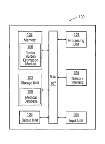
[0011] Figure 1 illustrates a block diagram of a system in which an embodiment
of a method
for determining tumor burden in a medical image can be implemented.
[0012] Figure 2 illustrates a flowchart of an embodiment of a method of
determining tumor
burden in a medical image.
3c
Date Recue/Date Received 2021-09-20
201722832
[0013] Figure 3 illustrates a flowchart of an embodiment of a method of
selecting the second
region of interest whose tumor burden is to be determined.
[0014] Figure 4 illustrates an embodiment of a configuration of deriving tumor
burden in a
defined region in a human body.
[0015] Figure 5 illustrates another embodiment of a configuration of deriving
tumor burden in a
defined region in a human body.
[0016] Figure 6 illustrates yet another embodiment of a configuration of
deriving tumor burden
in a defined region in a human body.
[0017] Figure 7 illustrates a flowchart of an embodiment of a method of
defining a segmentation
criterion for the second region of interest.
DETAILED DESCRIPTION
[0018] Hereinafter, embodiments for carrying out the present invention are
described in detail.
The various embodiments are described with reference to the drawings, wherein
like reference
numerals are used to refer to like elements throughout. In the following
description, for purpose
of explanation, numerous specific details are set forth in order to provide a
thorough
understanding of one or more embodiments. It may be evident that such
embodiments may be
practiced without these specific details. In other instances, well known
materials or methods
have not been described in detail in order to avoid unnecessarily obscuring
embodiments of the
present disclosure. While the disclosure is susceptible to various
modifications and alternative
forms, specific embodiments thereof are shown by way of example in the
drawings and will
herein be described in detail. It should be understood, however, that there is
no intent to limit the
disclosure to the particular forms disclosed, but on the contrary, the
disclosure is to cover all
4
CA 3047564 2019-06-21
201722832
modifications, equivalents, and alternatives falling within the spirit and
scope of the present
disclosure.
[0019] Figure 1 is a block diagram of a system 100 in which an embodiment can
be
implemented, for example, as a system to determine a tumor burden in a medical
image,
configured to perform the processes as described therein. In Figure 1, the
system 100 comprises
a processing unit 101, a memory 102, a storage unit 103, a network interface
104, an input unit
105, an output unit 106 and a standard interface or bus 107. The system 100
can be a (personal)
computer, a workstation, a virtual machine running on host hardware, a
microcontroller, or an
integrated circuit. As an alternative, the system 100 can be a real or a
virtual group of computers
(the technical term for a real group of computers is "cluster", the technical
term for a virtual
group of computers is "cloud").
[0020] The processing unit 101, as used herein, means any type of
computational circuit, such
as, but not limited to, a microprocessor, microcontroller, complex instruction
set computing
microprocessor, reduced instruction set computing microprocessor, very long
instruction word
microprocessor, explicitly parallel instruction computing microprocessor,
graphics processor,
digital signal processor, or any other type of processing circuit. The
processing unit 101 may also
include embedded controllers, such as generic or programmable logic devices or
arrays,
application specific integrated circuits, single-chip computers, and the like.
In general, a
processing unit 101 can comprise hardware elements and software elements. The
processing unit
101 can be configured for multithreading, i.e. the processing unit 101 can
host different
calculation processes at the same time, executing the either in parallel or
switching between
active and passive calculation processes.
CA 3047564 2019-06-21
201722832
[0021] The memory 102 may be volatile memory and non-volatile memory. The
memory 102
may be coupled for communication with the processing unit 101. The processing
unit 101 may
execute instructions and/or code stored in the memory 102. A variety of
computer-readable
storage media may be stored in and accessed from the memory 102. The memory
102 may
include any suitable elements for storing data and machine-readable
instructions, such as read
only memory, random access memory, erasable programmable read only memory,
electrically
erasable programmable read only memory, a hard drive, a removable media drive
for handling
compact disks, digital video disks, diskettes, magnetic tape cartridges,
memory cards, and the
like. In the present embodiment, the memory 102 includes a tumor burden
estimation module
108 stored in the form of machine-readable instructions on any of the above-
mentioned storage
media and may be in communication to and executed by processing unit 101. When
executed by
the processing unit 101, the tumor burden estimation module 108 causes the
processing unit 101
to determine a tumor load or tumor burden in the medical image. Method steps
executed by the
processing unit 101 to achieve the abovementioned functionality are elaborated
upon in detail in
Figure 2, 3,4, 5, and 6.
[0022] The storage unit 103 may be a non-transitory storage medium which
stores a medical
database 109. The medical database 109 is a repository of medical information
related to one or
more patients that is maintained by a healthcare service provider. The input
unit 105 may include
input means such as keypad, touch-sensitive display, camera (such as a camera
receiving
gesture-based inputs), etc. capable of receiving input signal. The bus 107
acts as interconnect
between the processing unit 101, the memory 102, the storage unit 103, the
network interface
104, the input unit 105 and the output unit 106.
6
CA 3047564 2019-06-21
201722832
[0023] Those of ordinary skilled in the art will appreciate that the hardware
depicted in Figure 1
may vary for particular implementations. For example, other peripheral devices
such as an
optical disk drive and the like, Local Area Network (LAN)/ Wide Area Network
(WAN)/
Wireless (e.g., Wi-Fi) adapter, graphics adapter, disk controller,
input/output (I/O) adapter also
may be used in addition or in place of the hardware depicted. The depicted
example is provided
for the purpose of explanation only and is not meant to imply architectural
limitations with
respect to the present disclosure.
[0024] A system in accordance with an embodiment of the present disclosure
includes an
operating system employing a graphical user interface. The operating system
permits multiple
display windows to be presented in the graphical user interface simultaneously
with each display
window providing an interface to a different application or to a different
instance of the same
application. A cursor in the graphical user interface may be manipulated by a
user through the
pointing device. The position of the cursor may be changed and/or an event
such as clicking a
mouse button, generated to actuate a desired response.
[0025] One of various commercial operating systems, such as a version of
Microsoft
WindowsTM, a product of Microsoft Corporation located in Redmond, Washington
may be
employed if suitably modified. The operating system is modified or created in
accordance with
the present disclosure as described.
[0026] Disclosed embodiments provide systems and methods for analysing a
medical image. In
particular, the systems and methods may determine a tumor load in a medical
image.
[0027] Figure 2 illustrates a flowchart of an embodiment of a method 200 of
determining a
tumor burden in a medical image. At step 201 of the method 200, a medical
image is obtained
from a source. The source may be, for example, a medical imaging device such
as a positron
7
CA 3047564 2019-06-21
201722832
emission tomography (PET) device. Alternatively, the medical image may also be
obtained from
the medical database 109. The medical image may be obtained from the source
through an
interface. The interface may be, for example, the network interface 105 or
standard interface 107.
The medical image may be, for example, a positron emission tomography image.
Alternatively,
depending on the type of medical imaging modality used, the medical image may
be, for
example, a computed tomography image or a magnetic resonance imaging image or
a
combination of different types of medical images obtained from one or more
imaging modalities.
The medical image may include imaging information pertaining to one or more
organs or
structures in the patient's body. Such imaging information may include, for
example,
segmentation volumes of organs such as, but not limited to, brain, heart,
liver, lungs, kidney,
bladder, and prostrate. The imaging information may also include, for example,
two-dimensional
or three-dimensional ranges or bounding boxes associated with head and neck,
thorax, abdomen,
pelvis, lower limbs, and lymph node stations. The imaging information may
further include, for
example, bones such as cortical bone, trabecular bone and marrow.
[0028] At step 202, a first region of interest is identified in the medical
image. The first region of
interest may include a combination of one or more organs and/or one or more
anatomical ranges.
The first region of interest may be defined by a physician and may be based on
the physical
volume of the patient's body that may require medical analysis. The first
region of interest may
further be identified based on the one or more organs or anatomical ranges or
combinations of
both that are to be analysed. Therefore, for example, if the physician intends
to determine tumor
burden of liver of the patient, the first region of interest identified may be
the abdominal range.
At step 203, a second region of interest whose tumor burden is to be
determined is selected from
the first region of interest. In an embodiment, the second region of interest
may include at least
8
CA 3047564 2019-06-21
201722832
one organ or anatomical range or a combination thereof, from the first region
of interest. The
second region of interest may be defined based on the anatomical,
physiological, and/or
pathophysiological relationship between one or more organs and or anatomical
ranges. Such
relationship may differ based on the radiopharmaceutical compound used in the
image
acquisition process. Such relationship may be determined based on cancer
staging and may
therefore depend on the organs or anatomical ranges that may have been
affected due to disease
progression. In an embodiment, the second region of interest may therefore be
pre-defined based
on the type and stage of a cancer. The physician may specify the second region
of interest to be
selected from the first region of interest for determination of tumor burden.
The physician may
access such information related to the one or more organs present in the first
region of interest
via a graphical user interface. The graphical user interface may be used by
the physician to
indicate at least one organ within the first region of interest whose tumor
burden is to be
determined. The graphical user interface may include information associated
with one or more
organs in the first region of interest that may be chosen by the physician for
tumor burden
determination. The physician may select one or more data fields, for example,
by clicking a
mouse button on the option. In an alternate embodiment, the second region of
interest may be
selected by excluding one or more organs and/or anatomical ranges from the
first region of
interest. For example, the physician may choose from the graphical user
interface the organs or
anatomical ranges to be excluded from the first region of interest for tumor
burden
determination. In one embodiment, the click of the mouse button may proceed in
checking or
unchecking the data fields associated with the one or more organs in the first
region of interest
on the graphical user interface.
9
CA 3047564 2019-06-21
201722832
100291 Figure 3 illustrates a flowchart of an embodiment of a method 300 of
selecting the second
region of interest from the first region of interest, whose tumor burden is to
be determined. At
step 301, presence of a spatial overlap between the second region of interest
and the first region
of interest is identified. A spatial overlap may occur when two or more organs
are present or
enclosed in the same anatomical region. Such spatial overlap may result in an
inaccurate
determination of tumor burden. Therefore, at step 302, a hierarchical priority
is defined between
the second region of interest and the spatially overlapping first region of
interest. In an
embodiment, the spatial hierarchy may be defined such that an organ takes
priority over an
anatomical range. At step 303, the second region of interest is selected based
on the defined
hierarchical priority. Advantageously, defining a hierarchical priority
eliminates the chances of
duplicate segmentations. Therefore, in an example of overlapping liver organ
and abdominal
region, the hierarchical prioritization for selection may be defined as:
a. Case 1: Include abdomen and exclude liver
i. For liver region: do nothing
ii. For abdominal region:
1. Segmentation volume: abdomen volume excluding liver volume
2. Segmentation criteria: abdomen segmentation criteria
b. Case 2: Exclude abdomen and include liver
i. For liver region:
1. Segmentation volume: liver volume
2. Segmentation criteria: liver segmentation criteria
ii. For abdominal region: do nothing.
c. Case 3: Include abdomen and include liver
CA 3047564 2019-06-21
201722832
i. For liver region:
1. Segmentation volume: liver volume
2. Segmentation criteria: liver segmentation criteria
ii. For abdominal region:
1. Segmentation volume: abdomen volume excluding liver volume
2. Segmentation criteria: abdomen segmentation criteria
[0030] Figure 4 illustrates an embodiment of a configuration 400 of
determining a tumor burden
in a whole body, excluding physiological uptake in the regions of brain,
heart, kidneys, bladder
and lower limbs. The first anatomy 401 illustrates a plurality of segmented
organs that are
present in different regions in the patient's body. The second anatomy 402
illustrates one or more
anatomical ranges that enable the physician to choose a region of interest. In
an embodiment, as
illustrated in Figure 4, the first region of interest identified is the whole
body of the patient. The
second region of interest, therefore, may include regions and/or organs
excluding brain, heart,
kidneys, bladder and lower limbs. Therefore, according to Figure 4, the second
region of interest
comprises lungs, liver and bone/skeleton. The organs in the second region of
interest are
indicated in light grey.
[0031] Similarly, Figure 5 illustrates an embodiment of a configuration 500 of
deriving organ-
specific tumor burden for liver. The anatomy 502 indicates the anatomical
ranges in the patient's
body. In the embodiment, the second region of interest, i.e. liver is selected
for further analysis.
The anatomy 501 depicts a plurality of organs included in the whole body of
the patient.
However, as the liver is selected or included as the second region of
interest, the liver is
represented in light grey. Therefore, all the other organs are excluded from
the second region of
interest and therefore represented in dark grey.
11
CA 3047564 2019-06-21
=
201722832
[0032] In yet another example, Figure 6 illustrates a configuration 600 of
deriving tumor burden
in the pathophysiological system for prostate cancer, providing pelvic,
abdominal and skeletal
tumour burden whilst excluding physiological PET uptake. The
pathophysiological system for
prostate cancer may include the prostate gland, the liver and the bone or
skeletal system of the
patient. The anatomy 602 illustrates the anatomical range chosen as the first
region of interest
and the anatomy 601 illustrates a plurality of organs selected in the first
region of interest. The
anatomy 602 includes pelvis and abdomen regions as the first regions of
interest as the
pathophysiological system for prostate cancer include organs or anatomical
regions present in
additional anatomical ranges apart from the pelvis range. The anatomy 601,
therefore, depicts the
second region of interest forming a part of the pathophysiological system for
prostate cancer, i.e.
prostate, liver and bone/skeleton are included and are indicated in light
grey. All the other organs
are excluded and are therefore represented in dark grey.
[0033] At step 204 of the method 200, a segmentation criterion is defined for
the second region
of interest. The segmentation criterion enables efficient segmentation of the
second region of
interest for further analysis. Therefore, in an embodiment, if only one organ
or an anatomical
range is to be analysed in the second region of interest, the segmentation
criterion may be
defined based on such organ or anatomical range. The segmentation criterion
may include at
least one segmentation algorithm associated with the one or more organs to be
analysed. Such
segmentation algorithms may be used to segment the one or more organs and or
anatomical
regions from the region of interest. Such segmentation algorithms may be well-
known to a
person skilled in the art.
[0034] Figure 7 illustrates a flowchart of an embodiment of a method 700 of
defining a
segmentation criterion for the second region of interest. At step 701 of the
method 700, a mean
12
CA 3047564 2019-06-21
201722832
and a standard deviation value is computed for pixel intensities in a
background tissue
surrounding the segmentation within the second region of interest. In an
embodiment, the
standardized uptake value threshold may be defined based on pixel intensities
of background
tissue surrounding segmentation(s) within the second region of interest. Such
tissue may be a
part of the first region of interest. In defining the standardized uptake
value threshold, a mean
and standard deviation value of pixel intensities of the background tissue is
calculated. Such
background tissue may be a non-cancerous tissue. Therefore, at step 702, based
on the pixel
intensity of the background tissue of the second region of interest, the
standardized uptake value
threshold may be adaptively defined. Such standardized uptake value threshold
may be
associated with the selected organ or the anatomical range in the second
region of interest whose
tumor burden is to be determined. Therefore, the segmentation criterion may be
adaptively
defined based on the background uptake region of the second region of
interest. A standardized
uptake value is a ratio of the image derived radioactivity concentration and
the whole body
concentration of the injected radioactivity. Each organ or anatomical range
may have an
associated standardized uptake value threshold. Such thresholds therefore vary
from one
organ/anatomical range to another. The standardized uptake value threshold
enables detection of
lesions in the patient's body. Therefore, based on the selected
organ/anatomical region the
standardized uptake value threshold is defined such that lesions may be
detected accurately in the
patient's body. For example, if radiopharmaceutical compound used in the
imaging process is
fluorodeoxyglucose (FDG), a low standard uptake value threshold may be set for
lungs as the
uptake of FDG is low for lungs. Similarly, a higher threshold may be set for
liver as the uptake
of fluorodeoxyglucose in liver is high. Therefore, origin of false positives
and false negatives in
the lesion detection process may be eliminated. At step 703 of the method 700,
a tumor region is
13
CA 3047564 2019-06-21
201722832
detected automatically in the second region of interest based on the
standardized tumor uptake
value threshold. Such automatic tumor detection may be performed, for example,
using inverted
grayscale look-up table. Alternatively, other methods well known to a person
skilled in the art
may be used for automated tumor detection in the selected organ. At step 704,
the tumor region
may be segmented from the second region of interest for determination of tumor
burden. The
tumor burden for the segmented tumor region may then be determined at step 205
of the method
200.
[0035] In an embodiment, the foregoing method steps may be used to evaluate
specific types of
cancer based on cancer staging criteria. A plurality of configuration of
organs and/or anatomical
ranges may be pre-defined based on the type of cancer to be evaluated. Such
configurations may
be defined based on the organ system to be considered for cancer analysis and
a knowledge of
the organs or anatomical ranges that may be affected. For example, a
configuration relevant for
prostate cancer may include anatomical regions such as prostate (for a
confined relapse); pelvic
region (for local lymph nodes); abdominal region (for distant lymph node and
visceral organ
metastases); and skeleton (for bone metastases). Each of these anatomical
regions may be
indicative of a different treatment pathway. According to another example, a
configuration
relevant for breast cancer may include anatomical regions such as breast (for
primary tumors);
contralateral breast and axillary, mammary and supraclavicular lymph nodes
(for regional
spread); lungs, liver, and brain (for visceral or distant metastases); and
skeleton (for bone
metastases). Yet another configuration may be for, for example, lung cancer.
Such configuration
may include anatomical regions such as lung (for primary tumor); contralateral
lung and
mediastinal, supraclavicular, and hilar lymph nodes (for regional spread);
liver, brain and adrenal
glands (for visceral or distant metastases) and skeleton (for bone
metastases). Therefore, in an
14
CA 3047564 2019-06-21
201722832
embodiment, a plurality of tumor regions may be determined based on the
standardized uptake
value threshold for the pre-defined configuration. This enables determination
of system-based
tumor burden for the patient.
[0036] In an embodiment, the pre-defined configurations for the system-based
tumor burden
determination may be stored in the medical database 109 and may be provided as
an option on
the graphical user interface. In another embodiment, the physician may create
a new
configuration for tumor burden analysis, including a plurality of organs
and/or anatomical
ranges, based on his expertise. Such new configurations may also be stored in
the medical
database 109 for future use and analysis. The tumor burden estimation module
108 may be
configured to determine tumor burden in the corresponding second region of
interest of each of
such stored configurations, every time such stored configuration is chosen by
the physician.
[0037] In another embodiment, a temporal trend of the configuration based
tumor burden
assessment may be represented for example, graphically as a trend graph. Such
trend graphs may
provide inputs on the progression of the disease in the region(s) of interest.
Therefore, clinical
decision making process is enhanced and made efficient.
[0038] The foregoing examples have been provided merely for the purpose of
explanation and
are in no way to be construed as limiting of the present invention disclosed
herein. While the
invention has been described with reference to various embodiments, it is
understood that the
words, which have been used herein, are words of description and illustration,
rather than words
of limitation. Further, although the invention has been described herein with
reference to
particular means, materials, and embodiments, the invention is not intended to
be limited to the
particulars disclosed herein; rather, the invention extends to all
functionally equivalent
structures, methods and uses, such as are within the scope of the appended
claims. Those skilled
CA 3047564 2019-06-21
201722832
in the art, having the benefit of the teachings of this specification, may
effect numerous
modifications thereto and changes may be made without departing from the scope
and spirit of
the invention in its aspects.
16
CA 3047564 2019-06-21