Note: Descriptions are shown in the official language in which they were submitted.
CA 03048102 2019-06-20
WO 2018/119174
PCT/US2017/067790
Microneedle Arrays and Methods of Making and Using
FIELD OF THE INVENTION
The present invention relates to devices for the transdermal administration of
benefit agents to patients through the skin. More particularly, this invention
relates to
microneedle arrays comprising a plurality of benefit agents, and methods for
making
and using these arrays.
BACKGROUND OF THE INVENTION
Transdennal drug delivery, provides several advantages over other routes for
administering a benefit agent formulation to a patient. For example, oral
administration
of some benefit agents may be ineffective because the benefit agent is
destroyed in the
gastrointestinal tract or eliminated by the liver, both of which are avoided
by
transdermal drug delivery. Parenteral injection with a conventional hypodermic
needle
also has drawbacks, as it is often painful and inconvenient.
Transdermal drug delivery avoids these problems. However, there are obstacles
to its use. In particular, the physical barrier properties of the stratum
comeum of human
skin pose a significant challenge to transdermal drug delivery. These barrier
properties
only allow relatively small molecules to be transported through the intact
stratum
comeum, and many useful drugs are too large to pass through the stratum comeum
without some type of modification of the stratum comeum or other transport
enhancement. Various transdennal enhancement methods are known, including
those
based on iontophoresis, ultrasound, and chemical penetration enhancers.
However,
these methods may be inadequate to assist in the delivery of many medications
through
an intact skin layer and/or they may be inconvenient or undesirably
complicated to use.
To address the challenge of intact skin, a variety of microneedle-array based
drug delivery devices have been developed. These known microneedle array
generally
fall into one of two design categories: (1) solid microneedles arrays with no
active
component, and (2) microneedles with a central hollow bore, which are similar
to
conventional hypodermic needle.
Solid microneedle arrays can pre-condition the skin by piercing the stratum
comeum and the upper layer of epidermis to enhance percutaneous drug
penetration
prior to topical application of a biologic-carrier or a traditional patch. If
solid
1
CA 03048102 2019-06-20
WO 2018/119174
PCT/US2017/067790
microneedle arrays are kept in the skin, then the drug cannot readily flow
into and
through the holes in the skin because the holes remain plugged by the
microneedles.
This method has been shown to significantly increase the skin's permeability;
however,
this method provides only limited ability to control the dosage and quantity
of delivered
drugs or vaccine.
To increase the dosage control some methods uses solid microneedles that are
surface-coated with a drug. Although this method provides somewhat better
dosage
control, it greatly limits the quantity of drug delivered. Also, the
deposition process is
unreliable, and the thin layer of drug formulation on the microneedle could be
easily
chipped off of the microneedle during storage, transport, or administration
(insertion)
of the microneedles. The application of a thicker and stronger layer of drug
formulation
can be undesirable because it reduced the sharpness of the microneedles and
therefore
made insertion more difficult and painful. This shortcoming has limited the
widespread
application of this approach and precludes, for example, the simultaneous
delivery, of
optimal quantities of combinations of antigens and/or adjuvant in vaccine
applications.
Microneedles with a central hollow bore attached to a reservoir of benefit
agents
are also known. The syringe needle-type characteristics of these arrays can
significantly
increase the speed and precision of delivery, , as well as the quantity of the
delivered
agent. However, reservoir-based microneedle arrays are expensive to make and
require
complex and expensive micromachining procedures. In particular, it is
difficult to make
sharp tips on hollow microneedles with machining techniques. Consequently,
insertion
of the microneedles into a patient's skin can be difficult and often painful.
In addition,
the central bore of the microneedle is quite small and may be easily plugged
by skin
tissue during the insertion process, thereby blocking the drug delivery
conduit. It may
be even slower than the diffusion of the drug through the stratum comeum in
the
absence of the microneedle. It therefore would be desirable to provide a
microneedle
array for drug delivery that avoids the disadvantages associated with known
hollow
microneedle array designs.
Also known methods involve using solid microneedle arrays that are
biodegradable, bioabsorbable, or dissolvable. This method combines the
physical
toughness of solid microneedles with relatively high bioactive material
capacity, while
retaining desired attributes of simple fabrication, storage and application.
Current
fabrication approaches for dissolvable polymer-based microneedles generally
use
2
CA 03048102 2019-06-20
WO 2018/119174
PCT/US2017/067790
microcasting processes. For example, a primary master mold is commonly
produced
using a combination of complex lithographic and laser etching technologies.
However,
lithographic and laser-based technologies are limited in the range of
geometric features
they can create, and the materials to which they can be applied. Also, these
highly
complex fabrication technologies do not allow rapid or low cost fabrication of
master
molds, which can be particularly useful for systematic testing of the bio-
effectiveness
of various different microneedle and array geometries.
Finally, the microcasting process for producing dissolvable polymer-based
microneedle arrays is limited to producing arrays of a single composition. If
there is a
desire for personalized treatment requiring dissolvable arrays using
microneedles with
different compositions or benefit agents, the microcasting process cannot
produce such
arrays.
In summary, transdennal delivery of benefit agents using microneedle-array
based devices offer attractive theoretical advantages over prevailing oral and
needle-
based drug delivery methods. However, considerable practical limitations exist
in the
design, fabrication, and testing associated with microneedle arrays
constructed using
conventional processes. Also, there is a need for a simple, effective, and
economically
desirable device for transdermal administration of using microneedle arrays
simultaneously delivering more than one benefit agent.
SUMMARY OF THE INVENTION
Surprisingly, we have found that an array of differing microneedles can be
accurately achieved including a film having first and second, outwardly facing
major
surfaces. The first, outwardly facing major surface has a plurality of stratum
comeum
piercing microneedles extending therefrom, and the plurality of microneedles
includes
a plurality of first microneedles having a first benefit agent and a plurality
of second
microneedles having a second benefit agent.
BRIEF DESCRIPTION OF THE DRAWINGS
FIG. I is a perspective view of one embodiment of a microneedle array;
FIG. 2 is a cross-sectional view of a section of the microneedle array of FIG.
1
alone the 2-2 plane:
FIG. 3 is a top view of a section of the microneedle array of FIG. 1;
3
CA 03048102 2019-06-20
WO 2018/119174
PCT/US2017/067790
FIG. 4 is a cross-sectional view of a section of a second embodiment
microneedle array;
FIG. 5 is a cross-sectional view of a section of a third embodiment
microne.edie
array;
FIG. 6 is a cross-sectional view of a section of a fourth embodiment
microneedle array;
FIG. 7 is a cross-sectional view of a section of the microneedle array of FIG.
6
after the microneedles have penetrated the patient's skin;
FIG. 8 is a cross-sectional view of a section of a fifth embodiment
microneedle
array; and
FIG. 9 is a cross-sectional view of a section of a sixth embodiment
microneedle
array.
DETAILED DESCRIPTION OF THE INVENTION
The present invention relates to devices for the transderunal administration
of a
plurality of benefit agents to patients through the skin using microneedle
array systems,
and methods for making and employing these systems. The following description
is
presented to enable one of ordinary skill in the art to make and use the
invention.
Various modifications to the embodiments and the generic principles and
features
described herein will be readily apparent to those skilled in the art. Thus,
the present
invention is not intended to be limited to the embodiments shown, but is to be
accorded
the widest scope consistent with the features described herein.
As used herein the specification and the claims, the term "topical" and
variants
thereof mean "of or applied to an isolated part of the body". This includes,
without
limitation skin, mucosa, and enamel, either directly or through an
intermediate such as
a biofilm.
As used herein, "benefit agent" means an ingredient or material that provides
a
benefit, e.g., improves, relieves, reduces, or treats symptoms or conditions
of the skin
or body; either cosmetic or therapeutic. Other tennis of use for "benefit
agent" include
"biologic," "active component," or "bioactive material". These terms all refer
to
pharmaceutically active agents, such as analgesic agents, anesthetic agents,
anti-
asthmatic agents, antibiotics, anti-depressant agents, anti-diabetic agents,
anti-ftingal
agents, anti-hypertensive agents, anti-inflammatory agents, anti-neoplastic
agents,
anxiolytic agents, enzymatically active agents, nucleic acid constructs,
4
CA 03048102 2019-06-20
WO 2018/119174
PCT/US2017/067790
immunostimulating agents, immunosuppressive agents, vaccines, and the like.
The
benefit agent material can comprise dissoluble materials, insoluble but
dispersible
materials, natural or formulated macro. micro and nano particulates, and/or
mixtures of
two or more of dissoluble, dispersible insoluble materials and natural and/or
formulated
macro, micro and nano particulates.
In some embodiments, the microneedle array systems described herein are
flexible so as to be conformable to the three-dimensional shape corresponding
to the
site of delivery of benefiting agent substance to the skin of the consumer. In
other
embodiments, the microneedle array may be more rigid; built as the described
three-
dimensional shape to match the topical contour. The array may have varying
personalized area-specific treatment zones to enable the treatment application
more
effectively. With an array matched to the individual user's body part profile
as physical
guides, the application becomes easier and more effective, and can help in
locating
specific target zones to the precise area for applications.
Referring to the drawings, FIG. I is a perspective view of one embodiment of a
microneedle array 10 which may be used in the present invention. Microneedle
array 10
includes a film 20 having first outwardly facing major surface 22 and second
outwardly
facing major surface 24. First outwardly facing major surface 22 has a
plurality of
stratum corneum piercing microneedles 30 extending therefrom. Each microneedle
30
has a proximal end 32 and a distal end 34, where proximal end 32 is the end of
microneedle 30 disposed on first outwardly facing major surface 22 of a
microneedle
array 1Ø
In FIG. 1, microneedle army 10 is shown to have a rectangular footprint. Film
20 of microneedle array 10 may also have a variety of shapes, depending on the
location of skin treatment. Possible shapes of the footprint left by film 20
include, but
are not limited to, squares, rectangles, triangles, circles, ovals, kidneys,
stars, crosses,
characters, etc. The corners of such shapes, if any, may be angular or curved
to reduce
potential lift/removal points. The zone of the treatment could be greater than
about
1,000 cm2, about 1,000 cm2, or about 100 cm2, or about 10 cm2, or about 1 cm2,
or less
than 1 cm2.
Film 20 element of microneedle array 10 preferably is relatively thin and
flexible, so that they preferably readily conform to the user's skin and are
comfortable
to wear, both because of the flexibility and conformability, as well as from
the thinness.
5
CA 03048102 2019-06-20
WO 2018/119174
PCT/US2017/067790
Microneedle array 10 of the present invention may be intended for extended
wear
preferably are also formed to be aesthetically elegant without either peeling,
wrinkling,
cracking, or appearing greasy or tacky, or otherwise unpleasant or unsightly
in nature.
Microneedle array 10 preferably is formed with sufficient rigidity and
integrity to be
able to withstand normal use when on the skin. In some embodiments,
microneedle
array 10 of the invention preferably is formed with sufficient strength to
stay intact on
the skin when exposed to normal external forces that the skin may experience,
rubbing
of clothing.
In some embodiments, first outwardly facing major surface 22 of film 20 has
disposed thereon an adhesive layer. The adhesive layer may be used to give
microneedle array 10 the sufficient strength to stay intact on the skin when
exposed to
normal external forces. Other means of creating sufficient strength to
microneedle array
10 so that the array stays intact on the skin will be discussed below.
FIG. 2 is a cross-sectional view of a section of the microneedle array along
the
2-2 plane of FIG. 1. The figure shows a plurality of first stratum corneum
piercing
microneedles 30a and a plurality of second stratum corneum piercing
microneedles
30a. Each microneedle 30a has a proximal end 32a and a distal end 34a, while
each
microneedle 30b has a proximal end 32b and a distal end 34b. Plurality of
first
microneedles 30a comprises a first benefit agent and plurality of second
microneedles
30b comprises a second benefit agent.
The dimensions of stratum corneum piercing microneedles 30a, 30b may vary
depending on a variety of factors such as the type of benefit agent to be
delivered, the
dosage of the benefit agent to be delivered, and the desired penetration
depth.
Generally, the stmttun corneum piercing microneedles are constructed to
provide skin-
piercing and benefit agent delivery functions and thus will be designed to be
sufficiently robust to withstand insertion into and withdrawal from the skin.
Each
microneedle has a length of about 1 micrometer (pm) to about 5000 micrometers
( m),
or about 1 pm to about 500 pm, or about 100 pm to about 500 ttrn. The
penetration
length of the microneedles into the biological barrier is about 50 pm to about
200 pm.
In addition, each of the microneedles has a width of about 1 pm to about 500
pm.
Furthermore, each microneedle has a thickness of about 1 tun to about 200 tun.
It will
be understood by one skilled in the art that the width and thickness of the
stratum
corneum piercing microneedle may vary along its length. For instance, the base
portion
6
CA 03048102 2019-06-20
WO 2018/119174
PCT/US2017/067790
may be wider (thicker) than the body portion, or the body portion may have a
slight
taper approaching the tip portion.
FIG. 3 is a top view of a section of the microneedle array of FIG. I. The
figure
shows stratum comeum piercing microneedles 30 which extend from first
outwardly
facing major surface 22 of microneedle army 10. Each microneedle 30 has a
proximal
end 32 and a distal end 34. As shown in the figure, microneedles 30 are
arranged in a
square pattern on first outwardly facing major surface 22 of microneedle array
10. In
other embodiments, microneedles 30 are arranged in other patterns, such as
triangular,
square, pentagonal, hexagonal, octagonal, etc.
Microneedles 30 in microneedle array 10 of the invention may also be of a
variety of lengths and geometries. FIG. 4 is a cross-sectional view of a
section of a
second embodiment microneedle array. In this embodiment, plurality of first
stratum
come= piercing microneedles 30a comprise a first benefit agent and plurality
of
second stratum comeum piercing microneedles 30c comprise a second benefit
agent. In
addition, plurality of first microneedles 30a extend from first outwardly
facing major
surface 22 of film 20 to a height of hi, while plurality of second
microneedles 30b
extend from first surface 22 of film 20 to a height of h2. In this embodiment,
there may
be a desire for a deeper penetration into the skin of the user for first
benefit agent
contained in plurality of first microneedles 30a than from second benefit
agent
contained in plurality of second microneedles 30b.
Although the figure shows first stratum comeum piercing microneedles 30a are
of uniform height hi, while second stratum corneum piercing microneedles 30b
are of
uniform height h2, it is to be understood that in other embodiments the
microneedles
may be of any number of different heights. In addition, it is important to
note that
neither all microneedles 30a are comprised of a first benefit agent, nor that
all
microneedles 30b are comprised of a second benefit agent. In some embodiments,
some
of the microneedles will not comprise any benefit agent.
Generally, stratum comeum piercing microneedles 30 can be in any elongated
shape suitable for providing the skin piercing and benefit agent delivery,
with minimal
pain to the patient. In various embodiments, an individual microneedle is
substantially
cylindrical, wedge-shaped, cone-shaped, or triangular (e.g., blade-like). The
cross-
sectional shape (cut along a plane approximately parallel to the planar
substrate or
approximately perpendicular to the longitudinal axis of the microneedle) of
the
7
CA 03048102 2019-06-20
WO 2018/119174
PCT/US2017/067790
microneedle, or at least the portion of the microneedle that is penetrable
into the skin,
may take a variety of forms, including rectangular, square, oval, circular,
diamond,
triangular, or star-shaped.
The tip portions of stratum comeum piercing microneedles 30 are designed to
.. pierce a biological barrier, e.g., to pierce the stratum comeum of the skin
of a patient, to
deliver benefit agents into the patient's tissue. Preferably, the tip portion
of each
microneedle should be sufficiently small and sharp to enable piercing and
penetration
of the skin with minimal pain. In a preferred embodiment, individual
microneedles 30
are tapered from the first, outwardly facing major surface 22 of microneedle
array 10 to
a point distal therefrom. In various embodiments, the tapered tip portion may
be in the
form of an oblique angle at the tip, or a pyramidal or conical or triangular
shape.
FIG. 5 is a cross-sectional view of a section of a third embodiment
microneedle
array showing a variety of stratum comeum piercing microneedle shapes.
Microneedle
30a is conical in shape, with a taper from proximal end 32a to distal end 34a.
Microneedle 30d has a cylindrical proximal end 32d, which tapers to a point at
distal
end 34d. Microneedle 30e has a proximal end 32e and a distal end 34e, and has
an
undulating shape. Microneedle 30f is cylindrical in shape, with no taper from
proximal
end 32f to distal end 34f. Finally, microneedle 30g is pyramidal in shape,
with a taper
from proximal end 32g to distal end 34g.
Although FIG. 5 shows all stratum comeum piercing microneedles 30 of
substantially uniform height, it is to be understood that in other embodiments
the
microneedles may be of any number of different heights. In addition,
microneedles 30a,
30d, 30e, 301, and 30g comprise at least one benefit agent. Some comprise a
first
benefit agent, while others comprise a second benefit agent, so that
microneedle arrays
10 comprises microneedles with two distinct benefit agents. Of course, not all
microneedles 30 of any given shape or height are required to all comprised
either first
or second benefit agent.
Microneedle arrays 10 of the present invention may also comprise stratum
comeum piercing microneedles 30 comprised of multiple compositions. FIG. 6 is
a
cross-sectional view of a section of a fourth embodiment microneedle army 10
with
such microneedles. The figure shows four different microneedles, with the
microneedles being of variable heights, and comprising at least two distinct
benefit
agents. Microneedle 30h has a cylindrical proximal end 32h, which tapers to a
point at
8
CA 03048102 2019-06-20
WO 2018/119174
PCT/US2017/067790
distal end 34h. In addition, proximal end 32h of microneedle 30h is of a
different
composition than distal end 34h of microneedle 30h. Microneedle 301 is
cylindrical,
and has a core section 321 and a sheath section 341. Here, core section 321 is
of a
different composition than sheath section 34i. Microneedle 30j has a
cylindrical
proximal end 32j and a cylindrical distal end 34j, and has a substantially
linear form.
Here, proximal end 32j of microneedle 30j is of a different composition than
distal end
34j of microneedle 30j. Finally, microneedle 30k is conical in shape, with a
taper from
proximal end 32k to distal end 34k. Proximal end 32k of microneedle 30k is of
a
different composition than distal end 34k of microneedle 30k.
Special attention is now paid to microneedle 301. Microneedle 301 comprises a
core section 32i and a sheath section 341. Core section 321 is of a different
composition
than sheath section 341. In some embodiments, core section 321 does not have
the
mechanical strength or rigidity to penetrate the skin, while sheath section
341 does. In
other embodiments, sheath section 34i does not have the mechanical strength or
rigidity
to penetrate the skin, while core section 321 does. Therefore, at least one of
the sheath
sections comprised a rigid composition. So, materials/active/drugs which are
not strong
enough to penetrate the skin can still be delivered.
Special attention is now paid to microneedle 30j. Microneedle 30j has a
cylindrical distal end 34j, and has an initial, substantially linear form.
Upon insertion
into the skin, distal end 34j is designed to curve to form a hook-like
structure or form.
As mentioned earlier, in some embodiments, first outwardly facing major
surface 22 of
film 20 has disposed thereon an adhesive layer to give microneedle array 10
the
sufficient strength to stay intact on the skin when exposed to normal external
forces. In
some embodiments, microneedle array 10 may have a plurality of microneedles
which
form hook-like structures. Hook-like microneedles 30j, once they penetrate the
skin,
may have sufficient strength so as to hold microneedle array 10 intact on the
skin
during use.
The figure also shows that the stratum corneum piercing microneedles are of
different lengths. In this embodiment, microneedles 30h and 301 extend from
first
outwardly facing major surface 22 of film 20 to a height of hi, microneedle
30j extends
from first surface 22 of film 20 to a height of h2, and microneedle 30k
extends from
first surface 22 of film 20 to a height of h3. In this embodiment, there may
be a desire
for a deeper penetration into the skin of the user for the different benefit
agents.
9
CA 03048102 2019-06-20
WO 2018/119174
PCT/US2017/067790
Although FIG. 6 figure shows stratum corneum piercing microneedles 30 of
different heights, it is to be understood that in other embodiments the
microneedles may
all be of the same height, or any number of different heights. In addition, it
is important
to note that all microneedles 30 are neither comprised of a first benefit
agent nor a
second benefit agent. Also, not all microneedles 30 are composed of multiple
benefit
agents. In some embodiments, some of the stratum corneum piercing microneedles
will
not comprise any benefit agent.
The different sizes, compositions, and geometries of the stratum corneum
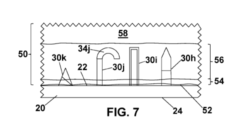
piercing microneedles are demonstrated in a prophetic use. FIG. 7 is a cross-
sectional
view of a section of the microneedle array of FIG. 6 after the microneedles
have have
been deployed and penetrated the patient's skin. The figure shows skin tissue
50 with
an outer surface 52. Beneath the outer surface 52 lie the epidermis 54, dermis
56, and
the subcutis or hypodernris 54 layers. The first outwardly facing major
surface 22 of
film 20 is in contact with outer surface 52 of skin tissue 50.
Microneedles 30h, 30i, 30j, and 30k all penetrate outer surface 52 and
epidermis 54. Microneedles 30h, 301 and 30j penetrate deeper into dermis 56
than
microneedle 30k. Also, since proximal end 32h of microneedle 30h is of a
different
composition than distal end 34h of microneedle 30h, the distal end composition
is
deposited deeper into the dermis than the proximal. The same is true for
microneedles
30j and 30k. So, if there is a desire for personalized treatment at different
skin depths,
microneedle arrays 10 of the present invention allow a degree of flexibility
not
available to microneedle arrays produced using the microcasting process.
Also, as discussed earlier, distal end 34j of microneedle 30j is designed to
curve
to form a hook-like deployed form upon insertion into the skin. Hook-like
microneedle
.. 30j may have sufficient strength so as to hold microneedle array 10 intact
on the skin
during use. This may allow first outwardly facing major surface 22 of film 20
to be free
of adhesive.
In the embodiments shown so far, microneedle array 10 is shown to be planar.
In some embodiments, the array may be curvilinear. FIG. 8 is a cross-sectional
view of
a section of a fifth embodiment microneedle array of the present invention.
Microneedle array 100 includes a curved film 120 having first outwardly facing
major
surface 122 and second outwardly facing major surface 124. First outwardly
facing
major surface 122 has a plurality of stratum corneum piercing microneedles 130
CA 03048102 2019-06-20
WO 2018/119174
PCT/US2017/067790
extending therefrom. The figure shows a plurality of first stratum comeum
piercing
microneedles 130a and a plurality of second stratum comeum piercing
microneedles
130a. Each microneedle 130a has a proximal end 132a and a distal end 134a,
while
each microneedle 130b has a proximal end 132b and a distal end 134b. Plurality
of first
microneedles 130a comprises a first benefit agent and plurality of second
microneedles
130b comprises a second benefit agent. Proximal ends 132a, 132b are the end of
microneedle 130a, 130b disposed on first outwardly facing major surface 122 of
a
microneedle array 100.
FIG. 8 shows microneedle array 109 having a concave shape with respect to
microneedles 130. FIG. 9 is a cross-sectional view of a section of a sixth
embodiment
microneedle array of the present invention. In this embodiment, microneedle
array 200
has concave and convex curvature within the array. Microneedle array 200
includes a
curved film 220 having first outwardly facing major surface 222 and second
outwardly
facing major surface 224. First outwardly facing major surface 222 has a
plurality of
stratum comeum piercing microneedles 230 extending therefrom. As with all
other
embodiments, microneedle array 200 comprise at least a first benefit agent and
a
second benefit agent.
Although FIGs. 8 and 9 show curvilinear microneedle arrays in one direction,
the array may have multiple axes of curvature in localized regions or overall.
Other
embodiments may employ multiple axes of curvature to shape the microneedle
array.
The curvilinear microneedle arrays shaped to the body surface provides the
microneedles oriented normal to that surface. This provides better penetration
of the
microneedles and retention of the array for treatment.
In preferred embodiments, film 20, 120, 220, stratum comeum piercing
microneedles 30, 130, 230, or both, are formed of, or coated with, a
biocompatible
material. Microneedles 30, 1.30, 230 may be formed from the same material used
in
film 20, 120, 220. or alternatively, the microneedles can include a material
different
from the film material. Representative examples of suitable materials of
construction
include metals and alloys such as stainless steels, palladium, titanium, and
aluminum;
plastics such as polyetheiimide, polycarbonate, polyetheretherketone,
polyimide,
polymethylpentene, polyvinylidene fluoride, polyphenylsulfone, liquid
crystalline
polymer, polyethylene terephthalate (PET), polyethylene terephthalate-glycol
modified
(PETG), and polyimide; and ceramics such as silicon and glass. The material
preferably
11
CA 03048102 2019-06-20
WO 2018/119174
PCT/US2017/067790
is selected such that the microneedle is strong enough at its designed
dimensions for the
microneedle to effectively pierce the skin without significant bending or
breaking of the
microneedle. The microneedle and substrate materials also should be non-
reactive with
the drug formulation being delivered by the microneedle array.
In some embodiments, film 20, 120, 220, microneedles 30, 130, 230, or both,
are formed of biodegradable or bioabsorbable materials. Representative
examples of
suitable materials include, but are not limited to, poly(lactic acid) (PLA),
poly(glycolic
acid) (PGA), polydioxanone (PDO), poly(epsilon-caprolactone) (PCL),
poly(lactic-co-
glycolic acid) (PLGA), poly(ortho ester) (POE), copoly(ether-ester) (CEE),
carboxymethylcellulose (CMC) based formulations, or combinations of such
materials.
Film 20, 120, 220, stratum corneum piercing microneedles 30, 130, 230, or
both, optionally may further include secondary materials of construction
embedded
therein or coated thereon. For example, microparticles, nanoparticles, fibers,
fibrids, or
other particulate materials may be included. These secondary materials may
enhance
one or more physical or chemical characteristics of microneedle array 10, 100,
200.
In some embodiments, stratum corneum piercing microneedles 30, 130, 230are
formed of biodegradable materials, while film 20, 120, 220is not
biodegradable. In
these embodiments the benefit agent material can comprise dissoluble materials
or
insoluble but dispersible materials. So, the mechanism of delivery of the
benefit agent
can be, for example, the simultaneous biodegradation of the microneedles with
the
dissolution or dispersing of the benefit agent. The rate of degradation of the
microneedles could be controlled to allow predetermined drug-delivery rates of
the
benefit agent. In some embodiments, the release rate of first benefit agent
could differ
from that of second benefit agent. At the point in time when all of the
stratum cornetun
piercing microneedles have degraded, film 20, 120, 220can be removed from the
site of
treatment.
In another embodiment, a number of hook-like microneedles 30j may have
sufficient strength so as to hold microneedle array 10 intact on the skin
during use. This
may allow first outwardly facing major surface 22 of film 20 to be free of
adhesive. In
this embodiment, proximal end 32j of microneedle 30j is of a different
composition
than distal end 34j of microneedle 30j. If distal end 34j composition is
biodegradable,
microneedle array 10 may be kept intact on the skin until distal end 34j of
hook-like
12
CA 03048102 2019-06-20
WO 2018/119174
PCT/US2017/067790
microneedles 30j have degraded. At this point in time, microncedle array 10
may be
easily removed from the patient's skin.
In some embodiments, the microneedle array 10 may be further coated with a
benefit agent, either the microneedles alone or in combination with the
substrate.
Alternatively, the microneedles may have a desired surface structure, such as
slight directional ridges, to hold the microneedles in place. The benefit
agents may
include lubricants, slip agents and the like. Alternatively, the benefit
agents may
provide one or more benefits to the targeted topical region. Such benefit
agents may be
any of a variety of compositions, including, without limitation, waxes, oils,
emollients,
.. moisturizers, and the like.
Benefit agents may include hyaluronic acid; hydroxyl acids (e.g., glycolic
acid,
lactic acid, malic acid, salicylic acid, citric acid, tartaric acid); anti-
acne agents (e.g.,
salicylic acid, retinol, retinoids, or other keratolytics, and benzoyl
peroxide, or other
antimicrobial agents used to treat acne); shine control agents (e.g., rice
protein, cotton
powder, elubiol (dichlorophenyl-imidazoltioxolan); a retinoid or its
derivative such as
tretinoin, isotretinoin, motretinide, adapalene, ta7arotene, azelaic acid, and
retinol; a 5-
alpha-reductase inhibitor of amino acids, e.g., glycine derivatives;
hydrolyzed
vegetable proteins, including soy protein and wheat protein, etc.,; green tea
(camellia
sinesis) extract, and cinnamon bark extract); moisturizers; anti-microbial
agents (e.g.,
cationic antimicrobials such as benzylkonium chloride, benzethonium chloride,
triclocarbon, polyhexamethylene biguanide, cety, 1pyridium chloride, methyl
and
benzothonium chloride; salts of chlorhexidine, such as lodopropynyl
butylcarbarnate,
diazolidinyl urea, chlorhexidene digluconate, chlorhexidene acetate,
chlorhexidine
isethionate, and chlorhexidene hydrochloride; halogenated phenolic compounds,
such
as 2,4,4'-trichloro-2-hydroxy diphenyl ether (Triclosan); parachlorometa
xylenol
(PCMX); short chain alcohols, such as ethanol, propanol, and the like);
antibiotics or
antiseptics (mupirocin, neomycin sulfate bacitracin, polymyxin B, 1-ofloxacin,
tetracyclines (chlortetracycline hydrochloride, oxytetracycline-
10hydrochloride and
tetracycline hydrochloride), clindamycin phosphate, gentarnicin sulfate,
metronidazole,
hexylresorcinol, methylbenzethonium chloride, phenol, quaternary ammonium
compounds, tea tree oil, and their pharmaceutically acceptable salts and
prodrugs), anti-
inflammatory agents (e.g., suitable steroidal anti-inflammatory agents such as
corticosteroids such as hydrocortisone, hydroxyltriarncinolone alpharnethyl
13
CA 03048102 2019-06-20
WO 2018/119174
PCT/US2017/067790
dexamethasone, dexamethasone-phosphate, beclomethasone dipropionate,
clobetasol
valerate, desonide, desoxymethasone, desoxycorticosterone acetate,
dexamethasone,
dichlorisone, diflorasone diacetate, diflucortolone valerate, fluadrenolone,
fluclarolone
acetonide, fludrocortisone, flumethasone pivalate, fluosinol one acetonide,
fluocinonide, flucortine butylester, fluocortolone, fluprednidene
(fluprednylidene)
acetate, flurandrenolone, halcinonide, hydrocortisone acetate, hydrocortisone
butyrate,
methylprednisolone, triamcinolone acetonide, cortisone, cortodoxone,
flucetonide,
fludrocortisone, difluorosone diacetate, fluradrenalone acetonide, medrysone,
amciafel,
amcinafide, betamethasone, chlorprednisone, chlorprednisone acetate,
clocortelone,
clescinolone, dichlorisone, difluprednate, flucloronide, flunisolide,
fluoromethalone,
fluperolone, fluprednisolone, hydrocortisone valerate, hydrocortisone
cyclopentylproprionate, hydrocortamate, meprednisone, paramethasone,
prednisolone,
prednisone, beclomethasone dipropionate, betamethasone dipropionate,
triamcinolone,
and salts, nonsteroidal anti-inflammatory agents, feverfew (Tanacetum
parthenium),
goji berry (Lycium barbarum), milk thistle extract (Silybum marianum),
amaranth oil
(Amaranthus cruentus), pomegranate (Punica granatum), yerbe mate (Ilex
paraguariensis leaf extract), white lily flower extract (Lilium Candidum),
olive leaf
extract (Olea europaea) and phloretin (apple extract)); anti-mycotic /
antifungal agents
(e.g., miconazole, econazole, ketoconazole, sertaconazole, itraconazole,
fluconazole,
voriconazole, clioquinol, bifoconazole, terconazole, butoconazole,
tioconazole,
oxiconazole, sulconazole, saperconazole, clotrimazole, undecylenic acid,
haloprogin,
butenafine, tolnafiate, nystatin, ciclopirox olamine, terbinafine, amorolfine,
naftifme,
elubiol, griseofulvin, and their pharmaceutically acceptable salts and
prodrugs; an
azole, an allylamine, or a mixture thereof); external analgesics (e.g.,
ibuprofen- or
diclofenac; capsaicin, fentanyl, and salts thereof such fentanyl citrate;
paracetamol (as
acetaminophen); non-steroidal anti-inflammatory drugs (NSAIDs) such as
salicylates;
opioid drugs such as morphine and oxycodone; ibuprofen- or diclofenac-
containing
gel); anti-oxidants (e.g., sulfhydryl compounds and their derivatives (e.g.,
sodium
metabisulfite and N-acetyl cysteine), lipoic acid and clihydrolipoic acid,
resveratrol.
lactoferrin; ascorbic acid, ascorbic acid esters, and ascorbic acid
derivatives (e.g.,
ascorbyl palmitate and ascorbyl polypeptide); butylhydroxy anisole, butylated
hydroxytoluene (butylhydroxy toluene), retinoids (e.g., retinol and retinyl
palmitate),
tocopherols (e.g., tocopherol acetate), tocotrienols, and ubiquinone;
cysteine, N-
14
CA 03048102 2019-06-20
WO 2018/119174
PCT/US2017/067790
acetylcysteine, sodium bisulfite, sodium metabisulfite, sodium
formaldehydesulfoxylate, acetone sodium bisulfite, tocopherols, and
nordihydroguaiaretic acid; extracts containing flavonoids and isoflavonoids
and their
derivatives (e.g., genistein and diadzein); extracts containing resveratrol
and the like;
grape seed, green tea, pine bark, and propolis; plant-derived polyphenol
antioxidants
such as clove, cinnamon, oregano, turmeric, cumin, parsley, basil, curry
powder,
mustard seed, ginger, pepper, chili powder, paprika, garlic, coriander, onion
and
cardamom; typical herbs such as sage, thyme, marjoram, tarragon, peppermint,
oregano, savory, basil and dill weed)); depilatory agents (e.g., calcium
thioglycolate or
potassium thioglycolate); vitamins (e.g., Vitamin A, Vitamin B, Vitamins C,
Vitamin
E; either alpha, beta, gamma or delta tocopherols, niacin or niacinamide) and
vitamin
salts or derivatives such as ascorbic acid diglucoside and vitamin E acetate
or
palmitate; sunblock (e.g., titanium dioxide) and / or sunscreen (e.g.,
inorganic
sunscreens such as titanium dioxide and zinc oxide; organic sunscreens such as
octyl-
.. methoxy cinnamates, octyl salicylate, homosalate, avobenzone); vasodilators
(e.g.,
niacin); humectants (e.g., glycerin); anti-aging agents (e.g., retinoids;
dimethylaminoathanol (DMAE), copper containing peptides); alpha hydrox3,,'
acids or
fruit acids and their precursors such as glycolic acid, citric acid, lactic
acid, malic acid,
mandelic acid, ascorbic acid, alpha-hydroxybutyric acid, alpha-
hydroxyisobutyric acid,
alphahydroxyisocaproic acid, atrrolactic acid, alpha-hydroxyisovaleric acid,
ethyl
pyruvate, ga1acturonic acid, glucoheptonic acid, glucoheptono 1,4-lactone,
gluconic
acid, gluconolactone, glucuronic acid, glucuronolactone, isopropyl pyruvate,
methyl
pyruvate, mucic acid, pyruvic acid, saccharic acid, saccaric acid 1,4-lactone,
tartaric
acid, and tartronic acid; beta hydroxy acids such as beta-hydroxybutyric acid,
beta-
phenyl-lactic acid, and beta-phenylpyruvic acid; zinc and zinc containing
compounds
such as zinc oxides; botanical extracts such as green tea, soy, milk thistle,
algae, aloe,
angelica, bitter orange, coffee, goldthread, grapefruit, hoellen, honeysuckle,
job's tears,
lithospennum, mulberry, peony, puerarua, nice, and safflower, and salts and
prodrugs
thereof); carotenoids, ceramides, fatty acids, enzymes, enzyme inhibitors,
minerals,
steroids, peptides, amino acids, botanical extracts, colorants, etc. The
substances may
affect the skin in any of a variety of manners, such as by moisturizing;
enhancing skin
tone or color (such as with pigments); treating or at least mitigating various
skin
conditions (such as dry or severe dry skin, eczema, psoriasis, atopic
dermatitis, allergic
CA 03048102 2019-06-20
WO 2018/119174
PCT/US2017/067790
rashes, acne, blackheads, pustules, comedones, rosacea, shingles, wrinkles,
cold sores,
herpes, corns, warts, sunburn, insect bites, poison ivy, etc.); applying a
mechanical
force (such as shrinkage) to smooth wrinkles; or, more generally, treating or
mitigating
the symptoms and appearance of undesired skin imperfections (such as under eye
dark
.. circle, redness of acne, fine lines and wrinldes, post inflammatory
hyperpigmentation
(P1H), redness, inflammation, cellulite, wrinkles, age spots, mottled
pigmentation, dark
spots, liver spots, under eye puffiness); removing unwanted facial or body
hair; aiding
in wound healing; etc.. For instance, lotions, creams, oils, and even masks
may be
applied to skin to treat or otherwise to affect the skin. Such personal or
consumer
healthcare substances are absorbed into the skin generally following the
principles of
diffusion, under which the rate of diffusion or transport across the skin is
correlated
with the difference in active concentration on both sides of the skin.
As mentioned earlier, the micromachining or microcasting process for
producing microneedle arrays are limited to producing arrays of a single
composition.
.. In the present invention, the personalized treatment uses stratum corneum
piercing
stratum corneum piercing microneedles with more than one benefit agent. So,
the
micromachining or microcasting process cannot be used.
The microneedle arrays of the present invention can be produced using Additive
Manufacturing technology. Additive Manufacturing is a group of techniques used
to
.. quickly fabricate a physical part or assembly using three-dimensional
computer aided
design (CAD) data. Construction of the part or assembly is usually done using
"additive
layer manufacturing" technologies such as 3D printing. Additive manufacturing
is a
simple, effective, and economically method of making microneedle arrays which
simultaneously delivering more than one benefit agent.
In general, the computer-aided-design - computer-aided manufacturing CAD-
CAM workflow is the traditional additive manufacturing process. The process
starts
with the creation of geometric data, either as a 3D solid using a CAD
workstation, or
2D slices using a scanning device. For Additive Manufacturing, this data must
represent a valid geometric model; namely, one whose boundary surfaces enclose
a
finite volume, contains no holes exposing the interior unless they are
designed into the
structure, and do not fold back on themselves. In other words, the object must
have an
"inside." The model is valid if for each point in 3D space the algorithm can
determine
uniquely whether that point lies inside, on, or outside the boundary surface
of the
16
CA 03048102 2019-06-20
WO 2018/119174
PCT/US2017/067790
model. CAD post-processors will approximate the internal CAD geometric forms
with
a simplified mathematical form, which in turn is expressed in a specified data
format
which is a common feature in Additive Manufacturing. To obtain the necessary
motion
control trajectories to drive the Additive Manufacturing mechanism, the
prepared
geometric model is typically sliced into layers, and the slices are scanned
into lines
(producing a "2D drawing" used to generate trajectory as in computer numerical
control toolpath), resulting in a layer-to-layer physical building process.
The 3D printing process enables the creation of different sizes and shapes
microneedles, as well as the ability to produce microneedle arrays with more
than one
benefit agent. The location, sharpness, cavitation, and material within
individual
microneedles can be much more easily controlled with 3D printing than
micromachining or microcasting. Soft materials, hard materials, and even
liquids can be
incorporated into individual microneedles. A change in delivery profile can be
designed
into the system to make a smart microneedle array. Incompatible compounds may
also
be built into different sections of the microneedle array without cross
contamination
fears.
The microneedles need to deliver active/drug at least 100 microns or deeper,
but
can be designed to have a variable penetration at or above 20 microns.
Different
applications and uses would need differing levels of penetration, solubility
and design
features (size, shape, angle, solubility, etc.). In some cases, the benefit
agent may be
dissolved into the microneedle material, whereas in others it may be stored in
a
reservoir and delivered through a microfluidic channel in the microneedle.
Although shown and described is what is believed to be the most practical and
preferred embodiments, it is apparent that departures from specific designs
and
methods described and shown will suggest themselves to those skilled in the
art and
may be used without departing from the spirit and scope of the invention. The
present
invention is not restricted to the particular constructions described and
illustrated, but
should be constructed to cohere with all modifications that may fall within
the scope of
the appended claims.
17