Note: Descriptions are shown in the official language in which they were submitted.
CA 03048654 2019-06-26
WO 2018/132678
PCT/US2018/013527
SYSTEMS AND METHODS FOR MUSCULOSKELETAL TISSUE TREATMENT
TECHNICAL FIELD
The present description relates to systems, apparatus, and methods of tissue
engineering involving devices and treatment regimens to enhance the growth of
musculoskeletal tissues.
BACKGROUND
An approach to treating various types of musculoskeletal issues involves
applying
specifically controlled pulsed electromagnetic fields (PEMF) to areas of the
body where the
musculoskeletal issues exist. PEMF involves low-energy, time-varying pulses of
magnetic
fields. PEMF can be therapeutic to various issues including bone fractures,
spinal fusion, and
osteoporosis as just a few examples. Specific forms of PEMF have been
clinically observed
to benefit in stimulating tissue differentiation and/or tissue generation when
performed
according to prescribed measures (i.e., duration of treatment per use,
intensity of treatment,
number of uses over time, etc.).
Some forms of PEMF treatments have been limited to indications for
osteogenesis.
However, there are many other types of injuries in need of therapeutic
treatment such as that
provided by PEMF, such as tendon and cartilage injuries (e.g., rotator cuff
injuries, Achilles
tendon injuries, etc.). Thus, there are several other indications for
treatments of other injuries
that are not currently available with PEMF and related treatment types.
BRIEF DESCRIPTION OF THE DRAWINGS
The present disclosure is best understood from the following detailed
description
when read with the accompanying figures.
FIG. 1 is an exemplary environment for musculoskeletal tissue engineering
according
to aspects of the present disclosure.
FIG. 2 is an organizational diagram of an exemplary tissue engineering device
according to aspects of the present disclosure.
FIG. 3A is an exemplary diagram of signal characteristics according to aspects
of the
present disclosure.
FIG. 3B is an exemplary diagram of signal characteristics according to aspects
of the
present disclosure.
1
CA 03048654 2019-06-26
WO 2018/132678
PCT/US2018/013527
FIG. 4 is an exemplary diagram of signal characteristics according to aspects
of the
present disclosure.
FIG. 5 is a flowchart illustrating an exemplary method for tissue treatment
according
to aspects of the present disclosure.
FIG. 6 is a diagram illustrating an exemplary application of a tissue
engineering
device according to aspects of the present disclosure.
FIG.7 is a diagram illustrating an exemplary application of a tissue
engineering device
according to aspects of the present disclosure.
FIG. 8 is a flowchart illustrating an exemplary method for tissue treatment
according
to aspects of the present disclosure.
DETAILED DESCRIPTION
All examples and illustrative references are non-limiting and should not be
used to
limit the claims to specific implementations and embodiments described herein
and their
equivalents. For simplicity, reference numbers may be repeated between various
examples.
This repetition is for clarity only and does not dictate a relationship
between the respective
embodiments. Finally, in view of this disclosure, particular features
described in relation to
one aspect or embodiment may be applied to other disclosed aspects or
embodiments of the
disclosure, even though not specifically shown in the drawings or described in
the text.
Various embodiments include systems, methods, and machine-readable media for
enhancing tissue engineering to a variety of tissues of a patient for a
variety of indications. A
tissue engineering device may include both low and high frequency signal
generation
components that may alternatively drive one or more coils to generate pulsed
electromagnetic
fields (PEMFs). These PEMFs may be applied to bone tissue, tendons, ligaments,
and/or
cartilage. The one or more coils may be suitably fixed or integrated with the
tissue
engineering device, or independently configured with communication and signal
generation
achieved wirelessly or in a wired configuration.
For example, in some embodiments of the present disclosure, a prescribed
treatment
regimen using the tissue engineering device may include a first period of time
where a first
pulse frequency is used in treatment that supports tissue proliferation. For
example, the first
pulse frequency may be a high frequency relative to the pulse frequency used
for supporting
tissue differentiation after aiding in proliferation. The treatment regimen
may include a
second period of time after the first period of time, where a second pulse
frequency is then
used in the treatment that supports the tissue differentiation. For example,
the second pulse
2
CA 03048654 2019-06-26
WO 2018/132678
PCT/US2018/013527
frequency may be a low frequency relative to the first pulse frequency ¨ i.e.,
it is less than the
first pulse frequency.
Given the different characteristics of the two pulse frequencies, various
other
treatment parameters vary between the two as well. For example, at the high
pulse frequency,
a treatment duration per periodic application (e.g., per day) may be multiple
hours, e.g. 5 to 7.
As another example, at the low pulse frequency, a treatment duration per
periodic application
may be less than an hour to greater than an hour, e.g. 50 minutes to 90
minutes. Transitioning
between the high and low pulse frequencies may occur on a schedule, a counter
(e.g., how
many times the device has been energized at the pulse frequency, etc.), or
alternatively may
be based on data obtained from sensor measurements of the treated area (e.g.,
status of
healing determined from the sensor data). Further, with either high or low
pulse frequencies,
different slew rates may be used. For example, at the low pulse or the high
pulse frequency,
the slew rate may be on the order of approximately 30 to 100 Tesla/second.
This may
correspond to a higher amplitude of the pulses at either high or low
frequency, versus lower
slew rates (e.g., on the order of 10 Tesla/second) due to a lower amplitude of
the pulses at
either the high or low frequencies.
In other embodiments, the treatment of an indication, e.g. rotator cuff
repair, may
occur with a low pulse frequency at the shorter duration (e.g., 50 to 90
minutes), which is in
contrast to prior approaches for bone healing that typically are on the order
of 3 or more
hours. Alternatively, repair may occur with a high pulse frequency for between
5 to 7 hours
per periodic application.
As a result of implementing the above-described approach, embodiments of the
present disclosure improve the field of pulsed electromagnetic field therapy
for tissue
engineering, such as for tissue differentiation and/or tissue proliferation.
In particular,
embodiments of the present disclosure improve the efficacy of PEMF treatment
for different
indications beyond merely bone growth stimulation, and further that the PEMF
treatment
may be achieved via a combination of high pulse frequency PEMF (for
proliferation) and low
pulse frequency PEMF (for differentiation) in a manner that better promotes
healing in a
patient.
FIG. 1 illustrates an exemplary environment 100 for musculoskeletal tissue
engineering according to aspects of the present disclosure. In the environment
100, a patient
102 may apply a tissue engineering device 104 to some tissue of the body of
the patient 102
for therapeutic effect for one or more indications.
3
CA 03048654 2019-06-26
WO 2018/132678
PCT/US2018/013527
The tissue engineering device 104 may be a PEMF device. The tissue engineering
device 104 may include a main housing 106 that includes the control, interface
110, and coil
components and one or more connecting structures 108 (e.g., one or more straps
to assist in
applying the main housing 106 to the patient 102). The tissue engineering
device 104
provides therapeutic treatment (e.g., PEMF) to musculoskeletal tissues of the
patient 102.
As used herein, musculoskeletal tissue may refer to any of a variety of
tissues of a
patient, including bone tissue, tendons, cartilage, etc., and/or some
combination thereof. In
addition to an ability to provide specific treatment in osteogenesis settings
such as to fractures
of bones of a patient, as an adjunctive treatment option for cervical fusion,
or spinal fusion
(as just a few examples), the tissue engineering device 104 may further
provide treatment to
other tissues such as tendons like rotator cuffs and Achilles tendons of the
patient 102.
According to embodiments of the present disclosure, the tissue engineering
device
104 may be designed and/or configured for treating a variety of indications,
including for
tendenogenesis, ligamatogenesis, and/or chondrogenesis. For example, the
tissue engineering
device 104 may include the capability to generate two different frequencies at
different
periods of a treatment regimen. For example, the tissue engineering device 104
may include a
prescribed treatment regimen that is stored (e.g., either pre-configured from
a plurality of
treatment regimen options, or dynamically entered by a user such as the
patient 102, a
representative of the physician (or the physician, or transmitted thereto) for
the patient 102.
The prescribed treatment regimen including two different frequencies may
include a
first portion that has a high pulse frequency parameter, e.g. higher than the
second, lower
pulse frequency parameter. For example, the high frequency parameter may be on
the order
of tens of kilohertz. The low pulse frequency parameter may be on the order of
a few
kilohertz. Further, the burst frequency for treatment may be on the order of
hertz, i.e. the
repetition of pulse frequency treatment over time in a given treatment session
(e.g., 5 to 15
hertz).The first portion and the second portion, each, of the prescribed
treatment regimen may
further include a periodicity of treatment (e.g., daily), a duration for each
application (e.g.,
several hours, such as 6 to name an example), and a total duration of
treatment under the first
portion of the regimen (e.g., approximately 8 weeks at the high pulse
frequency for someone
aged 50+, or approximately 4 weeks for someone aged closer to 35, whether
younger or older
than that, as just some examples; generally, treatment may occur over several
months, for
example around 6 months).
Further, the tissue engineering device 104, whether configured for multiple
pulse
frequencies of treatment or not, may be configured to provide therapeutic
treatment to rotator
4
CA 03048654 2019-06-26
WO 2018/132678
PCT/US2018/013527
cuff tears (e.g., in a configuration that can provide multiple pulse
frequencies for the PEMF, a
treatment regimen may be implemented specifically for rotator cuff
indications). For
example, a prescribed treatment regimen may include a pulse frequency
parameter on the
order of a few kilohertz, a burst frequency per treatment, a periodicity of
treatment (e.g.,
daily), a duration for each application, and a total duration of treatment
over time. For
example, the duration may be limited to a duration or time on the order of 60
to 90 minutes
per periodic treatment. In contrast, prior approaches typically are on the
order of 3 or more
hours. The tissue engineering device 104 may be further configured to attach
to a boot for
application to an Achilles' tendon tear treatment.
As another alternative, the tissue engineering device 104, whether configured
for
multiple pulse frequencies of treatment or not, may be configured to provide
therapeutic
treatment to rotator cuff tears at a high pulse frequency, e.g. on the order
of tens of kilohertz.
The prescribed treatment regimen for rotator cuff tears at high pulse
frequency may also
include a burst frequency per treatment, a periodicity of treatment (e.g.,
daily), a duration for
each application, and a total duration of treatment over time. For example, at
high pulse
frequency the duration may be on the order of six hours per periodic
treatment, with a given
burst frequency such as on the order of 5 to 15 hertz.
Treatment regimens may be provided to the tissue engineering device 104 for
the
patient 102 via entry to an interface of the tissue engineering device 104
directly, or via
wireless or wired transmission. For example, a physician providing the
treatment regimen for
a patient using a tissue engineering device 104 may enter the prescribed
treatment regimen at
a portal provided by a server. In such embodiments, the physician (or someone
associated
with the physician) may modify existing treatment regimens according to a
change in
prescription.
Turning now to FIG. 2, an organizational diagram of an exemplary tissue
engineering
device 104 as introduced in FIG. 1 is illustrated according to aspects of the
present
disclosure. In the example of FIG. 2, the tissue engineering device 104 may be
a PEMF
device having one of many configurations, depending upon the configuration for
a desired
indication as discussed further with respect to other figures below. The
tissue engineering
device 104 may include a processor 202, a memory 204, a high frequency pulse
generator
208, a low frequency pulse generator 210, a coil 212, a transceiver 214, an
antenna 216, and
optionally one or more sensors 218. These elements may be in direct or
indirect
communication with each other, for example via one or more buses.
5
CA 03048654 2019-06-26
WO 2018/132678
PCT/US2018/013527
The processor 202 may have various features as a specific-type processor. For
example, these may include a central processing unit (CPU), a digital signal
processor (DSP),
an application-specific integrated circuit (ASIC), a controller, a field
programmable gate
array (FPGA) device, another hardware device, a firmware device, or any
combination
thereof configured to perform the operations described herein with reference
to the tissue
engineering devices 104 introduced in FIG. 1 above. The processor 202 may also
be
implemented as a combination of computing devices, e.g., a combination of a
controller and a
microprocessor, a plurality of microprocessors, one or more microprocessors in
conjunction
with a DSP core, or any other such configuration.
The memory 204 may include a cache memory (e.g., a cache memory of the
processor
202), random access memory (RAM), magnetoresistive RAM (MRAM), read-only
memory
(ROM), programmable read-only memory (PROM), erasable programmable read only
memory (EPROM), electrically erasable programmable read only memory (EEPROM),
flash
memory, solid state memory device, hard disk drives, other forms of volatile
and non-volatile
memory, or a combination of different types of memory. In some embodiments,
the memory
204 may include a non-transitory computer-readable medium. The memory 204 may
store
instructions 206. The instructions 206 may include instructions that, when
executed by the
processor 202, cause the processor 202 to perform operations described herein
with reference
to a tissue engineering device 104 in connection with embodiments of the
present disclosure,
including treatment regimens (e.g., treatment parameters including pulse
frequency or
frequencies to apply, burst frequency, total duration of treatment for the
regimen, an amount
of treatment on a given periodic basis such as daily, etc.). The terms
"instructions" and
"code" may include any type of computer-readable statement(s). For example,
the terms
"instructions" and "code" may refer to one or more programs, routines, sub-
routines,
functions, procedures, etc. "Instructions" and "code" may include a single
computer-readable
statement or many computer-readable statements.
The high frequency pulse generator 208 is configured to generate the current
and/or
voltage sent to the coil 212 to generate the PEMF according to the treatment
regimen (e.g.,
pulses). For example, the processor 202 may generate a command to generate a
pulse (e.g., a
train of pulses) that is sent to the high frequency generator 208. The high
frequency pulse
generator 208, in turn, responds to the command with the current according to
the pulse
frequency setting specified in the treatment regimen (e.g., on the order of
tens of kilohertz,
such as between 35 and 50 kilohertz to name an example). The current from the
high
frequency pulse generator 208 may be in a form that results in the coil 212
generating a
6
CA 03048654 2019-06-26
WO 2018/132678
PCT/US2018/013527
quasi-rectangular pulse as a ratio of change in amplitude of a magnetic field
to a time to make
the change in amplitude (i.e., dB/dt ¨ the ratio of change in amplitude of the
B field
(magnetic field) (dB) to the time taken to achieve that change in amplitude
(dt)). The quasi-
rectangular pulse may be, for example, determined by a Fourier transform of a
sinusoidal
.. signal. In the B spectrum, this quasi-rectangular pulse becomes a
rectangular waveform.
An example of the output from the high frequency pulse generator 208 is
illustrated in
FIG. 3A, which is an exemplary diagram 300 of signal characteristics according
to aspects of
the present disclosure. The signal 301 is illustrated with the axis 302
representing the
magnetic field (e.g., in milliTesla units) and the axis 304 in time (e.g., in
milliseconds). The
amplitude 306 of the signal 301 may be on the order of 0.1 milliTesla (mT),
e.g. 0.095 mT,
plus or minus approximately 0.05 mT. As a result, for example, a total amount
of energy
delivered to tissue may be sufficiently low that notable heating of the tissue
is avoided (e.g.,
above a threshold temperature of a few degrees, for example). The slew rate
according to the
above characteristics may be on the order of around 20 Tesla/second. In other
examples, the
amplitude 306 of the signal 301 may be on the order of several mT, e.g. 10 mT
plus or minus
4 mT. With the same pulse width as the lower amplitude examples, the slew rate
of the
higher-amplitude 306 alternative may be on the several times larger than the
slew rate of the
lower-amplitude 306.
The pulse width 308 may be approximately 24 microseconds, with a number of
pulses
310 in a given burst 312 (e.g., approximately 21 pulses in a burst for
example) resulting in the
high pulse frequency. Further, the burst 312 may include both the pulses 310
as well as a
latent period 314 (e.g., some dozen of milliseconds, such as approximately 60
to 70
milliseconds as an example). For example, the burst frequency of the bursts
312 may be on
the order of hertz, for example between 5 and 15 hertz (though other ranges
are possible as
.. well). With these example characteristics, the high pulse frequency may be
between 35 and
50 kilohertz, such as around 40 kilohertz (40.85 kHz as one particular
example). This higher
pulse frequency for the PEMF can drive tissue proliferation in targeted areas
of tissue,
whether that be for osteogenesis, tendenogenesis, ligamatogenesis, and/or
chondrogenesis.
Returning to FIG. 2, the low frequency pulse generator 210 is configured to
generate
the current and/or voltage sent to the coil 212 to generate the PEMF according
to a low-
frequency pulse treatment regimen. For example, in response to the processor
202 generating
a command for the low frequency pulse generator 210 to generate a low
frequency pulse
(e.g., a train of pulses at the pulse frequency), the low frequency pulse
generator 210 may
provide current according to the pulse frequency setting specified in the
treatment regimen
7
CA 03048654 2019-06-26
WO 2018/132678
PCT/US2018/013527
for low pulse frequency PEMF (e.g., on the order of a few kilohertz, such as
approximately
between 2 and 6 kilohertz to name an example). The current from the low
frequency pulse
generator 210 may be in a form that results in the coil 212 generating a quasi-
rectangular
pulse as well, for example, determined by a Fourier transform of a sinusoidal
signal (in the
dB/dt spectrum, or a rectangular waveform in the B field spectrum).
In some embodiments, the high frequency pulse generator 208 and the low
frequency
pulse generator 210 may be implemented as part of physically separate circuits
(e.g., separate
printed circuit boards), as different circuits on the same circuit board, or
fully integrated
together as will be recognized. Further, in some embodiments the coil 212 may
include
multiple different coils, including one to generate low frequency pulses from
the current
provided by the low frequency pulse generator 210, and another one to generate
high
frequency pulses from the current provided by the high frequency pulse
generator 208.
Alternatively, the coil 212 may be one shared coil used to generate pulses for
each of the high
and low frequency pulse generators 208/210.
An example of the output from the low frequency pulse generator 210 is
illustrated in
FIG. 3B, which is an exemplary diagram 350 of signal characteristics according
to aspects of
the present disclosure. The signal 351 is illustrated with an amplitude 352.
In an embodiment,
the amplitude 352 may have a magnitude on the order of several of mT, e.g. 10
mT plus or
minus 4 mT. In such embodiments, the pulse width 354 may be on the order of
several
hundred microseconds, such as approximately 260 microseconds. There may be a
number of
pulses 356 in a given burst 358 (e.g., approximately 21 pulses in a burst for
example).
Further, the burst 358 may include both the pulses 356 as well as a latent
period 360 (e.g.,
some dozen of milliseconds, such as approximately 60 to 70 milliseconds as an
example).
With these example characteristics, the low pulse frequency may be between 2
and 6
kilohertz, such as around 5 kilohertz (3.85 kilohertz as one particular
example). The burst
frequency of the bursts 358 may be between 10 and 20 hertz, such as around 15
hertz (though
other ranges are possible as well). Further, the slew rate according to the
above characteristics
may be on the order of approximately 100 Tesla/second (e.g., 30-100 T/s),
which relative to
other slew rates in use may be notably larger, such as an order of magnitude
larger (e.g., 10
T/s) than prior approaches. This lower pulse frequency for the PEMF can drive
tissue
differentiation in targeted areas of tissue, whether that be for osteogenesis,
tendenogenesis,
ligamatogenesis, and/or chondrogenesis.
As another example with respect to amplitude 352, the signal 351 of FIG. 3B
may
have an amplitude of approximately 0.1 mT, plus or minus 0.05 mT. The pulse
frequency
8
CA 03048654 2019-06-26
WO 2018/132678
PCT/US2018/013527
may again be low relative to the high pulse frequency, e.g. again on the order
of
approximately 2 to 6 kilohertz. As can be seen, therefore, between FIGs. 3A
and 3B, the high
pulse frequency signal is "high" in that it is higher in pulse frequency that
the low pulse
frequency signal. Further, in FIG. 3B with the lower amplitude characteristics
and the same
pulse width 354, the corresponding slew rate is therefore lower (e.g., on the
order of 10 T/s).
Returning again to FIG. 2, the coil 212 provides PEMF pulses according to
embodiments of the present disclosure. The coil 212 may be constructed with
multiple
windings of any suitable material for generating electromagnetic fields
according to the
treatment regimen as provided by the processor 212 to the high frequency pulse
generator
208 and/or low frequency pulse generator 210. For example, the processor 202
may access
the treatment regimen stored in the memory 204 that identifies a set rise
and/or fall time, duty
cycle, amplitude, pulse frequency, burst frequency, slew rate, etc. The
processor 202 then
sends the appropriate commands to the applicable generator ¨ the high
frequency pulse
generator 208 for high pulse frequency PEMF treatment portions and/or the low
frequency
pulse generator 210 for low pulse frequency PEMF treatment portions. The
applicable
generator then causes current to pass through the coil 212, so as to generate
electromagnetic
frequency pulses of a desired duration, size, shape, and frequency according
the commands'
treatment regimen.
As noted above, the treatment regimen may include programmed pulse trains,
where
each pulse train includes a specified number of pulses with specified duration
(and rise/fall
times with specified amplitude) for a specified pulse frequency, and repeated
in a fixed
pattern over time (i.e., duty cycle) over the course of a given treatment
period (therefore, at a
specified burst frequency over the given treatment period). There may be a
number of
treatment periods specified over a longer duration of time. For example, a
given treatment
period may be specified to last for tens of minutes to several hours each day,
which may be
repeated for a longer duration such as over weeks or months, or a specified
number of
treatments. A heartbeat LED may indicate a treatment status for the periodic
application of
the PEMF over the long-term duration.
The tissue engineering device 104 may further include the transceiver 214. The
.. transceiver 212 may be configured to communicate bi-directionally with
other devices, such
as network elements in communication with a back-end server, i.e. an interface
with a
treating physician, mobile devices (such as tablets, cell phones, etc.),
and/or other tissue
engineering devices 104. The transceiver 214 may do so by providing modulated
and/or
processed data, e.g. data packets (or, more generally, data messages that may
contain one or
9
CA 03048654 2019-06-26
WO 2018/132678
PCT/US2018/013527
more data packets and other information), to the antenna 216 for transmission
to one or more
other devices. The antenna 216 may further receive data messages transmitted
from other
devices and provide the received data messages for processing and/or
demodulation at the
transceiver 214. Although FIG. 2 illustrates antenna 216 as a single antenna,
antenna 216
may include multiple antennas of similar or different designs in order to
sustain multiple
transmission links.
For example, the transceiver 214 may be a Bluetooth low energy (BLE) device.
In
other embodiments, the transceiver 212 may be a USB port, an Ethernet port, a
cell module
(e.g., LTE, 5G, etc.), a WiFi module, a ZigBee module, or a near field
communication (NFC)
module. The tissue engineering device 104 may further include multiple
transceivers 214 to
optionally communicate with different devices concurrently.
The tissue engineering device 104 may further include one or more sensors 218.
These may be any number of sensors that may monitor different aspects of
operation of the
tissue engineering device 104. For example, the tissue engineering device 104
may include an
impedance monitor sensor (also referred to as simply an impedance monitor).
The impedance
monitor may use impedance spectroscopy to identify different types of tissue
of the patient
and correlate that to the known types of tissues present in the different
stages of healing. This
data may be included to assist in monitoring the progress of healing. The
impedance monitor
may be an ultrasound or electromagnetic field.
As an alternative to the impedance monitor sensor, more generally the
impedance
monitor sensor may be a type of sensor to monitor healing. This may include an
impedance
monitor sensor as noted above. Alternatively, it may include a sensor such as
x-rays (e.g.,
low-energy x-rays), ultrasound, electrical impedance tomography, or other
approaches to
measure healing or density such as measuring electrical and/or electroacoustic
properties of
healing tissue, etc. (e.g., some combination of the above sensor types). All
of these
approaches may be referred to herein generically under "tissue monitoring" and
"impedance
monitoring sensors" for purposes of simplicity.
In some embodiments, the data may be used to estimate the progress of healing
in
embodiments where both high pulse frequency and low pulse frequency portions
are included
in a prescribed treatment regimen. For example, once data from the impedance
monitor
identifies predicted healing above a first impedance threshold (e.g., set by
the physician or
previously based on clinical trial results), the tissue engineering device 104
may transition
from a first portion of the treatment regimen ¨ such as a high pulse frequency
treatment
CA 03048654 2019-06-26
WO 2018/132678
PCT/US2018/013527
portion ¨ to a second portion of the treatment regimen that uses a low pulse
frequency
treatment.
Other examples of sensors may include other suitable options, including
accelerometers, infrared sensors, global positioning system, or some
combination thereof to
name just a few examples.
Another example of sensors may include a timer. For example, the timer may
track a
total amount of time that the tissue engineering device 104 has been in use
for a given
treatment regimen. This may occur, for example, from storing a beginning date
(and time, in
some embodiments) that treatment begins for a selected treatment regimen, and
logging each
subsequent date (and time in some embodiments) that the same selected
treatment regimen is
thereafter selected. The timer may compare, or send the tracked information to
the processor
202 for comparison, the duration of the treatment regimen actually being
applied (e.g., by
determining a difference between the current date and the beginning date, or
some finer
granularity based on tracked time periods for each application of the
treatment on each day,
etc.) to the specified total duration in the treatment regimen.
As another example, the timer may track an amount of time the coil 212 is
energized
and log that over multiple applications of treatment according to the
treatment regimen, and
comparison may be made (e.g., by the timer or the processor 202) of the total
treatment time
(over the multiple applications) against a threshold treatment time.
Alternatively, the timer may be a counter that counts a total number of times
that the
coil 212 is energized. This count may be compared, by the counter or by the
processor 202,
for example, against a threshold count specified in the treatment regimen. The
result under
either approach (e.g., timer or counter) may be to assist the tissue
engineering device 104
(either the processor 202 or a user of the tissue engineering device 104) to
determine whether
and/or when to transition from between high pulse frequency and low pulse
frequency
portions included in a prescribed treatment regimen. For example, once data
from the
timer/counter exceeds a first threshold (e.g., set by the physician or
previously based on
clinical trial results), the tissue engineering device 104 may transition from
a first portion of
the treatment regimen ¨ such as a high pulse frequency treatment portion ¨ to
a second
portion of the treatment regimen that uses a low pulse frequency treatment. As
another
example, once data from the timer/counter falls below a second threshold, the
tissue
engineering device 104 may transition from a second portion (e.g., low pulse
frequency) to a
first portion (e.g., high pulse frequency).
11
CA 03048654 2019-06-26
WO 2018/132678
PCT/US2018/013527
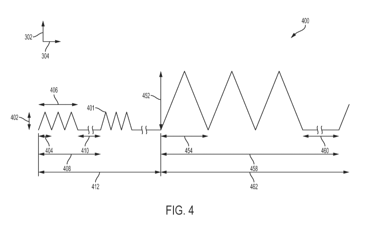
Turning now to FIG. 4, an exemplary diagram 400 of signal characteristics
according
to aspects of the present disclosure is illustrated. In particular, the
diagram 400 illustrates a
treatment regimen with two portions as touched on with respect to the previous
figures ¨ for
example, a first portion 412 at a first pulse frequency and a second portion
462 at a second
pulse frequency. In some embodiments, the first portion 412 constitutes a high
pulse
frequency portion and the second portion 462 constitutes a low pulse frequency
portion.
Further, the first portion 412 may constitute a first slew rate and the second
portion 462 may
constitute a second slew rate, for example where the first slew rate (e.g., as
a high pulse
frequency portion) may have a lower slew rate than the second slew rate (e.g.,
as a low pulse
frequency portion). Alternatively, the first slew rate (for a high pulse
frequency portion) may
have a slew rate greater than the second slew rate (for a low pulse frequency
portion), for
example where the amplitude in the first portion 412 is on the order of 5-15
mT. In further
examples, the pulse frequencies of each portion may be the same, e.g. a low
pulse frequency,
while the slew rates differ between the portions 412 and 462.
The first portion 412 includes a burst 408, similar to the burst 312 discussed
above
with respect to FIG. 3A. Within the burst 408, the signal 401 includes pulse
widths 404 for
each pulse within a train of pulses 406, similar to elements 308 and 310
respectively of FIG.
3A. Further, each burst 408 includes a latent period 410 similar to the latent
period 314
discussed above with respect to FIG. 3A. The pulses have amplitudes 402,
similar to the
exemplary amplitudes 306 and exemplary slew rates of FIG. 3A. There are
typically multiple
bursts 408 within the first portion 412 (e.g., multiple per a given a
treatment such as in a day,
as well as multiple days/weeks before the second portion 462 begins).
At some point, the treatment regimen may specify that the treatment should
transition
from the first portion 412 to the second portion 462. This may be according to
a pre-set time
frame, e.g. after some specified number of weeks (e.g., depending upon the age
of the patient
102). Alternatively, this may be in response to one or more sensors 218
providing data that
enables the tissue engineering device 104 to determine that the stage of
healing has reached a
threshold.
Regardless of how the transition is triggered to occur, in the second portion
462 is
included bursts 458. In a given burst 458, of which there will similarly be
multiple per a
given treatment day, as well as multiple days/weeks before the second portion
462 ends (e.g.,
either in response to a schedule expiring in the treatment regimen and/or data
from the
sensors reaching a second threshold). The signal 401 in the second portion 462
includes pulse
widths 454 for each pulse within a train of pulses 458, similar to elements
354 and 356
12
CA 03048654 2019-06-26
WO 2018/132678
PCT/US2018/013527
respectively in FIG. 3B. Further, each burst 458 includes a latent period 460
similar to the
latent period 360 discussed above with respect to FIG. 3B. The pulses 454 have
amplitudes
452 that may have characteristics similar to the exemplary amplitudes 352 and
exemplary
slew rates discussed above with respect to FIG. 3B.
Thus, according to the signal 401 characteristics illustrated in FIG. 4,
according to a
treatment regimen the high frequency pulse generator 208 may, for the first
portion 412 (of
time, typically over multiple treatments within a multi-week timespan)
generate the
appropriate currents to drive the coil 212 at the target high pulse frequency
when directed by
the processor 202. The processor 202 may determine when it is time to
transition to the
second portion 462, such as in response to a user input, a setting/timer
expiration/calendar
date entered previously in the treatment regimen stored in the memory 204,
and/or data from
the sensor(s) 218, to name just a few examples.
In response, the processor 202 may cause the signal 401 to transition to the
second
portion 462, in which the low frequency pulse generator 210 may take over in
generating the
appropriate currents to drive the coil 212 when directed by the processor 202
for providing a
low pulse frequency such as the examples given above. Thus, in the example of
FIG. 4,
during the first portion 412 the high pulse frequency characteristics of the
PEMF treatment
may drive tissue proliferation, after which in the second portion 462 tissue
differentiation
may be primarily driven by the low pulse frequency characteristics of the PEMF
treatment
according to the tissue's response thereto.
Turning now to FIG. 5, a flowchart illustrating an exemplary method 500 for
tissue
treatment according to aspects of the present disclosure. In particular, the
method 500
illustrates aspects of operation of the tissue engineering device 104
according to
embodiments of the present disclosure with respect to combined high/low pulse
frequency
PEMF treatment. It is understood that additional steps can be provided before,
during, and
after the steps of method 500, and that some of the steps described can be
replaced or
eliminated from the method 500.
At block 502, the tissue engineering device 104 powers up, whether from a
sleep
mode or from an off state, such as triggered by a user such as the patient 102
or an internal
timer (e.g., set according to the prescribed treatment regimen in effect at
the tissue
engineering device 104).
At decision block 504, the tissue engineering device 104 determines whether it
has
received an input. The input may be a command entered at that time by the user
of the tissue
engineering device 104, e.g. via the interface 110. Alternatively, the input
may be a
13
CA 03048654 2019-06-26
WO 2018/132678
PCT/US2018/013527
transmission received via the antenna 216/transceiver 214. As yet another
alternative, the
input may be a previously-stored, scheduled instruction regarding the
treatment regimen. As
another alternative, the input may be sensor input data, such as from an
impedance monitor,
or timer/counter data from a timer or counter (e.g., as discussed above with
respect to
sensor(s) 218).
If it is determined at decision block 504 that no input has been received yet,
then the
method 500 proceeds to block 506, where the tissue engineering device 104
waits for an
input. The method 500 returns in a loop to decision block 504 until an input
is detected.
If it is determined at decision block 504 that an input has been received,
then the
method 500 proceeds to block 508.
At block 508, the tissue engineering device 104 accesses one or more treatment
parameters based on the input detected. For example, the tissue engineering
device 104 may
access pulse frequency parameter, burst duration, burst frequency, number of
pulses,
amplitude of the pulses, rise time/slew rate of the pulses, shape of the
pulses, and/or any other
parameter or some combination thereof. These parameters may have been either
previously
stored as part of the treatment regimen in the memory 204, and/or updated via
user input via
the interface 110 and/or received via the transceiver 214 from some other,
remote source.
At decision block 510, the tissue engineering device 104 determines the pulse
frequency level of the parameters accessed for the current portion of the
treatment regimen
(e.g., determining whether the treatment regimen is now in the high or low
pulse frequency
portions of the treatment). According to embodiments of the present
disclosure, the high
pulse frequency treatment portion may occur first. Thus, if the schedule
currently identifies
treatment to be according to the first portion, that corresponds to a first
pulse frequency that
is a high pulse frequency (e.g., on the order of tens of kilohertz).
If high pulse frequency, the "first frequency" in FIG. 5, then the method 500
proceeds
to block 512.
At block 512, the processor 202 of the tissue engineering device 104 generates
the
command for the high frequency pulse generator 208 to generate high pulse
frequency pulses
according to the first portion (e.g., 412 of FIG. 4) of the treatment regimen.
At block 514, the tissue engineering device 104's high frequency pulse
generator 208
generates the PEMF (the pulses) according to the command(s) received from the
processor
202 from block 512.
At block 516, the tissue engineering device 104 maintains the treatment using
the
first, high pulse frequency for the specified period of time. Thus, for a
given periodic
14
CA 03048654 2019-06-26
WO 2018/132678
PCT/US2018/013527
application of treatment according to the treatment regimen (on a given day),
the tissue
engineering device 104 may maintain the treatment using the first, high pulse
frequency for
the duration of the period.
At decision block 518, the tissue engineering device 104 determines whether
the time
to transition to the second period of the treatment regimen has arrived. This
determination
may be made based on any of a plurality of factors (or some combination
thereof). For
example, the determination may be based on a schedule ¨ e.g., a timer tracking
use of the
tissue engineering device 104 over time in comparison to a beginning of use
for the current
portion of the treatment regimen. As another example, the determination may be
based on a
count ¨ e.g., a counter tracking each energization of the coil 212 over time
at the given pulse
frequency level (within an acceptable variance of that level, for example) and
that count
being compared to a specified number of times. As another example, the
determination may
be based on sensor data ¨ e.g., receiving impedance monitor data and comparing
that
impedance monitor data to a specified threshold level (or levels) stored in a
memory (or
transmitted to an external, remote system for comparison to levels) to aid in
a determination
whether healing of the target tissue has reached a target level to transition
to the other portion
of the treatment regimen.
If, at decision block 518, it is determined that the time has not arrived, and
the current
application is complete (e.g., the duration for the given periodic treatment
has been reached),
then the method 500 returns to block 506 to wait for the next input, which
could be from a
user, a schedule in the device, a transmission, etc.
If, instead, it is determined at decision block 518 that the time to
transition has
arrived, then the method 500 proceeds to block 520. In some embodiments, the
pulse
frequency level selected at the start of the periodic application remains the
same at the
conclusion of the periodic application. In other embodiments, if the scheduled
period of time
expires for using the first, high pulse frequency or one or more sensors 218
assist in
identifying a threshold as being met, then the treatment may transition to the
second, low
pulse frequency in the course of the current periodic application.
At block 520, the processor 202 of the tissue engineering device 104 generates
the
command for the low frequency pulse generator 210 to generate low pulse
frequency pulses
according to the second portion (e.g., 462 of FIG. 4) of the treatment
regimen.
At block 522, the tissue engineering device 104's low frequency pulse
generator 210
generates the PEMF (the pulses) according to the command(s) received from the
processor
202 from block 520.
CA 03048654 2019-06-26
WO 2018/132678
PCT/US2018/013527
At block 524, the tissue engineering device 104 maintains the treatment using
the
second, low pulse frequency (a pulse frequency lower than the first, higher
pulse frequency)
for the specified period of time. Thus, for a given periodic application of
treatment according
to the treatment regimen (on a given day), the tissue engineering device 104
may maintain the
treatment using the second, low pulse frequency for the duration of the
period.
Returning to decision block 510, if it is determined that the pulse frequency
level is
the low, second pulse frequency level, then the method 500 proceeds to block
520 and
proceeds as discussed above.
From block 524, the method 500 may return to block 506 and waiting for the
next
input as discussed above. Thus, in the method 500 of FIG. 5, tissue
proliferation may be first
driven by a high pulse frequency PEMF configuration, after which tissue
differentiation may
be primarily driven by the low pulse frequency PEMF configuration to achieve a
more
effective treatment of an indication for osteogenesis, tendenogenesis,
ligamatogenesis, and/or
chondrogenesis as some examples.
FIG. 6 is a diagram 600 illustrating an exemplary application of a tissue
engineering
device 602 according to aspects of the present disclosure. The tissue
engineering device 602
is an example of a tissue engineering device 104 configured for use in
tendenogenesis, or
more generally tendon tissue repair (e.g., including insertion point and
midsubstance repairs),
in particular for assisting with repairing rotator cuff injuries.
The shoulder 632 of a patient 604 is illustrated in a stylized manner in FIG.
6. As
shown, a patient 604's shoulder 632 includes the arm 634, humerus 630, rotator
cuff 624,
rotator cuff tear 622, subscapularis tendon 628, clavicle 620, and tendon 626.
The tissue
engineering device 602 includes the main housing 606 and strap 608. The main
housing 606
includes a bottom 612 in contact with the shoulder 632 of the patient 604
(e.g., the bottom
612 of the main housing 606 may be anatomically figured to generally conform
with the
shoulder 632) as well as a top 614 on which side an interface 610 is located.
Coil(s) 212
(FIG. 2) may be configured external to the main housing 606 to generate PEMFs
that reach
the rotator cuff 624 and, particularly, the area of the rotator cuff tear 622.
Alternatively, the
coil(s) 212 (FIG. 2) may be configured within the main housing 606.
In the embodiment illustrated in FIG. 6, the treatment regimen programmed into
the
tissue engineering device 602 may be configured to provide a one of either low
pulse
frequency (e.g., on the order of several kilohertz) or high pulse frequency
(e.g., on the order
of tens of kilohertz). Alternatively, the treatment regimen may in this
embodiment also
include both high pulse frequency and low pulse frequency components over
time. If the
16
CA 03048654 2019-06-26
WO 2018/132678
PCT/US2018/013527
tissue engineering device 602 is configured according to a low pulse frequency
profile (e.g.,
FIG. 3B with an amplitude of approximately 1 mT or 10 mT with lower slew rate
as some
examples), then the tissue treatment regimen may specify a unique treatment
duration for
each periodic application of the therapeutic signals. For example, the
duration may be limited
to a duration or time on the order of 30 to 120 minutes, for example 60 to 90
minutes, per
periodic treatment according to some embodiments for tendenogenesis (rotator
cuff repair).
In contrast, prior approaches typically are on the order of 3 or more hours
and are for a
different indication, such as osteogenesis (bone growth stimulation).
If the tissue engineering device 602 is configured to according to a high
pulse
frequency profile (e.g., FIG. 3A), then the tissue treatment regimen may
specify a unique
treatment duration for each periodic application of the therapeutic signals at
high pulse
frequency. For example, the duration may be on the order of 6 hours per
periodic treatment
according to some embodiments for the tendenogenesis that utilize the higher
pulse frequency
at approximately 35 kilohertz to 50 kilohertz, such as around 40 kilohertz.
Another application of embodiments of the present disclosure may be to other
tendons
such as the Achilles tendon. This is illustrated in FIG. 7, which introduces a
diagram 700
according to aspects of the present disclosure.
As shown, the foot of a patient 702 is in a boot 730. The boot 730 includes a
base 732,
a top 724, a front 734 towards where the toes 725 of the patient 702 face, a
rear 736 located
in a vicinity to the heel 720 of the patient 702, and (attachable to the rear
736) the tissue
engineering device 704 (an example of the tissue engineering device 104 of
FIGs. 1 and 2).
The tissue engineering device 704 may be integrally formed with one or more
parts of the
boot 730 or releasably connected thereto. Further, if it is a releasable
connection, then in
some embodiments the location of the releasable connection may be adjusted,
such as up or
down with reference with the bottom 732 of the boot 730, so as to better
locate the fields over
the target treatment area (e.g., where the tear is located on the Achilles'
tendon 722).
The tissue engineering device 704 is an example of a tissue engineering device
104
configured for use in tendenogenesis, in particular for assisting with
Achilles tendon 722
injuries. The tissue engineering device 704 includes the main housing 706. The
main housing
706 includes a bottom 712 and a top 714 on which side an interface 710 is
located. Coil(s)
212 (FIG. 2) may be configured within the main housing 706 to generate PEMFs
that reach
the Achilles tendon 722, and particularly the area of the injury to the
Achilles tendon (e.g.,
generally at a level approximate to the ankle 726 of the patient 702).
17
CA 03048654 2019-06-26
WO 2018/132678
PCT/US2018/013527
In the embodiment illustrated in FIG. 7, the treatment regimen programmed into
the
tissue engineering device 704 may also be configured to provide one of either
low pulse
frequency, high pulse frequency, or both high and low components over time
such as
discussed above with respect to FIGs. 3A, 3B, and/or 4, such as discussed with
respect to
FIG. 6.
FIG. 8 is a flowchart illustrating an exemplary method 800 for tissue
treatment
according to aspects of the present disclosure. In particular, the method 800
illustrates aspects
of operation of the tissue engineering device 602 of FIG. 6 (or the tissue
engineering device
704 of FIG. 7) according to embodiments of the present disclosure with respect
to combined
high/low pulse frequency PEMF treatment. For simplicity of discussion,
reference will be
made to tissue engineering device 602 with respect to method 800. The tissue
engineering
devices discussed herein could additionally be used with prescribed treatment
regimens with
other tendons, ligaments, cartilage, etc. It is understood that additional
steps can be provided
before, during, and after the steps of method 800, and that some of the steps
described can be
replaced or eliminated from the method 800.
At block 802, the tissue engineering device 602 powers up, whether from a
sleep
mode or from an off state, such as triggered by a user such as the patient 102
or an internal
timer (e.g., set according to the prescribed treatment regimen in effect at
the tissue
engineering device 104).
At block 804, the tissue engineering device 602 is placed on the patient in a
relevant
location, for example on the shoulder 632 directed toward the rotator cuff 624
and,
specifically, the rotator cuff tear 622. As another example, the tissue
engineering device 704
is attached to the boot 730 facing the location of the Achilles tendon 722.
At block 806, the tissue engineering device 602 accesses treatment
information. This
treatment information includes the parameters such as those discussed above ¨
including such
parameters as a pulse frequency parameter, a burst duration, a burst
frequency, a number of
pulses, an amplitude of the pulses, a rise time/slew rate of the pulses, a
shape of the pulses,
and/or any other parameter or some combination thereof. These parameters may
have been
either previously stored as part of the treatment regimen in the memory 204
(FIG. 2), and/or
updated via user input via the interface 610 and/or received via the
transceiver 214 (FIG. 2)
from some other, remote source.
At decision block 808, the tissue engineering device 602 determines whether
the
treatment regimen specifies a high or low pulse frequency treatment. In some
embodiments,
the pulse frequency level of treatment may be specified as a selection at the
tissue
18
CA 03048654 2019-06-26
WO 2018/132678
PCT/US2018/013527
engineering device 602 between one of multiple treatment regimens stored at
the tissue
engineering device 602 (and/or accessible by the tissue engineering device 602
via wired or
wireless connection(s)). That selection may be based on some input. For
example, the input
may be from a timer tracking use of the tissue engineering device 602 over
time in
comparison to a beginning of use for a treatment regimen. As another example,
the input may
be a count ¨ e.g., a counter tracking each energization of the tissue
engineering device 602
over time at the given pulse frequency level (within an acceptable variance of
that level, for
example) and that count being compared to a specified number of times. As
another example,
the input may be sensor data ¨ e.g., impedance monitor data that is compared
to a specified
threshold level (or levels) stored in a memory (or transmitted to an external,
remote system
for comparison to levels) to aid in a determination whether healing of the
target tissue has
reached a target level to transition to a different specified pulse frequency
level identified in
the same treatment regimen or a different treatment regimen.
If the treatment regimen specifies low pulse frequency, then the method 800
proceeds
to block 810.
At block 810, the processor 202 (FIG. 2) of the tissue engineering device 602
generates the command for the low frequency pulse generator 210 (FIG. 2) to
generate low
pulse frequency pulses, such as in the example given in FIG. 3B and discussed
above.
At block 812, the tissue engineering device 602's low frequency pulse
generator 210
(FIG. 2) generates the PEMF (the pulses) according to the command(s) received
from the
processor 202 from block 810.
Returning to decision block 808, if the treatment regimen specifies high pulse
frequency, then the method 800 instead proceeds to block 814.
At block 814, the processor 202 (FIG. 2) of the tissue engineering device 602
generates the command for the high frequency pulse generator 208 (FIG. 2) to
generate high
pulse frequency pulses, such as in the example given in FIG. 3A and discussed
above.
At block 816 the tissue engineering device 602's high frequency pulse
generator 208
(FIG. 2) generates the PEMF (the pulses) according to the command(s) received
from the
processor 202 from block 814.
From either block 810 or block 816, the method 800 proceeds to block 818.
At block 818, the tissue engineering device 602 tracks the time in use that
PEMFs are
generated for treatment of the indication (e.g., rotator cuff repair). This
may be performed by
the processor 202 (FIG. 2) with a timer function, or by polling a hardware
timer separate
from the processor 202 to name just a few examples.
19
CA 03048654 2019-06-26
WO 2018/132678
PCT/US2018/013527
At block 820, the tissue engineering device 602 compares the tracked time from
block
818 against a total application time for the current periodic application
(e.g., approximately
60 to 90 minutes for low pulse frequency PEMF signals and approximately 6
hours for high
pulse frequency PEMF signals), such as may be stored as part of the treatment
regimen in
memory 204 (FIG. 2).
At decision block 822, if the tracked time from block 818 has not reached the
time
frame specified in the treatment regimen, then the method 800 returns to block
818 to
continue tracking.
If the tracked time from block 818 has reached the time frame specified in the
treatment regimen, then the method 800 proceeds to block 824.
At block 824, the tissue engineering device 602 deactivates the PEMF for the
current
periodic application (e.g., automatically; alternatively, a notification may
be signaled to the
user via the interface 610 which may include textual, audible, video, etc.
information to the
user). Deactivation may be of the PEMF signals only, or of powering down the
entire tissue
engineering device 602.
In some embodiments, the computing system is programmable and is programmed to
execute processes including the processes of methods 500 and/or 700 discussed
herein.
Accordingly, it is understood that any operation of the computing system
according to the
aspects of the present disclosure may be implemented by the computing system
using
corresponding instructions stored on or in a non-transitory computer readable
medium
accessible by the processing system. For the purposes of this description, a
tangible
computer-usable or computer-readable medium can be any apparatus that can
store the
program for use by or in connection with the instruction execution system,
apparatus, or
device. The medium may include for example non-volatile memory including
magnetic
storage, solid-state storage, optical storage, cache memory, and Random Access
Memory
(RAM).
As a result of implementing the above-described approach, embodiments of the
present disclosure improve the field of pulsed electromagnetic field therapy
for tissue
engineering, such as for tissue differentiation and/or tissue proliferation.
In particular,
embodiments of the present disclosure improve the efficacy of PEMF treatment
for different
indications beyond merely bone growth stimulation, and further that the PEMF
treatment
may be achieved via a combination of high pulse frequency PEMF (for
proliferation) and low
pulse frequency PEMF (for differentiation) in a manner that better promotes
healing in a
patient.
CA 03048654 2019-06-26
WO 2018/132678
PCT/US2018/013527
The foregoing outlines features of several embodiments so that those skilled
in the art
may better understand the aspects of the present disclosure. Those skilled in
the art should
appreciate that they may readily use the present disclosure as a basis for
designing or
modifying other processes and structures for carrying out the same purposes
and/or achieving
the same advantages of the embodiments introduced herein. Those skilled in the
art should
also realize that such equivalent constructions do not depart from the spirit
and scope of the
present disclosure, and that they may make various changes, substitutions, and
alterations
herein without departing from the spirit and scope of the present disclosure.
21