Note: Descriptions are shown in the official language in which they were submitted.
CA 03049099 2019-07-02
- 1 -
Description
Title of Invention:
METHOD FOR PRODUCING MYOCARDIAL STEM CELL FOR USE IN
TREATMENT AND/OR PREVENTION OF CARDIAC ARREST
Technical Field
[0001]
The present invention relates to a method for
producing a novel myocardial stem cell for use in
treatment and/or prevention of cardiac failure, a cell
population including myocardial stem cells having
activated mitochondria, a cell preparation for use in
treatment and/or prevention of cardiac failure containing
the myocardial stem cell or the cell population, and a
liposome for use in producing the myocardial stem cell.
Background Art
[0002]
The cardiac failure refers to a symptom in which a
heart-related disease such as myocardial infarction,
cardiomyopathy or angina pectoris, or a disease other
than a heart-related disease, such as hypertension,
kidney disease or a side effect of chemotherapy against a
malignant tumor causes deterioration of the cardiac
function, so that a necessary amount of blood cannot be
supplied to the lung or throughout the body. The cardiac
CA 03049099 2019-07-02
- 2 -
failure is a second main cause of death behind cancer in
Japan, and fundamental methods for treatment of cardiac
failure include heart transplantation. However, the
heart transplantation has various disadvantages such as a
chronic shortage of transplant donors, limits of service
life of transplanted organs, rejection, oral
administration of immunosuppressants throughout life, and
frequent hospitalization for catheter tests.
[0003]
Studies have been conducted on cell transplantation
therapy using various cells including iPS cells since
cell transplantation was proposed as a promising method
for treatment of cardiac failure in the late 2000s. In
particular, myocardial stem cell transplantation has the
following advantages: it is immunologically safe because
it is transplantation using self-somatic cells; and it
can be carried out by a low-invasive method. The
myocardial stem cell transplantation has been shown to
have a certain effect in clinical trials (e.g. Non Patent
Literatures 1, 2 and 3).
[0004]
The myocardial stem cell transplantation has, for
example, the following disadvantages: the transplanted
cell engraftment effect is limited in myocardial stem
cell transplantation experiments with a pig ischemic
reperfusion model (Non Patent Literature 4); and the
viability improvement effect is limited in myocardial
CA 03049099 2019-07-02
- 3 -
stem cell transplantation experiments with a rat
doxorubicin cardiomyopathy model (Non Patent Literature
5). Thus, maintenance of the therapeutic effect for a
prolonged period is one of forthcoming challenges in
myocardial stem cell transplantation.
Citation List
Non Patent Literature
[0005]
Non Patent Literature 1: Bolli, R. et al., Lancet 2011,
378, pp. 1847-1857
Non Patent Literature 2: Makkar, R. R. et al., Lancet
2012, 379, pp. 895-904
Non Patent Literature 3: Ishigami, S. et al., Circulation
research 2015, 116, pp. 653-664
Non Patent Literature 4: Takehara, N. et al., J. Am. Coll.
Cardiol. 2008, 52, pp. 1858-65
Non Patent Literature 5: De Angelis, A. et al.,
Circulation 2010, 121, pp. 276-292
Disclosure of Invention
Technical Problem
[0006]
An object of the present invention is to provide a
novel myocardial stem cell for use in transplantation
which enables maintenance of a therapeutic effect on
and/or preventive effect against cardiac failure for a
CA 03049099 2019-07-02
- 4 -
prolonged period, a method for producing the myocardial
stem cell, and a cell preparation containing the
myocardial stem cell.
Solution to Problem
[0007]
The present inventors have found that by delivering
a mitochondria activating agent to mitochondria of a
myocardial stem cell, a cell for use in transplantation
can be produced which has an enhanced engraftment effect,
and maintains a therapeutic effect for a prolonged period,
leading to completion of the following inventions.
[0008]
(1) A method for producing a myocardial stem cell
for use in treatment and/or prevention of cardiac failure,
the method comprising the step of introducing a complex
of a mitochondria-targeting carrier and a mitochondria
activating agent into a myocardial stem cell.
(2) The method according to (1), wherein the complex
is a mitochondria-targeting liposome encapsulating the
mitochondria activating agent.
(3) The method according to (1) or (2), wherein the
complex is a mitochondria-targeting liposome containing
dioleylphosphatidylethanolamine and
phosphatidic acid and/or sphingomyelin
as constituent lipids of a lipid membrane, and having a
mitochondria-targeting molecule on a surface of the lipid
CA 03049099 2019-07-02
- 5 -
membrane, and encapsulating the mitochondria activating
agent.
(4) The method according to (3), wherein the
mitochondria-targeting molecule is a peptide consisting
of an amino acid sequence set forth in SEQ ID NO: 1.
(5) The method according to any one of (1) to (4),
wherein the mitochondria activating agent is resveratrol.
(6) A myocardial stem cell produced by introducing a
complex of a mitochondria-targeting carrier and a
mitochondria activating agent into a myocardial stem cell.
(7) The myocardial stem cell according to (6),
wherein the complex is a mitochondria-targeting liposome
encapsulating the mitochondria activating agent.
(8) The myocardial stem cell according to (6) or (7),
wherein the complex is a mitochondria-targeting liposome
containing
dioleylphosphatidylethanolamine and
phosphatidic acid and/or sphingomyelin
as constituent lipids of a lipid membrane, and having a
mitochondria-targeting molecule on a surface of the lipid
membrane, and encapsulating the mitochondria activating
agent.
(9) The myocardial stem cell according to (8),
wherein the mitochondria-targeting molecule is a peptide
consisting of an amino acid sequence set forth in SEQ ID
NO: 1.
CA 03049099 2019-07-02
- 6 -
(10) The myocardial stem cell according to any one
of (6) to (9), wherein the mitochondria activating agent
is resveratrol.
(11) A cell population comprising myocardial stem
cells, wherein an average value of ratios of fluorescence
intensity of JC-1 dimer to fluorescence intensity of JC-1
monomer when the cell population is stained with
fluorescent dye JC-1 is 1 to 4.
(12) A cell preparation for use in treatment and/or
prevention of cardiac failure, the cell preparation
comprising the myocardial stem cell or cell population
according to any one of (6) to (11).
(13) A liposome for use in introduction of an
encapsulated substance into mitochondria of a myocardial
stem cell, the liposome containing
dioleylphosphatidylethanolamine and
phosphatidic acid and/or sphingomyelin
as constituent lipids of a lipid membrane, and having a
mitochondria-targeting molecule on a surface of the lipid
membrane.
(14) The liposome according to (13), wherein the
mitochondria-targeting molecule is a peptide consisting
of an amino acid sequence set forth in SEQ ID NO: 1.
Effects of Invention
[0009]
CA 03049099 2019-07-02
- 7 -
According to the present invention, there can be
provided a myocardial stem cell which is capable of
maintaining a therapeutic effect and/or preventive effect
by cell transplantation for a prolonged period. The
myocardial stem cell can be used for treatment and/or
prevention of myocardial injury, recovery, protection or
suppression of deterioration of the cardiac function,
treatment and/or prevention of cardiac failure, or the
like.
Brief Description of Drawings
[0010]
[Figure 1] Figure 1 is a histogram of flow cytometry
showing a myocardial stem cell containing a mitochondria-
targeting liposome encapsulating resveratrol and
fluorescently labeled with NBD, where the abscissa
indicates a fluorescence level of NBD, and the ordinate
indicates the number of cells.
[Figure 2] Figure 2 shows photographs of a myocardial
stem cell observed with a confocal laser scanning
microscope (CLSM), the myocardial stem cell containing a
mitochondria-targeting liposome encapsulating resveratrol
and fluorescently labeled with NBD. Photograph B shows
RES-MITO-Porter stained with NBD (green), photograph C
shows mitochondria stained with MTDR (red), photograph D
shows a cell nucleus stained Hoechst 33342 (blue),
photograph A shows photographs B to D superimposed on one
CA 03049099 2019-07-02
- 8 -
another, and the scale bar for each photograph represents
a length of 20 m.
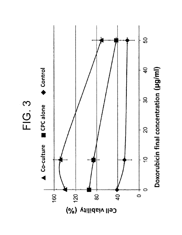
[Figure 3] Figure 3 is a graph showing a cell viability
under cell injury caused by doxorubicin (final
concentration: 10 g/mL, or 50 g/mL) in co-culture of a
myocardial blast cell and a myocardial stem cell
containing a mitochondria-targeting liposome
encapsulating resveratrol (MA-Cell) or an untreated
myocardial stem cell (CPC), where "co-culture" indicates
co-culture of a myocardial blast cell and MA-Cell, "CPC
alone" indicates coculture of a myocardial blast cell and
CPC, and "control" indicates monoculture of a myocardial
blast cell.
[Figure 4] Figure 4 shows a cell viability under cell
injury caused by doxorubicin (final concentration: 10
g/mL, 30 g/mL, or 50 g/mL) in co-culture of a
myocardial blast cell with a myocardial stem cell
containing a mitochondria-targeting liposome
encapsulating resveratrol (MA-Cell (+RES-MITO-Porter)), a
myocardial stem cell containing empty MITO-Porter (CPC
(+MITO-Porter)), a myocardial stem cell treated directly
with resveratrol (CPC (+RES)), or CPC.
[Figure 5] Figure 5 shows a cell viability after elapse
of 48 hours under cell injury caused by doxorubicin
(final concentration: 10 g/mL) in co-culture of a
myocardial blast cell with a myocardial stem cell
containing a mitochondria-targeting liposome
CA 03049099 2019-07-02
- 9 -
encapsulating resveratrol (CPC + RES-MITO-Porter), a
myocardial stem cell treated directly with resveratrol
(CPC (+RES)), or CPC.
[Figure 61 Figure 6 is a graph showing a cell viability
under cell injury caused by doxorubicin (final
concentration: 10 g/mL) at each dose of resveratrol in
coculture of a myocardial blast cell and MA-Cell.
[Figure 7] Figure 7 is a graph showing a Kaplan-Meier
curve for MA-Cell- or CPC-transplanted or untreated
doxorubicin cardiac failure model mice and healthy mice.
[Figure 8] Figure 8 is a graph showing a change in
average body weight of MA-Cell- or CPC-transplanted or
untreated doxorubicin cardiac failure model mice and
healthy mice.
[Figure 9] Figure 9 is a graph showing a dihydroethidium
(DHE) positive cell ratio in the cardiac tissues of MA-
Cell- or CPC-transplanted or untreated doxorubicin
cardiac failure model mice and healthy mice.
[Figure 10] Figure 10 is a graph showing an apoptosis
inductivity in the cardiac tissues of MA-Cell- or CPC-
transplanted or untreated doxorubicin cardiac failure
model mice and healthy mice.
[Figure 11] Figure 11 is a graph showing a left ventricle
shortening fraction for MA-Cell-transplanted doxorubicin
cardiac failure model mice and healthy mice.
[Figure 12] Figure 12 is a graph showing the relative
expression levels of the genes: PGCla, ESRRa, SDHA, Coxl
CA 03049099 2019-07-02
- 10 -
and ATPla in the cardiac tissues of MA-Cell- or CPC-
transplanted or untreated doxorubicin cardiac failure
model mice and healthy mice.
[Figure 13] Figure 13 is a graph showing a mitochondrial
respiratory chain complex formation ratio in the cardiac
tissues of MA-Cell- or CPC-transplanted or untreated
doxorubicin cardiac failure model mice and healthy mice.
[Figure 14] Figure 14 shows photographs showing MA-Cell
being engrafted in the mouse heart after transplantation,
where the lower left photograph shows myocardial actinin
stained with Alexa Flour 488 (green), the upper right
photograph shows a cell nucleus stained with Hoechst
33342 (blue), the lower right photograph shows MA-Cell
stained with CellVue Claret (red), and the upper left
photograph shows these photographs superimposed on one
another.
[Figure 15] Figure 15 shows photographs showing the
results of detecting the mitochondrial membrane
potentials of MA-Cell and CPC using fluorescent dye JC-1,
where the left photographs show CPC, the central
photographs show MA-Cell, the right photographs show CPC
to which FCCP has been added, the middle photographs show
green fluorescence with a wavelength of 529 nm which
corresponds to JC-1 monomer indicating depolarized
mitochondria, the lower photographs show red fluorescence
with a wavelength of 590 nm which corresponds to JC-1
dimer indicating polarized mitochondria, and the upper
CA 03049099 2019-07-02
- 11 -
photographs show the middle and lower photographs
superimposed on one another.
[Figure 16] Figure 16 is a graph showing the results of
calculating a ratio of fluorescence intensity between JC-
1 monomer (green) indicating depolarized mitochondria and
JC-1 dimer (red) indicating polarized mitochondria
(Dimer/Monomer), on the basis of the image used to detect
the mitochondrial membrane potentials of MA-Cell and CPC
using fluorescent dye JC-1, where "0" indicates a value
for each cell, and "-" indicates an average value (n =-
19).
Description of Embodiments
[0011]
A first aspect of the present invention relates to a
method for producing a myocardial stem cell for use in
treatment and/or prevention of cardiac failure, the
method comprising the step of introducing a complex of a
mitochondria-targeting carrier and a mitochondria
activating agent into a myocardial stem cell.
[0012]
The myocardial stem cell (also referred to as a
cardiac progenitor cell, which is hereinafter referred to
as CPC) is a stem cell having a self-replication ability
and a differentiation ability to the cardiac muscle, the
vascular endothelium, the vascular smooth muscle, the fat,
the bone, the cartilage or the like. CPC to be used in
CA 03049099 2019-07-02
- 12 -
the present invention can be separated from a cardiac
tissue by a method known to those skilled in the art.
One example of the CPC is a cell separated from a cardiac
tissue by, for example, Oh et al.'s method (PNAS., 2003,
100, pp. 12313-12318), or Ishigami et al.'s method (Circ
Res., 2015, 116, pp. 653-664). CPC differentiated and
induced from iPS cells (Funakoshi S. et al., Scientific
Reports 6, 2016, 19111) and CPC obtained by reprograming
fibroblasts or myocardial cells (Ieda M. et al., Cell,
2010, 142, pp. 375-386) can also be used in the present
invention. The documents are hereby incorporated by
reference.
[0013]
The CPC may be one derived from any animal species,
but it is preferable to use human CPC when the purpose is
to treat or prevent human cardiac failure. In the
present invention, it is especially preferable to use CPC
separated with the intension of transplantation using
self-somatic cells from cardiac tissues of a person
suffering from cardiac failure or having a risk of
cardiac failure.
[0014]
The CPC may be one isolated on the basis of
expression of a CPC specific marker such as Sca-1, and
purified, or may be one contained in a cell population,
e.g. a heterogeneous cell population obtained by spheroid
culture of cells separated from the heart. In the latter
CA 03049099 2019-07-02
- 13 -
case, the whole cell population is subjected to the step
of introducing a complex of a mitochondria-targeting
carrier and a mitochondria activating agent as described
later, whereby a cell population including CPC having
activated mitochondria is produced.
[0015]
For securing the number of cells necessary for
subsequent cell transplantation, the CPC may be used
after being grown by performing subculture in vitro as
long as the stemness thereof is maintained.
[0016]
The present invention includes the step of
introducing a complex of a mitochondria-targeting carrier
and a mitochondria activating agent into CPC.
[0017]
The mitochondria-targeting carrier is one having a
function to selectively reach mitochondria as one of
intracellular organelles when the carrier is introduced
into a cell. Examples of the mitochondria-targeting
carrier may include liposoluble cation substances such as
Lipophilic triphenylphosphonium cation (TPP) and
Rhodamine 123; polypeptides such as Mitochondrial
Targeting Sequence (MTS) peptide (Kong, BW. et al.,
Biochimica et Biophysica Acta 2003, 1625, pp. 98-108) and
S2 peptide (Szeto, H. H. et al., Pharm. Res. 2011, 28, pp.
2669-2679); and mitochondria-targeting liposomes such as
DQAsome (Weissig, V. et al., J. Control. Release 2001, 75,
CA 03049099 2019-07-02
- 14 -
pp. 401-408), MITO-Porter (Yamada, Y. et al., Biochim
Biophys Acta. 2008, 1778, pp. 423-432), DF-MITO-Porter
(Yamada, Y. et al., Mol. Ther. 2011, 19, pp. 1449-1456)
and modified DF-MITO-Porter modified with S2 peptide
(Kawamura, E. et al., Mitochondrion 2013, 13, pp. 610-
614). The documents are hereby incorporated by reference
regarding production and use of the carriers in the
present invention.
[0018]
A preferred mitochondria-targeting carrier in the
present invention is a mitochondria-targeting liposome,
and in particular, MITO-Porter, DF-MITO-Porter or
modified DF-MITO-Porter is preferable.
[0019]
The complex of a mitochondria-targeting carrier and
a mitochondria activating agent is a substance having a
configuration in which a mitochondria-targeting carrier
and a mitochondria activating agent behave in a unified
manner regardless of whether chemical bonding, physical
encapsulation or the like is used to form the complex.
For example, when the liposoluble cation lipid or
polypeptide is a mitochondria-targeting carrier, a
complex of a mitochondria-targeting carrier and a
mitochondria activating agent can be formed by bonding
the mitochondria-targeting carrier to the mitochondria
activating agent using a chemical method such as covalent
bonding or ionic bonding in accordance with, for example,
CA 03049099 2019-07-02
- 15 -
Murphy et al.'s method regarding a liposoluble cation
substance (G. F. Kelso et al., J. Biol. Chem., 2001, 276,
pp. 4588-4596) or a method regarding Szeto peptide as
described in J22007-503461A.
[0020]
Further, when the mitochondria-targeting carrier is
a liposome, a complex of a mitochondria-targeting carrier
and a mitochondria activating agent can be formed by
chemically bonding the mitochondria activating agent to a
surface of a lipid membrane of the liposome, or
physically encapsulating the mitochondria activating
agent in the liposome, i.e. an internal space blocked by
a lipid membrane.
[0021]
The complex can be introduced into CPC by a method
for introduction of the complex into a cell, which is
known for the mitochondria-targeting carrier. The
complex may be introduced into a cell by, for example,
culturing CPC in an appropriate medium containing the
complex, or incubating the complex and CPC in the
presence of a known substance capable of accelerating
uptake of a substance into a cell, such as lipofectamine
or polyethylene glycol.
[0022]
A preferred example of the step of introducing a
complex of a mitochondria-targeting carrier and a
mitochondria activating agent into CPC in the first
CA 03049099 2019-07-02
- 16 -
aspect of the present invention is a step of introducing
a complex into CPC by incubating CPC and a complex which
is a mitochondria-targeting liposome encapsulating a
mitochondria activating agent, particularly a complex
which is MITO-Porter or DF-MITO-Porter having the surface
modified with MTS peptide or S2 peptide and encapsulating
a mitochondria activating agent.
[0023]
The mitochondria activating agent is a substance
capable of activating a mitochondrial respiratory chain
complex (electron transport system), particularly a
substance capable of bringing mitochondria into a
polarized state in terms of a membrane potential, and in
particular, it is preferable to use a substance capable
of bringing mitochondria into a hyperpolarized state.
Examples of the mitochondria activating agent may include
antioxidants such as resveratrol (3,5,4'-trihydroxy-
trans-stilbene), coenzyme Q10, vitamin C, vitamin E, N-
acetylcysteine, TEMPO, SOD and glutathione, and in
particular, resveratrol is preferable.
[0024]
The resveratrol that is preferably used in the
present invention may be one extracted from a plant by a
known method, or one chemically synthesized by a known
method such as, for example, Andrus et al.'s method
(Tetrahedron Lett. 2003, 44, pp. 4819-4822).
[0025]
CA 03049099 2019-07-02
- 17 -
The CPC produced by the method according to the
first aspect of the present invention is one of
additional aspects of the present invention, and can
considerably improve the viability of mice receiving
doxorubicin as shown in Examples below. Further, the CPC
is suitably involved in reduction of oxidative stress,
suppression of apoptosis or maintenance of mitochondrial
functions in cardiac tissues of mice receiving
doxorubicin.
[0026]
It is clinically known that administration of
doxorubicin, a type of anthracycline-based pharmaceutical
agent, causes severe myocardial injury, and mice
receiving doxorubicin are used as cardiac failure model
mice. Therefore, the CPC produced by the method
according to the first aspect of the present invention
can be used for treatment and/or prevention of myocardial
injury, particularly severe myocardial injury, recovery,
protection or suppression of deterioration of the cardiac
function, treatment and/or prevention of cardiac failure,
or the like.
[0027]
Another aspect of the present invention relates to a
cell population including myocardial stem cells, wherein
an average value of ratios of fluorescence intensity of
JC-1 dimer to fluorescence intensity of JC-1 monomer
(fluorescence intensity of JC-1 dimer/fluorescence
CA 03049099 2019-07-02
- 18 -
intensity of JC-1 monomer) when the cell population is
stained with fluorescent dye JO-1 is 1 to 4.
[0028]
Mitochondria generate a proton concentration
gradient inside and outside the membrane under the action
of respiratory chain complexes existing in the
mitochondria, and come into a polarized state in which
there is a membrane potential. When receiving apoptosis,
metabolic stress or the like, the polarized mitochondria
are turned into a depolarized state in which the membrane
potential is reduced. In this way, the state of
polarization of mitochondria is a parameter indicating a
metabolism activity of mitochondria, and a cell having a
large number of polarized mitochondria is considered to
be a cell having activated mitochondria.
[0029]
It is known that fluorescent dye JO-1 (5,5',6,6'-
tetrachloro-1,1',3,3'-
tetraethylbenzimidazolylcarbocyanine iodide), which is a
mitochondrial membrane potential probe, is a monomer
emitting green fluorescence in depolarized mitochondria,
but forms a dimer emitting red fluorescence in polarized
mitochondria. Therefore, the ratio of fluorescence
intensity between JO-1 monomer and JO-1 dimer is an index
indicating a state of polarization of mitochondria. The
ratio of fluorescence intensity can be measured by, for
example, detecting a fluorescence ratio in accordance
CA 03049099 2019-07-02
- 19 -
with manufacturer's protocol using JC-1 commercially
available from Thermo Fisher Scientific, Cosmo Bio Co.,
Ltd. or the like.
[0030]
The cell population according to this aspect is a
cell population including CPC having activated
mitochondria, and the degree of activation of
mitochondria of CPC included in the population can be
represented by an average value of ratios of fluorescence
intensity of JC-1 dimer to fluorescence intensity of JC-1
monomer (fluorescence intensity of JC-1
dimer/fluorescence intensity of JC-1 monomer) when the
cell population is stained with JC-1.
[0031]
The average value of ratios of fluorescence
intensity can be determined by measuring a ratio of
fluorescence intensity of JC-1 dimer to fluorescence
intensity of JC-1 monomer (fluorescence intensity of JC-1
dimer/fluorescence intensity of JC-1 monomer) for each of
any number of CPCs, preferably more than 10 and less than
100 CPCs included in the cell population, and calculating
an average value of the measured ratios. The average
value of ratios of fluorescence intensity of JC-1 dimer
to fluorescence intensity of JC-1 monomer in a cell
population including CPC having activated mitochondria is
more than 1, preferably 1 to 4.
[0032]
CA 03049099 2019-07-02
- 20 -
The cell population according to this aspect is a
cell population mainly consisting of CPC, preferably a
cell population which does not substantially include
cells other than CPC. The cell population can be
produced typically by the foregoing method according to
the first aspect of the present invention.
[0033]
Both the CPC and the cell population including CPC
can be used for treatment and/or prevention of myocardial
injury, recovery, protection or suppression of
deterioration of the cardiac function, treatment and/or
prevention of cardiac failure, or the like. Therefore,
still another aspect of the present invention provides a
method for treating and/or preventing myocardial injury
or cardiac failure, the method comprising the step of
administering an effective amount of the CPC or the cell
population to a subject in need thereof. Further, still
another aspect of the present invention provides a method
for recovering the cardiac function, a method for
protecting the cardiac function or a method for
suppressing deterioration of the cardiac function, the
method comprising the step of administering an effective
amount of the CPC or the cell population to a subject in
need thereof.
[0034]
Further, still another aspect of the present
invention provides a cell preparation having the CPC or
CA 03049099 2019-07-02
- 21 -
the cell population including CPC as an active ingredient,
particularly a cell preparation for use in treatment
and/or prevention of myocardial injury or cardiac failure,
a cell preparation for use in recovery and/or protection
of the cardiac function, a cell preparation for use in
suppression of deterioration of the cardiac function, or
the like.
[0035]
The cell preparation as one aspect of the present
invention can be prepared by a method known to those
skilled in the art. For example, the cell preparation
can be prepared as a form of a suspension solution
obtained by suspending cells in water, other
pharmaceutically acceptable buffer solution or the like
as necessary. The cell preparation may contain
pharmaceutically acceptable additives such as a carrier
or medium, e.g. vegetable oil, an emulsifier, a
suspending agent, a surfactant, a stabilizer, an
excipient and a preservative.
[0036]
The cell preparation as one aspect of the present
invention contains an effective amount of the CPC or the
cell population including CPC. The term "effective
amount" as used herein means an amount of CPC necessary
for exhibiting an effect such as treatment and/or
prevention of myocardial injury, recovery, protection or
suppression of deterioration of the cardiac function, or
CA 03049099 2019-07-02
- 22 -
treatment and/or prevention of cardiac failure. The
effective amount, which depends on the condition of a
subject requiring treatment, is, for example, 1 x 10³
cells to 1 x 10⁹ cells, preferably 1 x 10⁶ cells to 1 x
109 cells, more preferably 1 x 10⁷ cells to 1 x 10⁹ cells
per individual subject, and the cell preparation may be
administered in such an amount once or two or more times
at appropriate intervals.
[0037]
The method for administering the cell preparation is
not particularly limited, and examples thereof include
administration methods that are commonly used, e.g.
intravascular administration (preferably intravenous
administration), intraperitoneal administration and local
administration. Intravenous administration or local
administration to the heart is preferable.
[0038]
Still another aspect of the present invention
provides a liposome for use in introduction of an
encapsulated substance into mitochondria of CPC, the
liposome containing dioleylphosphatidylethanolamine
(DOPE) and phosphatidic acid (PA) and/or sphingomyelin
(SM) as constituent lipids of a lipid membrane, and
having a mitochondria-targeting molecule on a surface of
the lipid membrane. A liposome in which the
mitochondria-targeting molecule is a peptide consisting
of an amino acid sequence set forth in SEQ ID NO: 1 (i.e.,
CA 03049099 2019-07-02
- 23 -
S2 peptide) can be produced by Kawamura et al.'s method
described above. The liposome according to this aspect
may further have, in addition to S2 peptide, a peptide
consisting of an amino acid sequence set forth in SEQ ID
NO: 2 (i.e., an octaarginine peptide) on a surface of a
lipid membrane, and such a liposome can be produced by a
method as described in JP5067733B. The substance to be
encapsulated is preferably the mitochondria activating
agent described in the first aspect of the present
invention.
[0039]
The present invention will be further described in
detail by way of the following Examples, but the present
invention is not limited to these Examples.
Examples
[0040]
Example 1. Preparation of CPCs with resveratrol delivered
to mitochondria
(1) Purification of mouse CPC
Mouse CPC was isolated and purified in the following
manner in accordance with Oh et al.'s method (PNAS. 2003,
100, pp. 12313-12318).
[0041]
The heart was excised from an 8-week-old c57BL6/J
male mouse, and subjected to collagenase treatment and
Percoll density gradient treatment to extract a cell
CA 03049099 2019-07-02
- 24 -
group including CPC. The obtained cell group was
subjected to primary culture, sorting was then performed
by MACS system to selectively extract Sca-1 positive CPC,
and the Sca-1 positive CPC was subjected to subculture to
isolate mouse CPC. For the isolated CPC, the amount of a
surface marker protein was determined by flow cytometry
(FACS), the gene expression levels of a myocardial
transcription factor and a structural protein were
determined by a PCR method, and the values thereof were
confirmed to agree with those reported previously (data
not shown).
[0042]
(2) Preparation of RES-MITO-Porter modified with S2
peptide
A mitochondria-targeting liposome (RES-MITO-Porter)
having the surface modified with S2 peptide and
encapsulating resveratrol was prepared in the following
manner.
[0043]
A mixed solution of 137.5 L of a 1 mM lipid ethanol
solution of 1,2-dioleyl-sn-glycero-3-
phosphatidylethanolamine (DOPE) and sphingomyelin (SM)
(DOPE/SM = 9:2) and 112.5 L of chloroform was dried
under reduced pressure to prepare a lipid membrane film.
250 L of a 10 mM HEPES buffer solution containing
resveratrol in an amount of 2.3 mg per mL was added to
the lipid membrane film to hydrate the lipid membrane
CA 03049099 2019-07-02
- 25 -
film (room temperature, 15 minutes), and ultrasonication
treatment was then performed with a bath-type sonicator
(AU-25C; Aiwa Ika Kogyo K.K.) to prepare a liposome. A
Stearyl S2 solution was added to the liposome in an
amount of 10% based on the total amount of lipid, and the
resulting mixture was incubated at room temperature for
30 minutes to prepare RES-MITO-Porter.
[0044]
It was confirmed that the prepared RES-MITO-Porter
was nanoparticles having an average particle diameter of
121 7 nm, a zeta potential of 49 1 mV and a
resveratrol encapsulation ratio of 87 4% and having a
positive charge, and maintained particle physical
properties even after storage at 4 C for 1 month.
[0045]
(3) Introduction of RES-MITO-Porter into CPC
RES-MITO-Porter was labeled with green fluorescent
dye NBD (7-nitrobenz-2-oxa-1,3-diazole) in accordance
with a previously reported method (Abe, J. et al., J.
Pharm. Sci. 2016, 105, pp. 734-740) to evaluate
introduction of RES-MITO-Porter into CPC. 200 L of
fluorescently NBD-labeled RES-MITO-Porter with a total
amount of lipid of 550 M (with a resveratrol
concentration of 100 M) was provided, and added to 10 mL
of DMEM-F12 medium containing 1 x 10⁶ CPCs, and the
resulting mixture was incubated for 1 hour to introduce
RES-MITO-Porter into CPC. Using FACS, a fluorescently
CA 03049099 2019-07-02
- 26 -
NBD-labeled carrier taken up in CPC was detected, and
thus it was confirmed that RES-MITO-Porter had been
introduced into CPC (Figure 1).
[0046]
Mitochondria of CPC containing fluorescently NBD-
labeled RES-MITO-Porter were stained with MTDR in
accordance with the previously reported method (Abe, J.
et al., J. Pharm. Sci. 2016, 105, pp. 734-740), and then
observed with a confocal laser scanning microscope (CLSM),
and resultantly, yellow dots were observed at which green
NBD and red MTDR overlapped each other. Thus, it was
confirmed that RES-MITO-Porter was integrated with
mitochondria of CPC (Figure 2).
[0047]
Example 2. Myocardial blast cell protection effect of CPC
with resveratrol delivered to mitochondria (in vitro
experiment)
(1) Introduction of RES-MITO-Porter into CPC
CPC suspended in DMEM-F12 medium was seeded in a 6-
well plate at a density of 1 x 10⁶ cells per well, and
cultured at 37 C for 24 hours. RES-MITO-Porter prepared
in section (2) in Example 1 was added to each well, and
the resulting mixture was incubated for 2 hours to
introduce RES-MITO-Porter into CPC for use in subsequent
experiments. The CPC containing RES-MITO-Porter is
referred to as MA-Cell.
[0048]
CA 03049099 2019-07-02
- 27 -
(2) Cell viability under cell injury caused by
doxorubicin
Rat myocardial blast cell, H9c2 cell (purchased from
ATCC) and MA-Cell prepared in section (1) were mixed in
DMEM-F12 medium in such a manner that the density of H9c2
cell was 3 x 10⁴ cells per well and the density of MA-
Cell was 1 x 10⁴ cells per well, and the mixed liquid was
subjected to coculture at 37 C for 24 hours. Doxorubicin
was added to the coculture liquid in a final
concentration of 10 g/mL (low dose) or 50 g/mL (high
dose) to induce cell injury, and culture was further
performed for 16 hours, followed by measuring a cell
viability using WST-1 reagent (Takara Bio Inc.). Further,
for H9c2 cell to which MA-Cell was not added but only the
medium was added (control) and H9c2 cell to which CPC was
added instead of MA-Cell (CPC alone), cell injury was
induced and a cell viability was measured in the same
manner as described above. Figure 3 shows results when
the cell viability of non-doxorubicin-treated H9c2 cell
is defined as 100%.
[0049]
MA-Cell was confirmed to suppress a decrease in cell
viability under the cell injury action of doxorubicin.
Further, when the mixing ratio between H9c2 cell and MA-
Cell in coculture was 6:1, the same result was observed
(MA-Cell in Figure 4). On the other hand, a decrease in
cell viability when instead of MA-Cell, CPC treated
CA 03049099 2019-07-02
- 28 -
directly with resveratrol was added to H9c2 cell (CPC
(+RES) in Figure 4) was comparable to a decrease in cell
viability when CPC alone was added (CPC in Figure 4), and
thus improvement by addition of resveratrol was not
observed.
[0050]
Further, the medium was changed from DMEM-F12 to
DMEM High glucose (Thermo), H9c2 cell and MA-Cell, CPC
treated directly with resveratrol, or CPC were mixed at a
mixing ratio of 6:1, and cocultured at 37 C for 24 hours,
doxorubicin was then added in a final concentration of 10
g/mL to induce cell injury, and culture was further
performed for 48 hours, followed by measuring a cell
viability. Figure 5 shows results when the cell
viability of non-doxorubicin-treated H9c2 cell is defined
as 100%. It was confirmed that suppression of a decrease
in cell viability under the cell injury action of
doxorubicin by MA-Cell was maintained even 48 hours after
induction of cell injury.
[0051j
(3) Change in cell viability depending on dose of
resveratrol
By performing the same operation as described in
section (1) above, MA-Cell was prepared while the amount
of resveratrol delivered was changed using RES-MITO-
Porter having a resveratrol concentration of 0 to 10 M.
The same experiment as described in section (2) above was
CA 03049099 2019-07-02
- 29 -
performed using each MA-Cell and doxorubicin in a final
concentration of 10 g/mL, and a cell viability was
measured at each dose of resveratrol. Figure 6 shows
results when the cell viability of non-doxorubicin-
treated H9c2 cell is defined as 100%.
[0052]
It was confirmed that a decrease in cell viability
under the cell injury action of doxorubicin was improved
depending on the dose of resveratrol.
[0053]
Example 3. Protective effect against myocardial injury by
CPC with resveratrol delivered to mitochondria (in vivo
experiment)
(1) Preparation of doxorubicin myocardial injury model
mouse and administration of each CPC
MA-Cell (I x 106 cells) was transplanted to the
heart of each of healthy mice (6 to 8-week-old male
057/BL6 mice) to provide an MA-Cell-transplanted group (n
= 6). 24 hours after the transplantation, myocardial
injury was induced by intraperitoneally administering 200
L of doxorubicin/PBS to the mouse at a level of 25 mg/kg
only once with reference to Zhang et al.'s method (Nature
Medicine 2012, 18, pp. 1639-1642). In a group of mice
which was not subjected to cell transplantation
(untreated group) and a group of mice subjected to
transplantation of CPC instead of MA-Cell (CPC-
transplanted group), myocardial injury was induced in the
CA 03049099 2019-07-02
- 30 -
same manner as described above. Further, for each of the
groups, a group of mice which did not receive doxorubicin
(healthy group) was provided.
[0054]
(2) Viability after induction of myocardial injury
A Kaplan-Meier curve for the viability of each group
after induction of myocardial injury was prepared, and
statistical processing was performed by log-rank analysis.
The results thereof are shown in Figure 7. It was
confirmed that the viability of MA-Cell-transplanted
group was significantly improved as compared to the
viabilities of the untreated group and the CPC-
transplanted group.
[0055]
(3) Average body weight
Figure 8 shows the mouse average body weights in the
groups on the third day and the seventh day after
induction of myocardial injury. The body weight
temporarily decreased in all the groups of mice receiving
doxorubicin, but it was observed that in the MA-Cell-
transplanted group the body weight tended to be recovered
on the seventh day.
[0056]
(4) Oxidative stress condition in cardiac tissue
On the third day after induction of myocardial
injury, the heart was excised, a cardiac tissue was
quickly stained with dihydroethidium (DHE) with reference
CA 03049099 2019-07-02
- 31 -
to the foregoing Zhang et al.'s method, red cells (DHE
positive cells) in the cardiac tissue were counted, and
the oxidative stress condition of the cardiac tissue was
quantitatively determined as a DHE positive cell ratio.
The results thereof are shown in Figure 9. The DHE is a
fluorescent probe which reacts with active oxygen species
in living cells to emit red fluorescence. The untreated
group and the CPC-transplanted group exhibited a DHE
positive cell ratio significantly higher than the DHE
positive cell ratio of the healthy group. On the other
hand, it was confirmed that in the MA-Cell-transplanted
group, an increase in the number of DHE positive cells
tended to be suppressed.
[0057]
(5) Apoptosis induction in cardiac tissue
The heart excised in section (4) was subjected to
tissue fixation, a TUNEL method was then carried out
using TUNNEL In Situ Cell Death Detection Kit,
Fluorescein kit (Sigma), and apoptosis positive cells in
the cardiac tissue were counted to calculate an apoptosis
inductivity. The results thereof are shown in Figure 10.
A large number of apoptosis induction cells were observed
in the untreated group and the CPC-transplanted group,
whereas it was confirmed that in the MA-Cell-transplanted
group, apoptosis induction was suppressed to a level
comparable to that in the healthy group.
[0058]
CA 03049099 2019-07-02
- 32 -
(6) Cardiac function (left ventricle shortening fraction)
The cardiac function (left ventricle shortening
fraction) of each of the mice in the healthy group and
the MA-Cell-transplanted group 5 weeks after induction of
myocardial injury was measured by carrying out cross-
sectional echocardiography. The results thereof are
shown in Figure 11. There was no significant difference
in left ventricle reduction ratio between the MA-Cell-
transplanted group and the healthy group.
[0059]
(7) Mitochondrial function
Total RNA was extracted from the cardiac tissue
excised in section (4), purified, and subjected to
reverse transcription reaction, and a real time PCR
method with GAPDH set as an internal standard was then
carried out to determine the expression levels of the
genes: PGCla and ESRRa related to mitochondrial
neogenesis, and SDHA, Coxl and ATPla related to the
mitochondrial respiratory chain complex. Figure 12 shows
relative expression levels in the groups when the
expression level of each gene in the healthy group is
defined as 1. It was confirmed that the expression level
of each gene significantly decreased in the untreated
group and the CPC-transplanted group, whereas in the MA-
Cell-transplanted group, a decrease in expression level
was suppressed. These results showed that in the cardiac
tissues of the MA-Cell-transplanted group, expression of
CA 03049099 2019-07-02
- 33 -
mitochondrial neogenesis and mitochondria oxidative
phosphorylation gene groups was significantly maintained
even after induction of myocardial injury.
[0060]
(8) Structure preservation of mitochondrial respiratory
chain complex
A protein was extracted from the cardiac tissue
excised in section (4), and Blue-Native PAGE was
performed. After the electrophoresis, Western-blotting
using a mitochondrial Complex I antibody (Abcam) and a
mitochondrial Complex II antibody (Abcam) was carried out
to quantitatively determine a band in the electron
transport system Super-complex band (1,000 kDa). Figure
13 shows Super-complex formation ratios calculated by
performing correction with the band quantitative value
from the Complex II antibody. It was confirmed that in
the CPC-transplanted group and the MA-Cell-transplanted
group, the mitochondrial respiratory chain complex
structure was preserved as compared to the untreated
group. It was confirmed that particularly in the MA-
Cell-transplanted group, the higher-order structure of
the mitochondrial respiratory complex was preserved at a
level comparable to that in the healthy group.
[0061]
Example 4. Engraftment of MA-Cell in myocardial tissue
MA-Cell and CPC stained red at the cell membrane
surface were prepared using CellVue Claret Far Red
CA 03049099 2019-07-02
- 34 -
(Sigma) in accordance with manufacturer's protocol.
Using these cells, cell transplantation and induction of
myocardial injury were performed in the same manner as in
Example 3, and the heart was excised on the seventh day
after induction of injury. The excised heart was stained
green at the cardiac muscle using a myocardial actinin
antibody (Ms monoclonal anti-sarcomeric a actinin Ab
(Sigma)) as a primary antibody and Alexa Fluor 488 Goat F
(ab')2 anti-Ms IgG (H+L) (Life Technologies) as a
secondary antibody, stained blue at the cell nuclei using
Hoechst 33342, and observed with a microscope. Figure 14
shows microscope photographs for MA-Cell-transplanted
mice.
[0062]
The cardiac tissues of the MA-Cell-transplanted
mouse were confirmed to have red transplanted cells on
green myocardial tissues (positions indicated by the
white arrow in Merge on the upper left in Figure 14). On
the other hand, red fluorescence was not observed in the
CPC-transplanted group (data is not shown).
[0063]
Example 5. Mitochondrial membrane potential of MA-Cell
For MA-Cell, and CPC prepared in Example 1, the
mitochondrial membrane potential was examined using
fluorescent dye JC-1 (Invitrogen) in accordance with
manufacturer's protocol. JC-1 emits red fluorescence
with a wavelength of 590 nm when accumulating on
CA 03049099 2019-07-02
- 35 -
polarized mitochondria, and JC-1 is diffused into the
cytoplasm to emit green fluorescence with a wavelength of
529 nm when mitochondria are depolarized (the membrane
potential is eliminated). The results are shown in
Figure 15.
[0064]
In CPC, localized red fluorescence as well as green
fluorescence was observed in mitochondria (CPC on the
left in Figure 15). Further, when CPC was treated with
FCCP, which is an uncoupling agent for the mitochondrial
membrane potential, red fluorescence was substantially
eliminated, so that only green fluorescence was observed
(FCCP on the right in Figure 15). On the other hand, a
larger amount of red fluorescence was observed in MA-Cell
than in CPC (MA-Cell in Figure 15).
[0065]
Further, for 19 cells for each of MA-Cell and CPC, a
ratio of the fluorescence intensity of polarized
mitochondria (red, Dimer) to the fluorescence intensity
of depolarized mitochondria (green, Monomer)
(Dimer/Monomer ratio) was calculated (Figure 16). The
results thereof showed that in MA-Cell, the ratio was in
the range of 0.5 to 4.5, with the average value being 1.9,
whereas in CPC, the ratio was in the range of 0 to 1,
with the average value being 0.4.