Note: Descriptions are shown in the official language in which they were submitted.
CA 03049190 2019-07-03
WO 2018/127689 PCT/GB2018/050004
UNIVERSAL INFLUENZA VACCINE COMPOSITIONS
Field of the invention
The invention relates to vaccine compositions comprising influenza peptides,
and
the use of such compositions for the treatment and prevention of influenza
virus infection.
Background to the invention
Influenza is a significant global health problem, infecting up to 20% of the
world's
population annually, causing up to 5 million cases of severe illness and
>300,000 deaths
worldwide. In the U.S. alone, an estimated >30,000 deaths and nearly 300,000
hospitalizations are attributed to influenza infection each year. With the
recent appearance
of new, severe and potentially recurrent seasonal disease, widespread
vaccination
campaigns that reduce the incidence of influenza-induced pneumonia are being
encouraged
by the World Health Organization. Effectively reducing the incidence of
influenza will
require continued intense surveillance, increased use of currently available
influenza
vaccines, and availability of alternative vaccines and antiviral medications
that can provide
broader protection against shift-and-drift strains of influenza. Successful
influenza
vaccination campaigns can have enormous societal and economic impact.
The immune response to influenza is governed by both innate and adaptive
immunity. The innate immune response to influenza limits initial viral
replication but is
relatively non-specific. Efficient clearance of influenza virus requires a
robust adaptive
immune response, activating both humoral and cell mediated immunity. Humoral
immunity as mediated by secretory IgA and IgM antibodies provides protection
against the
establishment of initial infection, while IgG antibodies neutralize newly
replicating virus in
established infection.
Conventional influenza vaccines aim to induce humoral immunity to influenza
virus. However, these vaccines are not completely protective due to occurrence
of
antigenic variations. In addition, it is thought that T-cell responses may
have a key role in
protecting against influenza. CD4+ T cells play a critical role in isotype-
switching to IgG
.. and in the generation of higher affinity antibodies and CTL memory. In
humans,
hemagglutinin (HA)-specific CD4+ T cells proliferate following influenza
vaccination and
aid the development of heterosubtypic influenza antibody responses. CD8+
cytotoxic T
1
CA 03049190 2019-07-03
WO 2018/127689
PCT/GB2018/050004
lymphocytes (CTLs) mediate viral clearance and have been shown to have cross-
reactive
responses to different subtypes of influenza A virus. This may explain the
relative paucity
of disease among individuals that are older, have been vaccinated against
influenza, or
have been previously exposed to influenza.
Influenza vaccines currently on the market are updated yearly. Their design is
based on annual WHO strain recommendations, and they are manufactured prior to
the
beginning of an influenza season or pandemic. Current vaccines for influenza
induce a
protective humoral immune response against the HA and neuraminidase (NA)
glycoproteins on the virion surface. However, viral HA and NA glycoproteins
are highly
susceptible to frequent and unpredictable antigenic shift and less frequent,
but more severe,
drift mutations, which result in loss of antibody recognition. This
necessitates the frequent
development of new vaccines to match the current viral serotype(s) infecting
the human
population. Accordingly, existing influenza vaccines are costly to produce and
are
unlikely to be protective against novel strains that emerge mid-season (e.g.
2009 H1N1
swine flu, H5N1, H7N9). Moreover, these vaccines are designed to provide
antibody-
based protection, with little consideration given to the induction of the T
cell responses that
are important for eliminating virus-infected cells from the body.
Several quadrivalent vaccines (protecting against against two influenza A and
two
influenza B viruses) have been approved by the FDA. While these vaccines
provide
broader protection than conventional influenza vaccines, they are still
unlikely to be
protective against novel strains that emerge mid-season and are costly to
produce.
Furthermore, like conventional influenza vaccines, the quadrivalent vaccines
are not
designed to elicit T cell responses that are important for eliminating virus-
infected cells
from the body.
A "universal" influenza vaccine providing broad protection against all
seasonal
influenza strains and pandemic strains for years, if not a whole lifetime, is
therefore
desirable. Development of an effective universal influenza vaccine would
lessen fears of
future influenza pandemics and would be more cost-effective than developing
and
manufacturing annual seasonal influenza vaccines as is the current practice.
Several universal vaccine formulations are under development. These universal
vaccines can be broadly characterized by the type of protective immune
response that they
stimulate: 1) B cell responses (antibody), 2) T cell responses, or 3) both B
and T cell
2
CA 03049190 2019-07-03
WO 2018/127689 PCT/GB2018/050004
responses. Kanekiyo et at. generated HA nanoparticles (HA fused to ferritin)
that induce
high titre antibody responses that provide coverage against multiple influenza
strains. This
vaccine has yet to enter into clinical trials. A T cell based vaccine that
targets four
relatively conserved epitopes in the viral genome is also under development. A
T cell
vaccine based on highly conserved CD4 epitopes has been evaluated in a phase
II
challenge study with positive protective responses against various influenza
strains
including pandemic strains. A recombinant polyepitope vaccine, called
Multimeric-001,
that incorporates B cell, CD4 T cell-, and CD8 T cell conserved epitopes from
nine
different influenza proteins is being tested in early stage clinical trials. A
fusion protein
vaccine consisting of nucleoprotein (NP) and the B cell epitope M2e linked to
an adjuvant
and M2e peptide in gold nanoparticle in combination with CpG are also under
development. Most of the above mentioned vaccines are formulated with various
adjuvants that often induce adverse reactions when used in the clinic.
Summary of the invention
The present invention relates to an influenza vaccine composition that
stimulates an
immune response while avoiding the adverse clinical effects often associated
with
adjuvant-containing vaccines. In one aspect, the vaccine composition
stimulates both the
production of antibodies specific for influenza virus and a T cell response
against influenza
virus. Stimulation of both humoral and cellular responses allows the vaccine
to mimic the
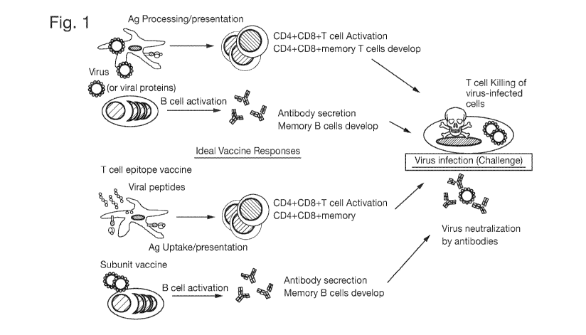
immune response to natural viral infection (Figure 1). The vaccine composition
may
provide protection against both seasonal and pandemic influenza strains, e.g.
the vaccine
composition may be a universal vaccine.
The present inventors have surprisingly identified that a nanoparticle, for
example a
gold nanoparticle, may be used to induce an efficient response to a vaccine
composition
designed to stimulate both the production of antibodies specific for influenza
virus and a T
cell response against influenza virus. Use of a nanoparticle abrogates the
need to include a
traditional adjuvant in the vaccine composition. Therefore, the likelihood of
an individual
experiencing an adverse reaction following administration of the vaccine
composition is
reduced.
The present inventors have also identified number of conserved peptides that
are
conserved between different influenza viruses and are presented by MHC
molecules on
3
CA 03049190 2019-07-03
WO 2018/127689 PCT/GB2018/050004
cells infected with those viruses. Inclusion of such conserved peptides in the
vaccine
composition may confer protective capability against both seasonal and
pandemic
influenza strains. Including in the vaccine composition multiple conserved
peptides that
bind to different HLA supertypes results in a vaccine that is effective in
individuals having
different HLA types.
Accordingly, the present invention provides a vaccine composition comprising
one
or more immunogenic influenza virus peptides attached to a nanoparticle.
The present invention further provides:
- a vaccine composition comprising an influenza virus peptide comprising a
CD8+
T cell epitope and an influenza virus peptide comprising a B cell epitope,
wherein
each peptide is attached to a nanoparticle;
a vaccine composition comprising an influenza virus peptide comprising one or
more of the CD8+ T cell epitopes set out in SEQ ID NOs: 1 to 18, wherein the
peptide is attached to a nanoparticle;
- a method of preventing or treating an influenza virus infection, comprising
administering the vaccine composition of the invention to an individual
infected
with, or at risk of being infected with, an influenza virus; and
- the vaccine composition of the invention for use in a method of preventing
or
treating an influenza virus infection in an individual.
Brief Description of the Figures
Figure 1: Vaccine to mimic adaptive immune response generated by viral
infection.
Figure 2: Confirmation of influenza specific peptide sequences.
Figure 3: Epitopes (P1-P5) specific CTLs generated in vitro with human PBMCs
recognize both peptide loaded (panel A) and various influenza virus infected
(panel B)
target cells.
Figure 4: Epitopes (P1-P5) specific CTLs generated in vivo in HLA A2
transgenic
mice recognize both peptide loaded (panel B) and various influenza virus
infected (panel
C) target cells.
Figure 5: Epitopes incorporated in NPs (NP 1-5) activates various influenza
virus
specific CTLs in HLA A2 transgenic mice. Panel A&B: ELISpot; Panel C: CD107
expression.
4
CA 03049190 2019-07-03
WO 2018/127689 PCT/GB2018/050004
Figure 6: Immunization of M2e peptide + adjuvant (Pep 6) or in NP (NP6) induce
specific antibody response (panel A). No antibody response with free peptide
without
adjuvant (panel B).
Figure 7: M2e peptide specific antisera binds to M2e epitope on influenza
virus
infected cells (PR8, X-31, or JAP (green, aqua, red respectively).
Figure 8: Figure 8: Anti-M2e mediated infection neutralization. Serum used in
the
top panels is isolated from peptide immunized mice, bottom panels from NP
immunized
mice.
Figure 9: Naturally occurring M2e antibody response in healthy individuals
exposed to influenza virus.
Figure 10: Detection of pre-existing epitope specific CTLs via dextramer
analysis
of PBMCs from dengue sero-positive individuals.
Figure 11: Figure 11: Epitope specific CTLs in seropositive individuals
recognize
peptide loaded (pep) and dengue virus infected (DV2) target cells.
Figure 12: CD8 epitopes nested within longer peptides are processed and
presented
on APCs. Top panel, SIIN:Kb complexes detected by flow cytometry. Bottom
panel, SIIN
specific T cell activation in a hybridoma assay.
Detailed Description of the Invention
Vaccine compositions stimulating humoral and cellular responses
The present invention provides a vaccine composition comprising one or more
immunogenic influenza virus peptides attached to a nanoparticle. In
particular, a vaccine
composition comprising an influenza virus peptide comprising a CD8+ T cell
epitope and
an influenza virus peptide comprising a B cell epitope, wherein each peptide
is attached to
a nanoparticle.
This vaccine composition has a number of advantageous over conventional
influenza vaccines known in the art. The key advantages are summarised here.
However,
further advantages will become apparent from the discussion below.
Firstly, the vaccine composition of the invention advantageously comprises an
influenza virus peptide comprising a CD8+ T cell epitope and an influenza
virus peptide
comprising a B cell epitope. The vaccine composition is therefore capable of
stimulating
5
CA 03049190 2019-07-03
WO 2018/127689 PCT/GB2018/050004
both cellular and humoral immune responses against an influenza virus. As
described
above, humoral immune responses provide a first line of defence against
influenza virus
infection. In particular, secretory IgA and IgM antibodies protect against the
establishment
of initial influenza virus infection, for instance by prevent virus from
attaching to epithelial
cells at mucosal surfaces. Later, during established infection, IgG antibodies
neutralize
newly replicating virus to help minimise viral reproduction. Humoral responses
therefore
have an important role in the prevention and treatment of influenza virus
infection.
However, cellular responses, particularly T cell responses, are also
important. CD4+ T
cells control isotype-switching to IgG and, therefore, the neutralisation of
replicating virus.
CD4+ T cells also contribute to the generation of higher affinity antibodies
and to
cytotoxic T lymphocyte (CTL) memory. CD8+ CTLs themselves mediate viral
clearance
via their cytotoxic activity against infected cells. Stimulating both humoral
and cellular
immunity therefore provides a beneficial double-pronged attack against
influenza virus
infection.
Secondly, each influenza virus peptide in the vaccine composition is attached
to a
nanoparticle, for example a gold nanoparticle. As described in more detail
below,
attachment to a nanoparticle reduces or eliminates the need to include an
adjuvant in the
vaccine composition. Thus, the vaccine composition is less likely to cause
adverse clinical
effects upon administration to an individual.
Influenza virus peptides
The vaccine composition of the invention comprises one or more immunogenic
influenza virus peptides. The vaccine composition may comprise from about one
to about
50 influenza virus peptides, such as about 2 to 40, 30 to 30, 4 to 25, 5 to
20, 6 to 15, 7, 8, 9
or 10 influenza virus peptides. The peptides each comprise one or more
epitope, which
may be a CD8+ T cell epitope, a CD4+ T cell epitope and/or a B cell epitope.
In one aspect, the vaccine composition comprises an influenza virus peptide
comprising a CD8+ T cell epitope and an influenza virus peptide comprising a B
cell
epitope.
An influenza virus peptide is a peptide that is expressed by one or more
influenza
viruses. Influenza viruses are well known members of the Orthomyxovirdae
family.
Hundreds of strains of influenza virus exist which may be classified in three
main
6
CA 03049190 2019-07-03
WO 2018/127689 PCT/GB2018/050004
categories, Influenza A, Influenza B or Influenza C, based on the HA and NA
proteins they
express. The vaccine composition may comprise influenza virus peptides from
multiple
strains of influenza, such as 1 to 2000, 100 to 1900, 200 to 1800, 300 to
1700, 400 to 1600,
500 to 1500, 600 to 1400, 700 to 1300, 800 to 1200 or 900 to 1100 strains of
influenza.
For example, the vaccine composition may comprise one more influenza virus
peptide
from Influenza A, Influenza B and/or Influenza C. Thus, the influenza virus
peptide
comprising a CD8+ T cell epitope comprised may be a peptide that is expressed
by
Influenza A, Influenza B and/or Influenza C virus. The influenza virus peptide
comprising
a B cell epitope may be a peptide that is expressed by Influenza A, Influenza
B and/or
Influenza C virus. The influenza virus peptide comprising a CD8+ T cell
epitope and the
influenza virus peptide comprising a B cell epitope may be peptides that are
expressed by
the same influenza strain, such as Influenza A, Influenza B or Influenza C.
Alternatively,
the influenza virus peptide comprising a CD8+ T cell epitope and the influenza
virus
peptide comprising a B cell epitope may be peptides that are expressed by
different
influenza strains.
If the influenza virus peptide is a peptide that is expressed by Influenza A
virus, the
Influenza A virus may be, for example, H1N1, H5N1, H7H9 or H3N2. Preferably,
the
influenza virus peptide is expressed by two or more of H1N1, H5N1, H7H9 and
H3N2
Influenza A virus, such as, for example, H1N1 and H3N2. The influenza virus
peptide
may be a peptide that is expressed by a human influenza virus, a swine
influenza virus,
and/or an avian influenza virus. The influenza virus may be a pandemic
influenza virus or
a potentially pandemic influenza virus. The influenza virus may be a zoonotic
influenza
virus.
The influenza virus peptide may be a peptide that is expressed on the surface
of one
or more influenza viruses, or intracellularly within one or more influenza
viruses. The
peptide may be a structural peptide or a functional peptide, such as a peptide
involved in
the metabolism or replication of the influenza virus. Preferably, the peptide
is an internal
peptide. Preferably, the peptide is conserved between two or more different
influenza
strains.
The influenza virus peptide may contain any number of amino acids, i.e. be of
any
length. Typically, the influenza virus peptide is about 8 to about 30, 35 or
40 amino acids
in length, such as about 9 to about 29, about 10 to about 28, about 11 to
about 27, about 12
7
CA 03049190 2019-07-03
WO 2018/127689 PCT/GB2018/050004
to about 26, about 13 to about 25, about 13 to about 24, about 14 to about 23,
about 15 to
about 22, about 16 to about 21, about 17 to about 20, or about 18 to about 29
amino acids
in length.
The influenza virus peptide may be chemically derived from a polypeptide
influenza virus antigen, for example by proteolytic cleavage. More typically,
the influenza
virus peptide may be synthesised using methods well known in the art.
The term "peptide" includes not only molecules in which amino acid residues
are
joined by peptide (-CO-NH-) linkages but also molecules in which the peptide
bond is
reversed. Such retro-inverso peptidomimetics may be made using methods known
in the
art, for example such as those described in Meziere et al (1997) J.
Immuno1.159, 3230-
3237. This approach involves making pseudopeptides containing changes
involving the
backbone, and not the orientation of side chains. Meziere et al (1997) show
that, at least
for MHC class II and T helper cell responses, these pseudopeptides are useful.
Retro-
inverse peptides, which contain NH-CO bonds instead of CO-NH peptide bonds,
are much
more resistant to proteolysis.
Similarly, the peptide bond may be dispensed with altogether provided that an
appropriate linker moiety which retains the spacing between the carbon atoms
of the amino
acid residues is used; it is particularly preferred if the linker moiety has
substantially the
same charge distribution and substantially the same planarity as a peptide
bond. It will
also be appreciated that the peptide may conveniently be blocked at its N-or C-
terminus so
as to help reduce susceptibility to exoproteolytic digestion. For example, the
N-terminal
amino group of the peptides may be protected by reacting with a carboxylic
acid and the C-
terminal carboxyl group of the peptide may be protected by reacting with an
amine. Other
examples of modifications include glycosylation and phosphorylation. Another
potential
modification is that hydrogens on the side chain amines of R or K may be
replaced with
methylene groups (-NH2 may be modified to -NH(Me) or -N(Me)2).
The term "peptide" also includes peptide variants that increase or decrease
the
half-life of the peptide in vivo. Examples of analogues capable of increasing
the half-life of
peptides used according to the invention include peptoid analogues of the
peptides, D-
amino acid derivatives of the peptides, and peptide-peptoid hybrids. A further
embodiment
of the variant polypeptides used according to the invention comprises D-amino
acid forms
of the polypeptide. The preparation of polypeptides using D-amino acids rather
than L-
8
CA 03049190 2019-07-03
WO 2018/127689 PCT/GB2018/050004
amino acids greatly decreases any unwanted breakdown of such an agent by
normal
metabolic processes, decreasing the amounts of agent which needs to be
administered,
along with the frequency of its administration.
CD8+ T cell epitopes
The vaccine composition of the invention preferably comprises an influenza
virus
peptide comprising a CD8+ T cell epitope. A CD8+ T cell epitope is a peptide
that is
capable of (i) presentation by a class I MHC molecule and (ii) recognition by
a T cell
receptor (TCR) present on a CD8+ T cell. Preferably, recognition by the TCR
results in
.. activation of the CD8+ T cell. CD8+ T cell activation may lead to increased
proliferation,
cytokine production and/or cyotoxic effects.
Typically, the CD8+ T cell epitope is around 9 amino acids in length. The CD8+
T
cell epitope may though be shorter or longer. For example, the CD8+ T cell
epitope may
be about 8, 9, 10, 11, 12, 13, 14 or 15 amino acids in length. The CD8+ T cell
epitope may
be about 8 to 15, 9 to 14 or 10 to 12 amino acids in length.
Influenza virus peptides comprising a CD8+ T cell epitope are known in the
art.
Methods for identifying CD8+ T cell epitopes are known in the art. Epitope
mapping
methods include X-ray co-crystallography, array-based oligo-peptide scanning
(sometimes
called overlapping peptide scan or pepscan analysis), site-directed
mutagenesis, high
throughput mutagenesis mapping, hydrogen¨deuterium exchange, crosslinking
coupled
mass spectrometry, phage display and limited proteolysis. MHC motif prediction
methodologies may also be used.
Preferably, CD8+ T cell epitopes presented by influenza virus-infected cells
can be
identified in order to directly identify CD8+ T cell epitopes for inclusion in
the vaccine
composition. This is an efficient and logical method which can be used alone
or to
confirm the utility of potential CD8+ T cell epitopes identified by MHC motif
prediction
methodologies. To perform the method, cells are infected with influenza virus
and
maintained in culture for a period of around 24 hours. The cells are then
harvested and
washed. Next, the cells are lysed and MHC/peptide complexes isolated by
immunoaffinity
.. chromatography using MHC molecule specific antibodies. The peptides
purified from the
MHC molecules are fractionated, for example using a C-18 reversed phase (RP)
column
using an offline HPLC. The peptide-containing fractions are collected, dried
under a
9
CA 03049190 2019-07-03
WO 2018/127689 PCT/GB2018/050004
vacuum, and analysed by mass spectrometry to identify the sequences of the
fractions. The
acquired spectral data is then searched against all databased influenza
proteins to identify
peptide sequences associated with influenza virus. Synthetic peptides may then
be made
according to the identified sequences and subjected to mass spectrometry to
confirm their
identity to the peptides in the peptide-containing fractions.
In this method, any type of cells may be infected with influenza virus. The
cells
may be antigen presenting cells. The cells may be hepatoma cells such as HepG2
cells,
EBV-transformed lymphoblastoid B cells such as JY cells, or lymphoblasts such
as T2
cells.
Likewise, any influenza virus of interest may be used to infect the cells. For
instance, the influenza virus may be an Influenza A, Influenza B or Influenza
C virus. The
Influenza A virus may, for example, be H1N1, H5N1, H7H9 or H3N2.
The direct identification of CD8+ T cell epitopes presented by influenza virus-
infected cells is advantageous compared to MHC motif prediction methodologies.
The
immune epitope database (IEDB; http://www.iedb.org) is generated by motif
prediction
methods, and not functional methods, and contains several hundreds of
predicted HLA-
specific influenza T cell epitopes, including a number of shared epitopes with
high MHC
binding scores and limited CTL characterization. As both dominant and
subdominant
epitopes are presented by influenza virus-infected cells, it is difficult to
sort out the
dominance hierarchies of naturally presented epitopes using the database.
Thus, it is not
clear from the immune epitope database alone which of the listed epitopes may
be
expected to efficiently induce a CD8+ T cell response when included in a
vaccine
composition. The direct identification method set out above provides a
mechanism for
confirming the utility of the epitopes.
Furthermore, the direct identification method may be used to identify
conserved
CD8+ T cell epitopes presented by cells infected by different influenza
viruses. In this
way, CD8+ T cell epitopes suitable for inclusion in a universal vaccine may be
identified.
As set out in the Examples, the present inventors used the direct
identification method to
identify CD8+ T cell epitopes conserved between Influenza A (H1N1 and H3N2)
and
Influenza B strains. The sequences identified are set out in Table 1.
Table 1
CA 03049190 2019-07-03
WO 2018/127689
PCT/GB2018/050004
SEQ
ID Peptide Protein
HLA motif
NO:
1 PVAGGTSSIYI (P2) polymerase PB2 A2
2 TVIKTNMI (P3) polymerase PB1 A2/A24
3 MTIIFLILM (P4) hemagglutinin A2/A24
4 ITFHGAKEI Matrix protein 1 A2/A24
AINGITNKV Hemagglutinin A2/A24
6 EEMGITTHF RNA-directed RNA polymerase B44
catalytic subunit
7 VETPIRNEW Matrix protein 2 B44
8 REILTKTTV Polymerase basic protein 2 B44
9 KESDEALNMTMASTP Non-structural protein NS1 B44
LENERTLDF Polymerase acidic protein B44
11 MEAVPLITI Hemagglutinin B44
12 VEQEIRTF Nuclear export protein B44
13 VEQELRTF nonstructural protein 2 B44
14 SPDDFALIVNA polymerase PB1 B7
YPDTGKVM Neuraminidase B7
16 YPDASKVM neuraminidase B7
17 QPETCNQSII Neuraminidase B7
18 VPESKRMSL nonstructural protein (NS1) B7
The inventors also confirmed that the known peptides YINTALLNA (P1; SEQ ID NO:
19), AIMDKNIIL (P5; SEQ ID NO: 20) and LPFDRTTIM (SEQ ID NO: 21) are CD8+ T
cell epitopes conserved between Influenza A (H1N1 and H3N2) and Influenza B
strains.
5 The influenza virus peptide comprising a CD8+ T cell epitope may
therefore
comprise one or more of SEQ ID NOs: 1 to 21. For instance, the influenza virus
peptide
comprising a CD8+ T cell epitope may comprise two or more, three or more, four
or more,
five or more, ten or more, fifteen or more, or twenty or more of SEQ ID NOs: 1
to 21 in
any combination. The influenza virus peptide comprising a CD8+ T cell epitope
may
10 comprise all of SEQ ID NOs: 1 to 21.
Similarly, the vaccine composition may comprise one or more influenza virus
peptides each comprising a CD8+ T cell epitope comprising a different sequence
selected
from SEQ ID NOs: 1 to 21. For instance, the vaccine composition may comprise
two or
more, three or more, four or more, five or more, ten or more, fifteen or more,
or twenty or
15 more influenza virus peptides each comprising a CD8+ T cell epitope
comprising a
different sequence selected from SEQ ID NOs: 1 to 21, in any combination. The
vaccine
composition may, for example, comprise 21 influenza virus peptides each
comprising a
CD8+ T cell epitope comprising a different sequence selected from SEQ ID NOs:
1 to 21.
11
CA 03049190 2019-07-03
WO 2018/127689 PCT/GB2018/050004
B cell epitopes
The vaccine composition of the invention may comprise an influenza virus
peptide
comprising a B cell epitope. A B cell epitope is a peptide that is capable of
recognition by
a B cell receptor (BCR) present on a B cell. Preferably, recognition by the
BCR results in
activation and/or maturation of the B cell. B cell activation may lead to
increased
proliferation, and/or antibody production.
Influenza virus peptides comprising a B cell epitope are known in the art. The
B
cell epitope may be a linear epitope, i.e. an epitope that is defined by the
primary amino
acid sequence of a particular region of an influenza virus protein.
Alternatively, the epitope
may be a conformational epitope, i.e. an epitope that is defined by the
conformational
structure of a native influenza protein. In this case, the epitope may be
continuous (i.e. the
components that interact with the antibody are situated next to each other
sequentially on
the protein) or discontinuous (i.e. the components that interact with the
antibody are
situated on disparate parts of the protein, which are brought close to each
other in the
folded native protein structure).
Typically, the B cell epitope is around 5 to 20 amino acids in length, such as
6 to
19, 7 to 18, 8 to 17, 9 to 16, 10 to 15, 11 to 14 or 12 to 13 amino acids in
length.
Methods for identifying B cell epitopes are also known in the art. For
instance,
epitope mapping methods may be used to identify B cell epitopes. These methods
include
structural approaches, wherein the known or modelled structure of a protein is
be used in
an algorithm based approach to predict surface epitopes, and functional
approaches,
wherein the binding of whole proteins, protein fragments or peptides to an
antibody can be
quantitated e.g. using an Enzyme-Linked Immunosorbent Assay (ELISA).
Competition
mapping, antigen modification or protein fragmentation methods may also be
used.
The B cell epitope may, for example, be an influenza matrix 2 protein (M2e)
epitope. The extracellular domain of M2e protein is an evolutionarily
conserved region in
influenza A viruses. Thus, inclusion of an influenza peptide comprising a M2e
B cell
epitope in the vaccine composition may help to confer universal utility.
Universal vaccine compositions
12
CA 03049190 2019-07-03
WO 2018/127689 PCT/GB2018/050004
The present invention also provides a vaccine composition comprising an
influenza
virus peptide comprising one or more of the CD8+ T cell epitopes set out in
SEQ ID NOs:
1 to 21, wherein the peptide is attached to a nanoparticle.
The inclusion of an influenza virus peptide comprising one or more of the CD8+
T
cell epitopes set out in SEQ ID NOs: 1 to 21 in the vaccine composition is
advantageous.
As set out above, the CD8+ T cell epitopes set out in SEQ ID NOs: 1 to 21 are
conserved
CD8+ T cell epitopes that are presented by WIC molecules on cells infected by
different
influenza viruses. Accordingly, an immune response generated by vaccination
with a
composition that comprises any of these epitopes should protect against
subsequent
infection with any influenza virus that shares that epitope. In other words,
the vaccine
composition has built-in cross-subtype efficacy, i.e. it is a universal
influenza vaccine
composition. Such a composition could prevent the significant spread of an
emerging or
re-emerging strain of influenza infection.
Furthermore, vaccine compositions based on epitopes presented by influenza
virus-
infected cells, such as the present vaccine composition, are superior to
vaccines based on a
viral protein subunit or a motif predicted epitope. Protein processing by the
immune
system is likely to alter native viral epitopes. Basing a vaccine composition
on peptides
demonstrated to be presented by infected cell removes this source of
uncertainty, because
the peptides have already undergone protein processing.
Attaching the influenza virus peptide to a nanoparticle, for example a gold
nanoparticle, is also beneficial. As described in more detail below,
attachment to a
nanoparticle reduces or eliminates the need to include an adjuvant in the
vaccine
composition. Thus, the vaccine composition is less likely to cause adverse
clinical effects
upon administration to an individual.
Influenza peptides comprising SEQ ID NOs: 1 to 21
The vaccine composition comprises an influenza virus peptide comprising one or
more of the CD8+ T cell epitopes set out in SEQ ID NOs: 1 to 21. Influenza
virus peptides
comprising SEQ ID NOs: 1 to 21 are described in detail above in relation to
the vaccine
compositions stimulating humoral and cellular responses. The influenza virus
peptide may
comprise two or more, such as three or more, four or more, five or more, ten
or more,
fifteen or more or 20 or more of the CD8+ T cell epitopes set out in SEQ ID
NOs: 1 to 21.
13
CA 03049190 2019-07-03
WO 2018/127689 PCT/GB2018/050004
In this case, the influenza virus peptide may comprise any combination of the
CD8+ T cell
epitopes set out in SEQ ID NOs: 1 to 21. The influenza virus peptide may
comprise all of
the CD8+ T cell epitopes set out in SEQ ID NOs: 1 to 21.
The vaccine composition may comprise two or more influenza virus peptides each
comprising a different CD8+ T cell epitope. Each of the two or more peptides
may
comprise a different CD8+ T cell epitope set out in SEQ ID NOs: 1 to 21.
Alternatively,
one or more of the two or more peptides may comprise a CD8+ T cell epitope
that is not
set out in SEQ ID NOs: 1 to 21. CD8+ T cell epitopes are known in the art.
Nanoparticles
In both the vaccine composition comprising an influenza virus peptide
comprising
a CD8+ T cell epitope and an influenza virus peptide comprising a B cell
epitope, and the
vaccine composition comprising an influenza virus peptide comprising one or
more of the
CD8+ T cell epitopes set out in SEQ ID NOs: 1 to 21, each of the influenza
virus peptides
is attached to a nanoparticle.
As set out above and demonstrated in the Examples below, attachment of the
influenza virus peptides to a nanoparticle (such as a gold nanoparticle)
reduces or
eliminates the need to include an adjuvant in the vaccine composition. The
nanoparticles
may contain immune "danger signals" that help to effectively induce an immune
response
to the influenza virus peptides. The nanoparticles may induce dendritic cell
(DC)
activation and maturation, required for a robust immune response. The
nanoparticles may
contain non-self components that improve uptake of the nanoparticles and thus
the
influenza virus peptides by cells, such as antigen presenting cells.
Attachment of an
influenza virus peptide to a nanoparticle may therefore enhance the ability of
antigen
presenting cells to stimulate virus-specific B and/or T cells. Attachment to a
nanoparticle
also facilitates delivery of the vaccine compositions via the subcutaneous,
intradermal,
transdermal and oral/buccal routes, providing greater flexibility in
administration that
conventional influenza vaccines.
Nanoparticles are particles between 1 and 100 nanometers (nm) in size which
can
be used as a substrate for immobilising ligands. In the vaccine compositions
of the
invention, the nanoparticle may have a mean diameter of 1 to 100, 20 to 90, 30
to 80, 40 to
70 or 50 to 60 nm. Preferably, the nanoparticle has a mean diameter of 20 to
40nm. A
14
CA 03049190 2019-07-03
WO 2018/127689 PCT/GB2018/050004
mean diameter of 20 to 40nm facilitates uptake of the nanoparticle to the
cytosol. The
mean diameter can be measured using techniques well known in the art such as
transmission electron microscopy.
Nanoparticles suitable for the delivery of antigen, such as an influenza virus
peptide, are known in the art. Methods for the production of such
nanoparticles are also
known.
The nanoparticle may, for example, be a polymeric nanoparticle, an inorganic
nanoparticle, a liposome, an immune stimulating complex (ISCOM), a virus-like
particle
(VLP), or a self-assembling protein. The nanoparticle is preferably a calcium
phosphate
.. nanoparticle, a silicon nanoparticle or a gold nanoparticle.
The nanoparticle may be a polymeric nanoparticle. The polymeric nanoparticle
may comprise one or more synthetic polymers, such as poly(d,l-lactide-co-
glycolide)
(PLG), poly(d,l-lactic-coglycolic acid) (PLGA), poly(g-glutamic acid) (g-PGA)m
poly(ethylene glycol) (PEG), or polystyrene. The polymeric nanoparticle may
comprise
one or more natural polymers such as a polysaccharide, for example pullulan,
alginate,
inulin, and chitosan. The use of a polymeric nanoparticle may be advantageous
due to the
properties of the polymers that may be include in the nanoparticle. For
instance, the
natural and synthetic polymers recited above may have good biocompatibility
and
biodegradability, a non-toxic nature and/or the ability to be manipulated into
desired
shapes and sizes. The polymeric nanoparticle may form a hydrogel nanoparticle.
Hydrogel nanoparticles are a type of nano-sized hydrophilic three-dimensional
polymer
network. Hydrogel nanoparticles have favourable properties including flexible
mesh size,
large surface area for multivalent conjugation, high water content, and high
loading
capacity for antigens. Polymers such as Poly(L-lactic acid) (PLA), PLGA, PEG,
and
polysaccharides are particularly suitable for forming hydrogel nanoparticles.
The nanoparticle may be an inorganic nanoparticle. Typically, inorganic
nanoparticles have a rigid structure and are non-biodegradable. However, the
inorganic
nanoparticle may be biodegradable. The inorganic nanoparticle may comprise a
shell in
which an antigen may be encapsulated. The inorganic nanoparticle may comprise
a core to
which an antigen may be covalently attached. The core may comprise a metal.
For
example, the core may comprise gold (Au), silver (Ag) or copper (Cu) atoms.
The core
may be formed of more than one type of atom. For instance, the core may
comprise an
CA 03049190 2019-07-03
WO 2018/127689 PCT/GB2018/050004
alloy, such as an alloy of Au/Ag, Au/Cu, Au/Ag/Cu, Au/Pt, Au/Pd or
Au/Ag/Cu/Pd. The
core may comprise calcium phosphate (CaP). The core may comprise a
semiconductor
material, for example cadmium selenide.
Other exemplary inorganic nanoparticles include carbon nanoparticles and
silica-
based nanoparticles. Carbon nanoparticles are have good biocompatibility and
can be
synthesized into nanotubes and mesoporous spheres. Silica-based nanoparticles
(SiNPs)
are biocompatible and can be prepared with tunable structural parameters to
suit their
therapeutic application.
The nanoparticle may be a silicon nanoparticle, such as an elemental silicon
nanoparticle. The nanoparticle may be mesoporous or have a honeycomb pore
structure.
Preferably, the nanoparticle is an elemental silicon particle having a
honeycomb pore
structure. Such nanoparticles are known in the art and offer tunable and
controlled drug
loading, targeting and release that can be tailored to almost any load, route
of
administration, target or release profile. For example, such nanoparticles may
increase the
bioavailability of their load, and/or improve the intestinal permeability and
absorption of
orally administered actives. The nanoparticles may have an exceptionally high
loading
capacity due to their porous structure and large surface area. The
nanoparticles may
release their load over days, weeks or months, depending on their physical
properties.
Since silicon is a naturally occurring element of the human body, the
nanoparticles may
elicit no response from the immune system. This is advantageous to the in vivo
safety of
the nanoparticles.
Any of the SiNPs described above may be biodegradable or non-biodegradable. A
biodegradable SiNP may dissolve to orthosilic acid, the bioavailable form of
silicon.
Orthosilic acid has been shown to be beneficial for the health of bones,
connective tissue,
hair, and skin.
The nanoparticle may be a liposome. Liposomes are typically formed from
biodegradable, non-toxic phospholipids and comprise a self-assembling
phospholipid
bilayer shell with an aqueous core. A liposome may be an unilameller vesicle
comprising
a single phospholipid bilayer, or a multilameller vesicle that comprises
several concentric
phospholipid shells separated by layers of water. As a consequence, liposomes
can be
tailored to incorporate either hydrophilic molecules into the aqueous core or
hydrophobic
16
CA 03049190 2019-07-03
WO 2018/127689 PCT/GB2018/050004
molecules within the phospholipid bilayers. Liposomes may encapsulate antigen
within the
core for delivery. Liposomes may incorporate viral envelope glycoproteins to
the shell to
form virosomes. A number of liposome-based products are established in the art
and are
approved for human use.
The nanoparticle may be an immune-stimulating complex (ISCOM). ISCOMs are
cage-like particles which are typically formed from colloidal saponin-
containing micelles.
ISCOMs may comprise cholesterol, phospholipid (such as
phosphatidylethanolamine or
phosphatidylcholine) and saponin (such as Quil A from the tree Quillaia
saponaria).
ISCOMs have traditionally been used to entrap viral envelope proteins, such as
envelope
proteins from herpes simplex virus type 1, hepatitis B, or influenza virus.
The nanoparticle may be a virus-like particle (VLP). VLPs are self-assembling
nanoparticles that lack infectious nucleic acid, which are formed by self-
assembly of
biocompatible capsid protein. VLPs are typically about 20 to about 150nm, such
as about
about 20 to about 40nm, about 30 to about 140nm, about 40 to about 130nm,
about 50 to
about 120nm, about 60 to about 110nm, about 70 to about 100nm, or about 80 to
about
90nm in diameter. VLPs advantageously harness the power of evolved viral
structure,
which is naturally optimized for interaction with the immune system. The
naturally-
optimized nanoparticle size and repetitive structural order means that VLPs
induce potent
immune responses, even in the absence of adjuvant.
The nanoparticle may be a self-assembling protein. For instance, the
nanoparticle
may comprise ferritin. Ferritin is a protein that can self-assemble into
nearly-spherical 10
nm structures. The nanoparticle may comprise major vault protein (MVP). Ninety-
six
units of MVP can self-assemble into a barrel-shaped vault nanoparticle, with a
size of
approximately 40 nm wide and 70 nm long.
The nanoparticle may be a calcium phosphate (CaP) nanoparticle. CaP
nanoparticles may comprise a core comprising one or more (such as two or more,
10 or
more, 20 or more, 50 or more, 100 or more, 200 or more, or 500 or more)
molecules of
CaP. CaP nanoparticles and methods for their production are known in the art.
For
instance, a stable nano-suspension of CAP nanoparticles may be generated by
mixing
inorganic salt solutions of calcium and phosphates in pre-determined ratios
under constant
mixing.
17
CA 03049190 2019-07-03
WO 2018/127689
PCT/GB2018/050004
The CaP nanoparticle may have an average particle size of about 80 to about
100nm, such as about 82 to about 98nm, about 84 to about 96nm, about 86 to
about 94nm,
or about 88 to about 92nm. This particle size may produce a better performance
in terms
of immune cell uptake and immune response than other, larger particle sizes.
The particle
size may be stable (i.e. show no significant change), for instance when
measured over a
period of 1 month, 2 months, 3 months, 6 months, 12 months, 18 months, 24
months, 36
months or 48 months.
CaP nanoparticles can be co-formulated with one or multiple antigens either
adsorbed on the surface of the nanoparticle or co-precipitated with CaP during
particle
synthesis. For example, a peptide, such as an influenza virus peptide, may be
attached to
the CaP nanoparticle by dissolving the peptide in DMSO (for example at a
concentration of
about 10 mg/ml), adding to a suspension of CaP nanoparticles together with N-
acetyl-
glucosamine (G1cNAc) (for example at 0.093mo1/L and ultra-pure water, and
mixing at
room temperature for a period of about 4 hours (for example, 1 hour, 2 hours,
3 hours, 5
hours, 6 hours, 7 hours, 8 hours, 9 hours or 10 hours).
The vaccine composition may comprise about 0.15 to about 0.8%, such as 0.2 to
about 0.75%, 0.25 to about 0.7%, 0.3 to about 0.6%, 0.35 to about 0.65%, 0.4
to about
0.6%, or 0.45 to about 0.55%, CaP nanoparticles. Preferably the vaccine
composition
comprises about 0.3% CaP nanoparticles.
CaP nanoparticles have a high degree of biocompatibility due to their chemical
similarity to human hard tissues such as bone and teeth. Advantageously,
therefore, CaP
nanoparticles are non-toxic when used for therapeutic applications. CaP
nanoparticles are
safe for administration via intramuscular, subcutaneous, oral, or inhalation
routes. CaP
nanoparticles are also simple to synthesise commercially. Furthermore, CaP
nanoparticles
.. may be associated with slow release of antigen, which may enhance the
induction of an
immune response to an influenza virus peptide attached to the nanoparticle.
CaP
nanoparticles may be used both as an adjuvant, and as a drug delivery vehicle.
The nanoparticle may be a gold nanoparticle. Gold nanoparticles are known in
the
art and are described in particular in WO 2002/32404, WO 2006/037979, WO
2007/122388, WO 2007/015105 and WO 2013/034726. The gold nanoparticle attached
to
each influenza virus peptide may be a gold nanoparticle described in any of WO
18
CA 03049190 2019-07-03
WO 2018/127689 PCT/GB2018/050004
2002/32404, WO 2006/037979, WO 2007/122388, WO 2007/015105 and WO
2013/034726.
Gold nanoparticles comprise a core comprising a gold (Au) atom. The core may
further comprise one or more Fe, Cu or Gd atoms. The core may be formed from a
gold
alloy, such as Au/Fe, Au/Cu, Au/Gd, Au/Fe/Cu, Au/Fe/Gd or Au/Fe/Cu/Gd. The
total
number of atoms in the core may be 100 to 500 atoms, such as 150 to 450, 200
to 400 or
250 to 350 atoms. The gold nanoparticle may have a mean diameter of 1 to 100,
20 to 90,
30 to 80, 40 to 70 or 50 to 60 nm. Preferably, the gold nanoparticle has a
mean diameter of
20 to 40nm.
One or more ligands other than the influenza virus peptides may be linked to
the
nanoparticle, which may be any of the types of nanoparticle described above.
The ligands
may form a "corona", a layer or coating which may partially or completely
cover the
surface of the core. The corona may be considered to be an organic layer that
surrounds or
partially surrounds the nanoparticle core. The corona may provide or
participate in
passivating the core of the nanoparticle. Thus, in certain cases the corona
may be a
sufficiently complete coating layer to stabilise the core. The corona may
facilitate
solubility, such as water solubility, of the nanoparticles of the present
invention.
The nanoparticle may comprise at least 10, at least 20, at least 30, at least
40 or at
least 50 ligands. The ligands may include one or more peptides, protein
domains, nucleic
.. acid molecules, lipidic groups, carbohydrate groups, anionic groups, or
cationic groups,
glycolipids and/or glycoproteins. The carbohydrate group may be a
polysaccharide, an
oligosaccharide or a monosaccharide group (e.g. glucose). One or more of the
ligands may
be a non-self component, that renders the nanoparticle more likely to be taken
up by
antigen presenting cells due to its similarity to a pathogenic component. For
instance, one
or more ligands may comprise a carbohydrate moiety (such as a bacterial
carbohydrate
moiety), a surfactant moiety and/or a glutathione moiety. Exemplary ligands
include
glucose, N-acetylglucosamine (G1cNAc), glutathione, 2'-thioethy1-13-D-
glucopyranoside
and 2'-thioethyl- D-glucopyranoside. Preferred ligands include
glycoconjugates, which
form glyconanoparticles
Linkage of the ligands to the core may be facilitated by a linker. The linker
may
comprise a thiol group, an alkyl group, a glycol group or a peptide group. For
instance, the
linker may comprise C2-C15 alkyl and/or C2-C15 glycol. The linker may comprise
a
19
CA 03049190 2019-07-03
WO 2018/127689 PCT/GB2018/050004
sulphur-containing group, amino-containing group, phosphate-containing group
or oxygen-
containing group that is capable of covalent attachment to the core.
Alternatively, the
ligands may be directly linked to the core, for example via a sulphur-
containing group,
amino-containing group, phosphate-containing group or oxygen-containing group
comprised in the ligand.
Attachment to nanoparticles
The influenza virus peptides may be attached at their N-terminus to the
nanoparticle. Typically, the influenza virus peptides are attached to the core
of the
nanoparticle, but attachment to the corona or a ligand may also be possible.
One or more of the influenza virus peptides may be directly attached to the
nanoparticle, for example by covalent bonding of an atom in a sulphur-
containing group,
amino-containing group, phosphate-containing group or oxygen-containing group
in the
peptide to an atom in the nanoparticle or its core.
A linker may be used to link one or more of the influenza virus peptides to
the
nanoparticle. The linker may comprise a sulphur-containing group, amino-
containing
group, phosphate-containing group or oxygen-containing group that is capable
of covalent
attachment to an atom in the core. For example, the linker may comprise a
thiol group, an
alkyl group, a glycol group or a peptide group.
The linker may comprise a peptide portion and a non-peptide portion. The
peptide
portion may comprise the sequence X1X2Z1, wherein Xi is an amino acid selected
from A
and G; X2 is an amino acid selected from A and G; and Zi is an amino acid
selected from
Y and F. The peptide portion may comprise the sequence AAY or FLAAY. The
peptide
portion of the linker may be linked to the N-terminus of the influenza virus
peptide. The
non-peptide portion of the linker may comprise a C2-C15 alkyl and/ a C2-C15
glycol, for
example a thioethyl group or a thiopropyl group.
The linker may be (i) HS-(CH2)2-CONH-AAY; (ii) HS-(CH2)2-CONH-LAAY; (iii)
HS-(CH2)3-CONH-AAY; (iv) HS-(CH2)3-CONH- FLAAY; (v) HS-(CH2)10-(CH2OCH2)7-
CONH-AAY; and (vi) HS-(CH2)10-(CH2OCH2)7-CON}{-FLAAY. In this case, the thiol
group of the non-peptide portion of the linker links the linker to the core.
Other suitable linkers for attaching an influenza virus to a nanoparticle are
known
in the art, and may be readily identified and implemented by the skilled
person.
CA 03049190 2019-07-03
WO 2018/127689 PCT/GB2018/050004
When the vaccine composition comprises an influenza virus peptide comprising a
CD8+ T cell epitope and a influenza virus peptide comprising a B cell epitope,
each
influenza virus peptide may be attached to a different nanoparticle. In this
case, the
nanoparticle to which each influenza virus peptide is attached may be the same
type of
nanoparticle. For instance, each influenza virus peptide may be attached to a
gold
nanoparticle. Each influenza virus peptide may be attached to a CaP
nanoparticle. The
nanoparticle to which each influenza virus peptide is attached may be a
different type of
nanoparticle. For instance, one influenza virus peptide may be attached to a
gold
nanoparticle, and the other influenza virus peptide may be attached to a CaP
nanoparticle.
.. Preferably though, the influenza virus peptide comprising a CD8+ T cell
epitope and the
influenza virus peptide comprising a B cell epitope are attached to the same
nanoparticle.
For example, the influenza virus peptide comprising a CD8+ T cell epitope and
the
influenza virus peptide comprising a B cell epitope may be attached to the
same gold
nanoparticle. This provides a single particle that is capable of stimulating
both an
influenza virus-specific cellular response, and an influenza virus-specific
humoral
response.
CD4+ T cell epitopes
A vaccine composition of the invention may further comprise an influenza virus
.. peptide comprising a CD4+ T cell epitope. CD4+ T cell epitopes are defined
above.
The vaccine composition may comprise two or more, such as three or more, four
or
more, five our more, ten or more, fifteen or more or twenty or more influenza
virus
peptides comprising a CD4+ T cell epitope. Such peptides are known in the art.
The influenza virus peptide comprising a CD4+ T cell epitope may be a
different
peptide from the influenza virus peptide encoding the CD8+ T cell and the
influenza virus
peptide encoding the B cell epitope. The influenza virus peptide comprising a
CD4+ T cell
epitope may be the same peptide as the influenza virus peptide encoding the
CD8+ T cell.
That is, the influenza virus peptide comprising a CD8+ T cell epitope may
further
comprise a CD4+ T cell epitope.
When the influenza virus peptide comprising a CD8+ T cell epitope comprises a
CD4+ T cell epitope., the CD8+ epitope may be nested within the CD4+ T cell
epitope.
CD4+ T cell epitopes are typically longer than CD8+ T cell epitopes.
Therefore, extending
21
CA 03049190 2019-07-03
WO 2018/127689 PCT/GB2018/050004
one or both termini of the CD8+ T cell epitope may yield a longer, CD4+ T cell
epitope
whose sequence still comprises the CD8+ T cell epitope. Therefore, the CD4+ T
cell
epitope may comprise a CD8+ T cell epitope, such as a CD8+ T cell epitope set
out in SEQ
ID NOs: 1 to 21, extended at its N-terminus or C-terminus. The CD8+ T cell
epitope may
be extended by 1, 2, 3, 4 or 5 amino acids at its N terminus. The CD8+ T cell
epitope may
be extended by 1, 2, 3, 4 or 5 amino acids at its C terminus. Preferably, the
CD8+ T cell
epitope is extended by 3 amino acids at the N terminus, and 3 amino acids at
the C
terminus. However, the CD8+ T cell epitope need not be extended by the same
number of
amino acids at each terminus.
The CD8+ T cell epitope nested within an extended peptides may be capable of
generating a robust CTL response. The extended peptide (CD4+ T cell epitope)
is capable
of inducing T helper mediated cytokine responses. Thus, inclusion of an
influenza virus
peptide comprising a CD8+ T cell epitope and a CD4+ T cell epitope in the
vaccine
composition may allow the vaccine composition to induce both cytotoxic and
helper T cell
.. responses, providing improved anti-influenza immunity.
As set out in the Examples, the inventors have identified a number of
potential
CD4+ T cell epitopes in which a CD8+ T cell epitope set out in SEQ ID NOs: 1
to 21 is
nested. These are set out in Table 2.
Table 2
Extended epitope
CD8 epitope (potential CD4 Protein HLA motif
epttope)
YINTALLNA kgvYINTALLNAsca
(SEQ ID NO: 19) (SEQ ID NO: 22) polymerase
PA A2
PVAGGTSSIYI rflPVAGGTSSIYIevl
(SEQ ID NO: 1) (SEQ ID NO: 23) polymerase
PB2 A2
TVIKTNMI igvTVIKTNMInnd
(SEQ ID NO:2) (SEQ ID NO: 24) polymerase PB1
A2/A24
AIMDKNIIL mdqAIMDKNIILkan nonstructural
A2/A24
(SEQ ID NO: 20) (SEQ ID NO: 25) protein 1
ITFHGAKEI kreITFHGAKEIsls
(SEQ ID NO: 4) (SEQ ID NO: 26) Matrix protein
1 A2/A24
AINGITNKV tqnAINGITNKVnsv
(SEQ ID NO: 5) (SEQ ID NO: 27) Hemagglutinin
A2/A24
22
CA 03049190 2019-07-03
WO 2018/127689 PCT/GB2018/050004
Thus, the influenza virus peptide comprising a CD8+ T cell epitope and a CD4+
T cell
epitope may comprise one or more of the peptides set out in SEQ ID NOs: 22 to
27. The
influenza virus peptide comprising a CD8+ T cell epitope and a CD4+ T cell
epitope may
comprise two or more, three or more, four or more or five or more of the
peptides set out in
SEQ ID NOs: 22 to 27, in any combination. The influenza virus peptide
comprising a
CD8+ T cell epitope and a CD4+ T cell epitope may comprise all of the peptides
set out in
SEQ ID NOs: 22 to 27
The influenza virus peptide comprising a CD4+ T cell epitope may be attached
to a
nanoparticle. The nanoparticle may be a gold nanoparticle. Nanoparticles and
attachment
thereto are described above.
Interaction with HLA supertypes
The vaccine composition may comprise at least two influenza virus peptides
comprising a CD8+ T cell epitope which each interacts with a different HLA
supertype.
Including a plurality of such peptides in the vaccine composition allows the
vaccine
composition to elicit a CD8+ T cell response in a greater proportion of
individuals to
which the vaccine composition is administered. This is because the vaccine
composition
should be capable of eliciting a CD8+ T cell response in all individuals of an
HLA
supertype that interacts with one of the CD8+ T cell epitopes comprised in the
plurality of
influenza virus peptides. Each CD8+ T cell epitope may interact with HLA-A1,
HLA-A2,
HLA-A3, HLA-A24, HLA-B7, HLA-B8, HLA-B27, HLA-B44, HLA-B58 or HLA-B62,
or any other HLA supertype know in the art. Any combination of influenza virus
peptides
comprising such a CD8+ T cell epitope is possible.
The vaccine composition may comprise at least one immunogenic peptide that
interacts with at least two different HLA supertypes. Again, this allows the
vaccine
composition to elicit a CD8+ T cell response in a greater proportion of
individuals to
which the vaccine composition is administered. The vaccine composition may
comprise at
least two, at least three, at least four, at least five, at least two, at
least fifteen, or at least
twenty immunogenic peptides that each interact with at least two different HLA
subtypes.
Each immunogenic peptide may interact with at least two, at least three, at
least four, at
least five, at least six, at least 7, at least 8, at least 9 or at least 10
different HLA
supertypes. Each immunogenic peptide may interact with two or more of HLA-A1,
HLA-
23
CA 03049190 2019-07-03
WO 2018/127689 PCT/GB2018/050004
A2, HLA-A3, HLA-A24, HLA-B7, HLA-B8, HLA-B27, HLA-B44, HLA-B58 or HLA-
B62, or any other HLA supertype known in the art, in any combination.
Preferably, the
vaccine composition comprises an immunogenic peptide that interacts with HLA-
A2 and
HLA-24. In this case, the vaccine composition may, for example, comprise an
influenza
virus peptide comprising a CD8+ T cell epitope comprises one or more of the
peptides set
out in SEQ ID NOs: 3 to 5.
Medicaments, methods and therapeutic use
The invention provides a method of preventing or treating an influenza virus
infection, comprising administering the vaccine composition of the inventions
to an
individual infected with, or at risk of being infected with, an influenza
virus. The
invention also provides a vaccine composition of the invention for use in a
method of
preventing or treating an influenza virus infection in an individual.
The influenza virus may be an Influenza A, Influenza B and/or Influenza C
virus.
.. The Influenza A virus may, for example, be H1N1, H5N1, H7H9 or H3N2. The
influenza
virus may be a human influenza virus, a swine influenza virus, or an avian
influenza virus.
The influenza virus may be a pandemic influenza virus or a potentially
pandemic influenza
virus.
The vaccine composition may be provided as a pharmaceutical composition. The
pharmaceutical composition preferably comprises a pharmaceutically acceptable
carrier or
diluent. The pharmaceutical composition may be formulated using any suitable
method.
Formulation of cells with standard pharmaceutically acceptable carriers and/or
excipients
may be carried out using routine methods in the pharmaceutical art. The exact
nature of a
formulation will depend upon several factors including the cells to be
administered and the
desired route of administration. Suitable types of formulation are fully
described in
Remington's Pharmaceutical Sciences, 19th Edition, Mack Publishing Company,
Eastern
Pennsylvania, USA.
The vaccine composition or pharmaceutical composition may be administered by
any route. Suitable routes include, but are not limited to, the intravenous,
intramuscular,
intraperitoneal, subcutaneous, intradermal, transdermal and oral/buccal
routes.
Compositions may be prepared together with a physiologically acceptable
carrier or
diluent. Typically, such compositions are prepared as liquid suspensions of
peptide-linked
24
CA 03049190 2019-07-03
WO 2018/127689 PCT/GB2018/050004
nanoparticles. The nanoparticles may be mixed with an excipient which is
pharmaceutically acceptable and compatible with the active ingredient.
Suitable excipients
are, for example, water, saline, dextrose, glycerol, of the like and
combinations thereof.
In addition, if desired, the pharmaceutical compositions may contain minor
amounts of auxiliary substances such as wetting or emulsifying agents, and/or
pH
buffering agents.
The peptide-linked nanoparticles are administered in a manner compatible with
the
dosage formulation and in such amount will be therapeutically effective. The
quantity to be
administered depends on the subject to be treated, the disease to be treated,
and the
capacity of the subject's immune system. Precise amounts of nanoparticles
required to be
administered may depend on the judgement of the practitioner and may be
peculiar to each
subject.
Any suitable number of nanoparticles may be administered to a subject. For
example, at least, or about, 0.2 x 10⁶, 0.25 x 10⁶, 0.5 x 10⁶, 1.5 x 10⁶, 4.0
x 106 or 5.0 x 10⁶
nanoparticles per kg of patient may administered. For example, at least, or
about, 105, 106,
10, 108, 109 cells may be administered. As a guide, the number of
nanoparticles of the
invention to be administered may be from 105 to 109, preferably from 106 to
108.
It is to be understood that different applications of the disclosed products
and
methods may be tailored to the specific needs in the art. It is also to be
understood that the
terminology used herein is for the purpose of describing particular
embodiments of the
invention only, and is not intended to be limiting.
In addition, as used in this specification and the appended claims, the
singular
forms "a", "an", and "the" include plural referents unless the content clearly
dictates
otherwise. Thus, for example, reference to "a CAR" includes "CARs", reference
to "a T
cell" includes two or more such T cells, reference to "a component" includes
two or more
such components, and the like.
All publications, patents and patent applications cited herein, whether supra
or
infra, are hereby incorporated by reference in their entirety.
Further Embodiments of the Invention
CA 03049190 2019-07-03
WO 2018/127689
PCT/GB2018/050004
1. An isolated oligopeptide or peptide in a pharmaceutical composition
comprising at
least one peptide having an amino acid sequence selected from the group
consisting of
SEQ ID NO: 1 to 18, said oligopeptide or peptide consisting of 8 to about 30
amino acid
residues, wherein said oligopeptide or peptide binds to class I MHC molecules
or can be
processed to bind to class I MHC molecules and activate T lymphocyte response
and
wherein the oligopeptide or peptide is in the form of a pharmaceutically
acceptable salt.
2. The oligopeptide of item 1 wherein said oligopeptide comprises at least
two
epitopic peptides.
3. The oligopeptide of item 1 wherein said oligopeptide comprises at least
three
epitopic peptides.
4. The oligopeptide of item 1 wherein said oligopeptide comprises at least
four
epitopic peptides.
5. The oligopeptide or peptide of item 1 wherein said oligopeptide or
peptide differs
from SEQ ID NO: 1, 2, 3, 4, 5, 6, 7, 8, 9, 10, 11, 12 13, 14, 15, 16,17 or 18
wherein said
difference is no more than one amino acid unit.
6. The oligopeptide or peptide of item 5 wherein said one amino acid
difference is the
result of a conservative amino acid substitution.
7. The oligopeptide or peptide of item 5 wherein said one amino acid
difference is the
substitution of one hydrophobic amino acid with another hydrophobic amino
acid.
8. The oligopeptide or peptide of item 5 wherein said amino acid difference
is the
addition or deletion of one amino acid to or from said epitopic peptide.
9. A polynucleotide in a pharmaceutical composition comprising a
polynucleotide
selected from the group consisting of: (a) a polynucleotide that encodes an
oligopeptide or
peptide of item 1, and (b) the full complement of (a) wherein the
polynucleotide is in a
form of a pharmaceutically acceptable salt.
10. The polynucleotide of item 9 wherein the polynucleotide of (a) is DNA.
11. The polynucleotide of item 9 wherein the polynucleotide of (a) is RNA.
12. A method for vaccinating and treating a subject for Influenza
infection, said
infected cells expressing any class I MHC molecule, comprising administering
to said
subject a composition that binds to class I MHC molecules or can be processed
to bind to
class I MHC molecules comprising: at least one polypeptide comprising an
epitopic
peptide comprising an amino acid sequence selected from the group consisting
of SEQ ID
26
CA 03049190 2019-07-03
WO 2018/127689 PCT/GB2018/050004
NO: 1 to 18 in an amount sufficient to induce a CTL response to said infected
cells and in
a form of a pharmaceutically acceptable salt; or at least one polypeptide
comprising an
epitopic peptide having at least one amino acid difference from an amino acid
sequence
selected from the group consisting of SEQ ID NO: 1 to 18 in an amount
sufficient to
induce a CTL response to said infected cells and in a form of a
pharmaceutically
acceptable salt.
13. A method for vaccinating and treating a subject with Influenza
infection, said
infected cells expressing any class I MEW molecule, said method comprising
administering
to said subject a composition that binds to class I MEW molecules or can be
processed to
bind to class I MHC molecules comprising: a polynucleotide comprising a
nucleic acid
sequence encoding at least one polypeptide comprising an amino acid sequence
selected
from the group consisting of SEQ ID NO: 1 to 18 in an amount sufficient to
induce a CTL
response to said infected cells and in a form of a pharmaceutically acceptable
salt; or at
least one polypeptide comprising an epitopic peptide comprising one amino acid
difference
from an amino acid sequence selected from the group consisting of SEQ ID NO: 1
to 18 in
an amount sufficient to induce a CTL response to said infected cells and in a
form of a
pharmaceutically acceptable salt.
14. A method for generating an immune response ex vivo using T cells from a
subject
infected with Influenza, said method comprising: stimulating the production of
CTL
response for use in passive immunotherapy, wherein said T cells react with at
least one
polypeptide comprising an amino acid sequence selected from the group
consisting of SEQ
ID NO: 1 to 18 and in a form of a pharmaceutically acceptable salt; or at
least one
polypeptide comprising one amino acid difference from an amino acid sequence
selected
from the group consisting of SEQ ID NO: 1 to 18 and in a form of a
pharmaceutically
acceptable salt.
15. The method of item 14, wherein said T cell adoptive therapy generated
from
autologous or HLA matched subjects.
16. A method for assessing or diagnosing an immune response in a subject
infected
with Influenza or vaccinated for Influenza and related viruses said method
comprising:
.. stimulating the production of CTL response, wherein said T cells react with
at least one
polypeptide comprising an amino acid sequence selected from the group
consisting of SEQ
ID NO: 1 to 18 and in a form of a pharmaceutically acceptable salt; or at
least one
27
CA 03049190 2019-07-03
WO 2018/127689 PCT/GB2018/050004
polypeptide comprising one amino acid difference from an amino acid sequence
selected
from the group consisting of SEQ ID NO: 1 to 18 and in a form of a
pharmaceutically
acceptable salt.
17. A method for vaccinating humans against Influenza infection using
SEQ IDs 1 to
18 in a form of a pharmaceutically acceptable salt
The following Examples illustrate the invention.
Example 1
Introduction
Influenza is a significant global health problem, infecting up to 20% of the
world's
population annually, causing up to 5 million cases of severe illness and >3
00,000 deaths
worldwide. In the U.S. alone, an estimated >30,000 deaths and nearly 300,000
hospitalizations are attributed to influenza infection each year (1). With the
recent
appearance of new, severe and potentially recurrent seasonal disease,
widespread
vaccination campaigns that reduce the incidence of influenza-induced pneumonia
are being
encouraged by the World Health Organization. Effectively reducing the
incidence of
influenza will require continued intense surveillance, increased use of
currently available
influenza vaccine, and availability of alternative vaccines and antiviral
medications that
can provide broader protection against shift-and-drift strains of influenza
(1). Successful
influenza vaccination campaigns can have enormous societal and economic impact
(2).
Immune response to influenza virus
The immune response to influenza is governed by both innate and adaptive
immunity. The innate immune response to influenza limits initial viral
replication but is
relatively non-specific (3). Efficient clearance of influenza virus requires a
robust adaptive
immune response, activating both humoral and cell mediated immunity. Humoral
immunity, as mediated by secretory IgA and IgM. Antibodies, provides
protection against
the establishment of initial infection, while IgG neutralizes newly
replicating virus in
established infection (4, 5). Although inducing humoral immunity to influenza
is the target
of current conventional influenza vaccines, these are not completely
protective due to
occurrence of antigenic variations (6) (7). Additionally, data indicate that T-
cell responses
28
CA 03049190 2019-07-03
WO 2018/127689 PCT/GB2018/050004
are extremely important for protection against influenza. CD4+ T cells play a
critical role
in isotype-switching to IgG and in the generation of higher affinity
antibodies (8) and CTL
memory (9-11). In humans, HA-specific CD4+ T cells proliferate following
influenza
vaccination (12) and aid the development of heterosubtypic influenza antibody
responses
(13, 14). CD8+ cytotoxic T lymphocytes (CTLs) mediate viral clearance and,
importantly,
were shown to have cross-reactive responses to different subtypes of influenza
A virus (15-
17). This may explain the relative paucity of disease among older, potentially
vaccinated,
or exposed individuals to H1N1 infection.
Current Status of influenza Virus Vaccine Development
Influenza vaccines now on the market are updated yearly and are designed based
on
annual WHO strain recommendations (16, 18) and manufactured prior to the
beginning of
an influenza season or pandemic. Current vaccines for influenza induce a
protective
humoral immune response against the HA and NA glycoproteins on the virion
surface (7,
19, 20) . However, viral HA and NA glycoproteins are highly susceptible to
frequent and
unpredictable antigenic shift and less frequent, but more severe, drift
mutations, which
result in loss of antibody recognition necessitating frequent development of
new vaccines
to match the current viral serotype(s) infecting the human population (21-25).
In addition,
these vaccines are costly to produce, and will not protect against novel
strains that may
emerge mid-season (i.e. 2009 H1N1 swine flu, H5N1, H7N9). Most importantly,
these
vaccines focused on antibody based protection and induce limited T cell
responses that are
essential for eliminating infected cells from the body.
Three improved seasonal influenza vaccines currently have FDA approval. These
quadrivalent vaccines provide coverage against two influenza A and two
influenza B
viruses. (In recent years, two influenza B viruses have co-circulated during
influenza
seasons and the trivalent vaccines offered did not protect against one of
these strains).
FluMist, a quadrivalent live attenuated vaccine that may provide more targets
for the
immune system because of limited protein synthesis (which is absent in
inactivated
vaccines). FluMist (http://www.cdc.goviflu/protectivaccinelvaccines.htm) is
currently
distributed as an alternative to the trivalent inactivated vaccine. Fluarix
and FluZone are
quadrivalent inactivated vaccines that stimulate a similar immune response to
the current
trivalent inactivated vaccine. These will be distributed in the 2013-2014
influenza season
29
CA 03049190 2019-07-03
WO 2018/127689 PCT/GB2018/050004
(http w . fda.gov /13 i 01(4 s 131 ood ac n e sNacc e s/Appro
vedProductsAtcm 29 5 0 5 7 ht
m). Despite the coverage offered by the quadrivalent vaccines, these are still
seasonal and
suffer from the same downfalls as the trivalent vaccine, namely production
times and costs
and the lack of strong T cell responses that eliminates infected cells and
protection against
mid-season emergents. It is readily apparent that a universal vaccine offering
protection
against most, if not all, influenza strains is necessary.
To date, multiple universal vaccine formulations are in development. These
vaccines can be broadly characterized by the type of protective immune
response that is
stimulated: 1) B cell responses (antibody), 2) T cell responses, or 3) both B
and T cell
responses. Kanekiyo et al. generated HA nanoparticles (hemagglutinin (HA)
protein of
influenza is fused to ferritin) that induce high titer antibody responses that
provide
coverage against multiple influenza strains (26). This vaccine has yet to
enter into clinical
trials. A T cell based vaccine that targets four relatively conserved epitopes
in the viral
genome (27) is also under development. A highly conserved CD4 epitopes based T
cell
vaccine has been evaluated in a phase II challenge study with positive
protective responses
against various influenza strains including pandemic strains (28). A
recombinant
polyepitope vaccine, called Multimeric-001, that incorporates B cell, CD4 T
cell-, and
CD8 T cell conserved epitopes from nine different influenza proteins (29) is
being tested in
early stage clinical trials. A fusion protein vaccine consisting of
nucleoprotein (NP) and
the B cell epitope M2e linked to an adjuvant and M2e peptide in gold
nanoparticle in
combination with CpG (30) are also under development. Most of the above
mentioned
vaccines are formulated as protein or peptides with various adjuvants that
induce clinical
adverse reactions. In contrast, our fully synthetic universal influenza
vaccine formulation
consists of a panel of conserved CD4, CD8 and B cell activating epitopes
formulated in a
gold nanoparticle vaccine delivery that has built in adjuvant properties,
designed to
stimulate potent T and B cell immune responses directed against many influenza
virus
strains including the newly emerged strains.
Synthetic universal vaccine concept for influenza infection
An ideal vaccine should attempt to mimic natural immunity (Fig 1) generated by
infection in a manner whereby adaptive humoral and cellular immune memory is
generated
CA 03049190 2019-07-03
WO 2018/127689 PCT/GB2018/050004
(31). Tremendous efforts are being made to develop a universal flu vaccine
that would
work against all types of influenza.
The goal is to provide protection for years, if not a whole lifetime, against
all
seasonal influenza strains and pandemic strains, making flu immunization much
more like
that for traditional vaccines. Development of an effective universal influenza
vaccine
would eliminate (or lessen) fears of future influenza pandemics and would be
cost effective
compared to development and manufacturing of the annual seasonal influenza
vaccines.
Such a universal vaccine must target conserved influenza virus epitopes that
do not vary
from strain to strain and more importantly, should be presented by the WIC
molecules on
the infected cells. Ultimately, the most promising universal influenza vaccine
candidate
may come from combining the antigens for both T and B cell immunity. Based on
animal
models and human studies, combining all possible T and B cell ligands (Fig 1)
to formulate
an active synthetic vaccine makes sense. In order to be active, the vaccine
must be in the
size range to permit cytosol uptake with built in danger signals to target
antigen presenting
cells. Novel gold glyconanoparticles with these properties have been shown to
induce
efficient vaccine responses (32, 33). Development of a synthetic universal
vaccine based
on shared T cell and B cell epitopes incorporated in the gold nanoparticle is
the focus of
this application.
Innovation
This proposal has two major, novel distinctions: (1) the development of a
fully
synthetic universal vaccine that is capable of inducing T helper, cytotoxic T
cell and
antibody responses against multiple strains of influenza infection and (2) the
characterization of a novel fully synthetic novel gold glyconanoparticle
vaccine delivery
platform armed with innate and adaptive immune stimulating molecules that will
serve as
potent adjuvant for any subunit vaccine. The proposed vaccine approach would
have
several advantages over the existing vaccines. First, it would have multiple
conserved
WIC class I restricted CD8+ T cell epitopes naturally processed and presented
on infected
cells (34), CD8 epitopes nested within CD4 epitopes, and antibody epitope/s
from various
influenza viral proteins. Secondõ these epitopes will be delivered in
synthetic
glyconanoparticle that contains immune "danger signals" (35, 36). Finally,
this 20-40nm
sized novel glyconanoparticles unlike other nanoparticles, contains non-self-
components
31
CA 03049190 2019-07-03
WO 2018/127689 PCT/GB2018/050004
(such as bacterial carbohydrates, GlcNAc) in addition to influenza specific T
and B cell
activating epitopes, is more likely to target and be taken up by antigen
presenting cells due
to its similarity to a pathogen, for efficient activation of antigen-specific
T cell and
antibody responses. These glyconanoparticles are also anticipated to induce DC
maturation/activation, and thus, a robust immune response, which are essential
for a
successful, specific, subunit vaccine response (37, 38). These nanoparticles
are also
amenable to various delivery routes including transdermal and oral/buccal in
addition to
intradermal and subcutaneous injection mode of delivery.
This proposal is also intended to accelerate the pre-clinical development of a
novel
emerging vaccine strategy that has built-in cross-subtype efficacy, which
could prevent the
significant spread of an emerging or re-emerging strain of influenza
infection. A cross-
subtype vaccine containing immunogenic consensus sequence epitopes could
achieve this
goal. Mounting evidence suggests that an efficient universal vaccine must
induce
activation of both cell mediated and humoral immunity. We hypothesize that
vaccines
based on defined epitopes presented by the infected cells would be far
superior to a viral
protein subunit or a motif predicted epitope based vaccines because protein
processing by
the immune system may alter the native viral epitopes. In addition, CD8
epitope nested
within extended peptides not only generate a robust CTL response but the
extended
peptides are also capable of inducing T helper mediated cytokine responses,
which are
critical to combat influenza infection (28). An addition of a universal
antibody epitope to
this synthetic vaccine formulation incorporates both arms of the immune system
for
complete protection.
As demonstrated by our preliminary data, direct identification of T cell
epitopes
presented by the infected cells is an efficient and logical method to identify
T cell epitopes
for vaccine applications. Our analysis confirmed a few T cell epitopes that
were selected
by MHC motif prediction methodologies. Since the immune epitope database
(http://www.iedb.org) contains several hundreds of predicted HLA specific
influenza T cell
epitopes, including a number of shared epitopes with high MHC binding scores
and limited
CTL characterization, it is critical to confirm and use for vaccine
application epitopes that
are presented by the virus infected cells,. Additionally, because the IEDB
database is
generated by motif prediction methods, and not functional methods, it would be
difficult to
sort out the dominance hierarchies of naturally presented epitopes. Thomas et
al. (39)
32
CA 03049190 2019-07-03
WO 2018/127689 PCT/GB2018/050004
elegantly demonstrated that both dominant and subdominant epitopes are
presented by the
infected cells, which would be difficult to sort out using the data in the
IEDB database.
One additional drawback of the motif predicted epitopes is that most often
they were
screened for functional specificity using CTL from infected individuals, which
may have
been tolerized.
Significant innovation will be realized in the first ever combination of T
cell (CD8
and CD4) epitopes and an antibody epitope (M2e) formulated in a targeted,
immune
activating and regulating, fully synthetic, flexible nanoparticle vaccine
delivery system.
We emphasize that the use of the conserved T cell epitopes that are naturally
presented by
the infected cells and B cell epitope incorporated in the nanoparticles
represent a major
leap in vaccine technology rather than a small adjustment to the vaccine
constituents. If
successful, this method could lead to a paradigm shift in how prophylactic and
therapeutic
vaccines are formulated. Our work will also be useful in advancing the general
understanding of the T cell mediated immune response to influenza virus
infection. This
information is critical to the development of anti-viral drugs and
prophylactic and
therapeutic universal vaccines to combat and prevent serious, and some cases,
lethal
influenza virus infection.
Results
We identified a panel of HLA-A2 specific conserved epitopes from the infected
cells. Using peripheral blood mononuclear cells (PBMC) obtained from HLA-A2
positive
healthy donors we have characterized selected epitopes for CTL activity and
demonstrated
cross reactivity in vitro.
Identification of MHC class I presented influenza epitopes
JY, an HLA-A2 and B7 positive EBV transformed B cell line and HLA-A2 and
A24 positive HepG2, a hepatoma cell line and Dendritic cells (DCs) generated
from HLA-
A2 positive peripheral blood lymphocytes (PBL) from healthy individuals were
infected
with Influenza A and B viral strains (A/New Caledonia/20/99 (H1N1),
A/Wisconsin/67/2005 (H3N2), B/Malaysia/2506/2004). For both JY and DC,
published
studies have shown cellular infection to be ¨50% upon using ¨10 HAU (40) for
16-24 h
are most successful for influenza infections (41). The infected cells were
used for the
33
CA 03049190 2019-07-03
WO 2018/127689 PCT/GB2018/050004
isolation of MHC peptides if they showed >50% infectivity. Cell lysates were
prepared
and subjected to two rounds of immunoprecipitation using 1 mg of W6/32 (pan
MHC class
I specific antibody) conjugated to 1 mL of Protein A/G beads (34). The bound
MHC
complexes were eluted from the beads and peptide mixtures were purified and
fractionated
.. using C18 reversed phase (RP) column. The peptide fractions were injected
individually
into LC-MS/MS system (nanoAcquity UPLC -LTQ orbitrap) to identify the MHC
peptides. MHC peptides and their corresponding proteins were identified by
searching the
raw data in NCBI influenza database using bioworks/proteome discoverer
software
(Thermo). Synthetic peptides were made to validate and confirm the sequences.
The
.. synthetic peptides were subjected to LC-MS/MS analysis under identical
collision
conditions as the experimental and their sequences were confirmed by
comparison of their
MS/MS spectra with that of their synthetic analogs.
Table 31 summarizes the MHC class I-associated peptides that we have
identified
from influenza virus infected cells. We identified 7 epitopes with the HLA-A2
binding
.. motif and 5 of the 7 epitopes had a HLA-A24 cross binding motif In
addition, we
identified several HLA-B7 and B44 motif containing epitopes. All the epitopes
originate
from regions of the proteins that are conserved between multiple influenza
strains. These
peptides were confirmed in co-elution experiments using synthetic peptides.
Interestingly,
we identified three previously reported epitopes, two HLA-A2 specific epitopes
(P1 and
P5) (42), which were identified using motif-prediction and ex vivo CTL
analysis and one
HLA-B7 specific epitope (LPF in table 3) that is shown to be conserved between
pandemic
H1N1-2009 and H1N1-1918 influenza A viruses (43).
34
CA 03049190 2019-07-03
WO 2018/127689 PCT/GB2018/050004
Seq ID Peptide Protein HLA motif
YINTALLNA* (P1) polmerase PA A2
1 PVAGGTSSIYI (P2) polymerase PB2 A2
2 TVIKTNMI (P3) polymerase PB1 A2/A24
3 MTIIFLILM (P4) hemagglutinin A2/A24
AIMDKNIIL* (P5) nonstructural protein 1 A2/A24
4 ITFHGAKEI Matrix protein 1 A2/A24
AINGITNKV Hemagglutinin A2/A24
6 EEMGITTHF RNA-directed RNA B44
polymerase catalytic
7 VETPIRNEW Matrix protein 2 B44
8 REILTKTTV Polymerase basic B44
protein 2
9 KESDEALNMTMASTP Non-
structural protein B44
NS1
LENERTLDF Polymerase acidic B44
protein
11 MEAVPLITI Hemagglutinin B44
12 VEQEIRTF Nuclear export
protein B44
13 VEQELRTF nonstructural protein
2 B44
LPFDRTTIM* nucleocapsid protein B7
14 SPDDFALIVNA polymerase PB1 B7
YPDTGKVM Neuraminidase B7
16 YPDASKVM neuraminidase B7
17 QPETCNQSII Neuraminidase B7
Table 3: MHC class I epitopes presented by influenza virus infected cells
* Previously reported epitopes
5
Prior to CTL functional characterization experiments, we confirmed the
authenticity of 5 HLA-A2 specific peptides, P1¨P5 (Table 3) using their
synthetic peptide
analogs. As illustrated in Figure 2, most of the fragment ions in the ms/ms
spectra of
experimentally identified peptides (Figure 2: P1-A¨P5-A) matched with spectra
of their
10 corresponding synthetic peptides as indicated by the denoted masses
(Figure 2: P 1 -B¨P5-
B) (34). In addition, we further verified the ms/ms spectra manually to
confirm the identity
of all the experimentally observed peptides. T cell epitopes were formulated
into the
nanoparticles and tested for induction of epitope specific CTL and cross
reactivity in vitro.
CA 03049190 2019-07-03
WO 2018/127689 PCT/GB2018/050004
To verify the presentation of these epitopes by infected cells, CTLs specific
for
each of the 5 peptides were generated using PBMCs from healthy HLA-A2+ donors
and
synthetic peptides corresponding to the identified epitopes. In ELISpot
assays, CTL
functionality was measured by detection of antigen specific IFNy secretion. As
illustrated
in Figure 3A, PR8-infected JY and HepG2 cells stimulated all five of the
influenza epitope-
specific T cells. Additionally, cross-reactivity to other strains was
demonstrated using
HepG2 target cells infected with various influenza A strains (X31, H3N2 and
JAP, H2N2),
indicating the presentation of these epitopes in various influenza strain-
infected cells
(Figure 3B).
To further characterize the immune response generated by these epitopes in
vivo,
we immunized HLA-A2+ transgenic mice with a mixture of the aforementioned five
epitopes. Immunizations were carried out using these peptides in the presence
of
Montanide ISA 51 as an adjuvant (Figure 4A). We determined the influenza-
specific T cell
response by measuring murine IFNy secretion in an ELISpot assay. Using T2
cells pulsed
with individual peptides 1-5, we observed a response to all 5 peptides after
immunization
(Figure 4B). In conjunction with above in vitro results, in vivo-generated
CTLs specific for
these peptides were stimulated equally well when HepG2 and JY cells infected
with
different strains of influenza were used as targets (Figure 4C) indicating
that these epitopes
are processed and presented in multiple influenza infections. In addition to
IFNy release,
we also measured the phenotypic changes of CD8+ T cells from splenocytes with
regards
to CD107a, an activation marker present on granulating effector CTLs (44, 45).
As
illustrated in Figure 4D, CD8+ T cells displayed a higher intensity of CD107a
staining
when incubated with infected targets compared to uninfected targets.
We generated gold nanoparticles (NP) incorporating HLA-A2 specific epitopes
(NP 1-5) and combined with a shared antibody epitope (NP 6) from influenza
matrix 2
protein (M2e). We immunized HLA-A2 transgenic mice with either
peptide+montanide
51 or peptides in NPs and assessed CTL response by IFNy release using ELISpots
(Fig 5A
and B) and CD107a expression (Fig 5C). Peptides incorporated in NPs activated
a strong
anti-viral CTL response in vivo without any addition of adjuvant. NPs
themselves acted as
adjuvant in addition to delivering peptides to the antigen presenting cells.
Peptides without
montanide adjuvant did not elicit a CTL response (data not shown).
36
CA 03049190 2019-07-03
WO 2018/127689 PCT/GB2018/050004
In addition to characterizing CTL responses, we also evaluated antibody
responses
to a universal antibody epitope from influenza matrix 2 protein (M2e). To this
end, we
immunized a group of mice with MHCI peptides (P1-5) or peptides in GNP (NP1-5)
in
addition to a peptide (P6 or NP6) from the ectodomain of M2 (M2e) (46, 47).
The T cell
response as measured by IFNy ELISpot assay (Figure 5A&B) and CD107a (Figure
5C)
flow cytometric analysis were comparable between the groups immunized with T
cell
epitopes alone (1-5) or T cell epitopes with the M2e peptide (1-6). The
concentration of
circulating M2e-specific antibody was then measured by a standard ELISA using
serum
collected from the terminal bleeds of immunized mice. As illustrated in Figure
6, mice
immunized with the M2e peptide with montanide adjuvant (Pep6) or in GNPs (NP6)
generated a robust and M2e specific IgG response (Fig. 6A). However, when the
M2e
peptide is combined with the MHC1 peptides (Pep1-5 or NP1-5), the antibody
titer against
M2e was slightly reduced. There was no difference between the
peptides+montanide
adjuvant and GNPs without any adjuvant in the antibody response as we observed
in the T
cell response. However, when we compared free peptide without any adjuvant
with NPs,
we observed a greater antibody response with NPs (Fig. 6B). In summary, we
demonstrate
that GNP incorporated MHCI and antibody epitopes generate robust both CTL and
antibody responses as compared to epitopes that require adjuvants, which
induce clinical
toxicity.
Functionality of the M2e antisera generated by peptide+montanide adjuvant (P1-
6,
P6) and peptides in NPs (NP1-6, NP6) for cross reactivity were then
characterized by
infecting HepG2 and JY cells with all three strains of influenza virus (PR8, X-
31, or JAP)
that we have thus far tested. Using flow cytometry, we treated the infected
cells with the
M2e-specific antisera obtained from peptide+montanide or NP immunized mice and
demonstrated the binding of the antibody specifically on PR8, X-31, or JAP
(green, aqua,
red respectively) infected cells indicating the presence of this epitope in
various strains of
influenza infected cells (Fig. 7).
Lastly, the neutralizing ability of these antibodies was determined by adding
antisera to HepG2 cells infected with PR8 virus. HepG2 cells were pulsed with
a low dose
of PR8 in the presence of serum from MHCI peptides (P1-5 or NP 1-5), or pM2e
peptide
(pM2e or NP M2e) immunized mice at a 1:50 dilution for 1 hr. Following
overnight
incubation with the same levels of serum, cells were fixed and intracellularly
stained for
37
CA 03049190 2019-07-03
WO 2018/127689 PCT/GB2018/050004
influenza NP. Percent positive cells are depicted. Gray-filled histograms are
uninfected
controls. As illustrated in Figure 8, infection levels were lower when the
serum containing
anti-M2e antibody obtained from M2e peptide with montanide adjuvant (Fig 8A)
or M2e
peptide in GNP (Fig 8B) was used indicating the functional ability of the M2e
specific
antibodies.
Summary
These are the first ever comprehensive study reported on WIC class I
associated
peptides analysis of influenza virus infected cells. Significantly, all the
epitopes are shared
between different strains of influenza. Interestingly, we also identified a
few already
reported motif predicted epitopes on the infected cells. Further to MHO
epitopes
characterization, we have also demonstrated that the broad anti-viral activity
of the
antibodies against the M2e antibody epitope. We have also generated gold
nanoparticle
(GNP) formulated with T and antibody epitopes and characterized anti-viral T
and
antibody responses in in vitro and in vivo studies. The data we have generated
will have a
significant impact on universal influenza vaccine development, formulation and
characterization. Our work will also be useful in advancing the general
understanding of
the T cell mediated immune response to influenza virus infection. This
information is
critical to the development of anti-viral drugs and prophylactic and
therapeutic universal
vaccines to combat and prevent serious, and some cases, lethal influenza virus
infection.
Future work
We propose to evaluate the frequency of T cells specific for these epitopes
and M2e
antibody response in the seasonal vaccinated individuals to assess the
endogenous cell
mediated and humoral responses in influenza infection. In line with this aim,
we have
preliminarily investigated the presence of M2e specific antibodies in a
general population
exposed to influenza infection and had seasonal vaccination. We obtained
(purchased
from Research Blood Components LLC, Brighton, MA) serum samples from 10
healthy
individuals (5 male and 5 female). The presence of antibodies against the
conserved M2e
epitope was assessed using a standard ELISA assay.
Various dilutions of the serum samples were applied to M2e peptide coated
ELISA
wells and treated with anti-human IgG secondary antibodies and Licor detecting
agent
38
CA 03049190 2019-07-03
WO 2018/127689 PCT/GB2018/050004
(34). The actual image of the ELISA wells is shown for each of the individuals
with 4
different dilutions. The data shown in figure 9 clearly indicates that some of
the
individuals do produce antibodies against the conserved M2e epitope. However,
the titers
sof some of the individuals were very low. It is interesting to note that
influenza infection,
and may be the seasonal vaccination, does induce endogenous antibody response
to this
universal antibody epitope. Immunization with this universal epitope and
generating a
robust antibody response may induce effective protection. We plan on testing
this
hypothesis in the phase II proposal.
In addition to the antibody response to the universal B ell epitope, we are
also
planning on evaluating the frequency of T cells specific for the MHCI epitopes
that we
have identified and characterized in the phase I proposal. To accomplish this
task, we
propose to synthesize MHC tetramer or dextramers with the specific epitopes
and screen
PBL from individuals who were exposed to influenza virus and have gotten
seasonal flu
vaccination. We have extensive experience in MHC dextramer analysis using T
cell
epitopes derived from dengue virus infection. We have screened several
patients
seropositive for dengue virus using 3 HLA-A2 specific T cell epitope
containing
dextramers. The dextramers were synthesized by Immudex (Fairfax, VA). PBMCs
were
purified from whole blood and stained with anti-CD8 antibodies and dextramers.
The
stained cells were analyzed in Guava flow cytometer and the percent positive
data were
generated using Guavasoft InCyte software. Four patients with three different
epitope
(NIQ, VTL, KLA) specific dextramer data is shown in figure 10. All patients
had some
level (0.5% ¨ 4%) of circulating CD8+ T cells that specifically bound to the T
cell epitope
dextramers. We also stimulated these T cells with specific peptides in in
vitro short term
cultures and obtained specific CTL response data (Fig. 11). PBL from dengue
seropositive
patient who showed high percentage of dextramer staining, p151 (Fig. 10) was
stimulated
with peptides in culture for 7 days. The activated T cells were assessed for
CTL function
by intracellular staining of IFNy production and flow cytometry analysis. As
shown in
figure 11, CTL activity was significant (0.7% - 1.1%) against individual
peptide loaded
targets (NIQ pep, KLA pep, VTL pep) as well as dengue virus serotype 2 (DV2)
infected
targets (NIQ DV2, KLA DV2, VTL DV2). The dextramer and CTL analysis data may
not
be directly comparable, however, it is interesting to note that the dextramer
positive cells
39
CA 03049190 2019-07-03
WO 2018/127689 PCT/GB2018/050004
are capable of being reactivated with specific epitopes and the CTLs are
functional in
recognizing virus infected cells.
In order to harness the activation of both virus specific CD4 and CD8 T cell
responses, we propose to synthesize the identified CD8 T cell epitopes nested
within
extended 15mer peptide containing endogenous sequences. These longer peptides
have the
potential to stimulate CD4+ T cells. To evaluate the efficacy of such longer
peptide for
inducing CTL activation, we tested a well characterized model CD8 T cell
epitope from
ovalbumin (SIINFEKL) (48). We synthesized an extended peptide by including
three
endogenus flanking aa residues at both the N and C terminus of SIINFEKL (QLE
SIINFEKL TEW) and assessed the longer peptide to be processedand present the
CD8
epitope for T cell activation.
As demonstrated in Figure 12A and B when the model ovalbumin H2Kb epitope
containing extended peptide was processed and presented by the APCs (LKb
cells) (Fig
12A) and T cell recognition and activation was achieved (Fig 12B) indicating
CD8 epitope
nested longer peptide is capable of being processed and activating T cells.
Although the
presentation of CD8 epitope was lower in Ext SIIN pulsed LKb cells as compared
to free
peptide loading, which was expected since Ext SIIN require internalization and
further
processing, the activation of T cells was equivalent in Ext SIIN pulsed APC,
which is more
critical. The assumption is that these longer peptides will also act as helper
peptide in vivo
for potent CTL and B cell activation for anti-influenza vaccine response.
Nanoparticle delivery safety validation
Though the GNP vaccine compositions have never been tested in humans, they
have been evaluated in a phase I clinical study for the safety and delivery of
insulin peptide
in healthy volunteers demonstrating good safety thus far. In addition,
previous in vivo and
in vitro safety studies indicate that the particles are safe at very high
concentrations in large
animal toxicology studies. All individual components of the nanoparticles are
synthetic
and have shown no toxicity when administered individually. The safety of the
GNP
formulations have been tested in in vitro studies using various cancer cell
lines, whole
blood, and human buccal mucosa for cell proliferation, cytokine release and
cytotoxicity
with no adverse effects observed. Various vaccine and metastasis tumor models
have been
used to perform toxicology studies using the nanoparticle vaccine formulations
with no
CA 03049190 2019-07-03
WO 2018/127689 PCT/GB2018/050004
toxicity detected. In addition, no toxicities were observed in CSIS in vivo
imaging studies
in a mouse brain GL261 glioma model and retinal vessel studies. GLP toxicology
studies
were performed with the nanoparticles administered intravenously daily to mice
for 5
consecutive days at a theoretical dose of 5.4 mg/kg with no clinical sign of
toxicity in
.. urine, fecal excretions or in organs including brain, liver, kidney, heart,
spleen and lung.
Overall conclusion of the prior work
The work presented in this section highlights the versatile nature of the GNPs
as a
multi-epitope vaccine delivery platform and our strengths in immune analysis
of viral
infection. Particularly relevant to this study is our extensive experience
with MEW-
associated peptide analysis, T cell epitope identification and
characterization of epitopes
for T cell functions in vaccine delivery systems (49-52). Based on our
preliminary work in
cancer (53, 54) and infectious diseases including influenza (34) and dengue
(55), we
believe that we will be successful in accomplishing the proposed project. It
is important
to note that the vaccine delivery platform and the T cell epitope
identification
methodologies are broadly applicable to other infectious diseases as well.
Methods
CTL frequency analysis
Buffy coat samples from HLA-A2 or A24 positive healthy individuals who
received the seasonal flu vaccination will be purchased from Research Blood
Components
LLC (Brighton, MA). We propose to assess 6-10 patients. Peptide MEW Tetramer
(Proimmune) and Dextramer (Immudex) constructs with HLA-A2 and A24 specific
peptides will be obtained. PBMCs will be purified from buffy coats following
standard
methods. PBMCs will be stained with tetramer (56) or dextramer (57) constructs
following
the manufacturer's protocol. The cells will be co-stained with anti-CD8
antibody FITC
conjugate. The stained cells were analyzed in Guava flow cytometer (Millipore)
and the
percent positive data were generated using Guavasoft InCyte software (Fig 10).
The double
stained cells will be enumerated as percent total PBMC. Irrelevant, off the
shelf peptide
MEW tetramer or dextramer will be used as negative controls.
41
CA 03049190 2019-07-03
WO 2018/127689 PCT/GB2018/050004
Activation of memory and naive CTL
For activation of naive T cells, peripheral blood from HLA-A2 or A24 positive
healthy donors (purchased from Research Blood Components, LLC. Brighton, MA)
will be
obtained. Peripheral blood mononuclear cells (PBMC) will be purified using
lymphocyte
separation medium (Mediatech) using differential centrifugation following
standard
methods. PBMC will be plated in complete RPMI 1640 medium overnight. Non-
adherent
cells will be removed and saved. Plastic adherent cells will be pulsed with
501.tg/mL
synthetic peptide and 1.511g/mL human 02-microglobulin (EMB Biosciences,
Gibbstown,
NJ) in complete medium for 2 h. Non-adherent cells will then be added back in
5 mL
complete medium supplemented with IL-7 at 5 ng/mL, Granulocyte Monocyte Colony
Stimulating Factor (GM-CSF) at 25 ng/mL and IL-4 at 50 ng/mL (all cytokines
and growth
factors were purchased from Peprotech, Rocky Hill, NJ). Plates will be
incubated at 37 C
in a humidified incubator with 5% CO2 for 12 days. T cells will be
restimulated once with
autologous CD4+/CD8+ T cell-depleted PBMCs pulsed with synthetic peptide at 10
1.tg/mL and 1.511g/mL human132-microglobulin in complete medium containing 5
ng/mL
IL-7 and 10 U/mL IL-2 for 5 days. Restimulation will be repeated three times
prior to use
in the CTL assays. CTL assays will be performed using peptide loaded T2 cells
and target
cells infected with different strains of influenza as described in the
publication (34). For
memory T cell activation, PBMCs will be activated once with the peptide pulsed
APCs as
above prior to the CTL assays (Fig 11).
Antigen stimulated interferon-y (IFN-y) and granzyme B release as a measure of
CTL activation will be assayed in the respective ELISPOT assays (58, 59).
Peptide-pulsed
T2 cells, along with irrelevant HLA-A2 peptide pulsed T2 cells (1 microg/mL
peptide for 2
hrs at 37 C) will be used as controls along with influenza infected and
uninfected cells as
targets. In addition, secretion of various cytokines (IFN-gamma, TNF-alpha,
Granzyme B)
will be assessed by MagPix Luminex technology (Millipore). Appropriate
controls,
including peptide-unpulsed T2s and PHAs, to stimulate non-specific IFN-gamma
induction
by the CTLs as negative and positive controls will be included in all assays.
M2e specific antibody response
Serum samples from healthy individuals who received the seasonal flu
vaccination
will be used to analyze the presence of conserved M2e epitope specific
antibodies using
42
CA 03049190 2019-07-03
WO 2018/127689 PCT/GB2018/050004
standard ELISA techniques (Fig 9). Additionally, serum samples will be used to
measure
the recognition of the M2e antibody epitope on the surface of infected cells.
HepG2 cells
will be infected with PR8, X31, and JAP viruses as described previously (34).
After
overnight incubation, cells will be stained with serum samples at a 1:50
dilution followed
by FITC-labeled anti-mouse IgG (Invitrogen) secondary antibody. Samples will
be
analyzed using a Guava flow cytometer and GuavaSoft InCyte software.
Formulate NPs with CD8 epitope nested longer peptide and characterize both CD4
and
CD8 responses in vitro and in vivo
Cell-mediated immunity (CMI), as elicited by major histocompatibility complex
(MHC) class I-restricted CD8+ cytotoxic T lymphocytes (CTLs), plays a central
role in
controlling influenza virus infection (60-63) (6). CMI generated by primary
influenza
infection provides substantial protection against serologically distinct
viruses due to the
recognition of cross-reactive epitopes, often from internal viral proteins
conserved between
viral subtypes (64-66). Importantly, in addition to the role of CTLs in
mediating viral
clearance (67, 68), CD8+ T cells in humans were shown to have cross-reactive
acute (15-
17) and memory responses (25) to different subtypes of influenza A virus.
Influenza
infection studies in mice have revealed that viral clearance is mediated by
antigen-specific
CD8+ effector T cells, whereas memory CD4+ T cells are important in
maintaining CD8+
T and B cell memory responses (69). Recently, both effector CD4+ and CD8+ T
cells
have been implicated in the control of pulmonary inflammation and limit
excessive tissue
damage by producing interleukin-10 (70). A potential protective role for CD4+
T cells has
been suggested by in vitro studies demonstrating reactivity against previously
unencoun-
tered strains, for example, against avian H5N1 by CD4+ T cells primed with
seasonal
strains (71-74). In the context of pandemics where there are no preexisting
protective
antibodies, T cells may mediate protection or limit the severity of influenza-
associated
illness in humans (75). Preexisting T cell responses have been shown to
modulate
influenza severity in the context of existing antibodies (15). Recently,
Wilkinson et al.
investigated the role of CMI in limiting influenza using extended CD4
activating peptides
derived from the conserved regions of influenza genome in a human challenge
model in
healthy volunteers who lack detectable humoral immunity to the challenge
strains and
demonstrated broad spectrum of protection (28). Interestingly in some cases
these
43
CA 03049190 2019-07-03
WO 2018/127689 PCT/GB2018/050004
extended peptides nested previously reported MHC class I epitopes that
activate CD8+ T
cell responses (28, 34). In light of the significant role of both the CD4 and
CD8 positive T
cell responses in protection against influenza infection, we propose to
formulate the
vaccine with the identified CD8 T cell epitopes nested within extended
peptides that has
potential to activate CD4+ T cells. In this aim we will synthesize extended
peptides by
including three endogenous flanking aa residues at both the N and C terminus
of the A2
and A24 epitopes. These extended peptides will be formulated in gold
nanoparticles and
tested for both CD4 and CD8 positive T cell responses in vitro with human
PBMCs and in
vivo in A2 transgenic mice for CTL responses.
Preparation of nanoparticles with epitopes
Both the CD8 and extended CD4 peptides (Table 4) will be derived at the
synthesis
stage to contain a mixed aliphatic/polyethylene linker coupled to a cathepsin
B cleavable
dipeptide which is contiguous with the selected peptide epitopes. Peptide
conjugates are
.. incorporated into the nanoparticles by a single step, self-associating,
chemical reaction
which results in nanoparticles with gold metal cores of ¨1.6nm and a mixed
ligand corona
decorated with glucose and two-five carbon spacers as previously described
(35). Peptide
mixtures (0.94 mg each, 0.35 micromol) will be dissolved in TFA (20 microL)
and the
solution will be concentrated under an argon stream until the formation of an
oil. Me0H
will be added (1250 microL) and the reaction will be vortexed for 20 seconds.
Glc-ligand
(1.34 mg, 5.60 micromol) and GlcNHAc-ligand (1.23 mg, 4.37 micromol) will then
be
added to the methanol solution and the pH will be adjusted to 2 with TFA (2
microL). An
aqueous solution of HAuC14 (300 microL, 7.5 micromol) will be added and the
solution
vortexed for 20 seconds. An aliquot of 50 microL will be taken for further
analysis of the
.. gold content. A 1N aqueous solution of NaBH4 (165 microL, 165 micromol)
will be added
to the remaining solution in several portions with rapid shaking. The black
suspension that
forms will be shaken and pelleted by centrifugation. The pellet will then be
dissolved in
water and dialyzed. The ratio of the different ligands on the nanocluster
surface will be
assessed by comparing the 1H NMR spectra of the initial mixtures, the formed
NPs and the
recovered mother liquors after the self-assembly process as previously
described (35).
Extended epitope HLA
CD8 epitope Protein
(potential CD4 motif
44
CA 03049190 2019-07-03
WO 2018/127689 PCT/GB2018/050004
eptiope)
YINTALLNA kgvYINTALLNAsca polymerase PA A2
PVAGGTSSIYI rflPVAGGTSSIYIevl polymerase PB2 A2
TVIKTNMI igvTVIKTNMInnd polymerase PB1
A2/A24
AIMD nonstructuralKNIIL mdqAIMDKNIILkan
A2/A24
protein 1
ITFHGAKEI kreITFHGAKEIsls Matrix protein 1
A2/A24
AINGITNKV tqnAINGITNKVnsv Hem agglutinin
A2/A24
Table 4: CD8 epitopes and the extended, potential CD4 epitopes. HLA
restriction for CD8
epitope is shown.
Evaluation of epitope specific CD4 + and CD8 + T cell responses in vitro
We propose to evaluate the ability of the nanoparticle vaccine formulations to
activate specific CD4 + and CD8 + T cell responses. PBMCs will be isolated
from donors as
described above and pulsed with either the NP vaccine formulation or, as a
control,
synthetic peptides alone. Epitope specific T cells will be purified via
positive selection
using beads conjugated to either CD4 or CD8 antibodies (Dynabeads,
Invitrogen),
detached from the beads (DetahABead, Invitrogen), and used in downstream
applications.
Freshly purified epitope specific CD4 + or CD8 + T cells will be cultured
overnight with
antigen presenting cells that are pulsed with relevant peptides or infected
with different
strains of influenza virus (PR8, X31, JAP). T cell activation will be assessed
in three ways:
1) IFN-gamma ELISpot assay, 2) cytokine secretion as detected by MagPix
Luminex
technology (IFN-gamma, TNF-alpha, Granzyme B, IL-2) and, for CD8 + T cell
responses,
3) flow cytometry detecting the expression of the degranulation marker CD107a
on CD8+
T cells. In all experiments, uninfected and unpulsed APCs will be used as
negative
controls.
Expected outcome and technical challenges: We predict that NP formulations
will
induce more robust CD4+ and CD8+ T cell responses than peptide alone and that
these T
cells will recognize target cells infected with each of the different strains
of influenza
virus. However, we may observe that the extended peptides do not activate CD4
T cells
efficiently. In this case, we can expand the peptide lengths to incorporate
more residues or
add validated CD4 T cell epitopes directly in the formulation. We do not
anticipate
problems with the experiments themselves as we have extensive experience with
the
CA 03049190 2019-07-03
WO 2018/127689 PCT/GB2018/050004
techniques proposed, including influenza virus infection. If any of the above
techniques do
become problematic, for example due to a cell number issue, we can use
multiparameter
flow cytometry assessing internal levels of key cytokines (IFN-g, TNF-alpha,
Granzyme
B) in order to identify activated CD4 and CD8+ T cells within a single
culture.
Evaluation of epitope specific CD8+ T cell responses in vivo
The HLA-A2 and A24 transgenic mouse (Taconic, Hudson, NY) studies will be
outsourced to Lampire Biologics Laboratories (Pipersville, PA). Briefly,
female mice
between 4-8 weeks of age will be immunized with 1011g of each vaccine
formulation
containing the extended peptides at two locations: i.d. at the base of the
tail and s.c. on the
flank. As controls, mice will be immunized with PBS or peptide + montanide
adjuvant. All
mice will be immunized three times each: at days 0, 10, and 30. Seven days
after the final
immunization, spleens will be harvested, crushed between sterile frosted glass
slides, and
filtered through a 0.451.tm mesh filter to obtain single cell suspensions.
Single cell
suspension will be used to purify CD8+ T cells via negative selection. The
cells will be
used immediately in the same overnight assays described above in the in vitro
section:
ELISpot, cytokine secretion, and flow cytometry to detect degranulation. In
addition to the
functional assays, CD8+ T cell specificities will be assessed using MHC-I
tetramer
technology as described in Aim 1.
Expected outcome and technical challenges: We expect that we will observe
peptide antigen specific T cells activation in vivo in the immunized mice.
Further, we
predict that mice immunized with the NP formulations will have a more robust T
cell
response in response to peptide pulsed or infected APCs when compared to PBS
or peptide
+ montanide groups. If we observed that some of the peptides do not induce a
response in
these mice we can amend the vaccine formulations with additional conserved
eptiopes in a
straightforward manner. We have extensive experience with the HLA-A2 and HLA-
A24
transgenic models, therefore we do not anticipate any problems in using these
transgenic
mouse model systems.
Assess in vivo protection of the NP vaccine formulations in virus challenge
model using
HLA-A2 and A24 transgenic mice
46
CA 03049190 2019-07-03
WO 2018/127689 PCT/GB2018/050004
Rationale: In order for a vaccine to be considered effective, it
must
significantly reduce both the duration and severity of an illness, if not
completely prevent a
pathogen from establishing infection. Vaccines against multi-strain pathogens,
like
influenza virus, will be significantly improved if broad protection against
most strains can
be induced. In this aim, we directly test the NP vaccine formulation
containing T and B
cell epitopes in a virus challenge model and determine if this vaccine can
reduce morbidity
and mortality against two different influenza strains. We will perform a
complete study
with the HLA-A2 transgenic mouse model and if successful, we will repeat the
weight loss
and lethal challenge studies in HLA-A24 transgenic mouse model.
Methods: To assess in vivo protection after NP vaccine
immunization, we will
use an influenza challenge model established by our collaborator Dr. Peter
Katsikis, Drexel
University (76). HLA-A2 transgenic mouse (Taconic, Hudson, NY) strains will be
used to
assess protection induced by the NP formulated vaccines containing influenza
virus
specific T and B cell epitopes. Briefly, we plan to use female mice and test
the proposed
vaccination schedule of 3 immunizations (day 0, 10, and 30). Mice will be
immunized with
10[tg of each peptide NP vaccine formulations at two locations: i.d. at the
tail base or s.c.
on the flank. As controls, peptides with and without adjuvant (Montanide ISA
51) will also
be administered.
Following vaccination we will address a number of important questions. Does NP
vaccination 1) induces a robust secondary CTL response upon influenza virus
challenge, 2)
reduce viral loads or accelerate viral clearance, 3) protect from the
morbidity induced by a
sublethal influenza virus infection and 4) protect against a lethal challenge
with a virulent
.. strain of influenza virus? For the morbidity studies we will challenge
vaccinated animals
45 days after immunization, with a sublethal dose of the infectious influenza
A virus PR8
(H1N1, A/Puerto Rico/8/34). Mice will be infected i.n. and morbidity will be
assessed by
two means, body weight and virus induced lung pathology. Changes in body
weight will be
monitored by weighing mice daily over the course of 14 days beginning the day
of virus
challenge (76, 77). We will use n=9 animals per group (see vertebrate animal
section of
proposal for power analysis). These numbers will be performed in three
independent
experiments. Based on preliminary data, when weight loss of wild type animals
from day
47
CA 03049190 2019-07-03
WO 2018/127689 PCT/GB2018/050004
8-10 post-infection is compared to day 0 the effect size is 20.4%. The effect
size for the
assuming a 50% reduction in weight loss of vaccinated mice the effect size is
10.2%. Such
a difference in effect size (E) with a standard deviation (E) of ¨5.2% would
require 6
animals per group, for alpha=0.05 at 80% power and two-tailed test. Since an
infection
may not always be generated or animals may be lost due to anesthesia or
infection, we are
using 9 mice per experimental group to ensure we will have adequate numbers of
animals.
Following PR8 infection, virus induced lung inflammation and pathology will be
assessed. At days 6 and 10 post-infection, mice will be sacrificed and lungs
harvested.
Lungs will be used for pathology and some lobes will be used to prepare
collagenase+DNAse digested single cells suspensions. . Single cell suspensions
will be
used to quantify absolute numbers of CD4+ T cells, CD8+ T cells, NK cells, B
cells, DC,
macrophages and granulocyte infiltrating the lungs. Additionally, the
activation profile of
these cells will be examined with flow cytometry by staining cell suspensions
for CD25,
CD69, CD80/86 and MHC-class II. In addition, we will assess lung pathology.
Both gross
pathological changes based on size, appearance, and color of the lungs and
histopathological changes assessed by hematoxylin and eosin staining will be
evaluated
(78, 79). Lung pathology will allow us to evaluate whether vaccination
protected from lung
damage. Lung pathology will be evaluated histologically by H&E stain and by
staff of the
Pathology Department. In addition to measurements of morbidity, viral load in
the lungs
will be assessed by real-time PCR amplifying the matrix gene of the virus (76)
on days 3, 6
and 10 post infection.
Important in the assessment of vaccine efficacy is the determination of
whether
potent secondary responses are elicited by the vaccine regiment. To assess
whether our
multi-epitope NP vaccination results in robust secondary responses, we will
measure the
CD8+ T cell response and the anti-M2e antibody response following influenza
virus
infection. Lungs and spleens will be harvested on days 3, 6 and 10 post-
infection and
CD8+ T cell responses will be analyzed by peptide loaded MHC-class I tetramers
and
peptide-stimulated intracellular IFNy stains. Blood will be collected at time
of harvest and
serum M2e antibodies will be measured by ELISA. Neutralizing anti-influenza
antibodies
in sera will be assayed using a hemagglutination inhibition assay.
Finally, to ensure that NP vaccine formulations can protect against a range of
influenza viruses and against lethal challenge, we will perform a lethal
challenge with 10x
48
CA 03049190 2019-07-03
WO 2018/127689 PCT/GB2018/050004
LD50 of influenza A virus Eq/Lon (H7N7 A/Equine/London/1416/73), a strain that
is
highly pathogenic in mice (80, 81). As with PR8 infections, body weight
morbidity will be
monitored as a read out of protection. In both viral models, B and T cell
responses will be
analyzed after i.n. challenge. For the lethal challenge studies with Eq/Lon
infections we are
including n=15 per group (see vertebrate animal section of proposal for power
analysis).
Power analysis for a probability of survival of 0.6 (60%) in the vaccinated
group and 0.1
(10%) in the control group for an alpha of 0.05 at a power of 0.8 and a group
ratio of 1,
requires a total sample size of 30 animals (15 per group). Following
challenge, animals
will be monitored twice daily by visual inspection for clinical signs. Animals
will be
sacrificed when they meet the following criteria: 1) unresponsive to
extraneous
stimulation, 2) prostration for >lh, 3) labored breathing, 4) persistent
tremors, or 5)
persistently hunched. All observations will be recorded. Animals losing >25%
weight will
be removed and will be counted non-survived in survival analysis. Death will
not be an
endpoint for this study. (IACUC does not permit death as an endpoint).
Aim 4: Toxicology and safety studies
Rationale: Although empty nanoparticles and nanoparticles with
insulin peptide
have been assessed for safety in human clinical studies (preliminary studies),
NPs with any
antigenic epitopes have never been tested for safety in humans. Since these
epitopes are
specific for human HLA-A2 and A24 molecules and have specific biological
functions
associated with human immune responses, it is critical to also assess the
safety of the
vaccine formulation in a relevant animal model. As described above, the HLA
transgenic
mouse model is widely used to assess the in vivo efficacy of epitope based
vaccine
formulations. In this aim, we will evaluate the safety of the nanoparticles
using these
transgenic mouse models.
Methods: The nanoparticle vaccine formulation will be administered at various
doses (up to 10X of the vaccine dose from aim 3) for 5 consecutive days either
intradermally, subcutaneously or intravenously. The volume administered will
be
100 L/animal. Body weights and feed consumption will be monitored over 48 hour
periods. Urine and feces samples will be collected over 24 hour periods at
different time
49
CA 03049190 2019-07-03
WO 2018/127689 PCT/GB2018/050004
points during the study and the total urine and feces for each animal will be
collected and
weighed at room temperature and stored at -80 10 C. Blood samples will be
obtained at
pre-established times and collected into EDTA K3 tubes and kept in a cold bath
until
centrifugation (3500 rpm, 10 minutes, 4 C). The plasma obtained will then be
frozen at -
80 10 C. Following blood extraction, the animals will be sacrificed to
obtain the
following tissue samples: brain, liver, kidney, heart, spleen and lung. Tissue
samples will
be weighed and snap frozen in liquid nitrogen and then stored at -80 10 C.
Toxicology
analysis of the blood and tissue samples will be performed and all the data
will be analyzed
for statistical significance. Blood chemistry analysis will be performed with
the collected
blood samples. Markers for liver function (alanine transaminase, aspartate
aminotrasferase,
albumin, alkaline phosphatase, total and direct bilirubin, and lactate
dehydrogenase),
kidney function (blood urea nitrogen, creatinine, uric acid and total
protein), and cardiac
function (aspartate aminotransferase and lactate dehydrogenase) will be
evaluated. In
addition, panels of markers for allergy (eosinophils, globulin, lymphocyte and
monocyte
counts) and hematology disorders (hemoglobin, hematocrit, RBC and WBC counts)
will
also be included in the test. Tissue samples will also be investigated for
gold particle
accumulation by transmission electron microscopy (TEM) (82, 83).
References
1. Neuzil KM. Influenza: New Insights Into an Old Disease. Curr Infect Dis
Rep.
2000;2(3):224-30. PubMed PMID: 11095860.
2. Mullooly JP, Bennett MD, Hornbrook MC, Barker WH, Williams WW, Patriarca
PA, et al. Influenza vaccination programs for elderly persons: cost-
effectiveness in a health
maintenance organization. Ann Intern Med. 1994;121(12):947-52. PubMed PMID:
7978721.
3. Hensley SE, Pinto AK, Hickman HD, Kastenmayer RJ, Bennink JR, Virgin HW,
et
al. Murine norovirus infection has no significant effect on adaptive immunity
to vaccinia
virus or influenza A virus. J Virol. 2009;83(14):7357-60. PubMed PMID:
19403665.
4. Murphy BR, Nelson DL, Wright PF, Tierney EL, Phelan MA, Chanock RM.
Secretory and systemic immunological response in children infected with live
attenuated
influenza A virus vaccines. Infect Immun. 1982;36(3):1102-8. PubMed PMID:
7095844.
CA 03049190 2019-07-03
WO 2018/127689 PCT/GB2018/050004
5. Burlington DB, Clements ML, Meiklejohn G, Phelan M, Murphy BR.
Hemagglutinin-specific antibody responses in immunoglobulin G, A, and M
isotypes as
measured by enzyme-linked immunosorbent assay after primary or secondary
infection of
humans with influenza A virus. Infect Immun. 1983;41(2):540-5. PubMed PMID:
6874068.
6. Subbarao K, Murphy BR, Fauci AS. Development of effective vaccines
against
pandemic influenza. Immunity. 2006;24(1):5-9. PubMed PMID: 16413916.
7. Johansson BE, Bucher DJ, Kilbourne ED. Purified influenza virus
hemagglutinin
and neuraminidase are equivalent in stimulation of antibody response but
induce
contrasting types of immunity to infection. J Virol. 1989;63(3):1239-46.
PubMed PMID:
2915381.
8. Paul WE, Benacerraf B. Functional specificity of thymus- dependent
lymphocytes.
Science. 1977;195(4284):1293-300. PubMed PMID: 320663.
9. Shedlock DJ, Shen H. Requirement for CD4 T cell help in generating
functional
CD8 T cell memory. Science. 2003;300(5617):337-9. PubMed PMID: 12690201.
10. Ngo-Giang-Huong N, Candotti D, Goubar A, Autran B, Maynart M, Sicard D,
et al.
HIV type 1-specific IgG2 antibodies: markers of helper T cell type 1 response
and
prognostic marker of long-term nonprogression. AIDS Res Hum Retroviruses.
2001;17(15):1435-46. PubMed PMID: 11679156.
11. Heeney JL. Requirement of diverse T-helper responses elicited by HIV
vaccines:
induction of highly targeted humoral and CTL responses. Expert Rev Vaccines.
2004;3(4
Suppl):553-64. PubMed PMID: 15285705.
12. Novak EJ, Liu AW, Nepom GT, Kwok WW. WIC class II tetramers identify
peptide-specific human CD4(+) T cells proliferating in response to influenza A
antigen. J
Clin Invest. 1999;104(12):R63-7. PubMed PMID: 10606632.
13. Kamperschroer C, Dibble JP, Meents DL, Schwartzberg PL, Swain SL. SAP
is
required for Th cell function and for immunity to influenza. J Immunol.
2006;177(8):5317-
27. PubMed PMID: 17015717.
14. Marshall D, Sealy R, Sangster M, Coleclough C. TH cells primed during
influenza
virus infection provide help for qualitatively distinct antibody responses to
subsequent
immunization. J Immunol. 1999;163(9):4673-82. PubMed PMID: 10528164.
51
CA 03049190 2019-07-03
WO 2018/127689 PCT/GB2018/050004
15. McMichael AJ, Gotch FM, Noble GR, Beare PA. Cytotoxic T-cell immunity
to
influenza. N Engl J Med. 1983;309(1):13-7. PubMed PMID: 6602294.
16. De Groot AS, Ardito M, McClaine EM, Moise L, Martin WD.
Immunoinformatic
comparison of T-cell epitopes contained in novel swine-origin influenza A
(H1N1) virus
with epitopes in 2008-2009 conventional influenza vaccine. Vaccine.
2009;27(42):5740-7.
PubMed PMID: 19660593.
17. Greenbaum J. http://iedb.zendesk.com/forums/45499/entries/35037
Knowledgebase and Forums / Epitope analysis in emerging H1N1 swine flu viruses
/
Analysis version 1.0) 2009.
18. Kilbourne ED. What are the prospects for a universal influenza vaccine?
Nat Med.
1999;5(10):1119-20. PubMed PMID: 10502805.
19. Choppin PW, Tamm I. Studies of two kinds of virus particles which
comprise
influenza A2 virus strains. II. Reactivity with virus inhibitors in normal
sera. J Exp Med.
1960;112:921-44. PubMed PMID: 13693272.
20. Epstein SL, Misplon JA, Lawson CM, Subbarao EK, Connors M, Murphy BR.
Beta
2-microglobulin-deficient mice can be protected against influenza A infection
by
vaccination with vaccinia-influenza recombinants expressing hemagglutinin and
neuraminidase. J Immunol. 1993;150(12):5484-93. PubMed PMID: 8390536.
21. Price GE, Ou R, Jiang H, Huang L, Moskophidis D. Viral escape by
selection of
cytotoxic T cell-resistant variants in influenza A virus pneumonia. J Exp Med.
2000;191(11):1853-67. PubMed PMID: 10839802.
22. Woodland DL, Hogan RJ, Zhong W. Cellular immunity and memory to
respiratory
virus infections. Immunol Res. 2001;24(1):53-67. PubMed PMID: 11485209.
23. Boon AC, de Mutsert G, Graus YM, Fouchier RA, Sintnicolaas K, Osterhaus
AD,
et al. Sequence variation in a newly identified HLA-B35-restricted epitope in
the influenza
A virus nucleoprotein associated with escape from cytotoxic T lymphocytes. J
Virol.
2002;76(5):2567-72. PubMed PMID: 11836437.
24. Ben-Yedidia T, Marcus H, Reisner Y, Arnon R. Intranasal administration
of
peptide vaccine protects human/mouse radiation chimera from influenza
infection. Int
Immunol. 1999;11(7):1043-51. PubMed PMID: 10383936.
25. Jameson J, Cruz J, Ennis FA. Human cytotoxic T-lymphocyte repertoire to
influenza A viruses. J Virol. 1998;72(11):8682-9. PubMed PMID: 9765409.
52
CA 03049190 2019-07-03
WO 2018/127689 PCT/GB2018/050004
26. Kanekiyo M, Wei CJ, Yassine HM, McTamney PM, Boyington JC, Whittle
JR, et
al. Self-assembling influenza nanoparticle vaccines elicit broadly
neutralizing H1N1
antibodies. Nature. 2013;499(7456):102-6. Epub 2013/05/24. doi:
10.1038/nature12202.
PubMed PMID: 23698367.
27. Pleguezuelos 0, Robinson S, Stoloff GA, Caparros-Wanderley W. Synthetic
Influenza vaccine (FLU-v) stimulates cell mediated immunity in a double-blind,
randomised, placebo-controlled Phase I trial. Vaccine. 2012;30(31):4655-60.
Epub
2012/05/12. doi: 10.1016/j.vaccine.2012.04.089. PubMed PMID: 22575166.
28. Wilkinson TM, Li CK, Chui CS, Huang AK, Perkins M, Liebner JC, et al.
Preexisting influenza-specific CD4+ T cells correlate with disease protection
against
influenza challenge in humans. Nat Med. 2012;18(2):274-80. Epub 2012/01/31.
doi:
10.1038/nm.2612. PubMed PMID: 22286307.
29. Atsmon J, Kate-Ilovitz E, Shaikevich D, Singer Y, Volokhov I, Haim KY,
et al.
Safety and immunogenicity of multimeric-001--a novel universal influenza
vaccine.
Journal of clinical immunology. 2012;32(3):595-603. Epub 2012/02/10. doi:
10.1007/s10875-011-9632-5. PubMed PMID: 22318394.
30. Tao W, Ziemer KS, Gill HS. Gold nanoparticle-M2e conjugate coformulated
with
CpG induces protective immunity against influenza A virus. Nanomedicine
(Lond). 2013.
Epub 2013/07/09. doi: 10.2217/nnm.13.58. PubMed PMID: 23829488.
31. Testa JS, Philip R. Role of T-cell epitope-based vaccine in
prophylactic and
therapeutic applications. Future virology. 2012;7(11):1077-88. Epub
2013/05/01. doi:
10.2217/fv1.12.108. PubMed PMID: 23630544; PubMed Central PMCID: PMC3636528.
32. Fifis T, Gamvrellis A, Crimeen-Irwin B, Pietersz GA, Li J, Mottram PL,
et al. Size-
dependent immunogenicity: therapeutic and protective properties of nano-
vaccines against
tumors. J Immunol. 2004;173(5):3148-54. PubMed PMID: 15322175.
33. Reddy ST, van der Vlies AJ, Simeoni E, Angeli V, Randolph GJ, O'Neil
CP, et al.
Exploiting lymphatic transport and complement activation in nanoparticle
vaccines. Nat
Biotechnol. 2007;25(10):1159-64. PubMed PMID: 17873867.
34. Testa JS, Shetty V, Hafner J, Nickens Z, Kamal S, Sinnathamby G, et al.
MHC
class I-presented T cell epitopes identified by immunoproteomics analysis are
targets for a
cross reactive influenza-specific T cell response. PLoS One.
2012;7(11):e48484. Epub
53
CA 03049190 2019-07-03
WO 2018/127689 PCT/GB2018/050004
2012/11/13. doi: 10.1371/journal.pone.0048484. PubMed PMID: 23144892; PubMed
Central PMCID: PMC3492461.
35. Ojeda R, de Paz JL, Barrientos AG, Martin-Lomas M, Penades S.
Preparation of
multifunctional glyconanoparticles as a platform for potential carbohydrate-
based
anticancer vaccines. Carbohydr Res. 2007;342(3-4)448-59. PubMed PMID:
17173881.
36. Kircheis R, Vondru P, Zinocker I, Haring D, Nechansky A, Loibner H, et
al.
Immunization of Rhesus monkeys with the conjugate vaccine IGN402 induces an
IgG
immune response against carbohydrate and protein antigens, and cancer cells.
Vaccine.
2006;24(13):2349-57. PubMed PMID: 16406172.
37. Beyer M, Schultze it. Immunoregulatory T cells: role and potential as a
target in
malignancy. Curr Oncol Rep. 2008;10(2):130-6. PubMed PMID: 18377826.
38. Dredge K, Marriott JB, Todryk SM, Dalgleish AG. Adjuvants and the
promotion of
Thl-type cytokines in tumour immunotherapy. Cancer Immunol Immunother.
2002;51(10):521-31. PubMed PMID: 12384803.
39. Thomas PG, Brown SA, Keating R, Yue W, Morris MY, So J, et al. Hidden
epitopes emerge in secondary influenza virus-specific CD8+ T cell responses. J
Immunol.
2007;178(5):3091-8. PubMed PMID: 17312156.
40. Huang Q, Liu D, Majewski P, Schulte LC, Korn IM, Young RA, et al. The
plasticity of dendritic cell responses to pathogens and their components.
Science.
2001;294(5543):870-5. PubMed PMID: 11679675.
41. Fonteneau IF, Gilliet M, Larsson M, Dasilva I, Munz C, Liu YJ, et al.
Activation of
influenza virus-specific CD4+ and CD8+ T cells: a new role for plasmacytoid
dendritic
cells in adaptive immunity. Blood. 2003;101(9):3520-6. PubMed PMID: 12511409.
42. Man S, Newberg MH, Crotzer VL, Luckey CJ, Williams NS, Chen Y, et al.
Definition of a human T cell epitope from influenza A non-structural protein 1
using HLA-
A2.1 transgenic mice. Int Immunol. 1995;7(4):597-605. Epub 1995/04/01. PubMed
PMID:
7547687.
43. Gras S, Kedzierski L, Valkenburg SA, Laurie K, Liu YC, Denholm JT, et
al. Cross-
reactive CD8+ T-cell immunity between the pandemic H1N1-2009 and H1N1-1918
influenza A viruses. Proc Natl Acad Sci USA. 2010;107(28):12599-604. Epub
2010/07/10. doi: 10.1073/pnas.1007270107. PubMed PMID: 20616031; PubMed
Central
PMCID: PMC2906563.
54
CA 03049190 2019-07-03
WO 2018/127689 PCT/GB2018/050004
44. Mittendorf EA, Storrer CE, Shriver CD, Ponniah S, Peoples GE.
Evaluation of the
CD107 cytotoxicity assay for the detection of cytolytic CD8+ cells recognizing
HER2/neu
vaccine peptides. Breast cancer research and treatment. 2005;92(1):85-93. Epub
2005/06/28. doi: 10.1007/s10549-005-0988-1. PubMed PMID: 15980996.
45. Betts MR, Brenchley JM, Price DA, De Rosa SC, Douek DC, Roederer M, et
al.
Sensitive and viable identification of antigen-specific CD8+ T cells by a flow
cytometric
assay for degranulation. J Immunol Methods. 2003;281(1-2):65-78. Epub
2003/10/29.
PubMed PMID: 14580882.
46. Fiers W, De Filette M, Birkett A, Neirynck S, Min Jou W. A "universal"
human
influenza A vaccine. Virus research. 2004;103(1-2):173-6. Epub 2004/05/28.
doi:
10.1016/j .virusres.2004.02.030. PubMed PMID: 15163506.
47. Grandea AG, 3rd, Olsen OA, Cox TC, Renshaw M, Hammond PW, Chan-Hui PY,
et al. Human antibodies reveal a protective epitope that is highly conserved
among human
and nonhuman influenza A viruses. Proc Natl Acad Sci USA. 2010;107(28):12658-
63.
Epub 2010/07/10. doi: 10.1073/pnas.0911806107. PubMed PMID: 20615945; PubMed
Central PMCID: PMC2906546.
48. Wherry EJ, Puorro KA, Porgador A, Eisenlohr LC. The induction of virus-
specific
CTL as a function of increasing epitope expression: responses rise steadily
until
excessively high levels of epitope are attained. J Immunol. 1999;163(7):3735-
45. PubMed
PMID: 10490969.
49. Ramakrishna V, Ross M, Petersson M, Gatlin C, Lyons C, Miller C, et al.
Naturally
occurring peptides associated with HLA-A2 in ovarian cancer cell lines
identified by mass
spectrometry are targets of HLA-A2-restricted cytotoxic T cells. Int Immunol.
2003;15(6).
50. Philip R, Murthy S, Krakover J, Sinnathamby G, Zerfass J, Keller L, et
al. Shared
immunoproteome for ovarian cancer diagnostics and immunotherapy: potential
theranostic
approach to cancer. J Proteome Res. 2007;6(7):2509-17. PubMed PMID: 17547437.
51. Sinnathamby G, Lauer, P., Zerfass, J., Hanson, B., Karabudak, A.,
Krakover, J.,
Secord, A.A., Clay, T.M., Morse, M.A., Dubensky, T.W., Brockstedt D.G.,
Philip, R., and
Giedlin, M. Priming and Activation of human ovarian and breast cancer-specific
CD8+ T
cells by polyvalent Listeria monocytogenes-based vaccines. J Immunotherapy.
2009;32(8):856-69.
CA 03049190 2019-07-03
WO 2018/127689 PCT/GB2018/050004
52. Karkada M, Weir, G.M., Quinton,T., Sammatur, L., MacDonald, L.D.,
Grant, A.,
Liwski, R., Juskevicius, R., Sinnathamby, G., Phillip, R., Mansour, M. A Novel
Breast/Ovarian Cancer Peptide Vaccine Platform that Promotes Specific Type-1
but not
Treg/Trl-Type Responses J Immunotherapy. 2009:in press.
53. Morse MA, Alvarez Secord A, Blackwell KL, Hobeika A, Sinnathamby G,
Osada
T, et al. MHC class I-presented tumor antigens identified in ovarian cancer by
immunoproteomic analysis are targets for T cell responses against breast and
ovarian
cancer. Clin Cancer Res. PubMed PMID: 21300761.
54. Shetty V, Sinnathamby G, Nickens Z, Shah P, Hafner J, Mariello L, et
al. MHC
class I-presented lung cancer-associated tumor antigens identified by
immunoproteomics
analysis are targets for cancer-specific T cell response. J
Proteomics.74(5):728-43.
PubMed PMID: 21362506.
55. Testa JS, Shetty V, Sinnathamby G, Nickens Z, Hafner J, Kamal S, et al.
Conserved MHC class I-presented dengue virus epitopes identified by
immunoproteomics
analysis are targets for cross-serotype reactive T-cell response. J Infect
Dis.
2012;205(4):647-55. Epub 2012/01/17. doi: 10.1093/infdis/jir814. PubMed PMID:
22246683.
56. Meidenbauer N, Marienhagen J, Laumer M, Vogl S, Heymann J, Andreesen R,
et
al. Survival and tumor localization of adoptively transferred Melan-A-specific
T cells in
melanoma patients. J Immunol. 2003;170(4):2161-9. PubMed PMID: 12574389.
57. Batard P, Peterson DA, Devevre E, Guillaume P, Cerottini JC, Rimoldi D,
et al.
Dextramers: new generation of fluorescent MEW class Ppeptide multimers for
visualization of antigen-specific CD8+ T cells. J Immunol Methods. 2006;310(1-
2):136-
48. PubMed PMID: 16516226.
58. Ramakrishna V, Ross MM, Petersson M, Gatlin CC, Lyons CE, Miller CL, et
al.
Naturally occurring peptides associated with HLA-A2 in ovarian cancer cell
lines
identified by mass spectrometry are targets of HLA-A2-restricted cytotoxic T
cells. Int
Immunol. 2003;15(6):751-63. PubMed PMID: 12750359.
59. Morse MA, Nair SK, Mosca PJ, Hobeika AC, Clay TM, Deng Y, et al.
Immunotherapy with autologous, human dendritic cells transfected with
carcinoembryonic
antigen mRNA. Cancer Invest. 2003;21(3):341-9. PubMed PMID: 12901279.
56
CA 03049190 2019-07-03
WO 2018/127689 PCT/GB2018/050004
60. Doherty PC, Allan W, Eichelberger M, Carding SR. Roles of alpha beta
and
gamma delta T cell subsets in viral immunity. Annu Rev Immunol. 1992;10:123-
51.
PubMed PMID: 1534240.
61. Eichelberger M, Allan W, Zijlstra M, Jaenisch R, Doherty PC. Clearance
of
influenza virus respiratory infection in mice lacking class I major
histocompatibility
complex-restricted CD8+ T cells. J Exp Med. 1991;174(4):875-80. PubMed PMID:
1919440.
62. Epstein SL, Lo CY, Misplon JA, Bennink JR. Mechanism of protective
immunity
against influenza virus infection in mice without antibodies. J Immunol.
1998;160(1):322-
7. PubMed PMID: 9551987.
63. Graham MB, Braciale TJ. Resistance to and recovery from lethal
influenza virus
infection in B lymphocyte-deficient mice. J Exp Med. 1997;186(12):2063-8.
PubMed
PMID: 9396777.
64. Rimmelzwaan GF, Osterhaus AD. Cytotoxic T lymphocyte memory: role in
cross-
protective immunity against influenza? Vaccine. 1995;13(8):703-5. PubMed PMID:
7483784.
65. Yewdell JW, Bennink JR, Smith GL, Moss B. Influenza A virus
nucleoprotein is a
major target antigen for cross-reactive anti-influenza A virus cytotoxic T
lymphocytes.
Proc Natl Acad Sci USA. 1985;82(6):1785-9. PubMed PMID: 3872457.
66. Tan PT, Khan AM, August JT. Highly conserved influenza A sequences as T
cell
epitopes-based vaccine targets to address the viral variability. Hum Vaccin.
2011;7(4):402-
9. Epub 2011/04/08. PubMed PMID: 21471731.
67. Yap KL, Braciale TJ, Ada GL. Role of T-cell function in recovery
from murine
influenza infection. Cell Immunol. 1979;43(2):341-51. PubMed PMID: 113108.
68. Yap KL, Ada GL, McKenzie IF. Transfer of specific cytotoxic T
lymphocytes
protects mice inoculated with influenza virus. Nature. 1978;273(5659):238-9.
PubMed
PMID: 306072.
69. Stambas J, Guillonneau C, Kedzierska K, Mintern JD, Doherty PC, La
Gruta NIL.
Killer T cells in influenza. Pharmacology & therapeutics. 2008;120(2):186-96.
Epub
2008/09/20. doi: 10.1016/j.pharmthera.2008.08.007. PubMed PMID: 18801385.
70. Sun J, Madan R, Karp CL, Braciale TJ. Effector T cells control lung
inflammation
during acute influenza virus infection by producing IL-10. Nat Med.
2009;15(3):277-84.
57
CA 03049190 2019-07-03
WO 2018/127689 PCT/GB2018/050004
Epub 2009/02/24. doi: 10.1038/nm.1929. PubMed PMID: 19234462; PubMed Central
PMCID: PMC2693210.
71. McKinstry KK, Strutt TM, Swain SL. Hallmarks of CD4 T cell immunity
against
influenza. Journal of internal medicine. 2011;269(5):507-18. Epub 2011/03/03.
doi:
10.1111/j.1365-2796.2011.02367.x. PubMed PMID: 21362069; PubMed Central PMCID:
PMC3395075.
72. Richards KA, Topham D, Chaves FA, Sant AJ. Cutting edge: CD4 T cells
generated from encounter with seasonal influenza viruses and vaccines have
broad protein
specificity and can directly recognize naturally generated epitopes derived
from the live
pandemic H1N1 virus. J Immunol. 2010;185(9):4998-5002. Epub 2010/10/05. doi:
10.4049/jimmuno1.1001395. PubMed PMID: 20889549.
73. Lee LY, Ha do LA, Simmons C, de Jong MD, Chau NV, Schumacher R, et al.
Memory T cells established by seasonal human influenza A infection cross-react
with
avian influenza A (H5N1) in healthy individuals. J Clin Invest.
2008;118(10):3478-90.
Epub 2008/09/20. doi: 10.1172/JCI32460. PubMed PMID: 18802496; PubMed Central
PMCID: PMC2542885.
74. Roti M, Yang J, Berger D, Huston L, James EA, Kwok WW. Healthy human
subjects have CD4+ T cells directed against H5N1 influenza virus. J Immunol.
2008;180(3):1758-68. Epub 2008/01/23. PubMed PMID: 18209073; PubMed Central
PMCID: PMC3373268.
75. Kreijtz JH, Bodewes R, van Amerongen G, Kuiken T, Fouchier RA,
Osterhaus AD,
et al. Primary influenza A virus infection induces cross-protective immunity
against a
lethal infection with a heterosubtypic virus strain in mice. Vaccine.
2007;25(4):612-20.
PubMed PMID: 17005299.
76. Boesteanu AC, Babu NS, Wheatley M, Papazoglou ES, Katsikis PD.
Biopolymer
encapsulated live influenza virus as a universal CD8+ T cell vaccine against
influenza
virus. Vaccine. 2010;29(2):314-22. Epub 2010/11/03. doi:
10.1016/j.vaccine.2010.10.036.
PubMed PMID: 21034826; PubMed Central PMCID: PMC3004745.
77. Manicassamy B, Manicassamy S, Belicha-Villanueva A, Pisanelli G,
Pulendran B,
Garcia-Sastre A. Analysis of in vivo dynamics of influenza virus infection in
mice using a
GFP reporter virus. Proc Natl Acad Sci USA. 2010;107(25):11531-6. Epub
2010/06/11.
58
CA 03049190 2019-07-03
WO 2018/127689 PCT/GB2018/050004
doi: 10.1073/pnas.0914994107. PubMed PMID: 20534532; PubMed Central PMCID:
PMC2895123.
78. Fukushi M, Ito T, Oka T, Kitazawa T, Miyoshi-Akiyama T, Kirikae T, et
al. Serial
histopathological examination of the lungs of mice infected with influenza A
virus PR8
strain. PLoS One. 2011;6(6):e21207. Epub 2011/06/28. doi:
10.1371/journal.pone.0021207. PubMed PMID: 21701593; PubMed Central PMCID:
PMC3118813.
79. Shirey KA, Lai W, Scott AJ, Lipsky M, Mistry P, Pletneva LM, et al. The
TLR4
antagonist Eritoran protects mice from lethal influenza infection. Nature.
2013;497(7450):498-502. Epub 2013/05/03. doi: 10.1038/nature12118. PubMed
PMID:
23636320.
80. Kawaoka Y. Equine H7N7 influenza A viruses are highly pathogenic in
mice
without adaptation: potential use as an animal model. J Virol. 1991;65(7):3891-
4. PubMed
PMID: 2041098.
81. Christensen JP, Doherty PC, Branum KC, Riberdy JM. Profound protection
against
respiratory challenge with a lethal H7N7 influenza A virus by increasing the
magnitude of
CD8(+) T-cell memory. J Virol. 2000;74(24):11690-6. PubMed PMID: 11090168.
82. Brandenberger C, Rothen-Rutishauser B, Muhlfeld C, Schmid 0, Ferron GA,
Maier KL, et al. Effects and uptake of gold nanoparticles deposited at the air-
liquid
interface of a human epithelial airway model. Toxicol Appl Pharmaco1.242(1):56-
65.
PubMed PMID: 19796648.
83. Uboldi C, Bonacchi D, Lorenzi G, Hermanns MI, Pohl C, Baldi G, et al.
Gold
nanoparticles induce cytotoxicity in the alveolar type-II cell lines A549 and
NCIH441. Part
Fibre Toxicol. 2009;6:18. PubMed PMID: 19545423.
59