Note: Descriptions are shown in the official language in which they were submitted.
DEMANDE OU BREVET VOLUMINEUX
LA PRESENTE PARTIE DE CETTE DEMANDE OU CE BREVET COMPREND
PLUS D'UN TOME.
CECI EST LE TOME 1 DE 2
CONTENANT LES PAGES 1 A 183
NOTE : Pour les tomes additionels, veuillez contacter le Bureau canadien des
brevets
JUMBO APPLICATIONS/PATENTS
THIS SECTION OF THE APPLICATION/PATENT CONTAINS MORE THAN ONE
VOLUME
THIS IS VOLUME 1 OF 2
CONTAINING PAGES 1 TO 183
NOTE: For additional volumes, please contact the Canadian Patent Office
NOM DU FICHIER / FILE NAME:
NOTE POUR LE TOME / VOLUME NOTE:
85409964
METHODS FOR NON-INVASIVE ASSESSMENT OF GENETIC ALTERATIONS
Related Applications
This application claims priority to U.S. Provisional Patent Application
62/448,600 filed January
20, 2017 and U.S. Provisional Patent Application 62/448,601, filed on January
20, 2017.
Field
Technology provided herein relates in part to methods, processes, machines and
apparatuses for
non-invasive assessment of genetic alterations. Technology provided herein is
useful for
classifying a genetic alteration for a sample as part of non-invasive pre-
natal (NIPT) testing and
oncology testing, for example.
Background
Genetic information of living organisms (e.g., animals, plants and
microorganisms) and other
forms of replicating genetic information (e.g., viruses) is encoded in
deoxyribonucleic acid
(DNA) or ribonucleic acid (RNA). Genetic information is a succession of
nucleotides or
modified nucleotides representing the primary structure of chemical or
hypothetical nucleic
acids. In humans, the complete genome contains about 30,000 genes located on
24
chromosomes (i.e., 22 autosomes, an X chromosome and a Y chromosome; see The
Human
Genome, T. Strachan, BIOS Scientific Publishers, 1992). =Each gene encodes a
specific protein,
which after expression via transcription and translation fulfills a specific
biochemical function
within a living cell.
Many medical conditions are caused by one or more genetic variations and/or
genetic alterations.
Certain genetic variations and/or genetic alterations cause medical conditions
that include, for
example, hemophilia, thalassemia, Duchenne Muscular Dystrophy (DMD),
Huntington's Disease
(HD), Alzheimer's Disease and Cystic Fibrosis (CF) (Human Genome Mutations, D.
N. Cooper
1
Date ecue/Date Received 2021-02-12
CA 03049682 2019-07-08
WO 2018/136888 PCT/US2018/014726
and M. Krawczak, BIOS Publishers, 1993). Such genetic diseases can result from
an addition,
substitution, or deletion of a single nucleotide in DNA of a particular gene.
Certain birth defects
are caused by a chromosomal abnormality, also referred to as an aneuploidy,
such as Trisomy 21
(Down's Syndrome), Trisomy 13 (Patau Syndrome), Trisomy 18 (Edward's
Syndrome),
Monosomy X (Turner's Syndrome) and certain sex chromosome aneuploidies such as
Klinefelter's Syndrome (X), for example. Another genetic variation is fetal
gender, which
can often be determined based on sex chromosomes X and Y. Some genetic
variations may
predispose an individual to, or cause, any of a number of diseases such as,
for example, diabetes,
arteriosclerosis, obesity, various autoimmune diseases and a cell
proliferative disorder such as a
cancer, tumor, neoplasm, metastatic disease, the like or combination thereof.
A cancer, tumor,
neoplasm, or metastatic disease sometimes is a disorder or condition of the
liver, lung, spleen,
pancreas, colon, skin, bladder, eye, brain, esophagus, head, neck, ovary,
testes, prostate, the like
or combination thereof.
Identifying one or more genetic variations and/or genetic alterations (e.g.,
copy number
alterations, copy number variations, single nucleotide alterations, single
nucleotide variations,
chromosome alterations, translocations, deletions, insertions, and the like)
or variances can lead
to diagnosis of, or determining predisposition to, a particular medical
condition. Identifying a
genetic variance can result in facilitating a medical decision and/or
employing a helpful medical
procedure. In certain embodiments, identification of one or more genetic
variations and/or
genetic alterations involves the analysis of circulating cell-free nucleic
acid. Circulating cell-free
nucleic acid (CCF-NA), such as cell-free DNA (CCF-DNA) for example, is
composed of DNA
fragments that originate from cell death and circulate in peripheral blood.
High concentrations
of CF-DNA can be indicative of certain clinical conditions such as cancer,
trauma, burns,
myocardial infarction, stroke, sepsis, infection, and other illnesses.
Additionally, cell-free fetal
DNA (CFF-DNA) can be detected in the maternal bloodstream and used for various
noninvasive
prenatal diagnostics.
2
CA 03049682 2019-07-08
WO 2018/136888 PCT/US2018/014726
Summary
In various embodiments, a method is provided for determining presence or
absence of a genetic
alteration for a test subject, comprising: obtaining a set of sequence reads
obtained from
.. circulating cell free sample nucleic acid from the test subject, wherein
the sequence reads each
comprise at least one single molecule barcode (SMB) sequence that is a non-
random
oligonucleotide sequence; assigning, by a computing system, the sequence reads
to read groups
according to a read group signature, wherein the read group signature
comprises an SMB
sequence and a start and end position of a nucleic acid fragment from the
circulating cell free
sample nucleic acid, and wherein the sequence reads comprising start and end
positions and an
SMB sequence similar to the read group signature are assigned to a read group
identified by the
read group signature; generating, by the computing system, a consensus for
each read group; and
determining, by the computing system, the presence or absence of a genetic
alteration based on
the consensus for each read group.
In some embodiments, the sequence reads comprising the start and end positions
and the SMB
sequence are determined to be similar to the read group signature when: i) an
SMB sequence is
identical to the SMB sequence of the read group signature, and a start and end
position is
identical to the start and end position of the read group signature; ii) an
SMB sequence is
identical to the SMB sequence of the read group signature, and a start and/or
end position is
different from the start and/or end position of the read group signature; or
iii) an SMB sequence
comprises one or more nucleotide differences compared to the SMB sequence of
the read group
signature, and a start and end position is identical to the start and end
position of the read group
signature.
In some embodiments, the method further comprises generating, by the computing
device, a
multiplicity table that includes a number of the sequence reads assigned to
each of the read
groups and/or a number of the read groups comprising a predetermined number of
reads, wherein
the computing system uses the multiplicity table for generating the consensus
for each read
group.
3
CA 03049682 2019-07-08
WO 2018/136888 PCT/US2018/014726
In some embodiments, the generating the consensus for each read group
comprises collapsing the
sequence reads assigned to each read group to generate a single nucleotide
sequence that
corresponds to a unique nucleic acid molecule in the circulating cell free
sample nucleic acid
from which the sequence reads were obtained.
In some embodiments, the generating the consensus for each read group
comprises sequence
error correction. In some embodiments, the sequence error correction comprises
determining a
total number and identity of nucleotide at each position covered by the
sequence reads, and
wherein a position in a consensus sequence is assigned a nucleotide identity
when about 90% or
more of the nucleotides from the sequence reads agree at the position. In some
embodiments, the
sequence error correction comprises determining: (i) a total number and
identity of nucleotide at
each position covered by the sequence reads, and (ii) an overall base quality
for the nucleotide at
each position covered by the sequence reads, wherein a position in a consensus
sequence is
assigned a nucleotide identity when about 90% or more of the nucleotides from
the sequence
reads agree at the position, and wherein a position in a consensus sequence is
assigned an overall
quality for the nucleotide identity when about 90% or more of the base
identities agree for the
nucleotide from the sequence reads. In some embodiments, the overall base
quality is a mean
base quality, a median base quality, or a maximal base quality. In some
embodiments, the
sequence error correction comprises read group correction, which comprises
designating a
nucleotide as an unreadable or low quality base ("N") in a read assigned to a
read group that does
not match a nucleotide at that position for other reads in the read group.
In some embodiments, the genetic alteration is a single nucleotide alteration
(SNA). In other
embodiments, the genetic alteration is a copy number alteration (CNA).
In some embodiments, the method further comprises tallying, by the computing
system,
consensus base counts at each position, which includes identifying a number of
times a particular
nucleotide at a particular base position appears in the consensus for each
read group. In some
embodiments, the method further comprises calculating, by the computing
system, allele depth
and allele fraction.
4
CA 03049682 2019-07-08
WO 2018/136888 PCT/US2018/014726
In some embodiments, the method further comprises determining, by the
computing system, the
presence or absence of a single nucleotide alteration (SNA) according to the
consensus base
counts at each position and/or allele depth and allele fraction.
In some embodiments, the method further comprises filtering, by the computing
system, the
sequence reads. In some embodiments, the filtering comprises filtering out at
least one of:
discordant reads, ambiguous reads, off-target reads, reads having an SMB
sequence with one or
more undetermined base calls, reads having a low quality sample index, and
reads having a low
quality barcode.
In some embodiments, the method further comprises identifying, by the
computing system,
whether each of the sequence reads is an on-target read, wherein a sequence
read is identified as
on-target when the sequence read aligns with a genomic region corresponding to
a probe
oligonucleotide sequence or part thereof, and/or aligns within a genomic
region adjacent to the
genomic region corresponding to a probe oligonucleotide sequence, or part
thereof.
In some embodiments, the genomic region adjacent to the genomic region
corresponding to the
probe oligonucleotide sequence comprises about 250 bases and is located
upstream and/or
downstream of the genomic region corresponding to the probe oligonucleotide
sequence.
In some embodiments, the assigning the sequence reads to read groups comprises
assigning the
on-target reads to the read groups according to the read group signature, and
wherein the read
group signature comprises an SMB sequence and a start and end position of a
nucleic acid
fragment from the circulating cell free sample nucleic acid, and wherein on-
target sequence reads
comprising start and end positions and an SMB sequence similar to the read
group signature are
assigned to a read group.
In some embodiments, the sequence reads are obtained from circulating cell
free sample nucleic
acid from a test sample obtained from the test subject, and the circulating
cell free sample
nucleic acid is captured by probe oligonucleotides under hybridization
conditions.
5
CA 03049682 2019-07-08
WO 2018/136888 PCT/US2018/014726
In some embodiments, the circulating cell free sample nucleic acid is
modified, thereby
generating modified circulating cell free sample nucleic acid, and the
modified circulating cell
free sample nucleic acid is contacted with probe oligonucleotides under
hybridization conditions,
thereby generating sample nucleic acid hybridized to the probe
oligonucleotides.
In some embodiments, the sample nucleic acid is modified by a process
comprising ligating one
or more adapter oligonucleotides to the sample nucleic acid. In some
embodiments, each of the
adapter oligonucleotides includes one or more of a primer annealing
polynucleotide, an index
polynucleotide and a barcode polynucleotide.
In some embodiments, the method further comprises processing and mapping, by
the computing
device, the sequence reads to genomic portions or enriched portions of a
reference genome. In
some embodiments, the processing and mapping comprises extracting SMB
sequences from the
sequence reads. In some embodiments, wherein the processing and mapping
comprises de-
multiplexing the sequence reads, which includes separating the sequence reads
according to
sample using a sample index In some embodiments, the processing and mapping
comprises
filtering the sequence reads according to a quality filtering procedure. In
some embodiments, the
processing and mapping comprises trimming adapter sequences from the sequence
reads. In
some embodiments, the processing and mapping comprises aligning the sequence
reads to the
genomic portions or the enriched portions of the reference genome, thereby
generating aligned
reads. In some embodiments, the processing and mapping comprises sorting and
indexing the
aligned reads.
In various embodiments, a system is provided for determining presence or
absence of a genetic
alteration for a test subject, comprising: one or more processors; and memory
coupled to the one
or more processors, the memory encoded with a set of instructions configured
to perform a
process comprising: obtaining a set of sequence reads obtained from
circulating cell free sample
nucleic acid from the test subject, wherein the sequence reads each comprise
at least one single
molecule barcode (SMB) sequence that is a non-random oligonucleotide sequence;
assigning, by
a computing system, the sequence reads to read groups according to a read
group signature,
wherein the read group signature comprises an SMB sequence and a start and end
position of a
6
CA 03049682 2019-07-08
WO 2018/136888 PCT/US2018/014726
nucleic acid fragment from the circulating cell free sample nucleic acid, and
wherein the
sequence reads comprising at least one of: (i) start and end positions and
(ii) an SMB sequence,
similar to the read group signature are assigned to a read group identified by
the read group
signature; generating a consensus for each read group; and determining the
presence or absence
of a genetic alteration based on the consensus for each read group.
In some embodiments, the SMB sequence is obtained from a SMB comprising a
predetermined
non-randomly generated molecular barcode sequence of nucleotides.
.. In some embodiments, the generating the consensus comprises determining a
total number and
identity of nucleotide at each position covered by the sequence reads, and
wherein a position in a
consensus sequence is assigned a nucleotide identity when about 90% or more of
the nucleotides
from the sequence reads agree at the position.
.. In some embodiments, the generating the consensus comprises determining:
(i) a total number
and identity of nucleotide at each position covered by the sequence reads, and
(ii) an overall base
quality for the nucleotide at each position covered by the sequence reads,
wherein a position in a
consensus sequence is assigned a nucleotide identity when about 90% or more of
the nucleotides
from the sequence reads agree at the position, and wherein a position in a
consensus sequence is
.. assigned an overall quality for the nucleotide identity when about 90% or
more of the base
identities agree for the nucleotide from the sequence reads.
In some embodiments, the process further comprises filtering the sequence
reads. In some
embodiments, the filtering comprises filtering out at least one of: discordant
reads, ambiguous
reads, off-target reads, reads having an SMB sequence with one or more
undetermined base
calls, reads having a low quality sample index, and reads having a low quality
barcode. In some
embodiments, sequence reads having a low quality barcode are identified
according to base
quality scores for one or more nucleotide positions in a sequence read, and
wherein the base
quality score is a prediction of a probability of an error in base calling. In
some embodiments,
.. the base quality score is generated by a quality table that uses a set of
quality predictor values,
and depends on characteristics of the sequencing platform used for generating
the sequence
7
CA 03049682 2019-07-08
WO 2018/136888 PCT/US2018/014726
reads. In some embodiments, reads having a low quality barcode comprise at
least one base
having a base quality score less than about 14 or two or more bases having a
base quality score
less than about 21.
In some embodiments, the process further comprises identifying, by the
computing system,
whether each of the sequence reads is an on-target read, wherein a sequence
read is identified as
on-target when the sequence read aligns with a genomic region corresponding to
a probe
oligonucleotide sequence or part thereof, andlor aligns within a genomic
region adjacent to the
genomic region corresponding to a probe oligonucleotide sequence, or part
thereof
In some embodiments, the assigning the sequence reads to read groups comprises
assigning the
on-target reads to the read groups according to the read group signature, and
wherein the read
group signature comprises an SMB sequence and a start and end position of a
nucleic acid
fragment from the circulating cell free sample nucleic acid, and wherein on-
target sequence reads
comprising start and end positions and an SMB sequence similar to the read
group signature are
assigned to a read group. In some embodiments, the on-target sequence reads
comprising start
and end positions and an SMB sequence are determined to be similar to the read
group signature
when an SMB sequence is identical to the SMB sequence of the read group
signature, and a start
and end position is identical to the start and end position of the read group
signature.
In some embodiments, the sequence reads comprising at least one of: (i) start
and end positions
and (ii) an SMB sequence, similar to the read group signature comprise single-
end sequencing
reads. In other embodiments, the sequence reads comprising at least one of:
(i) start and end
positions and (ii) an SMB sequence, similar to the read group signature
comprise paired-end
sequencing reads.
In some embodiments, each of the paired-end sequencing reads comprise a pair
of read mates,
where a start of a first member of the pair corresponds to the start position
of the read group
signature and a start of a second member of the pair corresponds to the end
position of the read
group signature.
8
85409964
In various embodiments, a non-transitory computer readable storage medium
storing
instructions that, when executed by one or more processors of a computing
system, cause
the computing system to perform operations comprising: obtaining a set of
sequence reads
obtained from circulating cell free sample nucleic acid from the test subject,
wherein the
sequence reads each comprise at least one single molecule barcode (SMB)
sequence that is
a non-random oligonucleotide sequence; assigning, by a computing system, the
sequence
reads to read groups according to a read group signature, wherein the read
group signature
comprises an SMB sequence and a start and end position of a nucleic acid
fragment from
the circulating cell free sample nucleic acid, and wherein the sequence reads
comprising
start and end positions and an SMB sequence similar to the read group
signature are
assigned to a read group identified by the read group signature; generating,
by the
computing system, a consensus for each read group; and determining, by the
computing
system, the presence or absence of a genetic alteration based on the consensus
for each
read group.
In an embodiment, there is provided a method for determining presence or
absence of a
genetic alteration for a test subject, comprising: (a) ligating adapters to
circulating cell free
sample nucleic acid from the test subject to generate sequencing constructs,
wherein each
of the sequencing constructs comprises at least one single molecule barcode
(SMB) that is
.. a non-random oligonucleotide ligated to an end of a nucleic acid molecule;
(b) amplifying
the sequencing constructs to generate an adapter-ligated sample nucleic acid
library; (c)
contacting the adapter-ligated sample nucleic acid library with probe
oligonucleotides
under hybridization conditions to obtain sample nucleic acid hybridized to the
probe
oligonucleotides; (d) sequencing the sequencing constructs hybridized to the
probe
oligonucleotides to obtain a set of sequence reads, wherein each of the
sequence reads
comprise at least one SMB sequence and a nucleic acid molecule sequence; (e)
assigning,
by a computing system, the sequence reads to read groups according to a read
group
signature, wherein the read group signature comprises an SMB sequence and
genomic
position data of a nucleic acid molecule from the circulating cell free sample
nucleic acid,
wherein the genomic position data is informative of a fragmentation pattern
defined by a
start and end position within a genome or chromosome, and wherein the sequence
reads
comprising the genomic position data and an SMB sequence similar to the read
group
signature are assigned to a read group identified by the read group signature,
wherein the
9
Date Recue/Date Received 2022-02-03
85409964
sequence reads comprising the genomic position data and the SMB sequence are
determined to be similar to the read group signature when: i) an SMB sequence
is identical
to the SMB sequence of the read group signature, and a start and end position
is identical
to the start and end position of the read group signature; ii) an SMB sequence
is identical
to the SMB sequence of the read group signature, and a start and/or end
position is
different from the start and/or end position of the read group signature; or
iii) an SMB
sequence comprises one or more nucleotide differences compared to the SMB
sequence of
the read group signature, and a start and end position is identical to the
start and end
position of the read group signature; (f) generating, by the computing system,
a consensus
for each read group; and (g) determining, by the computing system, the
presence or
absence of a genetic alteration based on the consensus for each read group.
In an embodiment, there is provided a system for determining presence or
absence of a
genetic alteration for a test subject, comprising: one or more processors; and
memory
coupled to the one or more processors, the memory encoded with a set of
instructions
configured to perform a process comprising: (a) sequencing a set of sequencing
constructs
hybridized to probe oligonucleotides, wherein the sequencing constructs
comprise adapters
ligated to circulating cell free sample nucleic acid from the test subject,
wherein each of
the sequencing constructs comprises at least one single molecule barcode (SMB)
that is a
non-random oligonucleotide ligated to an end of a nucleic acid molecule, to
obtain a set of
sequence reads, wherein each of the sequence reads comprise at least one SMB
sequence
and a nucleic acid molecule sequence; (b) assigning, by a computing system,
the sequence
reads to read groups according to a read group signature, wherein the read
group signature
comprises an SMB sequence and genomic position data of a nucleic acid molecule
from
the circulating cell free sample nucleic acid, wherein the genomic position
data is
informative of a fragmentation pattern defined by a start and end position
within a genome
or chromosome, and wherein the sequence reads comprising at least one of: (i)
start and
end positions and (ii) an SMB sequence, similar to the read group signature
are assigned to
a read group identified by the read group signature, wherein the sequence
reads
.. comprising the genomic position data and the SMB sequence are determined to
be similar
to the read group signature when: i) an SMB sequence is identical to the SMB
sequence of
the read group signature, and a start and end position is identical to the
start and end
position of the read group signature; ii) an SMB sequence is identical to the
SMB
9a
Date Recue/Date Received 2022-02-03
85409964
sequence of the read group signature, and a start and/or end position is
different from the
start and/or end position of the read group signature; or iii) an SMB sequence
comprises
one or more nucleotide differences compared to the SMB sequence of the read
group
signature, and a start and end position is identical to the start and end
position of the read
.. group signature; (c) generating a consensus for each read group; and (d)
determining the
presence or absence of a genetic alteration based on the consensus for each
read group.
In an embodiment, there is provided a non-transitory computer readable storage
medium
storing instructions that, when executed by one or more processors of a
computing system,
cause the computing system to perform operations comprising: (a) sequencing at
set of
sequencing constructs hybridized to probe oligonucleotides, wherein the
sequencing
constructs comprise adapters ligated to circulating cell free sample nucleic
acid from the
test subject, wherein each of the sequencing constructs comprises at least one
single
molecule barcode (SMB) that is a non-random oligonucleotide ligated to an end
of a
nucleic acid molecule, to obtain a set of sequence reads, wherein each of the
sequence
reads comprise at least SMB sequence and a nucleic acid molecule sequence; (b)
assigning, by a computing system, the sequence reads to read groups according
to a read
group signature, wherein the read group signature comprises an SMB sequence
and
genomic position data of a nucleic acid molecule from the circulating cell
free sample
.. nucleic acid, wherein the genomic position data is informative of a
fragmentation pattern
defined by a start and end position within a genome or chromosome, and wherein
the
sequence reads comprising the genomic position data and an SMB sequence
similar to the
read group signature are assigned to a read group identified by the read group
signature,
wherein the sequence reads comprising the genomic position data and the SMB
sequence
.. are determined to be similar to the read group signature when: i) an SMB
sequence is
identical to the SMB sequence of the read group signature, and a start and end
position is
identical to the start and end position of the read group signature; ii) an
SMB sequence is
identical to the SMB sequence of the read group signature, and a start and/or
end position
is different from the start and/or end position of the read group signature;
or iii) an SMB
sequence comprises one or more nucleotide differences compared to the SMB
sequence of
the read group signature, and a start and end position is identical to the
start and end
position of the read group signature; (c) generating, by the computing system,
a consensus
9b
Date Recue/Date Received 2022-02-03
85409964
for each read group; and (d) determining, by the computing system, the
presence or
absence of a genetic alteration based on the consensus for each read group.
Brief Description of the Drawings
The drawings illustrate certain embodiments of the technology and are not
limiting. For
clarity and ease of illustration, the drawings are not made to scale and, in
some instances,
various aspects may be shown exaggerated or enlarged to facilitate an
understanding of
particular embodiments.
Figs. 1A-1H show illustrative sequence constructs in accordance with various
embodiments.
Figs. 2A-2C show illustrative library constructs (sequence reads) in
accordance with
various embodiments.
Fig. 3 shows an illustrative embodiment of a system in which various
embodiments of the
technology may be implemented.
Fig. 4 shows a process flow in accordance with various embodiments.
9c
Date Recue/Date Received 2022-02-03
CA 03049682 2019-07-08
WO 2018/136888 PCT/US2018/014726
Fig. 5 shows a process flow in accordance with various embodiments.
Fig. 6 shows a process flow in accordance with various embodiments.
Fig. 7 shows a process flow in accordance with various embodiments.
Fig. 8 shows a process flow in accordance with various embodiments.
Fig. 9 shows a process flow in accordance with various embodiments.
Fig. 10 shows a process flow in accordance with various embodiments.
Fig. 11 shows a process flow in accordance with various embodiments.
Fig. 12 shows an alterations trend over three sample collection time points
for a subject.
Detailed Description
Provided herein are systems and methods for determining the presence or
absence of a genetic
alteration such as a copy number alteration for a test subject. In various
embodiments,
bioinformatic tools and processes are used to detect a genetic alteration in
sample
polynucleotides. The methods herein may be utilized for a variety of
polynucleotides including,
for example, fragmented or cleaved nucleic acid, nucleic acid templates,
cellular nucleic acid,
and/or cell-free nucleic acid. In some embodiments, sample nucleic acid
samples or templates
are extracted and isolated from a readily accessible bodily fluid such as
blood. Following the
isolation/extraction step, the samples or templates may be processed before
sequencing with one
or more reagents (e.g., enzymes, unique identifiers (e.g., barcodes), probes,
etc.). In certain
embodiments, the samples or templates are processed with at least one unique
identifier such as a
single molecule barcode. The samples or templates may be tagged individually
or in subgroups
with the at least one unique identifier. The tagged samples or templates are
then used in a
downstream application such as a sequencing process. After sequencing data of
the taped
CA 03049682 2019-07-08
WO 2018/136888 PCT/US2018/014726
samples or templates is collected, one or more bioinformatic processes may be
applied to the
sequence data to detect genetic features or aberrations such as the presence
or absence of a
genetic alteration. In some embodiments, in which genetic alteration analysis
is desired, the
sequence reads from the sequence data may be: 1) aligned with a reference
genome; 2) filtered
and mapped; 3) assigned to groups using a read group signature; 4) collapsed
into a consensus
sequence; 5) normalized using a stochastic or statistical modeling algorithm;
and/or 6) an output
file can be generated reflecting genetic alteration states at various
positions in the genome or a
portion of the genome such as a specific genomic region targeted by a target
capture probe.
Also provided are systems, machines and computer program products that, in
some
embodiments, carry out methods or parts of methods described herein.
Iniroduction
Detection of cell-free nucleic acid in fluid samples offers great potential
for use in non-invasive
prenatal testing and as a cancer biomarker. Fetal and tumor nucleic acid
fractions however can be
extremely low in these samples and ultra-sensitive methods are required for
their detection. In
particular, detection of sequence variants below 1% frequency remains a
challenge with
conventional next-generation DNA sequencing processes due to background noise,
much of
which may be introduced by polymerases during library construction. In both
non-invasive
prenatal diagnostics and cancer biomarker research, the introduction of
digital PCR technologies
has enabled improved detection and quantification of sequence variants.
However, digital PCR. is
most useful in situations where a known variant is being sought or where
disease-related variants
are well characterized and limited in number. For recessive disorders,
mutations in tumor
suppressor genes and even recurrent mutations in many oncogenes, de wvo
detection of variants
at many base positions is typically required and digital PCR is not a viable
solution. Instead,
sensitive sequencing approaches such as targeted sequencing, duplex sequencing
or molecular
barcoding offer an attractive alternative although they typically require
complex library
construction protocols. See for example, Anders Sla'hlberg, Paul M.
Krzyzanowski, Jennifer B.
Jackson, Matthew Egyud, Lincoln Stein, Tony E. Godfrey; Simple, multiplexed,
PCR-based
barooding of DNA enables sensitive mutation detection in liquid biopsies using
11
CA 03049682 2019-07-08
WO 2018/136888 PCT/US2018/014726
sequencing, Nucleic Acith Research, Volume 44, Issue 11, 20 June 2016, Pages
e105, httpsildoi.orgil 0.1093/narigkw224.
Introduction of molecular barcodes (random oligonucleotide sequences) to
uniquely tag
individual nucleic acid samples or templates has been used to overcome complex
library
construction protocols (e.g., identify and reduce noise such as sequencing
errors introduced
during next-generation DNA sequencing libraty construction) and enables robust
detection of
ultra-rare variants. Both non-invasive prenatal diagnostics and cancer
biomarker research
typically utilize next-generation DNA sequencing to sequence a whole genome or
a large portion
of a genome to screen for and determine the presence or absence of a genetic
alteration. In such
an instance, molecular barcodes, particularly random oligonucleotide
sequences, makes sense
because random oligonucleotide sequences can provide a much larger volume of
variant
sequences (e.g., typically greater than 50,000 different barcodes) to uniquely
tag the large
volume of individual target DNA. molecules found during next-generation DNA
sequencing of a
whole genome or a large portion of a genome. However, in instances where
targeted sequencing
is implemented, for example when sequence reads are obtained from circulating
cell free sample
nucleic acid captured by probe oligonucleotides under hybridization
conditions, then the use of
random oligonucleotide sequences, does not make as much sense. For example,
there is a much
smaller volume of individual target nucleic acid molecules found during
targeted sequencing,
and thus the larger volume of variant sequences provided by random
oligonucleotide sequences
is not needed and in many instances may actually be inefficient for library
construction.
In order to address these problem, various embodiments described herein
introduce the use of
molecular barcodes that are non-random oligonucleotide sequences to uniquely
tag individual
nucleic acid samples or templates captured by probe oligonucleotides under
hybridization
conditions. The use of non-random oligonucleotide sequences in such
circumstances has many
advantages over random oligonucleotide sequences. For example, synthesis of
non-random
oligonucleotide sequences is much easier and the use of the non-random
oligonucleotide
sequences to uniquely tag individual target nucleic acid molecules found
during targeted
sequencing is much easier, resulting in a much more efficient library
construction. In addition to
the use of molecular barcodes to reduce noise, post hoc filtering processing
of noise introduced
12
CA 03049682 2019-07-08
WO 2018/136888 PCT/US2018/014726
through errors in copying and/or reading a polynucleotide may be used by one
or more
bioinformatic tools to improve the detection of genetic alterations. For
example, the non-random
oligonucleotide sequences can be leveraged by one or more bioinformatic tools
to improve the
tools filtering performance and improve detection and quantification of
sequence variants such as
genetic sequence variants below 1% frequency. In some embodiments, sequence
reads with non-
random oligonucleotide sequences are grouped such that a consensus read of
each group can be
generated (the consensus process may include an error correction or filtering
effect on the
sequence reads), and the presence or absence of a genetic alteration can be
detected based on the
consensus for each read group. In certain embodiments, the one or more
bioinformatic tools may
leverage the non-random oligonucleotide sequences by utilizing a read group
signature for each
read group that is specific to a particular non-random oligonucleotide
sequence and genomic
positioning data (e.g., a fragmentation pattern defined by a start and end
position within the
genome) of the nucleic acid sample or template. The sequence reads comprising
start and end
positions and a non-random oligonucleotide sequence similar to the read group
signature may be
assigned to a read group. The easier to use non-random oligonucleotide
sequence allows for a
more robust grouping process that compares multiple pieces of data (the start
and end positions
and a non-random oligonucleotide) to the read group signature for purposes of
grouping and
subsequent analysis.
Detection of genetic alterations in probe oligonucleotide captured nucleic
acid
Provided herein are methods and processes for determining the presence or
absence of a genetic
alteration (e.g., single nucleotide alteration, copy number alteration). In
some embodiments,
determining the presence or absence of a genetic alteration is determined
according to a set of
sequence reads. As used herein, when an action such as a determination of
something is
"triggered by", "according to", or "based on" something, this means the action
is triggered,
according to, or based at least in part on at least a part of the something.
In some embodiments,
sequence reads are obtained from circulating cell free sample nucleic acid
from a test subject
captured by probe oligonucleotides under hybridization conditions. In some
embodiments,
sequence reads are mapped to genomic portions or enriched portions (e.g.,
target enrichment
probes) of a reference genome. In some embodiments, sequence reads are
assigned to read
13
CA 03049682 2019-07-08
WO 2018/136888 PCT/US2018/014726
groups and a consensus is generated for the read groups. In some embodiments,
the presence or
absence of a genetic alteration is determined according to a consensus, as
described herein.
In some embodiments, sample nucleic acid is captured by probe
oligonucleotides. A description
of a target capture method is described in further detail herein. Typically,
in such embodiments,
sample nucleic acid is contacted with probe oligonucleotides under
hybridization conditions. A
sample nucleic acid may comprise (or consist of) sample polynucleotides and
probe
oligonucleotides may comprise probe polynucleotides complementary to sample
polynucleotides
in a sample nucleic acid. In some embodiments, hybridization condition
stringency permits only
probe polynucleotides with 100% complementarity (i.e., no base pair
mismatches) to hybridize
to sample nucleic acid. In some embodiments, hybridization condition
stringency permits only
probe polynucleotides with at least 90% complementarity to hybridize to sample
nucleic acid. In
some embodiments, hybridization condition stringency permits only probe
polynucleotides with
at least 75% complementarity to hybridize to sample nucleic acid. In some
embodiments,
hybridization condition stringency permits probe polynucleotides having a
length of 10 to 50
nucleotides with one or two base pair mismatches to hybridize to sample
nucleic acid. In some
embodiments, hybridization condition stringency permits probe polynucleotides
haying a length
of 50 to 300 nucleotides with two to ten base pair mismatches to hybridize to
sample nucleic
acid. In some embodiments, modified sample nucleic acid is contacted with
probe
oligonucleotides under hybridization conditions. Sample nucleic acid may be
modified, for
example, by addition of one or more adapter oligonucleotides (e.g., adapter
oligonucleotides
described herein). In some embodiments, adapter oligonucleotides are added to
sample nucleic
acid by a ligation process. In some embodiments, amplified sample nucleic acid
is contacted
with probe oligonucleotides under hybridization conditions. Sample nucleic
acid may be
amplified by a suitable amplification process (e.g., an amplification process
described herein).
In some embodiments, sample nucleic acid hybridized to probe oligonucleotides
is sequenced.
In some embodiments, sample nucleic acid is dissociated from probe
oligonucleotides prior to
sequencing. In some embodiments, sample nucleic acid is dissociated from probe
oligonucleotides and amplified prior to sequencing. Any suitable sequencing
process may be
14
CA 03049682 2019-07-08
WO 2018/136888 PCT/US2018/014726
used such as, for example, a sequencing by synthesis process described herein.
In some
embodiments, a multiplexed sequencing process is performed.
In some embodiments, a method comprises processing and mapping sequence reads
to genomic
portions or enriched portions (e.g., target enrichment probes) of a reference
genome. In some
embodiments, processing and mapping comprises extracting single molecule
barcode (SMB)
sequences from sequence reads. SMB sequences are described in further detail
below. In some
embodiments, processing and mapping comprises de-multiplexing the sequence
reads (e.g.,
separating reads according to sample using a sample index). In some
embodiments, processing
and mapping comprises filtering the sequence reads according to a quality
filtering procedure. In
certain embodiments, the quality filtering procedure includes the use of a
chastity filter (e.g., a
quality control filter specific to a particular sequencing platform). The
chastity filter may be
defined as the ratio of the brightest base intensity divided by the sum of the
brightest and second
brightest base intensities. In some embodiments, processing and mapping
comprises trimming
adapter sequences from the sequence reads (e.g., bioinformatic or in silico
trimming).
In some embodiments, a method comprises aligning (i.e., mapping) sequence
reads to genomic
portions or enriched portions (e.g., target enrichment probes) of a reference
genome, thereby
generating aligned (mapped) reads. Certain methods for mapping sequence reads
to genomic
portions or enriched portions (e.g., target enrichment probes) of a reference
genome are
described herein. In some embodiments, genomic portions are of fixed length.
In some
embodiments, genomic portions are of equal length. In some embodiments,
genomic portions
are about 5 to 300 kilobases in length for example about 50 kilobases in
length. In some
embodiments, at least two genomic portions are of unequal length. In some
embodiments,
genomic portions do not overlap. In some embodiments, the 3' ends of genomic
portions abut
the 5' end of each adjacent and downstream genomic portion. In some
embodiments, at least
two genomic portions overlap. In some embodiments, aligned reads are sorted
and indexed as
described herein.
SMBs are useful for the identification (or tagging) of unique nucleic acid
molecules, and in
various embodiments, sequence reads are tagged with SMBs. In some embodiments,
each of the
CA 03049682 2019-07-08
WO 2018/136888 PCT/US2018/014726
SMBs are a predetermined non-randomly generated molecular barcode sequence (a
non-random
oligonucleotide sequence). The non-random oligonucleotide sequence may be
prepared using
predetermined barcode sequences, thus eliminating degenerate barcode synthesis
and purification
steps (easier synthesis than random nucleotide sequences). Advantageously,
using non-random
oligonucleotide adapters may allow for a more streamlined approach to
automated sequencing of
nucleic acid templates, while obtaining a low error rate and increase the
sensitivity of detection
of genetic alterations. However, it should be understood that although various
embodiments
described herein utilize SMBs that are non-random oligonucleotide sequences
for each of the
sequence reads, the use of SMBs that are random sequences of nucleotides,
fixed sequences of
nucleotides (e.g., a non-random oligonucleotide sequence, non-random molecular
barcode, a
non-randomly generated molecular barcode, or a nondegenerate or non
semidegenerate
molecular barcode), or a combination of fixed and random sequences of
nucleotides, has been
contemplated and is within the scope of certain embodiments described herein.
A
SMBs may be referred to herein as a barcodes, unique identifiers, tags, and
the like. A SMB
may be 1 , 2, 3, 4, 5, 6, 7, 8, 9, 10, I I, 12, 13, 14, 15, 16, 17, 18, 19,
20, 25, 30, 35, 40, 45, 50 or
more nucleotide bases in length, for example, about 4 to about 20 consecutive
nucleotides in
length. In various embodiments, the methods provided herein allow for
sequencing of nucleic
acid sample or templates using a relatively low number of non-random
oligonucleotide
sequences because the sequence reads are obtained from circulating cell free
sample nucleic acid
captured by probe oligonucleotides under hybridization conditions. In some
embodiments, at
least one SMB is included as part of an adapter sequence, as described herein.
In some
embodiments, a plurality of SMBs is used such that each nucleic acid molecule
is assigned a
unique barcode. In some embodiments, a plurality of SMBs is used such that
each nucleic acid
molecule is not always assigned a unique barcode. In certain embodiments, SMBs
may be
ligated to individual nucleic acid molecules such that the combination of the
SMB and the
sequence of the nucleic acid molecule provides a unique sequence that can be
individually
tracked. In other embodiments, SMBs may be ligated to individual nucleic acid
molecules such
that the combination of the SMB and genomic position data (e.g., a start and
end) of each nucleic
acid molecule provides a unique signature that can be individually tracked. In
yet other
embodiments, SMBs may be ligated to individual nucleic acid molecules such
that the
16
CA 03049682 2019-07-08
WO 2018/136888 PCT/US2018/014726
combination of the SMB, the sequence of the nucleic acid molecule, and genomic
position data
(e.g., a start and end) of each nucleic acid molecule provides a unique
sequence/signature that
can be individually tracked.
As shown in Fig. 1A, in some embodiments, each sequencing construct 100
comprises at least
one SMB 105. For example, a SMB may be ligated to a first end (i.e., the 5'
end or the 3' end) of
a nucleic acid molecule 107. As shown in Fig. 1B, in some embodiments, each
sequencing
construct 100 comprises at least two SMBs 105. For example, a first SMB 105
may be ligated to
a first end (i.e., the 5' end or the 3' end) of a nucleic acid molecule 107
and a second SMB 105'
may be ligated to the first end of the nucleic acid molecule 107 (e.g., a
double stranded nucleic
acid molecule such as a nucleic acid template with complementary first and
second strands). The
first SMB 105 may be a reverse complement of the second SMB 105' such that the
first SMB
105 is annealed to the complementary second SMB 105'. Alternatively, the first
SMB 105 may
have a nucleotide sequence that is different from the nucleotide sequence of
the second SMB
105' such that the first SMB 105 is not annealed to the second SMB 105' (not
shown). By
"different" or "unique" in this context is meant that when comparing the first
SMB 105 with the
second SMB 105', the two polynucleotides have a nucleotide sequence that
differs by at least
one nucleotide identity. As shown in Fig. 1C, in some embodiments, each
sequencing construct
100 comprises at least two SMBs 105. For example, a first SMB 105 may be
ligated to a first end
(i.e., the 5' end or the 3' end) of a nucleic acid molecule 107 and a second
SMB 105' may be
ligated to the second end (i.e., the end opposite of the first end) of the
nucleic acid molecule 107.
The first SMB 105 may have a nucleotide sequence that is different from the
nucleotide
sequence of the second SMB 105'.
As shown in Fig. 1D, in some embodiments, each sequencing construct 100
comprises at least
one adapter 110. The adapter 110 may be a single-stranded non-random
oligonucleotide or a
double-stranded non-random oligonucleotide. The adapter 110 may comprise a
spacer 115 and at
least one SMB 105. As shown in Fig. 1E, in some embodiments, each sequencing
construct 100
comprises at least two adapters 110. For example, a first adapter 110 may be a
single-stranded
non-random oligonucleotide or a double-stranded non-random oligonucleotide
ligated to a first
end (i.e., the 5' end or the 3' end) of a nucleic acid molecule and a second
adapter 110' may a
17
CA 03049682 2019-07-08
WO 2018/136888 PCT/US2018/014726
single-stranded non-random oligonucleotide or a double-stranded non-random
oligonucleotide
ligated to the second end (i.e., the end opposite of the first end) of the
nucleic acid molecule. The
adapter 110 may comprise a spacer 115 and at least one SMB 105 and the adapter
110' may
comprise a spacer 115' and at least one SMB 105'. The SMB 105 may have a
nucleotide
sequence that is different from the nucleotide sequence of the SMB 105'.
As shown in Fig. IF, in some embodiments, each sequencing construct 100
comprises two
double-stranded adapters 110. The first adapter 110 comprises a first SMB
species 105 (Al) of a
genus (A) (a set of SMBs generated using a predetermined number of
predetermined bases to
generate species having non-random sequences) and a second SMB species 105
(A2) of the
genus (A). For example, the first SMB species 105 (Al) is ligated to a first
end (i.e., the 5' end
or the 3' end) of a nucleic acid molecule and the second SMB species 105 (A2)
is ligated to the
first end of the nucleic acid molecule (e.g., a double stranded nucleic acid
molecule such as a
nucleic acid template with complementary first and second strands). The first
SMB species 105
.. (Al) may be a reverse complement of the second STAB species 105 (A2) such
that the first SMB
species 105 (Al) is annealed to the complementary second SMB species 105 (A2).
Alternatively,
the first SMB species 105 (Al) may have a nucleotide sequence that is
different from the
nucleotide sequence of the second SMB species 105 (A2) such that the first SMB
species 105
(Al) is not annealed to the second SMB species 105 (A2). The second adapter
110' comprises a
first SMB species 105 (A3) of a genus (A) and a second SMB species 105 (A4) of
the genus (A).
For example, the first SMB species 105 (A3) is ligated to a second end (i.e.,
the end opposite of
the first end) of a nucleic acid molecule and the second SMB species 105 (A4)
is ligated to the
second end of the nucleic acid molecule (e.g., a double stranded nucleic acid
molecule such as a
nucleic acid template with complementary first and second strands). The first
SMB species 105
(A3) may be a reverse complement of the second SMB species 105 (A4) such that
the first SMB
species 105 (A3) is annealed to the complementary second SMB species 105 (A3).
Alternatively,
the first SMB species 105 (A3) may have a nucleotide sequence that is
different from the
nucleotide sequence of the second SMB species 105 (A4) such that the first SMB
species 105
(A3) is not annealed to the second SMB species 105 (A4). In certain
embodiments, each of the
SMB species 105 (An) are predetermined, are non-randomly generated, are the
same length, and
are about 4 to about 20 consecutive nucleotides in length.
18
CA 03049682 2019-07-08
WO 2018/136888 PCT/US2018/014726
While the sequencing construct 100 has been described at some length and with
some
particularity with respect to several described embodiments, it is not
intended that the sequencing
construct 100 be limited to any such particular arrangement of SMBs, adapters,
spacers, primers,
etc. or particular embodiment. Instead, it should be understood that the
described embodiments
are provided as examples of sequencing constructs, and the sequencing
constructs are to be
construed with the broadest sense to include variations of particular
arrangements of SMBs,
adapters, spacers, etc. listed above, as well as other variations that are
well known to those of
ordinary skill in the art. For example, as shown further in Figs. 1G and 1H
each of the adapters
110, 110' may further comprise a plurality of SMBs 105 (e.g., from a different
genus B
constructed of random or non-random sequences of nucleotides) to create
constructs having
particular shapes such as a hairpin or "Y" shaped construct.
In various embodiments, the sequence constructs are subjected to nucleic acid
sequencing and
analysis, as described in further detail herein. In some embodiments, the
sequence constructs are
sequenced and the sequencing product (e.g., a collection of sequence reads) is
processed prior to,
or in conjunction with, an analysis of the sequenced nucleic acid. In some
embodiments, adapter
oligonucleotides when used in combination with amplification primers (e.g.,
universal
amplification primers) are designed to generate library constructs (i.e.,
sequence reads) for each
strand of a template molecule (e.g., sample nucleic acid molecule). For
example, the adapter
oligonucleotides when used in combination with primers may be designed to
generate library
constructs comprising an ordered combination of one or more of: universal
sequences, SMB
sequences, sample 113 sequences, spacer sequences, and a sample nucleic acid
sequence. As
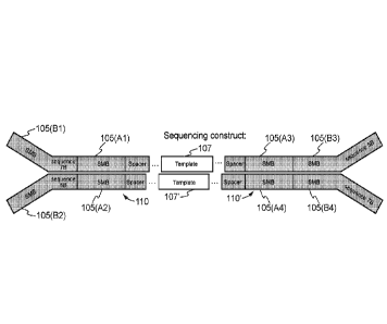
shown in Fig. 2A, a library construct 200 of the sequence construct 100 shown
in Fig. lE may
comprise a SMB sequence 205', followed by a spacer sequence 215', followed by
a template
sequence (e.g., sample nucleic acid sequence) 207, followed by a spacer
sequence 215, followed
by a SMB sequence 205. As shown in Fig. 2B, a first library construct 200 and
a second library
construct 200' may be obtained for the first and second strands of sequence
construct 100 shown
in Fig, IF, respectively. The first library construct 200 may comprise a SMB
sequence 205 (A3),
followed by a spacer sequence 215', followed by a template sequence (e.g.,
sample nucleic acid
sequence) 207, followed by a spacer sequence 215, followed by a SMB sequence
205 (Al). The
19
CA 03049682 2019-07-08
WO 2018/136888 PCT/US2018/014726
second library construct 200' may comprise a SMB sequence 205 (A4), followed
by a spacer
sequence 215, followed by a template sequence (e.g., sample nucleic acid
sequence) 207',
followed by a spacer sequence 215', followed by a SMB sequence 205 (A2). As
shown in Fig.
2C, a first library construct 200 and a second library construct 200' may be
obtained for the first
and second strands of sequence construct 100 shown in Fig, 1H, respectively.
The first library
construct 200 may comprise a SMB sequence 205 (C1), followed by a SMB sequence
205 (B1),
followed by a SMB sequence 205 (Al), followed by a spacer sequence 215',
followed by a
template sequence (e.g., sample nucleic acid sequence) 207, followed by a
spacer sequence 215,
followed by a SMB sequence 205 (A3), followed by a SMB sequence 205 (B3),
followed by a
SMB sequence 205 (C3). The second library construct 200' may comprise a SMB
sequence 205
(C2), followed by a SMB sequence 205 (B2), followed by a SMB sequence 205
(A2), followed
by a spacer sequence 215, followed by a template sequence (e.g., sample
nucleic acid sequence)
207', followed by a spacer sequence 215', followed by a SMB sequence 205 (A4),
followed by a
SMB sequence 205 (B4), followed by a SMB sequence 205 (C4).
In various embodiments, sequence data including the library construct and
genomic positioning
data (e.g., chromosome coordinates) allows for assignment of a unique identity
to a particular
molecule such as the template sequence of each sequence read. For example, at
least one SMB
sequence from the library construct and genomic positioning data informative
of the beginning
(start) and end (stop) of nucleic acid fragments associated with the template
sequence can be
used as unique identifier for the template sequence of each sequence read to
assign the sequence
read to a read group, as discussed in further detail herein. In some
embodiments, other sequence
data such as the sequence of the nucleic acid molecule, or the length of an
individual sequence
read or corresponding fragment may be used in conjunction with or as a
substitute for the SMB
sequence and/or genomic positioning data to establish the unique identity to
the particular
molecule. In some embodiments, sequence reads may further comprise a sample
index sequence
or sample identifier (sample ID as shown in Fig. 1H) (e.g., for tracking
nucleic acid from
different samples). In some embodiments, at least one SMB sequence from the
library construct,
the sample identifier, and optionally genomic positioning data informative of
the beginning
(start) and end (stop) of nucleic acid fragments associated with the template
sequence can be
CA 03049682 2019-07-08
WO 2018/136888 PCT/US2018/014726
used as the unique identifier for the template sequence of each sequence read
to assign the
sequence read to a read group.
In some embodiments, a method herein comprises filtering sequence reads. In
some
embodiments, certain sequence reads are filtered out (i.e., excluded from
sequence read analysis
for determining the presence or absence of a genetic alteration). Reads that
may be filtered out
include, for example, discordant reads, ambiguous reads, off-target reads,
reads having a SMB
sequence with one or more undetermined base calls, reads having a low quality
sample index,
and reads having a low quality barcode (e.g., single molecule barcode). Low
quality sequences
(e.g., barcode, index) may be identified according to base quality scores for
one or more
nucleotide positions in a sequence. A base quality score, or quality score, is
a prediction of the
probability of an error in base calling. Quality scores may be generated by a
quality table that
uses a set of quality predictor values, and can depend on certain
characteristics of the sequencing
platform used for generating sequence reads. Generally, a high quality score
indicates a base call
is more reliable and less likely is an incorrect base call. For example, for
base calls with a
quality score of 40, one incorrect base call in 10,000 base calls is
predicted. For base calls with a
quality score of 30, one incorrect base call in 1,000 base calls is predicted.
For base calls with a
quality score of 20, one incorrect base call in 100 base calls is predicted.
For base calls with a
quality score of 10, one incorrect base call in 10 base calls is predicted.
In some embodiments, a low quality sample index comprises at least one base
having a base
quality score less than about 20. For example, a low quality sample index may
comprises at least
one base having a base quality score less than about 20, less than about 19,
less than about 18,
less than about 17, less than about 16, less than about 15, less than about
14, less than about 13,
less than about 12, less than about 11, or less than about 10. In some
embodiments, a low quality
sample index comprises at least one base having a base quality score less than
about 14. In some
embodiments, a low quality sample index comprises at least two bases having a
base quality
score less than about 25. For example, a low quality sample index may
comprises at least two
bases having a base quality score less than about 25, less than about 24, less
than about 23, less
than about 22, less than about 21, less than about 20, less than about 19,
less than about 18, less
21
CA 03049682 2019-07-08
WO 2018/136888 PCT/US2018/014726
than about 17, less than about 16, or less than about 15. In some embodiments,
a low quality
sample index comprises at least two bases haying a base quality score less
than about 21.
In some embodiments, a low quality barcode comprises at least one base having
a base quality
.. score less than about 20. For example, a low quality barcode may comprises
at least one base
having a base quality score less than about 20, less than about 19, less than
about 18, less than
about 17, less than about 16, less than about 15, less than about 14, less
than about 13, less than
about 12, less than about 11, or less than about 10. In some embodiments, a
low quality barcode
comprises at least one base haying a base quality score less than about 14. In
some
embodiments, a low quality barcode comprises at least two bases haying a base
quality score less
than about 25. For example, a low quality barcode may comprises at least two
bases haying a
base quality score less than about 25, less than about 24, less than about 23,
less than about 22,
less than about 21, less than about 20, less than about 19, less than about
18, less than about 17,
less than about 16, or less than about 15. In some embodiments, a low quality
barcode comprises
at least two bases having a base quality score less than about 21.
In some embodiments, a method herein comprises identifying on-target reads. In
some
embodiments, a rend is identified as on-target when the read aligns with a
genomic region
corresponding to a probe oligonucleotide sequence. As described in further
detail herein, probe
oligonucleotide sequences generally align to (i.e., correspond to) specific
regions of a genome
(e.g., a reference genome) and often comprise nucleotide sequences
corresponding to certain
genomic sequences of interest. A read that aligns to a genomic region to which
a probe
oligonucleotide also aligns is considered an on-target read. A sequence read
may be considered
on target when the entire read length aligns to a genomic region to which a
probe oligonucleotide
also aligns, in some embodiments. In some embodiments, a read is identified as
on-target when
part of the read aligns with a genomic region corresponding to a probe
oligonucleotide sequence,
and part of the read aligns within a genomic region adjacent to a genomic
region corresponding
to a probe oligonucleotide sequence. Generally, in such instances, the road
aligns to a
contiguous genomic sequence comprising 1) part of a genomic region
corresponding to a probe
oligonucleotide sequence and 2) a genomic region adjacent to the genomic
region corresponding
to a probe oligonucleotide sequence. The latter genomic region may be located
upstream or
22
CA 03049682 2019-07-08
WO 2018/136888 PCT/US2018/014726
downstream of the genomic region corresponding to a probe oligonucleotide
sequence. For
example, a sequence read may be considered on target when part of the read
(e.g., at least about
5% of the read, 10% of the read, 20% of the read, 30% of the read, 40% of the
read, 50% of the
read, 60% of the read, 70% of the read, 80% of the read, 90% of the read) with
a genomic region
corresponding to a probe oligonucleotide sequence and the remainder of the
read aligns to a
genomic sequence directly upstream or downstream of a genomic region
corresponding to a
probe oligonucleotide sequence. A sequence read may be considered on target
when no part of
the read aligns to a probe sequence and the entire read length aligns to a
genomic sequence
directly upstream or downstream of a genomic region corresponding to a probe
oligonucleotide
sequence, in some embodiments.
A sequence comprising a probe sequence (i.e., genomic sequence corresponding
to a probe
sequence) and additional genomic sequence upstream and/or downstream to the
probe sequence
may be referred to as a padded probe sequence. A collection of padded probe
sequences may be
referred to as a padded panel. In some embodiments, a padded probe sequence
comprises at least
1 nucleotide of genomic sequence directly upstream and/or downstream to the
genomic sequence
corresponding to the probe sequence. For example, a padded probe sequence may
comprise at
least about 5, 10, 20, 30, 40, 50, 100, 150, 200, 250, 300, 400, 500 or 1000
nucleotides of
genomic sequence directly upstream and/or downstream to the genomic sequence
corresponding
to the probe sequence. In some embodiments, a padded probe sequence comprises
250
nucleotides of genomic sequence directly upstream and 250 nucleotides of
genomic sequence
directly downstream to the genomic sequence corresponding to the probe
sequence.
Probe oligonucleotide sequences may be stored as a panel of sequences in a
database. In some
embodiments, reads are aligned directly with probe oligonucleotide sequences
(e.g., probe
oligonucleotide sequences stored in a table or database, with or without
adjacent genomic region
sequences as described above), and such reads are identified as on-target
reads. For example,
sequence reads may be aligned to a panel of sequences in a database without
first being mapped
to a reference genome. A sequence read may be considered on-target when the
entire read length
aligns to a probe sequence, in some embodiments. In some embodiments, sequence
reads are
aligned directly to padded probe sequences, as described above. For example, a
sequence read
23
CA 03049682 2019-07-08
WO 2018/136888 PCT/US2018/014726
may be considered on-target when part of the read (e.g., at least about 5% of
the read, 10% of the
read, 20% of the read, 30% of the read, 40% of the read, 50% of the read, 60%
of the read, 70P/0
of the read, 80% of the read, 90% of the read) aligns to a probe sequence and
the remainder of
the read aligns to a genomic sequence directly upstream or downstream of the
probe sequence, in
some embodiments. A sequence read may be considered on-target when no part of
the read
aligns to a probe sequence and the entire read length aligns to a genomic
sequence directly
upstream or downstream of the probe sequence, in some embodiments.
In various embodiments, a method herein comprises assigning sequence reads to
read groups. In
some embodiments, a method herein comprises assigning on-target sequence reads
to read
groups. Sequence reads (or on-target sequence reads) may be assigned to read
groups according
to a read group signature. In some embodiments, a read group signature
comprises one or more
of the following: (i) at least one SMB sequence, (ii) genomic positioning data
informative of the
beginning (start) and end (stop) of a nucleic acid fragment, (iii) length of
an individual sequence
read or corresponding fragment, and (iv) a sample index sequence or sample
identifier. In some
embodiments, sequence reads (or on-target sequence reads) comprising one or
more of the
following: (i) at least one SMB sequence, (ii) genomic positioning data
informative of the
beginning (start) and end (stop) of a nucleic acid fragment, (iii) length of
an individual sequence
read or corresponding fragment, and (iv) a sample index sequence or sample
identifier that are
.. similar to the read group signature are assigned to a read group identified
by the read group
signature.
The process for assigning sequence reads (or on-target sequence reads) to a
read group may
include identifying the at least one SMB sequence, comparing one or more of
the following: (i)
the at least one SMB sequence, (ii) genomic positioning data informative of
the beginning (start)
and end (stop) of a nucleic acid fragment, (iii) length of an individual
sequence read or
corresponding fragment, and (iv) a sample index sequence or sample identifier
to the read group
signature, and determining whether the one or more pieces of sequence data are
similar to the
data within the read group signature. When the one or more pieces of sequence
data are similar
to the data within the read group signature, assigning the sequence read to a
read group identified
by the read group signature. In certain embodiments, sequence reads (or on-
target sequence
24
CA 03049682 2019-07-08
WO 2018/136888 PCT/US2018/014726
reads) comprising: (i) at least one SMB sequence and (ii) genomic positioning
data informative
of the beginning (start) and end (stop) of a nucleic acid fragment, that are
similar to the read
group signature are assigned to a read group identified by the read group
signature. The at least
one SMB sequence may be a predetermined non-randomly generated molecular
barcode
sequence (a non-random oligonucleotide sequence)..
In various embodiments, the at least one SMB sequence is two or more SMB
sequences. For
example, a first SMB sequence attached to the start of a nucleic fragment and
a second SMB
sequence attached to the end of the nucleic fragment. In such an instance, a
read group signature
may comprise one or more of the following: (i) a concatenation of the two or
more SMB
sequences, (ii) genomic positioning data informative of the beginning (start)
and end (stop) of a
nucleic acid fragment, (iii) length of an individual sequence read or
corresponding fragment, and
(iv) a sample index sequence or sample identifier. In some embodiments,
sequence reads (or on-
target sequence reads) comprising one or more of the following: (i) two or
more SMB sequences,
(ii) genomic positioning data informative of the beginning (start) and end
(stop) of a nucleic acid
fragment, (iii) length of an individual sequence read or corresponding
fragment, and (iv) a
sample index sequence or sample identifier that are similar to the read group
signature are
assigned to a read group identified by the read group signature.
The process for assigning sequence reads (or on-target sequence reads)
comprising two or more
SMB sequences to a read group may include identifying the two or more SMB
sequences,
concatenating the SMB sequences using a predetermined scheme (e.g., from start
to end on a
read) to generate a string or concatenation of the identified SMB sequences,
and comparing one
or more of the following: (i) the string or concatenation of the identified
SMB sequences, (ii)
genomic positioning data informative of the beginning (start) and end (stop)
of a nucleic acid
fragment, (iii) length of an individual sequence read or corresponding
fragment, and (iv) a
sample index sequence or sample identifier to the read group signature and
determining whether
the one or more pieces of sequence data are similar to the data within the
read group signature.
When the one or more pieces of sequence data are similar to the data within
the read group
signature, assigning the sequence read to a read group identified by the read
group signature. In
certain embodiments, sequence reads (or on-target sequence reads) comprising:
(i) two or more
CA 03049682 2019-07-08
WO 2018/136888 PCT/US2018/014726
SMB sequences and (ii) genomic positioning data informative of the beginning
(start) and end
(stop) of a nucleic acid fragment, that are similar to the read group
signature are assigned to a
read group identified by the read group signature. The two or more SMB
sequences may be a
predetermined non-randomly generated molecular barcode sequence (a non-random
oligonucleotide sequence).
In various embodiments, assigning sequence reads to read groups includes a
comparison between
multiple pieces of data of the sequence read and the read signature, and any
singular or
combination of similarities identified between the data (e.g., only the SMB
sequence being
similar to the SMB sequence in the read signature) can be used to make a
determination as to
whether the sequence read should be assigned to the read group identified by
the read signature.
The similarity between data may be identified using one or more predefined
rules or parameters.
In some embodiments, sequence reads comprising start and end positions and an
SMB sequence
similar to the read group signature comprise an SMB sequence identical to the
SMB sequence of
.. the read group signature, and a start and end position identical to the
start and end position of the
read group signature. In some embodiments, sequence reads comprising start and
end positions
and an SMB similar to the read group signature comprise an SMB sequence
identical to the SMB
sequence of the read group signature, and a start and/or end position that is
different from the
start and/or end position of the read group signature. in some embodiments,
sequence reads
comprising start and end positions and an SMB similar to the read group
signature comprise an
SMB sequence comprising one or more nucleotide differences compared to the SMB
sequence
of the read group signature, and a start and end position identical to the
start and end position of
the read group signature.
In some embodiments, a start and/or end position that is different from the
start and/or end
position of the read group signature differs by up to 10 base positions at the
start position. Such
differences generally refer to differences in base position and not
differences in base identity. In
some embodiments, a start and/or end position that is different from the start
and/or end position
of the read group signature differs by up to 10 base positions at the end
position. In some
.. embodiments, a start and/or end position that is different from the start
and/or end position of the
read group signature differs by up to 10 base positions at the start position
and differs by up to 10
26
CA 03049682 2019-07-08
WO 2018/136888 PCT/US2018/014726
base positions at the end position. For example, the start and/or end position
of a read assigned
to a read group may differ by 1 base position, 2 base positions, 3 base
positions, 4 base positions,
base positions, 6 base positions, 7 base positions, 8 base positions, 9 base
positions or 10 base
positions when compared to the corresponding start and/or end position of a
read group
5 signature.
In some embodiments, a start and/or end position that is different from the
start and/or end
position of the read group signature differs by up to 5 base positions at the
start position. Such
differences generally refer to differences in base position and not
differences in base identity. In
some embodiments, a start and/or end position that is different from the start
and/or end position
of the read group signature differs by up to 5 base positions at the end
position. In some
embodiments, a start and/or end position that is different from the start
andlor end position of the
read group signature differs by up to 5 base positions at the start position
and differs by up to 5
base positions at the end position. For example, the start and/or end position
of a read assigned
to a read group may differ by 1 base position, 2 base positions, 3 base
positions, 4 base positions
or 5 base positions when compared to the corresponding start and/or end
position of a read group
signature.
In some embodiments, an SMB sequence comprising one or more nucleotide
differences
compared to the SMB sequence of the read group signature comprises up to 5
nucleotide
differences. Such differences generally refer to nucleotide identity within an
SMB sequence and
not the start or end position of the SMB sequence. For example, an SMB
sequence may
comprise 1 nucleotide difference, 2 nucleotide differences, 3 nucleotide
differences, 4 nucleotide
differences, or 4 nucleotide differences when compared to the corresponding
SMB sequence of a
read group signature. In some embodiments, an SMB sequence comprises no more
than 1
nucleotide difference when compared to the corresponding SMB sequence of a
read group
signature.
In some embodiments, sequence reads comprising start and end positions and an
SMB sequence
similar to the read group signature comprise an SMB sequence identical to the
SMB sequence of
the read group signature, and a start and/or end position that is up to 5 base
positions different
27
CA 03049682 2019-07-08
WO 2018/136888 PCT/US2018/014726
from the start and/or end position of the read group signature. In some
embodiments, sequence
reads comprising start and end positions and an SMB sequence similar to the
read group
signature comprise an SMB sequence comprising one nucleotide difference
compared to the
SMB sequence of the read group signature, and a start and end position
identical to the start and
end position of the read group signature. In some embodiments, sequence reads
comprising start
and end positions and an SMB sequence similar to the read group signature
comprise an SMB
sequence comprising one nucleotide difference compared to the SMB sequence of
the read group
signature and a start and/or end position that is up to 5 base positions
different from the start
and/or end position of the read group signature.
In some embodiments, sequence reads comprising start and end positions and an
SMB sequence
similar to the read group signature comprise paired-end sequencing reads. The
terms "paired-end
sequencing reads" and "paired-end reads" are used synonymously herein and
refer to a pair of
sequencing reads where each member of the pair is derived from sequencing
complementary
strands of a polynucleotide fragment. Each read of a paired-end read is
referred to herein as a
"read mate." Thus, in some embodiments, sequence reads comprising start and
end positions
and an SMB sequence similar to the read group signature comprise read mates
generated by a
paired-end sequencing process. Paired-end reads are often mapped to opposing
ends of the same
polynucleotide fragment, according to a reference genome, as described herein.
Thus, sequence
reads comprising start and end positions similar to the read group signature
generally comprise a
pair (or a plurality of pairs) of read mates, where the start (i.e., 5' base
position) of the first
member of the pair corresponds to the start position of the read group
signature (i.e., the 5' end
of a nucleic acid fragment); and the start (i.e., 5' base position) of the
second member of the pair
corresponds to the end position of the read group signature (i.e., the 3' end
of a nucleic acid
fragment, which also may be considered the 5' end of the complementary strand
of a fragment).
In certain instances, both read mates are presented as sequences corresponding
to the first strand
(i.e., + strand, leading strand) of the corresponding fragment for
bioinformatic purposes, such
that when viewing sequences of read mates from left to right, the read
sequences are in a 5' to 3'
orientation. In such instances, reads comprising start and end positions
similar to the read group
signature generally comprise a pair (or a plurality of pairs) of read mates,
where the start (i.e., 5'
base position) of the first member of the pair corresponds to the start
position of the read group
28
CA 03049682 2019-07-08
WO 2018/136888 PCT/US2018/014726
signature (i.e., the 5' end of a nucleic acid fragment); and the end (i.e., 3'
base position) of the
second member of the pair corresponds to the end position of the read group
signature (i.e., the
3' end of a nucleic acid fragment).
In some embodiments, sequence reads comprising start and end positions similar
to the read
group signature generally comprise a pair (or a plurality of pairs) of read
mates, where the start
(i.e., 5' base position) of the first member of the pair corresponds to a
position within 5 bases of
the start position of the read group signature (i.e., the 5' end of a nucleic
acid fragment); and the
start (i.e., 5' base position) of the second member of the pair corresponds to
a position within 5
bases of the end position of the read group signature (i.e., the 3' end of a
nucleic acid fragment,
which also may be considered the 5' end of the complementary strand of the
fragment). A start
position (i.e., 5' base position) of a read in this context generally refers
to the portion of the read
that aligns to the reference genome and not any additional parts of the read
(e.g., adapter
sequence, barcode, index, and the like). In some embodiments, read mates will
comprise
different SMB sequences by way of being generated from opposing ends of a
nucleic acid
fragment. In such instances, one SMB sequence is used for a read group
signature and
generation of read groups. In some embodiments, a first read mate comprises a
region that is
complementary to a region in a second read mate. Such region may be referred
to as an overlap,
a sequence read overlap, or an overlapping region, and may be useful for
performing sequence
error correction, as described herein. An overlap may comprise one base pair,
a plurality of
contiguous base pairs, or may span the entire length of one or both read
mates.
In some embodiments, a method herein comprises generating a multiplicity
table. A multiplicity
table generally includes the number of reads in each read group and/or number
of read groups
having a predetermined number of reads, and can be useful for consensus
making. For methods
using paired-end sequencing, a multiplicity table generally includes the
number of read pairs (i.e.
read mates) in each read group and/or number of read groups having a
predetermined number of
read pairs. Typically, a multiplicity table is generated after read group
assignments and prior to
consensus making.
29
CA 03049682 2019-07-08
WO 2018/136888 PCT/US2018/014726
In some embodiments, a method herein comprises generating a consensus.
Generally, a
consensus is generated by collapsing a set of sequence reads (e.g., a set of
sequence read pairs) in
a read group to generate a single nucleotide sequence that corresponds to a
unique nucleic acid
molecule in the sample from which the sequence reads were generated. In
certain embodiments,
a consensus is generated for each read group containing a plurality of reads,
which includes
singleton read groups. Singleton read groups are read groups that include no
more than 1 pair of
read mates. In certain embodiments, a consensus is generated for each read
group containing a
plurality of reads, which includes doubleton read groups. Doubleton read
groups are read groups
that include no more than 2 pairs of read mates. In certain embodiments, a
consensus is
generated for each read group containing a plurality of reads, which includes
doubleton read
groups and excludes singleton read groups. In certain embodiments, a consensus
is generated for
each read group containing a plurality of reads, which excludes doubleton read
groups and
excludes singleton read groups. Consensus sequences can be generated from read
groups by any
suitable method including, for example, linear or non-linear methods for
consensus making
derived from digital communication theory, information theory, or
bioinformatics (e.g.,
averaging, voting, statistical, dynamic programming, maximum a posteriori or
maximum
likelihood detection, Bayesian, hidden Markov or support vector machine
methods, and the like).
In some embodiments, generating a consensus comprises sequence error
correction (e.g.,
sequence errors in sequence reads from a sequencing process and/or sequence
errors introduced
in an amplification reaction). In some embodiments, sequencing error
correction comprises read
group correction. Generally, read group correction comprises correction of a
nucleotide in a read
assigned to a read group that does not match the nucleotide at that position
for other reads in the
read group. In certain instances, read group correction comprises designating
a nucleotide as an
.. unreadable or low quality base ("N") in a read assigned to a read group
that does not match the
nucleotide at that position for other reads in the read group. In some
embodiments, sequence
error correction comprises sequence read overlap correction for paired-end
sequence reads. For
example, for a base pair in an overlap position (e.g., A and its complementary
base T), when a
non-complementary base is read (e.g., A and a non-complementary base C or G),
the base at the
complementary position is corrected to T (e.g., when the corresponding A has a
higher base
quality than the non-complementary C or G). In certain instances, read mates
from paired-end
CA 03049682 2019-07-08
WO 2018/136888 PCT/US2018/014726
sequencing are presented as sequences corresponding to the first strand (i.e.,
+ strand, leading
strand) of the corresponding fragment, as described above. In such instances,
when performing a
sequence error correction in an overlap region, mismatched bases are
identified and corrected
such that the base having the higher quality is kept.
In some embodiments, generating a consensus for a read group comprises
determining the total
number and identity of nucleotide at each position covered by reads. In some
embodiments, a
position in a consensus sequence is assigned a nucleotide identity when a
majority of the
nucleotides from the reads agree (i.e., are read as the same nucleotide
identity) at the position.
For example, a position in a consensus sequence may be assigned a nucleotide
identity when
more than 50,10, about 60% or more, about 70% or more, about 80% or more,
about 85% or
more, about 90% or more, about 95% or more, or about 99% or more of the
nucleotides from the
reads agree (i.e., are read as the same nucleotide identity) at the position.
In some embodiments,
a position in a consensus sequence is assigned a nucleotide identity when
about 90% or more of
the nucleotides from the reads agree (i.e., are read as the same nucleotide
identity) at the
position.
In some embodiments, generating a consensus for a read group comprises
determining an overall
base quality for the nucleotide at a position covered by reads. An overall
base quality may be
generated according to base quality scores from multiple reads (e.g., reads in
a read group) for a
nucleotide position in a consensus. An overall base quality may be expressed
as a mean base
quality, a median base quality, a maximal base quality, an average base
quality, and the like. In
some embodiments, generating a consensus for a read group comprises
determining a mean base
quality for the nucleotide at each position covered by reads. In some
embodiments, generating a
consensus for a read group comprises determining a median base quality for the
nucleotide at
each position covered by reads. In some embodiments, generating a consensus
for a read group
comprises determining a maximal base quality for the nucleotide at each
position covered by
reads. Generally, maximal base quality is the best base quality of all the
base qualities of the
bases for which the nucleotide identities agree.
31
CA 03049682 2019-07-08
WO 2018/136888 PCT/US2018/014726
In some embodiments, a position in a consensus sequence is assigned an overall
base quality
(e.g., mean base quality, median base quality, maximal base quality) for
nucleotide identity when
a majority of the base identities agree (i.e., have the same base identity)
for a nucleotide from
reads (e.g., reads in a read group). For example, a position in a consensus
sequence may be
.. assigned an overall base quality for nucleotide identity when more than
50%, about 60% or
more, about 70% or more, about 80% or more, about 85% or more, about 90% or
more, about
95% or more, or about 99% or more of the base identities agree for a
nucleotide from reads (e.g.,
reads in a read group). In some embodiments, a position in a consensus
sequence is assigned an
overall base quality for nucleotide identity when about 90% or more of the
base identities agree
.. (i.e., have the same base identity) for a nucleotide from reads (e.g.,
reads in a read group).
Generally, an overall base quality is calculated according to the base
qualities of the bases for
which the nucleotide identities agree and base qualities of bases for which
the nucleotide
identities do not agree are not included in the overall base quality
determination.
In some embodiments, a method herein comprises determining the presence or
absence of a
genetic alteration based on one or more consensus sequences for one or more
read groups. In
some embodiments, the presence or absence of a single nucleotide alteration
(SNA) is
determined according to one or more consensus sequences. In some embodiments,
the presence
or absence of a copy number alteration (CNA) is determined according to one or
more consensus
sequences. In some embodiments, a method comprises tallying consensus base
counts at each
position. Tallying consensus base counts at each position generally involves
identifying the
number of times a particular nucleotide at a particular base position appears
in the consensus for
each read group. In some embodiments, a method comprises calculating allele
depth (i.e., how
many times a particular base identity is counted) and allele fraction (e.g.,
proportion of a
.. particular base identity relative to total counted at a base position and
often relative to a reference
base in a reference genome). In some embodiments, the presence or absence of a
single
nucleotide alteration (SNA) is determined according to consensus base counts
at each position.
In some embodiments, the presence or absence of a single nucleotide alteration
(SNA) is
determined according to allele depth and fraction. For example, at a given
genomic base
.. position appearing in 10 consensus sequences (representing 10 unique
nucleic acid molecules
from a sample), 8 base positions are identified as A and 2 are identified as
G. Assuming the
32
CA 03049682 2019-07-08
WO 2018/136888 PCT/US2018/014726
reference base is an A (e.g., identified from a reference genome), an SNA of A
to G at that
position at an allele depth of 2 and an allele fraction of 20% would be
determined.
In some embodiments, a method comprises determining a probe coverage
quantification. In
some embodiments, determining the presence or absence of a copy number
alteration (CNA) is
determined according to a probe coverage quantification. Probe coverage
generally refers to a
quantification of sequence reads or consensus sequences mapped to each
nucleotide position in a
probe oligonucleotide (e.g., a probe oligonucleotide used in a nucleic acid
capture process,
described in further detail herein). In some embodiments, determining a probe
coverage
quantification comprises determining the number of sequence reads that map at
each of the
nucleotide positions in a probe oligonucleotide. Sequence reads may be shorter
in length than
probe oligonucleotides and/or may partially overlap with probe oligonucleotide
sequences.
Thus, the quantification of sequence reads mapped to each nucleotide in a
probe can vary along
the length of a probe oligonucleotide. Accordingly, in some embodiments,
determining a probe
coverage quantification comprises determining the median number of sequence
reads mapped at
each of the nucleotide positions in a probe. In some embodiments, the median
number of
sequence reads mapped at each of the nucleotide positions for each probe
oligonucleotide is the
probe coverage quantification for each probe oligonucleotide. In some
embodiments,
determining a probe coverage quantification comprises determining the number
of consensus
sequences that map at each of the nucleotide positions in a probe
oligonucleotide. Consensus
sequences may be shorter in length than probe oligonucleotides and/or may
partially overlap
with probe oligonucleotide sequences. Thus, the quantification of consensus
sequences mapped
to each nucleotide in a probe can vary along the length of a probe
oligonucleotide. Accordingly,
in some embodiments, determining a probe coverage quantification comprises
determining the
median number of consensus sequences mapped at each of the nucleotide
positions in a probe.
In some embodiments, a probe coverage quantification is a normalized probe
coverage
quantification.
An embodiment of nucleic acid sequence read processing is illustrated in Fig.
7. Probe-captured
nucleic acid reads 440 are subjected to sequence read processing steps to
generate sorted and
indexed reads 560. One embodiment of sequence read processing is illustrated
as process 700.
33
CA 03049682 2019-07-08
WO 2018/136888 PCT/US2018/014726
Probe-captured nucleic acid reads 440 are subjected to BCL conversion 705
which generates
FASTQ reads 710. FASTQ reads 710 are input for SMB extraction and de-
multiplexing 515
which generates SMB-extracted and de-multiplexed reads 720. SMB-extracted and
de-
multiplexed reads 720 are subjected to chastity filtering 725 which generates
filtered reads 730.
Filtered reads 730 are subjected to adapter trimming 730 which generates
trimmed reads 740.
Trimmed reads 740 are input for read alignment 745 which generates aligned
reads 750. Aligned
reads 750 are input for sorting and indexing 755 which generates sorted and
indexed reads 760.
An embodiment of consensus generating is illustrated in Fig. 8. Sorted and
indexed reads 760
are input for a consensus generating process which generates a consensus 860.
One embodiment
of a consensus generating process is illustrated as process 800. Sorted and
indexed reads 760 are
subjected to filtering 805 which generates filtered reads 810. Filtered reads
810 are input for
identifying on-target reads 815 which generates on-target reads 820. On-target
reads 820 are
input for assigning read groups 825 which generates candidate reads for read
groups 830.
Candidate reads for read groups 830 are subjected to position and SMB
jittering 835 which
generates jittered read groups 840. Jittered read groups 840 are input for a
consensus maker 845
which generates a consensus 860.
An embodiment of consensus generating for duplex sequencing is illustrated in
Fig. 9. Sorted
and indexed reads 760 are input for a consensus generating process which
generates a consensus
860. One embodiment of a consensus generating process is illustrated as
process 800. Sorted
and indexed reads 760 are subjected to filtering 805 which generates filtered
reads 810. Filtered
reads 810 are input for identifying on-target reads 815 which generates on-
target reads 820. On-
target reads 820 are input for assigning read groups 825 which generates
candidate reads for read
groups 830. Candidate reads for read groups 830 are subjected to position and
SMB jittering 835
which generates jittered read groups 840. uttered read groups 840 are input
for a consensus
maker 845 which generates a consensus 860 based on reads from the first strand
in the DNA
duplex. A second round of read group assignments and consensus making 826 is
performed to
generate a consensus 860 based on reads from the second strand in the DNA
duplex.
34
CA 03049682 2019-07-08
WO 2018/136888 PCT/US2018/014726
Embodiments for genetic alteration classification are illustrated in Fig. 10.
In one embodiment, a
consensus 860 is input for a copy number alteration (CNA) identifier 865 which
generates a
CNA classification 870. In another embodiment, a consensus 860 is input for a
single nucleotide
alteration (SNA) identifier 875 which generates an SNA classification 880.
Samples
Provided herein are systems, methods and products for analyzing nucleic acids.
In some
embodiments, nucleic acid fragments in a mixture of nucleic acid fragments are
analyzed.
Nucleic acid fragments may be referred to as nucleic acid templates, and the
terms may be used
interchangeably herein. A mixture of nucleic acids can comprise two or more
nucleic acid
fragment species having the same or different nucleotide sequences, different
fragment lengths,
different origins (e.g., genomic origins, fetal vs. maternal origins, cell or
tissue origins, cancer vs.
non-cancer origin, tumor vs. non-tumor origin, sample origins, subject
origins, and the like), or
combinations thereof.
Nucleic acid or a nucleic acid mixture utilized in systems, methods and
products described
herein often is isolated from a sample obtained from a subject (e.g., a test
subject). A subject can
be any living or non-living organism, including but not limited to a human, a
non-human animal,
a plant, a bacterium, a fungus, a protest or a pathogen. Any human or non-
human animal can be
selected, and may include, for example, mammal, reptile, avian, amphibian,
fish, ungulate,
ruminant, bovine (e.g., cattle), equine (e.g., horse), caprine and ovine
(e.g., sheep, goat), swine
(e.g., pig), camelid (e.g., camel, llama, alpaca), monkey, ape (e.g., gorilla,
chimpanzee), ursid
(e.g., bear), poultry, dog, cat, mouse, rat, fish, dolphin, whale and shark. A
subject may be a
male or female (e.g., woman, a pregnant woman). A subject may be any age
(e.g., an embryo, a
fetus, an infant, a child, an adult). A subject may be a cancer patient, a
patient suspected of
having cancer, a patient in remission, a patient with a family history of
cancer, and/or a subject
obtaining a cancer screen. In some embodiments, a test subject is a female. In
some
embodiments, a test subject is a human female. In some embodiments, a test
subject is a male. In
some embodiments, a test subject is a human male.
CA 03049682 2019-07-08
WO 2018/136888 PCT/US2018/014726
Nucleic acid may be isolated from any type of suitable biological specimen or
sample (e.g., a test
sample). A sample or test sample can be any specimen that is isolated or
obtained from a subject
or part thereof (e.g., a human subject, a pregnant female, a cancer patient, a
fetus, a tumor).
Non-limiting examples of specimens include fluid or tissue from a subject,
including, without
limitation, blood or a blood product (e.g., serum, plasma, or the like),
umbilical cord blood,
chorionic villi, amniotic fluid, cerebrospinal fluid, spinal fluid, lavage
fluid (e.g.,
bronchoalveolar, gastric, peritoneal, ductal, ear, arthroscopic), biopsy
sample (e.g., from pre-
implantation embryo; cancer biopsy), celocentesis sample, cells (blood cells,
placental cells,
embryo or fetal cells, fetal nucleated cells or fetal cellular remnants,
normal cells, abnormal cells
(e.g., cancer cells)) or parts thereof (e.g., mitochondrial, nucleus,
extracts, or the like), washings
of female reproductive tract, urine, feces, sputum, saliva, nasal mucous,
prostate fluid, lavage,
semen, lymphatic fluid, bile, tears, sweat, breast milk, breast fluid, the
like or combinations
thereof. In some embodiments, a biological sample is a cervical swab from a
subject. A fluid or
tissue sample from which nucleic acid is extracted may be acellular (e.g.,
cell-free). In some
embodiments, a fluid or tissue sample may contain cellular elements or
cellular remnants. In
some embodiments, fetal cells or cancer cells may be included in the sample.
A sample can be a liquid sample. A liquid sample can comprise extracellular
nucleic acid (e.g.,
circulating cell-free DNA). Non-limiting examples of liquid samples, include,
blood or a blood
product (e.g., serum, plasma, or the like), urine, biopsy sample (e.g., liquid
biopsy for the
detection of cancer), a liquid sample described above, the like or
combinations thereof In
certain embodiments, a sample is a liquid biopsy, which generally refers to an
assessment of a
liquid sample from a subject for the presence, absence, progression or
remission of a disease
(e.g., cancer). A liquid biopsy can be used in conjunction with, or as an
alternative to, a sold
biopsy (e.g., tumor biopsy). In certain instances, extracellular nucleic acid
is analyzed in a liquid
biopsy.
In some embodiments, a biological sample may be blood, plasma or serum. The
term "blood"
encompasses whole blood, blood product or any fraction of blood, such as
serum, plasma, buffy
coat, or the like as conventionally defined. Blood or fractions thereof often
comprise
nucleosomes. Nucleosomes comprise nucleic acids and are sometimes cell-free or
intracellular.
36
CA 03049682 2019-07-08
WO 2018/136888 PCT/US2018/014726
Blood also comprises buffy coats. Buffy coats are sometimes isolated by
utilizing a ficoll
gradient. Buffy coats can comprise white blood cells (e.g., leukocytes, T-
cells, B-cells, platelets,
and the like). Blood plasma refers to the fraction of whole blood resulting
from centrifugation of
blood treated with anticoagulants. Blood serum refers to the watery portion of
fluid remaining
after a blood sample has coagulated. Fluid or tissue samples often are
collected in accordance
with standard protocols hospitals or clinics generally follow. For blood, an
appropriate amount
of peripheral blood (e.g., between 3 to 40 milliliters, between 5 to 50
milliliters) often is
collected and can be stored according to standard procedures prior to or after
preparation.
An analysis of nucleic acid found in a subject's blood may be performed using,
e.g., whole
blood, serum, or plasma. An analysis of fetal DNA found in maternal blood, for
example, may
be performed using, e.g., whole blood, serum, or plasma. An analysis of tumor
DNA found in a
patient's blood, for example, may be performed using, e.g., whole blood,
serum, or plasma.
Methods for preparing serum or plasma from blood obtained from a subject
(e.g., a maternal
subject; cancer patient) are known. For example, a subject's blood (e.g., a
pregnant woman's
blood; cancer patient's blood) can be placed in a tube containing EDTA or a
specialized
commercial product such as Vacutainer SST (Becton Dickinson, Franklin Lakes,
N.J.) to prevent
blood clotting, and plasma can then be obtained from whole blood through
centrifugation.
Serum may be obtained with or without centrifugation-following blood clotting.
If
centrifugation is used then it is typically, though not exclusively, conducted
at an appropriate
speed, e.g., 1,500-3,000 times g. Plasma or serum may be subjected to
additional centrifugation
steps before being transferred to a fresh tube for nucleic acid extraction. In
addition to the
acellular portion of the whole blood, nucleic acid may also be recovered from
the cellular
fraction, enriched in the buffy coat portion, which can be obtained following
centrifugation of a
whole blood sample from the subject and removal of the plasma.
A sample may be heterogeneous. For example, a sample may include more than one
cell type
and/or one or more nucleic acid species. In some instances, a sample may
include (i) fetal cells
and maternal cells, (ii) cancer cells and non-cancer cells, and/or (iii)
pathogenic cells and host
cells. In some instances, a sample may include (i) cancer and non-cancer
nucleic acid, (ii)
pathogen and host nucleic acid, (iii) fetal derived and maternal derived
nucleic acid, and/or more
37
CA 03049682 2019-07-08
WO 2018/136888 PCT/US2018/014726
generally, (iv) mutated and wild-type nucleic acid. In some instances, a
sample may include a
minority nucleic acid species and a majority nucleic acid species, as
described in further detail
below. In some instances, a sample may include cells and/or nucleic acid from
a single subject
or may include cells and/or nucleic acid from multiple subjects.
Cell types
As used herein, a "cell type" refers to a type of cell that can be
distinguished from another type
of cell. Extracellular nucleic acid can include nucleic acid from several
different cell types.
Non-limiting examples of cell types that can contribute nucleic acid to
circulating cell-free
nucleic acid include liver cells (e.g., hepatocytes), lung cells, spleen
cells, pancreas cells, colon
cells, skin cells, bladder cells, eye cells, brain cells, esophagus cells,
cells of the head, cells of the
neck, cells of the ovary, cells of the testes, prostate cells, placenta cells,
epithelial cells,
endothelial cells, adipocyte cells, kidney/renal cells, heart cells, muscle
cells, blood cells (e.g.,
white blood cells), central nervous system (CNS) cells, the like and
combinations of the
foregoing. In some embodiments, cell types that contribute nucleic acid to
circulating cell-free
nucleic acid analyzed include white blood cells, endothelial cells and
hepatocyte liver cells.
Different cell types can be screened as part of identifying and selecting
nucleic acid loci for
which a marker state is the same or substantially the same for a cell type in
subjects having a
medical condition and for the cell type in subjects not having the medical
condition, as described
in further detail herein.
A particular cell type sometimes remains the same or substantially the same in
subjects having a
medical condition and in subjects not having a medical condition. In a non-
limiting example, the
number of living or viable cells of a particular cell type may be reduced in a
cell degenerative
condition, and the living, viable cells are not modified, or are not modified
significantly, in
subjects having the medical condition.
A particular cell type sometimes is modified as part of a medical condition
and has one or more
different properties than in its original state. In a non-limiting example, a
particular cell type
may proliferate at a higher than normal rate, may transform into a cell having
a different
38
CA 03049682 2019-07-08
WO 2018/136888 PCT/US2018/014726
morphology, may transform into a cell that expresses one or more different
cell surface markers
and/or may become part of a tumor, as part of a cancer condition. In
embodiments for which a
particular cell type (i.e., a progenitor cell) is modified as part of a
medical condition, the marker
state for each of the one or more markers assayed often is the same or
substantially the same for
the particular cell type in subjects having the medical condition and for the
particular cell type in
subjects not having the medical condition. Thus, the term "cell type"
sometimes pertains to a
type of cell in subjects not having a medical condition, and to a modified
version of the cell in
subjects having the medical condition. In some embodiments, a "cell type" is a
progenitor cell
only and not a modified version arising from the progenitor cell. A "cell
type" sometimes
pertains to a progenitor cell and a modified cell arising from the progenitor
cell. In such
embodiments, a marker state for a marker analyzed often is the same or
substantially the same
for a cell type in subjects having a medical condition and for the cell type
in subjects not having
the medical condition.
In certain embodiments, a cell type is a cancer cell. Certain cancer cell
types include, for
example, leukemia cells (e.g., acute myeloid leukemia, acute lymphoblastic
leukemia, chronic
myeloid leukemia, chronic lymphoblastic leukemia); cancerous kidney/renal
cells (e.g., renal cell
cancer (clear cell, papillary type 1, papillary type 2, chromophobe,
oncocytic, collecting duct),
renal adenocarcinoma, hypernephroma, Wilm's tumor, transitional cell
carcinoma); brain tumor
cells (e.g., acoustic neuroma, astrocytoma (grade I: pilocytic astrocytoma,
grade II: low-grade
astrocytoma, grade III: anaplastic astrocytoma, grade IV: glioblastoma (GBM)),
chordoma, cns
lymphoma, craniopharyngioma, glioma (brain stem glioma, ependymoma, mixed
glioma, optic
nerve glioma, subependymoma), medulloblastoma, meningioma, metastatic brain
tumors,
oligodendroglioma, pituitary tumors, primitive neuroectodermal (PNET),
schwannoma, juvenile
pilocytic astrocytoma (WA), pineal tumor, rhabdoid tumor).
Different cell types can be distinguished by any suitable characteristic,
including without
limitation, one or more different cell surface markers, one or more different
morphological
features, one or more different functions, one or more different protein
(e.g., histone)
modifications and one or more different nucleic acid markers. Non-limiting
examples of nucleic
acid markers include single-nucleotide polymorphisms (SNPs), methylation state
of a nucleic
39
CA 03049682 2019-07-08
WO 2018/136888 PCT/US2018/014726
acid locus, short tandem repeats, insertions (e.g., microinsertions),
deletions (microdeletions) the
like and combinations thereof. Non-limiting examples of protein (e.g.,
histone) modifications
include acetylation, methylation, ubiquitylation, phosphorylation,
sumoylation, the like and
combinations thereof.
As used herein, the term a "related cell type" refers to a cell type having
multiple characteristics
in common with another cell type. In related cell types, 75% or more cell
surface markers
sometimes are common to the cell types (e.g., about 80%, 85%, 90% or 95% or
more of cell
surface markers are common to the related cell types).
Nucleic acid
Provided herein are methods for analyzing nucleic acid. The terms "nucleic
acid," "nucleic acid
molecule," "nucleic acid fragment," and "nucleic acid template" may be used
interchangeably
throughout the disclosure. The terms refer to nucleic acids of any composition
from, such as
DNA (e.g., complementary DNA (cDNA), genomic DNA (gDNA) and the like), RNA
(e.g.,
message RNA (mRNA), short inhibitory RNA (siRNA), ribosomal RNA (rRNA), tRNA,
microRNA, RNA highly expressed by a fetus or placenta, and the like), and/or
DNA or RNA
analogs (e.g., containing base analogs, sugar analogs and/or a non-native
backbone and the like),
RNA/DNA hybrids and polyamide nucleic acids (PNAs), all of which can be in
single- or
double-stranded form, and unless otherwise limited, can encompass known
analogs of natural
nucleotides that can function in a similar manner as naturally occurring
nucleotides. A nucleic
acid may be, or may be from, a plasmid, phage, virus, bacterium, autonomously
replicating
sequence (ARS), mitochondria, centromere, artificial chromosome, chromosome,
or other
nucleic acid able to replicate or be replicated in vitro or in a host cell, a
cell, a cell nucleus or
cytoplasm of a cell in certain embodiments. A template nucleic acid in some
embodiments can
be from a single chromosome (e.g., a nucleic acid sample may be from one
chromosome of a
sample obtained from a diploid organism). Unless specifically limited, the
term encompasses
nucleic acids containing known analogs of natural nucleotides that have
similar binding
properties as the reference nucleic acid and are metabolized in a manner
similar to naturally
occurring nucleotides. Unless otherwise indicated, a particular nucleic acid
sequence also
CA 03049682 2019-07-08
WO 2018/136888 PCT/US2018/014726
implicitly encompasses conservatively modified variants thereof (e.g.,
degenerate codon
substitutions), alleles, orthologs, single nucleotide polymorphisms (SNPs),
and complementary
sequences as well as the sequence explicitly indicated. Specifically,
degenerate codon
substitutions may be achieved by generating sequences in which the third
position of one or
more selected (or all) codons is substituted with mixed-base and/or
deoxyinosine residues. The
term nucleic acid is used interchangeably with locus, gene, cDNA, and mRNA
encoded by a
gene. The term also may include, as equivalents, derivatives, variants and
analogs of RNA or
DNA synthesized from nucleotide analogs, single-stranded ("sense" or
"antisense," "plus" strand
or "minus" strand, "forward" reading frame or "reverse" reading frame) and
double-stranded
polynucleotides. The term "gene" refers to a section of DNA involved in
producing a
polypeptide chain; and generally includes regions preceding and following the
coding region
(leader and trailer) involved in the transcription/translation of the gene
product and the regulation
of the transcription/translation, as well as intervening sequences (introns)
between individual
coding regions (exons). A nucleotide or base generally refers to the purine
and pyrimidine
molecular units of nucleic acid (e.g., adenine (A), thymine (T), guanine (G),
and cytosine (C)).
For RNA, the base thymine is replaced with uracil. Nucleic acid length or size
may be expressed
as a number of bases.
Nucleic acid may be single or double stranded. Single stranded DNA, for
example, can be
generated by denaturing double stranded DNA by heating or by treatment with
alkali, for
example. In certain embodiments, nucleic acid is in a D-loop structure, formed
by strand
invasion of a duplex DNA molecule by an oligonucleotide or a DNA-like molecule
such as
peptide nucleic acid (PNA). D loop formation can be facilitated by addition of
E. Cob RecA
protein and/or by alteration of salt concentration, for example, using methods
known in the art.
Nucleic acid provided for processes described herein may contain nucleic acid
from one sample
or from two or more samples (e.g., from 1 or more, 2 or more, 3 or more, 4 or
more, 5 or more, 6
or more, 7 or more, 8 or more, 9 or more, 10 or more, 11 or more, 12 or more,
13 or more, 14 or
more, 15 or more, 16 or more, 17 or more, 18 or more, 19 or more, or 20 or
more samples).
41
CA 03049682 2019-07-08
WO 2018/136888 PCT/US2018/014726
Nucleic acid may be derived from one or more sources (e.g., biological sample,
blood, cells,
serum, plasma, buffy coat, urine, lymphatic fluid, skin, soil, and the like)
by methods known in
the art. Any suitable method can be used for isolating, extracting and/or
purifying DNA from a
biological sample (e.g., from blood or a blood product), non-limiting examples
of which include
methods of DNA preparation (e.g., described by Sambrook and Russell, Molecular
Cloning: A
Laboratory Manual 3d ed., 2001), various commercially available reagents or
kits, such as
Qiagen's Q1Aamp Circulating Nucleic Acid Kit, QiaAmp DNA Mini Kit or QiaAmp
DNA
Blood Mini Kit (Qiagen, Flilden, Germany), GenomicPrepTm Blood DNA Isolation
Kit
(Promega, Madison, Wis.), and GFXTm Genomic Blood DNA Purification Kit
(Amersham,
Piscataway, N.J.), the like or combinations thereof.
In some embodiments, nucleic acid is extracted from cells using a cell lysis
procedure. Cell lysis
procedures and reagents are known in the art and may generally be performed by
chemical (e.g.,
detergent, hypotonic solutions, enzymatic procedures, and the like, or
combination thereof),
physical (e.g., French press, sonication, and the like), or electrolytic lysis
methods. Any suitable
lysis procedure can be utilized. For example, chemical methods generally
employ lysing agents
to disrupt cells and extract the nucleic acids from the cells, followed by
treatment with
chaotropic salts. Physical methods such as freeze/thaw followed by grinding,
the use of cell
presses and the like also are useful. In some instances, a high salt and/or an
alkaline lysis
procedure may be utilized.
Nucleic acids can include extracellular nucleic acid in certain embodiments.
The term
"extracellular nucleic acid" as used herein can refer to nucleic acid isolated
from a source having
substantially no cells and also is referred to as "cell-free" nucleic acid,
"circulating cell-free
nucleic acid" (e.g., CCF fragments, ccf DNA) and/or "cell-free circulating
nucleic acid."
Extracellular nucleic acid can be present in and obtained from blood (e.g.,
from the blood of a
human subject). Extracellular nucleic acid often includes no detectable cells
and may contain
cellular elements or cellular remnants. Non-limiting examples of acellular
sources for
extracellular nucleic acid are blood, blood plasma, blood serum and urine. As
used herein, the
term "obtain cell-free circulating sample nucleic acid" includes obtaining a
sample directly (e.g.,
collecting a sample, e.g., a test sample) or obtaining a sample from another
who has collected a
42
CA 03049682 2019-07-08
WO 2018/136888 PCT/US2018/014726
sample. Without being limited by theory, extracellular nucleic acid may be a
product of cell
apoptosis and cell breakdown, which provides basis for extracellular nucleic
acid often having a
series of lengths across a spectrum (e.g., a "ladder"). In some embodiments,
sample nucleic acid
from a test subject is circulating cell-free nucleic acid. In some
embodiments, circulating cell
free nucleic acid is from blood plasma or blood serum from a test subject.
Extracellular nucleic acid can include different nucleic acid species, and
therefore is referred to
herein as "heterogeneous" in certain embodiments. For example, blood serum or
plasma from a
person having cancer can include nucleic acid from cancer cells (e.g., tumor,
neoplasia) and
nucleic acid from non-cancer cells. In another example, blood serum or plasma
from a pregnant
female can include maternal nucleic acid and fetal nucleic acid. In some
instances, cancer or
fetal nucleic acid sometimes is about 5% to about 50% of the overall nucleic
acid (e.g., about 4,
5, 6, 7, 8, 9, 10, 11, 12, 13, 14, 15, 16, 17, 18, 19, 20, 21, 22, 23, 24, 25,
26, 27, 28, 29, 30, 31,
32, 33, 34, 35, 36, 37, 38, 39, 40, 41, 42, 43, 44, 45, 46, 47, 48, or 49% of
the total nucleic acid is
cancer or fetal nucleic acid).
At least two different nucleic acid species can exist in different amounts in
extracellular nucleic
acid and sometimes are referred to as minority species and majority species.
In certain
instances, a minority species of nucleic acid is from an affected cell type
(e.g., cancer cell,
wasting cell, cell attacked by immune system). In certain embodiments, a
genetic variation or
genetic alteration (e.g., copy number alteration, copy number variation,
single nucleotide
alteration, single nucleotide variation, chromosome alteration, and/or
translocation) is
determined for a minority nucleic acid species. In certain embodiments, a
genetic variation or
genetic alteration is determined for a majority nucleic acid species.
Generally it is not intended
that the terms "minority" or "majority" be rigidly defined in any respect. In
one aspect, a nucleic
acid that is considered "minority," for example, can have an abundance of at
least about 0.1% of
the total nucleic acid in a sample to less than 50% of the total nucleic acid
in a sample. In some
embodiments, a minority nucleic acid can have an abundance of at least about
1% of the total
nucleic acid in a sample to about 40% of the total nucleic acid in a sample.
In some
embodiments, a minority nucleic acid can have an abundance of at least about
2% of the total
nucleic acid in a sample to about 30% of the total nucleic acid in a sample.
In some
43
CA 03049682 2019-07-08
WO 2018/136888 PCT/US2018/014726
embodiments, a minority nucleic acid can have an abundance of at least about
3% of the total
nucleic acid in a sample to about 25% of the total nucleic acid in a sample.
For example, a
minority nucleic acid can have an abundance of about 1%, 2%, 3%, 4%, 5%, 6%,
7%, 8%, 9%,
10%, 11%, 12%, 13%, 14%, 15%, 16%, 17%, 18%, 19%, 20%, 21%, 22%, 23%, 24%,
25%,
26%, 27%, 28%, 29% or 30% of the total nucleic acid in a sample. In some
instances, a minority
species of extracellular nucleic acid sometimes is about 1% to about 40% of
the overall nucleic
acid (e.g., about 10/o, 2%, 3%, 4%, 5%, 6%, 7%, 8%, TA, 10%, 11%, 12%, 13%,
14%, 15%,
16%, 17%, 18%, 19%, 20%, 21%, 22%, 23%, 24%, 25%, 26%, 27%, 28%, 29%, 30%,
31%,
32%, 33%, 34 A, 35%, 36%, 37%, 38%, 39% or 40% of the nucleic acid is minority
species
nucleic acid). In some embodiments, the minority nucleic acid is extracellular
DNA. In some
embodiments, the minority nucleic acid is extracellular DNA from apoptotic
tissue. In some
embodiments, the minority nucleic acid is extracellular DNA from tissue
affected by a cell
proliferative disorder. In some embodiments, the minority nucleic acid is
extracellular DNA
from a tumor cell. In some embodiments, the minority nucleic acid is
extracellular fetal DNA.
In another aspect, a nucleic acid that is considered "majority," for example,
can have an
abundance greater than 50% of the total nucleic acid in a sample to about
99.9% of the total
nucleic acid in a sample. In some embodiments, a majority nucleic acid can
have an abundance
of at least about 60% of the total nucleic acid in a sample to about 99% of
the total nucleic acid
in a sample. In some embodiments, a majority nucleic acid can have an
abundance of at least
about 70% of the total nucleic acid in a sample to about 98% of the total
nucleic acid in a
sample. In some embodiments, a majority nucleic acid can have an abundance of
at least about
75% of the total nucleic acid in a sample to about 97% of the total nucleic
acid in a sample. For
example, a majority nucleic acid can have an abundance of at least about 70%,
71%, 72%, 73%,
74%, 75%, 76%, 77%, 78%, 79%, 80%, 81%, 82%, 83%, 84%, 85%, 86%, 87%, 88%,
89%,
90%, 91%, 92%, 93%, 94%, 95%, 96%, 97%, 98% or 99% of the total nucleic acid
in a sample.
In some embodiments, the majority nucleic acid is extracellular DNA. In some
embodiments,
the majority nucleic acid is extracellular maternal DNA. In some embodiments,
the majority
nucleic acid is DNA from healthy tissue. In some embodiments, the majority
nucleic acid is
DNA from non-tumor cells.
44
CA 03049682 2019-07-08
WO 2018/136888 PCT/US2018/014726
In some embodiments, a minority species of extracellular nucleic acid is of a
length of about 500
base pairs or less (e.g., about 80, 85, 90, 91, 92,93, 94, 95, 96, 97, 98, 99
or 100% of minority
species nucleic acid is of a length of about 500 base pairs or less). In some
embodiments, a
minority species of extracellular nucleic acid is of a length of about 300
base pairs or less (e.g.,
about 80, 85, 90, 91, 92, 93, 94, 95, 96, 97, 98, 99 or 100% of minority
species nucleic acid is of
a length of about 300 base pairs or less). In some embodiments, a minority
species of
extracellular nucleic acid is of a length of about 250 base pairs or less
(e.g., about 80, 85, 90, 91,
92, 93, 94, 95, 96, 97, 98, 99 or 100% of minority species nucleic acid is of
a length of about 250
base pairs or less). In some embodiments, a minority species of extracellular
nucleic acid is of a
length of about 200 base pairs or less (e.g., about 80, 85, 90, 91, 92, 93,
94, 95, 96, 97, 98, 99 or
100% of minority species nucleic acid is of a length of about 200 base pairs
or less). In some
embodiments, a minority species of extracellular nucleic acid is of a length
of about 150 base
pairs or less (e.g., about 80, 85, 90, 91, 92, 93, 94, 95, 96, 97, 98, 99 or
100% of minority species
nucleic acid is of a length of about 150 base pairs or less). In some
embodiments, a minority
species of extracellular nucleic acid is of a length of about 100 base pairs
or less (e.g., about 80,
85, 90, 91, 92, 93, 94, 95, 96, 97, 98, 99 or 100% of minority species nucleic
acid is of a length
of about 100 base pairs or less). In some embodiments, a minority species of
extracellular
nucleic acid is of a length of about 50 base pairs or less (e.g., about 80,
85, 90, 91, 92, 93, 94, 95,
96, 97, 98, 99 or 100% of minority species nucleic acid is of a length of
about 50 base pairs or
less).
Nucleic acid may be provided for conducting methods described herein with or
without
processing of the sample(s) containing the nucleic acid. In some embodiments,
nucleic acid is
provided for conducting methods described herein after processing of the
sample(s) containing
the nucleic acid. For example, a nucleic acid can be extracted, isolated,
purified, partially
purified or amplified from the sample(s). The term "isolated" as used herein
refers to nucleic
acid removed from its original environment (e.g., the natural environment if
it is naturally
occurring, or a host cell if expressed exogenously), and thus is altered by
human intervention
(e.g., "by the hand of man") from its original environment. The term "isolated
nucleic acid" as
used herein can refer to a nucleic acid removed from a subject (e.g., a human
subject). An
isolated nucleic acid can be provided with fewer non-nucleic acid components
(e.g., protein,
CA 03049682 2019-07-08
WO 2018/136888 PCT/US2018/014726
lipid) than the amount of components present in a source sample. A composition
comprising
isolated nucleic acid can be about 50% to greater than 99% free of non-nucleic
acid components.
A composition comprising isolated nucleic acid can be about 90%, 91%, 92%,
93%, 94%, 95%,
96%, 97%, 98%, 99% or greater than 99% free of non-nucleic acid components.
The term
"purified" as used herein can refer to a nucleic acid provided that contains
fewer non-nucleic
acid components (e.g., protein, lipid, carbohydrate) than the amount of non-
nucleic acid
components present prior to subjecting the nucleic acid to a purification
procedure. A
composition comprising purified nucleic acid may be about 80%, 81%, 82%, 83%,
84%, 85%,
86%, 87%, 88%, 89%, 90%, 91%, 92%, 93%, 94%, 95%, 96%, 97%, 98%, 99% or
greater than
99% free of other non-nucleic acid components. The term "purified" as used
herein can refer to
a nucleic acid provided that contains fewer nucleic acid species than in the
sample source from
which the nucleic acid is derived. A composition comprising purified nucleic
acid may be about
90%, 91%, 92%, 93%, 94%, 95%, 96%, 97%, 98%, 99% or greater than 99% free of
other
nucleic acid species. For example, fetal nucleic acid can be purified from a
mixture comprising
maternal and fetal nucleic acid. in certain examples, small fragments of fetal
nucleic acid (e.g.,
30 to 500 bp fragments) can be purified, or partially purified, from a mixture
comprising both
fetal and maternal nucleic acid fragments. In certain examples, nucleosomes
comprising smaller
fragments of fetal nucleic acid can be purified from a mixture of larger
nucleosome complexes
comprising larger fragments of maternal nucleic acid. In certain examples,
cancer cell nucleic
acid can be purified from a mixture comprising cancer cell and non-cancer cell
nucleic acid. in
certain examples, nucleosomes comprising small fragments of cancer cell
nucleic acid can be
purified from a mixture of larger nucleosome complexes comprising larger
fragments of non-
cancer nucleic acid. In some embodiments, nucleic acid is provided for
conducting methods
described herein without prior processing of the sample(s) containing the
nucleic acid. For
example, nucleic acid may be analyzed directly from a sample without prior
extraction,
purification, partial purification, and/or amplification.
In some embodiments nucleic acids, such as, for example, cellular nucleic
acids, are sheared or
cleaved prior to, during or after a method described herein. The term
"shearing" or "cleavage"
generally refers to a procedure or conditions in which a nucleic acid
molecule, such as a nucleic
acid template gene molecule or amplified product thereof, may be severed into
two (or more)
46
CA 03049682 2019-07-08
WO 2018/136888 PCT/US2018/014726
smaller nucleic acid molecules. Such shearing or cleavage can be sequence
specific, base
specific, or nonspecific, and can be accomplished by any of a variety of
methods, reagents or
conditions, including, for example, chemical, enzymatic, physical shearing
(e.g., physical
fragmentation). Sheared or cleaved nucleic acids may have a nominal, average
or mean length
of about 5 to about 10,000 base pairs, about 100 to about 1,000 base pairs,
about 100 to about
500 base pairs, or about 10, 15, 20, 25, 30, 35, 40, 45, 50, 55, 60, 65, 70,
75, 80, 85, 90, 95, 100,
200, 300, 400, 500, 600, 700, 800, 900, 1000, 2000, 3000, 4000, 5000, 6000,
7000, 8000 or 9000
base pairs.
Sheared or cleaved nucleic acids can be generated by a suitable method, non-
limiting examples
of which include physical methods (e.g., shearing, e.g., sonication, French
press, heat, UV
irradiation, the like), enzymatic processes (e.g., enzymatic cleavage agents
(e.g., a suitable
nuclease, a suitable restriction enzyme, a suitable methylation sensitive
restriction enzyme)),
chemical methods (e.g., alkylation, DMS, piperidine, acid hydrolysis, base
hydrolysis, heat, the
like, or combinations thereof), processes described in U.S. Patent Application
Publication No.
2005/0112590, the like or combinations thereof. The average, mean or nominal
length of the
resulting nucleic acid fragments can be controlled by selecting an appropriate
fragment-
generating method.
The term "amplified" as used herein refers to subjecting a target nucleic acid
in a sample to a
process that linearly or exponentially generates amplicon nucleic acids having
the same or
substantially the same nucleotide sequence as the target nucleic acid, or part
thereof In certain
embodiments the term "amplified" refers to a method that comprises a
polymerase chain reaction
(PCR). In certain instances, an amplified product can contain one or more
nucleotides more than
the amplified nucleotide region of a nucleic acid template sequence (e.g., a
primer can contain
"extra" nucleotides such as a transcriptional initiation sequence, in addition
to nucleotides
complementary to a nucleic acid template gene molecule, resulting in an
amplified product
containing "extra" nucleotides or nucleotides not corresponding to the
amplified nucleotide
region of the nucleic acid template gene molecule).
47
CA 03049682 2019-07-08
WO 2018/136888 PCT/US2018/014726
Nucleic acid also may be exposed to a process that modifies certain
nucleotides in the nucleic
acid before providing nucleic acid for a method described herein. A process
that selectively
modifies nucleic acid based upon the methylation state of nucleotides therein
can be applied to
nucleic acid, for example. In addition, conditions such as high temperature,
ultraviolet radiation,
x-radiation, can induce changes in the sequence of a nucleic acid molecule.
Nucleic acid may be
provided in any suitable form useful for conducting a sequence analysis.
Enriching nucleic acids
In some embodiments, nucleic acid (e.g., extracellular nucleic acid) is
enriched or relatively
enriched for a subpopulation or species of nucleic acid. Nucleic acid
subpopulations can include,
for example, fetal nucleic acid, maternal nucleic acid, cancer nucleic acid,
patient nucleic acid,
nucleic acid comprising fragments of a particular length or range of lengths,
or nucleic acid from
a particular genome region (e.g., single chromosome, set of chromosomes,
and/or certain
chromosome regions). Such enriched samples can be used in conjunction with a
method
provided herein. Thus, in certain embodiments, methods of the technology
comprise an
additional step of enriching for a subpopulation of nucleic acid in a sample,
such as, for example,
cancer or fetal nucleic acid. In certain embodiments, a method for determining
fraction of cancer
cell nucleic acid or fetal fraction also can be used to enrich for cancer or
fetal nucleic acid. In
certain embodiments, nucleic acid from normal tissue (e.g., non-cancer cells)
is selectively
removed (partially, substantially, almost completely or completely) from the
sample. In certain
embodiments, maternal nucleic acid is selectively removed (partially,
substantially, almost
completely or completely) from the sample. In certain embodiments, enriching
for a particular
low copy number species nucleic acid (e.g., cancer or fetal nucleic acid) may
improve
quantitative sensitivity. Methods for enriching a sample for a particular
species of nucleic acid
are described, for example, in U.S. Patent No. 6,927,028, International Patent
Application
Publication No. W02007/140417, International Patent Application Publication
No.
W02007/147063, International Patent Application Publication No. W02009/032779,
International Patent Application Publication No. W02009/032781, International
Patent
Application Publication No. W02010/033639, International Patent Application
Publication No.
W02011/034631, International Patent Application Publication No. W02006/056480,
and
48
85409964
International Patent Application Publication No. W02011/143659.
In some embodiments, nucleic acid is enriched for certain target fragment
species and/or
reference fragment species. In certain embodiments, nucleic acid is enriched
for a specific
nucleic acid fragment length or range of fragment lengths using one or more
length-based
separation methods described below. In certain embodiments, nucleic acid is
enriched for
fragments from a select genomic region (e.g., chromosome) using one or more
sequence-based
separation methods described herein and/or known in the art.
Non-limiting examples of methods for enriching for a nucleic acid
subpopulation in a sample
include methods that exploit epigenetic differences between nucleic acid
species (e.g.,
methylation-based fetal nucleic acid enrichment methods described in U.S.
Patent Application
Publication No. 2010/0105049); restriction endonuclease enhanced polymorphic
sequence
approaches (e.g., such as a method described in U.S. Patent Application
Publication No.
2009/0317818); selective enzymatic degradation approaches; massively parallel
signature sequencing
(MPSS) approaches; amplification (e.g., PCR)-based approaches (e.g., loci-
specific amplification
methods, multiplex SNP allele PCR approaches; universal amplification
methods); pull-down
approaches (e.g., biotinylated uttramer pull-down methods); extension and
ligation-based
methods (e.g., molecular inversion probe (MIP) extension and ligation); and
combinations
thereof.
In some embodiments, nucleic acid is enriched for fragments from a select
genomic region (e.g.,
chromosome) using one or more sequence-based separation methods described
herein.
Sequence-based separation generally is based on nucleotide sequences present
in the fragments
of interest (e.g., target and/or reference fragments) and substantially not
present in other
fragments of the sample or present in an insubstantial amount of the other
fragments (e.g., 5% or
less). In some embodiments, sequence-based separation can generate separated
target fragments
and/or separated reference fragments. Separated target fragments and/or
separated reference
fragments often are isolated away from the remaining fragments in the nucleic
acid sample. In
49
Date ecue/Date Received 2021-02-12
CA 03049682 2019-07-08
WO 2018/136888 PCT/US2018/014726
certain embodiments, the separated target fragments and the separated
reference fragments also
are isolated away from each other (e.g., isolated in separate assay
compartments). In certain
embodiments, the separated target fragments and the separated reference
fragments are isolated
together (e.g., isolated in the same assay compartment). In some embodiments,
unbound
fragments can be differentially removed or degraded or digested.
In some embodiments, a selective nucleic acid capture process is used to
separate target and/or
reference fragments away from a nucleic acid sample. Commercially available
nucleic acid
capture systems include, for example, Nimblegen sequence capture system (Roche
NimbleGen,
Madison, WI); Illumina BEADARRAY platform (IIlumina, San Diego, CA);
Affymetrix
GENECHLP platform (Affymetrix, Santa Clara, CA); Agilent SureSelect Target
Enrichment
System (Agilent Technologies, Santa Clara, CA); and related platforms. Such
methods typically
involve hybridization of a capture oligonucleotide to a part or all of the
nucleotide sequence of a
target or reference fragment and can include use of a solid phase (e.g., solid
phase array) and/or a
solution based platform. Capture oligonucleotides (sometimes referred to as
"bait") can be
selected or designed such that they preferentially hybridize to nucleic acid
fragments from
selected genomic regions or loci (e.g., one of chromosomes 21, 18, 13, X or Y,
or a reference
chromosome). In certain embodiments, a hybridization-based method (e.g., using
oligonucleotide arrays) can be used to enrich for nucleic acid sequences from
certain
chromosomes (e.g., a potentially aneuploid chromosome, reference chromosome or
other
chromosome of interest), genes or regions of interest thereof. Thus, in some
embodiments, a
nucleic acid sample is optionally enriched by capturing a subset of fragments
using capture
oligonucleotides complementary to, for example, selected genes in sample
nucleic acid. In
certain instances, captured fragments are amplified. For example, captured
fragments containing
adapters may be amplified using primers complementary to the adapter
oligonucleotides to form
collections of amplified fragments, indexed according to adapter sequence. In
some
embodiments, nucleic acid is enriched for fragments from a select genomic
region (e.g.,
chromosome, a gene) by amplification of one or more regions of interest using
oligonucleotides
(e.g., PCR primers) complementary to sequences in fragments containing the
region(s) of
.. interest, or part(s) thereof.
CA 03049682 2019-07-08
WO 2018/136888 PCT/US2018/014726
In some embodiments, nucleic acid is enriched for a particular nucleic acid
fragment length,
range of lengths, or lengths under or over a particular threshold or cutoff
using one or more
length-based separation methods. Nucleic acid fragment length typically refers
to the number of
nucleotides in the fragment Nucleic acid fragment length also is sometimes
referred to as
nucleic acid fragment size. In some embodiments, a length-based separation
method is
performed without measuring lengths of individual fragments. In some
embodiments, a length
based separation method is performed in conjunction with a method for
determining length of
individual fragments. In some embodiments, length-based separation refers to a
size
fractionation procedure where all or part of the fractionated pool can be
isolated (e.g., retained)
and/or analyzed. Size fractionation procedures are known in the art (e.g.,
separation on an array,
separation by a molecular sieve, separation by gel electrophoresis, separation
by column
chromatography (e.g., size-exclusion columns), and microfluidics-based
approaches). In certain
instances, length-based separation approaches can include selective sequence
tagging
approaches, fragment circularization, chemical treatment (e.g., formaldehyde,
polyethylene
glycol (PEG) precipitation), mass spectrometry and/or size-specific nucleic
acid amplification,
for example.
Nucleic acid quantification
The amount of nucleic acid (e.g., concentration, relative amount, absolute
amount, copy number,
and the like) in a sample may be determined. The amount of a minority nucleic
acid (e.g.,
concentration, relative amount, absolute amount, copy number, and the like) in
nucleic acid is
determined in some embodiments. In certain embodiments, the amount of a
minority nucleic
acid species in a sample is referred to as "minority species fraction." In
some embodiments
"minority species fraction" refers to the fraction of a minority nucleic acid
species in circulating
cell-free nucleic acid in a sample (e.g., a blood sample, a serum sample, a
plasma sample, a urine
sample) obtained from a subject.
The amount of a minority nucleic acid in extracellular nucleic acid can be
quantified and used in
conjunction with a method provided herein. Thus, in certain embodiments,
methods described
herein comprise an additional step of determining the amount of a minority
nucleic acid. The
51
CA 03049682 2019-07-08
WO 2018/136888 PCT/US2018/014726
amount of a minority nucleic acid can be determined in a sample from a subject
before or after
processing to prepare sample nucleic acid. In certain embodiments, the amount
of a minority
nucleic acid is determined in a sample after sample nucleic acid is processed
and prepared, which
amount is utilized for further assessment. In some embodiments, an outcome
comprises
factoring the minority species fraction in the sample nucleic acid (e.g.,
adjusting counts,
removing samples, making a call or not making a call).
A determination of minority species fraction can be performed before, during,
or at any one point
in a method described herein, or after certain methods described herein (e.g.,
detection of a
genetic variation or genetic alteration). For example, to conduct a genetic
variation/genetic
alteration determination method with a certain sensitivity or specificity, a
minority nucleic acid
quantification method may be implemented prior to, during or after genetic
variation/genetic
alteration determination to identify those samples with greater than about 2%,
3%, 4%, 5%, 6%,
7%, 8%, 9%, 10%, 11%, 12%, 13%, 14%,15%,16%, 17%, 18%, 19%, 20%, 21%, 22%,
23%,
24%, 25% or more minority nucleic acid. In some embodiments, samples
determined as having
a certain threshold amount of minority nucleic acid (e.g., about 15% or more
minority nucleic
acid; about 4% or more minority nucleic acid) are further analyzed for a
genetic variation/genetic
alteration, or the presence or absence of a genetic variation/genetic
alteration, for example. In
certain embodiments, determinations of, for example, a genetic variation or
genetic alteration are
selected (e.g., selected and communicated to a patient) only for samples
having a certain
threshold amount of a minority nucleic acid (e.g., about 15% or more minority
nucleic acid;
about 4% or more minority nucleic acid).
The amount of cancer cell nucleic acid (e.g., concentration, relative amount,
absolute amount,
copy number, and the like) in nucleic acid is determined in some embodiments.
In certain
instances, the amount of cancer cell nucleic acid in a sample is referred to
as "fraction of cancer
cell nucleic acid," and sometimes is referred to as "cancer fraction" or
"tumor fraction." In some
embodiments "fraction of cancer cell nucleic acid" refers to the fraction of
cancer cell nucleic
acid in circulating cell-free nucleic acid in a sample (e.g., a blood sample,
a serum sample, a
plasma sample, a urine sample) obtained from a subject.
52
85409964
The amount of fetal nucleic acid (e.g., concentration, relative amount,
absolute amount, copy
number, and the like) in nucleic acid is determined in some embodiments. In
certain
embodiments, the amount of fetal nucleic acid in a sample is referred to as
"fetal fraction." In
some embodiments "fetal fraction" refers to the fraction of fetal nucleic acid
in circulating cell-
free nucleic acid in a sample (e.g., a blood sample, a serum sample, a plasma
sample, a urine
sample) obtained from a pregnant female. Certain methods described herein or
known in the art
for determining fetal fraction can be used for determining a fraction of
cancer cell nucleic acid
and/or a minority species fraction.
In certain instances, fetal fraction may be determined according to markers
specific to a male
fetus (e.g., Y-chromosome STR markers (e.g., DYS 19, DYS 385, DYS 392
markers); RhD
marker in RhD-negative females), allelic ratios of polymorphic sequences, or
according to one or
more markers specific to fetal nucleic acid and not maternal nucleic acid
(e.g., differential
epigenetic biomarkers (e.g., methylation) between mother and fetus, or fetal
RNA markers in
maternal blood plasma (see e.g., Lo, 2005, Journal of Histochemistry and
Cytochemistry 53 (3):
293-296)). Determination of fetal fraction sometimes is performed using a
fetal quantifier assay
(FQA) as described, for example, in U.S. Patent Application Publication No.
2010/0105049.
This type of assay allows for the detection and quantification of fetal
nucleic acid in a maternal
sample based on the methylation status of the nucleic acid in the sample.
In certain embodiments, a minority species fraction can be determined based on
allelic ratios of
polymorphic sequences (e.g., single nucleotide polymorphisms (SNPs)), such as,
for example,
using a method described in U.S. Patent Application Publication No.
2011/0224087.
In such a method for determining fetal fraction, for example, nucleotide
sequence
reads are obtained for a maternal sample and fetal fraction is determined by
comparing
the total number of nucleotide sequence reads that map to a first allele and
the total
number of nucleotide sequence reads that map to a second allele at an
informative polymorphic
site (e.g., SNP) in a reference genome.
53
Date ecue/Date Received 2021-02-12
85409964
A minority species fraction can be determined, in some embodiments, using
methods that
incorporate information derived from chromosomal aberrations as described, for
example, in
International Patent Application Publication No. W02014/055774. A minority
species
fraction can be determined, in some embodiments, using methods that
incorporate
information derived from sex chromosomes as described, for example, in U.S.
Patent
Application Publication No. 2013/0288244 and U.S. Patent Application
Publication
No. 2013/0338933.
A minority species fraction can be determined in some embodiments using
methods that
incorporate fragment length information (e.g., fragment length ratio (FLR)
analysis, fetal ratio
statistic (FRS) analysis as described in International Patent Application
Publication
No. W02013/177086). Cell-free fetal nucleic acid fragments generally are
shorter than maternally-derived nucleic acid fragments (see e.g., Chan et al.
(2004) Clin. Chem. 50:88-92; Lo et al. (2010) Sci. Transl. Med. 2:61ra91).
Thus, fetal
fraction can be determined, in some embodiments, by counting fragments under a
particular
length threshold and comparing the counts, for example, to counts from
fragments over a
particular length threshold and/or to the amount of total nucleic acid in the
sample. Methods for
counting nucleic acid fragments of a particular length are described in
further detail in
International Patent Application Publication No. W02013/177086.
A minority species fraction can be determined, in some embodiments, according
to portion-
specific fraction estimates (e.g., as described in International Patent
Application Publication No.
WO 2014/205401). Without being limited to theory, the amount of reads from
fetal CCF fragments (e.g., fragments of a particular length, or range of
lengths) often
map with ranging frequencies to portions (e.g., within the same sample, e.g.,
within the
same sequencing run). Also, without being limited to theory, certain portions,
when
compared among multiple samples, tend to have a similar representation of
reads from fetal CCF
fragments (e.g., fragments of a particular length, or range of lengths), and
that the representation
correlates with portion-specific fetal fractions (e.g., the relative amount,
percentage or ratio of
CCF fragments originating from a fetus). Portion-specific fetal fraction
estimates generally are
determined according to portion-specific parameters and their relation to
fetal fraction.
54
Date ecue/Date Received 2021-02-12
CA 03049682 2019-07-08
WO 2018/136888 PCT/US2018/014726
In some embodiments, the determination of minority species fraction (e.g.,
fraction of cancer cell
nucleic acid; fetal fraction) is not required or necessary for identifying the
presence or absence of
a genetic variation or genetic alteration. In some embodiments, identifying
the presence or
absence of a genetic variation or genetic alteration does not require a
sequence differentiation of
a minority nucleic acid versus a majority nucleic acid. In certain
embodiments, this is because
the summed contribution of both minority and majority sequences in a
particular chromosome,
chromosome portion or part thereof is analyzed. In some embodiments,
identifying the presence
or absence of a genetic variation or genetic alteration does not rely on a
priori sequence
information that would distinguish minority nucleic acid from majority nucleic
acid.
Nucleic acid library
In some embodiments a nucleic acid library is a plurality of polynucleotide
molecules (e.g., a
sample of nucleic acids) that are prepared, assembled and/or modified for a
specific process,
non-limiting examples of which include immobilization on a solid phase (e.g.,
a solid support, a
flow cell, a bead), enrichment, amplification, cloning, detection and/or for
nucleic acid
sequencing. In certain embodiments, a nucleic acid library is prepared prior
to or during a
sequencing process. A nucleic acid library (e.g., sequencing library) can be
prepared by a
suitable method as known in the art. A nucleic acid library can be prepared by
a targeted or a
non-targeted preparation process.
In some embodiments a library of nucleic acids is modified to comprise a
chemical moiety (e.g.,
a functional group) configured for immobilization of nucleic acids to a solid
support. In some
embodiments a library of nucleic acids is modified to comprise a biomolecule
(e.g., a functional
group) and/or member of a binding pair configured for immobilization of the
library to a solid
support, non-limiting examples of which include thyroxin-binding globulin,
steroid-binding
proteins, antibodies, antigens, haptens, enzymes, lectins, nucleic acids,
repressors, protein A,
protein G, avidin, streptavidin, biotin, complement component Cl q, nucleic
acid-binding
proteins, receptors, carbohydrates, oligonucleotides, polynucleotides,
complementary nucleic
acid sequences, the like and combinations thereof. Some examples of specific
binding pairs
CA 03049682 2019-07-08
WO 2018/136888 PCT/US2018/014726
include, without limitation: an avidin moiety and a biotin moiety; an
antigenic epitope and an
antibody or immunologically reactive fragment thereof; an antibody and a
hapten; a digoxigen
moiety and an anti-digoxigen antibody; a fluorescein moiety and an anti-
fluorescein antibody; an
operator and a repressor; a nuclease and a nucleotide; a lectin and a
polysaccharide; a steroid and
a steroid-binding protein; an active compound and an active compound receptor;
a hormone and
a hormone receptor; an enzyme and a substrate; an irrununoglobulin and protein
A; an
oligonucleotide or polynucleotide and its corresponding complement; the like
or combinations
thereof.
In some embodiments, a library of nucleic acids is modified to comprise one or
more
polynucleotides of known composition, non-limiting examples of which include
an identifier
(e.g., a tag, an indexing tag), a capture sequence, a label, an adapter, a
restriction enzyme site, a
promoter, an enhancer, an origin of replication, a stem loop, a complimentary
sequence (e.g., a
primer binding site, an annealing site), a suitable integration site (e.g., a
transposon, a viral
integration site), a modified nucleotide, the like or combinations thereof.
Polynucleotides of
known sequence can be added at a suitable position, for example on the 5' end,
3' end or within a
nucleic acid sequence. Polynucleotides of known sequence can be the same or
different
sequences. In some embodiments a polynucleotide of known sequence is
configured to
hybridize to one or more oligonucleotides immobilized on a surface (e.g., a
surface in flow cell).
For example, a nucleic acid molecule comprising a 5' known sequence may
hybridize to a first
plurality of oligonucleotides while the 3' known sequence may hybridize to a
second plurality of
oligonucleotides. In some embodiments a library of nucleic acid can comprise
chromosome-
specific tags, capture sequences, labels and/or adapters. In some embodiments,
a library of
nucleic acids comprises one or more detectable labels. In some embodiments one
or more
detectable labels may be incorporated into a nucleic acid library at a 5' end,
at a 3' end, and/or at
any nucleotide position within a nucleic acid in the library. In some
embodiments a library of
nucleic acids comprises hybridized oligonucleotides. In certain embodiments
hybridized
oligonucleotides are labeled probes. In some embodiments a library of nucleic
acids comprises
hybridized oligonucleotide probes prior to immobilization on a solid phase.
56
CA 03049682 2019-07-08
WO 2018/136888 PCT/US2018/014726
In some embodiments, a polynucleotide of known sequence comprises a universal
sequence. A
universal sequence is a specific nucleotide sequence that is integrated into
two or more nucleic
acid molecules or two or more subsets of nucleic acid molecules where the
universal sequence is
the same for all molecules or subsets of molecules that it is integrated into.
A universal sequence
is often designed to hybridize to and/or amplify a plurality of different
sequences using a single
universal primer that is complementary to a universal sequence. In some
embodiments two (e.g.,
a pair) or more universal sequences and/or universal primers are used. A
universal primer often
comprises a universal sequence. In some embodiments adapters (e.g., universal
adapters)
comprise universal sequences. In some embodiments one or more universal
sequences are used
to capture, identify and/or detect multiple species or subsets of nucleic
acids.
In certain embodiments of preparing a nucleic acid library, (e.g., in certain
sequencing by
synthesis procedures), nucleic acids are size selected and/or fragmented into
lengths of several
hundred base pairs, or less (e.g., in preparation for library generation). In
some embodiments,
library preparation is performed without fragmentation (e.g., when using cell-
free DNA).
In certain embodiments, a ligation-based library preparation method is used
(e.g., ILLUMINA
TRUSEQ, Illumina, San Diego CA). Ligation-based library preparation methods
often make use
of an adapter (e.g., a methylated adapter) design which can incorporate an
index sequence (e.g., a
sample index sequence to identify sample origin for a nucleic acid sequence)
at the initial
ligation step and often can be used to prepare samples for single-read
sequencing, paired-end
sequencing and multiplexed sequencing. For example, nucleic acids (e.g.,
fragmented nucleic
acids or cell-free DNA) may be end repaired by a fill-in reaction, an
exonuclease reaction or a
combination thereof. In some embodiments the resulting blunt-end repaired
nucleic acid can
then be extended by a single nucleotide, which is complementary to a single
nucleotide overhang
on the 3' end of an adapter/primer. Any nucleotide can be used for the
extension/overhang
nucleotides.
In some embodiments nucleic acid library preparation comprises ligating an
adapter
oligonucleotide (e.g., to a sample nucleic acid, to a sample nucleic acid
fragment, to a template
nucleic acid). Adapter oligonucleotides are often complementary to flow-cell
anchors, and
57
CA 03049682 2019-07-08
WO 2018/136888 PCT/US2018/014726
sometimes are utilized to immobilize a nucleic acid library to a solid
support, such as the inside
surface of a flow cell, for example. In some embodiments, an adapter
oligonucleotide comprises
an identifier, one or more sequencing primer hybridization sites (e.g.,
sequences complementary
to universal sequencing primers, single end sequencing primers, paired-end
sequencing primers,
multiplexed sequencing primers, and the like), or combinations thereof (e.g.,
adapter/sequencing,
adapter/identifier, adapter/identifier/sequencing). In some embodiments, an
adapter
oligonucleotide comprises one or more of primer annealing polynucleotide
(e.g., for annealing to
flow cell attached oligonucleotides and/or to free amplification primers), an
index polynucleotide
(e.g., sample index sequence for tracking nucleic acid from different samples;
also referred to as
a sample ID), and a barcode polynucleotide (e.g., single molecule barcode
(SMB) for tracking
individual molecules of sample nucleic acid that are amplified prior to
sequencing; also referred
to as a molecular barcode). In some embodiments, a primer annealing component
of an adapter
oligonucleotide comprises one or more universal sequences (e.g., sequences
complementary to
one or more universal amplification primers). In some embodiments, an index
polynucleotide
(e.g., sample index; sample ID) is a component of an adapter oligonucleotide.
In some
embodiments, an index polynucleotide (e.g., sample index; sample ID) is a
component of a
universal amplification primer sequence.
In some embodiments, adapter oligonucleotides when used in combination with
amplification
primers (e.g., universal amplification primers) are designed generate library
constructs
comprising one or more of: universal sequences, molecular barcodes, sample ID
sequences,
spacer sequences, and a sample nucleic acid sequence. In some embodiments,
adapter
oligonucleotides when used in combination with universal amplification primers
are designed
generate library constructs comprising an ordered combination of one or more
of: universal
sequences, molecular barcodes, sample ID sequences, spacer sequences, and a
sample nucleic
acid sequence. For example, a library construct may comprise a first universal
sequence,
followed by a second universal sequence, followed by first molecular barcode,
followed by a
spacer sequence, followed by a template sequence (e.g., sample nucleic acid
sequence), followed
by a spacer sequence, followed by a second molecular barcode, followed by a
third universal
sequence, followed by a sample ID, followed by a fourth universal sequence. In
some
embodiments, adapter oligonucleotides when used in combination with
amplification primers
58
CA 03049682 2019-07-08
WO 2018/136888 PCT/US2018/014726
(e.g., universal amplification primers) are designed generate library
constructs for each strand of
a template molecule (e.g., sample nucleic acid molecule). In some embodiments,
adapter
oligonucleotides are duplex adapter oligonucleotides, such as, for example,
duplex adapters
shown in Figs. 1G, 1H, and 2C.
An identifier can be a suitable detectable label incorporated into or attached
to a nucleic acid
(e.g., a polynucleotide) that allows detection and/or identification of
nucleic acids that comprise
the identifier. In some embodiments an identifier is incorporated into or
attached to a nucleic
acid during a sequencing method (e.g., by a polymerase). Non-limiting examples
of identifiers
include nucleic acid tags, nucleic acid indexes or barcodes, a radiolabel
(e.g., an isotope),
metallic label, a fluorescent label, a chemiluminescent label, a
phosphorescent label, a
fluorophore quencher, a dye, a protein (e.g., an enzyme, an antibody or part
thereof, a linker, a
member of a binding pair), the like or combinations thereof. In some
embodiments an identifier
(e.g., a nucleic acid index or barcode) is a unique, known and/or identifiable
sequence of
nucleotides or nucleotide analogues. In some embodiments identifiers are six
or more contiguous
nucleotides. A multitude of fluorophores are available with a variety of
different excitation and
emission spectra. Any suitable type and/or number of fluorophores can be used
as an identifier.
In some embodiments 1 or more, 2 or more, 3 or more, 4 or more, 5 or more, 6
or more, 7 or
more, 8 or more, 9 or more, 10 or more, 20 or more, 30 or more or 50 or more
different
identifiers are utilized in a method described herein (e.g., a nucleic acid
detection and/or
sequencing method). In some embodiments, one or two types of identifiers
(e.g., fluorescent
labels) are linked to each nucleic acid in a library. Detection and/or
quantification of an
identifier can be performed by a suitable method, apparatus or machine, non-
limiting examples
of which include flow cytometry, quantitative polymerase chain reaction
(qPCR), gel
electrophoresis, a luminometer, a fluorometer, a spectrophotometer, a suitable
gene-chip or
microarray analysis, Western blot, mass spectrometry, chromatography,
cytofluorimetric
analysis, fluorescence microscopy, a suitable fluorescence or digital imaging
method, confocal
laser scanning microscopy, laser scanning cytometry, affinity chromatography,
manual batch
mode separation, electric field suspension, a suitable nucleic acid sequencing
method and/or
nucleic acid sequencing apparatus, the like and combinations thereof.
59
CA 03049682 2019-07-08
WO 2018/136888 PCT/US2018/014726
In some embodiments, a transposon-based library preparation method is used
(e.g., EPICENTRE
NEXTERA, Epicentre, Madison, WI). Transposon-based methods typically use in
vitro
transposition to simultaneously fragment and tag DNA in a single-tube reaction
(often allowing
incorporation of platform-specific tags and optional barcodes), and prepare
sequencer-ready
libraries.
In some embodiments, a nucleic acid library or parts thereof are amplified
(e.g., amplified by a
PCR-based method). In some embodiments a sequencing method comprises
amplification of a
nucleic acid library. A nucleic acid library can be amplified prior to or
after immobilization on a
solid support (e.g., a solid support in a flow cell). Nucleic acid
amplification includes the
process of amplifying or increasing the numbers of a nucleic acid template
and/or of a
complement thereof that are present (e.g., in a nucleic acid library), by
producing one or more
copies of the template and/or its complement. Amplification can be carried out
by a suitable
method. A nucleic acid library can be amplified by a thermocycling method or
by an isothermal
amplification method. In some embodiments a rolling circle amplification
method is used. In
some embodiments amplification takes place on a solid support (e.g., within a
flow cell) where a
nucleic acid library or portion thereof is immobilized. In certain sequencing
methods, a nucleic
acid library is added to a flow cell and immobilized by hybridization to
anchors under suitable
conditions. This type of nucleic acid amplification is often referred to as
solid phase
amplification. In some embodiments of solid phase amplification, all or a
portion of the
amplified products are synthesized by an extension initiating from an
immobilized primer. Solid
phase amplification reactions are analogous to standard solution phase
amplifications except that
at least one of the amplification oligonucleotides (e.g., primers) is
immobilized on a solid
support. In some embodiments, modified nucleic acid (e.g., nucleic acid
modified by addition of
adapters) is amplified.
In some embodiments, solid phase amplification comprises a nucleic acid
amplification reaction
comprising only one species of oligonucleotide primer immobilized to a
surface. In certain
embodiments solid phase amplification comprises a plurality of different
immobilized
oligonucleotide primer species. In some embodiments solid phase amplification
may comprise a
nucleic acid amplification reaction comprising one species of oligonucleotide
primer
CA 03049682 2019-07-08
WO 2018/136888 PCT/US2018/014726
immobilized on a solid surface and a second different oligonucleotide primer
species in solution.
Multiple different species of immobilized or solution based primers can be
used. Non-limiting
examples of solid phase nucleic acid amplification reactions include
interfacial amplification,
bridge amplification, emulsion PCR, WildFire amplification (e.g., U.S. Patent
Application
Publication No. 2013/0012399), the like or combinations thereof
An embodiment of nucleic acid library preparation is illustrated in Fig. 6.
Sample nucleic acid
405 is subjected to adapter ligation and amplification to generate an adapter-
ligated sample
nucleic acid library 415. One embodiment of adapter ligation and amplification
is illustrated as
process 411. Sample nucleic acid 405 is subjected to adapter ligation 412
which generates
adapter-ligated sample nucleic acid 413. Adapter-ligated sample nucleic acid
413 is subjected to
amplification 414 which generates an adapter-ligated sample nucleic acid
library 415.
Nucleic acid capture
in some embodiments, a sample nucleic acid (or a sample nucleic acid library)
is subjected to a
target capture process. Generally a target capture process is performed by
contacting sample
nucleic acid (or a sample nucleic acid library) with a set of probe
oligonucleotides under
hybridization conditions. A set of probe oligonucleotides (e.g., capture
oligonucleotides)
generally includes a plurality of probe oligonucleotides having sequences that
are
complementary to, or substantially complementary to, sequences in sample
nucleic acid. A
plurality of probe oligonucleotides may include about 10 probe oligonucleotide
species, about 50
probe oligonucleotide species, about 100 probe oligonucleotide species, about
500 probe
oligonucleotide species, about 1,000 probe oligonucleotide species, 2,000
probe oligonucleotide
species, 3,000 probe oligonucleotide species, 4,000 probe oligonucleotide
species, 5000 probe
oligonucleotide species, 10,000 probe oligonucleotide species, or more.
Generally, a first probe
oligonucleotide species has a different nucleotide sequence than a second
probe oligonucleotide
species, and different species of probe oligonucleotides in a set each have a
different nucleotide
sequence.
61
CA 03049682 2019-07-08
WO 2018/136888 PCT/US2018/014726
A probe oligonucleotide typically comprises a nucleotide sequence capable of
hybridizing or
annealing to a nucleic acid fragment of interest (e.g. target fragment) or a
portion thereof. A
probe oligonucleotide may be naturally occurring or synthetic and may be DNA
or RNA based.
Probe oligonucleotides can allow for specific separation of, for example, a
target fragment away
from other fragments in a nucleic acid sample. The term "specific" or
"specificity," as used
herein, refers to the binding or hybridization of one molecule to another
molecule, such as an
oligonucleotide for a target polynucleotide. "Specific" or "specificity"
refers to the recognition,
contact, and formation of a stable complex between two molecules, as compared
to substantially
less recognition, contact, or complex formation of either of those two
molecules with other
molecules. As used herein, the terms "anneal" and "hybridize" refer to the
formation of a stable
complex between two molecules. The terms "probe," probe oligonucleotide,"
"capture probe,"
"capture oligonucleotide," "capture oligo," "oligo," or "oligonucleotide" may
be used
interchangeably throughout the document, when referring to probe
oligonucleotides.
A probe oligonucleotide can be designed and synthesized using a suitable
process, and may be of
any length suitable for hybridizing to a nucleotide sequence of interest and
performing separation
and/or analysis processes described herein. Oligonucleotides may be designed
based upon a
nucleotide sequence of interest (e.g., target fragment sequence, genomic
sequence, gene
sequence). An oligonucleotide (e.g., a probe oligonucleotide), in some
embodiments, may be
about 10 to about 300 nucleotides, about 50 to about 200 nucleotides, about 75
to about 150
nucleotides, about 110 to about 130 nucleotides, or about 111, 112, 113, 114,
115, 116, 117, 118,
119, 120, 121, 122, 123, 124, 125, 126, 127, 128, or 129 nucleotides in
length. An
oligonucleotide may be composed of naturally occurring and/or non-naturally
occurring
nucleotides (e.g., labeled nucleotides), or a mixture thereof.
Oligonucleotides suitable for use
with embodiments described herein, may be synthesized and labeled using known
techniques.
Oligonucleotides may be chemically synthesized according to the solid phase
phosphoramidite
triester method first described by Beaucage and Caruthers (1981) Tetrahedron
Letts. 22:1859-
1862, using an automated synthesizer, and/or as described in Needham-
VanDevanter et al.
(1984) Nucleic Acids Res. 12:6159-6168. Purification of oligonucleotides can
be effected by
native acrylamide gel electrophoresis or by anion-exchange high-performance
liquid
62
CA 03049682 2019-07-08
WO 2018/136888 PCT/US2018/014726
chromatography (FIPLC), for example, as described in Pearson and Regnier
(1983) J. Chrom.
255:137-149.
All or a portion of a probe oligonucleotide sequence (naturally occurring or
synthetic) may be
substantially complementary to a target sequence or portion thereof, in some
embodiments. As
referred to herein, "substantially complementary" with respect to sequences
refers to nucleotide
sequences that will hybridize with each other. The stringency of the
hybridization conditions can
be altered to tolerate varying amounts of sequence mismatch. Included are
target and
oligonucleotide sequences that are 55% or more, 56% or more, 57% or more,
580/o or more, 59%
or more, 60% or more, 61% or more, 62% or more, 63% or more, 64% or more, 65%
or more,
66% or more, 67% or more, 68% or more, 69% or more, 70% or more, 71% or more,
72% or
more, 73% or more, 74% or more, 75% or more, 76% or more, 77% or more, 78% or
more, 79%
or more, 80% or more, 81% or more, 82% or more, 83% or more, 84% or more, 85%
or more,
86% or more, 87% or more, 88% or more, 89% or more, 90% or more, 91% or more,
92% or
more, 93% or more, 94% or more, 95% or more, 96% or more, 97% or more, 98% or
more or
99% or more complementary to each other.
Probe oligonucleotides that are substantially complimentary to a nucleotide
sequence of interest
(e.g., target sequence) or portion thereof are also substantially similar to
the compliment of the
target sequence or relevant portion thereof (e.g., substantially similar to
the anti-sense strand of
the nucleic acid). One test for determining whether two nucleotide sequences
are substantially
similar is to determine the percent of identical nucleotide sequences shared.
As referred to
herein, "substantially similar" with respect to sequences refers to nucleotide
sequences that are
55% or more, 56% or more, 57% or more, 58% or more, 59% or more, 60% or more,
61% or
more, 62% or more, 63% or more, 64% or more, 65% or more, 66% or more, 67% or
more, 68%
or more, 69% or more, 70% or more, 71% or more, 72% or more, 73% or more, 74%
or more,
75% or more, 76% or more, 77% or more, 78% or more, 79% or more, 80% or more,
81% or
more, 82% or more, 83% or more, 84% or more, 85% or more, 86% or more, 87% or
more, 88%
or more, 89% or more, 90% or more, 91% or more, 92% or more, 93% or more, 94%
or more,
95% or more, 96% or more, 97% or more, 98% or more or 99% or more identical to
each other.
63
CA 03049682 2019-07-08
WO 2018/136888 PCT/US2018/014726
Hybridization conditions (e.g., annealing conditions) can be determined and/or
adjusted,
depending on the characteristics of the oligonucleotides used in an assay.
Oligonucleotide
sequence and/or length sometimes may affect hybridization to a nucleic acid
sequence of
interest. Depending on the degree of mismatch between an oligonucleotide and
nucleic acid of
interest, low, medium or high stringency conditions may be used to effect the
annealing As
used herein, the term "stringent conditions" refers to conditions for
hybridization and washing.
Methods for hybridization reaction temperature condition optimization are
known in the art, and
may be found in Current Protocols in Molecular Biology, John Wiley & Sons,
N.Y., 6.3.1-6.3.6
(1989). Aqueous and non-aqueous methods are described in that reference and
either can be
used. Non-limiting examples of stringent hybridization conditions are
hybridization in 6X
sodium chloridelsodium citrate (SSC) at about 45 C, followed by one or more
washes in 0.2X
SSC, 0.1% SDS at 50 C. Another example of stringent hybridization conditions
are
hybridization in 6X sodium chloride/sodium citrate (SSC) at about 45 C,
followed by one or
more washes in 0.2X SSC, 0.1% SDS at 55 C. A further example of stringent
hybridization
conditions is hybridization in 6X sodium chloride/sodium citrate (SSC) at
about 45 C, followed
by one or more washes in 0.2X SSC, 0.1% SDS at 60 C. Often, stringent
hybridization
conditions are hybridization in 6X sodium chloride/sodium citrate (SSC) at
about 45 C, followed
by one or more washes in 0.2X SSC, 0.1% SDS at 65 C. More often, stringency
conditions are
0.5M sodium phosphate, 7% SDS at 65 C, followed by one or more washes at 0.2X
SSC, 1%
SDS at 65 C. Stringent hybridization temperatures can also be altered (i.e.
lowered) with the
addition of certain organic solvents, formamide for example. Organic solvents,
like formamide,
reduce the thermal stability of double-stranded polynucleotides, so that
hybridization can be
performed at lower temperatures, while still maintaining stringent conditions
and extending the
useful life of nucleic acids that may be heat labile.
In some embodiments, one or more probe oligonucleotides are associated with an
affinity ligand
such as a member of a binding pair (e.g., biotin) or antigen that can bind to
a capture agent such
as avidin, streptavidin, an antibody, or a receptor. For example, a probe
oligonucleotide may be
biotinylated such that it can be captured onto a streptavidin-coated bead.
64
CA 03049682 2019-07-08
WO 2018/136888 PCT/US2018/014726
In some embodiments, one or more probe oligonucleotides and/or capture agents
are effectively
linked to a solid support or substrate. A solid support or substrate can be
any physically
separable solid to which a probe oligonucleotide can be directly or indirectly
attached including,
but not limited to, surfaces provided by microarrays and wells, and particles
such as beads (e.g.,
paramagnetic beads, magnetic beads, microbeads, nanobeads), microparticles,
and nanoparticles.
Solid supports also can include, for example, chips, columns, optical fibers,
wipes, filters (e.g.,
flat surface filters), one or more capillaries, glass and modified or
functionalized glass (e.g.,
controlled-pore glass (CPG)), quartz, mica, diazotized membranes (paper or
nylon),
polyformaldehyde, cellulose, cellulose acetate, paper, ceramics, metals.
metalloids,
semiconductive materials, quantum dots, coated beads or particles, other
chromatographic
materials, magnetic particles; plastics (including acrylics, polystyrene,
copolymers of styrene or
other materials, polybutylene, polyurethanes, TEFLON', polyethylene,
polypropylene,
polyamide, polyester, polyvinylidenedifluoride (PVDF), and the like),
polysaccharides, nylon or
nitrocellulose, resins, silica or silica-based materials including silicon,
silica gel, and modified
silicon, Sephadex , Sepharose , carbon, metals (e.g., steel, gold, silver,
aluminum, silicon and
copper), inorganic glasses, conducting polymers (including polymers such as
polypyrole and
polyindole); micro or nanostructured surfaces such as nucleic acid tiling
arrays, nanotube,
nanowire, or nanoparticulate decorated surfaces; or porous surfaces or gels
such as
methacrylates, acrylamides, sugar polymers, cellulose, silicates, or other
fibrous or stranded
polymers. In some embodiments, the solid support or substrate may be coated
using passive or
chemically-derivatized coatings with any number of materials, including
polymers, such as
dextrans, acrylamides, gelatins or agarose. Beads and/or particles may be free
or in connection
with one another (e.g., sintered). In some embodiments, the solid phase can be
a collection of
particles. In some embodiments, the particles can comprise silica, and the
silica may comprise
silica dioxide. In some embodiments the silica can be porous, and in certain
embodiments the
silica can be non-porous. In some embodiments, the particles further comprise
an agent that
confers a paramagnetic property to the particles. In certain embodiments, the
agent comprises a
metal, and in certain embodiments the agent is a metal oxide, (e.g., iron or
iron oxides, where the
iron oxide contains a mixture of Fe2+ and Fe3+). The probe oligonucleotides
may be linked to
the solid support by covalent bonds or by non-covalent interactions and may be
linked to the
solid support directly or indirectly (e.g., via an intermediary agent such as
a spacer molecule or
CA 03049682 2019-07-08
WO 2018/136888 PCT/US2018/014726
biotin). A probe oligonucleotide may be linked to the solid support before,
during or after
nucleic acid capture.
Nucleic acid that has been modified, such as modified by the addition of
adapter sequences
described herein, may be captured. In some embodiments, unmodified nucleic
acid is captured.
Nucleic acid may be amplified before and/or after capture, in some
embodiments, by an
amplification process such as PCR. The term "captured nucleic acid" generally
includes nucleic
acid that has been captured and includes nucleic acid that has been captured
and amplified.
Captured nucleic acid may be subjected to additional rounds of capture and
amplification, in
some embodiments. Captured nucleic acid may be sequenced, such as by a
sequencing process
described herein.
An embodiment of a nucleic acid target capture process is illustrated in Fig.
4. Sample nucleic
acid 405 is subjected a nucleic acid capture process which generates probe-
captured nucleic acid
sequence reads 440. One embodiment of a nucleic acid capture process is
illustrated as process
400. Sample nucleic acid 405 is subjected to probe hybridization 420 which
generates probe-
captured sample nucleic acid 425. Probe-captured sample nucleic acid 425 is
subjected to
nucleic acid sequencing 430 which generates probe-captured nucleic acid
sequence reads 440.
In some embodiments, a nucleic acid target capture process comprises probe
hybridization to an
adapter-ligated sample nucleic acid library. An embodiment of a nucleic acid
target capture
process comprising probe hybridization to an adapter-ligated sample nucleic
acid library is
illustrated in Fig. 5. Sample nucleic acid 405 is subjected to library
preparation and adapter
ligation 410 which generates an adapter-ligated sample nucleic acid library
415. Adapter-ligated
sample nucleic acid library 415 is input for a nucleic acid capture process
and probe-captured
nucleic acid reads 440 are generated. One embodiment of a nucleic acid capture
process is
illustrated as process 400. An adapter-ligated sample nucleic acid library 415
is subjected to
probe hybridization 420 which generates probe-captured sample nucleic acid
425. Probe-
captured sample nucleic acid 425 is subjected to nucleic acid sequencing 430
which generates
probe-captured nucleic acid sequence reads 440.
66
CA 03049682 2019-07-08
WO 2018/136888 PCT/US2018/014726
Nucleic acid sequencing and processing
Methods provided herein generally include nucleic acid sequencing and
analysis. In some
embodiments, nucleic acid is sequenced and the sequencing product (e.g., a
collection of
sequence reads) is processed prior to, or in conjunction with, an analysis of
the sequenced
nucleic acid. For example, sequence reads may be processed according to one or
more of the
following: aligning, mapping, filtering portions, selecting portions,
counting, normalizing,
weighting, generating a profile, and the like, and combinations thereof.
Certain processing steps
may be performed in any order and certain processing steps may be repeated.
For example,
portions may be filtered followed by sequence read count normalization, and,
in certain
embodiments, sequence read counts may be normalized followed by portion
filtering. In some
embodiments, a portion filtering step is followed by sequence read count
normalization followed
by a further portion filtering step. Certain sequencing methods and processing
steps are
described in further detail below.
Sequencing
In some embodiments, nucleic acid (e.g., nucleic acid fragments, sample
nucleic acid, cell-free
nucleic acid) is sequenced. In certain instances, a full or substantially full
sequence is obtained
and sometimes a partial sequence is obtained. Nucleic acid sequencing
generally produces a
collection of sequence reads. As used herein, "reads" (e.g., "a read," "a
sequence read") are
short nucleotide sequences produced by any sequencing process described herein
or known in
the art. Reads can be generated from one end of nucleic acid fragments
("single-end reads"), and
sometimes are generated from both ends of nucleic acid fragments (e.g., paired-
end reads,
double-end reads).
The length of a sequence read is often associated with the particular
sequencing technology.
High-throughput methods, for example, provide sequence reads that can vary in
size from tens to
hundreds of base pairs (bp). Nanopore sequencing, for example, can provide
sequence reads that
can vary in size from tens to hundreds to thousands of base pairs. In some
embodiments,
sequence reads are of a mean, median, average or absolute length of about 15
bp to about 900 bp
67
CA 03049682 2019-07-08
WO 2018/136888 PCT/US2018/014726
long. In certain embodiments sequence reads are of a mean, median, average or
absolute length
of about 1000 bp or more. In some embodiments sequence reads are of a mean,
median, average
or absolute length of about 1500, 2000, 2500, 3000, 3500, 4000, 4500, or 5000
bp or more. In
some embodiments, sequence reads are of a mean, median, average or absolute
length of about
100 bp to about 200 bp. In some embodiments, sequence reads are of a mean,
median, average
or absolute length of about 140 bp to about 160 bp. For example, sequence
reads may be of a
mean, median, average or absolute length of about 140, 141, 142, 143, 144,
145, 146, 147, 148,
149, 150, 151, 152, 153, 154, 155, 156, 157, 158, 159 or 160 bp.
In some embodiments the nominal, average, mean or absolute length of single-
end reads
sometimes is about 10 continuous nucleotides to about 250 or more contiguous
nucleotides,
about 15 contiguous nucleotides to about 200 or more contiguous nucleotides,
about 15
contiguous nucleotides to about 150 or more contiguous nucleotides, about 15
contiguous
nucleotides to about 125 or more contiguous nucleotides, about 15 contiguous
nucleotides to
about 100 or more contiguous nucleotides, about 15 contiguous nucleotides to
about 75 or more
contiguous nucleotides, about 15 contiguous nucleotides to about 60 or more
contiguous
nucleotides, 15 contiguous nucleotides to about 50 or more contiguous
nucleotides, about 15
contiguous nucleotides to about 40 or more contiguous nucleotides, and
sometimes about 15
contiguous nucleotides or about 36 or more contiguous nucleotides. In certain
embodiments the
nominal, average, mean or absolute length of single-end reads is about 20 to
about 30 bases, or
about 24 to about 28 bases in length. In certain embodiments the nominal,
average, mean or
absolute length of single-end reads is about 1, 2, 3, 4, 5, 6, 7, 8, 9, 10,
11, 12, 13, 14, 15, 16, 17,
18, 19, 21, 22, 23, 24, 25, 26, 27, 28 or about 29 bases or more in length. In
certain
embodiments the nominal, average, mean or absolute length of single-end reads
is about 20 to
about 200 bases, about 100 to about 200 bases, or about 140 to about 160 bases
in length. In
certain embodiments the nominal, average, mean or absolute length of single-
end reads is about
30, 40, 50, 60, 70, 80, 90, 100, 110, 120, 130, 140, 150, 160, 170, 180, 190,
or about 200 bases
or more in length. In certain embodiments, the nominal, average, mean or
absolute length of
paired-end reads sometimes is about 10 contiguous nucleotides to about 25
contiguous
nucleotides or more (e.g., about 10, 11, 12, 13, 14, 15, 16,17, 18, 19, 20,
21, 22, 23, 24 or 25
nucleotides in length or more), about 15 contiguous nucleotides to about 20
contiguous
68
CA 03099682 2019-07-08
WO 2018/136888 PCT/US2018/014726
nucleotides or more, and sometimes is about 17 contiguous nucleotides or about
18 contiguous
nucleotides. In certain embodiments, the nominal, average, mean or absolute
length of paired-
end reads sometimes is about 25 contiguous nucleotides to about 400 contiguous
nucleotides or
more (e.g., about 25, 30, 40, 50, 60,70, 80, 90, 100, 110, 120, 130, 140, 150,
160, 170, 180, 190,
200, 210, 220, 230, 240, 250, 260, 270, 280, 290, 300, 310, 320, 330, 340,
350, 360, 370, 380,
390, or 400 nucleotides in length or more), about 50 contiguous nucleotides to
about 350
contiguous nucleotides or more, about 100 contiguous nucleotides to about 325
contiguous
nucleotides, about 150 contiguous nucleotides to about 325 contiguous
nucleotides, about 200
contiguous nucleotides to about 325 contiguous nucleotides, about 275
contiguous nucleotides to
about 310 contiguous nucleotides, about 100 contiguous nucleotides to about
200 contiguous
nucleotides, about 100 contiguous nucleotides to about 175 contiguous
nucleotides, about 125
contiguous nucleotides to about 175 contiguous nucleotides, and sometimes is
about 140
contiguous nucleotides to about 160 contiguous nucleotides. In certain
embodiments, the
nominal, average, mean, or absolute length of paired-end reads is about 150
contiguous
nucleotides, and sometimes is 150 contiguous nucleotides.
In some embodiments, nucleotide sequence reads obtained from a sample are
partial nucleotide
sequence reads. As used herein, "partial nucleotide sequence reads" refers to
sequence reads of
any length with incomplete sequence information, also referred to as sequence
ambiguity.
Partial nucleotide sequence reads may lack information regarding nucleobase
identity and/or
nucleobase position or order. Partial nucleotide sequence reads generally do
not include
sequence reads in which the only incomplete sequence information (or in which
less than all of
the bases are sequenced or determined) is from inadvertent or unintentional
sequencing errors.
Such sequencing errors can be inherent to certain sequencing processes and
include, for example,
incorrect calls for nucleobase identity, and missing or extra nucleobases.
Thus, for partial
nucleotide sequence reads herein, certain information about the sequence is
often deliberately
excluded. That is, one deliberately obtains sequence information with respect
to less than all of
the nucleobases or which might otherwise be characterized as or be a
sequencing error. In some
embodiments, a partial nucleotide sequence read can span a portion of a
nucleic acid fragment.
In some embodiments, a partial nucleotide sequence read can span the entire
length of a nucleic
acid fragment. Partial nucleotide sequence reads are described, for example,
in International
69
85409964
Patent Application Publication No. W02013/052907.
Reads generally are representations of nucleotide sequences in a physical
nucleic acid. For
example, in a read containing an ATGC depiction of a sequence, "A" represents
an adenine
nucleotide, "T" represents a thymine nucleotide, "G" represents a guanine
nucleotide and "C"
represents a cytosine nucleotide, in a physical nucleic acid. Sequence reads
obtained from a
sample from a subject can be reads from a mixture of a minority nucleic acid
and a majority
nucleic acid. For example, sequence reads obtained from the blood of a cancer
patient can be
reads from a mixture of cancer nucleic acid and non-cancer nucleic acid. In
another example,
sequence reads obtained from the blood of a pregnant female can be reads from
a mixture of fetal
nucleic acid and maternal nucleic acid. A mixture of relatively short reads
can be transformed
by processes described herein into a representation of genomic nucleic acid
present in the
subject, and/or a representation of genomic nucleic acid present in a tumor or
a fetus. In certain
instances, a mixture of relatively short reads can be transformed into a
representation of a copy
number alteration, a genetic variation/genetic alteration or an aneuploidy,
for example. In one
example, reads of a mixture of cancer and non-cancer nucleic acid can be
transformed into a
representation of a composite chromosome or a part thereof comprising features
of one or both
cancer cell and non-cancer cell chromosomes. In another example, reads of a
mixture of
maternal and fetal nucleic acid can be transformed into a representation of a
composite
chromosome or a part thereof comprising features of one or both maternal and
fetal
chromosomes.
In some instances, circulating cell free nucleic acid fragments (CCF
fragments) obtained from a
cancer patient comprise nucleic acid fragments originating from normal cells
(i.e., non-cancer
fragments) and nucleic acid fragments originating from cancer cells (i.e.,
cancer fragments).
Sequence reads derived from CCF fragments originating from normal cells (i.e.,
non-cancerous
cells) are referred to herein as "non-cancer reads." Sequence reads derived
from CCF fragments
originating from cancer cells are referred to herein as "cancer reads." CCF
fragments from
which non-cancer reads are obtained may be referred to herein as non-cancer
templates and CCF
fragments from which cancer reads are obtained may be referred herein to as
cancer templates.
Date ecue/Date Received 2021-02-12
CA 03049682 2019-07-08
WO 2018/136888 PCT/US2018/014726
In some instances, circulating cell free nucleic acid fragments (CCF
fragments) obtained from a
pregnant female comprise nucleic acid fragments originating from fetal cells
(i.e., fetal
fragments) and nucleic acid fragments originating from maternal cells (i.e.,
maternal fragments).
Sequence reads derived from CCF fragments originating from a fetus are
referred to herein as
"fetal reads." Sequence reads derived from CCF fragments originating from the
genome of a
pregnant female (e.g., a mother) bearing a fetus are referred to herein as
"maternal reads." CCF
fragments from which fetal reads are obtained are referred to herein as fetal
templates and CCF
fragments from which maternal reads are obtained are referred herein to as
maternal templates.
In certain embodiments, "obtaining" nucleic acid sequence reads of a sample
from a subject
and/or "obtaining" nucleic acid sequence reads of a biological specimen from
one or more
reference persons can involve directly sequencing nucleic acid to obtain the
sequence
information. In some embodiments, "obtaining" can involve receiving sequence
information
obtained directly from a nucleic acid by another.
In some embodiments, some or all nucleic acids in a sample are enriched and/or
amplified (e.g.,
non-specifically, e.g., by a PCR based method) prior to or during sequencing.
In certain
embodiments specific nucleic acid species or subsets in a sample are enriched
and/or amplified
prior to or during sequencing. In some embodiments, a species or subset of a
pre-selected pool
of nucleic acids is sequenced randomly. In some embodiments, nucleic acids in
a sample are not
enriched and/or amplified prior to or during sequencing.
In some embodiments, a representative fraction of a genome is sequenced and is
sometimes
referred to as "coverage" or "fold coverage." For example, a 1-fold coverage
indicates that
roughly 100% of the nucleotide sequences of the genome are represented by
reads. In some
instances, fold coverage is referred to as (and is directly proportional to)
"sequencing depth." In
some embodiments, "fold coverage" is a relative term referring to a prior
sequencing run as a
reference. For example, a second sequencing run may have 2-fold less coverage
than a first
sequencing run. In some embodiments a genome is sequenced with redundancy,
where a given
region of the genome can be covered by two or more reads or overlapping reads
(e.g., a "fold
71
CA 03049682 2019-07-08
WO 2018/136888 PCT/US2018/014726
coverage" greater than 1, e.g., a 2-fold coverage). In some embodiments, a
genome (e.g., a
whole genome) is sequenced with about 0.01-fold to about 100-fold coverage,
about 0.1-fold to
20-fold coverage, or about 0.1-fold to about 1-fold coverage (e.g., about
0.015-, 0.02-, 0.03-,
0.04-, 0.05-, 0.06-, 0.07-, 0.08-, 0.09-, 0.1-, 0.2-, 0.3-, 0.4-, 0.5-, 0.6-,
0.7-, 0.8-, 0.9-, 1-, 2-, 3-,
4-, 5-, 6-, 7-, 8-, 9-, 10-, 15-, 20-, 30-, 40-, 50-, 60-, 70-, 80-, 90-fold
or greater coverage). In
some embodiments, specific parts of a genome (e.g., genomic parts from
targeted and/or probe-
based methods) are sequenced and fold coverage values generally refer to the
fraction of the
specific genomic parts sequenced (i.e., fold coverage values do not refer to
the whole genome).
In some instances, specific genomic parts are sequenced at 1000-fold coverage
or more. For
example, specific genomic parts may be sequenced at 2000-fold, 5,000-fold,
10,000-fold,
20,000-fold, 30,000-fold, 40,000-fold or 50,000-fold coverage. In some
embodiments,
sequencing is at about 1,000-fold to about 100,000-fold coverage. In some
embodiments,
sequencing is at about 10,000-fold to about 70,000-fold coverage. In some
embodiments,
sequencing is at about 20,000-fold to about 60,000-fold coverage. In some
embodiments,
.. sequencing is at about 30,000-fold to about 50,000-fold coverage.
In some embodiments, one nucleic acid sample from one individual is sequenced.
In certain
embodiments, nucleic acids from each of two or more samples are sequenced,
where samples are
from one individual or from different individuals. In certain embodiments,
nucleic acid samples
from two or more biological samples are pooled, where each biological sample
is from one
individual or two or more individuals, and the pool is sequenced. In the
latter embodiments, a
nucleic acid sample from each biological sample often is identified by one or
more unique
identifiers.
In some embodiments, a sequencing method utilizes identifiers that allow
multiplexing of
sequence reactions in a sequencing process. The greater the number of unique
identifiers, the
greater the number of samples and/or chromosomes for detection, for example,
that can be
multiplexed in a sequencing process. A sequencing process can be performed
using any suitable
number of unique identifiers (e.g., 4, 8, 12, 24, 48, 96, or more).
72
CA 03049682 2019-07-08
WO 2018/136888 PCT/US2018/014726
A sequencing process sometimes makes use of a solid phase, and sometimes the
solid phase
comprises a flow cell on which nucleic acid from a library can be attached and
reagents can be
flowed and contacted with the attached nucleic acid. A flow cell sometimes
includes flow cell
lanes, and use of identifiers can facilitate analyzing a number of samples in
each lane. A flow
cell often is a solid support that can be configured to retain and/or allow
the orderly passage of
reagent solutions over bound analytes. Flow cells frequently are planar in
shape, optically
transparent, generally in the millimeter or sub-millimeter scale, and often
have channels or lanes
in which the analyte/reagent interaction occurs. In some embodiments the
number of samples
analyzed in a given flow cell lane is dependent on the number of unique
identifiers utilized
during library preparation and/or probe design. Multiplexing using 12
identifiers, for example,
allows simultaneous analysis of 96 samples (e.g., equal to the number of wells
in a 96 well
microwell plate) in an 8 lane flow cell. Similarly, multiplexing using 48
identifiers, for example,
allows simultaneous analysis of 384 samples (e.g., equal to the number of
wells in a 384 well
microwell plate) in an 8 lane flow cell. Non-limiting examples of commercially
available
multiplex sequencing kits include Thumina's multiplexing sample preparation
oligonucleotide kit
and multiplexing sequencing primers and PhiX control kit (e.g., Illumina's
catalog numbers PE-
400-1001 and PE-400-1002, respectively).
Any suitable method of sequencing nucleic acids can be used, non-limiting
examples of which
include Maxim & Gilbert, chain-termination methods, sequencing by synthesis,
sequencing by
ligation, sequencing by mass spectrometry, microscopy-based techniques, the
like or
combinations thereof. In some embodiments, a first generation technology, such
as, for example,
Sanger sequencing methods including automated Sanger sequencing methods,
including
microfluidic Sanger sequencing, can be used in a method provided herein. In
some
embodiments, sequencing technologies that include the use of nucleic acid
imaging technologies
(e.g., transmission electron microscopy (TEM) and atomic force microscopy
(AFM)), can be
used. In some embodiments, a high-throughput sequencing method is used. High-
throughput
sequencing methods generally involve clonally amplified DNA templates or
single DNA
molecules that are sequenced in a massively parallel fashion, sometimes within
a flow cell. Next
generation (e.g., 2nd and 3rd generation) sequencing techniques capable of
sequencing DNA in a
massively parallel fashion can be used for methods described herein and are
collectively referred
73
CA 03049682 2019-07-08
WO 2018/136888 PCT/US2018/014726
to herein as "massively parallel sequencing" (MPS). In some embodiments, MPS
sequencing
methods utilize a targeted approach, where specific chromosomes, genes or
regions of interest
are sequenced. In certain embodiments, a non-targeted approach is used where
most or all
nucleic acids in a sample are sequenced, amplified and/or captured randomly.
In some embodiments a targeted enrichment, amplification and/or sequencing
approach is used.
A targeted approach often isolates, selects and/or enriches a subset of
nucleic acids in a sample
for further processing by use of sequence-specific oligonucleotides. In some
embodiments a
library of sequence-specific oligonucleotides are utilized to target (e.g.,
hybridize to) one or more
sets of nucleic acids in a sample. Sequence-specific oligonucleotides and/or
primers are often
selective for particular sequences (e.g., unique nucleic acid sequences)
present in one or more
chromosomes, genes, exons, introns, and/or regulatory regions of interest. Any
suitable method
or combination of methods can be used for enrichment, amplification and/or
sequencing of one
or more subsets of targeted nucleic acids. In some embodiments targeted
sequences are isolated
and/or enriched by capture to a solid phase (e.g., a flow cell, a bead) using
one or more
sequence-specific anchors. In some embodiments targeted sequences are enriched
and/or
amplified by a polymerase-based method (e.g., a PCR-based method, by any
suitable polymerase
based extension) using sequence-specific primers and/or primer sets. Sequence
specific anchors
often can be used as sequence-specific primers.
MPS sequencing sometimes makes use of sequencing by synthesis and certain
imaging
processes. A nucleic acid sequencing technology that may be used in a method
described herein
is sequencing-by-synthesis and reversible terminator-based sequencing (e.g.,
Illumina's Genome
Analyzer; Grenome Analyzer H; HISEQ 2000; H1SEQ 2500 011umina, San Diego CA)).
With
this technology, millions of nucleic acid (e.g., DNA) fragments can be
sequenced in parallel. In
one example of this type of sequencing technology, a flow cell is used which
contains an
optically transparent slide with 8 individual lanes on the surfaces of which
are bound
oligonucleotide anchors (e.g., adapter primers).
Sequencing by synthesis generally is performed by iteratively adding (e.g., by
covalent addition)
a nucleotide to a primer or preexisting nucleic acid strand in a template
directed manner. Each
74
CA 03049682 2019-07-08
WO 2018/136888 PCT/US2018/014726
iterative addition of a nucleotide is detected and the process is repeated
multiple times until a
sequence of a nucleic acid strand is obtained. The length of a sequence
obtained depends, in
part, on the number of addition and detection steps that are performed. In
some embodiments of
sequencing by synthesis, one, two, three or more nucleotides of the same type
(e.g., A, G, C or
T) are added and detected in a round of nucleotide addition. Nucleotides can
be added by any
suitable method (e.g., enzymatically or chemically). For example, in some
embodiments a
polymerase or a ligase adds a nucleotide to a primer or to a preexisting
nucleic acid strand in a
template directed manner. In some embodiments of sequencing by synthesis,
different types of
nucleotides, nucleotide analogues and/or identifiers are used. In some
embodiments reversible
terminators and/or removable (e.g., cleavable) identifiers are used. In some
embodiments
fluorescent labeled nucleotides and/or nucleotide analogues are used. In
certain embodiments
sequencing by synthesis comprises a cleavage (e.g., cleavage and removal of an
identifier) and/or
a washing step. In some embodiments the addition of one or more nucleotides is
detected by a
suitable method described herein or known in the art, non-limiting examples of
which include
any suitable imaging apparatus, a suitable camera, a digital camera, a CCD
(Charge Couple
Device) based imaging apparatus (e.g., a CCD camera), a CMOS (Complementary
Metal Oxide
Silicon) based imaging apparatus (e.g., a CMOS camera), a photo diode (e.g., a
photomultiplier
tube), electron microscopy, a field-effect transistor (e.g., a DNA field-
effect transistor), an
ISFET ion sensor (e.g., a CHEMFET sensor), the like or combinations thereof.
Any suitable MPS method, system or technology platform for conducting methods
described
herein can be used to obtain nucleic acid sequence reads. Non-limiting
examples of MPS
platforms include Illumina/Solex/HiSeq (e.g., Illumina's Genome Analyzer;
Genome Analyzer
II; HISEQ 2000; HISEQ), SOLiD, Roche/454, PACBIO and/or SMRT, Helicos True
Single
Molecule Sequencing, Ion Torrent and Ion semiconductor-based sequencing (e.g.,
as developed
by Life Technologies), WildFire, 5500, 5500x1W and/or 5500x1 W Genetic
Analyzer based
technologies (e.g., as developed and sold by Life Technologies, U.S. Patent
Application
Publication No. 2013/0012399); Polony sequencing, Pyrosequencing, Massively
Parallel
Signature Sequencing (MPSS), RNA polymerase (RNAP) sequencing, LaserGen
systems and
methods, Nanopore-based platforms, chemical-sensitive field effect transistor
(CHEMFET)
array, electron microscopy-based sequencing (e.g., as developed by ZS
Genetics, Halcyon
CA 03049682 2019-07-08
WO 2018/136888
PCT/US2018/014726
Molecular), nanoball sequencing, the like or combinations thereof. Other
sequencing methods
that may be used to conduct methods herein include digital PCR, sequencing by
hybridization,
nanopore sequencing, chromosome-specific sequencing (e.g., using DANSR
(digital analysis of
selected regions) technology.
In some embodiments, sequence reads are generated, obtained, gathered,
assembled,
manipulated, transformed, processed, and/or provided by a sequence module. A
machine
comprising a sequence module can be a suitable machine and/or apparatus that
determines the
sequence of a nucleic acid utilizing a sequencing technology known in the art.
In some
embodiments a sequence module can align, assemble, fragment, complement,
reverse
complement, and/or error check (e.g., error correct sequence reads).
Mapping reads
Sequence reads can be mapped and the number of reads mapping to a specified
nucleic acid
region (e.g., a chromosome or portion thereof) are referred to as counts. Any
suitable mapping
method (e.g., process, algorithm, program, software, module, the like or
combination thereof)
can be used. Certain aspects of mapping processes are described hereafter.
Mapping nucleotide sequence reads (i.e., sequence information from a fragment
whose physical
genomic position is unknown) can be performed in a number of ways, and often
comprises
alignment of the obtained sequence reads with a matching sequence (e.g.,
genomic portions or
enriched portions (e.g., target enrichment probes)) in a reference genome. In
such alignments,
sequence reads generally are aligned to a reference sequence and those that
align are designated
as being "mapped," as "a mapped sequence read" or as "a mapped read." In
certain
embodiments, a mapped sequence read is referred to as a "hit" or "count." In
some
embodiments, mapped sequence reads are grouped together according to various
parameters and
assigned to particular genomic portions, which are discussed in further detail
below.
The terms "aligned," "alignment," or "aligning" generally refer to two or more
nucleic acid
sequences that can be identified as a match (e.g., 1000/0 identity) or partial
match. Alignments
76
CA 03049682 2019-07-08
WO 2018/136888 PCT/US2018/014726
can be done manually or by a computer (e.g., a software, program, module, or
algorithm), non-
limiting examples of which include the Efficient Local Alignment of Nucleotide
Data (ELAND)
computer program distributed as part of the Illumina Genomics Analysis
pipeline. Alignment of
a sequence read can be a 100% sequence match. In some cases, an alignment is
less than a 100%
sequence match (i.e., non-perfect match, partial match, partial alignment). In
some embodiments
an alignment is about a 99%, 98%, 97%, 96%, 95%, 94%, 93%, 92%, 91%, 90%, 89%,
88%,
87%, 86%, 85%, 84%, 83%, 82%, 81%, 80%, 79%, 78%, 77%, 76% or 75% match. In
some
embodiments, an alignment comprises a mismatch. In some embodiments, an
alignment
comprises 1, 2, 3, 4 or 5 mismatches. Two or more sequences can be aligned
using either strand
(e.g., sense or antisense strand). In certain embodiments a nucleic acid
sequence is aligned with
the reverse complement of another nucleic acid sequence.
Various computational methods can be used to map each sequence read to a
portion. Non-
limiting examples of computer algorithms that can be used to align sequences
include, without
limitation, BLAST, BLITZ, FASTA, BOWTTE 1, BOWTTE 2, ELAND, MAQ,
PROBEMATCH, SOAP, BWA or SEQMAP, or variations thereof or combinations
thereof. In
some embodiments, sequence reads can be aligned with sequences in a reference
genome. In
some embodiments, sequence reads can be found and/or aligned with sequences in
nucleic acid
databases known in the art including, for example, GenBank, dbEST, dbSTS, EMBL
(European
Molecular Biology Laboratory) and DDBJ (DNA Databank of Japan). BLAST or
similar tools
can be used to search identified sequences against a sequence database. Search
hits can then be
used to sort the identified sequences into appropriate portions (described
hereafter), for example.
In some embodiments, a read may uniquely or non-uniquely map to portions
(e.g., genomic
portions or enriched portions (e.g., target enrichment probes)) in a reference
genome. A read is
considered as "uniquely mapped" if it aligns with a single sequence in the
reference genome. A
read is considered as "non-uniquely mapped" if it aligns with two or more
sequences in the
reference genome. In some embodiments, non-uniquely mapped reads are
eliminated from
further analysis (e.g. quantification). A certain, small degree of mismatch (0-
1) may be allowed
to account for single nucleotide polymorphisms that may exist between the
reference genome
77
CA 03049682 2019-07-08
WO 2018/136888 PCT/US2018/014726
and the reads from individual samples being mapped, in certain embodiments. In
some
embodiments, no degree of mismatch is allowed for a read mapped to a reference
sequence.
As used herein, the term "reference genome" can refer to any particular known,
sequenced or
characterized genome, whether partial or complete, of any organism or virus
which may be used
to reference identified sequences from a subject. For example, a reference
genome used for
human subjects as well as many other organisms can be found at the National
Center for
Biotechnology Information at World Wide Web URL ncbi.nlm.nih.gov. A "genome"
refers to
the complete genetic information of an organism or virus, expressed in nucleic
acid sequences.
As used herein, a reference sequence or reference genome often is an assembled
or partially
assembled genomic sequence from an individual or multiple individuals. In some
embodiments,
a reference genome is an assembled or partially assembled genomic sequence
from one or more
human individuals. In some embodiments, a reference genome comprises sequences
assigned to
chromosomes.
In certain embodiments, mappability is assessed for a genomic region (e.g.,
portion, genomic
portion). Mappability is the ability to unambiguously align a nucleotide
sequence read to a
portion of a reference genome, typically up to a specified number of
mismatches, including, for
example, 0, I, 2 or more mismatches. For a given genomic region, the expected
mappability can
be estimated using a sliding-window approach of a preset read length and
averaging the resulting
read-level mappability values. Genomic regions comprising stretches of unique
nucleotide
sequence sometimes have a high mappability value.
For paired-end sequencing, reads may be mapped to a reference genome by use of
a suitable
mapping and/or alignment program, non-limiting examples of which include BWA
(Li H. and
Durbin R. (2009)Bioinformatics 25, 1754-60), Novoalign [Novocmft (2010)],
Bowtie
(Langmead B, et al., (2009) Genome BioL 10:R25), SOAP2 (Li R, et al., (2009)
Bioinformatics
25, 1966-67), BFAST (Homer N, et al., (2009) PLoS ONE 4, e7767), GASSST (Rizk,
G. and
Lavenier, D. (2010) Bioinformatics 26, 2534-2540), and MPscan (Rivals E., et
al. (2009)
Lecture Notes in Computer Science 5724, 246-260), and the like. Paired-end
reads may be
mapped and/or aligned using a suitable short read alignment program. Non-
limiting examples of
78
CA 03049682 2019-07-08
WO 2018/136888 PCT/US2018/014726
short read alignment programs include BarraCUDA, BFAST, BLASTN, BLAT, Bowtie,
BWA,
CASHX, CUDA-EC, CUSHAW, CUSHAW2, drFAST, ELAND, ERNE, GNUMAP, GEM,
GensearchNGS, GMAP, Geneious Assembler, iSAAC, LAST, MAQ, mrFAST, mrsFAST,
MOSAIK, MPscan, Novoalign, NovoalignCS, Novocraft, NextGENe, Omixon,
PALMapper,
Partek , PASS, PerM, QPalma, RazerS, REAL, cREAL, RMAP, rNA, RTG, Segemehl,
SeqMap,
Shrec, SHRiMP, SLIDER, SOAP, SOAP2, SOAP3, SOCS, SSAHA, SSAIIA2, Stampy,
SToRM, Subread, Subjunc, Taipan, UGENE, VelociMapper, TimeLogic, XpressAlign,
ZOOM,
the like or combinations thereof. Paired-end reads are often mapped to
opposing ends of the
same polynucleotide fragment, according to a reference genome. In some
embodiments, read
.. mates are mapped independently. In some embodiments, information from both
sequence reads
(i.e., from each end) is factored in the mapping process. A reference genome
is often used to
determine and/or infer the sequence of nucleic acids located between paired-
end read mates. The
term "discordant read pairs" as used herein refers to a paired-end read
comprising a pair of read
mates, where one or both read mates fail to unambiguously map to the same
region of a reference
genome defined, in part, by a segment of contiguous nucleotides. in some
embodiments
discordant read pairs are paired-end read mates that map to unexpected
locations of a reference
genome. Non-limiting examples of unexpected locations of a reference genome
include (i) two
different chromosomes, (ii) locations separated by more than a predetermined
fragment size
(e.g., more than 300 bp, more than 500 bp, more than 1000 bp, more than 5000
bp, or more than
10,000 bp), (iii) an orientation inconsistent with a reference sequence (e.g.,
opposite
orientations), the like or a combination thereof. In some embodiments
discordant read mates are
identified according to a length (e.g., an average length, a predetermined
fragment size) or
expected length of template polynucleotide fragments in a sample. For example,
read mates that
map to a location that is separated by more than the average length or
expected length of
__ polynucleotide fragments in a sample are sometimes identified as discordant
read pairs. Read
pairs that map in opposite orientation are sometimes determined by taking the
reverse
complement of one of the reads and comparing the alignment of both reads using
the same strand
of a reference sequence. Discordant read pairs can be identified by any
suitable method and/or
algorithm known in the art or described herein (e.g., SVDetect, Lumpy,
BreakDancer,
BreakDancerMax, CREST, DELLY, the like or combinations thereof).
79
CA 03049682 2019-07-08
WO 2018/136888 PCT/US2018/014726
Portions
In some embodiments, mapped sequence reads are grouped together according to
various
parameters and assigned to particular genomic portions (e.g., portions of a
reference genome). A
"portion" also may be referred to herein as a "genomic section," "bin,"
"partition," "portion of a
reference genome," "portion of a chromosome" or "genomic portion."
A portion often is defined by partitioning of a genome according to one or
more features. Non-
limiting examples of certain partitioning features include length (e.g., fixed
length, non-fixed
length) and other structural features. Genomic portions sometimes include one
or more of the
following features: fixed length, non-fixed length, random length, non-random
length, equal
length, unequal length (e.g., at least two of the genomic portions are of
unequal length), do not
overlap (e.g., the 3' ends of the genomic portions sometimes abut the 5' ends
of adjacent
genomic portions), overlap (e.g., at least two of the genomic portions
overlap), contiguous,
consecutive, not contiguous, and not consecutive. Genomic portions sometimes
are about I to
about 1,000 kilobases in length (e.g., about 2, 3,4, 5, 6, 7, 8, 9, 10, 15,
20, 25, 30, 35, 40,45, 50,
55, 60, 65, 70. 75, 80, 85, 90, 95, 100, 200, 300, 400, 500, 600, 700, 800,
900 kilobases in
length), about 5 to about 500 kilobases in length, about 10 to about 100
kilobases in length, or
about 40 to about 60 kilobases in length.
Partitioning sometimes is based on, or is based in part on, certain
informational features, such as,
information content and information gain, for example. Non-limiting examples
of certain
informational features include speed and/or convenience of alignment,
sequencing coverage
variability, GC content (e.g., stratified GC content, particular GC contents,
high or low GC
content), uniformity of GC content, other measures of sequence content (e.g.,
fraction of
individual nucleotides, fraction of pyrimidines or purines, fraction of
natural vs. non-natural
nucleic acids, fraction of methylated nucleotides, and CpG content),
methylation state, duplex
melting temperature, amenability to sequencing or PCR, uncertainty value
assigned to individual
portions of a reference genome, and/or a targeted search for particular
features. In some
embodiments, information content may be quantified using a p-value profile
measuring the
CA 03049682 2019-07-08
WO 2018/136888 PCT/US2018/014726
significance of particular genomic locations for distinguishing between groups
of confirmed
normal and abnormal subjects (e.g. euploid and trisomy subjects,
respectively).
In some embodiments, partitioning a genome may eliminate similar regions
(e.g., identical or
homologous regions or sequences) across a genome and only keep unique regions.
Regions
removed during partitioning may be within a single chromosome, may be one or
more
chromosomes, or may span multiple chromosomes. In some embodiments, a
partitioned genome
is reduced and optimized for faster alignment, often focusing on uniquely
identifiable sequences.
In some embodiments, genomic portions result from a partitioning based on non-
overlapping
fixed size, which results in consecutive, non-overlapping portions of fixed
length. Such portions
often are shorter than a chromosome and often are shorter than a copy number
variation (or copy
number alteration) region (e.g., a region that is duplicated or is deleted),
the latter of which can
be referred to as a segment. A "segment" or "genomic segment" often includes
two or more
fixed-length genomic portions, and often includes two or more consecutive
fixed-length portions
(e.g., about 2 to about 100 such portions (e.g., 2, 3,4, 5,6, 7, 8, 9, 10, 11,
12, 13, 14, 15, 16, 17,
18, 19, 20, 25. 30, 35, 40, 45, 50, 60, 70, 80, 90 such portions)).
Multiple portions sometimes are analyzed in groups, and sometimes reads mapped
to portions
are quantified according to a particular group of genomic portions. Where
portions are
partitioned by structural features and correspond to regions in a genome,
portions sometimes are
grouped into one or more segments and/or one or more regions. Non-limiting
examples of
regions include sub-chromosome (i.e., shorter than a chromosome), chromosome,
autosome, sex
chromosome and combinations thereof. One or more sub-chromosome regions
sometimes are
genes, gene fragments, regulatory sequences, introns, exons, segments (e.g., a
segment spanning
a copy number alteration region), microduplications, microdeletions and the
like. A region
sometimes is smaller than a chromosome of interest or is the same size of a
chromosome of
interest, and sometimes is smaller than a reference chromosome or is the same
size as a reference
chromosome.
Filtering and/or selecting portions
81
CA 03049682 2019-07-08
WO 2018/136888 PCT/US2018/014726
In some embodiments, one or more processing steps can comprise one or more
portion filtering
steps and/or portion selection steps. The term "filtering" as used herein
refers to removing
portions or portions of a reference genome from consideration. In certain
embodiments one or
more portions are filtered (e.g., subjected to a filtering process) thereby
providing filtered
portions. In some embodiments a filtering process removes certain portions and
retains portions
(e.g., a subset of portions). Following a filtering process, retained portions
are often referred to
herein as filtered portions.
Portions of a reference genome can be selected for removal based on any
suitable criteria,
including but not limited to redundant data (e.g., redundant or overlapping
mapped reads), non-
informative data (e.g., portions of a reference genome with zero median
counts), portions of a
reference genome with over represented or underrepresented sequences, noisy
data, the like, or
combinations of the foregoing. A filtering process often involves removing one
or more portions
of a reference genome from consideration and subtracting the counts in the one
or more portions
of a reference genome selected for removal from the counted or summed counts
for the portions
of a reference genome, chromosome or chromosomes, or genome under
consideration. In some
embodiments, portions of a reference genome can be removed successively (e.g.,
one at a time to
allow evaluation of the effect of removal of each individual portion), and in
certain embodiments
all portions of a reference genome marked for removal can be removed at the
same time. In
some embodiments, portions of a reference genome characterized by a variance
above or below a
certain level are removed, which sometimes is referred to herein as filtering
"noisy" portions of a
reference genome. In certain embodiments, a filtering process comprises
obtaining data points
from a data set that deviate from the mean profile level of a portion, a
chromosome, or part of a
chromosome by a predetermined multiple of the profile variance, and in certain
embodiments, a
filtering process comprises removing data points from a data set that do not
deviate from the
mean profile level of a portion, a chromosome or part of a chromosome by a
predetermined
multiple of the profile variance. In some embodiments, a filtering process is
utilized to reduce
the number of candidate portions of a reference genome analyzed for the
presence or absence of
a genetic variation/genetic alteration andlor copy number alteration (e.g.,
aneuploidy,
microdeletion, microduplication). Reducing the number of candidate portions of
a reference
82
CA 03049682 2019-07-08
WO 2018/136888 PCT/US2018/014726
genome analyzed for the presence or absence of a genetic variation/genetic
alteration and/or
copy number alteration often reduces the complexity and/or dimensionality of a
data set, and
sometimes increases the speed of searching for and/or identifying genetic
variations/genetic
alteration and/or copy number alterations by two or more orders of magnitude.
Portions may be processed (e.g., filtered and/or selected) by any suitable
method and according
to any suitable parameter. Non-limiting examples of features and/or parameters
that can be used
to filter and/or select portions include redundant data (e.g., redundant or
overlapping mapped
reads), non-informative data (e.g., portions of a reference genome with zero
mapped counts),
portions of a reference genome with over represented or under represented
sequences, noisy data,
counts, count variability, coverage, mappability, variability, a repeatability
measure, read
density, variability of read density, a level of uncertainty, guanine-cytosine
((IC) content, CCF
fragment length and/or read length (e.g., a fragment length ratio (FLR), a
fetal ratio statistic
(FRS)), DNaseI-sensitivity, methylation state, acetylation, histone
distribution, chromatin
structure, percent repeats, the like or combinations thereof. Portions can be
filtered and/or
selected according to any suitable feature or parameter that correlates with a
feature or parameter
listed or described herein. Portions can be filtered and/or selected according
to features or
parameters that are specific to a portion (e.g., as determined for a single
portion according to
multiple samples) and/or features or parameters that are specific to a sample
(e.g., as determined
for multiple portions within a sample). In some embodiments portions are
filtered and/or
removed according to relatively low mappability, relatively high variability,
a high level of
uncertainty, relatively long CCF fragment lengths (e.g., low FRS, low FLR),
relatively large
fraction of repetitive sequences, high GC content, low GC content, low counts,
zero counts, high
counts, the like, or combinations thereof. In some embodiments portions (e.g.,
a subset of
portions) are selected according to suitable level of mappability,
variability, level of uncertainty,
fraction of repetitive sequences, count, GC content, the like, or combinations
thereof. In some
embodiments portions (e.g., a subset of portions) are selected according to
relatively short CCF
fragment lengths (e.g., high FRS, high FLR). Counts and/or reads mapped to
portions are
sometimes processed (e.g., normalized) prior to and/or after filtering or
selecting portions (e.g., a
subset of portions). In some embodiments counts and/or reads mapped to
portions are not
processed prior to and/or after filtering or selecting portions (e.g., a
subset of portions).
83
CA 03049682 2019-07-08
WO 2018/136888 PCT/US2018/014726
In some embodiments, portions may be filtered according to a measure of error
(e.g., standard
deviation, standard error, calculated variance, p-value, mean absolute error
(MAE), average
absolute deviation and/or mean absolute deviation (MAD)). in certain
instances, a measure of
error may refer to count variability. In some embodiments portions are
filtered according to
count variability. In certain embodiments count variability is a measure of
error determined for
counts mapped to a portion (i.e., portion) of a reference genome for multiple
samples (e.g.,
multiple sample obtained from multiple subjects, e.g., 50 or more, 100 or
more, 500 or more
1000 or more, 5000 or more or 10,000 or more subjects). In some embodiments,
portions with a
count variability above a pre-determined upper range are filtered (e.g.,
excluded from
consideration). In some embodiments portions with a count variability below a
pre-determined
lower range are filtered (e.g., excluded from consideration). In some
embodiments, portions
with a count variability outside a pre-determined range are filtered (e.g.,
excluded from
consideration). In some embodiments portions with a count variability within a
pre-determined
range are selected (e.g., used for determining the presence or absence of a
copy number
alteration). In some embodiments, count variability of portions represents a
distribution (e.g., a
normal distribution). In some embodiments portions are selected within a
quantile of the
distribution. In some embodiments portions within a 99% quantile of the
distribution of count
variability are selected.
Sequence reads from any suitable number of samples can be utilized to identify
a subset of
portions that meet one or more criteria, parameters and/or features described
herein. Sequence
reads from a group of samples from multiple subjects sometimes are utilized.
In some
embodiments, the multiple subjects include pregnant females. In some
embodiments, the
multiple subjects include healthy subjects. In some embodiments, the multiple
subjects include
cancer patients. One or more samples from each of the multiple subjects can be
addressed (e.g.,
Ito about 20 samples from each subject (e.g., about 2, 3, 4, 5, 6, 7, 8, 9,
10, 11, 12, 13, 14, 15,
16, 17, 18 or 19 samples)), and a suitable number of subjects may be addressed
(e.g., about 2 to
about 10,000 subjects (e.g., about 10, 20, 30, 40, 50, 60, 70, 80, 90, 100,
150, 200, 250, 300, 350,
.. 400, 500, 600, 700, 800, 900, 1000, 2000, 3000, 4000, 5000, 6000, 7000,
8000, 9000 subjects)).
84
85409964
In some embodiments, sequence reads from the same test sample(s) from the same
subject are
mapped to portions in the reference genome and are used to generate the subset
of portions.
Portions can be selected and/or filtered by any suitable method. In some
embodiments portions
.. are selected according to visual inspection of data, graphs, plots and/or
charts. In certain
embodiments portions are selected and/or filtered (e.g., in part) by a system
or a machine
comprising one or more microprocessors and memory. In some embodiments
portions are
selected and/or filtered (e.g., in part) by a non-transitory computer-readable
storage medium with
an executable program stored thereon, where the program instructs a
microprocessor to perform
the selecting and/or filtering
In some embodiments, sequence reads derived from a sample are mapped to all or
most portions
of a reference genome and a pre-selected subset of portions are thereafter
selected. For example,
a subset of portions to which reads from fragments under a particular length
threshold
preferentially map may be selected. Certain methods for pre-selecting a subset
of portions are
described in U.S. Patent Application Publication No. 2014/0180594. Reads from
a selected subset of portions often are utilized in further steps of a
determination
of the presence or absence of a genetic variation or genetic alteration, for
example.
Often, reads from portions not selected are not utilized in further steps of a
determination of the
presence or absence of a genetic variation or genetic alteration (e.g., reads
in the non-selected
portions are removed or filtered).
In some embodiments portions associated with read densities (e.g., where a
read density is for a
portion) are removed by a filtering process and read densities associated with
removed portions
are not included in a determination of the presence or absence of a copy
number alteration (e.g.,
a chromosome aneuploidy, microduplication, microdeletion). In some embodiments
a read
density profile comprises and/or consists of read densities of filtered
portions. Portions are
sometimes filtered according to a distribution of counts and/or a distribution
of read densities. In
some embodiments portions are filtered according to a distribution of counts
and/or read
densities where the counts and/or read densities are obtained from one or more
reference
samples. One or more reference samples may be referred to herein as a training
set. In some
Date ecue/Date Received 2021-02-12
CA 03049682 2019-07-08
WO 2018/136888 PCT/US2018/014726
embodiments portions are filtered according to a distribution of counts and/or
read densities
where the counts and/or read densities are obtained from one or more test
samples. In some
embodiments portions are filtered according to a measure of uncertainty for a
read density
distribution. In certain embodiments, portions that demonstrate a large
deviation in read
densities are removed by a filtering process. For example, a distribution of
read densities (e.g., a
distribution of average mean, or median read densities) can be determined,
where each read
density in the distribution maps to the same portion. A measure of uncertainty
(e.g., a MAD) can
be determined by comparing a distribution of read densities for multiple
samples where each
portion of a genome is associated with measure of uncertainty. According to
the foregoing
example, portions can be filtered according to a measure of uncertainty (e.g.,
a standard
deviation (SD), a MAD) associated with each portion and a predetermined
threshold. In certain
instances, portions comprising MAD values within the acceptable range are
retained and portions
comprising MAD values outside of the acceptable range are removed from
consideration by a
filtering process. In some embodiments, according to the foregoing example,
portions
comprising read densities values (e.g., median, average or mean read
densities) outside a pre-
determined measure of uncertainty are often removed from consideration by a
filtering process.
In some embodiments portions comprising read densities values (e.g., median,
average or mean
read densities) outside an inter-quartile range of a distribution are removed
from consideration
by a filtering process. In some embodiments portions comprising read densities
values outside
more than 2 times, 3 times, 4 times or 5 times an inter-quartile range of a
distribution are
removed from consideration by a filtering process. In some embodiments
portions comprising
read densities values outside more than 2 sigma, 3 sigma, 4 sigma, 5 sigma, 6
sigma, 7 sigma or
8 sigma (e.g., where sigma is a range defined by a standard deviation) are
removed from
consideration by a filtering process.
Sequence read quannfication
Sequence reads that are mapped or partitioned based on a selected feature or
variable can be
quantified to determine the amount or number of reads that are mapped to one
or more portions
(e.g., portion of a reference genome), in some embodiments. In certain
embodiments the
86
CA 03099682 2019-07-08
WO 2018/136888 PCT/US2018/014726
quantity of sequence reads that are mapped to a portion or segment is referred
to as a count or
read density.
A count often is associated with a genomic portion. In some embodiments a
count is determined
from some or all of the sequence reads mapped to (i.e., associated with) a
portion. In certain
embodiments, a count is determined from some or all of the sequence reads
mapped to a group of
portions (e.g., portions in a segment or region (described herein)).
A count can be determined by a suitable method, operation or mathematical
process. A count
sometimes is the direct sum of all sequence reads mapped to a genomic portion
or a group of
genomic portions corresponding to a segment, a group of portions corresponding
to a sub-region
of a genome (e.g., copy number variation region, copy number alteration
region, copy number
duplication region, copy number deletion region, microduplication region,
microdeletion region,
chromosome region, autosome region, sex chromosome region) and/or sometimes is
a group of
portions corresponding to a genome. A read quantification sometimes is a
ratio, and sometimes
is a ratio of a quantification for portion(s) in region a to a quantification
for portion(s) in region
b. Region a sometimes is one portion, segment region, copy number variation
region, copy
number alteration region, copy number duplication region, copy number deletion
region,
microduplication region, microdeletion region, chromosome region, autosome
region and/or sex
chromosome region. Region h independently sometimes is one portion, segment
region, copy
number variation region, copy number alteration region, copy number
duplication region, copy
number deletion region, microduplication region, microdeletion region,
chromosome region,
autosome region, sex chromosome region, a region including all autosomes, a
region including
sex chromosomes and/or a region including all chromosomes.
In some embodiments, a count is derived from raw sequence reads and/or
filtered sequence
reads. In certain embodiments a count is an average, mean or sum of sequence
reads mapped to
a genomic portion or group of genomic portions (e.g., genomic portions in a
region). In some
embodiments, a count is associated with an uncertainty value. A count
sometimes is adjusted.
A count may be adjusted according to sequence reads associated with a genomic
portion or
87
CA 03049682 2019-07-08
WO 2018/136888 PCT/US2018/014726
group of portions that have been weighted, removed, filtered, normalized,
adjusted, averaged,
derived as a mean, derived as a median, added, or combination thereof.
A sequence read quantification sometimes is a read density. A read density may
be determined
and/or generated for one or more segments of a genome. In certain instances, a
read density may
be determined and/or generated for one or more chromosomes. In some
embodiments a read
density comprises a quantitative measure of counts of sequence reads mapped to
a segment or
portion of a reference genome. A read density can be determined by a suitable
process. In some
embodiments a read density is determined by a suitable distribution and/or a
suitable distribution
function. Non-limiting examples of a distribution function include a
probability function,
probability distribution function, probability density function (PDF), a
kernel density function
(kernel density estimation), a cumulative distribution function, probability
mass function,
discrete probability distribution, an absolutely continuous univariate
distribution, the like, any
suitable distribution, or combinations thereof. A read density may be a
density estimation
derived from a suitable probability density function. A density estimation is
the construction of
an estimate, based on observed data, of an underlying probability density
function. In some
embodiments a read density comprises a density estimation (e.g., a probability
density
estimation, a kernel density estimation). A read density may be generated
according to a process
comprising generating a density estimation for each of the one or more
portions of a genome
where each portion comprises counts of sequence reads. A read density may be
generated for
normalized and/or weighted counts mapped to a portion or segment. In some
instances, each
read mapped to a portion or segment may contribute to a read density, a value
(e.g., a count)
equal to its weight obtained from a normalization process described herein. In
some
embodiments read densities for one or more portions or segments are adjusted.
Read densities
can be adjusted by a suitable method. For example, read densities for one or
more portions can
be weighted and/or normalized.
Reads quantified for a given portion or segment can be from one source or
different sources. In
one example, reads may be obtained from nucleic acid from a subject having
cancer or suspected
of having cancer. In such circumstances, reads mapped to one or more portions
often are reads
representative of both healthy cells (i.e., non-cancer cells) and cancer cells
(e.g., tumor cells). In
88
CA 03049682 2019-07-08
WO 2018/136888 PCT/US2018/014726
certain embodiments, some of the reads mapped to a portion are from cancer
cell nucleic acid
and some of the reads mapped to the same portion are from non-cancer cell
nucleic acid. in
another example, reads may be obtained from a nucleic acid sample from a
pregnant female
bearing a fetus. In such circumstances, reads mapped to one or more portions
often are reads
representative of both the fetus and the mother of the fetus (e.g., a pregnant
female subject). In
certain embodiments some of the reads mapped to a portion are from a fetal
genome and some of
the reads mapped to the same portion are from a maternal genome.
Levels
In some embodiments, a value (e.g., a number, a quantitative value) is
ascribed to a level. A
level can be determined by a suitable method, operation or mathematical
process (e.g., a
processed level). A level often is, or is derived from, counts (e.g.,
normalized counts) for a set of
portions. In some embodiments a level of a portion is substantially equal to
the total number of
.. counts mapped to a portion (e.g., counts, normalized counts). Often a level
is determined from
counts that are processed, transformed or manipulated by a suitable method,
operation or
mathematical process known in the art. In some embodiments a level is derived
from counts that
are processed and non-limiting examples of processed counts include weighted,
removed,
filtered, normalized, adjusted, averaged, derived as a mean (e.g., mean
level), added, subtracted,
transformed counts or combination thereof. In some embodiments a level
comprises counts that
are normalized (e.g., normalized counts of portions). A. level can be for
counts normalized by a
suitable process, non-limiting examples of which are described herein. A level
can comprise
normalized counts or relative amounts of counts. In some embodiments a level
is for counts or
normalized counts of two or more portions that are averaged and the level is
referred to as an
average level. In some embodiments a level is for a set of portions having a
mean count or mean
of normalized counts which is referred to as a mean level. In some embodiments
a level is
derived for portions that comprise raw and/or filtered counts. In some
embodiments, a level is
based on counts that are raw. In some embodiments a level is associated with
an uncertainty
value (e.g., a standard deviation, a MAD). In some embodiments a level is
represented by a Z-
.. score or p-value.
89
CA 03049682 2019-07-08
WO 2018/136888 PCT/US2018/014726
A level for one or more portions is synonymous with a "genomic section level"
herein. The term
"level" as used herein is sometimes synonymous with the term "elevation." A
determination of
the meaning of the term "level" can be determined from the context in which it
is used. For
example, the term "level," when used in the context of portions, profiles,
reads and/or counts
often means an elevation. The term "level," when used in the context of a
substance or
composition (e.g., level of RNA, plexing level) often refers to an amount. The
term "level,"
when used in the context of uncertainty (e.g., level of error, level of
confidence, level of
deviation, level of uncertainty) often refers to an amount.
Normalized or non-normalized counts for two or more levels (e.g., two or more
levels in a
profile) can sometimes be mathematically manipulated (e.g., added, multiplied,
averaged,
normalized, the like or combination thereof) according to levels. For example,
normalized or
non-normalized counts for two or more levels can be normalized according to
one, some or all of
the levels in a profile. In some embodiments normalized or non-normalized
counts of all levels
in a profile are normalized according to one level in the profile. In some
embodiments
normalized or non-normalized counts of a fist level in a profile are
normalized according to
normalized or non-normalized counts of a second level in the profile.
Non-limiting examples of a level (e.g., a first level, a second level) are a
level for a set of
portions comprising processed counts, a level for a set of portions comprising
a mean, median or
average of counts, a level for a set of portions comprising normalized counts,
the like or any
combination thereof. In some embodiments, a first level and a second level in
a profile are
derived from counts of portions mapped to the same chromosome. In some
embodiments, a first
level and a second level in a profile are derived from counts of portions
mapped to different
chromosomes.
In some embodiments a level is determined from normalized or non-normalized
counts mapped
to one or more portions. In some embodiments, a level is determined from
normalized or non-
normalized counts mapped to two or more portions, where the normalized counts
for each
portion often are about the same. There can be variation in counts (e.g.,
normalized counts) in a
set of portions for a level. In a set of portions for a level there can be one
or more portions
CA 03049682 2019-07-08
WO 2018/136888 PCT/US2018/014726
having counts that are significantly different than in other portions of the
set (e.g., peaks and/or
dips). Any suitable number of normalized or non-normalized counts associated
with any suitable
number of portions can define a level.
In some embodiments one or more levels can be determined from normalized or
non-normalized
counts of all or some of the portions of a genome. Often a level can be
determined from all or
some of the normalized or non-normalized counts of a chromosome, or part
thereof. In some
embodiments, two or more counts derived from two or more portions (e.g., a set
of portions)
determine a level. In some embodiments two or more counts (e.g., counts from
two or more
portions) determine a level. In some embodiments, counts from 2 to about
100,000 portions
determine a level. In some embodiments, counts from 2 to about 50,000, 2 to
about 40,000, 2 to
about 30,000, 2 to about 20,000, 2 to about 10,000, 2 to about 5000, 2 to
about 2500, 2 to about
1250, 2 to about 1000, 2 to about 500, 2 to about 250, 2 to about 100 or 2 to
about 60 portions
determine a level. In some embodiments counts from about 10 to about 50
portions determine a
level. In some embodiments counts from about 20 to about 40 or more portions
determine a
level. In some embodiments, a level comprises counts from about 2, 3, 4, 5, 6,
7, 8, 9, 10, 11, 12,
13, 14, 15, 16. 17, 18, 19, 20, 21, 22, 23, 24, 25, 26, 27,28, 29, 30, 31, 32,
33, 34, 35, 36, 37, 38,
39, 40, 45, 50, 55, 60 or more portions. In some embodiments, a level
corresponds to a set of
portions (e.g., a set of portions of a reference genome, a set of portions of
a chromosome or a set
of portions of a part of a chromosome).
In some embodiments, a level is determined for normalized or non-normalized
counts of portions
that are contiguous. In some embodiments portions (e.g., a set of portions)
that are contiguous
represent neighboring regions of a genome or neighboring regions of a
chromosome or gene.
For example, two or more contiguous portions, when aligned by merging the
portions end to end,
can represent a sequence assembly of a DNA sequence longer than each portion.
For example
two or more contiguous portions can represent of an intact genome, chromosome,
gene, intron,
exon or part thereof In some embodiments a level is determined from a
collection (e.g., a set) of
contiguous portions and/or non-contiguous portions.
Data processing and normalization
91
CA 03049682 2019-07-08
WO 2018/136888 PCT/US2018/014726
Mapped sequence reads that have been counted are referred to herein as raw
data, since the data
represents untnanipulated counts (e.g., raw counts). In some embodiments,
sequence read data in
a data set can be processed further (e.g., mathematically and/or statistically
manipulated) and/or
displayed to facilitate providing an outcome. In certain embodiments, data
sets, including larger
data sets, may benefit from pre-processing to facilitate further analysis. Pre-
processing of data
sets sometimes involves removal of redundant and/or uninformative portions or
portions of a
reference genome (e.g., portions of a reference genome with uninformative
data, redundant
mapped reads, portions with zero median counts, over represented or under
represented
sequences). Without being limited by theory, data processing and/or
preprocessing may (i)
remove noisy data, (ii) remove uninformative data, (iii) remove redundant
data, (iv) reduce the
complexity of larger data sets, and/or (v) facilitate transformation of the
data from one form into
one or more other forms. The terms "pre-processing" and "processing" when
utilized with
respect to data or data sets are collectively referred to herein as
"processing." Processing can
render data more amenable to further analysis, and can generate an outcome in
some
embodiments. In some embodiments one or more or all processing methods (e.g.,
normalization
methods, portion filtering, mapping, validation, the like or combinations
thereof) are performed
by a processor, a micro-processor, a computer, in conjunction with memory
and/or by a
microprocessor controlled apparatus.
The term "noisy data" as used herein refers to (a) data that has a significant
variance between
data points when analyzed or plotted, (b) data that has a significant standard
deviation (e.g.,
greater than 3 standard deviations), (c) data that has a significant standard
error of the mean, the
like, and combinations of the foregoing. Noisy data sometimes occurs due to
the quantity and/or
quality of starting material (e.g., nucleic acid sample), and sometimes occurs
as part of processes
for preparing or replicating DNA used to generate sequence reads. In certain
embodiments,
noise results from certain sequences being overrepresented when prepared using
PCR-based
methods. Methods described herein can reduce or eliminate the contribution of
noisy data, and
therefore reduce the effect of noisy data on the provided outcome.
92
CA 03049682 2019-07-08
WO 2018/136888
PCT/US2018/014726
The terms "uninformative data," "uninformative portions of a reference
genome," and
"uninformative portions" as used herein refer to portions, or data derived
therefrom, having a
numerical value that is significantly different from a predetermined threshold
value or falls
outside a predetermined cutoff range of values. The terms "threshold" and
"threshold value"
herein refer to any number that is calculated using a qualifying data set and
serves as a limit of
diagnosis of a genetic variation or genetic alteration (e.g., a copy number
alteration, an
aneuploidy, a microduplication, a microdeletion, a chromosomal aberration, and
the like). In
certain embodiments, a threshold is exceeded by results obtained by methods
described herein
and a subject is diagnosed with a copy number alteration. A threshold value or
range of values
often is calculated by mathematically and/or statistically manipulating
sequence read data (e.g.,
from a reference and/or subject), in some embodiments, and in certain
embodiments, sequence
read data manipulated to generate a threshold value or range of values is
sequence read data
(e.g., from a reference and/or subject). In some embodiments, an uncertainty
value is
determined. An uncertainty value generally is a measure of variance or error
and can be any
suitable measure of variance or error. In some embodiments an uncertainty
value is a standard
deviation, standard error, calculated variance, p-value, or mean absolute
deviation (MAD). In
some embodiments an uncertainty value can be calculated according to a formula
described
herein.
Any suitable procedure can be utilized for processing data sets described
herein. Non-limiting
examples of procedures suitable for use for processing data sets include
filtering, normalizing,
weighting, monitoring peak heights, monitoring peak areas, monitoring peak
edges, peak level
analysis, peak width analysis, peak edge location analysis, peak lateral
tolerances, determining
area ratios, mathematical processing of data, statistical processing of data,
application of
statistical algorithms, analysis with fixed variables, analysis with optimized
variables, plotting
data to identify patterns or trends for additional processing, the like and
combinations of the
foregoing. In some embodiments, data sets are processed based on various
features (e.g., GC
content, redundant mapped reads, centromere regions, telomere regions, the
like and
combinations thereof) and/or variables (e.g., subject gender, subject age,
subject ploidy, percent
contribution of cancer cell nucleic acid, fetal gender, maternal age, maternal
ploidy, percent
contribution of fetal nucleic acid, the like or combinations thereof). In
certain embodiments,
93
CA 03049682 2019-07-08
WO 2018/136888 PCT/US2018/014726
processing data sets as described herein can reduce the complexity and/or
dimensionality of large
and/or complex data sets. A non-limiting example of a complex data set
includes sequence read
data generated from one or more test subjects and a plurality of reference
subjects of different
ages and ethnic backgrounds. In some embodiments, data sets can include from
thousands to
millions of sequence reads for each test and/or reference subject.
Data processing can be performed in any number of steps, in certain
embodiments. For example,
data may be processed using only a single processing procedure in some
embodiments, and in
certain embodiments data may be processed using 1 or more, 5 or more, 10 or
more or 20 or
more processing steps (e.g., 1 or more processing steps, 2 or more processing
steps, 3 or more
processing steps, 4 or more processing steps, 5 or more processing steps, 6 or
more processing
steps, 7 or more processing steps, 8 or more processing steps, 9 or more
processing steps, 10 or
more processing steps, 11 or more processing steps, 12 or more processing
steps, 13 or more
processing steps, 14 or more processing steps, 15 or more processing steps, 16
or more
processing steps, 17 or more processing steps, 18 or more processing steps, 19
or more
processing steps, or 20 or more processing steps). In some embodiments,
processing steps may
be the same step repeated two or more times (e.g., filtering two or more
times, normalizing two
or more times), and in certain embodiments, processing steps may be two or
more different
processing steps (e.g., filtering, normalizing; normalizing, monitoring peak
heights and edges;
filtering, normalizing, normalizing to a reference, statistical manipulation
to determine p-values,
and the like), carried out simultaneously or sequentially. In some
embodiments, any suitable
number and/or combination of the same or different processing steps can be
utilized to process
sequence read data to facilitate providing an outcome. In certain embodiments,
processing data
sets by the criteria described herein may reduce the complexity and/or
dimensionality of a data
set.
In some embodiments one or more processing steps can comprise one or more
normalization
steps. Normalization can be performed by a suitable method described herein or
known in the
art. In certain embodiments, normalization comprises adjusting values measured
on different
scales to a notionally common scale. In certain embodiments, normalization
comprises a
sophisticated mathematical adjustment to bring probability distributions of
adjusted values into
94
85409964
alignment. In some embodiments normalization comprises aligning distributions
to a normal
distribution. In certain embodiments normalization comprises mathematical
adjustments that
allow comparison of corresponding normalized values for different datasets in
a way that
eliminates the effects of certain gross influences (e.g., error and
anomalies). In certain
embodiments normalization comprises scaling. Normalization sometimes comprises
division of
one or more data sets by a predetermined variable or formula. Normalization
sometimes
comprises subtraction of one or more data sets by a predetermined variable or
formula. Non-
limiting examples of normalization methods include portion-wise normalization,
normalization
by GC content, median count (median bin count, median portion count)
normalization, linear and
nonlinear least squares regression, LOESS, GC LOESS, LOWESS (locally weighted
scatterplot
smoothing), principal component normalization, repeat masking (RM), GC-
normalization and
repeat masking (GCRM), cQn and/or combinations thereof. In some embodiments,
the
determination of a presence or absence of a copy number alteration (e.g., an
aneuploidy, a
microduplication, a microdeletion) utilizes a normalization method (e.g.,
portion-wise
normalization, normalization by GC content, median count (median bin count,
median portion
count) normalization, linear and nonlinear least squares regression, LOESS, GC
LOESS,
LOWESS (locally weighted scatterplot smoothing), principal component
normalization, repeat
masking (RM), (IC-normalization and repeat masking (GCRM), cQn, a
normalization method
known in the art and/or a combination thereof). Described in greater detail
hereafter are certain
examples of normalization processes that can be utilized, such as LOESS
normalization,
principal component normalization, and hybrid normalization methods, for
example. Aspects of
certain normalization processes also are described, for example, in
International Patent
Application Publication No. W0201 3/0529l 3 and International Patent
Application Publication
No. W02015/051163.
Any suitable number of normalizations can be used. In some embodiments, data
sets can be
normalized 1 or more, 5 or more, 10 or more or even 20 or more times. Data
sets can be
normalized to values (e.g., normalizing value) representative of any suitable
feature or variable
(e.g., sample data, reference data, or both). Non-limiting examples of types
of data
.. normalizations that can be used include normalizing raw count data for one
or more selected test
or reference portions to the total number of counts mapped to the chromosome
or the entire
Date ecue/Date Received 2021-02-12
CA 03049682 2019-07-08
WO 2018/136888 PCT/US2018/014726
genome on which the selected portion or sections are mapped; normalizing raw
count data for
one or more selected portions to a median reference count for one or more
portions or the
chromosome on which a selected portion is mapped; normalizing raw count data
to previously
normalized data or derivatives thereof and normalizing previously normalized
data to one or
.. more other predetermined normalization variables. Normalizing a data set
sometimes has the
effect of isolating statistical error, depending on the feature or property
selected as the
predetermined normalization variable. Normalizing a data set sometimes also
allows comparison
of data characteristics of data having different scales, by bringing the data
to a common scale
(e.g., predetermined normalization variable). In some embodiments, one or more
normalizations
to a statistically derived value can be utilized to minimize data differences
and diminish the
importance of outlying data. Normalizing portions, or portions of a reference
genome, with
respect to a normalizing value sometimes is referred to as "portion-wise
normalization."
In certain embodiments, a processing step can comprise one or more
mathematical and/or
statistical manipulations. Any suitable mathematical and/or statistical
manipulation, alone or in
combination, may be used to analyze and/or manipulate a data set described
herein. Any suitable
number of mathematical and/or statistical manipulations can be used. In some
embodiments, a
data set can be mathematically and/or statistically manipulated 1 or more, 5
or more, 10 or more
or 20 or more times. Non-limiting examples of mathematical and statistical
manipulations that
can be used include addition, subtraction, multiplication, division, algebraic
functions, least
squares estimators, curve fitting, differential equations, rational
polynomials, double
polynomials, orthogonal polynomials, z-scores, p-values, chi values, phi
values, analysis of peak
levels, determination of peak edge locations, calculation of peak area ratios,
analysis of median
chromosomal level, calculation of mean absolute deviation, sum of squared
residuals, mean,
standard deviation, standard error, the like or combinations thereof A
mathematical and/or
statistical manipulation can be performed on all or a portion of sequence read
data, or processed
products thereof Non-limiting examples of data set variables or features that
can be statistically
manipulated include raw counts, filtered counts, normalized counts, peak
heights, peak widths,
peak areas, peak edges, lateral tolerances, P-values, median levels, mean
levels, count
.. distribution within a genomic region, relative representation of nucleic
acid species, the like or
combinations thereof.
96
CA 03049682 2019-07-08
WO 2018/136888 PCT/US2018/014726
In some embodiments, a processing step can comprise the use of one or more
statistical
algorithms. Any suitable statistical algorithm, alone or in combination, may
be used to analyze
and/or manipulate a data set described herein. Any suitable number of
statistical algorithms can
be used. In some embodiments, a data set can be analyzed using 1 or more, 5 or
more, 10 or
more or 20 or more statistical algorithms. Non-limiting examples of
statistical algorithms
suitable for use with methods described herein include principal component
analysis, decision
trees, countemulls, multiple comparisons, omnibus test, Behrens-Fisher
problem, bootstrapping,
Fisher's method for combining independent tests of significance, null
hypothesis, type I error,
type II error, exact test, one-sample Z test, two-sample Z test, one-sample t-
test, paired t-test,
two-sample pooled t-test having equal variances, two-sample unpooled t-test
having unequal
variances, one-proportion z-test, two-proportion z-test pooled, two-proportion
z-test unpooled,
one-sample chi-square test, two-sample F test for equality of variances,
confidence interval,
credible intenal, significance, meta analysis, simple linear regression,
robust linear regression,
.. the like or combinations of the foregoing. Non-limiting examples of data
set variables or
features that can be analyzed using statistical algorithms include raw counts,
filtered counts,
normalized counts, peak heights, peak widths, peak edges, lateral tolerances,
P-values, median
levels, mean levels, count distribution within a genomic region, relative
representation of nucleic
acid species, the like or combinations thereof.
In certain embodiments, a data set can be analyzed by utilizing multiple
(e.g., 2 or more)
statistical algorithms (e.g., least squares regression, principal component
analysis, linear
discriminant analysis, quadratic discriminant analysis, bagging, neural
networks, support vector
machine models, random forests, classification tree models, K-nearest
neighbors, logistic
regression and/or smoothing) and/or mathematical and/or statistical
manipulations (e.g., referred
to herein as manipulations). The use of multiple manipulations can generate an
N-dimensional
space that can be used to provide an outcome, in some embodiments. In certain
embodiments,
analysis of a data set by utilizing multiple manipulations can reduce the
complexity and/or
dimensionality of the data set. For example, the use of multiple manipulations
on a reference
data set can generate an N-dimensional space (e.g., probability plot) that can
be used to represent
the presence or absence of a genetic variation/genetic alteration and/or copy
number alteration,
97
CA 03049682 2019-07-08
WO 2018/136888 PCT/US2018/014726
depending on the status of the reference samples (e.g., positive or negative
for a selected copy
number alteration). Analysis of test samples using a substantially similar set
of manipulations
can be used to generate an N-dimensional point for each of the test samples.
The complexity
and/or dimensionality of a test subject data set sometimes is reduced to a
single value or N-
dimensional point that can be readily compared to the N-dimensional space
generated from the
reference data. Test sample data that fall within the N-dimensional space
populated by the
reference subject data are indicative of a genetic status substantially
similar to that of the
reference subjects. Test sample data that fall outside of the N-dimensional
space populated by
the reference subject data are indicative of a genetic status substantially
dissimilar to that of the
reference subjects. In some embodiments, references are euploid or do not
otherwise have a
genetic variation/genetic alteration and/or copy number alteration and/or
medical condition.
After data sets have been counted, optionally filtered, normalized, and
optionally weighted the
processed data sets can be further manipulated by one or more filtering and/or
normalizing
and/or weighting procedures, in some embodiments. A data set that has been
further
manipulated by one or more filtering and/or normalizing and/or weighting
procedures can be
used to generate a profile, in certain embodiments. The one or more filtering
and/or normalizing
and/or weighting procedures sometimes can reduce data set complexity and/or
dimensionality, in
some embodiments. An outcome can be provided based on a data set of reduced
complexity
and/or dimensionality. In some embodiments, a profile plot of processed data
further
manipulated by weighting, for example, is generated to facilitate
classification and/or providing
an outcome. An outcome can be provided based on a profile plot of weighted
data, for example.
Filtering or weighting of portions can be performed at one or more suitable
points in an analysis.
For example, portions may be filtered or weighted before or after sequence
reads are mapped to
portions of a reference genome. Portions may be filtered or weighted before or
after an
experimental bias for individual genome portions is determined in some
embodiments. In certain
embodiments, portions may be filtered or weighted before or after levels are
calculated.
After data sets have been counted, optionally filtered, normalized, and
optionally weighted, the
processed data sets can be manipulated by one or more mathematical and/or
statistical (e.g.,
98
CA 03049682 2019-07-08
WO 2018/136888 PCT/US2018/014726
statistical functions or statistical algorithm) manipulations, in some
embodiments. In certain
embodiments, processed data sets can be further manipulated by calculating Z-
scores for one or
more selected portions, chromosomes, or portions of chromosomes. In some
embodiments,
processed data sets can be further manipulated by calculating P-values. In
certain embodiments,
mathematical and/or statistical manipulations include one or more assumptions
pertaining to
ploidy and/or fraction of a minority species (e.g., fraction of cancer cell
nucleic acid; fetal
fraction). In some embodiments, a profile plot of processed data further
manipulated by one or
more statistical and/or mathematical manipulations is generated to facilitate
classification and/or
providing an outcome. An outcome can be provided based on a profile plot of
statistically and/or
mathematically manipulated data. An outcome provided based on a profile plot
of statistically
and/or mathematically manipulated data often includes one or more assumptions
pertaining to
ploidy and/or fraction of a minority species (e.g., fraction of cancer cell
nucleic acid; fetal
fraction).
In some embodiments, analysis and processing of data can include the use of
one or more
assumptions. A suitable number or type of assumptions can be utilized to
analyze or process a
data set. Non-limiting examples of assumptions that can be used for data
processing and/or
analysis include subject ploidy, cancer cell contribution, maternal ploidy,
fetal contribution,
prevalence of certain sequences in a reference population, ethnic background,
prevalence of a
selected medical condition in related family members, parallelism between raw
count profiles
from different patients and/or runs after GC-normalization and repeat masking
(e.g., GCRM),
identical matches represent PCR artifacts (e.g., identical base position),
assumptions inherent in a
nucleic acid quantification assay (e.g., fetal quantifier assay (FQA)),
assumptions regarding
twins (e.g., if 2 twins and only 1 is affected the effective fetal fraction is
only 50% of the total
measured fetal fraction (similarly for triplets, quadruplets and the like)),
cell free DNA (e.g.,
cfDNA) uniformly covers the entire genome, the like and combinations thereof.
In those instances where the quality and/or depth of mapped sequence reads
does not permit an
outcome prediction of the presence or absence of a genetic variation/genetic
alteration and/or
copy number alteration at a desired confidence level (e.g., 950/0 or higher
confidence level),
based on the normalized count profiles, one or more additional mathematical
manipulation
99
CA 03049682 2019-07-08
WO 2018/136888 PCT/US2018/014726
algorithms and/or statistical prediction algorithms, can be utilized to
generate additional
numerical values useful for data analysis and/or providing an outcome. The
term "normalized
count profile" as used herein refers to a profile generated using normalized
counts. Examples of
methods that can be used to generate normalized counts and normalized count
profiles are
described herein. As noted, mapped sequence reads that have been counted can
be normalized
with respect to test sample counts or reference sample counts. In some
embodiments, a
normalized count profile can be presented as a plot
Described in greater detail hereafter are non-limiting examples of processing
steps and
normalization methods that can be utilized, such as normalizing to a window
(static or sliding),
weighting, determining bias relationship, LOESS normalization, principal
component
normalization, hybrid normalization, generating a profile and performing a
comparison.
Normalizing to a window (static or sliding)
In certain embodiments, a processing step comprises normalizing to a static
window, and in
some embodiments, a processing step comprises normalizing to a moving or
sliding window.
The term "window" as used herein refers to one or more portions chosen for
analysis, and
sometimes is used as a reference for comparison (e.g., used for normalization
and/or other
mathematical or statistical manipulation). The term "normalizing to a static
window" as used
herein refers to a normalization process using one or more portions selected
for comparison
between a test subject and reference subject data set. In some embodiments the
selected portions
are utilized to generate a profile. A static window generally includes a
predetermined set of
portions that do not change during manipulations and/or analysis. The terms
"normalizing to a
moving window" and "normalizing to a sliding window" as used herein refer to
normalizations
performed to portions localized to the genomic region (e.g., immediate
surrounding portions,
adjacent portion or sections, and the like) of a selected test portion, where
one or more selected
test portions are normalized to portions immediately surrounding the selected
test portion. In
certain embodiments, the selected portions are utilized to generate a profile.
A sliding or moving
window normalization often includes repeatedly moving or sliding to an
adjacent test portion,
and normalizing the newly selected test portion to portions immediately
surrounding or adjacent
100
CA 03049682 2019-07-08
WO 2018/136888 PCT/US2018/014726
to the newly selected test portion, where adjacent windows have one or more
portions in
common. In certain embodiments, a plurality of selected test portions and/or
chromosomes can
be analyzed by a sliding window process.
In some embodiments, normalizing to a sliding or moving window can generate
one or more
values, where each value represents normalization to a different set of
reference portions selected
from different regions of a genome (e.g., chromosome). In certain embodiments,
the one or
more values generated are cumulative sums (e.g., a numerical estimate of the
integral of the
normalized count profile over the selected portion, domain (e.g., part of
chromosome), or
chromosome). The values generated by the sliding or moving window process can
be used to
generate a profile and facilitate arriving at an outcome. In some embodiments,
cumulative sums
of one or more portions can be displayed as a function of genomic position.
Moving or sliding
window analysis sometimes is used to analyze a genome for the presence or
absence of
microdeletions and/or microduplications. In certain embodiments, displaying
cumulative sums
of one or more portions is used to identify the presence or absence of regions
of copy number
alteration (e.g., microdeletion, m icrodupl i cation).
Weighting
In some embodiments, a processing step comprises a weighting. The terms
"weighted,"
"weighting" or "weight function" or grammatical derivatives or equivalents
thereof, as used
herein, refer to a mathematical manipulation of a portion or all of a data set
sometimes utilized to
alter the influence of certain data set features or variables with respect to
other data set features
or variables (e.g., increase or decrease the significance and/or contribution
of data contained in
one or more portions or portions of a reference genome, based on the quality
or usefulness of the
data in the selected portion or portions of a reference genome). A weighting
function can be
used to increase the influence of data with a relatively small measurement
variance, and/or to
decrease the influence of data with a relatively large measurement variance,
in some
embodiments. For example, portions of a reference genome with under
represented or low
quality sequence data can be "down weighted" to minimize the influence on a
data set, whereas
selected portions of a reference genome can be "up weighted" to increase the
influence on a data
101
CA 03049682 2019-07-08
WO 2018/136888 PCT/US2018/014726
set. A non-limiting example of a weighting function is [1 / (standard
deviation)1. Weighting
portions sometimes removes portion dependencies. In some embodiments one or
more portions
are weighted by an eigen function (e.g., an eigenfunction). In some
embodiments an eigen
function comprises replacing portions with orthogonal eigen-portions. A
weighting step
sometimes is performed in a manner substantially similar to a normalizing
step. In some
embodiments, a data set is adjusted (e.g., divided, multiplied, added,
subtracted) by a
predetermined variable (e.g., weighting variable). In some embodiments, a data
set is divided by
a predetermined variable (e.g., weighting variable). A predetermined variable
(e.g., minimized
target function, Phi) often is selected to weigh different parts of a data set
differently (e.g.,
increase the influence of certain data types while decreasing the influence of
other data types).
Bias relationships
In some embodiments, a processing step comprises determining a bias
relationship. For
example, one or more relationships may be generated between local genome bias
estimates and
bias frequencies. The term "relationship" as use herein refers to a
mathematical and/or a
graphical relationship between two or more variables or values. A relationship
can be generated
by a suitable mathematical and/or graphical process. Non-limiting examples of
a relationship
include a mathematical and/or graphical representation of a function, a
correlation, a distribution,
a linear or non-linear equation, a line, a regression, a fitted regression,
the like or a combination
thereof Sometimes a relationship comprises a fitted relationship. In some
embodiments a fitted
relationship comprises a fitted regression. Sometimes a relationship comprises
two or more
variables or values that are weighted. In some embodiments a relationship
comprise a fitted
regression where one or more variables or values of the relationship a
weighted. Sometimes a
regression is fitted in a weighted fashion. Sometimes a regression is fitted
without weighting. In
certain embodiments, generating a relationship comprises plotting or graphing.
In certain embodiments, a relationship is generated between GC densities and
GC density
frequencies. In some embodiments generating a relationship between (i) GC
densities and (ii)
GC density frequencies for a sample provides a sample GC density relationship.
In some
embodiments generating a relationship between (i) GC densities and (ii) GC
density frequencies
102
CA 03049682 2019-07-08
WO 2018/136888 PCT/US2018/014726
for a reference provides a reference GC density relationship. In some
embodiments, where local
genome bias estimates are GC densities, a sample bias relationship is a sample
GC density
relationship and a reference bias relationship is a reference GC density
relationship. GC
densities of a reference GC density relationship and/or a sample GC density
relationship are
often representations (e.g., mathematical or quantitative representation) of
local GC content.
In some embodiments a relationship between local genome bias estimates and
bias frequencies
comprises a distribution. In some embodiments a relationship between local
genome bias
estimates and bias frequencies comprises a fitted relationship (e.g., a fitted
regression). In some
.. embodiments a relationship between local genome bias estimates and bias
frequencies comprises
a fitted linear or non-linear regression (e.g., a polynomial regression). In
certain embodiments a
relationship between local genome bias estimates and bias frequencies
comprises a weighted
relationship where local genome bias estimates and/or bias frequencies are
weighted by a
suitable process. In some embodiments a weighted fitted relationship (e.g., a
weighted fitting)
can be obtained by a process comprising a quantile regression, parameterized
distributions or an
empirical distribution with interpolation. In certain embodiments a
relationship between local
genome bias estimates and bias frequencies for a test sample, a reference or
part thereof,
comprises a polynomial regression where local genome bias estimates are
weighted. In some
embodiments a weighed fitted model comprises weighting values of a
distribution. Values of a
.. distribution can be weighted by a suitable process. In some embodiments,
values located near
tails of a distribution are provided less weight than values closer to the
median of the
distribution. For example, for a distribution between local genome bias
estimates (e.g., GC
densities) and bias frequencies (e.g., GC density frequencies), a weight is
determined according
to the bias frequency for a given local genome bias estimate, where local
genome bias estimates
comprising bias frequencies closer to the mean of a distribution are provided
greater weight than
local genome bias estimates comprising bias frequencies further from the mean.
In some embodiments, a processing step comprises normalizing sequence read
counts by
comparing local genome bias estimates of sequence reads of a test sample to
local genome bias
estimates of a reference (e.g., a reference genome, or part thereof). In some
embodiments,
counts of sequence reads are normalized by comparing bias frequencies of local
genome bias
103
CA 03049682 2019-07-08
WO 2018/136888 PCT/US2018/014726
estimates of a test sample to bias frequencies of local genome bias estimates
of a reference. In
some embodiments counts of sequence reads are normalized by comparing a sample
bias
relationship and a reference bias relationship, thereby generating a
comparison.
Counts of sequence reads may be normalized according to a comparison of two or
more
relationships. In certain embodiments two or more relationships are compared
thereby providing
a comparison that is used for reducing local bias in sequence reads (e.g.,
normalizing counts).
Two or more relationships can be compared by a suitable method. In some
embodiments a
comparison comprises adding, subtracting, multiplying and/or dividing a first
relationship from a
second relationship. In certain embodiments comparing two or more
relationships comprises a
use of a suitable linear regression and/or a non-linear regression. In certain
embodiments
comparing two or more relationships comprises a suitable polynomial regression
(e.g., a 3rd order
polynomial regression). In some embodiments a comparison comprises adding,
subtracting,
multiplying and/or dividing a first regression from a second regression. In
some embodiments
two or more relationships are compared by a process comprising an inferential
framework of
multiple regressions. In some embodiments two or more relationships are
compared by a
process comprising a suitable multivariate analysis. In some embodiments two
or more
relationships are compared by a process comprising a basis function (e.g., a
blending function,
e.g., polynomial bases, Fourier bases, or the like), splines, a radial basis
function andior
wavelets.
In certain embodiments a distribution of local genome bias estimates
comprising bias frequencies
for a test sample and a reference is compared by a process comprising a
polynomial regression
where local genome bias estimates are weighted. In some embodiments a
polynomial regression
is generated between (i) ratios, each of which ratios comprises bias
frequencies of local genome
bias estimates of a reference and bias frequencies of local genome bias
estimates of a sample and
(ii) local genome bias estimates. In some embodiments a polynomial regression
is generated
between (i) a ratio of bias frequencies of local genome bias estimates of a
reference to bias
frequencies of local genome bias estimates of a sample and (ii) local genome
bias estimates. In
some embodiments a comparison of a distribution of local genome bias estimates
for reads of a
test sample and a reference comprises determining a log ratio (e.g., a 1og2
ratio) of bias
104
CA 03049682 2019-07-08
WO 2018/136888 PCT/US2018/014726
frequencies of local genome bias estimates for the reference and the sample.
In some
embodiments a comparison of a distribution of local genome bias estimates
comprises dividing a
log ratio (e.g., a 1og2 ratio) of bias frequencies of local genome bias
estimates for the reference
by a log ratio (e.g., a 1og2 ratio) of bias frequencies of local genome bias
estimates for the
sample.
Normalizing counts according to a comparison typically adjusts some counts and
not others.
Normalizing counts sometimes adjusts all counts and sometimes does not adjust
any counts of
sequence reads. A count for a sequence read sometimes is normalized by a
process that
comprises determining a weighting factor and sometimes the process does not
include directly
generating and utilizing a weighting factor. Normalizing counts according to a
comparison
sometimes comprises determining a weighting factor for each count of a
sequence read. A
weighting factor is often specific to a sequence read and is applied to a
count of a specific
sequence read. A weighting factor is often determined according to a
comparison of two or more
bias relationships (e.g., a sample bias relationship compared to a reference
bias relationship). A
normalized count is often determined by adjusting a count value according to a
weighting factor.
Adjusting a count according to a weighting factor sometimes includes adding,
subtracting,
multiplying and/or dividing a count for a sequence read by a weighting factor.
A weighting
factor and/or a normalized count sometimes are determined from a regression
(e.g., a regression
line). A normalized count is sometimes obtained directly from a regression
line (e.g., a fitted
regression line) resulting from a comparison between bias frequencies of local
genome bias
estimates of a reference (e.g, a reference genome) and a test sample. In some
embodiments each
count of a read of a sample is provided a normalized count value according to
a comparison of (i)
bias frequencies of a local genome bias estimates of reads compared to (ii)
bias frequencies of a
local genome bias estimates of a reference. In certain embodiments, counts of
sequence reads
obtained for a sample are normalized and bias in the sequence reads is
reduced.
LOESS normalization
In some embodiments, a processing step comprises a LOESS normalization. LOESS
is a
regression modeling method known in the art that combines multiple regression
models in a k-
105
CA 03049682 2019-07-08
WO 2018/136888 PCT/US2018/014726
nearest-neighbor-based meta-model. LOESS is sometimes referred to as a locally
weighted
polynomial regression. GC LOESS, in some embodiments, applies an LOESS model
to the
relationship between fragment count (e.g., sequence reads, counts) and GC
composition for
portions of a reference genome. Plotting a smooth curve through a set of data
points using
LOESS is sometimes called an LOESS curve, particularly when each smoothed
value is given by
a weighted quadratic least squares regression over the span of values of the y-
axis scattergram
criterion variable. For each point in a data set, the LOESS method fits a low-
degree polynomial
to a subset of the data, with explanatory variable values near the point whose
response is being
estimated. The polynomial is fitted using weighted least squares, giving more
weight to points
near the point whose response is being estimated and less weight to points
further away. The
value of the regression function for a point is then obtained by evaluating
the local polynomial
using the explanatory variable values for that data point. The LOESS fit is
sometimes
considered complete after regression function values have been computed for
each of the data
points. Many of the details of this method, such as the degree of the
polynomial model and the
.. weights, are flexible.
Principal component analysis
In some embodiments, a processing step comprises a principal component
analysis (PCA). In
some embodiments, sequence read counts (e.g., sequence read counts of a test
sample) is
adjusted according to a principal component analysis (PCA). In some
embodiments a read
density profile (e.g., a read density profile of a test sample) is adjusted
according to a principal
component analysis (PCA). A read density profile of one or more reference
samples and/or a
read density profile of a test subject can be adjusted according to a PCA.
Removing bias from a
read density profile by a PCA related process is sometimes referred to herein
as adjusting a
profile. A PCA can be performed by a suitable PCA method, or a variation
thereof. Non-
limiting examples of a PCA method include a canonical correlation analysis
(CCA), a
Karhunen-Loeve transform (KLT), a Hotelling transform, a proper orthogonal
decomposition
(POD), a singular value decomposition (SVD) of X, an eigenvalue decomposition
(EVD) of
XTX, a factor analysis, an Eckart- Young theorem, a Schmidt-Mirsky theorem,
empirical
orthogonal functions (EOF), an empirical eigenfunction decomposition, an
empirical component
106
CA 03049682 2019-07-08
WO 2018/136888 PCT/US2018/014726
analysis, quasiharmonic modes, a spectral decomposition, an empirical modal
analysis, the like,
variations or combinations thereof. A PCA often identifies and/or adjusts for
one or more biases
in a read density profile. A bias identified and/or adjusted for by a PCA is
sometimes referred to
herein as a principal component. In some embodiments one or more biases can be
removed by
adjusting a read density profile according to one or more principal component
using a suitable
method. A read density profile can be adjusted by adding, subtracting,
multiplying and/or
dividing one or more principal components from a read density profile. In some
embodiments,
one or more biases can be removed from a read density profile by subtracting
one or more
principal components from a read density profile. Although bias in a read
density profile is often
identified and/or quantitated by a PCA of a profile, principal components are
often subtracted
from a profile at the level of read densities. A PCA often identifies one or
more principal
components. In some embodiments a PCA identifies a 1st, 2ndt 31d, Ath,
4 5th,
6th, 7th, 8th, 9th, and a
10th or more principal components. In certain embodiments, 1, 2, 3, 4, 5, 6,
7, 8, 9, 10 or more
principal components are used to adjust a profile. In certain embodiments, 5
principal
components are used to adjust a profile. Often, principal components are used
to adjust a profile
in the order of appearance in a PCA. For example, where three principal
components are
subtracted from a read density profile, a 19', rd and 3'd principal component
are used.
Sometimes a bias identified by a principal component comprises a feature of a
profile that is not
used to adjust a profile. For example, a PCA may identify a copy number
alteration (e.g., an
aneuploidy, microduplication, microdeletion, deletion, translocation,
insertion) and/or a gender
difference as a principal component. Thus, in some embodiments, one or more
principal
components are not used to adjust a profile. For example, sometimes a 1', rd
and 4th principal
component are used to adjust a profile where a 3''d principal component is not
used to adjust a
profile.
A principal component can be obtained from a PCA using any suitable sample or
reference. In
some embodiments principal components are obtained from a test sample (e.g., a
test subject).
In some embodiments principal components are obtained from one or more
references (e.g.,
reference samples, reference sequences, a reference set). In certain
instances, a PCA is
.. performed on a median read density profile obtained from a training set
comprising multiple
samples resulting in the identification of a 1" principal component and a 2nd
principal
107
CA 03049682 2019-07-08
WO 2018/136888 PCT/US2018/014726
component. In some embodiments, principal components are obtained from a set
of subjects
devoid of a copy number alteration in question. In some embodiments, principal
components are
obtained from a set of known euploids. Principal component are often
identified according to a
PCA performed using one or more read density profiles of a reference (e.g., a
training set). One
or more principal components obtained from a reference are often subtracted
from a read density
profile of a test subject thereby providing an adjusted profile.
Hybrid normalization
In some embodiments, a processing step comprises a hybrid normalization
method. A hybrid
normalization method may reduce bias (e.g., GC bias), in certain instances. A
hybrid
normalization, in some embodiments, comprises (i) an analysis of a
relationship of two variables
(e.g., counts and GC content) and (ii) selection and application of a
normalization method
according to the analysis. A hybrid normalization, in certain embodiments,
comprises (i) a
regression (e.g., a regression analysis) and (ii) selection and application of
a normalization
method according to the regression. In some embodiments counts obtained for a
first sample
(e.g., a first set of samples) are normalized by a different method than
counts obtained from
another sample (e.g., a second set of samples). In some embodiments counts
obtained for a first
sample (e.g., a first set of samples) are normalized by a first normalization
method and counts
obtained from a second sample (e.g., a second set of samples) are normalized
by a second
normalization method. For example, in certain embodiments a first
normalization method
comprises use of a linear regression and a second normalization method
comprises use of a non-
linear regression (e.g., a LOESS, GC-LOESS, LOWESS regression, LOESS
smoothing).
In some embodiments a hybrid normalization method is used to normalize
sequence reads
mapped to portions of a genome or chromosome (e.g., counts, mapped counts,
mapped reads).
In certain embodiments raw counts are normalized and in some embodiments
adjusted, weighted,
filtered or previously normalized counts are normalized by a hybrid
normalization method. In
certain embodiments, levels or Z-scores are normalized. In some embodiments
counts mapped
to selected portions of a genome or chromosome are normalized by a hybrid
normalization
approach. Counts can refer to a suitable measure of sequence reads mapped to
portions of a
108
CA 03049682 2019-07-08
WO 2018/136888 PCT/US2018/014726
genome, non-limiting examples of which include raw counts (e.g., unprocessed
counts),
normalized counts (e.g., normalized by LOESS, principal component, or a
suitable method),
portion levels (e.g., average levels, mean levels, median levels, or the
like), Z-scores, the like, or
combinations thereof. The counts can be raw counts or processed counts from
one or more
samples (e.g., a test sample, a sample from a pregnant female). In some
embodiments counts are
obtained from one or more samples obtained from one or more subjects.
In some embodiments a normalization method (e.g., the type of normalization
method) is
selected according to a regression (e.g., a regression analysis) and/or a
correlation coefficient A
regression analysis refers to a statistical technique for estimating a
relationship among variables
(e.g., counts and GC content). In some embodiments a regression is generated
according to
counts and a measure of GC content for each portion of multiple portions of a
reference genome.
A suitable measure of GC content can be used, non-limiting examples of which
include a
measure of guanine, cytosine, adenine, thymine, purine (GC), or pyrimidine (AT
or ATLI)
content, melting temperature (Tm) (e.g., denaturation temperature, annealing
temperature,
hybridization temperature), a measure of free energy, the like or combinations
thereof A
measure of guanine (G), cytosine (C), adenine (A), thymine (T), purine ((IC),
or pyrimidine (AT
or ATU) content can be expressed as a ratio or a percentage. In some
embodiments any suitable
ratio or percentage is used, non-limiting examples of which include GC/AT,
GC/total nucleotide,
GC/A, GC/T, AT/total nucleotide, AT/GC, AT/G, AT/C, (I/A, C/A, Gil, G/A,
(I/AT, UT, the
like or combinations thereof. In some embodiments a measure of GC content is a
ratio or
percentage of GC to total nucleotide content. In some embodiments a measure of
GC content is
a ratio or percentage of GC to total nucleotide content for sequence reads
mapped to a portion of
reference genome. In certain embodiments the GC content is determined
according to and/or
from sequence reads mapped to each portion of a reference genome and the
sequence reads are
obtained from a sample. In some embodiments a measure of GC content is not
determined
according to and/or from sequence reads. In certain embodiments, a measure of
GC content is
determined for one or more samples obtained from one or more subjects.
In some embodiments generating a regression comprises generating a regression
analysis or a
correlation analysis. A suitable regression can be used, non-limiting examples
of which include
109
CA 03049682 2019-07-08
WO 2018/136888 PCT/US2018/014726
a regression analysis, (e.g., a linear regression analysis), a goodness of fit
analysis, a Pearson's
correlation analysis, a rank correlation, a fraction of variance unexplained,
Nash¨Sutcliffe model
efficiency analysis, regression model validation, proportional reduction in
loss, root mean square
deviation, the like or a combination thereof. In some embodiments a regression
line is generated.
In certain embodiments generating a regression comprises generating a linear
regression. In
certain embodiments generating a regression comprises generating a non-linear
regression (e.g.,
an LOESS regression, an LOWESS regression).
In some embodiments a regression determines the presence or absence of a
correlation (e.g., a
linear correlation), for example between counts and a measure of GC content.
In some
embodiments a regression (e.g., a linear regression) is generated and a
correlation coefficient is
determined. In some embodiments a suitable correlation coefficient is
determined, non-limiting
examples of which include a coefficient of determination, an R2 value, a
Pearson's correlation
coefficient, or the like.
In some embodiments goodness of fit is determined for a regression (e.g., a
regression analysis, a
linear regression). Goodness of fit sometimes is determined by visual or
mathematical analysis.
An assessment sometimes includes determining whether the goodness of fit is
greater for a non-
linear regression or for a linear regression. In some embodiments a
correlation coefficient is a
measure of a goodness of fit In some embodiments an assessment of a goodness
of fit for a
regression is determined according to a correlation coefficient and/or a
correlation coefficient
cutoff value. In some embodiments an assessment of a goodness of fit comprises
comparing a
correlation coefficient to a correlation coefficient cutoff value. In some
embodiments an
assessment of a goodness of fit for a regression is indicative of a linear
regression. For example,
in certain embodiments, a goodness of fit is greater for a linear regression
than for a non-linear
regression and the assessment of the goodness of fit is indicative of a linear
regression. In some
embodiments an assessment is indicative of a linear regression and a linear
regression is used to
normalized the counts. In some embodiments an assessment of a goodness of fit
for a regression
is indicative of a non-linear regression. For example, in certain embodiments,
a goodness of fit
is greater for a non-linear regression than for a linear regression and the
assessment of the
110
CA 03049682 2019-07-08
WO 2018/136888 PCT/US2018/014726
goodness of fit is indicative of a non-linear regression. In some embodiments
an assessment is
indicative of a non-linear regression and a non-linear regression is used to
normalized the counts.
In some embodiments an assessment of a goodness of fit is indicative of a
linear regression when
a correlation coefficient is equal to or greater than a correlation
coefficient cutoff. In some
embodiments an assessment of a goodness of fit is indicative of a non-linear
regression when a
correlation coefficient is less than a correlation coefficient cutoff. In some
embodiments a
correlation coefficient cutoff is pre-determined. In some embodiments a
correlation coefficient
cut-off is about 0.5 or greater, about 0.55 or greater, about 0.6 or greater,
about 0.65 or greater,
about 0.7 or greater, about 0.75 or greater, about 0.8 or greater or about
0.85 or greater.
In some embodiments a specific type of regression is selected (e.g., a linear
or non-linear
regression) and, after the regression is generated, counts are normalized by
subtracting the
regression from the counts. In some embodiments subtracting a regression from
the counts
provides normalized counts with reduced bias (e.g., GC bias). In some
embodiments a linear
regression is subtracted from the counts. In some embodiments a non-linear
regression (e.g., a
LOESS, GC-LOESS, LOWESS regression) is subtracted from the counts. Any
suitable method
can be used to subtract a regression line from the counts. For example, if
counts x are derived
from portion i (e.g., a portion 0 comprising a GC content of 0.5 and a
regression line determines
counts y at a GC content of 0.5, then x-y = normalized counts for portion i.
In some
embodiments counts are normalized prior to and/or after subtracting a
regression. In some
embodiments, counts normalized by a hybrid normalization approach are used to
generate levels,
Z-scores, levels and/or profiles of a genome or a part thereof. In certain
embodiments, counts
normalized by a hybrid normalization approach are analyzed by methods
described herein to
determine the presence or absence of a genetic variation or genetic alteration
(e.g., copy number
alteration).
In some embodiments a hybrid normalization method comprises filtering or
weighting one or
more portions before or after normalization. A suitable method of filtering
portions, including
methods of filtering portions (e.g., portions of a reference genome) described
herein can be used.
In some embodiments, portions (e.g., portions of a reference genome) are
filtered prior to
111
CA 03049682 2019-07-08
WO 2018/136888 PCT/US2018/014726
applying a hybrid normalization method. In some embodiments, only counts of
sequencing
reads mapped to selected portions (e.g., portions selected according to count
variability) are
normalized by a hybrid normalization. In some embodiments counts of sequencing
reads
mapped to filtered portions of a reference genome (e.g., portions filtered
according to count
variability) are removed prior to utilizing a hybrid normalization method. In
some embodiments
a hybrid normalization method comprises selecting or filtering portions (e.g.,
portions of a
reference genome) according to a suitable method (e.g., a method described
herein). In some
embodiments a hybrid normalization method comprises selecting or filtering
portions (e.g.,
portions of a reference genome) according to an uncertainty value for counts
mapped to each of
the portions for multiple test samples. In some embodiments a hybrid
normalization method
comprises selecting or filtering portions (e.g., portions of a reference
genome) according to count
variability. In some embodiments a hybrid normalization method comprises
selecting or filtering
portions (e.g., portions of a reference genome) according to GC content,
repetitive elements,
repetitive sequences, introns, exons, the like or a combination thereof.
Profiles
In some embodiments, a processing step comprises generating one or more
profiles (e.g., profile
plot) from various aspects of a data set or derivation thereof (e.g., product
of one or more
mathematical and/or statistical data processing steps known in the art and/or
described herein).
The term "profile" as used herein refers to a product of a mathematical and/or
statistical
manipulation of data that can facilitate identification of patterns and/or
correlations in large
quantities of data. A "profile" often includes values resulting from one or
more manipulations of
data or data sets, based on one or more criteria. A profile often includes
multiple data points.
Any suitable number of data points may be included in a profile depending on
the nature and/or
complexity of a data set In certain embodiments, profiles may include 2 or
more data points, 3
or more data points, 5 or more data points, 10 or more data points, 24 or more
data points, 25 or
more data points, 50 or more data points, 100 or more data points, 500 or more
data points, 1000
or more data points, 5000 or more data points, 10,000 or more data points, or
100,000 or more
data points.
112
CA 03049682 2019-07-08
WO 2018/136888 PCT/US2018/014726
In some embodiments, a profile is representative of the entirety of a data
set, and in certain
embodiments, a profile is representative of a part or subset of a data set.
That is, a profile
sometimes includes or is generated from data points representative of data
that has not been
filtered to remove any data, and sometimes a profile includes or is generated
from data points
representative of data that has been filtered to remove unwanted data. In some
embodiments, a
data point in a profile represents the results of data manipulation for a
portion. In certain
embodiments, a data point in a profile includes results of data manipulation
for groups of
portions. In some embodiments, groups of portions may be adjacent to one
another, and in
certain embodiments, groups of portions may be from different parts of a
chromosome or
genome.
Data points in a profile derived from a data set can be representative of any
suitable data
categorization. Non-limiting examples of categories into which data can be
grouped to generate
profile data points include: portions based on size, portions based on
sequence features (e.g., GC
.. content, AT content, position on a chromosome (e.g., short arm, long arm,
centromere,
telomere), and the like), levels of expression, chromosome, the like or
combinations thereof. In
some embodiments, a profile may be generated from data points obtained from
another profile
(e.g., normalized data profile renormalized to a different normalizing value
to generate a
renormalized data profile). In certain embodiments, a profile generated from
data points
obtained from another profile reduces the number of data points and/or
complexity of the data
set. Reducing the number of data points and/or complexity of a data set often
facilitates
interpretation of data and/or facilitates providing an outcome.
A profile (e.g., a genomic profile, a chromosome profile, a profile of a part
of a chromosome)
often is a collection of normalized or non-normalized counts for two or more
portions. A profile
often includes at least one level, and often comprises two or more levels
(e.g., a profile often has
multiple levels). A level generally is for a set of portions having about the
same counts or
normalized counts. Levels are described in greater detail herein. In certain
embodiments, a
profile comprises one or more portions, which portions can be weighted,
removed, filtered,
normalized, adjusted, averaged, derived as a mean, added, subtracted,
processed or transformed
by any combination thereof. A profile often comprises normalized counts mapped
to portions
113
CA 03049682 2019-07-08
WO 2018/136888 PCT/US2018/014726
defining two or more levels, where the counts are further normalized according
to one of the
levels by a suitable method. Often counts of a profile (e.g., a profile level)
are associated with an
uncertainty value.
A profile comprising one or more levels is sometimes padded (e.g., hole
padding). Padding (e.g.,
hole padding) refers to a process of identifying and adjusting levels in a
profile that are due to
copy number alterations (e.g., microduplications or microdeletions in a
patient's genome,
maternal microduplications or microdeletions). In some embodiments, levels are
padded that are
due to microduplications or microdeletions in a tumor or a fetus.
Microduplications or
microdeletions in a profile can, in some embodiments, artificially raise or
lower the overall level
of a profile (e.g., a profile of a chromosome) leading to false positive or
false negative
determinations of a chromosome aneuploidy (e.g., a trisomy). In some
embodiments, levels in a
profile that are due to microduplications and/or deletions are identified and
adjusted (e.g., padded
and/or removed) by a process sometimes referred to as padding or hole padding.
A profile comprising one or more levels can include a first level and a second
level. In some
embodiments a first level is different (e.g., significantly different) than a
second level. In some
embodiments a first level comprises a first set of portions, a second level
comprises a second set
of portions and the first set of portions is not a subset of the second set of
portions. In certain
embodiments, a first set of portions is different than a second set of
portions from which a first
and second level are determined. In some embodiments a profile can have
multiple first levels
that are different (e.g., significantly different, e.g., have a significantly
different value) than a
second level within the profile. In some embodiments a profile comprises one
or more first
levels that are significantly different than a second level within the profile
and one or more of the
first levels are adjusted. In some embodiments a first level within a profile
is removed from the
profile or adjusted (e.g., padded). A profile can comprise multiple levels
that include one or
more first levels significantly different than one or more second levels and
often the majority of
levels in a profile are second levels, which second levels are about equal to
one another. In some
embodiments greater than 50%, greater than 60%, greater than 70%, greater than
80%, greater
than 90% or greater than 95% of the levels in a profile are second levels.
114
CA 03049682 2019-07-08
WO 2018/136888 PCT/US2018/014726
A profile sometimes is displayed as a plot. For example, one or more levels
representing counts
(e.g., normalized counts) of portions can be plotted and visualized. Non-
limiting examples of
profile plots that can be generated include raw count (e.g., raw count profile
or raw profile),
normalized count, portion-weighted, z-score, p-value, area ratio versus fitted
ploidy, median
level versus ratio between fitted and measured minority species fraction,
principal components,
the like, or combinations thereof. Profile plots allow visualization of the
manipulated data, in
some embodiments. In certain embodiments, a profile plot can be utilized to
provide an outcome
(e.g., area ratio versus fitted ploidy, median level versus ratio between
fitted and measured
minority species fraction, principal components). The terms "raw count profile
plot" or "raw
profile plot" as used herein refer to a plot of counts in each portion in a
region normalized to
total counts in a region (e.g., genome, portion, chromosome, chromosome
portions of a reference
genome or a part of a chromosome). In some embodiments, a profile can be
generated using a
static window process, and in certain embodiments, a profile can be generated
using a sliding
window process.
A profile generated for a test subject sometimes is compared to a profile
generated for one or
more reference subjects, to facilitate interpretation of mathematical and/or
statistical
manipulations of a data set and/or to provide an outcome. In some embodiments,
a profile is
generated based on one or more starting assumptions, e.g., assumptions
described herein. In
.. certain embodiments, a test profile often centers around a predetermined
value representative of
the absence of a copy number alteration, and often deviates from a
predetermined value in areas
corresponding to the genomic location in which the copy number alteration is
located in the test
subject, if the test subject possessed the copy number alteration. In test
subjects at risk for, or
suffering from a medical condition associated with a copy number alteration,
the numerical value
for a selected portion is expected to vary significantly from the
predetermined value for non-
affected genomic locations. Depending on starting assumptions (e.g., fixed
ploidy or optimized
ploidy, fixed fraction of cancer cell nucleic acid or optimized fraction of
cancer cell nucleic acid,
fixed fetal fraction or optimized fetal fraction, or combinations thereof) the
predetermined
threshold or cutoff value or threshold range of values indicative of the
presence or absence of a
copy number alteration can vary while still providing an outcome useful for
determining the
115
CA 03049682 2019-07-08
WO 2018/136888 PCT/US2018/014726
presence or absence of a copy number alteration. In some embodiments, a
profile is indicative of
and/or representative of a phenotype.
In some embodiments, the use of one or more reference samples that are
substantially free of a
copy number alteration in question can be used to generate a reference count
profile (e.g., a
reference median count profile), which may result in a predetermined value
representative of the
absence of the copy number alteration, and often deviates from a predetermined
value in areas
corresponding to the genomic location in which the copy number alteration is
located in the test
subject, if the test subject possessed the copy number alteration. In test
subjects at risk for, or
suffering from a medical condition associated with a copy number alteration,
the numerical value
for the selected portion or sections is expected to vary significantly from
the predetermined value
for non-affected genomic locations. In certain embodiments, the use of one or
more reference
samples known to carry the copy number alteration in question can be used to
generate a
reference count profile (a reference median count profile), which may result
in a predetermined
value representative of the presence of the copy number alteration, and often
deviates from a
predetermined value in areas corresponding to the genomic location in which a
test subject does
not carry the copy number alteration. In test subjects not at risk for, or
suffering from a medical
condition associated with a copy number alteration, the numerical value for
the selected portion
or sections is expected to vary significantly from the predetermined value for
affected genomic
locations.
By way of a non-limiting example, normalized sample and/or reference count
profiles can be
obtained from raw sequence read data by (a) calculating reference median
counts for selected
chromosomes, portions or parts thereof from a set of references known not to
carry a copy
number alteration, (b) removal of uninformative portions from the reference
sample raw counts
(e.g., filtering); (c) normalizing the reference counts for all remaining
portions of a reference
genome to the total residual number of counts (e.g., sum of remaining counts
after removal of
uninformative portions of a reference genome) for the reference sample
selected chromosome or
selected genomic location, thereby generating a normalized reference subject
profile; (d)
removing the corresponding portions from the test subject sample; and (e)
normalizing the
remaining test subject counts for one or more selected genomic locations to
the sum of the
116
CA 03049682 2019-07-08
WO 2018/136888 PCT/US2018/014726
residual reference median counts for the chromosome or chromosomes containing
the selected
genomic locations, thereby generating a normalized test subject profile. In
certain embodiments,
an additional normalizing step with respect to the entire genome, reduced by
the filtered portions
in (b), can be included between (c) and (d).
In some embodiments a read density profile is determined. In some embodiments
a read density
profile comprises at least one read density, and often comprises two or more
read densities (e.g.,
a read density profile often comprises multiple read densities). In some
embodiments, a read
density profile comprises a suitable quantitative value (e.g., a mean, a
median, a Z-score, or the
like). A read density profile often comprises values resulting from one or
more read densities. A
read density profile sometimes comprises values resulting from one or more
manipulations of
read densities based on one or more adjustments (e.g., normalizations). In
some embodiments a
read density profile comprises unmanipulated read densities. In some
embodiments, one or more
read density profiles are generated from various aspects of a data set
comprising read densities,
or a derivation thereof (e.g., product of one or more mathematical and/or
statistical data
processing steps known in the art and/or described herein). In certain
embodiments, a read
density profile comprises normalized read densities. In some embodiments a
read density profile
comprises adjusted read densities. In certain embodiments a read density
profile comprises raw
read densities (e.g., unmanipulated, not adjusted or normalized), normalized
read densities,
weighted read densities, read densities of filtered portions, z-scores of read
densities, p-values of
read densities, integral values of read densities (e.g., area under the
curve), average, mean or
median read densities, principal components, the like, or combinations
thereof. Often read
densities of a read density profile and/or a read density profile is
associated with a measure of
uncertainty (e.g., a MAD). In certain embodiments, a read density profile
comprises a
distribution of median read densities. In some embodiments a read density
profile comprises a
relationship (e.g., a fitted relationship, a regression, or the like) of a
plurality of read densities.
For example, sometimes a read density profile comprises a relationship between
read densities
(e.g., read densities value) and genomic locations (e.g., portions, portion
locations). In some
embodiments, a read density profile is generated using a static window
process, and in certain
embodiments, a read density profile is generated using a sliding window
process. In some
117
CA 03049682 2019-07-08
WO 2018/136888 PCT/US2018/014726
embodiments a mad density profile is sometimes printed and/or displayed (e.g.,
displayed as a
visual representation, e.g., a plot or a graph).
In some embodiments, a read density profile corresponds to a set of portions
(e.g., a set of
portions of a reference genome, a set of portions of a chromosome or a subset
of portions of a
part of a chromosome). In some embodiments a read density profile comprises
read densities
and/or counts associated with a collection (e.g., a set, a subset) of
portions. In some
embodiments, a read density profile is determined for read densities of
portions that are
contiguous. In some embodiments, contiguous portions comprise gaps comprising
regions of a
reference sequence and/or sequence reads that are not included in a density
profile (e.g., portions
removed by a filtering). Sometimes portions (e.g., a set of portions) that are
contiguous
represent neighboring regions of a genome or neighboring regions of a
chromosome or gene.
For example, two or more contiguous portions, when aligned by merging the
portions end to end,
can represent a sequence assembly of a DNA sequence longer than each portion.
For example
two or more contiguous portions can represent an intact genome, chromosome,
gene, intron,
exon or part thereof. Sometimes a read density profile is determined from a
collection (e.g., a
set, a subset) of contiguous portions and/or non-contiguous portions. In some
cases, a read
density profile comprises one or more portions, which portions can be
weighted, removed,
filtered, normalized, adjusted, averaged, derived as a mean, added,
subtracted, processed or
transformed by any combination thereof
A read density profile is often determined for a sample and/or a reference
(e.g., a reference
sample). A read density profile is sometimes generated for an entire genome,
one or more
chromosomes, or for a part of a genome or a chromosome. In some embodiments,
one or more
read density profiles are determined for a genome or part thereof. In some
embodiments, a read
density profile is representative of the entirety of a set of read densities
of a sample, and in
certain embodiments, a read density profile is representative of a part or
subset of read densities
of a sample. That is, sometimes a read density profile comprises or is
generated from read
densities representative of data that has not been filtered to remove any
data, and sometimes a
read density profile includes or is generated from data points representative
of data that has been
filtered to remove unwanted data.
118
CA 03049682 2019-07-08
WO 2018/136888 PCT/US2018/014726
In some embodiments a read density profile is determined for a reference
(e.g., a reference
sample, a training set). A read density profile for a reference is sometimes
referred to herein as a
reference profile. In some embodiments a reference profile comprises a read
densities obtained
from one or more references (e.g., reference sequences, reference samples). In
some
embodiments a reference profile comprises read densities determined for one or
more (e.g., a set
of) known euploid samples. In some embodiments a reference profile comprises
read densities
of filtered portions. In some embodiments a reference profile comprises read
densities adjusted
according to the one or more principal components.
Performing a comparison
In some embodiments, a processing step comprises preforming a comparison
(e.g., comparing a
test profile to a reference profile). Two or more data sets, two or more
relationships and/or two
.. or more profiles can be compared by a suitable method. Non-limiting
examples of statistical
methods suitable for comparing data sets, relationships and/or profiles
include Behrens-Fisher
approach, bootstrapping, Fisher's method for combining independent tests of
significance,
Neyman-Pearson testing, confirmatory data analysis, exploratory data analysis,
exact test, F-test,
Z-test, T-test, calculating and/or comparing a measure of uncertainty, a null
hypothesis,
counternulls and the like, a chi-square test, omnibus test, calculating and/or
comparing level of
significance (e.g., statistical significance), a meta analysis, a multivariate
analysis, a regression,
simple linear regression, robust linear regression, the like or combinations
of the foregoing. In
certain embodiments comparing two or more data sets, relationships and/or
profiles comprises
determining and/or comparing a measure of uncertainty. A "measure of
uncertainty" as used
herein refers to a measure of significance (e.g., statistical significance), a
measure of error, a
measure of variance, a measure of confidence, the like or a combination
thereof. A measure of
uncertainty can be a value (e.g., a threshold) or a range of values (e.g., an
interval, a confidence
interval, a Bayesian confidence interval, a threshold range). Non-limiting
examples of a measure
of uncertainty include p-values, a suitable measure of deviation (e.g.,
standard deviation, sigma,
absolute deviation, mean absolute deviation, the like), a suitable measure of
error (e.g., standard
error, mean squared error, root mean squared error, the like), a suitable
measure of variance, a
119
CA 03049682 2019-07-08
WO 2018/136888 PCT/US2018/014726
suitable standard score (e.g., standard deviations, cumulative percentages,
percentile equivalents,
Z-scores, T-scores, R-scores, standard nine (stanine), percent in stanine, the
like), the like or
combinations thereof. In some embodiments determining the level of
significance comprises
determining a measure of uncertainty (e.g., a p-value). In certain
embodiments, two or more data
sets, relationships and/or profiles can be analyzed and/or compared by
utilizing multiple (e.g., 2
or more) statistical methods (e.g., least squares regression, principal
component analysis, linear
discriminant analysis, quadratic discriminant analysis, bagging, neural
networks, support vector
machine models, random forests, classification tree models, K-nearest
neighbors, logistic
regression and/or loss smoothing) and/or any suitable mathematical and/or
statistical
manipulations (e.g., referred to herein as manipulations).
In some embodiments, a processing step comprises a comparison of two or more
profiles (e.g.,
two or more read density profiles). Comparing profiles may comprise comparing
profiles
generated for a selected region of a genome. For example, a test profile may
be compared to a
reference profile where the test and reference profiles were determined for a
region of a genome
(e.g., a reference genome) that is substantially the same region. Comparing
profiles sometimes
comprises comparing two or more subsets of portions of a profile (e.g., a read
density profile). A
subset of portions of a profile may represent a region of a genome (e.g., a
chromosome, or region
thereof). A profile (e.g., a read density profile) can comprise any amount of
subsets of portions.
Sometimes a profile (e.g., a read density profile) comprises two or more,
three or more, four or
more, or five or more subsets. In certain embodiments, a profile (e.g., a read
density profile)
comprises two subsets of portions where each portion represents regions of a
reference genome
that are adjacent In some embodiments, a test profile can be compared to a
reference profile
where the test profile and reference profile both comprise a first subset of
portions and a second
subset of portions where the first and second subsets represent different
regions of a genome.
Some subsets of portions of a profile may comprise copy number alterations and
other subsets of
portions are sometimes substantially free of copy number alterations.
Sometimes all subsets of
portions of a profile (e.g., a test profile) are substantially free of a copy
number alteration.
Sometimes all subsets of portions of a profile (e.g., a test profile) comprise
a copy number
alteration. In some embodiments a test profile can comprise a first subset of
portions that
120
CA 03049682 2019-07-08
WO 2018/136888 PCT/US2018/014726
comprise a copy number alteration and a second subset of portions that are
substantially free of a
copy number alteration.
In certain embodiments, comparing two or more profiles comprises determining
and/or
comparing a measure of uncertainty for two or more profiles. Profiles (e.g.,
read density
profiles) and/or associated measures of uncertainty are sometimes compared to
facilitate
interpretation of mathematical and/or statistical manipulations of a data set
and/or to provide an
outcome. A profile (e.g., a read density profile) generated for a test subject
sometimes is
compared to a profile (e.g., a read density profile) generated for one or more
references (e.g.,
reference samples, reference subjects, and the like). In some embodiments, an
outcome is
provided by comparing a profile (e.g., a read density profile) from a test
subject to a profile (e.g.,
a read density profile) from a reference for a chromosome, portions or parts
thereof, where a
reference profile is obtained from a set of reference subjects known not to
possess a copy number
alteration (e.g., a reference). In some embodiments an outcome is provided by
comparing a
profile (e.g., a read density profile) from a test subject to a profile (e.g.,
a read density profile)
from a reference for a chromosome, portions or parts thereof, where a
reference profile is
obtained from a set of reference subjects known to possess a specific copy
number alteration
(e.g., a chromosome aneuploidy, a microduplication, a microdeletion).
In certain embodiments, a profile (e.g., a read density profile) of a test
subject is compared to a
predetermined value representative of the absence of a copy number alteration,
and sometimes
deviates from a predetermined value at one or more genomic locations (e.g.,
portions)
corresponding to a genomic location in which a copy number alteration is
located. For example,
in test subjects (e.g., subjects at risk for, or suffering from a medical
condition associated with a
copy number alteration), profiles are expected to differ significantly from
profiles of a reference
(e.g., a reference sequence, reference subject, reference set) for selected
portions when a test
subject comprises a copy number alteration in question. Profiles (e.g., read
density profiles) of a
test subject are often substantially the same as profiles (e.g., read density
profiles) of a reference
(e.g., a reference sequence, reference subject, reference set) for selected
portions when a test
subject does not comprise a copy number alteration in question. Profiles
(e.g., read density
profiles) may be compared to a predetermined threshold and/or threshold range.
The term
121
85409964
"threshold" as used herein refers to any number that is calculated using a
qualifying data set and
serves as a limit of diagnosis of a copy number alteration (e.g., an
aneuploidy, a
microduplication, a microdeletion, and the like). In certain embodiments a
threshold is exceeded
by results obtained by methods described herein and a subject is diagnosed
with a copy number
alteration. In some embodiments, a threshold value or range of values may be
calculated by
mathematically and/or statistically manipulating sequence read data (e.g.,
from a reference
and/or subject). A predetermined threshold or threshold range of values
indicative of the
presence or absence of a copy number alteration can vary while still providing
an outcome useful
for determining the presence or absence of a copy number alteration. In
certain embodiments, a
profile (e.g., a read density profile) comprising normalized read densities
and/or normalized
counts is generated to facilitate classification and/or providing an outcome.
An outcome can be
provided based on a plot of a profile (e.g., a read density profile)
comprising normalized counts
(e.g., using a plot of such a read density profile).
Decision Analysis
In some embodiments, a determination of an outcome (e.g., making a call) or a
determination of
the presence or absence of a copy number alteration (e.g., chromosome
aneuploidy,
microduplication, microdeletion) is made according to a decision analysis.
Certain decision
analysis features are described in International Patent Application
Publication
No. W02014/190286. For example, a decision analysis sometimes comprises
applying one or more methods that produce one or more results, an evaluation
of
the results, and a series of decisions based on the results, evaluations
and/or the
possible consequences of the decisions and terminating at some juncture of the
process where a
.. final decision is made. In some embodiments a decision analysis is a
decision tree. A decision
analysis, in some embodiments, comprises coordinated use of one or more
processes (e.g.,
process steps, e.g., algorithms). A decision analysis can be performed by a
person, a system, an
apparatus, software (e.g., a module), a computer, a processor (e.g., a
microprocessor), the like or
a combination thereof. In some embodiments a decision analysis comprises a
method of
determining the presence or absence of a copy number alteration (e.g.,
chromosome aneuploidy,
microduplication or microdeletion) with reduced false negative and reduced
false positive
122
Date ecue/Date Received 2021-02-12
CA 03049682 2019-07-08
WO 2018/136888 PCT/US2018/014726
determinations, compared to an instance in which no decision analysis is
utilized (e.g., a
determination is made directly from normalized counts). In some embodiments a
decision
analysis comprises determining the presence or absence of a condition
associated with one or
more copy number alterations.
In some embodiments a decision analysis comprises generating a profile for a
genome or a
region of a genome (e.g., a chromosome or part thereof). A profile can be
generated by any
suitable method, known or described herein. In some embodiments, a decision
analysis
comprises a segmenting process. Segmenting can modify and/or transform a
profile thereby
providing one or more decomposition renderings of a profile. A profile
subjected to a
segmenting process often is a profile of normalized counts mapped to portions
in a reference
genome or part thereof As addressed herein, raw counts mapped to the portions
can be
normalized by one or more suitable normalization processes (e.g., LOESS, GC-
LOESS, principal
component normalization, or combination thereof) to generate a profile that is
segmented as part
of a decision analysis. A decomposition rendering of a profile is often a
transformation of a
profile. A decomposition rendering of a profile is sometimes a transformation
of a profile into a
representation of a genome, chromosome or part thereof.
In certain embodiments, a segmenting process utilized for the segmenting
locates and identifies
one or more levels within a profile that are different (e.g., substantially or
significantly different)
than one or more other levels within a profile. A level identified in a
profile according to a
segmenting process that is different than another level in the profile, and
has edges that are
different than another level in the profile, is referred to herein as a level
for a discrete segment.
A segmenting process can generate, from a profile of normalized counts or
levels, a
decomposition rendering in which one or more discrete segments can be
identified. A discrete
segment generally covers fewer portions than what is segmented (e.g.,
chromosome,
chromosomes, autosomes).
In some embodiments, segmenting locates and identifies edges of discrete
segments within a
profile. In certain embodiments, one or both edges of one or more discrete
segments are
identified. For example, a segmentation process can identify the location
(e.g., genomic
123
CA 03049682 2019-07-08
WO 2018/136888 PCT/US2018/014726
coordinates, e.g., portion location) of the right and/or the left edges of a
discrete segment in a
profile. A discrete segment often comprises two edges. For example, a discrete
segment can
include a left edge and a right edge. In some embodiments, depending upon the
representation or
view, a left edge can be a 5'-edge and a right edge can be a 3'-edge of a
nucleic acid segment in
a profile. In some embodiments, a left edge can be a 3'-edge and a right edge
can be a 5'-edge
of a nucleic acid segment in a profile. Often the edges of a profile are known
prior to
segmentation and therefore, in some embodiments, the edges of a profile
determine which edge
of a level is a 5'-edge and which edge is 3'-edge. In some embodiments one or
both edges of a
profile and/or discrete segment is an edge of a chromosome.
In some embodiments, the edges of a discrete segment are determined according
to a
decomposition rendering generated for a reference sample (e.g., a reference
profile). In some
embodiments a null edge height distribution is determined according to a
decomposition
rendering of a reference profile (e.g., a profile of a chromosome or part
thereof). In certain
embodiments, the edges of a discrete segment in a profile are identified when
the level of the
discrete segment is outside a null edge height distribution. In some
embodiments, the edges of a
discrete segment in a profile are identified according a Z-score calculated
according to a
decomposition rendering for a reference profile.
In some instances, segmenting generates two or more discrete segments (e.g.,
two or more
fragmented levels, two or more fragmented segments) in a profile. In some
embodiments, a
decomposition rendering derived from a segmenting process is over-segmented or
fragmented
and comprises multiple discrete segments. Sometimes discrete segments
generated by
segmenting are substantially different and sometimes discrete segments
generated by segmenting
are substantially similar. Substantially similar discrete segments (e.g.,
substantially similar
levels) often refers to two or more adjacent discrete segments in a segmented
profile each having
a level that differs by less than a predetermined level of uncertainty. In
some embodiments,
substantially similar discrete segments are adjacent to each other and are not
separated by an
intervening segment. In some embodiments, substantially similar discrete
segments are
separated by one or more smaller segments. In some embodiments substantially
similar discrete
segments are separated by about 1 to about 20, about 1 to about 15, about 1 to
about 10 or about
124
CA 03049682 2019-07-08
WO 2018/136888 PCT/US2018/014726
1 to about 5 portions where one or more of the intervening portions have a
level significantly
different than the level of each of the substantially similar discrete
segments. In some
embodiments, the level of substantially similar discrete segments differs by
less than about 3
times, less than about 2 times, less than about 1 time or less than about 0.5
times a level of
uncertainty. Substantially similar discrete segments, in some embodiments,
comprise a median
level that differs by less than 3 MAD (e.g., less than 3 sigma), less than 2
MAD, less than 1
M.All or less than about 0.5 MAD, where a MAI) is calculated from a median
level of each of
the segments. Substantially different discrete segments, in some embodiments,
are not adjacent
or are separated by 10 or more, 15 or more or 20 or more portions.
Substantially different
discrete segments generally have substantially different levels. In certain
embodiments,
substantially different discrete segments comprises levels that differ by more
than about 2.5
times, more than about 3 times, more than about 4 times, more than about 5
times, more than
about 6 times a level of uncertainty. Substantially different discrete
segments, in some
embodiments, comprise a median level that differs by more than 2.5 MAD (e.g.,
more than 2.5
sigma), more than 3 MAD, more than 4 MAD, more than about 5 MA.D or more than
about 6
MAD, where a MAD is calculated from a median level of each of the discrete
segments.
In some embodiments, a segmentation process comprises determining (e.g.,
calculating) a level
(e.g., a quantitative value, e.g., a mean or median level), a level of
uncertainty (e.g., an
uncertainty value), Z-score, Z-value, p-value, the like or combinations
thereof for one or more
discrete segments in a profile or part thereof. In some embodiments a level
(e.g., a quantitative
value, e.g., a mean or median level), a level of uncertainty (e.g., an
uncertainty value), Z-score,
Z-value, p-value, the like or combinations thereof are determined (e.g.,
calculated) for a discrete
segment.
Segmenting can be performed, in full or in part, by one or more decomposition
generating
processes. A decomposition generating process may provide, for example, a
decomposition
rendering of a profile. Any decomposition generating process described herein
or known in the
art may be used. Non-limiting examples of a decomposition generating process
include circular
binary segmentation (CBS) (see e.g., Olshen et al. (2004) Biostatistics
5(4):557-72;
Venkatraman, ES, Olshen, AB (2007) Bioinformatics 23(6):657-63); Haar wavelet
segmentation
125
CA 03049682 2019-07-08
WO 2018/136888 PCT/US2018/014726
(see e.g., Haar, Alfred (1910) Mathematische Annalen 69(3):331-371); maximal
overlap discrete
wavelet transform (MODWT) (see e.g., Hsu et al. (2005) Biostatistics 6 (2):211-
226); stationary
wavelet (SWT) (see e.g., Y. Wang and S. Wang (2007) International Journal of
Bioinformatics
Research and Applications 3(2):206-222); dual-tree complex wavelet transform
(DTCWT) (see
e.g., Nguyen et al. (2007) Proceedings of the 7th IEEE International
Conference, Boston MA, on
October 14-17,2007, pages 137-144); maximum entropy segmentation, convolution
with edge
detection kernel, Jensen Shannon Divergence, Kullback¨Leibler divergence,
Binary Recursive
Segmentation, a Fourier transform, the like or combinations thereof.
In some embodiments, segmenting is accomplished by a process that comprises
one process or
multiple sub-processes, non-limiting examples of which include a decomposition
generating
process, thresholding, leveling, smoothing, polishing, the like or combination
thereof.
Thresholding, leveling, smoothing, polishing and the like can be performed in
conjunction with a
decomposition generating process, for example.
In some embodiments, a decision analysis comprises identifying a candidate
segment in a
decomposition rendering. A candidate segment is determined as being the most
significant
discrete segment in a decomposition rendering. A candidate segment may be the
most
significant in terms of the number of portions covered by the segment and/or
in terms of the
absolute value of the level of normalized counts for the segment. A candidate
segment
sometimes is larger and sometimes substantially larger than other discrete
segments in a
decomposition rendering. A candidate segment can be identified by a suitable
method. In some
embodiments, a candidate segment is identified by an area under the curve
(AUC) analysis. In
certain embodiments, where a first discrete segment has a level and/or covers
a number of
portions substantially larger than for another discrete segment in a
decomposition rendering, the
first segment comprises a larger AUC. Where a level is analyzed for AUC, an
absolute value of
a level often is utilized (e.g., a level corresponding to normalized counts
can have a negative
value for a deletion and a positive value for a duplication). In certain
embodiments, an AUC is
determined as an absolute value of a calculated AUC (e.g., a resulting
positive value). In certain
embodiments, a candidate segment, once identified (e.g., by an AUC analysis or
by a suitable
method) and optionally after it is validated, is selected for a z-score
calculation, or the like, to
126
CA 03049682 2019-07-08
WO 2018/136888 PCT/US2018/014726
determine if the candidate segment represents a genetic variation or genetic
alteration (e.g., an
aneuploidy, microdeletion or microduplic,ation).
In some embodiments, a decision analysis comprises a comparison. In some
embodiments, a
comparison comprises comparing at least two decomposition renderings. In some
embodiments,
a comparison comprises comparing at least two candidate segments. In certain
embodiments,
each of the at least two candidate segments is from a different decomposition
rendering. For
example, a first candidate segment can be from a first decomposition rendering
and a second
candidate segment can be from a second decomposition rendering. In some
embodiments, a
comparison comprises determining if two decomposition renderings are
substantially the same or
different. In some embodiments, a comparison comprises determining if two
candidate segments
are substantially the same or different. Two candidate segments can be
determined as
substantially the same or different by a suitable comparison method, non-
limiting examples of
which include by visual inspection, by comparing levels or Z-scores of the two
candidate
segments, by comparing the edges of the two candidate segments, by overlaying
either the two
candidate segments or their corresponding decomposition renderings, the like
or combinations
thereof
Classifications and uses thereof
Methods described herein can provide an outcome indicative of a genotype
and/or presence or
absence of a genetic variation/alteration in a genomic region for a test
sample (e.g., providing an
outcome determinative of the presence or absence of a genetic variation).
Methods described
herein sometimes provide an outcome indicative of a phenotype and/or presence
or absence of a
medical condition for a test sample (e.g., providing an outcome determinative
of the presence or
absence of a medical condition and/or phenotype). An outcome often is part of
a classification
process, and a classification (e.g., classification of presence or absence of
a genotype, phenotype,
genetic variation and/or medical condition for a test sample) sometimes is
based on and/or
includes an outcome. An outcome and/or classification sometimes is based on
and/or includes a
result of data processing for a test sample that facilitates determining
presence or absence of a
genotype, phenotype, genetic variation, genetic alteration, and/or medical
condition in a
127
CA 03049682 2019-07-08
WO 2018/136888 PCT/US2018/014726
classification process (e.g., a statistic value (e.g., standard score (e.g., z-
score)). An outcome
and/or classification sometimes includes or is based on a score determinative
of, or a call of,
presence or absence of a genotype, phenotype, genetic variation, genetic
alteration, and/or
medical condition. In certain embodiments, an outcome and/or classification
includes a
conclusion that predicts and/or determines presence or absence of a genotype,
phenotype, genetic
variation, genetic alteration, and/or medical condition in a classification
process.
A genotype and/or genetic variation often includes a gain, a loss and/or
alteration of a region
comprising one or more nucleotides (e.g., duplication, deletion, fusion,
insertion, short tandem
repeat (STR), mutation, single nucleotide alteration, reorganization,
substitution or aberrant
methylation) that results in a detectable change in the genome or genetic
information for a test
sample. A genotype and/or genetic variation often is in a particular genomic
region (e.g.,
chromosome, portion of a chromosome (i.e., sub-chromosome region), STR,
polymorphic
region, translocated region, altered nucleotide sequence, the like or
combinations of the
foregoing). A genetic variation sometimes is a copy number alteration for a
particular region,
such as a trisomy or monosomy for chromosome region, or a microduplication or
microdeletion
event for a particular region (e.g., gain or loss of a region of about 10
megabases or less (e.g.,
about 9 megabases or less, 8 megabases or less, 7 megabases or less, 6
megabases or less, 5
megabases or less, 4 megabases or less, 3 megabases or less, 2 megabases or
less or I megabase
or less)), for example. A copy number alteration sometimes is expressed as
having no copy or
one, two, three or four or more copies of a particular region (e.g.,
chromosome, sub-
chromosome, STR, microduplication or microdeletion region).
Presence or absence of a genotype, phenotype, genetic variation and/or medical
condition can be
determined by transforming, analyzing and/or manipulating sequence reads that
have been
mapped to genomic portions (e.g., counts, counts of genomic portions of a
reference genome).
In certain embodiments, an outcome and/or classification is determined
according to normalized
counts, read densities, read density profiles, and the like, and can be
determined by a method
described herein. An outcome and/or classification sometimes includes one or
more scores
and/or calls that refer to the probability that a particular genotype,
phenotype, genetic variation,
or medical condition is present or absent for a test sample. The value of a
score may be used to
128
CA 03049682 2019-07-08
WO 2018/136888 PCT/US2018/014726
determine, for example, a variation, difference, or ratio of mapped sequence
reads that may
correspond to a genotype, phenotype, genetic variation, or medical condition.
For example,
calculating a positive score for a selected genotype, phenotype, genetic
variation, or medical
condition from a data set, with respect to a reference genome, can lead to a
classification of the
genotype, phenotype, genetic variation, or medical condition, for a test
sample.
Any suitable expression of an outcome and/or classification can be provided.
An outcome
and/or classification sometimes is based on and/or includes one or more
numerical values
generated using a processing method described herein in the context of one or
more
considerations of probability. Non-limiting examples of values that can be
utilized include a
sensitivity, specificity, standard deviation, median absolute deviation (MAD),
measure of
certainty, measure of confidence, measure of certainty or confidence that a
value obtained for a
test sample is inside or outside a particular range of values, measure of
uncertainty, measure of
uncertainty that a value obtained for a test sample is inside or outside a
particular range of
values, coefficient of variation (CV), confidence level, confidence interval
(e.g., about 95%
confidence interval), standard score (e.g., z-score), chi value, phi value,
result of a t-test, p-value,
ploidy value, fitted minority species fraction, area ratio, median level, the
like or combination
thereof. In some embodiments, an outcome and/or classification comprises a
read density, a read
density profile and/or a plot (e.g., a profile plot). In certain embodiments,
multiple values are
analyzed together, sometimes in a profile for such values (e.g., z-score
profile, p-value profile,
chi value profile, phi value profile, result of a t-test, value profile, the
like, or combination
thereof). A consideration of probability can facilitate determining whether a
subject is at risk of
having, or has, a genotype, phenotype, genetic variation and/or medical
condition, and an
outcome and/or classification determinative of the foregoing sometimes
includes such a
consideration.
In certain embodiments, an outcome and/or classification is based on and/or
includes a
conclusion that predicts and/or determines a risk or probability of the
presence or absence of a
genotype, phenotype, genetic variation and/or medical condition for a test
sample. A conclusion
sometimes is based on a value determined from a data analysis method described
herein (e.g., a
statistics value indicative of probability, certainty and/or uncertainty
(e.g., standard deviation,
129
CA 03049682 2019-07-08
WO 2018/136888 PCT/US2018/014726
median absolute deviation (MAD), measure of certainty, measure of confidence,
measure of
certainty or confidence that a value obtained for a test sample is inside or
outside a particular
range of values, measure of uncertainty, measure of uncertainty that a value
obtained for a test
sample is inside or outside a particular range of values, coefficient of
variation (CV), confidence
level, confidence interval (e.g., about 95% confidence interval), standard
score (e.g., z-score), chi
value, phi value, result of a t-test, p-value, sensitivity, specificity, the
like or combination
thereof). An outcome and/or classification sometimes is expressed in a
laboratory test report
(described in greater detail hereafter) for particular test sample as a
probability (e.g., odds ratio,
p-value), likelihood, or risk factor, associated with the presence or absence
of a genotype,
phenotype, genetic variation and/or medical condition. An outcome and/or
classification for a
test sample sometimes is provided as "positive" or "negative" with respect a
particular genotype,
phenotype, genetic variation and/or medical condition. For example, an outcome
and/or
classification sometimes is designated as "positive" in a laboratory test
report for a particular test
sample where presence of a genotype, phenotype, genetic variation and/or
medical condition is
determined, and sometimes an outcome and/or classification is designated as
"negative" in a
laboratory test report for a particular test sample where absence of a
genotype, phenotype,
genetic variation and/or medical condition is determined. An outcome and/or
classification
sometimes is determined and sometimes includes an assumption used in data
processing.
An outcome and/or classification sometimes is based on or is expressed as a
value in or out of a
cluster, value over or under a threshold value, value within a range (e.g., a
threshold range),
and/or a value with a measure of variance or confidence. In some embodiments,
an outcome
and/or classification is based on or is expressed as a value above or below a
predetermined
threshold or cutoff value and/or a measure of uncertainty, confidence level or
confidence interval
associated with the value. In certain embodiments, a predetermined threshold
or cutoff value is
an expected level or an expected level range. In some embodiments, a value
obtained for a test
sample is a standard score (e.g., z-score), where presence of a genotype,
phenotype, genetic
variation and/or medical condition is determined when the absolute value of
the score is greater
than a particular score threshold (e.g., threshold between about 2 and about
5; between about 3
and about 4), and where the absence of a genotype, phenotype, genetic
variation and/or medical
condition is determined when the absolute value of the score is less than the
particular score
130
CA 03049682 2019-07-08
WO 2018/136888 PCT/US2018/014726
threshold. In certain embodiments, an outcome and/or classification is based
on or is expressed
as a value that falls within or outside a predetermined range of values (e.g.,
a threshold range)
and the associated uncertainty or confidence level for that value being inside
or outside the
range. In some embodiments, an outcome and/or classification comprises a value
that is equal to
a predetermined value (e.g., equal to 1, equal to zero), or is equal to a
value within a
predetermined value range, and its associated uncertainty or confidence level
for that value being
equal or within or outside the range. An outcome and/or classification
sometimes is graphically
represented as a plot (e.g., profile plot). An outcome and/or classification
sometimes comprises
use of a reference value or reference profile, and sometimes a reference value
or reference profile
is obtained from one or more reference samples (e.g., reference sample(s)
euploid for a selected
part of a genome (e.g., region)).
In some embodiments, an outcome and/or classification is based on or includes
use of a measure
of uncertainty between a test value or profile and a reference value or
profile for a selected
region. In some embodiments, a determination of the presence or absence of a
genotype,
phenotype, genetic variation and/or medical condition is according to the
number of deviations
(e.g., sigma) between a test value or profile and a reference value or profile
for a selected region
(e.g., a chromosome, or part thereof). A measure of deviation often is an
absolute value or
absolute measure of deviation (e.g., mean absolute deviation or median
absolute deviation
(MAD)). In some embodiments, the presence of a genotype, phenotype, genetic
variation and/or
medical condition is determined when the number of deviations between a test
value or profile
and a reference value or profile is about 1 or greater (e.g., about 1 .5, 2,
2.5, 2.6, 2.7, 2.8, 2.9, 3,
3.1, 3.2, 3.3, 3.4, 3.5, 3.6, 3.7, 3.8, 3.9, 4, 5 or 6 deviations or greater).
In certain embodiments,
presence of a genotype, phenotype, genetic variation and/or medical condition
is determined
when a test value or profile and a reference value or profile differ by about
2 to about 5 measures
of deviation (e.g., sigma, MAD), or more than 3 measures of deviation (e.g., 3
sigma, 3 MAD).
A deviation of greater than three between a test value or profile and a
reference value or profile
often is indicative of a non-euploid test subject (e.g., presence of a genetic
variation (e.g.,
presence of trisomy, monosomy, microduplication, microdeletion) for a selected
region. Test
values or profiles significantly above a reference profile, which reference
profile is indicative of
euploidy, sometimes are determinative of a trisomy, sub-chromosome duplication
or
131
CA 03049682 2019-07-08
WO 2018/136888 PCT/US2018/014726
microduplication. Test values or profiles significantly below a reference
profile, which reference
profile is indicative of euploidy, sometimes are determinative of a monosomy,
sub-chromosome
deletion or microdeletion. In some embodiments, absence of a genotype,
phenotype, genetic
variation and/or medical condition is determined when the number of deviations
between a test
value or profile and reference value or profile for a selected region of a
genome is about 3.5 or
less (e.g., about less than about 3.4, 3.3, 3.2, 3.1, 3, 2.9, 2.8, 2.7, 2.6,
2.5, 2.4, 2.3, 2.2, 2.1, 2, 1.9,
1.8, 1.7, 1.6, 1.5, 1.4, 1.3, 1.2, 1.1, 1 or less less). In certain
embodiments, absence of a
genotype, phenotype, genetic variation and/or medical condition is determined
when a test value
or profile differs from a reference value or profile by less than three
measures of deviation (e.g.,
3 sigma, 3 MAD). In some embodiments, a measure of deviation of less than
three between a
test value or profile and reference value or profile (e.g., 3-sigma for
standard deviation) often is
indicative of a region that is euploid (e.g., absence of a genetic variation).
A measure of
deviation between a test value or profile for a test sample and a reference
value or profile for one
or more reference subjects can be plotted and visualized (e.g., z-score plot).
In some embodiments, an outcome and/or classification is determined according
to a call zone.
In certain embodiments, a call is made (e.g., a call determining presence or
absence of a
genotype, phenotype, genetic variation and/or medical condition) when a value
(e.g., a profile, a
read density profile and/or a measure of uncertainty) or collection of values
falls within a pre-
defined range (e.g., a zone, a call zone). In some embodiments, a call zone is
defined according
to a collection of values (e.g., profiles, read density profiles, measures or
determination of
probability and/or measures of uncertainty) obtained from a particular group
of samples. In
certain embodiments, a call zone is defined according to a collection of
values that are derived
from the same chromosome or part thereof. In some embodiments, a call zone for
determining
.. presence or absence of a genotype, phenotype, genetic variation and/or
medical condition is
defined according a measure of uncertainty (e.g., high level of confidence or
low measure of
uncertainty) and/or a quantification of a minority nucleic acid species (e.g.,
about 1% minority
species or greater (e.g., about 2, 3, 4, 5, 6, 7, 8, 9, 10% or more minority
nucleic acid species))
determined for a test sample. A minority nucleic acid species quantification
sometimes is a
fraction or percent of cancer cell nucleic acid or fetal nucleic acid (i.e.,
fetal fraction) ascertained
for a test sample. In some embodiments, a call zone is defined by a confidence
level or
132
CA 03049682 2019-07-08
WO 2018/136888 PCT/US2018/014726
confidence interval (e.g., a confidence interval for 95% level of confidence).
A call zone
sometimes is defined by a confidence level, or confidence interval based on a
particular
confidence level, of about 90% or greater (e.g., about 91, 92, 93, 94, 95, 96,
97, 98, 99, 99.1,
99.2, 99.3, 99.4, 99.5, 99.6, 99.7, 99.8, 99.9% or greater). In some
embodiments, a call is made
using a call zone and additional data or information. In some embodiments, a
call is made
without using a call zone. In some embodiments, a call is made based on a
comparison without
the use of a call zone. In some embodiments, a call is made based on visual
inspection of a
profile (e.g., visual inspection of read densities).
In some embodiments, a classification or call is not provided for a test
sample when a test value
or profile is in a no-call zone. In some embodiments, a no-call zone is
defined by a value (e.g.,
collection of values) or profile that indicates low accuracy, high risk, high
error, low level of
confidence, high measure of uncertainty, the like or combination thereof. In
some embodiments,
a no-call zone is defined, in part, by a minority nucleic acid species
quantification (e.g., a
minority nucleic acid species of about 10% or less (e.g., about 9, 8, 7,6, 5,
4, 3, 2% or less
minority nucleic acid species)). An outcome and/or classification generated
for determining the
presence or absence of a genotype, phenotype, genetic variation and/or medical
condition
sometimes includes a null result. A null result sometimes is a data point
between two clusters, a
numerical value with a standard deviation that encompasses values for both the
presence and
absence of a genotype, phenotype, genetic variation and/or medical condition,
a data set with a
profile plot that is not similar to profile plots for subjects having or free
from the genetic
variation being investigated). In some embodiments, an outcome and/or
classification indicative
of a null result is considered a determinative result, and the determination
can include a
conclusion of the need for additional information and/or a repeat of data
generation and/or
analysis for determining the presence or absence of a genotype, phenotype,
genetic variation
and/or medical condition.
There typically are four types of classifications generated in a
classification process: true
positive, false positive, true negative and false negative. The term "true
positive" as used herein
refers to presence of a genotype, phenotype, genetic variation, or medical
condition correctly
determined for a test sample. The term "false positive" as used herein refers
to presence of a
133
CA 03049682 2019-07-08
WO 2018/136888 PCT/US2018/014726
genotype, phenotype, genetic variation, or medical condition incorrectly
determined for a test
sample. The term "true negative" as used herein refers to absence of a
genotype, phenotype,
genetic variation, or medical condition correctly determined for a test
sample. The term "false
negative" as used herein refers to absence of a genotype, phenotype, genetic
variation, or
medical condition incorrectly determined for a test sample. Two measures of
performance for a
classification process can be calculated based on the ratios of these
occurrences: (i) a sensitivity
value, which generally is the fraction of predicted positives that are
correctly identified as being
positives; and (ii) a specificity value, which generally is the fraction of
predicted negatives
correctly identified as being negative.
In certain embodiments, a laboratory test report generated for a
classification process includes a
measure of test performance (e.g., sensitivity and/or specificity) and/or a
measure of confidence
(e.g., a confidence level, confidence interval). A measure of test performance
and/or confidence
sometimes is obtained from a clinical validation study performed prior to
performing a
laboratory test for a test sample. In certain embodiments, one or more of
sensitivity, specificity
and/or confidence are expressed as a percentage In some embodiments, a
percentage expressed
independently for each of sensitivity, specificity or confidence level, is
greater than about 90%
(e.g., about 90, 91, 92, 93, 94, 95, 96, 97, 98 or 99%, or greater than 99%
(e.g., about 99.5%, or
greater, about 99.9% or greater, about 99.95% or greater, about 99.99% or
greater)). A
confidence interval expressed for a particular confidence level (e.g., a
confidence level of about
90% to about 99.9% (e.g., about 95%)) can be expressed as a range of values,
and sometimes is
expressed as a range or sensitivities and/or specificities for a particular
confidence level.
Coefficient of variation (CV) in some embodiments is expressed as a
percentage, and sometimes
the percentage is about 10% or less (e.g., about 10, 9, 8, 7, 6, 5, 4, 3, 2 or
1%, or less than 1%
.. (e.g., about 0.5% or less, about 0.1% or less, about 0.05% or less, about
0.01% or less)). A
probability (e.g., that a particular outcome and/or classification is not due
to chance) in certain
embodiments is expressed as a standard score (e.g., z-score), a p-value, or
result of a t-test. In
some embodiments, a measured variance, confidence level, confidence interval,
sensitivity,
specificity and the like (e.g., referred to collectively as confidence
parameters) for an outcome
and/or classification can be generated using one or more data processing
manipulations described
herein. Specific examples of generating an outcome and/or classification and
associated
134
85409964
confidence levels are described, for example, in International Patent
Application Publication
Nos. W02013/052913, W02014/190286 and W02015/051163.
An outcome and/or classification for a test sample often is ordered by, and
often is provided to, a
health care professional or other qualified individual (e.g., physician or
assistant) who transmits
an outcome and/or classification to a subject from whom the test sample is
obtained. In certain
embodiments, an outcome and/or classification is provided using a suitable
visual medium (e.g.,
a peripheral or component of a machine, e.g., a printer or display). A
classification and/or
outcome often is provided to a healthcare professional or qualified individual
in the form of a
report. A report typically comprises a display of an outcome and/or
classification (e.g., a value,
or an assessment or probability of presence or absence of a genotype,
phenotype, genetic
variation and/or medical condition), sometimes includes an associated
confidence parameter, and
sometimes includes a measure of performance for a test used to generate the
outcome and/or
classification. A report sometimes includes a recommendation for a follow-up
procedure (e.g., a
procedure that confirms the outcome or classification). A report sometimes
includes a visual
representation of a chromosome or portion thereof (e.g., a chromosome ideogram
or karyogram),
and sometimes shows a visualization of a duplication and/or deletion region
for a chromosome
(e.g., a visualization of a whole chromosome for a chromosome deletion or
duplication; a
visualization of a whole chromosome with a deleted region or duplicated region
shown; a
.. visualization of a portion of chromosome duplicated or deleted; a
visualization of a portion of a
chromosome remaining in the event of a deletion of a portion of a chromosome)
identified for a
test sample.
A report can be displayed in a suitable format that facilitates determination
of presence or
absence of a genotype, phenotype, genetic variation and/or medical condition
by a health
professional or other qualified individual. Non-limiting examples of formats
suitable for use for
generating a report include digital data, a graph, a 2D graph, a 3D graph, and
4D graph, a picture
(e.g., a jpg, bitmap (e.g., bmp), pdf, tiff, gif, raw, png, the like or
suitable format), a pictograph, a
chart, a table, a bar graph, a pie graph, a diagram, a flow chart, a scatter
plot, a map, a histogram,
a density chart, a function graph, a circuit diagram, a block diagram, a
bubble map, a
135
Date ecue/Date Received 2021-02-12
CA 03049682 2019-07-08
WO 2018/136888 PCT/US2018/014726
constellation diagram, a contour diagram, a cartogram, spider chart, Venn
diagram, nomogram,
and the like, or combination of the foregoing.
A report may be generated by a computer and/or by human data entry, and can be
transmitted
and communicated using a suitable electronic medium (e.g., via the internet,
via computer, via
facsimile, from one network location to another location at the same or
different physical sites),
or by another method of sending or receiving data (e.g., mail service, courier
service and the
like). Non-limiting examples of communication media for transmitting a report
include auditory
file, computer readable file (e.g., pdf file), paper file, laboratory file,
medical record file, or any
other medium described in the previous paragraph. A laboratory file or medical
record file may
be in tangible form or electronic form (e.g., computer readable form), in
certain embodiments.
After a report is generated and transmitted, a report can be received by
obtaining, via a suitable
communication medium, a written and/or graphical representation comprising an
outcome and/or
classification, which upon review allows a healthcare professional or other
qualified individual
to make a determination as to presence or absence of a genotype, phenotype,
genetic variation
and/or or medical condition for a test sample.
An outcome and/or classification may be provided by and obtained from a
laboratory (e.g.,
obtained from a laboratory file). A laboratory file can be generated by a
laboratory that carries
out one or more tests for determining presence or absence of a genotype,
phenotype, genetic
variation and/or medical condition for a test sample. Laboratory personnel
(e.g., a laboratory
manager) can analyze information associated with test samples (e.g., test
profiles, reference
profiles, test values, reference values, level of deviation, patient
information) underlying an
outcome and/or classification. For calls pertaining to presence or absence of
a genotype,
phenotype, genetic variation and/or medical condition that are close or
questionable, laboratory
personnel can re-run the same procedure using the same (e.g., aliquot of the
same sample) or
different test sample from a test subject A laboratory may be in the same
location or different
location (e.g., in another country) as personnel assessing the presence or
absence of a genotype,
phenotype, genetic variation andlor a medical condition from the laboratory
file. For example, a
laboratory file can be generated in one location and transmitted to another
location in which the
information for a test sample therein is assessed by a healthcare professional
or other qualified
136
CA 03049682 2019-07-08
WO 2018/136888 PCT/US2018/014726
individual, and optionally, transmitted to the subject from which the test
sample was obtained. A
laboratory sometimes generates and/or transmits a laboratory report containing
a classification of
presence or absence of genomic instability, a genotype, phenotype, a genetic
variation and/or a
medical condition for a test sample. A laboratory generating a laboratory test
report sometimes
is a certified laboratory, and sometimes is a laboratory certified under the
Clinical Laboratory
Improvement Amendments (CLIA).
An outcome and/or classification sometimes is a component of a diagnosis for a
subject, and
sometimes an outcome and/or classification is utilized and/or assessed as part
of providing a
diagnosis for a test sample. For example, a healthcare professional or other
qualified individual
may analyze an outcome and/or classification and provide a diagnosis based on,
or based in part
on, the outcome and/or classification. In some embodiments, determination,
detection or
diagnosis of a medical condition, disease, syndrome or abnormality comprises
use of an outcome
and/or classification determinative of presence or absence of a genotype,
phenotype, genetic
variation and/or medical condition. In some embodiments, an outcome and/or
classification
based on counted mapped sequence reads, normalized counts and/or
transformations thereof is
determinative of presence or absence of a genotype and/or genetic variation.
In certain
embodiments, a diagnosis comprises determining presence or absence of a
condition, syndrome
or abnormality. In certain instances, a diagnosis comprises a determination of
a genotype or
genetic variation as the nature and/or cause of a medical condition, disease,
syndrome or
abnormality. Thus, provided herein are methods for diagnosing presence or
absence of a
genotype, phenotype, a genetic variation and/or a medical condition for a test
sample according
to an outcome or classification generated by methods described herein, and
optionally according
to generating and transmitting a laboratory report that includes a
classification for presence or
absence of the genotype, phenotype, a genetic variation and/or a medical
condition for the test
sample.
An outcome and/or classification sometimes is a component of health care
and/or treatment of a
subject. An outcome and/or classification sometimes is utilized and/or
assessed as part of
providing a treatment for a subject from whom a test sample was obtained. For
example, an
outcome and/or classification indicative of presence or absence of a genotype,
phenotype,
137
CA 03049682 2019-07-08
WO 2018/136888 PCT/US2018/014726
genetic variation, and/or medical condition is a component of health care
and/or treatment of a
subject from whom a test sample was obtained. Medical care, treatment and or
diagnosis can be
in any suitable area of health, such as medical treatment of subjects for
prenatal care, cell
proliferative conditions, cancer and the like, for example. An outcome and/or
classification
determinative of presence or absence of a genotype, phenotype, genetic
variation and/or medical
condition, disease, syndrome or abnormality by methods described herein
sometimes is
independently verified by further testing. Any suitable type of further test
to verify an outcome
and/or classification can be utilized, non-limiting examples of which include
blood level test
(e.g., serum test), biopsy, scan (e.g., CT scan, MRI scan), invasive sampling
(e.g., amniocentesis
or chorionic villus sampling), karyotyping, microarray assay, ultrasound,
sonogram, and the like,
for example.
A healthcare professional or qualified individual can provide a suitable
healthcare
recommendation based on the outcome and/or classification provided in a
laboratory report. In
some embodiments, a recommendation is dependent on the outcome and/or
classification
provided (e.g., cancer, stage and/or type of cancer, Down's syndrome, Turner
syndrome, medical
conditions associated with genetic variations in T13, medical conditions
associated with genetic
variations in T18). Non-limiting examples of recommendations that can be
provided based on an
outcome or classification in a laboratory report includes, without limitation,
surgery, radiation
therapy, chemotherapy, genetic counseling, after-birth treatment solutions
(e.g., life planning,
long term assisted care, medicaments, symptomatic treatments), pregnancy
termination, organ
transplant, blood transfusion, further testing described in the previous
paragraph, the like or
combinations of the foregoing. Thus, methods for treating a subject and
methods for providing
health care to a subject sometimes include generating a classification for
presence or absence of
a genotype, phenotype, a genetic variation and/or a medical condition for a
test sample by a
method described herein, and optionally generating and transmitting a
laboratory report that
includes a classification of presence or absence of a genotype, phenotype,
genetic variation
and/or medical condition for the test sample.
Generating an outcome and/or classification can be viewed as a transformation
of nucleic acid
sequence reads from a test sample into a representation of a subject's
cellular nucleic acid. For
138
CA 03049682 2019-07-08
WO 2018/136888 PCT/US2018/014726
example, transmuting sequence reads of nucleic acid from a subject by a method
described
herein, and generating an outcome and/or classification can be viewed as a
transformation of
relatively small sequence read fragments to a representation of relatively
large and complex
structure of nucleic acid in the subject. In some embodiments, an outcome
and/or classification
results from a transformation of sequence reads from a subject into a
representation of an
existing nucleic acid structure present in the subject (e.g., a genome, a
chromosome,
chromosome segment, mixture of circulating cell-free nucleic acid fragments in
the subject).
In some embodiments, a method herein comprises treating a subject when the
presence of a
genetic alteration or genetic variation is determined for a test sample from
the subject In some
embodiments, treating a subject comprises performing a medical procedure when
the presence of
a genetic alteration or genetic variation is determined for a test sample. In
some embodiments, a
medical procedure includes an invasive diagnostic procedure such as, for
example,
amniocentesis, chorionic villus sampling, biopsy, and the like. For example, a
medical
procedure comprising amniocentesis or chorionic villus sampling may be
performed when the
presence of a fetal aneuploidy is determined for a test sample from a pregnant
female. In another
example, a medical procedure comprising a biopsy may be performed when
presence of a
genetic alteration indicative of or associated with the presence of cancer is
determined for a test
sample from a subject An invasive diagnostic procedure may be performed to
confirm a
determination of the presence of a genetic alteration or genetic variation
and/or may be
performed to further characterize a medical condition associated with a
genetic alteration or
genetic variation, for example. In some embodiments, a medical procedure may
be performed as
a treatment of a medical condition associated with a genetic alteration or
genetic variation.
Treatments may include one or more of surgery, radiation therapy,
chemotherapy, pregnancy
termination, organ transplant, cell transplant, blood transfusion,
medicaments, symptomatic
treatments, and the like, for example.
In some embodiments, a method herein comprises treating a subject when the
absence of a
genetic alteration or genetic variation is determined for a test sample from
the subject In some
embodiments, treating a subject comprises performing a medical procedure when
the absence of
a genetic alteration or genetic variation is determined for a test sample. For
example, when the
absence of a genetic alteration or genetic variation is determined for a test
sample, a medical
139
CA 03049682 2019-07-08
WO 2018/136888 PCT/US2018/014726
procedure may include health monitoring, retesting, further screening, follow-
up examinations,
and the like. In some embodiments, a method herein comprises treating a
subject consistent with
a euploid pregnancy or normal pregnancy when the absence of a fetal
aneuploidy, genetic
variation or genetic alteration is determined for a test sample from a
pregnant female. For
example, a medical procedure consistent with a euploid pregnancy or normal
pregnancy may be
performed when the absence of a fetal aneuploidy, genetic variation or genetic
alteration is
determined for a test sample from a pregnant female. A medical procedure
consistent with a
euploid pregnancy or normal pregnancy may include one or more procedures
performed as part
of monitoring health of the fetus and/or the mother, or monitoring feto-
maternal well-being. A
medical procedure consistent with a euploid pregnancy or normal pregnancy may
include one or
more procedures for treating symptoms of pregnancy which may include, for
example, one or
more of nausea, fatigue, breast tenderness, frequent urination, back pain,
abdominal pain, leg
cramps, constipation, heartburn, shortness of breath, hemorrhoids, urinary
incontinence, varicose
veins and sleeping problems. A medical procedure consistent with a euploid
pregnancy or
normal pregnancy may include one or more procedures performed throughout the
course of
prenatal care for assessing potential risks, treating complications,
addressing preexisting medical
conditions (e.g., hypertension, diabetes), and monitoring the growth and
development of the
fetus, for example. Medical procedures consistent with a euploid pregnancy or
normal
pregnancy may include, for example, complete blood count (CBC) monitoring, Rh
antibody
testing, urinalysis, urine culture monitoring, rubella screening, hepatitis B
and hepatitis C
screening, sexually transmitted infection (ST.1) screening (e.g., screening
for syphilis, chlamydia,
gonorrhea), human immunodeficiency virus (HIV) screening, tuberculosis (TB)
screening, alpha-
fetoprotein screening, fetal heart rate monitoring (e.g., using an ultrasound
transducer), uterine
activity monitoring (e.g., using toco transducer), genetic screening and/or
diagnostic testing for
genetic disorders (e.g., cystic fibrosis, sickle cell anemia, hemophilia A),
glucose screening,
glucose tolerance testing, treatment of gestational diabetes, treatment of
prenatal hypertension,
treatment of preeclampsia, group B streptococci (GBS) blood type screening,
group B strep
culture, treatment of group B strep (e.g., with antibiotics), ultrasound
monitoring (e.g., routine
ultrasound monitoring, level II ultrasound monitoring, targeted ultrasound
monitoring), non-
stress test monitoring, biophysical profile monitoring, amniotic fluid index
monitoring, serum
testing (e.g., plasma protein-A (PAPP-A), alpha-fetoprotein (AFP), human
chorionic
140
CA 03049682 2019-07-08
WO 2018/136888 PCT/US2018/014726
gonadotropin (hCG), =conjugated estriol (uE3), and inhibin-A (inhA) testing),
genetic testing,
amniocentesis diagnostic testing and chorionic villus sampling (CVS)
diagnostic testing.
In some embodiments, a method herein comprises treating a subject consistent
with having no
cancer when the absence of a genetic variation or genetic alteration is
determined for a test
sample from a subject. In certain embodiments, a medical procedure consistent
with a healthy
prognosis may be performed when absence of a genetic alteration or genetic
variation associated
with cancer is determined for a test sample. For example, medical procedures
consistent with a
healthy prognosis include without limitation monitoring health of the subject
from whom a test
sample was tested, performing a secondary test (e.g., a secondary screening
test), performing a
confirmatory test, monitoring one or more biomarkers associated with cancer
(e.g., prostate
specific antigen (PSA) in males), monitoring blood cells (e.g., red blood
cells, white blood cells,
platelets), monitoring one or more vital signs (e.g., heart rate, blood
pressure), and/or monitoring
one or more blood metabolites (e.g., total cholesterol, HDL (high-density
lipoprotein), LDL
(low-density lipo-protein), triglycerides, total cholesterol/HDL ratio,
glucose, fibrinogen,
hemoglobin, dehydroepiandrosterone (DHEA), homocysteine, C-reactive protein,
hormones
(e.g., thyroid stimulating hormone, testosterone, estrogen, estradiol),
creatine, salt (e.g.,
potassium, calcium), and the like). In some embodiments, a method herein
comprises
performing no medical procedure, and sometimes no medical procedure that
includes invasive
sampling, when the absence of a genetic alteration or genetic variation is
determined for a test
sample.
Machines, software and interfaces
Certain processes and methods described herein (e.g., mapping, counting,
normalizing, range
setting, adjusting, categorizing and/or determining sequence reads, counts,
levels and/or profiles)
often cannot be performed without a computer, microprocessor, software, module
or other
machine. Methods described herein typically are computer-implemented methods,
and one or
more portions of a method sometimes are performed by one or more processors
(e.g.,
microprocessors), computers, systems, apparatuses, or machines (e.g.,
microprocessor-controlled
machine).
141
CA 03049682 2019-07-08
WO 2018/136888 PCT/US2018/014726
Computers, systems, apparatuses, machines and computer program products
suitable for use
often include, or are utilized in conjunction with, computer readable storage
media. Non-
limiting examples of computer readable storage media include memory, hard
disk, CD-ROM,
flash memory device and the like. Computer readable storage media generally
are computer
hardware, and often are non-transitory computer-readable storage media.
Computer readable
storage media are not computer readable transmission media, the latter of
which are transmission
signals per se.
Provided herein are computer readable storage media with an executable program
stored thereon,
where the program instructs a microprocessor to perform a method described
herein. Provided
also are computer readable storage media with an executable program module
stored thereon,
where the program module instructs a microprocessor to perform part of a
method described
herein. Also provided herein are systems, machines, apparatuses and computer
program products
that include computer readable storage media with an executable program stored
thereon, where
the program instructs a microprocessor to perform a method described herein
Provided also are
systems, machines and apparatuses that include computer readable storage media
with an
executable program module stored thereon, where the program module instructs a
microprocessor to perform part of a method described herein.
Also provided are computer program products. A computer program product often
includes a
computer usable medium that includes a computer readable program code embodied
therein, the
computer readable program code adapted for being executed to implement a
method or part of a
method described herein. Computer usable media and readable program code are
not
transmission media (i.e., transmission signals per se). Computer readable
program code often is
adapted for being executed by a processor, computer, system, apparatus, or
machine.
In some embodiments, methods described herein (e.g., quantifying, counting,
filtering,
normalizing, transforming, clustering and/or determining sequence reads,
counts, levels, profiles
and/or outcomes) are performed by automated methods. In some embodiments, one
or more
steps of a method described herein are carried out by a microprocessor and/or
computer, and/or
142
CA 03049682 2019-07-08
WO 2018/136888 PCT/US2018/014726
carried out in conjunction with memory. In some embodiments, an automated
method is
embodied in software, modules, microprocessors, peripherals and/or a machine
comprising the
like, that perform methods described herein. As used herein, software refers
to computer
readable program instructions that, when executed by a microprocessor, perform
computer
operations, as described herein.
Sequence reads, counts, levels and/or profiles sometimes are referred to as
"data" or "data sets."
In some embodiments, data or data sets can be characterized by one or more
features or variables
(e.g., sequence based (e.g., GC content, specific nucleotide sequence, the
like), function specific
(e.g., expressed genes, cancer genes, the like), location based (genome
specific, chromosome
specific, portion or portion-specific), the like and combinations thereof). In
certain
embodiments, data or data sets can be organized into a matrix having two or
more dimensions
based on one or more features or variables. Data organized into matrices can
be organized using
any suitable features or variables. In certain embodiments, data sets
characterized by one or
more features or variables sometimes are processed after counting.
Machines, software and interfaces may be used to conduct methods described
herein. Using
machines, software and interfaces, a user may enter, request, query or
determine options for
using particular information, programs or processes (e.g., mapping sequence
reads, processing
mapped data and/or providing an outcome), which can involve implementing
statistical analysis
algorithms, statistical significance algorithms, statistical algorithms,
iterative steps, validation
algorithms, and graphical representations, for example. In some embodiments, a
data set may be
entered by a user as input information, a user may download one or more data
sets by suitable
hardware media (e.g., flash drive), and/or a user may send a data set from one
system to another
for subsequent processing and/or providing an outcome (e.g., send sequence
read data from a
sequencer to a computer system for sequence read mapping; send mapped sequence
data to a
computer system for processing and yielding an outcome and/or report).
A system typically comprises one or more machines. Each machine comprises one
or more of
memory, one or more microprocessors, and instructions. Where a system includes
two or more
machines, some or all of the machines may be located at the same location,
some or all of the
143
CA 03049682 2019-07-08
WO 2018/136888 PCT/US2018/014726
machines may be located at different locations, all of the machines may be
located at one
location and/or all of the machines may be located at different locations.
Where a system
includes two or more machines, some or all of the machines may be located at
the same location
as a user, some or all of the machines may be located at a location different
than a user, all of the
machines may be located at the same location as the user, and/or all of the
machine may be
located at one or more locations different than the user.
A system sometimes comprises a computing machine and a sequencing apparatus or
machine,
where the sequencing apparatus or machine is configured to receive physical
nucleic acid and
generate sequence reads, and the computing apparatus is configured to process
the reads from the
sequencing apparatus or machine. The computing machine sometimes is configured
to
determine a classification outcome from the sequence reads.
A user may, for example, place a query to software which then may acquire a
data set via
internet access, and in certain embodiments, a programmable microprocessor may
be prompted
to acquire a suitable data set based on given parameters. A programmable
microprocessor also
may prompt a user to select one or more data set options selected by the
microprocessor based on
given parameters. A programmable microprocessor may prompt a user to select
one or more
data set options selected by the microprocessor based on information found via
the internet, other
internal or external information, or the like. Options may be chosen for
selecting one or more
data feature selections, one or more statistical algorithms, one or more
statistical analysis
algorithms, one or more statistical significance algorithms, iterative steps,
one or more validation
algorithms, and one or more graphical representations of methods, machines,
apparatuses,
computer programs or a non-transitory computer-readable storage medium with an
executable
program stored thereon.
Systems addressed herein may comprise general components of computer systems,
such as, for
example, network servers, laptop systems, desktop systems, handheld systems,
personal digital
assistants, computing kiosks, and the like. A computer system may comprise one
or more input
means such as a keyboard, touch screen, mouse, voice recognition or other
means to allow the
user to enter data into the system. A system may further comprise one or more
outputs,
144
CA 03049682 2019-07-08
WO 2018/136888 PCT/US2018/014726
including, but not limited to, a display screen (e.g., CRT or LCD), speaker,
FAX machine,
printer (e.g., laser, ink jet, impact, black and white or color printer), or
other output useful for
providing visual, auditory and/or hardcopy output of information (e.g.,
outcome and/or report).
In a system, input and output components may be connected to a central
processing unit which
may comprise among other components, a microprocessor for executing program
instructions
and memory for storing program code and data. In some embodiments, processes
may be
implemented as a single user system located in a single geographical site. In
certain
embodiments, processes may be implemented as a multi-user system. In the case
of a multi-user
implementation, multiple central processing units may be connected by means of
a network. The
network may be local, encompassing a single department in one portion of a
building, an entire
building, span multiple buildings, span a region, span an entire country or be
worldwide. The
network may be private, being owned and controlled by a provider, or it may be
implemented as
an intemet based service where the user accesses a web page to enter and
retrieve information.
Accordingly, in certain embodiments, a system includes one or more machines,
which may be
local or remote with respect to a user. More than one machine in one location
or multiple
locations may be accessed by a user, and data may be mapped and/or processed
in series and/or
in parallel. Thus, a suitable configuration and control may be utilized for
mapping and/or
processing data using multiple machines, such as in local network, remote
network and/or
"cloud" computing platforms.
A system can include a communications interface in some embodiments. A
communications
interface allows for transfer of software and data between a computer system
and one or more
external devices. Non-limiting examples of communications interfaces include a
modem, a
network interface (such as an Ethernet card), a communications port, a PCMCIA
slot and card,
and the like. Software and data transferred via a communications interface
generally are in the
form of signals, which can be electronic, electromagnetic, optical and/or
other signals capable of
being received by a communications interface. Signals often are provided to a
communications
interface via a channel. A channel often carries signals and can be
implemented using wire or
cable, fiber optics, a phone line, a cellular phone link, an RF link andlor
other communications
145
CA 03049682 2019-07-08
WO 2018/136888
PCT/US2018/014726
channels. Thus, in an example, a communications interface may be used to
receive signal
information that can be detected by a signal detection module.
Data may be input by a suitable device and/or method, including, but not
limited to, manual
input devices or direct data entiy devices (DDEs). Non-limiting examples of
manual devices
include keyboards, concept keyboards, touch sensitive screens, light pens,
mouse, tracker balls,
joysticks, graphic tablets, scanners, digital cameras, video digitizers and
voice recognition
devices. Non-limiting examples of DDEs include bar code readers, magnetic
strip codes, smart
cards, magnetic ink character recognition, optical character recognition,
optical mark
recognition, and turnaround documents.
In some embodiments, output from a sequencing apparatus or machine may serve
as data that
can be input via an input device. In certain embodiments, mapped sequence
reads may serve as
data that can be input via an input device. In certain embodiments, nucleic
acid fragment size
(e.g., length) may serve as data that ca.n be input via an input device. In
certain embodiments,
output from a nucleic acid capture process (e.g., genomic region origin data)
may serve as data
that can be input via an input device. In certain embodiments, a combination
of nucleic acid
fragment size (e.g., length) and output from a nucleic acid capture process
(e.g., genomic region
origin data) may serve as data that can be input via an input device. In
certain embodiments,
simulated data is generated by an in silico process and the simulated data
serves as data that can
be input via an input device. The term "in silico" refers to research and
experiments performed
using a computer. In silico processes include, but are not limited to, mapping
sequence reads
and processing mapped sequence reads according to processes described herein.
A system may include software useful for performing a process or part of a
process described
herein, and software can include one or more modules for performing such
processes (e.g.,
sequencing module, logic processing module, data display organization module).
The term
"software" refers to computer readable program instructions that, when
executed by a computer,
perform computer operations. Instructions executable by the one or more
microprocessors
sometimes are provided as executable code, that when executed, can cause one
or more
microprocessors to implement a method described herein. A module described
herein can exist
146
CA 03049682 2019-07-08
WO 2018/136888 PCT/US2018/014726
as software, and instructions (e.g., processes, routines, subroutines)
embodied in the software can
be implemented or performed by a microprocessor. For example, a module (e.g.,
a software
module) can be a part of a program that performs a particular process or task.
The term
"module" refers to a self-contained functional unit that can be used in a
larger machine or
software system. A module can comprise a set of instructions for carrying out
a function of the
module. A module can transform data and/or information. Data and/or
information can be in a
suitable form. For example, data and/or information can be digital or
analogue. In certain
embodiments, data and/or information sometimes can be packets, bytes,
characters, or bits. In
some embodiments, data and/or information can be any gathered, assembled or
usable data or
information. Non-limiting examples of data and/or information include a
suitable media,
pictures, video, sound (e.g. frequencies, audible or non-audible), numbers,
constants, a value,
objects, time, functions, instructions, maps, references, sequences, reads,
mapped reads, levels,
ranges, thresholds, signals, displays, representations, or transformations
thereof. A module can
accept or receive data and/or information, transform the data and/or
information into a second
form, and provide or transfer the second form to an machine, peripheral,
component or another
module. A module can perform one or more of the following non-limiting
functions: mapping
sequence reads, providing counts, assembling portions, providing or
determining a level,
providing a count profile, normalizing (e.g., normalizing reads, normalizing
counts, and the like),
providing a normalized count profile or levels of normalized counts, comparing
two or more
levels, providing uncertainty values, providing or determining expected levels
and expected
ranges(e.g., expected level ranges, threshold ranges and threshold levels),
providing adjustments
to levels (e.g., adjusting a first level, adjusting a second level, adjusting
a profile of a
chromosome or a part thereof, and/or padding), providing identification (e.g.,
identifying a copy
number alteration, genetic variation/genetic alteration or aneuploidy),
categorizing, plotting,
and/or determining an outcome, for example. A microprocessor can, in certain
embodiments,
carry out the instructions in a module. In some embodiments, one or more
microprocessors are
required to carry out instructions in a module or group of modules. A module
can provide data
and/or information to another module, machine or source and can receive data
and/or
information from another module, machine or source.
147
CA 03049682 2019-07-08
WO 2018/136888 PCT/US2018/014726
A computer program product sometimes is embodied on a tangible computer-
readable medium,
and sometimes is tangibly embodied on a non-transitory computer-readable
medium. A module
sometimes is stored on a computer readable medium (e.g., disk, drive) or in
memory (e.g.,
random access memory). A module and microprocessor capable of implementing
instructions
from a module can be located in a machine or in a different machine. A module
and/or
microprocessor capable of implementing an instruction for a module can be
located in the same
location as a user (e.g., local network) or in a different location from a
user (e.g., remote
network, cloud system). In embodiments in which a method is carried out in
conjunction with
two or more modules, the modules can be located in the same machine, one or
more modules can
be located in different machine in the same physical location, and one or more
modules may be
located in different machines in different physical locations.
A machine, in some embodiments, comprises at least one microprocessor for
carrying out the
instructions in a module. Sequence read quantifications (e.g., counts)
sometimes are accessed by
a microprocessor that executes instructions configured to carry out a method
described herein.
Sequence read quantifications that are accessed by a microprocessor can be
within memory of a
system, and the counts can be accessed and placed into the memory of the
system after they are
obtained. In some embodiments, a machine includes a microprocessor (e.g., one
or more
microprocessors) which microprocessor can perform and/or implement one or more
instructions
(e.g., processes, routines and/or subroutines) from a module. In some
embodiments, a machine
includes multiple microprocessors, such as microprocessors coordinated and
working in parallel.
In some embodiments, a machine operates with one or more external
microprocessors (e.g., an
internal or external network, server, storage device and/or storage network
(e.g., a cloud)). In
some embodiments, a machine comprises a module (e.g., one or more modules). A
machine
comprising a module often is capable of receiving and transferring one or more
of data and/or
information to and from other modules.
In certain embodiments, a machine comprises peripherals and/or components. In
certain
embodiments, a machine can comprise one or more peripherals or components that
can transfer
data and/or information to and from other modules, peripherals and/or
components. In certain
embodiments, a machine interacts with a peripheral and/or component that
provides data and/or
148
CA 03049682 2019-07-08
WO 2018/136888 PCT/US2018/014726
information. In certain embodiments, peripherals and components assist a
machine in carrying
out a function or interact directly with a module. Non-limiting examples of
peripherals and/or
components include a suitable computer peripheral, I/0 or storage method or
device including
but not limited to scanners, printers, displays (e.g., monitors, LED, LCT or
CRTs), cameras,
microphones, pads (e.g., ipads, tablets), touch screens, smart phones, mobile
phones, USB I/0
devices, USB mass storage devices, keyboards, a computer mouse, digital pens,
modems, hard
drives, jump drives, flash drives, a microprocessor, a server, CDs, DVDs,
graphic cards,
specialized I/O devices (e.g., sequencers, photo cells, photo multiplier
tubes, optical readers,
sensors, etc.), one or more flow cells, fluid handling components, network
interface controllers,
ROM, RAM, wireless transfer methods and devices (Bluetooth, WiFi, and the
like,), the world
wide web (www), the internet, a computer and/or another module.
Software often is provided on a program product containing program
instructions recorded on a
computer readable medium, including, but not limited to, magnetic media
including floppy disks,
hard disks, and magnetic tape; and optical media including CD-ROM discs, DVD
discs,
magneto-optical discs, flash memory devices (e.g., flash drives), RAM, floppy
discs, the like,
and other such media on which the program instructions can be recorded. In
online
implementation, a server and web site maintained by an organization can be
configured to
provide software downloads to remote users, or remote users may access a
remote system
maintained by an organization to remotely access software. Software may obtain
or receive
input information. Software may include a module that specifically obtains or
receives data
(e.g., a data receiving module that receives sequence read data and/or mapped
read data) and
may include a module that specifically processes the data (e.g., a processing
module that
processes received data (e.g., filters, normalizes, provides an outcome and/or
report). The terms
"obtaining" and "receiving" input information refers to receiving data (e.g.,
sequence reads,
mapped reads) by computer communication means from a local, or remote site,
human data
entry, or any other method of receiving data. The input information may be
generated in the
same location at which it is received, or it may be generated in a different
location and
transmitted to the receiving location. In some embodiments, input information
is modified
.. before it is processed (e.g., placed into a format amenable to processing
(e.g., tabulated)).
149
CA 03049682 2019-07-08
WO 2018/136888 PCT/US2018/014726
Software can include one or more algorithms in certain embodiments. An
algorithm may be
used for processing data and/or providing an outcome or report according to a
finite sequence of
instructions. An algorithm often is a list of defined instructions for
completing a task. Starting
from an initial state, the instructions may describe a computation that
proceeds through a defined
series of successive states, eventually terminating in a final ending state.
The transition from one
state to the next is not necessarily deterministic (e.g., some algorithms
incorporate randomness).
By way of example, and without limitation, an algorithm can be a search
algorithm, sorting
algorithm, merge algorithm, numerical algorithm, graph algorithm, string
algorithm, modeling
algorithm, computational genometric algorithm, combinatorial algorithm,
machine learning
algorithm, cryptography algorithm, data compression algorithm, parsing
algorithm and the like.
An algorithm can include one algorithm or two or more algorithms working in
combination. An
algorithm can be of any suitable complexity class and/or parameterized
complexity. An
algorithm can be used for calculation andlor data processing, and in some
embodiments, can be
used in a deterministic or probabilistic/predictive approach. An algorithm can
be implemented in
a computing environment by use of a suitable programming language, non-
limiting examples of
which are C, C++, Java, Pen, Python, Fortran, and the like. In some
embodiments, an algorithm
can be configured or modified to include margin of errors, statistical
analysis, statistical
significance, and/or comparison to other information or data sets (e.g.,
applicable when using a
neural net or clustering algorithm).
In certain embodiments, several algorithms may be implemented for use in
software. These
algorithms can be trained with raw data in some embodiments. For each new raw
data sample,
the trained algorithms may produce a representative processed data set or
outcome. A processed
data set sometimes is of reduced complexity compared to the parent data set
that was processed.
Based on a processed set, the performance of a trained algorithm may be
assessed based on
sensitivity and specificity, in some embodiments. An algorithm with the
highest sensitivity
and/or specificity may be identified and utilized, in certain embodiments.
In certain embodiments, simulated (or simulation) data can aid data
processing, for example, by
training an algorithm or testing an algorithm. In some embodiments, simulated
data includes
hypothetical various samplings of different groupings of sequence reads.
Simulated data may be
150
CA 03049682 2019-07-08
WO 2018/136888 PCT/US2018/014726
based on what might be expected from a real population or may be skewed to
test an algorithm
and/or to assign a correct classification. Simulated data also is referred to
herein as "virtual"
data. Simulations can be performed by a computer program in certain
embodiments. One
possible step in using a simulated data set is to evaluate the confidence of
identified results, e.g.,
how well a random sampling matches or best represents the original data. One
approach is to
calculate a probability value (p-value), which estimates the probability of a
random sample
having better score than the selected samples. In some embodiments, an
empirical model may be
assessed, in which it is assumed that at least one sample matches a reference
sample (with or
without resolved variations). In some embodiments, another distribution, such
as a Poisson
distribution for example, can be used to define the probability distribution.
A system may include one or more microprocessors in certain embodiments. A
microprocessor
can be connected to a communication bus. A computer system may include a main
memory,
often random access memory (RAM), and can also include a secondary memory.
Memory in
some embodiments comprises a non-transitory computer-readable storage medium.
Secondary
memory can include, for example, a hard disk drive and/or a removable storage
drive,
representing a floppy disk drive, a magnetic tape drive, an optical disk
drive, memory card and
the like. A removable storage drive often reads from and/or writes to a
removable storage unit.
Non-limiting examples of removable storage units include a floppy disk,
magnetic tape, optical
disk, and the like, which can be read by and written to by, for example, a
removable storage
drive. A removable storage unit can include a computer-usable storage medium
having stored
therein computer software and/or data.
A microprocessor may implement software in a system. In some embodiments, a
microprocessor may be programmed to automatically perform a task described
herein that a user
could perform. Accordingly, a microprocessor, or algorithm conducted by such a
microprocessor, can require little to no supervision or input from a user
(e.g., software may be
programmed to implement a function automatically). In some embodiments, the
complexity of a
process is so large that a single person or group of persons could not perform
the process in a
timeframe short enough for determining the presence or absence of a genetic
variation or genetic
alteration.
151
CA 03049682 2019-07-08
WO 2018/136888 PCTIUS2018/014726
In some embodiments, secondary memory may include other similar means for
allowing
computer programs or other instructions to be loaded into a computer system.
For example, a
system can include a removable storage unit and an interface device. Non-
limiting examples of
such systems include a program cartridge and cartridge interface (such as that
found in video
game devices), a removable memory chip (such as an EPROM, or PROM) and
associated socket,
and other removable storage units and interfaces that allow software and data
to be transferred
from the removable storage unit to a computer system.
Fig. 3 illustrates a non-limiting example of a computing environment 310 in
which various
systems, methods, algorithms, and data structures described herein may be
implemented. The
computing environment 310 is only one example of a suitable computing
environment and is not
intended to suggest any limitation as to the scope of use or functionality of
the systems, methods,
and data structures described herein. Neither should computing environment 310
be interpreted
as having any dependency or requirement relating to any one or combination of
components
illustrated in computing environment 310. A subset of systems, methods, and
data structures
shown in Fig. 3 can be utilized in certain embodiments. Systems, methods, and
data structures
described herein are operational with numerous other general purpose or
special purpose
computing system environments or configurations. Examples of known computing
systems,
environments, and/or configurations that may be suitable include, but are not
limited to, personal
computers, server computers, thin clients, thick clients, hand-held or laptop
devices,
multiprocessor systems, microprocessor-based systems, set top boxes,
programmable consumer
electronics, network PCs, minicomputers, mainframe computers, distributed
computing
environments that include any of the above systems or devices, and the like.
The operating environment 310 of Fig. 3 includes a general purpose computing
device in the
form of a computer 320, including a processing unit 321, a system memory 322,
and a system
bus 323 that operatively couples various system components including the
system memory 322
to the processing unit 321. There may be only one or there may be more than
one processing
unit 321, such that the processor of computer 320 includes a single central-
processing unit
(CPU), or a plurality of processing units, commonly referred to as a parallel
processing
152
CA 03049682 2019-07-08
WO 2018/136888 PCT/US2018/014726
environment. The computer 320 may be a conventional computer, a distributed
computer, or any
other type of computer.
The system bus 323 may be any of several types of bus structures including a
memory bus or
memory controller, a peripheral bus, and a local bus using any of a variety of
bus architectures.
The system memory may also be referred to as simply the memory, and includes
read only
memory (ROM) 324 and random access memory (RAM). A basic input/output system
(BIOS)
326, containing the basic routines that help to transfer information between
elements within the
computer 320, such as during start-up, is stored in ROM 324. The computer 320
may further
include a hard disk drive interface 327 for reading from and writing to a hard
disk, not shown, a
magnetic disk drive 328 for reading from or writing to a removable magnetic
disk 329, and an
optical disk drive 330 for reading from or writing to a removable optical disk
331 such as a CD
ROM or other optical media.
The hard disk drive 327, magnetic disk drive 328, and optical disk drive 330
are connected to the
system bus 323 by a hard disk drive interface 332, a magnetic disk drive
interface 333, and an
optical disk drive interface 334, respectively. The drives and their
associated computer-readable
media provide nonvolatile storage of computer-readable instructions, data
structures, program
modules and other data for the computer 320. Any type of computer-readable
media that can
store data that is accessible by a computer, such as magnetic cassettes, flash
memory cards,
digital video disks, Bernoulli cartridges, random access memories (RAMs), read
only memories
(ROMs), and the like, may be used in the operating environment.
A number of program modules may be stored on the hard disk, magnetic disk 329,
optical disk
331, ROM 324, or RAM, including an operating system 335, one or more
application programs
336, other program modules 337, and program data 338. A user may enter
commands and
information into the personal computer 320 through input devices such as a
keyboard 340 and
pointing device 342. Other input devices (not shown) may include a microphone,
joystick, game
pad, satellite dish, scanner, or the like. These and other input devices are
often connected to the
processing unit 321 through a serial port interface 346 that is coupled to the
system bus, but may
be connected by other interfaces, such as a parallel port, game port, or a
universal serial bus
153
CA 03049682 2019-07-08
WO 2018/136888 PCT/US2018/014726
(USB). A monitor 347 or other type of display device is also connected to the
system bus 323
via an interface, such as a video adapter 348. In addition to the monitor,
computers typically
include other peripheral output devices (not shown), such as speakers and
printers.
The computer 320 may operate in a networked environment using logical
connections to one or
more remote computers, such as remote computer 349. These logical connections
may be
achieved by a communication device coupled to or a part of the computer 320,
or in other
manners. The remote computer 349 may be another computer, a server, a router,
a network PC,
a client, a peer device or other common network node, and typically includes
many or all of the
elements described above relative to the computer 320, although only a memory
storage device
350 has been illustrated in Fig. 3. The logical connections depicted in Fig. 3
include a local-area
network (LAN) 351 and a wide-area network (WAN) 352. Such networking
environments are
commonplace in office networks, enterprise-wide computer networks, intranets
and the Internet,
which all are types of networks.
When used in a LAN-networking environment, the computer 320 is connected to
the local
network 351 through a network interface or adapter 353, which is one type of
communications
device. When used in a WAN-networking environment, the computer 320 often
includes a
modem 354, a type of communications device, or any other type of
communications device for
establishing communications over the wide area network 352. The modem 354,
which may be
internal or external, is connected to the system bus 323 via the serial port
interface 346. In a
networked environment, program modules depicted relative to the personal
computer 320, or
portions thereof, may be stored in the remote memory storage device. It is
appreciated that the
network connections shown are non-limiting examples and other communications
devices for
establishing a communications link between computers may be used.
Transformations
As noted above, data sometimes is transformed from one form into another form.
The terms
"transformed," "transformation," and grammatical derivations or equivalents
thereof, as used
herein refer to an alteration of data from a physical starting material (e.g.,
test subject and/or
154
CA 03049682 2019-07-08
WO 2018/136888 PCT/US2018/014726
reference subject sample nucleic acid) into a digital representation of the
physical starting
material (e.g., sequence read data), and in some embodiments includes a
further transformation
into one or more numerical values or graphical representations of the digital
representation that
can be utilized to provide an outcome. In certain embodiments, the one or more
numerical
values and/or graphical representations of digitally represented data can be
utilized to represent
the appearance of a test subject's physical genome (e.g., virtually represent
or visually represent
the presence or absence of a genomic insertion, duplication or deletion;
represent the presence or
absence of a variation in the physical amount of a sequence associated with
medical conditions).
A virtual representation sometimes is further transformed into one or more
numerical values or
graphical representations of the digital representation of the starting
material. These methods
can transform physical starting material into a numerical value or graphical
representation, or a
representation of the physical appearance of a test subject's nucleic acid.
In some embodiments, transformation of a data set facilitates providing an
outcome by reducing
data complexity and/or data dimensionality. Data set complexity sometimes is
reduced during
the process of transforming a physical starting material into a virtual
representation of the
starting material (e.g., sequence reads representative of physical starting
material). A suitable
feature or variable can be utilized to reduce data set complexity and/or
dimensionality. Non-
limiting examples of features that can be chosen for use as a target feature
for data processing
include GC content, fetal gender prediction, fragment size (e.g., length of
CCF fragments, reads
or a suitable representation thereof (e.g., FRS)), fragment sequence,
identification of a copy
number alteration, identification of chromosomal aneuploidy, identification of
particular genes
or proteins, identification of cancer, diseases, inherited genes/traits,
chromosomal abnormalities,
a biological category, a chemical category, a biochemical category, a category
of genes or
proteins, a gene ontology, a protein ontology, co-regulated genes, cell
signaling genes, cell cycle
genes, proteins pertaining to the foregoing genes, gene variants, protein
variants, co-regulated
genes, co-regulated proteins, amino acid sequence, nucleotide sequence,
protein structure data
and the like, and combinations of the foregoing. Non-limiting examples of data
set complexity
and/or dimensionality reduction include; reduction of a plurality of sequence
reads to profile
plots, reduction of a plurality of sequence reads to numerical values (e.g.,
normalized values, Z-
1 5 5
CA 03049682 2019-07-08
WO 2018/136888 PCT/US2018/014726
scores, p-values); reduction of multiple analysis methods to probability plots
or single points;
principal component analysis of derived quantities; and the like or
combinations thereof.
Genetic variations/genetic alterations and medical conditions
The presence or absence of a genetic variation can be determined using a
method or apparatus
described herein. A genetic variation also may be referred to as a genetic
alteration, and the
terms are often used interchangeably herein and in the art. In certain
instances, "genetic
alteration" may be used to describe a somatic alteration whereby the genome in
a subset of cells
in a subject contains the alteration (such as, for example, in tumor or cancer
cells). In certain
instances, "genetic variation" may be used to describe a variation inherited
from one or both
parents (such as, for example, a genetic variation in a fetus).
In certain embodiments, the presence or absence of one or more genetic
variations or genetic
alterations is determined according to an outcome provided by methods and
apparatuses
described herein. A genetic variation generally is a particular genetic
phenotype present in
certain individuals, and often a genetic variation is present in a
statistically significant sub-
population of individuals. In some embodiments, a genetic variation or genetic
alteration is a
chromosome abnormality or copy number alteration (e.g., aneuploidy,
duplication of one or more
chromosomes, loss of one or more chromosomes, partial chromosome abnormality
or mosaicism
(e.g., loss or gain of one or more regions of a chromosome), translocation,
inversion, each of
which is described in greater detail herein). Non-limiting examples of genetic
variations/genetic
alterations include one or more copy number alterations/variations, deletions
(e.g.,
microdeletions), duplications (e.g., microduplications), insertions, mutations
(e.g., single
nucleotide variations, single nucleotide alterations), polymorphisms (e.g.,
single-nucleotide
polymorphisms), fusions, repeats (e.g., short tandem repeats), distinct
methylation sites, distinct
methylation patterns, the like and combinations thereof. An insertion, repeat,
deletion,
duplication, mutation or polymorphism can be of any length, and in some
embodiments, is about
1 base or base pair (bp) to about 250 megabases (Mb) in length. In some
embodiments, an
insertion, repeat, deletion, duplication, mutation or polymorphism is about 1
base or base pair
156
CA 03049682 2019-07-08
WO 2018/136888 PCT/US2018/014726
(bp) to about 50,000 kilobases (kb) in length (e.g., about 10 bp, 50 bp, 100
bp, 500 bp, 1 kb, 5
kb, 10kb, 50 kb, 100 kb, 500 kb, 1000 kb, 5000 kb or 10,000 kb in length).
A genetic variation or genetic alteration is sometime a deletion. In certain
instances, a deletion is
a mutation (e.g., a genetic aberration) in which a part of a chromosome or a
sequence of DNA is
missing. A deletion is often the loss of genetic material. Any number of
nucleotides can be
deleted. A deletion can comprise the deletion of one or more entire
chromosomes, a region of a
chromosome, an allele, a gene, an intron, an exon, any non-coding region, any
coding region, a
part thereof or combination thereof. A deletion can comprise a microdeletion.
A deletion can
comprise the deletion of a single base.
A genetic variation or genetic alteration is sometimes a duplication. In
certain instances, a
duplication is a mutation (e.g., a genetic aberration) in which a part of a
chromosome or a
sequence of DNA is copied and inserted back into the genome. In certain
embodiments, a
genetic duplication (e.g., duplication) is any duplication of a region of DNA.
In some
embodiments, a duplication is a nucleic acid sequence that is repeated, often
in tandem, within a
genome or chromosome. In some embodiments, a duplication can comprise a copy
of one or
more entire chromosomes, a region of a chromosome, an allele, a gene, an
intron, an exon, any
non-coding region, any coding region, part thereof or combination thereof. A
duplication can
comprise a microduplication. A duplication sometimes comprises one or more
copies of a
duplicated nucleic acid. A duplication sometimes is characterized as a genetic
region repeated
one or more times (e.g., repeated 1, 2, 3, 4, 5, 6, 7, 8, 9 or 10 times).
Duplications can range
from small regions (thousands of base pairs) to whole chromosomes in some
instances.
Duplications frequently occur as the result of an error in homologous
recombination or due to a
retrotransposon event. Duplications have been associated with certain types of
proliferative
diseases. Duplications can be characterized using genomic microarrays or
comparative genetic
hybridization (CGH).
A genetic variation or genetic alteration is sometimes an insertion. An
insertion is sometimes the
addition of one or more nucleotide base pairs into a nucleic acid sequence. An
insertion is
sometimes a microinsertion. In certain embodiments, an insertion comprises the
addition of a
157
CA 03049682 2019-07-08
WO 2018/136888 PCT/US2018/014726
region of a chromosome into a genome, chromosome, or part thereof. In certain
embodiments,
an insertion comprises the addition of an allele, a gene, an intron, an exon,
any non-coding
region, any coding region, part thereof or combination thereof into a genome
or part thereof. In
certain embodiments, an insertion comprises the addition (e.g., insertion) of
nucleic acid of
unknown origin into a genome, chromosome, or part thereof In certain
embodiments, an
insertion comprises the addition (e.g., insertion) of a single base.
As used herein a "copy number alteration" generally is a class or type of
genetic variation,
genetic alteration or chromosomal aberration. A copy number alteration also
may be referred to
as a copy number variation, and the terms are often used interchangeably
herein and in the art.
In certain instances, "copy number alteration" may be used to describe a
somatic alteration
whereby the genome in a subset of cells in a subject contains the alteration
(such as, for example,
in tumor or cancer cells). In certain instances, "copy number variation" may
be used to describe
a variation inherited from one or both parents (such as, for example, a copy
number variation in a
fetus). A copy number alteration can be a deletion (e.g., microdeletion),
duplication (e.g., a
microduplication) or insertion (e.g., a microinsertion). Often, the prefix
"micro" as used herein
sometimes is a region of nucleic acid less than 5 Mb in length. A copy number
alteration can
include one or more deletions (e.g., microdeletion), duplications and/or
insertions (e.g., a
microduplication, microinsertion) of a part of a chromosome. In certain
embodiments, a
.. duplication comprises an insertion. In certain embodiments, an insertion is
a duplication. In
certain embodiments, an insertion is not a duplication.
In some embodiments, a copy number alteration is a copy number alteration from
a tumor or
cancer cell. in some embodiments, a copy number alteration is a copy number
alteration from a
non-cancer cell. In certain embodiments, a copy number alteration is a copy
number alteration
within the genome of a subject (e.g., a cancer patient) and/or within the
genome of a cancer cell
or tumor in a subject. A copy number alteration can be a heterozygous copy
number alteration
where the variation (e.g., a duplication or deletion) is present on one allele
of a genome. A copy
number alteration can be a homozygous copy number alteration where the
alteration is present on
both alleles of a genome. In some embodiments, a copy number alteration is a
heterozygous or
homozygous copy number alteration. In some embodiments, a copy number
alteration is a
158
CA 03049682 2019-07-08
WO 2018/136888 PCT/US2018/014726
heterozygous or homozygous copy number alteration from a cancer cell or non-
cancer cell. A
copy number alteration sometimes is present in a cancer cell genome and a non-
cancer cell
genome, a cancer cell genome and not a non-cancer cell genome, or a non-cancer
cell genome
and not a cancer cell genome.
In some embodiments, a copy number alteration is a fetal copy number
alteration. Often, a fetal
copy number alteration is a copy number alteration in the genome of a fetus.
In some
embodiments, a copy number alteration is a maternal and/or fetal copy number
alteration. In
certain embodiments, a maternal and/or fetal copy number alteration is a copy
number alteration
within the genome of a pregnant female (e.g., a female subject bearing a
fetus), a female subject
that gave birth or a female capable of bearing a fetus. A copy number
alteration can be a
heterozygous copy number alteration where the alteration (e.g., a duplication
or deletion) is
present on one allele of a genome. A copy number alteration can be a
homozygous copy number
alteration where the alteration is present on both alleles of a genome. In
some embodiments, a
copy number alteration is a heterozygous or homozygous fetal copy number
alteration. In some
embodiments, a copy number alteration is a heterozygous or homozygous maternal
and/or fetal
copy number alteration. A copy number alteration sometimes is present in a
maternal genome
and a fetal genome, a maternal genome and not a fetal genome, or a fetal
genome and not a
maternal genome.
"Ploidy" is a reference to the number of chromosomes present in a subject. In
certain
embodiments, "ploidy" is the same as "chromosome ploidy." In humans, for
example,
autosomal chromosomes are often present in pairs. For example, in the absence
of a genetic
variation or genetic alteration, most humans have two of each autosomal
chromosome (e.g.,
chromosomes 1-22). The presence of the normal complement of 2 autosomal
chromosomes in a
human is often referred to as euploid or diploid. "Microploidy" is similar in
meaning to ploidy.
"Microploidy" often refers to the ploidy of a part of a chromosome. The term
"microploidy"
sometimes is a reference to the presence or absence of a copy number
alteration (e.g., a deletion,
duplication and/or an insertion) within a chromosome (e.g., a homozygous or
heterozygous
deletion, duplication, or insertion, the like or absence thereof).
159
CA 03049682 2019-07-08
WO 2018/136888 PCT/US2018/014726
A genetic variation or genetic alteration for which the presence or absence is
identified for a
subject is associated with a medical condition in certain embodiments. Thus,
technology
described herein can be used to identify the presence or absence of one or
more genetic
variations or genetic alterations that are associated with a medical condition
or medical state.
Non-limiting examples of medical conditions include those associated with
intellectual disability
(e.g., Down Syndrome), aberrant cell-proliferation (e.g., cancer), presence of
a micro-organism
nucleic acid (e.g., virus, bacterium, fungus, yeast), and preeclampsia.
Non-limiting examples of genetic variations/genetic alterations, medical
conditions and states are
described hereafter.
Chromosome abnormalities
In some embodiments, the presence or absence of a chromosome abnormality can
be determined
by using a method and/or apparatus described herein. Chromosome abnormalities
include,
without limitation, copy number alterations, and a gain or loss of an entire
chromosome or a
region of a chromosome comprising one or more genes. Chromosome abnormalities
include
monosomies, trisomies, polysomies, loss of heterozygosity, translocations,
deletions and/or
duplications of one or more nucleotide sequences (e.g., one or more genes),
including deletions
and duplications caused by unbalanced translocations. The term "chromosomal
abnormality" or
"aneuploidy" as used herein refer to a deviation between the structure of the
subject chromosome
and a normal homologous chromosome. The term "normal" refers to the
predominate karyotype
or banding pattern found in healthy individuals of a particular species, for
example, a euploid
genome (e.g., diploid in humans, e.g., 46,XX or 46,XY). As different organisms
have widely
varying chromosome complements, the term "aneuploidy" does not refer to a
particular number
of chromosomes, but rather to the situation in which the chromosome content
within a given cell
or cells of an organism is abnormal. In some embodiments, the term
"aneuploidy" herein refers
to an imbalance of genetic material caused by a loss or gain of a whole
chromosome, or part of a
chromosome. An "aneuploidy" can refer to one or more deletions and/or
insertions of a region
of a chromosome. The term "euploid," in some embodiments, refers a normal
complement of
chromosomes.
160
CA 03049682 2019-07-08
WO 2018/136888 PCT/US2018/014726
The term "monosomy" as used herein refers to lack of one chromosome of the
normal
complement. Partial monosomy can occur in unbalanced translocations or
deletions, in which
only a part of the chromosome is present in a single copy. Monosomy of sex
chromosomes (45,
X) causes Turner syndrome, for example. The term "disomy" refers to the
presence of two
copies of a chromosome. For organisms such as humans that have two copies of
each
chromosome (those that are diploid or "euploid"), disomy is the normal
condition. For
organisms that normally have three or more copies of each chromosome (those
that are triploid
or above), disomy is an aneuploid chromosome state. In uniparental disomy,
both copies of a
chromosome come from the same parent (with no contribution from the other
parent).
The term "trisomy" as used herein refers to the presence of three copies,
instead of two copies, of
a particular chromosome. The presence of an extra chromosome 21, which is
found in human
Down syndrome, is referred to as "Trisomy 21." Trisomy 18 and Trisomy 13 are
two other
human autosomal trisomies. Trisomy of sex chromosomes can be seen in females
(e.g., 47,
XXX in Triple X Syndrome) or males (e.g., 47, XXY in Klinefelter's Syndrome;
or 47,XYY in
Jacobs Syndrome). In some embodiments, a trisomy is a duplication of most or
all of an
autosome. In certain embodiments, a trisomy is a whole chromosome aneuploidy
resulting in
three instances (e.g., three copies) of a particular type of chromosome (e.g.,
instead of two
instances (e.g., a pair) of a particular type of chromosome for a euploid).
The terms "tetrasomy" and "pentasomy" as used herein refer to the presence of
four or five
copies of a chromosome, respectively. Although rarely seen with autosomes, sex
chromosome
tetrasomy and pentasomy have been reported in humans, including XXXX, XXXY,
X_XYY,
XYYY, XX.XXX, XXXXY , XXXYY , XXYYY and XYYYY
Medical disorders and medical conditions
Methods described herein can be applicable to any suitable medical disorder or
medical
condition. Non-limiting examples of medical disorders and medical conditions
include cell
proliferative disorders and conditions, wasting disorders and conditions,
degenerative disorders
161
CA 03049682 2019-07-08
WO 2018/136888 PCT/US2018/014726
and conditions, autoimmune disorders and conditions, pre-eclampsia, chemical
or environmental
toxicity, liver damage or disease, kidney damage or disease, vascular disease,
high blood
pressure, and myocardial infarction.
In some embodiments, a cell proliferative disorder or condition sometimes is a
cancer, tumor,
neoplasm, metastatic disease, the like or combination thereof. A cell
proliferative disorder or
condition sometimes is a disorder or condition of the liver, lung, spleen,
pancreas, colon, skin,
bladder, eye, brain, esophagus, head, neck, ovary, testes, prostate, the like
or combination
thereof. Non-limiting examples of cancers include hematopoietic neoplastic
disorders, which are
diseases involving hyperplasticineoplastic cells of hematopoietic origin
(e.g., arising from
myeloid, lymphoid or erythroid lineages, or precursor cells thereof), and can
arise from poorly
differentiated acute leukemias (e.g., erythroblastic leukemia and acute
megakaryoblastic
leukemia). Certain myeloid disorders include, but are not limited to, acute
promyeloid leukemia
(APML), acute myelogenous leukemia (AML) and chronic myelogenous leukemia
(CML).
Certain lymphoid malignancies include, but are not limited to, acute
lymphoblastic leukemia
(ALL), which includes B-lineage ALL and T-lineage ALL, chronic lymphocytic
leukemia
(CLL), prolymphocytic leukemia (PLL), hairy cell leukemia (HLL) and
Waldenstrom's
macroglobulinemia (WM). Certain forms of malignant lymphomas include, but are
not limited
to, non-Hodgkin lymphoma and variants thereof, peripheral T cell lymphomas,
adult T cell
leukemia/lymphoma (All,), cutaneous T-cell lymphoma (CTCL), large granular
lymphocytic
leukemia (LGF), Hodgkin's disease and Reed-Sternberg disease. A cell
proliferative disorder
sometimes is a non-endocrine tumor or endocrine tumor. Illustrative examples
of non-endocrine
tumors include, but are not limited to, adenocarcinomas, acinar cell
carcinomas, adenosquamous
carcinomas, giant cell tumors, intraductal papillary mucinous neoplasms,
mucinous
cystadenocarcinomas, pancreatoblastomas, serous cystadenomas, solid and
pseudopapillary
tumors. An endocrine tumor sometimes is an islet cell tumor.
In some embodiments, a wasting disorder or condition, or degenerative disorder
or condition, is
cirrhosis, amyotrophic lateral sclerosis (ALS), Alzheimer's disease,
Parkinson's disease, multiple
system atrophy, atherosclerosis, progressive supranuclear palsy, Tay-Sachs
disease, diabetes,
heart disease, keratoconus, inflammatory bowel disease (IBD), prostatitis,
osteoarthritis,
162
CA 03049682 2019-07-08
WO 2018/136888 PCT/US2018/014726
osteoporosis, rheumatoid arthritis, Huntington's disease, chronic traumatic
encephalopathy,
chronic obstructive pulmonary disease (COPD), tuberculosis, chronic diarrhea,
acquired immune
deficiency syndrome (AIDS), superior mesenteric artery syndrome, the like or
combination
thereof
In some embodiments, an autoimmune disorder or condition is acute disseminated
encephalomyelitis (ADEM), Addison's disease, alopecia areata, ankylosing
spondylitis,
antiphospholipid antibody syndrome (APS), autoimmune hemolytic anemia,
autoimmune
hepatitis, autoimmune inner ear disease, bullous pemphigoid, celiac disease,
Chagas disease,
chronic obstructive pulmonary disease, Crohns Disease (a type of idiopathic
inflammatory bowel
disease "BBD"), dermatomyositis, diabetes mellitus type 1, endometriosis,
Goodpasture's
syndrome, Graves' disease, Guillain-Barre syndrome (GBS), Hashimoto's disease,
hidradenitis
suppurativa, idiopathic thrombocytopenic purpura, interstitial cystitis, Lupus
erythematosus,
mixed connective tissue disease, morphea, multiple sclerosis (MS), myasthenia
gravis,
narcolepsy, euromyotonia, pemphigus vulgaris, pernicious anaemia,
polymyositis, primary
biliary cirrhosis, rheumatoid arthritis, schizophrenia, scleroderma, Sjogren's
syndrome, temporal
arteritis (also known as "giant cell arteritis"), ulcerative colitis (a type
of idiopathic inflammatory
bowel disease "IBD"), vasculitis, vitiligo, Wegener's granulomatosis, the like
or combination
thereof
Preeclampsia
In some embodiments, the presence or absence of preeclampsia is determined by
using a method
or apparatus described herein. Preeclampsia is a condition in which
hypertension arises in
pregnancy (e.g., pregnancy-induced hypertension) and is associated with
significant amounts of
protein in the urine. In certain instances, preeclampsia may be associated
with elevated levels of
extracellular nucleic acid and/or alterations in methylation patterns. For
example, a positive
correlation between extracellular fetal-derived hypermethylated RASSF1A levels
and the
severity of pre-eclampsia has been observed. In certain instances, increased
DNA methylation is
observed for the H19 gene in preeclarnptic placentas compared to normal
controls.
163
CA 03049682 2019-07-08
WO 2018/136888
PCT/US2018/014726
Pathogens
In some embodiments, the presence or absence of a pathogenic condition is
determined by a
method or apparatus described herein. A pathogenic condition can be caused by
infection of a
host by a pathogen including, but not limited to, a bacterium, virus or
fungus. Since pathogens
typically possess nucleic acid (e.g., genomic DNA, genomic RNA, rnRNA) that
can be
distinguishable from host nucleic acid, methods, machines and apparatus
provided herein can be
used to determine the presence or absence of a pathogen. Often, pathogens
possess nucleic acid
with characteristics unique to a particular pathogen such as, for example,
epigenetic state and/or
one or more sequence variations, duplications and/or deletions. Thus, methods
provided herein
may be used to identify a particular pathogen or pathogen variant (e.g.,
strain).
Use Wcellfree nucleic acid
In certain instances, nucleic acid from abnormal or diseased cells associated
with a particular
condition or disorder is released from the cells as circulating cell-free
nucleic acid (CCF-NA).
For example, cancer cell nucleic acid is present in CCF-NA, and analysis of
CCF-NA using
methods provided herein can be used to determining whether a subject has, or
is at risk of
having, cancer. Analysis of the presence or absence of cancer cell nucleic
acid in CCF-NA can
be used for cancer screening, for example. In certain instances, levels of CCF-
NA in serum can
be elevated in patients with various types of cancer compared with healthy
patients. Patients
with metastatic diseases, for example, can sometimes have serum DNA levels
approximately
twice as high as non-metastatic patients. Accordingly, methods described
herein can provide an
outcome by processing sequencing read counts obtained from CCF-NA extracted
from a sample
from a subject (e.g., a subject having, suspected of having, predisposed to,
or suspected as being
predisposed to, a particular condition or disease).
Markers
In certain instances, a polynucleotide in abnormal or diseased cells is
modified with respect to
nucleic acid in normal or non-diseased cells (e.g., single nucleotide
alteration, single nucleotide
164
CA 03049682 2019-07-08
WO 2018/136888 PCT/US2018/014726
variation, copy number alteration, copy number variation). In some instances,
a polynucleotide
is present in abnormal or diseased cells and not present in normal or non-
diseased cells, and
sometimes a polynucleotide is not present in abnormal or diseased cells and is
present in normal
or non-diseased cells. Thus, a marker sometimes is a single nucleotide
alteration/variation
and/or a copy number alteration/variation (e.g., a differentially expressed
DNA or RNA (e.g.,
mRNA)). For example, patients with metastatic diseases may be identified by
cancer-specific
markers and/or certain single nucleotide polymorphisms or short tandem
repeats, for example.
Non-limiting examples of cancer types that may be positively correlated with
elevated levels of
circulating DNA include breast cancer, colorectal cancer, gastrointestinal
cancer, hepatocellular
cancer, lung cancer, melanoma, non-Hodgkin lymphoma, leukemia, multiple
myeloma, bladder
cancer, hepatoma, cervical cancer, esophageal cancer, pancreatic cancer, and
prostate cancer.
Various cancers can possess, and can sometimes release into the bloodstream,
nucleic acids with
characteristics that are distinguishable from nucleic acids from non-cancerous
healthy cells, such
as, for example, epigenetic state and/or sequence variations, duplications
and/or deletions. Such
characteristics can, for example, be specific to a particular type of cancer.
Accordingly, methods
described herein sometimes provide an outcome based on determining the
presence or absence of
a particular marker, and sometimes an outcome is presence or absence of a
particular type of
condition (e.g., a particular type of cancer).
Certain methods described herein may be performed in conjunction with methods
described, for
example in International Patent Application Publication No. W02013/052913,
International
Patent Application Publication No. W02013/052907, International Patent
Application
Publication No. W02013/055817, International Patent Application Publication
No.
W02013/109981, International Patent Application Publication No. W02013/177086,
International Patent Application Publication No. W02013/192562, International
Patent
Application Publication No. W02014/116598, International Patent Application
Publication No.
W02014/055774, International Patent Application Publication No. W02014/190286,
International Patent Application Publication No. W02014/205401, International
Patent
Application Publication No. W02015/051163, International Patent Application
Publication No.
W02015/138774, International Patent Application Publication No. W02015/054080,
International Patent Application Publication No. W02015/183872, International
Patent
1 6 5
85409964
Application Publication No. W02016/019042, and International Patent
Application Publication
No. WO 2016/057901.
Examples
The examples set forth below illustrate certain embodiments and do not limit
the technology.
Example 1: Materials and Methods
The materials and methods set forth in this example were used, or can be used,
to perform certain
aspects of the methods and analysis described in Examples 2 and 3, except
where otherwise
noted.
DNA extraction
Whole blood is collected, for example in Streck BCT tubes, and processed to
plasma using the
methods previously described (see e.g., Jensen et al. (2013) PLoS One 8(3):
e57381). DNA
extraction from plasma is performed using Hamilton liquid handlers.
Library preparation
After extraction, ccf DNA is used to create sequencing libraries. This process
includes the
following enzymatic reactions: end repair, mono-adenylation (a-tailing),
adapter ligation, and
PCR. Adapters include single molecule barcodes or unique molecule identifiers.
Since the
ligation process occurs prior to PCR, single molecule barcodes enable the
differentiation of
unique template molecules and can be useful for error correction.
In one example, indexed and single molecule barcoded sequencing libraries were
prepared from
plasma DNA samples for sequencing on Illumina instruments using NEBNEXT ULTRA
biochemistry modified for Oncology library custom adapters. Specifically,
custom single
166
Date ecue/Date Received 2021-02-12
CA 03049682 2019-07-08
WO 2018/136888 PCT/US2018/014726
molecule barcoded library adapters were hybridized in plate format prior to
preparation of
sequencing libraries. Generation of Y-shaped custom single molecule barcoded
library adapters
was achieved by mixing custom P5 and P7 oligonucleotides in equimolar
concentration in STE
buffer, denaturing this mixture on a thermal cycler, and slowly ramping to
room temperature.
Sequencing libraries were prepared in a multi-step, automated process using
the ZEPHYR liquid
handler. The starting material was 40 pL of DNA extracted from plasma or 40 AL
of fragmented
and size selected DNA extracted from tissue or buffy coat samples. During
NEBNEXT
ULTRA/Oncology library preparation, a series of enzymatic reactions were
performed to modify
the dsDNA fragments such that the molecules were amenable to clustering and
sequencing on
Illumina sequencing platforms. These included: 1) end preparation, 2) adapter
ligation, and 3)
PCR. Adapter ligation and PCR were each followed by a cleanup step using
AMPURE XP
beads to remove excess proteins and nucleotides prior to further downstream
processing. These
cleanup steps were automated in a 96-well plate format. In the first enzymatic
step, combined
End Prep Enzyme Mix (NEB) and 10X End Repair Reaction Buffer (NEB) were used
to: 1)
create blunt-ended, 5' phosphorylated fragments via exonuclease and polymerase
activities and
2) add a single adenine nucleotide to 3' fragment ends (A-tailing) in order to
minimize the
incidence of template concatenation and facilitate adapter ligation. To
achieve these combined
activities, a brief heat inactivation step immediately after blunt-end
formation substituted for a
more traditional magnetic bead cleanup and catalyzed the A-tailing of DNA
fragments. These 3'
adenine overhangs were complementary to the thymine overhangs present on
custom single
molecule barcoded library adapters. The addition of double-stranded Y-shaped
adapters to the
A-tailed fragments was mediated by Blunt/TA Ligase Master Mix (NEB) and
Ligation Enhancer
(NEB) in the second enzymatic reaction. Finally, DNA fragments with adapters
properly ligated
at both ends were selectively amplified using universal forward and universal
reverse PCR
primers and NEBNEXT Hot Start High-Fidelity 2X PCR Master Mix. The library
preparation
process was performed in two lab spaces separated by traditional pre- and post-
PCR restrictions.
The final PCR cleanup step yielded stock libraries eluted in HPLC water that
were suitable for
dilution, QC, normalization, target capture enrichment, and sequencing on
Illumina instruments.
Ouantification
167
CA 03049682 2019-07-08
WO 2018/136888 PCT/US2018/014726
After library preparation, the libraries are quantified using capillary
electrophoresis (e.g.,
CaliperGX) or PCR-based methods (e.g., droplet digital PCR, quantitative PCR).
A fixed
amount of library is then used as the template for target enrichment. The
amount of library used
for target enrichment is dependent upon the number of samples multiplexed
together prior to
target enrichment (e.g., 1 to 24 samples).
Target enrichment
In order to enable for a certain level of sequencing (e.g., 30,000-fold to
50,000-fold) using as few
sequencing reads as possible, hybridization capture methods are utilized to
enrich for genomic
regions of interest. For this process, biotinylated probes (sometimes referred
to as baits) are
designed to span regions of interest, manufactured, and pooled together in a
single reaction well.
The target enrichment process works by first denaturing the library/libraries
and then hybridizing
biotinylated probes to the target libraries. This process occurs at an
elevated temperature (45 C
to 65 C) for an extended period of time (4 to 72 hours). Upon completion of
the hybridization
process, the hybridized probe/library complex is then precipitated using
streptavidin coated
beads. The beads are washed and the enriched libraries are then amplified
using an additional
PCR reaction, similar to the PCR reaction used during library preparation.
Target enrichment
probes may be commercially manufactured, for example by integrated DNA
technologies (IDT)
and/or Roche/Nimblegen, and may be about 60 to 120 bp in length.
In one example, single molecular barcode indexed libraries were target
captured using an
oncology probe panel for sequencing on Illumina instruments (certain genes
represented in the
oncology probe panel are described in Example 4 and presented in Table 2).
Specifically, single
molecular indexed libraries were captured in a multi-step manual process. The
starting material
was an adapter ligated, cleaned library eluted in 50 AL of water prepared on
the ZEPHYR liquid
handler using NEBNEXT Ultra Biochemistry. During the target capture procedure,
a series of
steps were performed to capture desired target loci which were amenable to
clustering and
sequencing on Illumina's H1SEQ 2500 instruments including 1) blocking of
repetitive elements
.. and adapter sequences, 2) hybridization of capture probes to target DNA, 3)
bead binding of
capture probes and washing, 3) PCR amplification, and 4) bead cleanup. In the
first step,
168
CA 03049682 2019-07-08
WO 2018/136888 PCT/US2018/014726
blocking oligos complementary to the Illumina adapters were added along with
Cot-1 DNA that
blocked repetitive elements in the genome. The blocked DNA was then dried in a
CENTRIVAP
concentrator centrifuge at 65 C until samples were completely evaporated. The
dried samples
were then immediately resuspended using hybridization buffer. During
hybridization, the
templates were denatured and the blocking elements and biotinylated capture
probes
subsequently were hybridized. The bound templates were captured with
streptavidin-coated
magnetic beads, washed to remove unbound template, and then PCR amplified. The
amplified
products were then SPRI cleaned using AMPURE beads and the entire process was
repeated.
The final PCR cleanup step yielded stock captured libraries that were ready
for dilution, QC, and
normalization.
Further quantification
After completing the target enrichment process, the enriched libraries are
quantified using
capillary electrophoresis (CaliperGX) or PCR-based methods (droplet digital
PCR, quantitative
PCR). Enriched libraries are then normalized to a fixed concentration and
loaded onto a next
generation sequencing instrument (e.g., Illumina HISEQ 2000/2500).
In one example, an Agilent Bioanalyzer 2100 was used to quantify sequencing
libraries that were
prepared from cell-free plasma DNA using NEBNEXT biochemistry and subsequently
double
captured with an oncology probe panel. Specifically, libraries were analyzed
to determine
average fragment size distribution and concentration via gel electrophoresis
on a micro fluidic
platform. The average fragment size of each captured library was determined
using smear
detection parameters and the concentration was calculated by integration of
the electropherogram
output. The calculated concentration was used in a subsequent normalization
process prior to
clustering and sequencing.
Sequencing
Sequencing by synthesis is performed using paired-end sequencing, for example.
Libraries are
sequenced for about 100 to 150 cycles for each of the paired reads.
169
CA 03049682 2019-07-08
WO 2018/136888 PCT/US2018/014726
In one example, sequencing was performed on the Illumina II1SEQ 2500
instrument. IIlumina's
sequencing by synthesis technology uses a reversible terminator-based approach
that is able to
detect single bases as they are incorporated into a growing DNA strand. A
fluorescently-labeled
terminator is imaged as each dNTP is added and then cleaved to allow
incorporation of the next
base. All four reversible terminator-bound dN'TPs are present at each cycle so
a natural
competition between the bases minimizes incorporation bias. After each round
of synthesis the
clusters are excited by a laser emitting a color that identifies the newly
added base. The
fluorescent label and blocking group are then removed allowing for the
addition of the next base.
This biochemistry allows for a single base to be read each cycle. Using the
HISEQ 2500
sequencer and reagent kits from Illumina for this example, the clusters on a
flow cell were used
as templates to generate paired 150 base pair sequencing reads of ---50
million uniquely aligned
sequences per sample (when assayed in 6-plex).
Data analysis
Sequencing reads are aligned to a reference genome with one or more distinct
parameter settings.
After alignment, certain processes described herein are utilized to evaluate
various types of
genetic alterations (e.g., single nucleotide alterations,
insertions/deletions, fusions, and copy
number alterations).
Example 2: De-multiplexing, alignment, read group generation and consensus
making
In this Example, nucleotide sequence reads were de-multiplexed, aligned, and
assigned to read
groups. A consensus was generated as described below.
De-multiplexing and alignment
The purpose of the process described below is to distribute reads according to
sample, extract the
single molecule barcode (SMB), and to align the reads to a reference genome.
This process uses
170
CA 03049682 2019-07-08
WO 2018/136888 PCT/US2018/014726
a complete IIlumina sequencing run as input. The output contains various FASTQ
files and
BAM alignment files.
In this Example, BCL convert was run on sequence read data prepared as
described in Example 1
using a script provided by 11lumina. This resulted in FASTQ reads. A custom
Perl script was
applied to match read pairs to sample IDs using the sample index read. In
certain instances, de-
multiplexing was performed using a custom de-multiplexing process, referred to
as a
"demultiplexer." The demultiplexer first parsed a sample sheet to associate
index values to
sample names. The demultiplexer then proceeded to read in two or three fastq
files (R1, R2, R3
optional) as generated by beltofastql.8. It assumed that each record in RI
file, had a
corresponding record in R2 in the same position, and a corresponding record in
R3 in the same
position for paired-end data. Given this assumption, the demultiplexer
processed each record
from RI. R2, and R3 (optional) as a set. The demultiplexer interpreted R2 as
the "index" for RI
and R3 (mate of RI), but only used part of the read as the actual index and
the other part as a
random barcode. The barcode (for the sample index) was appended to the header
of Ri and R3
for downstream processing whereas the index part was fuzzily matched against
the list of indexes
as determined by the sample sheet. This was the de-multiplexing part. If a
match occurred, the
updated records were written to a fastq file matching the sample to which the
index belongs.
Otherwise, the record was placed in an undetermined_index file. The random
barcode (i.e., the
SMB) sequence was split from the sample index read and concatenated to the
read name of both
paired-end reads. The barcode base quality values also were concatenated to
the read names.
Reads that did not pass an Illumina chastity filter were stored in separate
fastq files.
Trimmomatic (0.32) was applied to remove large adapter sequences remaining on
each read.
The trimmed reads were aligned using BWA mem (0.7.12) with default parameters
to H619.
The alignments were converted to BAM format, then sorted and indexed using
Samtools (1.1).
171
CA 03049682 2019-07-08
WO 2018/136888 PCT/US2018/014726
Read groups
The purpose of the process described below is to mark duplicate reads and
generate read groups.
Read groups generally are a collection of reads with similar start (i.e., the
start of the
corresponding DNA fragment), end (i.e., the end of the corresponding DNA
fragment), and
barcode. This process uses a sorted and indexed BAM file, with SMB (single
molecule barcode)
as part. It is run on each chromosome independently. It generates a new BAM
file with
duplicate reads marked, and read group lDs associated with each read. This
process also splits
on-target reads, off-target reads, and ambiguous reads into separate BAM
files.
In this Example, a few filters were applied on the raw aligned data provided
above. Barcodes
and/or indexes with ambiguous nucleotides, ambiguously aligned reads, and
discordantly
mapped reads were filtered out. For example, reads with an SMB sequence or
sample index
having a single base with a base quality score of less than 14, or two or more
bases with a base
quality score of less than 21, were filtered out. Then, using a custom set of
Perl scripts, PCR
duplicates were identified using the random single molecule barcode (SMB)
associated with each
read. The SMB was parsed from each aligned read and a molecule signature was
created by
concatenating the SMB with the chromosome, start position of the fragment, and
end position of
the fragment. Reads having identical molecule signatures were flagged as
duplicate reads by
adjusting the bit flag in the alignment file and were given a unique read
group numerical
identifier. Read groups which shared the same SMB and were within 5 bases of
each other from
either the start or end of the fragment were collapsed together by marking
them with the same
read group identifier. Read groups with similar SMBs were checked and read
groups with SMBs
that have an edit distance less than two were collapsed (reads assigned to a
read group have the
same SMB (zero mismatches) or nearly the same SMB (edit distance of 1)). The
final output
from these scripts was a duplicate marked alignment file with one entry for
each on-target read.
Intermediary files also were outputted. One file contained signatures for each
molecule and the
number of times a molecule signature was observed. Another file contained the
number of reads
per multiplicity of read groups.
172
CA 03049682 2019-07-08
WO 2018/136888 PCT/US2018/014726
Consensus
The purpose of consensus making is to collapse SMB read groups to compile a
sequence
representation of the original fragment. This process generates a single read
which will represent
duplicate reads that were grouped into read groups. In other words, this
process generates a
consensus read representing a collection of reads in a single read group. This
process uses a
marked duplicate chrom file (i.e., a BAM file with the read group id as the
first column) and
outputs a sorted BAM file with consensus reads. A call file also is outputted
which contains the
nucleotide count at each position in the panel.
In this Example, consensus sequences were made for each set of read groups
originating from
one fragment in the original sample. Consensus reads were generated for each
end of a
fragment; and for each pair of consensus reads having an overlapping region,
nucleotide identity
agreement for each base in the overlapping region was assessed. If nucleotide
identity
agreement was not present for a base in the overlapping region, the base with
the higher quality
was selected. Then, for each position in a fragment covered by any of the
reads, the total number
and identity of the nucleotides at that position was determined, and their
total qualities were
assessed. If a position had >=90% of the count of the nucleotides and ->=90%
of the quality of
the nucleotides agreeing on the same call, then that base and the mean quality
for that letter was
the output. Otherwise, an "N" with base quality "#" was the output. The base
calls were then
tallied for each position.
QC metrics
Table 1 provides a description of certain quality control (QC) metrics
assessed for each sample
run. Certain terms referred to in Table 1 include: panel (all positions
overlapped by a capture
probe); padded panel (panel with an additional 250 bases on either side of
capture probes);
singleton (a consensus read group that has only one read pair); doubleton (a
consensus read
group that has two read pairs); consensus coverage (coverage derived from
sequences that are
themselves a read group consensus; does not include singletons or doubletons);
consensus 1-2ton
coverage (coverage derived from sequences that are themselves a read group
consensus,
173
CA 03049682 2019-07-08
WO 2018/136888
PCT/US2018/014726
including singletons and doubletons; and raw coverage (coverage from all input
reads without
consensus or duplicate marking).
The QC metrics file contains the following metrics. Values listed here are
example values from
an arbitrary sample and are for illustrative purposes only.
Table: I
Metric Value Description
Total Reads 284312328 Total reads input into the pipeline,
typically chastity filtered reads
Aligned Reads 282922663 Number of reads that align to the
genome
Discordant Reads 226331 Number of reads that are discordant
On-Target Reads 245672560 Number of reads that align and overlap
the padded panel
Alignment Rate 0.995112188733511 Fraction of reads that align to the
genorne
Discordant Rate 0.000796064671525605 Fraction of reads that are discordant
between read 1 and read 2
On-Target Rate 0.864093941082991 Fraction of reads that align and
overlap
the padded panel
Mean Raw 73,881.17 Average raw panel coverage
Coverage
Median Raw 72,508 Median raw panel coverage
Coverage
10X Raw Coverage 0.999973028939947 Fraction of padded panel that has at
least 10X raw coverage
200X Raw 0.998783305513184 Fraction of padded panel that has at
Coverage least 200X raw coverage
500X Raw 0.998306816785589 Fraction of padded panel that has at
Coverage least 500X raw coverage
1000X Raw 0.997944205867105 Fraction of padded panel that has at
Coverage least 1000X raw coverage
Standard Deviation 32,948.79 Standard deviation of raw padded panel
174
CA 03049682 2019-07-08
WO 2018/136888
PCT/US2018/014726
Table: 1
Metric Value Description
Raw Coverage coverage
Mean Consensus 2,252.04 Average consensus padded panel
Coverage coverage
Median Consensus 1,249 Median consensus padded panel
Coverage coverage
OX Consensus 0.938361415544044 Fraction of padded panel with at least
Coverage 10X consensus coverage
200X Consensus 0.689131411644163 Fraction of padded panel with at least
Coverage 200X consensus coverage
500X Consensus 0.604114015720306 Fraction of padded panel with at least
Coverage 500X consensus coverage
1000X Consensus 0.530249664006354 Fraction of padded panel with at least
Coverage 1000X consensus coverage
Standard Deviation 2,528.09 Standard deviation of consensus
Consensus padded panel coverage
Coverage
Mean Consensus 2,507.85 Average of consensus 1-2ton padded
With 1-2ton panel coverage
Coverage
Median Consensus 1,406 Median of consensus 1-2ton padded
With 1-2ton panel coverage
Coverage
10X Consensus 0.948465279291839 Fraction of panel with at least 10X
Wtih 1-2ton consensus 1-2ton padded panel
Coverage coverage
200X Consensus 0.707896899850863 Fraction of panel with at least 200X
With 1-2ton consensus 1-2ton padded panel
Coverage coverage
500X Consensus 0.61798674040614 Fraction of panel with at least 500X
With 1-2ton consensus 1-2ton padded panel
Coverage coverage
1000X Consensus 0.545143932205541 Fraction of panel with at least 1000X
With 1-2ton consensus 1-2ton padded panel
Coverage coverage
175
CA 03049682 2019-07-08
WO 2018/136888
PCT/US2018/014726
Table: 1
Metric Value Description
Standard Deviation 2,802.25 Standard deviation of consensus 1-2ton
Consensus With 1- padded panel coverage
2ton Coverage
Mean Consensus 4,936.97 Mean consensus panel coverage
Coverage Panel
_._._._._._
Median Consensus 4,786 Median consensus panel coverage
Coverage Panel
10X Consensus 0.998078022698492 Fraction of panel with at least 10X
Coverage Panel consensus coverage
200X Consensus 0.996036296032404 Fraction of panel with at least 200X
Coverage Panel consensus coverage
500X Consensus 0.991503782583054 Fraction of panel with at least 500X
Coverage Panel consensus coverage
1000X Consensus 0.981390948744278 Fraction of panel with at least 1000X
Coverage Panel consensus coverage
Standard Deviation 2,225.5 Standard deviation of panel consensus
Consensus coverage
Coverage Panel
Mean Consensus 5,479.65 Average of consensus 1-2ton panel
With 1-2ton coverage
Coverage Panel
Median Consensus 5,307 Median of consensus 1-2ton panel
With 1-2ton coverage
Coverage Panel
10X Consensus 0.998194778328958 Fraction of panel with at least 10X
Wtih 1-2ton consensus 1-2ton coverage
Coverage Panel
200X Consensus 0.996551218300098 Fraction of panel with at least 200X
With 1-2ton consensus 1-2ton coverage
Coverage Panel
500X Consensus 0.992946762426242 Fraction of panel with at least 500X
With 1-2ton consensus 1-2ton coverage
Coverage Panel
1000X Consensus 0.984923554999386 Fraction of panel with at least 1000X
With 1-2ton consensus 1-2ton coverage
176
CA 03049682 2019-07-08
WO 2018/136888
PCTMS2018/014726
Table: 1
Metric Value Description
Coverage Panel
Standard Deviation 2,470.14 Standard deviation of consensus 1-2ton
Consensus With 1- panel coverage
2ton Coverage
Panel
Mean Probe Unique 2,060.08 Average of average probe consensus 1-
Coverage 2ton coverage
Median Probe 1,807.12 Median of average probe consensus 1-
Unique Coverage 21on coverage
Standard Deviation 1,144.92 Standard deviation of average probe
Probe Unique consensus 1-2ton coverage
Coverage
Reads Per SMB 8.10385332610447 Average number of read pairs in a
consensus read group
Singleton Rate 0.0686965529607867 Fraction of read groups that are
composed of singletons
Doubleton Rate 0.0314754249341031 Fraction of read groups that are
composed of doubletons
Median Upsample 17 The median number of bases sampled
Metric to observe all available barcodes.
This
is repeated 1000 times from randomly
chosen starting points.
Example 3: Identification of single nucleotide alterations
In this Example, single nucleotide alterations were detected by analyzing
reads and consensus
sequences generated using methods described in Examples 1 and 2, except where
otherwise
noted.
VCF maker
177
CA 03049682 2019-07-08
WO 2018/136888 PCT/US2018/014726
A pileup of reads was generated post consensus to generate allelic count at
each position in the
probe panel described above. The count of unique bases and qualities were
tallied independently
for a given position. In certain instances, the position based counting
information was converted
to a variant call format (VCF). The overall process for VCF conversion
included the following
steps:
1) Tally consensus base counts at each position
2) Calculate allele depth and fraction
3) Annotate each position with external data
a. Gene information
b. Effect e.g. intergenic.. region, intron_variant
c. Impact e.g. modifier, low, high
d. Amino acid change (if any)
e. Observed population frequencies in:
i. UK101( database
ii dbNSFP 1000 genomes database
dbSNP
iv. ESP6500
4) Annotate each position with internal data
a. List of actionable SNPs
b. Mappability scores
c. Homopolymer rate
5) Only positions covered by a probe reported
6) General sample level metrics embedded in the VCF header
7) True positives and false positives not identified
Described below is the script used to convert position-based counting
information output by the
methods above into VCF (variant call format) and MAP (mutation annotation
format) with
position-based annotation. 'VCF is a text file format containing meta-
information lines, a header
line, and data lines containing information about a position in a genome. A
MAF file (.inaf) is a
178
CA 03049682 2019-07-08
WO 2018/136888 PCT/US2018/014726
tab-delimited text file that lists mutations. The operation of the script
itself and the external data
sources used to annotate the resulting VCF are described.
Certain terms referred to in the general script algorithm include:
multiplicity (the number of raw
molecules that are combined to form a single consensus molecule, which is half
the number of
raw reads for paired-end sequencing); and singleton (a consensus molecule with
a multiplicity of
1).
Generally, the script tallies up all the consensus base counts at each
position, and calculates total
allele depth and fraction, then annotates the position based on certain
external resources and
outputs the results in VCF and MAP format The primary function is reformatting
the data in
industry standard formats.
For certain applications, Illumina uses 8 quality bins (numbered 0 through 7)
to describe base
qualities. When considering consensus counts, quality bins 5, 6 and 7 are
included,
corresponding to quality scores of >= 30. When referring to the probe panel
described above,
one can refer to regions that are both covered by a targeted probe
(inProbe=1), or are adjacent to
a probe (inProbe=0). By default an entry in the VCF and MAF files is generated
for every
position where inProbe=1, and no positions where inProbe=0.
The general script algorithm includes:
1) Read in external files with fixed position-based annotation:
a. List of actionable SNPs
b. List of mappability scores for each position in the panel
c. List of homopolymer rate for each position in the panel
d. List of external database annotations
i. Gene information
Effect e.g. intergenic_region, intron_variant
iii. Impact e.g. modifier, low, high
iv. Amino acide change (if any)
179
CA 03049682 2019-07-08
WO 2018/136888
PCT/US2018/014726
v. Observed population frequencies in:
1. UK1Ok database
2. dbNSFP 1000 genomes database
3. dbSNP
4. ESP6500
e. Read in multiplicity weights file (if applicable, see below)
2) Read in the consensus counts file line by line
a. Collect allele counts, fraction, total counts
b. Simultaneously read in the bias stats file line by line
3) Generate the VCF output entry.
a. ID is the dbSNP rsID when available, otherwise "."
h. REF is the reference base
c. ALT is all non-reference bases that have a non-zero consensus count
d. QUAL is always "."
e. FILTER one or more of:
i. MINCOV100: did not meet minimum coverage of 100
MINCOV500: did not meet minimum coverage of 500
MINALT2: did not meet minimum alternate depth of 2
iv. MINALT4: did not meet minimum alternate depth of 4
v. PASS: meets all filters
f. INFO
i. Contains all the information relevant to the position
read in from
external files in step #1 above
g. FORMAT
i. GT: Genotype, a "I" delimited string of indices of all present alleles
DP: Total consensus depth at that position
iii. AD: Allele depth for ref and alt in order listed
iv. AF: Allele fraction for ref and alt in order listed
v. SF: Singleton fraction for ref and alt in order listed
vi. SB: Strand bias for ref and alt in order listed
vii. EB: End bias for ref and alt in order listed
180
DEMANDE OU BREVET VOLUMINEUX
LA PRESENTE PARTIE DE CETTE DEMANDE OU CE BREVET COMPREND
PLUS D'UN TOME.
CECI EST LE TOME 1 DE 2
CONTENANT LES PAGES 1 A 183
NOTE : Pour les tomes additionels, veuillez contacter le Bureau canadien des
brevets
JUMBO APPLICATIONS/PATENTS
THIS SECTION OF THE APPLICATION/PATENT CONTAINS MORE THAN ONE
VOLUME
THIS IS VOLUME 1 OF 2
CONTAINING PAGES 1 TO 183
NOTE: For additional volumes, please contact the Canadian Patent Office
NOM DU FICHIER / FILE NAME:
NOTE POUR LE TOME / VOLUME NOTE: