Note: Descriptions are shown in the official language in which they were submitted.
CA 03050332 2019-07-15
WO 2018/183041 PCT/US2018/023344
TREATMENT OF TROP-2 EXPRESSING TRIPLE NEGATIVE BREAST CANCER
WITH SACITUZUMAB GOVITECAN AND A RAD51
INHIBITOR
RELATED APPLICATIONS
[01] This application claims the benefit under 35 U.S.C. 119(e) of U.S.
Provisional Patent
Application 62/477,216, filed 3/27/17, the text of which is incorporated
herein by reference
in its entirety.
SEQUENCE LISTING
[02] The instant application contains a Sequence Listing which has been
submitted in
ASCII format via EFS-Web and is hereby incorporated by reference in its
entirety. Said
ASCII copy, created on March 14, 2018, is named IMM372W01 SL and is 7,916
bytes in
size.
Field of the Invention
[03] This invention relates to combination therapy with anti-Trop-2 antibody-
drug
conjugates (ADCs) and inhibitors of Rad51, for treating Trop-2 expressing
cancers. Drugs
conjugated to the anti-Trop-2 antibody may be ones that induce DNA strand
breaks, such as
auristatins, colicheamicins, camptothecins (e.g., SN-38) or anthracyclines.
Exemplary drugs
inducing DNA strand breaks include, but are not limited to, SN-38, topotecan,
doxorubicin,
etoposide, cisplatinum, oxaliplatin, or carboplatin. Preferably, the cancer is
a triple-negative
breast cancer (TNBC), small cell lung cancer (SCLC), non-small cell lung
cancer (NSCLC),
urothelial cancer, gastric cancer, pancreatic cancer, colorectal cancer,
prostate cancer, ovarian
cancer, renal cancer or bladder cancer, although any Trop-2 expressing cancers
may be
treated. More preferably, the ADC is an anti-Trop-2-SN-38 conjugate, such as
sacituzumab
govitecan. In certain embodiments, a linker such as CL2A may be used to attach
the drug to
the antibody or antibody fragment. However, other linkers, other known
cytotoxic drugs, and
other known methods of conjugating drugs to antibodies may be utilized. The
antibody or
fragment may be attached to 1-12, 1-6, 1-5, 6-8 or 7-8 copies of drug moiety
or drug-linker
moiety per antibody or fragment. The ADCs are of use for therapy of patients
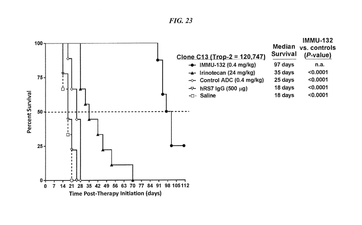
who are either
resistant to or relapsed from standard anti-cancer therapies, such as
chemotherapy. However,
1
CA 03050332 2019-07-15
WO 2018/183041 PCT/US2018/023344
in other embodiments, the ADCs may be of use for first-line therapy of Trop-2+
cancer, such
as TNBC. Surprisingly, the ADCs are of use in cancer patients who either
relapsed from or
fail to respond to irinotecan or topetecan therapy, despite them also being
inhibitors of
topoisomerase I. The combination therapy may be used alone or along with one
or more other
therapeutic modalities selected from the group consisting of surgery,
radiation therapy,
chemotherapy, immunomodulators, cytokines, chemotherapeutic agents, pro-
apoptotic
agents, anti-angiogenic agents, cytotoxic agents, drugs, toxins,
radionuclides, RNAi, siRNA,
a second antibody or antibody fragment, and an immunoconjugate. Any Rad51
inhibitor
known in the art may be utilized, such as B02 ((E)-3-benzy1-2(2-(pyridin-3-
yl)vinyl)
quinazolin-4(3H)-one) (Huang & Mazin, 2014, PLoS ONE 9(6):e100993); RI-1 (3-
chloro-1-
(3,4-dichloropheny1)-4-(4-morpholiny1)-1H-pyrrole-2,5-dione) (Budke et al.,
2012, Nucl
Acids Res 40:7347-57); DIDS (4,4'-diisothiocyanostilbene-2,2'-disulfonic acid)
(Ishida et al.,
2009, Nucl Acids Res 37:3367-76); halenaquinone (Takaku et al., 2011, Genes
Cells 16:427-
36); or imatinib (Choudhury et al., 2009, Mol Cancer Ther 8:203-13).
Related Art
[04] Antibody-drug conjugates (ADCs), comprising antibodies linked to active
therapeutic
agents for selected delivery of toxic payload to cancer cells, are approved
for Hodgkin
lymphoma, systemic anaplastic large cell lymphoma, and for HER2-positive
metastatic breast
cancers, with several new ADCs in development that target a variety of
antigens associated
with hematopoietic and solid tumors (de Goeij & Lambert, 2016, Curr Opin
Immunol 40:14-
231-4; Gravanis et al., 2016, Oncologist 21:102-9; Jerjian et al., 2016,
Pharmacotherapy
36:99-116; Govindan et al., 2016, Expert Opin Biol Ther 16:883-93). Nearly all
ADCs
utilize drugs with picomolar toxicity, such as calicheamicin, which
interchelates in DNA, or
auristatin or maytansine, as microtubule inhibitors. They are not used as
stand-alone agents
because of being highly toxic. However, when appropriately linked to
antibodies, their
therapeutic window is acceptable, with careful selection of the appropriate
antibody and
linker technology being crucial for successful clinical application.
[05] We have developed ADCs based on the well-known anticancer drug, SN-38, a
camptothecin that is the active component of irinotecan (CPT-11), a
topoisomerase I inhibitor
(Garcia-Carbonero & Supko, 2002, Clin Cancer Res 8:641-615; Mathijssen et al.,
2001, Clin
Cancer Res 7:2182-94). While SN-38 has a more moderate level of potency
compared to
drugs used by other ADCs, preclinical testing in murine xenograft models
indicated that
under optimized conditions (i.e., antibody and linker selection), ADCs
prepared with SN-38
2
CA 03050332 2019-07-15
WO 2018/183041 PCT/US2018/023344
were effective and had a highly favorable therapeutic window (Cardillo et al.,
2011, Clin
Cancer Res 17:3157-69; Govindan et al., 2009, Clin Cancer Res 15:6052-61; Moon
et al.,
2008, J Med Chem 51:6916-267-9). One of these SN-38 conjugates (company
designation
IMMU-132; USAN designation, sacituzumab govitecan) targets Trop-2
(trophoblastic cell-
surface antigen; also, termed epithelial glycoprotein-1, or EGP-1), a
glycoprotein that is
highly expressed by many epithelial cancers (Cubas et al., 2009, Biochim
Biophys Acta
1796:309-14, Trerotola et al., 2013, Oncogene 32:222-33). Trop-2 also is
present in many
corresponding normal tissues (Trerotola et al., 2013, Oncogene 32:222-33;
Stepan et al.,
2011, J Histochem Cytochem 59:701-10), but studies in Cynomolgus monkeys that
express
cross-reactive Trop-2 in similar tissues indicated only dose-limiting
neutropenia and
gastrointestinal toxicities (Cardillo et al., 2011, Clin Cancer Res 17:3157-
69). Since these
events are typically associated with irinotecan therapy, and because there was
no appreciable
damage to Trop-2-expressing normal tissues, even at relatively high human
equivalent doses,
we surmised that either the antigen is sheltered in some manner in normal
tissues or the
normal tissues are less affected by the more moderately-toxic SN-38.
[06] We reported results recently from the phase I dose-escalation study with
sacituzumab
govitecan (IMMU-132) given to patients with diverse metastatic epithelial
cancers for 2
weeks in 3-week treatment cycles (Starodub et al., 2015, Clin Cancer Res
21:3870-8). Doses
of 8, 10, 12, and 18 mg/kg were examined, and the maximum tolerated dose
(MTD), assessed
by tolerance to a single cycle of therapy, was determined to be 12 mg/kg.
However, at this
dose there were frequent delays within or between cycles, as well as dose
reductions required.
Therefore, for patients to receive multiple cycles of therapy with minimal
delays or dose
reductions, accrual was continued at 2 lower dose levels, 8 and 10 mg/kg.
[07] Despite the promising results with IMMU-132 (sacituzumab govitecan) as a
monotherapy, a need exists in the field for improved methods and compositions
for
combination therapies with IMMU-132 and other anti-cancer therapeutic agents.
SUMMARY
[08] In various embodiments, the present invention concerns compositions and
methods of
treatment of cancer with antibody-drug conjugates (ADCs), used in combination
therapies
with one or more other therapeutic modalities, such as surgery, radiation
therapy,
chemotherapy, immunomodulators, cytokines, chemotherapeutic agents, pro-
apoptotic
agents, anti-angiogenic agents, cytotoxic agents, drugs, toxins,
radionuclides, RNAi, siRNA,
a second antibody or antibody fragment, or an immunoconjugate. In preferred
embodiments,
3
CA 03050332 2019-07-15
WO 2018/183041 PCT/US2018/023344
the ADC is used in combination with one or more inhibitors of Rad51. The ADC
may be of
use for treatment of cancers for which standard therapies are not effective,
such as metastatic
pancreatic cancer, metastatic colorectal cancer or triple-negative breast
cancer. More
preferably, the combination of ADC and other therapeutic modality is more
efficacious than
either alone, or the sum of the effects of individual treatments.
[09] In a specific embodiment, an anti-Trop-2 antibody may be a humanized RS7
antibody
(see, e.g., U.S. Patent No. 7,238,785, the Figures and Examples section of
which are
incorporated herein by reference), comprising the light chain CDR sequences
CDR1
(KASQDVSIAVA, SEQ ID NO:1); CDR2 (SASYRYT, SEQ ID NO:2); and CDR3
(QQHYITPLT, SEQ ID NO:3) and the heavy chain CDR sequences CDR1 (NYGMN, SEQ
ID NO:4); CDR2 (WINTYTGEPTYTDDFKG, SEQ ID NO:5) and CDR3
(GGFGSSYWYFDV, SEQ ID NO:6). However, as discussed below other anti-Trop-2
antibodies are known and may be used in the subject ADCs. Preferably, the drug
conjugated
to the anti-Trop-2 antibody is one that induces DNA strand breaks, such as
auristatins,
colicheamicins, camptothecins (e.g., SN-38) or anthracyclines. Exemplary drugs
inducing
DNA strand breaks include, but are not limited to, SN-38, doxorubicin,
topotecan,
doxorubicin, etoposide, cisplatinum, oxaliplatin, or carboplatin. In a more
preferred
embodiment, the drug conjugated to the antibody is a camptothecin or
anthracycline, most
preferably SN-38 or doxorubicin (see, e.g., U.S. Patent No. 9,028,833, the
Figures and
Examples section of which are incorporated herein by reference).
[010] The antibody moiety may be a monoclonal antibody, an antigen-binding
antibody
fragment, a bispecific or other multivalent antibody, or other antibody-based
molecule. The
antibody can be of various isotypes, preferably human IgGl, IgG2, IgG3 or
IgG4, more
preferably comprising human IgG1 hinge and constant region sequences. The
antibody or
fragment thereof can be a chimeric, a humanized, or a human antibody, as well
as variations
thereof, such as half-IgG4 antibodies (referred to as "unibodies"), as
described by van der
Neut Kolfschoten et al. (Science 2007; 317:1554-1557). More preferably, the
antibody or
fragment thereof may be designed or selected to comprise human constant region
sequences
that belong to specific allotypes, which may result in reduced immunogenicity
when the ADC
is administered to a human subject. Preferred allotypes for administration
include a non-
Glml allotype (nG1m1), such as G1m3, G1m3,1, G1m3,2 or G1m3,1,2. More
preferably,
the allotype is selected from the group consisting of the nGlml, G1m3, nG1m1,2
and Km3
allotypes (Jefferies and Lefranc, 2009, mAbs 1(4):1-7).
4
CA 03050332 2019-07-15
WO 2018/183041 PCT/US2018/023344
[OM The drug to be conjugated to the antibody or antibody fragment may be
selected from
the group consisting of an anthracycline, a camptothecin, a tubulin inhibitor,
a maytansinoid,
a calicheamycin, an auristatin, a nitrogen mustard, an ethylenimine
derivative, an alkyl
sulfonate, a nitrosourea, a triazene, a folic acid analog, a taxane, a COX-2
inhibitor, a
pyrimidine analog, a purine analog, an antibiotic, an enzyme inhibitor, an
epipodophyllotoxin, a platinum coordination complex, a vinca alkaloid, a
substituted urea, a
methyl hydrazine derivative, an adrenocortical suppressant, a hormone
antagonist, an
antimetabolite, an alkylating agent, an antimitotic, an anti-angiogenic agent,
a tyrosine kinase
inhibitor, an mTOR inhibitor, a heat shock protein (HSP90) inhibitor, a
proteosome inhibitor,
an HDAC inhibitor, a pro-apoptotic agent, and a combination thereof
[012] Specific drugs of use may be selected from the group consisting of 5-
fluorouracil,
afatinib, aplidin, azaribine, anastrozole, anthracyclines, axitinib, AVL-101,
AVL-291,
bendamustine, bleomycin, bortezomib, bosutinib, bryostatin-1, busulfan,
calicheamycin,
camptothecin, carboplatin, 10-hydroxycamptothecin, carmustine, celecoxib,
chlorambucil,
cisplatinum, COX-2 inhibitors, irinotecan (CPT-11), SN-38, carboplatin,
cladribine,
camptothecans, crizotinib, cyclophosphamide, cytarabine, dacarbazine,
dasatinib, dinaciclib,
docetaxel, dactinomycin, daunorubicin, DM1, DM3, DM4, doxorubicin, cyano-
morpholino
doxorubicin, doxorubicin glucuronide, endostatin, epirubicin glucuronide,
erlotinib,
estramustine, epidophyllotoxin, erlotinib, entinostat, estrogen receptor
binding agents,
etoposide (VP16), etoposide glucuronide, etoposide phosphate, exemestane,
fingolimod,
floxuridine (FUdR), 3',5'-0-dioleoyl-FudR (FUdR-d0), fludarabine, flutamide,
farnesyl-
protein transferase inhibitors, flavopiridol, fostamatinib, ganetespib, GDC-
0834, GS-1101,
gefitinib, gemcitabine, hydroxyurea, ibrutinib, idarubicin, idelali sib,
ifosfamide, imatinib,
lapatinib, lenolidamide, leucovorin, LFM-A13, lomustine, mechlorethamine,
melphalan,
mercaptopurine, 6-mercaptopurine, methotrexate, mitoxantrone, mithramycin,
mitomycin,
mitotane, monomethylauristatin F (MMAF), monomethylauristatin D (MMAD),
monomethylauristatin E (MMAE), navelbine, neratinib, nilotinib, nitrosurea,
olaparib,
plicomycin, procarbazine, paclitaxel, PCI-32765, pentostatin, PSI-341,
raloxifene, semustine,
SN-38, sorafenib, streptozocin, SU11248, sunitinib, tamoxifen, temazolomide,
transplatinum,
thalidomide, thioguanine, thiotepa, teniposide, topotecan, uracil mustard,
vatalanib,
vinorelbine, vinblastine, vincristine, vinca alkaloids and ZD1839. Preferably,
the drug is SN-
38.
CA 03050332 2019-07-15
WO 2018/183041 PCT/US2018/023344
[013] Preferred optimal dosing of the subject ADCs may include a dosage of
between 4
mg/kg and 18 mg/kg, preferably given either weekly, twice weekly or every
other week. The
optimal dosing schedule may include treatment cycles of two consecutive weeks
of therapy
followed by one, two, three or four weeks of rest, or alternating weeks of
therapy and rest, or
one week of therapy followed by two, three or four weeks of rest, or three
weeks of therapy
followed by one, two, three or four weeks of rest, or four weeks of therapy
followed by one,
two, three or four weeks of rest, or five weeks of therapy followed by one,
two, three, four or
five weeks of rest, or administration once every two weeks, once every three
weeks or once a
month. Treatment may be extended for any number of cycles, preferably at least
2, at least 4,
at least 6, at least 8, at least 10, at least 12, at least 14, or at least 16
cycles. Exemplary
dosages of use may include 1 mg/kg, 2 mg/kg, 3 mg/kg, 4 mg/kg, 5 mg/kg, 6
mg/kg, 7
mg/kg, 8 mg/kg, 9 mg/kg, 10 mg/kg, 11 mg/kg, 12 mg/kg, 13 mg/kg, 14 mg/kg, 15
mg/kg, 16
mg/kg, 17 mg/kg, 18 mg/kg, 19 mg/kg, 20 mg/kg, 22 mg/kg and 24 mg/kg.
Preferred dosages
are 4, 6, 8, 9, 10, 12, 14, 16 or 18 mg/kg. More preferred dosages are 6-12, 6-
8, 7-8, 8-10, 10-
12 or 8-12 mg/ml. The person of ordinary skill will realize that a variety of
factors, such as
age, general health, specific organ function or weight, as well as effects of
prior therapy on
specific organ systems (e.g., bone marrow) may be considered in selecting an
optimal dosage
of ADC, and that the dosage and/or frequency of administration may be
increased or
decreased during the course of therapy. The dosage may be repeated as needed,
with evidence
of tumor shrinkage observed after as few as 4 to 8 doses. The optimized
dosages and
schedules of administration disclosed herein show unexpected superior efficacy
and reduced
toxicity in human subjects, which could not have been predicted from animal
model studies.
Surprisingly, the superior efficacy allows treatment of tumors that were
previously found to
be resistant to one or more standard anti-cancer therapies. More surprisingly,
the treatment
has been found effective in tumors that were previously resistant to
camptothecins, such as
irinotecan, the parent compound of SN-38.
[014] The Rad51 inhibitor used in combination therapy may be any Rad51
inhibitor known
in the art, including but not limited to B02 (available, e.g., from
Calbiochem, La Jolla, CA,
CAS 1290541-46-6); RI-1 (available, e.g., from Calbiochem, La Jolla, CA, CAS
415713-60-
9); DIDS (available, e.g., from Tocris Bioscience, Bristol, UK, CAS 67483-13-
0);
halenaquinone (available, e.g., from Angene International Ltd., Hong Kong,
China, CAS
86690-14-4); or imatinib (GLEE VAC , Novartis, East Hanover, NJ).
6
CA 03050332 2019-07-15
WO 2018/183041 PCT/US2018/023344
[015] Combination therapy with the anti-Trop-2 ADCs are of use for therapy of
cancers,
such as TNBC, SCLC, NSCLC, urothelial cancer, bladder cancer, gastric cancer
or ovarian
cancer. Such use may be front-line, second-line, or at later stages of cancer
progression. The
compositions and methods of use are efficacious in camptothecin-resistant as
well as
camptothecin sensitive cancers. Generally, the anti-Trop-2 ADCs are of use for
treating any
cancer that expresses the Trop-2 antigen.
BRIEF DESCRIPTION OF THE DRAWINGS
[016] FIG. 1. Preclinical in vivo therapy of athymic nude mice, bearing Capan
1 human
pancreatic carcinoma, with SN-38 conjugates of hRS7 (anti-Trop-2), hPAM4 (anti-
MUC5ac),
hMN-14 (anti-CEACAM5) or non-specific control hA20 (anti-CD20).
[017] FIG. 2. Preclinical in vivo therapy of athymic nude mice, bearing BxPC3
human
pancreatic carcinoma, with anti-TROP2-CL2A-SN-38 conjugates compared to
controls.
[018] FIG. 3A. Structures of CL2-SN-38 and CL2A-SN-38.
[019] FIG. 3B. Comparative efficacy of anti-Trop-2 ADC linked to CL2 vs. CL2A
linkers
versus hA20 ADC and saline control, using COLO 205 colonic adenocarcinoma.
Animals
were treated twice weekly for 4 weeks as indicated by the arrows. COLO 205
mice (N= 6)
were treated with 0.4 mg/kg ADC and tumors measured twice a week.
[020] FIG. 3C. Comparative efficacy of anti-Trop-2 ADC linked to CL2 vs. CL2A
linkers
versus hA20 ADC and saline control, using Capan-1 pancreatic adenocarcinoma.
Animals
were treated twice weekly for 4 weeks as indicated by the arrows. Capan-1 mice
(N= 10)
were treated with 0.2 mg/kg ADC and tumors measured weekly.
[021] FIG. 4A. Therapeutic efficacy of hRS7-SN-38 ADC in several solid tumor-
xenograft
disease models. Efficacy of hRS7-CL2-SN-38 and hRS7-CL2A-SN-38 ADC treatment
was
studied in mice bearing human non¨small cell lung, colorectal, pancreatic, or
squamous cell
lung tumor xenografts. All the ADCs and controls were administered in the
amounts
indicated (expressed as amount of SN-38 per dose; long arrows = conjugate
injections, short
arrows = irinotecan injections). Mice bearing Calu-3 tumors (N= 5-7) were
injected with
hRS7-CL2-SN-38 every 4 days for a total of 4 injections (q4dx4).
[022] FIG. 4B. Therapeutic efficacy of hRS7-SN-38 ADC in several solid tumor-
xenograft
disease models. Efficacy of hRS7-CL2-SN-38 and hRS7-CL2A-SN-38 ADC treatment
was
studied in mice bearing human non¨small cell lung, colorectal, pancreatic, or
squamous cell
lung tumor xenografts. All the ADCs and controls were administered in the
amounts
indicated (expressed as amount of SN-38 per dose; long arrows = conjugate
injections, short
7
CA 03050332 2019-07-15
WO 2018/183041 PCT/US2018/023344
arrows = irinotecan injections). COLO 205 tumor-bearing mice (N= 5) were
injected 8 times
(q4dx8) with the ADC or every 2 days for a total of 5 injections (q2dx5) with
the MTD of
irinotecan.
[023] FIG. 4C. Therapeutic efficacy of hRS7-SN-38 ADC in several solid tumor-
xenograft
disease models. Efficacy of hRS7-CL2-SN-38 and hRS7-CL2A-SN-38 ADC treatment
was
studied in mice bearing human non¨small cell lung, colorectal, pancreatic, or
squamous cell
lung tumor xenografts. All the ADCs and controls were administered in the
amounts
indicated (expressed as amount of SN-38 per dose; long arrows = conjugate
injections, short
arrows = irinotecan injections). Capan-1 (N= 10) were treated twice weekly for
4 weeks with
the agents indicated.
[024] FIG. 4D. Therapeutic efficacy of hRS7-SN-38 ADC in several solid tumor-
xenograft
disease models. Efficacy of hRS7-CL2-SN-38 and hRS7-CL2A-SN-38 ADC treatment
was
studied in mice bearing human non¨small cell lung, colorectal, pancreatic, or
squamous cell
lung tumor xenografts. All the ADCs and controls were administered in the
amounts
indicated (expressed as amount of SN-38 per dose; long arrows = conjugate
injections, short
arrows = irinotecan injections). BxPC-3 tumor-bearing mice (N= 10) were
treated twice
weekly for 4 weeks with the agents indicated.
[025] FIG. 4E. Therapeutic efficacy of hRS7-SN-38 ADC in several solid tumor-
xenograft
disease models. Efficacy of hRS7-CL2-SN-38 and hRS7-CL2A-SN-38 ADC treatment
was
studied in mice bearing human non¨small cell lung, colorectal, pancreatic, or
squamous cell
lung tumor xenografts. All the ADCs and controls were administered in the
amounts
indicated (expressed as amount of SN-38 per dose; long arrows = conjugate
injections, short
arrows = irinotecan injections). In addition to ADC given twice weekly for 4
week, SK-MES-
1 tumor-bearing (N = 8) mice received the MTD of CPT-11 (q2dx5).
[026] FIG. 5A. Tolerability of hRS7-CL2A-SN-38 in Swiss-Webster mice. Fifty-
six Swiss-
Webster mice were administered 2 i.p. doses of buffer or the hRS7-CL2A-SN-38 3
days apart
(4, 8, or 12 mg/kg of SN-38 per dose; 250, 500, or 750 mg conjugate protein/kg
per dose).
Seven and 15 days after the last injection, 7 mice from each group were
euthanized, with
blood counts and serum chemistries performed. Graphs show the percent of
animals in each
group that had elevated levels of AST.
[027] FIG. 5B. Tolerability of hRS7-CL2A-SN-38 in Swiss-Webster mice. Fifty-
six Swiss-
Webster mice were administered 2 i.p. doses of buffer or the hRS7-CL2A-SN-38 3
days apart
(4, 8, or 12 mg/kg of SN-38 per dose; 250, 500, or 750 mg conjugate protein/kg
per dose).
8
CA 03050332 2019-07-15
WO 2018/183041 PCT/US2018/023344
Seven and 15 days after the last injection, 7 mice from each group were
euthanized, with
blood counts and serum chemistries performed. Graphs show the percent of
animals in each
group that had elevated levels of ALT.
[028] FIG. 5C. Tolerability of hRS7-CL2A-SN-38 in Cynomolgus monkeys. Six
monkeys
per group were injected twice 3 days apart with buffer (control) or hRS7-CL2A-
SN-38 at
0.96 mg/kg or 1.92 mg/kg of SN-38 equivalents per dose (60 and 120 mg/kg
conjugate
protein). All animals were bled on day ¨1, 3, and 6. Four monkeys were bled on
day 11 in the
0.96 mg/kg group, 3 in the 1.92 mg/kg group. Changes in neutrophil counts in
Cynomolgus
monkeys.
[029] FIG. 5D. Tolerability of hRS7-CL2A-SN-38 in Cynomolgus monkeys. Six
monkeys
per group were injected twice 3 days apart with buffer (control) or hRS7-CL2A-
SN-38 at
0.96 mg/kg or 1.92 mg/kg of SN-38 equivalents per dose (60 and 120 mg/kg
conjugate
protein). All animals were bled on day ¨1, 3, and 6. Four monkeys were bled on
day 11 in the
0.96 mg/kg group, 3 in the 1.92 mg/kg group. Changes in platelet counts in
Cynomolgus
monkeys.
[030] FIG. 6. In vitro efficacy of anti-Trop-2-paclitaxel ADC against MDA-MB-
468 human
breast adenocarcinoma.
[031] FIG. 7. In vitro efficacy of anti-Trop-2-paclitaxel ADC against BxPC-3
human
pancreatic adenocarcinoma.
[032] FIG. 8A. Comparison of in vitro efficacy of anti-Trop-2 ADCs (hRS7-SN-38
versus
MAB650-SN-38) in Capan-1 human pancreatic adenocarcinoma.
[033] FIG. 8B. Comparison of in vitro efficacy of anti-Trop-2 ADCs (hRS7-SN-38
versus
MAB650-SN-38) in BxPC-3 human pancreatic adenocarcinoma.
[034] FIG. 8C. Comparison of in vitro efficacy of anti-Trop-2 ADCs (hRS7-SN-38
versus
MAB650-SN-38) in NCI-N87 human gastric adenocarcinoma.
[035] FIG. 9A. Comparison of cytotoxicity of naked or SN-38 conjugated hRS7
vs. 162-
46.2 antibodies in BxPC-3 human pancreatic adenocarcinoma.
[036] FIG. 9B. Comparison of cytotoxicity of naked or SN-38 conjugated hRS7
vs. 162-46.2
antibodies in MDA-MB-468 human breast adenocarcinoma.
[037] FIG. 10. IMMU-132 phase I/II data for best response by RECIST criteria.
[038] FIG. 11. IMMU-132 phase I/II data for time to progression and best
response
(RECIST).
9
CA 03050332 2019-07-15
WO 2018/183041 PCT/US2018/023344
[039] FIG. 12. Combination therapy with IMMU-132 and carboplatin or cisplatin,
compared
to IMMU-132, carboplatin or cisplatin alone, a non-targeting ADC or saline
control.
[040] FIG. 13. Combination therapy with IMMU-132 plus carboplatin, compared to
IMMU-
132 or caboplatin alone or saline control.
[041] FIG. 14. Combination therapy with IMMU-132 plus cisplatinum, compared to
IMMU-
132 or cisplatinum alone or saline control.
[042] FIG. 15. Cachexia in mice treated with combination therapy with IMMU-132
and
carboplatin or cisplatin, compared to IMMU-132, carboplatin or cisplatin
alone, or saline
control.
[043] FIG. 16. Cytotoxicity of IMMU-132 and irinotecan vs. saline and control
ADC in
MDA-MB-231 tumor cells.
[044] FIG. 17. Cytotoxicity of IMMU-132 and irinotecan vs. saline and control
ADC in
MDA-HCC1806 tumor cells.
[045] FIG. 18. Cytotoxicity of IMMU-132 and irinotecan vs. saline and control
ADC in
MDA-SK-MES-1 tumor cells.
[046] FIG. 19. Effect of IMMU-132 and irinotecan vs. control ADC, unconjugated
hRS7
IgG or saline on tumor growth with the parental MDA-MB-231 cell line.
[047] FIG. 20. Effect of IMMU-132 and irinotecan vs. control ADC, unconjugated
hRS7
IgG or saline on tumor growth with the C13 cell line.
[048] FIG. 21. Effect of IMMU-132 and irinotecan vs. control ADC, unconjugated
hRS7
IgG or saline on tumor growth with the parental C39 cell line.
[049] FIG. 22. Effect of IMMU-132 and irinotecan vs. control ADC, unconjugated
hRS7
IgG or saline on survival in mice injected with the parental MDA-MB-231 cell
line.
[050] FIG. 23. Effect of IMMU-132 and irinotecan vs. control ADC, unconjugated
hRS7
IgG or saline on survival in mice injected with the C13 cell line.
[051] FIG. 24. Effect of IMMU-132 and irinotecan vs. control ADC, unconjugated
hRS7
IgG or saline on survival in mice injected with the C39 cell line.
DETAILED DESCRIPTION
Definitions
[052] Unless otherwise specified, "a" or "an" means one or more.
[053] As used herein, "about" means plus or minus 10%. For example, "about
100" would
include any number between 90 and 110.
CA 03050332 2019-07-15
WO 2018/183041 PCT/US2018/023344
[054] An antibody, as described herein, refers to a full-length (i.e.,
naturally occurring or
formed by normal immunoglobulin gene fragment recombinatorial processes)
immunoglobulin molecule (e.g., an IgG antibody) or an immunologically active
(i.e.,
specifically binding) portion of an immunoglobulin molecule, like an antibody
fragment.
[055] An antibody fragment is a portion of an antibody such as F(ab')2, Fab',
Fab, Fv, sFy
and the like. Antibody fragments may also include single domain antibodies and
IgG4 half-
molecules, as discussed below. Regardless of structure, an antibody fragment
binds with the
same antigen that is recognized by the full-length antibody. The term
"antibody fragment"
also includes isolated fragments consisting of the variable regions of
antibodies, such as the
"Fv" fragments consisting of the variable regions of the heavy and light
chains and
recombinant single chain polypeptide molecules in which light and heavy
variable regions are
connected by a peptide linker ("scFv proteins").
[056] A chimeric antibody is a recombinant protein that contains the variable
domains
including the complementarity determining regions (CDRs) of an antibody
derived from one
species, preferably a rodent antibody, while the constant domains of the
antibody molecule
are derived from those of a human antibody. For veterinary applications, the
constant
domains of the chimeric antibody may be derived from that of other species,
such as a cat or
dog.
[057] A humanized antibody is a recombinant protein in which the CDRs from an
antibody
from one species; e.g., a rodent antibody, are transferred from the heavy and
light variable
chains of the rodent antibody into human heavy and light variable domains
(e.g., framework
region sequences). The constant domains of the antibody molecule are derived
from those of
a human antibody. In certain embodiments, a limited number of framework region
amino
acid residues from the parent (rodent) antibody may be substituted into the
human antibody
framework region sequences.
[058] A human antibody is, e.g., an antibody obtained from transgenic mice
that have been
"engineered" to produce specific human antibodies in response to antigenic
challenge. In this
technique, elements of the human heavy and light chain loci are introduced
into strains of
mice derived from embryonic stem cell lines that contain targeted disruptions
of the
endogenous murine heavy chain and light chain loci. The transgenic mice can
synthesize
human antibodies specific for particular antigens, and the mice can be used to
produce human
antibody-secreting hybridomas. Methods for obtaining human antibodies from
transgenic
mice are described by Green et al., Nature Genet. 7:13 (1994), Lonberg et al.,
Nature 368:856
11
CA 03050332 2019-07-15
WO 2018/183041 PCT/US2018/023344
(1994), and Taylor et al., Int. Immun. 6:579 (1994). A fully human antibody
also can be
constructed by genetic or chromosomal transfection methods, as well as phage
display
technology, all of which are known in the art. See for example, McCafferty et
al., Nature
348:552-553 (1990) for the production of human antibodies and fragments
thereof in vitro,
from immunoglobulin variable domain gene repertoires from unimmunized donors.
In this
technique, antibody variable domain genes are cloned in-frame into either a
major or minor
coat protein gene of a filamentous bacteriophage, and displayed as functional
antibody
fragments on the surface of the phage particle. Because the filamentous
particle contains a
single-stranded DNA copy of the phage genome, selections based on the
functional properties
of the antibody also result in selection of the gene encoding the antibody
exhibiting those
properties. In this way, the phage mimics some of the properties of the B
cell. Phage display
can be performed in a variety of formats, for review, see e.g. Johnson and
Chiswell, Current
Opinion in Structural Biology 3:5564-571 (1993). Human antibodies may also be
generated
by in vitro activated B cells. See U.S. Pat. Nos. 5,567,610 and 5,229,275, the
Examples
section of which are incorporated herein by reference.
[059] A therapeutic agent is a compound, molecule or atom which is
administered
separately, concurrently or sequentially with an antibody moiety or conjugated
to an antibody
moiety, i.e., antibody or antibody fragment, or a subfragment, and is useful
in the treatment
of a disease. Examples of therapeutic agents include antibodies, antibody
fragments, drugs,
toxins, nucleases, hormones, immunomodulators, pro-apoptotic agents, anti-
angiogenic
agents, boron compounds, photoactive agents or dyes and radioisotopes.
Therapeutic agents
of use are described in more detail below.
[060] An immunoconjugate is an antibody, antibody fragment or fusion protein
conjugated
to at least one therapeutic and/or diagnostic agent.
[061] A multispecific antibody is an antibody that can bind simultaneously to
at least two
targets that are of different structure, e.g., two different antigens, two
different epitopes on
the same antigen, or a hapten and/or an antigen or epitope. Multispecific,
multivalent
antibodies are constructs that have more than one binding site, and the
binding sites are of
different specificity.
[062] A bispecific antibody is an antibody that can bind simultaneously to two
different
targets. Bispecific antibodies (bsAb) and bispecific antibody fragments
(bsFab) may have at
least one arm that specifically binds to, for example, a tumor-associated
antigen and at least
one other arm that specifically binds to a targetable conjugate that bears a
therapeutic or
12
CA 03050332 2019-07-15
WO 2018/183041 PCT/US2018/023344
diagnostic agent. A variety of bispecific fusion proteins can be produced
using molecular
engineering.
Anti-Trop-2 Antibodies
[063] The subject ADCs may include an antibody or fragment thereof that binds
to Trop-2.
In a specific preferred embodiment, the anti-Trop-2 antibody may be a
humanized RS7
antibody (see, e.g., U.S. Patent No. 7,238,785, incorporated herein by
reference in its
entirety), comprising the light chain CDR sequences CDR1 (KASQDVSIAVA, SEQ ID
NO:1); CDR2 (SASYRYT, SEQ ID NO:2); and CDR3 (QQHYITPLT, SEQ ID NO:3) and
the heavy chain CDR sequences CDR1 (NYGMN, SEQ ID NO:4); CDR2
(WINTYTGEPTYTDDFKG, SEQ ID NO:5) and CDR3 (GGFGSSYWYFDV, SEQ ID
NO:6).
[064] The R57 antibody was a murine IgGi raised against a crude membrane
preparation of a
human primary squamous cell lung carcinoma. (Stein et al., Cancer Res. 50:
1330, 1990) The
R57 antibody recognizes a 46-48 kDa glycoprotein, characterized as cluster 13.
(Stein et al.,
Int. J. Cancer Supp. 8:98-102, 1994) The antigen was designated as EGP-1
(epithelial
glycoprotein-1), but is also referred to as Trop-2.
[065] Trop-2 is a type-I transmembrane protein and has been cloned from both
human
(Fornaro et al., Int J Cancer 1995; 62:610-8) and mouse cells (Sewedy et al.,
Int J Cancer
1998; 75:324-30). In addition to its role as a tumor-associated calcium signal
transducer
(Ripani et al., Int J Cancer 1998;76:671-6), the expression of human Trop-2
was shown to be
necessary for tumorigenesis and invasiveness of colon cancer cells, which
could be
effectively reduced with a polyclonal antibody against the extracellular
domain of Trop-2
(Wang et al., Mol Cancer Ther 2008;7:280-5).
[066] The growing interest in Trop-2 as a therapeutic target for solid cancers
(Cubas et al.,
Biochim Biophys Acta 2009;1796:309-14) is attested by further reports that
documented the
clinical significance of overexpressed Trop-2 in breast (Huang et al., Clin
Cancer Res
2005;11:4357-64), colorectal (Ohmachi et al., Clin Cancer Res 2006;12:3057-63;
Fang et al.,
Int J Colorectal Dis 2009;24:875-84), and oral squamous cell (Fong et al.,
Modern Pathol
2008;21:186-91) carcinomas. The latest evidence that prostate basal cells
expressing high
levels of Trop-2 are enriched for in vitro and in vivo stem-like activity is
particularly
noteworthy (Goldstein et al., Proc Natl Acad Sci USA 2008;105:20882-7).
[067] Flow cytometry and immunohistochemical staining studies have shown that
the R57
MAb detects antigen on a variety of tumor types, with limited binding to
normal human
13
CA 03050332 2019-07-15
WO 2018/183041 PCT/US2018/023344
tissue (Stein et al., 1990). Trop-2 is expressed primarily by carcinomas such
as carcinomas of
the lung, stomach, urinary bladder, breast, ovary, uterus, and prostate.
Localization and
therapy studies using radiolabeled murine R57 MAb in animal models have
demonstrated
tumor targeting and therapeutic efficacy (Stein et al., 1990; Stein et al.,
1991).
[068] Strong R57 staining has been demonstrated in tumors from the lung,
breast, bladder,
ovary, uterus, stomach, and prostate. (Stein et al., Int. J. Cancer 55:938,
1993) The lung
cancer cases comprised both squamous cell carcinomas and adenocarcinomas.
(Stein et al.,
Int. J. Cancer 55:938, 1993) Both cell types stained strongly, indicating that
the R57
antibody does not distinguish between histologic classes of non-small-cell
carcinoma of the
lung.
[069] The R57 MAb is rapidly internalized into target cells (Stein et al.,
1993). The
internalization rate constant for R57 MAb is intermediate between the
internalization rate
constants of two other rapidly internalizing MAbs, which have been
demonstrated to be
useful for immunotoxin production. (Id.) It is well documented that
internalization of
immunotoxin conjugates is a requirement for anti-tumor activity. (Pastan et
al., Cell 47:641,
1986) Internalization of drug immunoconjugates has been described as a major
factor in anti-
tumor efficacy. (Yang et al., Proc. Nat'l Acad. Sci. USA 85: 1189, 1988) Thus,
the R57
antibody exhibits several important properties for therapeutic applications.
[070] While the hRS7 antibody is preferred, other anti-Trop-2 antibodies are
known and/or
publicly available and in alternative embodiments may be utilized in the
subject ADCs.
While humanized or human antibodies are preferred for reduced immunogenicity,
in
alternative embodiments a chimeric antibody may be of use. As discussed below,
methods of
antibody humanization are well known in the art and may be utilized to convert
an available
murine or chimeric antibody into a humanized form.
[071] Anti-Trop-2 antibodies are commercially available from a number of
sources and
include LS-C126418, LS-C178765, LS-C126416, LS-C126417 (LifeSpan BioSciences,
Inc.,
Seattle, WA); 10428-MM01, 10428-MM02, 10428-R001, 10428-R030 (Sino Biological
Inc.,
Beijing, China); MR54 (eBioscience, San Diego, CA); sc-376181, sc-376746,
Santa Cruz
Biotechnology (Santa Cruz, CA); MM0588-49D6, (Novus Biologicals, Littleton,
CO);
ab79976, and ab89928 (ABCAM , Cambridge, MA).
[072] Other anti-Trop-2 antibodies have been disclosed in the patent
literature. For example,
U.S. Pub!. No. 2013/0089872 discloses anti-Trop-2 antibodies K5-70 (Accession
No. FERM
BP-11251), K5-107 (Accession No. FERM BP-11252), K5-116-2-1 (Accession No.
FERM
14
CA 03050332 2019-07-15
WO 2018/183041 PCT/US2018/023344
BP-11253), T6-16 (Accession No. FERM BP-11346), and T5-86 (Accession No. FERM
BP-
11254), deposited with the International Patent Organism Depositary, Tsukuba,
Japan. U.S.
Patent No. 5,840,854 disclosed the anti-Trop-2 monoclonal antibody BR110 (ATCC
No.
HB11698). U.S. Patent No. 7,420,040 disclosed an anti-Trop-2 antibody produced
by
hybridoma cell line AR47A6.4.2, deposited with the IDAC (International
Depository
Authority of Canada, Winnipeg, Canada) as accession number 141205-05. U.S.
Patent No.
7,420,041 disclosed an anti-Trop-2 antibody produced by hybridoma cell line
AR52A301.5,
deposited with the IDAC as accession number 141205-03. U.S. Publ. No.
2013/0122020
disclosed anti-Trop-2 antibodies 3E9, 6G11, 7E6, 15E2, 18B1. Hybridomas
encoding a
representative antibody were deposited with the American Type Culture
Collection (ATCC),
Accession Nos. PTA-12871 and PTA-12872. U.S. Publ. No. 2016/0297890 discloses
a
TINA1 anti-Trop-2 antibody. U.S. Patent No. 8,715,662 discloses anti-Trop-2
antibodies
produced by hybridomas deposited at the AID-ICLC (Genoa, Italy) with deposit
numbers PD
08019, PD 08020 and PD 08021. U.S. Patent Application Publ. No. 20120237518
discloses
anti-Trop-2 antibodies 77220, KM4097 and KM4590. U.S. Patent No. 8,309,094
(Wyeth)
discloses antibodies Al and A3, identified by sequence listing. The Examples
section of each
patent or patent application cited above in this paragraph is incorporated
herein by reference.
Non-patent publication Lipinski et al. (1981, Proc Natl. Acad Sci USA, 78:5147-
50)
disclosed anti-Trop-2 antibodies 162-25.3 and 162-46.2. A publication by King
et al. (Invest
New Drugs, Jan 15, 2018 Epub ahead of print) disclosed a PF-06664178 anti-Trop-
2
antibody-drug conjugate. A publication by Strop et al. (2016, Mol Cancer Ther
15:2698-708)
disclosed an RN927C anti-Trop-2 ADC.
[073] Numerous anti-Trop-2 antibodies are known in the art and/or publicly
available. As
discussed below, methods for preparing antibodies against known antigens were
routine in
the art. The sequence of the human Trop-2 protein was also known in the art
(see, e.g.,
GenBank Accession No. CAA54801.1). Methods for producing humanized, human or
chimeric antibodies were also known. The person of ordinary skill, reading the
instant
disclosure in light of general knowledge in the art, would have been able to
make and use the
genus of anti-Trop-2 antibodies in the subject ADCs.
[074] Use of anti-Trop-2 antibodies has been disclosed for immunotherapeutics
other than
ADCs. The murine IgG2a antibody edrecolomab (PANOREMD) has been used for
treatment
of colorectal cancer, although the murine antibody is not well suited for
human clinical use
(Baeuerle & Gires, 2007, Br. J Cancer 96:417-423). Low-dose subcutaneous
administration
CA 03050332 2019-07-15
WO 2018/183041 PCT/US2018/023344
of ecrecolomab was reported to induce humoral immune responses against the
vaccine
antigen (Baeuerle & Gires, 2007). Adecatumumab (MT201), a fully human anti-
Trop-2
antibody, has been used in metastatic breast cancer and early-stage prostate
cancer and is
reported to act through ADCC and CDC activity (Baeuerle & Gires, 2007). MT110,
a single-
chain anti-Trop-2/anti-CD3 bispecific antibody construct has reported efficacy
against
ovarian cancer (Baeuerle & Gires, 2007). Catumaxomab, a hybrid mouse/rat
antibody with
binding affinity for Trop-2, CD3 and Fc receptor, was reported to be active
against ovarian
cancer (Baeuerle & Gires, 2007). Proxinium, an immunotoxin comprising anti-
Trop-2 single-
chain antibody fused to Pseudomonas exotoxin, has been tested in head-and-neck
and bladder
cancer (Baeuerle & Gires, 2007). None of these studies contained any
disclosure of the use of
anti-Trop-2 antibody-drug conjugates.
Camptothecin Conjugates
[075] Non-limiting methods and compositions for preparing immunoconjugates
comprising a
camptothecin therapeutic agent attached to an antibody or antigen-binding
antibody fragment
are described below. In preferred embodiments, the solubility of the drug is
enhanced by
placing a defined polyethyleneglycol (PEG) moiety (i.e., a PEG containing a
defined number
of monomeric units) between the drug and the antibody, wherein the defined PEG
is a low
molecular weight PEG, preferably containing 1-30 monomeric units, more
preferably
containing 1-12 monomeric units, most preferably containing 6-8 monomeric
units.
[076] Preferably, a first linker connects the drug at one end and may
terminate with an
acetylene or an azide group at the other end. This first linker may comprise a
defined PEG
moiety with an azide or acetylene group at one end and a different reactive
group, such as
carboxylic acid or hydroxyl group, at the other end. Said bifunctional defined
PEG may be
attached to the amine group of an amino alcohol, and the hydroxyl group of the
latter may be
attached to the hydroxyl group on the drug in the form of a carbonate.
Alternatively, the non-
azide(or acetylene) moiety of said defined bifunctional PEG is optionally
attached to the N-
terminus of an L-amino acid or a polypeptide, with the C-terminus attached to
the amino
group of amino alcohol, and the hydroxy group of the latter is attached to the
hydroxyl group
of the drug in the form of carbonate or carbamate, respectively.
[077] A second linker, comprising an antibody-coupling group and a reactive
group
complementary to the azide (or acetylene) group of the first linker, namely
acetylene (or
azide), may react with the drug-(first linker) conjugate via acetylene-azide
cycloaddition
reaction to furnish a final bifunctional drug product that is useful for
conjugating to disease-
16
CA 03050332 2019-07-15
WO 2018/183041 PCT/US2018/023344
targeting antibodies. The antibody-coupling group is preferably either a thiol
or a thiol-
reactive group.
[078] Methods for selective regeneration of the 10-hydroxyl group in the
presence of the C-
20 carbonate in preparations of drug-linker precursor involving CPT analogs
such as SN-38
are provided below. Other protecting groups for reactive hydroxyl groups in
drugs such as the
phenolic hydroxyl in SN-38, for example t-butyldimethylsilyl or t-
butyldiphenylsilyl, may
also be used, and these are deprotected by tetrabutylammonium fluoride prior
to linking of
the derivatized drug to an antibody-coupling moiety. The 10-hydroxyl group of
CPT analogs
is alternatively protected as an ester or carbonate, other than 'WC', such
that the
bifunctional CPT is conjugated to an antibody without prior deprotection of
this protecting
group. The protecting group is readily deprotected under physiological pH
conditions after
the bioconjugate is administered.
[079] In the acetylene-azide coupling, referred to as 'click chemistry', the
azide part may be
on L2 with the acetylene part on L3. Alternatively, L2 may contain acetylene,
with L3
containing azide. 'Click chemistry' refers to a copper (+1)-catalyzed
cycloaddition reaction
between an acetylene moiety and an azide moiety (Kolb HC and Sharpless KB,
Drug Discov
Today 2003; 8: 1128-37), although alternative forms of click chemistry are
known and may
be used. Click chemistry takes place in aqueous solution at near-neutral pH
conditions, and is
thus amenable for drug conjugation. The advantage of click chemistry is that
it is
chemoselective, and complements other well-known conjugation chemistries such
as the
thiol-maleimide reaction.
[080] An exemplary preferred embodiment is directed to a conjugate of a drug
derivative and
an antibody of the general formula (1) shown below.
MAb-[L2]-[L1][AA1.4A' -Drug (1)
where MAb is a disease-targeting antibody; L2 is a component of the cross-
linker comprising
an antibody-coupling moiety and one or more of acetylene (or azide) groups; Li
comprises a
defined PEG with azide (or acetylene) at one end, complementary to the
acetylene (or azide)
moiety in L2, and a reactive group such as carboxylic acid or hydroxyl group
at the other end;
AA is an L-amino acid; m is an integer with values of 0, 1, 2, 3, or 4; and A'
is an additional
spacer, selected from the group of ethanolamine, 4-hydroxybenzyl alcohol, 4-
aminobenzyl
alcohol, or substituted or unsubstituted ethylenediamine. The L amino acids of
'AA' are
selected from alanine, arginine, asparagine, aspartic acid, cysteine,
glutamine, glutamic acid,
glycine, histidine, isoleucine, leucine, lysine, methionine, phenylalanine,
proline, serine,
17
CA 03050332 2019-07-15
WO 2018/183041 PCT/US2018/023344
threonine, tryptophan, tyrosine, and valine. If the A' group contains
hydroxyl, it is linked to
the hydroxyl group or amino group of the drug in the form of a carbonate or
carbamate,
respectively.
[081] In a preferred embodiment of formula 1, A' is a substituted ethanolamine
derived from
an L-amino acid, wherein the carboxylic acid group of the amino acid is
replaced by a
hydroxymethyl moiety. A' may be derived from any one of the following L-amino
acids:
alanine, arginine, asparagine, aspartic acid, cysteine, glutamine, glutamic
acid, glycine,
histidine, isoleucine, leucine, lysine, methionine, phenylalanine, proline,
serine, threonine,
tryptophan, tyrosine, and valine.
[082] In an example of the conjugate of the preferred embodiment of formula 1,
m is 0, A' is
L-valinol, and the drug is exemplified by SN-38. In another example of formula
1, m is 1 and
represented by a derivatized L-lysine, A' is L-valinol, and the drug is
exemplified by SN-38.
In this embodiment, an amide bond is first formed between the carboxylic acid
of an amino
acid such as lysine and the amino group of valinol, using orthogonal
protecting groups for the
lysine amino groups. The protecting group on the N-terminus of lysine is
removed, keeping
the protecting group on the side chain of lysine intact, and the N-terminus is
coupled to the
carboxyl group on the defined PEG with azide (or acetylene) at the other end.
The hydroxyl
group of valinol is then attached to the 20-chloroformate derivative of 10-
hydroxy-protected
SN-38, and this intermediate is coupled to an L2 component carrying the
antibody-binding
moiety as well as the complementary acetylene (or azide) group involved in the
click
cycloaddition chemistry. Finally, removal of protecting groups at both lysine
side chain and
SN-38 gives the product of this example.
[083] While not wishing to be bound by theory, the small MW SN-38 product,
namely
valinol-SN-38 carbonate, generated after intracellular proteolysis, has the
additional pathway
of liberation of intact SN-38 through intramolecular cyclization involving the
amino group of
valinol and the carbonyl of the carbonate.
[084] In another preferred embodiment, A' of the general formula 1 is A-OH,
whereby A-
OH is a collapsible moiety such as 4-aminobenzyl alcohol or a substituted 4-
aminobenzyl
alcohol substituted with a Ci-Cio alkyl group at the benzylic position, and
the latter, via its
amino group, is attached to an L-amino acid or a polypeptide comprising up to
four L-amino
acid moieties; wherein the N-terminus is attached to a cross-linker
terminating in the
antibody-binding group.
18
CA 03050332 2019-07-15
WO 2018/183041 PCT/US2018/023344
[085] In another example of a preferred embodiment, the A-OH of A' of general
formula 1 is
derived from a substituted 4-aminobenzyl alcohol, and 'AA' is comprised of a
single L-
amino acid with m =1 in the general formula 1, and the drug is exemplified
with SN-38.
Single amino acid of AA may be selected from any one of the following L-amino
acids:
alanine, arginine, asparagine, aspartic acid, cysteine, glutamine, glutamic
acid, glycine,
histidine, isoleucine, leucine, lysine, methionine, phenylalanine, proline,
serine, threonine,
tryptophan, tyrosine, and valine. The substituent R on 4-aminobenzyl alcohol
moiety (A-OH
embodiment of A') is hydrogen or an alkyl group selected from Cl-C10 alkyl
groups. An
example of this formula, wherein the single amino acid AA is L-lysine and R =
H, and the
drug is exemplified by SN-38 is referred to as MAb-CL2A-SN-38 (shown below).
The
structure differs from the linker MAb-CL2-SN-38 in the substitution of a
single lysine
residue for a Phe-Lys dipeptide found in the CL2 linker. The Phe-Lys dipeptide
was designed
as a cathepsin B cleavage site for lysosomal enzyme, which was considered to
be important
for intracellular release of bound drug. Surprisingly, despite the elimination
of the cathepsin-
cleavage site, immunoconjugates comprising a CL2A linker are apparently more
efficacious
in vivo than those comprising a CL2 linker.
o0 a
N=N 0
H
0 /
8 8
0 H
MAb-CL2A-SN-38 OH
NH2 (as amine salt)
[086] In a preferred embodiment, AA comprises a polypeptide moiety, preferably
a di, tri or
tetrapeptide, that is cleavable by intracellular peptidase. Examples are: Ala-
Leu, Leu-Ala-
Leu, and Ala-Leu-Ala-Leu (SEQ ID NO: 9) (Trouet et al., 1982).
[087] In another preferred embodiment, the Li component of the conjugate
contains a
defined polyethyleneglycol (PEG) spacer with 1-30 repeating monomeric units.
In a further
preferred embodiment, PEG is a defined PEG with 1-12 repeating monomeric
units. The
introduction of PEG may involve using heterobifunctionalized PEG derivatives
which are
available commercially. The heterobifunctional PEG may contain an azide or
acetylene
group.
[088] In a preferred embodiment, L2 has a plurality of acetylene (or azide)
groups, ranging
from 2-40, but preferably 2-20, and more preferably 2-5, and a single antibody-
binding
19
CA 03050332 2019-07-15
WO 2018/183041 PCT/US2018/023344
moiety. In a representative example, the 12' component is appended to 2
acetylenic groups,
resulting in the attachment of two azide-appended SN-38 molecules. The bonding
to MAb
may involve a succinimide.
[089] In preferred embodiments, when the bifunctional drug contains a thiol-
reactive moiety
as the antibody-binding group, the thiols on the antibody are generated on the
lysine groups
of the antibody using a thiolating reagent. Methods for introducing thiol
groups onto
antibodies by modifications of MAb's lysine groups are well known in the art
(Wong in
Chemistry of protein conjugation and cross-linking, CRC Press, Inc., Boca
Raton, FL (1991),
pp 20-22). Alternatively, mild reduction of interchain disulfide bonds on the
antibody
(Willner et al., Bioconjugate Chem. 4:521-527 (1993)) using reducing agents
such as
dithiothreitol (DTT) can generate 7-to-10 thiols on the antibody; which has
the advantage of
incorporating multiple drug moieties in the interchain region of the MAb away
from the
antigen-binding region. In a more preferred embodiment, attachment of SN-38 to
reduced
disulfide sulfhydryl groups results in formation of an antibody-SN-38
immunoconjugate with
6 to 8 SN-38 moieties covalently attached per antibody molecule. Other methods
of
providing cysteine residues for attachment of drugs or other therapeutic
agents are known,
such as the use of cysteine engineered antibodies (see U.S. Patent No.
7,521,541, the
Examples section of which is incorporated herein by reference.)
[090] In alternative preferred embodiments, the chemotherapeutic moiety is
selected from
the group consisting of doxorubicin (DOX), epirubicin, morpholinodoxorubicin
(morpholino-
DOX), cyanomorpholino-doxorubicin (cyanomorpholino-DOX), CPT, 10-hydroxy
camptothecin, SN-38, topotecan, lurtotecan, 9-aminocamptothecin, 9-
nitrocamptothecin,
taxanes, geldanamycin, ansamycins, and epothilones. In a more preferred
embodiment, the
chemotherapeutic moiety is SN-38. Preferably, in the conjugates of the
preferred
embodiments, the antibody links to at least one chemotherapeutic moiety;
preferably 1 to
about 12 chemotherapeutic moieties; most preferably about 6 to about 8
chemotherapeutic
moieties.
[091] Furthermore, in a preferred embodiment, the linker component 12'
comprises a thiol
group that reacts with a thiol-reactive residue introduced at one or more
lysine side chain
amino groups of said antibody. In such cases, the antibody is pre-derivatized
with a thiol-
reactive group such as a maleimide, vinylsulfone, bromoacetamide, or
iodoacetamide by
procedures well described in the art.
CA 03050332 2019-07-15
WO 2018/183041 PCT/US2018/023344
[092] In the context of this work, a process was surprisingly discovered by
which CPT drug-
linkers can be prepared wherein CPT additionally has a 10-hydroxyl group. This
process
involves, but is not limited to, the protection of the 10-hydroxyl group as a
t-
butyloxycarbonyl (BOC) derivative, followed by the preparation of the
penultimate
intermediate of the drug-linker conjugate. Usually, removal of the BOC group
requires
treatment with strong acid such as trifluoroacetic acid (TFA). Under these
conditions, the
CPT 20-0-linker carbonate, containing protecting groups to be removed, is also
susceptible
to cleavage, thereby giving rise to unmodified CPT. In fact, the rationale for
using a mildly
removable methoxytrityl (MMT) protecting group for the lysine side chain of
the linker
molecule, as enunciated in the art, was precisely to avoid this possibility
(Walker et at.,
2002). It was discovered that selective removal of phenolic BOC protecting
group is possible
by carrying out reactions for short durations, optimally 3-to-5 minutes. Under
these
conditions, the predominant product was that in which the 'BOC' at 10-hydroxyl
position
was removed, while the carbonate at '20' position was intact.
[093] An alternative approach involves protecting the CPT analog's 10-hydroxy
position
with a group other than 'BOC', such that the the final product is ready for
conjugation to
antibodies without a need for deprotecting the 10-0H protecting group. The 10-
hydroxy
protecting group, which converts the 10-0H into a phenolic carbonate or a
phenolic ester, is
readily deprotected by physiological pH conditions or by esterases after in
vivo
administration of the conjugate. The faster removal of a phenolic carbonate at
the 10 position
vs. a tertiary carbonate at the 20 position of 10-hydroxycamptothecin under
physiological
condition has been described by He et al. (He et al., Bioorganic &Medicinal
Chemistry 12:
4003-4008 (2004)). A 10-hydroxy protecting group on SN-38 can be 'COR' where R
can be
a substituted alkyl such as "N(CH3)2-(CH2),¨" where n is 1-10 and wherein the
terminal
amino group is optionally in the form of a quaternary salt for enhanced
aqueous solubility, or
a simple alkyl residue such as "CH3-(CH2).¨" where n is 0-10, or it can be an
alkoxy moiety
such as "CH3-(CH2)n-0¨" where n is 0-10, or "N(CH3)2-(CH2),-0¨" where n is 2-
10, or
"Ri0-(CH2-CH2-0),-CH2-CH2-0¨" where Ri is ethyl or methyl and n is an integer
with
values of 0-10. These 10-hydroxy derivatives are readily prepared by treatment
with the
chloroformate of the chosen reagent, if the final derivative is to be a
carbonate. Typically, the
10-hydroxy-containing camptothecin such as SN-38 is treated with a molar
equivalent of the
chloroformate in dimethylformamide using triethylamine as the base. Under
these conditions,
21
CA 03050332 2019-07-15
WO 2018/183041 PCT/US2018/023344
the 20-0H position is unaffected. For forming 10-0-esters, the acid chloride
of the chosen
reagent is used.
[094] In a preferred process of the preparation of a conjugate of a drug
derivative and an
antibody of the general formula 1, wherein the descriptors L2, Li, AA and A-X
are as
described in earlier sections, the bifunctional drug moiety, [L2]-[Li]AA1.-[A-
X]-Drug is
first prepared, followed by the conjugation of the bifunctional drug moiety to
the antibody
(indicated herein as "MAb").
[095] In a preferred process of the preparation of a conjugate of a drug
derivative and an
antibody of the general formula 1, wherein the descriptors L2, Li, AA and A-OH
are as
described in earlier sections, the bifunctional drug moiety is prepared by
first linking A-OH
to the C-terminus of AA via an amide bond, followed by coupling the amine end
of AA to a
carboxylic acid group of Ll. If AA is absent (i.e. m =0), A-OH is directly
attached to Li via
an amide bond. The cross-linker, [L1]-[AA].-[A-OH], is attached to drug's
hydroxyl or
amino group, and this is followed by attachment to the Li moiety, by taking
recourse to the
reaction between azide (or acetylene) and acetylene (or azide) groups in Li
and L2 via click
chemistry.
[096] In one embodiment, the antibody is a monoclonal antibody (MAb). In other
embodiments, the antibody may be a multivalent and/or multispecific MAb. The
antibody
may be a murine, chimeric, humanized, or human monoclonal antibody, and said
antibody
may be in intact, fragment (Fab, Fab', F(ab)2, F(ab')2), or sub-fragment
(single-chain
constructs) form, or of an IgGl, IgG2a, IgG3, IgG4, IgA isotype, or
submolecules therefrom.
Antibody Preparation
[097] Techniques for preparing monoclonal antibodies against virtually any
target antigen,
such as Trop-2, are well known in the art. See, for example, Kohler and
Milstein, Nature 256:
495 (1975), and Coligan et al. (eds.), CURRENT PROTOCOLS IN IMMUNOLOGY, VOL.
1, pages 2.5.1-2.6.7 (John Wiley & Sons 1991). Briefly, monoclonal antibodies
can be
obtained by injecting mice with a composition comprising an antigen, removing
the spleen to
obtain B-lymphocytes, fusing the B-lymphocytes with myeloma cells to produce
hybridomas,
cloning the hybridomas, selecting positive clones which produce antibodies to
the antigen,
culturing the clones that produce antibodies to the antigen, and isolating the
antibodies from
the hybridoma cultures.
[098] MAbs can be isolated and purified from hybridoma cultures by a variety
of well-
established techniques. Such isolation techniques include affinity
chromatography with
22
CA 03050332 2019-07-15
WO 2018/183041 PCT/US2018/023344
Protein-A or Protein-G Sepharose, size-exclusion chromatography, and ion-
exchange
chromatography. See, for example, Coligan at pages 2.7.1-2.7.12 and pages
2.9.1-2.9.3. Also,
see Baines et at., "Purification of Immunoglobulin G (IgG)," in METHODS IN
MOLECULAR BIOLOGY, VOL. 10, pages 79-104 (The Humana Press, Inc. 1992).
[099] After the initial raising of antibodies to the immunogen, the antibodies
can be
sequenced and subsequently prepared by recombinant techniques. Humanization
and
chimerization of murine antibodies and antibody fragments are well known to
those skilled in
the art, as discussed below.
[0100] Various techniques, such as production of chimeric or humanized
antibodies, may
involve procedures of antibody cloning and construction. The antigen-binding
Vic (variable
light chain) and VH (variable heavy chain) sequences for an antibody of
interest may be
obtained by a variety of molecular cloning procedures, such as RT-PCR, 5'-
RACE, and
cDNA library screening. The V genes of a MAb from a cell that expresses a
murine MAb can
be cloned by PCR amplification and sequenced. To confirm their authenticity,
the cloned VL
and VH genes can be expressed in cell culture as a chimeric Ab as described by
Orlandi et at.,
(Proc. Natl. Acad. Sc., USA, 86: 3833 (1989)). Based on the V gene sequences,
a humanized
MAb can then be designed and constructed as described by Leung et al. (Mot.
Immunol., 32:
1413 (1995)).
[0101] cDNA can be prepared from any known hybridoma line or transfected cell
line
producing a murine MAb by general molecular cloning techniques (Sambrook et
al.,
Molecular Cloning, A laboratory manual, 2nd Ed (1989)). The Vic sequence for
the MAb may
be amplified using the primers VKlBACK and VK1FOR (Orlandi et al., 1989) or
the
extended primer set described by Leung et al. (BioTechniques, 15: 286 (1993)).
The VH
sequences can be amplified using the primer pair VH1BACK/VH1FOR (Orlandi et
at., 1989)
or the primers annealing to the constant region of murine IgG described by
Leung et al.
(Hybridoma, 13:469 (1994)). Humanized V genes can be constructed by a
combination of
long oligonucleotide template syntheses and PCR amplification as described by
Leung et al.
(Mot. Immunol., 32: 1413 (1995)).
[0102] PCR products for Vic can be subcloned into a staging vector, such as a
pBR327-based
staging vector, VKpBR, that contains an Ig promoter, a signal peptide sequence
and
convenient restriction sites. PCR products for VH can be subcloned into a
similar staging
vector, such as the pBluescript-based VHpBS. Expression cassettes containing
the Vic and VH
sequences together with the promoter and signal peptide sequences can be
excised from
23
CA 03050332 2019-07-15
WO 2018/183041 PCT/US2018/023344
VKpBR and VHpBS and ligated into appropriate expression vectors, such as pKh
and pG1g,
respectively (Leung et al., Hybridoma, 13:469 (1994)). The expression vectors
can be co-
transfected into an appropriate cell and supernatant fluids monitored for
production of a
chimeric, humanized or human MAb. Alternatively, the Vic and VH expression
cassettes can
be excised and subcloned into a single expression vector, such as pdHL2, as
described by
Gillies et al. (I Immunol. Methods 125:191 (1989) and also shown in Losman et
al., Cancer,
80:2660 (1997)).
[0103] In an alternative embodiment, expression vectors may be transfected
into host cells
that have been pre-adapted for transfection, growth and expression in serum-
free medium.
Exemplary cell lines that may be used include the Sp/EEE, Sp/ESF and Sp/ESF-X
cell lines
(see, e.g., U.S. Patent Nos. 7,531,327; 7,537,930 and 7,608,425; the Examples
section of each
of which is incorporated herein by reference). These exemplary cell lines are
based on the
Sp2/0 myeloma cell line, transfected with a mutant Bcl-EEE gene, exposed to
methotrexate
to amplify transfected gene sequences and pre-adapted to serum-free cell line
for protein
expression.
Chimeric Antibodies
[0104] A chimeric antibody is a recombinant protein in which the variable
regions of a
human antibody have been replaced by the variable regions of, for example, a
mouse
antibody, including the complementarity-determining regions (CDRs) of the
mouse antibody.
Chimeric antibodies exhibit decreased immunogenicity and increased stability
when
administered to a subject. General techniques for cloning murine
immunoglobulin variable
domains are disclosed, for example, in Orlandi et at., Proc. Nat'l Acad. Sci.
USA 6: 3833
(1989). Techniques for constructing chimeric antibodies are well known to
those of skill in
the art. As an example, Leung et at., Hybridoma /3:469 (1994), produced an LL2
chimera by
combining DNA sequences encoding the VK and VH domains of murine LL2, an anti-
CD22
monoclonal antibody, with respective human lc and IgGi constant region
domains.
Humanized Antibodies
[0105] Techniques for producing humanized MAbs are well known in the art (see,
e.g., Jones
et at., Nature 321: 522 (1986), Riechmann et at., Nature 332: 323 (1988),
Verhoeyen et at.,
Science 239: 1534 (1988), Carter et al., Proc. Nat'l Acad. Sci. USA 89: 4285
(1992), Sandhu,
Crit. Rev. Biotech. 12: 437 (1992), and Singer et at., I Immun. 150: 2844
(1993)). A
chimeric or murine monoclonal antibody may be humanized by transferring the
mouse CDRs
from the heavy and light variable chains of the mouse immunoglobulin into the
24
CA 03050332 2019-07-15
WO 2018/183041 PCT/US2018/023344
corresponding variable domains of a human antibody. The mouse framework
regions (FR) in
the chimeric monoclonal antibody are also replaced with human FR sequences. As
simply
transferring mouse CDRs into human FRs often results in a reduction or even
loss of antibody
affinity, additional modification might be required in order to restore the
original affinity of the
murine antibody. This can be accomplished by the replacement of one or more
human residues
in the FR regions with their murine counterparts to obtain an antibody that
possesses good
binding affinity to its epitope. See, for example, Tempest et at.,
Biotechnology 9:266 (1991) and
Verhoeyen et at., Science 239: 1534 (1988). Preferred residues for
substitution include FR
residues that are located within 1, 2, or 3 Angstroms of a CDR residue side
chain, that are
located adjacent to a CDR sequence, or that are predicted to interact with a
CDR residue.
Human Antibodies
[0106] Methods for producing fully human antibodies using either combinatorial
approaches
or transgenic animals transformed with human immunoglobulin loci are known in
the art
(e.g., Mancini et al., 2004, New Microbiol. 27:315-28; Conrad and Scheller,
2005, Comb.
Chem. High Throughput Screen. 8:117-26; Brekke and Loset, 2003, Curr. Opin.
Pharmacol.
3:544-50). A fully human antibody also can be constructed by genetic or
chromosomal
transfection methods, as well as phage display technology, all of which are
known in the art.
See for example, McCafferty et al., Nature 348:552-553 (1990). Such fully
human antibodies
are expected to exhibit even fewer side effects than chimeric or humanized
antibodies and to
function in vivo as essentially endogenous human antibodies.
[0107] In one alternative, the phage display technique may be used to generate
human
antibodies (e.g., Dantas-Barbosa et al., 2005, Genet. Mol. Res. 4:126-40).
Human antibodies
may be generated from normal humans or from humans that exhibit a particular
disease state,
such as cancer (Dantas-Barbosa et al., 2005). The advantage to constructing
human
antibodies from a diseased individual is that the circulating antibody
repertoire may be biased
towards antibodies against disease-associated antigens.
[0108] In one non-limiting example of this methodology, Dantas-Barbosa et al.
(2005)
constructed a phage display library of human Fab antibody fragments from
osteosarcoma
patients. Generally, total RNA was obtained from circulating blood lymphocytes
(Id.).
Recombinant Fab were cloned from the II., y and lc chain antibody repertoires
and inserted
into a phage display library (Id.). RNAs were converted to cDNAs and used to
make Fab
cDNA libraries using specific primers against the heavy and light chain
immunoglobulin
sequences (Marks et al., 1991, 1 Mol. Biol. 222:581-97). Library construction
was performed
CA 03050332 2019-07-15
WO 2018/183041 PCT/US2018/023344
according to Andris-Widhopf et al. (2000, In: Phage Display Laboratory Manual,
Barbas et
al. (eds), 1st edition, Cold Spring Harbor Laboratory Press, Cold Spring
Harbor, NY pp. 9.1 to
9.22). The final Fab fragments were digested with restriction endonucleases
and inserted into
the bacteriophage genome to make the phage display library. Such libraries may
be screened
by standard phage display methods, as known in the art. Phage display can be
performed in a
variety of formats, for their review, see e.g. Johnson and Chiswell, Current
Opinion in
Structural Biology 3:5564-571(1993).
[0109] Human antibodies may also be generated by in vitro activated B-cells.
See U.S. Patent
Nos. 5,567,610 and 5,229,275, incorporated herein by reference in their
entirety. The skilled
artisan will realize that these techniques are exemplary and any known method
for making
and screening human antibodies or antibody fragments may be utilized.
[0110] In another alternative, transgenic animals that have been genetically
engineered to
produce human antibodies may be used to generate antibodies against
essentially any
immunogenic target, using standard immunization protocols. Methods for
obtaining human
antibodies from transgenic mice are disclosed by Green et al., Nature Genet.
7:13 (1994),
Lonberg et al., Nature 368:856 (1994), and Taylor et al., Int. Immun. 6:579
(1994). A non-
limiting example of such a system is the XENOMOUSE (e.g., Green et al., 1999,
Immunol. Methods 231:11-23, incorporated herein by reference) from Abgenix
(Fremont,
CA). In the XENOMOUSE and similar animals, the mouse antibody genes have been
inactivated and replaced by functional human antibody genes, while the
remainder of the
mouse immune system remains intact.
[0111] The XENOMOUSE was transformed with germline-configured YACs (yeast
artificial chromosomes) that contained portions of the human IgH and Igkappa
loci, including
the majority of the variable region sequences, along with accessory genes and
regulatory
sequences. The human variable region repertoire may be used to generate
antibody producing
B-cells, which may be processed into hybridomas by known techniques. A
XENOMOUSE
immunized with a target antigen will produce human antibodies by the normal
immune
response, which may be harvested and/or produced by standard techniques
discussed above.
A variety of strains of XENOMOUSE are available, each of which is capable of
producing
a different class of antibody. Transgenically produced human antibodies have
been shown to
have therapeutic potential, while retaining the pharmacokinetic properties of
normal human
antibodies (Green et al., 1999). The skilled artisan will realize that the
claimed compositions
26
CA 03050332 2019-07-15
WO 2018/183041 PCT/US2018/023344
and methods are not limited to use of the XENOMOUSE system but may utilize
any
transgenic animal that has been genetically engineered to produce human
antibodies.
Known Antibodies and Target Antigens
[0112] As discussed above, in preferred embodiments the ADCs are of use for
treatment of
cancer. In certain embodiments, the target cancer may express one or more
target tumor-
associated antigens (TAAs). Particular antibodies that may be of use for
therapy of cancer
include, but are not limited to, LL1 (anti-CD74), LL2 or RFB4 (anti-CD22),
veltuzumab
(hA20, anti-CD20), rituxumab (anti-CD20), obinutuzumab (GA101, anti-CD20),
lambrolizumab (anti-PD-1 receptor), nivolumab (anti-PD-1 receptor), ipilimumab
(anti-
CTLA-4), RS7 (anti-epithelial glycoprotein-1 (EGP-1, also known as Trop-2)),
PAM4 or
KC4 (both anti-mucin), MN-14 (anti-carcinoembryonic antigen (CEA, also known
as CD66e
or CEACAM5), MN-15 or MN-3 (anti-CEACAM6), Mu-9 (anti-colon-specific antigen-
p),
Immu 31 (an anti-alpha-fetoprotein), R1 (anti-IGF-1R), Al9 (anti-CD19), TAG-72
(e.g.,
CC49), Tn, J591 or HuJ591 (anti-PSMA (prostate-specific membrane antigen)), AB-
PG1-
XG1-026 (anti-PSMA dimer), D2/B (anti-PSMA), G250 (an anti-carbonic anhydrase
IX
MAb), L243 (anti-HLA-DR) alemtuzumab (anti-CD52), bevacizumab (anti-VEGF),
cetuximab (anti-EGFR), gemtuzumab (anti-CD33), ibritumomab tiuxetan (anti-
CD20);
panitumumab (anti-EGFR); tositumomab (anti-CD20); PAM4 (aka clivatuzumab, anti-
mucin) and trastuzumab (anti-ErbB2). Such antibodies are known in the art
(e.g., U.S. Patent
Nos. 5,686,072; 5,874,540; 6,107,090; 6,183,744; 6,306,393; 6,653,104;
6,730.300;
6,899,864; 6,926,893; 6,962,702; 7,074,403; 7,230,084; 7,238,785; 7,238,786;
7,256,004;
7,282,567; 7,300,655; 7,312,318; 7,585,491; 7,612,180; 7,642,239; and U.S.
Patent
Application Publ. No. 20050271671; 20060193865; 20060210475; 20070087001; the
Examples section of each incorporated herein by reference.) Specific known
antibodies of
use include hPAM4 (U.S. Patent No. 7,282,567), hA20 (U.S. Patent No.
7,151,164), hAl9
(U.S. Patent No. 7,109,304), hIMMU-31 (U.S. Patent No. 7,300,655), hLL1 (U.S.
Patent No.
7,312,318), hLL2 (U.S. Patent No. 5,789,554), hMu-9 (U.S. Patent No.
7,387,772), hL243
(U.S. Patent No. 7,612,180), hMN-14 (U.S. Patent No. 6,676,924), hMN-15 (U.S.
Patent No.
8,287,865), hR1 (U.S. Patent No. 9,441,043), hRS7 (U.S. Patent No. 7,238,785),
hMN-3
(U.S. Patent No. 7,541,440), AB-PG1-XG1-026 (U.S. Patent Application
11/983,372,
deposited as ATCC PTA-4405 and PTA-4406) and D2/B (WO 2009/130575) the text of
each
recited patent or application is incorporated herein by reference with respect
to the Figures
27
CA 03050332 2019-07-15
WO 2018/183041 PCT/US2018/023344
and Examples sections. In a preferred embodiment, the antibody is an hRS7 anti-
Trop-2
antibody.
[0113] Other useful tumor-associated antigens that may be targeted include
carbonic
anhydrase IX, B7, CCL19, CCL21, CSAp, HER-2/neu, BrE3, CD1, CD1a, CD2, CD3,
CD4,
CD5, CD8, CD11A, CD14, CD15, CD16, CD18, CD19, CD20 (e.g., C2B8, hA20, 1F5
MAbs), CD21, CD22, CD23, CD25, CD29, CD30, CD32b, CD33, CD37, CD38, CD40,
CD4OL, CD44, CD45, CD46, CD47, CD52, CD54, CD55, CD59, CD64, CD67, CD70,
CD74, CD79a, CD80, CD83, CD95, CD126, CD133, CD138, CD147, CD154, CEACAM5,
CEACAM6, CTLA-4, alpha-fetoprotein (AFP), VEGF (e.g., AVASTIN , fibronectin
splice
variant), ED-B fibronectin (e.g., L19), EGP-1 (Trop-2), EGP-2 (e.g., 17-1A),
EGF receptor
(ErbB1) (e.g., ERBITUX ), ErbB2, ErbB3, Factor H, FHL-1, Flt-3, folate
receptor, Ga
733,GRO-f3, HMGB-1, hypoxia inducible factor (HIF), HM1.24, HER-2/neu, histone
H2B,
histone H3, histone H4, insulin-like growth factor (ILGF), IFN-y, IFN-a, IFN-
f3, IFN-k, IL-
2R, IL-4R, IL-6R, IL-13R, IL-15R, IL-17R, IL-18R, IL-2, IL-6, IL-8, IL-12, IL-
15, IL-17,
IL-18, IL-25, IP-10, IGF-1R, Ia, HM1.24, gangliosides, HCG, the HLA-DR antigen
to which
L243 binds, CD66 antigens, i.e., CD66a-d or a combination thereof, MAGE, mCRP,
MCP-1,
MIP-1A, MIP-1B, macrophage migration-inhibitory factor (MIF), MUC1, MUC2,
MUC3,
MUC4, MUC5ac, placental growth factor (P1GF), PSA (prostate-specific antigen),
PSMA,
PAM4 antigen, PD-1 receptor, PD-L1, NCA-95, NCA-90, A3, A33, Ep-CAM, KS-1,
Le(y),
mesothelin, S100, tenascin, TAC, Tn antigen, Thomas-Friedenreich antigens,
tumor necrosis
antigens, tumor angiogenesis antigens, TNF-a, TRAIL receptor (R1 and R2), Trop-
2,
VEGFR, RANTES, T101, as well as cancer stem cell antigens, complement factors
C3, C3a,
C3b, C5a, C5, and an oncogene product. Preferably the TAA is Trop-2.
[0114] Cancer stem cells, which are ascribed to be more therapy-resistant
precursor
malignant cell populations (Hill and Perris, J. Natl. Cancer Inst. 2007;
99:1435-40), have
antigens that can be targeted in certain cancer types, such as CD133 in
prostate cancer
(Maitland et al., Ernst Schering Found. Sympos. Proc. 2006; 5:155-79), non-
small-cell lung
cancer (Donnenberg et al., J. Control Release 2007; 122(3):385-91), and
glioblastoma (Beier
et al., Cancer Res. 2007; 67(9):4010-5), and CD44 in colorectal cancer
(Dalerba er al., Proc.
Natl. Acad. Sci. USA 2007; 104(24)10158-63), pancreatic cancer (Li et al.,
Cancer Res. 2007;
67(3):1030-7), and in head and neck squamous cell carcinoma (Prince et al.,
Proc. Natl.
Acad. Sci. USA 2007; 104(3)973-8). Another useful target for breast cancer
therapy is the
LIV-1 antigen described by Taylor et al. (Biochem. J. 2003; 375:51-9).
28
CA 03050332 2019-07-15
WO 2018/183041 PCT/US2018/023344
[0115] Checkpoint inhibitor antibodies have been used in cancer therapy.
Immune
checkpoints refer to inhibitory pathways in the immune system that are
responsible for
maintaining self-tolerance and modulating the degree of immune system response
to
minimize peripheral tissue damage. However, tumor cells can also activate
immune system
checkpoints to decrease the effectiveness of immune response against tumor
tissues.
Exemplary checkpoint inhibitor antibodies against cytotoxic T-lymphocyte
antigen 4
(CTLA4, also known as CD152), programmed cell death protein 1 (PD1, also known
as
CD279) and programmed cell death 1 ligand 1 (PD-L1, also known as CD274), may
be used
in combination with one or more other agents, such as an anti-Trop-2 ADC, to
enhance the
effectiveness of immune response against disease cells, tissues or pathogens.
Exemplary anti-
PD1 antibodies include lambrolizumab (MK-3475, MERCK), nivolumab (BMS-936558,
BRISTOL-MYERS SQUIBB), AMP-224 (MERCK), and pidilizumab (CT-011,
CURETECH LTD.). Anti-PD1 antibodies are commercially available, for example
from
ABCAM (AB137132), BIOLEGEND (EH12.2H7, RMP1-14) and AFFYMETRIX
EBIOSCIENCE (J105, J116, MIH4). Exemplary anti-PD-Li antibodies include MDX-
1105
(MEDAREX), MEDI4736 (MEDIMMUNE) MPDL3280A (GENENTECH) and BMS-
936559 (BRISTOL-MYERS SQUIBB). Anti-PD-Li antibodies are also commercially
available, for example from AFFYMETRIX EBIOSCIENCE (MIH1). Exemplary anti-
CTLA4 antibodies include ipilimumab (Bristol-Myers Squibb) and tremelimumab
(PFIZER).
Anti-PD1 antibodies are commercially available, for example from ABCAM
(AB134090),
SINO BIOLOGICAL INC. (11159-H03H, 11159-H08H), and THERMO SCIENTIFIC
PIERCE (PA5-29572, PA5-23967, PA5-26465, MA1-12205, MA1-35914). Ipilimumab has
recently received FDA approval for treatment of metastatic melanoma (Wada et
al., 2013, J
Transl Med 11:89).
[0116] Macrophage migration inhibitory factor (MIF) is an important regulator
of innate and
adaptive immunity and apoptosis. It has been reported that CD74 is the
endogenous receptor
for MIF (Leng et al., 2003, J Exp Med 197:1467-76). The therapeutic effect of
antagonistic
anti-CD74 antibodies on MIF-mediated intracellular pathways may be of use for
treatment of
a broad range of disease states, such as cancers of the bladder, prostate,
breast, lung, and
colon (e.g., Meyer-Siegler et al., 2004, BMC Cancer 12:34; Shachar & Haran,
2011, Leuk
Lymphoma 52:1446-54). Milatuzumab (hLL1) is an exemplary anti-CD74 antibody of
therapeutic use for treatment of MIF-mediated diseases.
29
CA 03050332 2019-07-15
WO 2018/183041 PCT/US2018/023344
[0117] Various other antibodies of use are known in the art (e.g., U.S. Patent
Nos. 5,686,072;
5,874,540; 6,107,090; 6,183,744; 6,306,393; 6,653,104; 6,730.300; 6,899,864;
6,926,893;
6,962,702; 7,074,403; 7,230,084; 7,238,785; 7,238,786; 7,256,004; 7,282,567;
7,300,655;
7,312,318; 7,585,491; 7,612,180; 7,642,239 and U.S. Patent Application Publ.
No.
20060193865; each incorporated herein by reference.)
[0118] Antibodies of use may be commercially obtained from a wide variety of
known
sources. For example, a variety of antibody secreting hybridoma lines are
available from the
American Type Culture Collection (ATCC, Manassas, VA). A large number of
antibodies
against various disease targets, including tumor-associated antigens, have
been deposited at
the ATCC and/or have published variable region sequences and are available for
use in the
claimed methods and compositions. See, e.g., U.S. Patent Nos. 7,312,318;
7,282,567;
7,151,164; 7,074,403; 7,060,802; 7,056,509; 7,049,060; 7,045,132; 7,041,803;
7,041,802;
7,041,293; 7,038,018; 7,037,498; 7,012,133; 7,001,598; 6,998,468; 6,994,976;
6,994,852;
6,989,241; 6,974,863; 6,965,018; 6,964,854; 6,962,981; 6,962,813; 6,956,107;
6,951,924;
6,949,244; 6,946,129; 6,943,020; 6,939,547; 6,921,645; 6,921,645; 6,921,533;
6,919,433;
6,919,078; 6,916,475; 6,905,681; 6,899,879; 6,893,625; 6,887,468; 6,887,466;
6,884,594;
6,881,405; 6,878,812; 6,875,580; 6,872,568; 6,867,006; 6,864,062; 6,861,511;
6,861,227;
6,861,226; 6,838,282; 6,835,549; 6,835,370; 6,824,780; 6,824,778; 6,812,206;
6,793,924;
6,783,758; 6,770,450; 6,767,711; 6,764,688; 6,764,681; 6,764,679; 6,743,898;
6,733,981;
6,730,307; 6,720,155; 6,716,966; 6,709,653; 6,693,176; 6,692,908; 6,689,607;
6,689,362;
6,689,355; 6,682,737; 6,682,736; 6,682,734; 6,673,344; 6,653,104; 6,652,852;
6,635,482;
6,630,144; 6,610,833; 6,610,294; 6,605,441; 6,605,279; 6,596,852; 6,592,868;
6,576,745;
6,572;856; 6,566,076; 6,562,618; 6,545,130; 6,544,749; 6,534,058; 6,528,625;
6,528,269;
6,521,227; 6,518,404; 6,511,665; 6,491,915; 6,488,930; 6,482,598; 6,482,408;
6,479,247;
6,468,531; 6,468,529; 6,465,173; 6,461,823; 6,458,356; 6,455,044; 6,455,040,
6,451,310;
6,444,206' 6,441,143; 6,432,404; 6,432,402; 6,419,928; 6,413,726; 6,406,694;
6,403,770;
6,403,091; 6,395,276; 6,395,274; 6,387,350; 6,383,759; 6,383,484; 6,376,654;
6,372,215;
6,359,126; 6,355,481; 6,355,444; 6,355,245; 6,355,244; 6,346,246; 6,344,198;
6,340,571;
6,340,459; 6,331,175; 6,306,393; 6,254,868; 6,187,287; 6,183,744; 6,129,914;
6,120,767;
6,096,289; 6,077,499; 5,922,302; 5,874,540; 5,814,440; 5,798,229; 5,789,554;
5,776,456;
5,736,119; 5,716,595; 5,677,136; 5,587,459; 5,443,953, 5,525,338. These are
exemplary only
and a wide variety of other antibodies and their hybridomas are known in the
art. The skilled
artisan will realize that antibody sequences or antibody-secreting hybridomas
against almost
CA 03050332 2019-07-15
WO 2018/183041 PCT/US2018/023344
any disease-associated antigen may be obtained by a simple search of the ATCC,
NCBI
and/or USPTO databases for antibodies against a selected disease-associated
target of
interest. The antigen binding domains of the cloned antibodies may be
amplified, excised,
ligated into an expression vector, transfected into an adapted host cell and
used for protein
production, using standard techniques well known in the art.
Antibody Allotypes
[0119] Immunogenicity of therapeutic antibodies is associated with increased
risk of infusion
reactions and decreased duration of therapeutic response (Baert et al., 2003,
N Engl J Med
348:602-08). The extent to which therapeutic antibodies induce an immune
response in the
host may be determined in part by the allotype of the antibody (Stickler et
al., 2011, Genes
and Immunity 12:213-21). Antibody allotype is related to amino acid sequence
variations at
specific locations in the constant region sequences of the antibody. The
allotypes of IgG
antibodies containing a heavy chain y-type constant region are designated as
Gm allotypes
(1976, J Immunol 117:1056-59).
[0120] For the common IgG1 human antibodies, the most prevalent allotype is
Glml
(Stickler et al., 2011, Genes and Immunity 12:213-21). However, the G1m3
allotype also
occurs frequently in Caucasians (Stickler et al., 2011). It has been reported
that Glml
antibodies contain allotypic sequences that tend to induce an immune response
when
administered to non-Glml (nGlml) recipients, such as G1m3 patients (Stickler
et al., 2011).
Non-Glml allotype antibodies are not as immunogenic when administered to Glml
patients
(Stickler et al., 2011).
[0121] The human Glml allotype comprises the amino acids aspartic acid at
Kabat position
356 and leucine at Kabat position 358 in the CH3 sequence of the heavy chain
IgGl. The
nGlml allotype comprises the amino acids glutamic acid at Kabat position 356
and
methionine at Kabat position 358. Both Glml and nGlml allotypes comprise a
glutamic acid
residue at Kabat position 357 and the allotypes are sometimes referred to as
DEL and EEM
allotypes. A non-limiting example of the heavy chain constant region sequences
for Glml
and nGlml allotype antibodies is shown below for the exemplary antibodies
rituximab (SEQ
ID NO:7) and veltuzumab (SEQ ID NO:8).
Rituximab heavy chain variable region sequence (SEQ ID NO: 7)
ASTKGPSVFPLAPSSKSTSGGTAALGCLVKDYFPEPVTVSWNSGALTSGVHTFPAVL
QSSGLYSLSSVVTVPSSSLGTQTYICNVNHKPSNTKVDKKAEPKSCDKTHTCPPCPAP
ELLGGPSVFLFPPKPKDTLMISRTPEVTCVVVDVSHEDPEVKFNWYVDGVEVHNAK
31
CA 03050332 2019-07-15
WO 2018/183041 PCT/US2018/023344
TKPREEQYNSTYRVVSVLTVLHQDWLNGKEYKCKVSNKALPAPIEKTISKAKGQPR
EPQVYTLPPSRDELTKNQVSLTCLVKGFYPSDIAVEWESNGQPENNYKTTPPVLDSD
GSFFLYSKLTVDKSRWQQGNVFSCSVMHEALHNHYTQKSLSLSPGK
Veltuzumab heavy chain variable region (SEQ ID NO:8)
ASTKGPSVFPLAPSSKSTSGGTAALGCLVKDYFPEPVTVSWNSGALTSGVHTFPAVL
QSSGLYSLSSVVTVPSSSLGTQTYICNVNHKPSNTKVDKRVEPKSCDKTHTCPPCPAP
ELLGGPSVFLFPPKPKDTLMISRTPEVTCVVVDVSHEDPEVKFNWYVDGVEVHNAK
TKPREEQYNSTYRVVSVLTVLHQDWLNGKEYKCKVSNKALPAPIEKTISKAKGQPR
EPQVYTLPPSREEMTKNQVSLTCLVKGFYPSDIAVEWESNGQPENNYKTTPPVLDSD
GSFFLYSKLTVDKSRWQQGNVFSCSVMHEALHNHYTQKSLSLSPGK
[0122] Jefferis and Lefranc (2009, mAbs 1:1-7) reviewed sequence variations
characteristic
of IgG allotypes and their effect on immunogenicity. They reported that the
G1m3 allotype is
characterized by an arginine residue at Kabat position 214, compared to a
lysine residue at
Kabat 214 in the G1m17 allotype. The nG1m1,2 allotype was characterized by
glutamic acid
at Kabat position 356, methionine at Kabat position 358 and alanine at Kabat
position 431.
The G1m1,2 allotype was characterized by aspartic acid at Kabat position 356,
leucine at
Kabat position 358 and glycine at Kabat position 431. In addition to heavy
chain constant
region sequence variants, Jefferis and Lefranc (2009) reported allotypic
variants in the kappa
light chain constant region, with the Km1 allotype characterized by valine at
Kabat position
153 and leucine at Kabat position 191, the Km1,2 allotype by alanine at Kabat
position 153
and leucine at Kabat position 191, and the Km3 allotype characterized by
alanine at Kabat
position 153 and valine at Kabat position 191.
[0123] With regard to therapeutic antibodies, veltuzumab and rituximab are,
respectively,
humanized and chimeric IgG1 antibodies against CD20, of use for therapy of a
wide variety
of hematological malignancies and/or autoimmune diseases. Table 1 compares the
allotype
sequences of rituximab vs. veltuzumab. As shown in Table 1, rituximab
(G1m17,1) is a DEL
allotype IgGl, with an additional sequence variation at Kabat position 214
(heavy chain
CH1) of lysine in rituximab vs. arginine in veltuzumab. It has been reported
that veltuzumab
is less immunogenic in subjects than rituximab (see, e.g., Morchhauser et al.,
2009, J Clin
Oncol 27:3346-53; Goldenberg et al., 2009, Blood 113:1062-70; Robak & Robak,
2011,
BioDrugs 25:13-25), an effect that has been attributed to the difference
between humanized
and chimeric antibodies. However, the difference in allotypes between the EEM
and DEL
allotypes likely also accounts for the lower immunogenicity of veltuzumab.
32
CA 03050332 2019-07-15
WO 2018/183041 PCT/US2018/023344
Table 1. Allotypes of Rituximab vs. Veltuzumab
Heavy chain position and associated allotypes
Complete 214 (allotype) 356/358 (allotype) 431 (allotype)
allotype
Rituximab G1m17,1 K 17 D/L 1 A
Veltuzumab G1m3 R 3 E/M A
[0124] In order to reduce the immunogenicity of therapeutic antibodies in
individuals of
nGlml genotype, it is desirable to select the allotype of the antibody to
correspond to the
G1m3 allotype, characterized by arginine at Kabat 214, and the nG1m1,2 null-
allotype,
characterized by glutamic acid at Kabat position 356, methionine at Kabat
position 358 and
alanine at Kabat position 431. Surprisingly, it was found that repeated
subcutaneous
administration of G1m3 antibodies over a long period of time did not result in
a significant
immune response. In alternative embodiments, the human IgG4 heavy chain in
common with
the G1m3 allotype has arginine at Kabat 214, glutamic acid at Kabat 356,
methionine at
Kabat 359 and alanine at Kabat 431. Since immunogenicity appears to relate at
least in part to
the residues at those locations, use of the human IgG4 heavy chain constant
region sequence
for therapeutic antibodies is also a preferred embodiment. Combinations of
G1m3 IgG1
antibodies with IgG4 antibodies may also be of use for therapeutic
administration.
Nanobodies
[0125] Nanobodies are single-domain antibodies of about 12-15 kDa in size
(about 110
amino acids in length). Nanobodies can selectively bind to target antigens,
like full-size
antibodies, and have similar affinities for antigens. However, because of
their much smaller
size, they may be capable of better penetration into solid tumors. The smaller
size also
contributes to the stability of the nanobody, which is more resistant to pH
and temperature
extremes than full size antibodies (Van Der Linden et al., 1999, Biochim
Biophys Act
1431:37-46). Single-domain antibodies were originally developed following the
discovery
that camelids (camels, alpacas, llamas) possess fully functional antibodies
without light
chains (e.g., Hamsen et al., 2007, Appl Microbiol Biotechnol 77:13-22). The
heavy-chain
antibodies consist of a single variable domain (VHH) and two constant domains
(CH2 and
33
CA 03050332 2019-07-15
WO 2018/183041 PCT/US2018/023344
CH3). Like antibodies, nanobodies may be developed and used as multivalent
and/or
bispecific constructs. Humanized forms of nanobodies are in commercial
development that
are targeted to a variety of target antigens, such as IL-6R, vWF, TNF, RSV,
RANKL, IL-17A
& F and IgE (e.g., ABLYNX , Ghent, Belgium), with potential clinical use in
cancer and
other disorders (e.g., Saerens et al., 2008, Curr Opin Pharmacol 8:600-8;
Muyldermans, 2013,
Ann Rev Biochem 82:775-97; Ibanez et al., 2011, J Infect Dis 203:1063-72).
[0126] The plasma half-life of nanobodies is shorter than that of full-size
antibodies, with
elimination primarily by the renal route. Because they lack an Fc region, they
do not exhibit
complement dependent cytotoxicity.
[0127] Nanobodies may be produced by immunization of camels, llamas, alpacas
or sharks
with target antigen, following by isolation of mRNA, cloning into libraries
and screening for
antigen binding. Nanobody sequences may be humanized by standard techniques
(e.g., Jones
et al., 1986, Nature 321: 522, Riechmann et al., 1988, Nature 332: 323,
Verhoeyen et al.,
1988, Science 239: 1534, Carter et al., 1992, Proc. Nat'l Acad. Sci. USA 89:
4285, Sandhu,
1992, Crit. Rev. Biotech. 12: 437, Singer et al., 1993, J. Immun. 150: 2844).
Humanization is
relatively straight-forward because of the high homology between camelid and
human FR
sequences.
[0128] In various embodiments, the subject ADCs may comprise nanobodies for
targeted
delivery of conjugated drug to targeted cancer cells. Nanobodies of use are
disclosed, for
example, in U.S. Patent Nos. 7,807,162; 7,939,277; 8,188,223; 8,217,140;
8,372,398;
8,557,965; 8,623,361 and 8,629,244, the Examples section of each incorporated
herein by
reference.)
Antibody Fragments
[0129] Antibody fragments are antigen binding portions of an antibody, such as
F(ab') 2, Fab',
F(ab)2, Fab, Fv, sFv, scFv and the like. Antibody fragments which recognize
specific epitopes
can be generated by known techniques. F(ab')2fragments, for example, can be
produced by
pepsin digestion of the antibody molecule. These and other methods are
described, for
example, by Goldenberg, U.S. Pat. Nos. 4,036,945 and 4,331,647 and references
contained
therein. Also, see Nisonoff et al., Arch Biochem. Biophys. 89: 230 (1960);
Porter, Biochem.
J. 73: 119 (1959), Edelman et al., in METHODS IN ENZYMOLOGY VOL. 1, page 422
(Academic Press 1967), and Coligan at pages 2.8.1-2.8.10 and 2.10.-2.10.4.
Alternatively,
Fab' expression libraries can be constructed (Huse et al., 1989, Science,
246:1274-1281) to
allow rapid and easy identification of monoclonal Fab' fragments with the
desired specificity.
34
CA 03050332 2019-07-15
WO 2018/183041 PCT/US2018/023344
[0130] A single chain Fv molecule (scFv) comprises a VL domain and a VH
domain. The VL
and VH domains associate to form a target binding site. These two domains are
further
covalently linked by a peptide linker (L). A scFv molecule is denoted as
either VL-L-VH if
the VL domain is the N-terminal part of the scFv molecule, or as VH-L-VL if
the VH domain
is the N-terminal part of the scFv molecule. Methods for making scFv molecules
and
designing suitable peptide linkers are described in U.S. Pat. No. 4,704,692,
U.S. Pat. No.
4,946,778, R. Raag and M. Whitlow, "Single Chain Fvs." FASEB Vol 9:73-80
(1995) and R.
E. Bird and B. W. Walker, Single Chain Antibody Variable Regions, TIBTECH, Vol
9: 132-
137 (1991).
101311 Other antibody fragments, for example single domain antibody fragments,
are known
in the art and may be used in the claimed constructs. Single domain antibodies
(VHH) may
be obtained, for example, from camels, alpacas or llamas by standard
immunization
techniques. (See, e.g., Muyldermans et al., TIM 26:230-235, 2001; Yau et al.,
J Immunol
Methods 281:161-75, 2003; Maass et al., J Immunol Methods 324:13-25, 2007).
The VHH
may have potent antigen-binding capacity and can interact with novel epitopes
that are
inaccessible to conventional VH-VL pairs. (Muyldermans et al., 2001). Alpaca
serum IgG
contains about 50% camelid heavy chain only IgG antibodies (HCAbs) (Maass et
al., 2007).
Alpacas may be immunized with known antigens, such as TNF-a, and VHHs can be
isolated
that bind to and neutralize the target antigen (Maass et al., 2007). PCR
primers that amplify
virtually all alpaca VHH coding sequences have been identified and may be used
to construct
alpaca VHH phage display libraries, which can be used for antibody fragment
isolation by
standard biopanning techniques well known in the art (Maass et al., 2007).
[0132] An antibody fragment can also be prepared by proteolytic hydrolysis of
a full-length
antibody or by expression in E. coli or another host of the DNA coding for the
fragment. An
antibody fragment can be obtained by pepsin or papain digestion of full-length
antibodies by
conventional methods. For example, an antibody fragment can be produced by
enzymatic
cleavage of antibodies with pepsin to provide an approximate 100 kD fragment
denoted
F(al302. This fragment can be further cleaved using a thiol reducing agent,
and optionally a
blocking group for the sulfhydryl groups resulting from cleavage of disulfide
linkages, to
produce an approximate 50 Kd Fab' monovalent fragment. Alternatively, an
enzymatic
cleavage using papain produces two monovalent Fab fragments and an Fc fragment
directly.
[0133] Other methods of cleaving antibodies, such as separation of heavy
chains to form
monovalent light-heavy chain fragments, further cleavage of fragments, or
other enzymatic,
CA 03050332 2019-07-15
WO 2018/183041 PCT/US2018/023344
chemical or genetic techniques may also be used, so long as the fragments bind
to the antigen
that is recognized by the intact antibody.
Bispecific and Multispecific Antibodies
[0134] Bispecific antibodies are useful in a number of biomedical
applications. For instance,
a bispecific antibody with binding sites for a tumor cell surface antigen and
for a T-cell
surface receptor can direct the lysis of specific tumor cells by T cells.
Bispecific antibodies
recognizing gliomas and the CD3 epitope on T cells have been successfully used
in treating
brain tumors in human patients (Nitta, et al. Lancet. 1990; 355:368-371). A
preferred
bispecific antibody is an anti-CD3 X anti-Trop-2 antibody. In alternative
embodiments, an
anti-CD3 antibody or fragment thereof may be attached to an antibody or
fragment against a
B-cell associated antigen, such as anti-CD3 X anti-CD19, anti-CD3 X anti-CD20,
anti-CD3
X anti-CD22, anti-CD3 X anti-HLA-DR or anti-CD3 X anti-CD74. In certain
embodiments,
the techniques and compositions for therapeutic agent conjugation disclosed
herein may be
used with bispecific or multispecific antibodies as the targeting moieties.
[0135] Numerous methods to produce bispecific or multispecific antibodies are
known, as
disclosed, for example, in U.S. Patent No. 7,405,320, the Examples section of
which is
incorporated herein by reference. Bispecific antibodies can be produced by the
quadroma
method, which involves the fusion of two different hybridomas, each producing
a monoclonal
antibody recognizing a different antigenic site (Milstein and Cuello, Nature,
1983; 305:537-
540).
[0136] Another method for producing bispecific antibodies uses
heterobifunctional cross-
linkers to chemically tether two different monoclonal antibodies (Staerz, et
al. Nature, 1985;
314:628-631; Perez, et al. Nature, 1985; 316:354-356). Bispecific antibodies
can also be
produced by reduction of each of two parental monoclonal antibodies to the
respective half
molecules, which are then mixed and allowed to reoxidize to obtain the hybrid
structure
(Staerz and Bevan. Proc Natl Acad Sci USA. 1986; 83:1453-1457). Another
alternative
involves chemically cross-linking two or three separately purified Fab'
fragments using
appropriate linkers. (See, e.g.,
European Patent Application 0453082).
[0137] Other methods include improving the efficiency of generating hybrid
hybridomas by
gene transfer of distinct selectable markers via retrovirus-derived shuttle
vectors into
respective parental hybridomas, which are fused subsequently (DeMonte, et al.
Proc Natl
36
CA 03050332 2019-07-15
WO 2018/183041 PCT/US2018/023344
Acad Sci US A. 1990, 87:2941-2945); or transfection of a hybridoma cell line
with
expression plasmids containing the heavy and light chain genes of a different
antibody.
[0138] Cognate VH and VL domains can be joined with a peptide linker of
appropriate
composition and length (usually consisting of more than 12 amino acid
residues) to form a
single-chain Fv (scFv) with binding activity. Methods of manufacturing scFvs
are disclosed
in U.S. Pat. No. 4,946,778 and U.S. Pat. No. 5,132,405, the Examples section
of each of
which is incorporated herein by reference. Reduction of the peptide linker
length to less than
12 amino acid residues prevents pairing of VH and VL domains on the same chain
and forces
pairing of VH and VL domains with complementary domains on other chains,
resulting in the
formation of functional multimers. Polypeptide chains of VH and VL domains
that are joined
with linkers between 3 and 12 amino acid residues form predominantly dimers
(termed
diabodies). With linkers between 0 and 2 amino acid residues, trimers (termed
triabody) and
tetramers (termed tetrabody) are favored, but the exact patterns of
oligomerization appear to
depend on the composition as well as the orientation of V-domains (VH-linker-
VL or VL-
linker-VH), in addition to the linker length.
[0139] These techniques for producing multispecific or bispecific antibodies
exhibit various
difficulties in terms of low yield, necessity for purification, low stability
or the labor-
intensiveness of the technique. More recently, a technique known as "dock and
lock" (DNL)
has been utilized to produce combinations of virtually any desired antibodies,
antibody
fragments and other effector molecules (see, e.g., U.S. Patent Nos. 7,521,056;
7,527,787;
7,534,866; 7,550,143; 7,666,400; 7,858,070; 7,871,622; 7,906,121; 7,906,118;
8,163,291;
7,901,680; 7,981,398; 8,003,111 and 8,034,352, the Examples section of each of
which
incorporated herein by reference). The technique utilizes complementary
protein binding
domains, referred to as anchoring domains (AD) and dimerization and docking
domains
(DDD), which bind to each other and allow the assembly of complex structures,
ranging from
dimers, trimers, tetramers, quintamers and hexamers. These form stable
complexes in high
yield without requirement for extensive purification. The DNL technique allows
the
assembly of monospecific, bispecific or multispecific antibodies. Any of the
techniques
known in the art for making bispecific or multispecific antibodies may be
utilized in the
practice of the presently claimed methods.
Conjugation Protocols
[0140] In certain embodiments, a cytotoxic drug or other therapeutic or
diagnostic agent may
be covalently attached to an antibody or antibody fragment to form an
immunoconjugate. In
37
CA 03050332 2019-07-15
WO 2018/183041 PCT/US2018/023344
some embodiments, a drug or other agent may be attached to an antibody or
fragment thereof
via a carrier moiety. Carrier moieties may be attached, for example to reduced
SH groups
and/or to carbohydrate side chains. A carrier moiety can be attached at the
hinge region of a
reduced antibody component via disulfide bond formation. Alternatively, such
agents can be
attached using a heterobifunctional cross-linker, such as N-succinyl 3-(2-
pyridyldithio)propionate (SPDP). Yu et at., Int. I Cancer 56: 244 (1994).
General techniques
for such conjugation are well-known in the art. See, for example, Wong,
CHEMISTRY OF
PROTEIN CONJUGATION AND CROSS-LINKING (CRC Press 1991); Upeslacis et at.,
"Modification of Antibodies by Chemical Methods," in MONOCLONAL ANTIBODIES:
PRINCIPLES AND APPLICATIONS, Birch et al. (eds.), pages 187-230 (Wiley-Liss,
Inc.
1995); Price, "Production and Characterization of Synthetic Peptide-Derived
Antibodies," in
MONOCLONAL ANTIBODIES: PRODUCTION, ENGINEERING AND CLINICAL
APPLICATION, Ritter et at. (eds.), pages 60-84 (Cambridge University Press
1995).
Alternatively, the carrier moiety can be conjugated via a carbohydrate moiety
in the Fc region
of the antibody.
[0141] Methods for conjugating functional groups to antibodies via an antibody
carbohydrate moiety are well-known to those of skill in the art. See, for
example, Shih et at.,
Int.' Cancer 41: 832 (1988); Shih et al., Int.' Cancer 46: 1101 (1990); and
Shih et al.,U U.S.
Patent No. 5,057,313, the Examples section of which is incorporated herein by
reference. The
general method involves reacting an antibody having an oxidized carbohydrate
portion with a
carrier polymer that has at least one free amine function. This reaction
results in an initial
Schiff base (imine) linkage, which can be stabilized by reduction to a
secondary amine to
form the final conjugate.
[0142] The Fc region may be absent if the antibody component of the ADC is an
antibody
fragment. However, it is possible to introduce a carbohydrate moiety into the
light chain
variable region of a full length antibody or antibody fragment. See, for
example, Leung et at.,
Immunol. 154: 5919 (1995); U.S. Patent Nos. 5,443,953 and 6,254,868, the
Examples
section of which is incorporated herein by reference. The engineered
carbohydrate moiety is
used to attach the therapeutic or diagnostic agent.
[0143] An alternative method for attaching carrier moieties to a targeting
molecule involves
use of click chemistry reactions. The click chemistry approach was originally
conceived as a
method to rapidly generate complex substances by joining small subunits
together in a
modular fashion. (See, e.g., Kolb et al., 2004, Angew Chem Int Ed 40:3004-31;
Evans, 2007,
38
CA 03050332 2019-07-15
WO 2018/183041 PCT/US2018/023344
Aust J Chem 60:384-95.) Various forms of click chemistry reaction are known in
the art,
such as the Huisgen 1,3-dipolar cycloaddition copper catalyzed reaction
(Tornoe et al., 2002,
J Organic Chem 67:3057-64), which is often referred to as the "click
reaction." Other
alternatives include cycloaddition reactions such as the Diels-Alder,
nucleophilic substitution
reactions (especially to small strained rings like epoxy and aziridine
compounds), carbonyl
chemistry formation of urea compounds and reactions involving carbon-carbon
double bonds,
such as alkynes in thiol-yne reactions.
[0144] The azide alkyne Huisgen cycloaddition reaction uses a copper catalyst
in the
presence of a reducing agent to catalyze the reaction of a terminal alkyne
group attached to a
first molecule. In the presence of a second molecule comprising an azide
moiety, the azide
reacts with the activated alkyne to form a 1,4-disubstituted 1,2,3-triazole.
The copper
catalyzed reaction occurs at room temperature and is sufficiently specific
that purification of
the reaction product is often not required. (Rostovstev et al., 2002, Angew
Chem Int Ed
41:2596; Tornoe et al., 2002, J Org Chem 67:3057.) The azide and alkyne
functional groups
are largely inert towards biomolecules in aqueous medium, allowing the
reaction to occur in
complex solutions. The triazole formed is chemically stable and is not subject
to enzymatic
cleavage, making the click chemistry product highly stable in biological
systems. Although
the copper catalyst is toxic to living cells, the copper-based click chemistry
reaction may be
used in vitro for immunoconjugate formation.
[0145] A copper-free click reaction has been proposed for covalent
modification of
biomolecules. (See, e.g., Agard et al., 2004, J Am Chem Soc 126:15046-47.) The
copper-
free reaction uses ring strain in place of the copper catalyst to promote a [3
+ 2] azide-alkyne
cycloaddition reaction (Id.) For example, cyclooctyne is an 8-carbon ring
structure
comprising an internal alkyne bond. The closed ring structure induces a
substantial bond
angle deformation of the acetylene, which is highly reactive with azide groups
to form a
triazole. Thus, cyclooctyne derivatives may be used for copper-free click
reactions (Id.)
[0146] Another type of copper-free click reaction was reported by Ning et al.
(2010, Angew
Chem Int Ed 49:3065-68), involving strain-promoted alkyne-nitrone
cycloaddition. To
address the slow rate of the original cyclooctyne reaction, electron-
withdrawing groups are
attached adjacent to the triple bond (Id.) Examples of such substituted
cyclooctynes include
difluorinated cyclooctynes, 4-dibenzocyclooctynol and azacyclooctyne (Id.) An
alternative
copper-free reaction involved strain-promoted alkyne-nitrone cycloaddition to
give N-
alkylated isoxazolines (Id.) The reaction was reported to have exceptionally
fast reaction
39
CA 03050332 2019-07-15
WO 2018/183041 PCT/US2018/023344
kinetics and was used in a one-pot three-step protocol for site-specific
modification of
peptides and proteins (Id.) Nitrones were prepared by the condensation of
appropriate
aldehydes with N-methylhydroxylamine and the cycloaddition reaction took place
in a
mixture of acetonitrile and water (Id.) These and other known click chemistry
reactions may
be used to attach carrier moieties to antibodies in vitro.
[0147] Agard et al. (2004, J Am Chem Soc 126:15046-47) demonstrated that a
recombinant
glycoprotein expressed in CHO cells in the presence of peracetylated N-
azidoacetylmannosamine resulted in the bioincorporation of the corresponding N-
azidoacetyl
sialic acid in the carbohydrates of the glycoprotein. The azido-derivatized
glycoprotein
reacted specifically with a biotinylated cyclooctyne to form a biotinylated
glycoprotein, while
control glycoprotein without the azido moiety remained unlabeled (Id.)
Laughlin et al. (2008,
Science 320:664-667) used a similar technique to metabolically label cell-
surface glycans in
zebrafish embryos incubated with peracetylated N-azidoacetylgalactosamine. The
azido-
derivatized glycans reacted with difluorinated cyclooctyne (DIFO) reagents to
allow
visualization of glycans in vivo.
[0148] The Diels-Alder reaction has also been used for in vivo labeling of
molecules. Rossin
et al. (2010, Angew Chem Int Ed 49:3375-78) reported a 52% yield in vivo
between a tumor-
localized anti-TAG72 (CC49) antibody carrying a trans-cyclooctene (TCO)
reactive moiety
and an 'In-labeled tetrazine DOTA derivative. The TCO-labeled CC49 antibody
was
administered to mice bearing colon cancer xenografts, followed 1 day later by
injection of
"Tn-labeled tetrazine probe (Id.) The reaction of radiolabeled probe with
tumor localized
antibody resulted in pronounced radioactivity localization in the tumor, as
demonstrated by
SPECT imaging of live mice three hours after injection of radiolabeled probe,
with a tumor-
to-muscle ratio of 13:1 (Id.) The results confirmed the in vivo chemical
reaction of the TCO
and tetrazine-labeled molecules.
[0149] Modifications of click chemistry reactions are suitable for use in
vitro or in vivo.
Reactive targeting molecule may be formed either by either chemical
conjugation or by
biological incorporation. The targeting molecule, such as an antibody or
antibody fragment,
may be activated with an azido moiety, a substituted cyclooctyne or alkyne
group, or a
nitrone moiety. Where the targeting molecule comprises an azido or nitrone
group, the
corresponding targetable construct will comprise a substituted cyclooctyne or
alkyne group,
and vice versa. Such activated molecules may be made by metabolic
incorporation in living
cells, as discussed above.
CA 03050332 2019-07-15
WO 2018/183041 PCT/US2018/023344
[0150] Alternatively, methods of chemical conjugation of such moieties to
biomolecules are
well known in the art, and any such known method may be utilized. General
methods of
immunoconjugate formation are disclosed, for example, in U.S. Patent Nos.
4,699,784;
4,824,659; 5,525,338; 5,677,427; 5,697,902; 5,716,595; 6,071,490; 6,187,284;
6,306,393;
6,548,275; 6,653,104; 6,962,702; 7,033,572; 7,147,856; and 7,259,240, the
Examples section
of each incorporated herein by reference.
Therapeutic Treatment
[0151] In another aspect, the invention relates to a method of treating a
subject, comprising
administering to a subject a therapeutically effective amount of an antibody-
drug conjugate
(ADC) as described herein. Diseases that may be treated with the ADCs
described herein
include, but are not limited to B-cell malignancies (e.g., non-Hodgkin's
lymphoma, mantle
cell lymphoma, multiple myeloma, Hodgkin's lymphoma, diffuse large B cell
lymphoma,
Burkitt lymphoma, follicular lymphoma, acute lymphocytic leukemia, chronic
lymphocytic
leukemia, hairy cell leukemia) using, for example an anti-CD22 antibody such
as the hLL2
MAb (epratuzumab, see U.S. Patent No. 6,183,744), against another CD22 epitope
(hRFB4)
or antibodies against other B cell antigens, such as CD19, CD20, CD21, CD22,
CD23, CD37,
CD40, CD4OL, CD52, CD74, CD80 or HLA-DR. Other diseases include, but are not
limited
to, adenocarcinomas of endodermally-derived digestive system epithelia,
cancers such as
breast cancer and non-small cell lung cancer, and other carcinomas, sarcomas,
glial tumors,
myeloid leukemias, etc. In particular, antibodies against an antigen, e.g., an
oncofetal antigen,
produced by or associated with a malignant solid tumor or hematopoietic
neoplasm, e.g., a
gastrointestinal, stomach, colon, esophageal, liver, lung, breast, pancreatic,
liver, prostate,
ovarian, testicular, brain, bone, urothelial or lymphatic tumor, a sarcoma or
a melanoma, are
advantageously used. Such therapeutics can be given once or repeatedly,
depending on the
disease state and tolerability of the conjugate, and can also be used
optionally in combination
with other therapeutic modalities, such as surgery, external radiation,
radioimmunotherapy,
immunotherapy, chemotherapy, antisense therapy, interference RNA therapy, gene
therapy,
and the like. Each combination will be adapted to the tumor type, stage,
patient condition
and prior therapy, and other factors considered by the managing physician.
[0152] As used herein, the term "subject" refers to any animal (i.e.,
vertebrates and
invertebrates) including, but not limited to mammals, including humans. It is
not intended
that the term be limited to a particular age or sex. Thus, adult and newborn
subjects, as well
as fetuses, whether male or female, are encompassed by the term. Doses given
herein are for
41
CA 03050332 2019-07-15
WO 2018/183041
PCT/US2018/023344
humans, but can be adjusted to the size of other mammals, as well as children,
in accordance
with weight or square meter size.
[0153] In a preferred embodiment, therapeutic conjugates comprising an anti-
Trop-2
antibody such as the hRS7 MAb can be used to treat carcinomas such as
carcinomas of the
esophagus, pancreas, lung, stomach, colon and rectum, urinary bladder, breast,
ovary, uterus,
kidney and prostate, as disclosed in U.S. Patent No. 7,238,785; 7,517,964 and
8,084,583, the
Examples section of which is incorporated herein by reference. An hRS7
antibody is a
humanized antibody that comprises light chain complementarity-determining
region (CDR)
sequences CDR1 (KASQDVSIAVA, SEQ ID NO:1); CDR2 (SASYRYT, SEQ ID NO:2);
and CDR3 (QQHYITPLT, SEQ ID NO:3) and heavy chain CDR sequences CDR1
(NYGMN, SEQ ID NO:4); CDR2 (WINTYTGEPTYTDDFKG, SEQ ID NO:5) and CDR3
(GGFGSSYWYFDV, SEQ ID NO:6)
[0154] In a preferred embodiment, the antibodies that are used in the
treatment of human
disease are human or humanized (CDR-grafted) versions of antibodies; although
murine and
chimeric versions of antibodies can be used. Same species IgG molecules as
delivery agents
are mostly preferred to minimize immune responses. This is particularly
important when
considering repeat treatments. For humans, a human or humanized IgG antibody
is less
likely to generate an anti-IgG immune response from patients. Antibodies such
as hLL1 and
hLL2 rapidly internalize after binding to internalizing antigen on target
cells, which means
that the chemotherapeutic drug being carried is rapidly internalized into
cells as well.
However, antibodies that have slower rates of internalization can also be used
to effect
selective therapy.
[0155] In another preferred embodiment, a therapeutic agent used in
combination with the
camptothecin conjugate of this invention may comprise one or more isotopes.
Radioactive
isotopes useful for treating diseased tissue include, but are not limited to-
177Lu, 212Bi,
213Bi, 2.11A.t, 62cu, 67cb, 90y, 1251, 1311, 32p, 33p, 47se, 111Ag, 67Ga,
142pr, 1535m, 161Tb,
166Dy, 166H0, 186Re, 188Re, 189Re, 212pb, 223Ra, 225 Ac, A, 59Fe, 755e, 77AS,
895r, 99M0,
105b 109pd, 143pr, 149pm, 169Er, 1941r, 198Ab, 199Ab, 227Th and 211Pb. The
therapeutic
radionuclide preferably has a decay-energy in the range of 20 to 6,000 keV,
preferably in the
ranges 60 to 200 keV for an Auger emitter, 100-2,500 keV for a beta emitter,
and 4,000-
6,000 keV for an alpha emitter. Maximum decay energies of useful beta-particle-
emitting
nuclides are preferably 20-5,000 keV, more preferably 100-4,000 keV, and most
preferably
500-2,500 keV. Also preferred are radionuclides that substantially decay with
Auger-emitting
42
CA 03050332 2019-07-15
WO 2018/183041 PCT/US2018/023344
particles. For example, Co-58, Ga-67, Br-80m, Tc-99m, Rh-103m, Pt-109, In-111,
Sb-119, I-
125, Ho-161, Os-189m and Ir-192. Decay energies of useful beta-particle-
emitting nuclides
are preferably <1,000 keV, more preferably <100 keV, and most preferably <70
keV. Also
preferred are radionuclides that substantially decay with generation of alpha-
particles. Such
radionuclides include, but are not limited to: Dy-152, At-211, Bi-212, Ra-223,
Rn-219, Po-
215, Bi-211, Ac-225, Fr-221, At-217, Bi-213, Th-227 and Fm-255. Decay energies
of useful
alpha-particle-emitting radionuclides are preferably 2,000-10,000 keV, more
preferably
3,000-8,000 keV, and most preferably 4,000-7,000 keV. Additional potential
radioisotopes
of use include
nc, 13N, 150 75Br,
, 198Ab, 224Ac, 1261, 1331, 77Br, 113m- ,
97RU, M3RU,
io5Ru, io7Hg, 203Hg, inmTne, 122mTne, 125mTne, 165Tin, 167,-fm, 168Tm, 197pt,
109pd, 105Rb,
142pr, 143pr, 161Tb, 166H0, 199Ab, 57CO, 58CO, 51Cr, 59Fe, 755e, 201T1, 225Ac,
76Br, 169yb,
and the like.
[0156] Radionuclides and other metals may be delivered, for example, using
chelating
groups attached to an antibody or conjugate. Macrocyclic chelates such as
NOTA, DOTA,
and TETA are of use with a variety of metals and radiometals, most
particularly with
radionuclides of gallium, yttrium and copper, respectively. Such metal-chelate
complexes
can be made very stable by tailoring the ring size to the metal of interest.
Other ring-type
chelates, such as macrocyclic polyethers for complexing 223Ra, may be used.
[0157] Therapeutic agents of use in combination with the camptothecin
conjugates described
herein also include, for example, chemotherapeutic drugs such as vinca
alkaloids,
anthracyclines, epidophyllotoxins, taxanes, antimetabolites, tyrosine kinase
inhibitors,
alkylating agents, antibiotics, Cox-2 inhibitors, antimitotics, antiangiogenic
and proapoptotic
agents, particularly doxorubicin, methotrexate, taxol, other camptothecins,
and others from
these and other classes of anticancer agents, and the like. Other cancer
chemotherapeutic
drugs include nitrogen mustards, alkyl sulfonates, nitrosoureas, triazenes,
folic acid analogs,
pyrimidine analogs, purine analogs, platinum coordination complexes, hormones,
and the
like. Suitable chemotherapeutic agents are described in REMINGTON'S
PHARMACEUTICAL SCIENCES, 19th Ed. (Mack Publishing Co. 1995), and in
GOODMAN AND GILMAN'S THE PHARMACOLOGICAL BASIS OF
THERAPEUTICS, 7th Ed. (MacMillan Publishing Co. 1985), as well as revised
editions of
these publications. Other suitable chemotherapeutic agents, such as
experimental drugs, are
known to those of skill in the art.
43
CA 03050332 2019-07-15
WO 2018/183041 PCT/US2018/023344
[0158] Exemplary drugs of use include, but are not limited to, 5-fluorouracil,
afatinib,
aplidin, azaribine, anastrozole, anthracyclines, axitinib, AVL-101, AVL-291,
bendamustine,
bleomycin, bortezomib, bosutinib, bryostatin-1, busulfan, calicheamycin,
camptothecin,
carboplatin, 10-hydroxycamptothecin, carmustine, celecoxib, chlorambucil,
cisplatin, Cox-2
inhibitors, irinotecan (CPT-11), SN-38, carboplatin, cladribine,
camptothecans, crizotinib,
cyclophosphamide, cytarabine, dacarbazine, dasatinib, dinaciclib, docetaxel,
dactinomycin,
daunorubicin, doxorubicin, 2-pyrrolinodoxorubicine (2P-DOX), cyano-morpholino
doxorubicin, doxorubicin glucuronide, epirubicin glucuronide, erlotinib,
estramustine,
epidophyllotoxin, erlotinib, entinostat, estrogen receptor binding agents,
etoposide (VP16),
etoposide glucuronide, etoposide phosphate, exemestane, fingolimod,
floxuridine (FUdR),
3',5'-0-dioleoyl-FudR (FUdR-d0), fludarabine, flutamide, farnesyl -protein
transferase
inhibitors, flavopiridol, fostamatinib, ganetespib, GDC-0834, GS-1101,
gefitinib,
gemcitabine, hydroxyurea, ibrutinib, idarubicin, idelali sib, ifosfami de,
imatinib, L-
asparaginase, lapatinib, lenolidamide, leucovorin, LFM-A13, lomustine,
mechlorethamine,
melphalan, mercaptopurine, 6-mercaptopurine, methotrexate, mitoxantrone,
mithramycin,
mitomycin, mitotane, navelbine, neratinib, nilotinib, nitrosurea, olaparib,
plicomycin,
procarbazine, paclitaxel, PCI-32765, pentostatin, PSI-341, raloxifene,
semustine, sorafenib,
streptozocin, SU11248, sunitinib, tamoxifen, temazolomide, transplatinum,
thalidomide,
thioguanine, thiotepa, teniposide, topotecan, uracil mustard, vatalanib,
vinorelbine,
vinblastine, vincristine, vinca alkaloids and ZD1839. In a preferred
embodiment, the drug is
SN-38. Such agents may be part of the conjugates described herein or may
alternatively be
administered in combination with the described conjugates, either prior to,
simultaneously
with or after the conjugate. Alternatively, one or more therapeutic naked
antibodies as are
known in the art may be used in combination with the described conjugates.
Exemplary
therapeutic naked antibodies are described above.
[0159] In preferred embodiments, a therapeutic agent to be used in combination
with a
DNA-breaking antibody conjugate (e.g., an SN-38-ADC) is a microtubule
inhibitor, such as a
vinca alkaloid, a taxanes, a maytansinoid or an auristatin. Exemplary known
microtubule
inhibitors include paclitaxel, vincristine, vinblastine, mertansine,
epothilone, docetaxel,
discodermolide, combrestatin, podophyllotoxin, CI-980, phenylahistins,
steganacins,
curacins, 2-methoxy estradiol, E7010, methoxy benzenesuflonamides,
vinorelbine,
vinflunine, vindesine, dolastatins, spongistatin, rhizoxin, tasidotin,
halichondrins,
hemiasterlins, cryptophycin 52, MMAE and eribulin mesylate.
44
CA 03050332 2019-07-15
WO 2018/183041 PCT/US2018/023344
[0160] In an alternative preferred embodiment, a therapeutic agent to be used
in
combination with a DNA-breaking ADC, such as an SN-38-antibody conjugate, is a
PARP
inhibitor, such as olaparib, talazoparib (BMN-673), rucaparib, veliparib, CEP
9722, MK
4827, BGB-290, ABT-888, AG014699, BSI-201, CEP-8983 or 3-aminobenzamide.
[0161] In another alternative, a therapeutic agent used in combination with an
antibody or
immunoconjugate is a tyrosine kinase inhibitor, such as such as ibrutinib (PCI-
32765), PCI-
45292, CC-292 (AVL-292), ONO-4059, GDC-0834, LFM-A13 or RN486.
[0162] In yet another alternative, a therapeutic agent used in combination
with an antibody
or immunoconjugate is a PI3K inhibitor, such as idelalisib, Wortmannin,
demethoxyviridin,
perifosine, PX-866, IPI-145 (duvelisib), BAY 80-6946, BEZ235, RP6530, TGR1202,
SF1126, INK1117, GDC-0941, BKM120, XL147, XL765, Palomid 529, G5K1059615,
Z5TK474, PWT33597, IC87114, TG100-115, CAL263, PI-103, GNE477, CUDC-907,
AEZS-136 or LY294002.
[0163] Therapeutic agents that may be used in concert with the camptothecin
conjugates
also may comprise toxins conjugated to targeting moieties. Toxins that may be
used in this
regard include ricin, abrin, ribonuclease (RNase), DNase I, ranpirnase,
Staphylococcal
enterotoxin-A, pokeweed antiviral protein, gelonin, diphtheria toxin,
Pseudomonas exotoxin,
and Pseudomonas endotoxin. (See, e.g., Pastan. et al., Cell (1986), 47:641,
and Sharkey and
Goldenberg, CA Cancer J Clin. 2006 Jul-Aug;56(4):226-43.) Additional toxins
suitable for
use herein are known to those of skill in the art and are disclosed in U.S.
6,077,499.
[0164] Yet another class of therapeutic agent may comprise one or more
immunomodulators.
Immunomodulators of use may be selected from a cytokine, a stem cell growth
factor, a
lymphotoxin, a hematopoietic factor, a colony stimulating factor (C SF), an
interferon (IFN),
erythropoietin, thrombopoietin and a combination thereof Specifically useful
are
lymphotoxins such as tumor necrosis factor (TNF), hematopoietic factors, such
as interleukin
(IL), colony stimulating factor, such as granulocyte-colony stimulating factor
(G-CSF) or
granulocyte macrophage-colony stimulating factor (GM-CSF), interferon, such as
interferons-a, -13, -y or and stem cell growth factor, such as that
designated "51 factor".
Included among the cytokines are growth hormones such as human growth hormone,
N-
methionyl human growth hormone, and bovine growth hormone; parathyroid
hormone;
thyroxine; insulin; proinsulin; relaxin; prorelaxin; glycoprotein hormones
such as follicle
stimulating hormone (FSH), thyroid stimulating hormone (TSH), and luteinizing
hormone
(LH); hepatic growth factor; prostaglandin, fibroblast growth factor;
prolactin; placental
CA 03050332 2019-07-15
WO 2018/183041 PCT/US2018/023344
lactogen, OB protein; tumor necrosis factor-a and - B; mullerian-inhibiting
substance; mouse
gonadotropin-associated peptide; inhibin; activin; vascular endothelial growth
factor;
integrin; thrombopoietin (TP0); nerve growth factors such as NGF-B; platelet-
growth factor;
transforming growth factors (TGFs) such as TGF- a and TGF- B; insulin-like
growth factor-I
and -II; erythropoietin (EPO); osteoinductive factors; interferons such as
interferon-a, -13, -y
and -X.; colony stimulating factors (CSFs) such as macrophage-CSF (M-CSF);
interleukins
(ILs) such as IL-1, IL-la, IL-2, IL-3, IL-4, IL-5, IL-6, IL-7, IL-8, IL-9, IL-
10, IL-11, IL-12;
IL-13, IL-14, IL-15, IL-16, IL-17, IL-18, IL-21, IL-25, LIF, kit-ligand or FLT-
3, angiostatin,
thrombospondin, endostatin, tumor necrosis factor and lymphotoxin (LT). As
used herein,
the term cytokine includes proteins from natural sources or from recombinant
cell culture and
biologically active equivalents of the native sequence cytokines.
[0165] Chemokines of use include RANTES, MCAF, MIP1-alpha, MIP1-Beta and IP-
10.
[0166] The person of ordinary skill will realize that the subject
immunoconjugates,
comprising a camptothecin conjugated to an antibody or antibody fragment, may
be used
alone or in combination with one or more other therapeutic agents, such as a
second antibody,
second antibody fragment, second immunoconjugate, radionuclide, toxin, drug,
chemotherapeutic agent, radiation therapy, chemokine, cytokine,
immunomodulator, enzyme,
hormone, oligonucleotide, RNAi or siRNA. Such additional therapeutic agents
may be
administered separately, in combination with, or attached to the subject
antibody-drug
immunoconjugates.
Formulation and Administration
[0167] Suitable routes of administration of the conjugates include, without
limitation, oral,
parenteral, subcutaneous, rectal, transmucosal, intestinal administration,
intramuscular,
intramedullary, intrathecal, direct intraventricular, intravenous,
intravitreal, intraperitoneal,
intranasal, or intraocular injections. The preferred routes of administration
are parenteral.
Alternatively, one may administer the compound in a local rather than systemic
manner, for
example, via injection of the compound directly into a solid tumor.
[0168] Immunoconjugates can be formulated according to known methods to
prepare
pharmaceutically useful compositions, whereby the immunoconjugate is combined
in a
mixture with a pharmaceutically suitable excipient. Sterile phosphate-buffered
saline is one
example of a pharmaceutically suitable excipient. Other suitable excipients
are well-known
to those in the art. See, for example, Ansel et at., PHARMACEUTICAL DOSAGE
FORMS
AND DRUG DELIVERY SYSTEMS, 5th Edition (Lea & Febiger 1990), and Gennaro
(ed.),
46
CA 03050332 2019-07-15
WO 2018/183041 PCT/US2018/023344
REMINGTON'S PHARMACEUTICAL SCIENCES, 18th Edition (Mack Publishing
Company 1990), and revised editions thereof.
[0169] In a preferred embodiment, the immunoconjugate is formulated in Good's
biological
buffer (pH 6-7), using a buffer selected from the group consisting of N-(2-
acetamido)-2-
aminoethanesulfonic acid (ACES); N-(2-acetamido)iminodiacetic acid (ADA); N,N-
bis(2-
hydroxyethyl)-2-aminoethanesulfonic acid (BES); 4-(2-hydroxyethyl)piperazine-1-
ethanesulfonic acid (HEPES); 2-(N-morpholino)ethanesulfonic acid (IVIES); 3-(N-
morpholino)propanesulfonic acid (MOPS); 3-(N-morpholiny1)-2-
hydroxypropanesulfonic
acid (MOPS 0); and piperazine-N,N'-bis(2-ethanesulfonic acid) [Pipes]. More
preferred
buffers are IVIES or MOPS, preferably in the concentration range of 20 to 100
mM, more
preferably about 25 mM. Most preferred is 25 mM IVIES, pH 6.5. The formulation
may
further comprise 25 mM trehalose and 0.01% v/v polysorbate 80 as excipients,
with the final
buffer concentration modified to 22.25 mM as a result of added excipients. The
preferred
method of storage is as a lyophilized formulation of the conjugates, stored in
the temperature
range of -20 C to 2 C, with the most preferred storage at 2 C to 8 C.
[0170] The immunoconjugate can be formulated for intravenous administration
via, for
example, bolus injection, slow infusion or continuous infusion. Preferably,
the antibody of
the present invention is infused over a period of less than about 4 hours, and
more preferably,
over a period of less than about 3 hours. For example, the first 25-50 mg
could be infused
within 30 minutes, preferably even 15 min, and the remainder infused over the
next 2-3 hrs.
Formulations for injection can be presented in unit dosage form, e.g., in
ampoules or in multi-
dose containers, with an added preservative. The compositions can take such
forms as
suspensions, solutions or emulsions in oily or aqueous vehicles, and can
contain formulatory
agents such as suspending, stabilizing and/or dispersing agents.
Alternatively, the active
ingredient can be in powder form for constitution with a suitable vehicle,
e.g., sterile
pyrogen-free water, before use.
[0171] Additional pharmaceutical methods may be employed to control the
duration of
action of the therapeutic conjugate. Control release preparations can be
prepared through the
use of polymers to complex or adsorb the immunoconjugate. For example,
biocompatible
polymers include matrices of poly(ethylene-co-vinyl acetate) and matrices of a
polyanhydride
copolymer of a stearic acid dimer and sebacic acid. Sherwood et at.,
Bio/Technology 10:
1446 (1992). The rate of release of an immunoconjugate from such a matrix
depends upon
the molecular weight of the immunoconjugate, the amount of immunoconjugate
within the
47
CA 03050332 2019-07-15
WO 2018/183041 PCT/US2018/023344
matrix, and the size of dispersed particles. Saltzman et al., Biophys. 1 55:
163 (1989);
Sherwood et at., supra. Other solid dosage forms are described in Ansel et
at.,
PHARMACEUTICAL DOSAGE FORMS AND DRUG DELIVERY SYSTEMS, 5th
Edition (Lea & Febiger 1990), and Gennaro (ed.), REMINGTON'S PHARMACEUTICAL
SCIENCES, 18th Edition (Mack Publishing Company 1990), and revised editions
thereof
[0172] Generally, the dosage of an administered immunoconjugate for humans
will vary
depending upon such factors as the patient's age, weight, height, sex, general
medical
condition and previous medical history. It may be desirable to provide the
recipient with a
dosage of immunoconjugate that is in the range of from about 1 mg/kg to 24
mg/kg as a
single intravenous infusion, although a lower or higher dosage also may be
administered as
circumstances dictate. A dosage of 1-20 mg/kg for a 70 kg patient, for
example, is 70-1,400
mg, or 41-824 mg/m2 for a 1.7-m patient. The dosage may be repeated as needed,
for
example, once per week for 4-10 weeks, once per week for 8 weeks, or once per
week for 4
weeks. It may also be given less frequently, such as every other week for
several months, or
monthly or quarterly for many months, as needed in a maintenance therapy.
Preferred
dosages may include, but are not limited to, 1 mg/kg, 2 mg/kg, 3 mg/kg, 4
mg/kg, 5 mg/kg, 6
mg/kg, 7 mg/kg, 8 mg/kg, 9 mg/kg, 10 mg/kg, 11 mg/kg, 12 mg/kg, 13 mg/kg, 14
mg/kg, 15
mg/kg, 16 mg/kg, 17 mg/kg, 18 mg/kg, 19 mg/kg, 20 mg/kg, 22 mg/kg and 24
mg/kg. Any
amount in the range of 1 to 24 mg/kg may be used. However, in preferred
embodiments, the
dosage range may be 4 to 16 mg/kg, 6 to 12 mg/kg or 8 to 10 mg/kg.
[0173] The dosage is preferably administered multiple times, once or twice a
week. A
minimum dosage schedule of 4 weeks, more preferably 8 weeks, more preferably
16 weeks
or longer may be used. The schedule of administration may comprise
administration once or
twice a week, on a cycle selected from the group consisting of: (i) weekly;
(ii) every other
week; (iii) one week of therapy followed by two, three or four weeks off; (iv)
two weeks of
therapy followed by one, two, three or four weeks off; (v) three weeks of
therapy followed by
one, two, three, four or five week off; (vi) four weeks of therapy followed by
one, two, three,
four or five week off; (vii) five weeks of therapy followed by one, two,
three, four or five
week off; and (viii) monthly. The cycle may be repeated 4, 6, 8, 10, 12, 16 or
20 times or
more.
[0174] Alternatively, an immunoconjugate may be administered as one dosage
every 2 or 3
weeks, repeated for a total of at least 3 dosages. Or, twice per week for 4-6
weeks. If the
dosage is lowered to approximately 200-300 mg/m2(340 mg per dosage for a 1.7-m
patient,
48
CA 03050332 2019-07-15
WO 2018/183041 PCT/US2018/023344
or 4.9 mg/kg for a 70 kg patient), it may be administered once or even twice
weekly for 4 to
weeks. Alternatively, the dosage schedule may be decreased, namely every 2 or
3 weeks
for 2-3 months. It has been determined, however, that even higher doses, such
as 12 mg/kg
once weekly or once every 2-3 weeks can be administered by slow i.v. infusion,
for repeated
dosing cycles. The dosing schedule can optionally be repeated at other
intervals and dosage
may be given through various parenteral routes, with appropriate adjustment of
the dose and
schedule.
[0175] In preferred embodiments, the immunoconjugates are of use for therapy
of cancer.
Examples of cancers include, but are not limited to, carcinoma, lymphoma,
glioblastoma,
melanoma, sarcoma, and leukemia, myeloma, or lymphoid malignancies. More
particular
examples of such cancers are noted below and include: squamous cell cancer
(e.g., epithelial
squamous cell cancer), Ewing sarcoma, Wilms tumor, astrocytomas, lung cancer
including
small-cell lung cancer, non-small cell lung cancer, adenocarcinoma of the lung
and squamous
carcinoma of the lung, cancer of the peritoneum, gastric or stomach cancer
including
gastrointestinal cancer, pancreatic cancer, glioblastoma multiforme, cervical
cancer, ovarian
cancer, liver cancer, bladder cancer, hepatoma, hepatocellular carcinoma,
neuroendocrine
tumors, medullary thyroid cancer, differentiated thyroid carcinoma, breast
cancer, ovarian
cancer, colon cancer, rectal cancer, endometrial cancer or uterine carcinoma,
salivary gland
carcinoma, kidney or renal cancer, prostate cancer, vulvar cancer, anal
carcinoma, penile
carcinoma, as well as head-and-neck cancer. The term "cancer" includes primary
malignant
cells or tumors (e.g., those whose cells have not migrated to sites in the
subject's body other
than the site of the original malignancy or tumor) and secondary malignant
cells or tumors
(e.g., those arising from metastasis, the migration of malignant cells or
tumor cells to
secondary sites that are different from the site of the original tumor).
[0176] Other examples of cancers or malignancies include, but are not limited
to: Acute
Childhood Lymphoblastic Leukemia, Acute Lymphoblastic Leukemia, Acute
Lymphocytic
Leukemia, Acute Myeloid Leukemia, Adrenocortical Carcinoma, Adult (Primary)
Hepatocellular Cancer, Adult (Primary) Liver Cancer, Adult Acute Lymphocytic
Leukemia,
Adult Acute Myeloid Leukemia, Adult Hodgkin's Lymphoma, Adult Lymphocytic
Leukemia, Adult Non-Hodgkin's Lymphoma, Adult Primary Liver Cancer, Adult Soft
Tissue
Sarcoma, AIDS-Related Lymphoma, AIDS-Related Malignancies, Anal Cancer,
Astrocytoma, Bile Duct Cancer, Bladder Cancer, Bone Cancer, Brain Stem Glioma,
Brain
Tumors, Breast Cancer, Cancer of the Renal Pelvis and Ureter, Central Nervous
System
49
CA 03050332 2019-07-15
WO 2018/183041 PCT/US2018/023344
(Primary) Lymphoma, Central Nervous System Lymphoma, Cerebellar Astrocytoma,
Cerebral Astrocytoma, Cervical Cancer, Childhood (Primary) Hepatocellular
Cancer,
Childhood (Primary) Liver Cancer, Childhood Acute Lymphoblastic Leukemia,
Childhood
Acute Myeloid Leukemia, Childhood Brain Stem Glioma, Childhood Cerebellar
Astrocytoma, Childhood Cerebral Astrocytoma, Childhood Extracranial Germ Cell
Tumors,
Childhood Hodgkin's Disease, Childhood Hodgkin's Lymphoma, Childhood
Hypothalamic
and Visual Pathway Glioma, Childhood Lymphoblastic Leukemia, Childhood
Medulloblastoma, Childhood Non-Hodgkin's Lymphoma, Childhood Pineal and
Supratentorial Primitive Neuroectodermal Tumors, Childhood Primary Liver
Cancer,
Childhood Rhabdomyosarcoma, Childhood Soft Tissue Sarcoma, Childhood Visual
Pathway
and Hypothalamic Glioma, Chronic Lymphocytic Leukemia, Chronic Myelogenous
Leukemia, Colon Cancer, Cutaneous T-Cell Lymphoma, Endocrine Pancreas Islet
Cell
Carcinoma, Endometrial Cancer, Ependymoma, Epithelial Cancer, Esophageal
Cancer,
Ewing's Sarcoma and Related Tumors, Exocrine Pancreatic Cancer, Extracranial
Germ Cell
Tumor, Extragonadal Germ Cell Tumor, Extrahepatic Bile Duct Cancer, Eye
Cancer, Female
Breast Cancer, Gaucher's Disease, Gallbladder Cancer, Gastric Cancer,
Gastrointestinal
Carcinoid Tumor, Gastrointestinal Tumors, Germ Cell Tumors, Gestational
Trophoblastic
Tumor, Hairy Cell Leukemia, Head and Neck Cancer, Hepatocellular Cancer,
Hodgkin's
Lymphoma, Hypergammaglobulinemia, Hypopharyngeal Cancer, Intestinal Cancers,
Intraocular Melanoma, Islet Cell Carcinoma, Islet Cell Pancreatic Cancer,
Kaposi's Sarcoma,
Kidney Cancer, Laryngeal Cancer, Lip and Oral Cavity Cancer, Liver Cancer,
Lung Cancer,
Lymphoproliferative Disorders, Macroglobulinemia, Male Breast Cancer,
Malignant
Mesothelioma, Malignant Thymoma, Medulloblastoma, Melanoma, Mesothelioma,
Metastatic Occult Primary Squamous Neck Cancer, Metastatic Primary Squamous
Neck
Cancer, Metastatic Squamous Neck Cancer, Multiple Myeloma, Multiple
Myeloma/Plasma
Cell Neoplasm, Myelodysplastic Syndrome, Myelogenous Leukemia, Myeloid
Leukemia,
Myeloproliferative Disorders, Nasal Cavity and Paranasal Sinus Cancer,
Nasopharyngeal
Cancer, Neuroblastoma, Non-Hodgkin's Lymphoma, Nonmelanoma Skin Cancer, Non-
Small
Cell Lung Cancer, Occult Primary Metastatic Squamous Neck Cancer,
Oropharyngeal
Cancer, Osteo-/Malignant Fibrous Sarcoma, Osteosarcoma/Malignant Fibrous
Histiocytoma,
Osteosarcoma/Malignant Fibrous Histiocytoma of Bone, Ovarian Epithelial
Cancer, Ovarian
Germ Cell Tumor, Ovarian Low Malignant Potential Tumor, Pancreatic Cancer,
Paraproteinemias, Polycythemia vera, Parathyroid Cancer, Penile Cancer,
CA 03050332 2019-07-15
WO 2018/183041 PCT/US2018/023344
Pheochromocytoma, Pituitary Tumor, Primary Central Nervous System Lymphoma,
Primary
Liver Cancer, Prostate Cancer, Rectal Cancer, Renal Cell Cancer, Renal Pelvis
and Ureter
Cancer, Retinoblastoma, Rhabdomyosarcoma, Salivary Gland Cancer, Sarcoidosis
Sarcomas,
Sezary Syndrome, Skin Cancer, Small Cell Lung Cancer, Small Intestine Cancer,
Soft Tissue
Sarcoma, Squamous Neck Cancer, Stomach Cancer, Supratentorial Primitive
Neuroectodermal and Pineal Tumors, T-Cell Lymphoma, Testicular Cancer,
Thymoma,
Thyroid Cancer, Transitional Cell Cancer of the Renal Pelvis and Ureter,
Transitional Renal
Pelvis and Ureter Cancer, Trophoblastic Tumors, Ureter and Renal Pelvis Cell
Cancer,
Urethral Cancer, Uterine Cancer, Uterine Sarcoma, Vaginal Cancer, Visual
Pathway and
Hypothalamic Glioma, Vulvar Cancer, Waldenstrom's macroglobulinemia, Wilms'
tumor,
and any other hyperproliferative disease, besides neoplasia, located in an
organ system listed
above.
[0177] The methods and compositions described and claimed herein may be used
to treat
malignant or premalignant conditions and to prevent progression to a
neoplastic or malignant
state, including but not limited to those disorders described above. Such uses
are indicated in
conditions known or suspected of preceding progression to neoplasia or cancer,
in particular,
where non-neoplastic cell growth consisting of hyperplasia, metaplasia, or
most particularly,
dysplasia has occurred (for review of such abnormal growth conditions, see
Robbins and
Angell, Basic Pathology, 2d Ed., W. B. Saunders Co., Philadelphia, pp. 68-79
(1976)).
[0178] Dysplasia is frequently a forerunner of cancer, and is found mainly in
the epithelia. It
is the most disorderly form of non-neoplastic cell growth, involving a loss in
individual cell
uniformity and in the architectural orientation of cells. Dysplasia
characteristically occurs
where there exists chronic irritation or inflammation. Dysplastic disorders
which can be
treated include, but are not limited to, anhidrotic ectodermal dysplasia,
anterofacial dysplasia,
asphyxiating thoracic dysplasia, atriodigital dysplasia, bronchopulmonary
dysplasia, cerebral
dysplasia, cervical dysplasia, chondroectodermal dysplasia, cleidocranial
dysplasia,
congenital ectodermal dysplasia, craniodiaphysial dysplasia, craniocarpotarsal
dysplasia,
craniometaphysial dysplasia, dentin dysplasia, diaphysial dysplasia,
ectodermal dysplasia,
enamel dysplasia, encephalo-ophthalmic dysplasia, dysplasia epiphysialis
hemimelia,
dysplasia epiphysialis multiplex, dysplasia epiphysialis punctata, epithelial
dysplasia,
faciodigitogenital dysplasia, familial fibrous dysplasia of jaws, familial
white folded
dysplasia, fibromuscular dysplasia, fibrous dysplasia of bone, florid osseous
dysplasia,
hereditary renal-retinal dysplasia, hidrotic ectodermal dysplasia,
hypohidrotic ectodermal
51
CA 03050332 2019-07-15
WO 2018/183041 PCT/US2018/023344
dysplasia, lymphopenic thymic dysplasia, mammary dysplasia, mandibulofacial
dysplasia,
metaphysial dysplasia, Mondini dysplasia, monostotic fibrous dysplasia,
mucoepithelial
dysplasia, multiple epiphysial dysplasia, oculoauriculovertebral dysplasia,
oculodentodigital
dysplasia, oculovertebral dysplasia, odontogenic dysplasia,
opthalmomandibulomelic
dysplasia, periapical cemental dysplasia, polyostotic fibrous dysplasia,
pseudoachondroplastic spondyloepiphysial dysplasia, retinal dysplasia, septo-
optic dysplasia,
spondyloepiphysial dysplasia, and ventriculoradial dysplasia.
[0179] Additional pre-neoplastic disorders which can be treated include, but
are not limited
to, benign dysproliferative disorders (e.g., benign tumors, fibrocystic
conditions, tissue
hypertrophy, intestinal polyps or adenomas, and esophageal dysplasia),
leukoplakia,
keratoses, Bowen's disease, Farmer's Skin, solar cheilitis, and solar
keratosis. In preferred
embodiments, the method of the invention is used to inhibit growth,
progression, and/or
metastasis of cancers, in particular those listed above.
[0180] Additional hyperproliferative diseases, disorders, and/or conditions
include, but are
not limited to, progression, and/or metastases of malignancies and related
disorders such as
leukemia (including acute leukemias; e.g., acute lymphocytic leukemia, acute
myelocytic
leukemia [including myeloblastic, promyelocytic, myelomonocytic, monocytic,
and
erythroleukemia]) and chronic leukemias (e.g., chronic myelocytic
[granulocytic] leukemia
and chronic lymphocytic leukemia), polycythemia vera, lymphomas (e.g.,
Hodgkin's disease
and non-Hodgkin's disease), multiple myeloma, Waldenstrom's macroglobulinemia,
heavy
chain disease, and solid tumors including, but not limited to, sarcomas and
carcinomas such
as fibrosarcoma, myxosarcoma, liposarcoma, chondrosarcoma, osteogenic sarcoma,
chordoma, angiosarcoma, endotheliosarcoma, lymphangiosarcoma,
lymphangioendotheliosarcoma, synovioma, mesothelioma, Ewing's tumor,
leiomyosarcoma,
rhabdomyosarcoma, colon carcinoma, pancreatic cancer, breast cancer, ovarian
cancer,
prostate cancer, squamous cell carcinoma, basal cell carcinoma,
adenocarcinoma, sweat gland
carcinoma, sebaceous gland carcinoma, papillary carcinoma, papillary
adenocarcinomas,
cystadenocarcinoma, medullary carcinoma, bronchogenic carcinoma, renal cell
carcinoma,
hepatoma, bile duct carcinoma, choriocarcinoma, seminoma, embryonal carcinoma,
Wilm's
tumor, cervical cancer, testicular tumor, lung carcinoma, small cell lung
carcinoma, bladder
carcinoma, epithelial carcinoma, glioma, astrocytoma, medulloblastoma,
craniopharyngioma,
ependymoma, pinealoma, emangioblastoma, acoustic neuroma, oligodendroglioma,
meningioma, melanoma, neuroblastoma, and retinoblastoma.
52
CA 03050332 2019-07-15
WO 2018/183041 PCT/US2018/023344
[0181] Autoimmune diseases that may be treated with immunoconjugates may
include acute
and chronic immune thrombocytopenias, dermatomyositis, Sydenham's chorea,
myasthenia
gravis, systemic lupus erythematosus, lupus nephritis, rheumatic fever,
polyglandular
syndromes, bullous pemphigoid, diabetes mellitus, Henoch-Schonlein purpura,
post-
streptococcal nephritis, erythema nodosum, Takayasu's arteritis, ANCA-
associated
vasculiti des, Addison's disease, rheumatoid arthritis, multiple sclerosis,
sarcoidosis,
ulcerative colitis, erythema multiforme, IgA nephropathy, polyarteritis
nodosa, ankylosing
spondylitis, Goodpasture's syndrome, thromboangitis obliterans, Sjogren's
syndrome, primary
biliary cirrhosis, Hashimoto's thyroiditis, thyrotoxicosis, scleroderma,
chronic active
hepatitis, polymyositis/dermatomyositis, polychondritis, bullous pemphigoid,
pemphigus
vulgaris, Wegener's granulomatosis, membranous nephropathy, amyotrophic
lateral sclerosis,
tabes dorsalis, giant cell arteritis/polymyalgia, pernicious anemia, rapidly
progressive
glomerulonephritis, psoriasis or fibrosing alveolitis.
Kits
[0182] Various embodiments may concern kits containing components suitable for
treating
cancer in a patient. Exemplary kits may contain at least one drug-conjugated
antibody as
described herein. If the composition containing components for administration
is not
formulated for delivery via the alimentary canal, such as by oral delivery, a
device capable of
delivering the kit components through some other route may be included. One
type of device,
for applications such as parenteral delivery, is a syringe that is used to
inject the composition
into the body of a subject. Inhalation devices may also be used.
[0183] The kit components may be packaged together or separated into two or
more
containers. In some embodiments, the containers may be vials that contain
sterile,
lyophilized formulations of a composition that are suitable for
reconstitution. A kit may also
contain one or more buffers suitable for reconstitution and/or dilution of
other reagents. Other
containers that may be used include, but are not limited to, a pouch, tray,
box, tube, or the
like. Kit components may be packaged and maintained sterilely within the
containers.
Another component that can be included is instructions to a person using a kit
for its use.
EXAMPLES
[0184] The examples below are illustrative of embodiments of the current
invention and are
not limiting to the scope of the claims.
Example 1. Targeted Therapy of GI Cancers with IMMU-132 (Sacituzumab
Govitecan), an Anti-Trop-2-SN-38 Antibody Drug Conjugate (ADC)
53
CA 03050332 2019-07-15
WO 2018/183041 PCT/US2018/023344
[0185] Trop-2 is a tumor-associated glycoprotein highly prevalent in many
epithelial
cancers. Its elevated expression has been linked to more aggressive disease
and a poor
prognosis. A humanized mAb binding to the extracellular domain of Trop-2 was
conjugated
to SN-38 (IMMU-132; average drug:mAb ratio = 7.6), the active principle of CPT-
11. After
potent activity in human tumor xenografts, a Phase I/II trial was initiated in
patients (pts) with
diverse solid tumors, including GI cancers.
[0186] Methods: Patients with metastatic cancers were enrolled after failing
standard
therapy, starting at a dose of 8.0 mg/kg given on days 1 and 8 of a 3-week
cycle. The MTD
was determined to be 12 mg/kg; dose levels of 8 and 10 mg/kg were chosen for
Phase II
testing.
[0187] Results: Sixty patients with advanced GI cancers were enrolled in a
Phase I/II trial.
In 29 CRC patients (9 treated at 10 mg/kg, 20 at 8 mg/kg), 1 had a PR (partial
response) and
14 had SD (stable disease) as the best response by RECIST, with a time to
progression (TTP)
of 50+ wks for the PR (-65%) and a median of 21+ wks for the SD patients (5
continuing).
Thirteen CRC patients had KRAS mutations, 7 showing SD with a median TTP of
19.1+ wks
(range, 12.0-34.0; 3 continuing). Of 15 pancreatic cancer patients that were
treated (5 at 8, 7
at 10, and 3 at 12 mg/kg), 7 had SD as best response for a median TTP of 15.0
wks. Among
11 patients with esophageal cancer (9 started at 8, 1 at 10, and 1 at 18
mg/kg), 8 had CT
assessment, showing 1 PR with a TTP of 30+ wks, and 4 with SD of 17.4+, 21.9,
26.3, and
29.9 wks. Of 5 gastric cancer patients (2 at 8 and 3 at 10 mg/kg), only 3 have
had CT
assessment, all with SD (1 with 19% target lesion reduction and an ongoing TTP
of 29+
wks).
[0188] Neutropenia was the principal dose-limiting toxicity, with fatigue,
diarrhea, nausea,
and vomiting as other commonly reported toxicities. However, the toxicity
profile from 75
patients in the full trial showed only 17.3% and 2.7% Grade 3 and Grade 4
neutropenia,
respectively, and just 6.7% Grade 3 diarrhea.
[0189] Conclusions: IMMU-132 showed a high therapeutic index in patients with
diverse
relapsed metastatic GI cancers. It has a moderately-toxic drug conjugated to
an internalizing,
cancer-selective mAb, which can be given repeatedly over many months once
weekly x 2 in a
21-day cycle.
Example 2. Production and Use of anti-Trop-2-SN-38 Antibody-Drug Conjugate
[0190] The humanized R57 (hRS7) anti-Trop-2 antibody was produced as described
in U.S.
Patent No. 7,238,785, the Figures and Examples section of which are
incorporated herein by
54
CA 03050332 2019-07-15
WO 2018/183041 PCT/US2018/023344
reference. SN-38 attached to a CL2A linker was produced and conjugated to hRS7
(anti-
Trop-2), hPAM4 (anti-MUC5ac), hA20 (anti-CD20) or hMN-14 (anti-CEACAM5)
antibodies according to U.S. Patent 7,999,083 (Example 10 and 12 of which are
incorporated
herein by reference). The conjugation protocol resulted in a ratio of about 6
SN-38 molecules
attached per antibody molecule.
[0191] Immune-compromised athymic nude mice (female), bearing subcutaneous
human
pancreatic or colon tumor xenografts were treated with either specific CL2A-SN-
38
conjugate or control conjugate or were left untreated. The therapeutic
efficacies of the
specific conjugates were observed. FIG. 1 shows a Capan 1 pancreatic tumor
model, wherein
specific CL2A-SN-38 conjugates of hRS7 (anti-Trop-2), hPAM4 (anti-MUC-5ac),
and hMN-
14 (anti-CEACAM5) antibodies showed better efficacies than control hA20-CL2A-
SN-38
conjugate (anti-CD20) and untreated control. Similarly in a BXPC3 model of
human
pancreatic cancer, the specific hRS7-CL2A-SN-38 showed better therapeutic
efficacy than
control treatments (FIG. 2).
Example 3. Efficacy of anti-Trop-2-SN-38 ADC Against Diverse Epithelial
Cancers In
Vivo
Abstract
[0192] The purpose of this study was to evaluate the efficacy of an SN-38-anti-
Trop-2
(hRS7) ADC against several human solid tumor types, and to assess its
tolerability in mice
and monkeys, the latter with tissue cross-reactivity to hRS7 similar to
humans. Two SN-38
derivatives, CL2-SN-38 and CL2A-SN-38, were conjugated to the anti-Trop-
2¨humanized
antibody, hRS7. The immunoconjugates were characterized in vitro for
stability, binding, and
cytotoxicity. Efficacy was tested in five different human solid tumor-
xenograft models that
expressed Trop-2 antigen. Toxicity was assessed in mice and in Cynomolgus
monkeys.
[0193] The hRS7 conjugates of the two SN-38 derivatives were equivalent in
drug
substitution (-6), cell binding (K d¨ 1.2 nmol/L), cytotoxicity (IC50 ¨ 2.2
nmol/L), and
serum stability in vitro (tl, ¨ 20 hours). Exposure of cells to the ADC
demonstrated signaling
pathways leading to PARP cleavage, but differences versus free SN-38 in p53
and p21
upregulation were noted. Significant antitumor effects were produced by hRS7-
SN-38 at
nontoxic doses in mice bearing Calu-3 (P < 0.05), Capan-1 (P < 0.018), BxPC-3
(P < 0.005),
and COLO 205 tumors (P < 0.033) when compared to nontargeting control ADCs.
Mice
tolerated a dose of 2 x 12 mg/kg (SN-38 equivalents) with only short-lived
elevations in ALT
and AST liver enzyme levels. Cynomolgus monkeys infused with 2 x 0.96 mg/kg
exhibited
CA 03050332 2019-07-15
WO 2018/183041 PCT/US2018/023344
only transient decreases in blood counts, although, importantly, the values
did not fall below
normal ranges.
[0194] In summary, the anti-Trop-2 hRS7-CL2A-SN-38 ADC provided significant
and
specific antitumor effects against a range of human solid tumor types. It was
well tolerated in
monkeys, with tissue Trop-2 expression similar to humans, at clinically
relevant doses.
Introduction
[0195] Successful irinotecan treatment of patients with solid tumors has been
limited, due in
large part to the low conversion rate of the CPT-11 prodrug into the active SN-
38 metabolite.
Others have examined nontargeted forms of SN-38 as a means to bypass the need
for this
conversion and to deliver SN-38 passively to tumors. We conjugated SN-38
covalently to a
humanized anti-Trop-2 antibody, hRS7. This antibody¨drug conjugate has
specific antitumor
effects in a range of s.c. human cancer xenograft models, including non¨small
cell lung
carcinoma, pancreatic, colorectal, and squamous cell lung carcinomas, all at
nontoxic doses
(e.g., <3.2 mg/kg cumulative SN-38 equivalent dose). Trop-2 is widely
expressed in many
epithelial cancers, but also some normal tissues, and therefore a dose
escalation study in
Cynomolgus monkeys was performed to assess the clinical safety of this
conjugate. Monkeys
tolerated 24 mg SN-38 equivalents/kg with only minor, reversible, toxicities.
Given its
tumor-targeting and safety profile, hRS7-SN-38 provides a significant
improvement in the
management of solid tumors responsive to irinotecan.
Material and Methods
[0196] Cell lines, antibodies, and chemotherapeutics - All human cancer cell
lines used in
this study were purchased from the American Type Culture Collection. These
include Calu-3
(non¨small cell lung carcinoma), SK-MES-1 (squamous cell lung carcinoma), COLO
205
(colonic adenocarcinoma), Capan-1 and BxPC-3 (pancreatic adenocarcinomas), and
PC-3
(prostatic adenocarcinomas). Humanized R57 IgG and control humanized anti-CD20
(hA20
IgG, veltuzumab) and anti-CD22 (hLL2 IgG, epratuzumab) antibodies were
prepared at
Immunomedics, Inc. Irinotecan (20 mg/mL) was obtained from Hospira, Inc.
[0197] SN-38 immunoconjugates and in vitro aspects - Synthesis of CL2-SN-38
has been
described previously (Moon et al., 2008, J Med Chem 51:6916-26). Its
conjugation to hRS7
IgG and serum stability were performed as described (Moon et al., 2008, J Med
Chem
51:6916-26; Govindan et al., 2009, Clin Chem Res 15:6052-61). Preparations of
CL2A-SN-
38 (M.W. 1480) and its hRS7 conjugate, and stability, binding, and
cytotoxicity studies, were
conducted as described in the preceding Examples.
56
CA 03050332 2019-07-15
WO 2018/183041 PCT/US2018/023344
[0198] In vivo therapeutic studies - For all animal studies, the doses of SN-
38
immunoconjugates and irinotecan are shown in SN-38 equivalents. Based on a
mean SN-
38/IgG substitution ratio of 6, a dose of 500 [ig ADC to a 20-g mouse (25
mg/kg) contains
0.4 mg/kg of SN-38. Irinotecan doses are likewise shown as SN-38 equivalents
(i.e., 40 mg
irinotecan/kg is equivalent to 24 mg/kg of SN-38).
[0199] NCr female athymic nude (nu/nu) mice, 4 to 8 weeks old, and male Swiss-
Webster
mice, 10 weeks old, were purchased from Taconic Farms. Tolerability studies
were
performed in Cynomolgus monkeys (Macaca fascicularis; 2.5-4 kg male and
female) by
SNBL USA, Ltd.
[0200] Animals were implanted subcutaneously with different human cancer cell
lines.
Tumor volume (TV) was determined by measurements in 2 dimensions using
calipers, with
volumes defined as: L x w 2/2, where L is the longest dimension of the tumor
and w is the
shortest. Tumors ranged in size between 0.10 and 0.47 cm3 when therapy began.
Treatment
regimens, dosages, and number of animals in each experiment are described in
the Results.
The lyophilized hRS7-CL2A-SN-38 and control ADC were reconstituted and diluted
as
required in sterile saline. All reagents were administered intraperitoneally
(0.1 mL), except
irinotecan, which was administered intravenously. The dosing regimen was
influenced by our
prior investigations, where the ADC was given every 4 days or twice weekly for
varying
lengths of time (Moon et al., 2008, J Med Chem 51:6916-26; Govindan et al.,
2009, Clin
Chem Res 15:6052-61). This dosing frequency reflected a consideration of the
conjugate's
serum half-life in vitro, to allow a more continuous exposure to the ADC.
[0201] Statistics - Growth curves are shown as percent change in initial TV
over time.
Statistical analysis of tumor growth was based on area under the curve (AUC).
Profiles of
individual tumor growth were obtained through linear-curve modeling. Anf-test
was
employed to determine equality of variance between groups before statistical
analysis of
growth curves. A 2-tailed t-test was used to assess statistical significance
between the various
treatment groups and controls, except for the saline control, where a 1-tailed
t-test was used
(significance at P < 0.05). Statistical comparisons of AUC were performed only
up to the
time that the first animal within a group was euthanized due to progression.
[0202] Pharmacokinetics and biodistribution - "In-radiolabeled hRS7-CL2A-SN-38
and
hRS7 IgG were injected into nude mice bearing s.c. SK-IVIES-1 tumors (-0.3
cm3). One
group was injected intravenously with 20 [iCi (250-11g protein) of 111In-hRS7-
CL2A-SN-38,
whereas another group received 20 [iCi (250-m protein) of 111In-hRS7 IgG. At
various
57
CA 03050332 2019-07-15
WO 2018/183041 PCT/US2018/023344
timepoints mice (5 per timepoint) were anesthetized, bled via intracardiac
puncture, and then
euthanized. Tumors and various tissues were removed, weighed, and counted by y
scintillation to determine the percentage injected dose per gram tissue (%
ID/g). A third
group was injected with 250 [ig of unlabeled hRS7-CL2A-SN-38 3 days before the
administration ofillIn-hRS7-CL2A-SN-38 and likewise necropsied. A 2-tailed t-
test was
used to compare hRS7-CL2A-SN-38 and hRS7 IgG uptake after determining equality
of
variance using thef-test. Pharmacokinetic analysis on blood clearance was
performed using
WinNonLin software (Parsight Corp.).
[0203] Tolerability in Swiss-Webster mice and Cynomolgus monkeys - Briefly,
mice were
sorted into 4 groups each to receive 2-mL i.p. injections of either a sodium
acetate buffer
control or 3 different doses of hRS7-CL2A-SN-38 (4, 8, or 12 mg/kg of SN-38)
on days 0
and 3 followed by blood and serum collection, as described in Results.
Cynomolgus monkeys
(3 male and 3 female; 2.5-4.0 kg) were administered 2 different doses of hRS7-
CL2A-SN-
38. Dosages, times, and number of monkeys bled for evaluation of possible
hematologic
toxicities and serum chemistries are described in the Results.
Results
[0204] Stability and potency of hRS7-CL2A-SN-38 - Two different linkages were
used to
conjugate SN-38 to hRS7 IgG (FIG. 3A). The first is termed CL2-SN-38 and has
been
described previously (Moon et al., 2008, J Med Chem 51:6916-26; Govindan et
al., 2009,
Clin Chem Res 15:6052-61). A change in the synthesis of CL2 to remove the
phenylalanine
moiety within the linker was used to produce the CL2A linker. This change
simplified the
synthesis, but did not affect the conjugation outcome (e.g., both CL2-SN-38
and CL2A-SN-
38 incorporated ¨6 SN-38 per IgG molecule). Side-by-side comparisons found no
significant
differences in serum stability, antigen binding, or in vitro cytotoxicity.
This result was
surprising, since the phenylalanine residue in CL2 is part of a designed
cleavage site for
cathepsin B, a lysosomal protease.
[0205] To examine the effects of the change in SN-38 linker from CL2 to CL2A,
hRS7-
CL2A and hRS7-CL2-SN-38 were compared in mice bearing COLO 205 (FIG. 3B) or
Capan-1 tumors (FIG. 3C), using 0.4 mg or 0.2 mg/kg SN-38 twice weekly x 4
weeks,
respectively, and with starting tumors of 0.25 cm3 size in both studies. Both
the hRS7-CL2A
and CL2-SN-38 conjugates significantly inhibited tumor growth compared to
untreated
(AUCH,õ p< 0.002 vs. saline in COLO 205 model; AUC2,,õ P< 0.001 vs. saline in
Capan-1
model), and a nontargeting anti-CD20 control ADC, hA20-CL2A-SN-38 (AUCõthys P<
0.003
58
CA 03050332 2019-07-15
WO 2018/183041 PCT/US2018/023344
in COLO-205 model; AUC35: 1)< 0.002 in Capan-1 model). At the end of the study
(day
140) in the Capan-1 model, 50% of the mice treated with hRS7-CL2A-SN-38 and
40% of the
hRS7-CL2-SN-38 mice were tumor-free, whereas only 20% of the hA20-ADC-treated
animals had no visible sign of disease. As demonstrated in FIG. 3, the CL2A
linker resulted
in a somewhat higher efficacy compared to CL2.
[0206] Mechanism of action - in vitro cytotoxicity studies demonstrated that
hRS7-CL2A-
SN-38 had IC50 values in the nmol/L range against several different solid
tumor lines (Table
2). The IC50 with free SN-38 was lower than the conjugate in all cell lines.
Although there was
no apparent correlation between Trop-2 expression and sensitivity to hRS7-CL2A-
SN-38, the
IC50 ratio of the ADC versus free SN-38 was lower in the higher Trop-2-
expressing cells,
most likely reflecting the enhanced ability to internalize the drug when more
antigen is
present.
[0207] SN-38 is known to activate several signaling pathways in cells, leading
to apoptosis
(e.g., Cusack et al., 2001, Cancer Res 61:3535-40; Liu et al. 2009, Cancer
Lett 274:47-53;
Lagadec et al., 2008, Br J Cancer 98:335-44). Our initial studies examined the
expression of
2 proteins involved in early signaling events (p21waflic1P1 and p53) and 1
late apoptotic event
[cleavage of poly-ADP-ribose polymerase (PARP)] in vitro (not shown). In BxPC-
3, SN-38
led to a 20-fold increase in p2lwaflic1P1 expression (not shown), whereas hRS7-
CL2A-SN-38
resulted in only a 10-fold increase (not shown), a finding consistent with the
higher activity
with free SN-38 in this cell line (Table 2). However, hRS7-CL2A-SN-38
increased
p21WafliCi1)1 expression in Calu-3 more than 2-fold over free SN-38 (not
shown).
[0208] A greater disparity between hRS7-CL2A-SN-38- and free SN-38-mediated
signaling
events was observed in p53 expression (not shown). In both BxPC-3 and Calu-3,
upregulation of p53 with free SN-38 was not evident until 48 hours, whereas
hRS7-CL2A-
SN-38 upregulated p53 within 24 hours (not shown). In addition, p53 expression
in cells
exposed to the ADC was higher in both cell lines compared to SN-38 (not
shown).
Interestingly, although hRS7 IgG had no appreciable effect on p21Waf1iC1P1
expression, it did
induce the upregulation of p53 in both BxPC-3 and Calu-3, but only after a 48-
hour exposure
(not shown). In terms of later apoptotic events, cleavage of PARP was evident
in both cell
lines when incubated with either SN-38 or the conjugate (not shown). The
presence of the
cleaved PARP was higher at 24 hours in BxPC-3 (not shown), which correlates
with high
expression of p21 and its lower IC50. The higher degree of cleavage with free
SN-38 over the
ADC was consistent with the cytotoxicity findings.
59
CA 03050332 2019-07-15
WO 2018/183041 PCT/US2018/023344
[0209] Efficacy of hRS7-SN-38 - Because Trop-2 is widely expressed in several
human
carcinomas, studies were performed in several different human cancer models,
which started
using the hRS7-CL2-SN-38 linkage, but later, conjugates with the CL2A-linkage
were used.
Calu-3-bearing nude mice given 0.04 mg SN-38/kg of the hRS7-CL2-SN-38 every 4
days x 4 had a significantly improved response compared to animals
administered the
equivalent amount of non-targeting hLL2-CL2-SN-38 (TV = 0.14 0.22 cm3 vs.
0.80 0.91
cm3, respectively; AUC42days P < 0.026; FIG. 4A). A dose-response was observed
when the
dose was increased to 0.4 mg/kg SN-38 (FIG. 4A). At this higher dose level,
all mice given
the specific hRS7 conjugate were "cured" within 28 days, and remained tumor-
free until the
end of the study on day 147, whereas tumors regrew in animals treated with the
irrelevant
ADC (specific vs. irrelevant AUC98days: P - 0.05). In mice receiving the
mixture of hRS7 IgG
and SN-38, tumors progressed >4.5-fold by day 56 (TV = 1.10 0.88 cm3;
AUC56days
P < 0.006 vs. hRS7-CL2-SN-38) (FIG. 4A).
[0210] Efficacy also was examined in human colonic (COLO 205) and pancreatic
(Capan-1)
tumor xenografts. In COLO 205 tumor-bearing animals, (FIG. 4B), hRS7-CL2-SN-38
(0.4
mg/kg, q4dx8) prevented tumor growth over the 28-day treatment period with
significantly
smaller tumors compared to control anti-CD20 ADC (hA20-CL2-SN-38), or hRS7 IgG
(TV
= 0.16 0.09 cm3, 1.19 0.59 cm3, and 1.77 0.93 cm3, respectively;
AUC28days P <0.016).
Table 2. Expression of Trop-2 in vitro cytotoxicity of SN-38 and hRS7-SN-38 in
various
solid tumor lines
Trop-2 expression via FACS Cytotoxicity
results
Median fluorescence Percent hRS7-SN-
ADC/free SN-38
Cell line SN-38 95% CI 95% CI
(background) positive 38 ratio
IC50 IC50 IC50 IC50
(nmol/L) (nmol/L) (nmol/L) (nmol/L)
Calu-3 282.2 (4.7) 99.6% 7.19 5.77-8.95 9.97 8.12-
12.25 1.39
COLO 205141.5 (4.5) 99.5% 1.02 0.66-1.57 1.95 1.26-
3.01 1.91
Capan-1 100.0 (5.0) 94.2% 3.50 2.17-5.65 6.99 5.02-
9.72 2.00
PC-3 46.2 (5.5) 73.6% 1.86 1.16-2.99 4.24 2.99-
6.01 2.28
SK-MES-1 44.0 (3.5) 91.2% 8.61 6.30-11.76 23.14
17.98-29.78 2.69
BxPC-3 26.4 (3.1) 98.3% 1.44 1.04-2.00 4.03 3.25-
4.98 2.80
[0211] The MTD of irinotecan (24 mg SN-38/kg, q2dx5) was as effective as hRS7-
CL2-SN-
38 in COLO 205 cells, because mouse serum can more efficiently convert
irinotecan to SN-
CA 03050332 2019-07-15
WO 2018/183041 PCT/US2018/023344
38 (Morton et al., 2000, Cancer Res 60:4206-10) than human serum, but the SN-
38 dose in
irinotecan (2,400 [tg cumulative) was 37.5-fold greater than with the
conjugate (64 [tg total).
[0212] Animals bearing Capan-1 (FIG. 4C) showed no significant response to
irinotecan
alone when given at an SN-38-dose equivalent to the hRS7-CL2-SN-38 conjugate
(e.g., on
day 35, average tumor size was 0.04 0.05 cm3 in animals given 0.4 mg SN-
38/kg hRS7-SN-
38 vs. 1.78 0.62 cm3 in irinotecan-treated animals given 0.4 mg/kg SN-38;
AUCday35
P < 0.001; FIG. 4C). When the irinotecan dose was increased 10-fold to 4 mg/kg
SN-38, the
response improved, but still was not as significant as the conjugate at the
0.4 mg/kg SN-38
dose level (TV = 0.17 0.18 cm3 vs. 1.69 0.47 cm3, AUCday49P <0.001) (FIG.
4C). An
equal dose of nontargeting hA20-CL2-SN-38 also had a significant antitumor
effect as
compared to irinotecan-treated animals, but the specific hRS7 conjugate was
significantly
better than the irrelevant ADC (TV = 0.17 0.18 cm3 vs. 0.80 0.68 cm3,
AUCday49P < 0.018) (FIG. 4C).
[0213] Studies with the hRS7-CL2A-SN-38 ADC were then extended to 2 other
models of
human epithelial cancers. In mice bearing BxPC-3 human pancreatic tumors (FIG.
4D),
hRS7-CL2A-SN-38 again significantly inhibited tumor growth in comparison to
control mice
treated with saline or an equivalent amount of nontargeting hA20-CL2A-SN-38
(TV = 0.24
0.11 cm3 vs. 1.17 0.45 cm3 and 1.05 0.73 cm3, respectively; AUCday21P <
0.001), or
irinotecan given at a 10-fold higher SN-38 equivalent dose (TV = 0.27 0.18
cm3 vs. 0.90
0.62 cm3, respectively; AUCday25P < 0.004) (FIG. 4D). Interestingly, in mice
bearing SK-
MES-1 human squamous cell lung tumors treated with 0.4 mg/kg of the ADC (FIG.
4E),
tumor growth inhibition was superior to saline or unconjugated hRS7 IgG (TV =
0.36 0.25
cm3 vs. 1.02 0.70 cm3 and 1.30 1.08 cm3, respectively; AUC28 days, P
<0.043), but
nontargeting hA20-CL2A-SN-38 or the MTD of irinotecan provided the same
antitumor
effects as the specific hRS7-SN-38 conjugate (FIG. 5E).
[0214] In all murine studies, the hRS7-SN-38 ADC was well tolerated in terms
of body
weight loss (not shown).
[0215] Biodistribution of hRS7-CL2A-SN-38 - The biodistributions of hRS7-CL2A-
SN-38
or unconjugated hRS7 IgG were compared in mice bearing SK-MES-1 human squamous
cell
lung carcinoma xenografts (not shown), using the respective An-labeled
substrates. A
pharmacokinetic analysis was performed to determine the clearance of hRS7-CL2A-
SN-38
relative to unconjugated hRS7 (not shown). The ADC cleared faster than the
equivalent
amount of unconjugated hRS7, with the ADC exhibiting -40% shorter half-life
and mean
61
CA 03050332 2019-07-15
WO 2018/183041 PCT/US2018/023344
residence time. Nonetheless, this had a minimal impact on tumor uptake (not
shown).
Although there were significant differences at the 24- and 48-hour timepoints,
by 72 hours
(peak uptake) the amounts of both agents in the tumor were similar. Among the
normal
tissues, hepatic and splenic differences were the most striking (not shown).
At 24 hours
postinjection, there was >2-fold more hRS7-CL2A-SN-38 in the liver than hRS7
IgG (not
shown). Conversely, in the spleen there was 3-fold more parental hRS7 IgG
present at peak
uptake (48-hour timepoint) than hRS7-CL2A-SN-38 (not shown). Uptake and
clearance in
the rest of the tissues generally reflected differences in the blood
concentration (not shown).
[0216] Because twice-weekly doses were given for therapy, tumor uptake in a
group of
animals that first received a predose of 0.2 mg/kg (25011g protein) of the
hRS7 ADC 3 days
before the injection of the "In-labeled antibody was examined. Tumor uptake of
111In-hRS7-
CL2A-SN-38 in predosed mice was substantially reduced at every timepoint in
comparison to
animals that did not receive the predose (e.g., at 72 hours, predosed tumor
uptake was 12.5%
3.8% ID/g vs. 25.4% 8.1% ID/gin animals not given the predose; P = 0.0123;
not
shown). Predosing had no appreciable impact on blood clearance or tissue
uptake (not
shown). These studies suggest that in some tumor models, tumor accretion of
the specific
antibody can be reduced by the preceding dose(s), which likely explains why
the specificity
of a therapeutic response could be diminished with increasing ADC doses and
why further
dose escalation is not indicated.
[0217] Tolerability of hRS7-CL2A-SN-38 in Swiss-Webster mice and Cynomolgus
monkeys
Swiss-Webster mice tolerated 2 doses over 3 days, each of 4, 8, and 12 mg SN-
38/kg of the
hRS7-CL2A-SN-38, with minimal transient weight loss (not shown). No
hematopoietic
toxicity occurred and serum chemistries only revealed elevated aspartate
transaminase (AST,
FIG. 5A) and alanine transaminase (ALT, FIG. 5B). Seven days after treatment,
AST rose
above normal levels (>298 U/L) in all 3 treatment groups (FIG. 5A), with the
largest
proportion of mice being in the 2 x 8 mg/kg group. However, by 15 days
posttreatment, most
animals were within the normal range. ALT levels were also above the normal
range (>77
U/L) within 7 days of treatment (FIG. 5B) and with evidence of normalization
by Day 15.
Livers from all these mice did not show histologic evidence of tissue damage
(not shown). In
terms of renal function, only glucose and chloride levels were somewhat
elevated in the
treated groups. At 2 x 8 mg/kg, 5 of 7 mice had slightly elevated glucose
levels (range of
273-320 mg/dL, upper end of normal 263 mg/dL) that returned to normal by 15
days
postinjection. Similarly, chloride levels were slightly elevated, ranging from
116 to 127
62
CA 03050332 2019-07-15
WO 2018/183041 PCT/US2018/023344
mmol/L (upper end of normal range 115 mmol/L) in the 2 highest dosage groups
(57% in the
2 x 8 mg/kg group and 100% of the mice in the 2 x 12 mg/kg group), and
remained elevated
out to 15 days postinjection. This also could be indicative of
gastrointestinal toxicity, because
most chloride is obtained through absorption by the gut; however, at
termination, there was
no histologic evidence of tissue damage in any organ system examined (not
shown).
[0218] Because mice do not express Trop-2 identified by hRS7, a more suitable
model was
required to determine the potential of the hRS7 conjugate for clinical use.
Immunohistology
studies revealed binding in multiple tissues in both humans and Cynomolgus
monkeys
(breast, eye, gastrointestinal tract, kidney, lung, ovary, fallopian tube,
pancreas, parathyroid,
prostate, salivary gland, skin, thymus, thyroid, tonsil, ureter, urinary
bladder, and uterus; not
shown). Based on this cross-reactivity, a tolerability study was performed in
monkeys.
[0219] The group receiving 2 x 0.96 mg SN-38/kg of hRS7-CL2A-SN-38 had no
significant
clinical events following the infusion and through the termination of the
study. Weight loss
did not exceed 7.3% and returned to acclimation weights by day 15. Transient
decreases were
noted in most of the blood count data (neutrophil and platelet data shown in
FIG. 5C and
FIG. 5D), but values did not fall below normal ranges. No abnormal values were
found in the
serum chemistries. Histopathology of the animals necropsied on day 11 (8 days
after last
injection) showed microscopic changes in hematopoietic organs (thymus,
mandibular and
mesenteric lymph nodes, spleen, and bone marrow), gastrointestinal organs
(stomach,
duodenum, jejunum, ileum, cecum, colon, and rectum), female reproductive
organs (ovary,
uterus, and vagina), and at the injection site. These changes ranged from
minimal to moderate
and were fully reversed at the end of the recovery period (day 32) in all
tissues, except in the
thymus and gastrointestinal tract, which were trending towards full recovery
at this later
timepoint (not shown).
[0220] At the 2 x 1.92 mg SN-38/kg dose level of the conjugate, there was 1
death arising
from gastrointestinal complications and bone marrow suppression, and other
animals within
this group showed similar, but more severe adverse events than the 2 x 0.96
mg/kg group
(not shown). These data indicate that dose-limiting toxicities were identical
to that of
irinotecan; namely, intestinal and hematologic. Thus, the MTD for hRS7-CL2A-SN-
38 lies
between 2 x 0.96 and 1.92 mg SN-38/kg, which represents a human equivalent
dose of 2 x
0.3 to 0.6 mg/kg SN-38.
Discussion
63
CA 03050332 2019-07-15
WO 2018/183041 PCT/US2018/023344
[0221] Trop-2 is a protein expressed on many epithelial tumors, including
lung, breast,
colorectal, pancreas, prostate, and ovarian cancers, making it a potentially
important target
for delivering cytotoxic agents (Ohmachi et al., 2006, Clin Cancer Res 12:3057-
63; Fong et
al., 2008, Br J Cancer 99:1290-95; Cubas et al., 2009, Biochim Biophys Acta
1796:309-14).
The RS7 antibody internalizes when bound to Trop-2 (Shih et al., 1995, Cancer
Res
55:5857s-63s), which enables direct intracellular delivery of cytotoxics.
[0222] SN-38 is a potent topoisomerase-I inhibitor, with IC50 values in the
nanomolar range
in several cell lines. It is the active form of the prodrug, irinotecan, that
is used for the
treatment of colorectal cancer, and which also has activity in lung, breast,
and brain cancers.
We reasoned that a directly targeted SN-38, in the form of an ADC, would be a
significantly
improved therapeutic over CPT-11, by overcoming the latter's low and patient-
variable
bioconversion to active SN-38 (Mathijssen et al., 2001, Clin Cancer Res 7:2182-
94).
[0223] The Phe-Lys peptide inserted in the original CL2 derivative allowed for
possible
cleavage via cathepsin B. To simplify the synthetic process, in CL2A the
phenylalanine was
eliminated, and thus the cathepsin B cleavage site was removed. Interestingly,
this product
had a better-defined chromatographic profile compared to the broad profile
obtained with
CL2 (not shown), but more importantly, this change had no impact on the
conjugate's binding
or stability, and exhibited somewhat higher efficacy, in side-by-side testing.
These data
suggest that SN-38 in CL2 was released from the conjugate primarily by the
cleavage at the
pH-sensitive benzyl carbonate bond to SN-38's lactone ring and not the
cathepsin B cleavage
site.
[0224] In vitro cytotoxicity of hRS7 ADC against a range of solid tumor cell
lines
consistently had IC50 values in the nmol/L range. However, cells exposed to
free SN-38
demonstrated a lower IC50 value compared to the ADC. This disparity between
free and
conjugated SN-38 was also reported for ENZ-2208 (Sapra et al., 2008, Clin
Cancer Res
14:1888-96, Zhao et al., 2008, Bioconjug Chem 19:849-59) and NK012 (Koizumi et
al.,
2006, Cancer Res 66:10048-56). ENZ-2208 utilizes a branched PEG to link about
3.5 to 4
molecules of SN-38 per PEG, whereas NK012 is a micelle nanoparticle containing
20% SN-
38 by weight. With our ADC, this disparity (i.e., ratio of potency with free
vs. conjugated
SN-38) decreased as the Trop-2 expression levels increased in the tumor cells,
suggesting an
advantage to targeted delivery of the drug. In terms of in vitro serum
stability, both the CL2-
and CL2A-SN-38 forms of hRS7-SN-38 yielded a t/1/2 of ¨20 hours, which is in
contrast to
the short t/1/2 of 12.3 minutes reported for ENZ-2208 (Zhao et al., 2008,
Bioconjug Chem
64
CA 03050332 2019-07-15
WO 2018/183041 PCT/US2018/023344
19:849-59), but similar to the 57% release of SN-38 from NK012 under
physiological
conditions after 24 hours (Koizumi et al., 2006, Cancer Res 66:10048-56).
[0225] Treatment of tumor-bearing mice with hRS7-SN-38 (either with CL2-SN-38
or
CL2A-SN-38) significantly inhibited tumor growth in 5 different tumor models.
In 4 of them,
tumor regressions were observed, and in the case of Calu-3, all mice receiving
the highest
dose of hRS7-SN-38 were tumor-free at the conclusion of study. Unlike in
humans,
irinotecan is very efficiently converted to SN-38 by a plasma esterase in
mice, with a greater
than 50% conversion rate, and yielding higher efficacy in mice than in humans
(Morton et al.,
2000, Cancer Res 60:4206-10; Furman et al., 1999, J Clin Oncol 17:1815-24).
When
irinotecan was administered at 10-fold higher or equivalent SN-38 levels, hRS7-
SN-38 was
significantly better in controlling tumor growth. Only when irinotecan was
administered at its
MTD of 24 mg/kg q2dx5 (37.5-fold more SN-38) did it equal the effectiveness of
hRS7-SN-
38. In patients, we would expect this advantage to favor hRS7-CL2A-SN-38 even
more,
because the bioconversion of irinotecan would be substantially lower.
[0226] We also showed in some antigen-expressing cell lines, such as SK-MES-1,
that using
an antigen-binding ADC does not guarantee better therapeutic responses than a
nonbinding,
irrelevant conjugate. This is not an unusual or unexpected finding. Indeed,
the nonbinding
SN-38 conjugates mentioned earlier enhance therapeutic activity when compared
to
irinotecan, and so an irrelevant IgG-SN-38 conjugate is expected to have some
activity. This
is related to the fact that tumors have immature, leaky vessels that allow the
passage of
macromolecules better than normal tissues (Jain, 1994, Sci Am 271:58-61). With
our
conjugate, 50% of the SN-38 will be released in ¨13 hours when the pH is
lowered to a level
mimicking lysosomal levels (e.g., pH 5.3 at 37 C; data not shown), whereas at
the neutral pH
of serum, the release rate is reduced nearly 2-fold. If an irrelevant
conjugate enters an acidic
tumor microenvironment, it is expected to release some SN-38 locally. Other
factors, such as
tumor physiology and innate sensitivities to the drug, will also play a role
in defining this
"baseline" activity. However, a specific conjugate with a longer residence
time should have
enhanced potency over this baseline response as long as there is ample antigen
to capture the
specific antibody. Biodistribution studies in the SK-MES-1 model also showed
that if tumor
antigen becomes saturated as a consequence of successive dosing, tumor uptake
of the
specific conjugate is reduced, which yields therapeutic results similar to
that found with an
irrelevant conjugate.
CA 03050332 2019-07-15
WO 2018/183041 PCT/US2018/023344
[0227] Although it is challenging to make direct comparisons between our ADC
and the
published reports of other SN-38 delivery agents, some general observations
can be made. In
our therapy studies, the highest individual dose was 0.4 mg/kg of SN-38. In
the Calu-3
model, only 4 injections were given for a total cumulative dose of 1.6 mg/kg
SN-38 or 32 [ig
SN-38 in a 20 g mouse. Multiple studies with ENZ-2208 were done using its MTD
of 10
mg/kg x 5 (Sapra et al., 2008, Clin Cancer Res 14:1888-96; Pastorini et al.,
2010, Clin
Cancer Res 16:4809-21), and preclinical studies with NK012 involved its MTD of
30 mg/kg
x 3 (Koizumi et al., 2006, Cancer Res 66:10048-56). Thus, significant
antitumor effects were
obtained with hRS7-SN-38 at 30-fold and 55-fold less SN-38 equivalents than
the reported
doses in ENZ-2208 and NK012, respectively. Even with 10-fold less hRS7 ADC
(0.04
mg/kg), significant antitumor effects were observed, whereas lower doses of
ENZ-2208 were
not presented, and when the NK012 dose was lowered 4-fold to 7.5 mg/kg,
efficacy was lost
(Koizumi et al., 2006, Cancer Res 66:10048-56). Normal mice showed no acute
toxicity with
a cumulative dose over 1 week of 24 mg/kg SN-38 (1,500 mg/kg of the
conjugate), indicating
that the MTD was higher. Thus, tumor-bearing animals were effectively treated
with 7.5- to
15-fold lower amounts of SN-38 equivalents.
[0228] Biodistribution studies revealed the hRS7-CL2A-SN-38 had similar tumor
uptake as
the parental hRS7 IgG, but cleared substantially faster with 2-fold higher
hepatic uptake,
which may be due to the hydrophobicity of SN-38. With the ADC being cleared
through the
liver, hepatic and gastrointestinal toxicities were expected to be dose
limiting. Although mice
had evidence of increased hepatic transaminases, gastrointestinal toxicity was
mild at best,
with only transient loss in weight and no abnormalities noted upon
histopathologic
examination. Interestingly, no hematological toxicity was noted. However,
monkeys showed
an identical toxicity profile as expected for irinotecan, with
gastrointestinal and
hematological toxicity being dose-limiting.
[0229] Because Trop-2 recognized by hRS7 is not expressed in mice, it was
important to
perform toxicity studies in monkeys that have a similar tissue expression of
Trop-2 as
humans. Monkeys tolerated 0.96 mg/kg/dose (-12 mg/m2) with mild and reversible
toxicity,
which extrapolates to a human dose of ¨0.3 mg/kg/dose (-11 mg/m2). In a Phase
I clinical
trial of NK012, patients with solid tumors tolerated 28 mg/m2 of SN-38 every 3
weeks with
Grade 4 neutropenia as dose-limiting toxicity (DLT; Hamaguchi et al., 2010,
Clin Cancer Res
16:5058-66). Similarly, Phase I clinical trials with ENZ-2208 revealed dose-
limiting febrile
neutropenia, with a recommendation to administer 10 mg/m2 every 3 weeks or 16
mg/m2 if
66
CA 03050332 2019-07-15
WO 2018/183041 PCT/US2018/023344
patients were administered G-CSF (Kurzrock et al., AACR-NCI-EORTC
International
Conference on Molecular Targets and Cancer Therapeutics; 2009 Nov 15-19;
Boston, MA;
Poster No C216; Patnaik et al., AACR-NCI-EORTC International Conference on
Molecular
Targets and Cancer Therapeutics; 2009 Nov 15-19; Boston, MA; Poster No C221).
Because
monkeys tolerated a cumulative human equivalent dose of 22 mg/m2, it appears
that even
though hRS7 binds to a number of normal tissues, the MTD for a single
treatment of the
hRS7 ADC could be similar to that of the other nontargeting SN-38 agents.
Indeed, the
specificity of the anti¨Trop-2 antibody did not appear to play a role in
defining the DLT,
because the toxicity profile was similar to that of irinotecan. More
importantly, if antitumor
activity can be achieved in humans as in mice that responded with human
equivalent dose of
just at 0.03 mg SN-38 equivalents/kg/dose, then significant antitumor
responses may be
realized clinically.
[0230] In conclusion, toxicology studies in monkeys, combined with in vivo
human cancer
xenograft models in mice, have indicated that this ADC targeting Trop-2 is an
effective
therapeutic in several tumors of different epithelial origin.
Example 4. Anti-Trop-2 ADC Comprising hRS7 and Paclitaxel
[0231] A new antibody-drug conjugate (ADC) was made by conjugating paclitaxel
(TAXOLg) to the hRS7 anti-human Trop-2 antibody (hRS7-paclitaxel). The final
product
had a mean drug to antibody substitution ratio of 2.2. This ADC was tested in
vitro using two
different Trop-2-postive cell lines as targets: BxPC-3 (human pancreatic
adenocarcinoma)
and MDA-MB-468 (human triple negative breast carcinoma). One day prior to
adding the
ADC, cells were harvested from tissue culture and plated into 96-well plates
at 2000 cells per
well. The next day cells were exposed to free paclitaxel (6.1 x 10⁻¹¹ to 4 x
10' M) or the
drug-equivalent of hRS7-paclitaxel. For comparison, hRS7-SN-38 and free SN-38
were also
tested at a range of 3.84 x 10⁻¹² to 2.5 x 10⁻⁷M. Plates were incubated at 37
C for 96 h.
After this incubation period, an MTS substrate was added to all of the plates
and read for
color development at half-hour intervals until untreated control wells had an
OD492nm reading
of approximately 1Ø Growth inhibition was measured as a percent of growth
relative to
untreated cells using Microsoft Excel and Prism software (non-linear
regression to generate
sigmoidal dose response curves which yield IC50-values).
[0232] The hRS7-paclitaxel ADC exhibited cytotoxic activity in the MDA-MB-468
breast
cell line (FIG. 6), with an IC50-value approximately 4.5-fold higher than hRS7-
SN-38. The
free paclitaxel was much more potent than the free SN-38 (FIG. 6). While the
ICso for free
67
CA 03050332 2019-07-15
WO 2018/183041 PCT/US2018/023344
SN-38 was 1.54x10⁹ M, the IC50 for free paclitaxel was less than 6.1x10⁻¹¹ M.
Similar
results were obtained for the BxPC-3 pancreatic cell line (FIG. 7) in which
the hRS7-
paclitaxel ADC had an IC50-value approximately 2.8-fold higher than the hRS7-
SN-38 ADC.
These results show the efficacy of anti-Trop-2 conjugated paclitaxel in vitro,
with IC50-values
in the nanomolar range, similar to the hRS7-SN-38 ADC.
Example 5. Cell Binding Assay of Anti-Trop-2 Antibodies
[0233] Two different murine monoclonal antibodies against human Trop-2 were
obtained for
ADC conjugation. The first, 162-46.2, was purified from a hybridoma (ATCC, HB-
187)
grown up in roller-bottles. A second antibody, MAB650, was purchased from R&D
Systems
(Minneapolis, MN). For a comparison of binding, the Trop-2 positive human
gastric
carcinoma, NCI-N87, was used as the target. Cells (1.5x10⁵/well) were plated
into 96-well
plates the day before the binding assay. The following morning, a
dose/response curve was
generated with 162-46.2, MAB650, and murine R57 (0.03 to 66 nM). These primary
antibodies were incubated with the cells for 1.5 h at 4 C. Wells were washed
and an anti-
mouse-HRP secondary antibody was added to all the wells for 1 h at 4 C. Wells
are washed
again followed by the addition of a luminescence substrate. Plates were read
using Envision
plate reader and values are reported as relative luminescent units.
[0234] All three antibodies had similar KD-values of 0.57 nM for R57, 0.52 nM
for 162-46.2
and 0.49 nM for MAB650. However, when comparing the maximum binding (B.) of
162-
46.2 and MAB650 to R57 they were reduced by 25% and 50%, respectively (Bmax
11,250 for
R57, 8,471 for 162-46.2 and 6,018 for MAB650) indicating different binding
properties in
comparison to R57.
Example 6. Cytotoxicity of Anti-Trop-2 ADC (MAB650-SN-38)
[0235] A novel anti-Trop-2 ADC was made with SN-38 and MAB650, yielding a mean
drug
to antibody substitution ratio of 6.89. Cytotoxicity assays were performed to
compare the
MAB650-SN-38 and hRS7-SN-38 ADCs using two different human pancreatic
adenocarcinoma cell lines (BxPC-3 and Capan-1) and a human triple negative
breast
carcinoma cell line (MDA-MB-468) as targets.
[0236] One day prior to adding the ADCs, cells were harvested from tissue
culture and
plated into 96-well plates. The next day cells were exposed to hRS7-SN-38,
MAB650-SN-
38, and free SN-38 at a drug range of 3.84x10⁻¹² to 2.5x10⁻⁷ M. Unconjugated
MAB650 was
used as a control at protein equivalent doses as the MAB650-SN-38. Plates were
incubated at
37 C for 96 h. After this incubation period, an MTS substrate was added to all
of the plates
68
CA 03050332 2019-07-15
WO 2018/183041 PCT/US2018/023344
and read for color development at half-hour intervals until an OD492nm of
approximately 1.0
was reached for the untreated cells. Growth inhibition was measured as a
percent of growth
relative to untreated cells using Microsoft Excel and Prism software (non-
linear regression to
generate sigmoidal dose response curves which yield IC50-values.
[0237] As shown in FIG. 8, hRS7-SN-38 and MAB650-SN-38 had similar growth-
inhibitory
effects with IC50-values in the low nM range which is typical for SN-38-ADCs
in these cell
lines. In the human Capan-1 pancreatic adenocarcinoma cell line (FIG. 8A), the
hRS7-SN-38
ADC showed an ICso of 3.5 nM, compared to 4.1 nM for the MAB650-SN-38 ADC and
1.0
nM for free SN-38. In the human BxPC-3 pancreatic adenocarcinoma cell line
(FIG. 8B), the
hRS7-SN-38 ADC showed an ICso of 2.6 nM, compared to 3.0 nM for the MAB650-SN-
38
ADC and 1.0 nM for free SN-38. In the human NCI-N87 gastric adenocarcinoma
cell line
(FIG. 8C), the hRS7-SN-38 ADC showed an ICso of 3.6 nM, compared to 4.1 nM for
the
MAB650-SN-38 ADC and 4.3 nM for free SN-38.
[0238] In summary, in these in vitro assays, the SN-38 conjugates of two anti-
Trop-2
antibodies, hRS7 and MAB650, showed equivalent efficacies against several
tumor cell lines,
which was similar to that of free SN-38. Because the targeting function of the
anti-Trop-2
antibodies would be a much more significant factor in vivo than in vitro, the
data support that
anti-Trop-2-SN-38 ADCs as a class would be highly efficacious in vivo, as
demonstrated in
the Examples above for hRS7-SN-38.
Example 7. Cytotoxicity of Anti-Trop-2 ADC (162-46.2-SN-38)
[0239] A novel anti-Trop-2 ADC was made with SN-38 and 162-46.2, yielding a
drug to
antibody substitution ratio of 6.14. Cytotoxicity assays were performed to
compare the 162-
46.2-SN-38 and hRS7-SN-38 ADCs using two different Trop-2-postive cell lines
as targets,
the BxPC-3 human pancreatic adenocarcinoma and the MDA-MB-468 human triple
negative
breast carcinoma.
[0240] One day prior to adding the ADC, cells were harvested from tissue
culture and plated
into 96-well plates at 2000 cells per well. The next day cells were exposed to
hRS7-SN-38,
162-46.2-SN-38, or free SN-38 at a drug range of 3.84 x 10⁻¹² to 2.5 x 10⁻⁷M.
Unconjugated
162-46.2 and hRS7 were used as controls at the same protein equivalent doses
as the 162-
46.2-SN-38 and hRS7-SN-38, respectively. Plates were incubated at 37 C for 96
h. After
this incubation period, an MTS substrate was added to all of the plates and
read for color
development at half-hour intervals until untreated control wells had an
OD492nm reading of
approximately 1Ø Growth inhibition was measured as a percent of growth
relative to
69
CA 03050332 2019-07-15
WO 2018/183041 PCT/US2018/023344
untreated cells using Microsoft Excel and Prism software (non-linear
regression to generate
sigmoidal dose response curves which yield IC50-values).
[0241] As shown in FIG. 9A and FIG. 9B, the 162-46.2-SN-38 ADC had a
comparable
IC50-values when compared to hRS7-SN-38. When tested against the BxPC-3 human
pancreatic adenocarcinoma cell line (FIG. 9A), hRS7-SN-38 had an IC50 of 5.8
nM,
compared to 10.6 nM for 162-46.2-SN-38 and 1.6 nM for free SN-38. When tested
against
the MDA-MB-468 human breast adenocarcinoma cell line (FIG. 9B), hRS7-SN-38 had
an
IC50 of 3.9 nM, compared to 6.1 nM for 162-46.2-SN-38 and 0.8 nM for free SN-
38. The
free antibodies alone showed little cytotoxicity to either Trop-2 positive
cancer cell line.
[0242] In summary, comparing the efficacies in vitro of three different anti-
Trop-2
antibodies conjugated to the same cytotoxic drug, all three ADCs exhibited
equivalent
cytotoxic effects against a variety of Trop-2 positive cancer cell lines.
These data support
that the class of anti-Trop-2 antibodies, incorporated into drug-conjugated
ADCs, are
effective anti-cancer therapeutic agents for Trop-2 expressing solid tumors.
Example 8. Clinical Trials With IMMU-132 Anti-Trop-2 ADC Comprising hRS7
Antibody Conjugated to SN-38
Summary
[0243] The present Example reports results from a phase I clinical trial and
ongoing phase II
extension with IMMU-132, an ADC of the internalizing, humanized, hRS7 anti-
Trop-2
antibody conjugated by a pH-sensitive linker to SN-38 (mean drug-antibody
ratio = 7.6).
Trop-2 is a type I transmembrane, calcium-transducing, protein expressed at
high density (-1
x 105), frequency, and specificity by many human carcinomas, with limited
normal tissue
expression. Preclinical studies in nude mice bearing Capan-1 human pancreatic
tumor
xenografts have revealed IMMU-132 is capable of delivering as much as 120-fold
more SN-
38 to tumor than derived from a maximally tolerated irinotecan therapy.
[0244] The present Example reports the initial Phase I trial of 25 patients
who had failed
multiple prior therapies (some including topoisomerase-FII inhibiting drugs),
and the
ongoing Phase II extension now reporting on 69 patients, including in
colorectal (CRC),
small-cell and non-small cell lung (SCLC, NSCLC, respectively), triple-
negative breast
(TNBC), pancreatic (PDC), esophageal, and other cancers.
[0245] As discussed in detail below, Trop-2 was not detected in serum, but was
strongly
expressed (>2+) in most archived tumors. In a 3+3 trial design, IMMU-132 was
given on days
1 and 8 in repeated 21-day cycles, starting at 8 mg/kg/dose, then 12 and 18
mg/kg before
CA 03050332 2019-07-15
WO 2018/183041 PCT/US2018/023344
dose-limiting neutropenia. To optimize cumulative treatment with minimal
delays, phase II is
focusing on 8 and 10 mg/kg (n=30 and 14, respectively). In 49 patients
reporting related AE
at this time, neutropenia >G3 occurred in 28% (4% G4). Most common non-
hematological
toxicities initially in these patients have been fatigue (55%;>G3 = 9%),
nausea
(53%;>G3=0%), diarrhea (47%;>G3 = 9%), alopecia (40%), and vomiting (32%;>G3 =
2%).
Homozygous UGT1A1 *28/*28 was found in 6 patients, 2 of whom had more severe
hematological and GI toxicities. In the Phase I and the expansion phases,
there are now 48
patients (excluding PDC) who are assessable by RECIST/CT for best response.
Seven (15%)
of the patients had a partial response (PR), including patients with CRC (N =
1), TNBC (N =
2), SCLC (N = 2), NSCLC (N = 1), and esophageal cancers (N = 1), and another
27 patients
(56%) had stable disease (SD), for a total of 38 patients (79%) with disease
response; 8 of 13
CT-assessable PDC patients (62%) had SD, with a median time to progression
(TTP) of 12.7
wks compared to 8.0 weeks in their last prior therapy. The TTP for the
remaining 48 patients
is 12.6+ wks (range 6.0 to 51.4 wks).
[0246] Plasma CEA and CA19-9 correlated with responses. No anti-hRS7 or anti-
SN-38
antibodies were detected despite dosing over months. The conjugate cleared
from the serum
within 3 days, consistent with in vivo animal studies where 50% of the SN-38
was released
daily, with >95% of the SN-38 in the serum being bound to the IgG in a non-
glucoronidated
form, and at concentrations as much as 100-fold higher than SN-38 reported in
patients given
irinotecan. These results show that the hRS7-SN-38-containing ADC is
therapeutically active
in metastatic solid cancers, with manageable diarrhea and neutropenia.
Pharmacokine tics
[0247] Two ELISA methods were used to measure the clearance of the IgG
(capture with
anti-hRS7 idiotype antibody) and the intact conjugate (capture with anti-SN-38
IgG/probe
with anti-hRS7 idiotype antibody). SN-38 was measured by HPLC. Total IMMU-132
fraction (intact conjugate) cleared more quickly than the IgG (not shown),
reflecting known
gradual release of SN-38 from the conjugate. HPLC determination of SN-38
(Unbound and
TOTAL) showed >95% the SN-38 in the serum was bound to the IgG. Low
concentrations of
SN-38G suggest SN-38 bound to the IgG is protected from glucoronidation.
Comparison of
ELISA for conjugate and SN-38 HPLC revealed both overlap, suggesting the ELISA
is a
surrogate for monitoring SN-38 clearance.
[0248] A summary of the dosing regiment and patient poll is provided in Table
3.
71
CA 03050332 2019-07-15
WO 2018/183041 PCT/US2018/023344
Table 3. Clinical Trial Parameters
Once weekly for 2 weeks administered every 21 days for up to 8
cycles. In the initial enrollment, the planned dose was delayed and
Dosing regimen
reduced if > G2 treatment-related toxicity; protocol was amended
to dose delay and reduction only in the event of > G3 toxicity.
8, 12, 18 mg/kg; later reduced to an intermediate dose level of 10
Dose level cohorts
mg/kg.
Standard Phase I [3+3] design; expansion includes 15 patients in select
Cohort size
cancers.
G4 ANC > 7 d; >G3 febrile neutropenia of any duration; G4 Plt > 5 d;
DLT G4 Hgb; Grade 4 N/V/D any duration/G3 N/V/D for > 48 h; G3
infusion-related reactions; related >G3 non-hematological toxicity.
Maximum
Maximum dose where >2/6 patients tolerate 1st 21-d cycle w/o delay or
Acceptable Dose
reduction or > G3 toxicity.
(MAD)
Metastatic colorectal, pancreas, gastric, esophageal, lung (NSCLC,
SCLC), triple-negative breast (TNBC), prostate, ovarian, renal, urinary
bladder, head/neck, hepatocellular. Refractory/relapsed after standard
treatment regimens for metastatic cancer. Prior irinotecan-containing
Patients therapy NOT required for enrollment. No bulky lesion > 5
cm.
Must be 4 weeks beyond any major surgery, and 2 weeks beyond
radiation or chemotherapy regimen. Gilbert's disease or known CNS
metastatic disease are excluded.
Clinical Trial Status
[0249] A total of 69 patients (including 25 patients in Phase I) with diverse
metastatic cancers
having a median of 3 prior therapies were reported. Eight patients had
clinical progression
and withdrew before CT assessment. Thirteen CT-assessable pancreatic cancer
patients were
72
CA 03050332 2019-07-15
WO 2018/183041 PCT/US2018/023344
separately reported. The median TTP (time to progression) in PDC patients was
11.9 wks
(range 2 to 21.4 wks) compared to median 8 wks TTP for the preceding last
therapy.
[0250] A total of 48 patients with diverse cancers had at least 1 CT-
assessment from which
Best Response (FIG. 10) and Time to Progression (TTP; FIG. 11) were
determined. To
summarize the Best Response data, of 8 assessable patients with TNBC (triple-
negative
breast cancer), there were 2 PR (partial response), 4 SD (stable disease) and
2 PD
(progressive disease) for a total response [PR + SD] of 6/8 (75%). For SCLC
(small cell lung
cancer), of 4 assessable patients there were 2 PR, 0 SD and 2 PD for a total
response of 2/4
(50%). For CRC (colorectal cancer), of 18 assessable patients there were 1 PR,
11 SD and 6
PD for a total response of 12/18 (67%). For esophageal cancer, of 4 assessable
patients there
were 1 PR, 2 SD and 1 PD for a total response of 3/4 (75%). For NSCLC (non-
small cell lung
cancer), of 5 assessable patients there were 1 PR, 3 SD and 1 PD for a total
response of 4/5
(80%). Over all patients treated, of 48 assessable patients there were 7 PR,
27 SD and 14 PD
for a total response of 34/48 (71%). These results demonstrate that the anti-
Trop-2 ADC
(hRS7-SN-38) showed significant clinical efficacy against a wide range of
solid tumors in
human patients.
[0251] The reported side effects of therapy (adverse events) are summarized in
Table 4. As
apparent from the data of Table 4, the therapeutic efficacy of hRS7-SN-38 was
achieved at
dosages of ADC showing an acceptably low level of adverse side effects.
Table 4.
Related Adverse Events Listing for IMMU-132-01
Criteria: Total > 10% or > Grade 3
N = 47 patients
TOTAL Grade 3 Grade 4
Fatigue 55% 4 (9%) 0
Nausea 53% 0 0
Diarrhea 47% 4 (9%) 0
Neutropenia 43% 11(24%) 2 (4%)
73
CA 03050332 2019-07-15
WO 2018/183041
PCT/US2018/023344
Alopecia 40%
Vomiting 32% 1 (2%) 0
Anemia 13% 2 (4%) 0
Dysgeusia 15% 0 0
Pyrexia 13% 0 0
Abdominal pain 11% 0 0
Hypokalemia 11% 1 (2%) 0
WBC Decrease 6% 1 (2%) 0
Febrile Neutropenia 6% 1 (2%) 2 (4%)
Deep vein thrombosis 2% 1 (2%) 0
Grading by CTCAE v 4.0
[0252] Exemplary partial responses to the anti-Trop-2 ADC were confirmed by CT
data (not
shown). As an exemplary PR in CRC, a 62 year-old woman first diagnosed with
CRC
underwent a primary hemicolectomy. Four months later, she had a hepatic
resection for liver
metastases and received 7 mos of treatment with FOLFOX and 1 mo 5FU. She
presented
with multiple lesions primarily in the liver (3+ Trop-2 by immunohistology),
entering the
hRS7-SN-38 trial at a starting dose of 8 mg/kg about 1 year after initial
diagnosis. On her
first CT assessment, a PR was achieved, with a 37% reduction in target lesions
(not shown).
The patient continued treatment, achieving a maximum reduction of 65% decrease
after 10
months of treatment (not shown) with decrease in CEA from 781 ng/mL to 26.5
ng/mL),
before progressing 3 months later.
[0253] As an exemplary PR in NSCLC, a 65 year-old male was diagnosed with
stage TuB
NSCLC (sq. cell). Initial treatment of carboplatin/etoposide (3 mo) in concert
with 7000 cGy
XRT resulted in a response lasting 10 mo. He was then started on Tarceva
maintenance
therapy, which he continued until he was considered for IMMU-132 trial, in
addition to
74
CA 03050332 2019-07-15
WO 2018/183041 PCT/US2018/023344
undergoing a lumbar laminectomy. He received first dose of IMMU-132 after 5
months of
Tarceva, presenting at the time with a 5.6 cm lesion in the right lung with
abundant pleural
effusion. He had just completed his 6th dose two months later when the first
CT showed the
primary target lesion reduced to 3.2 cm (not shown).
[0254] As an exemplary PR in SCLC, a 65 year-old woman was diagnosed with
poorly
differentiated SCLC. After receiving carboplatin/etoposide (Topo-II inhibitor)
that ended
after 2 months with no response, followed with topotecan (Topo-I inhibitor)
that ended after
2 months, also with no response, she received local XRT (3000 cGy) that ended
1 month
later. However, by the following month progression had continued. The patient
started with
IMMU-132 the next month (12 mg/kg; reduced to 6.8 mg/kg; Trop-2 expression
3+), and
after two months of IMMU-132, a 38% reduction in target lesions, including a
substantial
reduction in the main lung lesion occurred (not shown). The patient progressed
3 months
later after receiving 12 doses.
[0255] These results are significant in that they demonstrate that the anti-
Trop-2 ADC was
efficacious, even in patients who had failed or progressed after multiple
previous therapies.
[0256] In conclusion, at the dosages used, the primary toxicity was a
manageable
neutropenia, with few Grade 3 toxicities. IMMU-132 showed evidence of activity
(PR and
durable SD) in relapsed/refractory patients with triple-negative breast
cancer, small cell lung
cancer, non-small cell lung cancer, colorectal cancer and esophageal cancer,
including
patients with a previous history of relapsing on topoisomerase-I inhibitor
therapy. These
results show efficacy of the anti-Trop-2 ADC in a wide range of cancers that
are resistant to
existing therapies.
Example 9. Conjugation of bifunctional SN-38 products to mildly reduced
antibodies
[0257] The anti-CEACAM5 humanized MAb, hMN-14 (also known as labetuzumab), the
anti-CD22 humanized MAb, hLL2 (also known as epratuzumab), the anti-CD20
humanized
MAb, hA20 (also known as veltuzumab), the anti-EGP-1 humanized MAb, hRS7, and
anti-
mucin humanized MAb, hPAM4 (also known as clivatuzumab), were conjugated to SN-
38
using a CL2A linker. Each antibody was reduced with dithiothreitol (DTT), used
in a 50-to-
70-fold molar excess, in 40 mM PBS, pH 7.4, containing 5.4 mM EDTA, at 37 C
(bath) for
45 min. The reduced product was purified by size-exclusion chromatography
and/or
diafiltration, and was buffer-exchanged into a suitable buffer at pH 6.5. The
thiol content was
determined by Ellman's assay, and was in the 6.5-to-8.5 SH/IgG range.
Alternatively, the
antibodies were reduced with Tris (2-carboxyethyl) phosphine (TCEP) in
phosphate buffer at
CA 03050332 2019-07-15
WO 2018/183041 PCT/US2018/023344
pH in the range of 5-7, followed by in situ conjugation. The reduced MAb was
reacted with
10-to-15-fold molar excess of CL2A-SN-38 using DMSO at 7-15 % v/v as co-
solvent, and
incubating for 20 min at ambient temperature. The conjugate was purified by
centrifuged
SEC, passage through a hydrophobic column, and finally by ultrafiltration-
diafiltration. The
product was assayed for SN-38 by absorbance at 366 nm and correlating with
standard
values, while the protein concentration was deduced from absorbance at 280 nm,
corrected
for spillover of SN-38 absorbance at this wavelength. This way, the SN-38/MAb
substitution
ratios were determined. The purified conjugates were stored as lyophilized
formulations in
glass vials, capped under vacuum and stored in a ¨20 C freezer. SN-38 molar
substitution
ratios (MSR) obtained for these conjugates were typically in the 5-to-7 range.
Example 10. Combination therapy with anti-Trop-2 ADC in SCLC
[0258] Since cisplatinum is one of the main chemotherapeutics used in
combination with
irinotecan in advanced small-cell lung cancer (SCLC), an experiment was
performed testing
the combination of cisplatinum with IMMU-132 in mice bearing human SCLC tumors
(DMS
53). Furthermore, carboplatin was tested in this tumor model since it too is
used clinically in
SCLC.
[0259] Female C.B.-17 SCID mice were injected s.c. with a tumor suspension
made from
of DMS 53 stock tumors (20% w/v) plus cells harvested from tissue culture
(5x10⁶ cells per
mouse) mixed 1:1 with matrigel. Once tumors reached a mean tumor volume of
0.270
0.048 cm3, the animals were divided up into nine different treatment groups of
9 mice each.
Three groups of mice received IMMU-132 (500, 250 or 100 g i.p.) while a
control group
received a non-tumor targeting antibody-drug conjugate made with h679, a
humanized anti-
histamine-succinyl-glycine IgG (h679-SN-38; 500 jig i.p.). All were
administered twice
weekly for 4 weeks. Two groups received only chemotherapy of either
cisplatinum (5 mg/kg
i.p.) or carboplatin (50 mg/kg i.p.) weekly for 4 weeks. Two groups received a
combination
of IMMU-132 (250 g i.v. weekly x 4 wks) plus either cisplatinum or
carboplatin. A final
untreated control group of mice received saline (100 L i.p. twice weekly x 4
wks). Tumors
were measured and mice weighed twice weekly.
RESULTS
[0260] Mean tumor volumes for the various groups are shown in FIG. 12. All
three doses
of IMMU-132 provided a significant antitumor effect compared to saline control
animals
(P<0.0161; AUC one-tailed t-test). Therapy with the highest dose of IMMU-132
(500 g)
76
CA 03050332 2019-07-15
WO 2018/183041 PCT/US2018/023344
produced significantly greater tumor regressions in tumor-bearing mice
compared to all the
monotherapy groups, including control h679-SN-38 (P<0.0032; AUC two-tailed t-
test).
[0261] For the combination groups, IMMU-132 (250 g) plus carboplatin (FIG.
13) was
approaching significance (P=0.0983; AUC two-tailed t-test) at the time the
first mouse in the
control carboplatin monotherapy group reached its end-point of tumor volume
>1.0 cm3 on
day 41 (therapy day 14). However, in terms of absolute mean tumor volumes, on
this day the
combination of IMMU-132 (250 g) plus carboplatin had significantly smaller
tumors when
compared to carboplatin monotherapy (0.166 0.019 cm3 vs. 0.602 0.224 cm3,
respectively; P=0.0004, two-tailed t-test). When compared to mice treated with
only IMMU-
132 (250 g twice weekly), the IMMU-132 (weekly) plus carboplatin treated
animals had
significantly smaller tumors as of day 73 (last day of comparison due to
several mice
reaching end-point; 0.745 0.162 cm3 vs. 0.282 0.153 cm3, respectively;
P=0.0003, AUC
two-tailed t-test).
[0262] Mice treated with the combination of IMMU-132 (250 g) plus cisplatinum
(FIG.
14) exhibited significant tumor-growth inhibition when compared to cisplatinum
monotherapy (P=0.0002, AUC two-tailed t-test). When tumor volumes were
compared on
day 73 (last day of comparison due to several mice in cisplatinum monotherapy
group
reaching end-point), the combination group had tumors that were ¨6.2-fold
smaller than those
in the cisplatinum monotherapy group (0.123 0.040 cm3 vs. 0.758 0.240 cm3,
respectively; P<0.0001, two-tailed t-test). Likewise, mice treated with the
weekly schedule
of IMMU-132 (250 g) combined with cisplatinum had significantly smaller
tumors than
animals treated with only IMMU-132 (250 g) even though in the monotherapy
group
IMMU-132 was administered twice weekly (P<0.0001, AUC two-tailed t-test).
Finally, the
combination of IMMU-132 plus cisplatinum proved to be superior to all the
other groups
including IMMU-132 plus carboplatin combination and high dose IMMU-132 (500
g)
monotherapy treatment groups (P<0.0066, AUC two-tailed t-test).
[0263] Both the IMMU-132 plus carboplatin treatment as well as the IMMU-132
plus
cisplatinum treatment were well tolerated by the mice. An unusual aspect of
this tumor
model was the observation that mice bearing DMS 53 tumors exhibited cachexia
(FIG. 15)
with weights dropping on average greater than 15% from the start. For example,
in the saline
control mice, as the tumor burden grew from 0.270 0.053 cm3 on day 27 to
0.683 0.185
cm3 on day 41, the animal's body weight dropped 16.9% 3.4%. However, as the
antitumor
effects of IMMU-132 (500 g) monotherapy or the combinations of IMMU-132 plus
77
CA 03050332 2019-07-15
WO 2018/183041 PCT/US2018/023344
carboplatin or cisplatinum became evident, the mice began to gain weight
again. For
example, in the IMMU-132 plus carboplatin group, mice lost their greatest
weight by day 38
(15.3% 5.6%), after which, they began to regain this lost weight as the
tumors regressed
with the mice returning to 100% of starting weight by day 58. During that
time, the tumors
regressed to 0.128 0Ø018 cm3 from a starting size of 0.270 0.050 cm3 on
day 27.
However, animals began to lose weight again as the tumors regrew to a size
larger than when
therapy started (0.282 0.153 cm3 on day 73). In short, as the tumor-burden
was decrease by
these therapies, the debilitating effects of cachexia were being reversed in
these mice, further
indicating the benefit of these combinations in this human SCLC disease model.
Example 11. Treatment of TNBC with Sacituzumab Govitecan Overcomes Homologous
Recombination Repair (HRR) Rescue Mediated by RAD51
Abstract
[0264] IMMU-132 is an antibody-drug conjugate composed of a humanized anti-
Trop-2 IgG
conjugated via a cleavable linker to SN-38, a topoisomerase I inhibitor and
active component
of irinotecan. It is currently under clinical investigation in a range of
solid tumors
(NCT01631552). We investigated the hypothesis that IMMU-132, through its
targeting of
Trop-2 in solid tumors, is superior to irinotecan in overcoming Rad51-mediated
HRR repair
of DNA breaks in TNBC tumors with high Trop-2 expression.
[0265] Rad51 and DNA-breaks (g-H2A.X) were determined by Western blot. Cells
with
different Trop-2 levels were exposed to IMMU-132 for 24 h (25 - 100 nM SN-38
equivalents), including squamous cell lung carcinoma (SK-MES-1; ¨30,000 Trop-
2/cell) and
TNBC (HCC1806, ¨90,000 Trop-2/cell and MDA-MB-231, ¨30,000 Trop-2/cell). Also,
two
Trop-2-transfectants of MDA-MB-231, designated C13 and C39 (4- and 25-fold
higher Trop-
2 levels, respectively), were likewise exposed to IMMU-132. Mice bearing MDA-
MB-231,
C13, or C39 tumors were treated with irinotecan (MTD, 40 mg/kg; q2dx5) or IMMU-
132
(0.5 mg; 9 mg SN-38 equivalent, twice wkly x 4). Tumors were measured and mice
weighed
twice weekly. Study survival endpoint was tumor progression to >1.0 cm3.
[0266] SK-IVIES-1 and HCC1806 are sensitive to IMMU-132 therapy whereas MBA-MB-
231 is resistant. IMMU-132 mediated a >2-fold increase in Rad51 levels in MDA-
MB-231
cells, but had no effect in SK-MES-1 or HCC1806. At 25 nM IMMU-132, there were
lower
levels of DNA breaks detected in MDA-MB-231 relative to SK-MES-1 and HCC1806
(2-
fold increase in MDA-MB-231 vs. >3-fold). At higher concentrations of IMMU-132
(100
nM), all 3 cell lines demonstrated similar levels of DNA breaks (-5-fold above
background),
78
CA 03050332 2019-07-15
WO 2018/183041 PCT/US2018/023344
suggesting that higher levels of SN-38 can overcome Rad51-mediated repair.
Both the C13
and C39 clones had a similar response as parental MDA-MB-231 upon IMMU-132
exposure.
Mice bearing MDA-MB-231, C13, or C39 tumors treated with irinotecan
demonstrated
significant improvements in median survival times (MST) compared to saline
(P<0.0009).
As expected, IMMU-132 was no different than saline in mice bearing MDA-MB-231
tumors
(MST=21d and 19.5d, respectively). However, in mice bearing high Trop-2 C13
and C39
tumors, IMMU-132 provided a significant survival benefit compared to
irinotecan-treated
mice (MST>70d vs. 35d, respectively for C13 and >70d vs. 28d for C39;
P<0.0007),
supporting the hypothesis that IMMU-132 is able to deliver more SN-38 to
tumors with high
Trop-2 than can be achieved by irinotecan, and can thus overcome Rad51-
mediated HRR.
Factors Influencing Resistance to IWU-132: HRR Rescue Pathways vs. Trop-2
Expression
[0267] Past studies have demonstrated that IMMU-132 mediated different
antitumor
responses in different tumor types despite that fact that some tumor lines
expressed similar
levels to Trop-2.
[0268] In MDA-MB-231 TNBC (Trop-2 ¨30,000 molecules per cell) neither IMMMU-
132
nor irinotecan demonstrated any efficacy (FIG. 16). For HCC1806 TNBC (3-fold
higher
Trop-2 levels of ¨90,000), IMMU-132 demonstrated significant antitumor effects
(FIG. 17).
Likewise, in SK-MES-1 squamous cell lung carcinoma, with similar Trop-2
expression levels
as MDA-MB-231 (-30,000), both IMMU-132 and irinotecan provide significant
antitumor
effects (FIG. 18).
[0269] We found that IMMU-132 exposure does result in an increase in several
different
proteins involved in HRR (including Rad51) in MDA-MB-231 (Cardillo TM et al.
Clin
Cancer Res 2017; Epub ahead of print). These data suggest that HRR repair by
some tumor
cells may play an important role in determining sensitivity to therapy with
IMMU-132.
IWU-Mediated Increased Rad51 Expression Correlates with Resistance
[0270] MDA-MB-231 demonstrated a higher constitutive level of Rad51 than
either SK-
MES-1 or HCC1806 (not shown). This level increased greater than 2-fold after a
24-h
exposure to IMMU-132 at 25 nM SN-38-equivalents up to 4-fold at 100 nM (not
shown).
Neither SK-MES-1 nor HCC1806 cells demonstrated any increases in Rad51, and in
fact,
show decreased levels at 100 nM in SK-MES-1 and at all three levels in HCC1806
(not
shown).
Double-Stranded DNA (dsDNA) Breaks
79
CA 03050332 2019-07-15
WO 2018/183041 PCT/US2018/023344
[0271] At 25 nM and 50 nM, there was >50% increase in dsDNA breaks in SK-MES-1
and
HCC1806, as evidenced by p-H2A.X levels, compared to MDA-MB-231 (not shown).
Only
at the highest concentration of IMMU-132 (100 nM) was there an equal amount of
DNA
damage among the three cell lines (not shown). These data demonstrate that
increases in
Rad51 expression mediated by IMMU-132 in MDA-MB-231 lessens the impact on DNA
damage at lower concentrations of SN-38 but at higher concentrations they are
unable to
maintain adequate DNA repair resulting in significant damage.
Development ofMDA-MB-231 High Trop-2 Expressing Transfectants
Table 5. Stable Expression in MDA-MB-231 Trop-2-Transfectants
6-Month Examination of Trop-2 Expression
(Trop-2 molecules per cell)
Cell Line 02-24-16 06-08-16 08-10-16 Mean s.d.
Parental 36,592 21,257 37,760 31,870 9,209
Clone C13 93,864 113,151 155,225 120,747 31,378
Clone C39 563,418 991,016 812,291 788,908 214,756
[0272] To prepare cell lines expressing high levels of Trop-2, the cDNA of
humanTrop-2
(GenBank: X77754.1) was transfected into MDA-MB-231 cells. FACS analysis
identified 7
clones that exhibited Trop-2 expression levels above that observed for MDA-MB-
231
parental cells. Of the 7 clones, two clones, C13 and C39, were selected for
further analysis
and shown to be stably transfected after growth in selection-free (i.e., G418-
free) media for
over 6 months (Table 5).
Characterization of MDA-MB-231 Parental vs. High Trop-2-Expressing Clones C13
and
C39
[0273] Despite higher Trop-2 levels in C13 and C39, both behave similar to
parental MDA-
MB-231 in that Rad51 levels are up-regulated ¨2.5-fold above untreated
background upon
IMMU-132 exposure for 24 h (not shown). Immunohistochemistry of parental MDA-
MB-
CA 03050332 2019-07-15
WO 2018/183041 PCT/US2018/023344
231, C13, and C39 tumor xenografts demonstrate greater Trop-2 expression in
C13 relative to
parental MDA-MB-231with an even higher expression in C39 (Table 5).
[0274] Similar to what was observed in vitro, parental tumors, as well as both
C13 and C39
tumors, taken from mice treated with either irinotecan or IMMU-132,
demonstrated up-
regulation of Rad5lwithin 24 h (not shown). Mean expression levels of Rad51 in
IMMU-132
and irinotecan treated mice show this up-regulation was significant relative
to untreated
control animals (P<0.05) (not shown). These data suggest that the cells in
parental tumors, as
well as those in clone C13 and C39 tumors, were behaving similar to in vitro
observations
and were undergoing DNA damage and attempting HRR with the help of Rad51.
Increased Trop-2 Expression in MDA-MB-231 Tumors Overcomes Resistance to IWU-
132
but not Irinotecan
[0275] Mice bearing ¨0.3 cm3 tumors derived from either MDA-MB-231 parental
cells
(FIG. 19), clone 13 cells (C13) (FIG. 20), or clone 39 cells (C39) (FIG. 21)
were
randomized into the various treatment groups and treated with irinotecan or
IMMU-132.
IMMU-132 had no effect on tumor growth in the parental tumor (FIG. 19), but
was effective
to inhibit tumor growth in cells with increased Trop-2 expression (FIG. 20,
FIG. 21). In the
Trop-2 overexpressing tumor cell lines, IMMU-132 was much more effective than
irinotecan
at inhibiting tumor growth (FIG. 20, FIG. 21).
[0276] In all three tumor types, irinotecan at its MTD (40 mg/kg q2dx5; SN-38-
equivalent of
24 mg/kg) provided a survival benefit relative to saline control animals, with
median survival
times ranging from 26.5 days in mice with parental tumors, to 35 days in mice
with C13
tumors (P<0.0009, log-rank) (FIG. 22 to 24). As expected, IMMU-132 had no
antitumor
effect on survival in parental tumors (FIG. 22) and, in fact, irinotecan
provided a better
survival benefit in comparison (P=0.0393). However, in mice bearing MDA-MB-231
tumors
with higher Trop-2 expression (i.e., C13 and C39 tumors), IMMU-132 therapy
mediated
significant increases in survival compared to all other treatments, including
irinotecan and
control ADC therapy (P<0.0001) (FIG. 23, FIG. 24).
Conclusions
[0277] The fact that irinotecan had similar, minor effects in all three tumor
types (not
significantly different between tumor types), indicates that this greatly
improved antitumor
activity of IMMU-132 is due to increased targeting and uptake of SN-38 in
these tumors, at
levels capable of overcoming Rad51-mediated HRR (and possible other HRR
proteins) and
not due to any gained sensitivity to SN-38 itself
81
CA 03050332 2019-07-15
WO 2018/183041 PCT/US2018/023344
[0278] Resistance to chemotherapy (e.g., irinotecan) may be due in part to
increased
expression of proteins used in the HRR pathway to rescue the cell from ensuing
DNA
damage. However, if enough drug (e.g., SN-38) enters the cell, significant DNA
damage will
occur resulting in apoptosis and cell death.
[0279] Using IMMU-132 to target SN-38 to Trop-2-positive tumor cells is a
novel way to
overcome this inherent resistance through accretion of enough SN-38 within the
tumor to
cause significant DNA damage that is not repairable via HRR. Evidence for this
is shown in
MDA-MB-231 in which higher Trop-2 expression levels have no effect on
irinotecan therapy
but result in significantly improved efficacy when treated with IMMU-132.
[0280] In vivo, manipulating levels of Trop-2 expression in order to overcome
tumor
resistance is not a practical approach. However, the data presented herein
supports the use of
RAD51 inhibitors in combination with IMMU-132 or other SN-38 conjugated ADCs
to
improve cancer therapy and decrease resistance to the ADC.
* *
[0281] It will be apparent to those skilled in the art that various
modifications and variations
can be made to the products, compositions, methods and processes of this
invention. Thus, it
is intended that the present invention cover such modifications and
variations, provided they
come within the scope of the appended claims and their equivalents.
82