Note: Descriptions are shown in the official language in which they were submitted.
CA 03050363 2019-07-16
WO 2018/154401
PCT/IB2018/050698
NON-INVASIVE BRAIN INJURY DIAGNOSTIC DEVICE
FIELD AND BACKGROUND OF THE INVENTION
The present invention, in some embodiments thereof, relates to a diagnostic
device and
method and, more particularly, but not exclusively, to a portable, user-
initiated visual assay
device and method for diagnosing brain injury.
Traumatic brain injury (TBI) is the leading cause of central nervous system
impairment
in these days, with more than 1.7 million individuals suffering annually from
TBI in the US
.. alone. According to the CDC, the highest incidence of TBI occurs among
children 0-4 years old,
adolescents 15-19 years old, and adults over 65 years of age. Despite the
broad range of the
population affected, TBI is still under-served and remains an unexplored
pathological condition.
Traditionally, TBI has been acutely diagnosed and classified by neurological
examinations, such as Glasgow Coma Scale (GCS). However, the use of the GCS as
a
diagnostic tool is subject to a number of important limitations. Recent
research has provided
evidence that the use of sedative drugs precluded accurate GCS assessment
during the first 24
hours. Further challenges to diagnosis are presented by the evolving nature of
some brain
lesions, which can lead to further neurological impairment. In addition,
neurological responses
after TBI can vary over time for reasons unrelated to the injury. Still
further challenges include
the trauma subject's possible unconsciousness or inability to communicate.
Neuroimaging techniques, such as x-ray, CT scanning and MRI, are used to
provide
information on injury magnitude and location, and are not influenced by the
aforementioned
disadvantages. However, CT scanning has low sensitivity to diffuse brain
damage, and
availability and utility of MRI is limited. MRI is also very impractical to
perform if subjects are
physiologically unstable, and can lead to inaccurate diagnoses in military
injuries in which metal
fragments are common.
Mild and moderate TBI represent more than 90 % of TBI injuries; this injury
range
represents the greatest challenges to accurate acute diagnosis and outcome
prediction. Unlike
severe TBI, there is no universally recognized neurologic assessment scale
such as the GCS, and
many cases of mild TBI are classified as subclinical brain injury (SCI). The
widespread
recognition of inadequate approaches to diagnose mild TBI suggests the need
for significant
improvement in the diagnosis and classification of TBI, such as the use of
biomarkers to
supplement functional and imaging-based assessments. These biomarkers can be
altered gene
expression, protein or lipid metabolites, or a combination of these changes
after traumatic brain
injury, reflecting the initial insult (the primary injury) and the evolution
of a cascade of
1
CA 03050363 2019-07-16
WO 2018/154401
PCT/IB2018/050698
secondary damage (the secondary injury). In particular, subclinical brain
injury status or SCI
could be diagnosed with a biomarker analysis.
As with many injuries, increased serum levels of cytokines and chemokines have
been
noted post-TBI and, as such, have been proposed as potential surrogate markers
for TBI
outcome. However, to date, there are no approved biomarkers for the diagnosis
or prognosis of
TBI. This is because of several obstacles to the development of reliable blood
biomarkers of
TBI. For instance, the blood-brain barrier (BBB) hinders the assessment of
biochemical changes
in the brain by use of blood biomarkers in mild TBI, although impaired BBB
integrity, as seen in
severe TBI, can increase the levels of brain-derived proteins in the blood.
Nevertheless, owing
to their dilution in the much larger plasma volume, biomarkers that are highly
expressed within
the central nervous system exist at very low concentrations in blood.
Moreover, some potential
biomarkers undergo proteolytic degradation in the blood, and their levels
might be affected by
clearance from blood via the liver or kidney. As a consequence, reliable blood
biomarkers have
been extremely difficult to identify.
WO/2016/166419 by the present assignee and one of the present inventors, which
is
incorporated by reference in its entirety, discloses glycan-based biomarkers
for the diagnosis and
prognosis of brain damage, such as traumatic brain injury (TBI), subclinical
brain injury (SCI)
and acquired brain injury (ABI). The glycan-based biomarker protocol disclosed
therein may be
used as an end point in clinical trials and in other diagnostic tests to
determine, qualify, and/or
assess brain injury status, for example, to diagnose brain injury, in an
individual, subject or
patient. As part of the diagnosis afforded by the glycan-based biomarker
disclosed therein, brain
injury status can include determination of a subject's subclinical brain
injury status or SCI status,
for example, to diagnose SCI, in an individual, subject or patient (conscious
or not).
Nonetheless, most diagnostic methodologies, such as the provisions of
WO/2016/166419,
call for sample extraction, preparation and assaying, which is carried out by
healthcare
specialists using special reagents and equipment, as well as analytical
protocols that require
specific professional training. Unfortunately, the ever-growing strain on the
healthcare system,
the increased prevalence of common injuries and diseases, and the substantial
delay in treatment
caused by instrument-access queues and remote testing, stands in the way of
utilizing
technologies such as provided in WO/2016/166419.
Historic obstacles to point-of-care devices include manufacturing challenges,
ease-of-use
limitations, and government regulations. Some of these obstacles have been
reduced through
advances in technology and recognition by governments and other regulatory
bodies of the
importance of point-of-care testing. However, important considerations,
including ease-of-use
2
CA 03050363 2019-07-16
WO 2018/154401
PCT/IB2018/050698
and accuracy, still render point-of-care tests unsuitable for many healthcare
facilities and
domestic settings, and more so for particular medical conditions, such as
brain injury.
The use of reagent-impregnated test strips in specific binding assays, such as
immunoassays, has previously been proposed. In such procedures a sample is
applied to one
portion of the test strip and is allowed to permeate through the strip
material, usually with the aid
of an eluting solvent such as water or an appropriate buffer solution. In so
doing, the sample
progresses into or through a detection zones in the test strip wherein a
specific binding reagent
for an analyte suspected of being in the sample is immobilized. Analyte
present in the sample
can therefore become bound within the detection zone. The extent to which the
analyte becomes
bound in that zone can be determined with the aid of labelled reagents which
can also be
incorporated in the test strip or applied thereto subsequently. Examples of
prior proposals
utilizing these principles are given in U.S. Patent Nos. 5,602,040, 8,802,427,
8,927,262,
8,999,728, 9,052,311 and 9,151,754, GB 1589234, EP 0225054, EP 0183442 and EP
0186799.
Additional prior art documents include, U.S. Patent Nos. 7,993,283 and
9,366,674, and
U.S. Patent Application Publication No. 20160257989.
SUMMARY OF THE INVENTION
The present invention provides, inter alia, a device for conducting a non-
invasive
analysis of a bodily fluid, such as saliva or urine, to determine the presence
and the level of a
certain glycan-based biomarkers that are indicative of brain injury, that are
carried by the bodily
fluid. The device includes an indicator formulation capable of changing color
in response to
exposure to the biomarkers to provide a visual indication of the presence and
the level of the
biomarkers carried by the bodily fluid. The device comprises a porous matrix
substrate for
establishing a high void volume within the carrier substrate, and an indicator
formulation carried
by the carrier substrate. The indicator formulation includes a chromogen agent
(a visually
detectable label) and a biomarker-specific agent selected from a variety of
agents responsive to
levels of any one of a plurality of different glycan-based biomarkers that are
indicative of brain
injury. In addition, the present invention provides a method for using the
device described
below.
Thus, one object of the present invention is to provide a test device which is
readily
usable by an unskilled person and which preferably merely requires that some
portion of the
device is contacted with the sample (e.g., saliva or urine), and thereafter no
actions or minimal
simple actions are required by the user before a diagnostic or an analytical
result can be
3
CA 03050363 2019-07-16
WO 2018/154401
PCT/IB2018/050698
observed. Preferably the diagnostic/analytical result is observable within a
matter of minutes
following sample application, e.g., ten minutes or less.
According to an aspect of some embodiments of the present invention, there is
provided a
device for diagnosing a brain injury in a subject, which includes:
a probe, the probe includes a porous matrix; and
an indicator formulation disposed in and/or on the porous matrix and includes
at least one
glycan-based biomarker binding reagent for selectively binding to a glycan-
based biomarker in a
sample, and a first visually detectable label;
wherein:
at least one of the glycan-based biomarker binding reagent and/or the first
visually
detectable label is immobilized in and/or on a detection zone in the porous
matrix;
the glycan-based biomarker is indicative of brain injury;
the first visually detectable label develops a color and becomes visible upon
a binding
event of the glycan-based biomarker to the glycan-based biomarker binding
reagent; and
the binding event is effected by contacting the sample with the probe.
In some embodiments, the glycan-based biomarker binding reagent is a lectin
and/or an
antibody.
In some embodiments, the first visually detectable label is attached to the
glycan-based
biomarker binding reagent.
In some embodiments, the probe further includes a control formulation, the
control
formulation includes a control binding reagent and a second visually
detectable label, the control
binding reagent binds at least one of the glycan-based biomarker binding
reagent, a glycan and
any complex thereof, and the second visually detectable label becomes visible
upon a binding
event of the control binding reagent to the glycan-based biomarker binding
reagent, the glycan
and/or the complex thereof, wherein the control binding reagent and/or the
second visually
detectable label is immobilized in and/or on a control zone in the porous
matrix.
In some embodiments, a change in an intensity level of the color is
proportional to a
concentration level of the glycan-based biomarker in the sample.
In some embodiments, the device further includes a semi-permeable layer
disposed over
the probe, the layer is permeable to aqueous media and aqueous solutes
therein, and is
impermeable to particles larger than 0.05 p.m.
In some embodiments, the device further includes a handle in communication
with the
probe.
4
CA 03050363 2019-07-16
WO 2018/154401
PCT/IB2018/050698
In some embodiments, the handle includes a tube in direct communication with
the probe
on a proximal end thereof, and open on a distal end thereof, the tube is for
transporting the
sample and/or a solution from an external source to or from the probe (a
portal).
In some embodiments, the device further includes a frame having an opening,
and the
probe is housed within the opening in the plane of the frame, and the frame is
mounted on the
handle.
In some embodiments, the frame includes a color intensity gauge, the gauge
includes a
plurality of areas arranged radially around the opening, each of the areas is
having a color
intensity level representing a concentration level of the glycan-based
biomarker in the sample,
for a visual comparison of a color intensity level in the probe with a color
intensity level in one
of the areas in the gauge, thereby providing a direct visual determination of
a concentration level
of the glycan-based biomarker in the sample.
In some embodiments, the device presented herein is essentially as presented
in FIG. 1.
In some embodiments, the device presented herein is essentially as presented
in FIGs.
2A-C.
In some embodiments, the device presented herein is essentially as presented
in FIG. 3.
In some embodiments, the device presented herein is essentially as presented
in FIG. 4.
In some embodiments, the device presented herein is essentially as presented
in FIGs.
6A-D.
In some embodiments, the sample is urine, and the handle is a tube configured
for
effecting the contacting.
In some embodiments, the sample is saliva, and the device is sized and shaped
for
insertion into the subject's mouth for effecting the contacting.
According an aspect of some embodiments of the present invention, there is
provided a
device for diagnosing a brain injury in a subject, which includes:
a flat round probe, the probe includes a porous matrix;
an indicator formulation disposed in and/or on a detection zone in the porous
matrix and
includes at least one glycan-based biomarker binding reagent for selectively
binding to a glycan-
based biomarker in a sample, and a first visually detectable label;
a control formulation disposed in and/or on a control zone in the porous
matrix and
includes a control binding reagent and a second visually detectable label; and
a handle in communication with the probe,
wherein:
the glycan-based biomarker is indicative of brain injury;
5
CA 03050363 2019-07-16
WO 2018/154401
PCT/IB2018/050698
at least one of the glycan-based biomarker binding reagent and/or the first
visually
detectable label is immobilized in and/or on the detection zone;
the first visually detectable label develops a color and becomes visible upon
a binding
event of the glycan-based biomarker to the glycan-based biomarker binding
reagent;
the control binding reagent binds at least one of the glycan-based biomarker
binding
reagent, a glycan and any complex thereof;
the control binding reagent and/or the second visually detectable label is
immobilized in
and/or on the control zone;
the second visually detectable label becomes visible upon a binding event of
the control
binding reagent to the glycan-based biomarker binding reagent, the glycan
and/or the complex
thereof; and
the binding event is effected by contacting the sample with the probe.
In some embodiments, the handle includes a tube in direct communication with
the probe
on a proximal end thereof, and open on a distal end thereof, the tube is for
transporting the
sample and/or a solution from an external source to the probe.
In some embodiments, the handle is configured in a shape selected from the
group
consisting of a syringe tip fitting/adaptor, a stretchable and elastic
fitting/adaptor, a screw
threaded fitting/adaptor, a piercing needle tip fitting/adaptor, a septum
membrane and a butterfly
needle fitting/adaptor.
In some embodiments, the handle is configured in a shape of a syringe.
In some embodiments, the control zone and the detection zone are perpendicular
to one
another and overlap at the center so as to form a cross pattern.
According an aspect of some embodiments of the present invention, there is
provided a
non-invasive method for diagnosing brain injury in a subject, the method is
effected by:
contacting the probe in any of the devices presented herein with the sample;
assessing a visible change in the control zone, if present; and
determining brain injury in a subject according to a color change in the
detection zone,
wherein the change in the color is effected by the binding event of the glycan-
based
biomarker to the glycan-based biomarker binding reagent, and indicative of a
brain injury in the
subject.
In some embodiments, the sample is saliva or urine.
In some embodiments, contacting the probe with the sample is effected by
inserting the
device to the mouth of the subject and wetting the probe with saliva.
6
CA 03050363 2019-07-16
WO 2018/154401
PCT/IB2018/050698
In some embodiments, contacting the probe with the sample is effected by
wetting the
probe with urine of the subject.
Unless otherwise defined, all technical and/or scientific terms used herein
have the same
meaning as commonly understood by one of ordinary skill in the art to which
the invention
pertains. Although methods and materials similar or equivalent to those
described herein can be
used in the practice or testing of embodiments of the invention, exemplary
methods and/or
materials are described below. In case of conflict, the patent specification,
including definitions,
will control. In addition, the materials, methods, and examples are
illustrative only and are not
intended to be necessarily limiting.
BRIEF DESCRIPTION OF THE SEVERAL VIEWS OF THE DRAWINGS
Some embodiments of the invention are herein described, by way of example
only, with
reference to the accompanying drawings. With specific reference now to the
drawings in detail,
it is stressed that the particulars shown are by way of example and for
purposes of illustrative
discussion of embodiments of the invention. In this regard, the description
taken with the
drawings makes apparent to those skilled in the art how embodiments of the
invention may be
practiced.
In the drawings:
FIG. 1 is a schematic illustration of an exemplary "strip" shaped device,
according to
some embodiments of the present invention, wherein device 10, having detection
zone 11 and
handle 12, is dipped into sample 13 not having a glycan-based biomarker, which
lead to no
coloring of wet detection zone 15, but when dipped into sample 14 having a
glycan-based
biomarker, wet detection zone 16 changes color;
FIG. 2A presents a schematic diagram of lollipop device, wherein probe 20 is
having
mobile labeled antibody or lectin (analyte-specific binding reagent) 21
disposed thereon, and
when a saliva or urine sample containing glycan-based biomarker (analyte) 22
is contacted with
probe 20, mobile labeled antibody/lectin-biomarker adduct 23 is formed;
FIG. 2B presents a schematic diagram of the device presented in FIG. 2A,
wherein some
of mobile labeled antibody or lectin 21 was at or has migrated to horizontal
control zone 24, in
which nonspecific antibody or lectin 25 is immobilized on the porous matrix of
probe 20, and the
binding event is made visible by the label on mobile labeled antibody or
lectin 21, now
immobilized and concentrated in control zone 24 as visibly detectable control
complex 26,
indicating that the device is functioning properly;
7
CA 03050363 2019-07-16
WO 2018/154401
PCT/IB2018/050698
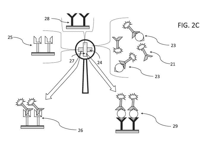
FIG. 2C presents a schematic diagram of the device presented in FIGs. 2A-B,
wherein
some of mobile labeled antibody/lectin-biomarker adduct 23 was at or has
migrated to
perpendicular detection zone 27, in which biomarker-specific antibody/lectin
28 is immobilized
on the porous matrix of probe 20, and the binding event is made visible by the
label on mobile
labeled antibody/lectin-biomarker adduct 23, now immobilized and concentrated
in detection
zone 27 as visibly detectable diagnostic complex 29, indicating that the
sample contacted with the
device contains glycan-based biomarkers indicative of brain injury;
FIG. 3 presents a schematic illustration of a device, according to some
embodiments of
the present invention, wherein device 30 is having probe 31 comprising porous
matrix 32 in
which control zone 33 and detection zone 34 form a "plus" sign and handle 35
is a rigid hollow
tube designed connect to the tip of generic syringe 36 and transfer the liquid
sample to probe 31;
FIG. 4 presents a schematic illustration of a device, according to some
embodiments of
the present invention, wherein device 40 includes probe 41 that comprises
indicator formulation
42, and housed within frame 44, mounted on handle 43, whereas the plurality of
areas 45a-g are
arranged radially around the opening in frame 44, and control zone 46 is
positioned at the center
of probe 41;
FIG. 5 presents a schematic illustration of a device, according to some
embodiments of
the present invention, wherein device 50 includes probe 51 that comprises
indicator formulation
52 and control zone 56 is positioned at the center of probe 51, mounted on
handle 53, and
separate gauge 54 having a plurality of areas 55a-g; and
FIGs. 6A-D present schematic illustrations of some embodiments of the present
invention,
wherein FIG. 6A shows a device having probe 61 in direct communication with
handle portal 62
and additional portals 63 branching off from handle portal 62, FIG. 6B shows a
device having
probe 61 and two portals 64 in direct communication with probe 61, FIG. 6C
shows a device
having portal 64 in direct communication with probe 61 and additional portals
63 branching off
from handle portal 62, and FIG. 6D shows a device having probe 61 in direct
communication
with handle reservoir 65 in the form of a syringe that is secured from
accidental or premature
ejection of its content by plunger stopper 66 as part of a kit and protective
sheath (such as
metallic or plastic pouch or container) 67 that can also serve as a sample
dipping container as part
of a kit.
8
CA 03050363 2019-07-16
WO 2018/154401
PCT/IB2018/050698
DESCRIPTION OF SOME SPECIFIC EMBODIMENTS OF THE INVENTION
The present invention, in some embodiments thereof, relates to a diagnostic
device and
method and, more particularly, but not exclusively, to a portable, user-
initiated visual assay
device and method for diagnosing brain injury.
The principles and operation of the present invention may be better understood
with
reference to the figures and accompanying descriptions.
Before explaining at least one embodiment of the invention in detail, it is to
be
understood that the invention is not necessarily limited in its application to
the details set forth in
the following description or exemplified by the Examples. The invention is
capable of other
embodiments or of being practiced or carried out in various ways.
As discussed hereinabove, given the great strain on the healthcare work force,
the
increased prevalence of many common injuries, diseases and the substantial
delay in treatment
caused by remote testing, the present inventors have recognized the need for
self-monitoring
non-invasive means for diagnosing brain injury. The present inventors have
contemplated a
user-friendly non-invasive mode for brain injury diagnosis, which allows the
use of an
inexpensive apparatus suitable for more widespread off-clinic use and
acceptance that enables
greater convenience in carrying about and use in testing and provides a
simplified visual mode
for monitoring test results. The present inventors have also contemplated an
off-the-shelf
product that enables the economical manufacture and distribution of relatively
low-cost, reliable
diagnostic device that can be used by non-professionals in educational,
sports, and other public
facilities, as well as homes and workplaces.
While reducing the present invention to practice, the present inventors have
envisioned
rapid, easy-to-use diagnostic devices and methods to enable efficient and
accurate point-of-care
(POC) detection of brain injury, which comprising a means for saliva
stimulation, a candy-like
(lollipop) component that may or may not feature a taste or aroma, a means for
visual change
activation, and a gauge for visual comparison of the results.
Portable non-invasive visual diagnosis device:
In the context of some embodiments of the present invention, the device for
diagnosing
brain injury is based on the detecting certain glycan-based biomarkers in a
sample, as these are
described in details hereinbelow, wherein the sample is obtained by non-
invasive means, such as
saliva and urine, and the indication of positive or negative diagnosis of a
brain injury is obtained
without need for special machinery and/or processes, and can be carried out by
a layman.
Nonetheless, it is noted herein that use of the provisions of the present
invention are not limited
9
CA 03050363 2019-07-16
WO 2018/154401
PCT/IB2018/050698
to samples extracted by non-invasive methods, meaning that the device and
methods provided
herein can be used to diagnose brain injury by sampling blood, plasma, spinal
fluid and the like.
The present inventors have considered that some colorimetric and enzymatic
reporter
systems useful in detecting glycan-based biomarkers are used as solutions and
the formed colors
.. spread by diffusion, which makes them less suitable in portable off-clinic
POC (at home) device,
designed for highlighting a pattern on a membrane or any solid support. In
addition,
concentrated acids, heating or harmful chemicals are used in several
colorimetric reactions
known in the art for detecting proteins and saccharides. Thus, in some
embodiments, the
detection of glycan-based biomarkers is effected by non-toxic, non-hazardous
and generally safe
reagents that evoke a chromatic reaction that is visually perceptible, which
requires no further
equipment or processing to be developed and observed by a non-professional
user.
One object of the present invention is to provide a test device which is
readily usable by
an unskilled person and which preferably merely requires that some portion of
the device is
contacted with the sample (e.g., saliva or urine), and thereafter no further
actions, or only
minimal simple actions, such as shaking, mixing, pushing a plunger etc., are
required by the user
before a diagnostic or an analytical result can be observed. Preferably the
diagnostic/analytical
result is observable within a matter of minutes following sample application,
e.g., ten minutes or
less. Such devices can be provided as kits suitable for home use, comprising a
plurality (e.g.,
more than one) of devices individually wrapped in moisture impervious wrapping
and packaged
together with appropriate instructions to the user.
Some embodiments of the present invention are focused on adapting and
improving some
of the known analyte detection techniques and methodologies, such as those
referred to herein, to
provide brain injury diagnostic test devices especially suitable for home use
which are quick and
convenient to use and which require the user to perform as few actions as
possible.
Thus, according to an aspect of some embodiments of the present invention,
there is
provided a device for diagnosing brain injury in a subject. The device
includes:
a probe that includes an porous matrix; and
an indicator formulation disposed in and/or on the porous matrix and comprises
at least
one glycan-based biomarker binding reagent capable of selectively binding to a
glycan-based
biomarker in a sample, and a first visually detectable label.
In some embodiments, the indicator formulation includes at least one glycan-
based
biomarker binding reagent capable of selectively binding to a glycan-based
biomarker in a liquid
sample taken non-invasively from the subject, and a visually detectable label,
wherein:
the glycan-based biomarker is indicative of brain injury;
CA 03050363 2019-07-16
WO 2018/154401
PCT/IB2018/050698
the glycan-based biomarker binding reagent and/or the visually detectable
label is
immobilized on the porous matrix;
the visually detectable label develops a color and becomes visible upon a
binding event
of the glycan-based biomarker to the glycan-based biomarker binding reagent;
and
the binding event is effected by contacting the sample with the probe.
In some embodiments, the glycan-based biomarker binding reagent is a lectin, a
galectin,
or an antibody. Herein and throughout, unless stated otherwise, the term
"glycan-based
biomarker binding reagent", refers to any one of the antibodies, lectins,
galectins or other
molecules which has been identified as capable of selectively bind to a glycan-
based biomarker.
In the context of embodiments of the present invention, the glycan-based
biomarker is indicative
of brain injury in a subject. It is also noted that unless stated otherwise, a
reference to an
antibody as glycan-based biomarker binding reagent, is meant to encompass
lectins, galectins or
other molecules which has been identified as capable of selectively bind to a
glycan-based
biomarker; a reference to a lectin as glycan-based biomarker binding reagent,
is meant to
encompass antibodies, galectins or other molecules which has been identified
as capable of
selectively bind to a glycan-based biomarker; and a reference to a galectin as
glycan-based
biomarker binding reagent, is meant to encompass lectins, antibodies or other
molecules which
has been identified as capable of selectively bind to a glycan-based biomarker
In some embodiments, a dye/colorant/chromogen forms a colored complex or
changes its
color in the presence of a glycan-base biomarker (chemical glycan assays). In
such
embodiments, the detection of glycan-based biomarkers is not necessarily based
on binding
thereof to a specific affinity binding reagent, but rather on the mere
presence of the biomarker
and its effect on other factors in the probe. For example, a reaction cascade
is initiated by the
presence of the biomarker, which causes a change in color in the probe. The
reaction may or
may not include enzymes. In some embodiments, an enzyme specific for the
glycan-based
biomarker starts a conversion reaction in the presence of the biomarker. The
enzymatic reaction
is coupled to a dye/colorant/chromogen which develops color or change it color
(enzymatic
activity). Such detection mechanism also does not require immobilization of
any element in the
indicator formulation, and the color change may be effected throughout the
probe. Such
approach is particularly suitable for the strip device embodiments described
hereinbelow.
Some embodiments of the present invention include diagnostic test devices
removably
encased in a wrapping material or a casing container constructed of moisture-
impervious solid
material.
11
CA 03050363 2019-07-16
WO 2018/154401
PCT/IB2018/050698
The device of the present invention comprises a probe that includes a dry
porous carrier
(matrix), referred to herein as a "porous matrix", which designed to carry the
indicator
formulation, and to be soaked with a liquid test sample that is applied to the
probe. The probe
may further be sectioned into zones, such as a detection zone and a control
zone, as these are
described hereinbelow.
In some embodiments, the device of the present invention further includes a
handle in
communication with the probe, designed for handling the probe for sample
contacting and the
like.
In some embodiments, the handle includes or is a tube, which is in direct
communication
with the porous matrix of the probe on the proximal end thereof (the end that
is connected to the
probe). The distal end of the tube handle is open to receive a liquid sample
such that the tube
can transport the sample from an external source to said probe. In such
embodiments of the
present invention, the handle is used also as an inlet and/or outlet portal to
infuse liquids and
reagents in solution into and/or out of the probe. The liquid can be a sample
and/or a standard
analyte solution and/or an indicator formulation reagent and/or a washing
liquid, and any
combination thereof. The handle and the distal end thereof can be shaped as a
syringe tip
fitting/adaptor, or be stretchable and elastic for fitting any tip of the
external source of the
sample, or be a screw threaded tip, a piercing needle tip, a septum membrane,
a butterfly needle
adaptor, and have any shape designed to connect to an external source of a
liquid sample.
In some embodiments, the purpose of the tube, in addition to introducing the
sample, is to
deliver additional reagents in solution to the probe to start/enhance/stop the
reaction, if needed.
The additional solution may carry an element that assists in the color
development, and or
supplement the indicator formulation with a detection element, if needed.
The term "portal", as used in the context of some embodiments of the present
invention,
refers to an element of the device, which is designed as an inlet and/or
outlet for infusing or
retracting liquids and reagents in solution into and/or out of the probe. In
some embodiments,
the device includes more than one portal, as described above, for letting into
the probe any one
or more of a sample and/or a standard analyte solution and/or an indicator
formulation reagent
and/or a washing liquid, and any combination thereof. In such embodiments the
handle can have
a multiple inlets and outlets portals, or be connected to a manifold of inlets
and outlets, or the
probe can be in communication with more than one portal regardless of a
handle.
In some embodiments, the device is equipped with at least one portal to which
a reservoir
is attached. The reservoir may be in the form of a piston/plunger and
cylinder/barrel)
combination (e.g., a syringe), wherein the plunger is retracted and the barrel
is the reservoir. In
12
CA 03050363 2019-07-16
WO 2018/154401
PCT/IB2018/050698
some embodiments, the reservoir can be pre-filled with a liquid that is used
in the diagnosis
process, and can be, for example, a standard analyte solution and/or an
indicator formulation
reagent and/or a washing liquid, and any combination thereof.
In some embodiments, the device has a shape of a strip, namely an elongated
flat thin
object, wherein one end thereof or a mid-section thereof, serves as a probe,
and one or two tips
or ends thereof serve as a handle. An illustration of an exemplary strip-
shaped device is
discussed hereinbelow and presented in FIG. 1.
In some embodiments, the probe is further coated or tightly wrapped with a
layer of a
semi-permeable material. The material of the layer is selected to be permeable
to aqueous media
and aqueous solutes therein, and to be impermeable to particles larger than a
certain threshold,
such as 0.01 p.m, 0.02 p.m, 0.03 p.m, 0.04 p.m, 0.05 p.m, 0.1 p.m, 0.2 p.m,
0.3 p.m, 0.4 p.m or 0.5
p.m. This layer provides a user interface and a mean to prevent passage of
mobile elements in
the probe to pass to the user when contacted to absorb a liquid sample, (e.g.,
when the probe is
inserted into the mouth to be soaked with saliva).
Suitable semi-permeable membranes, such as the type of biological or
synthetic,
polymeric membrane that will allow certain molecules or ions to pass through
it by diffusion, or
occasionally by more specialized processes of facilitated diffusion, passive
transport or active
transport. Suitable semi-permeable membranes, composed of either regenerated
cellulose or
cellulose esters (e.g., cellulose acetate) are manufactured through distinct
processes of modifying
and cross-linking cellulose fibers derived from wood pulp or cotton fibers to
form films with
differing properties and pore sizes. Variations in the manufacturing process
significantly change
the properties and pores sizes of the film. Cellulose-based membranes are also
suitable.
Glycerol is frequently added as a humectant to prevent cracking during drying
and to help
maintain the desired pore structure. Regenerated cellulose membranes are very
hydrophilic and
hydrate rapidly when introduced to water. Due to their additional
crosslinking, regenerated
cellulose membranes have better chemical compatibility and heat stability than
membranes made
from cellulose esters. Regenerated cellulose membranes are also more resistant
to organic
solvents and to the weak or dilute acids and bases that are commonly used in
protein and
molecular biology applications.
.. Porous matrix:
The probe may be constructed from a porous matrix "backed" with a support
material,
e.g. with a plastic sheet, to increase its handling strength. This can be
manufactured easily by
forming a thin layer of the porous matrix on a sheet of backing material.
Alternatively, a pre-
13
CA 03050363 2019-07-16
WO 2018/154401
PCT/IB2018/050698
formed sheet of porous matrix can be tightly sandwiched between two supporting
sheets of solid
material, e.g., plastic sheets.
The porous matrix, which is the sample receiving member, can be made from any
bibulous, porous or fibrous material capable of absorbing liquid rapidly. The
porosity of the
material can be unidirectional (i.e., with pores or fibers running wholly or
predominantly parallel
to an axis of the member) or multidirectional (omnidirectional, so that the
member has an
amorphous sponge-like structure). Porous plastics material, such as
polypropylene, polyethylene
(preferably of very high molecular weight), polyvinylidene flouride, ethylene
vinylacetate,
acrylonitrile and polytetrafluoroethylene can be used. It can be advantageous
to pre-treat the
member with a surface-active agent during manufacture, as this can reduce any
inherent
hydrophobicity in the member and therefore enhance its ability to take up and
deliver a moist
sample rapidly and efficiently. Porous sample receiving members can also be
made from paper
or other cellulosic materials, such as nitrocellulose. Materials that are
widely used in the nibs of
so-called fiber tipped pens are particularly suitable and such materials can
be shaped or extruded
in a variety of lengths and cross-sections appropriate in the context of the
invention. Preferably
the material comprising the porous receiving member should be chosen such that
the porous
member can be saturated with aqueous liquid within a matter of seconds.
Preferably the material
remains robust when moist.
In some embodiments of the invention, the porous matrix is selected from the
family of
nitrocellulose materials. This family has some advantage over conventional
synthetic or
cellulose materials, such as paper, because it has a natural ability to bind
proteins without
requiring prior sensitization. Specific binding reagents, such as lectins and
immunoglobulins
(antibodies), can be applied directly to nitrocellulose and immobilized
thereon. No chemical
treatment is required which might interfere with the essential specific
binding activity of the
reagent. Unused binding sites on the nitrocellulose can thereafter be blocked
using simple
materials, such as polyvinylalcohol. Moreover, nitrocellulose is generally
safe, non-toxic and
readily available in a range of pore sizes and this facilitates the selection
of a carrier material to
suit particularly requirements such as sample flow rate.
Preferably the porous matrix has a pore size of at least one micron.
Preferably the porous
matrix has a pore size not greater than about 20 microns. In some embodiments,
the average
pore size of the porous matrix ranges 1-10, 1-20, 1-30, 1-40 or 1-50 microns.
In some embodiments of the present invention, the probe includes a solid phase
porous
matrix which is linked to a porous receiving member to which the liquid sample
can be applied
and from which the sample can permeate into the porous matrix. Preferably, the
porous matrix
14
CA 03050363 2019-07-16
WO 2018/154401
PCT/IB2018/050698
is contained within a moisture-impermeable casing or housing and the porous
receiving member,
with which the porous matrix is linked, extends out of the housing and can act
as a means for
permitting a liquid sample to enter the housing and permeate the porous solid
phase material.
The housing should be provided with means, e.g., appropriately placed
apertures, which enable
the second zone of the porous solid phase material (carrying the immobilized
unlabeled specific
binding reagent) to be observable from outside the housing so that the result
of the assay can be
observed. If desired, the housing may also be provided with further means
which enable a
further zone of the porous solid phase material to be observed from outside
the housing and
which further zone incorporates control reagents which enable an indication to
be given as to
whether the assay procedure has been completed. Preferably the housing is
provided with a
removable cap or shroud which can protect the protruding porous receiving
member during
storage before use. If desired, the cap or shroud can be replaced over the
protruding porous
receiving member, after sample application, while the assay procedure is being
performed.
Optionally, the labeled reagent can be incorporated elsewhere within the
device.
Blocking of unused binding sites in the porous matrix can be achieved by
treatment with
protein (e.g. bovine serum albumin or milk protein), or with polyvinylalcohol
or ethanolamine,
or any combination of these agents, for example. The mobile reagent(s) can
then be dispensed
onto the dry matrix and will become mobile in the carrier when in the moist
state. Between each
of these various process steps (sensitization, application of unlabeled
reagent, blocking and
application of the labeled reagent), the porous matrix should be dried.
The various reagents can be applied to the probe in a variety of ways. Various
"printing"
techniques have previously been proposed for application of liquid reagents to
porous matrices,
e.g. micro-syringes, pens using metered pumps, direct printing and inkjet
printing, and any of
these techniques can be used in the present context. To facilitate
manufacture, the matrix can be
treated with the reagents and then subdivided into smaller portions, e.g.,
small narrow strips each
embodying the required reagent-containing zones, to provide a plurality of
identical carrier units.
Indicator formulation:
The porous matrix contains an indicator formulation, which is a general term
that is used
to refer to a system that comprises a number of reagents and labels, some may
be attached to
one-another, some may be immobilized on the matrix and some are freely mobile
therein in the
moist state, and all are selected to bind, label and immobilize an analyte of
interest found in the
sample, or to form a colored complex with the analyte, or to change color in
the presence of the
analyte, which are not necessarily affinity-pair binding-based assays. The
indicator formulation
thus includes specific binding reagents for an analyte, wherein the specific
binding reagents
CA 03050363 2019-07-16
WO 2018/154401
PCT/IB2018/050698
(glycan-based biomarker binding reagents) are typically lectins and/or
antibodies, and the
analyte is one or more glycan-based biomarkers, at least some of which are
indicative of a brain
injury. In some embodiments, the lectin and the antibody are specific to the
same glycan-based
biomarker(s), which are indicative of a brain injury in the subject being
tested.
The indicator formulation further includes a labeling agent, referred to
herein as a
"visually detectable label". In some embodiments, the lectin and/or antibody
is labelled with the
visually detectable label, namely the visually detectable label is chemically
attached to the lectin
and/or to the antibody. The specific binding reagents and the visually
detectable labels are
selected such that upon a binding event of the specific binding reagents to
the glycan-based
biomarker(s) (or upon contacting a glycan-based biomarker, when using
diffusible dyes and
colors), the visually detectable label develops a color having a color
intensity level, which has
not been visible prior to the binding event, and thus becomes visible thereby
making the binding
even visibly distinguishable. In some embodiments, wherein a control for non-
specific binding,
or a "timer" mechanisms are used, there may be two or more different kids on
visually detectable
labels employed in the device, and in such cases, the visually detectable
label used for
visualization of specific binding, is referred to herein as a first visually
detectable label. In these
cases, a visually detectable label used for the "control" or "timer"
mechanisms is referred to
herein as a second visually detectable label. In some cases, the first and
second visually
detectable labels are identical.
In the context of embodiments of the present invention, the term "visible"
refer to a
visual signal that can be detected by the naked eye (visible light which a
human eye can
perceive), without the use of additional machinery or processes. In the
context of embodiments
of the present invention, a visible signal is a change in a color of a certain
object or an area
thereon, relative to the color that has been characteristic to the object or
area prior to the change.
A change can also be assessed in comparison to the background of the object or
area, and in
comparison to the surrounding of the object or area.
In some embodiments, the labeled or unlabeled lectin and/or antibody is
permanently
immobilized in a detection zone in/on the porous matrix and is therefore not
mobile in the moist
state (when the probe is soaked with the liquid sample). The detection zone
can be the entire
area of the probe, or a predetermined area thereof, which can have a visibly
recognized shape,
such as a dot, a circle, a bar, a square and the like.
In some embodiments, a labeled or unlabeled specific binding reagent is freely
mobile
within the porous matrix when in the moist state, and another labeled or
unlabeled specific
binding reagent for the same analyte is permanently immobilized in the
detection zone on the
16
CA 03050363 2019-07-16
WO 2018/154401
PCT/IB2018/050698
porous matrix and is therefore not mobile in the moist state, and the relative
positioning of the
labelled reagent and detection zone being such that liquid sample containing
the analyte applied
to the probe of the device can pick up labelled reagent and thereafter
permeate into the detection
zone, wherein a three-membered binding event causes a color change in the
detection zone. The
color change may also be a change in the color intensity level.
In one example, the porous matrix contains an indicator formulation that
comprises a
labelled specific binding reagent for an analyte which is freely mobile within
the porous matrix
when in the moist state, and an unlabeled specific binding reagent for the
same analyte is
permanently immobilized in the detection zone on the porous matrix and is
therefore not mobile
in the moist state. In such configuration, typically referred to as a
"sandwich" configuration, the
analyte and the freely mobile labeled binding reagent bind to one-another,
thereby specifically
labeling the analyte indirectly with the visually detectable label, and the
formed labeled mobile
adduct is picked-up by the immobilized unlabeled specific binding reagent to
form a sandwich
that is positioned permanently at the detection zone, where the color develops
due to
accumulation of the visually detectable label at high concentration, relative
to other areas in the
probe not having an immobilized reagent, if those are present.
The immobilized specific binding reagent in the detection zone is preferably a
highly
specific antibody, lectin or galectin. In some embodiments, the immobilized
species is a
monoclonal antibody. In the embodiment of the invention involving the sandwich
reaction, the
labeled reagent is a lectin or also preferably a highly specific antibody, and
more preferably a
monoclonal antibody.
The basic elements in the foregoing can be utilized, according to some
embodiments of
the present invention, in a "competition" assay mode, wherein the analyte in
the sample (glycan-
based biomarker) is in competition with a labeled version thereof for the
limited number of
binding sites (immobilized specific binding reagents) on the probe. In such a
"competition"
assay, the detectible signal can be a decrease in the color intensity level,
or a change in color in
cases where the background color becomes more visible when the labeled version
of the analyte
depletes from the detection zone.
Thus, another embodiment of the invention is a device for use in an assay for
an analyte,
incorporating a porous solid phase material carrying in a first zone a
labelled reagent which is
retained in the first zone while the porous material is in the dry state but
is free to migrate
through the porous material when the porous matrix is moistened, for example
by the application
of an aqueous liquid sample suspected of containing the analyte, the porous
material carrying in
a second zone, which is spatially distinct from the first zone, an unlabeled
specific binding
17
CA 03050363 2019-07-16
WO 2018/154401
PCT/IB2018/050698
reagent having specificity for the analyte, and which is capable of
participating with the labelled
reagent in either a "sandwich" or a "competition" reaction, the unlabeled
specific binding reagent
being firmly immobilized on the porous material such that it is not free to
migrate when the
porous material is in the moist state.
The invention also provides an analytical method in which a device as set
forth in the
foregoing is contacted with an aqueous liquid sample suspected of containing
the analyte, such
that the sample permeates by capillary action through the solid phase porous
matrix via the first
zone into the second zone and the labelled reagent migrates therewith from the
first zone to the
second zone, the presence of analyte in the sample being determined by
observing the extent (if
any) to which the labeled reagent becomes bound in the second zone.
In another embodiment of the invention, the labeled reagent is a specific
binding partner
for the analyte. The labeled reagent, the analyte (if present) and the
immobilized unlabeled
specific binding reagent cooperate together in a "sandwich" reaction. This
results in the labeled
reagent being bound in the second zone if analyte is present in the sample.
The two binding
reagents have specificities for different epitopes on the analyte.
In another embodiment of the invention, the labeled reagent is either the
analyte itself
which has been conjugated with a label, or is an analyte analogue, i.e., a
chemical entity having
the identical specific binding characteristics as the analyte, and which
similarly has been
conjugated with a label. In the latter case, it is preferable that the
properties of the analyte
analogue which influence its solubility or dispersibility in an aqueous liquid
sample and its
ability to migrate through the moist solid phase porous matrix should be
identical to those of the
analyte itself, or at least very closely similar. In this embodiment, the
labeled analyte or analyte
analogue will migrate through the solid phase porous matrix into the second
zone and bind with
the immobilized reagent. Any analyte present in the sample will compete with
the labelled
reagent in this binding "competition" reaction. Such competition will result
in a reduction in the
amount of labeled reagent binding in the second zone, and a consequent
decrease in the intensity
of the signal observed in the second zone in comparison with the signal that
is observed in the
absence of analyte in the sample.
Embodiments of the present invention are meant to encompass any methodology
and
system for specific labeling and detection of analytes that is useful for
visual determination of an
analyte in a non-invasive and simple to use as the device presented herein.
Particular useful are
methodologies and systems for specific labeling and detection of lectins,
glycans and antibodies,
such as described below; and as provided in the art by, for example, Tao, S.C.
et al. nectin
microarrays identify cell-specific and functionally significant cell surface
glycan markers",
18
CA 03050363 2019-07-16
WO 2018/154401
PCT/IB2018/050698
Glycobiology, 2008, 18(10), pp. 761-769], Katrlik, J. et al. ["Glycan and
lectin microarrays for
glycomics and medicinal applications", Med Res Rev, 2010, 30(2), pp. 394-418,
ISSN: 0198-
6325], Hirabayashi, J. et al. [" Lectin-based structural glycomics: A
practical approach to
complex glycans", Electrophoresis, 2011, 32(10), pp. 1118-1128], and
Hirabayashi, J. et al.
["Lectin microarrays: concept, principle and applications", Chemical Society
Reviews, 2013,
42(10), pp. 4443-4458].
Visually detectable label:
The visually detectable label can be any entity the presence of which can be
readily
detected. Preferably the label is a direct label, i.e., an entity which, in
its natural state, is readily
visible either to the naked eye, or with the aid of an optical filter and/or
applied stimulation, e.g.,
UV light to promote fluorescence. For example, minute colored particles, such
as dye
sols/colloids, metallic sols/colloids (e.g., gold colloid), carbon black
particles and nanotubes, and
colored latex particles, are suitable in the context of some embodiments of
the present invention.
Of these options, colored latex particles are most preferred. Concentration of
the label into a
small zone or volume should give rise to a readily detectable signal, e.g. a
strongly-colored area.
This can be evaluated by eye, or by instruments if desired.
Indirect labels, such as enzymes, e.g. alkaline phosphatase and horseradish
peroxidase,
can be used. These labels usually require the addition of one or more
developing reagents such
as substrates before a visible signal can be detected. Such additional
reagents can be
incorporated in the porous matrix or in the sample receiving member, if
present, such that they
dissolve or disperse in the aqueous liquid sample. Alternatively, the
developing reagents can be
added to the sample before contact with the porous matrix or the porous matrix
can be exposed
to the developing reagents after the binding reaction has taken place. For
example, glycan
binding reagents, e.g. lectin, galectin or antibody, may be conjugated with an
enzyme (e.g., HRP
or alkaline phosphatase) with the intention to react with a color-generating
substrate. The
conjugate binds to the glycan which is captured by an immobilized agent on the
surface, and a
substrate that is present in the probe's matrix is used to generate a colored
species in the enzyme-
catalyzed reaction (the substrate can form e.g. a precipitate or a color).
Coupling of the label to the specific binding reagent can be by covalent
bonding, if
desired, or by hydrophobic bonding. Such techniques are commonplace in the
art, and form no
part of the present invention. In the preferred embodiment, where the label is
a direct label such
as a colored latex particle, hydrophobic bonding or passive adsorption is
preferred.
In some embodiments of the invention, the visually detectable label is a
"direct label",
attached to one of the specific binding reagents. Exemplary direct labels
include gold sols and
19
CA 03050363 2019-07-16
WO 2018/154401
PCT/IB2018/050698
dye sols, as these are known in the art. These labels can be used to produce
an instant analytical
result without the need to add further reagents in order to develop a
detectable visual signal.
They are robust and stable and can therefore be used readily in the device
presented herein,
which is stored in the dry state. Their release on contact with an aqueous
sample can be
modulated, for example by the use of soluble glazes.
Preferably, the result of the diagnosis assay should be discernable by eye,
and to facilitate
this, it is necessary for the visually detectable label to become concentrated
in the detection zone.
To achieve this, a direct labeling reagent should be transportable easily and
rapidly by the
developing liquid (the sample's medium). Furthermore, it is preferable that
the whole of the
developing sample liquid is directed through a comparatively small detection
zone in order that
the probability of an observable result being obtained in increased.
In some preferred embodiments, the visually detectable label is a colored
latex particle of
spherical or near-spherical shape and having a maximum diameter of not greater
than about 0.5
micron. A preferred size range for such particles is from about 0.05 to about
0.5 microns.
Additional methodologies for visualizing glycans are known in the art, and
include
analysis of glycans using the bioorthogonal chemical reporter strategy,
periodate oxidation,
acidic ninhydrin assay, orcinol assay (Bial' s test) for visual determination
of pentose found in
glycans, p-bromoaniline assay, phloroglucinol assay, hexokinase assay, glucose
oxidase assay,
glucose dehydrogenase assay, D-glucitoldehydrogenase assay, resorcinol assay,
and more.
Phenol-sulfuric acid chemistry aimed at generating a color with hexoses and
pentoses,
found in glycans, can also be used as a visually detectable labels. For a
review of this type of
labeling, the artisan can turn to, for example, Masuko, T. et al.
["Carbohydrate analysis by a
phenol¨sulfuric acid method in microplate format", Analytical Biochemistry,
2005, 339(1), pp.
69-72].
The chemistry of bicinchoninic acid (BCA) as a chromogen, can be used to
quantify the
amount of copper reduced by the aldehyde present in glycans; the method is
sensitive and useful
in the range of 1 ¨ 20 nmol sugar.
Another alternative to visualize glycans is by analysis of free sialic acids
from
glycoconjugates by thiobarbituric acid. Briefly, periodiate is used under
strongly acidic
conditions to oxidize N-acetylneuraminic acid (NANA) to P-formylpyruvic acid,
which is visible
at 549 nm [Crook, M. et al., "Measurement of urine total sialic acid:
Comparison of an
automated ultraviolet enzymatic method with a colorimetric assay", British
Journal of
Biomedical Science, 2002, 59(1), pp. 20-3].
CA 03050363 2019-07-16
WO 2018/154401
PCT/IB2018/050698
U.S. Patent No. 5,512,488 provides methods for detacting polysaccharide
dissolved in
water at alkaline conditions, with the use of Congo Red (sodium diphenyl-bis-a-
naphthyl-amine
sulfonate) visible at 540 nm, or Crystal Violet, Gentian Violet and Toluidine
Blue (Basic Blue or
tolonium chloride).
3-Methyl-2-benzothiazolinonehydrazone (MBTH) reacts with the aldehyde moiety
of
reducing sugars found in glycans, to form a colored adduct, in a reaction that
is not interfered by
proteins and reducing agents [Gordon E. et al., "Determination of Reducing
Sugars with 3-
Methyl-2-benzothiazolinonehydrazone", Anal Biochem, 2001, 305, pp. 287-289].
The purpald reagent (4-amino-3-hydrazino-5-mercapto-1,2,4-triazole, CAS# 1750-
12-5)
is remarkably sensitive and specific for aldehydes found in glycans. The
purpald reaction is
based on a condensation of formaldehyde with the reagent to form an aminal,
which then reacts
under aeration to form a purple colored oxidation product. The reaction is
sensitive for
aldehydes, as ketones are oxidized to an uncoloured product [Jendral, J.A. et
al., "Formaldehyde
in Alcoholic Beverages: Large Chemical Survey Using Purpald Screening Followed
by
Chromotropic Acid Spectrophotometry with Multivariate Curve Resolution",
International
Journal of Analytical Chemistry, 2011, 2011, 11 pages].
The indicator reagents system (formulation) may also include one or more
element that is
bound to a magnetic particle, such that immobilization thereof, permanent or
temporary, can be
achieved by means of a magnetic field. The magnetic particle can be attached
to the glycan-
based biomarker binding reagent, and/or to the visually detectable label. In
some embodiments,
the magnetic particle can be the visually detectable label. It is within the
scope of the present
invention to implement the use of magnetic particles in some embodiments of
the present
invention, as described, for example, in U.S. Patent Nos. 4,177,253,
5,320,944, 5,993,740,
5,736,349 and 8,945,469.
The presence or color intensity level of the signal from the label which
becomes bound in
the detection zone can provide a qualitative or quantitative measurement of
analyte in the
sample. A plurality of detection zones arranged in series on the porous
matrix, through which
the aqueous liquid sample can pass progressively, can also be used to provide
a quantitative
measurement of the analyte, or can be loaded individually with different
specific binding agents
to provide a multi-analyte test.
In some embodiments where there is a requirement for two or more
distinguishable color
signals to indicate different events and/or provide control/timing for the
diagnostic assay, the
device may include more than one type of visually detectable labels. As
mentioned above, for
the same of clarity, a label that signals the presence of a brain injury
glycan-based biomarker is
21
CA 03050363 2019-07-16
WO 2018/154401
PCT/IB2018/050698
referred to herein as the first visually detectable label, and a label that
signals other events or
conditions, such as sufficiency of sample amount, sufficiency of elapse time
or a positive non-
specific binding control, is referred to herein the second visually detectable
label. In some
embodiments the first and the second labels will be difference chemicals,
optionally giving-off
different colors, and in some embodiments the first and second labels are the
same (identical),
which may be attached similarly or differently to the same or different
elements in the indicator
formulation.
Alternative indicator formulations:
The present invention also encompasses indicator formulations which are not
necessarily
based on affinity binding of a glycan-based biomarker to an immobilized glycan-
based
biomarker binding reagent capable of selectively binding to the glycan-based
biomarker in a
sample. Such formulations can be based on soluble reagents for glycan-based
biomarker
detection.
For example, in an enzymatic glycan assay embodiment, the analyte (a glycan)
in the
sample reacts with an enzyme that catalyzes the glycan's decomposition. For
example, a
hexokinase is an enzyme that phosphorylates hexoses (six-carbon sugars),
forming hexose
phosphate, which in turn can with another reagent in the indicator formulation
to form a
substance that gives-off a color. This reagent can be present in the probe or
added thereto via a
portal, as described herein. Similarly, galactose oxidase is an enzyme that
catalyzes the
oxidation of D-galactose, and glucose oxidase is an oxido-reductase that
catalyses the oxidation
of glucose to hydrogen peroxide and D-glucono-6-lactone; these products of the
enzymatic
reactions can be detected directly or indirectly.
For example, in a "direct assay" embodiment, the glycan-based biomarker reacts
with a
chromogen, e.g. a reducing sugar generates reduction of a chromogen thereby
affording color
development.
In some embodiments based on soluble and diffusible labels, the detection
reagent used is
glycan-based biomarker binding reagent (e.g., an antibody or lectin) that is
conjugated to an
enzyme. The glycan biomarker and the detection conjugate are captured on the
porous matrix by
a pre-immobilized capture reagent (e.g., an antibody or a lectin). Thereafter
the unbound
conjugate is flushed away via a flushing portal using a flushing solution, and
a mixture of
substrates and chromogens are added via the same or other portal(s). In some
cases the
conjugated enzyme is horseradish peroxidase (HRP), the substrate is hydrogen
peroxide and the
chromogen is 3,3',5,5'-tetramethylbenzidine (TMB).
22
CA 03050363 2019-07-16
WO 2018/154401
PCT/IB2018/050698
In some embodiments based on soluble and diffusible labels, the glycan-based
biomarker
reacts with an enzyme that is specific for a moiety or a molecular structure
present in the glycan-
based biomarker. In some cases this moiety can be galactose and the enzyme is
galactose
oxidase. In the presence of HRP and a chromogen such as Amplex Red (10H-
phenoxazine-3,7-
diol, 10-acetyl, CAS 119171-73-2) the reaction results in color development
(abs max @ 560
nm).
In some embodiments based on soluble and diffusible labels, the glycan-based
biomarker
reacts directly with a chromogen/dye. In some cases the glycan biomarker can
consist of a
reducing sugar which in some cases reacts with 3-methyl-2-
benzothiazolinonehydrazone
(MBTH), developing a colored adduct (see, for example, Sawicki, E. et al.
[Anal. Chem., 1961,
33(1), pp 93-96]).
Such formulations can be implemented in a device of any shape and
configuration,
including a strip and a lollipop configurations, as these are described
herein.
Controls and timer:
One issue to be reckoned with in a device such as presented herein, is that it
takes a little
while, after removing the assay device from contact with the liquid sample,
for the visible signal
to appear (develop). Clearly the user would like to read the result of the
assay as soon as
possible but, equally, the user requires confidence that sufficient time has
elapsed for the proper
assay result to have been obtained and that the test is not being read too
early, without having to
wait an inordinately long period. In order to address this problem, it is
known to incorporate a
"timer" into an assay device, as described, for example, in U.S. Patent No.
9,052,311, and EP
0826777, which are incorporated herein by reference in their entirety. These
additional 'timer'
reagents are deposited in a "timer" or "control" zone of the probe and, upon
hydration by the
sample, interact to produce a color change. In addition, the "timer" or
"control" reagents are
also said to perform quality-control function. It is generally undesirable if
assay devices are
exposed to moisture. However, since the timer reagent when hydrated produces a
colored
product, the timer will reveal if the device has been exposed to moisture, and
thus has been
tampered with. The "quality control" function indicates whether the device has
been exposed to
a sufficient or insufficient amount of the sample.
In some embodiments, the probe contains a control formulation, which is
associated with
a specific zone in the probe, referred to herein a "control zone". If present,
the "control zone"
can be designed merely to convey a signal to the user that the device has
worked. This signal
can be unrelated to the signal indicating the presence of an agent indicative
of brain injury in the
sample. Preferably, the control zone is located at a different location than
the detection zone.
23
CA 03050363 2019-07-16
WO 2018/154401
PCT/IB2018/050698
For example, the control zone can be loaded with a control formulation that
includes a
control binding reagent that will bind to any labeled glycan, to confirm that
the sample has
permeated and that it contained sufficient analytes therein, and a second
visually detectable
label. Alternatively, the control formulation in the control zone can include
an immobilized
analyte which will react with excess labeled reagent, and the purpose of this
control zone is to
indicate to the user that the test has been completed. A positive control
indicator therefore tells
the user that the sample has permeated the required distance through the test
device. The control
binding reagent cab be selected to have binding affinity to the any mobile
reagent in the indicator
formulation, and a signal that develops from such control formulation will
indicate a working
device, a sufficient sample, and a timer for completion of the detection
process. If only the
control zone becomes visually detectable, and the detection zone does not,
this scenario indicates
that the device is functioning, that the sample acquisition was successful,
and that the sample
contains no glycan-based biomarker indicative of brain injury.
Alternatively, the control zone can contain an anhydrous reagent that, when
moistened,
produces a color change or color formation, e.g. anhydrous copper sulfate that
will turn blue
when moistened by an aqueous sample.
Exemplary embodiment of a device and mode of use:
An illustration of an exemplary device, according to some embodiments of the
present
invention, is designed in the form of a strip, having an elongated flat narrow
rectangular shape,
wherein one end is used as probe (a detection zone) and the other end is used
for holding the
strip. According to some embodiments of the present invention, the exemplary
device is
configured to perform a "yes/no" test for brain injury in a subject by
sweeping in the subject's
saliva in the mouth or inserting the probe into a container holding the
subject's liquid sample,
such as urine.
Alternatively, the probe can be contacted with the sample by
applying/dropping/smearing the liquid sample on the probe. The indicator
formulation disposed
in/on the probe comprised a labeled and mobile analyte-specific binding
reagent (e.g., an
antibody or a lectin), an analyte-specific binding reagent (e.g., an antibody
or a lectin)
immobilized in the detection zone, or chemical compounds or enzymes, which
upon presence of
the glycan-based biomarker form a color. An exemplary basic device is
presented in FIG. 1.
FIG. 1 is a schematic illustration of an exemplary "strip" shaped device,
according to
some embodiments of the present invention, wherein device 10, having detection
zone 11 and
handle 12, is dipped into sample 13 not having a glycan-based biomarker, which
lead to no
coloring of wet detection zone 15, but when dipped into sample 14 having a
glycan-based
biomarker, wet detection zone 16 changes color.
24
CA 03050363 2019-07-16
WO 2018/154401
PCT/IB2018/050698
A strip shaped device is an embodiment in which a wider range of detection
chemistries,
i.e. diffusible dyes and enzymes that react directly with the glycan-based
biomarker, can be used.
An illustration of another exemplary device, according to some embodiments of
the
present invention, is designed in the form of a lollipop (round flat probe
mounted on a stick
handle), having a perpendicular detection zone and a horizontal control zone,
relative to the
handle. According to some embodiments of the present invention, the exemplary
probe is
configured to perform a "sandwich" immunoassay to diagnose brain injury in a
subject by
inserting the probe into the subject's mouth to extract a saliva sample. The
indicator formulation
disposed in/on the exemplary probe comprised a labeled and mobile analyte-
specific binding
reagent (e.g., an antibody or a lectin), an analyte-specific binding reagent
(e.g., an antibody or a
lectin) immobilized in the detection zone, and a nonspecific binding reagent
(e.g., an antibody or
a lectin) immobilized in the control zone, as presented in FIGs. 2A-C.
FIG. 2A presents a schematic diagram of lollipop device, wherein probe 20 is
having
mobile labeled antibody (analyte-specific binding reagent) 21 disposed
thereon, and when a
saliva sample containing glycan-based biomarker (analyte) 22 is contacted with
probe 20, mobile
labeled antibody-biomarker adduct 23 is formed.
FIG. 2B presents a schematic diagram of the device presented in FIG. 2A,
wherein some
of mobile labeled antibody 21 was at or has migrated to horizontal control
zone 24, in which
nonspecific antibody 25 is immobilized on the porous matrix of probe 20, and
the binding event
is made visible by the label on mobile labeled antibody 21, now immobilized
and concentrated in
control zone 24 as visibly detectable control complex 26, indicating that the
device is
functioning properly.
FIG. 2C presents a schematic diagram of the device presented in FIGs. 2A-B,
wherein
some of mobile labeled antibody-biomarker adduct 23 was at or has migrated to
perpendicular
detection zone 27, in which biomarker-specific antibody 28 is immobilized on
the porous matrix
of probe 20, and the binding event is made visible by the label on mobile
labeled antibody-
biomarker adduct 23, now immobilized and concentrated in detection zone 27 as
visibly
detectable diagnostic complex 29, indicating that the sample contacted with
the device contains
glycan-based biomarkers indicative of brain injury.
As can be reckoned from the illustrative example presented in FIGs. 2A-C, a
sample
containing glycan-based biomarkers indicative of brain injury will evoke the
formation of a
"plus" symbol at the center of the probe; a sample not containing glycan-based
biomarkers
indicative of brain injury will evoke the formation of a "minus" symbol at the
center of the
probe; a misuse of the device with either no sample or insufficient sample
will not evoke any
CA 03050363 2019-07-16
WO 2018/154401
PCT/IB2018/050698
visible change at the center of the probe; and an attempt to use of an old,
wet, tampered-with
device will be prevented by the "minus" sign indicating the device has been
moist prior to use.
An illustration of another exemplary device, according to some embodiments of
the
present invention, is designed to introduce the liquid sample into the probe
by plunger-driven
motion. The handle, in such embodiments, is hollow and tubular and designed on
its distal end
to connect to a syringe or another tube or to any other form of liquid
transfer mean, while the
proximal end is tethered to the probe such that the liquid transported through
the handle will
soak the porous matrix therein.
FIG. 3 presents a schematic illustration of a device, according to some
embodiments of
the present invention, wherein device 30 is having probe 31 comprising porous
matrix 32 in
which control zone 33 and detection zone 34 form a "plus" sign and handle 35
is a rigid hollow
tube designed connect to the tip of generic syringe 36 and transfer the liquid
sample to probe 31.
Another illustration of a device, according to some embodiments of the present
invention,
is a "lollipop" configuration, wherein the probe is placed in a frame that is
used also as a gauge
for estimating the color intensity level in the probe. In such embodiments,
the frame is decorated
with a color intensity gauge comprising a plurality of areas arranged radially
around the opening
that houses the probe. Each of the areas is having a color intensity level
representing a
concentration level of the glycan-based biomarker in the sample, which is for
a visual
comparison of a color intensity level in the probe, thereby providing a direct
visual
determination of a concentration level of the glycan-based biomarker in the
sample. FIG. 4
presents a device having an integrated gauge frame, and FIG. 5 presents a
device where the
gauge is a separate part thereof.
FIG. 4, presents a schematic illustration of a device, according to some
embodiments of
the present invention, wherein device 40 includes probe 41 that comprises
indicator formulation
42, and housed within frame 44, mounted on handle 43, whereas the plurality of
areas 45a-g are
arranged radially around the opening in frame 44, and control zone 46 is
positioned at the center
of probe 41.
FIG. 5, presents a schematic illustration of a device, according to some
embodiments of
the present invention, wherein device 50 includes probe 51 that comprises
indicator formulation
52 and control zone 56 is positioned at the center of probe 51, mounted on
handle 53, and
separate gauge 54 having a plurality of areas 55a-g.
Method of diagnosing brain injury in a subject:
In one aspect thereof, the present invention also provides a diagnostic method
in which a
device as set forth in the foregoing is used to determine a brain injury in a
subject. The method
26
CA 03050363 2019-07-16
WO 2018/154401
PCT/IB2018/050698
is carried out by contacting the device with an aqueous liquid sample
suspected of containing the
analyte, such that the probe in soaked with the sample that reaches the
detection zone, and the
control zone if present, through the solid phase porous matrix, and the
presence of the analyte in
the sample is determined by observing the extent (if any) to which the
detection zone changes
color.
Thus, according to an aspect of some embodiments of the present invention,
there is
provided a non-invasive method for diagnosing brain injury in a subject, which
is carried out by:
contacting the probe in the device described herein with a sample extracted
from the
subject in a non-invasive manner;
assessing a visible change in the color of the control zone, if present; and
determining brain injury in a subject according to a color change in the
detection zone in
the probe,
wherein the change in said color is effected by the binding event of the
glycan-based
biomarker to the glycan-based biomarker binding reagent(s), and this color
change is indicative
of a brain injury in the subject.
In some embodiments, a reaction is initiated by the presence of the biomarker,
which
causes a change in color in the probe. This method is not necessarily based on
affinity binding
or on immobilization of any one of the elements in the indicator formulation.
For example, in
some embodiments, an enzyme specific for the glycan-based biomarker is
involved in a
conversion reaction in the presence of the biomarker. The enzymatic reaction
is coupled to a
dye/colorant/chromogen which develops color or change it color (enzymatic
activity). Such
detection method is particularly suitable for the strip device embodiments
described herein.
In some embodiments the sample is saliva or urine. The sample extraction may
be
effected by inserting the device to the mouth of the subject and wetting the
probe with saliva.
Alternatively, the sample is urine, and the method is effected by wetting the
probe with urine
taken from the subject.
Glycan-based brain injury biomarkers:
As discussed hereinabove, WO/2016/166419 discloses diagnostic and prognostic
glycan-
based brain injury biomarkers, which may be used e.g. for identifying subjects
with severe
TBI/ABI, who are at risk of secondary brain injury and therefore require
increased surveillance,
or subjects with mild TBI/ABI or subclinical brain injury (SCI), who otherwise
may remain
undiagnosed and untreated. The biomarkers disclosed in WO/2016/166419 may also
be applied
in cases where there are no external signs of injury or where the injured
person, such as a baby or
a coma patient, cannot describe the injury. For example, brain injury status
includes, without
27
CA 03050363 2019-07-16
WO 2018/154401
PCT/IB2018/050698
limitation, the presence or absence of brain injury in a subject, the risk of
developing brain
injury, the stage or severity of brain injury, the progress of brain injury
(e.g., progress of brain
injury over time) and the effectiveness or response to treatment of brain
injury (e.g., clinical
follow up and surveillance of brain injury after treatment). Based on this
status, further
.. procedures may be indicated, including additional diagnostic tests or
therapeutic procedures or
regimens.
As used herein, the term "biomarker" refers to a molecule that is detectable
in a
biological sample obtained from a subject and that is indicative of a brain
damage in the subject.
Markers of particular interest in the invention include glycan-based
biomarkers showing
differences in glycosylation between a sample from an individual with a brain
damage and a
healthy control.
As used herein, the term "glycan-based biomarker" refers to monosaccharides
and
polysaccharides, i.e. a polymer comprising two or more monosaccharide
residues, as well as to a
carbohydrate portion of a glycoconjugate, such as glycopeptides and
glycoproteins, glycolipid, a
peptidoglycan, or a proteoglycan, and any fragment thereof. Glycan-based
biomarkers may
comprise either homo-polymeric or hetero- polymeric monosaccharide residues,
and they may be
either linear or branched. As used herein, the terms "glycan",
"polysaccharide" and
"carbohydrate" are interchangeable, unless otherwise indicated. Glycan-based
biomarkers
include but are not limited to carbohydrates, sugars, glycans, monosaccharides
and/or
polysaccharides, glycoproteins and glycopolymers. These biomarkers may be
present in blood
plasma or serum after brain injury, in cerebrospinal fluid (CSF) after brain
injury, in lymph fluid
after brain injury, in urine after brain injury, in saliva after brain injury,
in tears after brain injury
or in exudate after brain injury.
Glycocalyx is an extracellular polymeric coating surrounding many prokaryotic
and
eukaryotic cells consisting of glycoproteins, glycolipids, proteoglycans and
glycosaminoglycans.
The constituents of the glycocalyx play an important role e.g. in the process
of cell signaling,
virus transfection, and immunity.
The biomarkers are differentially present in unaffected subjects (normal
control or non-
brain injury) and subjects with brain injury, and, therefore, are useful in
aiding in the
determination of brain injury status. In certain embodiments of the present
invention, the
biomarkers are measured in a sample taken from a subject using the methods
described herein
and compared, for example, to predefined biomarker levels and correlated to
brain injury status.
In particular embodiments, the measurement(s) may then be compared with a
relevant diagnostic
amount(s), cut-off(s), or multivariate model scores that distinguish a
positive brain injury status
28
CA 03050363 2019-07-16
WO 2018/154401
PCT/IB2018/050698
from a negative brain injury status. The diagnostic amount(s) represents a
measured amount of a
biomarker(s) above which or below which a subject is classified as having a
particular brain
injury status. For example, if the biomarker(s) is/are up-regulated compared
to normal during
brain injury, then a measured amount(s) above the diagnostic cut-offs(s)
provides a diagnosis of
.. brain injury. Alternatively, if the biomarker(s) is/are down-regulated
during brain injury, then a
measured amount(s) at or below the diagnostic cut-offs(s) provides a diagnosis
of non- brain
injury. As is well understood in the art, by adjusting the particular
diagnostic cut-off(s) used in
an assay, one can increase sensitivity or specificity of the diagnostic assay
depending on the
preference of the diagnostician. In particular embodiments, the particular
diagnostic cut-off can
be determined, for example, by measuring the amount of biomarkers in a
statistically significant
number of samples from subjects with the different brain injury statuses, and
drawing the cut-off
to suit the desired levels of specificity and sensitivity.
An advantage of cerebrospinal fluid biomarkers is that the CSF is in direct
contact with
the extracellular matrix in the brain and, thus, it mirrors biochemical
changes in the brain. For
these reasons, the CSF might be considered an optimal source of biomarkers of
brain injury.
However, given that CSF must be obtained by invasive lumbar puncture,
availability of
biomarkers of brain damage that can be assayed in blood samples would be
beneficial. Serum,
plasma, saliva or urine biomarkers are of special importance in especially
blast-induced TBI
because they are typically associated with military operations with limited
access to imaging and
other diagnostic tools of hospitals. The combination of physical damage and
psychological
effects makes blast-induced TBI especially difficult to diagnose. Thus,
plasma, serum, saliva or
urine biomarkers that can distinguish between the physical and psychological
components of the
injury would be of special value.
As used herein, the term "brain damage" refers to the destruction or
degeneration of brain
cells due to one or more internal or external factors. Non-limiting examples
of brain damage
include traumatic brain injury (TBI), acquired brain injury (ABI), subclinical
brain injury (SCI)
and neurodegenerative conditions. Non-limiting examples of typical
neurodegenerative
conditions include Huntington's disease, Parkinson's disease, Alzheimer's
disease and Chronic
Traumatic Encephalopathy. As used herein, the terms "brain damage" and "brain
injury" are
interchangeable, unless otherwise indicated.
As used herein, the term "traumatic brain injury" (TBI) refers to brain injury
caused by
external physical trauma, or by a sudden motion of the head, without a
physical contact with or
hit to an external object. Non-limiting examples of incidences resulting in
TBI include falls,
29
CA 03050363 2019-07-16
WO 2018/154401
PCT/IB2018/050698
vehicle collisions, sports collisions, and combats. The term includes both
mild and severe TBI
including closed-head injuries, concussions or contusions and penetrating head
injuries.
As used herein, the term "acquired brain injury" (ABI) refers to a brain
damage not
caused by an external brain injury or a hereditary condition. ABI may occur
after birth as a
result of complications, a disorder or congenital malady, or it may result
from, for instance,
stroke, surgery, removal of a brain tumor, infection, chemical and/or toxic
poisoning, hypoxia,
ischemia, sub- stance abuse, or a combination thereof.
The term "brain injury" also refers to subclinical brain injury, and anoxic-
ischemic brain
injury. The term "subclinical brain injury" (SCI) refers to brain injury
without overt clinical
evidence of brain injury. A lack of clinical evidence of brain injury when
brain injury actually
exists could result from degree of injury, type of injury, level of
consciousness and/or
medications, particularly sedation and anesthesia.
As used herein, the term "subject" refers to any mammal, including animals and
human
subjects. Animals include, but are not limited to, pets, farm animals, working
animals, sporting
animals, show animals, and zoo animals. Non-limiting examples of typical human
subjects
suffering from or pre-disposed to brain damage, TBI in particular, include
babies, infants,
children and young adults, particularly male; elderly; athletes, particularly
boxers, ice-hockey
players, soccer players, football (American) players, and skateboarders; and
soldiers. The terms
"human subject" and "individual" are interchangeable. Typically, the subject
is known to have
or suspected of having a brain injury, such as TBI or ABI.
As used herein, the term "diagnosis" means detecting an injury, a disease or a
disorder,
jointly referred to as a medical condition, or determining the stage or degree
of the medical
condition. Usually, a diagnosis of a medical condition is based on the
evaluation of one or more
factors (e.g., biomarkers) and/or symptoms that are indicative of the disease
and/or its progress.
That is, a diagnosis can be made based on the presence, absence or amount of a
factor which is
indicative of presence or absence of the medical condition. Each factor or
symptom that is
considered to be indicative for the diagnosis of a particular medical
condition does not need be
exclusively related to the particular medical condition, i.e. there may be
differential diagnoses
that can be inferred from a diagnostic factor or symptom. Likewise, there may
be instances
where a factor or symptom that is indicative of a particular medical condition
is present in an
individual that does not have the particular disease. The term "diagnosis"
also encompasses
determining the therapeutic effect of a drug therapy, or predicting the
pattern of response to a
drug therapy. The diagnostic methods may be used independently, or in
combination with other
diagnosing and/or staging methods known in the medical arts for a particular
medical condition.
CA 03050363 2019-07-16
WO 2018/154401
PCT/IB2018/050698
In the context of embodiments of the present invention, the term "diagnosis"
refers to the
determination of whether or not a subject has a brain damage, such as TBI or
ABI. The term is
also meant to include instances where the presence of a brain damage is not
finally determined
but that further diagnostic testing is warranted. In such embodiments, the
method is not by itself
determinative of the presence or absence of a brain damage in the subject but
can indicate that
further diagnostic testing is needed or would be beneficial. The methods,
therefore, can be
combined with one or more other diagnostic methods for the final determination
of the presence
or absence of a brain damage in the subject. Examples of such other diagnostic
methods include,
but are not limited to, CT and MRI, and are well known to a person skilled in
the art. As used
herein, a "final determination" or "final diagnosis" refers to ascertaining
the presence or absence
of a brain damage in a subject. The final determination or final diagnosis can
be the result of any
of the methods of the invention which, in some embodiments, can include more
than one
diagnostic test.
As used herein, the term "comparing" refers to making an assessment of how the
proportion, level or cellular localization of one or more biomarkers in a
sample from a subject
relates to the proportion, level or localization of the corresponding one or
more biomarkers in a
standard or control sample. For example, "comparing" may refer to assessing
whether the
proportion, level, or cellular localization of one or more biomarkers in a
sample from a subject is
the same as, more or less than, or different from the proportion, level, or
localization of the
corresponding one or more biomarkers in standard or control sample. More
specifically, the
term may refer to assessing whether the proportion, level, or cellular
localization of one or more
biomarkers in a sample from a subject is the same as, more or less than,
different from or
otherwise corresponds (or not) to the proportion, level, or cellular
localization of predefined
biomarker levels that correspond to, for example, a subject having subclinical
brain injury (SCI),
not having SCI, is responding to treatment for SCI, is not responding to
treatment for SCI, is/is
not likely to respond to a particular SCI treatment, or having/not having
another disease or
condition. In a specific embodiment, the term "comparing" refers to assessing
whether the level
of one or more biomarkers of the present invention in a sample from a subject
is the same as,
more or less than, different from other otherwise correspond (or not) to
levels of the same
biomarkers in a control sample (e.g., predefined levels that correlate to
uninfected individuals,
standard SCI levels, etc.).
The biomarkers and methods presented herein may be used not only for
diagnostic
purposes but also for prognosis or predicting the outcome of the brain damage,
or monitoring the
subject's survival from the brain damage or response to treatment.
31
CA 03050363 2019-07-16
WO 2018/154401
PCT/IB2018/050698
The biomarkers and methods presented herein may be used as a clinical end
point in
clinical trials for treating TBI or ABI, providing the outcome of the brain
damage, or monitoring
the subject's survival from the brain damage or response to treatment.
In some embodiments of the present invention, the diagnosis or prognosis of a
brain
damage may comprise determination of the presence or absence of one or more of
the present
glycan-based biomarkers in a biological sample obtained from a subject whose
possible brain
damage is to be determined. Multiplexed assays can provide substantially
improved diagnostic
precision. In a specific embodiment, the present invention provides methods
for determining the
risk of developing brain injury in a subject. Biomarker percentages, amounts
or patterns are
characteristic of various risk states, e.g., high, medium or low. The risk of
developing brain
injury is determined by measuring the relevant biomarkers and then either
submitting them to a
classification algorithm or comparing them with a reference amount, i.e., a
predefined level or
pattern of biomarkers that is associated with the particular risk level.
In some embodiments, the present invention provides methods for determining
the
severity of brain injury in a subject. Each grade or stage of brain injury
likely has a
characteristic level of a biomarker or relative levels of a set of biomarkers
(a pattern). The
severity of brain injury is determined by measuring the relevant biomarkers
and then either
submitting them to a classification algorithm or comparing them with a
reference amount, i.e., a
predefined level or pattern of biomarkers that is associated with the
particular stage.
In some embodiments of the present invention, the diagnosis or prognosis of a
brain
damage may comprise determination of the amount of one or more glycan-based
biomarkers, or
the relative amounts thereof as compared to, for example, the amount of each
other, one or more
other glycan, and/or a known standard. In some embodiments, diagnosis or
prognosis of brain
damage may be based on relative ratios of glycan-based biomarkers in different
body fluids,
such as a saliva/urine ratio, or a blood/CSF ratio.
In some embodiments, the amounts or relative ratios of one or more glycan-
based
biomarker may be compared to a predetermined threshold value which is
indicative of the
presence or absence of a brain damage or is useful in assessing the
progression or regression of
the brain damage. Such a comparison to a threshold value may result in a final
or non-final
diagnosis or a determination in regard to the progression or regression of the
brain damage.
Statistical methods for determining appropriate threshold values will be
readily apparent to those
of ordinary skill in the art. The threshold values may have been determined,
if necessary, from
samples of subjects of the same age, race, gender and/or disease status, etc.
The threshold value
32
CA 03050363 2019-07-16
WO 2018/154401
PCT/IB2018/050698
may originate from a single individual not affected by a brain damage or be a
value pooled from
more than one such individual.
In some embodiments, glycan-based biomarkers may also be detected and/or
quantified
with the use of lectins. Lectins are a well-known family of carbohydrate-
binding proteins, i.e.
macromolecules that are highly specific for given glycans on the basis of
their sugar moiety
structures and sequences. Lectins can be classified into distinct groups
according to their
carbohydrate specificity including, but not limited to, fucose-specific,
mannose specific, N-
acetylglucosamine-specific, and galactose/N-acetylglucosamine-specific
lectins. It is noted that
different sample types may exhibit different profiles of lectin-binding glycan
biomarkers.
Accordingly, lectins capable of identifying subjects with brain injury may be
used in either
individually or in any combination thereof.
In some embodiments, glycan-based biomarkers may also be detected and/or
quantified
with the use of galectins, the most widely expressed class of lectins in all
organisms. Galectins
are a family of proteins defined by their binding specificity for P-
galactoside sugars, such as N-
acetyllactosamine (Gai i -3G1cNAc or Gai i -4G1cNAc), which can be bound to
proteins by
either N-linked or 0-linked glycosylation. They are also termed S-type lectins
due to their
dependency on disulfide bonds for stability and carbohydrate binding. Among
fifteen galectins
discovered in mammals, only galectin- 1, -2, -3, -4, -7, -8, -9, -10, -12 and -
13 have been
identified in humans, to date. As used herein, "galectins" are encompassed by
the term "lectins",
unless otherwise indicated.
Biomarker analysis:
Standard techniques of protein microarray technology can be applied to analyze
the
glycan-based biomarkers. In such microarrays, lectins are immobilized on a
solid support, such
as a slide, in a high spatial density. Each lectin may be arrayed at several
concentrations and in
replicates on each slide. The concentration ranges may be tailored for each of
the lectins and
calibrated to provide a linear response within the same range, regardless of
the affinity of the
lectin. A sample of intact glycan-based biomarkers is applied to the array,
and its binding
pattern is detected by a label, such as a fluorescent label, a radioactive
label, or a
chemiluminescent label, which is placed either on the biomarker itself or on
the lectin directed
toward the carbohydrate moieties of the biomarker. Streptavidin may be used
for detecting
biotinylated samples. Also, "sandwich" based methods which utilize antibody
detection may be
employed, as is apparent to those with ordinary skill in the art.
Suitable microarray substrates include, but are not limited to, glass, silica,
aluminosilicates, borosilicates, metal oxides such as alumina and nickel
oxide, gold, various
33
CA 03050363 2019-07-16
WO 2018/154401
PCT/IB2018/050698
clays, nitrocellulose or nylon. In some embodiments a glass substrate is
preferred. In other
embodiments, the substrate may be coated with a compound to enhance binding of
the lectin to
the substrate. In some further embodiments, lectins have been arrayed on a
nitrocellulose
membrane-coated glass slide. In some still further embodiments, one or more
control lectins are
.. also attached to the substrate.
In some embodiments, a commercially available lectin array, which encompasses
one
standard glass slide, which is spotted with 8 wells of identical lectin
arrays, may be employed.
Each lectin, together with the positive controls is arrayed in duplicate. The
slide comes with an
8-well removable gasket which allows for the process of 8 samples using one
slide. Four-slide
slides can be nested into a tray, which matches a standard microplate and
allows for automated
robotic high throughput process of 64 arrays simultaneously. Unlike other
conventional
methods, e.g., liquid chromatography and mass spectrometry, lectin microarrays
enable rapid
and high-sensitivity profiling of complex glycan features without the need for
liberation of
glycans. Target samples include an extensive range of glycoconjugates involved
in cells, tissues,
body fluids, as well as synthetic glycans and their mimics. Various procedures
for rapid
differential glycan profiling have been developed for glycan-related
biomarkers and are
commercially available.
In one embodiment, the present invention provides methods for determining the
course
and prognosis of brain injury in a subject. Brain injury course refers to
changes in brain injury
status over time, including brain injury progression (worsening) and brain
injury regression
(improvement). Over time, the amount or relative amount (e.g., the pattern) of
the biomarkers
changes. For example, biomarker "X" may be increased with brain injury, while
biomarker "Y"
may be decreased with brain injury. Therefore, the trend of these biomarkers,
either increased or
decreased over time toward brain injury or non-brain injury indicates the
course of the condition.
Accordingly, this method involves measuring the level of one or more
biomarkers in a subject at
least two different time points, e.g., a first time and a second time, and
comparing the change, if
any. The course of brain injury is determined based on these comparisons.
In some embodiments of the present invention, methods for determining the
therapeutic
efficacy of a pharmaceutical drug are provides. These methods are useful in
performing clinical
trials of the drug, as well as monitoring the progress of a subject on the
drug. Therapy or clinical
trials involve administering the drug in a particular regimen. The regimen may
involve a single
dose of the drug or multiple doses of the drug over time. The doctor or
clinical researcher
monitors the effect of the drug on the patient or subject over the course of
administration. If the
drug has a pharmacological impact on the condition, the amounts or relative
amounts (e.g., the
34
CA 03050363 2019-07-16
WO 2018/154401
PCT/IB2018/050698
pattern or profile) of one or more of the biomarkers of the present invention
may change toward
a non- brain injury profile. Therefore, one can follow the course of one or
more biomarkers in
the subject during the course of treatment. Accordingly, this method involves
measuring one or
more biomarkers in a subject receiving drug therapy, and correlating the
biomarker levels with
the brain injury status of the subject (e.g., by comparison to predefined
levels of the biomarkers
that correspond to different brain injury statuses). One embodiment of this
method involves
determining the levels of one or more biomarkers in minimum at two different
time points
during a course of drug therapy, e.g., a first time and a second time, and
comparing the change in
levels of the biomarkers, if any. For example, the levels of one or more
biomarkers can be
measured before and after drug administration or at two different time points
during drug
administration. The effect of therapy is determined based on these
comparisons. If a treatment
is effective, then the one or more biomarkers will trend toward normal, while
if treatment is
ineffective, the one or more biomarkers will trend toward brain injury
indications.
Suitable methods for use in detecting or analyzing glycan-based biomarkers
include, but
are not limited to, Biocore studies, mass spectrometry, electrophoresis,
nuclear magnetic
resonance (NMR), chromatographic methods or a combination thereof.
Specifically, the mass
spectrometric method can be, for example, LC-MS, LC-MS/MS, MALDI-MS, MALDI-
TOF,
TANDEM-MS, FTMS, multiple reaction monitoring (MRM), quantitative MRM, or
Label-free
binding analysis. Examples of mass spectrometers are time-of-f light, magnetic
sector,
quadrupole filter, ion trap, ion cyclotron resonance, electrostatic sector
analyser, hybrids or
combinations of the foregoing, and the like. In yet another embodiment, mass
spectrometry can
be combined with another appropriate method(s) as may be contemplated by one
of ordinary
skill in the art. In another embodiment, the mass spectrometric technique is
multiple reaction
monitoring (MRM) or quantitative MRM. The electrophoretic method can be, for
example,
capillary electrophoresis (CE) or isoelectric focusing (IEF), and the
chromatographic methods
can be, for example, HPLC, chromatofocusing, or ion exchange chromatography.
In some embodiments, detecting, measuring and/or analyzing glycan-based
biomarkers in
a sample may be carried out by any appropriate enzyme assay available in the
art. Such assays
include, but are not limited to, galactose oxidase assays.
In some further embodiments, one or more different kinds of binding assays may
be used
for detecting, measuring and/or analyzing the present glycan-based biomarkers.
For instance, a
competitive lectin/galectin mode may be employed, wherein a pre-labelled
glycan competes with
a glycan from a sample to be analyzed for a limited number of binding sites
offered by the
lectin/galectin. Alternatively or in addition, said binding assay may be
carried out in a sandwich
CA 03050363 2019-07-16
WO 2018/154401
PCT/IB2018/050698
mode, wherein one lectin/galectin is used to bind a glycan contained in or
derived from a sample
to be analyzed from one side, and another lectin/galectin, conjugated with a
detectable label,
binds to the other side of the glycan or the glycan-lectin/galectin complex
formed.
In some embodiments, the biomarkers of the present invention can be detected
and/or
measured by immunoassays, either in a competitive or sandwich mode. Those
skilled in the art
know how to carry out such immunoassays. Furthermore, antibodies suitable for
this purpose
are available commercially. Further suitable antibodies may be produced by
methods well
known in the art.
In some embodiments, a combination of a lectin/galectin assay and an
immunoassay may
be employed for detecting, measuring and/or analyzing the present biomarkers
in a sample taken
from a subject. For this purpose, both a capture reagent and a detection
reagent are required.
Said capture reagent may be a lectin or a galectin, while said detection
reagent may be a
detectably labelled antibody, or vice versa.
The present invention also contemplates traditional immunoassays including,
for
example, sandwich immunoassays such as ELISA or fluorescence-based
immunoassays, as well
as other enzyme immunoassays. In a SELDI-based immunoassay, a bio specific
capture reagent
for the biomarker is attached to the surface of an MS probe, such as a pre-
activated lectin chip
array. The biomarker is then specifically captured on the biochip through this
reagent, and the
captured biomarker is detected by mass spectrometry.
As is readily understood by those skilled in the art, more than one type of
lectins/galectins and/or more than one type of antibodies may be used in the
binding assays set
forth above. In other words, several different lectins/galectins and
antibodies may be used in a
reaction to enhance the binding affinity or specificity. Furthermore, multiple
different reactions
may be carried out simultaneously or sequentially for detecting different
glycan-based
biomarkers in a sample to be analyzed.
It is also contemplated that glycans or glycan complexes contained in a sample
to be
analyzed may be immobilized directly to a surface, such as a microplate well,
a glass surface
(e.g. a slide), a metal surface (e.g. a silver or gold leaf) by opposite
charges, by a glue, of by
affinity binding, and be subsequently detected, for instance, by a detectably
labelled lectin or
antibody.
In accordance with the above, molecules suitable for use in detecting glycan-
based
biomarkers in a sample to be analyzed include, but are not limited to,
lectins, galectins,
antibodies, and competitive small molecules. Said detection molecules may be
visualized, or
made otherwise measurable, using for instance conjugated color reagents,
labels, or dyes.
36
CA 03050363 2019-07-16
WO 2018/154401
PCT/IB2018/050698
Enzyme labels suitable for this purpose include those that upon addition of a
substrate catalyze a
reaction leading to a measurable change in color, in luminescence, or in
production of a
precipitate. Non-limiting examples of such enzyme labels include horseradish
peroxidase (HRP)
and alkaline phosphatase (AP). Photoluminescent labels, including fluorescent
dyes (prompt),
lanthanide chelates (for time- resolved fluorescence), and photon upconversion
labels may be
used for detecting said detection molecules. Furthermore, the detection may be
based on
bioluminescence and chemiluminescence (as e.g. in luciferin-based detection),
or on
electrochemiluminescence (with e.g. ruthenium complexes). Also biotin and its
derivatives,
which enable binding and detection by labeled avidin or labeled streptavidin,
as well as various
radioactive isotopes may be used for the detection. The detection may also be
carried out using
beads and particles, including, for example, colored latex particles, colored
synthetic polymer
particles, colloidal metals such as gold and silver particles, (para)magnetic
beads, and
fluorophore-dyed particles.
In some embodiments, the biomarkers of the present invention may be detected
by means
of an electrochemical-luminescent assay developed by Meso Scale Discovery
(Gaithersrburg,
Md.). Electrochemiluminescence detection uses labels that emit light when
electrochemically
stimulated. Background signals are minimal because the stimulation mechanism
(electricity) is
decoupled from the signal (light). Labels are stable, non- radioactive and
offer a choice of
convenient coupling chemistries.
Furthermore, a sample may also be analyzed by means of a passive or active
biochip.
Biochips generally comprise solid substrates and have a generally planar
surface, to which a
capture reagent (also called an adsorbent or affinity reagent) is attached.
Frequently, the surface
of a biochip comprises a plurality of addressable locations, each of which has
the capture reagent
bound there. Lectin biochips are biochips adapted for the capture of glycans.
Many lectin
biochips are described in the art.
Kits:
According to an aspect of embodiments of the present invention, there is
provided a kit
for non-invasive diagnosis of brain injury in a subject, which can be carried
out by any layman at
any location and facility without the need for special training, procedures or
machinery.
In some embodiment, the kit comprises the device described herein. In some
embodiment, the kit further comprises instructions for use of the device and
for understanding
the various visual signals obtained as a result of using the device. In some
embodiment, the kit
further comprises a gauge for assessing the concentration of glycan-based
biomarkers in the
sample.
37
CA 03050363 2019-07-16
WO 2018/154401
PCT/IB2018/050698
In some embodiments the kit can be used to determine the presence or absence
of, or to
measure the levels of one or more glycan-based biomarker. In some embodiments,
the kit
comprises a package containing one or more glycan-based biomarker binding
reagent, such as a
lectin or an antibody which selectively binds to one or more glycan-based
biomarker, and a
control for comparing to a measured value of binding. In some embodiments, the
control is a
threshold value for comparing to the measured value. The kit can also include
a visually
detectable label.
According to some embodiments of the present invention, the kit may further
include a
device, a series of pre-measured (concentration and volume) liquids in
separate reservoirs, and a
mean to connect each of the reservoirs to the device so as to allow the
contents of the reservoir to
contact the probe. In some embodiments, the reservoirs are in the form of a
plunger/barrel type
(e.g., a syringe) which can connect directly to the probe via one of the
portals described
hereinabove. In some embodiments, the syringes are pre-filled and affixed to
the device. In
some embodiments, the kit also includes a protective sheath in the form of a
plastic or metal
container, which can also serve as a sample dipping container, for example,
when testing urine.
The device can be provided to the user in the protective sheath as a form of
packaging that can
be used for sample collection and contacting (e.g., dipping).
FIGs. 6A-D present schematic illustrations of some embodiments of the present
invention, wherein FIG. 6A shows a device having probe 61 in direct
communication with
handle portal 62 and additional portals 63 branching off from handle portal
62, FIG. 6B shows a
device having probe 61 and two portals 64 in direct communication with probe
61, FIG. 6C
shows a device having portal 64 in direct communication with probe 61 and
additional portals 63
branching off from handle portal 62, and FIG. 6D shows a device having probe
61 in direct
communication with handle reservoir 65 in the form of a syringe that is
secured from accidental
or premature ejection of its content by plunger stopper 66 as part of a kit
and protective sheath
67 that can also serve as a sample dipping container as part of a kit.
The kit for qualifying brain injury status may be provided as an immuno-
chromatography
strip comprising a membrane on which the antibodies are immobilized, and a
means for
detecting the biomarker(s). The kit may comprise a plastic plate on which a
sample application
pad, on which antibody bands and a secondary antibody band are immobilized and
an absorbent
pad are positioned in a serial manner, so as to keep continuous capillary flow
of blood serum.
In some embodiments, a subject can be diagnosed by adding blood, plasma or
serum
from the subject to the kit and detecting the relevant biomarkers conjugated
with antibodies,
specifically, by a method which comprises the steps of: (i) collecting blood,
plasma or serum
38
CA 03050363 2019-07-16
WO 2018/154401
PCT/IB2018/050698
from the subject; (ii) separating blood serum from the subject's blood; (iii)
adding the blood
plasma or serum from subject to a diagnostic kit; and, (iv) detecting the
biomarkers conjugated
with antibodies. In this method, the antibodies are brought into contact with
the subject's blood.
If the biomarkers are present in the sample, the antibodies will bind to the
sample, or a portion
thereof. In other kit and diagnostic embodiments, blood, plasma or serum need
not be collected
from the subject (i.e., it is already collected). Moreover, in other
embodiments, the sample may
comprise a tissue sample or a (non-invasive) clinical sample such as saliva,
urine or other body
fluids as described herein.
The kit can also comprise a washing solution or instructions for making a
washing
solution, in which the combination of the capture reagents and the washing
solution allows
capture of the biomarkers on the solid support or column for subsequent
detection by, e.g.,
antibodies or mass spectrometry. In a further embodiment, a kit can comprise
instructions for
suitable operational parameters in the form of a label or separate insert. For
example, the
instructions may inform a consumer about how to collect the sample, how to
wash the probe or
the particular biomarkers to be detected, etc. In yet another embodiment, the
kit can comprise
one or more containers with biomarker samples, to be used as standard(s) for
calibration.
As is apparent to a skilled person, the present lectin array kit can be used
with either a
label-based method or as a sandwich-based method. In one embodiment, the label
based method
is used for biotinylated samples containing proteoglycans and glycoproteins
for direct detection
on the array via a Cy3 equivalent dye-conjugated Biotin-Streptavidin complex.
In some
embodiments, a sandwich-based method is used for antibody detection of
glycocalyx elements
(glycolipids, glycoproteins, etc.) captured on the array. Labelled re- porter
antibodies specific
for the glycocalyx elements of interest may be provided in the kit or supplied
by the user of the
kit. An example protocol for this procedure with a general "Antibody Cocktail"
may be included
in a user manual. In some non-limiting embodiments, specific antibody
concentrations and
conditions may need to be determined by the end user.
In some embodiments, the biomarker detection kit comprises HRP protein and a
fluorescent light may be employed in order to detect the biomarker in a body
fluid and to
indicate the quantity of the biomarker in percentage. This may be incorporated
into a portable
application that indicates the severity of brain damage on a scale comprising,
but not limited to,
none, mild, moderate and severe. In another embodiment, an analogous yes/no
reply is received.
These examples do not exclude other possible embodiments.
In some embodiments, the present invention provides use of at least one
antibody in a kit
or in a device to detect brain damage, where the antibody may be a polyclonal
or a monoclonal
39
CA 03050363 2019-07-16
WO 2018/154401
PCT/IB2018/050698
antibody of any species, or a fragment thereof, either enzymatically cleaved
or recombinantly
produced, or a humanized antibody, and where the antibody recognizes and binds
glycan,
glycoprotein, peptidoglycan, proteoglycan, glycolipid, protein, small
molecule, lectin, or
antibody of another species (generally 'antigens').
An antibody may be used, for instance, as:
i) a capture reagent, wherein the antibody is immobilized on a solid substrate
to bind its
antigen from a sample medium;
ii) an antibody that is immobilized on a solid substrate to bind an analyte-
specific capture
reagent (for example lectin) so that the bound agent (lectin) is able to
capture the analyte
(glycan) from a sample;
iii) a primary detection reagent, wherein an antibody conjugated to any label
(labeled
antibody) recognizes and binds directly an antigen;
iv) a secondary detection reagent, wherein a labelled antibody recognizes and
binds a
primary detection reagent that is bound to the analyte. For example, a labeled
antibody binds to a
lectin that has bound to its cognate glycan, or a labeled antibody from one
species (e.g. goat) that
recognizes and binds an antibody of another species (e.g. mouse) which has
bound its antigen;
v) an antibody for recognizing and binding a non-glycan part of a glycan-
containing
molecule, e.g. a glycoprotein, where the glycoprotein or a fragment thereof is
first bound to e.g.
lectin via its glycan moiety and then is recognized and bound by an antibody
that is specific to
the peptide part of the molecule; or
vi) antibody for use in immunoblotting assays.
The kit may also comprise a combination of antibodies for different purposes.
All embodiments, details, advantages, and the like of the present device also
apply to a
device for use in different aspects and embodiments of the present invention.
Also, all
embodiments, details, advantages, and the like of the present methods apply to
the present kit,
and vice versa. In particular, one or more compounds, compositions, or
reagents disclosed as
suitable for carrying out the present methods may be comprised in the present
kit. Likewise,
anything disclosed with reference to the kit, apply to the present methods as
well.
Without further elaboration, it is believed that one skilled in the art, using
the preceding
description, can utilize the present invention to the fullest extent.
Non-limiting examples of advantages associated with the present glycan-based
biomarkers include that they are brain-tissue specific, able to cross the
blood-brain barrier into
the bloodstream within minutes of injury, and can be detected using a point-of-
care blood test or
other body fluids. Furthermore, the biomarkers may either increase or decrease
following the
CA 03050363 2019-07-16
WO 2018/154401
PCT/IB2018/050698
injury, but nevertheless they are in correlation with the severity of the
injury. Preferably, the
present biomarkers may correlate with injury magnitude, survivability, and/or
neurologic
outcome, or they may be indicative of the extent of neuronal and glial cell
loss, axonal, and
vascular damage. The present biomarkers can significantly add to the current
diagnostic palette
for brain damage.
It is expected that during the life of a patent maturing from this application
many relevant
saliva-based brain injury diagnosis devices will be developed and the scope of
the term saliva-
based brain injury diagnosis device is intended to include all such new
technologies a priori.
As used herein the term "about" refers to 10 %.
The terms "comprises", "comprising", "includes", "including", "having" and
their
conjugates mean "including but not limited to".
The term "consisting of' means "including and limited to".
The term "consisting essentially of" means that the composition, method or
structure may
include additional ingredients, steps and/or parts, but only if the additional
ingredients, steps
and/or parts do not materially alter the basic and novel characteristics of
the claimed
composition, method or structure.
As used herein, the phrases "substantially devoid of" and/or "essentially
devoid of" in the
context of a certain substance, refer to a composition that is totally devoid
of this substance or
includes less than about 5, 1, 0.5 or 0.1 percent of the substance by total
weight or volume of the
composition. Alternatively, the phrases "substantially devoid of" and/or
"essentially devoid of"
in the context of a process, a method, a property or a characteristic, refer
to a process, a
composition, a structure or an article that is totally devoid of a certain
process/method step, or a
certain property or a certain characteristic, or a process/method wherein the
certain
process/method step is effected at less than about 5, 1, 0.5 or 0.1 percent
compared to a given
standard process/method, or property or a characteristic characterized by less
than about 5, 1, 0.5
or 0.1 percent of the property or characteristic, compared to a given
standard.
The term "exemplary" is used herein to mean "serving as an example, instance
or
illustration". Any embodiment described as "exemplary" is not necessarily to
be construed as
preferred or advantageous over other embodiments and/or to exclude the
incorporation of
features from other embodiments.
The words "optionally" or "alternatively" are used herein to mean "is provided
in some
embodiments and not provided in other embodiments". Any particular embodiment
of the
invention may include a plurality of "optional" features unless such features
conflict.
41
CA 03050363 2019-07-16
WO 2018/154401
PCT/IB2018/050698
As used herein, the singular form "a", "an" and "the" include plural
references unless the
context clearly dictates otherwise. For example, the term "a compound" or "at
least one
compound" may include a plurality of compounds, including mixtures thereof.
Throughout this application, various embodiments of this invention may be
presented in
a range format. It should be understood that the description in range format
is merely for
convenience and brevity and should not be construed as an inflexible
limitation on the scope of
the invention. Accordingly, the description of a range should be considered to
have specifically
disclosed all the possible subranges as well as individual numerical values
within that range. For
example, description of a range such as from 1 to 6 should be considered to
have specifically
disclosed subranges such as from 1 to 3, from 1 to 4, from 1 to 5, from 2 to
4, from 2 to 6, from 3
to 6 etc., as well as individual numbers within that range, for example, 1, 2,
3, 4, 5, and 6. This
applies regardless of the breadth of the range.
Whenever a numerical range is indicated herein, it is meant to include any
cited numeral
(fractional or integral) within the indicated range. The phrases
"ranging/ranges between" a first
indicate number and a second indicate number and "ranging/ranges from" a first
indicate number
"to" a second indicate number are used herein interchangeably and are meant to
include the first
and second indicated numbers and all the fractional and integral numerals
therebetween.
As used herein the terms "process" and "method" refer to manners, means,
techniques
and procedures for accomplishing a given task including, but not limited to,
those manners,
means, techniques and procedures either known to, or readily developed from
known manners,
means, techniques and procedures by practitioners of the chemical, material,
mechanical,
computational and digital arts.
As used herein, the term "treating" includes abrogating, substantially
inhibiting, slowing
or reversing the progression of a condition, substantially ameliorating
clinical or aesthetical
symptoms of a condition or substantially preventing the appearance of clinical
or aesthetical
symptoms of a condition.
It is appreciated that certain features of the invention, which are, for
clarity, described in
the context of separate embodiments, may also be provided in combination in a
single
embodiment. Conversely, various features of the invention, which are, for
brevity, described in
the context of a single embodiment, may also be provided separately or in any
suitable
subcombination or as suitable in any other described embodiment of the
invention. Certain
features described in the context of various embodiments are not to be
considered essential
features of those embodiments, unless the embodiment is inoperative without
those elements.
42
CA 03050363 2019-07-16
WO 2018/154401
PCT/IB2018/050698
Various embodiments and aspects of the present invention as delineated
hereinabove and
as claimed in the claims section below find experimental and/or calculated
support in the
following examples.
EXAMPLES
Reference is now made to the following examples, which together with the above
descriptions illustrate some embodiments of the invention in a non-limiting
fashion.
Example I
Selection of Porous Matrix Material
Representative examples of porous matrix materials include paper,
nitrocellulose and
nylon membranes. Essential features of the material are its ability to bind
protein; speed of
liquid conduction; and, if necessary after pre-treatment, its ability to allow
the passage of labeled
binding reagents therethrough. In embodiments using direct label, it may be
desirable for the
material to allow flow of particles of size up to a few microns (usually less
than 0.5 1.tm).
Examples of flow rates obtained with various materials are presented in Table
1 below, showing
the time in minutes to flow through 45 mm of material.
Table I
Pore size Time
Material type
[1.tm] [minutes]
WhatmanCY s chromatography paper
(Schleicher & S chuell C)) 3 3.40
unbacked nitrocellulose sheet 5 3.30
8 3.00
12 2.20
backed polyester sheet 8 3.40
WhatmanCY s Nitrocellulose sheet 5 19.20
Pall 's Immunodyne (nylon) 3 4.00
5 3.20
43
CA 03050363 2019-07-16
WO 2018/154401
PCT/IB2018/050698
The travel rate of a test procedure will be determined by the flow rate of the
material
employed and while any of the above materials can be used some will give
faster tests than
others.
Nitrocellulose is advantageous of requiring no activation and will immobilize
proteins
strongly by absorption. Immunodyne is pre-activated and requires no chemical
treatment.
Papers, such as WhatmanCY s 3MM, require chemical activation with for example
carbonyldiimidazole in order to successfully immobilize proteins.
Example 2
Preparation of Labels
A selection of labels which may be used are described below. This list is not
exhaustive
and it is noted that other labeling methodologies and technologies are
contemplated within the
scope of the present invention.
Gold sol/colloid preparation:
Gold sols may be prepared for use in immunoassay from commercially-available
colloidal gold, and an antibody preparation. Metallic sol labels are
described, for example, in
European patent specification No, EP 7654.
For example, colloidal gold G20 (20 nm particle size, supplied by Janssen Life
Sciences
Products) is adjusted to pH 7 with 0.22 1.tm filtered 0.1 M K2CO3, and 20 ml
is added to a clean
glass beaker. 200 1.11 of antibody, prepared in 2 mM borax buffer pH 9 at 1
mg/ml, and 0.22 1.tm
filtered, is added to the gold sol, and the mixture stirred continuously for
two minutes. 0.1M
K2CO3 is used to adjust the pH of the antibody gold sol mixture to 9, and 2 ml
of 10 % (w/v)
BSA is added.
The antibody-gold is purified in a series of three centrifugation steps at
12000 g, 30
minutes, and 4 C, with only the loose part of the pellet being resuspended
for further use. The
final pellet is resuspended in 1 % (w/v) BSA in 20 mM Tris, 150 mM NaCl pH
8.2.
Dye sol preparation:
Dye sols (see, for example, European patent specification No. EP 32270) may be
prepared from commercially-available hydrophobic dyestuffs such as Foron Blue
SRP (Sandoz)
and Resolin Blue BBLS (Bayer). For example, fifty grams of dye is dispersed in
1 liter of
distilled water by mixing on a magnetic stirrer for 2-3 minutes. Fractionation
of the dye
dispersion can be performed by an initial centrifugation step at 1500 g for 10
minutes at room
temperature to remove larger sol particles as a solid pellet, with the
supernatant suspension being
retained for further centrifugation.
44
CA 03050363 2019-07-16
WO 2018/154401
PCT/IB2018/050698
The suspension is centrifuged at 3000 g for 10 minutes at room temperature,
the
supernatant being discarded and the pellet resuspended in 500 ml distilled
water. This procedure
is repeated a further three times, with the final pellet being resuspended in
100 ml distilled water.
The spectra of dye sols prepared as described above can be measured, giving
lambda-
max values of approximately 657 nm for Foron Blue, and 690 nm for Resolin
Blue. The
absorbance at lambda-max, for 1 cm path length, is used as an arbitrary
measure of the dye sol
concentration.
Colored particles:
Latex (polymer) particles for use in immunoassays are commercially available.
These
can be based on a range of synthetic polymers, such as polystyrene,
polyvinyltoluene,
polystyrene-acrylic acid and polyacrolein. The monomers used are normally
water-insoluble,
and are emulsified in aqueous surfactant so that monomer micellae are formed,
which are then
induced to polymerize by the addition of initiator to the emulsion.
Substantially spherical
polymer particles are produced.
Colored latex particles can be produced either by incorporating a suitable
dye, such as
anthraquinone (yellow), in the emulsion before polymerization, or by coloring
the pre-formed
particles. In the latter route, the dye should be dissolved in a water-
immiscible solvent, such a
chloroform, which is then added to an aqueous suspension of the latex
particles. The particles
take up the non-aqueous solvent and the dye, and can then be dried. Preferably
such latex
particles have a maximum dimension of less than about 0.5 micron.
Colored latex particles may be sensitized with protein, and in particular
antibody or
lectin, to provide selective binding reagents as described in the foregoing.
For example,
polystyrene beads of about 0.3 micron diameter, (supplied by Polymer
Laboratories) may be
sensitized with an anti-glycan-based biomarker antibody, in the process
described below:
0.5 ml (12.5 mg solids) of suspension is diluted with 1 ml of 0.1M borate
buffer pH 8.5
in an Eppendorf vial. These particles are washed four times in borate buffer,
each wash
consisting of centrifugation for 3 minutes at 13000 rpm in an MSE
microcentrifuge at room
temperature. The final pellet is resuspended in 1 ml borate buffer, mixed with
300 i.t.g of anti-
glycan-based biomarker antibody, and the suspension is rotated end-over-end
for 16-20 hours at
room temperature. The antibody-latex suspension is centrifuged for 5 minutes
at 13000 rpm, the
supernatant is discarded and the pellet resuspended in 1.5 ml borate buffer
containing 0.5 mg
bovine serum albumin. Following rotation end-over-end for 30 minutes at room
temperature, the
suspension is washed three times in 5 mg/ml BSA in phosphate buffered saline
pH7.2, by
CA 03050363 2019-07-16
WO 2018/154401
PCT/IB2018/050698
centrifugation at 13000 rpm for 5 minutes. The pellet is resuspended in 5
mg/ml BSA/5 % (w/v)
glycerol in phosphate buffered saline pH 7.2 and stored at 4 C until used.
Anti-glycan-based biomarker antibody-dye sol preparation:
Protein may be coupled to dye sol in a process involving passive adsorption.
The protein
may, for example, be a lectin or an antibody preparation such as anti-glycan-
based biomarker
antibody prepared in phosphate buffered saline pH 7.4 at 2 mg/ml. A reaction
mixture is
prepared which contains 100 Ill antibody solution, 2 ml dye sol, 2 ml 0.1M
phosphate buffer pH
5.8 and 15.9 ml distilled water. After gentle mixing of this solution, the
preparation is left for
fifteen minutes at room temperature. Excess binding sites may be blocked by
the addition of, for
example, bovine serum albumin: 4 ml of 150 mg/ml BSA in 5 mM NaCl pH 7.4 is
added to the
reaction mixture, and after 15 minutes incubation at room temperature, the
solution is
centrifuged at 3000 g for 10 minutes, and the pellet resuspended in 10 ml of
0.25 % (w/v
dextran/0.5 % w/v lactose in 0.04 M phosphate buffer). This antibody-dye sol
conjugate is best
stored in a freeze dried form.
Example 3
Preparation of a Strip Device
In an exemplary embodiment of the present invention, the device can be formed
in the
shape of a strip, as depicted in FIG. 1.
Zonal impregnation of liquid-conductive porous matrix materials:
Liquid-conducting porous matrix material with a restricted zone of immobilized
protein,
particularly antibody or lectin, can be prepared for example as follows:
A rectangular sheet of e.g., Schleicher & Schuell backed 8 1.tm nitrocellulose
paper
measuring 10 cm in length and 1 cm in width may have a detection zone formed
upon it by
applying an area of material about 1 cm long at one end of the strip. The
material can, for
example, be a suitably selected antibody preparation, prepared in phosphate
buffered saline pH
7.4 at 2 mg/ml, suitable for a labeled) lectin in a sandwich format. This
solution can be
deposited by means of a microprocessor-controlled microsyringe, which delivers
precise
volumes of reagent through a nozzle, preferably 2 mm diameter. When the
applied material is
been allowed to dry for 1 hour at room temperature, excess binding sites on
the nitrocellulose are
blocked with an inert compound such as polyvinyl alcohol (1 % w/v in 20 mM
Tris pH 7.4) for
30 minutes at room temperature, and sheets are thoroughly rinsed with
distilled water prior to
drying for 30 minutes at 30 C.
46
CA 03050363 2019-07-16
WO 2018/154401
PCT/IB2018/050698
In one embodiment, the liquid conductive porous matrix material can be
prepared in bulk
of wide format sheets, and then be cut up into numerous strips 10 cm in length
and 1 cm in
width, each strip carrying a detection zone of the immobilized antibody to
function as an
immunosorbent part at it tip. In this example the test strip is used with a
liquid label which is
mixed with sample. In use, this detection zone in which the immunoassay
reactions take place.
Example 4
Fetuin and Asialofetuin
In an exemplary embodiment of the present invention is a model of assaying
fetuin and
asialofetuin. Fetuin is an abundant glycoprotein in fetal serum, and
asialofetuin is its asialylated
form. Lectins or a lectin that selectively binds to the glycan part of fetuin
or asialofetuin is
permanently immobilized on solid matrix. Fetuin or asialofetuin in solution is
brought into
contact with the lectin and the binding reaction is subsequently taking place.
Thereafter, the
reaction compartment is washed and a labeled conjugate is added. The conjugate
binds to the
fetuin or asialofetuin that was captured on the surface in the preceding
phase.
Alternatively, fetuin or asialofetuin is first contacted with the labeled
conjugate to form a
complex. Thereafter the complex is brought into contact with the immobilized
lectin(s). The
conjugate comprises a fetuin-specific or asialofetuin-specific antibody which
is coupled to a
detectable label. The detectable label is one of those presented in the text,
preferably a
colloidal/particulate matter which enables visual detection.
Although the invention has been described in conjunction with specific
embodiments
thereof, it is evident that many alternatives, modifications and variations
will be apparent to
those skilled in the art. Accordingly, it is intended to embrace all such
alternatives, modifications
and variations that fall within the spirit and broad scope of the appended
claims.
All publications, patents and patent applications mentioned in this
specification are
herein incorporated in their entirety by reference into the specification, to
the same extent as if
each individual publication, patent or patent application was specifically and
individually
indicated to be incorporated herein by reference. In addition, citation or
identification of any
reference in this application shall not be construed as an admission that such
reference is
available as prior art to the present invention. To the extent that section
headings are used, they
should not be construed as necessarily limiting.
47