Note: Descriptions are shown in the official language in which they were submitted.
WO 2005/063126 PCT/US2004/043021
BIOPSY DEVICE WITH APERTURE ORIENTATION AND IMPROVED TIP
FIELD OF THE INVENTION
[0001] The present invention relates generally to tissue removing devices such
as
biopsy devices and the methods of using such devices. More specifically, it is
directed to an improved device and method for accessing and removing
pathologically suspect tissue from within a patient's body.
BACKGROUND OF THE INVENTION
[0002] In diagnosing and treating certain medical conditions, such as
potentially
cancerous tumors, it is usually desirable to perform a biopsy, in which a
specimen of
the suspicious tissue is removed for pathological examination and analysis. In
many
instances, the suspicious tissue is located in a subcutaneous site, such as
inide a
human breast. To minimize surgical intrusion into the patient's body, it is
desirable to
be able to insert a small instrument into the patient's body to access the
targeted site
and to extract the biopsy specimen therefrom.
[0003] Electrosurgical techniques have been used in a variety of biopsy
procedures. In electrosurgery, high frequency electrical energy is typically
applied to
patient tissue through an active electrode, the electrical circuit being
completed by a
return electrode in contact with the patent's tissue. Electrical energy
flowing through
the tissue from the active electrode is effective to ablate tissue near the
active
electrode, forming an opening in the tissue and so allowing insertion of the
instrument into a patient's body. A return electrode may be placed on the
exterior of
the patient's body or may be incorporated into the device itself. The return
electrode
is typically attached to the patient at a point remote from where the primary
or active
electrode contacts the tissue. However, in the case of a bipolar electrode for
CA 3050373 2019-07-19
example, the return electrode may be disposed near to the active electrode. An
electrosurgical
biopsy instrument is disclosed and claimed in United States Patent Application
Serial No.
09/159,467 for "Electrosurgical Biopsy Device and Method," now U.S. Patent No.
6,261,241. A
variety of needle like tip designs have been developed to aid in the accessing
of intracorporeal
sites for biopsy and other procedures. Electrosurgical techniques have also
been used in a variety
of biopsy procedures to gain access to an intracorporeal site. See for example
U.S. Patent No.
6,261,241.
[0004] The prior needle like tips do not always allow proper placement of the
biopsy or other
surgical device. Moreover, while the electrosurgical biopsy devices have been
found to be
effective in many instances, they are not suitable for use in conjunction with
magnetic resonance
imaging.
[0005] While these electrosurgical biopsy devices have been found to be
effective in many
instances, they may not always be suitable for use in conjunction with
magnetic resonance
imaging.
SUMMARY OF THE INVENTION
[0006] This invention is directed to devices for accessing and severing tissue
from a target site
within a patient and methods for utilizing such devices. The devices embodying
features of the
invention provide access to a targeted tissue site within a patient and
provide for the selection,
separation and capture of a tissue specimen from supporting tissue at the
targeted site.
[0007] A tissue accessing and severing device and system having features of
the invention
generally include an elongated, preferably disposable probe component
2
Date Recue/Date Received 2021-01-11
WO 2005/063126 PCT/US2004/043021
having a plurality of operative elements and a driver component configured to
receive the elongated probe component and drive the various operative elements
of
the probe component.
[0008] The elongated probe component has a distal shaft portion with a tissue
penetrating distal tip, a tubular section proximal to the distal tip, an inner
lumen
extending within the tubular section and an open, tissue receiving aperture in
the
tubular section which provides access to tissue at the targeted site. The
probe
component includes an elongated tissue-cutting member, which is preferably at
least
in part cylindrically shaped. The tissue cutting member is provided with at
least one
tissue cutting edge which is configured to sever tissue extending into the
interior of
the tubular section through the aperture thereof. The cutting edge on the
tissue
cutting member may be configured for longitudinal cutting movement and may
include oscillating rotational motion and/or reciprocating longitudinal motion
to sever
specimen tissue extending through the aperture from supporting tissue at the
targeted site. The cutting surfaces or edges are radially spaced from a
longitudinal
axis of the probe component and are generally transversely oriented with
respect to
the longitudinal axis. The tissue cutter is preferably slidably disposed
within the
inner lumen of the tubular section, although it may be disposed about the
tubular
section. The probe component may also have a handle which releasably engages
the driver component.
[0009] In one embodiment of the invention, the cutting member has an inner
lumen preferably extending to the proximal end thereof for tissue specimen
removal.
While mechanical withdrawal of the tissue specimen may be employed, it is
preferred to provide a vacuum within the cutting member from the proximal end
of
the cutting member. The proximal end of the cutting member may be configured
to
3
CA 3050373 2019-07-19
WO 2005/063126 PCT/US2004/043021
be in fluid communication with a vacuum source to aspirate the severed tissue
specimen through the inner lumen of the cutting member, to a tissue collection
station. A higher fluid pressure may be maintained in the inner lumen of the
cutting
member distal to the tissue specimen to aid in transporting the specimen
proximally
through the inner lumen. In this manner, the mechanical withdrawal and/or the
vacuum on the proximal end of the specimen and a higher pressure on the distal
end
of the specimen can move the specimen through the inner lumen of the cutting
member to a tissue collection station.
[0010] In at least one embodiment, the handle of the probe component is
secured, ,
preferably releasably secured, to the driver housing provided to operably
connect the
various operative elements of the probe with operative elements of the driver
component. The tissue cutting member is operatively connected to at least one
driver to provide the desired cutting motion. The proximal end of the tubular
section
is rotatably secured within the handle housing so that the orientation thereof
with
respect to the longitudinal axis and therefore the orientation of the tissue
receiving
aperture within the tubular section, can be selected. The orientation of the
aperture
may be selected manually such as described in copending application Serial
Number
10/642,406, filed February August 15, 2003 or it may be preset or selected
electronically by a control module which also controls the operation of the
cutting
member and electrical power. The aperture orientation setting may be selected
before or after the distal portion of the probe component is inserted into the
patient.
[0011] The tissue penetrating distal tip embodying features of the
invention has a
proximal base secured to the distal end of the probe shaft of the biopsy
device, and
a sharp distal point distal to the proximal base. The tissue penetrating
distal tip has
a first concave surface extending from the base to the sharp distal point. The
distal
4
CA 3050373 2019-07-19
WO 2005/063126 PCT/US2004/043021
tip also has a second concave surface, which intersects the first concave
surface
forming therewith a first curved cutting edge that leads to the sharp distal
point. The
distal tip also has a third concave surface which intersects the first concave
surface
forming therewith a second curved cutting edge leading to the sharp distal
point and
also intersects the second concave surface forming therewith a third curved
cutting
edge that leads to the sharp distal point. The concave surfaces preferably
have
center lines which extend from the proximal base of the distal tip to the
sharp distal
point. In a presently preferred embodiment the concave surfaces are of the
same
area. However, they may have different areas.
[0012] The driver component has at least two and preferably three driver units
for
operating the probe component secured to the driver component. Specifically,
the
driver component has a first driver unit for rotating the tubular section of
the probe
component, a second driver unit for moving the cutting member along a
longitudinal
axis of the cutting member and optionally a third driving unit for rotating or
oscillating
the cutting member about the longitudinal axis. The first driver unit rotates
the
tubular section of the probe component, preferably in discrete steps, so that
the
location of the tissue receiving aperture in the distal extremity of the
tubular section
can be selected prior to or during the procedure. The discrete rotational
steps of the
tubular section are preferably in 30 or multiples thereof so that the
rotational
movement will follow 12 hour clock markings. Preferably, the second and third
driver
units are operable together so that the cutting member may rotate or oscillate
about
a longitudinal axis as the cutter member is moved longitudinally. This allows
a
rotation or an oscillation of the cutter during the cutting process which can
aid in
cutting tissue.
CA 3050373 2019-07-19
WO 2005/063126 PCT/US2004/043021
[0013] The driver component may have one or more light sources in a distal
portion thereof to illuminate the accessing site during the procedure.
[0014] A method of cutting and collecting a tissue specimen with a tissue
collection device embodying features of the invention includes advancing such
a
device at least partially into tissue at a desired site within the patient's
body with the
tissue penetrating distal tip of the outer cannula disposed distal to the
tissue
specimen to be separated from the target site. A vacuum is established within
the
inner lumen of the tubular section to draw tissue through the aperture therein
into the
inner lumen of the tubular section. The cutting member, which is slidable
disposed
within the inner lumen of the tubular section, may then be moved
longitudinally to cut
a tissue specimen from supporting tissue at the target site by the
longitudinal motion,
which preferably includes oscillating rotational movement and/or reciprocating
longitudinal movement. The vacuum established within the inner lumen of the
tubular section may be applied through the inner lumen of the tissue cutting
member
when the tissue cutting member is disposed within the tubular section. The
applied
vacuum within the inner lumen of the tissue cutting member, may also be
utilized to
pull or aspirate the separated tissue sample proximally. In addition, or
alternatively, a
higher fluid pressure may be maintained in a distal part of the inner lumen of
the
tubular section, distal to the specimen, to push the tissue specimen
proximally,
Alternatively, the tissue specimen may be mechanically withdrawn. Fluid
pressure
may include pressure from a liquid delivered into the interior of the device,
such as a
physiological saline solution, and may include a gas, such as pressurized
carbon
dioxide, nitrogen or air, delivered into the interior of the device. Access to
ambient
air can also maintain a sufficiently high pressure differential to move the
specimen
through the inner lumen of the cutting member. Anesthetic may be injected to
the
6
CA 3050373 2019-07-19
WO 2005/063126 PCT/1JS2004/043021
target site through the outer cannula or the inner lumen of the cutting
member. Upon
removal from the patient, the tissue specimen may then be subjected to
pathological
examination. After acquisition of a tissue specimen or specimens, the tissue
separation system may be repositioned for further tissue separation and
collection or
it may be withdrawn from the patient.
[0015] The tubular section of the probe provides the support for the probe to
enable precise location of the accessing port to the desired location at the
target site
with its radial orientations being preset before the device is introduced into
the
patient or selected after the tubular section is disposed within the patient.
The
cutting member quickly and cleanly severs the tissue specimen drawn into the
interior of the tubular section though the aperture by the action of the
vacuum or
otherwise. Upon removal of the tissue specimen, the tissue receiving aperture
may
be radially repositioned about the longitudinal axis of the tubular section of
the probe
component so that a plurality of specimens may be taken from the target site.
The
orientation of the tissue receiving aperture during the procedure may follow a
preselected pattern or may be selected by the physician for other selected
tissue
specimens.
[0016] A tissue acquisition system assembly embodying features of the
invention .
may include a device for delivery of one or more marker bodies through a
tubular
member of a biopsy device such as the tubular cutting member. Such a marker
delivery device includes an elongated shaft having an inner lumen and a
discharge
opening in a distal portion of the elongated shaft, at least one marker body
which is
disposed within the inner lumen of the elongated shaft, a pusher element which
is
slidably disposed within the delivery device and which is configured to urge
at least
one marker body out the discharge opening in the distal portion of the
elongated
7
CA 3050373 2019-07-19
shaft. The marker delivery device has a distally flared guide member which is
slidably disposed on the elongated shaft to guide the distal portion of the
elongated
shaft into a proximal end of the tubular member of a biopsy device. This
invention is
directed to a tissue penetrating probe tip, particularly for biopsy devices.
These
devices provide access to a targeted tissue site and provide for the
separation and
capture of a tissue specimen from supporting tissue at the targeted site.
[0016a] This disclosure is also directed to a tissue biopsy system for
accessing and
collecting one or more tissue specimens from a target site within a patient,
comprising: a disposable elongated probe component including a distal shaft
portion
and an elongated tissue cutting member, the distal shaft portion having a
tubular
section and a tissue receiving aperture in the tubular section, the elongated
tissue
cutting member being disposed within the tubular section; a driver component
having
a distal portion with a distal end, the driver component configured to receive
a
proximal portion of the probe component in an operational relationship, which
has a
first driver unit configured to rotate the tubular section of the probe
component to
orient the tissue receiving aperture in the tubular section and which has a
second
driver unit configured to drive the tissue cutting member within the tubular
section to
cut tissue extending into the tubular section through the tissue receiving
aperture
thereof; and at least one illuminating element located in the distal portion
of the
driver component and configured to project light from the distal end of the
distal
portion of the driver component to provide localized illumination of an
operating site
on the patient.
[0016b] This disclosure is also directed to a tissue biopsy system for
accessing and
collecting one or more tissue specimens from a target site within a patient,
comprising: a disposable elongated probe component comprising an elongated
tubular section which has a longitudinal axis, which has an inner lumen
extending
therein, which has a tissue penetrating distal tip and which has an aperture
proximal
to the penetrating distal tip configured to receive tissue from the target
site, and an
elongated tissue cutting member which is disposed within the elongated tubular
section, which has at least one tissue cutting edge, which has an inner lumen
8
CA 3050373 2019-07-19
extending therein and which is configured to cut a tissue specimen from tissue
extending into the tissue receiving aperture of the elongated tubular section;
a driver
component having a distal portion with a distal end, the driver component
configured
to receive a proximal portion of the probe component in an operational
relationship,
which has a first driver unit configured to rotate the tubular section of the
probe
component to orient the tissue receiving aperture of the elongated tubular
section,
which has a second driver unit configured to drive the tissue cutting member
longitudinally within the tubular section of the probe to cut a tissue
specimen from
tissue extending into the tissue receiving aperture of the elongated tubular
section;
and at least one light source located in the distal portion of the driver
component
configured to project light from the distal end of the distal portion of the
driver
component to provide localized illumination at an operation site on the
patient, the
driver component being configured to automatically activate the at least one
light
source when the disposable elongated probe component is installed on the
driver
component.
[0016c] This
disclosure is also directed to a tissue biopsy system for accessing
and collecting one or more tissue specimens from a target site within a
patient,
comprising: a disposable elongated probe component comprising a distal shaft
portion with a penetrating distal tip, a tubular section proximal to the
distal tip, an
inner lumen extending within the tubular section and an open, tissue receiving
aperture in the tubular section which provides access to tissue at the
targeted site, a
proximal extremity configured to be secured to a driver unit, and an elongated
tissue
cutting member which is disposed within the tubular section, which has at
least one
tissue cutting edge and which has an inner lumen extending therein; a probe
housing for mounting the distal shaft portion and the elongated tissue cutting
member, the probe housing having a distal end; and a driver component having a
distal face, the driver component configured to receive a proximal portion of
the
probe component in an operational relationship, which has a first driver unit
configured to rotate the tubular section of the probe component to orient the
tissue
receiving aperture in the tubular section and which has a second driver unit
configured to drive the tissue cutting member longitudinally within the
tubular section
8a
CA 3050373 2019-07-19
to cut tissue extending into the tubular section through the tissue receiving
aperture
thereof; an orientation indicator secured to the tubular section adjacent the
distal end
of the probe housing and positioned to project in a direction away from the
distal
face of the driver component, the orientation indicator configured to provide
a visual
indication to a user of the orientation of the aperture in the tubular section
as the
tubular section is rotated by the first driver unit of the driver component;
and at least
one illuminating element located in the distal face of the driver component
and
configured to project light from the distal face of the driver component to
provide
localized illumination of an operating site on the patient.
[0016d] This disclosure is also directed to a tissue biopsy system for
collecting
one or more tissue specimens from a target site within a patient, comprising:
a
disposable elongated probe component comprising a distal shaft portion with a
penetrating distal tip, a tubular section proximal to the distal tip, an inner
lumen
extending within the tubular section and an open, tissue receiving aperture in
the
tubular section which provides access to tissue at the targeted site and a
proximal
extremity configured to be secured to a driver unit, and an elongated tissue
cutting
member which is disposed about the tubular section, which has at least one
tissue
cutting edge, which has an inner lumen extending therein and which has a
proximal
end configured to receive a distal tip of a marker delivery device with a
flared guide
to guide the distal tip into the proximal end of the tissue cutting member;
and a driver
component which is configured to receive a proximal portion of the probe
component
in an operational relationship, which has a first driver motor configured to
rotate the
tubular section of the probe component to orient the tissue receiving aperture
in the
distal end of the tubular section and which has a second driver motor
configured to
drive the tissue cutting member longitudinally to cut tissue extending through
the
tissue receiving aperture thereof.
[0016e] This disclosure is also directed to a device for delivery of one or
more
marker bodies through a tubular member of a biopsy device, comprising: an
elongated shaft having an inner lumen and a discharge opening in a distal
portion of
the elongated shaft; at least one marker body which is disposed within the
inner
lumen of the elongated shaft; a pusher element which is slidably disposed
within
8b
CA 3050373 2019-07-19
the delivery device and which is configured to urge at least one marker body
out the
discharge opening in the distal portion of the elongated shaft; and d. a
distally flared
guide member which is disposed on the elongated shaft and which is configured
to
guide the distal portion of the elongated shaft into a proximal end of a
tubular
member of a biopsy device.
[0016f] This
disclosure is also directed to a tissue biopsy system for collecting
at least one tissue specimen from a target site within a patient and
delivering at least
one marker to the target site after the at least one tissue specimen has been
removed from the site, comprising: a disposable elongated probe component
comprising a distal shaft portion with a penetrating distal tip, a tubular
section
proximal to the distal tip, an inner lumen extending within the tubular
section and an
open, tissue receiving aperture in the tubular section which provides access
to tissue
at the targeted site, and a proximal extremity configured to be secured to a
driver
unit, and an elongated tubular tissue cutting member which has at least one
tissue
cutting edge at a distal end, which has an opening at a proximal end and which
has
an inner lumen extending therein to the opening in the proximal end; a driver
component which is configured to receive a proximal portion of the probe
component
in an operational relationship, which is configured to rotate the tubular
section of the
probe component to orient the tissue receiving aperture in the distal end of
the
tubular section and which is configured to drive the tissue cutting member
longitudinally to cut tissue extending into the tubular section through the
tissue
receiving aperture thereof; and a marker delivery component for delivery of at
least
one marker bodies through the tubular tissue cutting member, including: an
elongated shaft which has an inner lumen, a discharge opening in a distal
portion of
the elongated shaft and an outer diameter smaller than the inner diameter of
the
tubular tissue cutting member of the probe component; at least one marker body
which is disposed within the inner lumen of the elongated shaft; a pusher
element
which is slidably disposed within the delivery device and which is configured
to urge
at least one marker body out the discharge opening in the distal portion of
the
elongated shaft; and a distally flared guide member which is disposed on the
elongated shaft, which has a distal diameter much greater than the outer
diameter of
8c
CA 3050373 2019-07-19
the proximal end of the tubular tissue cutting member and which is configured
to
guide the distal portion of the elongated shaft into the proximal end of the
tubular
cutting member.
[0016g] This disclosure is also directed to a biopsy device comprising: an
outer
cannula with a distal end, a tissue receiving aperture proximal to the distal
end and
an inner lumen extending within the outer cannula to the tissue receiving
aperture;
and an elongated tissue cutting member disposed within the inner lumen of the
outer
cannula, the elongated tissue cutting member comprising, a distal tubular
portion
having a beveled distal tip with opposed outer distal tissue cutting edges
comprised
of a first distal cutting edge portion and a second distal cutting edge
portion, and
configured such that the first distal cutting edge portion transitions to the
second
distal cutting edge portion at a distal extent of the beveled distal tip, the
opposed
outer distal tissue cutting edges configured to cut a tissue specimen from
tissue
extending into the tissue receiving aperture in the outer cannula, the distal
tubular
portion having a longitudinally extending tapered slot configured to form a
separation
between the first cutting edge portion and the second cutting edge portion at
a
perimeter of the distal tubular portion, the tapered slot widening in a distal
direction
toward the beveled distal tip to facilitate an outward flaring of the distal
tubular
portion, and a proximal tubular portion having an inner lumen configured to
receive
the tissue specimen cut by the opposed outer tissue cutting edges.
[0017] These and other advantages of the invention will become more
apparent from the following detailed description of the invention and the
accompanying exemplary drawings.
BRIEF DESCRIPTION OF THE DRAWINGS
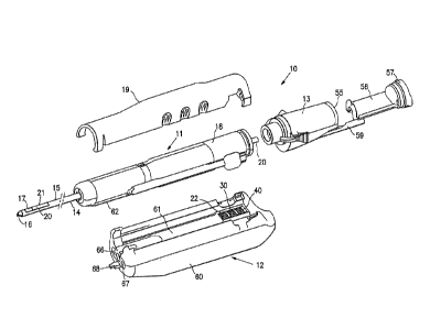
[0018] Figure 1 is an exploded view of the elongated tissue biopsy system
embodying features of the invention.
[0019] Figure 2 is a perspective view of the embodiment shown in Figure 1
in
an assembled condition without a housing cover for the probe component.
8d
CA 3050373 2019-07-19
[0020] Figure 3 is a side elevational view of the tissue biopsy device
shown in
the Figure 2.
[0021] Figure 4A is a longitudinal cross-section of the probe shown in
Fig. 3
taken along the lines 4-4 with the tissue cutting element in a withdrawn
position.
[0022] Figure 4B is a longitudinal cross-section of the probe shown in
Fig. 3
taken along the lines 4-4 with the tissue cutting element in a forward or
closed
position.
[0023] Figure 5 is a transverse cross-sectional view of the probe shown in
Figure 4B taken along the lines 5-5.
[0024] Figure 6 is a perspective view of the underside of the probe shown
in
Figure 1.
Se
CA 3050373 2019-07-19
WO 2005/063126 PCT/US2004/043021
[0025] Figure 7 is an enlarged perspective view of the distal end of the
driver unit
shown in Figure 1.
[0026] Figure 8 is an enlarged perspective view of the distal end of the probe
housing illustrating a marker element which depicts the orientation of the
aperture in
the tubular section of the biopsy device.
[0027] Figure 9 is a perspective view of the underside of the driver shown in
Figure 1.
[0028] Figure 10 is an elevational view of a tissue penetrating tip embodying
features of the invention.
[0029] Figure 11 is a perspective view of the underside of the tip shown in
Figure
10.
[0030] Figure 12 is a longitudinal, center line cross-sectional view of the
penetrating tip shown in Fig. 10.
[0031] Figure 13 is a longitudinal cross-sectional view of the penetrating tip
shown
in Figure 12 taken along the lines 13-13.
[0032] Figure 14 is a longitudinal cross-sectional view of the penetrating tip
shown
in Figure 12 taken along the lines 14-14.
[0033] Figure 15 is a longitudinal cross-sectional view of the penetrating tip
shown
in Figure 3 taken along the lines 15-15.
[0034] Figure 16 is a bottom view of the penetrating tip shown in Figure 10.
[0035] Figure 17 is a transverse cross-sectional view of the penetrating tip
shown
in Figure 16 taken along the lines 17-17.
[0036] Figure 18 is a transverse cross-sectional view of the penetrating tip
shown
in Figure 16 taken along the lines 18-18.
=
9
CA 3050373 2019-07-19
WO 2005/063126
PCT/U52004/043021
[0037] Figure 19 is a transverse cross-sectional view of the penetrating tip
shown
in Figure 16 taken along the lines 19-19.
[0038] Figure 20 is a transverse cross-sectional view of the penetrating tip
shown
in Figure 16 taken along the lines 20-20.
[0039] Figure 21 is a perspective view of the tissue biopsy system shown in
Figure
1 assembled and mounted on a stereotactic frame.
[0040] Figure 22 is an elevational view of a marker delivery device with a
flared
guide on the distal end of the shaft which facilitates guiding the distal tip
of a marker
delivery device into the interior of the proximal end of the tissue cutter.
[0041] Figure 23 is a longitudinal cross-sectional view of the distal end of
the
marker delivery device and flared guide disposed within the tissue collection
component shown in Figure 1.
[0042] Figure 24 is a longitudinal cross sectional view of the proximal end of
the
marker delivery device with the flared guide at the proximal end of the shaft
and with
the shaft deployed within the inner lumen of the tissue cutter.
DETAILED DESCRIPTION OF EMBODIMENTS OF THE INVENTION
[0043] Figures 1-3 illustrate a biopsy system 10 embodying features of the
invention which includes a disposable probe component 11, a driver component
12
and specimen collector 13.
[0044] The probe component 11 generally includes an elongated distal shaft 14
having a tubular section or cannula 15 with a tissue penetrating tip 16 on the
distal
end thereof and an open, tissue receiving aperture 17. The probe component 11
also includes a probe housing 18 with a housing cover 19 which is configured
to
interfit with the driver component 12. A tissue cutter 20 is slidably disposed
within
CA 3050373 2019-07-19
WO 2005/063126 PCT/US2004/043021
the probe and has a distal cutting surface 21 which severs tissue which
extends
through the tissue receiving aperture 17.
[0045] Details of the probe component 11 are further shown in Figs 4A and 4B.
The probe housing 18 has a mechanical system for rotating the housing and the
tubular section 15 secured thereto to control the angular position of the
tissue
receiving aperture 17 and for moving the tissue cutter 20 slidably disposed
within the
probe component 11.
[0046] The mechanical system of the driver component 12 has first driving gear
22
that is configured to engage the probe gear 23 and rotate the probe housing 18
so
as to adjust the orientation of aperture 17 in the distal extremity of the
tubular section
15. The probe gear 23 is secured to the rotating connector body 24 by adhesive
25.
The proximal extremity of the tubular section 15 is secured to the rotating
connector
body 24 by adhesive 26. An end cap 27 retains the connector body 24 within the
probe housing 18. Rotation of the probe gear 23 rotates the connector body 24
and
the attached tubular section 15. The rotation is preferably controlled so that
the
tubular section 15 rotates in discrete steps about the longitudinal axis 28 to
adjust
the angular orientation of the aperture 17 about the longitudinal axis.
Preferably
these discrete orientations may be provided in increments of 30 which can be
readily indicated by arrow 29 at the distal end of the probe housing 18 as
shown in
Figure 8.
[0047] The second driving gear 30 is configured to drive the tissue cutter 20
longitudinally. The driving gear 30 engages probe gear 31 which drives cutter
traverse nut 32 and cutter screw 33 threadably connected to the cutter
traverse nut.
The distal end of the cutter screw 33 is provided with a recess 34 which
receives the
rib 35 of the cutter shuttle 36. The cutter shuttle 36 is secured to the
tissue cutter 20
11
CA 3050373 2019-07-19
WO 2005/063126 PCT/US2004/043021
by adhesive 37. The probe gear 31 is secured to the cutter traverse nut 32 by
adhesive 38. Rotation of the probe gear 31 adjusts the relative axial position
of the
cutter screw 33 with respect to the cutter traverse nut 32 which is secured to
the
cutter shuttle 36. Longitudinal movement of the tissue cutter 20 follows the
longitudinal movement of the cutter shuttle 36 resulting from the movement of
cutter
screw 33. The length of the tissue receiving aperture 17, and as a result the
length
of the specimen, can be controlled by adjusting the initial longitudinal
position of the
distal end of the tissue cutter 20 within the aperture, before cutting.
[0048] The third driving gear 40 is configured to rotate or oscillate the
tissue cutter
20 as the cutter moves along the longitudinal axis 28 to facilitate the
cutting action of
the cutting surface 21 on the distal end of the cutter. The third driving gear
40
engages probe gear 41 which is secured to cutter oscillation shaft 42 by
adhesive
43. The probe gear 41 may be oscillated back and forth about the longitudinal
axis
28 or rotated continuously in a single direction about the longitudinal axis,
or both
depending upon the desired rotational movement of the tissue cutter.
[0049] A biased valve assembly 44 is provided in the distal end of the probe
housing 18 to ensure sealing when a vacuum is developed within the interior 45
of
the tissue cutter 20 while providing an atmospheric vent 46 between the
interior
surface 47 of the tubular section 15 and the exterior surface 48 of the tissue
cutter
20. The valve assembly 44 includes a spring 49, valve body 50 and a valve
collar 51
which is secured to the proximal end of the tubular section 15 by adhesive 52.
The
proximal end of the valve spring 49 rests against the shoulder 53 provided in
the
exterior of the valve body 50. A biased cutter shaft seal 54 slidably engages
the
exterior 48 of the tissue cutter 20.
12
CA 3050373 2019-07-19
WO 2005/063126 PCT/US2004/043021
[0050] The tissue specimen collector 13 is secured to the proximal end of the
housing of probe component 11 and has an interior 56 in fluid communication
with
the inner lumen 56 extending within the tissue cutter 20 and has a removable
proximal wall 67 of specimen receiving cartridge 58 which gives access to the
interior 55 and any tissue specimens which may have been drawn therein. A
vacuum is generated within the interior 55 to draw tissue specimens through
the
inner lumen 45 into the interior 55. Tubular member 59 has a distal end which
is in
fluid communication with the interior 55 of the tissue specimen collector 13
and has a
proximal end (not shown) which is configured to be connected to a vacuum
source.
Application of a vacuum within the tubular member 59 aids in pulling tissue
into the
interior 17 of the tubular section 15 and transfer of the severed tissue
specimen
through the inner lumen 45 of the tissue cutter 20 to the specimen cartridge
58.
[0051] The driver 12 has a housing 60 with an upper concave surface 61 which
is
configured to receive the lower surface 62 of the probe housing 18. Three
partially
exposed driving gears 22, 30 and 40 are provided on the proximal end of the
driver
12 which are configured to engage the probe gears 23, 31 and 41 respectively.
The
drive 12 is provided with three separately operating drive motors (not shown)
which
drive the drive gears 22, 30 and 40. The separate drive motors (not shown) are
connected to and the operation thereof controlled by a control module, such as
described in copending application Serial No. 10/847,699, filed on May 17,
2004.
The control module controls the motors which move the individual drive gears
22, 30
and 40. The gear 22 engages gear 23 in the probe 11 to control the rotation of
the
probe housing 18 and the location and orientation of the tissue receiving
aperture 17.
The drive gear 30 engages probe gear 31 to control the longitudinal position
and
motion of the tissue cutter 20 along the longitudinal axis 28. Drive gear 40
engages
13
CA 3050373 2019-07-19
WO 2005/063126 PCT/US2004/043021
probe gear 41 to control the oscillation or rotation of the tissue cutter 20
about the
longitudinal axis 28.
[0052] As shown in Figure 7, the front face of the driver component 12 is
provided
with light sources 66 and 67 and a manually activatable switch 68 to activate
the light
sources and enable the physician and other operating personnel to better view
the
operating site on the patient. Other manual switches, e.g. a foot activated
switch,
may be employed. Alternatively, the light sources may be automatically
activated
when the probe component 11 is installed on the driver 12 or other events such
as
when electrical power is turned on. The driver component 12 may have a battery
pack for the light sources 66 and 67.
[0053] The tissue penetrating distal tip 16 may have a variety of tip shapes.
A
particularly suitable distal tip embodying features of the invention is shown
in Figures
9-20. The tissue penetrating distal tip generally includes a base 70, a sharp
distal
point 71, a first concave surface 72, a second concave surface 73 and a third
concave surface 74.
[0054] The intersection between the first concave surface 72 and the second
concave surface 73 forms the first curved cutting edge 75. The intersection
between
the second concave surface 73 and the third concave surface 74 forms the
second
curved cutting edge 76. The intersection between the third concave surface and
the
first concave surface 72 forms the third curved cutting surface 77.
[0055] The concave surfaces 72, 73 and 74 are hollow ground and the pentrating
tip 16 is then electro-polished to increase the sharpness of the cutting edges
75, 76
and 77. The penetrating distal tip 16 may be formed of suitable surgical
stainless
steel such as 17-4 stainless steel. Other materials may be suitable. The
penetrating
distal tip 16 is preferably electro-polished in an acidic solution to sharpen
the curved
14
CA 3050373 2019-07-19
WO 2005/063126 PCT/U52004/043021
cutting edges and thereby facilitate tissue penetration. Suitable electro-
polishing
solutions include Electro Glo sold by the Electro Glo Distributing Co.
[0056] The base 70 of the tissue penetrating tip 16 may be secured to the
distal
end of the elongated shaft of the biopsy device 10 for accessing and
collecting tissue
from a target site within a patient. The sharp distal tip 16 embodying
features of the
invention readily penetrates a patient's tissue, particularly breast tissue
and
facilitates guiding the distal end of the biopsy or other device to a desired
intracorporeal location.
[0057] The tissue penetrating tips may also be employed on biopsy devices such
as those described in co-pending application Serial No. 10/642õ406, filed on
August
15, 2003, which is assigned to the present assignee. Alternatively, the distal
tip may
be provided with an arcuate RF electrode such as disclosed in U.S. Patent No.
6,261,241, and U.S. Patent No. 6,471,700, both assigned to the present
assignee.
[0058] The separate driver component 12 allows the probe unit 11 to be
disposable. The drive gears of the drive component 12 control the motion of
the
tissue cutting member 20 for cutting and the motion of the tubular section 15
to orient
the aperture 17. Other means (not shown) may provide mechanical and electrical
power, vacuum, and control to the probe device. Examples of replaceable snap-
in .
type probe units are disclosed in Burbank et al., U.S. Patent Application
10/179,933,
"Apparatus and Methods for Accessing a Body Site". Drive units such as that
described in WO 02/069808 (which corresponds to co-pending U.S. application
Serial No. 09/707,022, filed November 6, 2000 and U.S. application Serial No.
09/864,021, filed May 23, 2001), which are assigned to the present assignee,
may
be readily modified by those skilled in the art to accommodate the movement of
the
cutting member 20.
CA 3050373 2019-07-19
WO 2005/063126 PCT/U52004/043021
[0059] In use, the distal end of the probe component Ills advanced within the
patient with the tissue cutter 20 in a forward or closed position (Fig. 4B),
until the
aperture 17 of the tubular section 15 is located in a desired location for
taking a
tissue specimen. The tissue cutter 20 is then withdrawn proximally to an open
position to open the aperture 17. The withdrawal of the tissue cutter can be
used to
control the length of the aperture which is opened in order to control the
length of the
specimen which is severed. A vacuum Is applied to the interior 45 of the
tissue
cutter 20 to draw tissue at the site into the inner lumen of the tubular
section 15
through the aperture 17. The tissue cutter 20 is then driven distally by
rotation of
probe gear 30 and rotated or oscillated by drive gear 40 engaging probe gear
41 to
sever the aspirated tissue from the supporting tissue at the target site with
the tissue
cutting surface 21. The vacuum within the interior of the tissue cutter 20
causes the
tissue specimen to be drawn through the inner lumen 45 of the tissue cutter 20
and
into the cartridge 58 of specimen collector 13 shown in Fig. 2. Positive
pressure or
even ambient conditions distal to the tissue specimen can facilitate tissue
passing
through the interior 45 of tissue cutter 20. If another tissue specimen is
desired, the
tubular section 15 may be rotated by the drive gear 22 engaging the probe gear
23 in
one or more steps to repeat obtaining another tissue specimen in the same
manner
without otherwise moving the probe component 11. Typically, a first tissue
specimen
is obtained with the aperture 17 of the probe 11 in the 12 o-clock position,
the
second at the 3 o-clock position, the third at the 9 o-clock position and the
fourth at
the 6 o-clock position. The location of the second and third specimens may be
reversed. The position of the aperture 17 may be indicated by a marker arrow
29 at
the end cap 27 so that the physician or other operating personnel can readily
determine what the orientation of the aperture 17 within the patient.
16
CA 3050373 2019-07-19
WO 2005/063126 PCT/US2004/043021
[0060] The biopsy system 10 may be hand held for some biopsy procedures or the
system may be mounted on a stereotactic mounting stage 80 as shown in Figure
21.
A shoe 81 is slidably mounted to a rail 82 of a Fisher stage. The mounting
member
83 is secured to the shoe 81 by a threaded post (not shown) secured to
thumbwheel
84. As shown in Figure 10, the bottom surface 85 of the driver component 12 is
configured to conform at least in part to the upper surface of the mounting
member
83. The sampling and vacuum switches 86 and 87 respectively on the driver
component 12 are actuated by the optional sampling and vacuum actuating
elements 88 and 89 on the mounting member 83. Alternatively, sampling and
vacuum may be actuated with a foot pedal. As shown in Figure 22, the driver ,
component has an operator dial 90 which when turned opens a threaded hole 91
for
receiving a threaded post (not shown) secured to the thumbwheel 84 and the
locating pin holes 92 and 93 which receive the complementary posts (not shown)
in
the mounting member 83.
[0061] As mentioned above, positive pressure or even ambient conditions will
aid
in passing the severed tissue specimen through the inner lumen 45 of tissue
cutter
20 into the cartridge 58 of specimen collector 13. As shown in Figures 4A and
4B
venting valve can provide ambient pressure behind the tissue specimen in the
cutter
interior 45 from the interior of the tubular section 15. The valve body 50 is
opened
for atmospheric venting when the tissue cutter 20 is in the forward position
upon the
completion of severing the specimen from the tissue site. However, when the
tissue
cutter 20 is pulled back proximally the valve spring 49 urges the valve body
50 back
to a closed position. While the tissue cutter 20 is shown with a tissue
cutting surface
21 which is perpendicular to the longitudinal axis 28, the tissue cutting
surface may
17
CA 3050373 2019-07-19
WO 2005/063126 PCT/US2004/043021
be at an angle or even parallel to the longitudinal axis as described in co-
pending
application Serial No. 10/642,406, filed August 15, 2003.
[0062] The distal cutting edge 21 of the tissue cutter 20 may initially be
located
proximal to the aperture 17 to provide a full aperture for receiving tissue or
it can be
initially located within the aperture 17 in order to control the length of the
specimen.
The cutting action of tissue cutter 20 preferably continues until the beveled
cutting
surface 21 has completely traversed the aperture 17 to ensure that the tissue
drawn
through the aperture is completely severed from supporting tissue at the
biopsy site.
A vacuum may be applied to aspirate the severed tissue specimen through the
inner
lumen of the tissue cutter 20 to the cartridge in the specimen collector at
the
proximal end of the biopsy device. Positive pressure or access to ambient
conditions may be provided in the distal end of the tubular section to aid in
the
specimen transfer.
[0063] After the removable wall 57 of the specimen receiving cartridge 58 is
removed and the specimens therein removed, it is frequently desirable to
deliver one
or more markers to the target site from which the specimens have been removed.
Such marker delivery devices are shown in co-pending application Serial No.
10/753,694, filed on January 7, 2004 and co-pending application Serial No.
10/444,770, filed May 23, 2003. However, the distal ends of these marker
delivery
devices are very small and they can be difficult to insert into the proximal
end of the
tissue cutter 20 which is just slightly larger to accommodate the marker
delivery
shaft.
[0064] Figure 22 illustrates a marker delivery device 100 which is
particularly
suitable to facilitate the introduction of the distal end of the shaft 101
into the inner
lumen 45 of the tissue cutter 20 and the advancement therein. As shown in
Figure
18
CA 3050373 2019-07-19
WO 2005/063126 PCT/US2004/043021
23, to facilitate the insertion of the small diameter distal tip 101 of the
marker delivery
device 100 into the slightly larger inner lumen 45 of the tubular cutter 20 at
its
proximal end, the distal tip is preferably provided with an outwardly flared
guide 102
which is slidably mounted on the shaft 103 of the marker delivery device 100.
The
proximal end of the tubular cutter 20, the flared guide 102 and/or the distal
tip 101
may be provided with mating guide elements which orient the marker delivery
device
with the cannula 15 of the biopsy device. To ensure that one or more markers
are
discharged through the aperture 17 of the biopsy device 10 when the pusher
element slidably disposed within the delivery device is urged distally to
press at least
one marker body out the discharge opening in the distal portion of the
elongated
shaft of the marker delivery device. As indicated in Figure 23, the shaft 103
of the
marker delivery device 100 is advanced into the inner lumen 45 of the tissue
cutter
20 so that the distal end 101 of the marker delivery device 100 is adjacent to
the
aperture 17 of the cannula 15. Plunger 104 is pressed further into the inner
lumen of
shaft 103 to eject one or more markers 105 through the aperture 17 in the
tubular
section 15 before the biopsy device 10 is removed from the patient. The
delivery of
markers to the target site after specimen removal, while the distal end of the
biopsy
device is still at the biopsy site, ensures that the markers are properly
position at the
biopsy site.
[0065] While the slidably mounted, flared proximal guide 102 is described with
respect to being disposed on the shaft 103 of marker delivery device 101, the
flared
guide 102 has wide application within a variety of biopsy and other devices
where
one small diameter tubular member is to be inserted into a slightly larger,
but still
small diameter second tubular member.
=
19
CA 3050373 2019-07-19
WO 2005/063126 PCT/US2004/043021
[0066] The elongated probe component 11 of the biopsy system 10 has a length
of about 3 to about 20 cm, preferably, about 5 to about 13 cm, and more
specifically,
about 8 to about 9 cm for breast biopsy use. To assist in properly locating
the probe
11 during advancement thereof into a patient's body, the distal extremity of
the
tubular section may be provided with a marker at a desirable location that
provide
enhanced visualization by eye, by ultrasound, by X-ray, MRI or other imaging
or
visualization means. Manual palpation may also be employed. An echogenic
polymer coating that increases contrast resolution in ultrasound imaging
devices
(such as ECHOCOATTm by STS Biopolymers, of Henrietta, NY) is suitable for
ultrasonic visualization. Radiopaque markers may be made with, for example,
stainless steel, platinum: gold, iridium, tantalum, tungsten, silver, rhodium,
nickel,
bismuth, other radiopaque metals, alloys and oxides of these metals. In
addition, the
surfaces of the device in contact with tissue or other components of the
device may
be provided with a suitable lubricious coating such as a hydrophilic material
or a
fluoropolymer.
[0067] The tubular section and the tissue cutter are preferably formed of a
surgical
grade stainless steel. However, other high strength materials such as MP35N,
other
cobalt-chromium alloys, NiTi alloys, ceramics, glasses, and high strength
polymeric
materials or combinations thereof may be suitable.
[0068] A patient's skin usually must be breached in order to gain access to a
body
site where a tissue specimen is to be obtained. A scalpel or other surgical
instrument may be used to make an initial incision in the skin. After the
specimens
have been taken, the biopsy device may be removed from the patient. The entire
device may be removed; however, in some embodiments, the cartridge 58 may be
removed from the system 10 and a delivery cannula may be inserted through the
CA 3050373 2019-07-19
inner lumen of the cutter 20 to deliver 'markers to the biopsy site through
the aperture 17. In
addition, it will be readily appreciated that other types of instruments may
be inserted into the
tissue site through the tissue cutter in addition to or in place of the
instruments described
above. Moreover, therapeutic or diagnostic agents may be delivered through the
tissue cutter
20 or the tubular section 15.
[0069] While particular forms of the invention have been illustrated and
described herein, it
will be apparent that various modifications and improvements can be made to
the invention.
For example, while the various embodiments of the invention have been
described herein in
terms of a biopsy device, it should be apparent that the devices and methods
of utilizing the
device may be employed to remove tissue for purposes other than for biopsy,
i.e. for
treatment or other diagnoses. Additionally, the tissue penetrating distal tip
design may be
employed on probes for other uses. Other modifications include, for example, a
tissue cutter
slidably mounted around the tubular section of the probe component rather than
within the
tubular section. Moreover, individual features of embodiments of the invention
may be shown
in some drawings and not in others, but those skilled in the art will
recognize that individual
features of one embodiment of the invention can be combined with any or all
the features of
another embodiment. Accordingly, it is not intended that the invention be
limited to the
specific embodiments illustrated.
21
Date Recue/Date Received 2021-01-11