Note: Descriptions are shown in the official language in which they were submitted.
CA 03050691 2019-07-17
WO 2018/136435
PCT/US2018/013909
COMPOSITIONS AND METHODS FOR TREATING LYSOSOMAL STORAGE
DISEASES AND DISORDERS
CROSS-REFERENCE TO RELATED APPLICATIONS
This application claims priority to and the benefit of U.S. Provisional
Application Nos.
62/447,341, filed January 17, 2017 and 62/582,247, filed November 6, 2017,
respectively, the
disclosure of which is incorporated herein by reference in its entirety.
BACKGROUND OF THE INVENTION
Most lysosomal storage disorders (LSDs) with central nervous system (CNS)
involvement lack an effective and curative treatment and patients eventually
succumb to their
devastating disease. Frequently, disease onset occurs in very early infancy
and is characterized
by subtle manifestations, leading to diagnosis in clearly symptomatic if not
advanced stage.
LSDs are also characterized by a rapid early disease progression, particularly
in early onset
variants. For these reasons therapeutic approaches that have been applied with
some degree of
success in pre-symptomatic LSD children, including for example, hematopoietic
cell
transplantation (HCT) in Krabbe disease, or hematopoietic stem cell (HSC) gene
therapy (HSC
GT) in Metachromatic Leukodystrophy (MLD), are not beneficial for the majority
of LSD
patients, with benefit being associated almost exclusively to procedures
applied in pre- or early-
.. symptomatic cases. One of the key reasons for the failure of these HSC-
based approaches in
ameliorating rapidly progressing LSD brain diseases is the slow pace of
replacement of resident
CNS tissue macrophages/histiocytes and microglia by the transplanted
hematopoietic cell
progeny, compared to the rapid progression of the primary neurological
disease. Indeed, while a
rapid reconstitution of visceral organ macrophages by donor-derived cells has
been clearly
demonstrated following HCT, more limited and slower infiltration of the brain
parenchyme by
donor cells is supposed to occur. Moreover, efficiency of uptake by different
cell types and
intrinsic pathologic mechanisms related to enzyme deficiency are not entirely
overcome by
enzyme replacement and cross correction may account for the residual and long-
term progressing
disease observed in the transplanted patients. Importantly, in the majority of
LSDs lysosomal
enzyme deficiency triggers a cascade of events ultimately leading to
neuroinflammation,
1
CA 03050691 2019-07-17
WO 2018/136435
PCT/US2018/013909
activation of oxidative stress pathways and consequent neurodegeneration.
These mechanisms
are critically affecting response to treatment and are as well key therapeutic
targets for
comprehensive approaches.Accordingly, new compositions and methods of
treatment are needed
for patients afflicted with a lysosomal storage disorder.
SUMMARY
As described below, disclosed herein are compositions and methods for the
treatment or
prevention of a lysosomal storage disorder (e.g., Neuronal Ceroid
Lipofuscinoses) by increasing
the level, expression, or activity of a metallothionein polyepeptide or
polynucleotide in the
subject. In some embodiments, the methods involve replacing a patient's
endogenous microglia
with either donor derived or engineered cells able to contribute to disease
amelioration by
different mechanisms, such as protein delivery or regulation of local
inflammation and oxidative
stress or others.
Thus, In one aspect, disclosed herein are compositions and methods of treating
a
lysosomal storage disease or disorder in a subject, involving increasing the
level, expression, or
activity of a metallothionein polyepeptide or polynucleotide in the subject
relative to a reference.
In various embodiments of any aspect delineated herein, the lysosomal storage
disorder
with CNS involvement is Neuronal Ceroid Lipofuscinoses, globoid
leukodystrophy, GM1
gangliosidoses, Juvenile Hexosaminidase A Deficiency, Metachromatic
Leukodystrophy,
Mucopolysaccharidoses disorders, Multiple sulfatase deficiency, Tay-Sachs/GM2
gangliosidosis.
In various embodiments of any aspect delineated herein, the subject is pre-
selected by
detecting an increase in the level of a metallothionein (MT) polynucleotide or
polypeptide in a
sample of the subject relative to a reference.
In various embodiments of any aspect delineated herein, the metallothionein is
one or
more of metallothionein-1A (MT1A), metallothionein-1B (MT1B), metallothionein-
1E (MT1E),
metallothionein-1F (MT1F), metallothionein-1G (MT1G), metallothionein-1H
(MT1H),
metallothionein-lI pseudogene (MT lip or MTE), metallothionein-1L (LT1L or
MT1R),
metallothionem-1M (MT1M or MT1K), metallothionein-1X (MT lx), metallothionein-
2 (MT2),
metallothionein-2A (MT2A), metallothionein-3 (MT3), and metallothionein-4
(MT4).
2
CA 03050691 2019-07-17
WO 2018/136435
PCT/US2018/013909
In various embodiments of any aspect delineated herein, the method involves
administering one or more MTs to the subject.
In various embodiments of any aspect delineated herein, the method involves
generating
in a subject a sustained mixed hematopoietic chimerism in the brain and in the
extra-CNS tissues
with Hematopoietic Stem Cells (HSCs) encoding one or more MTs. In various
embodiments of
any aspect delineated herein, the method involves treating a subject having or
being at increased
risk of developing a lysosomal storage disorder, including by administering a
Hematopoietic
Stem Cell (HSC) that is one or more of CD34+, CD38-, where the HSC is
administered
intravenously (IV) or by Intra-cerebral Ventricular (ICV) Injection in
combination with ablative
conditioning. In various embodiments of any aspect delineated herein, the
isolated HSC is
transformed with a vector expressing one or more therapeutic polypeptide or
polynucleotide,
where the HSC is one or more of CD34+, CD38-. In various embodiments, the HSC
are
engineered with integrating vectors, i.e. lentiviral vectors, to express a one
or more
methallothioeins +/- a lysosomal enzyme of interest (defective in the target
disease). In various
.. embodiments of any aspect delineated herein, the method involves ablating
endogenous myeloid
cells and microglia and/or their progenitors by a conditioning regimen and
reconstituting the
microglia by HSC engraftment in a subject. In various embodiments of any
aspect delineated
herein, the HSC is administered in combination with ablative conditioning. In
various
embodiments, the ablative conditioning comprises administering a cytotoxic
agent to the subject.
.. In various embodiments, the alkylating agent is busulfan. In various
embodiments, the ablative
conditioning is performed prior to administering the HSC.
Other features and advantages of the invention will be apparent from the
detailed
description, and from the claims.
Definitions
Unless defined otherwise, all technical and scientific terms used herein have
the meaning
commonly understood by a person skilled in the art to which this invention
belongs. The
following references provide one of skill with a general definition of many of
the terms used in
this invention: Singleton et al., Dictionary of Microbiology and Molecular
Biology (2nd ed.
1994); The Cambridge Dictionary of Science and Technology (Walker ed., 1988);
The Glossary
3
CA 03050691 2019-07-17
WO 2018/136435
PCT/US2018/013909
of Genetics, 5th Ed., R. Rieger etal. (eds.), Springer Verlag (1991); and Hale
& Marham, The
Harper Collins Dictionary of Biology (1991). As used herein, the following
terms have the
meanings ascribed to them below, unless specified otherwise.
By "agent" is meant any small molecule chemical compound, antibody, nucleic
acid
molecule, or polypeptide, or fragments thereof.
By "ameliorate" is meant decrease, suppress, attenuate, diminish, arrest, or
stabilize the
development or progression of a disease.
The term "antibody," as used herein, refers to an immunoglobulin molecule
which
specifically binds with an antigen. The term "antibody fragment" refers to a
portion of an intact
antibody and refers to the antigenic determining variable regions of an intact
antibody.
By "alteration" or "change" is meant an increase or decrease. An alteration
may be by as
little as 1%, 2%, 3%, 4%, 5%, 10%, 20%, 30%, or by 40%, 50%, 60%, or even by
as much as
70%, 75%, 80%, 90%, or 100%.
By "biologic sample" is meant any tissue, cell, fluid, or other material
derived from an
organism.
By "capture reagent" is meant a reagent that specifically binds a nucleic acid
molecule or
polypeptide to select or isolate the nucleic acid molecule or polypeptide.
As used herein, the terms "determining", "assessing", "assaying", "measuring"
and
"detecting" refer to both quantitative and qualitative determinations, and as
such, the term
"determining" is used interchangeably herein with "assaying," "measuring," and
the like. Where
a quantitative determination is intended, the phrase "determining an amount"
of an analyte and
the like is used. Where a qualitative and/or quantitative determination is
intended, the phrase
"determining a level" of an analyte or "detecting" an analyte is used.
"Detect" refers to identifying the presence, absence or amount of the analyte
to be
detected.
By "detectable label" is meant a composition that when linked to a molecule of
interest
renders the latter detectable, via spectroscopic, photochemical, biochemical,
immunochemical, or
chemical means. For example, useful labels include radioactive isotopes,
magnetic beads,
metallic beads, colloidal particles, fluorescent dyes, electron-dense
reagents, enzymes (for
example, as commonly used in an ELISA), biotin, digoxigenin, or haptens.
4
CA 03050691 2019-07-17
WO 2018/136435
PCT/US2018/013909
By "disease" is meant any condition or disorder that damages or interferes
with the
normal function of a cell, tissue, or organ.
By "effective amount" is meant the amount of a required to ameliorate the
symptoms of a
disease relative to an untreated patient. The effective amount of active
compound(s) used to
practice the present invention for therapeutic treatment of a disease varies
depending upon the
manner of administration, the age, body weight, and general health of the
subject. Ultimately,
the attending physician or veterinarian will decide the appropriate amount and
dosage regimen.
Such amount is referred to as an "effective" amount.
By "fragment" is meant a portion of a protein or nucleic acid that is
substantially identical
1() to a reference protein or nucleic acid. In some embodiments the portion
retains at least 50%,
75%, or 80%, or more preferably 90%, 95%, or even 99% of the biological
activity of the
reference protein or nucleic acid described herein.
The terms "isolated," "purified," or "biologically pure" refer to material
that is free to
varying degrees from components which normally accompany it as found in its
native state.
"Isolate" denotes a degree of separation from original source or surroundings.
"Purify" denotes a
degree of separation that is higher than isolation. A "purified" or
"biologically pure" protein is
sufficiently free of other materials such that any impurities do not
materially affect the biological
properties of the protein or cause other adverse consequences. That is, a
nucleic acid or peptide
of this invention is purified if it is substantially free of cellular
material, viral material, or culture
medium when produced by recombinant DNA techniques, or chemical precursors or
other
chemicals when chemically synthesized. Purity and homogeneity are typically
determined using
analytical chemistry techniques, for example, polyacrylamide gel
electrophoresis or high
performance liquid chromatography. The term "purified" can denote that a
nucleic acid or
protein gives rise to essentially one band in an electrophoretic gel. For a
protein that can be
subjected to modifications, for example, phosphorylation or glycosylation,
different
modifications may give rise to different isolated proteins, which can be
separately purified.
By an "isolated polypeptide" is meant a polypeptide of the invention that has
been
separated from components that naturally accompany it. Typically, the
polypeptide is isolated
when it is at least 60%, by weight, free from the proteins and naturally-
occurring organic
molecules with which it is naturally associated. Preferably, the preparation
is at least 75%, more
5
CA 03050691 2019-07-17
WO 2018/136435
PCT/US2018/013909
preferably at least 90%, and most preferably at least 99%, by weight, a
polypeptide of the
invention. An isolated polypeptide of the invention may be obtained, for
example, by extraction
from a natural source, by expression of a recombinant nucleic acid encoding
such a polypeptide;
or by chemically synthesizing the protein. Purity can be measured by any
appropriate method,
for example, column chromatography, polyacrylamide gel electrophoresis, or by
HPLC analysis.
As used herein "lysosomal storage disorder (SD)" refers to any of a group of
diseases
resulting from abnormal metabolism leading to accumulation of a substrate (for
example
sulfatides, heparan sulphate, glycolipids, ceramide) in the lysosome. For
example, lysosomal
storage disorders (LSDs) are caused by lysosomal dysfunction usually as a
consequence of
1() deficiency of an enzyme required for the metabolism of lipids,
glycoproteins (sugar-containing
proteins) or so-called mucopolysaccharides.
By "marker" is meant any clinical indicator, protein, metabolite, or
polynucleotide having
an alteration associated with a disease, disorder, or condition.
By "microglia" is meant an immune cell of the central nervous system.
As used herein "neurodegenerative disease" refers to a disease characterized
by the
progressive loss of structure and/or function of neurons, including death of
neurons.
By "increasing proliferation" is meant increasing cell division of a cell in
vivo or in vitro.
As used herein, the terms "prevent," "preventing," "prevention," "prophylactic
treatment" and the like refer to reducing the probability of developing a
disorder or condition in a
subject, who does not have, but is at risk of or susceptible to developing a
disorder or condition.
The term "subject" or "patient" refers to an animal which is the object of
treatment,
observation, or experiment. By way of example only, a subject includes, but is
not limited to, a
mammal, including, but not limited to, a human or a non-human mammal, such as
a non-human
primate, murine, bovine, equine, canine, ovine, or feline.
By "reduces" is meant a negative alteration of at least 10%, 25%, 50%, 75%, or
100%.
By "reference" is meant a standard of comparison or control condition.
By "substantially identical" is meant a polypeptide or nucleic acid molecule
exhibiting at
least 50% identity to a reference amino acid sequence (for example, any one of
the amino acid
sequences described herein) or nucleic acid sequence (for example, any one of
the nucleic acid
sequences described herein). Preferably, such a sequence is at least 60%, more
preferably 80% or
6
CA 03050691 2019-07-17
WO 2018/136435
PCT/US2018/013909
85%, and more preferably 90%, 95%, 96%, 97%, 98%, or even 99% or more
identical at the
amino acid level or nucleic acid to the sequence used for comparison.
Sequence identity is typically measured using sequence analysis software (for
example,
Sequence Analysis Software Package of the Genetics Computer Group, University
of Wisconsin
Biotechnology Center, 1710 University Avenue, Madison, Wis. 53705, BLAST,
BESTFIT,
GAP, or PILEUP/PRETTYBOX programs). Such software matches identical or similar
sequences by assigning degrees of homology to various substitutions,
deletions, and/or other
modifications. Conservative substitutions typically include substitutions
within the following
groups: glycine, alanine; valine, isoleucine, leucine; aspartic acid, glutamic
acid, asparagine,
glutamine; serine, threonine; lysine, arginine; and phenylalanine, tyrosine.
In an exemplary
approach to determining the degree of identity, a BLAST program may be used,
with a
probability score between e-3 and e-m indicating a closely related sequence.
Nucleic acid molecules useful in the methods of the invention include any
nucleic acid
molecule that encodes a polypeptide of the invention or a fragment thereof
Such nucleic acid
molecules need not be 100% identical with an endogenous nucleic acid sequence,
but will
typically exhibit substantial identity. Polynucleotides having "substantial
identity" to an
endogenous sequence are typically capable of hybridizing with at least one
strand of a double-
stranded nucleic acid molecule. By "hybridize" is meant pair to form a double-
stranded
molecule between complementary polynucleotide sequences (e.g., a gene
described herein), or
portions thereof, under various conditions of stringency. (See, e.g., Wahl, G.
M. and S. L. Berger
(1987) Methods Enzymol. 152:399; Kimmel, A. R. (1987) Methods Enzymol.
152:507).
For example, stringent salt concentration will ordinarily be less than about
750 mM NaCl
and 75 mM trisodium citrate, preferably less than about 500 mM NaCl and 50 mM
trisodium
citrate, and more preferably less than about 250 mM NaCl and 25 mM trisodium
citrate. Low
stringency hybridization can be obtained in the absence of organic solvent,
e.g., formamide,
while high stringency hybridization can be obtained in the presence of at
least about 35%
formamide, and more preferably at least about 50% formamide. Stringent
temperature conditions
will ordinarily include temperatures of at least about 30 C, more preferably
of at least about 37
C, and most preferably of at least about 42 C. Varying additional parameters,
such as
hybridization time, the concentration of detergent, e.g., sodium dodecyl
sulfate (SDS), and the
7
CA 03050691 2019-07-17
WO 2018/136435
PCT/US2018/013909
inclusion or exclusion of carrier DNA, are well known to those skilled in the
art. Various levels
of stringency are accomplished by combining these various conditions as
needed. In a preferred:
embodiment, hybridization will occur at 30 C in 750 mM NaCl, 75 mM trisodium
citrate, and
1% SDS. In a more preferred embodiment, hybridization will occur at 37 C in
500 mM NaCl,
50 mM trisodium citrate, 1% SDS, 35% formamide, and 100 tg/m1 denatured salmon
sperm
DNA (ssDNA). In a most preferred embodiment, hybridization will occur at 42 C
in 250 mM
NaCl, 25 mM trisodium citrate, 1% SDS, 50% formamide, and 200 g/m1 ssDNA.
Useful
variations on these conditions will be readily apparent to those skilled in
the art.
For most applications, washing steps that follow hybridization will also vary
in
stringency. Wash stringency conditions can be defined by salt concentration
and by temperature.
As above, wash stringency can be increased by decreasing salt concentration or
by increasing
temperature. For example, stringent salt concentration for the wash steps will
preferably be less
than about 30 mM NaCl and 3 mM trisodium citrate, and most preferably less
than about 15 mM
NaCl and 1.5 mM trisodium citrate. Stringent temperature conditions for the
wash steps will
ordinarily include a temperature of at least about 25 C, more preferably of
at least about 42 C,
and even more preferably of at least about 68 C In a preferred embodiment,
wash steps will
occur at 25 C in 30 mM NaCl, 3 mM trisodium citrate, and 0.1% SDS. In a more
preferred
embodiment, wash steps will occur at 42 C in 15 mM NaCl, 1.5 mM trisodium
citrate, and 0.1%
SDS. In a more preferred embodiment, wash steps will occur at 68 C in 15 mM
NaCl, 1.5 mM
trisodium citrate, and 0.1% SDS. Additional variations on these conditions
will be readily
apparent to those skilled in the art. Hybridization techniques are well known
to those skilled in
the art and are described, for example, in Benton and Davis (Science 196:180,
1977); Grunstein
and Hogness (Proc. Natl. Acad. Sci., USA 72:3961, 1975); Ausubel et al.
(Current Protocols in
Molecular Biology, Wiley Interscience, New York, 2001); Berger and Kimmel
(Guide to
Molecular Cloning Techniques, 1987, Academic Press, New York); and Sambrook et
al.,
Molecular Cloning: A Laboratory Manual, Cold Spring Harbor Laboratory Press,
New York.
By "specifically binds" is meant a compound (e.g., peptide) that recognizes
and binds a
molecule (e.g., polypeptide), but which does not substantially recognize and
bind other
molecules in a sample, for example, a biological sample.
8
CA 03050691 2019-07-17
WO 2018/136435
PCT/US2018/013909
As used herein, the terms "treat," treating," "treatment," and the like refer
to reducing or
ameliorating a disorder and/or symptoms associated therewith. It will be
appreciated that,
although not precluded, treating a disorder or condition does not require that
the disorder,
condition or symptoms associated therewith be completely eliminated.
Unless specifically stated or obvious from context, as used herein, the term
"about" is
understood as within a range of normal tolerance in the art, for example
within 2 standard
deviations of the mean. About can be understood as within 10%, 9%, 8%, 7%, 6%,
5%, 4%, 3%,
2%, 1%, 0.5%, 0.1%, 0.05%, or 0.01% of the stated value. Unless otherwise
clear from context,
all numerical values provided herein are modified by the term about.
Ranges provided herein are understood to be shorthand for all of the values
within the
range. For example, a range of 1 to 50 is understood to include any number,
combination of
numbers, or sub-range from the group consisting 1, 2, 3, 4, 5, 6, 7, 8, 9, 10,
11, 12, 13, 14, 15,
16, 17, 18, 19, 20, 21, 22, 23, 24, 25, 26, 27, 28, 29, 30, 31, 32, 33, 34,
35, 36, 37, 38, 39, 40, 41,
42, 43, 44, 45, 46, 47, 48, 49, or 50.
Any compounds, compositions, or methods provided herein can be combined with
one or
more of any of the other compositions and methods provided herein.
As used herein, the singular forms "a", "an", and "the" include plural forms
unless the
context clearly dictates otherwise. Thus, for example, reference to "a
biomarker" includes
reference to more than one biomarker.
Unless specifically stated or obvious from context, as used herein, the term
"or" is
understood to be inclusive.
The term "including" is used herein to mean, and is used interchangeably with,
the phrase
"including but not limited to."
As used herein, the terms "comprises," "comprising," "containing," "having"
and the like
can have the meaning ascribed to them in U.S. Patent law and can mean
"includes," "including,"
and the like; "consisting essentially of' or "consists essentially" likewise
has the meaning
ascribed in U.S. Patent law and the term is open-ended, allowing for the
presence of more than
that which is recited so long as basic or novel characteristics of that which
is recited is not
changed by the presence of more than that which is recited, but excludes prior
art embodiments.
9
CA 03050691 2019-07-17
WO 2018/136435
PCT/US2018/013909
BRIEF DESCRIPTION OF THE DRAWINGS
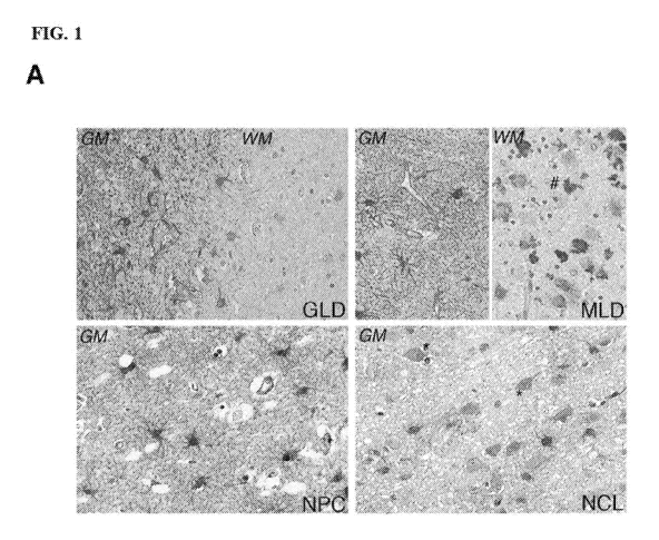
FIG. 1A, FIG. 1B, FIG. 1C and FIG. 1D show disease model selection for
preclinical
testing of MTs as therapeutic agents in LSDs.
FIG. 1A shows representative pictures of MT immunoreactivity in post-mortem
brain
samples from GLD, MILD, NPC and NCL patients, as indicated. Grey and white
matter (GM and
WM) are shown for leukodystrophies, while only GM is shown for NPC and NCL. MT
immunoreactivity is primarily associated to astrocytes in both cortex and
white matter. Neurons
showed MT immunoreactivity in NCL brain samples only (*), while MT positive
histiocytes (#)
were only observed in MILD. 40X magnification.
FIG. 1B is a graph that provides relative abundance of Lrp2 mRNA in brain of
LSDs
with neurologic involvement, compared to age-matched normal donor (ND)
samples. Mean
SEM. One-way Anova with Bonferroni post-test, **= P value <0.01. GLD n=2, NPC
n=3, MILD
n=3, NCL n=4, ND=12.
FIG. 1C is a Western blot showing immunoreactivity for megalin/Lrp2 protein on
protein
extract from 4 LSD brains (GLD n=1, NPC n=1, MLD n=1, NCL n=2) and 3 NDs. a-
actin
immunoreactivity was assessed as control for protein loading.
FIG. 1D is a graph showing MT1 mRNA expression levels in LSD mouse models.
mMT1 levels were measured in the following LSD mouse models: GLD (n=6 at 40
days), MILD
(n=4 at 10 months), Sandhoff (SD n=4, 3.5 months), INCL (n=4 at 200 days),
MPSI (n=4 at 10
months), MPSII (n=3 at 10 months), MPSIII (n=4 at 40 days), Multiple Sulfatase
Deficiency
(MSD n=5 at 2-3 weeks), compared to 20 WT mice at different ages. One-way
Anova, Dunnett's
correction, **= P value <0.01, *= P value <0.05.
FIG. 2A, FIG. 2B, FIG. 2C, FIG. 2D, FIG. 2E, FIG. 2F, FIG. 2G, and FIG. 2H
show
phenotypic effects of MTs in the GLD and INCL animal models.
FIG. 2A is a graph showing MT brain expression in naive and MT-transgenic Galc-
/- and
Pptl-/- mice. The MT-1 expression levels (MT-1 mRNA abundance) in MTtg (n=8),
GLD (n=8),
MT-GLD (n=8), INCL (n=5) and MT-INCL (n=5) mice were calculated as fold to WT
levels.
Mean with SEM. One-way ANOVA with Bonferroni post- test: **= P value <0.01, *=
P value
<0.05. In both naive animal disease models MT expression is increased over
wild type levels due
CA 03050691 2019-07-17
WO 2018/136435
PCT/US2018/013909
to reactive disease mechanisms; MT expression further increases upon affected
mice crossing
with the MT-transgenic line.
FIG. 2B shows representative confocal images of the pons region of a MT-GLD
mouse at
PND36 stained for astrocytes (GFAP-red), Metallothioneins (MT-green) and DAPI,
confirming
MT specificity of expression in astrocytes. 20X (left) and 40X (right)
magnification.
FIG. 2C shows representative confocal images of the pons region of a GLD and
of a MT-
GLD mouse at PND36 stained for microglia (IBA-red), Metallothioneins (MT-
green) and DAPI,
showing few MT-positive cells co-localizing with microglia signal in both
animals and more
intense MT staining in the MT-GLD sample. 20X magnification.
FIG. 2D and FIG. 2E depict experiments in which an MT1 transgenic (over-
expressing)
mouse was crossed with either Galc-/- or Pptl-/- mice, which are animal models
of the lysosomal
storage diseases globoid cell leukodystrophy (or Krabbe disease) and neuronal
ceroid
lypofuscinosis 1, respectively. FIG. 2D provides a Kaplan-Meier survival curve
of MT-GLD
and GLD mice. Data were analyzed by Log-Rank (Mantel-Cox) test; P
value<0.0001. FIG. 2E
provides a Kaplan-Meier survival curve of MT-INCL and INCL mice. Data are
analyzed by
Log-Rank (Mantel-Cox) test; P value<0.0001. Survival curves of naive and MT-
transgenic
Galc-/- (FIG. 2D) and Pptl-/- (FIG. 2E) mice were generated showing a survival
advantage of
the affected transgenic animals (over-expressing MTs) over the not transgenic
affected mice.
FIG. 2F is a graph showing Disease Severity Score (DSS) of MT-INCL and INCL
mice.
INCL, n=10 and MT-INCL, n=10. Two-way ANOVA repeated measures followed by
Bonferroni correction: * P value <0.05, *** P value <0.001. Mean disease score
in naive and
MT-transgenic Pptl-/- mice up to 250 days of survival. The disease score
accounts for motor
function, muscle strength and occurrence of seizures.
FIG. 2G is a graph depicting correlation of survival data of FIG. 2D and MT
levels
(expressed as fold to WT levels) presented in FIG. 2A, including both GLD and
MT-GLD data
sets. The figure represents the maximum survival of the natural occurring
disease model
interpolated with MT expression levels in the same animals, identifying the
minimum MT level
associated to survival gain. The range of MT levels detected in GLD and MT-GLD
mice is here
also shown.
11
CA 03050691 2019-07-17
WO 2018/136435
PCT/US2018/013909
FIG. 2H is a graph depicting correlation of survival data of FIG. 2E and MT
levels
(expressed as fold to WT levels) presented in FIG. 2A, including both INCL and
MT-INCL data
sets. The figure represents the maximum survival of the natural occurring
disease model
interpolated with MT expression levels in the same animals, identifying the
minimum MT level
associated to survival gain. The range of MT levels detected in INCL and MT-
INCL mice is
here also shown.
FIG. 3A, FIG. 3B, FIG. 3C, FIG. 3D, FIG. 3E, FIG. 3F, and FIG. 3G show
modulation of
anti-inflammatory, anti-apoptotic and anti- oxidative stress genes in MT-GLD
and MT-INCL
mice.
FIG. 3A provides a hierarchical clustering representing differentially
expressed genes,
up- regulated and down-regulated, in the four tested groups. As shown in the
bar, over-
expression was visualized in shades of red and under- expression in shades of
blue.
Transcriptome array was performed on cerebellar extracts from the following
mice all analyzed
at PND36: 3 WT, 3 MTtg, 3 MT-GLD and 3 GLD.
FIG. 3B, FIG. 3C, and FIG. 3D are graphs showing Ifi44 (FIG. 3B), Hpgd (FIG.
3C) and
Casp4 (FIG. 3D) expression variations in LSD and MT-LSD brain samples. The
left panels of
FIG. 3B ¨ FIG. 3C ¨ FIG- 3D show fold expression changes in the indicated
pairs, calculated
from transcriptome analysis data; fold change of Ifi44 for MT-GLD vs GLD is -
2,5528, P value
<0.001; fold change of Hpgd for MT-GLD vs GLD is -2,24130, P value <0.001;
fold change of
Casp4 for MT- GLD vs GLD is -1,75215, P value <0.01. The central and right
panels of FIG.
3B, FIG. 3C, and FIG. 3D, Ifi44 (FIG. 3B), Hpgd (FIG. 3C) and Casp4 (FIG. 3D)
show relative
mRNA abundances calculated by qPCR on MTtg, GLD and MT-GLD mice (central
panels) and
MT-Tg, INCL and MT-INCL mice (right panels); n=4 mice per group; mean with
SEM;
analyzed by one Way Anova with Bonferroni post-test, * P value<0.05, ** P
value<0.01, *** P
value<0.001. The expression of Casp4 is reduced in the transgenic affected
mice as compared to
naïve affected controls.
FIG. 3E shows representative pictures of nitrotyrosine (Nitro) staining in
brain sections
of WT, GLD and MT-GLD mice. Other regions were analyzed with the same
expression pattern.
All the animals were analyzed at PND36. Magnification 40X.
12
CA 03050691 2019-07-17
WO 2018/136435
PCT/US2018/013909
FIG. 3F is a graph that shows quantification of nitrotyrosine immunopositive
area in the
CNS (cerebellum, corpus callosum and brainstem analyzed) of GLD (n=3) and MT-
GLD (n=3)
at PND36, 3 slices per animal, 2 fields per slice, expressed as fold to WT
(n=3). Data were
analyzed by unpaired t-test comparing MT-GLD vs GLD, *** P value <0.001. Mean
with SEM.
The expression of nitrotyrosine is reduced in the transigenic affected mice as
compared to naive
affected controls.
FIG. 3G includes a graph and histograms showing intracellular reactive oxygen
species
(ROS) measured by fluorescent dye H2DCFDA incubated with myeloid cells
isolated from
mouse brains (WT n=5, GLD n=5 and MT-GLD n=5) and analyzed at flow cytometry.
FIG. 3G
(left panel) shows results presented as %DCFDA positive cells within total
live myeloid cells.
Mean with SEM. (G right panels) Representative histograms, inclusive of the
positive control
(Co+, WT cells supplemented with H202). The expression of DCDFA is reduced in
the
trangenic affected mice as compared to naive affected controls.
FIG. 4A, FIG. 4B, FIG. 4C, FIG. 4D, FIG. 4E, FIG. 4F, FIG. 4G, and FIG. 4H
show
Purkinje cell loss is rescued in both MT-GLD and MT-INCL models.
FIG. 4A, FIG. 4B, FIG. 4C, FIG. 4D, and FIG. 4E are representative images and
graphs
showing neuroprotective effect of MT in Galc-/- or Pptl-/- mice. Cerebellum
sections from wild
type, and naive and MT-transgenic Galc-/- (FIG. 4A, FIG. 4B) and Pptl-/- (FIG.
4C) mice are
shown. Purkinje cells are detectable by positive Calbindin (CALB) staining (A)
and by their
morphological features at crystal violet (C). Purkinje cells were quantified
and were shown to be
markedly reduced in the diseased animals, but not in the disease MT transgenic
mice.
FIG. 4A shows representative pictures of Calbindin staining on slices of the
cerebellum
of WT, GLD and MT-GLD mice. All the animals were analyzed at PND36.
FIG. 4B is a graph that shows quantification of Calbindin positive cells in WT
(n=3),
GLD (n=3) and MT- GLD (n=3) mice cerebella at PND36, expressed as number of
cells within
1001.tm (3 slices per animal, 2 fields per slice). Mean with SEM. Data are
analyzed by One-way
ANOVA with Bonferroni correction; *** P value <0.001.
FIG. 4C shows representative pictures of Parvalbumin staining in the
cerebellum of WT,
GLD and MT-GLD at PND36. Nuclei were stained with Topro III.
13
CA 03050691 2019-07-17
WO 2018/136435
PCT/US2018/013909
FIG. 4D is a graph that shows quantification of Parvalbumin positive cells in
WT (n=3),
GLD (n=3) and MT-GLD (n=3) mice cerebella at PND36, expressed as number of
cells within
100[tm (3 slices per animal, 2 fields per slice). Mean with SEM. Data are
analyzed by One-way
ANOVA with Bonferroni correction; **** P value <0.0001.
FIG. 4E shows representative pictures of Nissl staining to detect Purkinje
cells, in the
cerebellum of WT, INCL and MT-INCL. All the animals were analyzed at
intermediate disease
stage of 200 days.
FIG. 4F is a graph that shows a Calbindin count of WT (n=5), INCL (n=5) and MT-
INCL
(n=5) at PND200, expressed as number of cells within 100[tm (3 slices per
animal, 2 fields per
slice). Mean with SEM. Data are analyzed by One-way ANOVA with Bonferroni
correction;
**** P value <0.001.
FIG. 4G is a graph that shows quantification of lectin positive area in the
pons region (but
the same pattern of expression was detected in other brain regions as
cerebellum and corpus
callosum) of WT (n=3), GLD (n=3) and MT-GLD (n=3) (3 slices per animal, 2
fields per slice.
Data are expressed as ratio to WT levels. Mean with SEM. Data are analyzed by
One-way
ANOVA with Bonferroni correction.
FIG. 4H is a graph that shows quantification of autofluorescent positive area
in different
brain regions (cortex, thalamus, hippocampus) of WT (n=5), INCL (n=5) and MT-
INCL (n=5) at
PND200 (3 slices per animal, 2 fields per slice). Data were expressed as ratio
to WT, nuclei were
stained with DAPI. Mean with SEM. Data are analyzed by One-way ANOVA with
Bonferroni
correction.
FIG. 5A, FIG. 5B, FIG. 5C, FIG. 5D, FIG. 5E, FIG. 5F, FIG. 5G, FIG. 5H, FIG.
51, FIG.
5J, and FIG. 5K show MTs induce an anti-inflammatory M2-like microglia
phenotype in GLD
and INCL.
FIG. 5A shows representative pictures of GFAP staining in the pons region of
WT, GLD
and MT-GLD. All the animals were analyzed at PND36. 20X magnification.
FIG. 5B is a graph that shows quantification of GFAP-immunopositive area in
the pons
region of WT (n=3), GLD (n=3) and MT-GLD (n=3) at PND36 (3 slices per animal,
2 fields per
slice). For the INCL model the analysis was performed on WT (n=3), INCL (n=5)
and MT-INCL
(n=5) at PND200 (3 slices per animal and per region, 2 fields per slice from
thalamus, cortex and
14
CA 03050691 2019-07-17
WO 2018/136435
PCT/US2018/013909
hippocampus). Data are presented as ratio to WT for each model and analyzed by
unpaired t-test.
Mean with SEM.
FIG. 5C shows representative pictures of IBA staining in the pons region of
WT, GLD
and MT-GLD. All the animals were analyzed at PND36. 20X magnification.
FIG. 5D is a graph that shows quantification of IBA-immunopositive area in the
pons
region of WT (n=3), GLD (n=3) and MT-GLD (n=3) at PND36 (3 slices per animal,
2 fields per
slice). For the INCL model the analysis was performed on WT (n=3), INCL (n=5)
and MT-INCL
(n=5) at PND200 (3 slices per animal and per region, 2 fields per slice from
thalamus, cortex and
hippocampus). Data are presented as ratio to WT for each model and analyzed by
unpaired t-test.
Mean with SEM.
FIG. 5E, FIG. 5F, FIG. 5G, FIG. 5H, and FIG. 51 are graphs showing the effect
of
transgenic MT over-expression on microglia phenotype in Galc-/- or Pptl-/-
mice. Microglia
cells were sorted from the brain of wild type, naïve affected and MT
transgenic affected animals
and tested for the expression of the listed genes. Affected naïve animals have
a prevalent pro-
inflammatory microglia phenotype (IL1f3 and TNFa increased expression) that is
reduced in MT
transgenic affected animals. An increase of the expression of markers
associated to
neuroprotective microglia phenotype (CD206, ARG1, YM1) is also observed in MT
transgenic
affected animals. The graphs that show the relative abundance of CD206 (FIG.
5E), Arginasel
(FIG. 5F), YM1 (FIG 5G), IL1f3 (FIG. 5H), TNFa (FIG. 51) mRNAs in total
myeloid populations
isolated by sorting from the brain of WT (n=6), MTtg (n=3), GLD (n=5) and MT-
GLD mice
(n=5) at PND36, and from INCL (n=5) and MT-INCL (n=5) mice at PND200. Data are
expressed as fold to WT levels, analyzed by One-way ANOVA with Bonferroni
correction;
****P value <0.001. Mean with SEM.
FIG. 51 is a graph that shows quantification of CD206-immunopositive area in
the pons
region of WT (n=3), GLD (n=3) and MT-GLD (n=3) (3 slices per animal, 2 fields
per slice).
Data are expressed as ratio to WT and analyzed by unpaired t-test comparing MT-
GLD vs GLD;
** P value 0.0085.
FIG. 5K shows representative pictures of the pons region of MTtg, GLD and MT-
GLD
mice showing co-localization of IBA signal with CD206. 40X magnification.
CA 03050691 2019-07-17
WO 2018/136435
PCT/US2018/013909
FIG. 6A, FIG. 6B, FIG. 6C, FIG. 6D, FIG. 6E, FIG. 6F, and FIG. 6G show MT
delivery
by AAV-PHP.B vectors ameliorates the GLD phenotype.
FIG. 6A is a graph that shows relative mRNA abundance of MT1 in HEK293T
transduced with AAV- PHP.B (AAV) encoding 1 (AAV-MT) and 4 (AAV-4MT) MT-1
copies
in two independent experiments (duplicate) and reported as fold to UT samples
One-way
ANOVA with Bonferroni correction; * P value <0.05, *** P value <0.001. Mean
with SEM.
FIG. 6B is Kaplan-Meier survival curve of GLD mice injected intravenously (IV)
with
AAV-4MT (IV AAV) (n=7) compared to mice injected with PBS as control (n=5),
showing a
significant difference between the two groups. Data were analyzed by Log-Rank
(Mantel-Cox)
test, P value 0.0059.
FIG. 6C is a graph that shows relative mRNA abundance of MT-1 in the brain of
MTtg
over- expressing transgenic mice (n=8), GLD mice injected with the AAV-4MT
vector (IV
AAV)(n=7), GLD mice injected with PBS as control (n=5). Mean with SEM.
FIG. 6D is a graph depicting correlation of survival data of FIG. 6B and MT
levels
measured in the same mice, including both GLD and AAV-GLD data sets. The
figure represents
the maximum survival of the natural occurring disease model interpolated with
MT expression
levels in the same animals, identifying the minimum MT level associated to
survival gain. The
range of MT levels detected in GLD and AAV-GLD mice is here also shown.
FIG. 6E, FIG. 6F, and FIG. 6G include graphs that show relative mRNA abundance
of
Ifi44 (FIG. 6E), Hpgd (FIG. 6F), and Casp4 (FIG. 6G) in the brain of GLD mice
injected IV with
AAV-4MT or PBS, reported as fold to WT samples. * P value <0.05 with unpaired
t-test. Mean
with SEM.
DETAILED DESCRIPTION OF THE INVENTION
The invention features compositions and methods that are useful for the
treatment and
prevention of lysosomal diseases and disorders (e.g., Neuronal Ceroid
Lipofuscinoses). In
various embodiments, the methods involve increasing the level, expression, or
activity of a
metallothionein polypeptide or polynucleotide in the subject. In some
embodiments, the
methods involve ablating and/or reconstituting microglia.
16
CA 03050691 2019-07-17
WO 2018/136435
PCT/US2018/013909
The present invention is based at least in part on several discoveries
described herein. It
has been found that increasing levels of metallothionein polypeptides has a
therapeutic benefit in
subjects having lysosomal disease or disorder.
Lysosomal Storage Disorders (LSDs) are a broad class of inherited metabolic
diseases
caused by the defective activity of specific lysosomal enzymes. Central
nervous system (CNS)
manifestations are present in roughly 50% of LSD patients and represent an
unmet medical need
for patients. Disclosed herein are compositions and methods that explore the
therapeutic
potential of Metallothioneins (MTs), a newly identified family of proteins
with reported
neuroprotective roles, in murine models of two LSDs with CNS involvement.
Despite being classified and studied from more than 40 years, much knowledge
is still
lacking both on the pathological mechanisms responsible for the clinical
manifestations and on
the therapeutic approaches that could ameliorate their often fatal outcome.
Current therapies
include hematopoietic cell transplantation from healthy compatible donors and
enzyme
replacement, but for most LSDs they are not effective in treating the disease-
associated
neurological symptoms, due to the inability to either efficiently target the
central nervous system,
or to intervene on neurodegeneration in a timely manner (Escolar et al, 2005).
Gene therapy
using engineered autologous hematopoietic cells is an emerging promising
strategy which
couples the ability of transplanted-progeny cells to migrate to the
recipients' brain with the
possibility to reach supra-physiological levels of enzyme expression by the
same tissue
infiltrating cells. For some LSDs, it has already been proved to have a
positive clinical outcome
(Sessa M et al, 2016).
Metallothioneins (MTs) have been described as neuroprotectant molecules and
possible
therapeutic tools for acute and chronic brain diseases, but so far they have
never been proposed
for the treatment of neuronopathic LSDs. MTs are a family of metal-binding,
non-enzymatic
proteins that are known to exert an anti-oxidant and neuroprotective function
in the diseased
brain, where they are released from astrocytes and re-uptaken by astrocytes
themselves and
neurons through the receptor Lrp2/megalin (Chung et al, 2008). The systemic or
local
administration of higher than physiological levels of MTs has always been
showed to be
associated to a protective effect towards acute brain injury, but more
recently many groups have
reported a beneficial role for MT-over-expression in chronic diseases, as
Parkinson's disease
17
CA 03050691 2019-07-17
WO 2018/136435
PCT/US2018/013909
(Ebadi et al, 2005) Amyotrophic Lateral Sclerosis (Tokuda et al, 2014) and
Alzheimer's Disease
(Manso et al, 2016). We recently identified members of the Metallothionein
family as highly
expressed in the central nervous system of patients and mice affected by LSDs,
an observation
that suggests a putative role played by MTs in the pathogenesis of neural
damage in these
diseases (Cesani et al, 2014). Based on ours and other groups data,
Metallothioneins are
emerging for having a great potential as therapeutic agents for neurologic
conditions.
To fully exploit this potential, the therapeutic role of MTs were investigated
in alleviating
neurologic damage in LSDs. To assess the effects of constitutively high levels
of MTs on LSD
background, a transgenic mouse over-expressing MT-1 in all tissues (strain
B6.Cg-
Tg(Mt1)174Bria, The Jackson Laboratory) were cross bred with the naturally
occurring mouse
model of Globoid Cell Leukodystrophy (GLD, also called Twitcher mouse)
(Suzuki, 1995),
being GLD a typical neuronopathic lysosomal storage disease caused by
deficient activity of f3-
galactocerebrosidase (GALC), characterized by rapid and progressive
demyelination and
neuronal degeneration. The central nervous system pathology of cross-bred
animals were
specifically analyzed in order to gain clues on possible protective features
exerted by MTs in the
diseased brain. Despite a protective agent alone was not expected to cure a
severe LSD as GLD,
it was shown that MTs exert a beneficial effect resulting in an increased
survival. Moreover, the
same MT-overexpressing strategy was applied to Infantile Neuronal Ceroid
Lipofuscionosis
(INCL) mouse model, in order to assess MT-mediated effect in a specifically
neuronal disease
(Gupta et al, 2001), since neurons are the cell type mostly targeted by MT-
mediated
neuroprotection, and confirmed consistent beneficial effect of MT addition. In
line with MT
described functions, their effect is extensively related to anti-inflammatory,
anti-oxidative and
anti-apoptotic mechanisms.
Lysosomal Storage Diseases (LSDs)
Lysosomal Storage Disorders (LSDs) comprise more than 40 different diseases
characterized by disruption of lysosomal function. Most of these conditions
are characterized by
unrelenting neurodegeneration. (Platt FM, Nature 2014; 510(7503)) Lysosomal
dysfunction
leads to accumulation of incompletely degraded substrates causing mechanical
damage of the
cells and/or changes in cellular homeostasis that result in apoptosis.
(Futerman AH et al., Nat
Rev Mol Cell Biol 2004; 5(7)) In addition, perturbation of complex cell
signalling mechanisms
18
CA 03050691 2019-07-17
WO 2018/136435
PCT/US2018/013909
give rise to secondary structural and biochemical changes such as inflammation
that contribute to
tissue damage in LSDs. Central nervous system (CNS) manifestations are present
in roughly
50% of LSDs and represent an unmet medical need for patients.
Current therapies available to them comprise hematopoietic cell
transplantation from
healthy compatible donors, enzyme replacement therapy, and substrate reduction
strategies.
These approaches are generally not or only partially effective in treating the
LSD neurological
symptoms due to the inability to efficiently target the CNS, intervene on
neural damage in a
timely manner or target the complex LSD brain pathology, particularly in
symptomatic patients.
(Musolino PL et al., Neuropediatrics 2014; 45(3)) Innovative therapeutic
strategies have been or
are currently being tested in the context of early phase clinical trials.
These novel approaches aim
at effective enzyme delivery to the LSD CNS and comprise brain directed enzyme
replacement
strategies (i.e. ClinicalTrials.gov #NCT02055118), in vivo gene therapy by
direct intra-
parenchymal/intra-thecal gene transfer (i.e. ClinicalTrials.gov #NCT01801709
and
NCT02725580), or ex vivo gene therapy, i.e. based on hematopoietic stem cells
(i.e.
ClinicalTrials.gov #NCT01560182). Interestingly, promising results were
observed in patients
treated in pre-symptomatic stage by the latter strategy. (Biffi A., Hum Mol
Genet 2016; 25(R1);
Sessa M. et al., Lancet 2016; 388(10043)) However, despite these early
promising findings, most
LSD patients with CNS involvement lack a curative treatment.
Lysosomal storage diseases include, without limitation, Neuronal Ceroid
Lipofuscinoses
(NCL), GM1 and GM2 Gangliosidosis, Alpha-mannosidosis, Globoid Cell
Leukodystrophy
(GLD), Neuronal Ceroid Lipofuscinosis (NCL), Metachromatic Leukodystrophy
(MLD),
Mucopolysaccharidoses disorders (MPSs), Multiple sulfatase deficiency (MSD),
and Niemann-
Pick Disease. Approximately 50% of LSDs have involvement of the CNS, as in the
case of the
examples listed above. A non-limiting list of exemplary SDs and their
associated defective
protein is provided at Table 1.
19
CA 03050691 2019-07-17
WO 2018/136435
PCT/US2018/013909
Table 1. Lysosomal Storage Disorders (LSDs) and their associated defective
protein
Lysosomal Storage Disorder Defective Protein
Pompe disease Acid a-glucosidase
Gaucher disease Acid P-glucosidase or glucocerebrosidase
Gmi-gangliosidosis Acid P-galactosidase
Tay-Sachs disease I3-Hexosaminidase A
Sandhoff disease I3-Hexosaminidase B
Niemann-Pick disease Acid sphingomyelinase
Krabbe disease Galactocerebrosidase
Farber disease Acid ceramidase
Metachromatic leukodystrophy Arylsulfatase A
Hurler-Scheie disease a-L-Iduronidase
Hunter disease Iduronate-2-sulfatase
Sanfilippo disease A Heparan N-sulfatase
Sanfilippo disease B A-N-Acetylglucosaminidase
Sanfilippo disease C Acetyl CoA; a-glucosaminide N-
acetyltransferase
Sanfilippo disease D N-acetylglucosamine-6-sulfate sulfatase
Morquio disease A N-acetylgalactosamine-6-sulfate sulfatase
Morquio disease B Acid P-galactosidase
Maroteaux-Lamy disease Arylsulfatase B
Sly disease B-Glucoronidase
Alpha-mannosidosis Acid a-mannosidase
Beta-mannosidosis Acid I3-mannosidase
Fucosidosis Acid a-L-fucosidase
CA 03050691 2019-07-17
WO 2018/136435
PCT/US2018/013909
Sialidosis Sialidase
Schindler-Kanzaki disease a-N-acetylgalactosaminidase
In one aspect, disclosed herein is information of some LSDs of particular
relevance for
the use of HSC-transplant protocols as described in some aspects of the
present invention.
Neuronal Ceroid Lipofuscinoses (NCLs)
Neuronal Ceroid Lipofuscinoses are a class of inherited storage disorder that
result in
progressive neurological degeneration. Some variants, such as the late
infantile NCL (LINCL),
are caused by deficiency of a lysosomal enzyme. LINCL is caused by mutations
in the CLN2
gene that result in the deficiency of TPP-I, a lysosomal enzyme that is
responsible for degrading
membrane proteins. Neurons are particularly sensitive to the lysosomal
accumulation of this
storage material, and individuals with LINCL have extensive, progressive
neurodegeneration in
all parts of the brain, resulting in a vegetative state and death by the age
of 8-12 years.
Metachromatic Leukodystrophy (MLD)
Metachromatic Leukodystrophy (MLD), a demyelinating LSD due to mutations in
the
Arylsulfatase A (ARSA) gene is a prototypical example of LSD with progressive
accumulation
of un-degraded substrates in the nervous system and secondary
neuroinflammation and
degeneration. The genetic transmission of MLD is autosomal recessive and its
overall incidence
is estimated to be 1:40,000-1:100,000.
Clinical manifestations, consisting of severe and unrelenting motor and
cognitive
impairment, and disease progression are more severe in the early onset
clinical variants, leading
to death usually within the first decade of life. A correlation between the
phenotype of MLD
patients and the type of ARSA mutation they bear has been demonstrated. HSC
gene therapy
employing lentiviral vectors for autologous HSC transduction and exposure to
systemic busulfan
conditioning was shown to be effective in preventing or relenting disease
manifestations in
children affected by the most severe MLD variant and treated before symptom
onset.
Globoid Cell Leukodystrophy (GLD)
21
CA 03050691 2019-07-17
WO 2018/136435
PCT/US2018/013909
Globoid Cell Leukodystrophy (GLD), also known as Krabbe disease, is an
autosomal
recessive LSD caused by deficiency of the lysosomal enzyme
Galactocerebrosidase (GALC)
which catalyzes the catabolism of Galactosylceramide (GalCer), an important
myelin constituent.
GLD occurs in about 1 in 100,000 births. It typically occurs among infants and
takes rapidly a
fatal course, but rare late-onset forms also exist. The devastating
neurodegenerative disorder is
due to alterations in glycosphingolipid catabolism caused by GALC deficiency:
the resulting
accumulation of incompletely metabolized GalCer leads to progressive white
matter disease
which affects both the CNS and the Peripheral Nervous System (PNS).
Galactosylsphingosine
(or psycosine) is also a substrate of GALC and it is considered to play a
critical role in the
pathogenesis. GLD children can be treated when pre-symptomatic and below the
age of 4-
month-old by HCT from healthy compatible donors that delays disease onset and
attenuates
manifestations20. HSC gene therapy was also proven to be potentially effective
in GLD
preclinical models21.
Mucopolysaccharidoses (MPSs)
Mucopolysaccharidoses (MPS) are a group of LSDs caused by the absence or
malfunctioning of lysosomal enzymes needed to break down glycosaminoglycans.
MPS I is divided into three subtypes based on severity of symptoms. All three
types result from
an absence of, or insufficient levels of, the enzyme alpha-L-iduronidase. MPS
I H (also called
Hurler syndrome or a-L-iduronidase deficiency), is the most severe of the MPS
I subtypes while
MPS I S, Scheie syndrome, is the mildest form of MPS I. MPS I H-S, Hurler-
Scheie syndrome,
is less severe than Hurler syndrome alone. MPS II, Hunter syndrome or
iduronate sulfatase
deficiency, is caused by lack of the enzyme iduronate sulfatase. MPS III,
Sanfilippo syndrome,
is marked by severe neurological symptoms. There are four distinct types of
Sanfilippo
syndrome, each caused by alteration of a different enzyme needed to completely
break down the
heparan sulfate sugar chain. Sanfilippo A is the most severe of the MPS III
disorders and is
caused by the missing or altered enzyme heparan N-sulfatase. Children with
Sanfilippo A have
the shortest survival rate among those with the MPS III disorders. Sanfilippo
B is caused by the
missing or deficient enzyme alpha-N acetylglucosaminidase. Sanfilippo C
results from the
22
CA 03050691 2019-07-17
WO 2018/136435
PCT/US2018/013909
missing or altered enzyme acetyl-CoAlpha-glucosaminide acetyltransferase.
Sanfilippo D is
caused by the missing or deficient enzyme N-acetylglucosamine 6-sulfatase.
MPS IV, Morquio syndrome, results from the missing or deficient enzymes N-
acetylgalactosamine 6-sulfatase (Type A) or beta-galactosidase (Type B) needed
to break down
the keratan sulfate sugar chain. MPS VI, Maroteaux-Lamy syndrome, shares many
of the
physical symptoms found in Hurler syndrome and is caused by the deficient
enzyme N-
acetylgalactosamine 4-sulfatase. MPS VII, Sly syndrome, one of the least
common forms of the
mucopolysaccharidoses, is caused by deficiency of the enzyme beta-
glucuronidase. Some MPS
patients were shown to benefit from HCT from healthy compatible donors,
whereas for some
MPSs HSC GT strategies are being optimized22.
Neurodegenerative manifestations in LSDs
Neurodegenerative diseases are characterized by the progressive loss of the
structure
and/or function of neurons and/or neuronal cell death. Inflammation has been
implicated for a
role in several neurodegenerative diseases. Progressive loss of motor and
sensory neurons and
the ability of the mind to refer sensory information to an external object is
affected in different
kinds of neurodegenerative diseases. A health care professional may diagnose a
subject as
having a neurodegenerative disease by the assessment of one or more symptoms
of a
neurodegenerative disease in the subject. Non-limiting symptoms of a
neurodegenerative disease
in a subject include difficulty lifting the front part of the foot and toes;
weakness in arms, legs,
feet, or ankles; hand weakness or clumsiness; slurring of speech; difficulty
swallowing; muscle
cramps; twitching in arms, shoulders, and tongue; difficulty chewing;
difficulty breathing;
muscle paralysis; partial or complete loss of vision; double vision; tingling
or pain in parts of
body; electric shock sensations that occur with head movements; tremor;
unsteady gait; fatigue;
dizziness; loss of memory; disorientation; misinterpretation of spatial
relationships; difficulty
reading or writing; difficulty concentrating and thinking; difficulty making
judgments and
decisions; difficulty planning and performing familiar tasks; depression;
anxiety; social
withdrawal; mood swings; irritability; aggressiveness; changes in sleeping
habits; wandering;
dementia; loss of automatic movements; impaired posture and balance; rigid
muscles;
bradykinesia; slow or abnormal eye movements; involuntary jerking or writhing
movements
23
CA 03050691 2019-07-17
WO 2018/136435
PCT/US2018/013909
(chorea); involuntary, sustained contracture of muscles (dystonia); lack of
flexibility; lack of
impulse control; and changes in appetite. A health care professional may also
base a diagnosis,
in part, on the subject's family history of a neurodegenerative disease. A
health care professional
may diagnose a subject as having a neurodegenerative disease upon presentation
of a subject to a
health care facility (e.g., a clinic or a hospital). In some instances, a
health care professional may
diagnose a subject as having a neurodegenerative disease while the subject is
admitted in an
assisted care facility. Typically, a physician diagnoses a neurodegenerative
disease in a subject
after the presentation of one or more symptoms.
Metallothioneins
Metallothioneins (MTs) are a family of metal-binding, non-enzymatic proteins
known to
exert an anti-oxidant and neuroprotective function in the diseased brain in
several different
pathological conditions. (Ebadi MH et al., Brain Res Mol Brain Res 2005;
134(1); Tokuda E. et
al., Hum Mol Genet 2014; 23(5); Manso Y. et al., J Alzheimers Dis 2016; 51(1))
MTs are
released from astrocytes and re-uptaken by astrocytes themselves and neurons
through the
receptor Lrp2/megalin. (Chung, RS. et al., J Neurochem 2008; 104(1)) Recently,
it was shown
that members of the MT family are highly expressed in the CNS of patients and
mice affected by
LSDs, an observation that suggests a putative role for MTs in the LSD
neurodegenerative
process. (Cesani M. et al., Ann Neurol 2014; 75(1): 127-137) Mechanistically,
it was
demonstrated that MT expression in LSDs is a response to the oxidative and
inflammatory
processes that are associated with inhibition of autophagy caused by lysosomal
dysfunction.
(Cesani M. et al., Ann Neurol 2014; 75(1); Baird SK. et al., Biochem J 2006;
394(Pt 1)) Up-
regulation of MTs could represent an endogenous mechanism to counterbalance
the LSD-
associated inflammation and oxidative stress, and ultimately exert some
neuroprotective effects.
(Filippon L. et al. Mol Genet Metab 2011; 103(2)) Based on these assumption
and data, it ws
investigated whether delivery of MTs could exert a therapeutic effect and
alleviate neural
damage in LSDs. Two MT-transgenic disease models (of Neuronal Ceroid
Lipofuscinosis -
NCL, also known as Batten disease, and Globoid Cell Leukodystrophy - GLD, also
known as
Krabbe disease) were generated and analysed, characterized by the presence of
constitutively
.. high levels of MTs in all body tissues, including the CNS. Despite a
protective agent alone was
24
CA 03050691 2019-07-17
WO 2018/136435
PCT/US2018/013909
not expected to cure severe inborn errors of metabolism as the ones here
studied, MTs exerted a
beneficial effect on diseased mice phenotype. This beneficial effect, was also
achieved when MT
transcripts were delivered to mutant LSD mice by systemic administration of a
MT-encoding
AAV-PHP.B vector (Deverman BE. et al., Nat Biotechnol 2016; 34(2)), and was
extensively
related to anti-inflammatory, anti-oxidative and anti-apoptotic effects
exerted by the MTs in the
LSD CNS.
Thus, in one aspect, the compostions and methods disclosed herein, as
supported by the
data, indicate that exogenously delivered MTs could exert a therapeutic role
in LSDs severely
affecting the CNS by modulating disease- related mechanisms of neural damage.
Methods of Treatment
The present invention provides methods of treating a lysosomal storage disease
or
disorder in a subject involving increasing the level, expression, or activity
of a metallothionein
polyepeptide or polynucleotide in the subject. Metallothioneins (MTs) are a
family of small (-6-
7 kDa), heat-resistant proteins containing 25-30% cysteine residues that are
evolutionarily highly
conserved in a broad range of species from yeast to mammals. MTs are up-
regulated by
glucocorticoids, oxidative stress and a variety of heavy metals, such as
copper, cadmium,
mercury and zinc (Andrews (2000) Biochem. Pharmacol. 59, 95-104). Isoforms
range from MT-
1 to MT-4 and have slightly different amino acid composition. MTs bind metals
and protect
against their toxicity, as was first demonstrated in aquatic species, such as
fish, arthropods and
molluscs from contaminated waters. Apart from binding heavy metals, MTs are
considered to
act as antioxidants, although by undetermined mechanisms. Thus MTs have been
found to
protect against apoptosis/necrosis induced by oxidative stress, etoposide,
cisplatin, doxorubicin
and X-irradiation (Cai et al. (2004) Toxicol. Lett. 146, 217-226; Chimienti et
al. (2001) Free
Radicals Biol. Med. 31, 1179-1 190; Wang et al. (2001) J. Pharmacol. Exp.
Ther. 298, 461-468).
The MT transcript and protein described herein may be selected from, for
example,
metallothionein-1A (MT1A), metallothionein-1B (MT1B), metallothionein-1E
(MT1E),
metallothionein-1F (MT1F), metallothionein-1G (MT1G), metallothionein-1H
(MT1H),
metallothionein-lI pseudogene (MT lip or MTE), metallothionein-1L (LT1L or
MT1R),
CA 03050691 2019-07-17
WO 2018/136435
PCT/US2018/013909
metallothionem-1M (MT1M or MT1K), metallothionein-1X (MT lx), metallothionein-
2 (MT2),
metallothionein-2A (MT2A), metallothionein-3 (MT3) or metallothionein-4 (MT4).
The NCBI protein accession numbers of the main members of the family are: NP
005937
(MT1A). NP 005938 (MT1B). NP 783316 (MT1E); NP 005940 (MT1F); NP 005941
(MT1G). NP 005942 (MT1H). NP 789846 (MT1M); NP 005943 (MT lx); NP 005944
(MT2);
NP 005945 (MT3); and NP 116324 (MT4). Further NCBI accession numbers for MT1A,
MT1E, MT2A and MTE-MT1IP include: NM 005946, NM 075617, NM 005953 and
NR 0303669, respectively.
The present invention also provides methods of treating disease and/or
disorders or
symptoms thereof which comprise administering a therapeutically effective
amount of a
pharmaceutical composition comprising HSCs described herein to a subject
(e.g., a mammal
such as a human). Thus, one embodiment is a method of treating a subject
suffering from or
susceptible to a disease or disorder or symptom thereof. The method includes
the step of
administering to the mammal a therapeutic amount of a cell herein sufficient
to treat the disease
or disorder or symptom thereof, under conditions such that the disease or
disorder is treated.
The methods herein include administering to the subject (including a subject
identified as
in need of such treatment) an effective amount of a cell described herein, or
a composition
described herein to produce such effect. Identifying a subject in need of such
treatment can be in
the judgment of a subject or a health care professional and can be subjective
(e.g. opinion) or
objective (e.g. measurable by a test or diagnostic method).
Engraftment of transplanted cells provides the expression or activity of a
polypeptide or
other therapeutic agent. For example, a deficiency in or loss of function of a
lysosomal enzyme
results in a lysosomal storage disorder. Transplanted hematopoietic cells that
express the
therapeutic protein (e.g., an enzyme) either endogenously or via recombinant
methods engraft
and differentiate into microglia, thereby remedying the deficiency in the
enzyme. Additionally,
transplanted cells may serve as a vehicle for therapeutic polypeptides (e.g.,
one or more
metallothionein polypeptides).
In certain embodiments, engraftment is enhanced by ablating existing microglia
nd/or
their progenitors (e.g., with alkylating agents).
26
CA 03050691 2019-07-17
WO 2018/136435
PCT/US2018/013909
The methods herein include administering to the subject (including a subject
identified as
in need of such treatment) an effective amount of a compound described herein,
or a composition
described herein to produce such effect. Identifying a subject in need of such
treatment can be in
the judgment of a subject or a health care professional and can be subjective
(e.g. opinion) or
objective (e.g. measurable by a test or diagnostic method). Such treatment
will be suitably
administered to subjects, particularly humans, suffering from, having,
susceptible to, or at risk
for a disease, disorder, or symptom thereof. Determination of those subjects
"at risk" can be
made by any objective or subjective determination by a diagnostic test or
opinion of a subject or
health care provider (e.g., genetic test, enzyme or protein marker, Marker (as
defined herein),
family history, and the like).
Antibodies
As reported herein, antibodies that specifically bind a marker (e.g., of a
microglial cell or
precursor thereof) are useful in the methods of the invention, including
therapeutic methods. In
particular embodiments, the invention provides methods of ablating microglia
involving
contacting microglia with a nanoparticle having a capture molecule that
specifically binds a
marker of a microglial cell and containing a cytotoxic agent (e.g., an
alkylating agent).
Antibodies can be intact immunoglobulins derived from natural sources or from
recombinant sources and can be immunoreactive portions of intact
immunoglobulins. Antibodies
are typically tetramers of immunoglobulin molecules. Tetramers may be
naturally occurring or
reconstructed from single chain antibodies or antibody fragments. As used
herein, the term
"antibody" means not only intact antibody molecules, but also fragments of
antibody molecules
that retain immunogen-binding ability. Such fragments are also well known in
the art and are
regularly employed both in vitro and in vivo. Examples of antibody fragments
include, but are
not limited to, Fab, Fab', F(ab') 2, and Fv fragments, linear antibodies, scFv
antibodies, single-
domain antibodies, such as camelid antibodies (Riechmann, 1999, Journal of
Immunological
Methods 231:25-38), composed of either a VL or a VH domain which exhibit
sufficient affinity
for the target, and multispecific antibodies formed from antibody fragments.
The antibodies in the present invention may exist in a variety of forms
including, for
example, polyclonal antibodies, monoclonal antibodies, Fv, Fab and F(ab') 2,
as well as single
27
CA 03050691 2019-07-17
WO 2018/136435
PCT/US2018/013909
chain antibodies (scFv), humanized antibodies, and human antibodies (Harlow et
al., 1999, In:
Using Antibodies: A Laboratory Manual, Cold Spring Harbor Laboratory Press,
NY; Harlow et
al., 1989, In: Antibodies: A Laboratory Manual, Cold Spring Harbor, New York;
Houston et al.,
1988, Proc. Natl. Acad. Sci. USA 85:5879-5883; Bird et al., 1988, Science
242:423-426). For
example, F(ab')2, and Fab fragments that lack the Fc fragment of an intact
antibody, clear more
rapidly from the circulation, and may have less non-specific tissue binding
than an intact
antibody (Wahl et al., I Nucl. Med. 24:316-325 (1983). Thus, the antibodies of
the invention
comprise, without limitation, whole native antibodies, bispecific antibodies;
chimeric antibodies;
Fab, Fab', single chain V region fragments (scFv), fusion polypeptides, and
unconventional
antibodies.
Unconventional antibodies include, but are not limited to, nanobodies, linear
antibodies
(Zapata et al., Protein Eng. 8(10): 1057-1062,1995), single domain antibodies,
single chain
antibodies, and antibodies having multiple valencies (e.g., diabodies,
tribodies, tetrabodies, and
pentabodies). Nanobodies are the smallest fragments of naturally occurring
heavy-chain
antibodies that have evolved to be fully functional in the absence of a light
chain. Nanobodies
have the affinity and specificity of conventional antibodies although they are
only half of the size
of a single chain Fv fragment. The consequence of this unique structure,
combined with their
extreme stability and a high degree of homology with human antibody
frameworks, is that
nanobodies can bind therapeutic targets not accessible to conventional
antibodies. Recombinant
antibody fragments with multiple valencies provide high binding avidity and
unique targeting
specificity to cancer cells. These multimeric scFvs (e.g., diabodies,
tetrabodies) offer an
improvement over the parent antibody since small molecules of ¨60-100kDa in
size provide
faster blood clearance and rapid tissue uptake. See Power et al., (Generation
of recombinant
multimeric antibody fragments for tumor diagnosis and therapy. Methods Mol
Biol, 207, 335-50,
2003); and Wu et al. (Anti-carcinoembryonic antigen (CEA) diabody for rapid
tumor targeting
and imaging. Tumor Targeting, 4, 47-58, 1999).
Various techniques for making and using unconventional antibodies have been
described.
Bispecific antibodies produced using leucine zippers are described by Kostelny
et al. (J.
Immunol. 148(5):1547-1553, 1992). Diabody technology is described by Hollinger
et al. (Proc.
Natl. Acad. Sci. USA 90:6444-6448, 1993). Another strategy for making
bispecific antibody
28
CA 03050691 2019-07-17
WO 2018/136435
PCT/US2018/013909
fragments by the use of single-chain Fv (sFv) diners is described by Gruber et
al. (J. Immunol.
152:5368, 1994). Trispecific antibodies are described by Tutt et al. (J.
Immunol. 147:60, 1991).
Single chain Fv polypeptide antibodies include a covalently linked VH::VL
heterodimer which
can be expressed from a nucleic acid including VH- and VL-encoding sequences
either joined
directly or joined by a peptide-encoding linker as described by Huston, et al.
(Proc. Nat. Acad.
Sci. USA, 85:5879-5883, 1988). See, also, U.S. Patent Nos. 5,091,513,
5,132,405 and
4,956,778; and U.S. Patent Publication Nos. 20050196754 and 20050196754.
In various embodiments, an antibody is monoclonal. Alternatively, the antibody
is a
polyclonal antibody. The preparation and use of polyclonal antibodies are also
known the skilled
1() artisan. The invention also encompasses hybrid antibodies, in which one
pair of heavy and light
chains is obtained from a first antibody, while the other pair of heavy and
light chains is obtained
from a different second antibody. Such hybrids may also be formed using
humanized heavy and
light chains. Such antibodies are often referred to as "chimeric" antibodies.
In general, intact antibodies are said to contain "Fc" and "Fab" regions. The
Fc regions
are involved in complement activation and are not involved in antigen binding.
An antibody
from which the Fc' region has been enzymatically cleaved, or which has been
produced without
the Fc' region, designated an "F(ab')2" fragment, retains both of the antigen
binding sites of the
intact antibody. Similarly, an antibody from which the Fc region has been
enzymatically
cleaved, or which has been produced without the Fc region, designated an "Fab"
fragment,
.. retains one of the antigen binding sites of the intact antibody. Fab
fragments consist of a
covalently bound antibody light chain and a portion of the antibody heavy
chain, denoted "Fd."
The Fd fragments are the major determinants of antibody specificity (a single
Fd fragment may
be associated with up to ten different light chains without altering antibody
specificity). Isolated
Fd fragments retain the ability to specifically bind to immunogenic epitopes.
Methods of preparing antibodies are well known to those of ordinary skill in
the science
of immunology. Antibodies can be made by any of the methods known in the art
utilizing a
soluble polypeptide, or immunogenic fragment thereof, as an immunogen. One
method of
obtaining antibodies is to immunize suitable host animals with an immunogen
and to follow
standard procedures for polyclonal or monoclonal antibody production. The
immunogen will
facilitate presentation of the immunogen on the cell surface. Immunization of
a suitable host can
29
CA 03050691 2019-07-17
WO 2018/136435
PCT/US2018/013909
be carried out in a number of ways. Nucleic acid sequences encoding
polypeptides or
immunogenic fragments thereof, can be provided to the host in a delivery
vehicle that is taken up
by immune cells of the host. The cells will in turn express the polypeptide
thereby generating an
immunogenic response in the host. Alternatively, nucleic acid sequences
encoding human
polypeptides or immunogenic fragments thereof, can be expressed in cells in
vitro, followed by
isolation of the polypeptide and administration of the polypeptide to a
suitable host in which
antibodies are raised.
Alternatively, antibodies may, if desired, be derived from an antibody phage
display
library. A bacteriophage is capable of infecting and reproducing within
bacteria, which can be
engineered, when combined with human antibody genes, to display human antibody
proteins.
Phage display is the process by which the phage is made to 'display' the human
antibody proteins
on its surface. Genes from the human antibody gene libraries are inserted into
a population of
phage. Each phage carries the genes for a different antibody and thus displays
a different
antibody on its surface.
Antibodies made by any method known in the art can then be purified from the
host.
Antibody purification methods may include salt precipitation (for example,
with ammonium
sulfate), ion exchange chromatography (for example, on a cationic or anionic
exchange column
preferably run at neutral pH and eluted with step gradients of increasing
ionic strength), gel
filtration chromatography (including gel filtration HPLC), and chromatography
on affinity resins
such as protein A, protein G, hydroxyapatite, and anti-immunoglobulin.
Antibodies can be conveniently produced from hybridoma cells engineered to
express the
antibody. Methods of making hybridomas are well known in the art. The
hybridoma cells can
be cultured in a suitable medium, and spent medium can be used as an antibody
source.
Polynucleotides encoding the antibody of interest can in turn be obtained from
the hybridoma
that produces the antibody, and then the antibody may be produced
synthetically or
recombinantly from these DNA sequences. For the production of large amounts of
antibody, it is
generally more convenient to obtain an ascites fluid. The method of raising
ascites generally
comprises injecting hybridoma cells into an immunologically naive
histocompatible or
immunotolerant mammal, especially a mouse. The mammal may be primed for
ascites
production by prior administration of a suitable composition (e.g., Pristane).
CA 03050691 2019-07-17
WO 2018/136435
PCT/US2018/013909
Monoclonal antibodies (Mabs) produced by methods of the invention can be
"humanized" by methods known in the art. "Humanized" antibodies are antibodies
in which at
least part of the sequence has been altered from its initial form to render it
more like human
immunoglobulins. Techniques to humanize antibodies are particularly useful
when non-human
animal (e.g., murine) antibodies are generated. Examples of methods for
humanizing a murine
antibody are provided in U.S. patents 4,816,567, 5,530,101, 5,225,539,
5,585,089, 5,693,762 and
5,859,205.
Hematopoietic Cell Transplantation (HCT)
Recent pre-clinical and clinical evidences indicate that hematopoietic stem
and progenitor
cells (HSPCs) and/or their progeny can serve as vehicles for therapeutic
molecule delivery across
the blood brain barrier by contributing to the turnover of myeloid cell
populations in the brain.
However, the differentiation and functional characteristics of the cells
reconstituted after
transplantation are still to be determined, and in particular whether bona
fide microglia could be
reconstituted by the donor cell progeny post-transplant to be assessed. In the
last three decades,
Hematopoietic Cell Transplantation (HCT) and Hematopoietic Stem Cell (HSC)-
based gene
therapy have been applied with some benefit to patients affected by non-
hematological and non-
oncological diseases affecting the nervous system, such as lysosomal storage
diseases (LSDs)and
neurodegenerative diseases. These early clinical evidences, along with
preclinical supporting
data, suggest that hematopoietic stem and progenitor cells (HSPCs) and/or
their progeny could
serve as vehicles for therapeutic molecule delivery across the blood brain
barrier (BBB). Indeed,
HSPCs and/or their progeny could contribute to the turnover of myeloid cell
populations in the
brain, possibly including microglia, whose crucial role in the progression and
outcomes of these
disorders has been extensively described. Importantly, once integrated into
the affected tissue,
cells derived from the transplant were proven to favorably affect the local
environment, i.e. by
releasing therapeutic molecules in the brain of transplanted mice or patients.
This concept was
demonstrated in patients affected by the demyelinating LSD metachromatic
leukodystrophy
treated by HSC gene therapy. Normal or above-normal activity of arylsulfatase
A enzyme,
defective in the patients and whose expression was induced by lentiviral
vectors (LVs) integrated
into the patients HSCs and their progeny, was measured in the treated
children' cerebrospinal
31
CA 03050691 2019-07-17
WO 2018/136435
PCT/US2018/013909
fluid (CSF) long after the treatment. Notably, the enzyme is unable to
efficiently cross per se the
BBB. These findings, which were associated to marked clinical benefit in the
patients treated in
pre-symptomatic stage, formally prove that the patients' brain were seeded by
gene-corrected
HSPC progeny cells.
Members of the Metallothionein family were recently identified as highly
expressed in
the central nervous system of patients and mice affected by LSDs, an
observation that suggests a
putative role played by MTs in the pathogenesis of neural damage in these
diseases (Cesani et al,
2014). As disclosed herein, Metallothioneins are emerging for having a great
potential as
therapeutic agents for neurologic conditions.
Recombinant Polypeptide Expression
In order to express the polypeptides of the invention, DNA molecules obtained
by any of
the methods described herein or those that are known in the art, can be
inserted into appropriate
expression vectors by techniques well known in the art. For example, a double
stranded DNA
can be cloned into a suitable vector by restriction enzyme linking involving
the use of synthetic
DNA linkers or by blunt-ended ligation. DNA ligases are usually used to ligate
the DNA
molecules and undesirable joining can be avoided by treatment with alkaline
phosphatase.
Therefore, the invention includes vectors (e.g., recombinant plasmids) that
include
nucleic acid molecules (e.g., genes or recombinant nucleic acid molecules
encoding genes) as
described herein. The term "recombinant vector" includes a vector (e.g.,
plasmid, phage,
phasmid, virus, cosmid, fosmid, or other purified nucleic acid vector) that
has been altered,
modified or engineered such that it contains greater, fewer or different
nucleic acid sequences
than those included in the native or natural nucleic acid molecule from which
the recombinant
vector was derived. For example, a recombinant vector may include a nucleotide
sequence
encoding a polypeptide, or fragment thereof, operatively linked to regulatory
sequences, e.g.,
promoter sequences, terminator sequences, and the like, as defined herein.
Recombinant vectors
which allow for expression of the genes or nucleic acids included in them are
referred to as
"expression vectors."
In some of the molecules of the invention described herein, one or more DNA
molecules
having a nucleotide sequence encoding one or more polypeptides of the
invention are operatively
32
CA 03050691 2019-07-17
WO 2018/136435
PCT/US2018/013909
linked to one or more regulatory sequences, which are capable of integrating
the desired DNA
molecule into a prokaryotic host cell. Cells which have been stably
transformed by the
introduced DNA can be selected, for example, by introducing one or more
markers which allow
for selection of host cells which contain the expression vector. A selectable
marker gene can
either be linked directly to a nucleic acid sequence to be expressed, or be
introduced into the
same cell by co-transfection. Additional elements may also be needed for
optimal synthesis of
proteins described herein. It would be apparent to one of ordinary skill in
the art which additional
elements to use.
Factors of importance in selecting a particular plasmid or viral vector
include, but are not
ix) limited to, the ease with which recipient cells that contain the vector
are recognized and selected
from those recipient cells which do not contain the vector; the number of
copies of the vector
which are desired in a particular host; and whether it is desirable to be able
to "shuttle" the vector
between host cells of different species.
Once the vector(s) is constructed to include a DNA sequence for expression, it
may be
introduced into an appropriate host cell by one or more of a variety of
suitable methods that are
known in the art, including but not limited to, for example, transformation,
transfection,
conjugation, protoplast fusion, electroporation, calcium phosphate-
precipitation, direct
microinjection, etc.
After the introduction of one or more vector(s), host cells are usually grown
in a selective
medium, which selects for the growth of vector-containing cells. Expression of
recombinant
proteins can be detected by immunoassays including Western blot analysis,
immunoblot, and
immunofluorescence. Purification of recombinant proteins can be carried out by
any of the
methods known in the art or described herein, for example, any conventional
procedures
involving extraction, precipitation, chromatography and electrophoresis. A
further purification
procedure that may be used for purifying proteins is affinity chromatography
using monoclonal
antibodies which bind a target protein. Generally, crude preparations
containing a recombinant
protein are passed through a column on which a suitable monoclonal antibody is
immobilized.
The protein usually binds to the column via the specific antibody while the
impurities pass
through. After washing the column, the protein is eluted from the gel by
changing pH or ionic
strength, for example.
33
CA 03050691 2019-07-17
WO 2018/136435
PCT/US2018/013909
Methods for Evaluating Therapeutic Efficacy
In one approach, the efficacy of the treatment is evaluated by measuring, for
example, the
biological function of the treated organ (e.g., neuronal function). Such
methods are standard in
the art and are described, for example, in the Textbook of Medical Physiology,
Tenth edition,
(Guyton et al., W.B. Saunders Co., 2000). In particular, a method of the
present invention,
increases the biological function of a tissue or organ by at least 5%, 10%,
20%, 40%, 50%, 60%,
70%, 80%, 90%, 100%, 150%, 200%, or even by as much as 300%, 400%, or 500%.
Preferably,
the tissue is neuronal tissue and, preferably, the organ is brain.
In another approach, the therapeutic efficacy of the methods of the invention
is assayed
by measuring an increase in cell number in the treated tissue or organ as
compared to a
corresponding control tissue or organ (e.g., a tissue or organ that did not
receive treatment).
Preferably, cell number in a tissue or organ is increased by at least 5%, 10%,
20%, 40%, 60%,
80%, 100%, 150%, or 200% relative to a corresponding tissue or organ. Methods
for assaying
cell proliferation are known to the skilled artisan and are described, for
example, in Bonifacino et
al., (Current Protocols in Cell Biology Loose-leaf, John Wiley and Sons, Inc.,
San Francisco,
Calif.). For example, assays for cell proliferation may involve the
measurement of DNA
synthesis during cell replication. In one embodiment, DNA synthesis is
detected using labeled
DNA precursors, such as [3H]-Thymidine or 5-bromo-2*-deoxyuridine [BrdU],
which are added
to cells (or animals) and then the incorporation of these precursors into
genomic DNA during the
S phase of the cell cycle (replication) is detected (Ruefli-Brasse et al.,
Science 302(5650):1581-
4, 2003; Gu et al., Science 302 (5644):445-9, 2003).
Kits
The invention provides kits for the treatment or prevention of a lysosomal
storage disease
or disorder (e.g., Neuronal Ceroid Lipofuscinoses) by increasing the level,
expression, or activity
of one or more metallothionein polypeptides in a subject. In one embodiment,
the kit includes a
composition containing an isolated hematopoietic stem cell expressing one or
more
metallothionein polypeptides. In another embodiment, the kit includes a
nanoparticle for
ablative conditioning of endogenous microglial cells.
34
CA 03050691 2019-07-17
WO 2018/136435
PCT/US2018/013909
In some embodiments, the kit comprises a sterile container which contains a
therapeutic
or prophylactic cellular composition; such containers can be boxes, ampoules,
bottles, vials,
tubes, bags, pouches, blister-packs, or other suitable container forms known
in the art. Such
containers can be made of plastic, glass, laminated paper, metal foil, or
other materials suitable
for holding medicaments.
If desired an agent of the invention is provided together with instructions
for
administering the agent to a subject having or at risk of developing a
neurological disease or
disorder of the central nervous system. The instructions will generally
include information about
the use of the composition for the treatment or prevention of the disease or
disorder. In other
embodiments, the instructions include at least one of the following:
description of the therapeutic
agent; dosage schedule and administration for treatment or prevention of a
neurological disease
or symptoms thereof; precautions; warnings; indications; counter-indications;
overdosage
information; adverse reactions; animal pharmacology; clinical studies; and/or
references. The
instructions may be printed directly on the container (when present), or as a
label applied to the
container, or as a separate sheet, pamphlet, card, or folder supplied in or
with the container.
The practice of the present invention employs, unless otherwise indicated,
conventional
techniques of molecular biology (including recombinant techniques),
microbiology, cell biology,
biochemistry and immunology, which are well within the purview of the skilled
artisan. Such
techniques are explained fully in the literature, such as, "Molecular Cloning:
A Laboratory
Manual", second edition (Sambrook, 1989); "Oligonucleotide Synthesis" (Gait,
1984); "Animal
Cell Culture" (Freshney, 1987); "Methods in Enzymology" "Handbook of
Experimental
Immunology" (Weir, 1996); "Gene Transfer Vectors for Mammalian Cells" (Miller
and Cabs,
1987); "Current Protocols in Molecular Biology" (Ausubel, 1987); "PCR: The
Polymerase
Chain Reaction", (Mullis, 1994); "Current Protocols in Immunology" (Coligan,
1991). These
techniques are applicable to the production of the polynucleotides and
polypeptides of the
invention, and, as such, may be considered in making and practicing the
invention. Particularly
useful techniques for particular embodiments will be discussed in the sections
that follow.
The following examples are put forth so as to provide those of ordinary skill
in the art
with a complete disclosure and description of how to make and use the assay,
screening, and
CA 03050691 2019-07-17
WO 2018/136435
PCT/US2018/013909
therapeutic methods of the invention, and are not intended to limit the scope
of what the
inventors regard as their invention.
EXAMPLES
Example 1: Disease model selection for preclinical testing of Metallothioneins
as therapeutic
agents in LSDs
Based on our previous findings (Cesani M. et al., Ann Neurol 2014; 75(1)),
LSDs with
both white and grey matter involvement could be considered as good candidates
for testing MTs
as possible neuroprotective agents for LSDs with CNS involvement. To identify
relevant target
diseases for our strategies human brain samples were examined from LSD
patients. Samples
from patients affected by four different LSDs were accessed, two characterized
by primary white
matter damage namely MILD, caused by mutations in the Arylsulfatase A gene
(OMIM
#250100), and GLD, caused by mutations in the galactocerebrosidase gene (OMIM
#245200);
and two characterized by grey matter involvement, namely NPC type C (OMIM
#257220) and
NCL.. MT immunoreactivity was documented in all the tested LSD brains (FIG.
1A). MT signal
was mostly detected in the grey matter of all the LSD brain samples, including
the MILD and
GLD ones. Astrocytes acted as primary MT-over-expressing cells in every tested
sample. In the
MILD samples MT signal was also identified in histiocytic cells.
Interestingly, MT
immunoreactivity was identified in neurons only in the NCL brain samples.
Without being bound
by theory, this indicates a possible mechanism of re-uptake on going in this
specific disease. The
expression of Lrp2 was then measured, the MT receptor known to be responsible
for MT uptake
by neurons and for their neuroprotective activity, on the same samples. Both
RNA and protein
analysis demonstrated Lrp2 up-regulation in samples from the two LSDs with
grey matter
involvement (FIG. 1B and FIG. 1C) and particularly in INCL brains. High MT
transcript levels
were also measured in brain tissues retrieved from INCL-affected mice
(characterized by
palmitoyl protein thioesterase-1 (PPT1) deficiency) (FIG. 1D). Based on these
findings we thus
selected INCL as disease platform of potential value to test MT
neuroprotective role in LSDs,
and in particular we employed the INCL animal model. Moreover, to understand
if MTs could be
similarly beneficial in other LSDs also or primarily affecting the white
matter, based on the
36
CA 03050691 2019-07-17
WO 2018/136435
PCT/US2018/013909
immunoreactivity human data and the murine MT transcript levels, for further
experiments GLD
(due to Galactocerebrosidase ¨ GALC deficiency) were selected, in which MTs
were shown to
vary along with disease progression and upon therapeutic treatment (Cesani M.
et al., Ann
Neurol 2014; 75(1)).
Example 2: Phenotypic effects of Metallothioneins in the GLD and INCL animal
models
To assess whether exogenous MT delivery to the CNS of INCL and GLD mice could
favourably affect their disease phenotype, a MT-1 over-expressing transgenic
mouse (MTtg)
bearing multiple copies of the MT-1 gene (Palmiter RD. et al., Mol Cell Biol
1993;13(9)) and
expressing high MT-1 transcript levels both in the brain and peripheral
tissues was taken
advantage of (Comes G. et al., Int J Mol Sci 2017; 18(2); and data not shown).
MTtg mice were
cross-bred with GLD and INCL heterozygous animals to generate GLD and INCL
homozygous
defective mice with constitutive high MT-1 levels in their tissues (MT-GLD and
MT-INCL,
respectively). High levels of MT-1 expression (FIG. 2A) were measured in the
brain of these
mice. Interestingly, in both settings the MT-1 expression levels resulted from
the contribution of
both the MT transgenic background and the disease setting per se. MT signal in
the disease
transgenic brains mostly co-localized with astrocytes, in line with the
preferentially described
expression pattern in the brain (Vela JM et al., Brain Res 1997; 767(2)) (FIG.
2B), and only a
fraction of the microglia cells showed MT immunostaining (FIG. 2C).
Interestingly, MT over-
expression determined a therapeutic benefit in both the examined models.
Indeed, MT-GLD
transgenic mice showed a significantly increased survival as compared to not-
transgenic affected
GLD controls (approximately 10%, with a median survival of 40.5 days for MT-
GLD and 37.5
days for GLD), which have a very short life expectancy and an extremely severe
phenotype
(FIG. 2D). Similarly, MT-INCL mice showed an increased survival compared to
not-transgenic,
affected INCL mice (FIG. 2E) (approximately 5%, with a median survival of 248
days in MT-
INCL and of to 235.5 days in INCL), as well as an amelioration of their
phenotype (FIG. 2F), as
documented by a disease specific severity score. To identify the minimum
increase in MT
transcript levels in the brain associated to a survival increase in the two
tested animal models,
MT expression levels were plotted against survival data collected in both LSD
and MT-LSD
mice (FIGS. 2G and 2H). Analysis of these data showed that MT levels equal or
superior to 10-
37
CA 03050691 2019-07-17
WO 2018/136435
PCT/US2018/013909
20 folds the wild type MT expression levels are associated with increased
survival of MT-INCL
and MT-GLD mice versus the corresponding INCL and GLD animals, respectively
(FIGS. 2G
and 2H). Since both disease models in their original background are
characterized by disease-
associated high MT expression in the brain, this threshold for benefit
translates into the need to
increase the MT expression level of 1.5 to 2 folds on top of basal disease
levels for GLD and
INCL, respectively (FIGS. 2G and 2H).
Example 3: Modulation of anti-inflammatory, anti-apoptotic and anti-oxidative
stress genes in
MT-GLD and MT-INCL mice
To identify the aspects of the complex neurodegenerative LSD process that were
modulated by MT-over-expression, a whole transcriptome analysis was ran on
brain extracts
from wild type (WT), MTtg, GLD and MT-GLD mice (n=3 per group). Lists of
differentially
expressed genes were generated setting a cutoff fold change of 2, considering
only genes with an
associated RefSeq ID, and generating a hierarchical clustering aimed at
identifying intra-
condition expression profile. This distribution was reflecting a differential
gene expression in the
four tested groups with a major difference in MT-GLD as compared to the
others. Many down-
regulated genes were identified in MT-GLD mice as compared to the GLD group
(FIG. 3A).
Few genes were over-expressed in MTtg samples as compared to WT and they were
mostly non-
coding transcripts included in the Affymetrix platform. The genes down-
regulated in the MT-
GLD model versus GLD identified functions related to neurotransmitter receptor
activity, ion
channel activity (voltage-gated potassium and calcium channels), transport
channels, and others,
indicating a possible new mechanism of MT action regulating brain network
stability. Other
genes involved in inflammatory and apoptotic pathways were significantly down-
regulated in
MT-GLD versus GLD mice in the transcriptome array (FIG. 3B, FIG. 3C, FIG. 3D).
These genes
were validated through qPCR (FIG. 3B, FIG. 3C, FIG. 3D). They comprise Ifi44
(interferon-
induced protein 44), a protein-coding gene that plays a role in induced glia
inflammation, DNA-
damage and degeneration (Pachiappan A. et al., Toxicon 2005; 46(8)); HPGD -
hydroxyprostaglandin dehydrogenases are potent mediators of several biological
processes,
including inflammation and oxidative stress (Nakao R. et al., Chronobiol Int
2015; 32(4)); Casp4
that is known to be part of the apoptotic cascade (the gene is historically
known as Caspll)
38
CA 03050691 2019-07-17
WO 2018/136435
PCT/US2018/013909
(Villani GR et al., J Neurosci Res 2007; 85(3)); Ndst4, IL33, Dgkk and others
that are involved
always in inflammatory and oxidative stress pathways, but not included in the
validation list
(Table 2). This same set of genes was tested in MT-INCL mice at intermediate
disease stage
(FIG. 3B, FIG. 3C, FIG. 3D) and was shown to be similarly down-regulated
versus INCL
samples.
Table 2 Differentially expressed genes in MT-GLD vs GLD brains
Fold-Change
p-value(MT-GLD
Gene Symbol (MT-GLD vs.
vs. GLD)
GLD)
Ndst4 0.000 -3.089
Tubb2b 0.000 -2.022
Meis2 0.001 -2.532
Slc17a6 0.001 -5.600
Ifi44 0.002 -2.553
1133 0.002 -2.351
Hpgd 0.002 -2.241
CaIca 0.002 -2.274
P4ha3 0.002 -2.684
Thbs2 0.002 -2.025
Chrm2 0.002 -2.792
Zcchc12 0.002 -2.826
Htr2c 0.003 -3.387
Zfp125 0.003 -2.301
AW551984 0.003 -3.182
Dok6 0.003 -2.074
Cdh19 0.003 -2.798
Zkscan16 0.003 -2.273
Slc18a2 0.003 -2.908
Rasgrf2 0.004 -4.514
Sprria 0.004 -2.545
Kcnh5 0.004 -3.612
Gbp7 0.005 -2.087
Tad 1 0.005 -2.936
Slitrk6 0.005 -2.961
Gm10944 0.005 2.703
Mki67 0.005 -2.075
39
CA 03050691 2019-07-17
WO 2018/136435
PCT/US2018/013909
Kcnc2 0.005 -2.645
Mt1 0.005 2.327
Frmpd3 0.005 -2.091
Gfap 0.005 -2.150
Zfhx3 0.005 -2.093
Tacr1 0.006 -2.193
Mir1912 0.006 -2.285
Gpr165 0.006 -2.451
Casp4 0.006 -2.752
Tmem196 0.006 -2.050
Mit1 0.007 -2.800
Asah2 0.007 -2.258
Cntnap5a 0.007 -2.326
Arhgap36 0.007 -3.479
Dgkk 0.007 -3.594
Vwc2I 0.008 -3.608
Tacr3 0.008 -2.306
RMST_7 0.008 -2.526
Rbp1 0.008 -2.005
Cbln2 0.008 -2.349
Lrrc55 0.009 -2.149
Tekt5 0.009 -2.183
Fxyd7 0.009 -3.019
Slit2 0.009 -2.181
Gabra5 0.009 -5.903
Pcdh7 0.009 -2.041
Shox2 0.010 -2.013
To further explore the ability of MTs to mitigate oxidative stress in the
disclosed models
nitrotyrosine was measured, a marker of cell damage, inflammation, and nitric
oxide production,
in the brain of MT-GLD and control mice through immunofluorescence (FIG. 3E
and 3F)
(Hidalgo J. et al., Exp Biol Med (Maywood) 2006; 231(9)). Nitrotyrosine signal
was
significantly and extensively reduced in many regions of MT-GLD brain
(cerebellum, corpus
callosum, brainstem) as compared to naïve GLD animals (FIGS. 3E and 3F). MT-
GLD brain
cells also showed a tendency towards ROS reduction when stained for DCFDA
(DCFDA 6-
carboxy-21,7'-dichlorodihydrofluorescein diacetate) (FIG. 3G).
40
CA 03050691 2019-07-17
WO 2018/136435
PCT/US2018/013909
Example 4: Purkinje cell loss is rescued in MT-GLD and MT-INCL mice
Purkinje cell loss is known to contribute to the severe and complex phenotype
of GLD,
phenomenon that is known to be strictly linked to apoptosis (Lin DS et alGene
2015; 571(1)). It
was confirmed that Purkinje cells are progressively lost from an early
symptomatic to frankly
symptomatic stage in GLD mice. Importantly, prevention of Purkinje cell loss
was observed in
the brain of MT- GLD mice (FIG. 4A and FIG. 4B). This rescue was demonstrated
through both
qualitative and quantitative measures of Calbindin Parvalbumin signals on GLD
and MT-GLD
cerebellum slices obtained at advanced disease stage (FIG. 4C and FIG. 4D).
Profound cerebellar pathology is also present in INCL mice (Macauley SL. et
al., Exp
Neurol 2009; 217(1)), consistent with the human course of the disease, with
degenerating
Purkinje cell bodies and dendritic arborizations representing early neuronal
loss. Prevention of
Purkinje cell degeneration and loss were also observed in the brain of MT-INCL
mice already at
intermediate disease stage (200 days), suggesting a specific effect of MTs on
this neuronal
compartment (FIG. 4E and FIG. 4F).
Example 5: Metallothionein over-expression is not impacting primary disease
defects
MT over-expression was here shown to modulate secondary disease aspects of the
two
LSD models analyzed. For both models, however, the primary disease mechanism
is represented
by accumulation of storage material due to the disease-causing lysosomal
hydrolase defect.
Despite we did not expect this to happen, we anyhow assessed whether MTs could
have any
impact on the storage of undegraded substrates. As expected, MTs were not
significantly
affecting intracellular galactolipid storage in all brain regions analyzed in
MT-GLD animals
(FIG. 4G). Also in the MT-INCL model we did not observe any changes in the
accumulation of
autofluorescent storage material in the experimental groups analyzed by flow
cytometry and
immunofluorescence analysis (FIG. 4H).
Example 6: Metallothioneins induce an anti-inflammatory M2-like microglia
phenotype in GLD
and INCL
Next it was explored whether MTs could affect neuroinflammation in GLD and
INCL
mice. Immunofluorescence for astrocyte and microglia markers revealed similar
levels of
41
CA 03050691 2019-07-17
WO 2018/136435
PCT/US2018/013909
astrogliosis and microgliosis in MT-LSD mice and naive LSD animals analysed at
intermediate
(MT-INCL and INCL) and advanced (PND36, MT-GLD and GLD) disease stage (FIG.
5A, FIG.
5B, FIG. 5C, FIG. 5D), indicating that MTs, despite being mostly produced by
the astrocytes,
may not be able to affect the phenotype of these cells. To further explore
neuroinflammation in
our models, myeloid cells/microglia populations were isolated from MT-GLD and
control mouse
brains and quantified mRNA levels of some well-known markers of microglia
activation in these
samples. Interesting, a significant increase in the expression of the anti-
inflammatory markers
Arginasel, CD206 and YM1 (also referred as M2 myeloid cell markers) was
detected in the MT-
GLD and MT-INCL cells over control GLD and INCL samples (FIG 5E, FIG 5F, FIG.
5G).
Moreover, a decrease of the pro- inflammatory cytokines IL1f3 and TNFa was
observed, which
are produced by activated microglia cells (also referred as M1 myeloid cell
markers), in MT-
LSD samples versus naive LSD controls (FIG. 5H and FIG. 51). The levels of
expression of these
molecules in samples from MTtg mice were instead similar to WT controls,
suggesting that the
effect was present exclusively in the disease context. The expression of the
CD206 receptor, a C-
type lectin carbohydrate binding protein associated with M2 phenotype (Perego
C. et al., J
Neuroinflammation 2011; 8: 174), was also confirmed on MT-GLD brain sections
analysed by
immunofluorescence (FIG. 51 and FIG. 5K). CD206 positive area of signal was
increased in MT-
GLD mice brain myeloid cells as compared to naive GLD animals.
Overall, these data suggest that MTs induce a skewing of microglia towards a
M2-like
anti-inflammatory status and thus the establishment of a neuroprotective
environment that could
counteract disease progression.
Example 7: Metallothioneins delivery by AAV vectors ameliorates the GLD
phenotype
As disclosed herein, the data on the constitutive over-expression of MT-1 in
MT-LSD
transgenic mice suggest that MTs could be exploited as neuroprotective agents
for therapeutic
purposes in LSDs. Thus a simple proof of concept experiment was performed
injecting GLD
mice with an MT-1 expressing adeno associated virus (AAV) vector produced with
AAV-
PHP.B, which is a recently developed capsid capable of high efficiency
systemic CNS gene
delivery in adult mice (Deverman BE et al., Nat Biotechnol 2016; 34(2): 204-
9). AAV vectors
carrying either one or four copies of the MT-1 cDNA in HEK-293 cells were
compared, and as
42
CA 03050691 2019-07-17
WO 2018/136435
PCT/US2018/013909
expected the latter vector resulted in higher MT-1 expression levels (FIG.
6A). We thus chose
the AAV-4MT vector for in vivo studies. Two-day old (PND2) GLD mice were
injected
systemically with 2x101 vg of AAV-4MT vector and then monitored until the
terminal stage.
Interestingly, there was a significant increase in the survival of AAV treated
mice as compared to
GLD mice injected with PBS (FIG. 6B), consistent to what observed in the MT-
GLD transgenic
animals. This increased survival was paralleled by an increased MT-1
quantification in the brain
of the AAV-injected animals over controls (FIG. 6C). Also in this case MT
expression levels
versus survival were plotted for both PBS and AAV-injected mice in the attempt
to identify
therapeutic target levels for MT exogenous delivery (FIG. 6D). Interestingly,
the minimum fold
increase associated to a gain in survival was of 3.3 folds over basal GLD-
associated MT
transcript levels, in a range consistent with what observed in the MT-LSD
models. Importantly,
survival gain was associated to the down-regulation of inflammation, oxidative
stress, and
apoptosis genes in the AAV treated mice, as observed in MT-GLD mice (FIG. 6E,
FIG. 6F, FIG.
6G), Without being bound by theory, this indicates that MTs were affecting the
same pathway
regulation mediated also upon exogenous AAV-mediated delivery.
MTs were previously identified and characterized as biomarkers of brain
disease that
dynamically modify their expression in the course of disease progression and
response to
treatment in a variety of LSDs (Cesani M. et al., Ann Neurol 2014; 75(1): 127-
137). MTs could
exert a protective role in the diseased brain, since their expression levels
in astrocytes increase
upon administration of anti-inflammatory drugs. MTs were shown to have
neuroprotective
capacities in acute brain damage (West AK et al., Neurotoxicology 2008; 29(3))
and more
recently in chronic diseases, as Parkinson's disease (Ebadi MH et al., Brain
Res Mol Brain Res
2005; 134(1)), Amyotrophic Lateral Sclerosis (Tokuda E. et al., Hum Mol Genet
2014; 23(5));
and Alzheimer's disease (Manso Y. et al., J Alzheimers Dis 2016; 51(1)). Based
on these
indications, the possibility of exploiting MTs to exert neuroprotection in
LSDs characterized by
severe neurologic involvement was explored.
MTs are described to exert anti-oxidant, anti-inflammatory and anti-apoptotic
functions
in the diseased brain, but little is known about MT-activated pathways and
their mechanisms of
action in this compartment (Ito Y. et al., Curr Pharm Biotechnol 2013; 14(4)).
Experimental
evidences support the hypothesis that MTs, synthesized from astrocytes in the
diseased brain
43
CA 03050691 2019-07-17
WO 2018/136435
PCT/US2018/013909
upon pro-inflammatory and pro-oxidative stimuli, can be up-taken by neurons
through the
receptor Lrp2/Megalin and then exert their detoxifying activity (Chung RS et
al., J Biol Chem
2008; 283(22)), mostly in the context of neuronal damage. Interestingly,
increased levels not
only of MTs, but also of Lrp2 were documented in human brain LSD samples and
in murine
LSD brain samples, and particularly in diseases with neuronal involvement like
NCL. Thus, in
an attempt to assess the beneficial effects of MTs in the LSD brain, if any,
and the role of the
MT-Lrp2 axis in vivo, MTtg over-expressing mice were cross-bred with the INCL
mouse model.
Interestingly, MT constitutive expression ameliorated the INCL phenotype in
the MTtg disease
model. The improved phenotype and increased survival of MT-INCL mice supported
the initial
hypothesis and the role of MTs (and Lrp2) in this disease setting.
Importantly, a similar
beneficial effect of MTs was also observed in the extremely severe GLD animal
model of which
survival was also improved. These results are positively surprising being
solely depending on
MT addition, in the absence of any therapeutic intervention targeting the
primary lysosomal
enzyme deficiency causing the disease. Notably, relatively little increase of
MT expression levels
in addition to the natural occurring over-expression associated to the primary
disease was
sufficient to determine benefitical effects and a survival gain in both tested
animal models.
Importantly, survival gain was accompanied by consistent
transcriptional/expression
changes observed in the MT-LSD brains. These changes were represented by a
modulation of
neuroinflammation, microglia activation and oxidative stress, and neuronal
protection, at least at
the examined sites. A profound and specific effect was observed on Purkinje
cells that were
abundantly rescued from degeneration and apoptosis in both models. Many in
vitro studies have
shown that MT can exert neuroprotection in both neuronal and cerebellar
granule neuron cell
culture systems, meaning attributable to all neuronal cell types (Ambjorn M.
et al., J Neurochem
2008; 104(1)). Mechanisms of neuronal protection in the Purkinje cell layer
and overall
phenotype amelioration may involve many of the MT well known mechanisms of
action.
One of the most striking effects of MTs is the reduction of oxidative
stressand related
pathways, as it was shown in a mouse model of dystrophinopathy(Di Foggia V. et
al., J Exp Med
2014; 211(13)). It is also widely accepted that a dysregulation of different
pathways involved in
oxidative stress responses and inflammation occurs in LSDs as consequence of a
block of
autophagy (Settembre C. et al., Nat Rev Mol Cell Biol 2013; 14(5)). Finally,
MTs were shown to
44
CA 03050691 2019-07-17
WO 2018/136435
PCT/US2018/013909
protect against oxidative stress-induced lysosomal destabilization (Baird SK.
et al., Biochem J
2006; 394(Pt 1)). Based on these evidences and the data disclosed herein, MT
could be over-
expressed in response to oxidative stress and inflammation caused by lysosomal
dysfunction.
Thus, the therapeutic effects exerted by MTs in the LSD models could be
mediated by their
ability to modulate these events. Indeed, measurements performed in our models
indicate that
MTs can down-modulate and mitigate oxidative stress associated to lysosomal
dysfunction,
being potentially beneficial to the damaged Purkinje layer.
Moreover, despite macroscopic measurements that failed to demonstrate a MT
effect on
astrocytosis and microgliosis in the disease models, on closer observation a
change was detected
in the phenotype of microglia that acquired M2-like markers (with an increase
in Arginasel and
CD206 expression) and down-regulated IL10 and TNFa expression in both the MT-
LSD models.
This increase of alternatively activated M2-like microglia markers as compared
to classical Ml-
like pro-inflammatory ones indicates that a re-shaping of microglia phenotype,
and possibly their
functions, away from inflammation and towards neuro-protection, occurred when
MTs were
over-expressed in the LSD setting. Emerging data support the relevance of the
M1/M2 paradigm
in neurodegenerative diseases and more in the GLD setting (Nicaise AM et al.,
J Neurosci Res
2016; 94(11)). The possible impact of MTs on the M1/M2 balance in the MTtg-LSD
brains is
also further confirmed by the evidence of a reduction of oxidative stress in
microglia cells from
MT-GLD mice, which are endowed with M2 features, phenomenon that is widely
described in
literature (Rojo AT. et al., Antioxid Redox Signal 2014; 21(12)). Notably, the
involvement of
microglia in mediating MT-driven neuroprotection is reported here for the
first time.
Another aspect that could be of relevance in interpreting the findings
disclosed herein,
particularly in the INCL model, is the recent evidence of biometal
deregulation in different NCL
mouse models. In fact, altered biometal homeostasis was identified in three
different animal
models of NCL, including INCL, which showed significant accumulation of the
biometals zinc,
copper, manganese, iron and cobalt. Patterns of biometal accumulation in each
model preceded
significant neurodegeneration, and paralleled the relative severity of disease
known for each
mode140. It was similarly hypothesized that MTs are playing a protective role
in ALS disease
course potentially related to normalization of copper dyshomeostasis within
astrocytes,
CA 03050691 2019-07-17
WO 2018/136435
PCT/US2018/013909
promoting survival of motor neurons (Tokuda E. et al., Hum Mol Genet 2014;
23(5)). Similar
disease mechanisms could occur in the LSD context, but have still not yet been
explored.
Overall, these data may indicate that exogenously delivered MTs could exert a
therapeutic role in LSDs severely affecting the CNS by modulating disease-
related mechanisms
of neural damage. However, the artificial nature of the models employed where
MTs were
constitutively expressed at very high levels upon trans-genesis may not allow
faithfully
predicting clinical transferability of our findings. We thus addressed this
limitation by
performing a simple proof of concept study intended at assessing the
feasibility and therapeutic
relevance of MT delivery to LSD mice, and prospectively LSD patients.
Recently, the newly
developed AAV-PHP.B capsid was shown to mediate high efficiency widespread CNS
gene
transfer upon intravascular administration in adult mice (Deverman BE. et al.,
Nat Biotechnol
2016; 34(2)). Thus, as disclosed herein, AAV- PHP.B vectors were generated
containing one or
four MT copies linked by three different 2A peptides, and confirmed the
greater performance of
the expression system with four copies in driving increased MT expression both
at the RNA and
protein levels. When injected in vivo in GLD animals this AAV-4MT vector
reproduced and
validated the findings observed in MTtg-GLD mice. Indeed, AAV-mediated
delivery of MT-1
cDNA and its expression in the affected GLD brain exerted similar effects as
constitutive
expression by transgenesis, with significantly improved survival, modulation
of inflammation
and oxidative stress, and anti-apoptotic effects exerted in the GLD central
nervous system. These
findings thus validate the concept that MTs could be further explored as
therapeutic agents in
LSDs.
In conclusion, the neuroprotective features described so far are promising for
exploiting
MTs as novel therapeutic agents and/or targets for LSDs. MT supplementation
therapy is
envisaged in any form available in terms of clinical translation as a
neuroprotective strategy that
may be eventually coupled with other approaches aiming at enzyme activity
reconstitution.
The results described herein above, were obtained using the following methods
and
materials.
46
CA 03050691 2019-07-17
WO 2018/136435
PCT/US2018/013909
Human studies
Post-mortem snap-frozen and formalin-fixed human brain samples from patients
affected
by LSD (Globoid Cell Leukodystrophy - GLD n=2, Metachromatic Leukodystrophy -
MILD n=2,
NCL n=2, Niemann Pick disease - NPC n=2) and from 4 age- and sex-matched
controls were
obtained from the NICHD Brain and Tissue Bank for Developmental Disorders at
University of
Maryland, Baltimore. Temporal gap between death and tissue sampling was
inferior to 24 hours
for every sample. RNA extraction was performed as already described in our
previous study
(Cesani et al., 2014). For immuno-histochemical analysis, the MT clone E9
(Dako) was
employed at 1:1000 dilution. For Lrp2 mRNA quantification the Taqman assay
Hs00189741 ml
was used. Western blot was performed with a primary rabbit antibody a-Lrp2
(from Abcam)
used at 1:1000 (see e.g. Cesani et al., 2014)..
Mouse studies
All procedures were approved by the Animal Care and Use Committees of the
Fondazione San Raffaele del Monte Tabor (IACUC 573) and of The Dana Farber
Cancer
Institute Committee on Animals (IACUC 15-024 and 15-042). Murine MT1 levels
were
measured in the following LSD mouse models: GLD (n=6 at 40 days), MLD (n=4 at
10 months),
Sandhoff (SD, n=4, 3.5 months), infantile NCL (INCL, n=4 at 200 days),
Mucopolysaccharidosis type I (MPS I, n=4 at 10 months), MPS II (n=3 at 10
months), MPS III
(n=4 at 40 days), Multiple Sulfatase Deficiency (MSD, n=5 at 2-3 weeks),
compared to 20 wild
type (WT) mice at different ages.
Transgenic mice harboring murine MT-1 (strain B6.Cg-Tg(Mt1)174Bria, stock
number
002210) were purchased from the Jackson Laboratory. MT-1 mice were maintained
on a
C57BL/6J background. Heterozygous GLD mice were crossed with hemizygous MT-1.
Then
starting from the second generation, double transgenic mice affected by GLD
pathology
(homozygous defective mutant mice) carrying MT-1 were obtained. The same
strategy was
applied to INCL mouse model. INCL mice were scored for disease progression
according to
symptom appearance by using a validated disease severity score (extensively
described in
Peviani et al., Hematopoietic cell transplantation can mitigate neuronal
pathology in a mouse
model of infantile neuronal ceroid lipofuscinosis, submitted).
47
CA 03050691 2019-07-17
WO 2018/136435
PCT/US2018/013909
Immunofluorescence and IHC studies. 36-day-old GLD, MT-GLD mice and age-
matched
WT mice, 200-days-old INCL, MT-INCL and age-matched WT mice were sacrificed
under deep
anesthesia and perfused with Phosphate- buffer saline (PBS). Brains were
isolated and fixed for
16 hours in 4% paraformaldehyde, equilibrated in 10-30% sucrose gradient in
PBS for 48 hours
and then embedded in OCT compound for quick freezing. 16-micron cryostat
sections were
incubated overnight at 4 C with primary antibodies: mouse monoclonal to
Metallothionein
(DAKO) 1:100; rabbit anti-glial fibrillary acidic protein (GFAP) (MCA1909;
Serotec Ltd) 1:500;
rabbit anti-Ibal (Wako) 1:100, mouse monoclonal anti-Nitrotyrosine (Abcam)
1:500; rat anti-
mouse CD206 (AbD Serotec) 1:200; rabbit anti-Calbindin (Swant) 1:700; rabbit
anti-
Parvalbumin (Swant) 1:700; then for 1 hour 30 minutes at RT with secondary
antibodies: goat
anti-mouse Alexa Fluor488 1:1000; rabbit anti-goat Alexa Fluor488 1:1000, goat
anti-rabbit
AlexaFluor546 1:1000, goat anti-mouse AlexaFluor546 1:1000 (Molecular Probes).
Samples
were visualized with Zeiss Axioskop2 microscope and a 3-laser confocal
microscope (Radiance
2100; BioRad TCS 5P2) - fluorescent signals from single optical sections were
sequentially
acquired using constant settings for each channel, defined based on the
negative staining control.
Cryostat sections were also processed for lectin histochemistry, following
previously described
methods for staining and counting (Visigali et al., Neurobiol Dis 2009; 34(1):
51-62; Neri et al.,
Stem Cells 2011; 29(10): 1559-1571). Immunoistochemistry with DAB (3,3'-
diaminobenzidine)
and cresyl violet were performed as previously described (Peviani et al.,
Neurobiol Dis 2014; 62:
218-32). For computer aided image analysis, ImageJ software was used to
quantify the extension
of signal positive area on confocal images (total signal positive area). For
proper comparison,
slices to be compared for signal quantification were stained and images were
acquired
simultaneously.
Sorting of microglia populations. Brains after perfusion were processed as
described
(Capotondo et al., Intra-cerebral ventricular delivery of hematopoietic stem
and progenitor cells
allows efficiently generating microglia-like cells in myeloablated
recipients).
DCFDA assay. Levels of intracellular reactive oxygen species (ROS) were
determined
from the change in fluorescence resulting from oxidation of the fluorescent
probe H2DCFDA.
Briefly, myeloid cells isolated by Percoll selection were washed once with FBS-
free DMEM and
incubated in a 50 i.tM solution of the fluorescent probe H2DCFDA for lh at 37
C. The cells were
48
CA 03050691 2019-07-17
WO 2018/136435
PCT/US2018/013909
then washed twice with FBS-free medium, and fluorescence corresponding to
intracellular ROS
was analyzed at flow cytometry in FITC channel (LSR Fortessa).
RNA extraction. RNA from sorted microglia was extracted with RNeasy plus Micro
Kits
(Qiagen), RNA from cerebella (200m) used for Whole Transcriptome Assay was
extracted with
RNeasy Lipid Tissue Mini Kit and treated with DNase I (Qiagen). Quantitative
PCR was
performed for the following genes: MT1 Mm00496660 gl, IL1f3 Mm00434228 ml,
TNFa
Mm00443258 ml, Ifi44 Mm00505670 ml, Arginase Mm00477592 ml, CD206
Mm01329362 ml, YM1 Mm00657889 mH, Hpgd Mm00515121 ml, Casp4
Mm00432304 ml.
Whole transcriptome analysis. Total RNA was extracted from cerebella of 36-
day-old
GLD mice (n=3), MT-GLD mice (n=3) at the same age, age-matched WT mice (n=3)
and MTtg
over-expressing mice (n=3). The quality of total RNA was first assessed using
an Agilent
Bioanalyzer 2100 (Agilent Technologies, Palo Alto, CA). Biotin-labeled cDNA
targets were
synthesized starting from 150 ng of total RNA. Double stranded cDNA synthesis
and related
.. cRNA was performed with GeneChip WT Plus Kit (Affymetrix, Santa Clara,
CA). The sense
strand cDNA was synthesized with the same kit and then fragmented and
labelled. DNA
microarray hybridization and image acquisition, processing and bioinformatics
analysis were
performed . Hybridization was performed using the GeneChip Hybridization,
Wash and Stain
Kit that contains mix for target dilution, DMSO at a final concentration of 7%
and pre-mixed
biotin-labelled control oligo B2 and bioB, bioC, bioD and cre controls
(Affymetrix cat #900299)
at a final concentration of 50 pM, 1.5 pM, 5 pM, 25 pM and 100 pM,
respectively. Targets were
diluted in hybridization buffer at a concentration of 25 ng/ 1, denatured at
99 C for 5 minutes,
then incubated at 45 C for 5 minutes and centrifuged. A single GeneChip
Mouse
Transcriptome Array 1.0 was then hybridized with each biotin-labelled sense
target.
Hybridizations were performed for 16 h at 45 C in a rotisserie oven. GeneChip
cartridges were washed and stained with GeneChip . Hybridization, Wash and
Stain Kit in the
Affymetrix Fluidics Station 450 following the F5450 0002 standard protocol,
including the
following steps: (1) (wash) 10 cycles of 2 mixes/cycle with Wash Buffer A at
30 C; (2) (wash)
6 cycles of 15 mixes/cycle with Wash Buffer B at 50 C; (3) stain of the probe
array for 5 min in
SAPE solution at 35 C; (4) (wash) 10 cycles of 4 mixes/cycle with Wash Buffer
A at 30 C; (5)
49
CA 03050691 2019-07-17
WO 2018/136435
PCT/US2018/013909
stain of the probe array for 5 min in antibody solution at 35 C; (6) stain of
the probe array for 5
min in SAPE solution at 35 C; (7) (final wash) 15 cycles of 4 mixes/cycle
with Wash Buffer A
at 35 C; (8) fill the probe array with Array Holding buffer.
Image acquisition, processing and bio-informatic analysis. GeneChip arrays
were
scanned using an Affymetrix GeneChip Scanner 3000 7G using default
parameters.
Affymetrix GeneChip Command Console software (AGCC) was used to acquire
GeneChip
images and generate .DAT and .CEL files, which were used for subsequent
analysis with
proprietary software.
Preclinical studies employing AAV-PHP.B vector encoding MT-1
A cassette encoding either one or four copies of MT-1 separated by 2A peptides
was
cloned in place of GFP in the AAV-CBA.GFP-Wpre plasmid. AAV-PHP.B vectors were
produced by transient triple transfection of HEK- 293 cells with transfer
plasmid, Fd6 helper
plasmid and a plasmid generated in the Sena-Esteves laboratory carrying AAV2
rep and the
recently described AAV-PHP.B cap gene13. Vectors were purified by iodixanol
gradient
centrifugation followed by buffer exchange to phosphate buffered saline (PBS)
using 7K MWCO
Zeba Spin Desalting columns (Thermo Scientific) and finally concentrated with
100K Amicon
Ultra-15 centrifugal filters (Merck Millipore, Cork, Ireland). Titers were
determined by qPCR
using primers and probes to the BGH polyadenylation signal. AAV vectors were
injected via the
superficial temporal vein of two-day old (PND2) GLD mice as described
(Capotondo et al, 2017,
submitted). Control mice received PBS.
Other Embodiments
From the foregoing description, it will be apparent that variations and
modifications may
be made to the invention described herein to adopt it to various usages and
conditions. Such
embodiments are also within the scope of the following claims.
The recitation of a listing of elements in any definition of a variable herein
includes
definitions of that variable as any single element or combination (or
subcombination) of listed
elements. The recitation of an embodiment herein includes that embodiment as
any single
embodiment or in combination with any other embodiments or portions thereof
CA 03050691 2019-07-17
WO 2018/136435
PCT/US2018/013909
This application may be related in part to U.S. Patent Application Ser. No.
62/408,693,
the disclosures of which are hereby incorporated by reference in their
entirety.
All patents, publications, and accession numbers mentioned in this
specification are
herein incorporated by reference to the same extent as if each independent
patent, publication,
and accession number was specifically and individually indicated to be
incorporated by
reference.
51