Note: Descriptions are shown in the official language in which they were submitted.
CA 03050694 2019-07-17
WO 2018/136656
PCT/US2018/014273
NON-INVASIVE BLOOD PRESSURE MEASUREMENT USING PULSE WAVE
VELOCITY
CROSS-REFERENCE TO RELATED APPLICATIONS
[01] This patent application claims the benefit of and priority to U.S.
Provisional
Patent Application Serial No. 62/447,780 filed on January 18, 2017, entitled
"Non-Invasive
Blood Pressure (N1PB) Using Pulse Wave Velocity (PWV)," the disclosure of
which are hereby
incorporated by reference for all purposes.
BACKGROUND
[02] Knowing a patient's blood pressure is a critical component of medical
care.
Blood pressure is such an important vital sign that the occasions where it is
urgently needed to
determine therapy broadly range from chaotic emergency situations in the field
to
anesthesiology in the carefully controlled operating room to home monitoring
and research. In
some applications, it is desirable to measure the blood pressure at the
resolution of each
heartbeat.
[03] This disclosure distinguishes between two general types of blood pressure
measurements: Invasive blood pressure measurement and non-invasive blood
pressure
measurement. Invasive blood pressure measurement requires catheterization of a
vessel. Such
invasive procedures almost always come with the attendant risk of
complications as well as the
increased expense of materials and labor. While it may sometimes be necessary,
invasive blood
pressure measurement should be avoided if sufficient non-invasive means are
available.
[04] Non-invasive methods of determining blood pressure typically require the
use
of a cuff that restricts blood flow in the patient's appendage in which the
blood pressure is
being measured. There are several limitations with traditional non-invasive
blood pressure
measurement methods. One limitation is that non-invasive blood pressure
measurement
requires minimum rest periods between recurring measurements to obtain
acceptable levels of
clinical accuracy. Another limitation is that a typical non-invasive blood
pressure devices is
cumbersome to deploy and manage. Still another limitation is that conventional
NIBP devices
that use automated means are prone to error in either failing to obtain a
measurement altogether
-1-
CA 03050694 2019-07-17
WO 2018/136656
PCT/US2018/014273
or obtaining an inaccurate measurement due to motion and bumping of a
sensitive component,
such as a hose, of the NIBP device, during use. Often, the devices are subject
to movement
and bumping during transport and treatment of the patient, such as when a
patient is being
transported in an emergency vehicle, for example.
BRIEF DESCRIPTION OF THE DRAWINGS
[05] Figure I is a diagram of a medical treatment scene where a patient is
being
treated for an acute medical condition that benefits from monitoring at least
some of the
patient's vital signs, such as heart rate and blood pressure.
[06] Figure 2 is a conceptual illustration of a number of medical devices in
which
embodiments of the disclosure may be implemented.
1071 Figure 3 is a diagram showing components of a medical device in which
embodiments of the disclosure may be implemented.
[08] Figure 4 is a conceptual diagram of one embodiment of the disclosure.
[09] Figure 5 is another a conceptual illustration of one implementation of an
embodiment in operation.
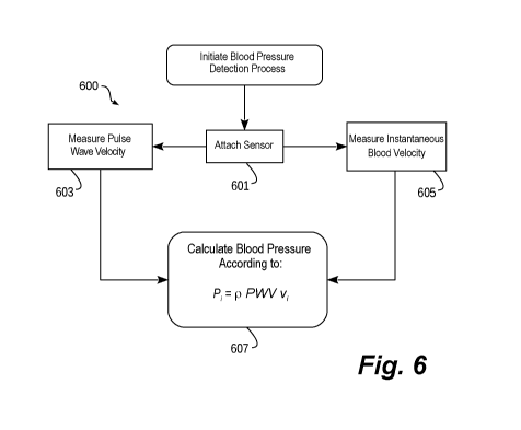
[010] Figure 6 is a conceptual flow diagram of a method that implements one
embodiment for measuring non-invasive blood pressure.
[011] Figure 7 is a conceptual illustration of one alternative embodiment that
uses two
or more elements rather than a single sensor.
[012] Figure 8 is a conceptual illustration of another alternative embodiment
which
implements an ultrasound sensor in combination with a sensor based on a
different technology.
DETAILED DESCRIPTION
[013] Generally stated, embodiments of this disclosure measure two values
which can
be used to compute a patient's instantaneous blood pressure. Embodiments of
this disclosure
measure the instantaneous Non-Invasive Blood Pressure (NIBP) of a patient with
an apparatus
that determines the values for, in one example, two of the unknowns in the
water hammer
equation: pulse wave velocity (PWV) and instantaneous blood
-2-
CA 03050694 2019-07-17
WO 2018/136656
PCT/US2018/014273
velocity (Vi). The water hammer equation relates instantaneous blood pressure
to pulse
wave velocity and blood flow velocity as follows:
= p PWV v,
[014] where PWV is the pulse wave velocity, p is the density of the blood
which may
be assumed to be a constant, for example, vi is the instantaneous velocity of
the blood, and Pi
is the desired instantaneous blood pressure.
[015] Some conventional NIBP measurement systems rely on PWV to measure NIBP,
but each requires an initial calibration measurement, taken at least once, to
convert a relative
blood pressure value to an actual blood pressure value. The required
calibration measurement
is taken using a traditional blood pressure cuff, for example on the arm or
perhaps the finger.
Such conventional NIBP measurement systems that require an initial calibration
and all
calculations are based on a difference or differential value of that initial
calibration
measurement to achieve an actual measurement.
[016] The disclosed NIBP systems and devices instead take an instantaneous
blood
pressure measurement rather than a change from an initial calibration
measurement. Avoiding
the need for a calibration measurement, prevents the patient from experiencing
blood flow
restriction altogether. Although PWV is highly correlated with blood pressure
(BP) so that
changes in blood pressure can be calculated from changes in PWV by relying on
an initial
calibration measurement relatively accurately, what has not been solved until
now is how to
eliminate the need to acquire and use a separate, initial calibration value or
values to register a
particular PWV to a particular value of blood pressure (as opposed to simply a
change in blood
pressure) for a patient. State of the art of NIBP using PWV typically uses a
standard cuff-
based measurement, to interrupt the blood flow, to measure and associate a
particular blood
pressure to a particular PWV measurement in a patient. Interrupting the blood
flow requires
that the patient's appendage being measured is compressed to restrict the
blood flow. Such
restriction of the patient's blood flow prevents such conventional methods of
measuring blood
pressure from being applied to areas of the patient's body that cannot
withstand restricted blood
flow, such as a patient's neck, for example.
-3-
CA 03050694 2019-07-17
WO 2018/136656
PCT/US2018/014273
[017] In this way, conventional methods and devices that provide NIBP
measurements
using PWV require a distinct calibration step. In contrast to the state of the
art, the disclosed
embodiments include a method and device that eliminate the requirement of a
distinct
calibration step, especially using a technology that temporarily restricts
blood flow. In short,
the disclosed embodiments include self-calibrating NIBP systems and methods
using PWV, or
alternatively, NIBP systems and methods using PWV without the temporary
interruption of
blood flow.
[018] The lack of need for a calibration step for devices using the method
taught herein
arises from the use of the water hammer equation in its integrated (non-
differential) form. In
the water hammer equation, the blood pressure is related to the PWV by a scale
factor that can
be known without a distinct calibration step. The scale factor is found using
the same
ultrasound technology that is used to measure the PWV. That scale factor is
related to the
blood velocity and blood density. In this way, a particular blood pressure is
calculated as the
PWV scaled by the blood density and the blood velocity.
[019] Blood velocity can be acquired using ultrasound as a time varying
waveform.
PWV can also be measured with ultrasound also as a time varying function. The
time-varying
nature of the PWV means that it can be updated from beat to beat if desired.
The time-varying
nature of the blood velocity means that blood velocity can be measured at a
much finer
resolution than a cardiac cycle, that is to say, continuously during the
cardiac cycle for as many
cardiac cycles as is desired. Because blood density is already sufficiently
known and is
relatively constant, not only can a particular blood pressure measurement be
known as if it
were obtained by a standard cuff-based measurement, but all manners of blood
pressure
measurements can be made as time-varying waveforms describing the
instantaneous pressure
at as many points during a cardiac cycle as desired. That is to say, blood
pressure can be
monitored continuously throughout the cardiac cycle with as fine a resolution
as is required,
and this can be done for as many contiguous cardiac cycles as is desired for
beat-to-beat
monitoring, or as intermittently as desired.
[020] Measuring the instantaneous blood pressure instead of its change
relative to a
calibrated baseline measurement means, for example, that as arterial walls
stiffen (due to
-4-
CA 03050694 2019-07-17
WO 2018/136656
PCT/US2018/014273
disease. drug therapy, and/or normal vasculature responses, for example) which
increases the
PWV, this new PWV value is measured along with any corresponding change in
blood velocity
to produce an updated blood pressure waveform. Additionally, if the heart
pumps more or less
energetically, the blood velocity changes accordingly, which results in the
blood pressure
changing proportionately, all else equal. This updated blood velocity
measurement at the
prevailing PWV (which characterizes the state of the vasculature) corresponds
to the updated
blood pressure after being scaled by blood density. In other words, since
there are two
measurements made, PWV and blood velocity, and not just PWV alone, a distinct
calibration
step is not needed, as the ambiguity of PWV by itself is remedied by adding
the second
measured value of blood velocity. This is of great value over conventional
patient NIBP
monitoring using PWV alone where typically the calibration step requires a
blood pressure
measurement performed by restricting blood flow, which can be more costly,
time consuming,
and/or uncomfortable to the patient. In the embodiments discussed below,
ultrasound
technology is used to acquire both the PWV and the blood velocity although
other methods of
obtaining the PWV and the blood velocity can alternatively or additionally be
used. Further
embodiments implement various techniques and devices to measure or detect both
pulse wave
velocity and instantaneous blood velocity. As is described in greater detail
below, specific
embodiments simplify the task of measuring NIBP without sacrificing
reliability. Still further,
embodiments enable the measurement of (or at least an estimation of) NIBP
without requiring
calibration that relies on a separate means for detecting blood pressure,
which simplifies the
treatment and evaluation of the patient.
[021] This disclosure begins with a description of one example of a medical
device
that may be used in specific embodiments. Next is a discussion of one
embodiment of a sensor
for measuring NIBP using ultrasound. Alternative embodiments for sensors which
measure
NIBP are further discussed.
Description of Operative Environment for Embodiments
[022] Figure 1 is a diagram of a medical treatment scene. As illustrated, a
person 82
is lying supine. Person 82 could be a patient in a hospital or someone found
unconscious.
Person 82 is experiencing a medical condition which requires monitoring of the
person 82.
-5-
CA 03050694 2019-07-17
WO 2018/136656
PCT/US2018/014273
Person 82 may be a victim of cardiac arrest, or some other emergency, and
consequently a
patient.
[023] In the example shown in Figure 1, a portable vital signs monitor 100 has
been
brought close to person 82. The vital signs monitor can also be, for example,
a hybrid
monitor/defibrillator. As illustrated, a number of physiologic sensors may be
attached to
person 82, such as vital signs monitoring (VSM) sensors 104, 108, and
connected to vital signs
monitor 100. The vital signs monitor 100 provides a user with information
about the vital signs
of person 82 collected using VSM sensors 104, 108. Vital signs monitor 100 can
be one of
many different types, such as a monitor or monitor/defibrillator, each with
different sets of
features and capabilities. The set of capabilities of vital signs monitor 100
is determined,
generally, by who would use it, and what training they would likely have.
Although illustrated
as a vital signs monitor in Figure 1. many other medical devices may be used
in the medical
treatment scene, and may implement various embodiments of the disclosure.
[024] Turning briefly to Figure 2, various medical devices in which
embodiments may
be implemented are shown. By way of example, embodiments may be implemented in
a
monitor/defibrillator 200. A defibrillator-monitor (or monitor-defibrillator)
is typically formed
as a single unit with a defibrillator in combination with a patient monitor.
Alternatively, the
defibrillator-monitor may be a modular device with separable components. For
example, in
one embodiment, the defibrillator-monitor may include a base component and a
plurality of
detachable pods. Each pod may communicate with the base component, perhaps
wirelessly.
Certain pods may be used to collect information about a patient, such as vital
statistics.
[025] As a patient monitor, the device 200 may have features additional to
what may
be needed, but can be there should a need arise or because they are customized
to a person.
These features can be for monitoring physiological indicators of a person in
an emergency
scenario. These physiological indicators are typically monitored as signals,
such as a person's
full ECG (electrocardiogram) signals, or impedance between two electrodes.
Additionally,
these signals can be about the person's temperature, non-invasive blood
pressure (NIBP),
arterial oxygen saturation / pulse oximetry (Sp02), the concentration or
partial pressure of
-6-
CA 03050694 2019-07-17
WO 2018/136656
PCT/US2018/014273
carbon dioxide in the respiratory gases, which is also known as capnography,
and so on. These
signals can be further stored and/or transmitted as patient data.
[026] In addition, embodiments may be implemented in an ultrasound machine
210.
As shown in Figure 2, an ultrasound machine 210 may be variously sized and
shaped, although
common ultrasound machines are adapted to be deployed at bedside, such as may
be used in a
hospital or other controlled health care environment.
[027] In the illustrated embodiment, the ultrasound machine 210 may include a
chassis
and a transducer. The chassis, for example, may be made of molded plastic,
metal, or some
combination of both. The chassis houses a module for generating electrical
signals which are
conveyed to the transducer to be transformed into ultrasonic energy. The
transducer transmits
ultrasound waves into a subject (e.g., a patient) by converting the electrical
signals to ultrasonic
energy. The transducer further receives ultrasound waves backscattered from
the subject by
converting received ultrasonic energy to analog electrical signals.
[028] The ultrasound machine 210 may also include an operator interface
through
which an operator inputs information to affect the operating mode of the
ultrasound machine.
Through the interface, the ultrasound machine 210 may also output status
information for
viewing by the operator. The interface may provide a visual readout, printer
output, or an
electronic copy of selected information regarding the examination.
[029] Other embodiments may be implemented as a standalone device, such as a
handheld NIBP monitor 220. The handheld NIBP monitor 220 may be sized and
configured
for easy portable use. In such a case, the handheld NIBP monitor 220 may
include a transducer
and a housing. The transducer of the handheld NIBP monitor 220 may operate in
similar
fashion to the transducer of the ultrasound machine 210. Likewise, electronic
components that
perform computational functions may be contained within the housing.
[030] Yet other embodiments may be implemented as an ultraportable device
(e.g,
wearable NIBP monitor 230), such as a smartwatch, wearable bracelet, wearable
adhesive
sensor, or the like. In such an embodiment, components of the transducer may
be integrated
into a unitary housing and attached or affixed to a person for vital signs
monitoring.
-7-
CA 03050694 2019-07-17
WO 2018/136656
PCT/US2018/014273
[031] Illustrative examples of various devices, including medical devices,
show
various embodiments. However, the scope of this disclosure is not limited to
these
embodiments and the disclosure may be implemented in many other embodiments
not shown
or described. For example, embodiments may be implemented within electronic
wearable
devices, such as a smartwatch or other wireless-enabled portable electronic
device, or other
smart wearables, such as sensor clothing or skin prints. In one specific
example, an electronic
piece of jewelry (or the like) may be implemented which includes ultrasound
sensing
technology that is capable of measuring or estimating non-invasive blood
pressure based on
the teachings of this disclosure. All such embodiments are within the
teachings of this
disclosure and fall within the scope of the appended claims.
[032] Figure 3 is a diagram showing components of a medical device made
according
to embodiments. In this particular example, the medical device is a vital
signs monitor 300,
although many components are common to other medical devices. These components
can be,
for example, in vital signs monitor 100 of Figure 1. The components shown in
Figure 3 can be
provided in a housing 301, also known as a casing. It will be appreciated
that, in other
embodiments, these components may be implemented in separate housings or as
sub-
components of various other devices.
[033] In this illustrative embodiment, vital signs monitor 300 includes a
processor 330
and a memory 338. The processor 330 is a computing component operative to
execute
programming instructions. The processor 330 may be implemented in any number
of ways.
Such ways include, by way of example and not of limitation, digital and/or
analog processors
such as microprocessors and digital-signal processors (DSPs); controllers such
as
microcontrollers; software running in a machine; programmable circuits such as
Field
Programmable Gate Arrays (FPGAs), Field-Programmable Analog Arrays (FPAAs),
Programmable Logic Devices (PLDs), Application Specific Integrated Circuits
(ASICs), any
combination of one or more of these, and so on.
[034] The memory 338 stores instructions (e.g., programs or applications) to
be
executed by processor 330 and can also store data collected from various
physiological sensors
used with the vital signs monitor. For example, memory 338 can store patient
data, such as,
-8-
CA 03050694 2019-07-17
WO 2018/136656
PCT/US2018/014273
for example, blood pressure measurements taken or computed by the vital signs
monitor 300.
In addition, memory 338 can store prompts for the user, etc. Memory 338 may be
implemented
in any number of ways. Such ways include, by way of example and not of
limitation,
nonvolatile memories (NVM), read-only memories (ROM), random access memories
(RAM),
any combination of these, and so on.
[035] Processor 330 is further preferably connected to a display screen 382,
which can
also be remote from the sensor. If display screen 382 is a touch sensitive
screen, microprocessor
330 can both send data to and receive data from the display screen 382. The
processor 330 can
further optionally communicate with other external computing peripherals (not
shown), such
as a personal computer and/or an external printer.
[036] Various sensors are included for detecting physiologic characteristics
of a
patient. For instance, a temperature sensor 386 and a pulse oximeter sensor
388 may be
connected to processor 330 via AID converter 354. AID converter 354 is capable
of converting
analog data to digital data, and digital data to analog data. A NIBP cuff (or
sphygmomanometer
cuff) 394 is pneumatically connected to a blood pressure pump 340 used to
pressurize the blood
pressure cuff 394. Like the pulse oximeter sensor 388 and temperature sensor
386, blood
pressure sensor 340 is connected to AID converter 354. Those skilled in the
art will understand
that use of fully digital sensors can eliminate analog to digital conversion
of sensor signals and
thus eliminate AID converter 354.
[037] The vital signs monitor 300 preferably receives power by a line voltage
connection 350 regulated by at least one voltage regulator 348. However, the
vital signs
monitor 300 may also rely on a battery 346 as a power source. Reliance on
battery power may
be advantageous in some circumstances because it allows the vital signs
monitor 300 to be
portable. It should be understood that voltage regulator 348 may be configured
to produce a
number of different power outputs connected to a number of different
components. The sensor
may also wirelessly collect ambient energy from available sources (not shown)
especially when
neither battery nor line voltage are available.
[038] Vital signs monitor 300 can optionally include other components. For
example,
a communication module 390 may be provided for communicating with other
machines. Such
-9-
CA 03050694 2019-07-17
WO 2018/136656
PCT/US2018/014273
communication can be performed wirelessly, or via wire, or by infrared
communication, and
so on. This way, data can be communicated, such as patient data, incident
information, therapy
attempted, cardiopulmonary resuscitation (CPR) performance, blood pressure,
and so on.
[039] In one embodiment, vital signs monitor 300 further includes an
ultrasound
transducer 360. The transducer 360 is preferably enclosed in a case to
insulate it from electrical
interference. The transducer 360 includes, in this embodiment, an active
element 361 is made
of piezoelectric material (e.g., PZT) or, alternatively, tnicromachined
ultrasonic transducers
(MUT) or other MEMS devices (e.g., PMUT devices, or the like). The active
element 361 may
be a single element or an array. The active element 361 is responsible for
radiating an
ultrasound wave and detecting reflected signals, in this embodiment. The
active element 361
may, in some embodiments, employ separate transmitting and receiving elements.
Alternatively, other embodiments may combine both functions into a single
piezoelectric
transceiver, or other sensor technology or material that converts mechanical
energy to electrical
energy and vice versa.
[040] In one embodiment, a connection 362 couples the transducer 360 to the
vital
signs monitor 300 for operative communication. Connection 362 is illustrated
in Figure 3 as
an ordinary wire or similar direct electrical connection (either detachable or
fixed) between the
transducer 360 and the vital signs monitor 300. Alternatively, the connection
362 may be a
wireless connection between the transducer 360 and a wireless transceiver of
the vital signs
monitor 300 (e.g., communication module 390).
[041] In some embodiments, transducer 360 may deliver an analog signal or
signals
to the vital signs monitor 300. In such an embodiment, connection 362 may
route through A/D
converter 354 (illustrated in dashed line) where the analog signals are
converted to digital
signals which may be operated upon by processor 330. In other alternatives,
the transducer
360 may deliver either a combination of analog and digital signals, or all
digital signals. In
such a case, the connection 362 may either partially or entirely avoid the A/D
converter 354.
[042] In some embodiments, the transducer 360 is a peripheral component of the
vital
signs monitor 300. In such embodiments, transducer 360 may rely on the
computational
functions of the vital signs monitor 300. In other embodiments, the transducer
360 may be a
-10-
CA 03050694 2019-07-17
WO 2018/136656
PCT/US2018/014273
completely self-contained item. In such embodiments, the transducer 360 may
further include
its own computational components, such as a dedicated processor 340, memory
341, A/D
converter 343, and power supply 342.
[043] In one embodiment, the vital signs monitor 300 includes an NIBP
detection
component 325. In one specific implementation, the NIBP detection component
325 includes
functions which, when executed by processor 330, operate to measure a
patient's blood
pressure based on at least a sound wave analysis. The NIBP detection component
325 may be
coupled to the transducer 360 via a port 326, which causes a sound wave to be
generated and
transmitted, via the transducer 360, to a patient. A return signal received at
the transducer may
be communicated back to the NIBP detection component 325 via the transducer
port 326 using
connection 362.
[044] In alternative embodiments where the transducer 360 is self-contained,
the
NIBP detection component 325 could be implemented in the memory 341 of the
transducer
360 for execution by the processor 340. In yet another alternative, the NIBP
detection
component 325 may be remotely executable, via connection 362, using the
processor 340 of
the transducer 360.
10451 In accordance with various embodiments, the NIBP detection component is
configured to perform a sound wave analysis that determines, for example, two
values: a pulse
wave velocity and an instantaneous blood velocity. The NIBP detection
component 325 is
further configured to compute an instantaneous blood pressure based on the
pulse wave
velocity and the instantaneous blood velocity. The pulse wave velocity
computation may be
performed by analyzing ultrasound imaging, such as B-mode, M-mode, or 2D-mode
imaging,
combined with physical dimensions either directly measured or computed using
data received
through the sound wave analysis. The blood velocity computation may be
implemented as any
appropriate Doppler detection technique, for example, such as by correlation
(e.g.,
autocorrelation, cross-correlation, or the like) or Fourier transform
processing, to determine
Doppler characteristics of blood within a vessel.
[046] NIBP detection component 325 may provide notice of its analysis in many
ways.
In one example, the NIBP detection component 325 may be an automatic detector
which
-11-
CA 03050694 2019-07-17
WO 2018/136656
PCT/US2018/014273
provides an on-screen indication, via display 382, of its analysis.
Alternatively, NIBP detection
component 325 may output to a more direct indicator, such as a speaker or
other output.
[047] Embodiments of the disclosure implement various techniques and devices
to
measure or detect both pulse wave velocity and instantaneous blood velocity.
As is described
in greater detail below, specific embodiments simplify the task of measuring
NIBP without
sacrificing reliability.
[048] Various other components may also be used to provide added functionality
not
shown. Non-exhaustive examples of such components include a speaker,
microphone, digital
camera interface, additional environmental or physiological sensors,
accelerometers, and the
like.
Illustrative Embodiments of the Disclosure
[049] Figure 4 is a conceptual diagram of one embodiment of the disclosure. As
shown in Figure 4, one embodiment for measuring NIBP employs an ultrasound
transducer
411 that may be affixed to a patient (e.g., patient 82) adjacent to any
appropriate vein or artery.
As illustrated, the ultrasound transducer 411 is affixed to the patient 82
adjacent to the patient's
carotid artery.
[050] In this particular embodiment, the ultrasound transducer 411 is
implemented
with a single sensor 413, which reduces size and cost. Alternative
implementations and
embodiments may employ more sensors in addition to the single ultrasound
sensor. In use,
ultrasound transducer 411 may self-dispense a wetting agent, such as
ultrasound gel, to
eliminate air from between the patient's skin and ultrasound sensor 413. To
enhance reliability,
the field of view and the signal to noise ratio should be significantly high
enough to allow the
sensor to be easily applied and still achieve good results.
[051] As noted above, embodiments of the disclosure measure the patient's
pulse
wave velocity and instantaneous blood velocity, which then reveal the
patient's NIBP via the
water hammer equation. There are a number of different techniques for
measuring each of
PWV and blood velocity. However, implementations of the embodiment measure
both as
discussed here. Certain alternative implementations and embodiments are
discussed later in
this document.
-12-
CA 03050694 2019-07-17
WO 2018/136656
PCT/US2018/014273
[052] Turning now to Figure 5, a conceptual illustration is shown of one
implementation of an embodiment in operation. As shown in Figure 5, an
ultrasound
transducer 511 is affixed to a patient's skin 510 with the axis of the
ultrasound transducer 511
roughly aligned parallel to a proximate vessel, such as a vessel located in
the neck or arm or
finger, where the direction is known. A proximate vessel is a blood vessel in
the patient that
is being used to measure the patient's blood pressure and can be both a
targeted blood vessel
chosen to be used as the vessel within which the blood pressure is measured or
can be a blood
vessel that is found through discovery by or a vessel that is simply nearby
the disclosed NIBP
devices and/or systems. In this particular example. a carotid artery 520 is
selected because of
its relative ease of access and relatively linear presentation. The
construction of the sensor in
the perpendicular axis is either sufficiently narrow so that the field of view
is wide or is
augmented with a lens that achieves a wide field of view from a larger
element. In this way,
the sensor is able to be placed with a correct orientation so that a proximate
vessel is within the
field of view of the sensor. In one embodiment, the transducer 511 may be
curved to provide
a larger field of view.
[053] In one embodiment, ultrasound waveforms of the length of about 1 mm and
of
a center frequency of about 6 MHz are pulsed at a repetition rate of up to 10
kHz. The
ultrasound waveforms are composed of a main lobe 530 and two grating lobes
(left grating lobe
535 and right grating lobe 540). Other components of the ultrasound waveforms
which may
be present are not illustrated. In one embodiment, the ultrasound transducer
511 is constructed
such that the active pressure sensitive areas of its single sensor are
interdigitated with inactive
areas having a periodic spacing so that the grating lobes (535, 540) are
intentionally formed at
a desired separation angle 0. By so doing, accurate distance measurements may
be obtained
between any two points in the field of view of the sensor using triangulation
techniques.
[054] Generally stated, this implementation of the embodiment radiates
ultrasound
waveforms as discussed above. The return signals are used in two ways: to
determine pulse
wave velocity and to determine instantaneous blood velocity. With those two
values, the
patient's NIBP may be determined using, for example, the water hammer
equation. Generally
stated, this embodiment detects pulse wave velocity by analyzing blood wave
motion using,
-13-
CA 03050694 2019-07-17
WO 2018/136656
PCT/US2018/014273
for example, B-mode or M-mode images captured by the ultrasound transducer. In
addition,
blood velocity is determined by analyzing the Doppler effect on the return
signals.
Measuring Pulse Wave Velocity
[055] Blood pumping through the vessel 520 causes a localized expansion (pulse
521)
in the vessel 520. Knowing the rate at which the pulse 521 travels along a
given distance in a
given time provides the pulse wave velocity. To make that determination, the
ultrasound
transducer 511 identifies the vessel wall motion as the pulse 521 moves past
the left grating
lobe 535, main lobe 530, and right grating lobe 540.
[056] The most identifiable return signal will be the specular reflection from
the main
lobe 530. A depth 545 of the vessel 520 directly under the sensor 511 is
derived from the
location of the specular response. The depth 545 of the vessel 520 and the
grating lobe angle
0 reveal the slant range depth of the vessel 520 at the point of incidence of
both the left grating
lobe 535 and the right grating lobe 540.
[057] Blood vessel wall motion may then be identified by cross correlation
between
small regions at the same time vicinity corresponding to the slant range depth
of the vessel,
that is, around the point in time the signal returns from the intersection of
the ultrasound beam
and vessel. The pulse wave velocity is calculated as the distance along the
vessel 520 between
any two lobes (e.g., the two grating lobes, either grating lobe and the main
lobe, or the like)
divided by the time between motion at the slant range depths corresponding to
the pulse 521
passing by those two lobes.
[058] The time may be measured to as low as 100 microsecond resolution and can
be
interpolated for finer resolution. Pulse wave velocities are typically in the
6 to 10 m/sec range.
For grating lobes to be 2 cm apart at the vessel depth, the time for the pulse
wave to pass from,
for example, one grating lobe to another would be as short as 2 milliseconds
which is 20 times
larger than the 100 microsecond resolution of the disclosed embodiment. It
should be
appreciated that ultrasound shear wave imaging techniques may also be used to
measure pulse
wave velocity.
-14-
CA 03050694 2019-07-17
WO 2018/136656
PCT/US2018/014273
Measuring Instantaneous Blood Velocity
[059] Using the same data, the velocity of the blood may also be measured
using
ultrasound pulse wave Doppler (PWD) techniques. This is accomplished by
performing
Doppler analysis in the vicinity of the vessel center. This Doppler analysis
identifies the phase
change of the returned signal from the blood between each of the 10 kHz
repetitions after
filtering out any static returns. The phase change of the returned signal over
a corresponding
change in time is the blood velocity.
[060] In one embodiment two measurements are made as blood flow towards the
transducer 511 can be resolved from blood flow away from the transducer 511.
This velocity
is then corrected by the sine of the known grating lobe angle 0 normal to the
transducer 511.
For small vessels (where the 1 mm pulse length encompasses much of the
diameter of the
vessel), this integrated velocity may be mapped to a true instantaneous
velocity at the center of
the vessel based upon previously gathered empirical databases or tabulated or
computed
relationships between mean blood flow and peak blood flow under various flow
conditions and
vessel sizes.
[061] Once the instantaneous blood velocity and the pulse wave velocity are
known,
instantaneous blood pressure is computed using the water hammer equation.
Again, the blood
density may be treated as a constant to yield the NIBP. Alternatively, the
actual blood density
of the patient may be used in the equation if that value is known, such as
from prior testing or
analysis.
[062] Turning briefly to Figure 6, a conceptual flow diagram is shown that
implements
one method 600 for measuring non-invasive blood pressure. To begin, the method
600 starts
when a sensor configured in accordance with embodiments of this disclosure is
attached to a
patient (step 601). As discussed at length above, the sensor includes an
ultrasound sensor and
may include one or more alternative sensors.
[063] In its most basic form, the method 600 proceeds by substantially
simultaneously
measuring pulse wave velocity (step 603) and instantaneous blood velocity
(step 605).
Although summarized here, each of those two basic steps may be accomplished in
numerous
ways.
-15-
CA 03050694 2019-07-17
WO 2018/136656
PCT/US2018/014273
[064] For example, pulse wave velocity may be measured using a sound analysis
based on information known about the configuration of the sensor. In one
specific embodiment,
the sensor is configured such that an ultrasound waveform radiated by the
sensor will produce
grating lobes having known characteristics, such as a grating lobe separation
angle of O. The
sound analysis may further compute a depth from the sensor to a subject blood
vessel. Based
on those data, ultrasound imaging combined with triangulation techniques may
be used to
compute a rate at which a pulse travels through the vessel, thereby revealing
the pulse wave
velocity of the vessel.
[065] Similarly, instantaneous blood velocity may be measured using Doppler
effect
techniques. In one specific embodiment, the Doppler analysis may identify the
phase change
of a returned signal from the blood between each of the 10 kHz repetitions.
[066] Once pulse wave velocity and instantaneous blood velocity are known, the
method 600 continues by calculating the instantaneous blood pressure (step
607) in accordance
with, for example, the water hammer equation. Based on that equation, pulse
wave velocity,
instantaneous blood velocity, and blood pressure are related as follows:
= p PWV v,
10671 Once calculated, the blood pressure measurement may be presented to a
user for
use in treatment of the patient. It should be appreciated that, in another
alternative, continuous
wave Doppler (CWD) may be used as an alternative to pulse wave Doppler (PWD).
Alternafive Embodiments of the Disclosure
[068] Although the embodiment discussed above is one embodiment, many other
embodiments may also be implemented which employ the teachings of this
disclosure.
Discussed here are but a few of the many alternative embodiments which
implement this
disclosure.
[069] Figure 7 is a conceptual illustration of one alternative embodiment
which uses
two or more elements rather than a single sensor. For example, a composite
sensor of two or
three elements may be implemented such that each element does not have a
grating lobe but
are mounted at known angles such as the grating lobe angle in the figure
below. Each sensor
-16-
CA 03050694 2019-07-17
WO 2018/136656
PCT/US2018/014273
would need its own analog path to digitization. Such a sensor could be
integrated with other
sensors such as pulse oximetry.
[070] Figure 8 is a conceptual illustration of another alternative embodiment
which
implements an ultrasound sensor in combination with a sensor based on a
different technology.
Other techniques are emerging for measuring pulse wave velocity which may be
combined, in
certain embodiments, with an ultrasound sensor to detect instantaneous blood
pressure.
Accordingly, the alternative embodiment illustrated in Figure 8 implements an
optical sensor
which is employed to determine velocity of pulse 521 within vessel 520. The
alternative
embodiment shown in Figure 8 makes use of an ultrasound sensor radiating an
ultrasound
waveform 830 in combination with a second sensor radiating a second signal 835
based on
some other technology, such as a light emitting diode or the like.
[071] In this particular embodiment, the transducer assembly 811 is
illustrated as a
stand-alone component. However, in other embodiments the transducer assembly
811 may be
combined with or integrated into another component. In one example, a portable
external
monitor-defibrillator may be specially configured to support the Doppler
detection of NIBP.
In such an embodiment, the transducer assembly 811 may be combined with or
integrated into
a set of ECG leads, one or more defibrillation electrodes, or some other
component of a vital
signs monitor. In this way, the function of detecting NIBP may be incorporated
into a medical
device which is already in use in medical emergency situations, thereby
eliminating a need to
employ yet another, separate medical device.
[072] Many other embodiments are also envisioned to be within the scope of the
disclosure. For example, embodiment that are implemented in devices other than
medical
devices (e.g., exercise equipment, or other non-medical equipment) are also
envisioned.
Similarly, embodiments that compute or estimate blood pressure by virtue of a
non-invasive
measurement of two or more characteristics of a patient using an equation
other than the water
hammer equation are also possible. Any appropriate equation which associates
blood pressure
to measurable characteristics of a patient's physiology other than the blood
pressure are also
envisioned.
-17-
CA 03050694 2019-07-17
WO 2018/136656
PCT/US2018/014273
[0731 In summary, the disclosed embodiments overcome shortcomings of existing
systems by obviating the need to manually attempt to detect non-invasive blood
pressure by
using a cuff, or the like. In these and other ways, which will become apparent
upon a study of
the disclosed teachings, these embodiments provide a superior treatment
technique and
transducer assembly for the non-invasive detection of blood pressure in a
patient.
10741 Other embodiments may include combinations and sub-combinations of
features described above or shown in the Figures, including, for example,
embodiments that
are equivalent to providing or applying a feature in a different order than in
a described
embodiment, extracting an individual feature from one embodiment and inserting
such feature
into another embodiment; removing one or more features from an embodiment; or
both
removing one or more features from an embodiment and adding one or more
features extracted
from one or more other embodiments, while providing the advantages of the
features
incorporated in such combinations and sub-combinations. As used in this
paragraph, "feature"
or "features" can refer to structures and/or functions of an apparatus,
article of manufacture or
system, and/or the steps, acts, or modalities of a method.
[075] It should be readily apparent to those skilled in the art that what is
described
herein may be modified in numerous ways. Such ways can include equivalents to
what is
described herein. In addition, the invention may be practiced in combination
with other systems.
The following claims define certain combinations and sub-combinations of
elements, features,
steps, and/or functions, which are regarded as novel and non-obvious.
Additional claims for
other combinations and sub-combinations may be presented in this or a related
document.
-18-