Note: Descriptions are shown in the official language in which they were submitted.
CA 03050914 2019-07-18
WO 2018/140733
PCT/US2018/015459
VACCINE COMPOSITIONS OF HERPES VIRUS ENVELOPE PROTEIN
COMBINATIONS TO INDUCE IMMUNE RESPONSE
CROSS REFERENCE TO RELATED APPLICATIONS
[0001] This application claims the benefit of, and relies on the filing date
of, U.S.
provisional patent application number 62/451,396, filed 27 January 2017, the
entire
disclosure of which is incorporated herein by reference.
GOVERNMENT INTEREST
[0002] This invention was made with government support under grant Q574LJ15
awarded by the Uniformed Services University. The government has certain
rights in the
invention.
SEQUENCE LISTING
[0003] The instant application contains a Sequence Listing which has been
submitted
electronically in ASCII format and is hereby incorporated by reference in its
entirety. Said
ASCII copy, created on 25 January 2018, is named HMJ-153-PCT_SL.txt and is
207,545
bytes in size.
BACKGROUND
[0004] Human herpes viruses are a group of enveloped DNA viruses responsible
for
significant global morbidity and mortality in humans. (Eisenberg et al.,
Viruses, 4:800-32,
2012). There are eight types of known human herpes virus (HHV), including: (i)
Type 1
human herpes virus (HHV-1), which is herpes simplex virus-1 (HSV-1); (ii) HHV-
2 which is
herpes simplex virus-2 (HSV-2); (iii) HHV-3 which is varicella-Zoster virus
(VZV); (iv)
HHV-4 which is Epstein Barr virus (EBV); (v) HHV-5, which is human
cytomegalovirus
(HCMV); (vi) HHV-6; (vii) HHV-7; and (viii) HHV-8 which is Kaposi's sarcoma-
associated
herpesvirus (KSHV).
[0005] In humans, these viruses are known to cause the following diseases. HSV-
1
causes oral herpes, HSV-2 causes genital herpes, and VZV causes chickenpox and
shingles.
EBV causes infectious mononucleosis and is strongly associated with several B
cell
lymphomas, nasopharyngeal carcinoma, and gastric adenocarcinoma. HCMV causes
severe
infection in immunosuppressed patients and is the leading non-genetic cause of
hearing loss.
-1-
CA 03050914 2019-07-18
WO 2018/140733
PCT/US2018/015459
HHV-6 and 7 cause roseola infantum (Sixth disease), and HVV-8 causes Kaposi's
sarcoma in
several clinical settings including in patients infected with human
immunodeficiency virus
(HIV).
[0006] EBV primarily infects B cells and nasopharyngeal epithelial cells. EBV
infection of B cells is initiated by binding of the EBV envelope protein gp350
to the
complement receptor CR2/CD21. (Hutt-Fletcher, J. Virol., 81:7825-32, 2007; and
Shannon-
Lowe et al., Curr. Opin. Virol., 4:78-84, 2014). Upon binding to B cell CR2,
EBV gp42
interacts with cell surface MHC-II receptors, leading to its association with
the heterodimeric
EBV gH/gL protein. The heterodimer gH/gL then undergoes a conformational
change upon
binding gp42, leading to activation of the EBV fusion protein gB, that
directly mediates viral-
host cell membrane fusion. (Neuhierl et al., Proc. Natl. Acad. Sci. U.S.A.,
99:15036-41,
2002). Like EBV, the binding, fusion and host cell entry of other HHV family
members is
mediated primarily by the gB, gH, and gL polypeptides, in conjunction with
other accessory
proteins, which typically bind to different receptors on the host cell
surface.
[0007] There is currently no prophylactic EBV vaccine in clinical use. Studies
in non-
human primates using gp350-based vaccination strategies have shown protection
against
EBV-induced lymphoma and EBV replication. (Cohen, Clin. Transl. Immunology,
4:e32,
2015). A phase II clinical trial conducted in EBV-seronegative young adults
using a
recombinant monomeric gp350 protein versus placebo suggested a partial
protective effect of
gp350 vaccination on infectious mononucleosis (IM) development. (Sokal et al.,
J. Infect.
Dis., 196:1749-53, 2007; and Moutschen et al., Vaccine, 25:4697-705, 2007).
However, the
vaccine did not prevent asymptomatic EBV infection. A phase I trial of
recombinant
monomeric gp350 protein given to children with chronic kidney disease
demonstrated only a
minority of subjects developing detectable neutralizing serum anti-gp350
titers. (Rees et al.,
Transplantation, 88:1025-9, 2009).
[0008] There is also no prophylactic HCMV vaccine commercially available
today.
Earlier clinical trials using live attenuated Towne or AD169 HCMV viral
vaccines, both of
which lacked expression of a pentameric complex (gH/gL/UL128/UL130/UL131A),
proved
to be ineffective in preventing HCMV infection in either healthy volunteers or
renal
transplant recipients, though some efficacy was demonstrated in overt HCMV
disease in high
risk Recipient-Donor+ renal transplant recipients (Fu et al., Vaccine, 32:2525-
33, 2014). New
HCMV viral strains engineered to express the pentameric complex are currently
being
evaluated, but safety concerns persist using this approach. A phase II
clinical trial using
recombinant HCMV gB protein derived from the Towne strain of HCMV (Spaete RR,
-2-
CA 03050914 2019-07-18
WO 2018/140733
PCT/US2018/015459
Transplant Proc., 23:90-6, 1991) demonstrated 50% efficacy in preventing HCMV
infection
in HCMV seronegative women (Pass RF, J. Clin. Virol., 46 Suppl 4:S73-6, 2009)
and 50%
efficacy in preventing HCMV viremia in solid organ transplantation patients.
The HCMV gB
protein used in Phase II clinical trials had been modified to remove the furin
cleavage site.
Thus, the gB did not assume its native trimeric conformation (Sharma et al.,
Virology,
435:239-49, 2013). Although these two studies have encouraged further
evaluation of gB as a
prophylactic HCMV vaccine, they indicate a compelling need for a more
effective
prophylactic vaccine formulation.
[0009] W02014/018858 and W02015/089340 describe strategies for enhancing
immunity that involve multimerizing antigens. For example, W02014/018858
describes
fusion proteins comprising at least two antigens, separated by a linker
sequence, and an
oligomerization domain, including multimeric HHV antigens, such as gp350, gB,
gH, and gL.
W02015/089340 describes a modified herpesvirus gB obtained by inserting a
peptide linker
at the furin cleavage site in the herpesvirus gB polypeptide extracellular
domain. Inserting the
peptide linker removes the furin recognition sequence, such that expression of
the modified
herpesvirus gB results in the production of a homotrimeric gB complex that
provides
enhanced immunogenicity.
[0010] Combining multiple antigens in a vaccine does not necessarily result in
enhanced immunity or even additive effects. In fact, when multiple antigens
are co-
administered as part of a multicomponent vaccine or as part of a sequential
immunization
schedule, the antibody response to one or more of the antigens may be reduced
or diminished
due to vaccine or immune interference. (PrabhuDas et al., Nature Immunology,
12(3):189-
194, 2011). Similarly, when certain haptens are combined with a carrier
protein, the antibody
response to the hapten is often inhibited if the recipient has been previously
immunized with
the carrier protein. This phenomenon has been called carrier-induced epitope
suppression and
has been demonstrated to occur with a number of peptide-carrier protein
conjugates. (Peeters
et al., Infection and Immunity, 59(10):3504-3510, 1991). It can also occur
when certain
saccharides are combined with a carrier protein, particularly when the
recipient is primed
with a high dose of the carrier protein (i.e., a dose high enough to induce an
antibody
response to the carrier protein). (Peeters et al., Infection and Immunity,
59(10):3504-3510,
1991). Thus, often times, when two or more antigens are administered to a
subject, the
antibody response to one or more of the antigens is diminished due to immune
interference.
Therefore, when administering multiple proteins as part of a vaccination or
immunization
-3-
CA 03050914 2019-07-18
WO 2018/140733
PCT/US2018/015459
schedule, it is important to carefully evaluate the interactions between the
proteins and how
those interactions might affect the immune system's response.
[0011] New and improved antigen compositions for enhancing immune responses to
HHV are needed.
SUMMARY
[0012] Human herpes viruses share a general strategy for infection of host
cells.
Specifically, the envelope membrane of the virus fuses with the plasma
membrane of the host
cell, with subsequent entry into the cytoplasm, or the envelope membrane of
the virus fuses
with the endosomal membrane after the virus is endocytosed and then enters the
cytoplasm of
the host cell. The core HHV envelope proteins involved in the fusion process
are the
conserved glycoprotein B (gB), glycoprotein H (gH), and glycoprotein L (gL).
The gH and
gL proteins typically form a noncovalently associated heterodimeric complex
during the
fusion process.
[0013] As disclosed in this application, immunization with a combination of
two or
more of these HHV proteins involved in mediating HHV binding, fusion, and
entry into host
cells, such as gp350, gH, gL, and gB, produces additive or synergistic
antibody responses.
These robust results are particularly unexpected in view of the art-recognized
problem of
vaccine or immune interference, commonly observed when administering multiple
antigens
as part of a multi-component vaccine or a sequential vaccination schedule.
Without intending
to be bound by any theory, it appears that the combination of two or more HHV
polypeptides
elicits high-titer, neutralizing antibody responses that block different steps
of the virus-host
cell fusion process and, thus, provide improved protection against HHV
infection in vivo.
[0014] Although strategies for multimerizing HHV proteins to enhance
immunogenicity have recently been reported (see e.g., W02014/018858 and
W02015/089340), we have discovered that unexpected additive and synergistic
antibody
responses can be obtained by combining monomeric or multimeric forms of the
HHV fusion
and host cell entry protein. Thus, in certain embodiments, one or more of the
HHV fusion and
host cell entry proteins is monomeric and/or multimeric. The HHV fusion and
host cell entry
proteins can be recombinant proteins or native proteins. In certain
embodiments, the HHV
fusion and host cell entry proteins have been modified and are not naturally
occurring
proteins. For example, the proteins may be truncated, multimerized, or
combined in a fusion
protein.
-4-
CA 03050914 2019-07-18
WO 2018/140733
PCT/US2018/015459
[0015] Although typically administered as polypeptides, it is also possible to
administer nucleic acids encoding the HHV fusion and host cell entry proteins
as a DNA
vaccine, an RNA vaccine, or a viral vector vaccine. It is also possible to
administer virus-like
particles that express the HHV fusion and host cell entry proteins.
[0016] The present disclosure also discloses for the first time that high
titer anti-HHV
antibodies, such as antibodies generated in response to the HHV protein
combinations
disclosed herein, can passively transfer immunity and protect against HHV
infection. This
aspect covers methods of identifying biological samples that contain high
titer anti-HHV
antibodies and collecting antibodies and/or immune cells from individuals that
are highly
seropositive for HHV antigens, and/or individuals who have been administered
the antigenic
compositions disclosed herein, and administering those antibodies and/or
immune cells to a
subject in need thereof, thereby passively transferring immunity to the
subject and protecting
the subject from HHV infection, particularly in individuals who are
immunocompromised or
otherwise at risk of developing an HHV infection.
[0017] In a first aspect, the present disclosure provides antigenic
compositions that
include at least two of the following antigenic human herpesvirus polypeptides
(or one or
more nucleic acids encoding the same): a glycoprotein B (gB) polypeptide
comprising an
extracellular domain of human herpesvirus gB; a glycoprotein 350 (gp350)
polypeptide
comprising an extracellular domain of human herpesvirus gp350; a glycoprotein
L (gL)
polypeptide; and a glycoprotein H (gH) polypeptide comprising an extracellular
domain of
human herpesvirus gH. Such compositions may optionally include adjuvants
and/or
excipients common in the field of vaccine development.
[0018] The human herpes virus from which the polypeptides are obtained can be
human cytomegalovirus (HCMV), Herpes Simplex Virus-1 (HSV-1), Herpes Simplex
Virus-
2 (HSV-2), Varicella-Zoster Virus (VZV), Epstein-Barr Virus (EBV), Human
Herpes Virus 6
(HHV 6), Human Herpes Virus 7 (HHV 7), and/or Kaposi Sarcoma-related Herpes
Virus
(HSHV). In one embodiment, the polypeptides are EBV polypeptides.
[0019] In certain embodiments, the gB polypeptide, the gp350 polypeptide, the
gL
polypeptide, and/or the gH polypeptide, when present in the antigenic
composition, each
further comprises a corresponding intracellular domain. The extracellular
domain of the
selected polypeptides can be fused to the intracellular domain via a
polypeptide linker
sequence of about 6 to about 70 amino acids in length, or in particular about
15 amino acids
in length, for example. In other embodiments, at least two, or optionally
three, of the human
-5-
CA 03050914 2019-07-18
WO 2018/140733
PCT/US2018/015459
herpesvirus polypeptides form a fusion protein, wherein the fusion protein
optionally
comprises a polypeptide linker sequence that covalently links the
polypeptides.
[0020] In a further embodiment, the antigenic composition includes the gB
polypeptide and one or more of the gp350, gL, and gH polypeptides. In various
embodiments
mentioned herein, the gB polypeptide can be monomeric or multimeric (e.g.,
dimeric,
trimeric, tetrameric, etc.). In certain embodiments, the antigenic composition
comprises the
gB polypeptide, the gH polypeptide, and the gL polypeptide. The gL and gH
polypeptides
can optionally be present as a heterodimer. In certain embodiments, the
heterodimer is a
fusion protein. In other embodiments, the heterodimer is a non-covalently
associated protein
complex. In one embodiment, the gB polypeptide is monomeric, dimeric, or
trimeric and the
gL and gH polypeptides form a heterodimer. In another embodiment, the gB
polypeptide is
monomeric and the gL and gH polypeptides form a monomeric heterodimer.
[0021] In HCMV embodiments of the antigenic compositions, at least the
following
combinations are contemplated: gB polypeptide, the gH polypeptide, and the gL
polypeptide.
In one embodiment, the gB polypeptide is monomeric, dimeric, or trimeric and
the gL and gH
polypeptides form a heterodimer, which can be monomeric or multimeric (e.g.,
monomeric,
dimeric, trimeric, or tetrameric). In another embodiment, the gB polypeptide
is monomeric or
trimeric and the gL and gH polypeptides form a monomeric or trimeric
heterodimer. These
antigenic compositions can further include a HCMV glycoprotein 0 (g0)
polypeptide or an
HCMV unique long 128 (UL128) polypeptide, an HCMV unique long 130 (UL130)
polypeptide, and an HCMV unique long 131A (UL131A) polypeptide, and optionally
an
HCMV glycoprotein M polypeptide, and/or an HCMV glycoprotein N polypeptide.
[0022] In EBV embodiments of the antigenic compositions, at least the
following
combinations are contemplated: (a) the gp350 polypeptide and the gB
polypeptide, wherein
the gp350 polypeptide is monomeric or tetrameric gp350, and wherein the gB
polypeptide is
trimeric gB; (b) the gp350 polypeptide, the gH polypeptide, and the gL
polypeptide, where (i)
the polypeptides are monomeric, or (ii) the gp350 polypeptide is tetrameric,
and the gH and
gL polypeptides are trimeric; (c) the gB polypeptide, the gH polypeptide, and
the gL
polypeptide, where the gB polypeptide is trimeric gB, and where the gH
polypeptide and gL
polypeptide are both monomeric or trimeric; and (d) monomeric gp350
polypeptide,
monomeric gH polypeptide and monomeric gL polypeptide, and trimeric gB
polypeptide,
where the gp350 polypeptide is tetrameric, the gH and gL polypeptides are
monomeric or
trimeric, and the gB polypeptide is trimeric. EBV antigen compositions can
also optionally
-6-
CA 03050914 2019-07-18
WO 2018/140733
PCT/US2018/015459
include a human EBV glycoprotein 42 (gp42) polypeptide, BDLF2 polypeptide,
and/or a
human EBV BamH1-M rightward reading frame 2 (BMRF-2) polypeptide.
[0023] In HSV-1 and/or HSV-2 embodiments of the antigenic compositions, at
least
the following combinations are contemplated: the gH polypeptide, the gL
polypeptide, and
the gB polypeptide, wherein each polypeptide is monomeric or multimeric and
optionally
wherein the gH and gL polypeptides form a gH/gL heterodimer. In certain
embodiments, the
gH/gL heterodimer is monomeric, dimeric, trimeric, or tetrameric and the gB
polypeptide is
monomeric, dimeric, or trimeric. In one embodiment, the combination comprises
a
monomeric or trimeric gH/gL heterodimer and a monomeric or trimeric gB
polypeptide.
These antigenic compositions can also optionally include an HSV-1 or HSV-2
glycoprotein D
(gD) polypeptide, in monomeric, dimeric, trimeric, or tetrameric form.
[0024] In VZV embodiments of the antigenic compositions, at least the
following
combinations are contemplated: the gH polypeptide, the gL polypeptide, and the
gB
polypeptide, wherein each polypeptide is monomeric or multimeric and
optionally wherein
the gH and gL polypeptides form a gH/gL heterodimer. In certain embodiments,
the gH/gL
heterodimer is monomeric, dimeric, trimeric, or tetrameric and the gB
polypeptide is
monomeric, dimeric, or trimeric. In one embodiment, the combination comprises
a
monomeric or trimeric gH/gL heterodimer and a monomeric or trimeric gB
polypeptide.
These antigenic compositions can also optionally include one or more of a
human VZV
glycoprotein C (gC) polypeptide, human VZV glycoprotein E (gE) polypeptide,
and/or
human VZV glycoprotein I (gI) polypeptide.
[0025] In HHV-6 or HHV-7 embodiments of the antigenic compositions at least
the
following combinations are contemplated: the gH polypeptide, the gL
polypeptide, and the
gB polypeptide, wherein each polypeptide is monomeric or multimeric and
optionally
wherein the gH and gL polypeptides form a gH/gL heterodimer. In certain
embodiments
wherein the gH/gL heterodimer is monomeric, dimeric, trimeric, or tetrameric
and the gB
polypeptide is monomeric, dimeric, or trimeric. In one embodiment, the
combination
comprises a monomeric or trimeric gH/gL heterodimer and a monomeric or
trimeric gB
polypeptide.
[0026] In KSHV embodiments of the antigenic compositions, at least the
following
combinations are contemplated: the gH polypeptide, the gL polypeptide, and the
gB
polypeptide, wherein each polypeptide is monomeric or multimeric and
optionally wherein
the gH and gL polypeptides form a gH/gL heterodimer. In certain embodiments,
the gH/gL
heterodimer is monomeric, dimeric, trimeric, or tetrameric and the gB
polypeptide is
-7-
CA 03050914 2019-07-18
WO 2018/140733
PCT/US2018/015459
monomeric, dimeric, or trimeric. In one embodiment, the combination comprises
a
monomeric or trimeric gH/gL heterodimer and a monomeric or trimeric gB
polypeptide.
These antigenic compositions can also optionally include one or more of a
human KSHV
glycoprotein M (gM) polypeptide, a human KSHV glycoprotein N (gN) polypeptide,
a
human KSHV Open Reading Frame 68 (0RF68) polypeptide, and/or a human KSHV K8.1
polypeptide.
[0027] In antigenic compositions comprising nucleic acids, the nucleic acids
can be in
a viral vector that permits expression of the human herpesvirus polypeptides.
[0028] Also provided are methods for preventing or treating a human
herpesvirus
infection in a subject by administering a therapeutically effective amount of
two or more of
the HHV polypeptides that comprise the disclosed antigen compositions.
Further, provided
are methods for inducing immunity to a human herpesvirus in a subject by
administering a
therapeutically effective amount of two or more of the HHV fusion and host
cell entry
proteins that comprise one or more of the disclosed antigenic compositions.
The two or more
HHV fusion and host cell entry proteins may be administered simultaneously or
separately.
[0029] The treated subjects can be those who are at risk of developing post-
transplantation lymphoproliferative disorder (PTLD) following hematopoietic
stem cell or
solid organ transplantation and/or those suffering from a primary
immunodeficiency
syndrome. In the disclosed methods, the antigenic compositions can be
administered
sequentially or concurrently.
[0030] Recombinant nucleic acid constructs for expressing the HHV polypeptides
or
protein complexes are also disclosed, as well as their corresponding encoded
polypeptides.
[0031] In one embodiment, the recombinant nucleic acid construct includes a
first
nucleic acid molecule encoding a HHV gL polypeptide, a second nucleic acid
molecule
encoding a HHV gH polypeptide, a third nucleic acid molecule encoding a HHV
UL128
polypeptide, a fourth nucleic acid molecule encoding a HHV UL130 polypeptide,
and a fifth
nucleic acid molecule encoding a HHV UL131A polypeptide. In certain
embodiments, a
pentameric gH/gL/UL128/UL130/UL131A protein complex is formed when the
polypeptides
are expressed from the nucleic acid construct in a host cell. The polypeptides
optionally do
not include a transmembrane domain and/or an intracellular domain. In one
embodiment, the
recombinant nucleic acid construct further includes a first promoter
operatively linked to the
first nucleic acid and a second promoter operatively linked to the third
nucleic acid molecule.
The nucleic acid construct optionally also includes a first internal ribosome
entry site (IRES)
located between the first nucleic acid molecule and the second nucleic acid
molecule, a
-8-
CA 03050914 2019-07-18
WO 2018/140733
PCT/US2018/015459
second IRES located between the third nucleic acid molecule and the fourth
nucleic acid
molecule, and a third IRES located between the fourth nucleic acid molecule
and the fifth
nucleic acid molecule. Optionally, the nucleic acid construct also includes a
first, second,
third, fourth, and fifth nucleotide sequence encoding an IgG kappa light chain
leader peptide,
wherein the first, second, third, fourth, and fifth nucleotide sequence
encoding the IgG kappa
light chain leader peptide is in frame with the first, second, third, fourth,
and fifth nucleic acid
molecules, respectively. In certain embodiments, the HHV is HCMV, EBV, HSV-1,
HSV-2,
VZV, KSHV.
[0032] In another embodiment, the recombinant nucleic acid construct includes
a first
nucleic acid molecule encoding a HHV gL polypeptide, a second nucleic acid
molecule
encoding a HHV gH polypeptide, and a third nucleic acid molecule encoding a
HHV g0
polypeptide. In certain embodiments, a trimeric gL/gH/g0 protein complex is
formed when
the polypeptides are expressed from the nucleic acid construct in a host cell.
In certain
embodiments, the HHV is HCMV, EBV, HSV-1, HSV-2, VZV, or KSHV.
[0033] Methods of passively transferring immunity against Epstein-Barr virus
(EBV)
are also disclosed. These methods are achieved by administering to a subject
in need thereof
immune cells or high titer anti-EBV immunoglobulins, wherein the immune cells
or high titer
anti-EBV immunoglobulins have been obtained from one or more blood, plasma, or
serum
samples, optionally human blood, plasma, or serum samples, that have been
selected for the
high titer anti-EBV immunoglobulins. In these embodiments, the titer of the
high titer anti-
EBV immunoglobulins can be up to 25-fold, 4- to 25-fold, or 10- to 20-fold,
higher than the
average titer of anti-EBV immunoglobulins obtained from unselected blood,
plasma, or
serum samples. The blood, plasma, or serum samples can be obtained from a
donor who was
immunized with two or more EBV fusion and host cell entry proteins. The blood,
plasma, or
serum samples can also be obtained from a donor who was immunized with a
single
multimeric EBV protein involved in mediating EBV binding, fusion, and entry
into host cells,
including but not limited to, tetrameric gp350, trimeric gH/gL, or trimeric
gB. Subjects in
need thereof can be subjects that are at risk of developing post-
transplantation
lymphoproliferative disorder (PTLD) following hematopoietic stem cell or solid
organ
transplantation, or that have or are at risk of developing nasopharyngeal
carcinoma (NPC),
Burkitt lymphoma, Hodgkin's lymphoma, non-Hodgkin's lymphoma, gastric
carcinoma,
severe infectious mononucleosis, chronic active EBV infection, multiple
sclerosis, systemic
lupus erythematosus, or rheumatoid arthritis. In certain embodiments, the
subject is
seronegative for EBV.
-9-
CA 03050914 2019-07-18
WO 2018/140733
PCT/US2018/015459
[0034] In one embodiment, the method of passively transferring immunity
against
EBV is performed on a subject that is concurrently receiving one or more of
anti-CD20
antibody administration, anti-viral therapy, interferon alpha administration,
radiotherapy, and
chemotherapy.
[0035] In another embodiment of the passive transfer method, the method
includes
one or more of the following steps: (i) identifying a blood, plasma, or serum
sample obtained
from one or more human subjects that contain high EBV neutralizing activity;
and/or (ii)
collecting high titer anti-EBV immunoglobulins from the blood, plasma or serum
sample
containing high EBV neutralizing activity. In this embodiment and related
method
embodiments, the identifying step optionally includes subjecting the blood,
plasma, or serum
sample to a Raji B cell neutralization assay and/or a HeLa cell neutralization
assay. In this
embodiment, the HeLa cell neutralization assay includes the steps of infecting
HeLa cells
with GFP labeled EBV to yield EBV-infected HeLa cells, incubating the blood,
plasma, or
serum sample with the EBV-infected HeLa cells, analyzing the neutralization
activity of the
blood, plasma, or serum sample with flow cytometry or ELISpot assay and
optionally
calculating the IC5() of the blood, plasma, or serum sample. Also in this
embodiment, the
blood, plasma, or serum sample is identified as containing high EBV
neutralizing activity if
the blood, plasma, or serum sample has an IC5() that is 4- to 25-fold, or 10-
to 20-fold, higher
than the average IC5() of unselected blood, plasma or serum samples.
[0036] In another embodiment of the passive transfer method, the method
includes
administering to one or more human donor subjects at least two of the
following EBV
polypeptides: an EBV gp350 polypeptide, an EBV gH/gL heterodimer comprising an
EBV
gH polypeptide and an EBV gL polypeptide, and an EBV gB polypeptide, in an
amount
sufficient to generate high titer anti-EBV immunoglobulin, and collecting the
high titer anti-
EBV immunoglobulins from the one or more human donor subjects before the step
of
administering to the subject the high titer anti-EBV immunoglobulins. In
certain
embodiments, the EBV gp350 polypeptide is monomeric, dimeric, trimeric, or
tetrameric, the
EBV gB polypeptide is monomeric, dimeric, or trimeric, and the gH/gL
heterodimer is
monomeric, dimeric, trimeric, or tetrameric.
[0037] In a further embodiment, methods are provided for passively
transferring
immunity against human cytomegalovirus (HCMV). The methods include the step of
administering to a subject in need thereof immune cells or high titer anti-
HCMV
immunoglobulins, where the immune cells or high titer anti-HCMV
immunoglobulins have
been obtained from one or more blood, plasma, or serum samples, optionally
human blood,
-10-
CA 03050914 2019-07-18
WO 2018/140733
PCT/US2018/015459
plasma, or serum samples, that have been selected for the high titer anti-HCMV
immunoglobulins. Optionally, the blood, plasma or serum samples have been
obtained from a
donor who was immunized with two or more HCMV fusion and host cell entry
proteins. The
blood, plasma, or serum samples can also be obtained from a donor who was
immunized with
a single multimeric HCMV protein involved in mediating HCMV binding, fusion,
and entry
into host cells, including but not limited to, trimeric gH/gL or trimeric gB.
In one
embodiment of this passive transfer method, the subject is at risk of
contracting HCMV
infection is a pregnant woman, a transplantation patient, a patient who is
immunosuppressed
during chemotherapy or radiotherapy, or a patient infected with human
immunodeficiency
virus (HIV).
[0038] In another embodiment of the HCMV passive transfer method, the method
also includes one or more of the following steps performed before the step of
administering
to the subject the high titer anti-HCMV immunoglobulins: (i) administering to
one or more
human donor subjects at least two of an HCMV gB polypeptide, an HCMV gH/gL
heterodimer comprising an HCMV gH polypeptide and an HCMV gL polypeptide, an
HCMV glycoprotein 0 (g0) polypeptide, an HCMV UL128 polypeptide, an HCMV UL130
polypeptide, and an HCMV unique UL131A polypeptide, in an amount sufficient to
generate
a high titer anti-HCMV immunoglobulin response in the subject; and (ii)
collecting the high
titer anti-HCMV immunoglobulins from the one or more human donor subjects. In
certain
embodiments, the HCMV gB polypeptide is monomeric, dimeric, or trimeric, and
the gH/gL
heterodimer is monomeric, dimeric, trimeric, or tetrameric.
[0039] Also disclosed are methods of passively transferring immunity against
Herpes
Simplex Virus Type 1 (HSV-1) or Herpes Simplex Virus Type 2 (HSV-2). These
methods
achieve passive transfer by administering to a subject in need thereof immune
cells or high
titer anti-HSV-1 and/or anti-HSV-2 immunoglobulins, wherein the immune cells
or high titer
anti-HSV-1 or anti-HSV-2 immunoglobulins have been obtained from one or more
blood,
plasma, or serum samples, optionally human blood, plasma, or serum samples,
that have been
selected for the high titer anti-HSV-1 or anti-HSV-2 immunoglobulins.
Optionally, the blood,
plasma or serum samples have been obtained from a donor who was immunized with
two or
more HSV-1 or HSV-2 fusion and host cell entry proteins. The blood, plasma, or
serum
samples can also be obtained from a donor who was immunized with a single
multimeric
HSV-1 or HSV-2 protein involved in mediating HSV-1 or HSV-2 binding, fusion,
and entry
into host cells, including but not limited to, trimeric gH/gL or trimeric gB.
In another
embodiment of this method, the subject is at risk of developing encephalitis
caused by HSV-1
-11-
CA 03050914 2019-07-18
WO 2018/140733
PCT/US2018/015459
or HSV-2 infection, or wherein the subject is a pregnant woman with active HSV-
2 or HSV-1
infection and/or HSV encephalitis.
[0040] In another embodiment of the HSV-2 or HSV-1 passive transfer method,
the
method also includes one or more of the following steps performed before the
step of
administering to the subject the high titer anti-HSV-2 or HSV-1
immunoglobulins: (i)
administering to one or more human donor subjects at least two of an HSV-1 or
HSV-2
glycoprotein D (gD) polypeptide, an HSV-1 or HSV-2 gH/gL heterodimer
comprising an
HSV-1 or HSV-2 gH polypeptide and an HSV-1 or HSV-2 gL polypeptide, an HSV-1
or
HSV-2 gB polypeptide, in an amount sufficient to generate high titer anti-HSV-
1 or HSV-2
immunoglobulins; and/or (ii) collecting the high titer anti-HSV-1 and/or anti-
HSV-2
immunoglobulins from the one or more human donor subjects. In certain
embodiments, the
HSV-1 or HSV-2 gB polypeptide is monomeric, dimeric, or trimeric, and the HSV-
1 or HSV-
2 gH/gL heterodimer is monomeric, dimeric, trimeric or tetrameric.
[0041] Also disclosed are methods of passively transferring immunity against
VZV.
These methods achieve passive transfer by administering to a subject in need
thereof immune
cells or high titer anti-VZV immunoglobulins, wherein the immune cells or high
titer anti-
VZV immunoglobulins have been obtained from one or more blood, plasma, or
serum
samples, optionally human blood, plasma, or serum samples, that have been
selected for the
high titer anti-VZV immunoglobulins. Optionally, the blood, plasma or serum
samples have
been obtained from a donor who was immunized with two or more VZV fusion and
host cell
entry proteins. The blood, plasma, or serum samples can also be obtained from
a donor who
was immunized with a single multimeric VZV protein involved in mediating VZV
binding,
fusion, and entry into host cells, including but not limited to, trimeric
gH/gL or trimeric gB.
In another embodiment of this method, the subject is at risk of developing
Zoster (shingles)
or Varicella (chickenpox).
[0042] In another embodiment of the VZV passive transfer method, the method
also
includes one or more of the following steps performed before the step of
administering to the
subject the high titer anti-VZV immunoglobulins: (i) administering to one or
more human
donor subjects at least two of a VZV gH/gL heterodimer comprising a VZV gH
polypeptide
and a VZV gL polypeptide, a VZV gB polypeptide, a VZV glycoprotein C (gC)
polypeptide,
a VZV glycoprotein E (gE) polypeptide, and a VZV glycoprotein I (gI)
polypeptide, in an
amount sufficient to generate high titer anti-VZV immunoglobulins; and/or (ii)
collecting the
high titer anti-VZV immunoglobulins from the one or more human donor subjects.
In certain
-12-
CA 03050914 2019-07-18
WO 2018/140733
PCT/US2018/015459
embodiments, the VZV gB polypeptide is monomeric, dimeric, or trimeric, and
the VZV
gH/gL heterodimer is monomeric, dimeric, trimeric, or tetrameric.
[0043] Also disclosed are methods of passively transferring immunity against
human
herpesvirus 6 (HHV-6) or human herpesvirus 7 (HHV-7). These methods achieve
passive
transfer by administering to a subject in need thereof immune cells or high
titer anti-HHV-6
or anti-HHV-7 immunoglobulins, wherein the immune cells or high titer anti-HHV-
6 or anti-
HHV-7 immunoglobulins have been obtained from one or more blood, plasma, or
serum
samples, optionally human blood, plasma, or serum samples, that have been
selected for the
high titer anti-HHV-6 or anti-HHV-7 immunoglobulins. Optionally, the blood,
plasma or
serum samples have been obtained from a donor who was immunized with two or
more
HHV-6 or HHV-7 fusion and host cell entry proteins. The blood, plasma, or
serum samples
can also be obtained from a donor who was immunized with a single multimeric
HHV-6 or
HHV-7 protein involved in mediating HHV-6 or HHV-7 binding, fusion, and entry
into host
cells, including but not limited to, trimeric gH/gL or trimeric gB. In another
embodiment of
the HHV-6 or HHV-7 passive transfer method, the method also includes one or
more of the
following steps performed before the step of administering to the subject the
high titer anti-
HHV-6 or anti-HHV-7 immunoglobulins: (i) administering to one or more human
donor
subjects at least a HHV-6 or HHV-7 gH/gL heterodimer and a HHV-6 or HHV-7 gB
polypeptide, in an amount sufficient to generate high titer anti-HHV-6 or anti-
HHV-7
immunoglobulins; and/or (ii) collecting the high titer anti-HHV-6 or anti-HHV-
7
immunoglobulins from the one or more human donor subjects. In certain
embodiments, the
HHV-6 or HHV-7 gB polypeptide is monomeric, dimeric, or trimeric, and the
gH/gL
heterodimer is monomeric, dimeric, trimeric, or tetrameric,
[0044] Also disclosed are methods of passively transferring immunity against
Kaposi's sarcoma herpesvirus (KSHV). These methods achieve passive transfer by
administering to a subject in need thereof immune cells or high titer anti-
KSHV
immunoglobulins, wherein the immune cells or high titer anti-KSHV
immunoglobulins have
been obtained from one or more blood, plasma, or serum samples, optionally
human blood,
plasma, or serum samples, that have been selected for the high titer anti-KSHV
immunoglobulins. Optionally, the blood, plasma or serum samples have been
obtained from a
donor who was immunized with two or more KSHV fusion and host cell entry
proteins. The
blood, plasma, or serum samples can also be obtained from a donor who was
immunized with
a single multimeric KSHV protein involved in mediating KSHV binding, fusion,
and entry
into host cells, including but not limited to, trimeric gH/gL or trimeric gB.
In another
-13-
CA 03050914 2019-07-18
WO 2018/140733
PCT/US2018/015459
embodiment of this method, the subject is at risk of developing KSHV-
associated Kaposi' s
sarcoma, primary effusion lymphoma, multicentric Cattleman's disease, KSHV-
associated
inflammatory cytokine syndrome, or KSHV immune reconstitution inflammatory
syndrome.
[0045] In another embodiment of the KSHV passive transfer method, the method
also
includes one or more of the following steps performed before the step of
administering to the
subject the high titer anti-KSHV immunoglobulins: (i) administering to one or
more human
donor subjects at least two of a KSHV gH/gL heterodimer comprising a KSHV gH
polypeptide and a KSHV gL polypeptide, a KSHV gB polypeptide, a KSHV gM
polypeptide,
a KSHV gN polypeptide, a KSHV 0RF68 polypeptide, and a KSHV K8.1 polypeptide,
in an
amount sufficient to generate high titer anti-KSHV immunoglobulins; and/or
(ii) collecting
the high titer anti-KSHV immunoglobulins from the one or more human donor
subjects. In
certain embodiments, the KSHV gB polypeptide is monomeric, dimeric, or
trimeric, and the
gH/gL heterodimer is monomeric, dimeric, trimeric, or tetrameric.
BRIEF DESCRIPTION OF THE DRAWINGS
[0046] The accompanying drawings, which are incorporated in and constitute a
part
of this specification, illustrate certain embodiments, and together with the
written description,
serve to explain certain principles of the constructs and methods disclosed
herein.
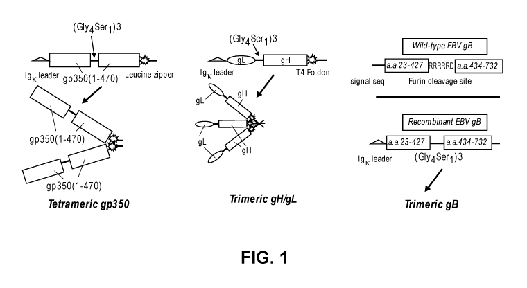
[0047] Figure 1 shows a schematic of recombinant constructs for expressing non-
limiting embodiments of multimeric EBV gp350, gH/gL, and gB. Figure 1
discloses
"(Gly4Ser1)3" as SEQ ID NO: 3, "His6" as SEQ ID NO: 49, and "RRRRRD" as SEQ ID
NO:
55.
[0048] Figures 2A-C show images of a Western blot of monomeric and multimeric
EBV gH/gL (Figure 2A), EBV gB (Figure 2B), and EBV gp350 (Figure 2C)
polypeptides.
[0049] Figure 3 shows EBV in vitro neutralization analysis of the sera from
rabbits
immunized with gp350 monomer (left panel, open circles), gp350 tetramer (left
panel, closed
circles), gB trimer (right panel), gH/gL monomer (middle panel, open circles),
and gH/gL
trimer (middle panel, closed circles).
[0050] Figures 4A-B show neutralization titers of serum from rabbits immunized
with
monomeric or tetrameric EBV gp350, monomeric or trimeric EBV gH/gL, or
trimeric EBV
gB in alum + CpG-ODN adjuvant in either Raji cells (Figure 4A) or naïve
peripheral blood
human B cells (Figure 4B).
-14-
CA 03050914 2019-07-18
WO 2018/140733
PCT/US2018/015459
[0051] Figure 5 shows EBV neutralization activity of immune sera from rabbits
immunized with trimeric EBV gB or monomeric EBV gH/gL or the synergistic
combination
of trimeric EBV gB and monomeric EBV gH/gL.
[0052] Figures 6A-B show EBV neutralization activity of pooled immune sera
from
rabbits (n=5) immunized with tetrameric EBV gp350, trimeric EBV gB, trimeric
EBV
gH/gL, or combinations thereof (Figure 6A) demonstrating synergism, or with
monomeric
EBV gp350, trimeric EBV gB, monomeric EBV gH/gL, or synergistic combinations
thereof
(Figure 6B).
[0053] Figures 7A-C show that passive transfer of immune rabbit sera prior to
EBV-
infection of humanized mice decreased EBV DNA load and increased survival rate
of
challenged mice. Figure 7A shows survival rate of mice exposed to high-dose,
live EBV
infection after passive transfer of sera from rabbits immunized with
tetrameric EBV gp350,
trimeric EBV gH/gL, trimeric EBV gB, or adjuvant alone (control). Figure 7B
shows pooled
immune sera from rabbits immunized with tetrameric EBV gp350 or trimeric EBV
gH/gL
decreased the copy number of EBV DNA in multiple organs of three humanized
mice
(geometric mean). Figure 7C shows pooled immune sera from rabbits immunized
with
tetrameric EBV gp350, trimeric EBV gH/gL or trimeric EBV gB markedly decreased
the
EBV viral load in peripheral blood (geometric mean of 3 mice) compared to the
control.
[0054] Figure 8 shows a schematic of a wild type HCMV gB polypeptide and a
recombinant construct for expressing a non-limiting embodiment of a trimeric
HCMV gB
polypeptide. Figure 8 discloses "GGGGSGGGGSGGGGS" as SEQ ID NO: 3, "His6" as
SEQ
ID NO: 49, and "RTKRS" as SEQ ID NO: 53.
[0055] Figures 9A-E show images of a Western blot of monomeric HCMV gB
(Figure 9A), trimeric HCMV gB (Figure 9B), monomeric HCMV gH/gL (Figure 9C),
trimeric HCMV gH/gL (Figure 9D), and monomeric HCMV UL128/130/131A (Figure
9E).
[0056] Figure 10 shows a schematic representing a non-limiting cloning
strategy for
expressing recombinant trimeric UL128/130/131A. Figure 10 discloses
"(Gly4Ser)3" as SEQ
ID NO: 3 and "His6" as SEQ ID NO: 49.
[0057] Figure 11 shows the serum IgG titers of anti-gH/gL antibodies (left
panel) and
anti-gB antibodies (right panel) following immunization of rabbits with
monomeric HCMV
gH/gL, trimeric HCMV gB, trimeric HCMV gB + monomeric HCMV gH/gL, or a complex
of trimeric HCMV gB + monomeric HCMV gH/gL.
[0058] Figure 12A shows in vitro HCMV neutralization titers (IC50) of non-heat
inactivated serum from rabbits immunized with monomeric HCMV gH/gL, HCMV
-15-
CA 03050914 2019-07-18
WO 2018/140733
PCT/US2018/015459
UL128/UL130/UL131A, monomeric HCMV gB (Sino gB), trimeric gB, or certain
synergistic combinations thereof using the ARPE19 epithelial cell line.
[0059] Figure 12B shows in vitro HCMV neutralization titers (IC5()) of heat-
inactivated serum from rabbits immunized with monomeric HCMV gB (Sino gB),
trimeric
HCMV gB, monomeric HCMV gH/gL, or a synergistic combination of trimeric HCMV
gB
and monomeric HCMV gH/gL using the MRC-5 fibroblast cell line.
[0060] Figure 13 shows a schematic diagram of a non-limiting DNA construct for
expression of the pentameric complex gH/gL/UL128/UL130/UL131A.
[0061] Figure 14 shows a schematic diagram of a non-limiting DNA construct for
expression of a gH/gL/g0 complex.
[0062] Figure 15 shows in vitro HCMV neutralization activity of pooled immune
sera
from rabbits immunized with monomeric HCMV gB.
[0063] Figure 16 shows in vitro HCMV neutralization activity of pooled immune
sera
from rabbits immunized with trimeric HCMV gB.
[0064] Figure 17 shows in vitro HCMV neutralization activity of pooled immune
sera
from rabbits immunized with monomeric HCMV gH/gL.
[0065] Figure 18 shows in vitro HCMV neutralization activity of in vitro
combined
immune sera from rabbits immunized with monomeric HCMV gB and monomeric HMCV
gH/gL.
[0066] Figure 19 shows in vitro HCMV neutralization activity of in vitro
combined
immune sera from rabbits immunized with trimeric HCMV gB and monomeric HMCV
gH/gL.
[0067] Figure 20 compares the in vitro HCMV neutralization activity of pooled
immune sera from rabbits immunized with individual HCMV proteins (monomeric
gB,
trimeric gB, and monomeric gH/gL) or in vitro combinations of sera from
rabbits immunized
with HCMV proteins (monomeric gB and monomeric gH/gL or trimeric gB and
monomeric
gH/gL) and shows that the combination of HCMV proteins exhibit synergy.
[0068] Figure 21A shows mouse serum titers of gB-specific IgG from mice
immunized with different amounts of HCMV trimeric gB or HCMV monomeric gB.
[0069] Figures 21B-C show neutralization titers (IC50) of heat-inactivated
serum
(Figure 21B) or non-heat inactivated-serum (Figure 21C) from mice immunized
with
monomeric HCMV gB or trimeric HCMV gB at various amounts (1 lig, 5 lig, and 25
lig) or
CytoGam IVIg at 10 mg/mL as a control (CSL Behring, King of Prussia, PA,
USA).
-16-
CA 03050914 2019-07-18
WO 2018/140733
PCT/US2018/015459
DETAILED DESCRIPTION
[0070] It is to be understood that the following detailed description is
provided to give
the reader a fuller understanding of certain embodiments, features, and
details of aspects of
the invention, and should not be interpreted as a limitation of the scope of
the invention.
Definitions
[0071] In order that the present invention may be more readily understood,
certain
terms are first defined. Additional definitions are set forth throughout the
detailed description.
[0072] The term "antibody" as used in this disclosure refers to an
immunoglobulin or
an antigen-binding fragment thereof. The term includes but is not limited to
polyclonal,
monoclonal, monospecific, polyspecific, non-specific, humanized, human, single-
chain,
chimeric, synthetic, recombinant, hybrid, mutated, grafted, and in vitro
generated antibodies.
The antibody can include a constant region, or a portion thereof, such as the
kappa, lambda,
alpha, gamma, delta, epsilon and mu constant region genes. For example, heavy
chain
constant regions of the various isotypes can be used, including: IgGi, IgG2,
IgG3, IgG4, IgM,
IgAi, IgA2, IgD, and IgE. By way of example, the light chain constant region
can be kappa or
lambda.
[0073] The terms "antigen-binding domain" and "antigen-binding fragment" refer
to a part of an antibody molecule that comprises amino acids responsible for
the specific
binding between the antibody and antigen. For certain antigens, the antigen-
binding domain
or antigen-binding fragment may only bind to a part of the antigen. The part
of the antigen
that is specifically recognized and bound by the antibody is referred to as
the "epitope" or
"antigenic determinant." Antigen-binding domains and antigen-binding fragments
include
Fab (Fragment antigen-binding); a F(ab')2 fragment, a bivalent fragment having
two Fab
fragments linked by a disulfide bridge at the hinge region; Fv fragment; a
single chain Fv
fragment (scFv) see e.g., Bird et al. (1988) Science 242:423-426; and Huston
et al. (1988)
Proc. Natl. Acad. Sci. USA 85:5879-5883); a Fd fragment having the two VH and
Cul
domains; dAb (Ward et al., (1989) Nature 341:544-546), and other antibody
fragments that
retain antigen-binding function. The Fab fragment has Vu-Cul and VL-CL domains
covalently
linked by a disulfide bond between the constant regions. The Fv fragment is
smaller and has
VH and VL domains non-covalently linked. To overcome the tendency of non-
covalently
linked domains to dissociate, a scFv can be constructed. The scFv contains a
flexible
polypeptide that links (1) the C-terminus of VH to the N-terminus of VL, or
(2) the C-terminus
of VL to the N-terminus of VH. A 15-mer (Gly4Ser)3 peptide (SEQ ID NO:3) may
be used as
-17-
CA 03050914 2019-07-18
WO 2018/140733
PCT/US2018/015459
a linker, but other linkers are known in the art. These antibody fragments are
obtained using
conventional techniques known to those with skill in the art, and the
fragments are evaluated
for function in the same manner as are intact antibodies.
[0074] As used in this application, "antigen" means a protein or fragment
thereof or a
polysaccharide linked to a protein carrier that, when expressed in an animal
or human cell or
tissue, is capable of triggering an immune response. The protein or fragment
thereof may be
glycosylated or non-glycosylated.
[0075] The term "extracellular domain" means refers to the portion of a full
length
polypeptide that extends beyond the cellular membrane and into the media in
which the cell
harboring the polypeptide resides. Polypeptides are known to generally contain
an
intracellular domain, transmembrane domain, and the remaining is the
extracellular domain
("ECD"). When the term "extracellular domain" or "ECD" is used herein, it
refers to the
amino acids of a polypeptide that in wild type form extend beyond the cellular
membrane, or
any portion thereof recognizable by an antibody. Thus, the extracellular
domain includes the
entire domain, or any number of residues amenable to recombinant expression
and inclusion
in an antigenic composition, including polypeptides representing 75%, 80%,
85%, 90%, 95%,
96%, 97%, 98%, 99%, or 100% of the entire wild type extracellular domain of a
polypeptide.
That is, the extracellular domain may be shortened, or truncated, by known
methods in the
art, to remove extraneous domains, on either the carboxy-terminus or amino-
terminus end, or
both, of the polypeptide as needed to obtain more efficient and robust
expression of the
extracellular domain of the polypeptide.
[0076] The term "full length" with respect to a given polypeptide means the
form of
the polypeptide naturally translated from the coding DNA sequence, beginning
with the ATG
start codon, which encodes the first methionine in the amino acid sequence,
and ending at the
TGA, TAG, or TTA stop codon, or whichever stop codon employed by the organism.
[0077] The term "fusion protein" refers to a protein translated from a nucleic
acid
transcript generated by combining a first nucleic acid sequence that encodes a
first protein
and at least a second nucleic acid that encodes a second protein, where the
fusion protein is
not a naturally occurring protein. The nucleic acid construct may encode two
or more
proteins that are joined in the fusion protein to create a single polypeptide
chain. The two or
more nucleic acid sequences are optionally operatively linked to a single
promoter, or
operatively linked to two or more separate promoters.
[0078] The term "glycoprotein" means a polypeptide that has covalently
attached to
it one or more carbohydrate moieties, or oligosaccharide chains. The
carbohydrate moieties
-18-
CA 03050914 2019-07-18
WO 2018/140733
PCT/US2018/015459
are normally attached to glycoproteins co-translationally or as post-
translational
modifications.
[0079] The term "isolated," when used in the context of a polypeptide or
nucleic acid
refers to a polypeptide or nucleic acid that is substantially free of its
natural environment and
is thus distinguishable from a polypeptide or nucleic acid that might happen
to occur
naturally. For instance, an isolated polypeptide or nucleic acid is
substantially free of cellular
material or other polypeptides or nucleic acids from the cell or tissue source
from which it
was derived. The term also refers to preparations where the isolated
polypeptide or nucleic
acid is sufficiently pure for pharmaceutical compositions; or at least 70-80%
(w/w) pure; or at
least 80-90% (w/w) pure; or at least 90-95% pure; or at least 95%, 96%, 97%,
98%, 99%, or
100% (w/w) pure.
[0080] The term "leader sequence" refers to a short peptide sequence at the N-
terminus of a recombinant protein that directs the recombinant protein to be
secreted from a
host cell.
[0081] The term "HHV fusion and host cell entry protein" refers to a human
herpesvirus gB polypeptide, gH polypeptide, gL polypepide, gH/gL heterodimer,
or gp350
polypeptide.
[0082] The term "HHV accessory protein" refers to a human herpes virus
polypeptide other than gB, gH, gL, gH/gL, or gp350 that are involved in
mediating viral
binding, fusion, and host cell entry including, but not limited to, gp42, gM,
gN, gI, gC, gD,
0RF68, BMRF-2, BDLF2, UL128, UL130, UL131A, and gpK8.1.
[0083] The term "immune cell" means any cell of hematopoietic lineage involved
in
regulating an immune response against an antigen (e.g., an autoantigen). In
typical
embodiments, an immune cell is a leukocyte, such as a white blood cell. Immune
cells
include neutrophils, eosinophils, basophils, lymphocytes, and/or monocytes.
Lymphocytes
include T lymphocytes and B lymphocytes. Immune cells can also be dendritic
cells, natural
killer (NK) cells, and/or a mast cell.
[0084] The term "intracellular domain" means the portion of a polypeptide that
resides in the cytoplasm of a host cell. The intracellular domain includes
that portion of the
polypeptide that is not the transmembrane domain and is not the extracellular
domain.
[0085] The term "gH/gL heterodimer" refers to a polypeptide or polypeptide
complex comprising a HHV gH polypeptide and a HHV gL polypeptide. For example,
the
heterodimer can be a non-covalently associated complex between a HHV gH
polypeptide and
a HHV gL polypeptide. Alternatively, the heterodimer can be a recombinant
fusion protein
-19-
CA 03050914 2019-07-18
WO 2018/140733
PCT/US2018/015459
comprising a HHV gH protein joined to a HHV gL protein. The HHV gH protein can
be
joined to the HHV gL protein with a peptide linker.
[0086] As used herein, the term "modified gB polypeptide," refers to a HHV gB
polypeptide in which the furin cleavage site in the extracellular domain of
the gB polypeptide
is replaced by a linker sequence, as described in WO 2015/089340.
[0087] The term "operatively linked" means that a promoter, or similar
regulatory
element, is positioned next to an expressible nucleotide sequence or coding
region such that
the transcription of that coding region is controlled and regulated by that
promoter.
[0088] The terms "polypeptide," "peptide," and "protein" are used
interchangeably
herein to refer to polymers of amino acids.
[0089] The term "peptide linker" refers to a short, non-native peptide
sequence that
links two proteins or fragments of a protein.
[0090] The term "recombinant" when used in the context of a nucleic acid means
a
nucleic acid having nucleotide sequences that are not naturally joined
together and can be
made by artificially combining two otherwise separated segments of sequence.
This artificial
combination is often accomplished by chemical synthesis or, more commonly, by
the
artificial manipulation of isolated segments of nucleic acids, for example, by
genetic
engineering techniques. Recombinant nucleic acids include nucleic acid vectors
comprising
an amplified or assembled nucleic acid, which can be used to transform or
transfect a suitable
host cell. A host cell that comprises the recombinant nucleic acid is referred
to as a
"recombinant host cell." The gene is then expressed in the recombinant host
cell to produce a
"recombinant polypeptide." A recombinant nucleic acid can also serve a non-
coding function
(for example, promoter, origin of replication, ribosome-binding site and the
like).
[0091] The term "transmembrane domain" (or "TM") means the portion of a
polypeptide that naturally and completely traverses the cell membrane, which
is a
hydrophobic phospholipid bilayer that separates the cytoplasm from the
external media in
which the host cell resides. Transmembrane domains are typically between about
20 to about
25 amino acids in length, depending on the polypeptide. The transmembrane is
typically
lipophilic and therefore typically not included in antigenic compositions
disclosed herein
because it is difficult to express, purify and solubilize.
[0092] The term "pharmaceutically acceptable carrier" or "pharmaceutically
acceptable excipient" means solvents, dispersion media, coatings,
antibacterial agents and
antifungal agents, isotonic agents, and absorption delaying agents, and the
like, that are
compatible with pharmaceutical administration. The use of such media and
agents for
-20-
CA 03050914 2019-07-18
WO 2018/140733
PCT/US2018/015459
pharmaceutically active substances is well known in the art. In certain
embodiments, the
pharmaceutically acceptable carrier or excipient is not naturally occurring.
[0093] The term "preventing" when used in the context of a disease or disease
condition means prophylactic administration of a composition that stops or
otherwise delays
the onset of a pathological hallmark or symptom of a disease or disorder.
[0094] The term "treating" when used in the context of a disease or disease
condition
means ameliorating, improving or remedying a disease, disorder, or symptom of
a disease or
condition associated with the disease, or can mean completely or partially
stopping, on a
molecular level, the biochemical basis of the disease, such as halting
replication of a virus,
etc.
[0095] The term "therapeutically effective amount" when used in the context of
an
amount of an active agent means an amount that results in an improvement or
remediation of
the disease, disorder, or symptoms of the disease or condition.
[0096] The term "passive transfer" or "passive immunotherapy" or "passive
immunity" means obtaining antibodies and/or immune cells from a subject
exposed to an
antigen and administering those antibodies and/or immune cells to a second
subject, thereby
providing the second subject with immune protection against challenge with the
antigen.
Antibodies or immune cells can be transferred in the form of blood, plasma,
purified
antibodies or immune cells, serum, etc. The second subject may be
immunocompromised
and/or naïve (never exposed to the antigen). (See, Keller et al., Clin.
Microbiol. Rev.,
13 (4): 602-614 , 2000).
[0097] Human Herpes Viruses. Herpesviridae are subdivided into three
subfamilies:
alphaherpesvirus, betaherpesvirus, and gammaherpes, based on biological
properties and
DNA genome similarities (Davison et al., Antiviral Res., 56:1-11, 2002;
MacDonald et al.,
Am. J. Cardiol., 64:359-362, 1989). (See Table 1; Willis et al., Br. Med.
Bull., 62(1):125-138,
2002). The alphaherpesviruses include HHV-1, HHV-2, VZV, and pseudorabies
virus (PRY),
and are neurotropic, i.e., they tend to infect or attack mainly the nervous
system of hosts. The
alphaherpesvirus family has the broadest host range and spread rapidly in a
cell culture.
Latent alphaherpesvirus infections are usually established in sensory neurons
and lytic
infection occurs in epidermal cells (Roizman B, Sears AE. Herpes simplex
viruses and their
replication. In: Fields BN, Knipe DM, Howley PM, eds. Fields virology.
Philadelphia:
Lippincott-Raven, 1996:2231-95).
-21-
CA 03050914 2019-07-18
WO 2018/140733
PCT/US2018/015459
TABLE 1
Germane
Sub- Site of latency
Common name Designation size (kb
family and persistence
pairs)
Herpes simplex Human herpes 1 52 Neurones
a
virus 1 virus 1 (sensory ganglia)
Herpes simplex Human herpes 152 Neurones
virus 2 virus 2 (sensory ganglia)
Varicella zoster Human herpes 125 Neurones
a
virus virus 3 (sensory ganglia)
B lymphocytes
Human herpes
Epstein-Barr virus 172 (oropharylageal
virus 4
epithelium)
Blood rnonocytes
Human Human herpes
13 235 (probably
cytomegalo virus virus 5
epithelial cells)
Human herpes Monocytes, T
170
virus 6 lymphocytes
Human herpes 145 Monocytes, T
virus 7 lymphocytes
Kaposi's sarcoma
Human herpes
associated herpes 230 Uncertain
virus 8
virus
[0098] The betaherpesvirus subfamily consists of all cytomegaloviruses
including
human cytomegalovirus (HCMV, HHV-8), HHV-6, and HHV-7 and are commonly
referred
to as the roseoloviruses. The betaherpesvirus family has a restricted host
range and a long
infection cycle. Virus latency of betaherpesvirus is maintained in secretory
glands, kidneys
and other tissues (Hendrix et al., Expert Rev. Anti Infect. Ther., 5:427-439,
2007).
[0099] The gammaherpesvirus subfamily is divided into the Lymphocryptoviruses,
which includes EBV, Rhadinovirus, and HHV-8 (KSHV). Gammaherpesviruses have a
very
narrow host range, and virus replication typically occurs in lymphoblastoid
cells but can also
lytically infect epithelial cells and fibroblasts. The latent form of
gammaherpes virus
-22-
CA 03050914 2019-07-18
WO 2018/140733
PCT/US2018/015459
infection is primarily observed in B and T lymphocytes (Ackerman, Vet.
Microbiol., 113:211-
222, 2006).
Gammaherpesvirases: Epstein Barr Virus (EBV, HHV-4), and Kaposi's Sarcoma
Virus-
Associated Herpes (KSHV, HHV-8)
[0100] Epstein Barr Virus (EBV, HHV-4). Epstein-Barr virus (EBV) is the first
human cancer virus discovered, and it is strongly implicated in the etiology
of post-transplant
lymphoproliferative disorder (PTLD) and undifferentiated nasopharyngeal
carcinoma (NPC).
In both instances, the onset and severity of disease is positively correlated
with the level of
EBV viremia, strongly suggesting a role for lytic EBV re-activation in
perpetuating disease.
Epstein Barr virus (EBV), also known as human herpesvirus 4 (HHV-4), is a
major, global
source of morbidity and mortality, responsible for such pathologic entities as
Burkitt
lymphoma, nasopharyngeal carcinoma, infectious mononucleosis, a subset of
Hodgkin's
disease, and the lymphoproliferative syndrome in immunosuppressed patients.
(Cohen JI,
Curr. Opin. Immunol., 1999 Aug;11(4):365-70; Thorley-Lawson DA, J., Allergy
Clin.
Immunol., 2005 Aug; 116(2):251-61; quiz 62; and Vetsika EK, Callan M., Expert
Rev. Mol.
Med., 2004 Nov 5;6(23):1-16). EBV has a double stranded, linear DNA genome.
The
nucleotide sequence of the EBV genome and the amino acid sequences of the
viral proteins
encoded thereby are known and set forth under the NCBI Reference Number
NC_009334,
Version NC_009334.1, GI:139424470, which sequences are hereby incorporated by
reference.
[0101] EBV is a member of the gammaherpesvirus subfamily, which is further
divided into lymphocryptoviruses, of which KSHV (HHV-8) is also a member.
Replication
for these family members typically occurs in lymphoblastoid cells, however
they can also
infect epithelial cells (e.g., nasopharyngeal epithelial cells) and
fibroblasts. Latent infection is
primarily observed in B and T lymphocytes. (Ackerman, Vet. Microbiol., 113:211-
222,
2006).
[0102] Post-Transplant Lymphoproliferative Disease (PLTD). Patients undergoing
solid organ or stem cell transplantation are at risk of developing post-
transplantation
lymphoproliferative disorder (PTLD), characterized by uncontrolled EBV-driven
B cell
proliferation that can evolve into non-Hodgkin lymphoma. (LaCasce, Oncologist,
11:674-80,
2006). PTLD may arise from EBV reactivation in seropositive recipients, or
from primary
EBV infection from the donor allograft, which poses even greater risk.
(Dhamidharka et al.,
-23-
CA 03050914 2019-07-18
WO 2018/140733
PCT/US2018/015459
Am. J. Transplant, 12:976-83, 2012). A similar phenomenon also occurs in
patients with
AIDS.
[0103] Most cases of PTLD involve excessive EBV-driven proliferation of B
cells,
with a minority (10-15%) of cases being of the NK cell/T cell type (Petrara et
al., Cancer
Lett., 369(1):37-44, 2015; and Starzl et al., Lancet, 1:583-7, 1984). The
frequency of PTLD
ranges from 1-20% depending on the type of transplant, age of recipient,
duration and type of
immunosuppres sive treatment (Ibrahim et al., Adv Hematol., 2012:230173, 2012;
and Smets
et al., Recent Results Cancer Res., 193:173-90, 2014). Younger patients, who
are EBV
seronegative, are at highest risk of developing PTLD following hematopoietic
stem cell or
solid organ transplantation, due to a lack of prior immunity. Patients with
primary
immunodeficiency syndromes are also at high risk for developing EBV-driven B
cell
lymphoproliferation and lymphoma (Rickinson et al., Trends Immunol., 35:159-
69, 2014).
The WHO defines three major histological types of PTLD of increasing severity:
early
lesions, polymorphic (P-PTLD), and monomorphic (M-PTLD) (Harris et al., Semin.
Diagn.
Pathol., 14:8-14, 1997), with the latter typically manifesting as non-Hodgkin
lymphoma.
[0104] The initial management of PTLD is a reduction in immunosuppression.
Additional therapeutic options include B cell-depleting anti-CD20 mAb
treatment, anti-viral
therapy, intravenous immunoglobulin (IVIg) and interferon (IFN)-y (LaCasce AS,
Oncologist, 11:674-80, 2006). Although IVIg in particular has been used
empirically in
combination with other therapies to treat PTLD, there have been no studies
assessing its
potential clinical benefit.
[0105] Nasopharyngeal carcinoma and EBV. The non-keratinizing variant of
squamous cell carcinoma of the nasopharynx (NPC) is endemic in east and
southeast Asia
and in parts of north and east Africa, and in 2012 accounted for 86,500 cases
of cancer
worldwide. (Chua et al., Lancet, 387(10022):1012-1024, 2016). NPC manifests
clinically as
epistaxis, unilateral nasal obstruction, auditory complaints, and cranial
nerve palsies, with
frequent metastasis to cervical lymph nodes. Radiotherapy is the primary
treatment for NPC,
with additional chemotherapy utilized for more advanced cases. (Id.). 5-year
survival is 70-
98% depending upon the stage, but NPC has a tendency to recur.
[0106] Undifferentiated NPC is invariably associated with EBV, which is
believed to
play a pathogenic role in tumor development and progression. (Tsang et al.,
Virol. Sin.,
30:107-21, 2015). Establishment of latent EBV infection in pre-malignant
nasopharyngeal
epithelial cells appears to drive further malignant transformation. Rising
levels of serum IgA
specific for EBV lytic antigens such as viral capsid antigen and early antigen
correlate with
-24-
CA 03050914 2019-07-18
WO 2018/140733
PCT/US2018/015459
progression to NPC. (Ji et al., Br. J. Cancer, 96:623-30, 2007). The level of
plasma EBV
DNA is directly correlated with NPC tumor burden. (To et al., Clin. Cancer
Res., 9:3254-9,
2003). Thus, latent EBV reactivation is a key feature of NPC formation and
progression,
suggesting a possible role for antibody-based immunotherapy. Although multiple
strains of
EBV can be isolated from the blood and saliva of healthy seropositive
individuals, only a
single strain of EBV is typically isolated from NPC cells, consistent with its
pathogenic role.
(Tsang et al., Virol. Sin., 30:107-21, 2015). Although strain variations in
the sequences of
EBNA2, 3A, 3B, and 3C have been described, the envelope proteins gp350, gH/gL,
and gB
are highly conserved, making these latter proteins ideal vaccine candidates
for cross-strain
protection. (Sample et al., J. Virol., 64:4084-92, 1990; and Rowe et al., J.
Virol., 63:1031-9,
1989).
[0107] Circulating EBV DNA copy number is positively correlated with imminent
onset of EBV-associated malignancies and clinical severity. EBV qPCR assays
are
commonly used post-transplantation. (Meerbach et al., J. Med. Virol., 80:441-
54, 2008; Tsai
et al., Am. J. Transplant, 8:1016-24, 2008; Wagner et al., Transplantation,
74:656-64, 2002;
and van Esser et al., Blood 98:972-8, 2001). Elevated EBV DNA in the blood is
associated
with an increased risk for PTLD, whereas decreases correlate with treatment
success.
(Baldanti et al., J. Clin. MicrobioL, 38:613-9, 2000; Hakim et al., J. Clin.
Microbiol.,
45:2151-5, 2007; Wagner et al., Transplantation, 72:1012-9, 2001; and Clave et
al.,
Transplantation, 77:76-84, 2004). Circulating EBV DNA is also positively
correlated with
adverse survival outcomes in NPC (Jin et al., Eur. J. Cancer, 48:882-8, 2012;
Hsu et al.,
Head Neck, 34:1064-70, 2012; and Hsu et al., Oral Oncol., 49:620-5, 2013), as
well as
Hodgkin (Kanakry et al., Blood, 121:3547-53, 2013) and extranodal NK/T cell
lymphomas,
which also linked pathogenically with EBV (Wang et al., Oncotarget.,
6(30):30317-30326,
2015).
[0108] In the developing world, EBV seroconversion typically occurs in
infancy,
whereas in developed countries it is more likely contracted in adolescence.
Infectious
mononucleosis typically occurs only in this latter group (Vetsika et al.,
Expert Rev. Mol.
Med., 2004 Nov 5; 6(23):1-16). The major human reservoir for latent EBV and
EBV
transmission is the resting memory B lymphocyte (Babcock et al., Immunity,
1998 Sep;
9(3):395-404). EBV is dependent upon the gp350-CD21 binding event for viral
entry into the
B cell (Tanner et al., Cell, 1987 Jul 17; 50(2):203-13; and Tanner et al., J.
Virology, 1988;
62(12):4452-64), an event that is critical for infectivity and B cell
neoplastic transformation
(Thorley-Lawson DA, J. Allergy Clin. Immunol., 2005 Aug; 116(2):251-61; quiz
62). Gp350
-25-
CA 03050914 2019-07-18
WO 2018/140733
PCT/US2018/015459
is the major EBV outer membrane glycoprotein, while CD21, also known as
complement
receptor type 2 (CR2), is a receptor on the surface of B cells that binds to
iC3b complement
protein. Sera from patients with active EBV infection contain antibody that
prevent EBV
entry into B cells ("neutralizing" antibody). Adsorption of these sera with
gp350, eliminates
most of this neutralizing activity (Thorley-Lawson et al., J. Virology, 1982
Aug; 43(2):730-
6), indicating that gp350 serves as the major EBV antigen to which a
protective humoral
immune response is directed.
[0109] A number of studies have demonstrated that immunization of non-human
primates with a subunit gp350 vaccine in adjuvant protects against
experimental EBV-
induced lymphoma or EBV replication. Thus, purified native gp350, injected
into cottontop
marmosets (CTM), in association with liposomes, ISCOM's, or muramyl dipeptide,
protected
against EBV-induced lymphoma. (Morgan et al., J. Med. Virol., 1984;13(3):281-
92; and
Morgan et al., J. Med. Virol., 1989 Sep; 29(1):74-8). Recombinant gp350 in
alum or muramyl
dipeptide was similarly protective. (Finerty et al., J. Gen. Virol., 1992 Feb;
73 (Pt 2):449-53;
and Finerty et al., Vaccine, 1994 Oct; 12(13):1180-4). Common marmosets also
showed
decreased viral replication after EBV challenge following immunization with
recombinant
gp350 in alum. (Cox et al., J. Med. Virol., 1998 Aug; 55(4):255-61). Non-human
primate
studies using gp350 expressed by adenoviral or vaccinia viral vectors have
similarly shown
protection against experimental EBV-induced lymphoma or EBV replication in CTM
or
common marmosets. (Mackett et al., J. Med. Virol., 1996 Nov; 50(3):263-71;
Ragot et al., J.
Gen. Virol., 1993 Mar; 74 (Pt 3):501-7; and Morgan et al., J. Med. Virol.,
1988 Jun;
25(2): 189-95).
[0110] A pilot study in humans has also suggested a potential role for gp350
vaccination in host protection against EBV. In a study by Gu et al. (Dev.
Biol. Stand., 1995;
84:171-7) a single dose of gp350/220 expressed by vaccinia virus (VV) was
given by
scarification to 1- to 3-year-olds who were EBV-seronegative, and VV-
seronegative. These
children developed neutralizing antibodies to EBV (1:40-1:160). Whereas 10/10
unvaccinated controls became infected at 16 months of follow-up, only 3/9
vaccinated
children became infected at this time. More recently, Phase I/II studies were
conducted in
which healthy EBV-seronegative adults were immunized with a recombinant
monomeric
gp350 protein in alum +/- monophosphoryl lipid A. (Sokal et al., J. Infect.
Dis., 2007 Dec 15;
196(12):1749-53; and Moutschen et al., Vaccine, 2007 Jun 11; 25(24):4697-705).
Following
3 doses, up to 82% of subjects had detectable neutralizing serum anti-gp350
antibody titers.
The vaccine demonstrated an efficacy of 78.0% in preventing the development of
infectious
-26-
CA 03050914 2019-07-18
WO 2018/140733
PCT/US2018/015459
mononucleosis but not in preventing asymptomatic EBV infection. Finally, an
additional
phase I trial of recombinant monomeric gp350 protein in alum given to children
with chronic
kidney disease demonstrated only a minority of subjects developing detectable
neutralizing
serum anti-gp350 titers. (Rees et al., Transplantation, 2009 Oct 27;
88(8):1025-9).
[0111] There is currently no effective immunotherapy for EBV-associated
diseases,
or a clinically licensed prophylactic EBV vaccine. EBV gp350, gH/gL complex,
and gB are
three envelope proteins that represent potential vaccine target antigens for
EBV. EBV gp350
mediates EBV attachment to B cells through its binding to CD21. EBV gH/gL and
gB are
involved in mediating EBV fusion and entry into both B cells and epithelial
cells.
[0112] EBV gp350/gp220. The EBV glycoprotein gp350 and the related splice
variant gp220 are responsible for attachment of EBV with high affinity to CR2
on B cells.
Antibodies to gp350 or gp220 that block EBV binding neutralize B-cell
infection. Each of
gp350 and gp220 is a highly glycosylated single-pass membrane protein. As a
result of
alternative splicing, the viral glycoprotein appears in two forms, with
approximate masses of
350 and 220 kDa. The 200 kDa splice form lacks residues 500-757 of the full
length gp350.
Both gp350 and gp220 retain the CR2 binding domain at the amino terminus. A
truncated
version of gp350 or gp220 having amino acids 1-470 of gp350 retains the
ability to bind CR2
and can inhibit the binding of EBV to CR2 and can be substituted for full
length gp350 or
gp200 in the compositions described herein or for extracellular domain forms
of gp350.
(Sarrias et al., J. Immunol., 2001 Aug 1;167(3):1490-9). In addition, portions
of the gp350
and gp220 protein between amino acids 21-26 or between amino acids 372-378 of
the gp350
sequence have been linked to CR2 binding. (Tanner et al., Cell, 203-213
(1987), and
Nemerow et al., Cell, 61:1416-20, 1987). Thus, the term gp350 protein or gp350
antigen (or
gp220 protein or antigen) refers to the full length gp350 or gp220 proteins as
well as
fragments or modified versions thereof that retain the ability to bind the
CR2.
[0113] The amino acid and nucleic acid sequence of gp350, set forth in GenBank
under Accession Number M10593, Version M10593.1, GI 330360, is hereby
incorporated by
reference. The amino acid sequence of gp350 is (SEQ ID NO: 1):
MEAALLVCQY TIQSLIHLTG EDPGFFNVEI PEFPFYPTCN VCTADVNVTI 50
NFDVGGKKHQ LDLDFGQLTP HTKAVYQPRG AFGGSENATN LFLLELLGAG 100
ELALTMRSKK LPINVTTGEE QQVSLESVDV YFQDVFGTMW CHHAEMQNPV 150
YLIPETVPYI KWDNCNSTNI TAVVRAQGLD VTLPLSLPTS AQDSNFSVKT 200
EMLGNEIDIE CIMEDGEISQ VLPGDNKFNI TCSGYESHVP SGGILTSTSP 250
VATPIPGTGY AYSLRLTPRP VSRFLGNNSI LYVFYSGNGP KASGGDYCIQ 300
SNIVFSDEIP ASQDMPTNTT DITYVGDNAT YSVPMVTSED ANSPNVTVTA 350
-27-
CA 03050914 2019-07-18
WO 2018/140733
PCT/US2018/015459
FWAWPNNTET DFKCKWTLTS GTPSGCENIS GAFASNRTFD ITVSGLGTAP 400
KTLIITRTAT NATTTTHKVI FSKAPESTTT SPTLNTTGFA DPNTTTGLPS 450
STHVPTNLTA PASTGPTVST ADVTSPTPAG TTSGASPVTP SPSPWDNGTE 500
SKAPDMTSST SPVTTPTPNA TSPTPAVTTP TPNATSPTPA VTTPTPNATS 550
PTLGKTSPTS AVTTPTPNAT SPTLGKTSPT SAVTTPTPNA TSPTLGKTSP 600
TSAVTTPTPN ATGPTVGETS PQANATNHTL GGTSPTPVVT SQPKNATSAV 650
TTGQHNITSS STSSMSLRPS SNPETLSPST SDNSTSHMPL LTSAHPTGGE 700
NITQVTPASI STHHVSTSSP EPRPGTTSQA SGPGNSSTST KPGEVNVTKG 750
TPPQNATSPQ APSGQKTAVP TVTSTGGKAN STTGGKHTTG HGARTSTEPT 800
TDYGGDSTTP RPRYNATTYL PPSTSSKLRP RWTFTSPPVT TAQATVPVPP 850
TSQPRFSNLS MLVLQWASLA VLTLLLLLVM ADCAFRRNLS TSHTYTTPPY 900
DDAETYV 907
[0114] The amino acid sequence of gp220, set forth in GenBank under Accession
Number M10593, Version M10593.1, GI 330360, and hereby incorporated by
reference, is
(SEQ ID NO: 2):
MEAALLVCQY TIQSLIHLTG EDPGFFNVEI PEFPFYPTCN VCTADVNVTI 50
NFDVGGKKHQ LDLDFGQLTP HTKAVYQPRG AFGGSENATN LFLLELLGAG 100
ELALTMRSKK LPINVTTGEE QQVSLESVDV YFQDVFGTMW CHHAEMQNPV 150
YLIPETVPYI KWDNCNSTNI TAVVRAQGLD VTLPLSLPTS AQDSNFSVKT 200
EMLGNEIDIE CIMEDGEISQ VLPGDNKFNI TCSGYESHVP SGGILTSTSP 250
VATPIPGTGY AYSLRLTPRP VSRFLGNNSI LYVFYSGNGP KASGGDYCIQ 300
SNIVFSDEIP ASQDMPTNTT DITYVGDNAT YSVPMVTSED ANSPNVTVTA 350
FWAWPNNTET DFKCKWTLTS GTPSGCENIS GAFASNRTFD ITVSGLGTAP 400
KTLIITRTAT NATTTTHKVI FSKAPESTTT SPTLNTTGFA DPNTTTGLPS 450
STHVPTNLTA PASTGPTVST ADVTSPTPAG TTSGASPVTP SPSPWDNGTE 500
STPPQNATSP QAPSGQKTAV PTVTSTGGKA NSTTGGKHTT GHGARTSTEP 550
TTDYGGDSTT PRPRYNATTY LPPSTSSKLR PRWTFTSPPV TTAQATVPVP 600
PTSQPRFSNL SMLVLQWASL AVLTLLLLLV MADCAFRRNL STSHTYTTPP 650
YDDAETYV 658
[0115] EBV gH, gL, gB, and gp42. The minimal requirement for viral fusion with
B
cells includes EBV glycoproteins gH, gL, gB, and gp42. For infection of B
cells, gp42 binds
to the host cell MHC class II molecules to trigger viral cell membrane fusion.
On the other
hand, for infection of epithelial cells, gp42 is not required. Rather, the EBV
gH, gL, and gB
proteins are sufficient for viral fusion with epithelial cells. EBV gH/gL
exists in certain
environments as a noncovalently associated complex.
[0116] The amino acid sequence of EBV gH is (SEQ ID NO: 4):
MQLLCVFCLV LLWEVGAASL SEVKLHLDIE GHASHYTIPW TELMAKVPGL 50
-28-
CA 03050914 2019-07-18
WO 2018/140733
PCT/US2018/015459
SPEALWREAN VTEDLASMLN RYKLIYKTSG TLGIALAEPV DIPAVSEGSM 100
QVDASKVHPG VISGLNSPAC MLSAPLEKQL FYYIGTMLPN TRPHSYVFYQ 150
LRCHLSYVAL SINGDKFQYT GAMTSKFLMG TYKRVTEKGD EHVLSLIFGK 200
TKDLPDLRGP FSYPSLTSAQ SGDYSLVIVT TFVHYANFHN YFVPNLKDMF 250
SRAVTMTAAS YARYVLQKLV LLEMKGGCRE PELDTETLTT MFEVSVAFFK 300
VGHAVGETGN GCVDLRWLAK SFFELTVLKD IIGICYGATV KGMQSYGLER 350
LAAVLMATVK MEELGHLTTE KQEYALRLAT VGYPKAGVYS GLIGGATSVL 400
LSAYNRHPLF QPLHTVMRET LFIGSHVVLR ELRLNVTTQG PNLALYQLLS 450
TALCSALEIG EVLRGLALGT ESGLFSPCYL SLRFDLTRDK LLSMAPQEAM 500
LDQAAVSNAV DGFLGRLSLE REDRDAWHLP AYKCVDRLDK VLMIIPLINV 550
TFIISSDREV RGSALYEAST TYLSSSLFLS PVIMNKCSQG AVAGEPRQIP 600
KIQNFTRTQK SCIFCGFALL SYDEKEGLET TTYITSQEVQ NSILSSNYFD 650
FDNLHVHYLL LTTNGTVMEI AGLYEERAHV VLAIILYFIA FALGIFLVHK 700
IVMFFL 706
[0117] The amino acid sequence of EBV gL is(SEQ ID NO: 5):
MRTVGVFLAT CLVTIFVLPT WGNWAYPCCH VTQLRAQHLL ALENISDIYL 50
VSNQTCDGFS LASLNSPKNG SNQLVISRCA NGLNVVSFFI SILKRSSSAL 100
TGHLRELLTT LETLYGSFSV EDLFGANLNR YAWHRGG 137
[0118] The amino acid sequence of EBV gB is (SEQ ID NO: 6):
MTRRRVLSVV VLLAALACRL GAQTPEQPAP PATTVQPTAT RQQ1SFPFRV 50
CELSSHGDLF RFSSDIQCPS FGTRENHTEG LLMVFKDNII PYSFKVRSYT 100
KIVTNILIYN GWYADSVTNR HEEKFSVDSY ETDQMDTIYQ CYNAVKMTKD 150
GLTRVYVDRD GVNITVNLKP TGGLANGVRR YASQTELYDA PGWLIWTYRT 200
RTTVNCLITD MMAKSNSPFD FFVTTTGQTV EMSPFYDGKN KETFHERADS 250
FHVRTNYKIV DYDNRGTNPQ GERRAFLDKG TYTLSWKLEN RTAYCPLQHW 300
QTFDSTIATE TGKSIHFVTD EGTSSFVTNT TVGIELPDAF KCIEEQVNKT 350
MHEKYEAVQD RYTKGQEAIT YFITSGGLLL AWLPLTPRSL ATVKNLTELT 400
TPTSSPPSSP SPPAPPAARG STSAAVLRRR RRDAGNATTP VPPAAPGKSL 450
GTLNNPATVQ IQFAYDSLRR QINRMLGDLA RAWCLEQKRQ NMVLRELTKI 500
NPTTVMSSIY GKAVAAKRLG DVISVSQCVP VNQATVTLRK SMRVPGSETM 550
CYSRPLVSFS FINDTKTYEG QLGTDNEIFL TKKMTEVCQA TSQYYFQSGN 600
EIHVYNDYHH FKTIELDGIA TLQTFISLNT SLIENIDFAS LELYSRDEQR 650
ASNVFDLEGI FREYNFQAQN IAGLRKDLDN AVSNGRNQFV DGLGELMDSL 700
GSVGQSITNL VSTVGGLFSS LVSGFISFFK NPFGGMLILV LVAGVVILVI 750
SLTRRTRQMS QQPVQMLYPG IDELAQQHAS GEGPGINPIS KTELQAIMLA 800
LHEQNQEQKR AAQRAAGPSV ASRALQAARD RFPGLRRRRY HDPETAAALL 850
GEAETEF 857
-29-
CA 03050914 2019-07-18
WO 2018/140733
PCT/US2018/015459
[0119] The amino acid sequence of EBV gp42 is (SEQ ID NO: 7):
MVSFKQVRVP LFTAIALVIV LLLAYFLPPR VRGGGRVSAA AITWVPKPNV 50
EVWPVDPPPP VNFNKTAEQE YGDKEIKLPH WTPTLHTFQV PKNYTKANCT 100
YCNTREYTFS YKERCFYFTK KKHTWNGCFQ ACAELYPCTY FYGPTPDILP 150
VVTRNLNAIE SLWVGVYRVG EGNWTSLDGG TFKVYQIFGS HCTYVSKFST 200
VPVSHHECSF LKPCLCVSQR SNS 223
[0120] The amino acid sequence of EBV BMRF-2 is (SEQ ID NO: 8):
MFSCKQHLSL GACVFCLGLL ASTPFIWCFV FANLLSLEIF SPWQTHVYRL 50
GFPTACLMAV LWTLVPAKHA VRAVTPAIML NIASALIFFS LRVYSTSTWV 100
SAPCLFLANL PLLCLWPRLA IEIVYICPAI HQRFFELGLL LACTIFALSV 150
VSRALEVSAV FMSPFFIFLA LGSGSLAGAR RNQIYTSGLE RRRSIFCARG 200
DHSVASLKET LHKCPWDLLA ISALTVLVVC VMIVLHVHAE VFFGLSRYLP 250
LFLCGAMASG GLYLGHSSII ACVMATLCTL TSVVVYFLHE TLGPLGKTVL 300
FISIFVYYFS GVAALSAAMR YKLKKFVNGP LVHLRVVYMC CFVFTFCEYL 350
LVTFIKS
[0121] The amino acid sequence of EBV BDLF2 is (SEQ ID NO: 9):
MVDEQVAVEH GTVSHTISRE EDGVVHERRV LASGERVEVF YKAPAPRPRE 50
GRASTFHDFT VPAAAAVPGP EPEPEPHPPM PIHANGGGET KTNTQDQNQN 100
QTTRTRTNAK AEERTAEMDD TMASSGGQRG APISADLLSL SSLTGRMAAM 150
APSWMKSEVC GERMRFKEDV YDGEAETLAE PPRCFMLSFV FIYYCCYLAF 200
LALLAFGFNP LFLPSFMPVG AKVLRGKGRD FGVPLSYGCP TNPFCKVYTL 250
IPAVVINNVT YYPNNTDSHG GHGGFEAAAL HVAALFESGC PNLQAVTNRN 300
RTFNVTRASG RVERRLVQDM QRVLASAVVV MHHHCHYETY YVFDGVGPEF 350
GTIPTPCFKD VLAFRPSLVT NCTAPLKTSV KGPNWSGAAG GMKRKQCRVD 400
RLTDRSFPAY LEEVMYVMVQ
[0122] The antigenic compositions and methods of this application typically
involve
two or more HHV proteins involved in mediating HHV binding, fusion, and entry
into host
cells. In certain embodiments, two or more EBV proteins disclosed herein are
combined in an
antigenic composition. The two or more EBV proteins can be administered
simultaneously or
separately to induce an immune response or to treat or prevent an EBV
infection in a subject.
In certain embodiments, the antigenic composition (or method of
administration) comprises
two or more of the following EBV polypeptides (or nucleic acids encoding the
same): gB,
gH, gL, and gp350. In some embodiments, the gB polypeptide is monomeric,
dimeric, or
trimeric. In some embodiments, the gH and gL polypeptides are monomeric,
dimeric,
-30-
CA 03050914 2019-07-18
WO 2018/140733
PCT/US2018/015459
trimeric, or tetrameric. Typically, gH and gL form a gH/gL heterodimer. In
some
embodiments, the gp350 polypeptides are monomeric, dimeric, trimeric, or
tetrameric.
[0123] In certain embodiments, the two or more EBV proteins (or nucleic acids
encoding the same) comprise a monomeric or multimeric gp350 and monomeric or
multimeric gB. In certain embodiments, the gp350 is monomeric or tetrameric
and the gB is
monomeric or trimeric. In certain embodiments, the gp350 is monomeric and the
gB is
trimeric. In certain embodiments, the gp350 is tetrameric and the gB is
trimeric.
[0124] In certain embodiments, the two or more EBV proteins (or nucleic acids
encoding the same) comprise a monomeric or multimeric gp350 and a monomeric or
multimeric gH/gL heterodimer. In certain embodiments, the gp350 is monomeric
or
tetrameric and the gH/gL heterodimer is monomeric or trimeric. In certain
embodiments, the
gp350 is monomeric and the gH/gL heterodimer is monomeric. In certain
embodiments, the
gp350 is tetrameric and the gH/gL heterodimer is trimeric.
[0125] In certain embodiments, the two or more EBV proteins (or nucleic acids
encoding the same) comprise a monomeric or multimeric gB and a monomeric or
multimeric
gH/gL heterodimer. In certain embodiments, the gB is monomeric, dimeric or
trimeric and
the gH/gL heterodimer is monomeric or trimeric. In certain embodiments, the gB
is
monomeric and the gH/gL heterodimer is monomeric or trimeric. In certain
embodiments, the
gB is trimeric and the gH/gL heterodimer is monomeric. In certain embodiments,
the gB is
trimeric and the gH/gL heterodimer is trimeric. In certain embodiments, the
EBV gB, gH,
and gL polypeptides form a protein complex when mixed together. In certain
embodiments,
the EBV gB, gH, and gL polypeptides are not administered as a protein complex
comprising
the gB, gH, and gL polypeptides. For example, the gB can be administered
separately from
the gH and/or gL or administered with the gH and gL but not as a protein
complex.
[0126] In certain embodiments, the two or more EBV proteins (or nucleic acids
encoding the same) comprise a monomeric or multimeric gp350, a monomeric or
multimeric
gB and a monomeric or multimeric gH/gL heterodimer. In certain embodiments,
the gp350 is
monomeric or tetrameric, the gB is monomeric or trimeric and the gH/gL
heterodimer is
monomeric or trimeric. In certain embodiments, the gp350 is monomeric, the gB
is trimeric
and the gH/gL heterodimer is monomeric. In certain embodiments, the gp350 is
tetrameric,
the gB is trimeric and the gH/gL heterodimer is trimeric.
[0127] In some embodiments, the two or more EBV proteins further comprises one
or
more of a BMRF-2 polypeptide, a BDLF2 polypeptide, and/or a gp42 polypeptide,
which can
be monomeric or multimeric (e.g., dimeric, trimeric, or tetrameric).
-31-
CA 03050914 2019-07-18
WO 2018/140733
PCT/US2018/015459
[0128] Kaposi's Sarcoma Virus-Associated Herpes (KSHV, HHV-8). The two
human gammaherpesviruses, Epstein¨Barr virus (EBV), a gamma 1
lymphocryptovirus, and
Kaposi's sarcoma associated virus (KSHV), a gamma 2 rhadinovirus, have many
features in
common. They share an architecture that is typical of all members of the
herpesvirus family,
they share an ability to establish latency in lymphocytes, and they are both
initiators or
potentiators of human tumors. (Chandran et al., Human Herpesviruses: Biology,
Therapy, and
Imunoprophylaxis, Eds. Arvin, A., Campadelli-Fiume, G., and Mocarski E., et
al., Cambridge
University Press, 2007, Ch. 23). KSHV broadly infects many types of host
cells, including B-
cells from the peripheral blood, B-cells in primary effusion lymphomas (PEL)
or body-cavity
based B-cell lymphomas (BCBL) and multicentric Cattleman's disease (MCD), flat
endothelial cells lining the vascular spaces of Kaposi's sarcoma (KS) lesions,
typical KS
spindle cells, CD 45+/CD68+ monocytes in KS lesions, keratinocytes, and
epithelial cells.
(Id.). Further, KSHV infection has been associated with multiple myeloma.
(Rettig et al.,
Science, 276:1851-4, 1997). Like EBV, KSHV also expresses gB, gH, and gL that
mediate
cell fusion and entry. KSHV also expresses the conserved glycoproteins, gM and
gN, which
mediate similar, if not identical, roles as compared to their EBV
counterparts. (Id.).
[0129] However, the gp350 glycoprotein of EBV is replaced in KSHV with a
polypeptide termed K8.1. The K8.1 gene encodes a 197-amino acid with a
predicted
molecular weight of about 22 kDa and possessing no sequence corresponding to a
TM
domain. Similar to the EBV gp350/220, the KSHV K8.1 gene encodes two ORF s,
designated
gpK8.1A and gpK8.1B, from spliced messages. The larger cDNA is 752 bp long
(76,214 ¨
76,941 bp) and utilizes the polyadenylation signal sequence (AATAAA) at
position 77 013
bp. The 228-aa long encoded protein is designated gpK8.1A, which contains a
signal
sequence, transmembrane domain, and four N-glycosylation sites. Otherwise, the
KSHV
gpK18.1 polypeptide performs similar functions as reported for EBV gp350,
forming a
complex with gB and binding to a cell surface heparin sulfate molecule on the
host cell.
[0130] KSHV 0RF68 is a late lytic, delayed early structural and assembly gene
encoding a transmembrane glycoprotein that is a component of the KSHV
envelope.
(Nakamura et al., J. Virol., 77(7):4205-20, 2003; and Jha et al., mBio,
5(6):e02261-14, 2014;
and Stilrzl et al., Thromb. Haemost., 102:1117-34, 2009). 0RF68 is known to
interact with
and inhibit the host cell's ubiquitin proteasome pathway, thereby inhibiting
protein
degradation. (Gardner, M., 8th Annual CEND Symposium, 22 March 2016). 0RF68 is
essential for viral genome replication in KSHV. It is postulated that KSHV
0RF68 encodes a
-32-
CA 03050914 2019-07-18
WO 2018/140733
PCT/US2018/015459
protein that suppresses the proteasome-mediated degradation of a protein in
the cytoplasm of
the host cell that is essential for KSHV DNA replication. (Id.).
[0131] The antigenic compositions and methods of this application typically
involve
two or more HHV proteins involved in mediating HHV binding, fusion, and entry
into host
cells. In certain embodiments, two or more KSHV proteins disclosed herein are
combined in
an antigenic composition. The two or more KSHV proteins can be administered
simultaneously or separately to induce an immune response or to treat or
prevent a KSHV
infection in a subject. In certain embodiments, the antigenic composition (or
method of
administration) comprises two or more of the following KSHV polypeptides (or
nucleic acids
encoding the same): gB, gH, and gL. In some embodiments, the gB polypeptide is
monomeric, dimeric, or trimeric. In some embodiments, the gH and gL
polypeptides are
monomeric, dimeric, trimeric, or tetrameric. Typically, gH and gL form a gH/gL
heterodimer.
[0132] In certain embodiments, the two or more KSHV proteins (or nucleic acids
encoding the same) comprise a monomeric or multimeric gB and a monomeric or
multimeric
gH/gL heterodimer. In certain embodiments, the gB is monomeric, dimeric or
trimeric and
the gH/gL heterodimer is monomeric or trimeric. In certain embodiments, the gB
is
monomeric and the gH/gL heterodimer is monomeric or trimeric. In certain
embodiments, the
gB is trimeric and the gH/gL heterodimer is monomeric. In certain embodiments,
the gB is
trimeric and the gH/gL heterodimer is trimeric. In certain embodiments, the
KSHV gB, gH,
and gL polypeptides form a protein complex when mixed together. In certain
embodiments,
the KSHV gB, gH, and gL polypeptides are not administered as a protein complex
comprising the gB, gH, and gL polypeptides. For example, the gB can be
administered
separately from the gH and/or gL or administered with the gH and gL but not as
a protein
complex.
[0133] In certain embodiments, the two or more KSHV proteins further comprises
one or more of the gN polypeptide, the gM polypeptide, the 0RF68 polypeptide
and/or the
gpK8.1 polypeptide, which can be monomeric or multimeric (e.g., dimeric,
trimeric, or
tetrameric).
[0134] The amino acid and nucleic acid sequence of KSHV gpK8.1A, set forth in
GenBank under Accession Number AAC63270.1, GI 3414867, is hereby incorporated
by
reference. The amino acid sequence of gpK8.1 is (SEQ ID NO: 10):
1 MSSTQIRTEI PVALLILCLC LVACHANCPT YRSHLGFWQE GWSGQVYQDW LGRMNCSYEN
61 MTALEAVSLN GTRLAAGSPS SEYPNVSVSV EDTSASGSGE DAIDESGSGE EERPVESHVT
121 FMTQSVQATT ELTDALISAF SGSYSSGEPS RTTRIRVSPV AENGRNSGAS NRVPFSATTT
-33-
CA 03050914 2019-07-18
WO 2018/140733
PCT/US2018/015459
181 TTRGRDAHYN AEIRTHLYIL WAVGLLLGLV LILYLCVPRC RRKKPYIV
[0135] The amino acid and nucleic acid sequence of KSHV gpK8.1B, set forth in
GenBank under Accession Number AJE29698.1, GI 748016404, and hereby
incorporated by
reference. The amino acid sequence of gpK8.1B is (SEQ ID NO: 11):
1 MSSTQIRTEI PVALLILCLC LVACHANCPT YRSHLGFWQE GWSGQVYQDW LGRMNCSYEN
61 MTALEAVSLN GTRLAAGSPS RSYSSGEPSR TTRIRVSPVA ENGRNSGASN RVPFSATTTT
121 TRGRDAHYNA EIRTHLYILW AVGLLLGLVL ILYLCVPRCR RKKPYIV
[0136] The amino acid sequence of KSHV gH is (SEQ ID NO: 12):
MQGLAFLAAL ACWRCISLTC GATGALPTTA TTITRSATQL INGRTNLSIE 50
LEFNGTSFFL NWQNLLNVIT EPALTELWTS AEVAEDLRVT LKKRQSLFFP 100
NKTVVISGDG HRYTCEVPTS SQTYNITKGF NYSALPGHLG GFGINARLVL 150
GDIFASKWSL FARDTPEYRV FYPMNVMAVK FSISIGNNES GVALYGVVSE 200
DFVVVTLHNR SKEANETASH LLFGLPDSLP SLKGHATYDE LTFARNAKYA 250
LVAILPKDSY QTLLTENYTR IFLNMTESTP LEFTRTIQTR IVSIEARRAC 300
AAQEAAPDIF LVLFQMLVAH FLVARGIAEH RFVEVDCVCR QYAELYFLRR 350
ISRLCMPTFT TVGYNHTTLG AVAATQIARV SATKLASLPR SSQETVLAMV 400
QLGARDGAVP SSILEGIAMV VEHMYTAYTY VYTLGDTERK LMLDIHTVLT 450
DSCPPKDSGV SEKLLRTYLM FTSMCTNIEL GEMIARFSKP DSLNIYRAFS 500
PCFLGLRYDL HPAKLRAEAP QSSALTRTAV ARGTSGFAEL LHALHLDSLN 550
LIPAINCSKI TADKIIATVP LPHVTYIISS EALSNAVVYE VSEIFLKSAM 600
FISAIKPDCS GFNFSQIDRH IPIVYNISTP RRGCPLCDSV IMSYDESDGL 650
QSLMYVTNER VQTNLFLDKS PFFDNNNLHI HYLWLRDNGT VVEIRGMYRR 700
RAASALFLIL SFIGFSGVIY FLYRLFSILY
[0137] The amino acid sequence of KSHV gL is (SEQ ID NO: 13):
MGIFALFAVL WTILLVTSHA YVALPCCAIQ ASAASTLPLF FAVHSIHFAD 50
PNHCNGVCIA KLRSKTGDIT VETCVNGFNL RSFLVAVVRR LGSWASQENL 100
RLLWYLQRSL TAYTVGFNAT TADSSIHNVN IIIISVGKAM NRTGSVSGSQ 150
TRAKSSSRRA HAGQKGK
[0138] The amino acid sequence of KSHV gB is (SEQ ID NO: 12):
MQGLAFLAAL ACWRCISLIC GATGALPIIA IIIIRSATQL INGRTNLSIE 50
LEFNGTSFFL NWQNLLNVIT EPALTELWTS AEVAEDLRVT LKKRQSLFFP 100
NKTVVISGDG HRYTCEVPTS SQTYNITKGF NYSALPGHLG GFGINARLVL 150
GDIFASKWSL FARDTPEYRV FYPMNVMAVK FSISIGNNES GVALYGVVSE 200
DFVVVTLHNR SKEANETASH LLFGLPDSLP SLKGHATYDE LTFARNAKYA 250
LVAILPKDSY QTLLTENYTR IFLNMTESTP LEFTRTIQTR IVSIEARRAC 300
-34-
CA 03050914 2019-07-18
WO 2018/140733
PCT/US2018/015459
AAQEAAPDIF LVLFQMLVAH FLVARGIAER RFVEVDCVCR QYAELYFLRR 350
ISRLCMPTFT TVGYNHTTLG AVAATQIARV SATKLASLPR SSQETVLAMV 400
QLGARDGAVP SSILEGIAMV VEHMYTAYTY VYTLGDTERK LMLDIHTVLT 450
DSCPPKDSGV SEKLLRTYLM FTSMCTNIEL GEMIARFSKP DSLNIYRAFS 500
PCFLGLRYDL HPAKLRAEAP QSSALTRTAV ARGTSGFAEL LHALHLDSLN 550
LIPAINCSKI TADKIIATVP LPHVTYIISS EALSNAVVYE VSEIFLKSAM 600
FISAIKPDCS GFNFSQIDRH IPIVYNISTP RRGCPLCDSV IMSYDESDGL 650
QSLMYVTNER VQTNLFLDKS PFFDNNNLHI HYLWLRDNGT VVEIRGMYRR 700
RAASALFLIL SFIGFSGVIY FLYRLFSILY
[0139] The amino acid sequence of KSHV gN is (SEQ ID NO: 14):
MTASTVALAL FVASILGHCW VTANSTGVAS STERSSPSTA GLSARPSPGP 50
TSVTTPGFYD VACSADSFSP SLSSFSSVWA LINALLVVVA TFFYLVYLCF 100
FKFVDEVVHA
[0140] The amino acid sequence of KSHV gM is (SEQ ID NO: 15):
MRASKSDRFL MSSWVKLLFV AVIMYICSAV VPMAATYEGL GFPCYFNNLV 50
NYSALNLTVR NSAKHLTPTL FLEKPEMLVY IFWTFIVDGI AIVYYCLAAV 100
AVYRAKHVHA TTMMSMQSWI ALLGSHSVLY VAILRMWSMQ LFIHVLSYKH 150
VLMAAFVYCI HFCISFAHIQ SLITCNSAQW EIPLLEQHVP DNTMMESLLT 200
RWKPVCVNLY LSTTALEMLL FSLSTMMAVG NSFYVLVSDA IFGAVNMFLA 250
LTVVWYINTE FFLVKFMRRQ VGFYVGVFVG YLILLLPVIR YENAFVQANL 300
HYIVAINISC IPILCILAIV IRVIRSDWGL CTPSAAYMPL ATSAPTVDRT 350
PTVHQKPPPL PAKTRARAKV KDISTPAPRT QYQSDHESDS EIDETQMIFI 400
[0141] The amino acid sequence of KSHV 0RF68 is (SEQ ID NO: 16):
MFVPWQLGTI TRHRDELQKL LAASLLPEHP EESLGNPIMT QIHQSLQPSS 50
PCRVCQLLFS LVRDSSTPMG FFEDYACLCF FCLYAPHCWT STMAAAADLC 100
EIMHLHFPEE EATYGLFGPG RLMGIDLQLH FFVQKCFKTT AAEKILGISN 150
LQFLKSEFIR GMLTGTITCN FCFKTSWPRT DKEEATGPTP CCQITDTTTA 200
PASGIPELAR ATFCGASRPT KPSLLPALID IWSTSSELLD EPRPRLIASD 250
MSELKSVVAS HDPFFSPPLQ ADTSQGPCLM HPTLGLRYKN GTASVCLLCE 300
CLAAHPEAPK ALQTLQCEVM GHIENNVKLV DRIAFVLDNP FAMPYVSDPL 350
LRELIRGCTP QEIHKHLFCD PLCALNAKVV SEDVLFRLPR EQEYKKLRAS 400
AAAGQLLDAN TLFDCEVVQT LVFLFKGLQN ARVGKTTSLD IIRELTAQLK 450
RHRLDLAHPS QTSHLYA
-35-
CA 03050914 2019-07-18
WO 2018/140733
PCT/US2018/015459
Betaherpesviruses: Human Cytomegalovirus (HCMV, HHV-5); Human Herpes Virus 6
(HHV-6); & Human Herpes Virus 7 (HHV-7)
[0142] Human Cytomegalovirus (HCMV, HHV-5). Human cytomegalovirus
(HCMV) is an enveloped, double-stranded DNA 0-herpesvirus of the Herpesviridae
family.
HCMV further belongs to the betaherpesvirus subfamily, of which HHV-6 and HHV-
7 are
also members. Cells infected with this family of viruses often become enlarged
(cytomegaly).
HCMV is the leading non-genetic cause of hearing loss in childhood and a
significant cause
of neurodevelopmental delay, including mental retardation. (Demmler-Harrison
GJ, J. Clin.
Virol., 46 Suppl 4, 2009: S1-5; Jeon et al., Infect. Dis. Obstet. GynecoL,
2006:80383, 2006;
and Morton et al., N Engl. J. Med., 354:2151-64, 2006). In the U.S., between
20,000 and
40,000 infants per year are born with HCMV infection, accounting for an annual
8,000
permanent disabilities and a healthcare cost of S1.86 billion. HCMV also
causes significant
clinical diseases in immunosuppressed individuals, including transplant
recipients and
patients with AIDS. (Bonaros et al., Clin. Transplant., 22:89-97, 2008; and
Steininger et al.,
J. Clin. ViroL, 37:1-9, 2006). Although HCMV infection in immunocompetent
individuals is
generally asymptomatic, it may produce a mononucleosis syndrome in 10% of
primary
infections of older children and adults. (Horwitz et al., Medicine
(Baltimore), 65:124-34,
1986). In 2001, the Institute of Medicine of the U.S. National Academy of
Sciences stated
that a vaccine to prevent congenital HCMV infection is among the highest U. S.
priorities.
(Stratton et al., "Vaccines for the 21st Century: A tool for decisionmaking,"
Washington, DC,
National Academy Press, 2001).
[0143] HCMV is spread mainly through saliva and urine, and via transplacental
transmission to the fetus (Krause et al., Vaccine, 32:4-10, 2014). HCMV can
also be
transmitted to infants through breast milk (Maschmann et al., Clin. Infect.
Dis., 33:1998-
2003, 2001), through sexual activity, through solid organ or hematopoietic
stem cell
transplantation, and rarely by transfusion of blood products. HCMV primarily
infects
fibroblasts, epithelial cells, endothelial cells, monocyte-macrophages,
hepatocytes, and
neurons. The mechanism of HCMV fusion and entry into mammalian cells is
analogous to
that employed by other members of the herpesvirus family (Heldwein et al.,
Cell. Mol. Life
Sci., 65:1653-68, 2008; and White et al., Grit. Rev. Biochem. Mol. Biol.,
43:189-219, 2008).
HCMV enters cells by fusing its envelope with either the plasma membrane
(fibroblasts)
(Compton et al., Virology, 191:387-95, 1992) or endosomal membrane (epithelial
and
endothelial cells) (Ryckman et al., J. ViroL, 80:710-22, 2006).
-36-
CA 03050914 2019-07-18
WO 2018/140733
PCT/US2018/015459
[0144] HCMV gB, gH, gL, g0 (UL74), gM, gN (gpUL73), and UL128/130/131A.
The nine glycoproteins gB, gH, gL, g0 (UL74), gM, gN (gpUL73), and
UL128/130/131A,
have collectively been identified as the envelope glycoproteins that play
important roles in
HCMV fusion and entry into host cells (Hahn et al., J. Virol., 78:10023-33,
2004; Ryckman
et al., J. Virol., 82:60-70, 2008; Wang et al., Proc. Nall. Acad. Sci. USA,
102:18153-8, 2005;
and Wille et al., J. Virol., 84:2585-96, 2010). Similar to gammaherpesvirus
family members,
HCMV gH/gL and gB proteins play an important role in HCMV fusion and entry
into host
cells. The gB protein is the direct mediator of HCMV fusion with all host cell
membranes.
Activation of HCMV gB and its fusogenic activity requires association with
gH/gL and gO,
which together form a gH/gL/g0 heterotrimer protein complex. However, the
gH/gL/UL128/130/131A (pentameric complex) protein is also important for
efficient
targeting of HCMV to epithelial and endothelial cells, since UL128/130/131A
mutants failed
to infect these cells (Ryckman et al., J. Virol., 80:710-22, 2006; Hahn et
al., J. Virol.,
78:10023-33, 2004; Adler et al., J. Gen. Virol., 87: 2451-60, 2006; and Wang
et al., J. Virol.,
79:10330-8, 2005). In contrast, g0 seems to be involved in HCMV fusion with
all HCMV
host cells, since g0 null HCMV failed to infect all cell types tested
including fibroblasts,
epithelial and endothelial cells, and infection of both fibroblasts and
epithelial cells was
generally correlated with the abundance of gH/gL/g0 complex, but not with
pentameric
complex gH/gL/UL128/UL130/UL131A (Wille et al., J. Virol., 84:2585-96, 2010;
Jiang et
al., J. Virol., 82:2802-12, 2008; and Zhou et al., J. Virol., 89(17):8999-
9009, 2015). All three
of the UL128-131 genes share a common architecture including an amino-terminal
signal
peptide, a central chemokine-like domain, and a carboxy-terminal domain with
no homology
to any known class of proteins. (Patrone et al., J. Virol., 79(13):8361-8373,
2005). HCMV gB
or gH/gL proteins have been shown to elicit serum HCMV neutralizing antibodies
for both
fibroblasts and epithelial cells. However, the pentameric complex induces the
highest serum
neutralizing titers for epithelial and endothelial cells, though with no
further improvement for
fibroblasts (Wen et al., Vaccine, 32:3796-804, 2014; Freed et al., Proc. Nall.
Acad. Sci. USA,
110:E4997-5005, 2013; and Schuessler et al., J. Virol., 86:504-12, 2012).
Although an
HCMV gH/gL/g0 complex was produced in mammalian cells (HEK-239) (Kinzler et
al., J.
Clin. Virol., 25 Suppl 2:S87-95, 2002), there have been no reports on its
ability to induce
HCMV neutralizing antibodies.
[0145] The glycoprotein M and N polypeptides are glycoprotein complex II
(GCII)
antigens. Glycoprotein N is an envelope component of the mature viral particle
with a portion
exposed at the virus surface and a portion extending to the internal side of
the envelope. It is
-37-
CA 03050914 2019-07-18
WO 2018/140733
PCT/US2018/015459
present in the matrix of defense bodies and "block holes." (Pignatelli, et
al., Arch. Virol.,
147:1247, 2002). HCMV gM polypeptide is 372 amino acids in length and has an
approximate molecular weight of 42 kDa, possessing seven TM domains. HCMV gN
is 129
amino acids in length and has a predicted molecular weight of about 15 kDa,
but due to heavy
glycosylation tends to appear as a 40 to 50 kDa protein. The glycoprotein M
(gM, UL100)
and glycoprotein N (gN, UL73) form a gM/gN protein complex which is the most
abundant
protein component of the HCMV envelope. Recent studies have indicated that
deletion of the
viral gene encoding either gM or gN is lethal for HCMV, but not for other HHV.
(Baines et
al., J. ViroL , 67:1441-1452, 1993; Fuchs et al., Virus Res., 112:108-114,
2005; Hobom et al.,
J. Virol., 74:7720-7729, 2000; Mach et al., J. Virol., 81:5212-5224, 2007; and
MacLean et
al., J. Gen. Virol., 74(pt. 6):975-983, 1993).
[0146] The antigenic compositions and methods of this application typically
involve
two or more HHV proteins involved in mediating HHV binding, fusion, and entry
into host
cells. In certain embodiments, two or more HCMV proteins disclosed herein are
combined in
an antigenic composition. The two or more HCMV proteins can be administered
simultaneously or separately to induce an immune response or to treat or
prevent an HCMV
infection in a subject. In certain embodiments, the antigenic composition (or
method of
administration) comprises two or more of the following HCMV polypeptides (or
nucleic
acids encoding the same): gB, gH, and gL. In some embodiments, the gB
polypeptide is
monomeric, dimeric, or trimeric. In some embodiments, the gH and gL
polypeptides are
monomeric, dimeric, trimeric, or tetrameric. Typically, gH and gL form a gH/gL
heterodimer.
[0147] In certain embodiments, the two or more HCMV proteins (or nucleic acids
encoding the same) comprise a monomeric or multimeric gB and a monomeric or
multimeric
gH/gL heterodimer. In certain embodiments, the gB is monomeric, dimeric or
trimeric and
the gH/gL heterodimer is monomeric or trimeric. In certain embodiments, the gB
is
monomeric and the gH/gL heterodimer is monomeric or trimeric. In certain
embodiments, the
gB is trimeric and the gH/gL heterodimer is monomeric. In certain embodiments,
the gB is
trimeric and the gH/gL heterodimer is trimeric. In certain embodiments, the
HCMV gB, gH,
and gL polypeptides form a protein complex when mixed together. In certain
embodiments,
the HCMV gB, gH, and gL polypeptides are not administered as a protein complex
comprising the gB, gH, and gL polypeptides. For example, the gB can be
administered
separately from the gH and/or gL or administered with the gH and gL but not as
a protein
complex.
-38-
CA 03050914 2019-07-18
WO 2018/140733
PCT/US2018/015459
[0148] In some embodiments, the two or more HCMV proteins (or nucleic acids
encoding the same) further comprises the g0 polypeptide, which is optionally
multimeric
(e.g., dimeric, trimeric, or tetrameric). In other embodiments, the two or
more HCMV
proteins (or nucleic acids encoding the same) further comprises a gN and/or a
gM
polypeptide, which can be monomeric or multimeric (e.g., dimeric, trimeric, or
tetrameric). In
still other embodiments, the two or more HCMV proteins (or nucleic acids
encoding the
same) comprise the gB polypeptide, the gH polypeptide, the gL polypeptide, and
the UL128,
UL130, and UL131A polypeptides. In certain embodiments, the two or more HCMV
proteins
(or nucleic acids encoding the same) comprise trimeric gB, monomeric gH/gL and
UL128,
UL130, and UL131A, wherein UL128, UL130, and UL131A are preferably combined as
a
fusion protein. In certain embodiments, these five HCMV polypeptides are
present in the
composition as a pentameric protein complex. In certain embodiments, they are
present in the
composition as a fusion protein.
[0149] Also disclosed is a recombinant nucleic acid encoding a protein complex
or a
fusion protein comprising HHV polypeptides gH, gL, UL128, UL130, and UL131A.
The
sequences of these HHV polypeptides making up the pentameric complex can be
from any
betaherpesvirus subfamily member, including, for example, HCMV. An embodiment
of a
nucleic acid construct encoding all five HCMV polypeptides of the pentameric
complex is
depicted in Figure 13, including exemplary operably linked promoter sequences
and the like.
Additional nucleic acid sequences can be included in such a nucleic acid
sequence to aide in
purification, such as his-tag sequences or immunoglobulin kappa sequences,
etc. known in
the art as protein purification tags, etc. In another embodiment, the nucleic
acid construct can
comprise sequences encoding the HHV polypeptides gH, gL, and gB. These highly
conserved
polypeptides are found in all HHV genomes and therefore can correspond to any
known
HHV gB, gH, and/or gL sequence.
[0150] The amino acid sequence of HCMV gH is (SEQ ID NO: 17):
MRPGLPPYLT VFTVYLLSHL PSQRYGADAA SEALDPHAFH LLLNTYGRPI 50
RFLRENTTQC TYNSSLRNST VVRENAISFN FFQSYNQYYV FHMPRCLFAG 100
PLAEQFLNQV DLTETLERYQ QRLNTYALVS KDLASYRSFS QQLKAQDSLG 150
QQPTTVPPPI DLSIPHVWMP PQTTPHDWKG SHTTSGLHRP HFNQTCILFD 200
GHDLLFSTVT PCLHQGFYLM DELRYVKITL TEDFFVVTVS IDDDTPMLLI 250
FGHLPRVLFK APYQRDNFIL RQTEKHELLV LVKKAQLNRH SYLKDSDFLD 300
AALDFNYLDL SALLRNSFHR YAVDVLKSGR CQMLDRRTVE MAFAYALALF 350
AAARQEEAGT EISIPRALDR QAALLQIQEF MITCLSQTPP RTTLLLYPTA 400
VDLAKRALWT PDQITDITSL VRLVYILSKQ NQQHLIPQWA LRQIADFALQ 450
-39-
CA 03050914 2019-07-18
WO 2018/140733
PCT/US2018/015459
LHKTHLASFL SAFARQELYL MGSLVHSMLV HTTERREIFI VETGLCSLAE 500
LSHFTQLLAH PHHEYLSDLY TPCSSSGRRD HSLERLTRLF PDATVPATVP 550
AALSILSTMQ PSTLETFPDL FCLPLGESFS ALTVSEHVSY VVTNQYLIKG 600
ISYPVSTTVV GQSLIITQTD SQTKCELTRN MHTTHSITAA LNISLENCAF 650
CQSALLEYDD TQGVINIMYM HDSDDVLFAL DPYNEVVVSS PRTHYLMLLK 700
NGTVLEVTDV VVDATDSRLL MMSVYALSAI IGIYLLYRML KTC
[0151] The amino acid sequence of HCMV gL is (SEQ ID NO: 18):
MCRRPDCGFS FSPGPVILLW CCLLLPIVSS AAVSVAPTAA EKVPAECPEL 50
TRRCLLGEVF EGDKYESWLR PLVNVTGRDG PLSQLIRYRP VTPEAANSVL 100
LDEAFLDTLA LLYNNPDQLR ALLTLLSSDT APRWMTVMRG YSECGDGSPA 150
VYTCVDDLCR GYDLTRLSYG RSIFTEHVLG FELVPPSLFN VVVAIRNEAT 200
RTNRAVRLPV STAAAPEGIT LFYGLYNAVK EFCLRHQLDP PLLRHLDKYY 250
AGLPPELKQT RVNLPAHSRY GPQAVDAR
[0152] The amino acid sequence of HCMV gB is (SEQ ID NO: 19):
MESRIWCLVV CVNLCIVCLG AAVSSSSTSH ATSSTHNGSH TSRTTSAQTR 50
SVYSQHVTSS EAVSHRANET IYNTTLKYGD VVGVNTTKYP YRVCSMAQGT 100
DLIRFERNII CTSMKPINED LDEGIMVVYK RNIVAHTFKV RVYQKVLTFR 150
RSYAYIYTTY LLGSNTEYVA PPMWEIHHIN KFAQCYSSYS RVIGGTVFVA 200
YHRDSYENKT MQLIPDDYSN THSTRYVTVK DQWHSRGSTW LYRETCNLNC 250
MLTITTARSK YPYHFFATST GDVVYISPFY NGTNRNASYF GENADKFFIF 300
PNYTIVSDFG RPNAAPETHR LVAFLERADS VISWDIQDEK NVTCQLTFWE 350
ASERTIRSEA EDSYHFSSAK MTATFLSKKQ EVNMSDSALD CVRDEAINKL 400
QQIFNTSYNQ TYEKYGNVSV FETSGGLVVF WQGIKQKSLV ELERLANRSS 450
LNITHRTRRS TSDNNTTHLS SMESVHNLVY AQLQFTYDTL RGYINRALAQ 500
IAEAWCVDQR RTLEVFKELS KINPSAILSA IYNKPIAARF MGDVLGLASC 550
VTINQTSVKV LRDMNVKESP GRCYSRPVVI FNFANSSYVQ YGQLGEDNEI 600
LLGNHRTEEC QLPSLKIFIA GNSAYEYVDY LFKRMIDLSS ISTVDSMIAL 650
DIDPLENTDF RVLELYSQKE LRSSNVFDLE EIMREFNSYK QRVKYVEDKV 700
VDPLPPYLKG LDDLMSGLGA AGKAVGVAIG AVGGAVASVV EGVATFLKNP 750
FGAFTIILVA IAVVIITYLI YTRQRRLCTQ PLQNLFPYLV SADGTTVTSG 800
STKDTSLQAP PSYEESVYNS GRKGPGPPSS DASTAAPPYT NEQAYQMLLA 850
LARLDAEQRA QQNGTDSLDG QTGTQDKGQK PNLLDRLRHR KNGYRHLKDS 900
DEEENV
[0153] The amino acid sequence of HCMV gN is (SEQ ID NO: 20):
MEWNTLVLGL LVLSVVAESS GNNSSTSTSA TTSKSSASVS TTKLTTVATT 50
SATTTTTTTL STTSTKLSST THDPNVMRRH ANDDFYKAHC TSHMYELSLS 100
SFAAWWTMLN ALILMGAFCI VLRHCCFQNF TATTTKGY
-40-
CA 03050914 2019-07-18
WO 2018/140733
PCT/US2018/015459
[0154] The amino acid sequence of HCMV gM is (SEQ ID NO: 21):
MAPSHVDKVN TRTWSASIVF MVLTFVNVSV HLVLSNFPHL GYPCVYYHVV 50
DFERLNMSAY NVMHLHTPML FLDSVQLVCY AVFMQLVFLA VTIYYLVCWI 100
KISMRKDKGM SLNQSTRDIS YMGDSLTAFL FILSMDTFQL FTLTMSFRLP 150
SMIAFMAAVH FFCLTIFNVS MVTQYRSYKR SLFFFSRLHP KLKGTVQFRT 200
LIVNLVEVAL GFNTTVVAMA LCYGFGNNFF VRTGHMVLAV FVVYAIISII 250
YFLLIEAVFF QYVKVQFGYH LGAFFGLCGL IYPIVQYDTF LSNEYRTGIS 300
WSFGMLFFIW AMFTTCRAVR YFRGRGSGSV KYQALATASG EEVAVLSHHD 350
SLESRRLREE EDDDDDEDFE DA
[0155] The amino acid sequence of HCMV g0 is (SEQ ID NO: 22):
MGRKEMMVRD VPKMVFLISI SFLLVSFINC KVMSKALYNR PWRGLVLSKI 50
GKYKLDQLKL EILRQLETTI STKYNVSKQP VKNLTMNMTE FPQYYILAGP 100
IQNYSITYLW FDFYSTQLRK PAKYVYSQYN HTAKTITFRP PPCGTVPSMT 150
CLSEMLNVSK RNDTGEQGCG NFTTFNPMFF NVPRWNTKLY VGPTKVNVDS 200
QTIYFLGLTA LLLRYAQRNC THSFYLVNAM SRNLFRVPKY INGTKLKNTM 250
RKLKRKQAPV KEQFEKKAKK TQSTTTPYFS YTTSAALNVT TNVTYSITTA 300
ARRVSTSTIA YRPDSSFMKS IMATQLRDLA TWVYTTLRYR QNPFCEPSRN 350
RTAVSEFMKN THVLIRNETP YTIYGTLDMS SLYYNETMFV ENKTASDSNK 400
TTPTSPSMGF QRTFIDPLWD YLDSLLFLDE IRNFSLRSPT YVNLTPPEHR 450
RAVNLSTLNS LWWWLQ
[0156] The amino acid sequence of HCMV UL128 is (SEQ ID NO: 23):
MSPKNLTPFL TALWLLLGHS RVPRVRAEEC CEFINVNHPP ERCYDFKMCN 50
RFTVALRCPD GEVCYSPEKT AEIRGIVTTM THSLTRQVVH NKLTSCNYNP 100
LYLEADGRIR CGKVNDKAQY LLGAAGSVPY RWINLEYDKI TRIVGLDQYL 150
ESVKKHKRLD VCRAKMGYML Q
[0157] The amino acid sequence of HCMV UL130 is (SEQ ID NO: 24):
MLRLLLRHYF HCLLLCAVWA TPCLASSWST LTANQNPSPP WSKLTYSKPH 50
DAATFYCPFL YPSPPRSPSQ FSGFQRVSTG PECRNETLYL LYNREGQTLV 100
ERSSTWVKKV IWYLSGRNQT ILQRMPRTAS KPSDGNVQIS VEDAKIFGAH 150
MVPKQTKLLR FVVNDGTRYQ MCVMKLESWA HVFRDYSVSF QVRLTFTEAN 200
NQTYTFCTHP NLIV
[0158] The amino acid sequence of HCMV UL131A is (SEQ ID NO: 25):
-41-
CA 03050914 2019-07-18
WO 2018/140733
PCT/US2018/015459
MRLCRVWLSV CLCAVVLGQC QRETAEKNDY YRVPHYWDAC SRALPDQTRY 50
KYVEQLVDLT LNYHYDASHG LDNFDVLKRI NVTEVSLLIS DFRRQNRRGG 100
TNKRTTFNAA GSLAPHARSL EFSVRLFAN
[0159] Haman Herpes Virus 6 (HHV-6) and Haman Herpes Virus 7 (HHV-7).
Although HHV-6 and HHV-7 are distinct from HCMV in terms of genomic sequence,
they
retained a core of 80 herpesvirus-common ORFs that are also conserved in
rodent CMVs.
(Mocarski E., Cell. Microb., 6(8):707-717, 2004). HHV-6 was first isolated in
1986 from
peripheral blood leukocytes in patients presenting with lymphoproliferative
disorders and
AIDS. (Flamand et al., J. Virol., 67(11):6768-6777, 1993). It is estimated
that about 90% of
individuals are infected by HHV-6 by the age of two, and approaches 100% in
non-
industrialized countries. (Salahuddin et al., Science, 234:596, 1986; Willis
et al., Br. Med.
Bull., 62(1):125-138, 2002). HHV-6 infections cause roseola infantum (sixth
disease),
exanthem subitum rash (roseola) and is associated with heterophile-negative
infectious
mononucleosis, as well as meningoencephalitis, hepatitis, fatal hemophagocytic
syndrome,
and interstitial pneumonitis. (Id.). Further, there is some evidence
suggesting a role in HHV-6
in certain cancers due to the detection of its genomic sequences in some B-
cell lymphomas
and the potential of HHV-6 to transform rodent cells. (Ablashi et al., J.
Virol. Methods,
21:29-48, 1988; Josephs et al., Science, 234:601-603, 1986; Razzaque, A.,
Oncogene,
5:1356-1370, 1990; and Torelli et al., Blood, 77:2251-2258, 1991). There are
two variants of
HHV-6 confirmed by genetic sequencing: HHV-6A and HHV-6B. (Ablashi et al.,
Arch.
Virol., 159(5):863-870, 2014). The genomes of the two variants are co-linear
and share an
overall sequence identity of 90%. (Id.). Even the highly conserved
glycoproteins gH, gB, gN,
and g0 are distinguishably different in sequence, and consistently different
(conserved across
isolates). The two variants also appear to exhibit slightly different
epidemiology and disease
associations. (Id.). Nonetheless, the same glycoproteins present in other HHV
family
members are encoded by the HHV-6 and HHV-7 genomes.
[0160] HHV-6 encodes many of the same surface glycoproteins as previously
mentioned for other HHV family members, including gB, gH, gL, and gM, for
which
relatively conserved homologs have been identified in all known mammalian
herpesviruses.
(Santoro et al., J. Biol. Chem., 278:25964-25969, 2003; and Dockrell, D. H.,
J. Med.
Microbiol., 52:5-18, 2003). As with other family members, glycoproteins gH and
gL play
prominent roles in HHV-6 membrane fusion based on inhibitory activities of
specific
antibodies. (Foa-Tomasi et al., J. Virol., 65:4124-4129, 1991; Gompels et al.,
J. Virol.,
-42-
CA 03050914 2019-07-18
WO 2018/140733
PCT/US2018/015459
65:2393-2401, 1991; Liu et al., Virology, 197:12-22, 1993; and Qian et al.,
Virology, 194:380-386, 1993). As in other herpesviruses, these glycoproteins
form a
heterodimeric complex, with gL being required for correct folding,
intracellular maturation,
and surface expression of gH. (Anderson et al., J. Gen. Virol., 80:1485-1494,
1999;
Hutchinson et al., J. Virol., 66:2240-2250, 1992; Liu et al., J. Gen. Virol.,
74:1847-1857,
1993; and Roop et al., J. Virol., 67:2285-2297, 1993). HHV-6 glycoprotein gB,
known to be
the most highly conserved glycoprotein among herpesviruses, and glycoprotein
gp82-gp105
(only found in HHV-6 and the related 0-herpesvirus, HHV-7) are important for
the
fusion/entry process. (Takeda et al., Virology, 222:176-183, 1996; Pfeiffer et
al., J. Virol.,
69:3490-3500, 1995; and Pfeiffer et al., J. Virol., 67:4611-4620, 1993).
[0161] The antigenic compositions and methods of this application typically
involve
two or more HHV proteins involved in mediating HHV binding, fusion, and entry
into host
cells. In certain embodiments, two or more HHV-6 and HHV-7 proteins disclosed
herein are
combined in an antigenic composition. The two or more HHV-6 and HHV-7 proteins
can be
administered simultaneously or separately to induce an immune response or to
treat or
prevent an HHV-6 or HHV-7 infection in a subject. In certain embodiments, the
antigenic
composition (or method of administration) comprises two or more of the
following HHV-6
and HHV-7 polypeptides (or nucleic acids encoding the same): gB, gH, and gL.
In some
embodiments, the gB polypeptide is monomeric, dimeric, or trimeric. In some
embodiments,
the gH and gL polypeptides are monomeric, dimeric, trimeric, or tetrameric.
Typically, gH
and gL form a gH/gL heterodimer.
[0162] In certain embodiments, the two or more HHV-6 or HHV-7 proteins (or
nucleic acids encoding the same) comprise a monomeric or multimeric gB and a
monomeric
or multimeric gH/gL heterodimer. In certain embodiments, the gB is monomeric,
dimeric or
trimeric and the gH/gL heterodimer is monomeric or trimeric. In certain
embodiments, the gB
is trimeric and the gH/gL heterodimer is monomeric. In certain embodiments,
the gB is
trimeric and the gH/gL heterodimer is trimeric. In certain embodiments, the gB
is monomeric
and the gH/gL heterodimer is monomeric or trimeric. In certain embodiments,
the HHV-6 or
HHV-7 gB, gH, and gL polypeptides form a protein complex when mixed together.
In certain
embodiments, the HHV-6 or HHV-7 gB, gH, and gL polypeptides are not
administered as a
protein complex comprising the gB, gH, and gL polypeptides. For example, the
gB can be
administered separately from the gH and/or gL or administered with the gH and
gL but not as
a protein complex.
[0163] The amino acid sequence of HHV-6A gH is (SEQ ID NO: 26):
-43-
CA 03050914 2019-07-18
WO 2018/140733
PCT/US2018/015459
MLLRLWVFVL LTPCYGWRPL NISNSSHCRN GNFENPIVRP GFITFNFYTK 50
NDTRIYQVPK CLLGSDITYH LFDAINTTES LTNYEKRVIR FYEPPMNDIL 100
RLSPVPSVKQ FNLDRSIQPQ VVYSLNMYPS QGIYYVRVVE VRQMQYDNVS 150
CKLPNSLKEL IFPVQVRCAK ITRYVGEDIY THFFTPDFMI LYIQNPAGDL 200
IMMYGNITSI NFKAPYKKSS FIFKQTLTDD LLLIVEKDVI DVQYRFISDA 250
TFVDETLNDV DEVEALLLKF NNLGIQTLLR GDCKKPNYAG IPQMMFLYGI 300
VHFSYSTKNT GPMPVLRVLK THENLLSIDS FVNRCVNVSE GTLQYPKMKE 350
FLKYEPSDYS YITKNKSISV STLLTYLATA YESNVTISKY KWTDIANTLQ 400
NIYEKHMFFT NLTFSDRETL FMLAEIANII PTDERMQRHM QLLIGNLCNP 450
VEIVSWARML TADRAPNLEN IYSPCASPVR RDVTNSFLKT VLTYASLDRY 500
RSDMMEMLSV YRPPNMERVA AIQCLSPSEP AASLTLPNVT FVISPSYVIK 550
GVSLTITTTI VATSIIITAI PLNSTCVSTN YKYAGQDLLV LRNISSOICE 600
FCQSVVMEYD DIDGPLQYIY IKNIDELKTL TDPNNNLLVP NTRTHYLLLA 650
KNGSVFEMSE VGIDIDQVSI ILVIIYILIA IIALFGLYRL IRLC
[0164] The amino acid sequence of HHV-6B gH is (SEQ ID NO: 27):
MLFRLWVFVL LTPCYSWRPW TISDESHCKN GNSENPIVRP GFITFNFYTK 50
NDTRIYQVPK CLLGSDITYH LFDAINTTES LTNYEKRVTR FYEPPMNDIL 100
RLSTVPAVKQ FNLDHSIQPQ IVYSLNLYPS HGIYYIRVVE VRQMQYDNVS 150
CKLPNSLNEL IFPVQVRCAK ITRYAGENIY THFFTPDFMI LYIQNPAGDL 200
IMMYGNITDI NFKAPYRKSS FIFKQTLTDD LLLIVEKDVV DEEYRFISDA 250
TFVDETLDDV DEVEALLLKF NNLGIQTLLR GDCKKPDYAG IPQMMFLYGI 300
VHFSYSTKNT GPMPVLRVLK THENLLSIDS FVNRCVNVSE GTIQYPKMKE 350
FLKYEPSDYS YITKNKSIPV STLLTYLATA YETNVTISRY KWSDIANTLQ 400
KIYEKHMFFT NLTFSDRETL FMLAEIANFI PADERMQRHM QLLIGNLCNP 450
VEIVSWAHML TADKAPNLEN IYSPCASPVR RDVTNSFVKT VLTYASLDRY 500
RSDMMEMLSV YRPPDMARVA AIQCLSPSEP AASLPLPNVT FVISPSYVIK 550
GVSLTITTTI VATSIIITAI PLNSTCVSTN YKYAGQDLLV LRNISSQTCE 600
FCQSVVMEYD DIDGPLQYIY IKNIDELKTL TDPNNNLLVP NTRTHYLLLA 650
KNGSVFEMSE VGIDIDQVSI ILVIIYVLIA IIALFGLYRL IRLC
[0165] The amino acid sequence of HHV-6A gL is (SEQ ID NO: 28):
MELLLFVMSL ILLIFSKAIP LFNHNSFYFE KLDDCIAAVI NCTKSEVPLL 50
LEPIYQPPAY NEDVMSILLQ PPTKKKPFSR IMVTDEFLSD FLLLQDNPEQ 100
LRTLFALIRD PESRDNWLNF FNGFQICSPS VGITTCIRDN CRKYSPEKIT 150
YVNMFFVDNI AGLEFNISEN TDSFYSNIGF LLYLENPAKG VTKIIRFPFN 200
SLTLFDTILN CLKYFHLKTG VELDLLKHME TYNSKLPFRS SRPTILIRNT 250
[0166] The amino acid sequence of HHV-6B gL is (SEQ ID NO: 29):
-44-
CA 03050914 2019-07-18
WO 2018/140733
PCT/US2018/015459
MELLLFVMSL ILLTFSKAMP LFDHNSFYFE KLDDCIAAVI NCTRSEVPLL 50
LEPIYQPPVY NEDVMSILLK PPTKKKPFSR IMVTNEFLSD FLLLQDNPEQ 100
LRTLFALIGD PESRDNWLNF FNGFQTCSPS VGITTCISDN CRKYLPERIT 150
YVNNFFVDNI AGLEFNISEN TDSFYSNIGF LLYLENPATG ITKIIRFPFN 200
SLTLFDTILN CLKYFHLKTG VEFDLLKQME AYNSKLPFRS SRPTILIRNT 250
[0167] The amino acid sequence of HHV-6A gB is (SEQ ID NO: 30):
MSKMAVLFLA VFLMNSVLMI YCDPDHYIRA GYNHKYPFRI CSIAKGTDLM 50
RFDRDISCSP YKSNAKMSEG FFIIYKTNIE TYTFPVRTYK KELTFQSSYR 100
DVGVVYFLDR TVMGLAMPVY EANLVNSHAQ CYSAVAMKRP DGTVFSAFHE 150
DNNKNNTLNL FPLNFKSITN KRFITTKEPY FARGPLWLYS TSTSLNCIVT 200
EATAKAKYPF SYFALTTGEI VEGSPFFNGS NGKHFAEPLE KLTILENYTM 250
IEDLMNGMNG ATTLVRKIAF LEKADTLFSW EIKEENESVC MLKHWTTVTH 300
GLRAETNETY HFISKELTAA FVAPKESLNL TDPKQTCIKN EFEKIINEVY 350
MSDYNDTYSM NGSYQIFKTT GDLILIWQPL VQKSLMFLEQ GSEKIRRRRD 400
VGDVKSRHDI LYVQLQYLYD TLKDYINDAL GNLAESWCLD QKRTITMLHE 450
LSKISPSSIV SEVYGRPISA QLHGDVLAIS KCIEVNQSSV QLHKSMRVVD 500
AKGVRSETMC YNRPLVTFSF VNSTPEVVPG QLGLDNEILL GDHRTEECEI 550
PSTKIFLSGN HAHVYTDYTH TNSTPIEDIE VLDAFIRLKI DPLENADFKV 600
LDLYSPDELS RANVFDLENI LREYNSYKSA LYTIEAKIAT NTPSYVNGIN 650
SFLQGLGAIG TGLGSVISVT AGALGDIVGG VVSFLKNPFG GGLMLILAIV 700
VVVIIIVVFV RQRHVLSKPI DMMFPYATNP VTTVSSVTGT TVVKTPSVKD 750
VDGGTSVAVS EKEEGMADVS GQVSDDEYSQ EDALKMLKAI KSLDESYRRK 800
PSSSESHASK PSLIDRIRYR GYKSVNVEEA
[0168] The amino acid sequence of HHV-6B gB is (SEQ ID NO: 31):
MSKMRVLFLA VFLMNSVLMI YCDSDDYIRA GYNHKYPFRI CSIAKGTDLM 50
RFDRDISCSP YKSNAKMSEG FFIIYKTNIE TYTFPVRTYK NELTFPTSYR 100
DVGVVYFLDR TVMGLAMPVY EANLVNSRAQ CYSAVAIKRP DGTVFSAYHE 150
DNNKNETLEL FPLNFKSVTN KRFITTKEPY FARGPLWLYS TSTSLNCIVT 200
EATAKAKYPF SYFALTTGEI VEGSPFFDGS NGKHFAEPLE KLTILENYTM 250
IEDLMNGMNG ATTLVRKIAF LEKGDTLFSW EIKEENESVC MLKHWTTVTH 300
GLRAETDETY HFISKELTAA FVASKESLNL TDPKQTCIKN EFEKIITDVY 350
MSDYNDAYSM NGSYQIFKTT GDLILIWQPL VQKSLMVLEQ GSVNLRRRRD 400
LVDVKSRHDI LYVQLQYLYD TLKDYINDAL GNLAESWCLD QKRTITMLHE 450
LSKISPSSIV SEVYGRPISA QLHGDVLAIS KCIEVNQSSV QLYKSMRVVD 500
AKGVRSETMC YNRPLVTFSF VNSTPEVVLG QLGLDNEILL GDHRTEECEI 550
PSTKIFLSGN HAHVYTDYTH TNSTPIEDIE VLDAFIRLKI DPLENADFKL 600
LDLYSPDELS RANVFDLENI LREYNSYKSA LYTIEAKIAT NTPSYVNGIN 650
SFLQGLGAIG TGLGSVISVT AGALGDIVGG VVSFLKNPFG GGLMLILAIV 700
-45-
CA 03050914 2019-07-18
WO 2018/140733
PCT/US2018/015459
VVVIIIVVFV RQKHVLSKPI DMMFPYATNP VTTVSSVTGT TVVKTPSVKD 750
ADGGTSVAVS EKEEGMADVS GQISGDEYSQ EDALKMLKAI KSLDESYRRK 800
PSSSESHASK PSLIDRIRYR GYKSVNVEEA
[0169] The amino acid sequence of HHV-7 gH is (SEQ ID NO: 32):
MYFYINSLLL IVSINGWKHW NILNSSICVN EKTNQIIIQP GLITFNFHDY 50
NETRVYQIPK CLFGYTFVSN LFDSVNFDES FDQYKHRITR FFNPSTEKAV 100
KIYAQKFQTN IKPVSHTKTI TVSFLPLFYE KDVYFANVSE IRKLYYNQYI 150
CTLSNGLTDY LFPITERCVM RHYNYLNTVF MLALTPSFFI ISVETGMDDV 200
VFIFGNVSRI FFKAPFRKSS FIYRQTVSDD LLLITKKITI ERFYPFLKID 250
FLDDIWKQNY DISFLIAKFN KLATVYIMEG FCGKPVNKDT FHLMFLFGLT 300
HFLYSTRGDG LLPLLEILNT HQSIITMGRF LEKCFKMTKS HLLYPEMEKL 350
QNFQLVDYSY ITSDLTIPIS AKLAFLSLAD GRIVTVPQNK WKEIENNIET 400
LYEKHKLFTN LTQPERANLF LLSEIGNSLV FQEKIKRKIH VLLASLCNPL 450
EMYFWTHMLD NVMDIETMFS PCATATRKDL TQRVVNNILS YKNLDAYTNK 500
VMNTLSVYRK KRLDMFKSIS CVSNEQAAFL TLPNITYTIS SKYILAGTSF 550
SVISTVISTT IIITVVPLNS TCTPTNYKYS VKNIKPIYNI SSHDCVFCES 600
LVVEYDDIDG IIQFVYIMDD KQLLKLIDPD INFIDVNPRT HYLLFLRNGS 650
VFEITALDLK SSQVSIMLVL LYLIIIIIVL FGIYHVFRLF
[0170] The amino acid sequence of HHV-7 gL is (SEQ ID NO: 33):
MKTNIFFIFL ISILNQIYAL FNNSYYSNLE QECIKNILNC TQSKTLSLLE 50
PIDQAPIPKS DIISRLLYHT PYISRRDQVL IDEDFLETFY LLYNNPNQLH 100
TLLSLIKDSE SGHNWLGFLN NFERCLSDNT LLTCRDNVCK SYSYEKLKFT 150
GNIFVENIIG FEFNIPSNMI NFNMSILIYL ENEETRTQRI VRIDHHGINV 200
FDALLNCLRY FSRYYNFSFP LIQEMEKYNE VLPFRSEFSN LLIRTY
[0171] The amino acid sequence of HHV-7 gB is (SEQ ID NO: 34):
MKILFLSVFI TFSLQLSLQT EADFVMTGHN QHLPFRICSI ATGTDLVRFD 50
REVSCASYGS NIKTTEGILI IYKTKIEAHT FSVRTFKKEL TFQTTYRDVG 100
TVYFLDRIVT TLPMPIEEVH MVNTEARCLS SISVKRSEEE EYVAYHKDEY 150
VNKTLDLIPL NFKSDTVRRY ITTKEPFLRN GPLWFYSTST SINCIVTDCI 200
AKTKYPFDFF ALSTGETVEG SPFYNGINSK TFNEPTEKIL FRNNYTMLKT 250
FDDGSKGNFV TLTKMAFLEK GNTIFSWEVQ NEESSICLLK HWMTIPHALR 300
AENANSFHFI AQELTASFVT GKSNYTLSDS KYNCINSNYT SILDEIYQTQ 350
YNNSHDKNGS YEIFKTEGDL ILIWQPLIQR KLTVLENFSN ASRKRRKREL 400
ETNKDIVYVQ LQYLYDTLKD YINTALGKLA EAWCLNQKRT ITVLHELSKI 450
SPSGIISAVY GKPMSAKLIG DVLAVSKCIE VNQTSVQLHK SMRLTKDSSY 500
DALRCYSRPL LTYSFANSSK ETYLGQLGLD NEILLGNHRT EECEQSNTKI 550
FLSGKFAHIF KDYTYVNSSL ITEIEALDAF VDLNIDPLEN ADFTLLELYT 600
-46-
CA 03050914 2019-07-18
WO 2018/140733
PCT/US2018/015459
KDELSKANVF DLETILREYN SYKSALHHIE TKIATVTPTY IGGIDTFFKG 650
LGALGLGLGA VLGVTAGALG DVVNGVFSFL KNPFGGALTI LLTLGVIGLV 700
IFLFLRHKRL AQTPIDILFP YTSKSTNSVL QATQSVQAQV KEPLDSSPPY 750
LKTNKDTEPQ GDDITHTNEY SQVEALKMLK AIKLLDESYK KAEIAEAKKS 800
QRPSLLERIQ YRGYQKLSTE EL
Alphaherpesviruses: Type 1 Human Herpes Virus (HHV-1), Type 2 Human Herpes
Virus
(HHV-2), & Varicella-Zoster Virus (VZV, HHV-3)
[0172] HHV-1, or herpes simplex virus-1 (HSV-1), causes oral herpes, HHV-2, or
herpes simplex virus-1 (HSV-2) causes genital herpes, and HHV-3, or VZV,
causes
chickenpox and shingles. Each of these viruses belong to the alphaherpesvirus
sub-family of
the herpesvirus family and are neurotropic viruses. VZV infects nearly all
humans and
primary infection causes chickenpox (varicella). Latent VZV resides most
commonly in the
cranial nerve ganglia, dorsal root ganglia, and autonomic ganglia along the
neuroaxis. The
viruses of this sub-family and reactivate spontaneously, resulting in shingles
(zoster). Zoster
skin lesions usually last more than a week, but in some individuals infection
can lead to
chronic pain or postherpetic neuralgia (PHN, pain that lasts more than three
months) as well
as vasculopathy can occur in about 40% of patients older than 60 years of age.
Zoster paresis
(zoster with lower motor neuron type weakness) may also occur in the arms,
legs, diaphragm,
and/or abdominal muscles. Pathological features of zoster include inflammation
and
haemorrhagic necrosis with associated neuritis, localized leptomeningitis,
unilateral
segmental poliomyelitis, and degeneration of related motor and sensory roots.
Demyelination
is seen in areas with mononuclear cell (MNC) infiltration and microglial
proliferation.
Intranuclear inclusions, viral antigen, and herpesvirus particles have been
found in acutely
infected ganglia. Vasculopathy (or stroke) can be caused by productive virus
infection of
cerebral arteries and is referred to as granulomatous angiitis, VZV
vasculitis/encephalitis,
post-varicella arteriopathy, and herpes zoster ophthalmicus with delayed
contralateral
hemiparesis. Symptoms can include fever, altered mental status, headaches, and
focal
neurological deficits. (Gilden et al., Neuropathol. Appl. Neurobiol.,
37(5):441-463, 2012).
Other serious complications of VZV infection include Mollaret's meningitis,
zoster
multiplex, muelitis, herpes ophthalmicus (zoster sine herpete), and Ramsay
Hunt Syndrome.
Studies have indicated an increased risk of stroke after zoster. (Kang et al.,
Stroke,
40(11):3443-3448, 2009; and Lin et al., Neurology, 74(10):792-797, 2010).
Acute infections
of VZV can lead to mengitis, meningoencephalitis, meningoradiculitis, and
cerebellitis.
(Habib et al., J. Neurovirol., 15(2):206-208, 2009; Klein et al., Scan. J.
Infect. Dis.,
-47-
CA 03050914 2019-07-18
WO 2018/140733
PCT/US2018/015459
42(8):631-633, 2010; Gunson et al., J. Clin. Virol., 50(3):191-193, 2011; and
Moses et al.,
Lancet Neurol., 5(11):984-988, 2006).
[0173] The VZV genome was the first herpesvirus genome to be completely
sequenced, in 1986. The VZV genome is exceedingly stable, yielding only three
point
mutations in over 1200 passages. (Liu et al., Arch. Virol., 153(10):1943-7,
2008). Infection
proceeds from Langerhans cells to resident T cells near draining lymph nodes.
T cells are
induced to express skin-homing factors that transport the virus-loaded T cell
to the dermis
where fibroblasts and keratinocytes are exposed to infection and produce
proinflammatory
cytokines yielding varicella. (Taylor et al., J. Virol., 79(17):11501-6, 2005;
and Huch et al., J.
Virol., 84(8):4060-72, 2010). VZV triggers apoptosis in several cell types,
including kidney
cells, melanoma cells, fibroblasts, and others. (Pugazhenthi et al., J.
Virol., 83(18):9273-82,
2009).
[0174] Various pharmaceutical treatments are available for VZV infections,
including
acyclovir for the chicken pox, famciclovir, valaciclovir for the shingles,
zoster-immune
globulin (ZIG), and vidarabine. VZV immune globulin is also a treatment.
(Centers for
Disease Control and Prevention (CDC), March 2012, "FDA approval of an extended
period
for administering VariZIG for postexposure prophylaxis of varicella," Morb.
Mortal. Wkly.
Rep., 61(12):212, PMID 2245612).
[0175] VZV and HSV-1/HSV-2 produce the known envelope glycoproteins gB, gH
and gL, gM, gN, corresponding to the same or similar glycoproteins and
associated protein
functions found in other HHV species. Although there is no equivalent of the
HHV-1/HHV-2
glycoprotein gD in VZV, glycoprotein gE of VZV performs a similar function.
(Cohen, J. I.,
Curr. Top. Microbiol. Immunol., 342:1-14, 2010). Expression of gB, gH, and gL
is necessary
and sufficient to induce membrane fusion, prior to virion entry into a host
cell, allowing the
nucleocapsid to gain access to the cytoplasm. Other accessory glycoproteins
similar to gp42,
gD, gO, or UL128-130, are not needed for fusion. (Eisenberg et al., Viruses,
4:800-832, 2012;
Vleck et al., Proc. Natl. Acad. Sci. USA, 108:18412-7, 2011; and Oliver et
al., Proc. Natl.
Acad. Sci. USA, 110:1911-6, 2013).
[0176] At least two cell proteins, insulin-degrading enzyme (IDE), and myelin-
associated glycoprotein (MAG), are thought to function as receptors for VZV
entry into host
cells; however, other studies implicate the aV subunit of integrins as playing
a role in
membrane fusion for VZV. (Yang et al., J. Virol., 90(16):7567-78, 2016).
[0177] The antigenic compositions and methods of this application typically
involve
two or more HHV proteins involved in mediating HHV binding, fusion, and entry
into host
-48-
CA 03050914 2019-07-18
WO 2018/140733
PCT/US2018/015459
cells. In certain embodiments, two or more VZV proteins disclosed herein are
combined in an
antigenic composition. The two or more VZV proteins can be administered
simultaneously or
separately to induce an immune response or to treat or prevent a VZV infection
in a subject.
In certain embodiments, the antigenic composition (or method of
administration) comprises
two or more of the following VZV polypeptides (or nucleic acids encoding the
same): gB,
gH, and gL. In some embodiments, the gB polypeptide is monomeric, dimeric, or
trimeric. In
some embodiments, the gH and gL polypeptides are monomeric, dimeric, trimeric,
or
tetrameric. Typically, gH and gL form a gH/gL heterodimer.
[0178] In certain embodiments, the two or more VZV proteins (or nucleic acids
encoding the same) comprise a monomeric or multimeric gB and a monomeric or
multimeric
gH/gL heterodimer. In certain embodiments, the gB is monomeric, dimeric or
trimeric and
the gH/gL heterodimer is monomeric or trimeric. In certain embodiments, the gB
is
monomeric and the gH/gL heterodimer is monomeric. In certain embodiments, the
gB is
trimeric and the gH/gL heterodimer is trimeric. In certain embodiments, the gB
is trimeric
and the gH/gL heterodimer is monomeric or trimeric. In certain embodiments,
the VZV gB,
gH, and gL polypeptides form a protein complex when mixed together. In certain
embodiments, the VZV gB, gH, and gL polypeptides are not administered as a
protein
complex comprising the gB, gH, and gL polypeptides. For example, the gB can be
administered separately from the gH and/or gL or administered with the gH and
gL but not as
a protein complex. In certain embodiments, the two or more VZV proteins
further comprise
one or more of the following glycoproteins: gI, gC, and/or gE, which can be
monomeric or
multimeric (e.g., dimeric, trimeric, or tetrameric).
[0179] The amino acid sequence of VZV gH is (SEQ ID NO: 35):
MFALVLAVVI LPLWTTANKS YVTPTPATRS IGHMSALLRE YSDRNMSLKL 50
EAFYPTGFDE ELIKSLHWGN DRKHVFLVIV KVNPTTHEGD VGLVIFPKYL 100
LSPYHFKAEH RAPFPAGRFG FLSHPVTPDV SFFDSSFAPY LTTQHLVAFT 150
TFPPNPLVWH LERAETAATA ERPFGVSLLP ARPTVPKNTI LEHKAHFATW 200
DALARHTFFS AEAIITNSTL RIHVPLFGSV WPIRYWATGS VLLTSDSGRV 250
EVNIGVGFMS SLISLSSGPP IELIVVPHTV KLNAVTSDTT WFQLNPPGPD 300
PGPSYRVYLL GRGLDMNFSK HATVDICAYP EESLDYRYHL SMAHTEALRM 350
TTKADQHDIN EESYYHIAAR IATSIFALSE MGRTTEYFLL DEIVDVQYQL 400
KFLNYILMRI GAGAHPNTIS GTSDLIFADP SQLHDELSLL FGQVKPANVD 450
YFISYDEARD QLKTAYALSR GQDHVNALSL ARRVIMSIYK GLLVKQNLNA 500
TERQALFFAS MILLNFREGL ENSSRVLDGR TTLLLMTSMC TAAHATQAAL 550
NIQEGLAYLN PSKHMFTIPN VYSPCMGSLR TDLTEEIHVM NLLSAIPTRP 600
GLNEVLHTQL DESEIFDAAF KTMMIFTTWT AKDLHILHTH VPEVFTCQDA 650
-49-
CA 03050914 2019-07-18
WO 2018/140733
PCT/US2018/015459
AARNGEYVLI LPAVQGHSYV ITRNKPQRGL VYSLADVDVY NPISVVYLSR 700
DTCVSEHGVI ETVALPHPDN LKECLYCGSV FLRYLTTGAI MDIIIIDSKD 750
TERQLAAMGN STIPPFNPDM HGDDSKAVLL FPNGTVVTLL GFERRQAIRM 800
SGQYLGASLG GAFLAVVGFG IIGWMLCGNS RLREYNKIPL T
[0180] The amino acid sequence of VZV gL is (SEQ ID NO: 36):
MASHKWLLQI VFLKTITIAY CLHLQDDTPL FFGAKPLSDV SLIITEPCVS 50
SVYEAWDYAA PPVSNLSEAL SGIVVKTKCP VPEVILWFKD KQMAYWTNPY 100
VTLKGLAQSV GEEHKSGDIR DALLDALSGV WVDSTPSSTN IPENGCVWGA 150
DRLFQRVCQ
[0181] The amino acid sequence of VZV gB is (SEQ ID NO: 37):
MSPCGYYSKW RNRDRPEYRR NLRFRRFFSS IHPNAAAGSG FNGPGVFITS 50
VTGVWLCFLC IFSMFVTAVV SVSPSSFYES LQVEPTQSED ITRSAHLGDG 100
DEIREAIHKS QDAETKPTFY VCPPPTGSTI VRLEPTRTCP DYHLGKNFTE 150
GIAVVYKENI AAYKFKATVY YKDVIVSTAW AGSSYTQITN RYADRVPIPV 200
SEITDTIDKF GKCSSKATYV RNNHKVEAFN EDKNPQDMPL IASKYNSVGS 250
KAWHTTNDTY MVAGTPGTYR TGTSVNCIIE EVEARSIFPY DSFGLSTGDI 300
IYMSPFFGLR DGAYREHSNY AMDRFHQFEG YRQRDLDTRA LLEPAARNFL 350
VTPHLTVGWN WKPKRTEVCS LVKWREVEDV VRDEYAHNFR FTMKTLSTTF 400
ISETNEFNLN QIHLSQCVKE EARAIINRIY TTRYNSSHVR TGDIQTYLAR 450
GGFVVVFQPL LSNSLARLYL QELVRENTNH SPQKHPTRNT RSRRSVPVEL 500
RANRTITTTS SVEFAMLQFT YDHIQEHVNE MLARISSSWC QLQNRERALW 550
SGLFPINPSA LASTILDQRV KARILGDVIS VSNCPELGSD TRIILQNSMR 600
VSGSTTRCYS RPLISIVSLN GSGTVEGQLG TDNELIMSRD LLEPCVANHK 650
RYFLFGHHYV YYEDYRYVRE IAVHDVGMIS TYVDLNLTLL KDREFMPLQV 700
YTRDELRDTG LLDYSEIQRR NQMHSLRFYD IDKVVQYDSG TAIMQGMAQF 750
FQGLGTAGQA VGHVVLGATG ALLSTVHGFT TFLSNPFGAL AVGLLVLAGL 800
VAAFFAYRYV LKLKTSPMKA LYPLTTKGLK QLPEGMDPFA EKPNATDTPI 850
EEIGDSQNTE PSVNSGFDPD KFREAQEMIK YMTLVSAAER QESKARKKNK 900
TSALLTSRLT GLALRNRRGY SRVRTENVTG V
[0182] The amino acid sequence of VZV gI is (SEQ ID NO: 38):
MFLIQCLISA VIFYIQVTNA LIFKGDHVSL QVNSSLTSIL IPMQNDNYTE 50
IKGQLVFIGE QLPTGTNYSG TLELLYADTV AFCFRSVQVI RYDGCPRIRT 100
SAFISCRYKH SWHYGNSTDR ISTEPDAGVM LKITKPGIND AGVYVLLVRL 150
DHSRSTDGFI LGVNVYTAGS HHNIHGVIYT SPSLQNGYST RALFQQARLC 200
DLPATPKGSG TSLFQHMLDL RAGKSLEDNP WLHEDVVTTE TKSVVKEGIE 250
NHVYPTDMST LPEKSLNDPP ENLLIIIPIV ASVMILTAMV IVIVISVKRR 300
-50-
CA 03050914 2019-07-18
WO 2018/140733
PCT/US2018/015459
RIKKHPIYRP NTKTRRGIQN ATPESDVMLE AAIAQLATIR EESPPHSVVN 350
PFVK
[0183] The amino acid sequence of VZV gC is (SEQ ID NO: 39):
MKRIQINLIL TIACIQLSTE SQPTPVSITE LYTSAATRKP DPAVAPTSAA 50
SRKPDPAVAP TSAASRKPDP AVAPTSAASR KPDPAVAPTS AATRKPDPAV 100
APTSAASRKP DPAVAPTSAA TRKPDPAVAP TSAASRKPDP AANTQHSQPP 150
FLYENIQCVH GGIQSIPYFH TFIMPCYMRL TTGQQAAFKQ QQKTYEQYSL 200
DPEGSNITRW KSLIRPDLHI EVWFTRHLID PHRQLGNALI RMPDLPVMLY 250
SNSADLNLIN NPEIFTHAKE NYVIPDVKTT SDFSVTILSM DATTEGTYIW 300
RVVNTKTKNV ISEHSITVTT YYRPNITVVG DPVLTGQTYA AYCNVSKYYP 350
PHSVRVRWTS RFGNIGKNFI TDAIQEYANG LFSYVSAVRI PQQKQMDYPP 400
PAIQCNVLWI RDGVSNMKYS AVVTPDVYPF PNVSIGIIDG HIVCTAKCVP 450
RGVVHFVWWV NDSPINHENS EITGVCDQNK RFVNMQSSCP TSELDGPITY 500
SCHLDGYPKK FPPFSAVYTY DASTYATTFS VVAVIIGVIS ILGTLGLIAV 550
IATLCIRCCS
[0184] The amino acid sequence of VZV gE is (SEQ ID NO: 40):
MGTVNKPVVG VLMGFGIITG TLRITNPVRA SVLRYDDFHT DEDKLDTNSV 50
YEPYYHSDHA ESSWVNRGES SRKAYDHNSP YIWPRNDYDG FLENAHEHHG 100
VYNQGRGIDS GERLMQPTQM SAQEDLGDDT GIHVIPTLNG DDRHKIVNVD 150
QRQYGDVFKG DLNPKPQGQR LIEVSVEENH PFTLRAPIQR IYGVRYTETW 200
SFLPSLTCTG DAAPAIQHIC LKHTTCFQDV VVDVDCAENT KEDQLAEISY 250
RFQGKKEADQ PWIVVNTSTL FDELELDPPE IEPGVLKVLR TEKQYLGVYI 300
WNMRGSDGTS TYATFLVTWK GDEKTRNPTP AVTPQPRGAE FHMWNYHSHV 350
FSVGDTFSLA MHLQYKIHEA PFDLLLEWLY VPIDPTCQPM RLYSTCLYHP 400
NAPQCLSHMN SGCTFTSPHL AQRVASTVYQ NCEHADNYTA YCLGISHMEP 450
SFGLILHDGG TTLKFVDTPE SLSGLYVFVV YFNGHVEAVA YTVVSTVDHF 500
VNAIEERGFP PTAGQPPATT KPKEITPVNP GTSPLLRYAA WTGGLAAVVL 550
LCLVIFLICT AKRMRVKAYR VDKSPYNQSM YYAGLPVDDF EDSESTDTEE 600
EFGNAIGGSH GGSSYTVYID KTR
[0185] The antigenic compositions and methods of this application typically
involve
two or more HHV proteins involved in mediating HHV binding, fusion, and entry
into host
cells. In certain embodiments, two or more HSV-1 or HSV-2 proteins disclosed
herein are
combined in an antigenic composition. The two or more HSV-1 or HSV-2 proteins
can be
administered simultaneously or separately to induce an immune response or to
treat or
prevent an HSV-1 or HSV-2 infection in a subject. In certain embodiments, the
antigenic
composition (or method of administration) comprises two or more of the
following HSV-1 or
-51-
CA 03050914 2019-07-18
WO 2018/140733
PCT/US2018/015459
HSV-2 polypeptides (or nucleic acids encoding the same): gB, gH, and gL. In
some
embodiments, the gB polypeptide is monomeric, dimeric, or trimeric. In some
embodiments,
the gH and gL polypeptides are monomeric, dimeric, trimeric, or tetrameric.
Typically, gH
and gL form a gH/gL heterodimer.
[0186] In certain embodiments, the two or more HSV-1 or HSV-2 proteins (or
nucleic
acids encoding the same) comprise a monomeric or multimeric gB and a monomeric
or
multimeric gH/gL heterodimer. In certain embodiments, the gB is monomeric,
dimeric or
trimeric and the gH/gL heterodimer is monomeric or trimeric. In certain
embodiments, the gB
is monomeric and the gH/gL heterodimer is monomeric or trimeric. In certain
embodiments,
the gB is trimeric and the gH/gL heterodimer is monomeric. In certain
embodiments, the gB
is trimeric and the gH/gL heterodimer is trimeric. In certain embodiments, the
HSV-1 or
HSV-2 gB, gH, and gL polypeptides form a protein complex when mixed together.
In certain
embodiments, the HSV-1 or HSV-2 gB, gH, and gL polypeptides are not
administered as a
protein complex comprising the gB, gH, and gL polypeptides. For example, the
gB can be
administered separately from the gH and/or gL or administered with the gH and
gL but not as
a protein complex.
[0187] In certain embodiments, the two or more HSV-1 or HSV-2 proteins further
comprises a gD polypeptide, which can be monomeric or multimeric (e.g.,
dimeric, trimeric,
or tetrameric).
[0188] The amino acid sequence of HSV-1 gH is (SEQ ID NO: 41):
MGNGLWFVGV IILGAAWGQV HDWTEQTDPW FLDGLGMDRM YWRDTNTGRL 50
WLPNTPDPQK PPRGFLAPPD ELNLTTASLP LLRWYEERFC FVLVTTAEFP 100
RDPGQLLYIP KTYLLGRPPN ASLPAPTTVE PTAQPPPAVA PLKGLLHNPT 150
ASVLLRSRAW VTFSAVPDPE ALTFPRGDNV ATASHPSGPR DTPPPRPPVG 200
ARRHPTTELD ITHLHNASTT WLATRGLLRS PGRYVYFSPS ASTWPVGIWT 250
TGELVLGCDA ALVRARYGRE FMGLVISMHD SPPAEVMVVP AGQTLDRVGD 300
PADENPPGAL PGPPGGPRYR VFVLGSLTRA DNGSALDALR RVGGYPEEGT 350
NYAQFLSRAY AEFFSGDAGA EQGPRPPLFW RLTGLLATSG FAFVNAAHAN 400
GAVCLSDLLG FLAHSRALAG LAARGAAGCA ADSVFFNVSV LDPTARLQLE 450
ARLQHLVAEI LEREQSLALH ALGYQLAFVL DSPSAYDAVA PSAAHLIDAL 500
YAEFLGGRVV TTPVVHRALF YASAVLRQPF LAGVPSAVQR ERARRSLLIA 550
SALCTSDVAA ATNADLRTAL ARADHQKTLF WLPDHFSPCA ASLRFDLDES 600
VFILDALAQA TRSETPVEVL AQQTHGLAST LTRWAHYNAL IRAFVPEASH 650
RCGGQSANVE PRILVPITHN ASYVVTHSPL PRGIGYKLTG VDVRRPLFLT 700
YLTATCEGST RDIESKRLVR TQNQRDLGLV GAVFMRYTPA GEVMSVLLVD 750
TDNTQQQIAA GPTEGAPSVF SSDVPSTALL LFPNGTVIHL LAFDTQPVAA 800
IAPGFLAASA LGVVMITAAL AGILKVLRTS VPFFWRRE
-52-
CA 03050914 2019-07-18
WO 2018/140733
PCT/US2018/015459
[0189] The amino acid sequence of HSV-1 gL is (SEQ ID NO: 42):
MGILGWVGLI AVGVLCVRGG LPSTEYVIRS RVAREVGDIL KVPCVPLPSD 50
DLDWRYETPS AINYALIDGI FLRYHCPGLD TVLWDRHAQK AYWVNPFLFV 100
AGFLEDLSYP AFPANTQETE TRLALYKEIR QALDSRKQAA SHTPVKAGCV 150
NFDYSRTRRC VGRQDLGPTN GTSGRTPVLP PDDEAGLQPK PLTTPPPIIA 200
TSDPTPRRDA ATKSRRRRPH SRRL
[0190] The amino acid sequence of HSV-1 gB is (SEQ ID NO: 43):
MHQGAPSWGR RWFVVWALLG LTLGVLVASA APTSPGTPGV AAATQAANGG 50
PATPAPPPLG AAPTGDPKPK KNKKPKNPTP PRPAGDNATV AAGHATLREH 100
LRDIKAENTD ANFYVCPPPT GATVVQFEQP RRCPTRPEGQ NYTEGIAVVF 150
KENIAPYKFK ATMYYKDVTV SQVWFGHRYS QFMGIFEDRA PVPFEEVIDK 200
INAKGVCRST AKYVRNNLET TAFHRDDHET DMELKPANAA TRTSRGWHTT 250
DLKYNPSRVE AFHRYGTTVN CIVEEVDARS VYPYDEFVLA TGDFVYMSPF 300
YGYREGSHTE HTTYAADRFK QVDGFYARDL TTKARATAPT TRNLLTTPKF 350
TVAWDWVPKR PSVCTMTKWQ EVDEMLRSEY GGSFRFSSDA ISTTFTTNLT 400
EYPLSRVDLG DCIGKDARDA MDRIFARRYN ATHIKVGQPQ YYQANGGFLI 450
AYQPLLSNTL AELYVREHLR EQSRKPPNPT PPPPGASANA SVERIKTTSS 500
IEFARLQFTY NHIQRHVNDM LGRVAIAWCE LQNHELTLWN EARKLNPNAI 550
ASVTVGRRVS ARMLGDVMAV STCVPVAADN VIVQNSMRIS SRPGACYSRP 600
LVSFRYEDQG PLVEGQLGEN NELRLTRDAI EPCTVGHRRY FTFGGGYVYF 650
EEYAYSHQLS RADITTVSTF IDLNITMLED HEFVPLEVYT RHEIKDSGLL 700
DYTEVQRRNQ LHDLRFADID TVIHADANAA MFAGLGAFFE GMGDLGRAVG 750
KVVMGIVGGV VSAVSGVSSF MSNPFGALAV GLLVLAGLAA AFFAFRYVMR 800
LQSNPMKALY PLTTKELKNP TNPDASGEGE EGGDFDEAKL AEAREMIRYM 850
ALVSAMERTE HKAKKKGTSA LLSAKVTDMV MRKRRNTNYT QVPNKDGDAD 900
EDDL
[0191] The amino acid sequence of HSV-1 gD is (SEQ ID NO: 44):
MGGAAARLGA VILFVVIVGL HGVRGKYALA DASLKMADPN RFRGKDLPVP 50
DRLTDPPGVR RVYHIQAGLP DPFQPPSLPI TVYYAVLERA CRSVLLNAPS 100
EAPQIVRGGS EDVRKQPYNL TIAWFRMGGN CAIPITVMEY TECSYNKSLG 150
ACPIRTQPRW NYYDSFSAVS EDNLGFLMHA PAFETAGTYL RLVKINDWTE 200
ITQFILEHRA KGSCKYALPL RIPPSACLSP QAYQQGVTVD SIGMLPRFIP 250
ENQRIVAVYS LKIAGWHGPK APYTSTLLPP ELSETPNATQ PELAPEDPED 300
SALLEDPVGT VAPQIPPNWH IPSIQDAATP YHPPATPNNM GLIAGAVGGS 350
LLAALVICGI VYWMRRRTQK GPKRIRLPHI REDDQPSSHQ PLFY
-53-
CA 03050914 2019-07-18
WO 2018/140733
PCT/US2018/015459
[0192] The amino acid sequence of HSV-2 gH is (SEQ ID NO: 45):
MGPGLWVVMG VLVGVAGGHD TYWTEQIDPW FLHGLGLART YWRDTNTGRL 50
WLPNTPDASD PQRGRLAPPG ELNLTTASVP MLRWYAERFC FVLVTTAEFP 100
RDPGQLLYIP KTYLLGRPRN ASLPELPEAG PTSRPPAEVT QLKGLSHNPG 150
ASALLRSRAW VTFAAAPDRE GLTFPRGDDG ATERHPDGRR NAPPPGPPAG 200
APRHPTTNLS IAHLHNASVT WLAARGLLRT PGRYVYLSPS ASTWPVGVWT 250
TGGLAFGCDA ALVRARYGKG FMGLVISMRD SPPAEIIVVP ADKTLARVGN 300
PTDENAPAVL PGPPAGPRYR VFVLGAPTPA DNGSALDALR RVAGYPEEST 350
NYAQYMSRAY AEFLGEDPGS GTDARPSLFW RLAGLLASSG FAFINAAHAH 400
DAIRLSDLLG FLAHSRVLAG LAARGAAGCA ADSVFLNVSV LDPAARLRLE 450
ARLGHLVAAI LEREQSLAAH ALGYQLAFVL DSPAAYGAVA PSAARLIDAL 500
YAEFLGGRAL TAPMVRRALF YATAVLRAPF LAGAPSAEQR ERARRGLLIT 550
TALCTSDVAA ATHADLRAAL ARTDHQKNLF WLPDHFSPCA ASLRFDLAEG 600
GFILDALAMA TRSDIPADVM AQQTRGVASA LTRWAHYNAL IRAFVPEATH 650
QCSGPSHNAE PRILVPITHN ASYVVTHTPL PRGIGYKLTG VDVRRPLFIT 700
YLTATCEGHA REIEPKRLVR TENRRDLGLV GAVFLRYTPA GEVMSVLLVD 750
TDATQQQLAQ GPVAGTPNVF SSDVPSVALL LFPNGTVIHL LAFDTLPIAT 800
IAPGFLAASA LGVVMITAAL AGILRVVRTC VPFLWRRE
[0193] The amino acid sequence of HSV-2 gL is (SEQ ID NO: 46):
MGFVCLFGLV VMGAWGAWGG SQATEYVLRS VIAKEVGDIL RVPCMRTPAD 50
DVSWRYEAPS VIDYARIDGI FLRYHCPGLD TFLWDRHAQR AYLVNPFLFA 100
AGFLEDLSHS VFPADTQETT TRRALYKEIR DALGSRKQAV SHAPVRAGCV 150
NFDYSRTRRC VGRRDLRPAN TTSTWEPPVS SDDEASSQSK PLATQPPVLA 200
LSNAPHGGSP RREVGAGILA SDATSHVCIA SHPGSGAGQP TRLAAGSAVQ 250
RRRPRGCPPG VMFSASTTPE QPLGLSGDAT PPLPTSVPLD WAAFRRAFLI 300
DDAWRPLLEP ELANPLTARL LAEYDRRCQT EEVLPPREDV FSWTRYCTPD 350
DVRVVIIGQD PYHHPGQAHG LAFSVRADVP VPPSLRNVLA AVKNCYPDAR 400
MSGRGCLEKW ARDGVLLLNT TLTVKRGAAA SHSKLGWDRF VGGVVRRLAA 450
RRPGLVFMLW GAHAQNAIRP DPRQHYVLKF SHPSPLSKVP FGTCQHFLAA 500
NRYLETRDIM PIDWSV
[0194] The amino acid sequence of HSV-2 gB is (SEQ ID NO: 47):
MRGGGLICAL VVGALVAAVA SAAPAAPAAP RASGGVAATV AANGGPASRP 50
PPVPSPATTK ARKRKTKKPP KRPEATPPPD ANATVAAGHA TLRAHLREIK 100
VENADAQFYV CPPPTGATVV QFEQPRRCPT RPEGQNYTEG IAVVFKENIA 150
PYKFKATMYY KDVTVSQVWF GHRYSQFMGI FEDRAPVPFE EVIDKINTKG 200
VCRSTAKYVR NNMETTAFHR DDHETDMELK PAKVATRTSR GWHTTDLKYN 250
PSRVEAFHRY GTTVNCIVEE VDARSVYPYD EFVLATGDFV YMSPFYGYRE 300
GSHTEHTSYA ADRFKQVDGF YARDLTTKAR ATSPTTRNLL TTPKFTVAWD 350
-54-
CA 03050914 2019-07-18
WO 2018/140733
PCT/US2018/015459
WVPKRPAVCT MTKWQEVDEM LRAEYGGSFR FSSDAISTTF TTNLTEYSLS 400
RVDLGDCIGR DAREAIDRMF ARKYNATHIK VGQPQYYLAT GGFLIAYQPL 450
LSNTLAELYV REYMREQDRK PRNATPAPLR EAPSANASVE RIKTTSSIEF 500
ARLQFTYNHI QRHVNDMLGR IAVAWCELQN HELTLWNEAR KLNPNAIASA 550
TVGRAVSARM LGDVMAVSTC VPVAPDNVIV QNSMRVSSRP GTCYSRPLVS 600
FRYEDQGPLI EGQLGENNEL RLTRDALEPC TVGHRRYFIF GGGYVYFEEY 650
AYSHQLSRAD VTTVSTFIDL NITMLEDHEF VPLEVYTRHE IKDSGLLDYT 700
EVQRRNQLHD LRFADIDTVI RADANAAMFA GLCAFFEGMG DLGRAVGKVV 750
MGVVGGVVSA VSGVSSFMSN PFGALAVGLL VLAGLVAAFF AFRYVLQLQR 800
NPMKALYPLT TKELKTSDPG GVGGEGEEGA EGGGFDEAKL AEAREMIRYM 850
ALVSAMERTE HKARKKGTSA LLSSKVTNMV LAKANKARYS PLHNEDEAGD 900
EDEL
[0195] The amino acid sequence of HSV-2 gD is (SEQ ID NO: 48):
MGRLTSGVGT AALLVVAVGL RVVCAKYALA DPSLKMADPN RFRGKNLPVL 50
DRLTDPPGVK RVYHIQPSLE DPFQPPSIPI TVYYAVLERA CRSVLLHAPS 100
EAPQIVRGAS DEARKHTYNL TIAWYRMGDN CAIPITVMEY TECPYNKSLG 150
VCPIRTQPRW SYYDSFSAVS EDNLGFLMHA PAFETAGTYL RLVKINDWTE 200
ITQFILEHRA RASCKYALPL RIPPAACLTS KAYQQGVTVD SIGMLPRFIP 250
ENQRTVALYS LKIAGWHGPK PPYTSTLLPP ELSDTTNATQ PELVPEDPED 300
SALLEDPAGT VSSQIPPNWH IPSIQDVAPH HAPAAPSNPG LIIGALAGSI 350
LAVLVIGGIA FWVARRAQMA PKALRLPHIR DDDAPPSHQP LFY
[0196] HHV Proteins. This application demonstrates that various combinations
of
HHV proteins involved in mediating viral binding, fusion, and host cell entry
unexpectedly
induce synergistic or additive neutralizing antibody responses,
notwithstanding concerns in
the art about vaccine or immune interference. The HHV proteins that are
combined in the
antigenic compositions disclosed herein (e.g., gB, gH, gL, gp350) or
administered
(simultaneously or separately) to prevent or treat a HHV infection or induce
immunity in a
subject can be made using any conventional technique.
[0197] For example, in certain embodiments, one or more of the HHV proteins
are
naturally occurring. In other embodiments, one or more of the HHV proteins are
recombinant
(i.e., prepared using recombinant DNA techniques). In certain embodiments, the
recombinant
HHV proteins have one or more differences in the glycosylation pattern of the
naturally
occurring HHV proteins. In certain embodiments one or more of the HHV proteins
have been
modified and are not naturally occurring proteins. In certain embodiments all
of the HHV
proteins have been modified and are not naturally occurring proteins. For
example, the HHV
-55-
CA 03050914 2019-07-18
WO 2018/140733
PCT/US2018/015459
proteins may be a mutated version of the wild type protein, a truncated
version of the wild
type protein, a multimerized protein, or a fusion protein.
[0198] In certain embodiments, the modified HHV protein is a protein that
binds to a
specific target molecule and the modified HHV protein retains its ability to
bind to the target
molecule. In certain embodiments, the truncated HHV protein consists of the
extracellular
domain of the HHV protein or a portion thereof that retains the ability to
bind to its target
molecule, including, for example, the extracellular domain of one or more of
gB, gp350, gL,
or gH. By way of example, gp350 binds to CD21 (aka CR2) on the surface of B
cells; gp42
binds to HLA class II molecules; gD binds to nectin-1 (HveC, CD111) and
Herpesvirus Entry
Mediator (HVEM); and gpK8.1A and gpK8.1B bind to a cell surface heparin
sulfate
molecule.
[0199] In certain embodiments, the HHV polypeptide is a variant HHV
polypeptide
comprising one or more deletions, insertions, or substitutions. For example,
gp350 and gp220
polypeptides that bind to CR2 include naturally-occurring or synthetically
programmed
variant polypeptides substantially identical to either the gp350 or gp220
polypeptides, but
which have an amino acid sequence different from that of gp350 or gp220
because of one or
more deletions, insertions or substitutions. Some gp350/220 variant sequences
have already
been identified by sequencing the DNA of different strains of EBV, and are
readily available
to one of ordinary skill in the art (Beisel et al., J. Viriol., 1985,
54(3):665-74).
[0200] Similarly, variant gH, gL, gB, gp42, gM, gN, gI, gC, gE, gD, 0RF68,
BMRF-
2, UL128, UL130, UL131A, and gpK8.1 polypeptides can include naturally-
occurring or
synthetically programmed variant polypeptides substantially identical to
either the gH, gL,
gB, gp42, gM, gN, gI, gC, gE, gD, 0RF68, BMRF-2, UL128, UL130, UL131A, and
gpK8.1
polypeptides, but which have an amino acid sequence different from that of gH,
gL, gB,
gp42, gM, gN, gI, gC, gE, gD, 0RF68, BMRF-2, UL128, UL130, UL131A, and gpK8.1
because of one or more deletions, insertions or substitutions.
[0201] The variant amino acid sequence preferably is at least 60%, 65%, 70%,
or
80%, identical to a gp350, a gp220 polypeptide or a gH, gL, gB, gp42, gM, gN,
gI, gC, gE,
gD, 0RF68, BMRF-2, UL128, UL130, UL131A, and gpK8.1, more preferably at least
85%
identical, still more preferably at least 90% identical, and most preferably
at least 95%
identical. The percent identity can be determined, for example, by comparing
sequence
information using the GAP computer program, version 6.0 described by Devereux
et al.
(Nucl. Acids Res., 12:387, 1984) and available from the University of
Wisconsin Genetics
Computer Group (UWGCG). The GAP program utilizes the alignment method of
Needleman
-56-
CA 03050914 2019-07-18
WO 2018/140733
PCT/US2018/015459
and Wunsch (J. MoL Biol., 48:443, 1970), as revised by Smith and Waterman
(Adv. Appl.
Math, 2:482, 1981). The preferred default parameters for the GAP program
include: (1) a
unary comparison matrix (containing a value of 1 for identities and 0 for non-
identities) for
nucleotides, and the weighted comparison matrix of Gribskov and Burgess, Nucl.
Acids Res.,
14:6745, 1986, as described by Schwartz and Dayhoff, eds., Atlas of Protein
Sequence and
Structure, National Biomedical Research Foundation, pp. 353-358, 1979; (2) a
penalty of 3.0
for each gap and an additional 0.10 penalty for each symbol in each gap; and
(3) no penalty
for end gaps.
[0202] Variant polypeptides can be obtained by mutation of nucleotide
sequences
encoding the gp350, gp220, gH, gL, gB, gp42, gM, gN, gI, gC, gE, gD, 0RF68,
BMRF-2,
UL128, UL130, UL131A, and/or gpK8.1 polypeptides. Alterations of the amino
acid
sequence can occur naturally, or be accomplished by any of a number of
conventional
methods. Mutations can be introduced at particular loci by synthesizing
oligonucleotides
containing a mutant sequence, flanked by restriction sites enabling ligation
to fragments of
the native sequence. Following ligation, the resulting reconstructed sequence
encodes an
analog having the desired amino acid insertion, substitution, or deletion.
[0203] Alternatively, oligonucleotide-directed site-specific mutagenesis
procedures
can be employed to provide an altered gene wherein predetermined codons can be
altered by
substitution, deletion or insertion. Exemplary methods of making the
alterations set forth
above are disclosed by Walder et al. (Gene, 42:133, 1986); Bauer et al. (Gene
37:73, 1985);
Craik, (BioTechniques, Jan. 12-19, 1985); Smith et al. (Genetic Engineering:
Principles and
Methods, Plenum Press, 1981); Kunkel (Proc. Natl. Acad. Sci. USA, 82:488,
1985); Kunkel et
al. (Methods in EnzymoL, 154:367, 1987); and U.S. Pat. Nos. 4,518,584 and
4,737,462, all of
which are incorporated by reference.
[0204] Even though multimerizing HHV proteins has been shown to enhance their
immunogenicity (see U52015-0174237 Al and U52016-0303225 Al, which are
incorporated
by reference in their entirety), unexpected additive and synergistic antibody
responses were
observed when both multimeric and/or monomeric HHV proteins were combined.
Thus, in
certain embodiments, one or more of the HHV proteins is a monomeric form of
the protein.
In certain embodiments, one or more of the HHV proteins is a multimeric form
of the protein.
In certain embodiments, one or more of the HHV proteins is monomeric and one
or more of
the HHV proteins is multimeric. In certain embodiments, the antigenic
composition
comprises a HHV gB polypeptide that is monomeric or multimeric. In certain
embodiments,
the multimeric gB polypeptide is dimeric or trimeric and preferably trimeric.
In certain
-57-
CA 03050914 2019-07-18
WO 2018/140733
PCT/US2018/015459
embodiments, the gp350 polypeptide is monomeric or multimeric. In certain
embodiments,
the multimeric gp350 is dimeric, trimeric, or tetrameric and preferably
tetrameric. Methods
for multimerizing HHV proteins are known in the art and are discussed
elsewhere in this
application.
[0205] The HHV gH and gL polypeptides can be combined as individual
polypeptides in the antigenic compositions and methods described herein. In
other
embodiments, gH and gL form a gH/gL heterodimer. In certain embodiments, the
gH/gL
heterodimer is a non-covalently associated protein complex, such as the gH/gL
protein
complex that occurs naturally and can form spontaneously under certain in
vitro conditions.
In other embodiments, the gH/gL heterodimer is a fusion protein. If the HHV
antigenic
composition comprises the gH polypeptide and gL polypeptide in the form of a
gH/gL
heterodimer, the antigenic composition further comprises the gB polypeptide
or, for EBV, the
antigenic composition further comprises gB and/or the gp350 polypeptide. In
certain
embodiments, the gH or gL polypeptides are monomeric or multimeric. In certain
embodiments, the gH or gL polypeptide is dimeric, trimeric, or tetrameric and
preferably
trimeric. In certain embodiments, the gH/gL heterodimer is monomeric or
multimeric. In
certain embodiments, the multimeric gH/gL heterodimer is dimeric, trimeric, or
tetrameric
and preferably trimeric.
[0206] Multimerizing HHV Proteins. As discussed above, the two or more HHV
polypeptides in the disclosed antigenic compositions may be multimerized or
they may be
monomeric. For instance, it is known that at least the gH and gL polypeptides
under some
conditions form heterodimers. Further, it is known that under some conditions
the gB
polypeptide exists as a multimer, for instance at least as a homotrimer. (Ma,
A., Virology,
178(2):588-592, 1990). Further, it is known that polypeptide gB associates
with the
heterodimer gH/gL to form a heterotrimer complex of gB/gH/gL under certain
circumstances.
Thus, upon introducing such HHV polypeptides into a composition,
multimerization can
spontaneously occur under some circumstances.
[0207] While multimerization of the HHV polypeptides can occur spontaneously
for
some polypeptides under appropriate conditions, others do not form multimers
under natural
conditions. In some embodiments it is advantageous to modify the two or more
HHV
polypeptides to form multimers to enhance their immunogenicity. In certain
embodiments, a
trimeric HHV gB polypeptide is formed by expressing a modified HHV gB
polypeptide in a
host cell. In the modified gB polypeptide, the furin cleavage site in the
extracellular domain
of the gB polypeptide is replaced by a linker sequence, as described in
W02015/089340 (also
-58-
CA 03050914 2019-07-18
WO 2018/140733
PCT/US2018/015459
published as US2016-0303225 Al, which is incorporated by reference in its
entirety). Figure
1, right panel, and Figure 7 depict an exemplary modified EBV and HCMV gB
constructs,
which form a homotrimeric gB complex when expressed in a host cell. In these
embodiments,
a linker sequence (e.g., (Gly4Ser)3 (SEQ ID NO: 3)) replaces the furin
cleavage site in the
extracellular domain of the EBV or HCMV gB polypeptide. An optional leader
sequence can
be added to the construct to direct secretion of the recombinant polypeptide.
Although these
embodiments are shown with the EBV and HCMV gB polypeptides, any HHV gB
sequence
can be substituted in the construct to produce the desired, modified gB
polypeptide.
[0208] In certain embodiments, multimeric HHV proteins can be synthesized
using
recombinant cloning techniques to combine oligomerization domains with a HHV
polypeptide, which is optionally expressed as a fusion protein, as described,
for example, in
W02014/018858 (also published as US 2015 -0174237 Al, which is incorporated by
reference
in its entirety).
[0209] Fusion Proteins. The fusion proteins used to make multimeric HHV
proteins
can be synthesized using standard, recombinant cloning techniques. For
instance, one strategy
for making a fusion protein involves creating nucleic acid constructs
comprising
oligomerization motif sequences and a linker sequence separating two or more
antigens such
that the encoded fusion protein can form a dimeric, trimeric, tetrameric,
hexameric,
heptameric, or octameric complex from a single nucleic acid construct. (See,
WO
2014/018858, incorporated herein by reference for all purposes). This platform
can be used to
create multimeric fusion proteins comprising multiple copies of a single
antigen of interest,
including, for example, a gp350, gp220 polypeptide, or gB. For example, a
homodimer,
homotrimer, or homotetramer can be created using two, three, or four copies of
the same
polypeptide with a dimerization, trimerization, or tetramerization domain,
respectively. When
the oligomerization domains associate together, the construct will form a
tetramer (if a
dimerization domain is used) comprising four copies of the same polypeptide, a
hexamer (if a
trimerization domain is used) comprising six copies of the same polypeptide,
or an octamer
comprising eight copies of the same polypeptide (if a tetramerization domain
is used).
[0210] Alternatively, this platform can be used to create multimeric fusion
proteins
comprising two or more different antigens of interest. For example, a
heterodimer can be
created with a first HHV polypeptide linked to a second different, HHV
polypeptide (or a
heterotrimer comprising two or three different antigens), such as a
heterodimer formed
between HHV gH and gL. When the oligomerization domains associate together,
the
construct will form a tetramer (if a dimerization domain is used) that is
dimeric for both the
-59-
CA 03050914 2019-07-18
WO 2018/140733
PCT/US2018/015459
first and second HHV polypeptide, a hexamer (if a trimerization domain is used
in the
construct) that is dimeric for at least the first and second HHV polypeptide,
or trimeric for the
first, second, and third HHV polypeptide, or an octamer (if a tetramerization
domain is used).
[0211] In one embodiment, a trimeric protein can be formed if the original
polypeptide is presented in monomeric form in association with the
trimerization domain.
The fusion protein may optionally further comprise a third polypeptide and a
second linker
sequence, where the second linker sequence joins the second polypeptide to the
third
polypeptide, the first polypeptide, or the oligomerization domain. In other
embodiments, the
fusion protein comprises four or more polypeptides and additional linkers. In
one
embodiment, the fusion protein forms a multimeric polypeptide when expressed
in a host
cell. In another embodiment, the first and second polypeptides do not occur
naturally as a
multimeric protein.
[0212] In some embodiments, only a portion of the extracellular domain of each
the
HHV polypeptide is engineered into the nucleic acid construct encoding the
fusion protein.
Shorter polypeptides are easier to express in larger quantities and in some
embodiments only
a portion of the HHV polypeptide is needed or desired to achieve the desired
immunological
effect, i.e., those portions of the HHV polypeptides that elicit an immune
response.
[0213] The nucleic acid constructs optionally include a signal peptide-
encoding
nucleic acid so that the expressed fusion protein is excreted from the
mammalian host cell,
e.g. a tissue culture comprising one or more host cells, such as, for
instance, a HeLa cells,
yeast cells, insect cells, Chinese Hamster Ovary (CHO) cells, Human Embryonic
Kidney
(HEK) cells, COS cells, Vero cells, NSO mouse myeloma cells, and others
disclosed in the
art, such as Khan, K., Adv. Pharm. Bull., 3(2):257-263, 2013. Secretion of the
fusion protein
provides an easy means for protein harvesting and purification by known
methodologies.
[0214] In one embodiment, the fusion protein is formed from expression of a
nucleic
acid construct comprising nucleic acid sequences encoding one or more gp350
polypeptides,
for example two such sequences, such that when expressed with a dimerization
domain, such
as a leucine zipper oligomerization domain, a gp350 tetramer, is formed. (See,
Figure 1, left
panel). The gp350 nucleic acid sequence can be from any HHV genome comprising
such a
sequence. Alternatively, the gp350 sequences can be substituted with one or
more other HHV
polypeptide disclosed herein.
[0215] As depicted in the middle panel of Figure 1, in another embodiment the
fusion
protein can be encoded by a first nucleic acid construct encoding gH and a
second nucleic
acid encoding gL, and a trimerization domain, such as the T4 foldon
oligomerization domain,
-60-
CA 03050914 2019-07-18
WO 2018/140733
PCT/US2018/015459
thereby yielding upon expression, for example, a trimeric gH/gL heterodimer.
The gH and gL
polypeptides can be any gH/gL polypeptide sequence found in any of the known
HHV
genomes. Alternatively, in another embodiment, the gH and/or gL polypeptides
can be
substituted with one or more other HHV polypeptides to form the desired HHV
protein
complex as described herein.
[0216] In such embodiments, it is not necessary that the nucleic acid
constructs
comprise full length HHV polypeptide sequences. The sequences can be modified.
For
instance, the modified sequence can be a partial, truncated, or otherwise
altered or mutated
sequence. Such modified sequences can improve protein expression, for instance
by
removing the transmembrane and intracellular domain sequences, or can elicit a
more robust
immune response, for instance by strategically arranging highly immunogenic
epitopes of the
HHV polypeptides discussed herein.
[0217] Linker Sequences. Linker sequences are used in the modified gB
polypeptide
to replace the furin cleavage site in the extracellular domain of the gB
polypeptide. Linker
sequences are also used in the fusion proteins to separate different
components of the fusion
protein. Thus, in certain embodiments, the amino terminal end of the linker
sequence is
joined by a peptide bond to a first polypeptide and the carboxy terminal end
of the linker
sequence is joined by a peptide bind to a second polypeptide. The first and
second
polypeptides are each one of the HHV fusion and host cell entry proteins or
one of the HHV
accessory proteins. In certain embodiments, the first and second polypeptides
are the same
(e.g., gp350). In other embodiments, the first and second polypeptides are
different (e.g., gH
and gL). Such a linker sequence joins the first polypeptide and the second
polypeptide, in
contrast to a first polypeptide and a second polypeptide that are joined
together without an
intervening polypeptide sequence. It is understood that the linker sequence is
not a sequence
that naturally separates a first and second polypeptide, if the first and
second polypeptide
happen to naturally exist in combination together.
[0218] In one embodiment, the linker sequence is a polypeptide having 5-25
amino
acids, particularly a length of 6, 7, 8, 9, 10, 11, 12, 13, 14, 15, 16, 17,
18, 19 or 20 amino
acids. In another embodiment, the linker sequence is a polypeptide having 10-
25 amino acids.
The linker sequence preferably comprises glycine and serine amino acids. In
one
embodiment, the linker sequence is 15 amino acids in length and has the amino
acid sequence
(Gly4Ser)3(SEQ ID NO: 3).
[0219] Other suitable peptide linkers are those described in U.S. Pat. Nos.
4,751,180,
4,935,233, and 5,073,627, each of which is hereby incorporated by reference in
its entirety. A
-61-
CA 03050914 2019-07-18
WO 2018/140733
PCT/US2018/015459
DNA sequence encoding a desired linker sequence may be inserted between, and
in the same
reading frame as, for example, DNA sequences encoding the first and second
polypeptide
using conventional techniques known in the art. For example, a chemically
synthesized
oligonucleotide encoding the linker may be ligated between sequences encoding
the first and
second polypeptide.
[0220] Oligomerization Domains. Oligomerization domains are used in certain
embodiments to make multimeric HHV polypeptides. Oligomerization domains are
stretches
of amino acid residues that cause polypeptides comprising them to oligomerize,
i.e., to form
covalent and/or non-covalent associations with another polypeptide comprising
a
corresponding or cognate oligomerization domain. Thus, two or more
polypeptides are
"oligomerized" if they are bound to each other via their oligomerization
domains. Any
oligomerization domain known in the art can be used. Examples include leucine
zipper
domains, complement Clq domains, a-helical coiled coil domains, thrombospondin
domains,
and fibritin domains. (See, Engel et al., Matrix Biol., 19(4):283-288, 2000).
The polypeptides
in an oligomer can have identical polypeptide sequences, similar polypeptide
sequences, or
different polypeptide sequences.
[0221] Homodimerization and homo-oligomerization refer to the association of
the
same polypeptide components to form dimers or oligomers. Heterodimerization
and hetero-
oligomerization refer to the association of different polypeptides to form
dimers or oligomers.
Homo-oligomers thus comprise an association of multiple copies of a particular
polypeptide,
while hetero-oligomers comprise an association of copies of different
polypeptides.
"Oligomerization," "oligomerize," and "oligomer," with or without prefixes,
are intended to
encompass "dimerization," "dimerize," and "dimer." Thus, in one embodiment,
the
oligomerization domain is a dimerization domain that mediates the self-
association of two
HHV polypeptides and/or two HHV fusion proteins. In another embodiment, the
oligomerization domain is a trimerization domain that mediates the self-
association of three
HHV polypeptides and/or three HHV fusion proteins. In another embodiment, the
oligomerization domain is a tetramerization domain that mediates the self-
association of four
HHV polypeptides and/or four HHV fusion proteins. In one embodiment, the
trimerization
domain is fibritin motif or a eukaryotic GCN4 transcription factor motif or
derivative thereof.
[0222] In one embodiment, the oligomerization domain comprises a leucine
zipper
domain. Leucine zipper domains are peptides that promote oligomerization of
the proteins in
which they are found. Leucine zippers were originally identified in several
DNA-binding
proteins (Landschulz et al., Science, 240:1759, 1988), and have since been
found in a variety
-62-
CA 03050914 2019-07-18
WO 2018/140733
PCT/US2018/015459
of different proteins. Among the known leucine zippers are naturally occurring
peptides and
derivatives thereof that dimerize or trimerize. For example, the yeast GCN4
leucine zipper
can be used to dimerize polypeptides of interest. (Czerwinski et al.,
Transfusion, 35(2):137-
44, 1995; and O'Shea et al., Science, 243(4890):538-42, 1989). Other examples
of leucine
zipper domains suitable for producing soluble multimeric proteins are
described in PCT
application WO 94/10308, and the leucine zipper derived from lung surfactant
protein D
(SPD) described in Hoppe et al. FEBS Lett. 344:191, 1994. The use of a
modified leucine
zipper that allows for stable trimerization of a heterologous protein fused
thereto is described
in Fanslow et al., Semin. Immunol., 6:267, 1994.
[0223] In yet another embodiment, the oligomerization domain is a fibritin
trimerization motif, particularly a bacteriophage fibritin trimerization
motif, more particularly
a fibritin trimerization domain from bacteriophage T4 (also called T4 foldon
or foldon
domain) or phage RB69 or phage AR1 or a derivative thereof. The T4 fibritin
trimerization
domain and variants thereof are described in U.S. Pat. No. 6,911,205; U.S.
Patent No.
8,147,843, and WO 01/19958, which are hereby incorporated by reference in
their entirety.
[0224] Protein Complexes. In certain embodiments, the HHV polypeptides
disclosed
herein are present in the antigenic composition as a protein complex. For
example, in certain
embodiments, the HHV gB, gL, and gH are present in the antigenic composition
as a protein
complex. In other embodiments, the HHV gH, gL, UL128, UL130, and UL131A
polypeptides are present in the antigenic composition as a protein complex. In
yet another
embodiment, the HHV gH, gL, and g0 polypeptides are present in the antigenic
composition
as a protein complex.
[0225] Proteins in the protein complex are typically linked by non-covalent
protein¨
protein interactions, including but not limited to hydrogen bonding and salt
bridges. The
protein complex has a quaternary structure, corresponding to the arrangement
or shape
resulting from the assembly and interaction of the individual proteins, and,
therefore, is useful
for inducing neutralizing antibodies against conformation epitopes on the HHV
protein
complex. In some embodiments, the protein complex, as used herein, does not
refer to the
native protein complex as it exists on the surface of a herpesvirus. Rather,
the protein
complex is formed by incubating the individual proteins in vitro, to create a
reconstructed
protein complex.
[0226] Nucleic Acids, Cloning, and Expression Systems. The present disclosure
further provides isolated nucleic acids encoding the disclosed monomeric or
multimeric HHV
polypeptides. The nucleic acids may comprise DNA or RNA and may be wholly or
partially
-63-
CA 03050914 2019-07-18
WO 2018/140733
PCT/US2018/015459
synthetic or recombinant. Reference to a nucleotide sequence as set out herein
encompasses a
DNA molecule with the specified sequence and encompasses an RNA molecule with
the
specified sequence in which U is substituted for T, unless context requires
otherwise.
[0227] The present disclosure also provides constructs in the form of
plasmids,
vectors, phagemids, transcription or expression cassettes which comprise at
least one nucleic
acid encoding a monomeric or multimeric HHV fusion or host cell entry protein
or a portion
thereof. The disclosure further provides a host cell which comprises one or
more constructs
as above.
[0228] Also provided are methods of making the monomeric or multimeric HHV
polypeptides encoded by these nucleic acids. The monomeric or multimeric HHV
polypeptides may be produced using recombinant techniques. The production and
expression
of recombinant proteins is well known in the art and can be carried out using
conventional
procedures, such as those disclosed in Sambrook et al., Molecular Cloning: A
Laboratory
Manual (4th Ed. 2012), Cold Spring Harbor Press. For example, expression of
the fusion
protein may be achieved by culturing under appropriate conditions recombinant
host cells
containing the nucleic acid encoding the monomeric or multimeric HHV
polypeptides.
Following production by expression a monomeric or multimeric HHV polypeptides
may be
isolated and/or purified using any suitable technique, then used as
appropriate. As discussed
herein, under certain conditions, two or more the HHV fusion and host cell
entry proteins and
optionally one or more HHV accessory proteins form a protein complex when
incubated in
vitro.
[0229] Systems for cloning and expression of a polypeptide in a variety of
different
host cells are well known in the art. Any protein expression system compatible
with the
constructs disclosed in this application may be used to produce the disclosed
monomeric or
multimeric HHV polypeptides.
[0230] Suitable vectors can be chosen or constructed, so that they contain
appropriate
regulatory sequences, including promoter sequences, terminator sequences,
polyadenylation
sequences, enhancer sequences, marker genes and other sequences as
appropriate.
[0231] A further aspect of the disclosure provides a host cell comprising a
nucleic
acid as disclosed herein. A still further aspect provides a method comprising
introducing such
nucleic acid into a host cell. The introduction may employ any available
technique. For
eukaryotic cells, suitable techniques may include calcium phosphate
transfection, DEAE-
Dextran, electroporation, liposome-mediated transfection and transduction
using retrovirus or
other virus, e.g., vaccinia or, for insect cells, baculovirus. For bacterial
cells, suitable
-64-
CA 03050914 2019-07-18
WO 2018/140733
PCT/US2018/015459
techniques may include calcium chloride transformation, electroporation and
transfection
using bacteriophage. These techniques are well known in the art. (See, e.g.,
"Current
Protocols in Molecular Biology," Ausubel et al. eds., John Wiley & Sons,
2010). DNA
introduction may be followed by a selection method (e.g., antibiotic
resistance) to select cells
that contain the vector.
[0232] gH/gL/UL128/UL130/UL131A. Recombinant nucleic acid constructs were
designed to produce a HHV protein complex comprising gH, gL, UL128, UL130, and
UL131A. In one embodiment, the recombinant nucleic acid construct comprises a
first
nucleic acid encoding a HHV gH polypeptide, a second nucleic acid encoding a
HHV gL
polypeptide, a third nucleic acid encoding a HHV UL128 polypeptide, a fourth
nucleic acid
encoding a HHV UL130 polypeptide, and a fifth nucleic acid encoding a HHV
UL131A
polypeptide. In certain embodiments, a pentameric complex is formed when the
recombinant
nucleic acid is expressed in a host cell. In certain embodiments, none of the
encoded
polypeptides comprise a transmembrane domain or an intracellular domain. In
certain
embodiments, the recombinant nucleic acid comprises one or more internal
ribosome entry
cites (IRES) to facilitate expression of multiple proteins from a single
transcript. In certain
embodiments, the recombinant nucleic acid comprises a first IRES between the
first and
second nucleic acids, a second IRES between the second and third nucleic
acids, and/or a
third IRES between the fourth and fifth nucleic acids. In certain embodiments,
the
recombinant nucleic acid comprises one or more promoter sequences to
facilitate expression
of the HHV polypeptides. In certain embodiments the recombinant nucleic acid
comprises a
first promoter operatively linked to the first nucleic acid and a second
promoter operatively
linked to the third nucleic acid. In one embodiment, the promoter is a CMV
promoter. In
certain embodiments, the HHV is a betaherpesvirus subfamily member, including,
for
example, HCMV. A non-limiting, exemplary embodiment of such a recombinant
nucleic acid
is depicted in Figure 13. Additional nucleic acid sequences can be included in
such a nucleic
acid sequence to aid in purification, such as a protein purification tag
(e.g., his-tag sequences)
or a leader sequence to promote secretion from the host cell (e.g.,
immunoglobulin kappa
light chain leader sequences). In certain embodiments, the leader sequence is
inserted in
frame with each of the first, second, third, fourth, and fifth nucleic acid.
[0233] gH/gL/g0. Recombinant nucleic acid constructs were designed to produce
a
HHV complex comprising gH, gL, and gO. In one embodiment, the recombinant
nucleic acid
construct comprises a first nucleic acid encoding a HHV gH polypeptide, a
second nucleic
acid encoding a HHV gL polypeptide, a third nucleic acid encoding a HHV g0
polypeptide.
-65-
CA 03050914 2019-07-18
WO 2018/140733
PCT/US2018/015459
In certain embodiments, a trimeric complex is formed when the recombinant
nucleic acid is
expressed in a host cell. In certain embodiments, none of the encoded
polypeptides comprise
a transmembrane domain or an intracellular domain. In certain embodiments, the
recombinant nucleic acid comprises one or more internal ribosome entry cites
(IRES) to
facilitate expression of multiple proteins from a single transcript. In
certain embodiments, the
recombinant nucleic acid comprises an IRES between the first and second
nucleic acids. In
certain embodiments, the recombinant nucleic acid comprises one or more
promoter
sequences to facilitate expression of the HHV polypeptides. In certain
embodiments the
recombinant nucleic acid comprises a first promoter operatively linked to the
first nucleic
acid and a second promoter operatively linked to the third nucleic acid. In
one embodiment,
the promoter is a CMV promoter. In certain embodiments, the HHV is a
betaherpesvirus
subfamily member, including, for example, HCMV. An exemplary embodiment of
such a
recombinant nucleic acid is depicted in Figure 14. Additional nucleic acid
sequences can be
included in such a nucleic acid sequence to aid in purification, such as a
protein purification
tag (e.g., his-tag sequences) or a leader sequence (e.g., immunoglobulin kappa
light chain
leader sequences). In certain embodiments, the leader sequence is inserted in
frame with each
of the first, second and third nucleic acid.
[0234] Vaccine Compositions. The combinations of monomeric and/or multimeric
HHV polypeptides and nucleic acids encoding the same that are described in
this application
provide an improved platform for developing a HHV vaccine.
[0235] Thus, one aspect is directed to an antigenic composition as described
herein
comprising two or more HHV fusion and host cell entry proteins (or nucleic
acids encoding
the same). In certain embodiments, the vaccine comprises virus like particles.
In certain
embodiments, the antigenic composition further comprises at least one
pharmaceutically
acceptable excipient, and optionally an adjuvant (hereinafter referred to as
"vaccine
composition"). In certain embodiments, the vaccine composition does not
include an
adj uvant.
[0236] In certain embodiments, the vaccine is a nucleic acid vaccine,
comprising a
nucleic acid encoding the two or more HHV fusion and host cell entry proteins.
In certain
embodiments, the nucleic acid vaccine is a DNA vaccine. In other embodiments,
the nucleic
acid vaccine is an RNA vaccine. In certain embodiments, the nucleic acid
vaccine is a viral
vector vaccine.
[0237] The pharmaceutically acceptable excipient can be chosen from, for
example,
diluents such as starch, microcrystalline cellulose, dicalcium phosphate,
lactose, sorbitol,
-66-
CA 03050914 2019-07-18
WO 2018/140733
PCT/US2018/015459
mannitol, sucrose, methyl dextrins; binders such as povidone, hydroxypropyl
methylcellulose, dihydroxy propylcellulose, and sodium
carboxylmethylcellulose; and
disintegrants such as crospovidone, sodium starch glycolate, croscarmellose
sodium, and
mixtures of any of the foregoing. The pharmaceutically acceptable excipient
can further be
chosen from lubricants such as magnesium stearate, calcium stearate, stearic
acid, glyceryl
behenate, hygrogenated vegetable oil, glycerine fumerate and glidants such as
colloidal
silicon dioxide, and mixtures thereof. In some embodiments, the
pharmaceutically acceptable
excipient is chosen from microcrystalline cellulose, starch, talc, povidone,
crospovidone,
magnesium stearate, colloidal silicon dioxide, sodium dodecyl sulfate, and
mixtures of any of
the foregoing. The excipients can be intragranular, intergranular, or mixtures
thereof.
[0238] The vaccine composition can be formulated as freeze-dried or liquid
preparations according to any means suitable in the art. Non-limiting examples
of liquid form
preparations include solutions, suspensions, syrups, slurries, and emulsions.
Suitable liquid
carriers include any suitable organic or inorganic solvent, for example,
water, alcohol, saline
solution, buffered saline solution, physiological saline solution, dextrose
solution, water
propylene glycol solutions, and the like, preferably in sterile form. After
formulation, the
vaccine composition can be incorporated into a sterile container which is then
sealed and
stored at a low temperature (e.g., 4 C), or it can be freeze dried.
[0239] The vaccine composition can be formulated in either neutral or salt
forms.
Pharmaceutically acceptable salts include the acid addition salts (formed with
the free amino
groups of the active polypeptides) and which are formed with inorganic acids
such as, for
example, hydrochloric or phosphoric acids, or organic acids such as acetic,
oxalic, tartaric,
mandelic, and the like. Salts formed from free carboxyl groups can also be
derived from
inorganic bases such as, for example, sodium, potassium, ammonium, calcium, or
ferric
hydroxides, and such organic bases as isopropylamine, trimethylamine, 2-
ethylamino ethanol,
histidine, procaine, and the like.
[0240] The vaccine composition can optionally comprise agents that enhance the
protective efficacy of the vaccine, such as adjuvants. Adjuvants include any
compound or
compounds that act to increase an immune response to the two or more HHV
fusion and host
cell entry proteins, thereby reducing the quantity of proteins (or nucleic
acid encoding the
same) necessary in the vaccine, and/or the frequency of administration
necessary to generate
a protective immune response. Adjuvants can include for example, emulsifiers,
muramyl
dipeptides, avridine, aqueous adjuvants such as aluminum hydroxide, chitosan-
based
adjuvants, and any of the various saponins, oils, and other substances known
in the art, such
-67-
CA 03050914 2019-07-18
WO 2018/140733
PCT/US2018/015459
as Amphigen, LPS, bacterial cell wall extracts, bacterial DNA, CpG sequences,
synthetic
oligonucleotides and combinations thereof (Schijns et al. (2000) Curr. Opin.
Immunol.,
12:456), Mycobacterial phlei (M. phlei) cell wall extract (MCWE) (U.S. Patent
No.
4,744,984), M phlei DNA (M-DNA), and M. phlei cell wall complex (MCC).
Compounds
which can serve as emulsifiers include natural and synthetic emulsifying
agents, as well as
anionic, cationic and nonionic compounds. Among the synthetic compounds,
anionic
emulsifying agents include, for example, the potassium, sodium and ammonium
salts of
lauric and oleic acid, the calcium, magnesium and aluminum salts of fatty
acids, and organic
sulfonates such as sodium lauryl sulfate. Synthetic cationic agents include,
for example,
cetyltrimethylammonium bromide, while synthetic nonionic agents are
exemplified by
glycerylesters (e.g., glyceryl monostearate), polyoxyethylene glycol esters
and ethers, and the
sorbitan fatty acid esters (e.g., sorbitan monopalmitate) and their
polyoxyethylene derivatives
(e.g., polyoxyethylene sorbitan monopalmitate). Natural emulsifying agents
include acacia,
gelatin, lecithin and cholesterol.
[0241] Other suitable adjuvants can be formed with an oil component, such as a
single oil, a mixture of oils, a water-in-oil emulsion, or an oil-in-water
emulsion. The oil can
be a mineral oil, a vegetable oil, or an animal oil. Mineral oils are liquid
hydrocarbons
obtained from petrolatum via a distillation technique, and are also referred
to in the art as
liquid paraffin, liquid petrolatum, or white mineral oil. Suitable animal oils
include, for
example, cod liver oil, halibut oil, menhaden oil, orange roughy oil and shark
liver oil, all of
which are available commercially. Suitable vegetable oils, include, for
example, canola oil,
almond oil, cottonseed oil, corn oil, olive oil, peanut oil, safflower oil,
sesame oil, soybean
oil, and the like. Freund's Complete Adjuvant (FCA) and Freund's Incomplete
Adjuvant
(FIA) are two common adjuvants that are commonly used in vaccine preparations,
and are
also suitable for use in the present invention. Both FCA and FIA are water-in-
mineral oil
emulsions; however, FCA also contains a killed Mycobacterium sp.
[0242] Immunomodulatory cytokines can also be used in the vaccine compositions
to
enhance vaccine efficacy, for example, as an adjuvant. Non-limiting examples
of such
cytokines include interferon alpha (IFN-a), interleukin-2 (IL-2), and
granulocyte
macrophage-colony stimulating factor (GM-CSF), or combinations thereof.
[0243] The vaccine composition can be prepared using techniques well known to
those skilled in the art including, but not limited to, mixing, sonication and
microfluidation.
The adjuvant can comprise from about 10% to about 80% (v/v) of the vaccine
composition,
-68-
CA 03050914 2019-07-18
WO 2018/140733
PCT/US2018/015459
more preferably about 20% to about 50% (v/v), and more preferably about 20% to
about 30%
(v/v), or any integer within these ranges.
[0244] The vaccine composition can be administered to any animal, and
preferably is
a mammal such as a human, mouse, rat, hamster, guinea pig, rabbit, cat, dog,
monkey, cow,
horse, pig, and the like. Humans are most preferred.
[0245] Administration of the vaccine composition can be by infusion or
injection
(e.g., intravenously, intramuscularly, intracutaneously, subcutaneously,
intrathecal,
intraduodenally, intraperitoneally, and the like). The vaccine composition can
also be
administered intranasally, vaginally, rectally, orally, intratonsilar, or
transdermally.
Additionally, the vaccine composition can be administered by "needle-free"
delivery systems.
[0246] The effective amount of the vaccine composition may be dependent on any
number of variables, including without limitation, the species, breed, size,
height, weight,
age, overall health of the patient, the type of formulation, or the mode or
manner or
administration. The appropriate effective amount can be routinely determined
by those of
skill in the art using routine optimization techniques and the skilled and
informed judgment
of the practitioner and other factors evident to those skilled in the art.
Preferably, a
therapeutically effective dose of the vaccine composition described herein
will provide the
therapeutic preventive benefit without causing substantial toxicity to the
subject.
[0247] The vaccine composition can be administered to a patient on any
schedule
appropriate to induce and/or sustain an immune response against the two or
more HHV
fusion and host cell entry proteins. For example, patients can be administered
a vaccine
composition as a primary immunization as described and exemplified herein,
followed by
administration of a secondary immunization, or booster, to bolster and/or
maintain protective
immunity.
[0248] The vaccine administration schedule, including primary immunization and
booster administration, can continue as long as needed for the patient, for
example, over the
course of several years, to over the lifetime of the patient. The frequency of
primary vaccine
and booster administration and dose administered can be tailored and/or
adjusted to meet the
particular needs of individual patients, as determined by the administering
physician
according to any means suitable in the art.
[0249] The vaccine composition may be administered prophylactically (before
exposure to the antigen or pathogen of interest) or therapeutically (after
exposure to the
antigen or pathogen of interest).
-69-
CA 03050914 2019-07-18
WO 2018/140733
PCT/US2018/015459
[0250] Methods of Inducing an Immune Response. In another aspect, two or more
HHV fusion and host cell entry proteins (or nucleic acid encoding the same)
can be used in a
method of inducing an immune response or otherwise treating or preventing a
HHV infection
in a subject. The immune response can be induced in a naïve subject who has
not previously
been exposed to HHV. Alternatively, the immune response can be induced in a
subject who
has been previously exposed to HHV and used to enhance an existing immune
response.
[0251] In one embodiment, the method of inducing an immune response comprises
administering to a subject two or more HHV fusion and host cell entry
proteins, as described
herein, in an amount sufficient to induce an immune response against the two
or more HHV
fusion and host cell entry proteins in the subject. In another embodiment, the
method of
inducing an immune response comprises administering to a subject one or more
nucleic acid
constructs encoding the two or more HHV fusion and host cell entry proteins,
as described
herein, in an amount sufficient to induce an immune response against the two
or more HHV
polypeptides in the subject. In certain embodiments, the method induces an
additive antibody
response to the two or more HHV fusion and host cell entry proteins. In
certain embodiments,
the method induces a synergistic antibody response to the two or more HHV
fusion and host
cell entry proteins.
[0252] In these methods of inducing an immune response, the immune response
can
be measured using routine methods in the art, such as those disclosed in this
application.
These routine methods include, but are not limited to, measuring an antibody
response, such
as an antibody response directed against an HHV protein, and measuring
cellular
proliferation, including, for example, by measuring tritiated thymidine
incorporation or
cytokine (e.g., IFN-y) production.
[0253] In certain embodiments, the method of treating or preventing an HHV
infection comprises administering to a subject a therapeutically effective
amount of two or
more HHV polypeptides, as described herein, or one or more nucleic acid
constructs
encoding the same.
[0254] In these methods that comprise a step of administering two or more HHV
fusion and host cell entry proteins, the proteins can be administered
simultaneously or
sequentially. In certain embodiments, the HHV proteins that make up the
antigenic
compositions disclosed herein are administered simultaneously (concomitantly),
for example,
as part of the same composition or as part of different compositions
administered at the same
time. In other embodiments, the HHV proteins that make up the antigenic
compositions
disclosed herein are administered separately (sequentially), for example,
administered as
-70-
CA 03050914 2019-07-18
WO 2018/140733
PCT/US2018/015459
individual compositions at different times. That is, the at least two HHV
polypeptides in the
compositions can be simultaneously or separately administered to achieve the
effects
disclosed herein. Further, compositions can be administered in one or more
doses to achieve
the desired result.
[0255] Typically, the subject is a human. In certain embodiments, the subject
is at risk
of developing PTLD following a transplant, such as a hematopoietic stem cell
or solid organ
transplant. In certain embodiments, the subject suffers from a primary
immunodeficiency
syndrome, including, for example, AIDS. In certain embodiments, the subject is
at risk of
developing nasopharynegeal carcinoma. In certain embodiments, the subject has
nas opharynge al carcinoma.
[0256] Subjects in some embodiments concurrently receive one or more of an
anti-
CD20 antibody, anti-viral therapy, interferon alpha, radiotherapy, and/or
chemotherapy. CD-
20 antibody therapy and related biologics are known in the art, as are
radiotherapy and
chemotherapy. Any of the known therapy regimens of these categories can be
concurrently
administered to the subject in need thereof.
[0257] Passive Immunotherapy and Adoptive Transfer of Cell-Mediated Immunity.
Passive immunotherapy methods for various indications are known in the art and
have been
employed in various forms for over 120 years. (See, Waldman, T.A., Nature
Medicine,
9(3):269-277, 2003; and Chippeaux et al., J. Venom. Anim. Toxins Incl. Trop.
Dis., 21:3,
2013; see also Casadevall et al., Clin. Infect. Dis., 21(1):150-61, 1995). The
benefits of
passively transferring antibodies for inflammation, immune deficiency, acute
and chronic
autoimmune diseases, and cancer is well established. (Kivity et al., Clin.
Rev. Allergy
Immunol., 38:201-69, 2010; and Toubi et al., Clin. Rev. Allergy Immunol.,
29:167-72, 2005).
Studies have documented multifunctional mechanisms of passively transferred
antibodies,
including mediation of humoral and cellular immune responses through both its
Fab and Fc
portions with neutralization and enhanced clearance of pathogens. Passive
immunotherapy is
also sometimes referred to optionally as cell transfer therapy, immunoglobulin
therapy,
antiserum therapy, passive transfer, or passive immunity. When immune cells
are the immune
components or neutralizing agent administered to the subject in need thereof,
the method is
often referred to as adoptive transfer, adoptive cellular therapy (ACT), or
adoptive
immunotherapy.
[0258] In passive immunotherapy, antibodies (or immunoglobulins) or other
immune
system components, i.e., agents that possess antigen neutralizing activity,
such as immune
cells, are made outside of the subject being administered these components,
typically made in
-71-
CA 03050914 2019-07-18
WO 2018/140733
PCT/US2018/015459
a laboratory and/or produced ex vivo by a second subject (or several other
subjects). In some
embodiments, the immune system component administered to the subject is a
monoclonal
antibody. In other embodiments, the immune component is a polyclonal antibody.
In still
other embodiments, the immune component is one or more immune cells. In all
instances, the
immune component includes antibodies or cells that specifically recognize a
target antigen,
such as a target antigen present on an HHV fusion and host cell entry protein.
[0259] Having shown that various combinations of HHV fusion and host cell
entry
proteins induce high-titer neutralizing antibodies, it was contemplated that
such high-titer
neutralizing antibodies could be used to passively transfer immunity against
HHV. Thus,
antibodies generated by a subject who was immunized with two or more HHV
fusion and
host cell entry proteins, as described herein, can be harvested from the
subject and isolated.
The donor subject can be immunized with any combination of HHV (e.g., EBV,
HCMV,
HSV-1 or HSV-2, VZV, HHV-6, HHV-7, or KSVH) fusion and host cell entry
proteins as
described herein to induce the high-titer anti-HHV antibodies.
[0260] In an EBV passive immunization or adoptive transfer embodiment, a donor
subject is immunized with, for example, a tetrameric EBV gp350 protein and the
induced
high-titer neutralizing antibodies obtained therefrom are employed in a
passive transfer of
immunity to an acceptor subject who benefits therefrom. In a further EBV
embodiment, a
donor subject is immunized with, for example, a trimeric EBV gH/gL protein and
the induced
high-titer neutralizing antibodies obtained therefrom are employed in a
passive transfer of
immunity to an acceptor subject who benefits therefrom. In another exemplary
EBV
embodiment, a donor subject is immunized with, for example, a trimeric gB
protein and the
induced high-titer neutralizing antibodies obtained therefrom are employed in
a passive
transfer of immunity to an acceptor subject who benefits therefrom.
[0261] In an HCMV passive immunization or adoptive transfer embodiment, a
donor
subject is immunized with, for example, a trimeric HCMV gB protein and the
induced high-
titer neutralizing antibodies obtained therefrom are employed in a passive
transfer of
immunity to an acceptor subject who benefits therefrom. In a further HCMV
embodiment, a
donor subject is immunized with, for example, a trimeric HCMV gH/gL protein
and the
induced high-titer neutralizing antibodies obtained therefrom are employed in
a passive
transfer of immunity to an acceptor subject who benefits therefrom.
[0262] In an HSV passive immunization or adoptive transfer embodiment, a donor
subject is immunized with, for example, a trimeric HSV gB protein and the
induced high-titer
neutralizing antibodies obtained therefrom are employed in a passive transfer
of immunity to
-72-
CA 03050914 2019-07-18
WO 2018/140733
PCT/US2018/015459
an acceptor subject who benefits therefrom. In a further HSV embodiment, a
donor subject is
immunized with, for example, a trimeric HSV gH/gL protein and the induced high-
titer
neutralizing antibodies obtained therefrom are employed in a passive transfer
of immunity to
an acceptor subject who benefits therefrom.
[0263] In a VZV passive immunization or adoptive transfer embodiment, a donor
subject is immunized with, for example, a trimeric HSV gB protein and the
induced high-titer
neutralizing antibodies obtained therefrom are employed in a passive transfer
of immunity to
an acceptor subject who benefits therefrom. In a further VZV embodiment, a
donor subject is
immunized with, for example, a trimeric VZV gH/gL protein and the induced high-
titer
neutralizing antibodies obtained therefrom are employed in a passive transfer
of immunity to
an acceptor subject who benefits therefrom.
[0264] In a KSHV passive immunization or adoptive transfer embodiment, a donor
subject is immunized with, for example, a trimeric KSHV gB protein and the
induced high-
titer neutralizing antibodies obtained therefrom are employed in a passive
transfer of
immunity to an acceptor subject who benefits therefrom. In a further KSHV
embodiment, a
donor subject is immunized with, for example, a trimeric KSHV gH/gL protein
and the
induced high-titer neutralizing antibodies obtained therefrom are employed in
a passive
transfer of immunity to an acceptor subject who benefits therefrom.
[0265] Immunization with the two or more HHV fusion and host cell entry
proteins
can be simultaneous, in multiple doses, or in staggered doses, as long as the
desired
neutralizing activity is obtained in the donor subject. These antibodies
induced in the donor
subject can then be administered to another subject in need thereof.
Alternatively, the high-
titer neutralizing antibodies against the HHV fusion and host cell entry
proteins can be
obtained from one or more blood, serum, or plasma samples that have been
selected for the
high-titer antibodies. In certain embodiments, the one or more blood, serum,
or plasma
samples are obtained from a human donor.
[0266] The immune components can also be obtained synthetically, as in
monoclonal
antibodies, produced in tissue culture or by animals, or can be obtained from
another, donor,
subject who is either seropositive for immune components specifically
recognizing the
desired antigen, or who has been exposed to the antigen and thereby has
developed
seropositivity. In certain embodiments, the donor subject possesses a high
degree of
responsiveness to the antigen, i.e., possess a high concentration, or high
titer, of the antigen-
neutralizing immune components (e.g., antibodies or immune cells). These
immune
components are then extracted from the donor subject, or obtained from tissue
culture or
-73-
CA 03050914 2019-07-18
WO 2018/140733
PCT/US2018/015459
animals, purified or otherwise manipulated in the laboratory as needed to
avoid possible graft
vs. host reactions or other adverse reactions, and then administered to the
subject in need
thereof.
[0267] Steps for implementing a passive immunotherapy or adoptive transfer
protocol
or methodology involve, in some embodiments, first identifying a donor subject
possessing a
high neutralizing activity against HHV. In certain embodiments, high titer
anti-HHV
antibodies or immune cells are obtained from blood, serum, and/or plasma
samples collected
from the donor subject. These immune components are then transferred to a
second subject in
need thereof, in order to induce an immunoprotective effect in the second
subject, thereby
preventing or treating an HHV infection. The second subject can be infected
with an HHV, or
susceptible to infection with HHV. The antibodies can be optionally extracted
and/or purified
prior to administration to the subject in need thereof. Further, in other
embodiments,
optionally the donor subject is histocompatible with the subject in need
thereof, such that
blood, serum, and/or plasma may be administered to the subject in need
thereof. In some
embodiments, the blood, serum, and/or plasma is obtained from a human donor.
[0268] The term "high-titer" as used herein, refers to an antibody having a
titer
specific for the desired HHV polypeptide in an amount that is 2-fold, 3-fold,
4-fold, 5-fold, 6-
fold, 7-fold, 8-fold, 9-fold, 10-fold, 12-fold, 14-fold, 16-fold, 18-fold, 20-
fold, 25-fold, or in
some embodiments, as much as 30-fold higher than an average titer from
unselected plasma,
serum, or blood samples from a general population of donor subjects and that
comprises
antibodies possessing the same specificity. In certain embodiments, the donor
subject has
been exposed to the HHV polypeptide antigen and is not seronegative or naïve.
In certain
embodiments, the donor subject, or donor subjects, has/have been administered
two or more
of the HHV fusion and host cell entry proteins, in order to generate a high-
titer antibody
response in the donor subject(s). High-titer antibodies can be identified or
selected using the
methods described in this application (e.g., Raji B cell neutralization assay
or a HeLa cell
neutralization assay) or any known method in the art. Antibody titers can be
determined by
various art-recognized screening methods or by the methods disclosed in this
application. In
one embodiment, high-titer antibodies are identified or selected using a Raji
B cell
neutralization assay or a HeLa cell neutralization assay, as described, for
example, in the
examples of this application. In certain embodiments, the HeLa cell
neutralization assay
comprises, infecting HeLa cells with labeled EBV (e.g., EBV with a fluorescent
label, such
as green fluorescent protein) to yield EBV-infected HeLa cells, incubating the
blood, plasma
or serum sample with the EBV-infected HeLa cells, analyzing the neutralization
activity of
-74-
CA 03050914 2019-07-18
WO 2018/140733
PCT/US2018/015459
the blood, plasma, or serum sample (e.g., using flow cytometry or an ELISpot
assay) and
optionally calculating the IC5() of the blood, plasma, or serum sample.
[0269] The subject in need thereof is a subject who is naïve (seronegative for
HHV),
immunocompromised, or otherwise susceptible to infection, or already infected
with one or
more HHV. In certain embodiments, the subject is administered high titer anti-
EBV
antibodies and is at risk of developing post-transplantation
lymphoproliferative disorder
(PTLD) following hematopoietic stem cell or solid organ transplantation, or
has or is at risk
of developing nasopharyngeal carcinoma (NPC), Burkitt lymphoma, Hodgkin's
lymphoma,
non-Hodgkin's lymphoma, gastric carcinoma, severe infectious mononucleosis,
chronic
active EBV infection, multiple sclerosis, systemic lupus erythematosus, or
rheumatoid
arthritis. In certain embodiments, the subject is at risk of developing PTLD,
for example,
following hematopietic stem cell or solid organ transplant. In certain
embodiments, the
subject is at risk of developing nasopharyngeal carcinoma.
[0270] In another embodiment, the subject is administered high titer anti-HCMV
antibodies and is a pregnant woman, a transplantation patient, a patient who
is
immunosuppressed during chemotherapy or radiotherapy, or a patient infected
with human
immunodeficiency virus (HIV). In another embodiment, the subject is
administered high titer
anti-HSV-1 or HSV-2 antibodies and is at risk of developing encephalitis
caused by HSV-1
or HSV-2 infection, or is a pregnant woman with active HSV-2 or HSV-1
infection and/or
HSV encephalitis. In another embodiment, the subject is administered high
titer anti-ZVZ
antibodies and is at risk of developing Zoster (shingles) or Varicella
(chickenpox). In a
further embodiment, the subject is administered high titer anti-KSHV
antibodies and is at risk
of developing KSHV-associated Kaposi's sarcoma, primary effusion lymphoma,
multicentric
Cattleman's disease, KSHV-associated inflammatory cytokine syndrome, or KSHV
immune
reconstitution inflammatory syndrome.
[0271] In another embodiment, the subject in need thereof is concurrently
receiving
anti-viral therapy, anti-CD20 antibody compositions, interferon-alpha,
radiotherapy, and/or
chemotherapy.
[0272] Unless otherwise defined, all technical and scientific terms used
herein have
the same meaning as commonly understood by one of ordinary skill in the art.
Although
methods and materials similar or equivalent to those described herein can be
used in the
practice or testing of the present invention, suitable methods and materials
are described
below. All publications, patent applications, patents, and other references
mentioned herein
are incorporated by reference in their entirety. In case of conflict, the
present specification,
-75-
CA 03050914 2019-07-18
WO 2018/140733
PCT/US2018/015459
including definitions, will control. In addition, the materials, methods, and
examples are
illustrative only and not intended to be limiting.
EXAMPLES
1. Epstein Bar Virus (EBV)
Example 1.1¨ Production of EBV gH and EBV gL Polypeptides
[0273] To recombinantly produce EBV gH and gL polypeptides, coding sequences
for EBV gH and gL were downloaded from the NCBI website, reference sequence
NC_009334.1, including EBV gH nucleotides 129454 through 131574, and EBV gL
nucleotides 98500 through 98913. The gL sequence encoding amino acids 23-137
was used,
and the signal peptide at amino acids 1-22 was replaced with an IgG lc leader
sequence. The
gH sequence coding corresponding to amino acids 19-678 was linked to the 3'
end of the gL
sequence and separated by a 15-amino acid linker (Gly4Ser)3 (SEQ ID NO: 3)
sequence. (See
representative schematic in Figure 1). A foldon trimerization domain coding
sequence
derived from T4 phage fibritin (see e.g., U.S. Pat. No. 6,911,205; U.S. Patent
No. 8,147,843,
and WO 01/19958) was linked to the 3' end of gH, followed by a His6 (SEQ ID
NO: 49)
coding sequence. DNA coding for the trimeric gH/gL was synthesized and cloned
into the
vector pOptiVEV (Invitrogen, Carlsbad, CA, USA), and the sequence verified by
sequencing.
The monomeric EBV gH/gL construct was made by PCR amplification of EBV gH/gL
without the foldon trimerization coding sequence, and cloned into pOptiVEV.
The sequence
was verified by sequencing.
[0274] Chinese Hamster Ovary (CHO) cells (strain DG44, Invitrogen, Carlsbad,
CA,
USA) were transfected with the resultant pOptiVEV-gH/gL constructs and
positive cells were
selected with gradually increased concentrations of methotrexate (MTX), up to
4 M.
Selected CHO cells were loaded into "Fibercell" cartridges (FiberCell Systems,
Frederick,
MD, USA) for protein production. Supernatants were concentrated and purified
using cobalt
affinity purification (Thermo Fisher Scientific, Waltham, MA, USA).
Recombinant proteins
were further purified by size exclusion chromatography using Sephadex G200
column or
Superose 6 Increase 10/300 GL column (GE Healthcare, Little Chalfont, UK).
[0275] Western blot analysis of trimeric gH/gL polypeptides using an anti-His6
(SEQ
ID NO: 49) mAb or an anti-EBV gH/gL mAb (clone E1D1, gift from Dr. L. M. Hutt-
Fletcher, Louisiana State University Health Sciences Center, Shreveport, LA,
USA), under
reducing conditions that disrupt the native oligomers, revealed a molecular
weight (MW)
band of about 90 kiloDaltons (kDa), consistent with the predicted size of
monomeric gH/gL
-76-
CA 03050914 2019-07-18
WO 2018/140733
PCT/US2018/015459
(Figure 2A). Under non-reducing conditions, a MW band of about 270 kDa was
observed,
consistent with predicted size of trimeric gH/gL (Figure 2A).
Example 1.2 ¨ Production of EBV gB Polypeptides
[0276] To recombinantly produce EBV gB polypeptides, the coding sequence for
EBV gB was downloaded from the NCBI website, corresponding to reference
sequence
NC_009334.1, nucleotides 157775 through 160348. The sequence encoding the
extracellular
domain of EBV gB (amino acids 23-732 of wild type EBV) was used to design the
construct
for trimeric gB expression. The signal peptide, corresponding to amino acids 1-
22, was
replaced with an IgG lc leader sequence, and the coding sequence of the furin
cleavage site
(RRRRD) (SEQ ID NO: 50) between amino acids 427 (L) and 434 (A) was replaced
with a
15-amino acid (Gly4Ser)3 (SEQ ID NO: 3) linker sequence (Figure 1). A His6
(SEQ ID NO:
49) sequence was linked to the 3' end for protein purification. All the
following steps were as
described above for EBV gH/gL.
[0277] Western blot analysis under fully reducing conditions using an anti-
His6 (SEQ
ID NO: 49) mAb or an anti-gB mAb (Virusys Corp., Taneytown, MD, USA)
demonstrated
that the EBV gB protein was the predicted size of the monomeric form (about 80
kDa)
(Figure 2B). Under modified non-reducing conditions that allows for detection
of the native
form of EBV gB protein, a uniform band with the predicted size of a trimeric
EBV gB (about
240 kDa) was observed (Figure 2B).
Example 1.3 ¨ Production of EBV gp350 Polypeptides
[0278] EBV gp350 polypeptides were expressed as previously described (see, Cui
et
al., Vaccine, 31:3039-45, 2013; see also WO 2014/018858, which is hereby
incorporated by
reference in its entirety). Briefly, an EBV monomeric gp350 construct was made
by PCR
amplification of the gp350 cDNA, strain B95-8. A sequence encoding amino acids
1-470 was
cloned with an IgG lc leader sequence added to the 5' end and His6 (SEQ ID NO:
49) coding
sequence added to the 3' end. The tetrameric gp350 construct was made by
ligation of a
second gp350 fragment (1-470) to the 3' end of the monomeric gp350 construct
(without His6
(SEQ ID NO: 49)). The second gp350 fragment has a (Gly4Ser)3 (SEQ ID NO: 3)
linker at
the 5' end and a leucine zipper sequence at the 3' end for homodimerization,
followed by
His6 (SEQ ID NO: 49) sequence for protein purification (Figure 1). Monomeric
and
-77-
CA 03050914 2019-07-18
WO 2018/140733
PCT/US2018/015459
tetrameric gp350 DNA were cloned into pOptiVEV, and their sequences verified
by
sequencing. All of the following steps were as described above for EBV gH/gL.
[0279] Western blot analysis using anti-gp350 mAbs, clone 2L10 (Merck
Millipore,
Billerica, MA, USA), 72A1 (ATCC, Manassas, VA, USA), or an anti-His6 (SEQ ID
NO: 49)
mAb, under denatured (reducing) condition, revealed a single ¨100 kDa band
corresponding
to monomeric gp350, and a single band at about 200 kDa consistent with a gp350
dimer,
resulting from the dissociation of the two gp350 dimers that form the
tetrameric gp350
(Figure 2C). Under native (non-reducing) condition, a single band at about 100
kDa was
revealed, consistent with monomeric gp350, and a single band at about 400 kDa
was
observed, consistent with the tetrameric gp350 (Figure 2C).
Example 1.4¨ Induction of EBV Immune Response in Rabbits
[0280] The obtained EBV polypeptides were examined in vaccine preparations for
their ability to induce an immune response in rabbits. In this study and the
example following
this example, the level of immune response was determined by the level of EBV
polypeptide-
specific antibodies found in serum. In this study, groups of five male New
Zealand white
rabbits, 12 to 15 weeks old, were immunized subcutaneously with 25 lig of each
of the EBV
antigens, including tetrameric EBV gp350, trimeric EBV gH/gL, or trimeric EBV
gB, versus
monomeric EBV gp350, or monomeric EBV gH/gL. The antigens were adsorbed to
aluminum hydroxide (alum; 0.25 lig alum/mg of protein) and mixed with 50 lig
of a 12-mer
phosphorothioate-modified CpG oligodeoxynucleotide (ODN) with optimization for
use in
rabbits (hereinafter, ODN 2007, TCGTCGTTGTCGTTTTGTCGTT (SEQ ID NO: 51)) prior
to injection (see, Ioannou et al., Vaccine, 21:4368-72, 2003). The activity of
ODN 2007 was
confirmed by its ability to stimulate IgM secretion when added to rabbit
splenocytes (Id.).
Rabbits immunized with alum and CpG-ODN alone served as the negative control.
Rabbits
were immunized on day 0, day 21, and day 42. Serum samples were taken before
initial
immunization, and 10 days following each immunization.
[0281] Sera were obtained 10 days after the last immunization for measurement
of
IC5() neutralization titers in cultures of Raji B lymphoma cells and green
fluorescent protein
(GFP)-labeled EBV. IC5() values shown in Figure 3 represent the reciprocal
serum titer that
generates 50% EBV neutralization. EBV infection was measured by flow
cytometry. As
illustrated in Figure 3, tetrameric gp350 and trimeric gH/gL elicited
significantly (*p < 0.05)
higher IC5() titers than their monomeric counterparts. Of note, significant
differences (p <
-78-
CA 03050914 2019-07-18
WO 2018/140733
PCT/US2018/015459
0.05) in IC5() titers were also observed among the multimeric proteins with
gH/gL (IC50=506)
> gB (IC5()=89) > gp350 (IC5()=22).
[0282] Thus, as illustrated in Figure 3, each of the five EBV polypeptides
induced
augmented IgG responses following the first booster immunization, including
monomeric
gp350 (Figure 3, left panel, open circles) and monomeric gH/gL (Figure 3,
middle panel,
open circles). Further significant augmentation in serum IgG titers followed
the second
booster immunization. Tetrameric EBV gp350 (Figure 3, left panel, closed
circles) induced
>20-fold serum gp350-specific IgG titers relative to monomeric EBV gp350
(Figure 3, left
panel, open circles) following the first and second booster immunizations.
Trimeric EBV
gH/gL (Figure 3, middle panel, closed circles) induced greater than30-fold and
greater than
90-fold increases in serum gH/gL-specific IgG titers following the primary
immunization and
the first booster immunization, respectively, with the titers equalizing by
the second booster
immunization. These data are consistent with a previous study performed in
mice using
tetrameric and monomeric gp350 (Cui et al., Vaccine, 31:3039-45, 2013), that
showed that
multimerization of tetrameric fusion EBV gp350 polypeptides induce marked
increases in
immunogenicity.
Example 1.5 ¨ EBV Antibody Titers Induced by Monomeric gH/gL, Trimeric gH/gL,
and
Trimeric gB, as Compared to Titers Induced by Monomeric and Tetrameric gp350
[0283] Determination of serum in vitro EBV-neutralizing titers, using Raji
cells
(EBV-positive human Burkitt lymphoma cell line), were performed as described
(Sashihara
et al., Virology, 391:249-56, 2009). Briefly, GFP-EBV (B95-8/F) was prepared
by
transfection of 293/2089 cells with plasmids p509 and p2670 expressing EBV
BZLF1 and
EBV BALF4, respectively (gift from Dr. Jeffrey I. Cohen, N.I.H., Bethesda, MD,
USA)
(Neuhierl et al., Proc. Natl. Acad. Sci. U.S.A., 99:15036-41, 2002; and
Delecluse et al., Proc.
Natl. Acad. Sci. U.S.A, 95:8245-50, 1998). Serial serum dilutions were mixed
for 2h with
GFP-EBV in 96-well plates, followed by addition of Raji cells for 1 additional
hour. Cells
were then washed and re-cultured in medium alone for 3 days, fixed in
paraformaldehyde and
analyzed by flow cytometry for GFP+ Raji cells. The serum dilution that
inhibited infectivity
by 50% (IC5()), based on reduction of the number of GFP+ cells, was calculated
by non-linear
regression analysis using Prism 6 software (GraphPad Software, Inc., La Jolla,
CA, USA).
An EBV-neutralizing anti-gp350 mAb (72A1) was used as a positive control. Pre-
immune
sera and sera from rabbits immunized with alum + CpG-ODN alone served as
negative
controls. For determination of serum neutralizing titers using peripheral
blood naïve human B
-79-
CA 03050914 2019-07-18
WO 2018/140733
PCT/US2018/015459
cells, naïve human B cells isolated from peripheral blood of healthy donors
were incubated
with GFP-EBV and cultured in RPMI 1640 medium containing 100 ng/ml IL-4
(BioLegend,
San Diego, CA, USA) and 1 pg/ml CD40 antibody (R&D Systems, Minneapolis, MN,
USA).
[0284] As illustrated in Figure 4A, tetrameric EBV gp350 induced significantly
higher IC5() titers (the effective dilution of antibody that inhibited
infectivity by 50%) than
monomeric EBV gp350 (IC5(22 versus less than 5, respectively). Of note,
trimeric gH/gL
induced significantly higher IC5() titers than monomeric gH/gL (IC5() 506
versus 107,
respectively), titer levels that are markedly and significantly higher than
that induced by
tetrameric gp350. Similarly, trimeric EBV gB induced significantly higher
IC5() titers (IC5()
89) than tetrameric gp350 (IC5022) and was comparable to that elicited by
monomeric gH/gL
(IC5() 107). Compared to monomeric gp350, which has been previously tested in
a phase II
clinical trial, trimeric gH/gL, monomeric gH/gL, trimeric gB, and tetrameric
gp350 elicited
greater than 100-, 20- 18-, and 4-fold higher IC5() titers respectively.
Similar data was
obtained from sera that were pooled from each of the groups shown in Figure
4A, utilizing
GFP-EBV and naïve peripheral blood human B cells from healthy donors for
determination
of EBV neutralization titers (Figure 4B), except that monomeric and tetrameric
gp350
showed slightly higher IC5() titers compared to those calculated using Raji
cells (Figure 4A).
Thus, EBV gH/gL and EBV gB proteins, like EBV gp350, elicit antibodies in
rabbits that
block EBV entry into Raji Burkitt lymphoma and naïve peripheral human B cells.
However,
EBV gH/gL and EBV gB proteins appear to be significantly more potent on a per
weight
basis than EBV gp350.
Example 1.6 - Immunization of Rabbits with EBV Trimeric gB and Monomeric gH/gL
[0285] New Zealand white rabbits, 12-15 weeks old, were immunized
subcutaneously
with a combination of EBV trimeric gB and monomeric gH/gL, each 25 lig
adsorbed to
aluminum hydroxide (alum; 0.25 ug alum/mg protein) and mixed with 100 lig of a
12-mer
phosphorothioate-modified CpG-ODN (TCATAACGTTCC (SEQ ID NO: 52)) optimized for
rabbits (Ioannou et al., Vaccine, 21:4368-72, 2003). Rabbits were immunized on
day 0, day
21, and day 42, and serum samples were taken before initial immunization, and
10 days
following each immunization. EBV neutralization assay based on flow cytometric
analysis of
GFP-labeled EBV entry into Raji Burkitt lymphoma B cells was used to measure
serum EBV
neutralizing titers that inhibit infectivity of 50% of Raji B cells (IC50).
Administering both
EBV trimeric gB and monomeric gH/gL yielded synergistic results as compared to
administering the individual EBV proteins. More specifically, at day 52,
rabbits immunized
-80-
CA 03050914 2019-07-18
WO 2018/140733
PCT/US2018/015459
with the EBV trimeric gB and monomeric gH/gL demonstrated 16-fold and 14-fold
higher
EBV neutralization activity compared to the rabbits immunized with EBV
trimeric gB or
monomeric gH/gL alone, respectively (Figure 5).
Example 1.7- EBV Neutralization in vitro with Anti-Sera Combinations
[0286] Different combinations of the sera obtained from rabbits immunized with
trimeric EBV gB, monomeric EBV gH/gL, or monomeric EBV gp350, were analyzed
for in
vitro EBV-neutralizing titers using Raji cells. Trimeric gB + monomeric gH/gL
sera, trimeric
gB + monomeric gp350 sera, monomeric gH/gL + monomeric gp350 sera, and
trimeric gB +
monomeric gH/gL + monomeric gp350 sera, all showed more than 2-fold increased
EBV
neutralization activity compared to the sum of the neutralization activity of
individual protein
immune serum, clearly demonstrating synergistic effects in EBV neutralization
activity
(Figure 6B).
[0287] Different combinations of the sera from rabbits immunized with EBV
trimeric
gB, trimeric gH/gL or tetrameric gp350 were also analyzed for in vitro EBV-
neutralizing
titers using Raji cells. Trimeric gB + trimeric gH/gL sera, trimeric gH/gL +
tetrameric gp350
sera, and trimeric gB + trimeric gH/gL + tetrameric gp350 sera showed EBV
neutralization
activity comparable to the sum of the neutralization activity of individual
protein immune
serum (Figure 6B). Trimeric gB + tetrameric gp350 sera showed more than 2-fold
increased
EBV neutralization activity compared to the sum of the neutralization activity
of individual
protein immune serum, demonstrating synergism (Figure 6A).
[0288] The synergistic results obtained when certain EBV proteins were
combined
was not expected. The additive results obtained when other EBV proteins were
combined
were similarly unexpected given the potential for diminished antibody
responses due to
vaccine or immune interference.
Example 1.8 ¨ Passive Transfer of Immunity Against EBV in NOG Mice
[0289] In this study, mice were challenged with live EBV to determine whether
anti-
sera from the rabbits exposed to EBV polypeptides, above, can protect the mice
from EBV
infection, i.e. through a passive immunity transfer model. NOD/Shi-scid/IL-
2Ry"11 (NOG)
mice are an art-recognized humanized mouse model of EBV infection, mirroring
key aspects
of EBV infection in humans (Yajima et al., J. Infect. Dis., 198:673-82, 2008).
NOG mice are
immunodeficient, lacking mature T, B, and natural killer cells. The immune
system of NOG
mice can be reconstituted with a functional human immune system to generate
humanized
-81-
CA 03050914 2019-07-18
WO 2018/140733
PCT/US2018/015459
NOG (hu-NOG) mice by transplanting hematopoietic stem cell (HSC) from human
cord
blood (Yajima et al., J. Infect. Dis., 198:673-82, 2008). Inoculation of the
mice with about
1x10³ TD5() (50% transforming dose) of EBV causes B cell lymphoproliferation
with
histopathological findings and latent EBV gene expression similar to that
observed in
immunocompromised humans, and mortality by 10 weeks post-infection and are
thus
considered a useful model for EBV-driven PTLD in humans. (Dittmer et al.,
Curr. Opin.
Virol., 14:145-50, 2015).
[0290] Hu-NOG mice are still defective in eliciting specific human IgG
responses to
protein antigens and thus not appropriate for direct vaccination studies
(Seung et al., J. Infect.
Dis., 208 Suppl 2:S155-9, 2013), necessitating passive immunization studies to
determine a
protective role for EBV-specific antibodies. In this regard, an earlier study
reported that 85%
of SCID mice injected i.p. with peripheral blood mononuclear cells (PBMCs)
from an EBV-
seropositive healthy blood donor developed B cell lymphomas over a 150-day
period.
However, tumor formation was prevented by weekly treatments with 2 different
commercial
IVIg preparations (not specifically selected for high EBV neutralizing
activity) or by purified
IgG from EBV-seropositive, but not seronegative donors. (Abedi et al., Int. J.
Cancer,
71:624-9, 1997).
[0291] In this study, hu-NOG mice were derived by intravenous injection of
human
CD34(+) HSCs isolated from cord blood (about 1x10⁴ to 1.2x10⁵ cells/female
mouse at 6-10-
week-old). After the human hemato-immune system was reconstituted, four groups
(n=4) of
hu-NOG mice were injected with 300 ul i.p. of the day 52 pooled sera from
rabbits
immunized with tetrameric EBV gp350, trimeric EBV gH/gL, trimeric EBV gB, or
control
(adjuvant (alum + CpG-ODN) alone). Two hours following i.p. injection of
rabbit sera, hu-
NOG mice were infected intravenously with about 1x10³ TD5() of EBV (AKATA
Burkitt
lymphoma cell line), a dose that induces B cell lymphoproliferation and
fatality within or at
about 10 weeks. (Yajima et al., J. Infect. Dis., 198:673-682, 2008).
[0292] Seventy-five (75) days after EBV infection, the three hu-NOG mice
receiving
sera from alum + CpG-ODN-injected rabbits all died, whereas all three mice
receiving
trimeric gB-specific pooled antisera survived after 132 days of EBV infection
(Figure 7A).
One hu-NOG mouse receiving tetrameric gp350-specific pooled antisera survived
for 119
days, and one hu-NOG mouse receiving trimeric gH/gL-specific pooled antisera
survived 132
days (Figure 7A). Compared to the hu-NOG mice receiving control (alum + CpG-
ODN sera),
the copy number of EBV from multiple organs of the mice receiving trimeric
gH/gL-specific
pooled antisera or tetrameric gp350-specific pooled antisera was significantly
lower relative
-82-
CA 03050914 2019-07-18
WO 2018/140733
PCT/US2018/015459
to sera from rabbits injected with alum + CpG-ODN alone in multiple organs
(Figure 7B).
The effects of gB-specific pooled antisera on EBV organ involvement were not
reported as
the experiment was ongoing. Hu-NOG mice receiving gB-, gH/gL-, or gp350-
specific pooled
antisera also showed markedly lower EBV DNA blood levels relative to the
adjuvant control,
though the hu-NOG mice receiving trimeric gB-specific pooled antisera had
higher EBV load
in peripheral blood compared to the mice receiving tetrameric gp350-specific
pooled antisera
or trimeric gH/gL-specific pooled antisera (Figure 7 C).
2. Human Cytomegalovirus (HCMV)
Example 2.1 ¨ Production of Trimeric HCMV gB
[0293] The above results with EBV fusion/cell entry proteins show unexpectedly
high
levels of antibody induction when the EBV polypeptides were combined. Based on
these
novel findings, we expected to obtain similar results when combining
fusion/cell entry
proteins from other HHV families, such as HCMV. To this end, similar studies
were designed
to show that the observations made in the EBV studies can be extended to other
HHV family
members, like HCMV.
[0294] For HCMV, a coding sequence for HCMV gB was obtained from the NCBI
website, reference sequence NC_006273.2, strain Merlin, nucleotides 82066
through 84789.
The DNA sequence encoding for amino acids 23-750 of HCMV gB (corresponding to
the
extracellular domain of gB) was used, and the signal peptide (corresponding to
amino acids
1-22) was replaced with an IgG lc leader sequence. To make a trimeric version
of the gB
polypeptide, the coding sequence for the cleavage site, RTKRS (SEQ ID NO: 53)
between
amino acids 456 (N) and 462 (T), was replaced with a 15-amino acid (Gly4Ser)3
(SEQ ID
NO: 3) linker sequence (Figure 8A). A His6 (SEQ ID NO: 49) sequence was added
to the 3'
end for protein purification. The DNA coding for the gB protein was
synthesized, cloned into
pOptiVEV (Invitrogen, Carlsbad, CA, USA), and the sequence verified by
sequencing. CHO
cells (strain DG44; Invitrogen, Carlsbad, CA, USA) were stably transfected
with pOptiVEC-
gB, and positive cells selected with increasing concentrations of methotrexate
up to 4 M.
Supernatants were concentrated for affinity purification using a cobalt column
(Thermo
Fisher Scientific, Waltham, MA, USA).
[0295] Purified proteins were analyzed by electrophoresis on 3-8% NuPAGE Tris-
Acetate Mini-Gels, under reducing condition. Purified HCMV gB was boiled for
10 minutes
in lithium dodecyl sulfate sample loading buffer containing 50 mM DTT, blotted
with anti-
gB monoclonal antibody 2F12 (Virusys Corp., Taneytown, MD, USA) or LS-C64457
-83-
CA 03050914 2019-07-18
WO 2018/140733
PCT/US2018/015459
(LifeSpan BioSciences, Inc., Seattle, WA, USA), and both showed 120 kDa band
corresponding to monomer (Figure 9A). Purified HCMV gB was also analyzed by
PAGE
under modified non-reducing condition (mixed protein with Lithium dodecyl
sulfate sample
buffer without DTT, resolved on 3-8% PAGE in native running buffer), and
blotted with anti-
gB monoclonal antibody LS-C64457, which showed a band with molecular weight of
about
360 kDa, consistent with trimeric gB (Figure 9B).
Example 2.2 ¨ Production of Monomeric and Trimeric HCMV gH/gL Polypeptides
[0296] Likewise, the coding sequences for HCMV gH and gL were obtained from
the
NCBI website, reference sequence NC_006273.2, strain Merlin, gH nucleotides
109224
through 111452, gL nucleotides 165022 through 165858. The construct for
trimeric HCMV
gH/gL expression was synthesized using MacVector (MacVector, Inc., Apex, NC,
USA) and
following the design used to express trimeric EBV gH/gL. The gL sequence
encoding amino
acids 31-278 was used, and the signal peptide corresponding to amino acids 1-
30 was
replaced with an IgG lc leader sequence. The gH sequence encoding amino acids
24-718 was
linked to the 3' end of gL and separated by a 15-amino acid linker (Gly4Ser)3
(SEQ ID NO:
3) sequence. A foldon trimerization domain coding sequence derived from T4
phage fibritin
was linked to the 3' end of gH, followed by a His6 (SEQ ID NO: 49) coding
sequence for
protein purification. DNA coding for the trimeric gH/gL was synthesized,
cloned into
pOptiVEV (Invitrogen, Carlsbad, CA, USA), and the sequence was verified by
sequencing.
The monomeric HCMV gH/gL construct was made by PCR amplification of the
trimeric
HCMV gH/gL without the foldon trimerization domain coding sequence, cloned
into
pOptiVEV, and the sequence verified by sequencing.
[0297] Chinese Hamster Ovary (CHO) cells (strain DG44) (Invitrogen) were
stably
transfected with the obtained pOptiVEC-gH/gL constructs using Free-style Max
reagent
(Invitrogen, Carlsbad, CA, USA), and positive transformants were selected with
gradually
increased concentration of methotrexate up to 4 M. Supernatants were
concentrated and
purified using Cobalt affinity purification (Thermo Fisher Scientific,
Waltham, MA, USA),
and analyzed by Western blot using both an anti-His6 (SEQ ID NO: 49) antibody
and anti
HCMV gH/gL antibody (Santa Cruz Biotech, Dallas, TX, USA). Under reducing
conditions,
the Western blot showed monomeric gH/gL as a band of about 110 kDa (Figure
9C), and
under non-reducing conditions, the trimeric gH/gL appeared as a band of about
330 kDa
(Figure 9D).
-84-
CA 03050914 2019-07-18
WO 2018/140733
PCT/US2018/015459
Example 2.3 ¨ Induction of HCMV IgG with Trimeric gB and Monomeric gH/gL
[0298] Having generated the desired HCMV polypeptide constructs, comparative
studies were conducted to determine whether multimeric polypeptides and/or
various
polypeptide combinations generated substantially greater immune response than
monomeric
polypeptides. Thus, seven groups of five male New Zealand white rabbits, 12 to
15 weeks old
were immunized subcutaneously with 25 lig of a single HCMV envelope protein or
a
combination of HCMV envelope proteins (25 lig of each protein in the
combination).
Twenty-five lig of each protein was adsorbed to aluminum hydroxide (alum; 0.25
lig
alum/mg protein) and mixed with 25 lig of CpG-ODN with known activity in
rabbits (ODN
2007 having the sequence TCGTCGTTGTCGTTTTGTCGTT (SEQ ID NO: 51)). The
HCMV proteins/combinations used were monomeric gH/gL, monomeric
UL128/UL130/UL131A, monomeric gB (Sino gB), trimeric gB, monomeric gH/gL +
monomeric UL128/UL130/UL131A, trimeric gB + monomeric gH/gL, or trimeric gB +
monomeric gH/gL + monomeric UL128/UL130/UL131A. Rabbits were immunized on Day
0, Day 21, and Day 42, and serum samples were taken before initial
immunization, and at
days 10, 31, 52, and 72 following immunization. Serum titers of antigen-
specific IgG against
live HCMV were determined using fibroblasts (cell line MRC-5, ATCC, Manassas,
VA,
USA) and epithelial cells (cell line ARPE-19, ATCC, Manassas, VA, USA).
Recombinant
trimeric HCMV gB and monomeric HCMV gH/gL proteins were incubated together at
room
temperature of 30 minutes and were found to induce high titers of protein-
specific IgG
(Figure 11).
[0299] HCMV neutralization assay. Pooled Day 52 and Day 72 sera from the five
rabbits in each cohort immunized with a single HCMV envelope protein or a
combination of
HCMV envelope proteins were either heat inactivated at 56 C for 30 minutes to
eliminate
complement activity or not heat treated. Serum HCMV neutralizing antibody
titers were
determined using ELISpot assay. Each serum sample was prepared 1:2 serial
dilutions with
culture medium in in quadruplicates. Each dilution was mixed with an equal
volume of
culture medium containing HCMV strain AD169WT131, incubated for 4 hours at 37
C then
added to the wells of 96-well plates containing ARPE-19 (epithelial line,
ATCC, Manassas,
VA, USA) or MRC-5 (fibroblast line, ATCC, Manassas, VA, USA) monolayers and
cultured
overnight at 37 C, with 5% CO2. Cells were fixed with absolute ethanol,
rehydrated and
blocked with 5% normal horse serum in PBS, followed by incubation with anti-
IE1
monoclonal antibody MAB810 (Merck Millipore, Burlington, MA, USA), goat anti-
mouse
secondary antibody (Jackson ImmunoResearch Labs, West Grove, PA, USA) each for
1 hour,
-85-
CA 03050914 2019-07-18
WO 2018/140733
PCT/US2018/015459
and VECTASTAIN ABC reagent (Vector Labs, Burlingame, CA, USA) for 30 minutes.
Plates were washed three times with 0.05 Tween 20
in PBS between each step, and
TrueBlue (Sigma-Aldrich, St. Louis, MO, USA) was added for color development.
The plates
were scanned and analyzed using a CTL-ImmunoSpot S6 Micro Analyzer
(ImmunoSpot,
Cellular Technology Limited, Cleveland, OH, USA). Fifty percent inhibitory
concentration
(IC5()) values and standard errors of the means were calculated using GraphPad
Prism6
software by plotting the means of triplicate values for each serum dilution
against log serum
concentration, calculating the best fit four-parameter equation for the data,
and interpolating
the serum dilution at the mid-point of the curve as the IC5() neutralizing
titer.
[0300] Figure 12A shows the HCMV neutralization activity analyzed using ARPE-
19
cells, where the rabbit immune sera were not heat inactivated. Immunization of
rabbits with
monomeric UL128/UL130/UL131A elicited little HCMV neutralization activity,
yielding an
IC5() titer of less than 10 (Figure 12A). Immunization with monomeric gH/gL
elicited low
level complement-dependent HCMV neutralization activity (IC5() of 190.9,
Figure 12A).
Immunization of rabbits with the combination of monomeric gH/gL + monomeric
UL128/UL130/UL131A elicited 3-fold higher complement-dependent HCMV
neutralization
activity (IC5() of 676.9) than the sum of the HCMV neutralization elicited by
monomeric
gH/gL or monomeric UL128/UL130/UL131A alone (Figure 12A). Immunization of
rabbits
with monomeric gB (Sino gB) elicited moderate complement-dependent HCMV
neutralization activity (IC5() 528.0), and trimeric gB elicited 4-fold higher
complement-
dependent HCMV neutralization activity related to monomeric gB (IC5() of
2168.8). Figure
12A. Immunization with a combination of trimeric gB and monomeric gH/gL
elicited 2-fold
higher complement-dependent HCMV neutralization activity (IC5() of 4299.2)
than the sum of
the HCMV neutralization elicited by trimeric gB and monomeric gH/gL
individually,
demonstrating a synergistic effect (Figure 12A). Immunization of rabbits with
a combination
of trimeric gB, monomeric gH/gL and monomeric UL128/UL130/UL131A elicited 5-
fold
higher complement-dependent HCMV neutralization activity (IC5() of 10910.8)
than the sum
of the HCMV neutralization elicited by trimeric gB, monomeric gH/gL and
monomeric
UL128/UL130/UL131A individually, demonstrating a synergistic effect (Figure
12A). The
complement-dependent HCMV neutralization activity elicited by the immunization
with
combination of trimeric gB, monomeric gH/gL, and monomeric UL128/UL130/UL131A
is
20-fold higher than that of the monomeric gB (Sino gB), which demonstrated 50%
efficacy in
prevention of HCMV infection in phase II clinical trials.
-86-
CA 03050914 2019-07-18
WO 2018/140733
PCT/US2018/015459
[0301] The HCMV neutralization activity analyzed using fibroblast cell line
MRC-5,
where the rabbit immune sera were heat inactivated at 56 C for 30 minutes to
eliminate
complement activity, is shown in Figure 12B. Immunization of rabbits with
monomeric gB
(Sino gB) elicited low levels of complement-independent HCMV neutralization
activity (IC5()
103.5), and trimeric gB elicited 20-fold higher complement-independent HCMV
neutralization activity as compared to monomeric gB (IC5() of 2185.2, Figure
12B).
Immunization of rabbits with monomeric gH/gL also elicited low level
complement-
independent HCMV neutralization activity (IC5() of 167.7). In contrast,
immunization with a
combination of trimeric gB and monomeric gH/gL elicited 5-fold higher
complement-
independent HCMV neutralization activity (IC5() of 12299.4) than the sum of
the HCMV
neutralization activity elicited by trimeric gB and monomeric gH/gL
individually,
demonstrating a synergistic effect (Figure 12B). The complement-independent
HCMV
neutralization activity elicited by the immunization with a combination of
trimeric gB and
monomeric gH/gL was more than 100-fold higher than monomeric gB (Sino gB),
which
demonstrated 50% efficacy in prevention of HCMV infection in phase II clinical
trials.
Example 2.4¨ In vitro Neutralization Assays using HCMV gB and gH/gL Anti-Sera
[0302] Serum HCMV neutralizing antibody titers were determined using an
ELISpot
assay. Serum samples were combined, and then divided by 1:2 serial dilutions
with culture
medium in triplicates. Each dilution was mixed with an equal volume of culture
medium
containing 200 pfu of HCMV strain AD169wT131, incubated for 3h at 37 C, then
added to the
wells of 96-well plates containing MRC-5 monolayers and cultured overnight at
37 C, with
5% CO2. Cells were fixed with absolute ethanol, rehydrated, and blocked with
1% BSA in
PBS, followed by incubation with anti-IE1 monoclonal antibody MAB810
(Millipore),
biotin-labeled goat anti-mouse secondary antibody, and ABC reagent (Vector
Laboratories)
each for lh. Plates were washed three times with 0.05% Tween 20 in PBS
between each
step, and TrueBlue was added for color development. The plates were scanned
and analyzed
using a CTL-ImmunoSpot S6 Micro Analyzer (Cellular Technology Limited,
Cleveland,
OH). Fifty percent inhibitory concentration (IC50) values and standard errors
of the means
were calculated using GraphPad Prism7 software by plotting the means of
triplicate values
for each serum dilution against log serum concentration, calculating the best
fit four-
parameter equation for the data, and interpolating the serum dilution at the
mid-point of the
curve as the IC5() neutralizing titer.
-87-
CA 03050914 2019-07-18
WO 2018/140733
PCT/US2018/015459
[0303] The in vitro HCMV neutralization results obtained using pooled immune
sera
from rabbits immunized with monomeric HCMV gB, trimeric HCMV gB, monomeric
HCMV gH/gL, and in vitro combinations thereof are provided in Figures 15-20.
Multimerizing the HCMV polypeptides significantly enhanced the neutralizing
activity of
antibodies generated against the multimerized polypeptides, as compared to a
monomeric
version of the polypeptide. For example, the IC5() of monomeric HCMV gB was
91.94
compared to 2283 for trimeric HCMV gB (Figures 15 and 16). Combining HCMV gB
immune sera and HCMV gH/gL immune sera unexpectedly induced higher HCMV
neutralizing activity than the sum of the neutralizing activity induced by
each of the proteins
individually, demonstrating synergism. For example, the IC5() of the in vitro
combination of
monomeric HCMV gB immune sera and monomeric gH/gL immune sera was 836.4
(Figure
18), as compared to an IC5() of 91.94 and 169.6, respectively for each of
proteins individually
(Figures 15 and 17). Similarly, the IC5() of the in vitro combination of
trimeric HCMV gB
immune sera and monomeric gH/gL immune sera was 3093 (Figure 19), as compared
to an
IC5() of 2283 and 169.6 (Figures 16 and 17, respectively for each of the
proteins individually.
These synergistic results are summarized in Figure 20.
[0304] Thus, as with EBV, these comparative tests demonstrate that combining
HCMV fusion/cell entry proteins (e.g., gB and gH/gL) unexpectedly enhances
HCMV
neutralization activity in vivo. Immunization of rabbits with a combination of
HCMV trimeric
or monomeric gB and monomeric gH/gL elicited significantly higher HCMV
neutralization
activity than the sum of individual proteins, demonstrating unexpected
synergistic effects.
Example 2.5 ¨ Production of HCMV Monomeric and Trimeric UL128/130/131
Polyp eptides
[0305] In an effort to further characterize the possibilities of generating
heightened
antibody titers by administering antigen compositions comprising HHV
polypeptides, the
HCMV proteins UL128, UL130, and UL131 were recombinantly produced. Briefly,
the
coding sequences for HCMV UL128 were obtained from the NCBI website, reference
sequence GQ121041.1, strain Towne, nucleotides 175653 through 176410. Coding
sequences
for HCMV UL130 and UL131A were also obtained from the NCBI website, reference
sequence NC_006273.2, strain Merlin, UL130 nucleotides 176984 through 177628,
and
UL131A nucleotides 177649 through 177802 joined to nucleotides 177911 through
178146.
UL128 from strain Towne was used because the UL128 from strain Merlin has a
mutation
and is not functional. The construct for trimeric UL128-UL130-UL131A
expression was
-88-
CA 03050914 2019-07-18
WO 2018/140733
PCT/US2018/015459
designed using MacVector. The UL128 sequence encoding amino acids 28-171,
UL130
sequence encoding amino acids 26-214, and UL131A sequence encoding amino acids
19-
129, were linked by a 15-amino acid linker (Gly4Ser)3 (SEQ ID NO: 3) between
each coding
sequence (Figure 10). A foldon trimerization domain coding sequence derived
from T4 phage
fibritin was linked to the 3' end of UL131A, followed by a His6 (SEQ ID NO:
49) coding
sequence, and an IgGI< leader sequence was placed 5' to the UL128 sequence for
secretion of
recombinant protein (Figure 10). DNA coding for the trimeric UL128-UL130-
UL131A was
synthesized, cloned into pOptiVEV (Invitrogen, Carlsbad, CA, USA), and the
sequence was
verified. The monomeric UL128-UL130-UL131A construct was made by PCR
amplification
of trimeric UL128-UL130-UL131A without the foldon trimerization domain coding
sequence, cloned into pOptiVEV, and the sequence was verified.
[0306] CHO cells (strain DG44, Invitrogen, Thermo Fisher Scientific, Carlsbad,
CA,
USA) were stably transfected with the resultant pOptiVEC-UL128-UL130-UL131A
construct using the Free-style Max reagent (Invitrogen, Carlsbad, CA), and
positive
transfectants were selected with gradually increased concentrations of
methotrexate, up to 4
M. Supernatants were concentrated and purified using Cobalt affinity
purification (Thermo
Fisher Scientific, Waltham, MA, USA). Western blot analysis of the
supernatants from CHO
cells transfected with the monomeric UL128-UL130-UL131A construct using anti-
His6 (SEQ
ID NO: 49) and anti-UL128 antibodies exhibited a band of about 57 kDa,
consistent with
monomeric UL128/UL130/UL131A (Figure 9E).
Example 2.6¨ Production of HCMV Pentameric gH/gL/UL128/130/131 Complex
[0307] The coding sequences for HCMV gH, gL, UL128, UL130 and UL131A were
obtained from the NCBI website. A construct for pentameric complex
gH/gL/UL128/UL130/UL131A expression was designed using MacVector and is
depicted in
Figure 13. The construct includes a gL sequence encoding amino acids 31-278, a
gH
sequence encoding amino acids 24-718, where the signal peptide of both
sequences were
replaced with an IgG lc leader sequence. The EV71 Internal Ribosome Entry Site
(IRES)
sequence was inserted between the sequences of gH and gL, and a His6 (SEQ ID
NO: 49)
encoding sequence was attached to the 3' end of gH for protein purification.
The signal
peptides of UL128, UL130, and UL131A were also replaced with an IgG lc leader
sequence,
and the UL128 sequence encoding amino acids 28-171, UL130 sequence encoding
amino
acids 26-214, and UL131A sequence encoding amino acids 19-129, were linked
together by
insertion of the EV71 IRES sequence between each. The UL128, UL130, and UL131A
were
-89-
CA 03050914 2019-07-18
WO 2018/140733
PCT/US2018/015459
driven by a second CMV promoter, which was placed 5' end of UL128, and 3' end
of gH-
His6 (SEQ ID NO: 49) coding sequence. HCMV gL and gH were driven by a first
CMV
promoter derived from vector pOptiVEC.
[0308] DNA coding for the pentameric complex gH/gL/UL128/UL130/UL131A will
be synthesized, cloned into pOptiVEV (Invitrogen), and verified. CHO cells
(strain DG44;
Invitrogen) will be transfected with pOptiVEC-gH/gL/UL128/UL130/UL131A, and
positive
transformants can be selected with increasing concentrations of methotrexate
up to 4 uM,
using the procedures already outlined above for similar constructs.
Example 2.7¨ Production of HCMV gH/gL/g0 complex
[0309] As with the other HCMV constructs discussed above, the coding sequences
for
HCMV gH, gL were also obtained from the NCBI website, and the coding sequences
for
HCMV g0 was also obtained from the NCBI website, reference sequence
NC_006273.2,
strain Merlin, g0 nucleotides 107430 through 108848. The construct for
gH/gL/g0 complex
expression was designed using MacVector and is depicted in Figure 14,
including the gL
sequence encoding amino acids 31-278 and the gH sequence encoding amino acids
24-718.
The signal peptides of both sequences were replaced with an IgGic leader
sequence. The
EV71 Internal Ribosome Entry Site (IRES) sequence was inserted between the gH
and gL
sequences, and a His6 (SEQ ID NO: 49) encoding sequence was attached to the 3'
end of gH
for protein purification. The signal peptide of g0 was also replaced with an
IgG lc leader
sequence, and the g0 sequence coding amino acids 31-466 was driven by the
second CMV
promoter, which was placed 5' end of gO, and 3' end of gH-His6 (SEQ ID NO: 49)
coding
sequence. HCMV gH and gL were driven by the first CMV promoter derived from
vector
pOptiVEC.
[0310] DNA coding for the gH/gL/g0 complex will be synthesized and cloned into
pOptiVEV as previously described. Stable CHO transformants will be purified
and analyzed
with size exclusion chromatography and multi-angle light scattering (SEC-
MALS).
Example 2.8 ¨ Immunization of Mice with HCMV Trimeric gB and Monomeric gB
[0311] Six groups of 7- to 10-week old Balb/c mice (n = 5) were immunized by
the
intraperitoneal (i.p.) route with 1 lig, 5 lig, or 25 lig of HCMV trimeric gB
or 1 lig, 5 lig, or
25 tg HCMV monomeric gB (Sino gB, Sino Biological Inc., China). Antigen was
adsorbed
to aluminum hydroxide (alum; 0.25 lig alum/mg protein) and mixed with 25 lig
of a 30-mer
phophorothioate-modified CpG-ODN (AAAAAAAAAAAAAACGTTAAAAAAAAAAAA
-90-
CA 03050914 2019-07-18
WO 2018/140733
PCT/US2018/015459
(SEQ ID NO: 54)) optimized for mice. Mice immunized with only alum+CpG-ODN
served
as negative controls. Mice were immunized on day 0, day 21, and day 42, and
serum samples
were taken before initial immunization, 10 days following each immunization,
and at day 63.
Individual mouse serum samples were analyzed for titers of gB-specific IgG by
ELISA, and
in vitro neutralizing activity using fibroblasts (MRC-5) and epithelial cells
(ARPE-19).
[0312] HCMV neutralization assay. Sera from mice immunized with monomeric or
trimeric gB were either heat inactivated at 56 C for 30 minutes to eliminate
complement
activity or not heat treated. Serum HCMV neutralizing antibody titers were
determined using
ELISpot assay. Each serum sample was prepared 1:2 serial dilutions with
culture medium in
triplicates. Each dilution was mixed with an equal volume of culture medium
containing
HCMV strain AD169WT131, incubated for 4 hours at 37 C and then added to the
wells of
96-well plates containing MRC-5 (fibroblast line, ATCC, Manassas, VA, USA)
monolayers
and cultured overnight at 37 C, with 5% CO2. Cells were fixed with absolute
ethanol,
rehydrated, and blocked with 5% normal horse serum in PBS, followed by
incubation with
anti-IE1 monoclonal antibody MAB810 (Merck Millipore, Burlington, MA, USA),
goat anti-
mouse secondary antibody (Jackson ImmunoResearch Labs, West Grove, PA, USA)
each for
1 hour, and VECTASTAIN ABC reagent (Vector Labs, Burlingame, CA, USA) for 30
minutes. Plates were washed three times with 0.1% Tween 20 in PBS between each
step, and
TrueBlue (Sigma-Aldrich, St. Louis, MO, USA) was added for color development.
The plates
were scanned and analyzed using a CTL-ImmunoSpot S6 Micro Analyzer
(ImmunoSpot,
Cellular Technology Limited, Cleveland, OH, USA). Fifty percent inhibitory
concentration
(ICs()) values and standard errors of the means were calculated using GraphPad
Prism6
software by plotting the means of triplicate values for each serum dilution
against log serum
concentration, calculating the best fit four-parameter equation for the data,
and interpolating
the serum dilution at the mid-point of the curve as the IC5() neutralizing
titer.
[0313] Monomeric and trimeric HCMV gB were directly compared side-by-side for
elicitation of total serum titers of antigen-specific IgG. As shown in Figure
21A, each group
of the HCMV proteins induced augmented serum IgG responses following the first
booster
immunization, and further significant augmentation in serum IgG titers
following the second
booster immunization. Trimeric HCMV gB induced 5-fold to 11-fold higher serum
titers of
gB-specific antibody IgG titers relative to monomeric HCMV gB after the first
and second
immunization, with greater differences observed at the lower doses. The
difference of HCMV
gB specific IgG titers elicited by trimeric and monomeric HCMV gB decreased
after the third
immunization, with less differences observed at the higher doses. Five lig of
trimeric HCMV
-91-
CA 03050914 2019-07-18
WO 2018/140733
PCT/US2018/015459
gB elicited optimal antigen specific IgG response. 25 lig of trimeric HCMV gB
elicited
slightly higher gB specific IgG titers, but not significantly different
compared to that of 5 lig
of HCMV trimeric gB.
[0314] Using the MRC-5 fibroblast cell line, immune sera from mice immunized
with
trimeric HCMV gB that was heat inactivated at 56 C for 30 minutes (to
eliminate
complement activity), demonstrated 50-fold higher HCMV neutralization activity
against
HCMV strain AD169wt131 compared to that of immune sera from mice immunized
with
monomeric HCMV gB (Figure 21B). The non-heat inactivated sera from mice
immunized
with monomeric HCMV gB (Figure 21C) demonstrated 6-fold higher HCMV
neutralization
activity compared to heat inactivated sera (Figure 21B), whereas the non-heat
inactivated sera
from mice immunized with trimeric gB demonstrated 2 to 3-fold higher HCMV
neutralization activity compared to heat inactivated sera. Without heat
inactivation, the
HCMV neutralization activity against HCMV strain AD169wt131 elicited by
trimeric HCMV
gB was 20-fold higher than that of monomeric HCMV gB, suggesting that
monomeric
HCMV gB induces a more complement-dependent response (Figure 21C). CytoGam , a
commercial cytomegalovirus CMV-IgIV immunoglobulin containing high titers of
HCMV
neutralizing antibody derived from the plasma of HCMV seropositive healthy
donors (CSL
Behring, King of Prussia, PA, USA) showed much lower HCMV neutralization
activity
against HCMV strain AD169wt131 relative to trimeric gB. Using the MRC-5 cell
line, 10
mg/ml CytoGam demonstrated about one-thirtieth of the complement-independent
HCMV
neutralization activity of the sera from mice immunized with trimeric HCMV gB.
Heat
inactivation has no effect on CytoGam , which made its complement-dependent
HCMV
neutralization activity even lower compared to non-heat inactivated sera from
mice
immunized with trimeric HCMV gB or monomeric HCMV gB.
-92-