Note: Descriptions are shown in the official language in which they were submitted.
CA 03051158 2019-07-22
1
Orthopaedic foot bed and method for producing an orthopaedic foot bed
The present invention relates to an orthopaedic footbed having the features of
the preamble of
claim 1, and to a method for providing an orthopaedic footbed that is adapted
with respect to a
structural and/or functional disorder of a foot of a person, having the
features of the preamble of
claim 22.
A problem of modern civilized societies is that the deformation of people's
feet is increasing.
The deformation of the feet can in turn cause postural defects, which can lead
to significant
health complaints in the long term. It has been found that at least 90% of all
humans have
healthy feet at birth, while at least 60% of all humans have structurally
and/or functionally
damaged feet as adults.
In primitive societies, in contrast, in which regular use of shoes is the
exception, foot
deformations are much rarer. Healthy feet are essentially maintained for
longer.
The increased foot deformation in people in civilized societies results inter
alia from walking on
hard ground and from separation of the feet from their natural perception of
the surroundings,
which in particular leads to weakening of the feet. In this case, a
distinction is made in principle
between structural damage of the feet and functional damage of the feet.
Structural damage is
for example pes equinovarus and pes calcaneus, serious toe deformities such as
HaIlux valgus
and hammer toe, i.e. damage to the foot itself. The proportion of structurally
damaged feet has
also increased in civilized societies, on account of the problems described
above. A functional
disorder of the foot, in contrast, is understood to be a dysfunction of the
foot such as unhealthy
rolling behaviour or impaired statics. In this case, impaired statics of this
kind can in particular
lead to bad posture of the person, as a result of which further muscular
problems or joint or
spinal problems may be caused. Examples for this are splayfoot and in
particular also talipes
valgus and pes planus. These disorders, too, have increased significantly.
Against this background, the object of the invention is that of providing an
improved orthopaedic
footbed by means of which the above-described problems can be solved in a cost-
effective
manner and as far as possible for the population as a whole. Furthermore, a
cost-effective
method for providing an orthopaedic footbed for the population as a whole is
intended to be
CA 03051158 2019-07-22
- 2 -
provided.
In order to achieve the object, according to the invention an orthopaedic
footbed having the
features of claim 1, and a method for providing an orthopaedic footbed, having
the features of
claim 22, is proposed. Further preferred developments can be found in the
dependent claims,
the figures and the associated description.
According to the basic concept of the invention, it is proposed, according to
claim 1, that the foot
contact surface of the footbed should be formed by a planar base surface
having a plurality of
pimples that are arranged in accordance with a distribution that is optimized
for podiatry.
Commercially available shoes are generally provided with a footbed having a
foot contact
surface, the foot contact surface being purposely adapted to the foot shape.
In particular, the
foot contact surface is raised in the region of the foot arch, for example by
what is known as a
truss pad, such that said foot arch is supported, in particular irrespective
of whether or not the
foot has a structural and/or functional disorder. This causes the load on the
foot to be reduced,
with the result that the strength and shape of the foot can in turn be
permanently weakened.
The invention takes an entirely different approach in comparison.
Specifically, according to the
invention the foot contact surface of the footbed is formed by a planar base
surface, the foot
thus rests on a deliberately planar foot contact surface, and is thus
deliberately stressed and
loaded, such that the foot is strengthened by the regular loading, as a result
of which, in turn,
the likelihood of structural and/or functional disorders of the foot is
reduced. In this case, the
solution according to the invention follows the example of primitive societies
where people walk
barefoot and thus do not walk on a foot contact surface that is purposely
adapted to the sole of
the foot. The proposed orthopaedic footbed essentially makes it possible to
combine shoes
used in modern civilized societies with loading of the foot that approximates
that of walking
barefoot, by means of the planar base surface in the shoe. The proposed
footbed is also
referred to as a standard footbed. A base surface having slight unevenness,
such as elevations,
is also understood to be a planar base surface, which unevenness may be due to
long use,
uneven ground, or manufacturing inaccuracies.
It is furthermore proposed that the base surface should comprise a plurality
of pimples which
CA 03051158 2019-07-22
- 3 -
are arranged in a distribution that is optimized for podiatry. A distribution
of this kind may for
example be a distribution in accordance with the anatomical distribution,
projected in the foot
contact surface, of the bony parts, and/or the distribution, projected in the
foot contact surface,
of the pathways comprising the lymphs and/or the nerves and/or the vessels, of
a human foot
that is resting on the foot contact surface. As a result, the pimples
purposely form stimulation
points for the sole of the foot which are arranged in accordance with the bony
parts and/or the
pathways of the foot. Pathways are assigned to the bony parts, which pathways
are similar, with
minor variations, in all humans, and can be considered a universal basic
arrangement resulting
from evolution. The basic arrangement of the course of the pathways is thus a
representation of
the bony parts of the foot. The proposed arrangement of the pimples in
accordance with the
bony parts and/or the pathways deliberately stimulates the human biotensegrity
system. In this
case, the proposed distribution of the pimples is defined with respect to the
bony parts and/or
the pathways of a foot contact surface in a foot that comes into contact in a
normal position or in
a pre-determined orientation and position, such that the pimples on the foot
contact surface are
associated with pre-determined zones of the sole of the foot or of the bony
parts and/or the
pathways of a foot resting on the foot contact surface.
The biotensegrity system is an important scientifically accepted fundamental
principle of the
human body, and describes an underlying tension system or a self-regulating
stabilizing system
in the human body. The term "biotensegrity", or in general also "tensegrity"
is a compound
coinage of the words "tension" and "integrity". According to the principle of
the tensegrity
concept, the ligaments and fasciae correspond to fixed tensile ligaments in
the medical field,
while the bones correspond to the fixed thrust pieces of the model. These are
supplemented by
the dynamic stabilizers of the system, i.e. the muscles which impart the
pretension of the
system. The pretension generated by the muscles determines the reaction of the
tensegrity
system to loads that arise. Under biological loading and everyday conditions,
the stability of the
entire system depends on the dynamic stabilizers. The greater the pretension
in the bond, the
more stable the system. Too low a pretension results in the system giving way
and in posture
problems, while too great a pretension in turn causes restriction of mobility
and other undesired
medical after-effects such as tennis elbow and other instances of orthopaedic
enthesitis or foot
deformities such as pes cavus. In the case of a skeleton that is still
growing, this incorrect
organization of the forces leads to scoliosis, postural abnormalities, foot
deformities and other
anatomical states that develop adversely in later life. Since, in humans,
muscle tension is
CA 03051158 2019-07-22
- 4 -
lowered in sleep, in contrast with some animals, humans are unable to stand up
when asleep. If
the structure, e.g. the thrust parts, the geometry and the biomechanics, is
impaired, this leads to
the tensile parts no longer being able to be pretensioned by the dynamic
tensioning means,
formed by the muscles, in such a way as to form an inherently stable system.
If a structural
disorder, such as cerebral palsy, is present, this can often be remedied only
by mechanistical
measures or by means of an operation. In this case, the balancing and symmetry-
defining
pretension of the system is not provided, owing to central neurological
damage, meaning that
malalignments of the joints may result. If the dynamic tensile ligaments
(tendons and fasciae)
are too weak, such as in the case of a connective tissue disease, the healthy
dynamic
tensioning muscle likewise cannot pretension the biotensegrity system to a
stable system. In
both these cases of structural disorder, the proposed orthopaedic footbed is a
compensation
and training aid for the remaining function of the biotensegrity system that
is present and can be
developed. Complex treatment that is based on this understanding thus always
also involves
equalization of the tension in the dynamic portions of the system, i.e. the
muscles in the human
system. Humans themselves can generally achieve this by an improvement in the
sensory
system, in that the correct muscle tension is generated by means of matching
to centrally stored
target values, it being possible for the improvement in the sensory system to
be further
promoted by muscular stamina training and targeted relaxation and stretching
of shortened
structures, and strengthening of structures that are too weak. The proposed
footbed helps in all
aspects of this scientific basis.
The distribution of the pimples corresponds to the anatomical distribution of
the bony parts or
the pathways of the human foot, and thus also to the distribution of the
sensory fields in the sole
of the foot. In this case, they in particular follow the distribution of the
bony parts in the heel, the
tarsus and the transition region between the tarsus and the metatarsus, and in
particular in the
metatarsal tendon region. As a result, the stimulation points of the sole of
the foot are stimulated
in a specific manner and sequence in the case of the physiological rolling
pattern of walking.
The sequence of the stimulation leads to perception in the person's brain,
which is coded in a
particular manner and leads to reflex-based adjustment of posture and gait.
The human biotensegrity system further comprises a plurality of chambers in
which individual
cells up to entire organs are arranged in each case, in a scaled manner. In
this case,
irrespective of the size thereof, each chamber is subjected to pressure
changes on account of
CA 03051158 2019-07-22
- 5 -
the movement, muscle power and body weight. The cyclical movement of a person
while
walking results in a temporally coded pressure change in the foot, i.e.
initially a pressure
increase in the heel, then a pressure increase in the tarsus region, followed
by a pressure
increase from the metatarsus region as far as the toes, until the foot leaves
the ground in order
to prepare for the next step. This pressure wave which extends cyclically
through the foot is the
main pump for all the fluids of the foot, both venous and lymphatic. In
contrast to human blood,
which is pumped arterially from the heart to the foot, the lymphatic and
venous fluid is not
moved by the heart, but instead this function is essentially performed by the
foot. Accordingly,
the foot could also be understood as the heart or as a pump for the low-
pressure system of the
veins and the lymphatic vessels. This pump is activated or stimulated by the
proposed
distribution of the pimples, and the biotensegrity system is essentially
strengthened thereby.
This function can also be referred to as a "heart of feet function".
It is furthermore proposed that the pimples be arranged, in a zone of the foot
contact surface on
which a foot comes into contact by a front ball of the foot, in five rows,
corresponding to the
orientation of the bony parts that form the five toes and/or along the
pathways assigned thereto.
The pimples thus extend in five rows along the bone structures of the foot
that form the toes and
the metatarsus bones, proceeding from the metatarsus as far as the tips of the
toes or the last
bone member of the toes, and thus stimulate, in a targeted manner, the
stimulation points
arranged on the bony parts or the pathways of the toes. In this case it is
assumed that the foot
comes into contact with the footbed in the intended normal orientation, which
necessarily results
when the shoe is put on, when the footbed is arranged in the shoe. This
applies in principle for
the entire application, when the footbed is described with respect to a foot
that is in contact
therewith.
Furthermore, the pimples are preferably arranged in a circular manner in a
zone of the foot
contact surface on which a heel of a foot comes into contact. The pimples that
are arranged in
an imaginary circle cause the foot to be stimulated in a uniform manner, in
the region of the
heel, the arrangement of the pimples in the heel region more preferably being
formed by a circle
of pimples comprising further, uniformly distributed pimples that are arranged
within the circle.
This produces a specific stimulation effect which results in a specific
movement vector.
It is furthermore proposed that the pimples be arranged, in a zone of the foot
contact surface on
CA 03051158 2019-07-22
- 6 -
which a midfoot region of a foot comes into contact, in at least two lines
that diverge towards the
inside of the foot contact surface. In this case, the inside of the foot
contact surface is the side of
the foot contact surface which is assigned to the inside of a foot resting
thereon, the inside of
the foot in turn being the side of the foot which is adjacent to the
respective other foot of a
person in normal standing posture of the person. In principle, the spacings of
the stimulation
points in the longitudinal direction of the sole of the foot increase from the
outside of the foot to
the inside, which is taken into account in the solution according to the
invention by the pimples
on the foot contact surface that are arranged in divergent lines.
It is furthermore proposed that the spacings between the pimples reduce, in
the longitudinal
direction of the foot contact surface, proceeding from a zone of the foot
contact surface on
which a midfoot region of a foot comes into contact, to a front and/or rear
face of the foot
contact surface. The pimple density on the foot contact surface thus increases
from the midfoot
to the front and rear side of the foot contact surface, resulting in
particular in stimulation of the
stimulation zones of the sole of the foot in the region of the zones of the
sole of the foot that
come into contact on the front and rear face of the foot contact surface,
which zones are of
particular importance for the biotensegrity system and the effect described
above.
It is furthermore proposed that the pimples be of an identical height. The
pimples can preferably
be of an identical height at least in the new state, in order that the upper
faces thereof make up
a planar base surface, or in order that they raise a planar base surface by an
identical amount.
The entire sole of the foot is thus used as a sensory surface, in order to
change the biological
perception of symmetry, posture, muscle tension, weight distribution and
positioning in space,
by means of targeted sensory impulses to the entire sole of the foot, such
that the person using
the footbed experiences an effect that improves posture and gait. However, if
stimulation of the
foot is useful or advantageous only in a designated region, it would also be
conceivable to
provide the pimples only in defined portions of the base surface or to
emphasize said pimples in
specified regions on which the region of the foot to be stimulated comes into
contact.
In this case, the pimples can preferably be of a height of from 2 to 3 mm with
respect to the foot
contact surface, and have a diameter of from 3 to 5 mm, which has been found
to be sufficient
for bringing about the sensory stimulation effect. In special cases, however,
deviations from this
standard are expedient.
CA 03051158 2019-07-22
- 7 -
It is furthermore proposed that it be possible for the diameter of the pimples
to be dependent on
the shoe size. In this case, the pimples are preferably of smaller diameters
and heights in
smaller shoe sizes, i.e. for children, than in larger shoes for adults.
It is furthermore proposed that the foot contact surface may comprise at least
one functional
zone which is raised or depressed relative to the base surface and/or has a
greater or lesser
hardness than the footbed in the remaining portion. The functional zone makes
it possible for
the footbed to be individually adapted to a structural and/or functional
disorder of the foot, it
being possible for the functional zone to be formed only by local raising or
depressing, while the
remainder of the base surface is formed unchanged as a planar surface.
In this case, the footbed can also have a greater hardness in the region of
the functional zone
and/or can preferably be formed so as to have a greater hardness by means of a
group of
pimples of a different hardness with respect to the remaining pimples. The
proposed
developments make it possible for the stimulation effect, which is intended to
be achieved by
the pimples or the footbed, to be further intensified locally, in regions of
the sole of the foot
coming into contact therewith which are defined by the position of the
functional zones.
In this case, if necessary, in order to fulfil the purpose thereof, the
functional zone may be raised
or depressed by 3 to 5 mm, preferably by 4 mm, relative to the base surface.
In this case, the
functional zones can transition in a harmonious manner, having corresponding
radii, into the
base surface and continuously rise or lower.
In particular, the functional zone may be formed by a talipes valgus
correction surface which is
raised relative to the base surface and is arranged in a zone of the foot
contact surface on
which the foot comes into contact by the inside of the front ball of the foot
and the inside of the
foot arch. The talipes valgus correction surface protrudes upwards from the
base surface and
supports the foot on the inside, such that the person's tendency to bend the
knees in towards
one another is counteracted.
Furthermore, the functional zone may also be formed by a statics correction
surface which is
raised relative to the base surface and is arranged in a zone of the foot
contact surface on
CA 03051158 2019-07-22
- 8 -
which a heel of the foot comes into contact, such that the person's foot is
slightly raised at the
heel and the foot statics is corrected. It is thus possible to bring about
static leg length
compensation.
According to a further preferred embodiment, the functional zone is formed by
a pes cavus
correction surface which is raised relative to the base surface and is
arranged in a zone of the
foot contact surface on which the entire width of the front foot part of the
foot comes into
contact. In this case, the pes cavus correction surface can preferably be
combined with the
statics correction surface, in order to counteract the tendency for pes cavus,
because the foot is
thereby slightly raised both in the front region and in the region of the
heel.
Furthermore, the functional zone may also be formed by a calcaneal spur
correction surface
which is depressed relative to the base surface and is arranged in a zone of
the foot contact
surface on which the foot comes into contact by a central portion of the heel
thereof and/or by a
central portion of the foot arch, as a result of which the load on the foot is
purposely relieved in
the region of a calcaneal spur.
Furthermore, the functional zone may preferably be formed by a portion which
is raised relative
to the base surface and is of a lesser hardness than the base surface, which
portion is arranged
in a zone of the foot contact surface on which a foot comes into contact, on
an inside, by a
midfoot region that is arranged between a heel and a ball of the foot. The
proposed functional
zone of the orthopaedic footbed makes it possible for the pumping system of
the lymphatic and
venous fluids in the human body, referred to in the invention as the "heart of
feet function", to be
stimulated and intensified. Raising the functional zone, and the lower
hardness thereof in the
described portion of the foot contact surface means that the stimulation
points are stimulated in
a targeted manner in this region, the shape of the described functional zone
not being purposely
adapted to the shape of the foot arch, in contrast with the truss pads known
in the prior art, but
is instead only raised, since the task of said functional zone is not to
support the sole of the foot
but instead only to build up pressure more intensively in this region. For
this purpose, it is
sufficient, for example, for the functional zone to be raised by a constant
height in this portion, in
contrast with the truss pad.
If present, an anatomical leg length inequality can also take place by means
of adapting the
CA 03051158 2019-07-22
- 9 -
material thickness of the orthopaedic footbed on the shortened side. In this
case, the material
thickness is adapted or increased in particular by adapting the thickness of
the base layer
located below the stimulation zone.
In this case, the functional zones and the base surface are each formed by
uniform, pre-defined,
person-independent surfaces, and the various orthopaedic footbeds are formed
as a range,
having different foot contact surfaces, by means of combining the base surface
with different
functional zones. The base surface and the functional zones for the different
shoe sizes are first
designed in a person-independent manner, and are combined to form the foot
contact surface of
an orthopaedic footbed for a specific foot type. In this case, a different
combination of the base
surface and the functional zones, or even just using the base surface alone,
makes it possible to
manufacture different orthopaedic footbeds, having different foot contact
surfaces, in a range in
large numbers, which footbeds can then be used for different people having
different structural
problems and/or functional disorders of the feet. Since the footbeds are not
manufactured in a
person-specific manner, but rather in a range in very large numbers, the
production costs and
the marketing costs can be significantly reduced by means of the proposed
orthopaedic footbed
and the method for manufacturing the footbed. Overall, the orthopaedic
footbeds can thus be
manufactured in a cost-effective manner, for a large number of people, as a
result of which it is
possible to achieve a significant contribution to improving the health of a
large number of people
from very wide population strata and in all age groups.
In this case, the footbed can alternatively be formed by a main body onto
which the base
surface and/or the functional zones are moulded or out of which they are
worked. The main
body may be a foam block for example, into which both the base surface and the
functional
zones are for example cut. Alternatively, manufacture in a 3D printing
process, a spray process
or a sintering process would also be conceivable. Furthermore, the footbed can
also be
manufactured from different layers of different materials having different
properties, in particular
different strengths, the layer thicknesses and strengths of which can in turn
vary over the foot
contact surface, in order to achieve the desired effect that is described in
the application.
If the footbed is manufactured from a main body, the functional zones can
either be worked into
the base surface after manufacture of the base surface, if said functional
zones are depressed,
or, if they are raised, can already be taken into account during manufacture
of the base surface.
CA 03051158 2019-07-22
- 10 -
Furthermore, in order to achieve the object, a method for providing a footbed
that is adapted
with respect to a structural and/or functional disorder of a person's foot is
proposed, in which
method a plurality of different footbeds of a defined size are provided, which
footbeds in each
case comprise different, modularly assembled, foot contact surfaces, the
modular foot contact
surfaces of the different footbeds being formed by one of the following pre-
defined, person-
independent surfaces: a planar base surface or a combination of a planar base
surface and a
person-independent functional zone that is adapted to a structural and/or
functional disorder of
the foot, the person being examined for the presence of a structural and/or
functional disorder of
the foot and, if no structural and/or functional disorder of the foot is
present, a footbed of a
person-dependent size and comprising a foot contact surface formed by a planar
base surface
is selected, and, if a structural and/or functional disorder is present, a
footbed of a person-
dependent size and comprising a foot contact surface formed by a combination
of a planar base
surface and at least one functional zone is selected.
The footbeds are prefabricated in large numbers and are stored on-site, for
example in a shoe
shop, a physiotherapy or osteopathy practice, a podiatry practice, a medical
practice, a sports
shop, a trekking shop, a fitness centre, or a tai-chi training centre, etc.
The person is then
visually assessed, e.g. by a correspondingly professionally qualified
specialist, for the presence
of structural and/or functional disorders of the foot, it also being possible,
alternatively or in
addition, for suitable sensor means, such as pressure-sensitive sensor plates,
on which the
person stands, to be used as aids. If, in this case, no structural and/or
functional disorder of the
foot is identified, a footbed comprising a simple, planar base surface is
selected, which footbed
can also be referred to as a standard footbed. If, in contrast, a structural
and/or functional
disorder is identified, a footbed that is provided specifically for the
structural and/or functional
disorder, as a result of the individual functional zone(s), is selected, in a
corresponding shoe
size.
The invention will be explained in the following, on the basis of preferred
embodiments and with
reference to the accompanying figures, in which:
Fig. 1 is an oblique view of an orthopaedic footbed according to the
invention,
comprising a planar base surface; and
CA 03051158 2019-07-22
- 11 -
Fig. 2 is a side view of the footbed of Fig. 1; and
Fig. 3 to 6 show various footbeds comprising different functional zones;
and
Fig. 7 shows an orthopaedic footbed comprising the pimples provided
thereon; and
Fig. 8 is a view from below of a human foot, showing the bony parts and
pathways
thereof; and
Fig. 9 is a process flow diagram of a method for providing an orthopaedic
footbed;
and
Fig. 10 to 14 show various footbeds having the distribution according to the
invention of the
pimples, and functional zones arranged thereon; and
Fig. 15 is a view from above, and in two different sectional views, of an
orthopaedic
footbed according to the invention having a "heart of feet function".
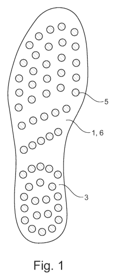
Fig. 1 shows an orthopaedic footbed 1 in the form of an insole 6, which is
shown in a side view,
having a foot 16 resting thereon, in Fig. 2. The insole 6 comprises a foot
contact surface 3 that
is formed by a continuous, planar base surface 4 and comprises pimples 5
arranged thereon.
The foot contact surface 3 is continuous and planar, and is intended for
people having
structurally and/or functionally healthy feet. The insole 6 furthermore
comprises a planar main
surface 7 on the lower face. The insole 6 is thus intended for shoes having a
planar inner
contact surface. The pimples 5 have an identical height of from 3 to 5 mm,
preferably 4 mm, and
an identical diameter of from 3 to 5 mm at the foot contact surface 3 or the
base surface 4, and
the end faces thereof together again form a planar surface to be contacted by
the indicated foot
16.
Fig. 7 is a projection from below of the orthopaedic footbed 1, together with
the foot 16 shown in
Fig. 8 having the bony parts 100 and pathways 200 thereof. If, in the
description of the
invention, reference is generally made to a foot 16 that is resting on the
foot contact surface 3
CA 03051158 2019-07-22
- 12 -
and that can be identified in Fig. 2 and 8, it is assumed that the foot 16 is
resting on the foot
contact surface 3 in accordance with the normal or the pre-determined
position.
The reference signs 1 to 56 in each case denote an individual pimple 5 on the
orthopaedic
footbed 1, and therefore the distribution of the reference signs 1 to 56
corresponds to the
distribution of the pimples 5 on the foot contact surface 3. In the basic
arrangement thereof, the
distribution of the pimples 5 corresponds to the arrangement of the essential
bony parts 100 of
the bone structure of the foot 16 and the pathways 200 extending thereon, in
the projection in
the foot contact surface 3 of the orthopaedic footbed 1, as can be easily
identified by way of a
comparison of Fig. 7 and 8.
In the basic structure thereof, the foot 16 which can be seen in a view from
below in Fig. 8
comprises the bony parts 100 comprising the heel 102, the midfoot 103, the
ball of the foot 104,
and finally the toes 101. In the event of the person walking with healthy
rolling behaviour, said
person first puts down the heel 102, and then rolls on over the midfoot 103,
the ball of the foot
104 and finally the toes 101. In this case, the heel 102 forms an
approximately circular contact
region, from which the midfoot 103 extends forwards in the walking direction.
Proceeding from
the midfoot 103, the bony parts 100 extend further in the form of chains of
individual bone
members which together form the ball of the foot 104, and the final bone
members of the chain
form the individual toes 101. The pathways 200 are arranged along the bony
parts 100, and the
distribution and course of said pathways thus correspond to the distribution
and the course of
the bony parts 100. The pathways 200 comprise the lymphs and/or the nerves
and/or the
vessels of the foot 16. In this case, in the basic structure thereof,
proceeding from the midfoot
103 the bony parts 100 extend in five bone member chains that are arranged in
lines and the
individual bone members of which are interconnected by joints.
Fig. 7 shows the orthopaedic footbed 1 comprising the pimples 5 and the
various zones on
which the relevant bony parts 100 of the foot come into contact, the different
zones of the bony
parts 100 with which the foot 16 comes into contact on the foot contact
surface 3 being denoted
by reference signs 101 to 104. The orthopaedic footbed 1 can thus also be
considered an
anatomical and neurological footbed 1, because it is specifically adapted to
the anatomy and
neurology of the foot 16 owing to the proposed distribution of the pimples 5.
The orthopaedic
footbed 1 thus forms a type of "bio-interface" via which the stimulation
points in the sole of the
CA 03051158 2019-07-22
- 13 -
foot are stimulated during walking, the proposed distribution of the pimples 5
being of particular
significance because, as a result thereof, the walking movement is used for
additional
stimulation of the stimulation points and the associated improvement of the
biotensegrity
system. The orthopaedic footbed 1 quasi forms an interface, in the contact
surface of the sole of
the foot, that is specifically adapted to the stimulation points of the sole
of the foot.
In the rear zone of the foot contact surface 3, on which the heel 102 of the
foot 16 comes into
contact, the pimples 5 are arranged in a circular manner in an imaginary ring,
which can be
seen from the reference signs 41 to 52. Four further pimples 5, having
reference signs 53 to 56,
are arranged in the centre of the imaginary ring, in as uniform a distribution
as possible and in a
square, having identical spacings in the longitudinal direction and
transversely to the
longitudinal direction of the foot contact surface 3. As a result, at the
start of the rolling
movement the foot 16 is uniformly stimulated in the stimulation zones of the
heel 102, as a
result of which the pumping process described at the outset is initiated. In
this case, the
stimulation signals triggered in the stimulation zones generate corresponding
signals, in the
person's brain, for pressure change in the associated chambers of the cells or
the organs of the
person, as a result of which the segmented nervous system and the organs are
deliberately
vitalized.
During the further rolling movement, a midfoot region 103 of the person's foot
16 rolls on the
foot contact surface 3, and in this case rolls over a zone of the foot contact
surface 3 in which
the pimples 5 are arranged in two imaginary lines that extend transversely to
the longitudinal
direction of the foot contact surface 3 and diverge towards the inside 105 of
the foot contact
surface 3, according to reference signs 40 to 36 and 35 to 31. The divergent
orientation of the
lines means that the spacings of the pimples 5 in the longitudinal direction
of the foot contact
surface 3 are greater on the inside 105 of the foot contact surface 3 than on
the outside 106 of
the foot contact surface 3. This arrangement of the pimples 5 is advantageous
because the
spacings of the stimulation points are smaller on the outside 106 of the foot
16 than on the
inside 105. Owing to the divergent orientation, the spacings of the pimples 5
increase in the
longitudinal direction of the foot contact surface from the outside 106 to the
inside 105, i.e.
transversely to the longitudinal direction. Furthermore, the foot arch of the
foot 16 is taken into
account thereby.
CA 03051158 2019-07-22
- 14 -
During the further rolling movement, the ball of the foot 104 and the toes 101
of the foot come
into contact on the foot contact surface 3 in a zone in which the pimples 5
are arranged in the
longitudinal direction of the foot contact surface 3 in five imaginary lines,
corresponding to the
bony parts 100 of the toes 101. In this case, the pimples 5 are arranged in
imaginary lines
corresponding to the reference signs 1,6, 11, 16, 21, 26, the reference signs
2,7, 12, 17, 22,
27, the reference signs 3, 8, 13, 18, 23, 28, the reference signs 4, 9, 14,
19, 24, 29 and finally
corresponding to the reference signs 5, 10, 15, 20, 25, 30. The distribution
of the pimples 5 thus
corresponds to the representation of the bony parts 100 that form the toes
101, and the
pathways 200 arranged along said bony parts, such that in this zone the
stimulation points of
the toes 101 arranged on the bony parts 100 or the pathways 200 are stimulated
in a targeted
manner by the pimples 5 during the rolling movement.
The insole 6 shown in Fig. 1 comprises a continuously planar foot contact
surface 3 and can
also be considered a standard footbed SFB. When viewed from the inner, medial
side, the
person's foot 16 rests in particular on the heel 102 and the front ball of the
foot 104, such that, in
the case of a healthy foot, a gap results between the foot arch and the foot
contact surface 3
and the foot 16 is deliberately not supported in the region of the foot arch.
As a result, the foot
16 is deliberately "stressed", and thus strengthened, in the event of a load,
i.e. when standing or
walking. Furthermore, the foot 16 experiences sensormotoric stimulation from
the pimples 5, as
a result of which the muscle control changes and the foot 16 is dynamically
strengthened by the
tensegrity system that was explained in greater detail at the outset. In
addition, the person's
venous and lymphatic system is stimulated.
Fig. 3 to 6 show different variants of the orthopaedic footbed 1 which
additionally comprise
different raised or strengthened or depressed or weakened functional zones 8,
as well as the
planar base surface 4. In this case, the functional zones 8 are formed by
surfaces which
deliberately stimulate the foot 16 by means of their geometry and their
arrangement on specific
sensor surfaces. For this purpose, the functional zones 8 can either be raised
or strengthened
to different extents relative to the base surface 4 or can be weakened or
depressed relative
thereto. The base surface 4 having the standard pimple distribution forms the
ideal contact
surface for long-term maintenance of the health of the feet in the case of
structurally and
functionally healthy feet, while the stimulation units in the functional zones
8, which units are
modified according to need, supplement or adapt the base surface 4 to form an
adapted foot
CA 03051158 2019-07-22
- 15 -
contact surface 3 in order to take account of individual disorders of the
structure and/or the
function of the feet. In order to simplify comprehension, the foot contact
surface 3 of the
orthopaedic footbed 1 is divided, in Fig. 3 to 6, into different points or
portions comprising
pimples 5 corresponding to reference signs 1 to 56. In these cases, the
functional zones 8 are
formed by pimples 5 that create increased stimulation in groups and the end
faces of which
together have an increased contact stimulation effect for specific zones of
the foot 16. The
increased stimulation effect is generated by zonal hardening or thickening of
the EVA base layer
110 that can be seen in Fig. 15, or alternatively by raising particular
pimples 5 and the
stimulation points formed thereby. Alternatively, the pimples 5 in the
functional zones 8 may
also be of a greater strength than the pimples 5 in the region of the
remaining foot contact
surface 3.
In Fig. 3, the functional zone 8 is formed by a talipes valgus correction
surface 9 which is
arranged on the foot contact surface 3 such that the foot 16 comes into
contact by the inside of
the front ball 104 of the foot in points 11, 16, 22 and 21, in a region 9b,
and by the inside of the
foot arch in points 26, 27, 31, 32, 36 and 37, in region 9a, of the foot
contact surface 3, or is
more intensively stimulated in said region. The increased stimulation makes it
possible to
stimulate counter-control by the central nervous system and to prevent the
foot 16 from bending
in towards the inside, in the direction of the other foot. Talipes valgus is a
functional disorder of
the foot 16, in which the person does not step in a straight line with the
foot 16, but instead
bends the foot in towards the inside, which may subsequently also lead to
knock knees and
postural abnormalities of the hips and spine.
Fig. 4 shows an orthopaedic footbed 1 in which an additional functional zone
8, in the form of a
statics correction surface 10, is provided in addition to the talipes valgus
correction surface 9.
The statics correction surface 10 is formed by raised pimples 5 and the
resulting stimulation
effect in the foot contact surface 3 in the region of the points 41 to 56,
which points form the
region on which the person comes into contact with the heel 102. As a result,
the stimulation
points or pressure receptors in the heel region are stimulated more intensely,
such that the
statics is adjusted by means of neurological regulation, in the sense of a
correction. This is
based on the sensormotoric effect of the more intense stimulation, in
accordance with the law of
the vector addition model.
Fig. 5 shows a further orthopaedic footbed 1 comprising a planar base surface
4 and two
CA 03051158 2019-07-22
- 16 -
functional zones 8. One of the functional zones 8, in the region of the heel
102, is again
provided as a statics correction surface 10, and a further functional zone 8
is provided in the
region of the front ball of the foot 104, over the entire width of the points
11 to 21, in the form of
a pes cavus correction surface 11. Both the statics correction surface 10 and
the pes cavus
correction surface 11 are in each case functional zones 8 which are raised by
3 to 5 mm with
respect to the base surface 4 and which slightly stimulate the person's foot
16 at the heel 102
and in the front foot region, and are formed by stimulation pimples.
Fig. 6 shows a further alternative embodiment of the orthopaedic footbed 1, in
which a
functional zone 8 in the form of a calcaneal spur correction surface 12 is
provided in the base
surface 4, which calcaneal spur correction surface is formed by a depression
of 3 to 5 mm in the
central region of the heel 102 and of the rear, adjoining central portion 103
in the region of
points 32, 33, 37, 38, 42, 43, 44 and 53 to 56. The calcaneal spur correction
surface 12 is free
of pimples, or depressed, relative to the base surface 4, such that the foot
16 is deliberately not
stimulated and/or relieved of loading in this region.
The orthopaedic footbed 1 can be designed both as an insole 6 and as a part of
a lower shoe.
All that is important is that the foot contact surface 3 is correspondingly
shaped or that the foot
contact surface 1 forms the corresponding foot contact surface 3 in the shoe.
In this case, the
footbed 1 can in addition comprise a leather coating or textile coating, as a
result of which
wearing the shoe 2 can be made more comfortable. Furthermore, the footbed 1
should be
designed so as to be permanently elastic, breathable, liquid-absorbing and
conducting. The
resiliency of the footbed 1 should be such that it subjects the foot contact
surface 16 to sufficient
resistance, the resiliency being intended to allow for slight penetration of
the foot 16 into the foot
contact surface 3 without the basic distribution of the contact surface,
according to the principle,
being lost. In particular, the resiliency should be selected such that the
foot 16 does not sink in
so far as to be in contact over the entire surface thereof, since otherwise
the desired loading of
the foot 16 is not achieved. This is the case in particular if the foot 16 is
structurally and
functionally healthy and the foot contact surface 3 is formed only by a planar
base surface 4, as
is shown in Fig. 1 and 2. In this case, the foot 16 is intended to be in
contact deliberately in a
bridged shape, and not to be supported in the region of the foot arch. In this
case, the resiliency
can be matched, in a targeted manner, to the type of person wearing the shoe
2. It would thus
be conceivable, for example, to use a particularly soft footbed 1 specifically
for diabetics, and a
CA 03051158 2019-07-22
- 17 -
particularly hard footbed 1 for sportspeople. A possible material would be
ethylene vinyl acetate
(EVA), for example.
If the orthopaedic footbed 1 is designed as an insole 6, this may be
intrinsically resilient, and the
functional zones therein may be formed having a greater strength or hardness.
In this case, the
insole 6 can have a resiliency that is such that said insole can be put into a
bag folded, bent
back or rolled up, without being damaged in the process. After the insole 6
has been removed
from the bag, it unfolds automatically or with slight assistance, owing to the
resiliency thereof,
back into the original shape, and can thus be inserted into the shoe 2. Simply
owing to the
greater hardness of the footbed 1 in the region of the functional zones 8, the
foot 16
experiences greater support and stimulation here than in the remaining regions
of the base
surface 4. Furthermore, in addition to the greater hardness thereof, the
functional zones 8 can
of course also be of a greater height or thickness and optionally comprise
additional pimples 5
for stimulation of the sole of the foot.
Fig. 9 shows a flow diagram of a method according to the invention for
providing an orthopaedic
footbed 1.
Firstly, a range of different orthopaedic footbeds 1 having differently shaped
foot contact
surfaces 3 for different foot types, in different shoe sizes, shoe last widths
and possibly also
having different hardnesses, is kept available in a shop or a clinic, in which
the people can
select and test their orthopaedic footbed 1, which is matched individually to
their feed 16, under
specialist guidance from correspondingly trained consultants. In this case,
the left and right foot
16 may also be different, and therefore different orthopaedic footbeds 1 may
be deliberately
selected for the left and right foot 16.
Firstly, the customer K is assessed visually and by means of measurements,
within the context
of an initial assessment E, by the consultant, optionally with the aid of
corresponding sensor
means such as pressure-sensitive standing surfaces or treadmills. In this
case, further aids such
as foot and shoe size measurement, a measuring device for measuring the
posture and in
particular the statics, may be used within the context of a diagnosis D. The
consultant then
identifies a specific foot type, with or without structural and/or functional
disorders and/or with or
without impaired statics.
CA 03051158 2019-07-22
- 18 -
Structurally healthy is denoted in the flow diagram by SG, structural impaired
by SK, functionally
healthy by FG, and functionally impaired by FK.
If it is ascertained that the feet are both structurally and functionally
healthy SG, FG, it is firstly
determined that a standard footbed SFB comprising an orthopaedic footbed 1
having a planar
base surface 4 according to Fig. 1 and 2 and having a distribution of the
pimples 5 according to
Fig. 10 should be selected. In this case, it is possible inter alia to
ascertain that the person has a
posture that is slightly bent forward, which is also referred to as anterior
statics AS. The slightly
bent forward statics, or neutral statics, corresponds to healthy posture and
generally does not
require any correction, i.e. also does not require a functional zone 8.
Subsequently, in a second
step, a check is performed as to whether there is medial lowering of the foot
arch MGA. If there
is no medial lowering, the standard footbed SFB of type la is selected, which
footbed comprises
a foot contact surface 3 that is formed by a planar base surface 4 according
to Fig. 1 and has a
distribution of the pimples 5 according to Fig. 10. If, in contrast, there is
medial lowering of this
kind, a standard footbed SFB of type lb is selected, in which the planar base
surface 4 is
supplemented by a functional zone 8 in the form of slight raising by
approximately 2 mm or in
the form of a portion having a greater hardness in the region 9a of the
talipes valgus correction
surface 9 reduced thereto. The slight medial lowering of the foot arch is
considered a functional
disorder of the foot 16, it being possible for the tendency of continuing
worsening of the disorder
to be counteracted by the raising or stiffening in the region 9a and the
resultant intensification of
the stimulation.
If one of the feet 16 is structurally healthy SG and functionally impaired FK,
the type of the
functional disorder is firstly determined in a further step DFK and a
correspondingly
individualized orthopaedic footbed 1 comprising a functional zone 8
individually provided for the
disorder is selected. A functional disorder of this kind may be talipes valgus
for example, the
functional zone 8 in this case being the talipes valgus correction surface 9
shown in Fig. 3.
Subsequently, a check is performed, in a further step, as to whether posterior
statics PS, i.e.
backwardly inclined posture of the person, is present in addition. If no
posterior statics PS is
present, an orthopaedic footbed 1 without a statics correction surface 10 is
selected, whereas in
the event of posterior statics PS a statics correction surface 10 is added. In
a second step, a
check is performed as to whether or not there is medial lowering of the foot
arch MGA. If this is
CA 03051158 2019-07-22
- 19 -
not present, and the foot 16 at the same time exhibits posterior statics PS,
an orthopaedic
footbed 1 of type 2a is selected, which footbed comprises a foot contact
surface 3 which is
formed only by the base surface 4 and the statics correction surface 10. If,
in contrast, medial
lowering of the foot arch MAG is identified, and the foot 16 at the same time
exhibits posterior
statics PS, the foot contact surface 3 is additionally supplemented to type
2b, by stiffening or
strengthening of the foot contact surface 3 in the region 9a in addition to
the statics correction
surface 10.
The orthopaedic footbeds 1 shown in Fig. 5 and 6, comprising the individual
foot contact
surfaces 3, are further examples of the range of the orthopaedic footbeds 1
which can be
selected by assessing or identifying further disorders. It is not impossible
for the range of the
orthopaedic footbeds 1 to be supplemented by footbeds 1 having differently
shaped foot contact
surfaces 3 or for further individual functional zones 8 to be developed which
can be combined
with the base surface 4 and the described functional zones.
If the diagnosis identifies both a structurally impaired SK foot 16 and a
functionally impaired FK
foot 16, a recommendation is made for a medical examination (EAU), and
optionally a
recommendation is made for wearing a standard footbed SFB until the results of
the medial
examination are available. A subsequent follow-up appointment KT may in
addition also be
arranged.
The advantage of the proposed solution is considered to be that the health of
the feet and the
posture of a very large number of people can be improved, or the likelihood of
the development
of disorders and postural defects can be reduced, by means of preventative
measures, using
simple means basic knowledge of specialists which can be conveyed in
specialist seminars for
example. In this case, the invention makes use of the advantage that the
orthopaedic footbeds 1
are kept available not specifically depending on the individual foot 16, but
instead in a person-
independent manner for various foot disorders, in the form of a range. The
person-specific
manufacture of the insoles used hitherto firstly requires production of an
individual footprint, on
the basis of which the insole is then manufactured. The person could therefore
not take the
insole immediately, but said insole instead had to be manufactured in an
orthopaedics workshop
that is specialized in this. As a result, the insole could be collected and
worn only after a waiting
time of several days or weeks. Overall, providing the insoles was thus
associated with
CA 03051158 2019-07-22
- 20 -
corresponding time expenditure and manufacturing outlay, resulting in a drop
in the acceptance
of wearing insoles. Insoles were worn only if already serious, medically
identified disorders of
the function and structure of the feet were already present.
According to the method according to the invention for providing the footbed,
the orthopaedic
footbeds 1 are manufactured in large numbers, having various foot contact
surfaces 3 which,
although not person-specific, are instead type-specific, i.e. are adapted to
the type of the foot 16
by means of the planar base surface 4 or by means of the combination of the
base surface 4
with different functional zones 8 that are specially adapted to the structural
and functional
disorders, and thus allow for significantly more healthy walking. Since the
footbeds 1 are tested
on-site and can be taken away immediately after being selected, the outlay for
obtaining a
footbed 1 of this kind is significantly reduced, as a result of which a
significantly larger number
of people can be convinced to wear footbeds 1 of this kind, at least as a
trial. As a result, the
health of the population can be significantly improved, on average, by the
increased acceptance
of orthopaedic footbeds 1, which footbeds can be described as a new
biointerface owing to the
special distribution of the pimples 5. The distribution of the pimples 5
essentially achieves a
biointerface which is used to stimulate the stimulation points of the sole of
the foot during
walking, and thus to strengthen the biotensegrity system. As a result, the
person's normal
walking movement itself is used for stimulating the biotensegrity system and
for associated
improvement of posture and gait.
Fig. 10 shows the orthopaedic footbed 1 according to the invention comprising
the pimples 5
provided thereon in a distribution according to the invention, according to
Fig. 7, only in this
case the pimples 5 are indicated by circles, corresponding to the geometry
thereof, instead of by
reference signs 1 to 56.
The foot contact surface 3 comprising the pimples 5 provided thereon can be
divided, in the
same manner, into different regions in which the person comes into contact by
the heel 102, the
midfoot region 103, the ball of the foot 104 and finally with the toes 101 of
the foot 16.
Fig. 11 shows the orthopaedic footbed 1 comprising an additional functional
zone 8 in the form
of a portion 107 of the central region 103 on the inside 105 that is at a
higher level but is of a
lower hardness. In this case, the portion 107 having the lower hardness can be
achieved by a
CA 03051158 2019-07-22
-21 -
raised design of the base surface 4, such that the pimples 5 arranged thereon
stimulate the
stimulation points, in the portion of the sole of the foot coming into contact
therewith, earlier and
rather more firmly, during walking. The proposed functional zone 8, formed by
the higher portion
107, makes it possible for the pumping system of the sole of the foot, for the
lymphatic and
venous fluids, to be particularly strengthened, and therefore the footbed 1
shown in Fig. 11 is
preferably advantageous for people having lymphatic and/or venous
insufficiency. The proposed
variant of the orthopaedic footbed 1 shown in Fig. 11 makes it possible for
the "heart of feet
function" of the sole of the foot to be stimulated and intensified. In
principle, any variant of the
orthopaedic footbed 1 can be provided with the "heart of feet function" for
optimizing the
lymphatic and venous fluid transport, by means of providing the functional
zone 8 in the portion
107.
Fig. 12 shows a further orthopaedic footbed 1 comprising a statics correction
surface 10 which,
in the embodiment of Fig. 13, is supplemented by a functional zone 8 in the
form of a talipes
valgus correction surface 9. Both the statics correction surface 10 and the
talipes valgus
correction surface 9 are formed by portions of the footbed 1 having a greater
hardness, which
portions can be formed either by a portion of the base surface 4 having a
greater hardness or by
pimples 5 having a greater hardness that are arranged in said portions, or by
a combination of a
base surface 4 having a greater hardness in said portions and pimples 5 having
a greater
hardness.
Both locally arranging pimples 5 having a greater hardness, and forming the
base surface 4 so
as to have a greater hardness and/or so as to be at a higher level locally
result in the desired
local stimulation effect on the sole of the foot being intensified. As a
result, both the gait and the
posture of the person can be positively influenced and corrected, since the
perception in the
sole of the foot leads to a postural change, in accordance with what is known
as the vector
addition model.
Fig. 14 shows the orthopaedic footbed 1 comprising a functional zone 8 formed
by the talipes
valgus correction surface 9, which functional zone is specifically intended
for a foot 16 having
neutral statics and an unstable medial foot arch.
Furthermore, Fig. 15 again shows the orthopaedic footbed 1 having a
distribution of the pimples
CA 03051158 2019-07-22
- 22 -
corresponding to the embodiment of Fig. 11, for intensifying the above-
described "heart of feet
function". The bottom drawing shows the structure of the footbed, in a cross
section through the
base surface 4, on the left-hand side, and in a cross section through the base
surface 4 that is
raised relative to the functional surface 8, on the right-hand side.
In the base surface 4, the orthopaedic footbed 1 comprises an EVA base layer
110 and an EVA
cover layer 111 which are separated from one another by a stimulation layer
113. The EVA
base layer 110 is covered, on the lower face, by a carrier layer 114, and the
EVA cover layer
111 is covered, on the top face, by a functional tissue layer 112 having fluid-
conducting and
breathable properties, which layer simultaneously forms the foot contact
surface 3. The EVA
base layer 110, the EVA cover layer 111, the stimulation layer 113, the
functional tissue layer
112, and the carrier layer 114 each have a constant thickness, such that the
orthopaedic
footbed 1 has a constant thickness in the region of the base surface 4, apart
from the pimples 5
(not visible) which are arranged thereon.
In the right-hand drawing, the EVA base layer 110 is of a greater thickness,
in order to form the
functional zone 8, while the thickness of the remaining layers is constant.
The raising of the foot
contact surface 3 in the region of the functional zone 8 is thus achieved
merely by increasing
the thickness in the EVA base layer 110. The thickening of the EVA base layer
110 is shown in
the right-hand drawing, by the zone II of the EVA base layer 110 in the region
of the functional
zone 8, above the zone I.
The functional tissue layer 112 is preferably formed by a breathable and fluid-
permeable textile
material, while the carrier layer 114 is formed by a wear-resistant plastics
material, for example
having a carbon effect.
Both the EVA cover layer 111 and the EVA base layer 110 are manufactured from
an EVA
material, and virtually form the volume material of the footbed 1. The
stimulation layer 113 is
manufactured from a hard plastics material and defines the hardness of the
footbed 1.
The "heart of feet function" brought about by the orthopaedic footbed 1
according to the
invention will be explained again, in greater detail, in the following.
The sole of the foot is a blood and lymphatic pump and assists the return
transport of the blood
CA 03051158 2019-07-22
- 23 -
supplied by the heart. This return transport is brought about by the muscle-
vein pump in the
foot, formed by the vessels, the fasciae system, the bones and the muscles,
together with
gravity.
Owing to the particular anatomical structure comprising the tissues encased in
fasciae, natural
movements and the pressure changes in the foot that are induced or stimulated
by the footbed
1 according to the invention result in fluid-displacement effects owing to the
constant change in
tissue pressure gradients. When compressed and elongated, the pressure on the
tissue
portions protruding into the fasciae-encased chambers is significant, the
alternating peaks and
troughs of the pressure build-up brings about a pumping mechanism which is
weakened but still
present in the case of impaired venous and lymphatic vessels.
The theory of the pumping movement in the sole of the feet of people can be
explained as
follows: The basis of the pumping movement is a grille having pressure
gradients that are
generated by deformation: Upon stretching, the pressure in the enclosed
chambers increases
owing to the movement of the connective tissue fasciae lines, and pressurized
movement of the
fluid takes place in a manner channelled from the foot to the centre of the
body. If the movement
of the grille recedes, the pressure gradient reduces again, resulting in fluid
collecting in the
chambers, between the grille elements. These movements alternate cyclically,
as a result of
which the body transports lymphatic and venous fluid from the narrowest tissue
gaps to the
heart, outside of vessels and in the smallest of vessels.
The fasciae lines around each cell are thus inter alia also a person's "other
heart", which
represents and is therefore responsible for the centripetal pumping movement,
in the way in
which the heart represents a large portion of the centrifugal pumping
movement.
The material of the "heart of feet" zone of the orthopaedic footbed 1
according to the invention,
in portion 107 of Fig. 15, should be made up in the following manner:
For example a 5-15 mm high, soft material layer may be provided, which layer
is arranged
between the EVA base layer 110 and the stimulation layer 113, or alternatively
optionally also
between the stimulation layer 113 and the EVA cover layer 111, in the region
marked in the
drawing. In this case, the additional material layer is represented by the
region II. The material is
CA 03051158 2019-07-22
- 24 -
sufficiently soft to not support the foot, but robust enough to compress the
connective tissue of
the foot from the skin, via the subcutaneous adipose tissue, as far as the
vascular region and
the muscles of the longitudinal foot arch, in which some of the venous and
lymphatic vessels
are located. Silicones, EVA materials, or other similarly functioning soft and
permanently elastic
materials, exhibiting a soft restoring force and quick return to the original
shape following the
relevant deformation with each step, are used as materials for said functional
zone.
The pressure increase results in expulsion of at least 20 to 40 cubic
centimetres of blood and
lymphs from the foot back towards the heart with each tread. Increasing the
pressure relative to
the orthopaedic footbed 1 without a "heart of feet" function significantly
assists the venous and
lymphatic return flow to the heart, in order to assist the insufficient, i.e.
weakly pumping, venous
and lymphatic vessels of the foot in their natural function.