Note: Descriptions are shown in the official language in which they were submitted.
DYNAMIC RAMAN SIGNAL ACQUISITION SYSTEM, METHOD AND APPARATUS
FIELD OF THE DISCLOSURE
[0001] The present disclosure relates generally to optical tissue
analysis instruments and
systems, used for example, within the context of a medical or surgical
procedure, test or analysis,
and, in particular, to a dynamic Raman signal acquisition system, method and
apparatus.
BACKGROUND
[0002] Raman spectroscopy is a powerful technique for analyzing the
composition of liquids,
gases, and solids. It is based on the Raman scattering and it is widely used
in scientific research
and industry. Among other applications, it has been recently demonstrated that
Raman
spectroscopy can be used to identify tumour margins by successfully detecting
and differentiating
the unique signatures of tumorous and healthy tissues (reference: Jermyn,
Michael, et al.
"Intraoperative brain cancer detection with Raman spectroscopy in humans."
Science translational
medicine 7.274 (2015)).
[0003] Despite being a mature technique, application of Raman
spectroscopy is still generally
challenging for several reasons. Raman signals are rather weak and can be
easily overwhelmed by
competing optical signals such as auto-fluorescence. Also, because the Raman
signals are weak,
sufficiently long integration times are often required for achieving
acceptable signal to noise ratios
(SNR) which increases the probability of noise interference. However, if the
integration times are
too long there is a different challenge. Since the competing optical signals
(e.g. fluorescence) are
frequently much more intense, long integration times can lead to detector
saturation and wasted
acquisition. For these reasons, setting signal acquisition parameters in
advance for an unknown
sample can be problematic.
[0004] Besides general difficulties there could be additional ones
related to specific application
circumstances. For example, in some cases the acquisition has to happen
without a human operator
or in some occasions there is fixed pattern noise. Application of Raman
spectroscopy in
intraoperative surgical situations has its own characteristic concerns. First,
there are maximum
applicable excitation light intensities incident at the tissue surfaces since
tissues can be damaged if
the excitation light is too intense. Second, the overall time available for
measurements is usually
1
CA 3051348 2019-08-07
limited due to time constrained surgical environment. Third, in the case of
handheld Raman
probes, the acquisitions are frequently performed in unstable environments.
Movements of the
surgeon's hand holding the probe during the acquisition may change the
coupling efficiency for
both the excitation light and the signal causing the artifacts in the acquired
signals. Other possible
reason for instability is the presence of the background ambient lighting
typically used in surgical
rooms. These light sources¨e.g. fluorescence bulbs¨may flicker for short
periods of time but
long enough to cause additional signal artifacts. Moreover, acquisitions may
become unstable for a
more critical reason if tissue structure starts to change due to presence of
the excitation light. This
may happen in unlikely but still possible scenario when tissue contains
exogenous components
(e.g. drugs) that strongly absorb the excitation light which can lower the
general damage threshold
for that type of tissue.
[0005] There have been prior attempts to automatize setting and
processing of Raman signal
acquisitions but for applications other than surgery. A system disclosed in US
7,605,918 performs
a trial measurement for a set acquisition TO and arbitrary excitation optical
power. The results is
compared against predetermined value for signal to noise ratio (SNR) in order
to set new
acquisition time and number of acquisitions. The method disclosed in US7557915
involves a two
phase process including a photobleaching phase and a spectral acquisition
phase. In the
photobleaching phase, a series of spectral data sets of a sample are
collected. A relative difference
is determined between the background of subsequent spectral data sets is
determined and
compared to a predetermined threshold value. If threshold difference is less
than the relative
difference between the background of subsequent spectral data sets, the steps
of collecting a series
of spectra data sets is automatically repeated. In the spectrum acquisition
phase, a series of Raman
data sets of the sample are collected until a target SNR is obtained. The
system disclosed in
US9074932B2 performs noise reduction iteratively based on difference in value
between an
extremal point of measurement data-blocks making up input spectrum data, and a
mean value of
measurement data-blocks in the vicinity of the extremal point. Finally, in
reference Lopez-Reyes,
Guillermo, and Fernando Rull Perez. "A method for the automated Raman spectra
acquisition."
Journal of Raman Spectroscopy 48.11(2017): 1654-1664, an algorithm is
presented that reduces
the auto-fluorescence background noise by photo-bleaching and then single
acquisition time and
number of acquisition are determined.
2
CA 3051348 2019-08-07
[0006] In all prior optimization algorithms, no consideration was given
to optimizing the
intensity of the excitation light which is one of the critical parameters in
surgical applications. In
methods described in US 7,605,918 and Lopez-Reyes et al, final acquisition
parameters are not
adjusted dynamically. The methods disclosed in US7557915 and US9074932B2 are
based on
specific noise reduction strategies which are not generally applicable in
surgical settings.
[0007] This background information is provided to reveal information
believed by the
applicant to be of possible relevance. No admission is necessarily intended,
nor should be
construed, that any of the preceding information constitutes prior art or
forms part of the general
common knowledge in the relevant art.
SUMMARY
[0008] The following presents a simplified summary of the general
inventive concept(s)
described herein to provide a basic understanding of some aspects of the
disclosure. This summary
is not an extensive overview of the disclosure. It is not intended to restrict
key or critical elements
of embodiments of the disclosure or to delineate their scope beyond that which
is explicitly or
implicitly described by the following description and claims.
[0009] A need exists for a dynamic Raman signal acquisition system,
method and apparatus
that overcome some of the drawbacks of known techniques, or at least, provides
a useful
alternative thereto. Some aspects of this disclosure provide examples of such
systems, methods and
apparatus.
[0010] For instance, in accordance with some aspects of the present
disclosure, a dynamic
Raman signal acquisition system, method and apparatus are described for use in
a surgical
environment to provide real-time optimization of Raman system parameters in
use. In some
examples, such dynamic adjustments can increase the number of useful
acquisitions without
invoking significant post-acquisition processing or signal repairs, which, in
some applications such
as intraoperative or handheld surgical tools, may be time prohibitive if not
mostly inaccessible. In
some examples, such dynamic adjustments may also, or alternatively, minimize
if not entirely
avoid the need for manual system adjustments, which again, can be
prohibitively time consuming
in some applications. These and other aspects, objects, advantages and
features of the herein
described embodiments will be described in greater detail below.
3
CA 3051348 2019-08-07
[0011]
For example, in accordance with one particular aspect, there is provided a
Raman
system for analyzing biological tissue, the system comprising: an excitation
light source operable
at a designated irradiation power and for a designated acquisition time for
each Raman data
acquisition; a Raman probe operatively associated with said excitation light
source to irradiate the
biological tissue at said designated irradiation power and for said designated
acquisition time, and
capture an optical Raman response therefrom;
a spectrometer operable to spectrally analyze
said optical Raman response; and a controller in operative communication with
said excitation
light source and said spectrometer to automatically adjust at least one signal
acquisition parameter
by: acquiring a Raman response signal for said designated irradiation power
being set to a
predetermined initial irradiation power and at said designated acquisition
time; processing an
amplitude of said Raman response signal against a designated threshold; and
upon said Raman
response signal being greater than said designated threshold, said controller
is further operable to
operatively lower said designated irradiation power and repeat for a
subsequent said Raman
response signal.
[0012] In one embodiment, the predetermined initial irradiation power is a
predetermined
maximum irradiation power.
[0013]
In one embodiment, upon said Raman response signal being below said
designated
threshold, said controller is further operable to increase said designated
acquisition time so to
increase subsequent Raman response signals toward said threshold.
[0014] In one embodiment, the Raman response signal comprises a maximum
signal level for a
series of initial Raman response signals.
[0015]
In one embodiment, once said signal acquisition parameter has been adjusted,
said
controller is further operable to operatively serially acquire a set of
background-corrected Raman
response signals until a signal-to-noise ratio (SNR) thereof is greater than a
designated SNR
threshold.
[0016]
In one embodiment, the controller is further operable to: acquire a first
set of
background signals to process said background-corrected Raman response
signals; and upon said
SNR being greater than said designated SNR threshold, acquire a complementary
set of
background signals such that a total number of acquired background signals is
equal to a total
4
CA 3051348 2019-08-07
number of said background-corrected Raman signals to be used in post-
processing said
background-corrected Raman signals.
[0017] In one embodiment, the controller is further operable to
spectrally identify and
automatically remove narrow band outliners from said Raman response signals.
[0018] In one embodiment, the controller is further operable to spectrally
identify an adverse
safety feature from said Raman response signals and immediately suspend
further acquisition.
[0019] In one embodiment, the Raman probe comprises a handheld probe.
[0020] In one embodiment, the excitation light source is directly
controlled by said controller
to adjust said designated irradiation power.
[0021] In one embodiment, the system further comprises a power controller
operatively
disposed between said excitation light source and said Raman probe, and in
operative
communication with said controller to adjust said designated irradiation
power.
[0022] In accordance with another aspect, there is provided a
computerised method for
dynamically acquiring Raman signals for analyzing biological tissue, the
method comprising:
irradiating the tissue at a designated irradiation power, initially set to a
predetermined initial
irradiation power, for a designated acquisition time; acquiring a Raman
response signal from said
irradiating at said designated irradiation maximum irradiation power and at
said designated
acquisition time; processing an amplitude of said Raman response signal
against a designated
threshold; upon said Raman response signal being greater than said designated
threshold,
dynamically decreasing said designated irradiation power; and repeating for a
subsequent said
Raman response signal.
[0023] In one embodiment, upon said Raman response signal being below
said designated
threshold, the method further comprises: dynamically increasing said
designated acquisition time
so to increase subsequent Raman response signals toward said threshold.
[0024] In one embodiment, the Raman response signal comprises a maximum
signal level for a
series of initial Raman response signals.
5
CA 3051348 2019-08-07
[0025] In one embodiment, the method further comprises serially
acquiring a set of
background-corrected Raman response signals until a signal-to-noise ratio
(SNR) thereof is greater
than a designated SNR threshold.
[0026] In one embodiment, the method further comprises: acquiring a
first set of background
signals to process said background-corrected Raman response signals; and upon
said SNR being
greater than said designated SNR threshold, acquiring a complementary set of
background signals
such that a total number of acquired background signals is equal to a total
number of said
background-corrected Raman signals to be used in post-processing said
background-corrected
Raman signals.
[0027] In one embodiment, the method further comprises spectrally
identifying and removing
narrow band outliners from said Raman response signals.
[0028] In one embodiment, the method further comprises spectrally
identifying an adverse
safety feature in said Raman response signal and immediately suspending
further acquisition.
[0029] In one embodiment, the predetermined initial irradiation power is
a predetermined
maximum irradiation power.
[0030] In accordance with another aspect, there is provided a non-
transitory computer-readable
medium having instructions stored thereon for execution by a digital data
processor of a Raman
system to dynamically acquire Raman signals for analyzing biological tissue
by: causing
irradiation of the tissue at a designated irradiation power, initially set to
a predetermined initial
irradiation power, and for a designated acquisition time; acquiring a Raman
response signal from
said irradiating at said designated irradiation power and at said designated
acquisition time;
processing an amplitude of said Raman response signal against a designated
threshold; upon said
Raman response signal being greater than said designated threshold,
dynamically decreasing said
designated irradiation power; and repeating for a subsequent said Raman
response signal.
[0031] In one embodiment, the non-transitory computer-readable medium
further comprises
instructions for, upon said Raman response signal being below said designated
threshold,
dynamically increasing said designated acquisition time so to increase
subsequent Raman response
signals toward said threshold.
6
CA 3051348 2019-08-07
[0032] In one embodiment, the non-transitory computer-readable medium
further comprises
instructions for serially acquiring a set of background-corrected Raman
response signals until a
signal-to-noise ratio (SNR) thereof is greater than a designated SNR
threshold.
[0033] In one embodiment, the predetermined initial irradiation power is
a predetermined
maximum irradiation power.
[0034] Other aspects, features and/or advantages will become more
apparent upon reading of
the following non-restrictive description of specific embodiments thereof,
given by way of
example only with reference to the accompanying drawings.
BRIEF DESCRIPTION OF THE FIGURES
[0035] Several embodiments of the present disclosure will be provided,
by way of examples
only, with reference to the appended drawings, wherein:
[0036] Figure 1 is a diagram of a dynamic Raman signal acquisition
system, in accordance
with one embodiment;
[0037] Figure 2 is a process flow diagram of an illustrative dynamic Raman
signal acquisition
process, in accordance with one embodiment, in accordance with one embodiment;
[0038] Figure 3A is a process flow diagram of an exemplary process for
estimating a
maximum initial signal level in the dynamic Raman signal acquisition process
of Figure 2;
[0039] Figure 3B is an approximate histogram of Raman signals for a
particular wavelength in
.. the presence of random noise where the random noise is a combination of
shot noise, thermal
noise, and readout noise.
[0040] Figure 4 is a process flow diagram of an exemplary acquisition
parameter optimization
process in the dynamic Raman signal acquisition process of Figure 2, in
accordance with one
embodiment;
[0041] Figure 5 is a process flow diagram of an exemplary temporary
background signal
process in the dynamic Raman signal acquisition process of Figure 2, in
accordance with one
embodiment;
7
CA 3051348 2019-08-07
[0042] Figure 6 is a schematic diagram illustrating a distinction
between signal intensity and
noise intensity, in accordance with one embodiment;
[0043] Figure 7 is a process flow diagram of an exemplary final
background signal process
following the temporary background signal process of Figure 5, in the dynamic
Raman signal
acquisition process of Figure 2, in accordance with one embodiment;
[0044] Figure 8 is a schematic diagram illustrating a series of Raman
response signals acquired
over time, in accordance with one embodiment;
[0045] Figure 9 is a diagram of a dynamic Raman signal acquisition
system, in accordance
with another embodiment;
[0046] Figure 10 is a process flow diagram of an exemplary safety shutdown
process of the
dynamic Raman signal acquisition process of Figure 2, in accordance with one
embodiment;
[0047] Figure 11 is a schematic diagram of an irradiation intensity over
time for a given
acquisition window, in accordance with one embodiment;
[0048] Figure 12 is a schematic diagram of an irradiation intensity over
time for a set of
.. sequential acquisition windows, in accordance with one embodiment;
[0049] Figure 13 is a diagram of a dynamic Raman signal acquisition
system, in accordance
with another embodiment;
[0050] Figure 14 is a process flow diagram of a pre-acquisition process
of the dynamic Raman
signal acquisition process of Figure 2, in accordance with one embodiment
[0051] Figure 15 is a diagram of a dynamic Raman signal acquisition system,
in accordance
with another embodiment.
[0052] Elements in the several figures are illustrated for simplicity
and clarity and have not
necessarily been drawn to scale. For example, the dimensions of some of the
elements in the
figures may be emphasized relative to other elements for facilitating
understanding of the various
.. presently disclosed embodiments. Also, common, but well-understood elements
that are useful or
8
CA 3051348 2019-08-07
necessary in commercially feasible embodiments are often not depicted in order
to facilitate a less
obstructed view of these various embodiments of the present disclosure.
DETAILED DESCRIPTION
[0053] Various implementations and aspects of the specification will be
described with
reference to details discussed below. The following description and drawings
are illustrative of the
specification and are not to be construed as limiting the specification.
Numerous specific details
are described to provide a thorough understanding of various implementations
of the present
specification. However, in certain instances, well-known or conventional
details are not described
in order to provide a concise discussion of implementations of the present
specification.
1() [0054] Various apparatuses and processes will be described below
to provide examples of
implementations of the system disclosed herein. No implementation described
below limits any
claimed implementation and any claimed implementations may cover processes or
apparatuses that
differ from those described below. The claimed implementations are not limited
to apparatuses or
processes having all of the features of any one apparatus or process described
below or to features
common to multiple or all of the apparatuses or processes described below. It
is possible that an
apparatus or process described below is not an implementation of any claimed
subject matter.
[0055] In this specification, elements may be described as "configured
to" perform one or
more functions or "configured for" such functions. In general, an element that
is configured to
perform or configured for performing a function is enabled to perform the
function, or is suitable
for performing the function, or is adapted to perform the function, or is
operable to perform the
function, or is otherwise capable of performing the function.
[0056] It is understood that for the purpose of this specification,
language of "at least one of X,
Y, and Z" and "one or more of X, Y and Z" may be construed as X only, Y only,
Z only, or any
combination of two or more items X, Y, and Z (e.g., XYZ, XY, YZ, ZZ, and the
like). Similar
logic may be applied for two or more items in any occurrence of "at least one
..." and "one or
more..." language.
[0057] The systems and methods described herein provide, in accordance
with different
embodiments, different examples of a dynamic Raman acquisition system and a
method to be used
therewith in which operational parameters, such as the excitation light source
power (i.e. laser
9
CA 3051348 2019-08-07
power) and/or acquisition time is dynamically optimized to ensure proper Raman
measurements
when operating in a dynamic environment. Raman scattering is a nonlinear
effect resulting the
inelastic scattering of light off a sample, said light having a shift in
wavelength from a known
monochromatic source. This shift is equal to the vibrational frequency of the
molecular bonds in
the material and may be used to identify different materials comprising an
organic and/or inorganic
sample. However, when taking measurements in a non-controlled operating
environment like an
operating room or similar, multiple sources of noise may be present (i.e.
motion of handheld
device, tissue variations, flickering ambient light, etc.) and their
importance on the measured signal
may change rapidly as a function of time and space. Hence, each new Raman
measurement may
find a different signal to noise ratio (SNR) in the acquired signals.
Therefore, there is a need for
dynamic and reliable tissue identification systems and methods using Raman
spectroscopy which
do not require a user to manually fine tune the acquisition parameters on-the-
fly, but dynamically
optimized these parameters in a way that minimizes the signal to noise ratio
(SNR) for each
measurement.
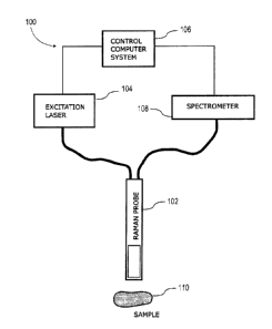
[0058] With reference to Figure 1, and in accordance with one exemplary
embodiment, a
dynamic Raman signal acquisition system, generally referred to using the
numeral 100, will now
be described. In this exemplary embodiment, the system comprises a Raman probe
102,
operatively connected via an optical waveguide to an excitation light source
(laser) 104, and
operable to direct and focus the monochromatic excitation therefrom to a
biological tissue sample
110. The probe is further operable to capture the scattering light from sample
110 and direct it, via
a waveguide, to a spectrometer 108 for analysis. The probe 102 may, in some
embodiments, be
integrated into a handheld device or similar. Spectrometer 108 may be operable
to optically
respond from a range of about 785 ¨ 1200 nm for Stokes Raman detection with a
laser excitation at
785 nm and from a range of 633 ¨ 790 nm for Stokes Raman detection with a
laser excitation at
.. 633 nm, for example. The system further comprises a controller (control
computer system) 106
operatively connected to both excitation light source 104 and spectrometer 108
and programmed to
dynamically adjust at least one signal acquisition parameter, such as the
irradiation power and/or
acquisition time as a function of the measured optical response of one or
multiple successive
acquisitions. It will be appreciated that controller 106 may take various
forms, which may include,
but is not limited to, a dedicated computing or digital processing device,
microprocessor, a general
computing device, tablet and/or smartphone interface/application, and/or other
computing device
as may be readily appreciated by the skilled artisan. Furthermore, this
controller 106 may further
CA 3051348 2019-08-07
comprise a digital screen display (not shown) to at least output information
about the measured
Raman response.
100591 With reference to Figure 2, and in accordance with one exemplary
embodiment, an
illustrative dynamic Raman signal acquisition process 200 will now be
described. A common
problem when trying to take Raman measurements on a new unknown sample in an
operating
environment such as an operating room or similar is that it is not always
known what type of noise
is to be expected. Moreover, the optimal acquisition parameters may also
change depending on the
time or specific location and/or orientation of the Raman probe. In some
cases, for instance there
may be metals or highly fluorescent tissue present, etc. As such, the method
as described in this
Jo exemplary embodiment seeks to optimize the acquisition parameters in
light of the currently
experienced noise levels and local environment.
[0060] In this exemplary embodiment, the method first determines, in
steps 202 to 212, the
optimal excitation laser power and acquisition time operable to maximize the
acquired raw signal.
By "raw signal", what is meant is the optical response, characterized by a
measured optical
intensity vs. a Raman shift (in cm-1), as captured by the probe when the
sample is irradiated. This
raw signal includes contributions from the Raman signal, but also a
fluorescence contribution, an
ambient light contribution, and multiple noise contributions, which include
readout noise, dark
noise (due to thermal excitations), shot noise (photon counting noise), cosmic
rays, etc. Initially
(step 202), both the laser power and acquisition time are set to an initial
known value. It is
important to emphasize that for biological in-vivo applications the laser
power cannot exceed
safety limit for the tissue that is being interrogated. One such guideline for
laser intensity safety
limits is given in Matthes, R., et al. "Revision of guidelines on limits of
exposure to laser radiation
of wavelengths between 400 nm and 1.4 mu m." Health Physics 79.4 (2000): 431-
440, the entire
contents of which are hereby incorporated herein by reference These initial
values may be
determined beforehand by the user or programmed into controller 106. The
system then acquires
iteratively kR initial raw signals RSinit (step 204), wherein each initial raw
signal is further
processed (step 206) to determine the maximum signal level value (RSmax) that
is expected to be
measured using the current initial acquisition parameters. The method then
checks to see if this
RSmax value is larger than a maximum allowable signal limit (RSIffini) (step
208). If the measured
maximum (estimated) initial raw signal value (RMmax) is deemed to be too
large, the system
reduces the laser power by 50% (step 210) and again acquires another set of kR
initial raw signals
11
CA 3051348 2019-08-07
(step 204-208). If not, the system then uses the RSmax value to determine
(step 212) the optimal
values of the acquisition parameters (laser power (LPset) and acquisition time
(Tset)) to be used for
the following real data acquisition process.
[0061] In the steps 214 to 218, a temporary background signal (BGtemp)
is generated. This
BGtemp is used in the iterative acquisition process of following steps 220 to
236. In the presently
discussed embodiment, the background signal comprises both the dark spectrum
(accumulated
dark current) and ambient light contributions to the measured raw signal. The
dark spectrum is
generated by the accumulation of thermally excited electrons (e.g. dark
current) in the Raman's
probe detector. It is dependent not on illumination intensity but on the
detector's temperature and
exposition time. Ambient light sources may include any source of
electromagnetic radiation
overlapping with the detection range of the system, such as surgical lights
that provide lighting in
and around the operative field, conventional fluorescent light sources used to
light-up the room,
windows with a view to the exterior of the operating room or similar. To
remove these
contributions, the laser is turned off (step 214) and a series of kB
background signal measurements
are made (step 216) with the same integration time as a tissue measurement.
This series of
temporary background signals are processed to create a temporary
representative background
signal BGtemp (step 218). This BGtemp is used for subtraction from all
subsequent raw signal
acquisitions to be recorded under similar conditions.
[0062] After optimizing the acquisition parameters (LPset, Tset) and
acquiring the temporary
background signal BGtemp, the main acquisition loop may be started (steps 220
to 236). The laser
power is first set to the previously calculated optimal value LPset (step 220)
before beginning the
measurement loop. This starts with the acquisition of a first raw signal RS,.
This signal is then
analyzed in case the maximum signal value measured is larger than the maximum
allowable signal
strength (step 224). If this is the case, then step 222 is repeated, if not,
the raw signal RS, may be
further processed. To do so, the narrowband outliers are removed (step 226).
These narrowband
outliers are commonly produced by ambient cosmic rays that are detected and
produce a very
narrow spike or peak in the signal (spectrum) that does not correspond to a
Raman emission. The
techniques used to remove these spikes are well known in the art. Once all the
narrowband outliers
are removed, the signal RS, is then used with the set of all previously taken
raw signals (1 to i-1) to
produce an averaged raw signal RSaver (step 228). The temporary background
signal is then
subtracted (step 230) from this averaged raw signal to produce a RSwB
("without background")
12
CA 3051348 2019-08-07
signal. Next, the baseline (BS), which represents the fluorescence
contribution to the signal is
identified and subtracted to produce the final Raman signal (Ramansig=RSwB-BS)
for the current
iteration (step 232).
[0063]
In one embodiment, the step 232 of extracting Raman spectra from raw signals
by
removing baselines can be defined as follows. The baseline originates mostly
from fluorescence
that gets co-excited with Raman signal. In case of tissues, the fluorescence
signal is typically
strong so extracting a weak Raman spectrum can be a challenge. There are many
algorithms for
baseline removal known to people skilled in the art. An example of such an
algorithm can be found
in Zhao, Jianhua, et al. "Automated autofluorescence background subtraction
algorithm for
biomedical Raman spectroscopy." Applied spectroscopy 61.11 (2007): 1225-1232.
Most of these
baseline removal algorithms are based on the general approach: (1) An optional
first step is to
remove high frequency noise and outliers from the raw signal; (2) Make an
initial estimate for the
baseline; (3) Then iteratively repeat the next two steps until a predefined
convergence criterium is
satisfied: (a) Calculate the deviation of the estimated baseline from the
signal using a predefined
cost function; (b) Based on the cost function values, estimate the new
baseline function.
[0064]
In this section, we describe a specific efficient baseline removal strategy
suitable for the
present disclosure; but other baseline removal algorithms known to people
skilled in the art can be
used as well. All signals are represented as vectors of size N where N is
typically the number of
spectrometer detector pixels across the spectroscopic axis or smaller than
that in case some parts of
spectra are purposely truncated because they don't carry useful information.
The indices i for the
defined quantities run in the range (1,...,N). If yi is a raw signal then
optimal baseline bi
minimizes the cost function:
L(b) = EZ=i(p(bi ¨ Fyi)
(1)
discussed in Mazet, Vincent, et al. "Background removal from spectra by
designing and
minimising a non-quadratic cost function." Chemometrics and intelligent
laboratory systems 76.2
(2005): 121-133. The cost function is given by:
13
CA 3051348 2019-08-07
(p(x) = t X22 if x < s
(2)
s otherwise
with s is a constant of our choosing and normally taken as zero. The filter F
is a filter with some
parameters of our choosing. Savitsky-Golay filter has be confirmed as a good
choice in most of the
cases which has been confirmed by other authors as well (e.g. Chen, Kun, et
al. "Improved
Savitzky¨Golay-method-based fluorescence subtraction algorithm for rapid
recovery of Raman
spectra." Applied optics 53.24 (2014): 5559-5569.). The degree and number of
channels for
Savitsky-Golay filter can be optimized for the particular class of Raman
signals that are being
investigated. For tissues, we found that degrees of one or two; and a number
of channels between
50 and 100 usually provide satisfactory results. The iterative steps (a) and
(b) as described above
can be defined in our case as:
bk = Fsk (3)
k+1 (Yi if Yi <
4
bt if b< y1
( )
where k is the iterative step and so is defined as the output of the opening
operator (the opening
operator is defined below) applied on raw signal yi. The raw signal can be
preprocessed
beforehand to remove outliers and high frequency noise as mentioned above. The
opening operator
is defined with its window size W (must be odd) and this set of operations
applied on signal yi, as
follows:
M=( W-1 )/2
Erroded = Opened =y
for M< i < N-M-1 do
Interval = i-M: i+M
Eroded(i) = minjElntervaly
end
for M< <N-M-1 do
Interval = i-M: i+M
14
CA 3051348 2019-08-07
Opened(i) = maxiErntervarroded
end
The implementation window size W is typically in the range of 50 to 100. More
details about
general aspects of Savitsky-Golay filter application can be found in
Orfanidis, Sophocles J.
Introduction to Signal Processing. Englewood Cliffs, NJ: Prentice-Hall, 1996.
100651 The signal to noise ratio (SNR) is calculated on this Ramansig at
the wavelength shift of
interest (step 234) and compared to the pre-determined desired threshold of
SNRiimit (step 236).
The fact that the SNR is calculated, in this embodiment, at every acquisition
iteration ensures
efficiency, as it avoids doing unnecessary acquisitions in the case where the
SNR is already found
to be acceptable. If this is the case, the method then proceeds to the next
step. Instead, if the SNR
is found to be unsatisfactory (smaller than SNRIimit), the system proceeds
once more with a new
acquisition iteration i+1 (steps 222 to 236).
100661 Once the SNR is satisfactory, the laser power is turned off once
more and an additional
series of background signals are measured (step 238) to create an averaged
background with a
reduced noise level. This is done to ensure that as many background signals
have been acquired as
the number of signal acquisitions, so that the noise levels in both sets are
comparable. Once this is
done, the acquisition phase is over (step 240) and only the post-acquisition
analysis steps remain.
100671 In the post-acquisition analysis, the final Raman signal and the
final set of background
signals are processed to produce a final statistical analysis (step 242). This
analysis produces the
final Raman spectra (signal intensity vs Raman shift in cm-I) and may use
additional techniques,
such as Laplacian transforms, to help identify the one or more Raman spikes
characteristic of the
sample being irradiated. Furthermore, the results may be shown in the form of
one or more graphs
(including 3D graphs) or the like. Finally, the system may then report and
record the results (step
244).
100681 With reference to Figures 3a and 3b, and in accordance with one
exemplary
embodiment, an exemplary process 306 for estimating a maximum initial signal
level in the
dynamic Raman signal acquisition process of Figure 2 will now be described. In
this exemplary
process, the set of kR initial raw signals acquired in step 204 of Figure 2
are first processed (step
346) to remove any narrowband outliers (e.g., cosmic rays contribution). Once
this is done, the
CA 3051348 2019-08-07
resulting signals are analyzed (step 348) to extract the maximum signal level
found in each one.
Since only a limited number of raw signals are acquired during this phase, it
is statistically possible
that an arbitrary raw signal acquired for this set of acquisition parameters
can be bigger than the
maximum signal level from the initial limited set of raw signals. This is due
to random noise
composed of shot noise, thermal noise, and readout noise that is superimposed
on the signals
which can be artificially increased or decreased in this way. Fortunately,
concepts of shot noise,
thermal noise, and readout noise as well as estimates of their values are well
known to persons
skilled in the art. These noise estimates can be used to set upper limits for
raw signals based on a
limited set of raw signals. The estimates for readout noise and thermal noise
are provided by the
optical detector manufacturer and the estimate for the shot noise related to a
signal level L is given
with sqrt(L). Since the thermal noise, TN, readout noise, RN, and the shot
noise are independent
from each other, they are added in quadrature to create an estimate (step 350)
for the upper limit of
the acquired raw signals within one standard deviation:
RSmax = Lrnõ, + + RN2 + TN2).
[0069] An alternative way to create an estimate for RSmax in cases where
additional sources of
random noise may be present is to perform a study prior to measurements which
includes acquiring
a large number of raw signals for a given set of acquisition parameters and
creating a histogram
352 as shown in Figure 3b. The histogram data can provide an estimate for
typical uncertainties
involved in raw signal acquisitions.
[0070] With reference to Figure 4, and in accordance with one exemplary
embodiment, an
exemplary acquisition parameter optimization process 412 in the dynamic Raman
signal
acquisition process of Figure 2 will now be described. In this exemplary
embodiment, the process
assumes that the acquired signal strength RS, from which the dark current
contribution has been
subtracted, is directly proportional to both the excitation laser power (LP)
and the acquisition time
(T). Using these linear relationships, it is then possible to estimate the
optimized values for these
two operational parameters during the acquisition process (Lset, Tset) from at
least one previous
measurement done with initial values (L., Tir11). These optimized values are
chosen to maximize
the signal level without exceeding the maximum allowable signal level
(RShmit).
[0071] Two cases may be identified. To identify which case is relevant
to the present
acquisition, the system first extrapolates (step 454) from the initial max
signal level acquired
16
CA 3051348 2019-08-07
(RSmax) at the initial laser power (LS) the raw signal level expected to
measure if the maximum
laser power (LPmax) is used. In the first case, using the maximum allowable
laser power (LP.) is
found to lead to a maximum signal level (LP.) that is smaller than the maximum
allowable signal
level (RSiimit):
(RS,,,, ¨ D = Tini) LPini
_______________________________________ <
(RSumit ¨ D = Tini) LPmax
or iLP
D = Tinitial max/L-Pinitial) = (RSmax ¨ D = Tinitini) < RStimit
where D is the dark current, Tint is the initial acquisition time and D = Tini
gives the accumulated
dark spectra contribution to the measured raw signal. If this inequality is
true, then (step 456) the
laser power may be safely set to maximum (LPset = LPmax) and the acquisition
time is increased to
maximize the acquired raw signal level to RStimit. As mentioned above,
assuming a proportional
relationship between the acquisition time and signal level acquired, the
acquisition time may be set
to:
Tset = RStimit/(D LPmax = (RSmax D = Tinitial)/(LP - initial = Tinitica))=
[0072]
In the second case (i.e. the above inequality is found to be false),
extrapolating from an
initial raw signal measurement shows that using the maximum laser power would
lead to a signal
level higher than RSIimit. In this case (step 458), the acquisition time is
kept the same (Tset = Tim)
but here the laser power (LPset) is set, again assuming a proportional
relationship between RS and
LP, to a value lower than LPffia, but estimated to lead to the maximum
allowable signal (RS1,,,t):
LPset = LPinitial(RSlimit D = Tinitiat)/(RSmax D = Tinitiat)=
[0073] With reference to Figure 5, and in accordance with one exemplary
embodiment, an
exemplary temporary background signal process in the dynamic Raman signal
acquisition process
of Figure 2 will now be described. In this exemplary embodiment, the step 218
of Figure 2 is
expanded to include the set of steps 518 comprising steps 560 and 562. In step
560, each of the kB
background signals acquired in step 216 of Figure 2 are processed to identify
and remove any
narrowband outliers such as those created by cosmic rays. The kB background
signals are then
compared to one another to find the one indicative of the smallest signal
strength, which is used to
define the temporary background signal BGtemp (step 562). BGtemp is not a
single value but covers
17
CA 3051348 2019-08-07
the full spectral range. Min () in this context thus means that the value of
BGtemp for each
wavenumber (or wavelength) is equal to the minimal value among BG1,
BG2,...,BGkb values at
that wavenumber. The reason it is done in this way is because it is possible
to pick up external
unstable noise i.e. flickering of fluorescence bulbs, which would create an
artifact in the
background signals. Instabilities such as light bulb flickering are dealt with
in the main iterative
loop since more complex algorithms may be needed to distinguish such artifacts
from genuine
Raman signals.
100741 With reference to Figure 6, and in accordance with one exemplary
embodiment, a
distinction between signal intensity and noise intensity will now be
described. In general, the
1() intensity measured at a given Raman shift (cm-1) comprises
contributions from the Raman
response to which is added a multiplicity of noise sources contribution. As
mentioned above, these
noise sources include readout noise, shot noise, dark noise, etc. Readout
noise is caused by
electronic noise in the detector output stage and related circuitry, which
largely dictates the
detection limit of the spectrometer. Shot noise is associated with the
statistical variation in the
number of photons incident on the detector, which follows from a Poisson
distribution. Dark noise
(dark current) is associated with the statistical changes in the number of
thermally generated
electrons in the detector with the excitation source turned off. In figure 6,
the dark horizontal line
represents the average intensity of the signal that is obtained by averaging
over multiple
acquisitions.
100751 With reference to Figure 7, and in accordance with one exemplary
embodiment, an
exemplary final background signal process following the temporary background
signal process of
Figure 5, in the dynamic Raman signal acquisition process of Figure 2 will now
be described. As
mentioned above, it may be important to acquire at least as many background
signals as the
number of Raman acquisitions. This is because one needs to have similar levels
of confidence in
both types of signals for the post-acquisition statistical analysis. In this
exemplary embodiment,
step 238 of Figure 2 is expanded to include the process 738. Herein, the
method first checks (step
764) to see if the total number of acquisition (i) done in steps 222 to 236 of
Figure 2 is larger than
the total number of temporary background signals acquired (kB) in steps 216 of
Figure 2. If this is
the case, then the system proceeds with the acquisition of an additional (i-
kB) background signals
.. (step 766). If not, then only the last i temporary background signals are
kept (step 768). Either
way, only an equal number of background signals and raw signals are used.
18
CA 3051348 2019-08-07
[0076] With reference to Figure 8, and in accordance with one exemplary
embodiment, an
example of a series of Raman response signals acquired over time will now be
described. Figure 8
shows different collection of successive signals (k = 1, 2 ,3 ...) acquired
over time, each signal
similar to the one shown in Figure 6. This illustrates the large noise
variation that may be found in
each acquisition but that the noise may be averaged out to reveal the
underlying signal (dark
horizontal line in Figure 6).
[0077] With reference to Figure 9, and in accordance with one exemplary
embodiment, a
dynamic Raman signal acquisition system 900 will now be described. In this
exemplary
embodiment, similarly to the one shown in Figure 1, the system again comprises
a Raman probe
902, operatively connected via an optical waveguide to an excitation light
source (laser) 904, and
operable of directing and focusing the monochromatic excitation therefrom to a
biological tissue
sample 910. The probe is further operable to capture the scattering light from
sample 910 and
directing it, via a waveguide, to a spectrometer 908 for analysis. The probe
902 may, in some
embodiments, be integrated into a handheld device or similar. Spectrometer 908
may be operable
to optically respond from a range of about 785 ¨ 1200 nm for Stokes Raman
detection with a laser
excitation at 785 nm and from a range of 633 ¨ 790 nm for Stokes Raman
detection with a laser
excitation at 633 nm. It will be appreciated that controller 906 may take
various forms, which may
include, but is not limited to, a dedicated computing or digital processing
device, microprocessor, a
general computing device, tablet and/or smartphone interface/application,
and/or other computing
device as may be readily appreciated by the skilled artisan. Furthermore, this
controller 906 may
further comprise a digital screen display (not shown) to at least output
information about the
measured Raman response. In addition, this exemplary embodiment further
comprises a laser
power control device 912, usually comprising a mechanical shutter and/or
optical modulator,
operatively connected to controller 906 and connected to the optical waveguide
at the output of the
.. excitation light source 904. This laser power control device 912 is
operable to modulate the
irradiation power incident on sample 910 without the need to turn on and off
the light source 904
itself. This laser power control device is also operationally linked to
controller 906 and may be
controlled therefrom as a function of the data acquired by spectrometer 908.
[0078] With reference to Figure 10, and in accordance with one exemplary
embodiment, an
exemplary safety shutdown process of the dynamic Raman signal acquisition
process of Figure 2
will now be described. In this embodiment, steps 222 and 224 of Figure 2 are
further detailed. As
19
CA 3051348 2019-08-07
in the original step 222, in step 1022 a new raw signal RS, is acquired. This
new raw signal is now
analyzed to find if it contains any critical features (step 1070)
representative of hazardous
chemicals and/or materials produced by burning tissue. In some embodiments, a
list of critical
features to look may be stored in a database accessible to the system's
controller. If the system
does detect the presence of any critical features, then the acquisition
process is stopped completely,
the laser power is set to zero (LP = 0) and the problem is reported to the
user (step 1072). If not,
then the system checks to see if the maximum value found in RS, (step 1024) is
higher than the raw
signal limit (RSI,,,t). If this is the case, the system goes back to step 1022
and acquires a new raw
signal RS,. In the opposite case, RS, is deemed to be acceptable and the
process continues to step
226 of Figure 2.
100791 With reference to Figure 11, and in accordance with one
exemplary embodiment, an
example of an irradiation intensity over time for a given acquisition window
will now be
described. In general, the excitation laser may have a turn-on delay period.
This is illustrated in
Figure 11, wherein the excitation laser is turned on at to, but the laser
intensity only increases to a
stable value after a wait time of t at ti. To avoid this long delay, in some
embodiments, it may
be useful to use a mechanical shutter and/or a modulator instead of turning
the excitation light
source on and off.
100801 With reference to Figure 12, and in accordance with one
exemplary embodiment, an
example of an irradiation intensity over time for a set of sequential
acquisition windows will now
be described. Figure 12 shows the change with respect to time of the
irradiation intensity on the
sample for two consecutive acquisitions. We see that there is a time delay of
At between
acquisition i and acquisition i+1. This is done to avoid photobleaching or
fluorescence quenching.
Photobleaching is a process wherein the acquired signal intensity associated
with the background
fluorescence is reduced by prolonged exposure to the excitation radiation. It
is sometimes used to
remove the fluorescence background from the acquired signal to improve the
signal to noise ratio
of the Raman measurements. However, photobleaching may be undesirable as it
may lead to
sample damage, long acquisition times and it may introduce new unknown noise
sources to the
measured signal. As such, it is preferable to avoid photobleaching the sample
by briefly stopping
the irradiation of the targeted sample between each consecutive acquisition,
usually a few
milliseconds. The fluorescence background may be removed post-acquisition by
methods well
known in art.
CA 3051348 2019-08-07
100811 With reference to Figure 13, and in accordance with one exemplary
embodiment,
another example of a dynamic Raman signal acquisition system 1300 will now be
described. This
exemplary embodiment again comprises a Raman probe 1302, operatively connected
via an optical
waveguide to an excitation light source (laser) 1304, and operable of
directing and focusing the
monochromatic excitation therefrom to a biological tissue sample 1310. The
probe is further
operable to capture the scattering light from sample 1310 and directing it,
via a waveguide, to a
spectrometer 1308 for analysis. The probe 1302 may, in some embodiments, be
integrated into a
handheld device or similar. Spectrometer 1308 may be operable to optical
responses from a range
of about 785 ¨ 1200 nm for Stokes Raman detection with a laser excitation at
785 nm and from a
range of 633 ¨ 790 nm for Stokes Raman detection with a laser excitation at
633 nm. The system
further comprises a controller (control computer system) 1306 operatively
connected to both
excitation light source 1304 and spectrometer 1308 and programmed to
dynamically adjust at least
one signal acquisition parameter, such as the irradiation power and/or
acquisition time as a
function of the measured optical response of one or multiple successive
acquisitions. It will be
appreciated that controller 1306 may take various forms, which may include,
but is not limited to,
a dedicated computing or digital processing device, microprocessor, a general
computing device,
tablet and/or smartphone interface/application, and/or other computing device
as may be readily
appreciated by the skilled artisan. Furthermore, this controller 1306 may
further comprise a digital
screen display (not shown) to at least output information about the measured
Raman response. In
addition, this exemplary embodiment further comprises a light indicator device
1314, which may
comprise one or more lighting devices (i.e. LED or similar), operationally
connected to controller
1306 and operable to notify the user (via a combination of light colors,
intensities, blinking, etc.) of
the status of the acquisition process. This may include information about
whether the device is
proceeding with an acquisition or if it is stopped and/or on stand-by, if
sample 1310 is damaged or
if any hazardous chemicals were detected, if the probe has a good or bad
optical contact with
sample 1310, etc.
100821 With reference to Figure 14, and in accordance with one
embodiment, an alternative
pre-acquisition process of the dynamic Raman signal acquisition process of
Figure 2 will now be
described. In this exemplary embodiment, the step 202 shown in Figure 2 is
replaced by a series of
steps 1402 wherein the system first probes the sample before continuing to
step 204. For example,
in may be useful for a surgeon using, for instance, a handheld embodiment of
the Raman probe, to
have the system autonomously start the acquisition phase (automated start-up
process) if it detects
21
CA 3051348 2019-08-07
that the probe is in stable optical contact with the sample. For example, the
system may determine
that the probe is indeed targeting a fixed sample by seeing detecting fixed
Raman characteristics
may the acquisition phase may begin. More precisely, the series of new steps
1402 start with step
1472, wherein the initial Laser Power is set to a lower laser power value
optimized for probing the
sample (LPprobe) instead of the normal initial Laser Power value (LPinitiai).
The system then acquires
a first probe raw signal (step 1474) and a second raw signal (step 1476),
which are then compared
(step 1478) to determine if each raw signal is large enough and if both
signals are similar enough.
If either one of the signals is found to be too small or if large spectral
differences are found
between the two signals, then the system acquires a new probe raw signal and
compares it to the
last one (i.e. repeats step 1478). This is done until both raw signal
strengths are deemed
satisfactory and both raw signals are found to be similar enough. The
similarity between the two
probe raw signals is made to ensure that the probe is indeed targeting a fixed
sample and that the
real acquisition measurements can begin. As in the original step 202, step
1480 entails setting the
laser power to LP
-
and the acquisition time is set to Tinitial, as explained above. The system
then
automatically continues the procedure detailed in Figure 2.
[0083]
With reference to Figure 15, and in accordance with one embodiment, another
dynamic
Raman signal acquisition system 1500 will now be described. This exemplary
embodiment again
comprises a Raman probe 1502, operatively connected via an optical waveguide
to an excitation
light source (laser) 1504, and operable of directing and focusing the
monochromatic excitation
therefrom to a biological tissue sample 1510. The probe is further operable to
capture the scattering
light from sample 1510 and directing it, via a waveguide, to a spectrometer
1508 for analysis. The
probe 1502 may, in some embodiments, be integrated into a handheld device or
similar.
Spectrometer 1508 may be operable to optical responses from a range of about
785 ¨ 1200 nm for
Stokes Raman detection with a laser excitation at 785 nm and from a range of
633 ¨ 790 nm for
Stokes Raman detection with a laser excitation at 633 nm. The system further
comprises a
controller (control computer system) 1506 operatively connected to both
excitation light source
1504 and spectrometer 1508 and programmed to dynamically adjust at least one
signal acquisition
parameter, such as the irradiation power and/or acquisition time as a function
of the measured
optical response of one or multiple successive acquisitions. It will be
appreciated that controller
1506 may take various forms, which may include, but is not limited to, a
dedicated computing or
digital processing device, microprocessor, a general computing device, tablet
and/or smartphone
interface/application, and/or other computing device as may be readily
appreciated by the skilled
22
CA 3051348 2019-08-07
artisan. Furthermore, this controller 1506 may further comprise a digital
screen display (not
shown) to at least output information about the measured Raman response. In
addition, this
exemplary embodiment further comprises an optical directional coupler 1516
connected to the
optical waveguide between the excitation laser 1504 and Raman probe 1502, said
directional
coupler operationally connected to a light detector 1518. In some embodiments,
the directional
coupler 1516 and light detector 1518 may be integrated to probe 1502. The
directional coupler is
operable to detect back reflections of the light outputted from probe 1502,
via light detector 1518,
and identify that a good optical contact with sample 1510 has been
established. (MORE). The
presence of an directional coupler allows for a very low probing excitation
power (as explained
above with respect to Figure 14) when verifying that probe 1502 has a stable
optical contact with
sample 1510.
100841 While the present disclosure describes various embodiments for
illustrative purposes,
such description is not intended to be limited to such embodiments. On the
contrary, the
applicant's teachings described and illustrated herein encompass various
alternatives,
modifications, and equivalents, without departing from the embodiments, the
general scope of
which is defined in the appended claims. Except to the extent necessary or
inherent in the
processes themselves, no particular order to steps or stages of methods or
processes described in
this disclosure is intended or implied. In many cases the order of process
steps may be varied
without changing the purpose, effect, or import of the methods described.
100851 Information as herein shown and described in detail is fully capable
of attaining the
above-described object of the present disclosure, the presently preferred
embodiment of the present
disclosure, and is, thus, representative of the subject matter which is
broadly contemplated by the
present disclosure. The scope of the present disclosure fully encompasses
other embodiments which
may become apparent to those skilled in the art, and is to be limited,
accordingly, by nothing other than
the appended claims, wherein any reference to an element being made in the
singular is not
intended to mean "one and only one" unless explicitly so stated, but rather
"one or more." All
structural and functional equivalents to the elements of the above-described
preferred
embodiment and additional embodiments as regarded by those of ordinary skill
in the art are hereby
expressly incorporated by reference and are intended to be encompassed by the
present claims.
Moreover, no requirement exists for a system or method to address each and
every problem
sought to be resolved by the present disclosure, for such to be encompassed by
the present
23
CA 3051348 2019-08-07
claims. Furthermore, no element, component, or method step in the present
disclosure is intended
to be dedicated to the public regardless of whether the element, component, or
method step is
explicitly recited in the claims. However, that various changes and
modifications in form, material,
work-piece, and fabrication material detail may be made, without departing
from the spirit and scope
of the present disclosure, as set forth in the appended claims, as may be
apparent to those of
ordinary skill in the art, are also encompassed by the disclosure.
24
CA 3051348 2019-08-07