Note: Descriptions are shown in the official language in which they were submitted.
APPARATUS, SYSTEMS AND METHODS FOR INTEGRATIVE PHOTO-
OPTICAL/MECHANICAL TEST FOR NONCONTACT MEASUREMENT OF
POLYMERIZATION
CROSS-REFERENCE TO RELATED APPLICATION(S)
[001] This application claims priority to U.S. Provisional Application No.
62/449,404 filed
January 23, 2017 and entitled "Integrative Photo-Optical/Mechanical Test for
Noncontact
Measurement of Polymerization".
GOVERNMENT SUPPORT
[002] This work was supported in part by grant number 1438537 awarded by the
National
Science Foundation. The government has certain rights in this invention.
TECHNICAL FIELD
[003] The disclosed technology relates generally to noncontact methods,
devices and
systems for measuring polymerization of a sample.
BACKGROUND
[004] The disclosure relates to apparatus, systems and methods for measuring
polymerization of a sample, such as coagulation of blood or polymerization of
another
biological material.
[005] Blood coagulation is the process in which the blood changes from a
liquid to gel state
in response to blood loss, referred to as the hemostatic process. The
coagulation cascade is
initiated by adhesion and activation of platelets at the injury site of the
vessel wall and occurs
through two separate pathways: the extrinsic and intrinsic, both converging on
the common
pathway. The extrinsic pathway is triggered by tissue factor (TF) in response
to vascular
trauma, and the intrinsic pathway is triggered by contact of the blood with
dysfunctional
endothelium or collagen. During the common pathway, fibrinogen is converted
into fibrin by
thrombin. The fibrin polymerization and its crosslinking by Factor XIII forms
a blood clot.
The hemostasis process is the result of a delicate balance between pro- and
anti-coagulants,
platelets and blood cells.
- 1 -
CA 3051365 2023-02-16
CA 03051365 2019-07-23
W02018/136949 oi
PCT/US2018/014879
[006] Due to a significant loss of blood during trauma or major surgery,
patients
often develop coagulopathy, i.e., a pathophysiological condition characterized
by depletion of
both pro- and anti-coagulants in blood. Coagulopathic patients are at high
risks of both
hemorrhage and thrombotic complications, which significant increase patient
morbidity and
mortality. The coagulation status of such patients could rapidly change from
an anti- to pro-
coagulant state during injury and resuscitation. Therefore, monitoring the
coagulation status
of coagulopathic patients, especially during blood transfusion or surgery is
critical.
[007] The devices currently available for rheological measurements induce
contact
with device walls or other artificial surfaces, which causes large measurement
errors.
Additionally, testing for coagulation parameters using available contact
techniques requires a
significant amount of time to obtain diagnostic data (at least 30 minutes) and
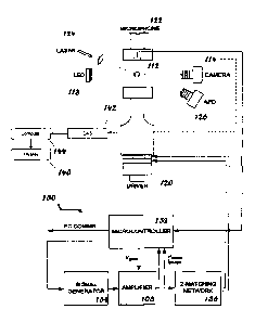
a large sample
volume (at least 0.4 milliliters).
[008] Thus, there is a need in the art for fast and reliable noncontact
devices,
systems and methods that can work with low-volume samples.
BRIEF SUMMARY
[009] Discussed herein are various devices, systems and methods relating to
methods, systems and devices for the real-time assessment of whole blood or
blood plasma
coagulation by non-contact acoustic tweezing technology and for measuring
polymerization
characteristics of a sample, including but not limited to rheological
measurements and
polymerization kinetics.
[010] No feature of the disclosed implementations is critical or essential
unless it is
expressly stated as being "critical" or "essential."
[011] In one Example, a system of one or more computers can be configured
to
perform particular operations or actions by virtue of having software,
firmware, hardware, or
a combination of them installed on the system that in operation causes or
cause the system to
perform the actions. One or more computer programs can be configured to
perform
particular operations or actions by virtue of including instructions that,
when executed by
data processing apparatus, cause the apparatus to perform the actions.
[012] One Example includes a noncontact, acoustic-tweezing method of
measuring
time-dependent rheological and polymerization properties of a sample
including: levitating
the sample, modulating the amplitude of acoustic pressure applied to the
sample so as to
induce deformation, capturing at least one image of the sample, collecting at
least one photo-
-2-
CA 03051365 2019-07-23
y WO 2018/136949 410*
PCT/US2018/014879
optical measurement and at least one mechanical measurement from the captured
images of
the levitating sample during deformation, and determining at least one
rheological property of
the sample. Other embodiments of this Example include corresponding computer
systems,
apparatus, and computer programs recorded on one or more computer storage
devices, each
configured to perform the actions of the methods.
[013]
Implementations may include one or more of the following features. The
method where the deformation is quasi-static or oscillatory. The method
further including
determining at least one kinetic property of sample polymerization. The method
where the
determined kinetic properties of sample polymerization are selected from the
group including
of: photo-optical tweezograph, mechanical tweezograph, reaction time, monomer
formation
rate, maximum monomer level, polymerization onset, polymerization rate,
polymerization
time, gel firmness and polymer network formation time. The method where the
polymer
network formation time is the time difference between the polymerization rate
in a
mechanical tweezograph and he monomer formation rate in a photo-optical
tweezograph. The
method where the rheological property is coagulation. The method where the at
least one
photo-optical measurement is selected from the group including of: light
intensity, laser
scattering intensity and turbidity. The method where the at least one
mechanical measurement
is elasticity. The method where the at least one image is photographic. The
method where the
at least one image is a laser scattering image. The method further including
executing data
analysis on the collected at least one photo-optical measurement and at least
one mechanical
measurement. The method where the sample is a biological material selected
from the group
including of: whole blood, blood plasma, mucus, sperm, lymph, synovial fluid,
cerebrospinal
fluid and soft biological tissue. The method where the sample is selected from
the group
including of: a polymer, a polymer gel and a polymeric liquid. The method
where the at least
one photo-optical measurement is selected from the group including of: average
light
intensity through central area of the sample and turbidity of the sample over
time. The
method where the one or more mechanical measurements are determined from quasi-
static
and oscillatory deformation of the sample for different acoustic pressure
amplitudes at
different times. The method where the one or more rheological property is
selected from the
group including of: elastic modulus, shear elasticity, shear viscosity,
dynamic modulus,
storage modulus, and loss modulus. The method where the fibrin network
formation time
(FNFT) is the time difference between the clotting rate (CR) in a mechanical
tweezograph
and the fibrin formation rate (FFR) in a photo-optical tweezograph. The method
where the
extracted coagulation kinetics data is selected from the group including of:
photo-optical
-3-
CA 03051365 2019-07-23
W02018/136949 *
PCT/US2018/014879
tweezograph, mechanical tweezograph, reaction time (RT), fibrin formation rate
(FFR),
maximum fibrin level (MFL), clot initiation time (CIT), clotting rate (CR),
time to firm clot
formation (TFCF), maximum clot firmness (MCF), and fibrin network formation
time
(FNFT). The method further including extracting coagulation kinetics data. The
method
further including evaluating functional levels of fibrinogen from at least one
of RT, MFL,
MCF, and FNFT data extracted from the photo-optical and mechanical
tweezographs. The
method further including evaluating functional levels of factor XIII. The
method may also
include from at least one of RT, MFL, MCF, and FNFT data extracted from the
photo-optical
and mechanical tweezographs. The method further including monitoring
functional levels of
fibrinogen or factor xiii to assess blood coagulation disorder. The method
further including
assessing the effects of a cross-linker from the determined polymerization
kinetics. The
method further including assessing the effects of cross-link breakers on the
sample from the
determined polymerization kinetics. The method further including assessing the
effects of a
cross-link inhibitors on the sample from the determined polymerization
kinetics.
Implementations of the described techniques may include hardware, a method or
process, or
computer software on a computer-accessible medium. The method further
including
extracting coagulation kinetics data. The method further including evaluating
a functional
level of fibrinogen. The method further including evaluating a functional
level of Factor XIII.
The method further including evaluating coagulation factor deficiency. The
method may also
include from at least one of RT, MFL, MCF, and FNFT data extracted from the
photo-optical
and mechanical tweezographs. The method further including monitoring
functional levels of
coagulation factors to assess blood coagulation disorder.
[014] One Example includes a noncontact, acoustic-tweezing system for
measuring
time-dependent rheological and polymerization properties of a sample
including: a levitator
configured to levitate the sample, an amplitude modulator configured to
modulate acoustic
pressure applied to the sample so as to induce deformation, a camera
configured to capture at
least one image of the sample and generate captured images, and an analysis
system
configured to: collect: at least one photo-optical measurement of the sample
and at least one
mechanical measurement of the sample. The system also includes capturing
images during
deformation. The system also includes determining at least one rheological
property of the
sample.
[015] While multiple embodiments are disclosed, still other embodiments of
the
disclosure will become apparent to those skilled in the art from the following
detailed
description, which shows and describes illustrative embodiments of the
disclosed apparatus,
-4-
systems and methods. As will be realized, the disclosed apparatus, systems and
methods are
capable of modifications in various obvious aspects, all without departing
from the spirit and
scope of the disclosure. Accordingly, the drawings and detailed description
are to be
regarded as illustrative in nature and not restrictive.
[015a] Accordingly, in one aspect, the present invention resides in a
noncontact, acoustic-
tweezing method of measuring time-dependent rheological and polymerization
properties of a
sample comprising: a. levitating the sample; b. modulating the amplitude of
acoustic pressure
applied to the sample so as to induce deformation; c. capturing at least one
image of the
sample; d. collecting at least one photo-optical measurement and at least one
mechanical
measurement from the captured images of the levitating sample during
deformation; e.
determining at least one rheological property of the sample; and f.
determining at least one
kinetic property of sample polymerization, wherein the determined kinetic
properties of
sample polymerization: i. comprise polymer network formation time measured by
the time
difference between the time to maximum polymerization rate in a mechanical
tweezograph
and the time to maximum monomer formation rate in a photo-optical tweezograph,
and ii.are
selected from the group consisting of: photo-optical tweezograph, mechanical
tweezograph,
reaction time, monomer formation rate, maximum monomer level, polymerization
onset,
polymerization rate, polymerization time, gel firmness and polymer network
formation time.
[015b] In another aspect, the present invention resides in a noncontact,
acoustic-tweezing
system for measuring time-dependent rheological and polymerization properties
of a sample
comprising: a. levitator configured to levitate the sample; b. an amplitude
modulator
configured to modulate acoustic pressure applied to the sample so as to induce
deformation;
c. a camera configured to capture at least one image of the sample and
generate captured
images; and d. a data acquisition system configured to: i. collect: A. at
least one photo-optical
measurement of the sample; and B. at least one mechanical measurement of the
sample
during deformation from the captured images, and ii. determine at least one
rheological
property of the sample, wherein: i. the data acquisition system is configured
to determine at
least one kinetic property of sample polymerization, ii. the determined
kinetic properties of
sample polymerization are selected from the group consisting of: photo-optical
tweezograph,
mechanical tweezograph, reaction time, monomer formation rate, maximum monomer
level,
polymerization onset, polymerization rate, polymerization time, gel firmness
and polymer
network formation time, and iii. the polymer network formation time is the
time difference
between reaching the maximum polymerization rate in a mechanical tweezograph
and
reaching the maximum monomer formation rate in a photo-optical tweezograph.
- 5 -
CA 3051365 2023-02-16
[015c] In a further aspect, the present invention resides in a noncontact,
acoustic-tweezing
system for measuring time-dependent rheological and polymerization properties
of a sample
comprising: a. levitator configured to levitate the sample; b. an amplitude
modulator
configured to modulate acoustic pressure applied to the sample so as to induce
deformation;
c. a camera configured to capture at least one image of the sample and
generate captured
images; and d. a data acquisition system configured to: i. collect: A. at
least one photo-optical
measurement of the sample; and B. at least one mechanical measurement of the
sample
during deformation from the captured images, and ii. determine at least one
rheological
property of the sample, wherein: i. the one or more rheological property is
selected from the
group consisting of: elastic modulus, shear elasticity, shear viscosity,
dynamic modulus,
storage modulus, and loss modulus, and ii. the fibrin network formation time
(FNFT) is the
time difference between reaching the maximum clotting rate (CR) in a
mechanical
tweezograph and reaching the maximum fibrin formation rate (FFR) in a photo-
optical
tweezograph.
BRIEF DESCRIPTION OF THE DRAWINGS
[016] The following drawings form part of the present specification and are
included to
further demonstrate certain aspects of the disclosed ATPA methods, systems and
devices.
The disclosure may be better understood by reference to one or more of these
drawings in
combination with the description of specific embodiments presented herein.
[017] FIG. 1A depicts a schematic of the acoustic tweezing system, according
to an
exemplary embodiment.
[018] FIG. 1B depicts a perspective view of the an exemplary levitator of the
acoustic
tweezing system, according to one embodiment.
[019] FIG. 1C depicts a close-up side view of a levitating sample, according
to the
embodiment of FIG. 1B.
[020] FIG. 1D depicts a perspective view of a camera, according to an
exemplary
embodiment.
[021] FIG. lE depicts front view of the function generator and amplifier,
according to an
exemplary embodiment.
[022] FIG. 2 is a schematic diagram of the acoustic tweezing system, according
to a further
embodiment.
- 5a -
CA 3051365 2023-02-16
[023] FIG. 3A depicts a sequence of photos of a drop of whole blood under
quasi-static
acoustic tweezing.
[024] FIG. 3B is a mechanical tweezograph of 47 drops of citrated whole blood
undergoing
coagulation initiated by Ca.C12.
[025] FIGS. 4A-4C depict raw mechanical data (location vs. aspect ratio) for
porcine gelatin
and alginate samples obtained by increasing and decreasing pressure amplitude.
FIG. 4A
shows 0.90- mm radius drop of 3% gelatin at increasing times. FIG. 4B shows
0.90, 0.89 and
0.86mm drops of 2%, 3% and 4% gelatin at 2 min. FIG. 4C plots location vs.
aspect ratio
curves of a 4% alginate drop with radius of 0.98 mm from 0 to 34 min.
- 5b -
CA 3051365 2023-02-16
CA 03051365 2019-07-23
WO 2018/136949 =
PCT/US2018/014879
[026] FIG. 4D is a mechanical tweezograph (0 vs. time) of 5 drops of 3%
alginate
and 5 drops of 4% alginate for 18 min of tweezing. Nominal radii of drops are
0.98 mm, on
average.
[027] FIG. 5A depicts mechanical tweezographs of EDTA-treated whole blood
with
added CaCl2 and exposed to 0.9% saline (8 drops), tissue factor (TF) (8 drops)
or
cytochalasin D (8 drops).
[028] FIG. 5B depicts the effect of 'TF on 0 at selected times.
[029] FIG. 5C depicts the effect of cytochalasin D on 0 at selected times.
[030] FIG. 6A depicts mechanical tweezographs of citrated control plasma
with
added CaCl2, exposed to 0.9% saline (9 drops), Fibrinogen (9 drops), or GPRP
(9 drops).
[031] FIG. 6B depicts the effect of Fibrinogen and GPRP on slope angle 0 at
5, 6,
and 7 minutes. **p <0.01, ***p <0.001.
[032] FIG. 6C depicts mechanical tweezographs of plasma with fibrinogen
levels of
100, 300, and 500 mg/dL indicating an increase in MCF with fibrinogen
concentration.
[033] FIG. 7 depicts a representative sequence of levitating blood plasma
drop
images during the onset of coagulation, used for photo-optical measurements.
[034] FIG. 8A depicts the photo-optical tweezograph of Factor Assay Control
Plasma (FACT) samples exposed to ellagic acid, showing measurement of RT and
FFR.
[035] FIG. 8B depicts the photo-optical tweezograph of blood plasma samples
with
fibrinogen levels of 100, 300, and 500 mg/dL, indicating an increase in MFL
with fibrinogen
concentration.
[036] FIG. 8C depicts significant correlation of fibrinogen concentration
with MCF
(R2=0.90) and MFL (R2=0.94).
[037] FIG. 9 depicts the illustration of integrated photo-optical and
mechanical
measurement on a blood plasma drop.
[038] FIG. 10A depicts the combined photo-optical and mechanical
tweezographs of
normal plasma (FACT), indicating that the fibrin network formation time (FNFT)
is 3.5
minutes.
[039] FIG. 10B depicts the combined photo-optical and mechanical
tweezographs of
Factor XIII deficient plasma, indicating the FNFT is 4.75 minutes.
[040] FIGS. 11A-11B depict the RT measurement of pooled plasma (PP) as well
as
Factor VII-, Factor VIII-, Factor IX-, Factor X-, and Factor XIII-deficient
plasma from
photo-optical tweezographs.. In FIG. 11A, the deficient plasma is exposed to
tissue factor
(PT test). In FIG. 11B, the deficient plasma is exposed to ellagic acid
-6-
CA 03051365 2019-07-23
W02018/136949
PCT/US2018/014879
DETAILED DESCRIPTION
[041] The various embodiments disclosed or contemplated herein relate to a
unique,
integrated noncontact method for perioperative monitoring of whole blood or
blood plasma
coagulation. The disclosed systems, methods and devices relate to an acoustic
tweezing
polymerization analyzer (ATPA). The disclosed embodiments of the ATPA method,
system
and associated devices are referred to herein variously for brevity, including
as the "ATPA
method," though no specific modality is contemplated.
[042] In various implementations, the disclosed ATPA method provides
technology
to measure the dynamics of polymerization in polymeric or biological fluids
including the
steps such as monomer production and cross-linked polymer network formation.
In
exemplary embodiments, the ATPA method integrates photo-optical measurements
(such as
light intensity or turbidity changes in the sample over time) with mechanical
measurements
(such as changes in bulk deformability of the sample over time), though each
of the photo-
optical and mechanical methods can be utilized without the other portion. In
various
implementations of the integrated ATPA method, these measurements are taken
simultaneously using one single drop of sample fluid levitating or "tweezing"
in air or an
aqueous medium by acoustic radiation forces. Various implementations of the
ATPA
methods, systems and devices are disclosed herein. While much of this
discussion focuses on
blood, it is well understood that other samples of biological and other
material are clearly
contemplated and would be readily recognized by one of skill in the art.
[043] Critical care patients such as trauma and major surgery patients
often develop
coagulopathy due to depletion of both pro- and anti-coagulants. They are at
high risk of both
bleeding and thrombotic complications and require monitoring of their
coagulation status. The
contact of a blood sample with artificial surfaces and its exposure to clot
activators, which
happen in all commercially available coagulation analyzers, may lead to
improper assessment
of blood coagulation and thus errors in predicting bleeding/thrombosis risks.
[044] The levels of fibrinogen and Factor XIII in the blood correlate with
the how
and when blood coagulates. The lack of these factors leads to severe bleeding
due to unstable
clot structure and/or slow clotting. Therefore, a method of measuring these
factors and
monitoring of their functional levels is crucial for treatment of critical
care patients and
patients with coagulation disorders.
-7-
CA 03051365 2019-07-23
1 W02018/136949
PCT/US2018/014879
[045] When applied to blood coagulation, the integrated photo-
optical/mechanical
method can measure the coagulation parameters of whole blood or blood plasma
without
exposing the blood sample to artificial reagents (ellagic acid, kaolin) or
inducing sample
contact with artificial surfaces. The method integrates "acoustic tweezing"-
based photo-
optical and mechanical tests to allow for accurate measurement of parameters
of coagulation,
including: reaction time (RT), fibrin formation rate (FFR), maximum fibrin
level (MFL), clot
initiation time (CIT), clotting rate (CR), time to firm clot formation (TFCT),
maximum clot
firmness (MCF), and fibrin network formation time (FNET). The last parameter
has not been
measureable until the development of the presently disclosed ATPA method and
associated
systems and devices. Through these measurements, one can use the method to
assess the
functional levels of fibrinogen and Factor XIII in a blood sample, which are
necessary for
blood clot formation. When applied to other fluids, the method can detect the
activity of
molecules involved in the polymerization process or in the formation and cross-
linking of
fibrous proteins in biological tissues.
[046] The integrated photo-optical and mechanical test is performed on the
same
sample drop during its levitation in the acoustic tweezing device. The data
indicate that this
integrated test provide the information about coagulation parameters
(including the MCF)
within 10 minutes (while current devices requiring at least 30 minutes) using
the sample
volume of just 4 microliters (-100 times less than the sample volume required
in available
coagulation analyzers).
[047] In certain implementations, the system provides a method of measuring
time-
dependent rheological properties of a sample such as a biological sample,
comprising several
steps, none of which are essential. One step involves levitating the sample.
Another step
requires modulating the amplitude of acoustic pressure around the sample.
Another step
requires taking one or more images of the sample at different times. Another
step requires
taking one or more photo-optical measurements and one or more mechanical
measurements
from the one or more images. It would be apparent to one of skill in the art
that certain of
these steps may be performed in any order.
[048] Another step requires determining the one or more rheological
properties of
the sample at different times from the one or more mechanical measurements.
Another step
requires assessing the polymerization kinetics from the one or more
rheological properties
and one or more photo-optical measurements. It will be appreciated by those of
skill in the art
that various additional steps may be performed, and that certain of these
steps may be
performed in any order and any number of times.
-8-
4 =
[049] Various embodiments of the disclosed non-contact acoustic tweezing
technology can be performed using the devices and methods disclosed in U.S.
Patent
Application No. 15/068,126fi1ed on March 11, 2016, and Patent Cooperation
Treaty Patent
Application No. PCT/US2014/055559, filed on September 15, 2014, both of which
are
entitled "Apparatus, Systems & Methods for Non-Contact Rheological
Measurements of
Biological Materials".
[050] While certain novel features of this invention shown and described
below are
pointed out in the annexed claims, the invention is not intended to be limited
to the details
specified, since a person of ordinary skill in the relevant art will
understand that various
omissions, modifications, substitutions and changes in the forms and details
of the invention
illustrated and in its operation may be made without departing in any way from
the spirit of
the disclosed embodiments of the ATPA method.
EXPERIMENTAL TECHNIQUES & EXAMPLES
[051] It is understood that in some embodiments the tweezograph is the
graph of
sample deformability ("mechanical tweezograph") or sample light intensity /
turbidity ("photo-
optical tweezograph") versus time. All kinetic data are determined from
tweezographs.
[052] In certain embodiments and Examples, "reaction time" refers to the
onset of
light intensity or turbidity change in a photo-optical tweezograph.
[053] In certain embodiments and Examples, "polymerization onset" or "clot
initiation
time" is the onset of sample deformability change in a mechanical tweezograph.
[054]
In certain embodiments and Examples, "monomer formation rate" or "fibrin
_
formation rate" is the time to reach the maximum rate light intensity or
turbidity change in a
photo-optical tweezograph.
[055] In certain embodiments and Examples, "polymerization rate" or
"clotting rate" is
the maximum rate of sample deformability change in a mechanical tweezograph.
[056] In certain embodiments and Examples, "polymerization time",
"solidification
time", or "time to firm clot formation" is the time it takes to reach a
plateau in a mechanical
tweezograph.
[057] In certain embodiments and Examples, "maximum monomer level" or
"maximum fibrin level" is the plateau value of light intensity or turbidity in
a photo-optical
tweezograph.
[058] In certain embodiments and Examples, "gel firmness" or "maximum clot
firmness" is the plateau value of the sample elasticity in a mechanical
tweezograph.
- 9 -
CA 3051365 2023-02-16
CA 03051365 2019-07-23
1 WO 2018/136949
PCT/US2018/014879
[059] In certain embodiments and Examples, "polymer network formation time"
or
"fibrin network formation time" is the time difference between reaching the
"polymerization rate" or the CR in a mechanical tweezograph and the "monomer
formation
rate" or the FFR in a photo-optical tweezograph. The physical meaning of this
parameter
is the time delay between the processes of monomer formation and
polymerization /
clotting.
[060] In certain embodiments and Examples, the sample may be whole blood,
blood plasma, mucus, sperm, lymph, synovial fluid, cerebrospinal fluid, soft
biological
tissue or other known biological material, a polymer, a polymer gel, a
polymeric liquid.
[061] In certain embodiments and Examples, the functional level of
fibrinogen is
determined by integrating the RT, MFL, and MCF data from photo-optical and
mechanical
tweezographs. A higher fibrinogen level corresponds to a smaller RT, a higher
MFL, and a
higher MCF.
[062] In certain embodiments and Examples, the functional level of a cross-
linker (in
case of polymerization) or Factor XIII (in case of coagulation) is determined
from the FNFT data.
It is understood that in these implementations, the lower the FNFT, the higher
the functional
level (activity) of a cross-linker or Factor XIII would be.
[063] Blood coagulation. Blood coagulation is the process in which the
blood
changes from a liquid to gel state in response to blood loss, referred to as
the hemostatic
process. The coagulation cascade is initiated by adhesion and activation of
platelets at the
injury site of the vessel wall and occurs through two separate pathways: the
extrinsic and
intrinsic ones, both converging on the common pathway. The extrinsic pathway
is triggered
by tissue factor (TF) in response to vascular trauma, and the intrinsic
pathway is triggered by
contact of the blood with dysfunctional endothelium or collagen. During the
common
pathway, fibrinogen is converted into fibrin by thrombin. The fibrin
polymerization and its
crosslinking by Factor XIII forms a blood clot. The hemostasis process is the
result of a
delicate balance between pro- and anti-coagulants, platelets and blood cells.
Due to a
significant loss of blood during trauma or major surgery, patients often
develop
coagulopathy, a pathophysiological condition characterized by depletion of
both pro- and
anti-coagulants in blood. Coagulopathic patients are at high risks of both
hemorrhage and
thrombotic complications, which significant increase patient morbidity and
mortality. The
coagulation status of such patients could rapidly change from an anti- to pro-
coagulant state
during injury and resuscitation. Therefore, monitoring the coagulation status
of coagulopathic
patients, especially during blood transfusion or surgery is critical.
-10-
CA 03051365 2019-07-23
I W02018/136949 =
PCT/US2018/014879
[064] Measurement of blood coagulation. Blood coagulation analysis is
routinely
performed to assess bleeding or thrombosis risks in surgical and critical care
patients, patients
on anticoagulant therapy, patients with chronic coagulation disorders such as
coagulation
factor deficiency, hemophilia and thrombophilia, and patients with other
diseases that can
impair the coagulation system (e.g., cancer, atherosclerosis, diabetes, and
sickle cell disease).
Two main approaches are currently used in this field. The first approach is
photo-optical
measurement of coagulation onset in blood plasma exposed to certain
activators. Prothrombin
Time/International Normalized Ratio (PT/INR), activated Partial Thromboplastic
Time
(aPTT), and Thrombin Time (TI') are all the result of such measurements. While
each of
these tests can measure different aspect of coagulation profile, they cannot
provide a globe
picture about hemostasis, even in combination. With the absence of platelet
and red blood
cells, the information yielded from these assays is further limited. The
second approach, used
in whole blood (global) coagulation analysis, is measurement of temporal
changes in
elasticity (stiffness or firmness) of coagulating blood. Whole blood
coagulation tests are
typically presented in a graphical form, as cigar-like traces overlaid with a
reference curve.
Numeric data (clot initiation time, coagulation rate, maximum clot firmness
and the like),
extracted from traces, are also provided to clinicians for proper diagnosis.
[065] Mechanical measurement of blood coagulation. Contact "pin-and-cup"
methods such as thromboelastography (TEG) and rotational. thromboelastometry
(ROTEM)
are currently available to measure the coagulation status of whole blood.
These methods
measure temporal changes in the shear force between a disposable cup
containing a 0.3 ¨ 0.4
ml sample of whole blood and a pin immersed in the blood sample. Intrinsic
pathway
activators such as kaolin or ellagic acid are required to initiate coagulation
using this
approach. The "pin-and-cup" techniques accurately diagnose hyperfibrinolysis
and are
helpful but not reliable tools in screening for hypercoagulable states and
transfusion
guidance. However, the contact of a blood sample with the pin and cup surfaces
creates
artificial conditions for blood coagulation, leading to substantial
differences from the
dynamics of hemostasis in the body. This inherent deficiency is an important
reason behind
poor standardization and high variability of these methods, their inability to
determine
disorders of primary hemostasis, unreliability in detection of impaired
platelet function and
prediction of bleeding after major surgery, insensitivity to warfarin effects
and a strong
effect of heparin flush on thromboelastographic parameters leading to the
necessity of
discarding a large volume of blood before measurement. Previous studies also
indicated that
the. shear stress applied to blood sample has exceed the linear region of
sample elasticity
-11-
CA 03051365 2019-07-23
WO 2018/136949
PCT/US2018/014879
which has been showed to interfere clot formation process and limit the
sensitivity and speed
of measurements. Even with intrinsic pathway activators present, the
coagulation process
occurring in "pin-and-cup" devices remain slow. A significant amount of time
(30-60
minutes) is required to obtain the results needed for diagnosis unless the
extrinsic pathway
activators (e.g., tissue factor) are used.
[066] Acoustic Levitation. Drops, bubbles, solid particles, and other
objects exposed
to an acoustic wave field experience acoustic radiation pressure. In the case
of intense
standing waves, the radiation pressure is significant and can balance the
gravitational force,
levitating the object at a certain spatial position. In the past few decades,
several acoustic
levitation-based methods have been employed to measure the mechanical
properties of fluid
samples, often with complex surface properties. In these methods, the
hydrodynamic theory
and perturbation analysis were applied to infer some of the material constants
from
experimental data on quadrupole shape oscillations of the samples.
[067] Non-Contact Rheology System. According to one implementation, a
system
for levitating the sample, which can be a biological sample, is provided. One
previously-
disclosed exemplary implementation of such an acoustic tweezing system 1 and
associated
components are depicted in FIGS. 1A-1E. FIG. 1A depicts a schematic overview
of the
acoustic tweezing system 1, comprising a levitator 100 that is in operational
communication
with an oscilloscope 102, a function generator 104, and an amplifier 106. As
is shown in
FIG. 1B, in exemplary embodiments of the acoustic tweezing system 1, the
levitator
comprises a transducer 108, such as an acoustic transducer 108 and reflector
110. FIG. IC
shows a detailed depiction of a sample 112 being levitated according to this
embodiment. As
is shown in FIG. 1D, in exemplary embodiments, the acoustic tweezing system 1
further
comprises a camera 114 and an environmental control chamber. A further
implementation of
the levitator 100 comprising the function generator 104 and an amplifier 106
is further shown
in FIG. 1E.
[068] A further implementation of the acoustic tweezing system I is
depicted in the
implementation of FIG. 2. In this implementation, the sample 112 is levitated
above a driver
120 and below a microphone 122, wherein images can be captured via a camera
114
illuminated by a light source 118, such as an LED 118.
[069] In this implementation, the system 1 comprises a laser 124 and diode
126 such
as an avalanche photodiode (APD) 126 for the capture and measurement of
transmitted or
scattered photo-optical signal at a defined wavelength range, therefore being
configured to
measure various optical properties of the levitated sample 112.
-12-
CA 03051365 2019-07-23
), WO 2018/136949
PCT/US2018/014879
[070] Further, in the implementation of FIG. 2, the driver 120 and
microphone 122
are in operable communication with an operations system 130. In this
implementation of the
operations system 130, a microcontroller 132, signal generator 134, amplifier
106 and 2-
matching network 136 are provided, such that an input signal can be generated
and amplified
before being sent to the transducer 108 resulting in the radiation vibration
of the driver 120.
The photographs and
[071] It is understood that by placing a reflector at a specified distance
from the
transducer surface (either a full or half wavelength apart), the acoustic
tweezing system 1
generates a standing wave field with pressure node and antinode with minimum
and
maximum pressure, respectively. The acoustic radiation pressure applied on the
surface of the
drop is able to levitate objects between the node and antinode, where the
resulting acoustic
radiation force balances gravity.
[072] In use, according to certain implementations, a small drop of blood
or other
biological fluid 112 will be dripped into the opening 116, where it will be
levitated in a
standing acoustic wave field 150 and forced into shape oscillation. The sample
112 is
levitated above a driver 120 and below a microphone 122, wherein images can be
captured
via a camera 114 illuminated by a uniform soft light source 118, such as an
LED
118. Greyscale images can be recorded at different frame per second (FPS)
depending on the
requirements of the experiment or implementation and stored in the data
acquisition system
through a communications system such as a high speed USB 3.0 cable, wireless
transmission
or the like for further shape deformation and/or photo-optical properties
analysis by
customized MATLAB program via a data acquisition system 142.
[073] Certain implementations feature at least one data acquisition system
142,
which may include the oscilloscope and amplifier (depicted in FIG. 1A), and
other means of
data acquisition and transmission as would be apparent to one of skill in the
art. The shape
deformation of the sample will be recorded using an optical camera 114 and
analyzed on a
computer 144 using theoretical and computational models. The rheological data
are displayed
on a monitor 146. Further implementations may comprise a pressure control
system, a
pressure vessel and/or housing, though these components are not essential.
[074] The driver or transducer consists of two 3.175-mm thick piezoelectric
discs
(Channel Industries, Santa Barbara, CA) and homemade aluminum bottom mass and
horn to
amplify and concentrate the radiation pressure. The working frequency of this
transducer is
nominally 30 kHz, requiring slight retuning to compensate for temperature
shifts. The
transducer and the reflector (an aluminum cylinder) were mounted either a full
or half
-13-
CA 03051365 2019-07-23
W02018/136949
PCT/US2018/014879
wavelength apart, and the assembly could be optionally inserted into a custom
fabricated and
sealed environmental chamber for pressure, temperature and humidity control or
a 3-D
positioning system custom built using parts bought from Thorlabs (Newton, NJ).
The 30 kHz
sinusoidal input signal was generated by a function synthesizer (Agilent
33220A, Santa
Clara, CA) and amplified (Krohn-Hite 7500, Brockton, MA) before being sent to
the
transducer, whose resulting vibration creates an acoustic standing wave in the
air gap
between the transducer and the reflector.
[075] Modulating Amplitude of Acoustic Pressure. Various implementations
require
the variation of the acoustic pressure amplitude (often called a pressure
sweep) in order to
induce sample deformation. This step is accomplished by a way of a
"mechanical"
intervention, such as by varying the amplifier input voltage at a fixed
frequency, or by
varying the frequency at a fixed voltage input. The pressure sweep is
completed in 30 s or
less, which is much shorter than the blood clotting time.
[076] Measurement of whole blood or blood plasma coagulation. Microliter
drops of
whole blood collected from healthy volunteers or commercial control plasma
were levitated in
air by acoustic radiation forces using the disclosed acoustic tweezing device.
The coagulation
kinetics of the blood or plasma, including reaction time (RT), fibrin
formation rate (FFR),
maximum fibrin level (MFL), clot initiation time (CIT), clotting rate (CR),
time to firm clot
formation (TFCT), maximum clot firmness (MCF), and fibrin network formation
time (FNF1)
were assessed from photo-optical (light intensity) and mechanical (drop shape)
data. FNFT
was determined as the time difference to reach the CR and FFR in mechanical
and photo-
optical tweezographs, respectively.
[077] Measurement of blood coagulation in the presence of activators and
inhibitors. Whole blood and blood plasma samples were exposed to pro-
coagulants /
coagulation activators (tissue factor, fibrinogen) and coagulation inhibitors
including
antiplatelet agent Cytochalasin D and anti-thrombotic agent GPRP during
levitation in the
disclosed acoustic tweezing device. Changes in the coagulation status between
different
experimental groups were detected within 10 minutes. Similarly, less than 7
minutes was
required to detect significant changes in RT, CIT. and MCF between blood
plasma samples
exposed or not to coagulation activators or inhibitors.
[078] Image Collection. Another step requires taking one or more images of
the
sample by a camera at different times, as is discussed below in relation to
the Examples
surrounding FIGS. 3A-3B.
-14-
.
CA 03051365 2019-07-23
r WO 2018/136949
PCT/US2018/014879
[079] Photo-Optical and Mechanical Measurements. Another step requires
taking
one or more photo-optical measurements and one or more mechanical measurements
from the
one or more images, as is shown below in relation to the Examples surrounding
FIGS. 3-10.
[080] Evaluation of Rheological Properties. Another step requires
determining the
one or more rheological properties of the sample at different times from the
one or more
mechanical measurements, as is discussed below in relation to the Examples
surrounding
FIGS. 3-6.
[081] Assessing Polymerization kinetics. Another step requires assessing
the
polymerization kinetics from the one or more rheological properties and one or
more photo-
optical measurements as discussed below in relation to the Examples
surrounding FIG. 10.
EXAMPLES
[082] The following Examples are put forth so as to provide those of
ordinary skill
in the art with a complete disclosure and description of how the articles,
devices and/or
methods claimed herein are made and evaluated, and are intended to be purely
exemplary of
the invention and are not intended to limit the scope of what the inventors
regard as their
invention. However, those of skill in the art should, in light of the present
disclosure,
appreciate that many changes can be made in the specific embodiments which are
disclosed
and still obtain a like or similar result without departing from the spirit
and scope of the
invention.
[083] Further representative Examples are provided herein.
Example 1: Quasi-static Acoustic Tweezing
[084] Quasi-static experimental procedure. Samples (-4 p.L nominally) were
deployed manually into a pressure minimum of the standing wave using a
gastight 100 lir.
glass syringe (Hamilton 7656, Reno, NV) with a polytetrafluoroethylene-coated
stainless
steel blunt-tipped needle (Hamilton 8646).
[085] In an implementation of a quasi-static experiment, the input is fixed
at 400
mV, with frequency starting around 29.5kHz, which is always lower than the
resonant
frequency. In these implementations, sample deformation is induced by
increasing the
standing wave pressure amplitude. This is accomplished by slowly tune up
frequency towards
the resonant frequency. In the beginning of the experiment, according to this
implementation,
a sample drop is injected into the pressure field by syringe, an increased
pressure is applied to
trap the sample in the central area while pulling out the syringe. After the
drop is levitated,
-15-
CA 03051365 2019-07-23
4 WO 2018/136949
PCT/US2018/014879
the pressure will be decreased to an aspect ratio of about 1.2, which can be
referred as
"resting status," to maintain stable status with minimum pressure on bulk
surface.
[086] To induce deformation, slowly tuning frequency toward to resonant
frequency
will increase the pressure level in the field therefore raising the aspect
ratio to about 1.5 and
pushing the drop towards the pressure node location. This compression process
normally
takes about 10 to 15 seconds and the information of vertical location and drop
shape
deformation is obtained by an acA1920-25um camera (Basler, Ahrensburg,
Germany) at 4
FPS. After compression, pressure is reduced to "resting status" for holding
until the next
compression. In one representative example, the interval between compressions
can be about
1-3 minutes, though it is understood that the duration, as with all of the
above described
steps, can depend on the implementation. The spatial resolution of the images
can be, for
example, 0.012 mm, as they were in one assessed implementation.
[087] FIG. 3A shows representative shapes of a whole blood sample
undergoing an
acoustic tweezing experiment according to one implementation. As the quasi-
static technique
relies on the fact that, as the acoustic pressure amplitude changes (pressure
sweep), the
location and deformation of a sample drop are uniquely determined by its
rheological
properties and size as seen in this FIG. 3A, an increase in the deformation of
the sample
correlates with an increase in its vertical position. It is understood that
with an increase in
pressure (photos from left to right), the drop center lifts up and the drop
experiences higher
deformation.
[088] FIG. 3B is a mechanical tweezograph of citrated whole blood undergoes
coagulation cascade, initiated by CaCl2 solution. The slope of the initial
portion of a location
vs aspect ratio curve (which is an effective stress/strain curve) shown in FIG
3B represents
elasticity or stiffness of the sample. It is understood that at least four
kinetic parameters can
be measured from the mechanical tweezograph: clot initiation time (CIT), time
to firm clot
formation (TFCF), clotting rate (CR), and maximum clot firmness (MCF).
Example 2: Quantification of Drop Size, Deformation and Location
[089] Location and shape deformation of tweezing samples were obtained by
analyzing the image sequences using a custom program written in MATLAB
(Mathworks,
Natick, MA) which relied on the MATLAB image processing toolbox. The analysis
began
with edge detection using a modified Canny method, as has been previously
described. The
"blob analysis" tools within MATLAB were then used to find the centroid of the
drop and
quantify deformation as an aspect ratio (width b/height a, cf. FIG. 3A).
Location was
-16-
CA 03051365 2019-07-23
.= W02018/136949
PCT/US2018/014879
measured as a vertical distance from the sample centroid to a fixed location
on the apparatus.
This Example plots location, a measure of the acoustic stress applied to the
drop to lift it, as a
function of aspect ratio, a measure of the strain resulting from the applied
acoustic stress
(FIG. 3B). The effective stress/strain curve shape begins at low aspect ratio,
where the
location increases approximately linearly with aspect ratio until the gel drop
begins to yield,
and thereafter the drop deforms more readily than its location increases.
Slopes of the initial
portion of location vs aspect ratio curves were obtained by linear regression,
and quantified
by calculating the angle of inclination to the horizontal (aspect ratio) axis.
Hence, for a line of
slope m, reference is made to an angle 0 = arctan(m). For convenience, the
slope angle vs.
time curves are referred to herein as "tweezographs."
Example 3: Application of the Quasi-Static Acoustic Tweezing Method to
Biological
Polymers to Measure the Changes in Rheological Properties
[090] When gelatin or alginate are diluted in water, they form hydrogels
characterized by much higher bulk elasticity that the initial solutions of
these polymers.
FIGS. 3A-3B and 4A-4D demonstrate that quasi-static deformation tests can
capture changes
in the sample elasticity during gelation of those proteins.
[091] Methods. In one Example, two gel mixtures were used: 300-bloom
gelatin
from porcine skin (Sigma-Aldrich) and sodium alginate (Sigma Aldrich). Gel
solutions were
prepared by hydrating gelatin or sodium alginate in distilled water for ten
minutes, then
adding boiling distilled water to achieve the desired concentration. Calcium
carbonate CaCO3
(Sigma-Aldrich) in combination with 6% (w/w) D-(+)-Gluconic acid c5-lactone
(GDL, Sigma-
Aldrich) was used as a source of calcium ions to initiate gelation of sodium
alginate. The
molar ratio of a basic calcium ion to carboxyl was kept at 0.36. The sodium
alginate solution
was mixed and vortexed with the CaCO3 suspension for one minute. A fresh
aqueous GDL
solution was then added to the resulting mixture to initiate gelation by
increasing the pH
value therefore increase the solubility of CaCO3. The samples were levitated
and quasi-static
acoustic tweezing was performed to measure the changes in rheological
properties. The
statistical data were presented as mean - standard error of the mean (SEM).
Statistically
significant differences were set at p < 0.05 (95% confidence).
[092] Results: The location vs. aspect ratio curves plotted in FIGS. 4A-4D
depict the
location compared to the aspect ratio for porcine gelatin and alginate samples
obtained by
increasing and decreasing pressure amplitude. This Example and FIGS. 4A-4D
demonstrate
CA 03051365 2019-07-23
A WO 2018/136949 '
PCT/US2018/014879
that quasi-static acoustic tweezing is sensitive to changes in bulk elasticity
occurring during
gelation process. For both gelatin and alginate, the sample location increases
approximately
linearly with aspect ratio until breaching the gravity-controlled limit.
Beyond this limit, the
drop deforms more readily without much changes in its location. In the linear
regime, the
slope of the location vs. aspect ratio curve for gelatin increases with time
until 13 min later,
when the sample is fully gelled, as is shown in FIG. 4A.
[093] FIG. 4B shows the concentration dependence of gelatin drops at 2 min
into an
experiment, when they are partially gelled. The slope in the linear region of
the location vs.
aspect ratio curves increases with increasing gelatin concentration.
[094] For alginate, the gelation process will only start after exposure to
calcium ions.
Accordingly, a GDL solution was used to initiate alginate gelation in this
Example. FIG. 4C
shows the full stress/strain curves at selected times up to 28 minutes. The
box indicates the
portion of the data for which linear regression was performed for location vs.
aspect ratio to
obtain h. The linear response region is restricted to the data in the bounding
box in the lower
left corner of FIG. 4C.
[095] Mechanical Tweezograph. As shown in FIG. 4D, after performing linear
regression on the data in the box, the mechanical tweezograph is obtained,
which plots the
linear slope angle 0 vs. time. The mechanical tweezograph shows that, as with
the gels, the
higher concentration is always stiffer. It is understood that at least four
kinetic parameters
can be measured from this tweezograph: polymerization onset, polymerization
rate (PR),
polymerization time and gel firmness. Those of skill in the art will
appreciate other possible
implementations.
[096] Additionally, the elastic modulus (firmness), as quantified by angle
0,
dramatically increases for 4% alginate at about 4 minutes after the alginate
droplet injection
into the levitator. After about 14 minutes, 0 begins to level off with
increasing time,
indicating the approach to the fully gelled state. For this Example, these
three distinct
regions as Stage I (initial gelation or coagulation), Stage 11 (rapid gelation
or coagulation),
and Stage III (convergence to fully gelled or coagulated) in FIG. 4D. Taken
together, FIG. 4
confirms that in this Example and other related implementations, it is
possible to take 0 as a
measure of the sample elastic modulus, and the method itself is capable of
measuring time
dependent changes in the elastic modulus of reacting samples.
-18-
CA 03051365 2019-07-23
e WO 2018/136949 '
PCT/US2018/014879
[097] Examples 4-6 show application of the quasi-static acoustic tweezing
method to
whole blood samples from human subjects with different heathy conditions to
assess their
whole blood coagulation status.
Example 4: Application of The ATPA Method to Healthy Volunteer Whole Blood
Samples
and Identification of the Normal Ranges of Coagulation Parameters.
[098] Like the previously described gelation process implementations, the
blood
coagulation implementations involve fibrin polymerization and cross-linking,
leading to the
formation of a blood clot: a fibrin network with embedded red blood cells and
platelets. It is
understood that the blood clot - and its major constituents - demonstrate
viscoelastic
behavior. Previous studies have demonstrated that fibrinogen is cleaved into
fibrin early in
the coagulation process, and then Factor XIII cross-links fibrin, thus
stabilizing the blood clot
and increasing its elasticity (firmness). Therefore, for blood coagulation
analysis, it is
essential to have a technique which is highly sensitive to elasticity change.
[099] During this clotting process, the elasticity of the blood sample
increases until
reaching a plateau, where the clot behaves as a purely elastic material. In
these examples, the
elastic modulus of the blood drop starts increasing at a certain time point
(referred to as "clot
initiation time") and reaches its maximum level ("maximum clot firmness") at
the time point
referred to as "time to firm clot formation". Most experimental studies on
clot viscoelasticity
were done with fibrinogen solutions, hut not with whole blood.
[0100] In this Example, 25 volunteers' data were analyzed for the results
reported in
FIG. 3B. 12-15 volunteers' blood were subject to TF and Cytochalasin D, as
reported in
FIGS. 5A-5C. The results were evaluated with t-test and one-way ANOVA using
GraphPad
Prism (GraphPad Software, La Jolla, CA). The statistical data were presented
as mean
standard error of the mean (SEM). Statistically significant differences were
set at p <0.05
(95% confidence).
[0101] The mechanical tweezograph of citrated whole blood (FIG. 3B) shows
three
stages of blood elasticity increase, similar to what observed during hydrogel
gelation (FIG.
4D). At short times (Stage I), normalized angle (0/0o) increases gradually as
coagulation
proceeds. Following this initial mild increase in firmness (Stage I), there
follows a period of
rapid increase (Stage II) in Woo. This period of rapid increase in firmness is
then followed by
its leveling off (Stage III). It is noteworthy that the coagulation process
converges at long
times to roughly the same Stage III path, with normalized angle reaching MCF
value of 5.27
0.16 (mean SEM) at about 32 minutes.
-19-
.
CA 03051365 2019-07-23
A WO 2018/136949
PCT/US2018/014879
[0102] FIGS. 5A-5C demonstrate that the disclosed ATPA method is able to
identify
the impact of TF and Cytochalasin D on whole blood coagulation process. FIG.
5A depicts
mechanical tweezographs of EDTA-treated whole blood with added CaCl2 and
exposed to
0.9% saline (8 drops), tissue factor (TF) (8 drops) or cytochalasin D (8
drops). All three
groups had similar MCF. FIG. 5B depicts the effect of TF on 0 at selected
times. Samples
treated with TF are immediately stiffer compared to untreated group. FIG. 5C
depicts the
effect of cytochalasin D on 0 at selected times. Samples treated with
cytochalasin D remain
less stiff compared to untreated group through 14 minutes.
[0103] In this Example, samples treated with TF are immediately stiffer
(TF: initial 0
= 57.3 1.63 , untreated: 52.32 1.64 ) and, because they show almost
immediate Stage II
rapid growth in firmness and continue to be stiffer than the untreated group
throughout the
untreated group's Stage I and 11 coagulation, as seen in FIG. 5B. On the other
hand, samples
treated with Cytochalasin D (FIG. 5C). display a longer Stage I with a delayed
onset of Stage
II, and thus remain less stiff relative to the untreated group. By 25 min,
both treated and
untreated samples converge to a similar Stage III firmness (mean SEM:
untreated, 85.46
0.27'; TF, 85.51 0.60'; Cytochalasin D, 84.79 0.49').
Example 5: Application of the ATPA Method to Commercial Control Plasma Samples
and the
Effects of Pro- and Anti-thrombotic Agents on Blood Plasma Coagulation.
[0104] To establish that the ATPA method has the ability to identify
abnormal
coagulation status, commercial blood plasma (FACT) samples were exposed to pro-
or anti-
thrombotic agents (fibrinogen and Gly-Pro-Arg-Pro (GPRP). Low levels of
fibrinogen in
plasma are associated with weak clot strength leading to an increased risk of
bleeding.
However, high fibrinogen concentration in plasma may increase a risk of
thrombosis. The
accurate and timely measurement of functional fibrinogen levels is important.
GPRP is a
strong inhibitor of fibrin polymerization by blocking the y chains of the
fibrinogen molecule.
Increasing concentrations of GPRP is expected to have a distinctive inhibition
effect on
coagulation process.
[0105] Methods. Factor assay control (FACT) plasma, which is blood plasma
pooled
from 30 or more healthy human donors, was purchased from George King Bio-
Medical
(Overland Park, I(A). Low fibrinogen control plasma with concentration of 100
mg/dL was
purchased from Fisher Scientific (Hampton, NH). A high fibrinogen solution
with
concentration of 4000 mWdL was prepared as stock solution. Three different
concentrations
-20-
CA 03051365 2019-07-23
WO 2018/136949
PCT/US2018/014879
of fibrinogen (100, 300, and 500 mg/dL) in blood plasma were tested. GPRP was
diluted in
PBS at 100 mmol/L as stock solution. The final concentration of GPRP in blood
plasma was
2, 4, or 8 mmol/L. The linear regression analysis of the photo-optical and
mechanical
tweezographs was done using GraphPad Prism to determine the values of RT, MFL,
and
MCF at different fibrinogen and GPRP concentrations. The results were
evaluated with one-
way ANOVA using GraphPad Prism. The statistical data were presented as mean .
standard
error of the mean (SEM). Statistically significant differences were set at p <
0.05 (95%
confidence).
[0106] Results. The mechanical tweezograph of FACT plasma depicted in FIGS.
6A-
6C shows a three-stage increase in elasticity, similar to what was observed
for whole blood.
As shown in FIG. 6A, high fibrinogen plasma produces much stiffer clots (0/0o
=1.10) than
FACT (0/00 =1.05) or GPRP-treated plasma (0/0o =1.02). According to FIG. 6B, a
significant
difference in clot firmness between high fibrinogen plasma and FACT was
already observed
at 5 minutes of sample tweezing (FACT: 0/0o = 1.02 0.002, high fibrinogen:
1.04 0.01),
while GPRP-treated plasma showed a significant decrease in clot firmness as
compared to
FACT starting at 7 minutes (FACT: 1.03 0.002, GPRP: 1.02 0.003).
[0107] According to FIG. 6C, a change in fibrinogen concentration has no
effect on
sample elasticity during Stage I (first 5 minutes of measurement). Maximum
clot firmness
(MCF) of each group can be identified. However, the effect of fibrinogen on
blood plasma
becomes pronounced at Stage II, leading to different MCF values: 1.059
0.0003 at
100mg/dL, 1.085 0.0030 at 300mg/dL, and 1.112 0.0051 at 500mg/dL. In this
Example,
it was possible to find a good correlation (R2=0.90) between fibrinogen
concentration and
MCF, indicating that the ATPA can reliably measure the fibrinogen
concentration in blood
plasma.
Example 6: Application of the ATPA Method to Tobacco Products Consuming
Subjects and
Identification of the Impact of Tobacco Products on Whole Blood Coagulation.
[0108] Previous studies have suggested that smoking could raise epinephrine
in
plasma, thus leading to the high concentration of fibrinogen and thrombin in
the circulating
system. Smoking was also found to increase platelet activation, which disturbs
the hemostatic
equilibrium, accelerates the coagulation, and leads to a pro-coagulated state.
Scanning
electron microscopy was used to document the fibrin polymer formation after
smoking - this
study showed a significantly thinner, denser fibers within the clot matrix,
and an increased
activity of Factor XIII, leading to stronger clots.
-21-
CA 03051365 2019-07-23
WO 2018/136949
PCT/US2018/014879
[0109] Methods. Whole blood was collected into Vacutainers with EDTA and
sodium
citrate via venipuncture from heathy non-smoking and smoking volunteers. The
ATPA test,
together with standard coagulation assays, was performed to assess volunteers'
coagulation
status. Within 4 hours after blood collection, half of the blood samples will
be centrifuged to
collect platelet poor plasma (PPP), the rest of samples were re-calcified by
mixing with
calcium chloride solution, as done previously. By applying the quasi-static
acoustic tweezing
technique to a blood drop, the ATPA parameters such as CIT, CR, TFCF, MCF were
measured and compared between smokers and non-smoking individuals. The results
were
evaluated with one-way or two-way ANOVA using GraphPad Prism. The statistical
data are
presented as mean standard error of the mean (SEM). Statistically
significant differences
were set at p < 0.05 (95% confidence).
[0110] Both whole blood and blood plasma samples from smokers have shorter
CIT
and TFCF and higher CR and MCF than samples from non-smoking subjects.
[0111] Examples 7-10 establish the application of the ATPA method to the
measurement of functional levels of coagulation factors in blood plasma:
development of
light intensity (optical density) reading of blood plasma and integration of
photo-optical and
mechanical data for measurement of the coagulation process.
Example 7: Development of Photo-Optical Tweezographs for the Measurement Of
Coagulation in Blood Plasma Samples.
[0112] Traditionally, the coagulation status of patients is routinely
measured by using
platelet poor plasma (PPP) exposed to coagulation activators such as tissue
factor (TF) or
ellagic acid, which trigger the extrinsic and intrinsic pathways of
coagulation, respectively.
The assessment of prothrombin time (PT) and the associated international
normalized ratio
(INR) in TF-exposed plasma or active thromboplastin time (aPTT) in elagic acid-
exposed
plasma was performed based on light intensity reading. Specifically, the PT
and aPTT values
were defined as the reaction time (RT) of TF- and elagic acid-exposed plasma,
respectively.
Accordingly, the ATPA system and method provides a non-contact environment for
coagulation measurement of blood plasma using photo-optical data.
[0113] Photo-Optical Methods. In this Example, uniform soft light was
applied to the
central part of a levitated sample drop (as is shown in FIG. 7), using a
levitated sample and
photo-optical tweezing, as was described above. In this Example, the sample
drop was
injected into the system as previously described, and maintained at defined
aspect ratio (1.2-
1.3). The image acquisition rate was adjusted to 1 FPS for 3-10 minutes. The
average light
-22-
CA 03051365 2019-07-23
W02018/136949 '
PCT/US2018/014879
intensity of the sample center area was determined from acquired images using
the edge
detection method, grey scale reading functions including pixel density reading
and central
node detection. Similar to a mechanical tweezog,raph, the average light
intensity was plotted
as a function of time, leading to a sigmoid shape curve (photo-optical
tweezograph),
[0114] Factor assay control (FACT) plasma, purchased from George King Bio-
Medical (Overland Park, KA), and high fibrinogen control and low fibrinogen
control
plasma samples, purchased from Fisher Scientific (Hampton, NH) were used in
these
experiments. Commercial PT/aPTT activators and re-calcification solution were
purchased
from Thermo Fisher Scientific to reproduce the conditions used in commercial
coagulation
analyzers for PT and aPTT tests. For comparative analysis, this Example also
features the
unique air-triggered method to measure blood coagulation.
[0115] Results. The photo-optical tweezographs of coagulating blood plasma
from
this Example are shown in FIGS. 8A-8C. The tweezographs were normalized to
100% and
the RT was defined as the time when the light intensity (in this case,
darkness) reached 5% of
its maximum value.
[0116] One implementation of the disclosed ATPA method predicts that the TF-
activated plasma samples start coagulation, on average, at RT = 13 sec (FIG.
8A), while the
manufacturer range of PT values for these samples is 11 ¨ 14 sec. The
implementation further
predicts that the ellagic acid-activated plasma samples start coagulation, on
average, at 27
sec, which is within the range of aPTT values (25 ¨ 35 sec) provided by the
manufacturer. It
is understood that at least two kinetic parameters can be measured from these
implementations, including: reaction time (RT) and fibrin formation rate
(FFR).
[0117] FIG. 8B depicts the photo-optical tweezographs of blood plasma
samples with
fibrinogen concentration of 100, 300, and 500 mg/dL. Maximum fibrin level
(MFL) for each
group has been measured from photo-optical tweezographs. FIG. 8C depicts
significant
correlation of fibrinogen concentration with MCF (R2=0.90) and MFL (R2=0.94).
[0118] It is understood that the optical reading method (Clauss assay, PT-
derived
method) is widely used in hospitals for estimation of fibrinogen
concentration, along with a
viscoelastic method like fhG, which extrapolate fibrinogen level from the clot
strength. This
Example again established that the APTA is able to accurately measure
coagulation
parameters such as RT (including PT and aPTT values) and MFL.
Example 8: Development of An Integrated Photo-Optical / Mechanical Measurement
of
Blood Plasma Coagulation to Estimate the Functional Level of Factor XIII
-23-
CA 03051365 2019-07-23
WO 2018/136949
PCT/US2018/014879
[0119] Factor XIII is the enzyme that crosslinks fibrin, thus forming a
stabilized
fibrin matrix. The Factor XIII deficiency in blood causes the vulnerable clot
formation and
severe bleeding tendency. Because the Factor XIII deficiency does not affect
the fibrin
formation process, the RT data such as PT and aPTT values are often within
normal ranges.
Currently, the concentration of Factor XIII in blood can be measured in
specialized
hematology laboratories using a very expensive, antibody-based method, and
clinicians in
hospitals often wait for weeks to get results back from these laboratories. By
integrating the
mechanical and photo-optical measurements (FIG. 9), the disclosed ATPA method
provides a
unique and simple way to measure the functional level of Factor XIII in blood
samples.
[0120] Blood samples used in measurement of Factor XIII levels. FACT,
Factor XIII
deficient plasma and a mixture of FACT and Factor XIII deficient plasmas were
used in this
experiment. Both photo-optical and mechanical tweezographs were plotted. The
time delay
between these graphs represents the fibrin network formation time (FNFT).
[0121] Results: FIGS. 10A-B show the photo-optical and mechanical
tweezographs of
A) FACT and B) Factor XIII deficient plasma, respectively. Specifically, the
left axis is for
mechanical tweezograph (green line) and the right axis is for photo-optical
tweezograph (red
line). The FNFT increases from 3.5 min in normal plasma (A) to 4.75 min to
Factor XIII
deficient plasma (B). Thus, this parameter can be used to detect the
functional level of Factor
XIII.
Example 9: Application of the ATPA Method to Commercial Human Plasma Samples
Including Factor Assay Control Samples and Plasma Samples with Coagulation
Factor
Deficiency to Identify the Impact of Single Factor Deficiency on Coagulation
Process.
[0122] This Example establishes standard and borderline coagulation curves
using
commercial factor assay control plasma. From these curves, it is possible to
identify the
impact of coagulation factors (e.g., fibrinogen, Factors V/VII/X/XII/XIII) and
define normal
ranges for the following coagulation parameters: reaction time (RT), clot
initiation time
(CIT), fibrin network formation time (FNFT), time to firm clot formation
(TFCF), and
maximum clot firmness (MCF).
[0123] Methods: Based on the preliminary fibrinogen and Factor XIII data
shown in
FIG. 9, it was anticipated that the ATPA method is sensitive enough for
identify specific
faator deficiency of blood plasma. In this Example, every 1 or 2 minutes, a
sequence of
photos of the blood plasma sample drop under quasi-static acoustic tweezing
were recorded
-24-
CA 03051365 2019-07-23
W02018/136949
PCT/US2018/014879
to obtain bulk deformability at different time points. The photo-optical
intensity data was
obtained from the same sequence of images.
[0124] Results: FIGS. 11A-11B depict the RT of pool plasma (PP) and
Factor VII-,
Factor VIII-, Factor IX-, Factor X-, and Factor XIII-deficient plasma. In FIG.
11A, the factor
deficient plasma samples were exposed to tissue factor (PT test, extrinsic
pathway of
coagulation). In FIG. 11B, the factor deficient plasma samples were exposed to
ellagic acid
(aPTT test, intrinsic pathway of coagulation). Factor VII- and Factor X-
deficient plasma
samples showed a significant prolonged PT, as compared to PP samples,
indicating a slow
response to the extrinsic pathway of coagulation (vascular trauma) and thus a
high risk of
bleeding during trauma. Factor VIII-, Factor IX-, and Factor X-plasma showed a
significant
prolonged aPTT, as compared to PP samples, indicating a slow response to the
intrinsic
pathway of coagulation (blood contact with collagen or dysfunctional
endothelium). Both PT
and aPTT are prolonged in Factor X-deficient plasma, indicating a risk of
excessive bleeding.
DISCUSSION
[0125] The Examples of the ATPA method show that the various
implementations of
the disclosed acoustic tweezing-based photo-optical method can accurately
measure the
reaction time and fibrinogen levels in blood plasma. The Examples of the ATPA
method also
demonstrate that the MFL and MCF increase with an increase in the fibrinogen
concentration.
[0126] The disclosed Examples of the ATPA method also show that the
disclosed
acoustic tweezing-based photo-optical method can accurately measure the
reaction time,
including PT/aPTT values, and fibrinogen levels in blood plasma. It also
demonstrates that
the MFL and MCF increase with an increase in the fibrinogen concentration.
[0127] The disclosed Examples of the ATPA method also show that the
disclosed
ATPA method can identify functional deficiencies of coagulation factors such
as Factors VII,
VIII, IX, and X and is an unique method to measure the functional level of
Factor XIII in
blood samples.
[0128] The integrated photo-optical and mechanical test is performed on
the same
sample drop during its levitation in the acoustic tweezing device. The data
indicate that this
integrated test provide the information about coagulation parameters
(including the MCF)
within 10 minutes (while current devices requiring at least 30 minutes) using
the sample
volume of just 4 microliters (-100 times less than the sample volume required
in available
coagulation analyzers).
-25-
CA 03051365 2019-07-23
WO 2018/136949
PCT/US2018/014879
[0129] The disclosed embodiments of the ATPA method can also be used in the
chemical and pharmaceutical industries, e.g., for cross-linked polymer
formulations.
Similarly, the method can be used to assess the effects of various cross-
linkers or cross-link
breakers/inhibitors (used as drugs for treatment of fibrous disorders, cancer,
neurodegenerative conditions) on the mechanical properties of soft biological
tissues.
[0130] In various implementations, the ATPA method uses a small volume of
blood
(-4 L) and robust coagulation measurements compared to currently available
contact
techniques. In addition to whole blood analysis, the various implementations
of the ATPA
can monitor blood plasma coagulation status by both photo-optical and
mechanical
techniques. For example, by integrating photo-optical and mechanical
measurements the time
delay between fibrin formation and coagulation was determined, referred to as
fibrin network
formation time (FNFT).
[0131] The disclosed implementations of the ATPA method feature a variety
of
improvements over the prior art. In various implementations, the non-contact
ATPA
technology is able to measure the rheological properties of sample fluids over
time and can
provide a unique assay for evaluation of polymerization kinetics of sample
fluids and
coagulation status of whole blood and blood plasma via a combination of photo-
optical and
mechanical tests.
[0132] In certain implementations, the disclosed ATPA method can create an
air or
liquid contact only environment via levitating a sample drop with acoustic
radiation pressure.
The information about fluid polymerization kinetics is obtained by measuring
changes in
deformability of a levitating fluid sample with time.
[0133] In certain implementations, the disclosed ATPA method can measure
changes
in deformability of whole blood or blood plasma samples, and generate a
mechanical
tweezograph reflecting the coagulation status of blood samples.
[0134] In certain implementations, the disclosed ATPA method is modified
with
integrated photo-optical and mechanical methods and can provide a unique
bridge to perform
two different types of assays on one blood drop, thus identify the abnormality
of coagulation
status.
[0135] In one embodiment, the disclosed implementations allow for the
measurement
of the reaction time of blood and other kinetic parameters of blood
coagulation without
exposing the blood sample to artificial reagents or inducing sample contact
with artificial
surfaces. This provides more natural environment for blood coagulation and
thus makes the
disclosed implementations more accurate in the assessment of a risk of
bleeding or
-26-
CA 03051365 2019-07-23
WO 2018/136949
PCT/US2018/014879
thrombosis than currently available blood coagulation assays. Ranges can be
expressed herein
as from "about" one particular value, and/or to "about" another particular
value. When such a
range is expressed, a further aspect includes from the one particular value
and/or to the other
particular value. Similarly, when values are expressed as approximations, by
use of the
antecedent "about," it will be understood that the particular value forms a
further aspect. It
will be further understood that the endpoints of each of the ranges are
significant both in
relation to the other endpoint, and independently of the other endpoint. It is
also understood
that there are a number of values disclosed herein, and that each value is
also herein disclosed
as "about" that particular value in addition to the value itself. For example,
if the value "10"
is disclosed, then "about 10" is also disclosed. It is also understood that
each unit between
two particular units are also disclosed. For example, if 10 and 15 are
disclosed, then 11, 12,
13, and 14 are also disclosed.
[0136] Although the disclosure has been described with reference to certain
embodiments, persons skilled in the art will recognize that changes may be
made in form and
detail without departing from the spirit and scope of the disclosed apparatus,
systems and
methods.
-27-