Note: Descriptions are shown in the official language in which they were submitted.
CA 03051489 2019-07-24
WO 2018/137039
PCT/CA2018/05009-1
NEEDLE ASSEMBLY AND SYSTEM FOR COLLECTION AND OPTICAL
INTERROGATION OF A BIOLOGICAL SAMPLE
TECHNICAL FIELD
The technical field generally relates to needles for biopsies and the like,
and more
particularly concerns a needle assembly providing for the collection of a
biological
sample and its analysis using optical interrogation techniques as well as
sample
analysis systems and methods using such a needle assembly.
to BACKGROUND
As is known in the art, optical techniques can be used in the analysis of
biological
samples, such as for biopsies and the like, in a multitude of fashions.
By way of example, U.S. Patent No. 9,186,064 (SHUMATE et al) entitled
"Internal
optical spectroscope and method for real time in-situ diagnosis in living
cells"
teaches an approach to make optical measurements in living tissue. Similarly,
U.S.
U.S. Patent No. 9,179,845 (FARCY et al) entitled "Sharp fibrous needle probe
for
the in-depth optical diagnostics of tumours by endogenous fluorescence"
discloses
in vivo optical diagnosis and screening. No sample is collected in both cases,
the
tissues being optically interrogated in situ of the subject. Optical
techniques are
also known to guide biopsies, as disclosed for example in International Pat.
Appl.
Pub. No. WO 2014/068468 to BIERHOFF et al ("System with photonic biopsy
device for obtaining pathological information").
It is also known in the art to use a needle to collect a tissue to be
biopsied.
Techniques for needle biopsies can be divided into two main types: Fine Needle
Aspiration (FNA), where a small needle (21-25 gauge is typical) is used to
collect
tissue, typically in palpable tumors; and Core Needle Biopsy (CNB) where a
larger,
and more invasive needle is used to collect and extract a larger sample for
analysis. All-optical needle biopsy techniques, in which an optical
measurement
replaces the physical biopsy, are also known in the art. Advantageously, such
CA 03051489 2019-07-24
WO 2018/137039
PCT/CA2018/050094
2
techniques provide information without the need to remove a sample from the
subject. However, the absence of a collected sample precludes performing
further
standard analysis after the initial measurement.
It is also known in the art to perform optically-guided needle biopsies and
optically-
guided surgical tumor resections. In such approaches an optical measurement is
used to guide the physical biopsy, as a form of pre-screening to confirm areas
of
interest. Optical guiding can avoid damaging normal tissues as it precludes
the
need to perform a physical biopsy to test for abnormality. Measurements made
in
.. bulk tissues can however be subject to background noise, and validation
that the
optical measurement was taken in exactly the location and volume of the biopsy
can be difficult.
Ex vivo optical biopsy or measurements remain a widespread practice. Tissues
or
other biological samples are collected from the body of the subject using a
standard needle, transferred to a suitable support medium and an optical
measurement is taken. Typically, the sample is then sent for further analysis,
such
as pathology. Performing such optical analysis immediately or shortly after
the
collection of the sample can provide useful feedback on positive vs. negative
margins during surgical procedures, i.e. let the surgeon know if an entire
tumor
was removed (negative margins) or if cancer cells remain in the body (positive
margins). Unfortunately, such an approach has some drawbacks. Firstly, ex vivo
margin assessment techniques involve extra handling of the collected tissues,
which can dry out or otherwise be compromised. Another drawback is defining
and
maintaining fiducials to validate location with pathology.
There remains a need for devices and methods that improve on at least some of
the above-mentioned techniques.
CA 03051489 2019-07-24
WO 2018/137039
PCT/CA2018/050094
3
SUMMARY
In accordance with one aspect, there is provided a needle assembly for
collection
and optical interrogation of a biological sample.
In some implementations, the needle assembly includes a needle hub and a
needle tip. A shaft portion extends between the needle hub and the needle tip.
The
shaft portion includes a cavity extending from the needle hub to the needle
tip. The
cavity includes a sample receiving region opened at the needle tip.
In some embodiments, the shaft portion includes a cladding structure
surrounding
the cavity. In some configurations the needle assembly may be configured for
light
guidance along an optical axis extending along the longitudinal axis of the
shaft
portion to perform an optical interrogation of the sample in the sample-
receiving
region.
In other embodiments, the shaft portion includes a capillary having a
longitudinal
cavity. The shaft portion further includes an optical window transversally
aligned
with the sample-receiving region so as to allow optical interrogation of the
sample
within the sample receiving region transversally to the longitudinal axis of
the shaft
portion.
In some implementations, the needle assembly may advantageously be used to
collect, within the sample-receiving region, a biological sample from a
biological
medium and perform an optical interrogation of this sample directly in the
needle
assembly. In some implementations, the optical interrogation may be performed
in
situ of the patient immediately or shortly after the sample is drawn into the
needle
assembly. In other implementations, optical interrogation of the sample within
the
needle assembly may be performed subsequently to its removal from the sampling
site.
4
In accordance with some implementations, there is also provided a sample
analysis system making use of needle assemblies as described herein or
equivalents thereof.
In accordance with further implementations, there are also provided methods
for
the diagnosis, prognosis or treatment of a disease or a condition in a subject
involving the use of a needle assembly or analysis system such as described
herein.
to
According to one aspect, there is provided a needle assembly for collection
and
optical interrogation of a biological sample. The needle assembly includes:
- a needle hub;
- a needle tip; and
- a shaft portion disposed longitudinally between the needle hub and the
needle tip, the shaft portion comprising a cavity extending from the needle
hub to the needle tip, the cavity comprising a sample-receiving region
opened at the needle tip for collection of the biological sample through said
needle tip, the shaft portion further comprising a cladding structure
surrounding said cavity, the cladding structure comprising an optical
cladding layer made of an optical material suitable for light propagation
therein, the shaft portion being configured for light guidance therein along a
longitudinal optical axis to perform an optical interrogation of the sample in
the sample-receiving region.
In accordance with some implementations, the cavity and the cladding structure
have jointly beveled extremities at the needle tip. The needle assembly may
further
incude a sheath surrounding the shaft portion and having a beveled extremity
at
the needle tip. In accordance with some implementations, the sheath is made of
a
biocompatible material.
Date Re9ue/Date Received 2021-01-22
5
In accordance with some implementations, the shaft portion further includes
one
or more coating layers surrounding the cladding structure. Each of the one or
more
coating layers may be made of a polyimide, an acrylate, a low-index polymer,
silicon or a metal.
In accordance with some implementations, the optical material of the optical
cladding layer of the cladding structure is for example silica, suitable for
light
propagation therein. The optical cladding layer of the cladding structure is
preferably contiguous to the cavity, and the cladding structure may further
include
an air hole layer extending within said optical cladding layer in an air-clad
configuration, the optical cladding layer defining an interstitial ring
between the
cavity and the air hole layer, the interstitial ring providing said light
guidance.
In accordance with some implementations, the cladding structure further
includes
an optical fiber core extending within the optical cladding layer and parallel
to said
cavity, the optical fiber core providing said light guidance. The optical
fiber core
may have an elliptical cross section. In some variants, the cavity is
positioned
eccentrically with respect to a central axis of the shaft portion and the
optical fiber
core extends along said central axis.
In accordance with some implementations, the cladding structure further
includes
a plurality of optical fiber cores extending within the optical cladding layer
in parallel
to the cavity and distributed around said cavity, the plurality of optical
fiber cores
defining an array of light paths providing said light guidance. Alternatively,
the
cladding structure may include an integrated optical fiber having an optical
fiber
core and an optical fiber cladding, the integrated optical fiber having a
longitudinal
surface polished through to the optical fiber core and extending in
longitudinal
contact with said cavity. In yet another set of variants, the cladding
structure may
include one or more partial optical fiber cores extending in longitudinal
contact with
the cavity.
Date Re9ue/Date Received 2021-01-22
6
In accordance with some implementations, the optical cladding layer is a
plurality
of concentric layers including, concentrically and outwardly from the cavity:
- a ring core layer;
- a low refractive index cladding layer; and
¨ a high refractive index cladding layer;
whereby said ring core layer provides said light guidance.
In accordance with some implementations, the needle assembly further includes
a
reflective coating deposited on an extremity of the cladding structure at the
needle
tip.
In accordance with another aspect, there is provided a sample analysis system
for
collection and optical interrogation of a biological sample.
The sample analysis system includes a needle assembly according to one of the
variants described above. The sample analysis system further includes an
optical
assembly including a light source generating an interrogation light beam, the
light
source being connectable to the needle hub so as to allow optical coupling of
the
interrogation light beam into the cladding structure of the shaft portion of
the needle
.. assembly for light guidance therein along the longitudinal optical axis of
the shaft
portion, the optical assembly further comprising an optical detector
connectable to
the needle assembly to detect light resulting from an interaction of said
interrogation light beam with the biological sample.
In accordance with some implementations, the light source of the optical
assembly
includes a plurality of light source components collectively generating the
interrogation light beam. The light source of the optical assembly may for
example
include at least one of a broadband light source, a LED or a laser.
In accordance with some implementations, the optical detector includes a
spectrometer, a photomultiplier tube or an image capture device.
Date Re9ue/Date Received 2021-01-22
7
In accordance with some implementations, the optical assembly further includes
at least one input optical fiber link optically coupling the interrogation
light beam
from the light source to the needle assembly.
In accordance with some implementations, the optical assembly further includes
at least one output optical fiber link optically coupling the light resulting
from an
interaction of said interrogation light beam with the biological sample from
the
needle assembly to the optical detector.
In accordance with some implementations, the optical assembly includes:
- at least one input optical fiber link optically coupling the
interrogation
light beam from the light source to the needle assembly;
- at least one output optical fiber link optically coupling the light
resulting
from an interaction of said interrogation light beam with the biological
sample from the needle assembly to the detector; and
- an optical coupler comprising a first connector affixed to the needle hub
and a second connector engageable with the first connector and housing
extremities of said input and output optical fiber links.
In accordance with some implementations, the optical assembly further includes
an optical reader comprising a cap shaped to fit over the needle tip of the
needle
assembly, the detector being affixed within said cap and positioned to receive
light
exiting from the needle assembly at the needle tip when said needle tip is
inserted
in said cap.
In accordance with some implementations, the sample analysis system further
includes a syringe assembly connectable to the needle hub of the needle
assembly
so as to provide a suction force to draw the biological sample into the sample-
receiving region of the shaft portion of the needle assembly.
Date Re9ue/Date Received 2021-01-22
8
In accordance with another aspect, there is provided a needle assembly for
collection and optical interrogation of a biological sample. The needle
assembly
includes:
- a needle hub;
- a needle tip; and
- a shaft portion disposed longitudinally between the needle hub and the
needle tip, the shaft portion comprising a capillary having a cavity
extending longitudinally from the needle hub to the needle tip, the cavity
comprising a sample-receiving region opened at the needle tip for
collection of the biological sample through said needle tip, the capillary
being made of a transparent material, a portion thereof defining each
one of at least one optical window in line of sight alignment with the
sample-receiving region and allowing optical interrogation of the sample
within the sample-receiving region therethrough.
In accordance with some implementations, the transparent material is for
example
silica or a plastic.
In accordance with some implementations, the the at least one optical window
includes an input window and an output window. The input and output windows
are preferably provided on opposite sides of the capillary in optical
alignment.
In accordance with some implementations, the needle assembly further includes
a
sheath made of a biologically compatible material surrounding the shaft
portion.
The biocompatible material may for example include a metal or a polyimide. In
some variants, the sheath has a bevelled edge defining the needle tip.
Furthermore, the sheath may be movable over the capillary between a sampling
position enabling drawing of the sample inside the sample-receiving region and
a
retracted position exposing the at least one optical window to optical
interrogation
therethrough. In other variants, the sheath may be made of a transparent
material.
Date Re9ue/Date Received 2021-01-22
9
The sheath may alternatively include at least one opening or transparent
inclusion
optically aligned with the at least one optical window of the capillary.
In accordance with yet another aspect, there is provided a sample analysis
system
for collection and optical interrogation of a biological sample, including a
needle
assembly according to the aspect just described above.
The sample analysis system includes an optical reader which itself includes:
o a reading chamber sized to receive the needle tip therein;
o at least one light source generating one or more interrogation light
beams and configured to project said interrogation light beams on
the biological sample in the sample-receiving region through the at
least one optical window when the needle tip is inserted into the
reading chamber; and
o at least one optical detector positioned and configured to detect light
resulting from an interaction of the biological sample with said one or
more interrogation light beams.
In accordance with some implementations, the at least one light source and the
at
least one optical detector are disposed on opposite sides of the reading
chamber.
In other implementations, the at least one light source and the at least one
optical
detector are disposed on a same side of the reading chamber.
In accordance with some implementations, the sample analysis system further
includes a syringe assembly connectable to the needle hub so as to provide a
suction force to draw the biological sample into the sample-receiving region
of the
cavity of the needle assembly.
In accordance with another aspect, there is provided a needle assembly for
collection and optical interrogation of a biological sample, including:
- a needle hub
Date Re9ue/Date Received 2021-01-22
9a
- a needle tip; and
- a shaft portion disposed longitudinally between the needle hub and the
needle tip, the shaft portion comprising a cavity extending longitudinally
from the needle hub to the needle tip, the cavity comprising a sample-
receiving region opened at the needle tip for collection of the biological
sample through said needle tip, the shaft portion being configured to
Date Re9ue/Date Received 2021-01-22
CA 03051489 2019-07-24
WO 2018/137039
PCT/CA2018/050094
allow optical interrogation of the sample within the sample-receiving
region.
In accordance with some implementations, the shaft portion has at least one of
a
5 longitudinal optical interrogation configuration and a transversal optical
interrogation configuration.
The longitudinal optical interrogation configuration may include a cladding
structure surrounding the cavity and providing light guidance therein along a
10 longitudinal optical axis of the shaft portion. the cladding
structure for example
includes an optical cladding layer made of an optical material and configured
to
provide said light guidance therein. The shaft portion may further include at
least
one optical fiber core extending within the optical cladding layer parallel to
said
cavity, the optical fiber core being configured to provide said light guidance
therein.
The transversal optical interrogation configuration may include at least one
optical
window provided in the shaft portion in line of sight alignment with the
sample-
receiving region and allowing said optical interrogation of the sample within
the
sample-receiving region therethrough. The needle assembly may further include
a
sheath made of a biocompatible material surrounding the shaft portion, the
sheath
being movable over the shaft portion between a sampling position enabling
drawing of the sample inside the sample-receiving region and a retracted
position
exposing the at least one optical window to optical interrogation
therethrough.
In accordance with yet another aspect, there is provided a sample analysis
system
for collection and optical interrogation of a biological sample, including:
¨ a needle assembly comprising a needle hub, a needle tip and a shaft portion
disposed longitudinally between the needle hub and the needle tip, the shaft
portion comprising a cavity extending longitudinally from the needle hub to
the needle tip, the cavity comprising a sample-receiving region opened at
the needle tip for collection of the biological sample through said needle
tip,
CA 03051489 2019-07-24
WO 2018/137039
PCT/CA2018/050094
11
the shaft portion being configured to allow optical interrogation of the
sample within the sample-receiving region; and
- an optical assembly for optical interrogation of the sample within the
sample-receiving region, the optical assembly comprising;
0 at least one light source generating an interrogation light beam and
configured to optically interrogate the biological sample in the
sample-receiving region with said interrogation light beam; and
o at least one optical detector positioned and configured to detect light
resulting from an interaction of the biological sample with said
interrogation light beam.
In accordance with some implementations, the sample analysis system further
includes a syringe assembly connectable to the needle hub of the needle
assembly
so as to provide a suction force to draw the biological sample into the sample-
receiving region of the shaft portion of the needle assembly.
In accordance with some implementations, each of the at least one light source
comprises a broadband light source, a LED or a laser.
In accordance with some implementations, each of the at least one optical
detector
comprises a spectrometer, a photomultiplier tube or an image capture device.
In accordance with some implementations, the shaft portion of the needle
assembly lay include a cladding structure surrounding the cavity and providing
light
guidance therein along a longitudinal optical axis of the shaft portion, and
the
optical assembly may include at least one input optical fiber link optically
coupling
the interrogation light beam from the light source to the cladding structure
of the
needle assembly.
In accordance with some implementations, the optical assembly further includes
an optical reader comprising a cap shaped to fit over the needle tip of the
needle
assembly, the detector being affixed within said cap and positioned to receive
light
CA 03051489 2019-07-24
WO 2018/137039
PCT/CA2018/050094
12
exiting from the needle assembly at the needle tip when said needle tip is
inserted
in said cap.
In accordance with some implementations, the needle assembly includes at least
one optical window provided in the shaft portion in line of sight alignment
with the
sample-receiving region and allowing said optical interrogation of the sample
within
the sample-receiving region therethrough, and the sample analysis system
includes an optical reader having a reading chamber sized to receive the
needle
tip therein and incorporating said optical assembly.
In accordance with another aspect, there is provided a use of the needle
assembly
according to some of the embodiments described above, for an optical
interrogation of a biological sample, wherein the sample is in the sample-
receiving
region of said needle assembly during said optical interrogation. In
accordance
with some implementations, the biological sample is from a subject's body, and
said optical analysis is carried out in situ or ex vivo of the subject's body.
The
biological sample may for example be a tissue or a biological fluid.
In accordance with another aspect, there is provided a use of the needle
assembly
according to some of the embodiments described above, for an optical analysis
of
a biological sample of a subject to aid in diagnosis, or guide treatment of, a
disease
or condition in said subject, wherein said sample is in said sample-receiving
region
of said needle assembly during said optical analysis. The optical analysis may
be
carried out in situ or ex vivo of the subject. The biological sample is for
example a
tissue or a biological fluid. The sample is preferably in the sample-receiving
region
of said needle assembly during said optical interrogation. In some
embodiments,
the biological sample is from a subject's body, and said optical analysis is
carried
out ex vivo of the subject's body. The biological sample is for example a
tissue or
a biological fluid.
CA 03051489 2019-07-24
WO 2018/137039
PCT/CA2018/050094
13
In accordance with yet another aspect, there is provided a method for
analyzing a
biological sample, comprising the steps of:
- obtaining a needle assembly according to one of the embodiments
described above;
- inserting the needle
tip of the needle assembly in a target site comprising
a biological sample;
- collecting the biological sample from said target site in the sample-
receiving region of the shaft portion of the needle assembly;
- optically interrogating the biological sample within the sample-receiving
region of the shaft portion of the needle assembly using an interrogation
light beam; and
- detecting and analyzing light resulting from an interaction of the
biological
sample with said interrogation light beam.
According to another aspect, there is provided a method to aid in the
diagnosis,
prognosis or to guide treatment of a disease or a condition in a subject,
comprising
the steps of:
- obtaining a needle assembly as defined in one of the embodiments
described above;
- inserting the needle tip of said needle assembly in a body of the subject
such that said needle tip reaches a target site; and
- collecting a biological sample from said target site in the sample-
receiving
region of the shaft portion of the needle assembly;
- optically interrogating the biological sample within the sample-receiving
region of the shaft portion of the needle assembly using an interrogation
light beam;
- detecting light resulting from an interaction of the biological sample
with said
interrogation light beam;
- analyzing said light to determine therefrom at least one characteristic
specific of said disease or condition; and
CA 03051489 2019-07-24
WO 2018/137039
PC17CA2018/050094
14
- transmitting said at least one characteristic specific of said disease or
condition to an instrument or a physician.
In some implementations, the step of optically interrogating the biological
sample
in the methods above may involve optically coupling the interrogation light
beam
into the cladding structure of the shaft portion of the needle assembly at the
needle
hub for light guidance in said cladding structure along the longitudinal
optical axis
of the shaft portion.
In some implementations, the step of collecting a biological sample in the
methods
above may involve drawing said biological sample within the sample-receiving
region using a syringe assembly connected to the needle hub of the needle
assembly.
In some implementation, the methods above may involve a step of withdrawing
the
needle tip from said body part of the patient between the steps of collecting
the
biological sample and optically interrogating said biological sample.
In accordance with some implementations of in the methods above, the
biological
sample is a tissue or a biological fluid.
In some implementations, the analyzing step in the methods above may involve
using an optical analysis technique selected from visible or near-infrared
brightfield
or fluorescence microscopy, visible or near-infrared optical coherence
tomography, Raman spectroscopy, autofluorescence measurements, diffuse
reflectance spectroscopy and refractive index measurements.
Needle assemblies and sample analysis systems according to embodiments of the
present description may be of use in a variety of contexts. In some
implementations, the needle assembly may be used to perform a biopsy-type
analysis, where biological samples such as tissues, blood, cells or biological
liquids
need to be collected for further analysis, such as pathology, cytology,
histology,
CA 03051489 2019-07-24
WO 2018/137039
PCT/CA2018/050094
etc. The expression "optical interrogation" can be understood to refer to one
of any
number of techniques involving the interaction of an interrogation light beam
with
the sample and extracting information from light reflected, transmitted,
absorbed,
emitted or otherwise resulting from this interaction. Embodiments of the
needle
5 assembly
described herein may be used in conjunction with a variety of optical
analysis techniques such as visible or near-infrared brightfield or
fluorescence
microscopy, visible or near-infrared optical coherence tomography, Raman
spectroscopy, autofluorescence measurements, diffuse reflectance spectroscopy,
refractive index measurements, evanescent wave sensing, and the like.
10 Advantageously, embodiments of the needle assembly described herein allow
optical measurements to be made on the sample directly in the needle assembly,
providing for rapid testing and ensuring that the sample has not been damaged
or
otherwise transformed through its transfer to a different support medium.
15 At least some
implementations of the devices and methods described herein may
improve on one or more of the following aspects of known techniques for the
collection and analysis of biological samples: providing additional
information from
an optical measurement with minimal impact to the work flow of the biopsy
procedure; minimizing or eliminating extra handling of the tissue to perform
the
optical measurement; ensuring that the exact collected tissue is interrogated;
inherent validation of the tissue sampled between the optical measurement with
pathology; and providing a geometry that minimizes unwanted background signal.
Furthermore, some implementations may allow for pre-screening of sample
adequacy for further analysis such as pathology, and ensures that the exact
collected tissue is interrogated. Another opportunity using some embodiments
is
to improve standard FNA adequacy rates by optically analysing the contents of
the
needle assembly, allowing for more sample to be collected immediately, as
needed
to ensure sufficient sample is collected.
Embodiments of the needle assembly described herein may be disposable and
suitable for manufacturing using existing systems.
CA 03051489 2019-07-24
WO 2018/137039
PCT/CA2018/050094
16
Other features and advantages of the invention will be better understood upon
a
reading of preferred embodiments thereof with reference to the appended
drawings.
BRIEF DESCRIPTION OF THE DRAWINGS
FIG. 1 is a side elevation view of a needle assembly for longitudinal optical
interrogation according to an embodiment; FIG. 1A is a cross-sectional view
along
line 1A-1A of FIG. 1; FIG. 1B is a cross-sectional view along line 1B-1B of
FIG. 1.
to
FIGs. 2A and 2B are schematized representations of steps of a sampling process
using a needle assembly such as shown in FIG. 1.
FIG. 3 is a side elevation view of a needle assembly for longitudinal optical
interrogation according to another embodiment; is a cross-sectional view along
line
3A-3A of FIG. 3; FIG. 3B is a cross-sectional view along line 3B-3B of FIG. 3.
FIGs. 4A and 4B are respectively a schematized transversal cross-sectional
view
and an image of a needle assembly including an air-clad configuration.
FIG. 5A is a schematized transversal cross-sectional view of a needle assembly
including an integrated elliptical optical fiber core; FIG. 5B is an image of
a needle
assembly embodying the configuration schematized in FIG. 5A; FIG. 5C is an
enlarged view of the elliptical optical fiber core of the needle assembly of
FIG. 5B;
FIGs. 5D to 5G are schematized transversal cross-sectional views of needle
assembly configurations incorporating one or more optical fiber cores.
FIG. 6 is a schematized transversal cross-sectional view of a needle assembly
having a multi-layered cladding structure.
CA 03051489 2019-07-24
WO 2018/137039 PC T/CA
2018/050094
17
FIG. 7 is a side view of an analysis system including a needle assembly for
longitudinal optical interrogation according to one variant, FIG. 7A is a side
elevation representation of connectors of an optical coupler for use in the
analysis
system of FIG. 7. FIG 7B is a front view of one of the connectors of FIG. 7A.
FIG. 8 is a side view of an analysis system including a needle assembly for
longitudinal optical interrogation according to another variant.
FIG. 9 is a side elevation view of a needle assembly for transversal optical
interrogation according to an embodiment.
FIG. 10 is a side view of an analysis system including a needle assembly for
transversal optical interrogation according to one variant.
FIG. 11A is a side elevation view of a needle assembly for transversal optical
interrogation according to an embodiment. including a retractable sheath; FIG.
11B
is a side view of an analysis system including an optical reader for
interrogating
the needle assembly of FIG. 11A.
DETAILED DESCRIPTION
In accordance with some implementations, there is provided a needle assembly
for collection and optical interrogation of a biological sample. As will be
readily
understood from the description below, the use of a needle assembly as
described
herein may advantageously allow a health practitioner to draw a biological
sample
from a patient, and perform an optical interrogation of the sample directly in
the
needle assembly. In some implementations, the optical interrogation may be
performed immediately or shortly after the sample is drawn into the needle
assembly. In some implementations, the optical interrogation may be performed
in
situ of the target sampling site, such as for example, a subject's or
patient's body,
while the needle assembly is still in the target site or subject's body.
Alternatively,
CA 03051489 2019-07-24
WO 2018/137039
PCT/CA2018/050094
18
the optical interrogation may be performed immediately or shortly after the
needle
assembly is withdrawn from the sampling site (ex vivo). Of course, in other
implementations. the optical interrogation may be performed in situ of the
sampling
site, when, for example, the biological sample is a cell culture. In other
implementations, optical interrogation of the sample within the needle
assembly
may be performed subsequently to its removal from the sampling site (ex vivo
or
in vitro). Methods of using such a needle assembly according to various
embodiments will be described in detail further below.
In accordance with some implementations, there is also provided a sample
analysis system making use of needle assemblies as described herein or
equivalents thereof.
In accordance with further implementations, there are also provided methods
for
aiding the diagnosis, prognosis or for guiding treatment of a disease or a
condition
in a subject involving the use of a needle assembly or analysis system such as
described herein.
Needle assemblies and sample analysis systems according to embodiments of the
present description may be of use in a variety of contexts. In some
implementations, the needle assembly may be used to perform a biopsy-type
analysis, where biological samples such as tissues, blood, cells or biological
liquids
need to be collected for further analysis, such as pathology, cytology,
histology,
etc. The expression "optical interrogation" can be understood to refer to one
of any
number of techniques involving the interaction of an interrogation light beam
with
the sample and extracting information from light reflected, transmitted,
scattered,
absorbed, emitted or otherwise resulting from this interaction. Embodiments of
the
needle assembly described herein may be used in conjunction with a variety of
optical analysis techniques such as visible or near-infrared brightfield or
fluorescence microscopy, visible or near-infrared optical coherence
tomography,
Raman spectroscopy, autofluorescence measurements, diffuse reflectance
CA 03051489 2019-07-24
WO 2018/137039
PCT/CA20181050094
19
spectroscopy, refractive index measurements, evanescent wave sensing, and the
like. Advantageously, embodiments of the needle assembly described herein
allow
optical measurements to be made on the sample directly in the needle assembly,
providing for rapid testing and ensuring that the sample has not been damaged
or
otherwise transformed through its transfer to another support medium. This
approach allows for the sample being analyzed to be the same sample as
collected
while still enabling the subsequent transfer of the sample to a support such
as a
microscope slide for sample analysis with more conventional means.
to
Examples of needle assemblies and analysis systems
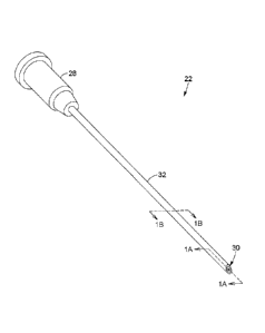
Referring to FIGs. 1, 1A and 1B, a needle assembly 22 according to one
embodiment is shown. The needle assembly 22 of this embodiment includes a
needle hub 28 and a needle tip 30. A shaft portion 32 extends and is disposed
longitudinally between the needle hub 28 and the needle tip 30. The shaft
portion
32 includes a cavity 34 extending from the needle hub 28 to the needle tip 30.
The
cavity 34 may be understood as an empty channel extending the entire length of
the shaft portion and opened to air at both extremities. The cavity 34
includes a
sample receiving region 36 opened at the needle tip 30. The needle assembly 22
.. according to the present embodiment may therefore be used to collect,
within the
sample-receiving region 36, a biological sample from a biological medium and
perform an optical interrogation of this sample directly in the needle
assembly 22.
In this variant, the shaft portion 32 further includes a cladding structure 38
surrounding the cavity 34. In some variants the cladding structure may be made
of
or include at least one optical cladding layer made of SiO2 or other optical
material
suitable for light propagation and guiding. As illustrated in examples
described
further below, in some configurations the shaft portion of the needle assembly
22
may be configured for light guidance therein along a longitudinal optical axis
to
.. perform an optical interrogation of the sample in the sample-receiving
region 36.
CA 03051489 2019-07-24
WO 2018/137039 PC T/CA
2018/050094
Still in the illustrated embodiment of FIGs. 1, 1A and 1B, the shaft portion
32 may
further include a sheath 39 surrounding the cladding structure 38. The sheath
39
preferably lends rigidity and solidity to the needle assembly 22 and may for
example be made of metal or another robust and biocompatible material.
5 Preferably, the
sheath 39 is made of a biologically compatible material if relevant
to the intended use. The sheath 39 may have a beveled extremity defining the
needle tip 30. In some embodiments, the sheath 39, the cavity 34 and the
cladding
structure 38 are jointly beveled at the needle tip 30, such that their
respective
endfaces extend in a same plane. In another variant the cavity 34 and cladding
10 structure 38 may
be slightly recessed within the sheath 39 at the needle tip 30,
inasmuch as such a configuration does not impede the drawing of the sample in
the sample-receiving region 36.
FIGs. 2A and 2B illustrate the drawing of a biological sample 41 from a
biological
15 medium 40 using a
needle assembly such as shown in FIG. 1. This may for
example be achieved by inserting the needle tip 30 into the biological medium
40
from which the sample is to be collected, and drawing the sample inside the
sample
receiving region 36 (FIG. 2A). In some variants, the simple insertion of the
needle
tip 30 in the biological medium 40 may suffice to collect a suitable quantity
of
20 biological
material inside the needle assembly 22, this quantity defining the
biological sample 41. For example, liquid samples may enter the needle tip
through
capillary action. Tissues may require repetitive small "stabbing" passes,
typically
leaving the needle tip within the medium 40 with the needle to push tissue up
into
the needle mechanically. In other variants, it may be desired to apply a
suction
force, which may for example be provided by a syringe-type device to which the
needle assembly 22 is connected. Some tissues may require both a repetitive
motion and suction. It will be noted that in the illustrated embodiment the
sample
receiving region 36 is simply embodied by the front portion of the cavity 34,
that is,
the portion of the cavity 34 closer to the needle tip 30. In alternative
embodiments
(not shown) the sample receiving region 36 may have a shape that differs from
the
rest of the cavity 34.
CA 03051489 2019-07-24
WO 2018/137039
PCT/CA2018/050094
21
Optionally, once the biological sample 41 has been drawn into the sample
receiving region 36 of the cavity 34, the needle assembly 22 may be removed
from
the biological medium 40, as shown in FIG. 2B. In other variants, the needle
assembly 22 may remain in the biological medium 40 (from a cell culture, or a
patient's body sampling site) during the optical interrogation process.
Referring to FIGs. 3, 3A and 3B, there is shown another variant of a needle
assembly 22. Again, the needle assembly includes a needle hub 28, a needle tip
30 and a shaft portion 32 therebetween. The shaft portion includes a cavity 34
surrounded by a cladding structure 38, both as explained above. The cavity 36
includes a sample-receiving portion 36. This variant differs from the one
illustrated
in FIG. 1 by the absence of a sheath 39. In this variant, the cladding
structure 38
may have a beveled extremity 40 defining the needle tip 30. The cladding
structure
38 of this embodiment is preferably provided with one or more coating layers
43 to
improve its rigidity, biocompatibility, optical properties, etc. In one
example the
coating layer or layers may include a structural coating made of a material
sufficiently resistant to prevent breaking of the needle assembly 22 during
the
sample collection process. The one or more coating layers 43 may for example
.. include a polyimide layer. As well known in the art, polyimide may be
coated on
optical fibers for medical use, as it is biocompatible and suitable for
sterilization,
and can provide strength and temperature resistance to the fiber. Other
coating
materials such as acrylate, low-index polymers, silicon and metal may also be
considered. These coating materials may also be further jacketed.
In some embodiments, such as for example shown in FIG. 3A, a reflective
coating
45 may be deposited on an extremity of the cladding structure 38 at the needle
tip
30. The reflective coating 45 may for example be useful to reflect light back
towards
the needle hub 28 for extraction and detection after its interaction with the
biological sample.
22
Various configurations may be envisioned for the cladding structure 38 without
departing from the scope of the present invention.
Referring to FIGs. 4A and 4B, in one embodiment the cladding structure 38 may
.. include a silica layer 46 and an air hole layer 48 within this silica layer
46, defining
an air-clad configuration. The low refractive index of the air filling the
holes of the
air hole layer 48 and the cavity 34 allows the light to propagate in an
interstitial ring
49 of the silica layer 46 extending between them.
Referring to FIGs. 5A to 5C, there is shown another example of a cladding
structure
38, including a silica layer 50 surrounding the cavity 34. In this variant,
the cavity
34 is positioned eccentrically with respect to the central axis of the shaft
portion
32. An elliptical optical fiber core 52 extends concentrically within the
silica layer
50, along the central axis of the shaft portion and therefore parallel to the
cavity
34. The elliptical optical fiber core 52 guides light from the needle hub to
the needle
tip and evanescent wave coupling can for example occur between the travelling
light and the sample present in the sample-receiving region of the cavity 34.
An
example of such a fiber is for example shown in U.S. patent No. 7,405,673
(CARON et al). In other variants, the optical fiber core 52 may be circular or
having
another shape than elliptical. In another variant, as for example illustrated
in FIG.
5D, several elliptical cores 52a, 52b, 52c, ..., 52h may be distributed around
the
cavity 34 and therefore provide an array of light paths surrounding the cavity
34
and the sample within. Such a variant may be used with a cavity 34
concentrically
disposed within the shaft portion 32. Of course, it will be understood by one
skilled
in the art that the number, shape and distribution of the elliptical cores may
vary
and that the configuration shown in FIG. 5D is provided by way of example
only.
Referring to FIG. 5E, in yet another variant the cladding structure may
include an
integrated optical fiber 53 extending along the cavity 34. The integrated
optical
fiber 53 has an optical fiber core 54 and an optional optical fiber cladding
55
configured for guiding light within the optical fiber core 54. The optical
fiber core
54 may for
Date Recue/Date Received 2021-03-24
CA 03051489 2019-07-24
WO 2018/137039
PCT/CA2018/050094
23
example be made of pure SiO2 or Si02 doped with a material having a higher
refractive index such as Ge02, P205, Ti02, etc. while the optical fiber
cladding 55
can be made of SiO2 doped with a lower-index material such as fluorine or
B203.
The optical fiber cladding 55 may be omitted if the refractive index of the
optical
fiber core 54 is higher than that of the cladding structure 50. The integrated
optical
fiber 53 is polished along its length on one side and positioned such that the
optical
fiber core 54 is exposed to the cavity 34 and therefore to the sample within.
Referring to FIGs. 5F and 5G, in other implementations the cladding structure
may
include one or more partial optical fiber cores 56 or 56a, 56b, 56h
extending
along the cavity 34 and exposed to the cavity 34 and to the sample within. The
partial optical fiber core or cores 56 may for example be made of Si02 doped
with
higher-index materials such as Ge02, P205, Ti02, etc. Of course, the number of
partial cores 56 and their configuration may vary.
Referring to FIG. 6, in another example, the cladding structure 38 is multi-
layered,
that is, it is made up of a plurality of concentric cladding layers. In the
illustrated
example these layers include, concentrically and outwardly from the cavity 34:
a
ring core layer 57, for example made of S 102, a low refractive index cladding
layer
58 and a high refractive index cladding layer 59. As will be readily
understood by
one skilled in the art, such a configuration may guide light within the ring
core layer
57. In another variant with a similar configuration, the cladding structure
surrounding the silica ring core may include a cladding layer made of Si02
doped
with F surrounded by a polyimide or other coating layer.
Referring to FIG. 7, in accordance with one aspect. there is provided a sample
analysis system 20 for collection and optical interrogation of a biological
sample.
The sample analysis system 20 first includes a needle assembly 22 according to
any one of the variants described above or equivalents thereof.
CA 03051489 2019-07-24
WO 21118/137039
PCT/CA2018/050094
24
In some variants, the sample analysis system 20 may further include a syringe
assembly 24 connectable to the needle hub 28 of the needle assembly 22. The
syringe assembly 24 can be used to provide a suction force to draw the
biological
sample into the sample receiving region 36 of the needle assembly 22. The
syringe
assembly 24 may be of standard construction, and preferably includes a barrel
60.
The barrel 60 typically has a cylindrical shape and has a proximal end 72
connectable to the needle hub 28 and an open distal end 74. The syringe
assembly
24 further includes a plunger 62 inserted in the barrel 60 from the distal end
74 and
slideable longitudinally within the barrel 60. The end of the plunger 62
extending
within the barrel 60 is provided with a plunger seal 64, creating a seal with
the inner
wall of the barrel 60. As well known in the art, when the needle assembly 22
is
connected to the proximal end 72 of the syringe assembly 24 the movement of
the
plunger 62 within the barrel 60 provides the suction force which can draw the
biological sample into the sample receiving region 36 of the needle assembly
22.
The syringe assembly may be connected to the needle hub 28 in a variety of
manners. By way of example, a "Luer-lock" (trademark) type connection may be
provided in which the proximal end 72 of the barrel 60 is provided with a male
connection fitting and the needle hub with an associated female fitting.
Typically,
a tabbed hub on the female fitting screws into threads in a sleeve on the male
fitting to provide a secure engagement. In another variant, a "Luer-slip"
(trademark)
or "slip tip" engagement can be provided where the male and female fittings
are
pressed together without involving threads.
It will be readily understood that some embodiments of the sample analysis
system
may exclude a syringe assembly, for example in variants where the sample is to
be drawn in the sample-receiving region through capillary action or
mechanically
pushed therein.
The sample analysis system 20 further includes an optical assembly 26. The
optical assembly 26 first includes a light source 76 generating an
interrogation light
CA 03051489 2019-07-24
WO 2018/137039
PCT/CA2018/050094
beam. The interrogation light beam may have any optical characteristics
suitable
in view of the type of optical testing to be made on the sample. For example,
for
visible or near-infrared brightfield or fluorescence microscopy a white light
source
may be used, or a laser or LED emitting for instance at 488 nm, 532 nm, 568
nm,
5 633/647 nm or
676 nm. Optical sources emitting light of wavelengths centered at
800 nm, 1050 nm or 1310 nm are typically used for near-infrared optical
coherence
tomography. Lasers or LED sources emitting at a suitable wavelength may also
be
used for Raman spectroscopy (e.g. at 785 nm, 830 nm, 980 nm, 1064 nm) or for
autofluorescence measurements (e.g. at 308 nm, 337 nm, 360 nm, 425 nm).
10 Diffuse
reflectance spectroscopy may also be performed with a broadband or white
light source emitting light with a wavelength spectrum lying somewhere in the
400-
1000 nm range (e.g. a tungsten or xenon lamp, or a combination of broadband
LEDs to cover this wavelength range either partially or fully). Refractive
index
measurements, for example, by evanescent wave sensing, may also be performed
15 at one or more
visible or near infrared wavelengths generated by a laser or LED.
Of course, it will be readily understood that the types of light sources and
corresponding spectral information listed above is given by way of example
only
and is in no way considered !imitative to the scope of the invention.
20 In some
implementations, the light source 76 may be connectable to the needle
hub 28 so as to inject the interrogation light beam into the needle assembly
22 for
propagation towards the biological sample when drawn into the sample receiving
region 36. The interrogation light beam may be injected for propagation in
different
components of the shaft portion 32 depending on the construction of the shaft
25 portion 32 and
of the interrogation scheme being applied, as will be further
explained below. One or more input optical fiber links 68 may be provided to
guide
the interrogation light beam from the light source to the needle assembly 22.
The optical assembly 26 may further include an optical detector 78. In the
implementation shown in FIG. 7, the detector 78 is connectable to the needle
hub
28 to collect the light travelling in a backward direction in the needle
assembly 22.
CA 03051489 2019-07-24
WO 2018/137039
PCT/CA2018/050094
26
The detector 78 may be embodied by various devices, depending on the nature of
the optical analysis to perform. For example, a spectrometer may be used in
the
context of Raman spectroscopy, diffuse reflectance spectroscopy, multi-
wavelength refractive index sensing, spectral-domain optical coherence
tomography, etc. Photomultiplier tubes may be used for microscopy and single
emission wavelength fluorescence measurements, whereas CCD or CMOS
cameras may be useful for various microscopy and imaging applications. One or
more output optical fiber links 70 may be provided to guide the light
resulting from
an interaction of said interrogation light beam with the biological sample
from the
needle assembly 22 to the detector 78.
With reference to FIGs. 7A and 7B, in some implementations the optical
assembly
26 may include an optical coupler 66 connectable to the needle hub 28.
Preferably,
the optical coupler 66 provides an optical connection between the shaft
portion of
.. the needle assembly and the optical assembly 26. The optical coupler 66 may
for
example be engageable in a locking engagement through a first connector 65 and
a second connector 67 embodying a male-female "Luer-lock" or "Luer-slip"
connection such as mentioned above. The optical coupler 66 may include light
guides or otherwise provide for the propagation of light towards the needle
assembly. In some implementations, the first connector 65 may be affixed to
the
needle hub and the second connector 67 is engageable with the first connector
and houses extremities of the input and output optical fiber links. In the
illustrated
embodiment, for example suitable for connection to a needle assembly having a
cladding structure such as shown in FIG. 6, the optical coupler 66 includes a
light
ring 69 being sized, shaped and positioned to provide optical coupling with
the ring
core layer of the cladding structure. The light ring 69 may be for example
embodied
by circularly disposed endfaces 71 of optical fibers, which may for example be
composed of the input optical fiber links (for coupling light from the light
source into
the needle assembly), or the output optical fiber links (for coupling light
from the
needle assembly to the detector), or a combination of both.
CA 03051489 2019-07-24
WO 2018/137(139 PCT/CA201
8/(150094
27
Referring to FIG. 8, there is shown another implementation of a sample
analysis
system 20 including an optical assembly 26. The optical assembly 26 again
includes a light source 76 which may be configured according to any suitable
embodiment such as for example described above, and an optical detector 78. In
this variant, the detector 78 is provided within an optical reader 84 which is
a
component separate from the needle assembly 22. In this configuration, the
needle
tip 30 may be inserted into the optical reader 84, which may for example take
the
shape of a cap. Light is injected into the shaft portion 32 of the needle
assembly
22 at the needle hub 28, and propagates towards the needle tip 30, interacting
with
the sample along the way. The detector 78 is affixed within the cap and
positioned
to detect light which exits from the needle tip 30, so that the impact of the
interaction of the propagating light with the sample can be measured.
Referring now to FIG. 9, there is shown a needle assembly 22 for collection
and
optical interrogation of a biological sample according to another embodiment.
In this embodiment, the needle assembly 22 includes a needle hub 28, a needle
tip 30 and a shaft portion 32 disposed longitudinally between the needle hub
28
and the needle tip 30. The shaft portion 32 includes a capillary 80 having a
cavity
34 extending longitudinally from the needle hub 28 to the needle tip 30. The
cavity
34 has a sample receiving region 36 opened at the needle tip 30, similarly to
described above. The shaft portion 32 further includes at least one optical
window
82 transversally aligned or in line of sight alignment with the sample-
receiving
region 36 and allowing optical interrogation of the sample within the sample
receiving region 36 therethrough. In the illustrated variant of FIG. 9, the
shaft
portion 32 of the needle assembly 22 entirely consists of the capillary 80,
which is
made of a transparent material. The optical window 82 thus corresponds to the
portion of the capillary 80 that defines the sample receiving region 36. In
other
embodiments (not shown), the optical window or windows may extend over a
portion only of the shaft portion, for example as an opening or insert through
the
capillary. In this context, it will be understood that the positioning of the
optical
CA 03051489 2019-07-24
WO 2018/137039
PCT/CA2018/050094
28
window or windows with respect to the sample-receiving region may be offset
from
a direct alignment inasmuch as light may travel inwards through the optical
window
towards the biological sample on the one hand, and outwards from the sample to
exit the needle assembly substantially unobstructed or unattenuated on the
other
hand. In some embodiments the light may travel in both directions through a
same
optical window. In other embodiments light may cross different optical windows
in
the input and output directions.
The needle assembly 22 according to the present embodiment may be used to
collect a biological sample 41 from a biological medium (from a cell culture,
or a
target sampling site) and perform an optical interrogation of this sample 41
directly
in the needle assembly 22. Similarly to the process described above with
reference
to FIGs. 2A and 2B, this may for example be achieved by inserting the needle
tip
30 in the biological medium 40 and drawing the sample 41 inside the sample
receiving region 36. In some variants, as explained above, the simple
insertion of
the needle tip 30 in the biological medium 40 may suffice to collect a
suitable
quantity of biological material inside the needle assembly 22, either by
capillary
action or through repetitive "stabbing". In other variants, it may be desired
to apply
a suction force, which may for example be provided by a syringe-type device.
It
will be noted that in the illustrated embodiment the sample receiving region
36 is
simply embodied by the front portion of the hollow fiber core 34, that is, the
portion
of the cavity 34 closer to the needle tip 30. In alternative embodiments the
sample
receiving region 36 may have a different shape than the rest of the cavity 34.
Once the biological sample 41 has been drawn into the sample receiving region
36 of the cavity 34, the needle assembly 22 is removed from the biological
medium
40. Optical interrogation of the sample 41 is then performed by projecting one
or
more interrogation light beams towards the sample 41 through the optical
window
82 or windows. Light resulting from the interaction of the interrogation light
beam
with the sample can be transmitted light exiting the needle assembly through
the
needle tip, or light reflected or otherwise travelling backwards with respect
to the
CA 03051489 2019-07-24
WO 2018/137039
PCT/CA2018/050094
29
direction of the optical interrogation. It will be readily understood that the
reference
to a transversal optical interrogation includes impinging and transmitting the
interrogation light beam through the optical window at various possible
incidence
angles differing from a purely longitudinal light propagation scheme and is
not
limited to light injection at a right angle with respect to the longitudinal
axis of the
shaft portion. Advantageously, in some embodiments this variant may provide
the
ability to image or enable spatially distributed sampling.
Referring to FIG. 10, there is shown a portion of a sample analysis system
including a needle assembly 22 such as shown in FIG. 9. In this
implementation,
the analysis system 20 includes an optical reader 84, and optical
interrogation of
the sample involves inserting the needle tip 30 and the part of the shaft
portion
which includes the sample-receiving region into the optical reader 84. The
optical
reader 84 may for example have a portion forming a reading chamber 86 sized to
receive the needle tip 30. The optical reader 84 preferably includes at least
one
light source 76 and at least one optical detector 78. The light source or
sources
are configured to generate one or more interrogation light beams 42 having
suitable optical properties for the type of analysis to be performed on the
sample
41. As known to those skilled in the art, the interaction of the interrogation
light
beam(s) 42 with a sample leads to the generation of either return or
transmitted
light having optical properties representative of characteristics of the
sample 41
and can be analyzed through various techniques to yield information about the
sample. As will be readily understood by one skilled in the art, the optical
reader
84 may also include a moving source/detector assembly or several sources and
detectors for distributed analysis along the length of the needle or for multi-
spectral
analysis or any other analysis method.
In the illustrated embodiment of FIG. 10, the light sources and detectors (one
or
many of each) are aligned or otherwise positioned such that an interrogation
light
beam 42 or beams can be generated by the light sources and traverse the
sample,
light 44 resulting from the interaction of the interrogation light beam with
the sample
CA 03051489 2019-07-24
WO 2018/137039
PCT/CA2018/050094
being detected by the detector on the opposite side of the sample. While FIG.
10
shows a direct line-of-sight alignment between the light source and detector,
it will
be readily understood by one skilled in the art that in other implementations
(not
shown) either the interrogation light beam 42 or the transmitted light beam
may be
5 redirected, by
mirrors, lenses and the like. Furthermore, it will be readily
understood that other optical components directing, shaping, modulating or
otherwise affecting either the interrogation light beam 42, the transmitted
light
beam or both may be provided within the reader 84 without departing from the
scope of the invention. In other variants (not shown), the light source and
the
10 detector may be
positioned on a same side of the sample such that light reflected,
scattered, re-emitted or otherwise returning in the direction from which the
interrogation light beam 42 impinged the sample is collected and analyzed. The
optical reader 84 may take various shapes, and in some embodiments may be
formed as a cap which can be placed over the needle tip similarly to the
variant
15 shown in FIG. 8.
It will further be understood that in some embodiments the optical
reader 84 as described above may be used in conjunction with a needle assembly
defining a light guiding structure such as described with respect to FIG. 1 or
the
like, provided that the cladding structure of the needle assembly is
sufficiently
transparent or includes an optical window allowing optical interrogation
20 therethrough.
Referring to FIGs. 11A and 11B, there is shown another variant of a needle
assembly 22 that can be used for transversal interrogation of the sample
within the
sample-receiving region 36, either in a light transmission mode or in a light
25 reflectance
mode. The needle assembly 22 of this embodiment includes a needle
hub 28, a needle tip 30 and a shaft portion 32 between the needle hub 28 and
the
needle tip 30. The shaft portion 32 includes a capillary 80 having a
longitudinal
cavity 34 extending from the needle hub 28 to the needle tip 30. The cavity 34
has
a sample receiving region 36 opened at the needle tip 30, similarly to what
has
30 been described above. The shaft portion 32 includes an optical
window 82
transversally aligned with the sample-receiving region 36 so as to allow
optical
CA 03051489 2019-07-24
WO 2018/137039
PCT/CA2018/050094
31
interrogation of the sample within the sample receiving region 36
transversally to
the longitudinal axis of the shaft portion 32. Preferably, the capillary 80 is
entirely
made of a transparent material, inherently embodying an optical window 82. The
shaft portion 32 further includes a sheath 39 surrounding the capillary 80.
The
sheath 39 preferably lends rigidity and solidity to the needle assembly 22 and
may
for example be made of metal, polyimide or an equivalent material. Preferably,
the
sheath 39 is made of a biologically compatible material if relevant to the
intended
use. In the illustrated embodiment the sheath 39 is retractable and has a
sampling
position (see FIG. 11A) where its front extremity is flush with the front
extremity of
the capillary 80, thereby enabling drawing of the sample inside the sample
receiving region 36. The sheath 39 also has a retracted position (see FIG. 11
B)
wherein it is retracted with respect to the needle tip 30 in order to allow
the needle
tip 30 and sample-receiving region 36 to be exposed. The sheath may be affixed
in the sample-receiving position during the sampling process, and the reader
84
may be configured so that insertion of the needle tip therein pushes the
sheath in
the retracted position, such that the sample-receiving region is exposed for
optical
interrogation of the sample as explained above. In such an implementation the
sheath is preferably shorter than the shaft portion of the needle assembly,
and
additional protecting and/or blocking implements may be provided around the
shaft
portion at the extremity of the needle hub when the sheath is in the sample-
receiving position. In some implementations, the optical reader may include a
recess or other means for guiding the needle tip in the reading chamber 86 and
avoid breakage of the capillary in the process of inserting the needle tip in
the
reader.
In a different variant (not shown), the sheath 39 may be designed such that
optical
interrogation is allowed through this sheath 39, either in transmission or in
reflectance. For example, the sheath 39 may be made of an optically
transparent
material. Alternatively, the sheath 39 may have one or more openings or
transparent inclusions defining the optical window 82 and allowing optical
access
to the sample receiving region 36.
CA 03051489 2019-07-24
WO 208/137039
PCT/CA2018/950094
32
Examples of methods for the diagnosis, prognosis or treatment of a disease
or a condition
In accordance with some implementations, needle assemblies and analysis
systems such as described above or equivalents thereof may be used in
different
contexts.
In some embodiments, there is provided a use of a needle assembly according to
variants as defined above, and the like, for an in situ optical interrogation
of a
biological sample of a subject. Such a use involves the sample being within
the
sample-receiving portion of the needle assembly and optical interrogation
longitudinally along the needle assembly, while the needle assembly is still
within
or in contact with the sampling site.
In some embodiments, there is provided a use of a needle assembly according to
any of the variants above, and the like, for an ex vivo optical interrogation
of a
biological sample of a subject. Such a use involves the sample being within
the
sample-receiving portion of the needle assembly during this optical
interrogation.
In some implementations, there may be provided a method for analyzing a
biological sample. The method may include the following steps:
- obtaining a needle assembly according to an embodiment described
above or the like;
- inserting
the needle tip of the needle assembly in a target site comprising
a biological tissue or liquid;
collecting a biological sample of the target site in the sample-receiving
region of the needle tip;
- optically interrogating the biological sample within the sample-receiving
region of the needle tip using an interrogation light beam; and
- detecting and analyzing light resulting from an interaction of the
biological sample with the interrogation light beam.
CA 03051489 2019-07-24
WO 2018/137039
PCT/CA2018/050094
33
In some implementations the biological sample may be a tissue, such as for
example normal or abnormal, e.g. malignant, tissue or cells of the breast,
lymph
nodes, thyroid, salivary glands, liver, pancreas, metastatic lesions. In other
implementations the biological sample may be liquid such as, for example, a
biological fluid, such as for example blood, lymph, tears, sweat, saliva, or
urine.
Alternatively, the biological sample may be liquid such as a cell suspension
from a
cell culture.
In some implementations, the biological sample's tissue or liquid may be
collected
from a body of a subject. Particularly, the subject is an animal or a human.
The optically interrogating, detecting and analyzing steps of the method may
be
carried out in a variety of fashions, depending on the desired information,
structure
of the needle assembly and capabilities of the components of the analysis
system.
In some examples, for example using a needle assembly such as or equivalent to
those described in FIGs. 1, 3, 4A, 4B, 5A through 5G and 6, the optical
interrogation of the sample may involve propagating the interrogation light
beam
longitudinally in the needle assembly. For example, the interrogation light
beam
may be injected into the needle assembly at the needle hub and propagates
towards the needle tip. The interrogation light beam may be guided or
otherwise
propagate along an interstitial ring, a ring core, one or more elliptical
optical fiber
core, one or more integrated optical fiber, one or more partial optical fiber
cores,
the cavity or the like. The light resulting from an interaction of the
biological sample
with the interrogation light may be collected at the needle tip or at the
needle hub.
In some implementations the optical interrogation is performed subsequently to
the
removal of the needle tip from the body part. In other variants the optical
interrogation may be performed in vivo, after collection of the sample from
the
subject but while the needle tip is still within the body part of the subject.
CA 03051489 2019-07-24
WO 2018/137039
PCT/CA2018/050094
34
In alternative examples, for using a needle assembly such as or equivalent to
those
described in FIGs. 9, 11A and 11B. optical interrogation of the sample may
involve
propagating the interrogation light beam towards the sample transversally to
the
needle assembly. In some variants, the interrogation light beam may enter the
needle assembly through one side of the shaft portion and the resulting light
exits
from the opposite side. In other variants the resulting light may exit the
needle
assembly on the same side from which the interrogation light beam entered or
at
any angle. Both these sets of variants are understood to fall within the scope
of
"transversal" optical interrogation. Light may enter and exit the needle
assembly at
different angles with respect to the longitudinal axis of the shaft portion.
In various embodiments, the interrogation light beam may be absorbed,
scattered
or transmitted by the biological sample. In some embodiments, the
interrogation
light beam may interact with the sample through evanescent wave coupling from
a waveguiding core parallel to the cavity (such as for example the elliptical
optical
fiber cores of FIGs. 5A and 5D).
In some implementations, the analysis of the light resulting from the
interaction of
the interrogation light beam with the sample may provide one or more
information
of different types, such as for example:
- The presence of a sample within the sample receiving region and/or the
quantity of biological material present within the sample receiving region;
Adequacy/cellularity information. This may for example take the form of the
amount of cells, density or ratio with respect to the entire volume collected
which may contain unwanted fluids like blood, lymph or other biofluids. In
some implementations, adequacy/cellularity information may include an
indication of whether or not enough of the sample has been collected to
make a diagnosis. If not, the pathologist may receive the sample and
classify it as 'inadequate' for diagnosis. In other implementations,
adequacy/cellularity information may include a measure of the refractive
index of the sample, e.g. 1.33 for water, 1.37 for blood and 1.4 for tissue.
CA 03051489 2019-07-24
WO 2i1S/13709
PCT/CA2018/050094
This information may also involve the density of the sample by scattering
(OCT or reflectance spectroscopy), the use of microscopy to count cells or
measure cellular density, etc. In other variants, the present method may be
used to confirm that the collected tissue is indeed the target tissue. Such a
5 variant may
require more specific measurements, such as Raman spectra
or microscopic features enabling the differentiation of a target lesion with
respect to the surrounding tissue.
- The nature of the sample or properties thereof such as malignant or
benign,
etc. This type of information may for example be obtained through
113 spectroscopic analysis such as Raman spectroscopy, fluorescence, etc.
In some embodiments, there is also provided a use of the needle assembly
according to any of the variants above, and the like, for an ex vivo optical
analysis
of a biological sample of a subject to diagnose a disease or condition in the
subject,
15 the sample being
within the sample-receiving portion of the needle assembly
during the optical analysis.
In some implementations, there may be provided a method to aid in diagnosis,
prognosis or guide treatment of a disease or a condition in a subject,
comprising
20 the steps of:
- obtaining a needle assembly according to an embodiment described above
or the like;
- inserting the needle tip of the needle assembly in a body of a subject
such
that the needle tip reaches a target site; and
25 - collecting a
biological sample from the target site in the sample-receiving
region of the needle assembly;
- optically interrogating the biological sample within the sample-receiving
region of the needle tip using an interrogation light beam;
- detecting light resulting from an interaction of the biological sample
with the
30 interrogation light beam;
CA 03051489 2019-07-24
WO 2018/137039
PCT/CA2018/050094
36
- analyzing the resulting light to determine at least one characteristic
specific
of said disease or condition; and
- transmitting at least one characteristic specific of the disease or
condition
to a physician.
The sampling site may be, for example, a biological sample that is outside of
its
natural environment, such as in vitro or, alternatively, in a cell culture
environment.
Alternatively, the sampling site is a body of a subject as defined above. The
sample's tissue or cells may therefore be collected from a body part such as,
for
example, a body member (e.g. arm, leg, trunk, head), an organ such as breast,
lymph nodes, thyroid, salivary glands, liver, pancreas, a specific tissue
(e.g.
metastatic lesions), or a biological fluid such as blood, urine, lymph, tears,
saliva
or sweat.
The disease or condition of the subject may for example be cancer, diabetes,
malaria, or other conditions in which the cells, tissues or biofluids have
optical
properties differing from those usually observed with healthy cases.
Example characteristics measurable with optical techniques and specific of the
disease or condition may for example be increased nucleus/cell size and/or
decreased extracellular tissue organization in the case of cancer, the
presence of
proteins or blood cells in urine in the case of diabetes, spectral or
structural
changes in blood cells due to malaria infection, etc.
The step of collecting a biological sample comprises drawing the biological
tissue
or cells within the sample-receiving portion, for example using a syringe
assembly
connected to the needle hub or the mechanical stabbing method described above,
with or without a syringe for suction.
CA 03051489 2019-07-24
WO 2018/137039
PCT/CA2018/050094
37
The method may include a step of withdrawing the needle tip from the body of
the
patient between the steps of collecting the biological sample and optically
interrogating the biological sample.
As mentioned above, in other variants the optical interrogation may be
performed
in situ, after collection of the sample from the sampling site or the body
part of the
subject but while the needle tip is still within the tissue, cell culture or
body of the
subject. A one skilled in the art will readily understand, such variants are
of interest
when using a configuration of the needle assembly allowing the interrogation
light
.. beam to propagate longitudinally in the needle assembly.
The analyzing step of the present method for the diagnosis, prognosis or
treatment
of a disease or a condition in a subject may involve using an optical analysis
technique such as visible or near-infrared brightfield or fluorescence
microscopy,
visible or near-infrared optical coherence tomography, Raman spectroscopy,
autofluorescence measurements, diffuse reflectance spectroscopy and refractive
index measurements, and the like.
It will be readily understood that while the present methods advantageously
provide for the optical interrogation of the sample while it is within the
sample
receiving region of the needle assembly, in some implementations the sample
may
subsequently be extracted from the needle assembly and processed or further
analyzed according to techniques known in the art in order to obtain further
information from this sample.
Of course, numerous modifications could be made to the embodiments described
above without departing from the scope of the invention.