Note: Descriptions are shown in the official language in which they were submitted.
CA 03051963 2019-07-26
WO 2018/140978 -1- PCT/US2018/016041
METHOD FOR NON-INVASIVE MONITORING OF FLUORESCENT TRACER
AGENT WITH DIFFUSE REFLECTION CORRECTIONS
CROSS-REFERENCE TO RELATED APPLICATION
[0001] This application claims the benefit of U.S. Provisional
Application No.
62/452,025 filed January 30, 2017, which is incorporated herein in its
entirety.
BACKGROUND OF THE DISCLOSURE
[0002] The present disclosure relates generally to methods for non-
invasive
monitoring of a fluorescent tracer agent within a medium characterized by
scattering and/or
absorption of light. More particularly, the present disclosure relates to
methods for non-
invasive assessment of kidney function by monitoring the clearance of an
exogenous
fluorescent tracer within the tissues of a patient in vivo.
[0003] Dynamic monitoring of renal function in patients at the bedside in
real time
is highly desirable in order to minimize the risk of acute renal failure
brought on by various
clinical, physiological and pathological conditions. It is particularly
important in the case of
critically ill or injured patients because a large percentage of these
patients face the risk of
multiple organ failure (MOF) incited by one or more severe dysfunctions, such
as: acute
lung injury (ALI), adult respiratory distress syndrome (ARDS),
hypermetabolism,
hypotension, persistent inflammation, and/or sepsis. Renal function may also
be impaired
due to kidney damage associated with administration of nephrotoxic drugs as
part of a
procedure such as angiography, diabetes, auto-immune disease, and other
dysfunctions
and/or insults causally linked to kidney damage. In order to assess a
patient's status and to
monitor the severity and/or progression of renal function over extended
periods, there
exists considerable interest in developing a simple, accurate, and continuous
method for the
determination of renal failure, preferably by non-invasive procedures.
[0004] Serum creatinine concentration, an endogenous marker of renal
function, is
typically measured from a blood sample and used, in combination with patient
demographic factors such as weight, age, and/or ethnicity to estimate
glomerular filtration
rate (GFR), one measure of renal function. However, creatinine-based
assessments of renal
function may be prone to inaccuracies due to many potential factors,
including: age, state
of hydration, renal perfusion, muscle mass, dietary intake, and many other
anthropometric
CA 03051963 2019-07-26
WO 2018/140978 -2- PCT/US2018/016041
and clinical variables. To compensate for these variances, a series of
creatinine-based
equations (most recently extended to cystatin C) have been developed which
incorporate
factors such as sex, race and other relevant factors for the estimation of
glomerular
filtration rate (eGFR) based on serum creatinine measurements. However, these
eGFR
equations are not provided with any means of compensating for most of the
above sources
of variance, and therefore have relatively poor accuracy. Further, the eGFR
method
typically yields results that lag behind true GFR by up to 72 hrs.
[0005] Exogenous marker compounds, such as inulin, iothalamate, 51Cr-
EDTA,
Gd-DTPA and 99111Tc-DTPA have been used in existing methods for measuring GFR.
Other
endogenous markers, such as 123I and 125I labeled o-iodohippurate or 99111Tc-
MAG3 have
been used to in other existing methods for assessing the tubular secretion
process.
However, the use of typical exogenous marker compounds may be accompanied by
various
undesirable effects including the introduction of radioactive materials and/or
ionizing
radiation into the patient, and laborious ex vivo handling of blood and urine
samples,
rendering existing methods using these exogenous markers unsuitable for real-
time
monitoring of renal function at a patient's bedside.
[0006] The availability of a real-time, accurate, repeatable measure of
renal
excretion rate using exogenous markers under patient-specific yet potentially
changing
circumstances would represent a substantial improvement over any currently
practiced
method. Moreover, a method that depends solely on the renal elimination of an
exogenous
chemical entity would provide a direct and continuous pharmacokinetic
measurement
requiring less subjective interpretation based upon age, muscle mass, blood
pressure, etc.
BRIEF DESCRIPTION OF THE DRAWINGS
[0007] The patent or application file contains at least one drawing
executed in
color. Copies of this patent or patent application publication with color
drawing(s) will be
provided by the Office upon request and payment of the necessary fee.
[0008] The disclosure will be better understood, and features, aspects
and
advantages other than those set forth above will become apparent when
consideration is
given to the following detailed description thereof. Such detailed description
makes
reference to the following drawings, wherein:
CA 03051963 2019-07-26
WO 2018/140978 -3- PCT/US2018/016041
[0009] FIG. 1 is a schematic illustration of a single-wavelength renal
monitoring
device in one aspect;
[0010] FIG. 2 is a schematic illustration of a dual-wavelength renal
monitoring
system in one aspect;
[0011] FIG. 3 is a graph summarizing the absorption, transmission, and
emission
spectra of various devices, materials, and compounds associated with the non-
invasive
monitoring of an exogenous fluorescent agent in vivo defined over light
wavelengths
ranging from about 430 nm to about 650 nm;
[0012] FIG. 4 is a graph summarizing the absorption spectra of
oxyhemoglobin
(Hb02) and deoxyhemoglobin (Hb) defined over light wavelengths ranging from
about 200
nm to about 650 nm;
[0013] FIG. 5 is a schematic illustration of the timing of light pulse
cycles
associated with data acquisition by a dual-wavelength renal monitoring system
in one
aspect, in which each light pulse cycle includes light pulses produced at the
excitation
wavelength and at the emission wavelength in sequence;
[0014] FIG. 6 is a side view of a sensor head of a renal function
monitoring system
in one aspect;
[0015] FIG. 7 is a bottom view of the sensor head of FIG. 6;
[0016] FIG. 8 is a top interior view of the sensor head of FIG. 6
illustrating an
arrangement of various electrical components within a housing of a sensor head
of a renal
function monitoring system in one aspect;
[0017] FIG. 9 is an enlargement of the interior view of FIG. 8;
[0018] FIG. 10 is a schematic illustration of the apertures formed within
a contact
surface of a sensor head of a renal function monitoring system in one aspect;
[0019] FIG. 11 is a schematic illustration of synchronous detection of
light by a
light detector of a sensor head in one aspect;
CA 03051963 2019-07-26
WO 2018/140978 -4- PCT/US2018/016041
[0020] FIG. 12 is a schematic illustration of light signal modulation and
demodulation by the sensor head in one aspect;
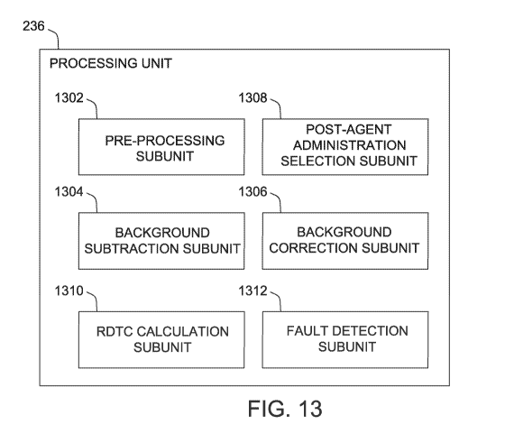
[0021] FIG. 13A is a block diagram illustrating the subunits of a
processing unit in
one aspect;
[0022] FIG. 13B is a block diagram illustrating the subunits of a
processing unit in
a second aspect;
[0023] FIG. 14A is a flow chart illustrating the steps of a global error
mapping
method of determining the parameters of a diffuse reflectance correction
equation in one
aspect;
[0024] FIG. 14B is a flow chart illustrating the steps of a global error
mapping
method of determining the parameters of a diffuse reflectance correction
equation in a
second aspect;
[0025] FIG. 15A is a graph of representative intrinsic fluorescence
measurements
of the fluorescent agent (IFagen) detected by a renal monitoring device
obtained before and
after injection of an exogenous fluorescent agent. A subset of the data
selected for analysis
to determine correction factors are shown highlighted in orange.
[0026] FIG. 15B is a graph of representative intrinsic fluorescence
measurements
of the fluorescent agent (IFagen) detected by a renal monitoring device
obtained before and
after injection of an exogenous fluorescent agent. A subset of the data
selected for analysis
by fitting the IFagõt to a plasma-derived IFagõt to determine correction
factors are shown
highlighted in orange.
[0027] FIG. 16 is a graph comparing the log-transformed single-
exponential curve
fit of the corrected fluorescence signal measurements (log[Fit], black dashed
line) and the
corrected fluorescence signal measurements from FIG. 15 (/Fagent, red line)
over a portion
of the selected analysis region from FIG. 15.
[0028] FIG. 17 is a map of a representative error surface summarizing
normalized
root-mean-square errors (RMSE, color scale) calculated for the difference
between the
linear fit to the log of the fluorescence and the corrected fluorescence
signal measurements
CA 03051963 2019-07-26
WO 2018/140978 -5- PCT/US2018/016041
for a range of correction factors lc, and kem,filtered, with a minimum RSME
region identified
by a white arrow overlaid on the map;
[0029] FIG. 18 is a graph comparing raw (F, blue line) fluorescence
signal
measurements and corrected (IF, red line) fluorescence signal measurements
obtained
before and after injection of an exogenous fluorescent agent.
[0030] FIG. 19 is a flow chart summarizing the steps of a linear
regression model
method of determining the parameters of a diffuse reflectance correction
equation in one
aspect;
[0031] FIG. 20 is a graph of log-transformed raw fluorescence signal
(Log(F1r))
showing the regions of the data used as project fits for: a linear regression
model used to
develop a data correction algorithm (orange line), the response variable for
the linear
regression model (black dashed line), and the region of highly varying data
used to train the
linear regression model (blue line);
[0032] FIG. 21A is a graph of raw fluorescence signal measurements
obtained
before and after injection of an exogenous fluorescent agent. Measurements of
raw
fluorescence signals were obtained during exposure to various perturbations
denoted as
colored regions starting at about 13:50 hours. The various perturbations
included
variations in blood oxygenation in the test subject, application and removal
of pressure to
the measured region, administration of blood pressure medication to the test
subject,
cooling of the measured region, and removal/replacement of the sensor head of
the device;
[0033] FIG. 21B is a graph of corrected fluorescence signal measurements
of FIG.
21A;
[0034] FIG. 21C is a graph of diffuse reflectance signal measurements
measured
simultaneously with the raw fluorescence signal measurements of FIG. 21A.
These signals
are used in the correction of the raw fluorescence signal measurements of FIG.
21A to
produce the corrected signal shown in FIG. 21B;
[0035] FIG. 22A is a block diagram illustrating a plurality of modules of
a pre-
processing subunit in one aspect;
CA 03051963 2019-07-26
WO 2018/140978 -6- PCT/US2018/016041
[0036] FIG. 22A is a block diagram illustrating a plurality of modules of
a pre-
processing subunit in one aspect;
[0037] FIG. 22B is a block diagram illustrating a plurality of modules of
a pre-
processing subunit in a second aspect;
[0038] FIG. 23 is an isometric view of a sensor head of a renal function
monitoring
system in a second aspect;
[0039] FIG. 24 is a bottom view of the sensor head of a renal function
monitoring
system illustrated in FIG. 23;
[0040] FIG. 25 is an isometric view of the sensor head of a renal
function
monitoring system illustrated in FIG. 23 with the upper housing and various
electrical
components removed to expose an inner housing; and
[0041] FIG. 26 is an exploded view of the inner housing of the sensor
head
illustrated in FIG. 25.
[0042] FIG. 27 is a graph
[0043] This written description uses examples to disclose the invention,
including
the best mode, and also to enable any person skilled in the art to practice
the invention,
including making and using any devices or systems and performing any
incorporated
methods. The patentable scope of the invention is defined by the claims, and
may include
other examples that occur to those skilled in the art. Such other examples are
intended to
be within the scope of the claims if they have structural elements that do not
differ from the
literal language of the claims, or if they include equivalent structural
elements with
insubstantial differences from the literal languages of the claims.
DETAILED DESCRIPTION
[0044] Unless defined otherwise, all technical and scientific terms used
herein have
the same meaning as commonly understood by one of ordinary skill in the art to
which the
disclosure belongs. Although any methods and materials similar to or
equivalent to those
described herein may be used in the practice or testing of the present
disclosure, the
preferred materials and methods are described below.
CA 03051963 2019-07-26
WO 2018/140978 -7- PCT/US2018/016041
[0045] A sample, as used herein, refers to a single, discrete data value
acquired
from a signal and/or telemetry analog-to-digital converter (ADC) for a single
ac qui sition/telemetry channel.
[0046] A measured value, as used herein, refers to a single, discrete
data value
created by demodulating or accumulating a sequence of samples from one
acquisition
channel.
[0047] A measurement, as used herein, refers to a set comprising the
Demodulated
In-Phase, Demodulated Out-of-Phase, and Averaged measurement values from one
acquisition channel.
[0048] A measurement subset, as used herein, refers to a set comprising
all
measurements for all acquisition channels during a single source LED
illumination. For
example, all measurements of an acquisition channel may include demodulated in-
phase,
demodulated out-of-phase, and averaged measurements.
[0049] A measurement set, as used herein, refers to a set comprising one
measurement subset for each source LED.
[0050] An acquisition, as used herein, refers to the overall process by
which a
measurement set is obtained.
[0051] A measurement sequence, as used herein, refers to a sequence of
one or
more measurement sets.
[0052] A telemetry value, as used herein, refers to a single, discrete
data value
acquired from a single channel of a telemetry ADC.
[0053] A telemetry set, as used herein, refers to a set comprising one
telemetry
value from each telemetry channel.
[0054] FIG. 1 is a schematic illustration of a system 100, provided as a
non-limiting
example, in which fluorescence 102 with an emission wavelength (kern) is
detected from a
region of interest of a patient 104 using a light detector 110 configured to
detect only those
photons with an emission wavelength (kern). In general, the exogenous
fluorescent agent
112 produces fluorescence 102 in response to an excitation event including,
but not limited
CA 03051963 2019-07-26
WO 2018/140978 -8- PCT/US2018/016041
to: illumination by light 106 at an excitation wavelength (ex), occurrence of
an enzymatic
reaction, changes in local electrical potential, and any other known
excitation event
associated with exogenous fluorescent agents. In an aspect, the system 100 may
include a
light source 108 configured to deliver light 106 at an excitation wavelength
(ex) to the
patient 104. In this aspect, the fluorescence 102 is produced in response to
illumination by
the light 106. In addition, the excitation wavelength (kõ) of the light 106
and the emission
wavelength (kern) of the fluorescence 102 are spectrally distinct (i.e., k, is
sufficiently
different from kern) so that the light detector 110 may be configured to
selectively detect
only the fluorescence 102 by the inclusion of any known optical wavelength
separation
device including, but not limited to, an optical filter.
[0055] In some aspects, changes in the fluorescence 102 may be monitored
to
obtain information regarding a physiological function or status of the
patient. By way of
non-limiting example, the time-dependent decrease in the fluorescence 102
measured after
introduction of the exogenous fluorescent agent 112 into a circulatory vessel
of the patient
104 may be analyzed to obtain information regarding renal function of the
patient 104. In
this non-limiting example, the rate of decrease in fluorescence 102 may be
assumed to be
proportional to the rate of removal of the exogenous fluorescent agent 112 by
the kidneys
of the patient 104, thereby providing a measurement of renal function
including, but not
limited to: renal decay time constant (RDTC) and glomerular filtration rate
(GFR).
[0056] Without being limited to any particular theory, the intensity of
fluorescence
102 detected by the light detector 110 may be influenced by any one or more of
numerous
factors including, but not limited to: the intensity or power of the light 106
at ke, delivered
to the patient 104, the scattering and absorption of the light 106 passing
through
intervening tissues 114 of the patient 104 between the light source 108 and
the exogenous
fluorescent agents 112, the concentration of exogenous fluorescent agents 112
illuminated
by the light 106, and the scattering and absorption of the fluorescence 102 at
kern passing
through intervening tissues 114 of the patient 104 between the exogenous
fluorescent
agents 112 and the light detector 110.
[0057] Existing methods typically assume that the optical properties
within the
intervening tissue 114 remain essentially unchanged throughout the period
during which
measurements are obtained by the system 100. As a result, existing methods
typically
CA 03051963 2019-07-26
WO 2018/140978 -9- PCT/US2018/016041
obtain initial measurements through the intervening tissue 114 of the patient
104 prior to
introduction of the exogenous fluorescent agent 112, and these initial
measurements are
subtracted to correct all subsequent data obtained after introduction of the
exogenous
fluorescent agent 112. However, during long-term monitoring of the patient
104, changes
in the optical properties of the intervening tissue 114 may occur due to
changes in at least
one characteristic including, but not limited to: optical coupling efficiency
of the light
detector 110 to the patient 104; concentration of chromophores such as
hemoglobin due to
changes in blood volume caused by vascular dilation, constriction, or
compression;
changes in the optical properties of chromophores such as hemoglobin due to
changes in
oxygenation status; and changes in tissue structure such as changes related to
edema.
[0058] These dynamic changes in the optical properties of the intervening
tissue
114 may introduce uncertainty into long-term measurements of fluorescence 102.
By way
of non-limiting example, changes in the optical properties of the intervening
tissue 114
may modulate the intensity or power of the light 106 illuminating the
exogenous
fluorescent agents 112, causing a modulation of the fluorescence 102 produced
by the
exogenous fluorescent agents 112 that may be erroneously interpreted as a
modulation in
the concentration of the exogenous fluorescent agents 112. By way of another
non-limiting
example, changes in the optical properties of the intervening tissue 114 may
modulate the
intensity or power of the fluorescence 102 reaching the light detector 110
that may also be
erroneously interpreted as a modulation in the concentration of the exogenous
fluorescent
agents 112. The potential modulation of changes in the optical properties of
the intervening
tissue 114 may introduce uncertainty into measurements of fluorescence 102, in
particular
those measurements associated with long-term monitoring of fluorescence 102 as
described
herein above.
[0059] In various aspects, a method of correcting in vivo real-time
measurements of
fluorescence from an exogenous fluorescent agent to remove the effects of
changes in the
optical properties within the tissue of the patient is provided. The inclusion
of an
additional measurement of light passing through the tissue of the patient via
a separate
optical pathway (i.e. diffuse reflectance) from the optical pathway of the
fluorescence
measurements enhanced the quantification of changes in the optical properties
of the tissue
during prolonged monitoring of fluorescence from an exogenous fluorescent
agent within a
patient. The inclusion of this additional measurement in the correction method
in various
CA 03051963 2019-07-26
WO 2018/140978 -10- PCT/US2018/016041
aspects was discovered to significantly enhance the fidelity of fluorescence
measurements,
even in the presence of substantial perturbations as described herein below.
[0060] Detailed descriptions of devices for monitoring the fluorescence
of an
exogenous fluorescent agent in vivo and methods of correcting the fluorescence
measurements to remove the effects of the diffuse reflectance of light within
the tissue of
the patient are provided herein below.
[0061] Although the devices and methods are described herein below in the
context
of a non-invasive optical renal function monitor, it is to be understood that
the correction
method described herein, with appropriate modification, may be applied to any
compatible
device configured to perform measurements by delivering EM radiation from an
external
source through any scattering medium and/or receiving EM radiation propagated
through
any scattering medium to an external detector. Non-limiting examples of EM
radiation
include visible light, near-IR light, IR light, UV radiation, and microwave
radiation. The
scattering media may include any living or non-living material capable of
propagating EM
radiation of at least one EM frequency without limitation. At least a portion
of the
scattering media may further include one or more substructures or compounds
capable of
reflecting and/or absorbing the EM radiation. Non-limiting examples of
scattering media
include: a tissue of a living or dead organism, such as a skin of a mammal; a
gas such as air
with or without additional particles such as dust, fluid droplets, or a solid
particulate
material; a fluid such as water with or without additional particles such as
gas bubbles or a
solid particulate material. Further, the devices and methods described herein
below are not
limited to detection of renal function, but may be modified for use in the
detection of the
function of other physiological systems including, but not limited to, liver
systems, or
gastro-intestinal systems.
System Description
[0062] In various aspects, the methods of correcting fluorescence
measurements to
remove the effects of variations in local skin properties may be incorporated
into any
fluorescence monitoring system including, but not limited to, a system for
optically
monitoring renal function in vivo and in real time by measuring changes in
fluorescence of
an exogenous fluorescent agent injected into a patient as the agent is renally
eliminated
from the patient. FIG. 2 is a block diagram of a system 200 for optically
monitoring renal
CA 03051963 2019-07-26
WO 2018/140978 -11- PCT/US2018/016041
function of a patient 202 via measurements of the fluorescence of an injected
exogenous
fluorescent agent in the patient 202, in one aspect. The system 200 may
include at least
one sensor head 204 configured to deliver light at an excitatory wavelength
(kõ) into a first
region 206 of the patient 202. The system 200 is further configured to detect
light at an
emission wavelength (kern), at a second region 208 of the patient 202, and to
detect light at
the excitatory wavelength (ex), and/or emission wavelength (kern), at a third
region 210 of
the patient 202.
[0063] The system 200 may further include a controller 212 operatively
coupled to
the at least one sensor head 204, an operation unit 214, and a display unit
216. In various
aspects, the controller 212 is configured to control the operation of the at
least one sensor
head 204 as described in additional detail herein below. The controller 212 is
further
configured to receive measurements of light from the at least one sensor head
204. The
controller 212 is further configured to correct the light measurements
corresponding to
fluorescence from exogenous fluorescent agents according to at least one
method
including, but not limited to, the disclosed methods of correcting
fluorescence
measurements using measurements of the diffuse reflectance of light. The
controller 212 is
further configured to transform the fluorescence measurements received from
the at least
one sensor head 204 into a summary parameter representative of the renal
function of the
patient 202. In addition, the controller 212 is configured to receive at least
one signal
representing user inputs from the operation unit 214 and to generate one or
more forms for
display on the display unit 216 including, but not limited to, a graphical
user interface
(GUI).
[0064] A detailed description of the sensor head 204 and controller 212
are
provided herein below.
A. Sensor Head
[0065] In various aspects, the sensor head 204 includes at least one
light source and
at least one light detector in a housing. FIG. 6 is a side view of a housing
600 for the
sensor head 204 in one aspect that includes an upper housing 602 and a lower
housing 604
attached together to enclose two light sources and two light detectors. The
bottom surface
608 of the lower housing 604 further includes a contact surface 606 configured
to be
attached to the skin of a patient 202 using a biocompatible adhesive material
including, but
CA 03051963 2019-07-26
WO 2018/140978 -12- PCT/US2018/016041
not limited to, a surgical adhesive. In use, the surface of the adhesive
material opposite to
the contact surface 606 may be affixed to the skin of the patient 202. In
various aspects,
the adhesive material may be configured to transmit light through the light
sources into the
patient and to further transmit the fluorescence from the patient to the light
detectors. In
one aspect, the adhesive material may be an optically transparent material. In
another
aspect, the adhesive material may be produced from a non-fluorescing material
to prevent
the production of confounding fluorescence by the adhesive material.
[0066] In various other aspects, the upper housing 602 may further
include one or
more openings 806 configured to provide access to the interior for a cable
including, but
not limited to, a USB cable, and/or to provide a window for a display
generated by the
circuitry contained within the housing 600, such as an indicator LED.
[0067] FIG. 7 is a bottom view of the housing 600 illustrated in FIG. 8.
The
contact surface 606 may include an aperture plate 702 including one or more
apertures 704
configured to transmit light between the skin of the patient and the light
sources and light
detectors contained inside the housing 600. In one aspect, the aperture plate
702 may be
epoxied into the lower housing 604 to prevent liquid ingress into the interior
of the housing
600. In various aspects, the dimensions, arrangement, and/or spacing of the
one or more
apertures 704 may be selected to enhance various aspects of the operation of
the system
200, as described in additional detail herein below. In another aspect, the
contact surface
606 may further include a temperature sensor opening 706 configured to provide
a thermal
path from the skin surface of the patient to an additional temperature sensor
228 configured
to monitor the temperature at the skin surface of the patient.
[0068] FIG. 8 is a schematic diagram illustrating the arrangement of the
electrical
components within the housing 600. Referring to FIG. 8, the upper housing 602
and the
lower housing 604 may be affixed together with screws 802, and the screw holes
and the
interface between the two housing pieces may be filled with a water-resistant
filler material
804 including, but not limited to, a silicone material such as room
temperature
vulcanization silicone (RTV) to inhibit liquid ingress into the interior of
the housing 600.
[0069] In an aspect, the housing 600 may further include a cable opening
806
formed through the upper housing 602. The cable opening 806 may be configured
to
provide access to the interior for an electrical cable including, but not
limited to, a USB
CA 03051963 2019-07-26
WO 2018/140978 -13- PCT/US2018/016041
cable. In one aspect, the cable may enable the supply of power to the light
sources, light
detectors, indicator lights, and associated electrical devices and circuits as
described herein
below. In another aspect, the cable may further enable the communication of
control
signals into the housing to enable the operation of the electrical components
within the
housing 600, and the cable may further enable the communication of data
signals encoding
measurements obtained by one of more of the sensor devices contained within
the housing
600 including, but not limited to: the first light detector 222, the second
light detector 224,
any additional light detectors, such as a first monitor photodiode 904 and a
second monitor
diode 906, and any additional temperature sensors 228 (see FIG. 9). In an
aspect, the cable
may be attached to the cable opening 806 and adjacent upper housing 602 with a
light
absorbent adhesive including, but not limited to, black epoxy and may further
be sealed
against water incursion using a water resistant filler material including, but
not limited to,
RTV.
[0070] In an additional aspect, the housing 600 may further include at
least one
display opening 808 formed through the upper housing 602. In one aspect, each
display
opening 808 may be configured to provide a window for a display generated by
the
circuitry contained within the housing 600, such as an indicator LED 810. In
an aspect,
each indicator LED 810 may be positioned on a circuit board 812. In an aspect,
a light pipe
814 may be epoxied into the display opening 808 within the upper housing 602
above each
indicator LED 810. Each a light pipe 814 may be filled with a water-resistant
filler material
such as RTV for liquid ingress protection. In various aspects, the at least
one indicator LED
810 may illuminate in a predetermined pattern to enable a user of the system
200 to
monitor the operational status of the sensor head 204.
[0071] FIG. 9 is a close-up view of the interior optical region of the
sensor head
204 showing the arrangement of the light sources 218/220 and the light
detectors 222/224
within the housing 600 in one aspect. In an aspect, the light sources 218/220
are separated
from the light detectors 222/224, and the first light detector 222 is
separated from the
second light detector 224 are separated from one another by a sensor mount 912
affixed to
the aperture plate 702. In an aspect, the sensor mount 912 ensures that light
from the light
sources 218/220 does not reach the light detectors 222/224 without coupling
through the
skin of the patient 202. The separation between the first light detector 222
within the first
detection well 908 and the second light detector 224 within the second
detection well 910
CA 03051963 2019-07-26
WO 2018/140978 -14- PCT/US2018/016041
ensures that the fluorescence signal produced by the exogenous fluorescent
agent within
the tissues of the patient 202 is distinguishable from the unfiltered
excitation light
introduced by the first light source 218.
[0072] Referring again to FIG. 9, the sensor mount 912 may be aligned to
a circuit
board (not shown) containing the light sources 218/220 and light detectors
222/224 using
alignment pins 914 and held in place using screws 916. In an aspect, the
sensor mount 912
may be affixed to the circuit board containing the light sources 218/220 and
light detectors
222/224 using a light absorbent adhesive including, but not limited to, black
epoxy. In this
aspect, this light-resistant join between the circuit board and the sensor
mount 912 inhibits
leakage of light between the light sources 218/220 and the light detectors
222/224, and
further inhibits the leakage of light between the first light detector 222 and
the second light
detector 224. The apertures 704 configured to transmit light to and from the
skin
underlying the contact surface 606 of the sensor head 204 are formed through a
structurally
separate aperture plate 702 (see FIG. 7) to provide for precise alignment of
the apertures
704 to the corresponding light sources 218/220 and light detectors 222/224,
described in
additional detail herein below.
[0073] In various aspects, the sensor mount 912 may further provide
electrical
shielding for any sensitive electrical devices within the sensor head 204
including, but not
limited to, the light detectors 222/224. In one aspect, the sensor mount 912
may be
constructed of an electrically conductive material including, but not limited
to: aluminum
and aluminum alloy. In this aspect, the sensor mount 912 may be electrically
coupled to the
ground of the circuit board using conductive screws 916. In addition, any
glass windows
positioned within the source well 902 and/or detector wells 908/910 adjacent
to the
aperture plate 702 including, but not limited to, an optical filter 244 and
clear glass 246 as
described herein below (see FIG. 2) may further include an electrically
conductive coating.
Non-limiting examples of suitable electrically conductive coatings for the
glass windows of
the sensor mount include a conductive indium tin oxide (ITO) coating and any
other
suitable transparent and electrically conductive coating.
[0074] Without being limited to any particular theory, the conductive
material of
the sensor mount 912 provides a partial Faraday cage to shield the
electrically sensitive
detectors 222/224 from electrical noise generated by or conducted through the
patient's
CA 03051963 2019-07-26
WO 2018/140978 -15- PCT/US2018/016041
body. The partial Faraday cage provided by the sensor mount 912 may be
completed with
the conductive ITO coating on the glass windows within the source well 902
and/or
detector wells 908/910. In an aspect, the electrically conductive coating on
the glass
windows, such as an ITO coating, are sufficiently conductive to provide
electrical shielding
while remaining sufficiently transparent for the transmission of light to and
from the skin
surface of the patient 202. In another aspect, the ITO coating of each glass
window may be
grounded to an electrically conductive sensor mount 912 using any known
electrical
grounding method including, by not limited to: a wire connecting the glass
coating to the
sensor mount 912 that is attached at both wire ends with conductive epoxy, or
attaching the
coated glass directly to a glass fitting such as a ledge or frame formed
within each of the
source well 902 and/or detector wells 908/910 using an electrically conductive
epoxy.
[0075] In various aspects, the contact surface 606 of the housing 600 may
be
attached the patient's skin using a biocompatible and an adhesive material 610
including,
but not limited to, a clear double-sided medical grade adhesive, as
illustrated in FIG. 6 and
FIG. 7. Any adhesive material selected to be optically transmissive at the
excitation and
emission wavelengths used by the system 100 as described herein. The adhesive
material
610 may be positioned on the contact surface 606 such that the adhesive
material covers
the apertures 704, but exposes the temperature sensor opening 706 to ensure
sufficient
thermal contact with the skin of the patient 202. In one aspect, the sensor
head 204 may be
further secured to the patient 202 as needed using one or more additional
biocompatible
medical fastener devices including, but not limited to: Tegaderm bandages,
medical tape,
or any other suitable biocompatible medical fastener devices.
[0076] In an aspect, the contact surface 606 may be located near the
front edge of
the sensor head 204 to provide for accurate positioning of the contact surface
606 on a
selected region of the patient's skin. In another aspect, the apertures 704
may be positioned
towards the center of the contact surface 606 to reduce ambient light ingress.
Without
being limited to any particular theory, ambient light may enter one or more of
the apertures
704 due to incomplete adhesion of the contact surface 606 to the patient's
skin and/or due
to the propagation of ambient light passing through the patient's exposed skin
situated just
outside of the footprint of the contact surface 606 into the apertures 704.
CA 03051963 2019-07-26
WO 2018/140978 -16- PCT/US2018/016041
[0077]
Referring again to FIG. 6, the bottom surface 608 of the sensor head 204
curves away from the plane of the contact surface 606 to enable attachment of
the sensor
head 204 to varied body type and locations. For attachment of the sensor head
204 to
relatively flat or concave surfaces, any gap 612 between the bottom surface
608 and the
skin surface of the patient 202 may be filled with a biocompatible foam to
ensure
consistent contact with the patient 202.
1) Light sources
[0078] In
various aspects, each sensor head 204 includes a first light source 218
and a second light source 220 configured to deliver light to a first region
206 of a patient
202. The first light source 218 is configured to deliver the light at the
excitatory
wavelength and the second light source 220 is configured to deliver light at
the emission
wavelength. In one aspect, the excitatory wavelength may be selected to fall
within a
spectral range at which the exogenous fluorescent agent exhibits relatively
high
absorbance. In another aspect, the emission wavelength may be selected to fall
within a
spectral range at which the exogenous fluorescent agent exhibits relatively
high emission.
The exogenous fluorescent agent may be selected for enhanced contrast relative
to other
chromophores within the tissues of the patient 202 including, but not limited
to hemoglobin
within red blood cells and/or melanin within melanocytes. In various aspects,
the
exogenous fluorescent agent may be selected to conduct measurements within
spectral
ranges with lower variation in absorption by other chromophores such as
hemoglobin
within the tissues of the patient 202 during use.
[0079]
Without being limited to any particular theory, hemoglobin (Hb) is an
absorber of visible light in the tissues of the patient 202, and has the
potential to interfere
with the measurements of fluorescence of the exogenous fluorescent agent if
the Hb
absorbance varies over the measurement period of the system 200. Because
hemoglobin
(Hb) enables gas exchange within virtually all tissues containing circulatory
vessels,
virtually all tissues are vulnerable to interference with fluorescence
measurements of the
system 200 due to fluctuations in hemoglobin concentration. Within most
tissues,
externally applied pressure may cause blood pooling which may be manifested as
an
apparent decay of the fluorescence measured at the skin surface. Periodic
opening and
closing of blood vessels ("vasomotion") near the surface of the skin may also
cause
CA 03051963 2019-07-26
WO 2018/140978 -17- PCT/US2018/016041
fluctuations in hemoglobin concentration which may introduce additional noise
in to
measurements of fluorescence of the exogenous fluorescent agent by the system
200.
Further, in some patients 202, such as those with pulmonary disorders,
variation in the Hb
oxygenation state may also be observed, leading to additional potential
variations in the
background skin absorbance due to differences in the absorption spectra of
deoxyhemoglobin (Hb) and oxyhemoglobin (Hb02), shown illustrated in FIG. 3.
[0080] In an
aspect, the excitation and emission wavelengths for the exogenous
fluorescent agent may be selected to coincide with a pair of Hb02/Hb
isosbestic points,
each isosbestic point defined herein as a wavelength characterized by about
equal light
absorbance by Hb02 and Hb. Without being limited to any particular theory,
fluorescence
measurements conducted at each isosbestic wavelength are less sensitive to
variation due to
changes in the oxygenation of hemoglobin, so long as the combined
concentration of Hb02
and Hb remains relatively stable during measurements of fluorescence by the
system 200.
Non-limiting examples of Hb/Hb02 isosbestic wavelengths include: about 390 nm,
about
422 nm, about 452 nm, about 500 nm, about 530 nm, about 538 nm, about 545 nm,
about
570 nm, about 584 nm, about 617 nm, about 621 nm, about 653 nm, and about 805
nm.
[0081] In
various aspects, the excitation and emission wavelengths may be selected
based on the absorption and emission wavelengths of the selected exogenous
fluorescent
agent of the system 200. In one aspect, the excitatory wavelength may be an
Hb02/Hb
isosbestic wavelength and simultaneously may be a wavelength within a spectral
range of
high absorbance of the exogenous fluorescent agent. In
another aspect, the emission
wavelength may be an Hb02/Hb isosbestic wavelength and simultaneously may be a
wavelength within a spectral range of emission by the exogenous fluorescent
agent. Table 3
provides a summary of Hb02/Hb isosbestic wavelengths within the spectral range
of 200
nm to about 1000 nm. FIG. 4 is a graph of the absorption spectra used to
identify the
Hb02/Hb isosbestic wavelengths of Table 1.
Table 1. Hb02/Hb Isosbestic Wavelengths X= 200 - 1000 nm
Excitation Hb Molar Hb02 dA/dX Hb dA/dX
Wavelength Extinct. Coeff. (ve cm' nm-) (ve cm' nm-)
(nm) (ve cm')
260 1.2x10⁵ 1.8x10³ 6.3x10²
288 1.1x10⁵ -2.9x10³ -3.4x10³
CA 03051963 2019-07-26
WO 2018/140978 -18- PCT/US2018/016041
298 7.0x10⁴
-3.3x10³
-3.2x10³
314 6.5x10⁴
1.6x10³
1.5x10³
324 8.2x10⁴
1.9x10³
1.8x10³
340 1.1x10⁵
6.5x10²
1.6x10³
390 1.7x10⁵
1.0x10⁴
5.1x10³
422 4.3x10⁵
-2.6x10⁴
1.3x10⁴
452 6.3x10⁴
-2.3x10³
-1.7x10⁴
500 2.1x10⁴
-1.7x10²
4.8x10²
530 3.9x10⁴
2.0x10³
7.2x10²
545 5.1x10⁴
-1.3x10³
7.0x10²
570 4.5x10⁴
2.2x10³
-9.0x10²
584 3.4x10⁴
-4.1x10³
-7.1x10²
738 1.1x10³ 6.8x10 3.5x10
796 8.0x10² 8.8x10 1.1x101
[0082] By way of illustrative example, FIG. 3 is a graph summarizing the
absorption spectra for Hb02 and Hb, as well as the absorption and emission
spectra of
frequency spectra of MB-102, an exogenous fluorescent agent in one aspect.
Emission
spectra for a blue LED light source and a green LED light source are also
shown
superimposed over the other spectra of FIG. 3. In this aspect, the system 200
may include
a blue LED as the first light source 218, and the excitatory wavelength for
the system 200
may be the isosbestic wavelength of about 450 nm. As listed in Table 1 and
shown in FIG.
3, the Hb absorbance spectra is strongly sloped at the isosbestic wavelengths
of about 420
nm to about 450 nm (see columns 3 and 4 of Table 1), indicating that the
relative
absorbance of Hb02 and Hb at the isosbestic wavelength of about 450 nm is
sensitive to
small changes in excitatory wavelength. However, at wavelengths above about
500 nm, the
Hb02/Hb spectra are less steeply sloped, and a broader band light source
including, but not
limited to, an LED with a bandpass filter may suffice for use as a first light
source 218.
[0083] In another aspect, the excitatory wave length may be selected to
enhance the
contrast in light absorbance between the exogenous fluorescent agent and the
chromophores within the tissues of the patient 202. By way of non-limiting
example, as
shown in FIG. 3 at the isosbestic wavelength of 452 nm, the light absorption
of the MB-
102 is more than three-fold higher than the light absorption of the Hb02 and
the Hb.
Without being limited to any particular theory, a higher proportion of light
illuminating the
tissue of the patient 202 at a wavelength of about 450 nm will be absorbed by
the MB-102
relative to the Hb02 and Hb, thus enhancing the efficiency of absorption by
the MB-102
CA 03051963 2019-07-26
WO 2018/140978 -19- PCT/US2018/016041
and reducing the intensity of light at the excitatory wavelength needed to
elicit a detectable
fluorescence signal.
[0084] In various aspects, a second isosbestic wavelength may also be
selected as
the emission wavelength for the system 200. By way of non-limiting example,
FIG. 3
shows an emission spectrum of the MB-102 exogenous contrast agent that is
characterized
by an emission peak at a wavelength of about 550 nm. In this non-limiting
example, the
isosbestic wavelength of 570 nm may be selected as the emission wavelength to
be
detected by first and second detectors 222/224. In various other aspects, the
emission
wavelength of the system 200 may be selected to fall within a spectral range
characterized
by relatively low absorbance of the chromophores within the tissues of the
patient 202.
Without being limited to any particular theory, the low absorbance of the
chromophores at
the selected emission wavelength may reduce the losses of light emitted by the
exogenous
fluorescent agent and enhancing the efficiency of fluorescence detection.
[0085] In various aspects, the first light source 218 and the second
light source 220
may be any light source configured to deliver light at the excitatory
wavelength and at the
emission wavelength. Typically, the first light source 218 delivers light at
an intensity that
is sufficient to penetrate the tissues of the patient 202 to the exogenous
fluorescent agent
with sufficient intensity remaining to induce the emission of light at the
emission wave
length by the exogenous fluorescent agent. Typically, the first light source
218 delivers
light at an intensity that is sufficient to penetrate the tissues of the
patient 202 to the
exogenous fluorescent agent with sufficient intensity remaining after
scattering and/or
absorption to induce fluorescence at the emission wave length by the exogenous
fluorescent agent. However, the intensity of light delivered by the first
light source 218 is
limited to an upper value to prevent adverse effects such as tissue burning,
cell damage,
and/or photo-bleaching of the exogenous fluorescent agent and/or the
endogenous
chromophores in the skin ("auto-fluorescence").
[0086] Similarly, the second light source 220 delivers light at the
emission
wavelength of the exogenous fluorescent agent at an intensity configured to
provide
sufficient energy to propagate with scattering and absorption through the
first region 206 of
the patient and out the second region 208 and third region 210 with sufficient
remaining
intensity for detection by the first light detector 222 and the second light
detector 224,
CA 03051963 2019-07-26
WO 2018/140978 -20- PCT/US2018/016041
respectively. As with the first light source 218, the intensity of light
produced by the
second light source 220 is limited to an upper value to prevent the adverse
effects such as
tissue injury or photobleaching described previously.
[0087] In various aspects, the first light source 218 and the second
light source 220
may be any light source suitable for use with fluorescent medical imaging
systems and
devices. Non-limiting examples of suitable light sources include: LEDs, diode
lasers,
pulsed lasers, continuous waver lasers, xenon arc lamps or mercury-vapor lamps
with an
excitation filter, lasers, and supercontinuum sources. In one aspect, the
first light source
218 and/or the second light source 220 may produce light at a narrow spectral
bandwidth
suitable for monitoring the concentration of the exogenous fluorescence agent
using the
method described herein. In another aspect, the first light source 218 and the
second light
source 220 may produce light at a relatively wide spectral bandwidth.
[0088] In one aspect, the selection of intensity of the light produced by
the first
light source 218 and the second light source 220 by the system 200 may be
influenced any
one or more of at least several factors including, but not limited to, the
maximum
permissible exposure (MPE) for skin exposure to a laser beam according to
applicable
regulatory standards such as ANSI standard Z136.1. In another aspect, light
intensity for
the system 200 may be selected to reduce the likelihood of photobleaching of
the
exogenous fluorescent source and/or other chromophores within the tissues of
the patient
202 including, but not limited to: collagen, keratin, elastin, hemoglobin
within red blood
cells and/or melanin within melanocytes. In yet another aspect, the light
intensity for the
system 200 may be selected in order to elicit a detectable fluorescence signal
from the
exogenous fluorescent source within the tissues of the patient 202 and the
first light
detector 222 and/or second light detector. In yet another aspect, the light
intensity for the
system 200 may be selected to provide suitably high light energy while
reducing power
consumption, inhibiting heating/overheating of the first light source 218 and
the second
light source 220, and/or reducing the exposure time of the patient's skin to
light from the
first light detector 222 and/or second light detector.
[0089] In various aspects, the intensity of the first light source 218
and the second
light source 220 may be modulated to compensate any one or more of at least
several
factors including, but not limited to: individual differences in the
concentration of
CA 03051963 2019-07-26
WO 2018/140978 -21- PCT/US2018/016041
chromophores within the patient 202, such as variation in skin pigmentation.
In various
other aspects, the detection gain of the light detectors may be modulated to
similarly
compensate for variation in individual differences in skin properties. In an
aspect, the
variation in skin pigmentation may be between two different individual
patients 202, or
between two different positions on the same patient 202. In an aspect, the
light modulation
may compensate for variation in the optical pathway taken by the light through
the tissues
of the patient 202. The optical pathway may vary due to any one or more of at
least several
factors including but not limited to: variation in separation distances
between the light
sources and light detectors of the system 200; variation in the secure
attachment of the
sensor head 204 to the skin of the patient 202; variation in the light output
of the light
sources due to the exposure of the light sources to environmental factors such
as heat and
moisture; variation in the sensitivity of the light detectors due to the
exposure of the light
detectors to environmental factors such as heat and moisture; modulation of
the duration of
illumination by the light sources, and any other relevant operational
parameter.
[0090] In various aspects, the first light source 218 and the second
light source 220
may be configured to modulate the intensity of the light produced as needed
according to
any one or more of the factors described herein above. In one aspect, if the
first light
source 218 and the second light source 220 are devices configured to
continuously vary
output fluence as needed, for example LED light sources, the intensity of the
light may be
modulated electronically using methods including, but not limited to,
modulation of the
electrical potential, current, and/or power supplied to the first light source
218 and/or the
second light source 220. In another aspect, the intensity of the light may be
modulated
using optical methods including, but not limited to: partially or fully
occluding the light
leaving the first light source 218 and the second light source 220 using an
optical device
including, but not limited to: an iris, a shutter, and/or one or more filters;
diverting the path
of the light leaving the first light source 218 and the second light source
220 away from the
first region 206 of the patient using an optical device including, but not
limited to a lenses,
a mirror, and/or a prism.
[0091] In various aspects, the intensity of the light produced by the
first light source
218 and the second light source 220 may be modulated via control of the laser
fluence,
defined herein as the rate of energy within the produced light beam. In one
aspect, the laser
fluence may be limited to ranges defined by safety standards including, but
not limited to,
CA 03051963 2019-07-26
WO 2018/140978 -22- PCT/US2018/016041
ANSI standards for exposure to laser energy such as ANSI Z136.1. Without being
limited
to any particular theory, the maximum fluence of light delivered to a patient
202 may be
influenced by a variety of factors including, but not limited to the
wavelength of the
delivered light and the duration of exposure to the light. In various aspects,
the maximum
fluence of light may range from about 0.003 J/cm2 for light at delivered at
wavelengths of
less than about 302 nm to about 1 J/cm2 for light delivered at wavelengths
ranging from
about 1500 nm to about 1800 nm for a duration of up to about 10 sec. For light
delivered
at wavelengths ranging from about 400 nm to about 1400 nm (visible/NIR light)
the
maximum fluence may be about 0.6 J/cm2 for a duration of up to about 10 sec,
and up to
about 0.2 J/cm2 for a duration ranging from about 10 sec to about 30,000 sec.
For
extended exposures, the delivered light is limited to a maximum power density
(W/cm2)
according to ANSI standards: visible/NIR light is limited to 0.2 W/cm2 and far
IR light is
limited to about 0.1 W/cm2. Without being limited to a particular theory,
extended
exposure to light delivered at UV wavelengths is not typically recommended
according to
ANSI standards.
[0092] In another aspect, the fluence of light at the excitatory
wavelength produced
by the first light source 218 may be modulated in order to provide sufficient
energy to
propagate through the skin in the first region 206 of the patient 202 to the
exogenous
fluorescent agent without photobleaching, and to illuminate the exogenous
fluorescent
agent with energy sufficient to induce detectable fluorescence at the first
light detector 222
and/or the second light detector 224. In an additional aspect, the fluence of
light at the
emission wavelength produced by the second light source 220 may be modulated
in order
to provide sufficient energy to propagate through the skin in the first region
206 of the
patient 202 and through the skin in the second region 208 and the third region
210 without
photobleaching to emerge as detectable light at the first light detector 222
and the second
light detector 224, respectively. By way of non-limiting example, the fluence
of light
produced by a light source at 450 nm or 500 nm may be limited to 1.5 and 5
mW/cm2,
respectively, to prevent photo-bleaching.
[0093] In various aspects, the fluence of the light produced by the first
light source
218 and the second light source 220 may be modulated by any suitable systems
and/or
devices without limitation as described herein above. This modulation may be
enabled a
single time during operation of the system 200, and as a result, the fluence
of the light
CA 03051963 2019-07-26
WO 2018/140978 -23- PCT/US2018/016041
produced by each of the first light source 218 and the second light source 220
may be
relatively constant throughout the operation of the system 200. In another
aspect, the light
modulation may be enabled at discrete times over the duration of operation of
the system
200, or the light modulation may be enabled continuously over the duration of
operation of
the system 200.
[0094] In one aspect, the fluence of the light may be modulated via
manual
adjustment of any of the power source settings and/or optical device settings
as described
above when the system 200 is configured in an Engineering Mode. In another
aspect, the
fluence of the light may be modulated automatically via one or more control
schemes
encoded in the light source control unit of the controller 212 as described
herein below. In
this aspect, the degree of modulation may be specified at least in part on the
basis of
feedback measurements obtained by various sensors provide in the sensor head
204 of the
system 200 including, but not limited to, additional light detectors 226 and
temperature
sensors 228 as described in additional detail herein below.
[0095] In various aspects, light produced by the first light source 218
and the
second light source 220 are further characterized by a pulse width, defined
herein as the
duration of the produced light. Although pulse width is typically used to
characterize the
performance of a light source that produces light in discrete pulses, such as
a pulsed laser,
it is to be understood that the term "light pulse", as used herein, refers to
any discrete burst
of light produced by a single light source at a single wavelength to enable
the acquisition of
a single measurement of fluorescence by the system 200. Similarly, the term
"pulse
width", as used herein, refers to the duration of a single light pulse
produced by a single
light source. The pulse width is typically selected based on one or more of at
least several
factors including, but not limited to: delivery of sufficient light energy to
elicit detectable
fluorescence from the exogenous fluorescent agent without photobleaching the
exogenous
fluorescent agent or other chromophores within the tissues of the patient 202;
compliance
with safety standards for light delivery to patients such as ANSI standards;
light delivery at
sufficiently high rate to enable data acquisition at a rate compatible with
real-time
monitoring of renal function; performance capabilities of the selected light
sources, light
detectors, and other devices of the system 200; preservation of the working
life of light
sources, light detectors, and other devices related to producing and detecting
light energy;
and any other relevant factors.
CA 03051963 2019-07-26
WO 2018/140978 -24- PCT/US2018/016041
[0096] In various aspects, the pulse width of the light produced by the
first light
source 218 and the second light source 220 may be independently selected to be
a duration
ranging from about 0.0001 seconds to about 0.5 seconds. In various other
aspects, the
pulse width of the light produced by the first light source 218 and the second
light source
220 may be independently selected to be a duration ranging from about 0.0001
seconds to
about 0.001 seconds, from about 0.0005 seconds to about 0.005 seconds, from
about 0.001
seconds to about 0.010 seconds, from about 0.005 seconds to about 0.05
seconds, from
about 0.01 seconds to about 0.1 seconds, from about 0.05 seconds to about 0.15
seconds,
from about 0.1 seconds to about 0.2 seconds, from about 0.15 seconds to about
0.25
seconds, from about 0.2 seconds to about 0.3 seconds, from about 0.25 seconds
to about
0.35 seconds, from about 0.3 seconds to about 0.4 seconds, from about 0.35
seconds to
about 0.45 seconds, and from about 0.4 seconds to about 0.5 seconds. In one
aspect, the
pulse widths of the light produced by the first light source 218 and the
second light source
220 are both about 0.1 seconds, as illustrated schematically in FIG. 5.
[0097] In another aspect, the light produced by the first light source
218 and the
second light source 220 may be further characterized by a pulse rate, defined
herein as the
number of pulses produced by a light source per second. Although pulse rate is
typically
used to characterize the performance of a light source that produces light in
discrete pulses,
such as a pulsed laser, it is to be understood that the term "pulse rate", as
used herein,
refers to the rate of production of a discrete light pulse by a single light
source at a single
wavelength in association with the acquisition of measurements of fluorescence
by the
system 200. In various aspects, the pulse rate may be selected based on one or
more of at
least several factors including, but not limited to: compliance with safety
standards for light
delivery to patients such as ANSI standards; the performance capabilities of
the selected
light sources, light detectors, and other devices of the system 200; light
delivery rates
compatible with data acquisition rates sufficiently rapid for real-time
monitoring of renal
function; preserving the working life of light sources, light detectors, and
other devices
related to producing and detecting light energy; and any other relevant
factor.
[0098] In various aspects, the light sources are configured to deliver
light into the
tissues of the patient 202 at a single position such as a first region 206,
illustrated
schematically in FIG. 2. In one aspect, the delivery of light at both the
excitatory
wavelength and the emission wavelength to the same first region 206 enables
both light
CA 03051963 2019-07-26
WO 2018/140978 -25- PCT/US2018/016041
pulses to share at least a portion of the optical path traveled through the
tissues of the
patient 202 between the point of entry at the first region 206 and the point
of detection at
the second region 208 and the third region 210. As discussed in detail herein
below, this
arrangement of optical paths enhances the quality of data produced by the
system 200.
[0099] In one aspect, the first light source 218 and the second light
source 220 may
be operatively coupled to a common means of light delivery. In one aspect (not
illustrated)
the first light source 218 and the second light source 220 may each be
operatively coupled
to a first optic fiber and a second optic fiber, respectively, and the first
and second optic
fibers may be joined to a third optic fiber configured to direct light from
the first optic fiber
and/or the second optic fiber into the first region 206 of the patient 202. In
another aspect,
the first light source 218 and the second light source 220 may be operatively
coupled to a
common optic fiber or other optical assembly configured to direct the light
from the first
light source 218 and/or the second light source 220 into the first region 206
of the patient
202. In this aspect, the light produced by the first light source 218 and the
second light
source 220 may be directed in an alternating pattern into the common optic
fiber or other
optical assembly using an adjustable optical device including, but not limited
to, dichroic
mirror or a rotating mirror.
[0100] In an aspect, the system 200 may include the sensor head 204
provided with
a sensor mount 912 configured with one or more wells within which the light
sources
218/220 and light detectors 222/224 may be attached in a predetermined
arrangement. In
one aspect, illustrated in FIG. 9 and FIG. 10, the first light source 218 and
the second light
source 220 may be situated within a source well 902 of the sensor mount 912
positioned
within the sensor head 204 (see FIG. 9). In an aspect, the source well 902 may
contain a
first LED light source 218 producing light at the excitation wavelength and a
second LED
light source 220 producing light at the emission wavelength operatively
coupled to a single
light delivery aperture 1002 (see FIG. 10) formed through the aperture plate
702, which
ensures that both wavelengths of light (i.e. excitatory and emission) enter
the skin of the
patient 202 at approximately the same location including, but not limited to,
a first region
206 as illustrated schematically in FIG. 2. In an aspect, the source well 902
further
contains a first monitor photodiode 904 and a second monitor photodiode 906,
which are
used to correct for variations in output power from the LED light sources as
described in
further detail herein below.
CA 03051963 2019-07-26
WO 2018/140978 -26- PCT/US2018/016041
[0101] In an aspect, only a fraction of the light energy produced by the
LED light
sources is delivered to the skin of the patient 202 via the single light
delivery aperture
1002. In one aspect, the skin of the patient 202 receives about 1% of the
light energy
produced by the LED light sources. In various other aspects, the skin of the
patient 202
receives about 2%, about 3%, about 4%, about 5%, about 7.5%, about 10%, about
20%,
and about 50% of the light energy produced by the LED light sources. Without
being
limited to any particular theory, the fraction of light produced by the LED
light sources
delivered to the skin of the patient 202 may be increased by the incorporation
of additional
optical elements configured to focus and/or direct the light from each LED
light source to
the light delivery aperture 1002. In another aspect, a diffuser may be used to
mix the
output of the light sources so that the light energy is rendered homogeneous
at the surface
of the skin of the patient.
ii) Light detectors
[0102] Referring again to FIG. 2, the system 200 further includes a first
light
detector 222 and a second light detector 224 in various aspects. In an aspect,
the first light
detector 222 is configured to measure unfiltered light emitted from the tissue
of the patient
202 at the second region 208, and the second light detector 224 is configured
to measure
filtered light emitted from the tissue of the patient 202 at the third region
210. In this
aspect, the second light detector 224 further comprises an optical filter 244
configured to
block light at the excitation wavelength. As a result, the first light
detector 222 is
configured to measure light received at both the excitation and emission
wavelengths and
the second light detector 224 is configured to detect light received at the
emission
wavelength only. Combined with the illumination of the tissues of the patient
202 with
light at the excitatory wavelength only and at the emission wavelength only in
an
alternating series (see FIG. 5) the measurements from the first light detector
222 and a
second light detector 224 may be analyzed as described herein below to measure
the
fluorescence of an exogenous fluorescence agent and to correct the
fluorescence
measurements by removing the effects of the diffuse reflectance of light
according to the
correction methods described herein below.
CA 03051963 2019-07-26
WO 2018/140978 -27- PCT/US2018/016041
[0103] In various aspects, the second region 208 and third region 210
within the
tissues of the patient 202, from which light is detected by the first light
detector 222 and a
second light detector 224, respectively, are each separated by a nominal
distance from the
first region 206 to which light produced by the first light source 218 and the
second light
source 220 is delivered. This nominal separation distance may be selected to
balance two
or more effects that may impact the quality of data detected by the light
detectors. Without
being limited to any particular theory, as the nominal separation distance
increases, the
total detected signal from the light detectors may decrease due to light
scattering along the
longer optical path between light source and light detector. This effect may
be mitigated
by the choice of emission wavelength, which may result in a less pronounced
decrease in
the detected fluorescence signal (i.e. light at the emission wavelength)
relative to the
signals associated with detected light at the excitation wavelengths as the
nominal
separation distance increases. Longer nominal separation distances result in
higher
sensitivity to signal changes due to changing tissue optical properties.
[0104] In one aspect, the nominal separation distance may range from 0 mm
(i.e.
colocation of light sources and light detectors) to about 10 mm. In various
other aspects,
the nominal separation distance may range from about 1 mm to about 8 mm, from
about 2
mm to about 6 mm, and from about 3 mm to about 5 mm. In various additional
aspects, the
nominal separation distance may be 0 mm, about 1 mm, about 2 mm, about 3 mm,
about 4
mm, about 5 mm, about 6 mm, about 8 mm, and about 10 mm. In one aspect, the
nominal
separation distance may be about 4 mm to balance these competing effects of
logarithmic
drop-off of signal and reduced size of the background signal relative to the
signal from the
exogenous fluorescent agent.
[0105] Referring again to FIG. 9, the first light detector 222 may be
positioned
within a first detection well 908 of the sensor mount 912 and the second light
detector 224
may be positioned within a second detection well 910 of the sensor mount 912
within the
sensor head 204. The first light detector 222 and the second light detector
224 may receive
light from tissue of the patient 202 through a first detector aperture 1004
and second
detector aperture 1006, respectively. In an aspect, the first detector
aperture 1004, the
second detector aperture 1006, and the light delivery aperture 1002 are
mutually separated
from one another by the nominal separation distance disclosed herein above
including, but
not limited to, a nominal separation distance of 4 mm. In an aspect, the first
detection well
CA 03051963 2019-07-26
WO 2018/140978 -28- PCT/US2018/016041
908, second detection well 910, and light source well 902 of the sensor mount
912 may be
optically isolated from one another to ensure that light from the light
sources 218/220 does
not reach the light detectors 222/224 without coupling through the skin of the
patient 202.
The separation between the two detection wells 908/910 ensures that the
detected
fluorescence signal from the exogenous fluorescent agent is distinguishable
from the
unfiltered excitation light, as described in detail herein below.
[0106] In an
aspect, the three apertures 704 of the aperture plate 702 (see FIG. 7)
are circular with a diameter ranging from about 0.5 mm to about 5 mm. In
various other
aspects, the diameters of the apertures may range from about 0.5 mm to about
1.5 mm,
about 1 mm to about 2 mm, about 1.5 mm to about 2.5 mm, about 2 mm to about 3
mm,
about 2.5 mm to about 3.5 mm, about 3 mm to about 4 mm, about 3.5 mm to about
4.5
mm, and about 4 mm to about 5 mm.
[0107] In
one aspect, the three apertures 704 of the aperture plate 702 are circular
apertures with a diameter of about 1 mm diameter. This finite width of the
apertures may
result in an effective source-detector separation of less than the nominal
separation distance
because of the logarithmic drop-off of signal with increasing separation
distance from the
light sources at the skin interface of the sensor head 204.
[0108] In
various aspects, the light detectors 222/224 of the system 200 may be any
suitable light detection device without limitation. Non-limiting examples of
suitable light
detection devices include: photoemission detectors such as photomultiplier
tubes,
phototubes, and microchannel plate detectors; photoelectric detectors such as
LEDs
reverse-biased to act as photodiodes, photoresistors, photodiodes,
phototransistors; and any
other suitable light detection devices. In an aspect, the light detectors
222/224 are
sufficiently sensitive to detect the fluorescence emitted by the exogenous
fluorescent
agents within the tissues of patients 202 that include melanin ranging from
about 1% to
about 40% melanin in the epidermis and blood volume ranging from about 0.5% to
about
2% of the skin volume. In
one aspect, the light detectors 222/224 may be silicon
photomultiplier (SPM) devices.
[0109] In an
aspect, the first light detector 222 may be configured to detect light at
both the excitatory frequency and at the emission frequency, and the second
light detector
224 may be configured to detect light at the emission frequency only. In one
aspect, the
CA 03051963 2019-07-26
WO 2018/140978 -29- PCT/US2018/016041
second light detector 224 may respond only to light of the emission wavelength
as a result
of the design and materials of the sensor elements of the second light
detector 224. In
another aspect, the second light detector 224 may respond to a wider range of
light
wavelengths, but may be positioned downstream from an optical filter
configured to pass
only the portion of incoming light with the emission wavelength and further
configured to
block the passage of light wavelengths outside of the emission wavelength.
[0110] Any suitable optical filter may be selected for use with the
second light
detector 224 to detect light selectively at the emission wavelength. Non-
limiting examples
of suitable optical filters include absorptive filters and
interference/dichroic filters.
Without being limited to any particular theory, the performance of an
absorption filter does
not vary significantly with the angle of incident light, whereas the
performance of an
interference/dichroic filter is sensitive to the angle of incident light and
may require
additional collimation optics to effectively filter the Lambertian light
distribution
representative of light emitted from the skin of the patient 202.
[0111] In one aspect, the second light detector 224 may be positioned
downstream
of an absorptive long-pass filter configured to pass light above a
predetermined wavelength
to the second light detector 224. By way of non-limiting example, the second
light detector
224 may be positioned downstream of an long-pass 0G530 filter configured to
pass light
with wavelengths above about 530 nm. Other non-limiting examples of suitable
filters
include a Hoya 054 filter and a Hoya CM500 filter.
[0112] In various aspects, an absorption filter 244 configured to absorb
excitation
wavelength light may be positioned within the second detection well 910
between the
second light detector 224 and the second detector aperture 1006. In one
aspect, the
absorption filter 244 may be constructed from 0G530 Schott glass. The
thickness of the
absorption filter 244 may be selected to enable an optical density sufficient
to filter the
excitation light by about three orders of magnitude. In one aspect, the
thickness of the
absorption filter 244 may range from about 1 mm to about 10 mm. In various
other aspects,
the thickness of the absorption filter 244 may range from about 1 mm to about
8 mm, from
about 2 mm to about 6 mm, and from about 3 mm to about 5 mm. In various
additional
aspects, the thickness of the absorption filter 244 may be about 1 mm, about 2
mm, about 3
mm, about 4 mm, about 5 mm, about 6 mm, about 7 mm, about 8 mm, about 9 mm,
and
CA 03051963 2019-07-26
WO 2018/140978 -30- PCT/US2018/016041
about 10 mm. In one aspect, the absorption filter 244 is a 3-mm thick filter
constructed of
0G530 Schott glass.
[0113] In an additional aspect, an optical diffuser may be provided
within the light
source well 902. In this aspect, the optical diffuser enables mixing of the
light entering the
light source well 902 from the first and second light sources 218/220. By
mixing the light
from the first and second light sources 218/220 using the optical diffuser
prior to
illumination of the first region 206 of the patient 202, the similarity of the
optical paths
taken by emission-wavelength light and excitation-wavelength light through the
tissues of
the patient is enhanced relative to the corresponding optical paths taken by
unmixed light,
thereby reducing a potential source of variation.
[0114] In an aspect, a transparent material configured to pass light of
both the
excitatory and emission wavelengths may be positioned within the first
detection well 908
between the first light detector 222 and the first detector aperture 1004. In
this aspect, the
transparent material may be any material with similar optical properties to
the material of
the absorption filter 244 including, but not limited to, thickness and index
of refraction. In
one aspect, the transparent material within the first detection well 908 may
be fused silica
glass of the same thickness as the absorption filter 244.
[0115] By way of non-limiting example, the transmission spectrum of the
OG 530
filter is provided in FIG. 3. As illustrated in FIG. 3, the transmission
spectrum of the OG
530 filter overlaps with the emission spectrum of the MB-102 exogenous
fluorescent agent
and the emission spectrum of a green LED used as a second light source 220
(emission
wavelength). In addition, the transmission spectrum of the OG 530 filter
excludes the
emission spectrum of the blue LED used as a first light source 218 and the
absorbance
spectrum of the MB-102 exogenous fluorescent agent (excitation wavelength).
[0116] In an aspect, the transparent material such as glass 246 and the
absorption
filter 244 may be secured to ledges formed within the first detection well 908
and the
second detection well 910, respectively. The transparent material such as
glass 246 and the
optical filter 244 may be secured in place using an opaque and/or light
absorbing adhesive
including, but not limited to, black epoxy to ensure that all light received
through the first
detector aperture 1004 and the second detector aperture 1006 travels through
the optical
filter 244 or glass 246 before detection by the first and second light
detectors 222/224. In
CA 03051963 2019-07-26
WO 2018/140978 -31- PCT/US2018/016041
another aspect, the sides of the optical filter 244 or glass 246 may be
painted black with a
light-absorbing coating including, but not limited to, India ink to ensure
that light does not
reach the first and second light detectors 222/224 without passing through the
optical filter
244 or glass 246.
[0117] In an aspect, the height of the detection wells 908/910, combined
with the
diameter of the detector apertures 1004/1006 may limit the fraction of the
light emitted
from the second region 208 and third region 210 of the patient's skin that
reaches the active
areas of the light detectors 222/224 due to the Lambertian distribution of the
angle of the
light leaving the patient's skin. In one aspect, the fraction of light emitted
from the second
region 208 and third region 210 of the patient's skin received by the light
detectors 222/224
may range from about 5% to about 90%. In various other aspects, the fraction
of light may
range from about 5% to about 15%, from about 10% to about 20%, from about 15%
to
about 25%, from about 20% to about 30%, from about 25% to about 35%, from
about 30%
to about 40%, from about 35% to about 45%, from about 40% to about 60%, from
about
50% to about 70%, and from about 60% to about 90%.
[0118] In one aspect, for the sensor head 204 illustrated in FIG. 6 and
FIG. 7 with
1-mm diameter apertures 1002/1004/1006, about 10% of the light emitted from
the surface
of the patient's skin may reach the active area of the light detectors 222/224
to be detected.
In various aspects, the sensor head 204 may further include additional optical
elements
including, but not limited to, lenses and/or prisms configured to compensate
for the
Lambertian distribution of light angles in order to enhance the fraction of
light emitted
from the patient's skin that is directed to the active area of the light
detectors 222/224.
in) Temperature sensors
[0119] Referring to FIG. 2, the sensor head 204 may further include one
or more
additional temperature sensors 228 configured to monitor temperatures of
various regions
within the sensor head 204 and in the vicinity of the sensor head 204. Non-
limiting
examples of suitable regions for which the temperature may be monitored by the
one or
more additional temperature sensors 228 include: temperature at the skin
surface of the
patient 202; temperature in the vicinity of the first light source 218 and/or
second light
source 220; ambient temperature outside of the sensor head 204; temperature of
housing
600 of sensor head 204; and any other suitable region. In one aspect,
additional
CA 03051963 2019-07-26
WO 2018/140978 -32- PCT/US2018/016041
temperature sensors 228 may be configured to monitor the temperatures in the
vicinity of
temperature-sensitive electrical components including, but not limited to:
light sources
218/220 such as LEDs, light detectors 222/224 such as silicon photomultipliers
(SPMs),
and any other temperature-sensitive electrical components of the sensor head
204. In some
aspects, one or more temperatures measured by one or more additional
temperature sensors
228 may be used as feedbacks in a control method for one or more of the
temperature-
sensitive devices of the system 200 as described herein below.
[0120] By way of non-limiting example, a temperature measurement may be
used
to control the amount of light energy produced by an LED used as a first or
second light
source 218/220. In this example, LED temperatures measured by an second
temperature
sensor 1108 (see FIG. 11) may be used in a control scheme to modulate the
amount of
power supplied to an LED light source to compensate for the effect of LED
temperature on
the light output of the LED. In another aspect, additional temperature sensors
228 may
monitor the temperatures of LED light sources 218/220 to monitor and/or
compensate for
temperature variations of the LEDs as well as to monitor and/or compensate for
temperature-dependent transmission of the optical filters to maintain
relatively constant
output wavelengths.
[0121] By way of another non-limiting example, an additional temperature
sensor
228 may be included in the sensor head 204 in the form of a thermistor 816
(see FIG. 8)
configured to monitor the temperature of the housing 600 in the vicinity of
the contact
surface 606 of the sensor head 204. Referring to FIG. 7, FIG, 8, and FIG. 9,
the thermistor
816 may be epoxied into the temperature sensor opening 706 in the aperture
plate 702 in
one aspect. In this aspect, the space 918 between the circuit board (not
shown) and the
lower housing 604 may be filled with a thermally conductive putty to ensure
good thermal
conduction and dissipation.
[0122] In this example, the measured housing temperature may be used to
modulate
the light output of the sensor head 204 to prevent overheating of the skin of
the patient 202
during use. In another aspect, additional temperature sensors 228 may monitor
the
temperatures of LED light sources 218/220 to monitor and/or compensate for
temperature
variations of the LEDs to enable the maintenance of relatively constant output
wavelengths
by the LED light sources 218/220.
CA 03051963 2019-07-26
WO 2018/140978 -33- PCT/US2018/016041
[0123] In an additional aspect, temperatures measured by one or more
additional
temperature sensors 228 may provide for subject safety by disabling one or
more electrical
devices including the light sources 218/220 and/or light detectors 222/224 if
an over-
temperature condition is detected. In one aspect, an over-temperature
condition may be
indicated if the case temperature detected by the thermistor 816 is greater
than about 40 C.
In various other aspects, an over-temperature condition may be detected of the
case
temperature is greater than about 40.5 C or greater than about 41.0 C.
B. Controller
[0124] Referring again to FIG. 2, the system 200 in various aspects may
include a
controller 212 configured to operate the light sources 218/200 and light
detectors 222/224
in a coordinated fashion to obtain a plurality of measurements used to obtain
the
fluorescence of the exogenous fluorescent agent within the tissues of the
patient 202, to
correct the fluorescence data to remove the effects of the diffuse reflectance
of light as
described herein below, and to transform the fluorescence measurements into a
parameter
representative of the renal function of the patient 202. FIG. 11 is a
schematic diagram of
an electronic circuit 1100 that illustrates the arrangement of various
electrical components
that enable the operation of the system 200 in an aspect. In one aspect, the
controller 212
may be a computing device further including an operation unit 214 and a
display unit 216.
1) Light source control unit
[0125] Referring again to FIG. 2, the controller 212 may include a light
source
control unit 230 configured to operate the first light source 218 and the
second light source
220 to produce light at the excitation wavelength and emission wavelength,
respectively in
a coordinated manner to produce a repeating pulse sequence as illustrated
schematically in
FIG. 5. In various aspects, the light source control unit 230 may produce a
plurality of
light control signals encoding one or more light control parameters including,
but not
limited to: activation or deactivation of each light source; relative timing
of activation and
deactivation of each light source to enable light pulse width, pulse
repetition rate, electrical
power delivered to the light source or other parameter associated with light
pulse fluence or
light pulse power; other light source-specific parameters controlling the
light output of the
light source; and any other relevant light control parameter. In an aspect,
the light source
control unit 230 may receive one or more feedback measurements used to
modulate the
CA 03051963 2019-07-26
WO 2018/140978 -34- PCT/US2018/016041
plurality of control signals to compensate for variations in performance of
the light sources
in order to maintain a relatively stable output of light from the light
sources. Non-limiting
examples of feedback measurements used by the light source control unit 230
include: light
output of the light sources 218/220 measured within the source well 902 by the
first
monitor photodiode 904 and the second monitor photodiode 906, respectively,
temperatures of the light sources 218/220, and any other feedback measurement
relevant to
monitoring the performance of light sources 218/220.
[0126] By way of non-limiting example, the light source control unit 230
may be
configured to operate LED light sources 218/220. In this example, the light
output of the
LED light sources 218/220 may be controlled by controlling the magnitude of
current
provided to each LED. In an aspect, the light source control unit 230 may
include at least
one waveform generator 1122 including, but not limited to, a field
programmable gate
array FPGA with a 16-bit DAC 1124 operatively coupled to a LED current source
1126, as
illustrated in FIG. 11. In an aspect, waveforms generated by the at least one
waveform
generator 1122 including, but not limited to square waves, may control the
output from the
LED current source 1126. In an aspect, the magnitude of the current supplied
to the LED
light sources 218/220 may be adjustable based on the waveform signals provided
by the
waveform generator/FPGA 1122.
[0127] Referring to FIG. 5, in one aspect, each light pulse sequence 500
includes an
emission wavelength light pulse 502 and an excitatory wavelength light pulse
504 that are
both made up of a plurality of square waves 506 produced by the first and
second LED
light sources 218/220. Referring to FIG. 11, square waves generated by the
waveform
generator 1122 are received by the LED current source 1126. The current
generated by the
LED current source includes a square waveform similar to the waveform
generated by the
waveform generator 1122. Without being limited to any particular theory,
because the
intensity of light produced by the LED light sources 218/220 is proportional
to the
magnitude of the current received, the light produced by the LED light sources
218/220
also includes the square waveform as illustrated in FIG. 5. In another aspect,
discussed in
additional detail below, the square waves produced by the waveform generator
1122 may
also be used by the acquisition unit 234 in a synchronous detection method to
reduce the
effects of various confounding factors including, but not limited to, the
detection of
ambient light, from the detector signals generated by the light detectors
222/224 during
CA 03051963 2019-07-26
WO 2018/140978 -35- PCT/US2018/016041
illumination of the tissues of the patient at the emission and excitatory
wavelengths by the
first and second light sources 218/220, respectively.
[0128] In various other aspects, a variety of alternate LED pulse
modulation
schemes may be equivalently employed without limitation. In one aspect, the
excitation
and emission pulses are delivered in an alternating series interspersed with a
dark period
after each pulse. In another aspect, the first and second LED light sources
218/220 are
each modulated with a 50% duty cycle but at different modulation frequencies,
allowing
the signals associated with the excitation and emission pulses to be separated
by frequency
filtering.
[0129] Without being limited to any particular theory, the overall
optical power
delivered to the patient's skin may be limited by at least two factors:
photobleaching of the
exogenous fluorescent agent and/or endogenous chromophores, as well as
overheating of
the patient's tissues illuminated by the system 200. In one aspect, tissue
heating may
impose an absolute limit of about 9 mW on the optical power that can be
delivered to the
skin, based on safety standards including, but not limited to, ANSPIESNA RP-
27.1-05. In
another aspect, photobleaching of the skin autofluorescence associated with
endogenous
chromophores including, but not limited to, collagen, hemoglobin, and melanin
may
contribute a background signal to the measured fluorescence that remains
relatively
constant so long as no autobleaching of the chromophores occurs. This constant
autofluorescence background may be subtracted from the raw fluorescence
signal, but if
autofluorescence varies over time due to photobleaching, this background
correction may
interfere with the kinetic calculation of the renal decay time constant
(RDTC). In an aspect,
the light output power of the first light source 218 and/or second light
source 220 may be
limited to levels below power thresholds associated with chromophore
photobleaching.
[0130] Referring again to FIG. 9, the light output of the light sources
218/220 may
be measured using monitor photodiodes 904/906 in various aspects. Because the
light
intensity reaching these monitor photodiodes 904/906 is typically much
stronger than the
light intensity that reaches the light detectors 222/224 through the patient's
skin, less
sensitive light detecting devices including, but not limited to, PIN
photodiodes may be used
to monitor the output of the light sources 218/220.
CA 03051963 2019-07-26
WO 2018/140978 -36- PCT/US2018/016041
[0131] In various aspects, the system 200 may be configured to operate
over a
range of skin tones observed in the human population. Without being limited to
any
particular theory, variations in skin tones between different patients 202 may
result in
variations in the detected fluorescence signals ranging over about three
orders of
magnitude. In addition, variations in the concentrations of exogenous
fluorescent agent
within each patient 202 may vary over a range of about two orders of magnitude
due to
renal elimination of the agent over time. In various aspects, the system 200
may be
configured to detect fluorescence from the endogenous fluorescent agent over
an intensity
range of more than five orders of magnitude. In these various aspects, the
system 200 may
be configured by modulation of at least one operational parameter including,
but not
limited to: magnitude of light output by the light sources 218/220 and
sensitivity of light
detectors 222/224 corresponding to detector gains.
[0132] In one aspect, the intensity of the light output by the light
sources 218/220
may be manually set by a user via the operation unit 214. In another aspect,
the light
source control unit 230 may be configured to modulate the intensity of light
produced by
the light sources 218/220 automatically. In an aspect, the light source
control unit 230 may
be configured to control the light intensity produced by the LED light sources
218/220
within a range of normalized output intensities from 0 (off) to 1 (maximum
power). In an
aspect, the intensity of the light sources 218/220 may be set by the light
source control unit
230 in coordination with the detector gains of the light detectors 222/224 set
by the light
detector control unit 232, as described herein below.
[0133] In one aspect, signals obtained during the first 10 detection
cycles obtained
by the system 200 after initialization of data acquisition, but prior to the
injection of the
exogenous fluorescent agent, may be used by the light source control unit 230
to
automatically adjust the light intensity produced by the LED light sources
218/220, as well
as the gain of the light detectors 222/224. In this example, the initial
detection cycle may be
obtained with the LED light sources 218/220 set at about 10% of maximum LED
intensity
(corresponding to a normalized output intensity of 0.1) and with a low gain
setting for the
light detectors 222/224. Based on the detected intensity of light received at
the light
detectors 222/224 at the excitation and emission wavelengths for one detection
cycle, the
corresponding LED intensities may be modulated to enable the analog signals
produced by
the light detectors 222/224 to correspond to about 1/4 of the full range of
each detector
CA 03051963 2019-07-26
WO 2018/140978 -37- PCT/US2018/016041
analog-to-digital convertor (ADC) at the low detector gain setting. If the
signals produced
by the light detectors 222/224 in response to the light produced by the second
LED light
source 220 at the emission wavelength do not agree, the larger signal may be
used to
modulate the power setting of the second LED light source 220. If the method
described
above results in modulation to an LED intensity setting higher than the
maximum intensity
(corresponding to a normalized output intensity of 0.1), the LED intensity
setting is set to
the maximum setting. Without being limited to any particular theory, the
targeted levels of
signals produced by the light detectors 222/224 (i.e. 1/4 of the ADC range) is
selected to
reserve additional light detection capacity to detect signals resulting from
variations in
optical properties of the tissues of the patient 202 during the study due to
any one or more
of a plurality of factors including, but not limited to, the introduction of
the exogenous
fluorescent agent into the patient 202.
[0134] In the above one aspect, once the LED intensities are set by the
light source
control unit 230 in coordination with the detector gains of the light
detectors 222/224 set by
the light detector control unit 232 over the first 10 detection cycles, an
additional 10
detection cycles are obtained to confirm the suitability of these settings for
operation of the
system 200 given the tissue properties of the particular patient 202, followed
by a
recalculation of the LED intensity settings and detector gains as described
herein. If the
newly calculated LED intensity is within a factor of two of the previously
determined
setting, and the detector gains are not changed, the previously determined
settings are
maintained for subsequent data acquisition cycles used to determine renal
function.
Otherwise, the settings are updated using the same method described herein and
another 10
data acquisition cycles conducted to confirm the stability of the settings.
This process
repeats until either the settings are determined to be acceptably stable or 10
data acquisition
cycles are conducted to obtain the settings, in which case the most recently
determined
settings are used for all subsequent data acquisitions, and the user may be
notified via the
display unit 216 that the settings may not be optimal.
) Light detector control unit
[0135] Referring again to FIG. 2, the controller 212 may include a light
detector
control unit 232 configured to operate the first light detector 222 and the
second light
detector 224 to enable the detection of light at the emission wavelength and
unfiltered light
CA 03051963 2019-07-26
WO 2018/140978 -38- PCT/US2018/016041
at all wavelengths, respectively. In various aspects, the light detector
control unit 232 may
produce a plurality of detector control signals encoding one or more detector
control
parameters including, but not limited to, detector gains. In various other
aspects, the light
detector control unit 232 may produce a plurality of light measurement signals
encoding
the intensity of light detected by the light detectors 222/224 including, but
not limited to
raw detector signals that may be received by an analog-to-digital convertor
(ADC) 1102
(see FIG. 11) in various aspects. In another aspect, the detector gains and/or
other detector
control signals may be manually set by a user detector gains when the system
200 is
configured in an Engineering Mode.
[0136] In various other aspects, the amount of light received by the
light detectors
222/224 may vary due to any one or more of at least several factors including,
but not
limited to: variation in skin tones observed between individual patients 202,
variations in
the concentrations of exogenous fluorescent agent within each patient 202, and
any other
relevant parameter. In one aspect, gains of the first light detector 222 and
the second light
detector 224 may be set by a user via the operation unit 214. In another
aspect, the light
detector control unit 232 may be configured to modulate the gain of the light
detectors
222/224 automatically via a bias voltage gain of the bias voltage generator
1112 (see FIG.
11).
[0137] In one aspect, signals obtained during the first 10 detection
cycles obtained
by the system 200 after initialization of data acquisition, but prior to the
injection of the
exogenous fluorescent agent, may be used by the light detector control unit
232 to
automatically adjust the gains of the light detectors 222/224, as well as the
output
intensities of the light sources 218/220. As described herein previously, the
initial detection
cycle may be obtained with the LED light sources 218/220 set at about 10% of
maximum
LED intensity (corresponding to a normalized output intensity of 0.1) and with
a low gain
setting for the light detectors 222/224 and the LED intensities may be
modulated to enable
the analog signals produced by the light detectors 222/224 to correspond to
about 1/4 of the
full range of each detector analog-to-digital convertor (ADC) at the low
detector gain
setting.
CA 03051963 2019-07-26
WO 2018/140978 -39- PCT/US2018/016041
[0138] In this one aspect, if the intensity of the first LED light source
218
(producing light at the excitation wavelength) is set to the maximum of the
LED power
range, a high detector gain may be considered for the second light detector
224
corresponding to the filtered measurements of the excitation wavelength only.
In various
aspects, the high detector gain may be 10-fold higher than the corresponding
low detector
gain for a given light detector. Without being limited to any particular
theory, the expected
peak detected fluorescence signal from the exogenous fluorescence agent over
the course
of injection and renal elimination is typically expected to be about 10% of
the magnitude of
the signal received during illumination at the excitation wavelength by the
first light source
218, assuming that the exogenous fluorescence agent is MB-102 introduced into
the patient
202 at a dose level of about 4 [tmol/kg of patient weight. In an aspect, if
the expected
detector signal received during illumination at maximum LED intensity and with
the
detector gain set to the high setting remains below 10% of the range of the
detector ADC,
the detector gain for that measurement be increased by ten-fold. In another
aspect, the
saturation condition may persist for a pre-defined period of time including,
but not limited
to, a 30-second period before adjustments are made to the detector gain or LED
power to
avoid reacting to spurious signal spikes.
[0139] In another aspect, the light detector control unit 232 may adjust
the detector
gain to a lower gain level if the detected light signals from one of the light
detectors
222/224 exceed a threshold percentage of the maximum ADC range to avoid signal
saturation. Although the highest threshold percentage of the maximum ADC range
associated with signal saturation is 100%, the onset of severe detector non-
linearity takes
place at threshold percentages of about 40% or more, and mild detector non-
linearity
occurs at threshold percentages in excess of about 15%. In various aspects,
the threshold
percentage of the maximum ADC range may be 40%, 35%, 30%, 25%, 20%, 18%, 17%,
16%, 15%, 14%, 13%, 12%, 11%, 10%, 9%, 8%, 7%, 6%, or 5% of the maximum ADC
range. In one aspect, if the detected light signals from one of the light
detectors 222/224
exceed about 8% of the maximum ADC range, the gain setting will be adjusted.
By way of
non-limiting example, if the detector gain on the nearly saturated signal is
high, it will be
adjusted to low. If the current detector gain is set to low and the
corresponding detected
light signal remains above the threshold percentage of the maximum ADC range,
the LED
output power setting of the corresponding LED light source may be reduced ten-
fold.
CA 03051963 2019-07-26
WO 2018/140978 -40- PCT/US2018/016041
[0140] In an aspect, the light detector control unit 232 may receive one
or more
feedback measurements used to modulate the plurality of detector signals to
compensate
for variations in the performance of the light detectors due to variations in
temperature
and/or light source output. Non-limiting examples of feedback measurements
used by the
light detector control unit 232 include: light output of the light sources
218/220 measured
within the source well 902 by the first monitor photodiode 904 and the second
monitor
photodiode 906, respectively (see FIG. 11), temperatures of the light
detectors 222/224
measured by a first temperature sensor 1106, LED temperatures measured by a
second
temperature sensor 1108, temperature of the sensor head housing measured by a
third
temperature sensor 1128, LED supply current from the LED current source 1126,
and any
other feedback measurement relevant to monitoring the performance of light
detectors
222/224.
[0141] In various aspects, the light detectors 222/224 may be silicon
photon
multiplier (SPM) detectors that may include low-noise internal amplification,
and may
function at lower light levels relative to other light sensor devices such as
PIN photodiodes.
The detector signal generated by the SPM detectors 222/224 may be amplified
using
transimpedance amplifiers 1120/1118, respectively (see FIG. 11) to translate a
current
generated by each SPM light detector 222/224 into a measurable detector
voltage. The
transimpedance amplifier 1118 on the second SPM light detector 224 (i.e.
detects filtered
lights at the excitation wavelength only) may include a switchable detector
gain that may
select a low gain configured to detect a larger dynamic range for fluorescence
measurements when the first LED light source 218 is activated to produce light
at the
emission wavelength. The switchable detector gain that may further select a
high gain
setting for the second SPM light detector 224 when the second light source 220
is inactive
to enhance the sensitivity of the second SPM light detector 224 during the
phase of the
detection cycle when light at the emission wavelength produced by the
exogenous
fluorescent agent within the tissues of the patient 202 is detected, to ensure
that the
expected dark current from the second SPM light detector 224 occupies less
than 1/4 of the
total ADC output range. In one aspect, the second transimpedance amplifier of
the second
SPM light detector 224 may include a low detector gain configured to provide a
transimpedance gain of about 4 kS2 corresponding to about twice the value of
the
transimpedance resistor due to differential operation, and may further include
a high
CA 03051963 2019-07-26
WO 2018/140978 -41- PCT/US2018/016041
detector gain configured to provide a transimpedance gain of about 40 ka In
another
aspect, the first transimpedance amplifier of the first SPM light detector 222
may include a
fixed detector gain configured to provide a transimpedance gain of about 2 ka
in) Acquisition Unit
[0142] Referring again to FIG. 2, the controller 212 may further include
an
acquisition unit 234 in various aspects. The acquisition unit 234 may be
configured to
receive a plurality of signals from the light sources 218/220, light detectors
222/224, and
additional light detectors 226 and additional temperature sensors 228 and
processing the
plurality of signals to produce one or more raw signals including, but not
limited to, raw
fluorescence signals encoding the intensity of fluorescence detected by the
second light
detector 224 during illumination at the excitation wavelength, and raw
internal reflectance
signals corresponding to the intensity of light at the excitation wavelength
detected by the
first light detector 222 during illumination at the excitation wavelength as
well as the
intensity of light at the emission wavelength detected by the both light
detectors 222/224
during illumination at the emission wavelength.
[0143] The plurality of signals received from the various sensors and
devices
described herein above are typically analog signals including, but not limited
to, electrical
voltages and currents. In various aspects, the acquisition unit 234 may enable
the
transmission of the analog signals to one or more analog-to-digital converters
(ADCs) to
convert the analog signals into digital signals for subsequent processing by
the processing
unit 236. FIG. 11 is a schematic diagram of a circuit 1100 illustrating the
arrangement of
various electrical devices and components of the sensor head 204. In one
aspect, the
analog signals encoding the intensity of light detected by the first light
detector 222 and the
second light detector 224 may be received by a first ADC 1102.
[0144] In various aspects, the analog signals produced by the light
detectors
222/224 and various monitor sensors may be digitized using at least one 24-bit
Sigma-
Delta ADC. Referring again to FIG. 11, analog signals encoding the
measurements from
time-sensitive sensors may be digitized using a high-speed 24-bit Sigma-Delta
ADC 1102
in one aspect. In this aspect, time-sensitive sensors include sensors
associated with the
production and detection of light pulses characterized by potentially rapidly-
changing
signals. Non-limiting examples of time-sensitive sensors of the system 200
include: first
CA 03051963 2019-07-26
WO 2018/140978 -42- PCT/US2018/016041
and second light detectors 1118/1120, and first and second monitor photodiodes
904/906.
In another aspect, analog signals encoding the measurements from less time-
sensitive
sensors may be digitized using a low-speed 24-bit Sigma-Delta ADC 1104. In
this other
aspect, the less time-sensitive sensors include sensors associated with
monitoring system
conditions characterized by typically slow-changing signals including, but not
limited to,
temperatures of various system components and/or regions. Non-limiting
examples of less
time-sensitive sensors of the system 200 include: a first and second
thermistor 1106/1108
configured to monitor the temperatures of the light sensors 222/224 and light
sources
218/220, respectively, and a third temperature sensor 1128 configured to
monitor a
temperature of the housing 600 of the sensor head 204.
[0145] In various aspects, the acquisition unit 234 may be further
configured to
enable synchronous detection of light by detectors 222/224. Without being
limited to any
particular theory, synchronous detection methods are thought to reject noise
from the
detector signals associated with the detection of light produced by the light
sources
118/120 and fluorescence produced by the exogenous fluorescent agents within
the tissues
of the patient 202 by distinguishing the detector signals from noise
associated with the
detection of ambient light or other sources of interference.
[0146] FIG. 12 is a schematic illustration of a synchronous detection
method in one
aspect. Referring to FIG. 11 and FIG. 12, the waveform generator/FPA 1122 may
generate
a digital square wave 1202 that is received by the DAC 1124, and the resulting
analog-
converted square wave is received by the LED current source 1126. The
resulting current
produced by the LED current source 1126, also characterized by a waveform
proportional
to the analog-converted square wave drives LED light sources 218/220. The
light produced
by LED light sources 218/220, after passing through the tissues of the patient
202 are
detected, along with the fluorescence produced by the endogenous fluorescent
agent, by the
light detectors 222/224 and are digitized by the high-speed ADC 1102.
[0147] Referring again to FIG. 11 and FIG. 12, the digital square wave
1202
generated by the waveform generator/FPA 1122 may also be converted by a DAC
1110
(see FIG. 11) to an in-phase reference sine wave 1210 and an out-of-
phase/quadrature
reference cosine wave 1212. In an aspect, the digitized detector signals from
the ADC
1102 and the in-phase reference sine wave 1210 may be sampled and subjected to
signed
CA 03051963 2019-07-26
WO 2018/140978 -43- PCT/US2018/016041
multiplication at a first multiplier 1214 to generate a plurality of in-phase
modulated
signals. In addition, the digitized detector signals and the quadrature
reference cosine wave
1212 may be sampled and subjected to signed multiplication at a second
multiplier 1216 to
generate a plurality of quadrature (out-of-phase) modulated signals. In this
aspect, the
acquisition unit 234 may delay the samples from the reference waves 1210/1214
by an
amount equivalent to the relative delay between the DAC 1124 generating the
reference
waves 1210/1214 and the ADC 1102 digitizing the detector signals to
synchronize the
reference waves 1210/1214 to the detector data being acquired.
[0148] Referring again to FIG. 12, the in-phase modulated signals may be
summed
in a first accumulator 1218 to generate an in-phase intensity signal 1224.
Similarly, the
quadrature modulated signals may be summed in a third accumulator 1222 to
generate a
quadrature intensity signal 1228. The raw digitized detector signal may also
be summed in
a second accumulator 1220 to generate an average intensity signal 1226. In
addition, the
in-phase intensity signal 1224 and the quadrature intensity signal 1228 may be
root-sum
squared to generate a magnitude signal 1230.
[0149] Without being limited to any particular theory, the integration
interval of the
accumulators 1218/1220/1222 may correspond to an integer number of modulation
cycles
(corresponding to cycles of the digital square wave 1202) to avoid a bias on
the measured
signal. The phase accumulators 1218/1220/1222 used to control the synchronous
detection
operates on integer numbers, but the sample clock frequency and the modulation
frequency
are not integer-divisible, so the number of cycles is not exactly an integer.
However, the
error associated with this mismatch may be minimized by adjusting the actual
modulation
frequency to match as closely as possible with the achievable sampling
intervals and
allocating an appropriate number of bits to the phase accumulator. In one
aspect, the error
associated with the mismatch between the modulation frequency and the sampling
intervals
may be on the order of about one part in 106.
[0150] In one aspect, the digital square wave 1202 used to modulate the
LED light
sources 218/220 and to enable synchronous detection method as described herein
above is
produced at a frequency of about 1 kHz. Without being limited to any
particular theory, a
square wave was selected as the modulating waveform to enable an enhancement
in signal
CA 03051963 2019-07-26
WO 2018/140978 -44- PCT/US2018/016041
to noise ratio (SNR), as compared to a pure sinusoidal wave as the modulating
waveform
for the same peak power level.
[0151] In another aspect, the acquisition unit 234 may be further
configured to
enable demodulation of the in-phase intensity signal 1224, average intensity
signal 1226,
and quadrature intensity signal 1228. In one aspect, the acquisition unit 234
may pick out
each component at the fundamental harmonic, which is characterized by an
amplitude that
is (4/7c) times larger than the amplitude of the square wave 1202 used to
modulate the
intensity signals 1224/1226/1228. In various aspects, to reject 50/60 Hz
electrical noise
generated by the alternating current electrical power sources, and
corresponding 100/120
Hz optical noise generated by ambient light sources powered from those
electrical power
sources, the integration period of the accumulators 1218/1220/1222 may be
selected to be a
multiple of 100 ms. In these various aspects, this selected integration period
ensures that
integration by the accumulators 1218/1220/1222 occurs over an integer number
of cycles
for the 50, 60, 100, and 120 Hz signals.
iv) Processing unit
[0152] Referring again to FIG. 2, the controller 212 may further include
a
processing unit 236 configured to apply corrections to the demodulated
detector signals and
to transform a selected portion of the corrected detector signals into a
measure of renal
function in various aspects. FIG. 13 is a block diagram illustrating the
subunits of
processing unit 236 in an aspect. Referring to FIG. 13, the processing unit
236 may
include a pre-processing subunit 1302 configured to determine and correct the
detector
signals to remove signal artifacts associated with a variety of confounding
effects
including, but not limited to, physiologically-induced signal variations,
variations in power
supplied to the light sources 218/220, non-linearities in detector response,
ambient
temperature variation, and tissue heterogeneity. The processing unit 236 may
further
include a baseline subtraction subunit 1304 configured to remove the portion
of the
detector signals attributable to extraneous factors such as autofluorescence
of the tissues
and/or leakage of light at the excitation wavelength through the optical
filter 244 of the
second light detector 224. The processing unit 236 may additionally include a
diffuse
reflectance correction subunit 1306 configured to enable a method of applying
a diffuse
reflectance correction method to remove the effects of the diffuse reflectance
of light
CA 03051963 2019-07-26
WO 2018/140978 -45- PCT/US2018/016041
within the tissues of the patient 202. The processing unit 236 may further
include a post-
equilibrium selection subunit 1308 configured to select a portion of the
detector data
associated with the post-agent administration period for subsequent analysis
to determine
renal function of the patient. The processing unit 236 may further include an
RDTC
calculation subunit 1310 configured to transform the detector signals obtained
over the
post-agent administration period to produce a renal decay time constant
indicative of the
renal function of the patient. The processing unit 236 may also include a
fault detection
subunit 1312 configured to monitor the magnitudes of the detector signals to
detect any
malfunctions of the system.
a) pre-processing subunit
[0153] In one aspect, the raw signals corresponding to the light
intensity detected
by light detectors 222/224 corresponding to illumination by the first light
source 218 and
the second light source 220 at the excitation and emission wavelength,
respectively, are
pre-processed using various modules of the pre-processing subunit 1302 to
remove the
effects of a plurality of confounding factors from the raw signals, resulting
in signals that
more accurately reflect the underlying specific signals of interest.
[0154] By way of several non-limiting examples, the intensity of light
produced by
a light source may vary due to one or more of a plurality of factors
including, but not
limited to: fluctuations in the electrical current supplied to the light
source and variations in
the ambient temperature of the light source. Light characterized by two or
more
wavelengths emanating from the same source aperture of the sensor head may not
share the
same path to the same detector. The detectors may have thermally-dependent
sensitivity
and gain. Further, the optical filter associated with the second light
detector 224 may have
temperature-dependent transmission properties.
[0155] In one aspect, the pre-processing subunit 1302 is configured to
process the
raw signals corresponding to light intensities detected by the first and
second light
detectors 222/224 in order to remove one or more of the measurement errors
associated
with the devices and elements of the system 200 and patient-specific factors
including, but
not limited to, the plurality of factors described above. FIG. 22A is a block
diagram
illustrating the modules of the pre-processing subunit 1302 in one aspect.
FIG. 22B is a
CA 03051963 2019-07-26
WO 2018/140978 -46- PCT/US2018/016041
block diagram illustrating the modules of the pre-processing subunit 1302a in
a second
aspect.
[0156] In
one aspect, illustrated in FIG. 22A, the pre-
processing subunit 1302 1) resamples the signals using the methods of the
resampling module 2202 as described below, 2) removes saturated detector
signals using the methods of the detector output saturation detection and
removal module 2204 as described below, 3) corrects for temperature-
dependent detector gain using the methods of the detector temperature
correction module 2206 described below, 4) corrects the signals for instrument
light directionality using the methods of the light directionality correction
module 2208 described below, 5) corrects the signals for filter throughput and
temperature-dependent variation of fluorescence light using the methods of the
filter throughput temperature correction (emission) module 2212 described
below, 6) corrects for tissue heterogeneity using the methods of the tissue
heterogeneity correction module 2216 described below, 7) corrects the signals
for filter throughput and temperature-dependent variation of excitation light
and signal decomposition using the methods of the filter throughput
temperature correction (excitation) module and signal decomposition module
2214 as described below, 8) corrects for optical power variation using the
methods of the fractional photon normalization module 2218 as described
below.
[0157] In
one aspect, illustrated in FIG. 22B, the pre-processing subunit 1302a
calculates signal magnitudes using the methods of the detector temperature
correction
module 2206a as described below, resamples the signals using the methods of
the
resampling module 2202a as described below, removes saturated samples using
the
methods of the detector output saturation detection and removal module 2204a
as described
below, corrects the signals for temperature-dependent detector gain using the
methods of
the detector temperature correction module 2206a described below, corrects the
signals for
optical power variation using the methods of the fractional photon
normalization module
2218a as described below, corrects for excitation light leakthrough onto the
measured
fluorescence signal using the filter throughput temperature correction
(excitation) module
and signal decomposition module 2214a as described below, and corrects for
fluorescence
CA 03051963 2019-07-26
WO 2018/140978 -47- PCT/US2018/016041
light leakthrough onto the measured excitation diffuse reflectance signal
using the filter
throughput temperature correction (emission) module 2212a as described below.
- resampling module
[0158]
Referring to FIG. 22A and FIG. 22B the pre-processing subunit 1302/1302a
in various aspects includes a resampling module 2202/2202a configured to
reduce signal
variations associated with physiological processes of the patient 202
including, but not
limited to, heartbeat and breathing. Typically, an acquisition sequence is
characterized by
alternating interval of illumination at the excitation and emission separated
by intervals of
no illumination (i.e. dark intervals). Although both illumination intervals
(excitation/emission) are time-stamped with the same time-stamp value as
described above,
the dark interval between the excitation and emission illumination intervals
results in a
separation interval between the excitation and emission illumination
intervals. Without
being limited to any particular theory, if the separation interval associated
with an
acquisition sequence is on the order of a separation interval between
physiological events,
such as heartbeats or respiration, physiological noise may be introduced to
the signals. In
various aspects, this physiological noise may be reduced by resampling the
signals
associated with the excitation and emission illumination to overlap prior to
subsequent
processing of the signals.
[0159] By
way of non-limiting example, a sample sequence may include a 100 ms
dark interval, a 100 ms interval of illumination at the excitatory wavelength,
a second 100
ms dark interval, and a 100 ms interval of illumination at the emission
wavelength. Each
sample packet is logged with a single timestamp, and each sample packet is
separated by a
400 ms interval. Because physiological signal variations, such as from
heartbeats, occur on
this same timescale, the 200 ms difference between signal acquisition
associated with the
excitatory and emission wavelengths becomes apparent in the signals. This
physiological
signal noise may be reduced using the pre-processing subunit 1302 by first
resampling the
signals associated with illumination at the excitatory and emission wavelength
illumination
to overlap prior to performing any additional signal processing as described
below. In this
non-limiting example, the signals associated with illumination at the
excitatory wavelength
may be shifted forward by 100 ms and the signals associated with illumination
at the
CA 03051963 2019-07-26
WO 2018/140978 -48- PCT/US2018/016041
emission wavelength may be shifted backwards by 100 ms, resulting in an
overlap of the
signals.
[0160] In various aspects, the resampling module 2202 performs resampling
as
described above on signals detected by both the first and second detectors
222/224. In one
aspect, the resampling module 2202 functions as a form of low-pass filter.
- detector output saturation detection and removal module
[0161] Referring again to FIG. 22A and FIG. 22B the pre-processing
subunit
1302/1302a in various aspects includes a detector output saturation detection
and removal
module 2204/2204a configured to detect and remove signal values that fall
outside the
detection range of the light detectors 222/224. In one aspect, the pre-
processing subunit
1302 compares the detected signals to the maximum ADC signal. If any signal
falls within
a threshold range of the maximum ADC signal using the average or peak signal
value, the
detector output saturation detection and removal module 2204 identifies and
removes that
value from further processing.
- detector temperature correction module
[0162] FIG. 22A and FIG. 22B the pre-processing subunit 1302/1302a in
various
aspects includes a detector temperature correction module 2206/2206a
configured to enable
a temperature correction to compensate for the thermal sensitivity of the
light detectors
222/224. In one aspect, the intrinsic detector gain for a silicon
photomultiplier (SPM)
device typically used as a light detector is proportional to the difference
between the device
breakdown voltage and the bias voltage applied by the bias voltage generator
1112 (see
FIG. 11), referred to herein as an overvoltage. In this aspect, the breakdown
voltage varies
with temperature in a well-characterized manner. In one aspect, the
temperature correction
accounts for both this internal detector gain variation and additionally
temperature-related
variation in the photon detection efficiency.
[0163] In one aspect, the temperature correction may be a scaling
correction
applied to the detector measurements in which the scaling correction is based
on a
measured detector temperature. In an aspect, the measured light detector
signals may be
CA 03051963 2019-07-26
WO 2018/140978 -49- PCT/US2018/016041
divided by the calculated gain G(t) to remove the temperature dependency. The
scaling
correction G(t) may be calculated according to Eqn. (2):
G(T) = Cv = V
bias ¨ Vbreakdown(1 + CT)T -T Eqn. (2)
[0164] In Eqn. (2), the monitor temperature T is obtained from a first
temperature
sensor 1106 (see FIG. 11) configured to monitor the temperature of the sensors
222/224.
The bias voltage ( Vbias ) may be measured by the bias voltage generator 1112.
The
breakdown voltage (V
breakdown) and reference temperature (T0) are constants specific to
the particular light detector device included in the system 200. By way of non-
limiting
example, if the light detectors 222/224 are silicon photomultiplier (SPM)
devices,
Vbreakdown may be 24.5 V and T0 may be 21 degrees C. In another aspect, the
coefficients
G and CT used in Eqn. (2) may be derived empirically based on measurements
obtained
using a constant phantom over an ambient temperature ranging from about 18
degrees C to
about 26 degrees C.
[0165] In another aspect, the temperature portion of the gain correction
is
determined by the Eqns. (3)-(5).
GuseCase = v = Vbiasmeasured Vbreakdown(1 + CT)Tmeasured¨To Eqn. (3)
G nominal = v = Vbiasnominal Vbreakdown(1 + CT)Tnominal¨To Eqn. (4)
GuseCase
G correction = Eqn. (5)
t,nominal
[0166] This gain correction can be applied to each of the signal
magnitudes as
measured by the first and second light detectors 222/224 as follows:
SPMmagnitude
SPMmagnitudecorrected = Eqn. (6)
u correction
[0167] In an aspect, the magnitudes of the temperature-corrected
measurements
from each detector and monitor photodiode are calculated from the root sum-
squares of the
in-phase intensity signals 1224 (/) and quadrature intensity signals 1228 (Q)
according to
Eqn. (1):
= \i/2 + Q2 Eqn. (1)
CA 03051963 2019-07-26
WO 2018/140978 -50- PCT/US2018/016041
[0168] The signal magnitudes from the light detectors 222/224 calculated
using
Eqn. (1) are normalized by the monitor photodiode magnitude for each
measurement set
corresponding to the measurements obtained during illumination by one of the
LED light
sources 218/220 at either the excitation or emission wavelength. In one
aspect, if one
photodiode is positioned in the source well 902, the single photodiode
magnitude from the
corresponding measurement set is used for this normalization. In another
aspect, if two
monitor photodiodes 904/906 are positioned in the same source well 902 as both
LED light
sources 218/220 (see FIG. 9), the average of the two monitor photodiode
magnitudes from
the corresponding measurement set is used for this normalization.
[0169] In an aspect, the in-phase intensity signal 1224, quadrature
intensity signal
1228, and average intensity signal 1226 (see FIG. 12) are further processed
for the number
of accumulated samples and ADC scaling such that the intensity signals
1224/1226/1228
are returned as fraction of the full range of the high-speed ADC 1102 (i.e.
ranging from a
minimum of 0 to a maximum of 1). The measurements of the monitor photodiodes
904/906 (see FIG. 11) are similarly scaled as a fraction of the full range of
the low-speed
ADC 1104.
[0170] In one aspect, G correction may incorporate a power correction to
correct for
the effects of fluctuations in the LED power supply. In this aspect, the
signals from the
first monitor photodiode 904 and the second monitor photodiode 906 are
calibrated by
measuring optical output power with a power meter as light intensities from
the light
sources 218/220 are varied. The calibration coefficients for each light source
218/220,
Csearce/ and Csource2, are calculated as detector-measured milliWatts per
recorded monitor
photodiode signal value. Csotircei and Csource2 are used to determine the
absolute light output
into tissue at each wavelength.
[0171] Referring again to FIG. 22B, the detector temperature correction
module
2206a corrects signal magnitudes for the varying intensity of the LEDs by
normalizing the
temperature-corrected detected signals using the LED output signal PD
magnitude measured
by the first monitor photodiode 904 and/or the second monitor photodiode 906.
In this case,
the Gcorrection variable for each light source 218/220 from above is amended
as follows:
Gusecase
'-'correction = * P Dmagnitude Eqn. (7)
Gnominal
CA 03051963 2019-07-26
WO 2018/140978 -51- PCT/US2018/016041
- light directionality correction module
[0172] Referring again to FIG. 22A, the pre-processing subunit 1302 in
this aspect
includes a light directionality correction module 2208 configured to enable a
correction to
variations in the detected signals associated with differences in the
scattering and
absorption of light of different wavelengths through the tissues of the
patient 202 during
data acquisition. In one aspect, a correction term for light directionality
may be measured
by acquiring data from one or more homogeneous tissue phantoms and using a
sensor
configuration in which no emission filters are present. The ratio of the
signals detected by
the first light detector 222 (Detl) and the signals detected by the second
light detector 224
(Det2) measured are used to determine a coefficient G, or Gem for signals
obtained in
association with illumination by light at the excitation and emission
wavelengths,
respectively. The coefficients are used to modify the signal detected by the
first light
detector 222. In one aspect, the correction of the signals acquired in a
homogeneous
medium by the first light detector 222 using the coefficients G, or Gem render
the signals
measured by the first and second detectors 222/224, as equivalent to within
20% of one
another. In other aspects, the correction of the signals acquired in a
homogeneous medium
by the first light detector 222 using the coefficients G, or Gem render the
signals measured
by the first and second detectors 222/224 as equivalent to within about 10%,
to within
about 5%, to within about 2%, and to within about 1%.
- detector non-linear response correction module
[0173] Referring again to FIG. 22A, the pre-processing subunit 1302 in
this aspect
includes a detector non-linear response correction module 2210 configured to
enable a
correction to variations in the detected signals associated with non-linear
response of the
detectors. In this aspect, a calibration curve based on average data may be
used to scale the
magnitude data obtained by the detectors 222/224.
-filter throughput temperature correction (emission) module
[0174] Referring again to FIG. 22A, the pre-processing subunit 1302 in
this aspect
includes a filter throughput temperature correction (emission) module 2212
configured to
enable a correction to variations in the detected signals associated with
temperature-
dependent optical properties of the optical filter 244 associated with the
second light
CA 03051963 2019-07-26
WO 2018/140978 -52- PCT/US2018/016041
detector 224 during emission-wavelength illumination. In this aspect, the
signals Det2
detected by the second light detector 224 may be corrected according to Eqn.
(8):
Det2¨Det2(CemF,slopeT(T¨Tnom))
Det2 = _________________________________________ Eqn. (8)
CemF,nom
[0175] In various aspects, the signal Det2 measured by the second light
detector
224 may be monitored while ambient temperature is cycled over a range
including the
operating temperature range or a large enough subset of the range to
adequately determine
the temperature-dependence of the emission filter. These data are acquired
with the optical
filter 244 installed on the second light detector 224 from a homogeneous, non-
fluorescent
phantom. Further, simultaneous measurements are monitored from the first light
detector
222, and a ratio of the measurements Det2IDet1 is determined. The nominal
filter
coefficient, Ce,,F,õ0,, is calculated as the nominal ratio of Det2IDetl
obtained at a nominal
operating temperature Tiiorn. In this aspect, the coefficient CemF,slopeT is
obtained from the
slope of Det2IDet1 obtained over a range of ambient temperatures during
emission-
wavelength illumination of the homogeneous, non-fluorescent phantom.
- tissue heterogeneity correction module
[0176] Referring again to FIG. 22A, the pre-processing subunit 1302 in
this aspect
includes a tissue heterogeneity correction module 2216 configured to enable a
correction to
variations in the detected signals associated with heterogeneity of the
tissues intervening
between the first region 206 illuminated by light sources 218/220 and the
second and third
regions 208/210 at which the light detectors 222/224 are positioned. In this
aspect, the
signal Detl corrected for light directionality by the light directionality
correction module
2208 and the signal Det2 corrected for filter effects by the filter throughput
temperature
correction (emission) module 2212 are used to calculate Chetem, a coefficient
to correct for
tissue heterogeneity, according to Eqn. (9):
Chetero = Det2IDet1 Eqn. (9)
-filter throughput temperature correction (excitation) and signal
decomposition module
CA 03051963 2019-07-26
WO 2018/140978 -53- PCT/US2018/016041
[0177] Referring again to FIG. 22A, the pre-processing subunit 1302 in
this aspect
includes a filter throughput temperature correction (excitation) module and
signal
decomposition module 2214 configured to enable a correction to variations in
the detected
signals associated with temperature-dependent optical properties of the
optical filter 244
associated with the second light detector 224 during excitation-wavelength
illumination. In
this aspect, because the emission filter is configured to block light at the
excitation
wavelength, the filter throughput temperature correction (excitation) module
and signal
decomposition module 2214 performs a correction to variance to the amount of
excitation
light leakthrough due to temperature-related changes in the optical properties
of the optical
filter 244. Further, the filter throughput temperature correction (excitation)
module and
signal decomposition module 2214 enables corrections of the signals measured
by the first
light detector 222 during excitation-wavelength illumination due to the
presence of
fluorescence induced by the excitation-wavelength illumination superimposed
over the
portion of the signal associated with the excitation-wavelength illumination.
[0178] In this aspect, the effects of temperature-dependent variation on
leakthrough
of excitation-wavelength by the optical filter 244 are calculated as expressed
in Eqn. (10):
CexLT = CexLT,nom CexLT,slopeT(T Tnom) Eqn. (10)
[0179] In this aspect, C
exLT,nom is calculated from the ratio of signals Detl and
Det2 measured from a homogeneous, non-fluorescent phantom at the nominal
operating
temperature Tnom during excitation-wavelength illumination. CexLT,slopeT is
calculated as
the slope of the signal Det2 measured from a homogeneous, non-fluorescent
phantom at a
range of operating temperatures T during excitation-wavelength illumination.
[0180] In this aspect, the filter throughput temperature correction
(excitation)
module and signal decomposition module 2214 further performs a signal
extraction to
isolate portions of the detected signals associated with diffuse reflectance
of the excitation-
wavelength illumination and fluorescence. DRex2 , which is the amount of
excitation light
impingent on the second light detector 224 in the absence of an optical filter
244, is not
measurable, due to the presence of the optical filter 244. Further, the signal
Detl measured
by the first light detector 222 is a composite signal from both diffuse
reflectance of the
excitation-wavelength illumination DRexi and fluorescence Fir] . CHetero is
obtained using
CA 03051963 2019-07-26
WO 2018/140978 -54- PCT/US2018/016041
the tissue heterogeneity correction module 2216 as described above. The
underlying
signals are extracted by use of the following system of equations:
Det2 = CexLTDRex2 + Flr2 Eqn. (11)
Dal = D Rexi Eqn. (12)
Flr2 = CHeteroFlri Eqn. (13)
DRex2 = CHeterop Rexl Eqn. (14)
[0181] In this aspect, Flr2 is determined by solving the above system of
equations
using only measurable signals Detl and Det2 as demonstrated below:
Det2 = CexLTCHeteroDRexi Flr2 Eqn. (15)
Det2 = CexLTCHetero(Det, - Flrl) + Flr2 Eqn. (16)
Det2 = CexLTCHeteroD et, - Cexia-CHetero + Flr2 Eqn. (17)
Det2 CexLTCHeterope tl = Flr2(1 CexLT) Eqn. (18)
Det2¨ CexLTCHeteropeti
Flr2 = Eqn. (19)
1-CexLT
[0182] In this aspect, once Flr2 is obtained as described above, the
other signals
D Rexi , and DRex2 may be readily obtained through insertion into the system
of
equations (Eqns. (11) - (14)) presented above.
-fractional photon normalization module
[0183] Referring again to FIG. 22A, the pre-processing subunit 1302 in
this aspect
includes a fractional photon normalization module 2218 configured to convert
the detector
signals, after preprocessing as described above, into units of fractional
photons for use in
subsequent background subtraction and intrinsic fluorescence correction
algorithms as
described herein. In this aspect, the detector signals may be converted to
photocurrent by
reversing the scaling associated with the ADC and the transimpedance amplifier
used to
acquire the detected signals to obtain the signals in units of photocurrents.
Once
photocurrent is obtained, a detector responsivity supplied by the light
detector's
CA 03051963 2019-07-26
WO 2018/140978 -55- PCT/US2018/016041
manufacturer is used to convert the detector photocurrents to units of Watts.
The detector
signals in Watts are then ratioed to the source power in Watts as measured by
additional
light detectors 226 used to monitor the output of the light sources 218/220 to
obtain the
number of fractional photons detected.
- optical power correction module
[0184] Referring again to FIG. 22A and FIG. 22B, the pre-processing
subunit
1302/1302a in this aspect includes a fractional photon normalization module
2218/2218a
configured to convert the detector signals, after preprocessing as described
above, into
units of fractional photons for use in subsequent background subtraction and
intrinsic
fluorescence correction algorithms as described herein. In this aspect, the
detector signals
may be converted to photocurrent by reversing the scaling associated with the
ADC and the
transimpedance amplifier used to acquire the detected signals to obtain the
signals in units
of photocurrents. Once photocurrent is obtained, a detector responsivity
supplied by the
light detector's manufacturer is used to convert the detector photocurrents to
units of Watts.
The detector signals in Watts are then ratioed to the source power in Watts as
measured by
additional light detectors 226 used to monitor the output of the light sources
218/220 to
obtain the number of fractional photons detected.
- excitation light leakthrough subtraction module
[0185] Referring again to FIG. 22B, the pre-processing subunit 1302a in
this aspect
includes a fractional photon normalization module 2222 configured to perform
an
excitation leakthrough subtraction on the Firmeas signal. To arrive at a
fluorescence signal
due only to fluorescent photons (Flrphotons), an excitation leakthrough
subtraction is
performed. To remove the contribution of excitation light, the excitation
leakthrough is
taken to be a fraction of the diffuse reflectance excitation (DRexmeas)
signal, where a
universal calibration factor, CExLT, determines the fraction of the signal to
subtract from
Firmeasas expressed below:
ExLT
= - C
ExLT * DR
exmeas
CA 03051963 2019-07-26
WO 2018/140978 -56- PCT/US2018/016041
where CExLTis a calibration factor that is obtained by computing the ratio
between the
excitation light detected by both detectors on a non-fluorescing optical
phantom as
described below:
Flrmeas
CExLT =
DR
exmeas
[0186] This signal is then subtracted from Flrmeasto provide a
fluorescence signal
due only to fluorescent photons as expressed below:
Firpnotons = Flr,peas ¨ ExLT
-fluorescence light leakthrough subtraction module
[0187] Referring again to FIG. 22B, the pre-processing subunit 1302a in
this aspect
includes a fluorescence light leakthrough subtraction module 2224a configured
to perform
a fluorescence leakthrough subtraction on the Flrrneas signal. To obtain the
diffuse
reflectance, defined herein as the excitation signal due to only excitation
photons
(DRexpnotons), a fluorescence leakthrough subtraction is performed. To remove
the
fluorescence leakthrough, a calibration factor, Cnria- , was determined based
on the
relationship between the amount of fluorescence leakthrough observed on a
database of
human subject data and tissue heterogeneity as measured by the relationship
between the
diffuse reflectance, emission signals (DRemFilt). The relationship is a linear
relation as
DRem
expressed below:
DRem. p2
CF1rLT = 191 * ___________________________
where p1 and p2 are approximately 0.61 and 0.01, respectively, in one aspect,
as
determined by the above-mentioned relationship. In another aspect, p1 and p2
may assume
any other value without limitation as defined by the above relationship.
[0188] The DRexphotons signal is then calculated by subtracting this
fraction of
measured fluorescence from the diffuse reflectance excitation signal, as
follows:
DRexpnotons DRex ¨ Flr,peas * CF1rLT
meas
CA 03051963 2019-07-26
WO 2018/140978 -57- PCT/US2018/016041
b) baseline subtraction subunit
[0189] Referring again to FIG. 13, the processing unit 236 further
includes a
baseline subtraction subunit 1304. In an aspect, the baseline subtraction
subunit 1304
subtracts a baseline signal from the light detector measurements to correct
for the effects of
autofluorescence and light leakage. The baseline period, as used herein,
refers to an initial
time period of measurements obtained prior to injection of the exogenous
fluorescent
agent. During the baseline period, the fluorescence signal measured by the
system 200 may
be assumed to associated with tissue autofluorescence and/or excitation light
from the LED
light sources 218/220 leaking through the absorption filter 244 of the second
light detector
224. In an aspect, the average signal measured during the baseline period,
referred to herein
as a baseline signal, may be subtracted from subsequent fluorescence
measurements to
yield a measurement associated solely with the fluorescence produced by the
exogenous
fluorescent agent within the tissues of the patient.
[0190] In another aspect, the corrections for excitation light leak-
through and
autofluorescence may be implemented separately. In this other aspect, a
subtraction of the
effects of excitation light leak-through may be performed prior to the diffuse
reflectance
correction described herein below, and a subtraction of the effects of
autofluorescence may
be performed after the diffuse reflectance correction.
c) diffuse reflectance correction subunit
[0191] Referring again to FIG. 13, the processing unit 236 further
includes a diffuse
reflectance correction subunit 1306. In an aspect, the diffuse reflectance
correction subunit
1306 may correct the measured fluorescence data to remove the effects of
changes to the
optical properties (absorption and scattering) of the tissues of the patient
202 during
monitoring of renal extraction of an exogenous fluorescent agent within the
tissues of a
patient. As described herein above, the optical properties of the tissues may
change due to
any one or more factors including, but not limited to: vasodilation,
vasoconstriction,
oxygen saturation, hydration, edema, and any other suitable factor within the
region of
interest monitored by the system, associated with changes in the
concentrations of
endogenous chromophores such as hemoglobin and melanin.
CA 03051963 2019-07-26
WO 2018/140978 -58- PCT/US2018/016041
[0192]
Without being limited to any particular theory, the fluorescence
measurements obtained by the system 200 that are used to determine renal
function include
emission-wavelength photons that are detected by the second (filtered) light
detector 224.
These emission-wavelength photons are emitted by the exogenous fluorescence
agent
introduced into the tissues of the patient in response to illumination by
excitation-
wavelength photons. The
emission-wavelength photons travel from the fluorescence
source (i.e. the exogenous fluorescence agent) to the second (filtered) light
detector 224
through third region 210 of the patient's skin. However, the emission-
wavelength light that
is detected by the second (filtered) light detector 224 may also include
autofluorescence
emitted by endogenous fluorophores such as keratin and collagen within the
tissues of the
patient, as well as leak-through of excitatory-wavelength light through the
optical filter 244
of the second light detector 224. The excitation-wavelength photons that
induce
fluorescence of the exogenous fluorescent agent are produced by the first
light source 218
and are directed into the first region 206 of the patient's skin. If the
optical properties of the
patient's skin (scattering and/or absorption) varies over the time interval at
which the
detector data used to determine renal function is acquired (i.e. from a few
hours to about 24
hours or more), the accuracy of the fluorescence measurements may be impacted,
as
discussed previously above.
[0193]
During each measurement cycle in an aspect, the system 200 may direct
light into the first region 206 of the patient's skin with a pulse of emission-
wavelength light
and a pulse of excitation-wavelength light in an alternating series and may
detect all light
emerging from the second region of the patients skin using the first
(unfiltered) light
detector 222 and a portion of the light emerging from the third region 210 of
the patient's
skin using the second (filtered) light detector 224. The light intensity
detected by each
combination of excitation and emission wavelength illumination of the first
region 206 and
detection by the unfiltered/filtered light detectors 222/224 contain
information not only
about the concentration of the exogenous fluorescent agent in the patient's
tissues, but also
information about the optical properties of the patient's skin.
Table 2: Light Detector Measurements After Temperature and Power Fluctuation
Corrections
CA 03051963 2019-07-26
WO 2018/140978 -59- PCT/US2018/016041
Illumination First (Reference) Second (Primary)
wavelength Light Detector Light Detector
Unfiltered Filtered
Excitation-wavelength F lr
meas F lr
meas
Emission-wavelength D Rem D Rem,filtered
[0194] The primary measurement of fluorescence is F lrmeas the intensity
of
fluorescent light measured at the filtered detector.
[0195] The diffuse reflectance measurement F lrmeas represents the
propagation of
photons to the non-filtered arm and is composed primarily of excitation
photons.
[0196] DRernand D Remjilteredrepresent the propagation of emission-only
photons.
[0197] Referring to Table 2, light intensity measured by the second
(filtered) light
detector 224 during illumination by the excitation-wavelength light captures
the raw
intensity of light emitted by the exogenous fluorescent agents (F lrme as)
prior to any
corrections for tissue optical properties in various aspects. After baseline
subtraction
corrections as described herein previously, the emission-wavelength light
contained in
F lrmeasi assumed to originate predominantly from the exogenous fluorescent
agent, with
-
s
only minor contributions due to auto-fluorescence by endogenous chromophores,
and is
therefore termed Firagent = In an aspect, if no change in the optical
properties of the
patient's skin is assumed, all autofluorescence contributions would be
subtracted off during
the baseline correction described herein above.
[0198] However, if the optical properties of the patient's skin change
during the
acquisition of data, slightly more or less of the autofluorescence may emerge
from the
patient's skin at the emission wavelength, thereby introducing uncertainty
into the accuracy
of the background subtraction correction performed previously. In addition,
varied skin
optical properties may further alter the intensity of light at the excitation
wavelength
reaching the exogenous fluorescent agent, thereby altering the amount of
energy absorbed
by the exogenous fluorescent agent and the intensity of induced fluorescence
from the
exogenous fluorescent emitted in response to illumination by the excitation-
wavelength
CA 03051963 2019-07-26
WO 2018/140978 -60- PCT/US2018/016041
light. In various aspects, the remaining three light measurements enable
monitoring of the
optical properties of the patient's skin and provide data that may be used to
adjust for any
changes in the optical properties of the patient's skin.
[0199] Referring again to Table 2, the represented signals DRex,peas and
Flr,peas,
which have been corrected for variations in temperature and optical output are
further
processed to signals attributed only to photons of the desired wavelength
prior to applying
a diffuse reflectance correction. The number of photons due to either diffuse
reflectance,
excitation or fluorescence on either detector depends on light directionality
and the gain of
the detector at the detected wavelength, as shown below:.
DRex,peas = A1* DRexphotons + B1 * Flrphotons
Flrmeas = A2 * DRexpaataas + B2 * - Flr
photons
where the coefficients Al, A2, Bl, B2 are composed of a directionality and
gain factor, e.g.
A1 = d450spm1 * GSPM1@450
[0200] Isolation of the signals arising from fluorescence emission and
diffuse
reflectance, excitation wavelength photons is performed as follows:
B1(B2 A2 A2
¨ Flrphotons = Flrmeas ¨DRexmeas
B1 A1 A1
Bi
A2( ¨ )DReXphotons = DRexineas ¨ Flrmeas
A2 B2 B2
[0201] Since the renal function monitor measures rate, which is
independent of
magnitude, the constant terms in front of the photons signals (e.g. B1 ¨
are not
Bi Ai
needed, as demonstrated below.
IF = Co + Cie-t/ ¨> log(IF) = log(C1) ¨ ¨t
A2
[0202] As such, the terms ¨(or CExLT) and ¨ Cor (
\-- -FIrLT) can be determined
B2
experimentally to isolate Flrphotons and DRexphotons, respectively.
CA 03051963 2019-07-26
WO 2018/140978 -61- PCT/US2018/016041
[0203] The table below represents the names of the signals used to
represent each
of the four measured signals in the diffuse reflectance correction
development. Note that
either of the described preprocessing paths can be followed to arrive at
signals that can be
used to develop the correction.
Table 3: Light Detector Measurements Used to Obtain Fluorescence Measurements
Corrected for Variable Tissue Optical Properties
Generic signal name Preprocessed signals for use
DRõ õ2
D ReXphotons or DR
ex
1r F irphotons or F17'2
DRem DRem
DRem,filtered DRem,f iltered
where either of the excitation wavelength signals may be used as alternate
methods for
obtaining a diffuse reflectance correction with either of the described pre-
processing
methods.
[0204] Referring again to Table 2, light intensity measured by the first
(unfiltered
reference) light detector 222 during illumination by excitation-wavelength
light captures a
measure of the diffuse reflectance of excitation-wavelength light propagated
through the
patient's skin (D Rexmeas). Although the first light detector 222 is
configured to detect both
excitation-wavelength and emission-wavelength light, the intensity of the
excitation-
wavelength light is orders of magnitude higher than the intensity of the
emission-
wavelength light as a result of the lower efficiency of producing light via
fluorescence. In
various aspects, the proportion of emission-wavelength light within DRexmeas
is assumed to
be negligible. In other aspects, the proportion of emission-wavelength light
within
D Rexmeas is estimated and subtracted. Without being limited to any particular
theory,
because the intensity of the excitation-wavelength light directed into the
patient's skin is
assumed to be relatively constant with negligible losses due to absorption by
the exogenous
fluorescent agent, and is subject to power corrections as described herein
previously,
D Rexmeas serves as a benchmark measurement to assess changes in the optical
properties of
the patient's skin with respect to the excitation-wavelength light.
CA 03051963 2019-07-26
WO 2018/140978 -62- PCT/US2018/016041
[0205] Light
intensity measured by the first (unfiltered reference) light detector 222
during illumination by emission-wavelength light captures a measure of the
diffuse
reflectance of emission-wavelength light propagated through the patient's skin
(DR.).
Without being limited to any particular theory, because the exogenous
fluorescent agent is
not induced to emit emission-wavelength light due to the absence of excitation-
wavelength
illumination during this phase of the data acquisition cycle, and because the
intensity of the
emission-wavelength light directed into the patient's skin is relatively
constant and subject
to power corrections as described herein previously, DRem serves as a
benchmark
measurement to assess changes in the optical properties of the patient's skin
with respect to
the emission-wavelength light.
[0206] Light
intensity measured by the second (filtered) light detector 224 during
illumination by emission-wavelength light captures a second measure of the
diffuse
reflectance of emission-wavelength light propagated through the patient's skin
(DRem,filtered)= In one aspect, Me/otter,/ is subject to the same assumptions
as DRem as
described herein above. In
addition, DRemigtered provides a means of assessing
heterogeneity of the tissue's optical properties. Because DRemfittered is
measured by the
second light detector 224 configured to detect light emerging from the
patient's skin at the
third region 210 (see FIG. 2), the intensity of light measured in DX/otter,/
has propagated
along an optical path through the skin of the patient that is different from
the optical path
travelled by the light measured in DRem. Without being limited to any
particular theory,
because the distances of the first detector aperture 1004 and second light
aperture 2006
through which light is delivered to the first and second light detectors
222/224, respectively
are designed to be equidistant from the light delivery aperture 1002 (see FIG.
10), any
differences between DRemfiltered and DRem are assumed to be a result of
heterogeneity on the
optical properties of the skin traversed by the two different optical paths.
[0207] In
one aspect, the intrinsic fluorescence (IF), defined here as the measured
fluorescence at the emission wavelength attributable only to emission by the
exogenous
fluorescent agent, may be calculated according to Eqn. (20):
IF = Fmeas Eqn. (20)
kex kem k em, f iltered
DR ex DRem DR em,! iltered
CA 03051963 2019-07-26
WO 2018/140978 -63- PCT/US2018/016041
[0208] The factors IF, Fir, DRex, DRerõ, and DR enolleõ are defined
herein above. As
expressed in Eqn. (20), each of the diffuse reflectance correction measurement
signals DR,
DRern, and DR en,fillered factors are raised to the powers kõ, kern, and
kemfiztered respectively. In
an aspect, each measurement in Table 2 is subjected to the power/temperature
corrections
and background subtraction corrections as described herein above (see FIGS.
22A and
22B) before applying the diffuse reflectance correction of Eqn. (20).
[0209] In various aspects, the values of kõ, kern, and kemptered may be
determined
empirically. Non-limiting examples of suitable empirical methods for
determining suitable
values for kõ, kern, and kemfiztered include a global error map method and a
linear regression
method, both described in detail herein below.
[0210] In one aspect, once the values for each of the powers (kõ, kern,
kern, filtered) are
identified, the same set of exponents may be reused for subsequent
measurements of
intrinsic fluorescence. Non-limiting examples of applications of the systems
and methods
described herein in which a set of selected exponents may be reused includes:
repeated
measurements on the same patient using the same sensor head 204; repeated
measurements
using the same sensor head on different patients of the same species; repeated
measurements using different sensor heads with the same design on patients of
the same
species; repeated measurements using different sensor heads with different
designs on
patients of the same species; repeated measurements using different sensor
heads with the
different designs on patients of the different species; and any other suitable
applications of
the systems and methods described herein. In another aspect, the exponents may
be
updated with repeated use of the systems and methods described herein. In this
other
aspect, new sets of exponents may be determined for each use of the system and
methods,
and the stored sets of exponents may be periodically or continuously evaluated
to assess
whether an updated selection of exponents is indicated. By way of non-limiting
example,
if an analysis of multiple sets of exponents determined that the exponents did
not vary
outside of a threshold range in previous uses of the system, the system may be
used to
conduct measurements using the previous set of exponents, a mean/median of all
previous
sets of exponents, or any other estimate of suitable exponents based on
previous values of
exponents. In this non-limiting example, if an analysis of multiple sets of
previous
CA 03051963 2019-07-26
WO 2018/140978 -64- PCT/US2018/016041
exponents determined that the exponents varied outside of a threshold range,
de novo
selection of exponents using one of the methods described herein below may be
indicated.
Global Error Map Method
[0211] In one aspect, the values of the powers used in Eqn. (20) above
are
determined empirically using a global error surface method.
[0212] A flow chart illustrating the various steps of a global error
surface method
1400 is illustrated in FIG. 14. The method in this aspect includes selecting
ranges of values
for each of the powers (k,, kern, kern, filtered) for each of the diffuse
reflectance signals (DRex,
DReni, DRemjiltered) are selected by a user at step 1402. In various aspects,
the ranges of
values for each of the powers may be influenced by any one or more of a
variety of factors
including, but not limited to: the design of the system 200, including the
design of the
sensor head 204; the properties of the selected exogenous fluorescent agent
such as
excitatory/emission wavelengths, absorption efficiency, emission efficiency,
and
concentration of initial dose in the patient's tissues; the species of the
patient 202 and
corresponding concentrations of endogenous chromophores; the position of the
sensor head
204 on the patient 202; and any other relevant factor.
[0213] In one aspect, the method may include choosing a wide range for
each
coefficient (k,, kern, kern, filtered) and conduct a broad search. The error
surfaces from this
broad search may be analyzed to locate wells in the error surface and the
associated ranges
for each of the coefficients. The method in this one aspect includes adapting
the ranges of
each coefficient to include the regions from the broad search within which
wells in the
error surface were observed and repeating the analysis. This method may be
iterated until a
suitably fine resolution is achieved that is capable of accurately capturing
the minimum
error. In one non-limiting example, for a human patient, the selected ranges
of potential
factors may be [0,2] for kex, [0,4] for kern, and [-4,0] for kern filtered.
[0214] Referring again to FIG. 14, step sizes may be selected at 1404 for
the ranges
of values selected at 1402 for each power k,, kern, kern, filtered. In an
aspect, the step size for
each factor may be selected based on any one or more of at least several
factors including,
but not limited to: the anticipated sensitivity of the IF values calculated by
Eqn. (20) to
changes in each factor; a suitable total number of combinations of powers used
to calculate
CA 03051963 2019-07-26
WO 2018/140978 -65- PCT/US2018/016041
IF considered factors including available computational resources, acceptable
data
processing times, or any other relevant factors; and any other suitable
criterion for step
size.
[0215] In various aspects, the step sizes may be the same value for all
powers kõ,
kern, kein, filtered. By way of non-limiting example, the step size for all
powers may be 0.5. In
various other aspects, the step sizes may be constant for all values of a
single power kex,
kern, kern, filtered, but the step sizes selected for each power may be
different between different
powers. By way of non-limiting example, the selected step size for kõ may be
0.01 and the
selected step size for kern and kern, filtered may be 0.6. In various
additional aspects, the step
size within one or more of the powers may vary within the range of values for
each power.
By way of non-limiting example, the selected step sizes for kõ may be non-
linearly
distributed about the mean value. In this non-limiting example, the vector of
potential
values for kõ may be [0 0.5 0.75 0.9 1 1.1 1.25 1.5 2]. In these various
additional aspects,
the step size may be reduced within subranges of values for a power for which
the IF
calculated by Eqn. (20) is predicted to be more sensitive to small changes in
that power.
Non-limiting examples of suitable varying step sizes within a range of values
for a single
power include: different step sizes selected by a user, random step sizes, a
linear increase
and/or decrease in step size, a non-linear distribution of different step
sizes such as a
logarithmic distribution, an exponential distribution, or any other suitable
non-linear
distribution of step sizes.
[0216] Referring again to FIG. 14, the ranges of exponents selected at
1402,
together with the step sizes selected at step 1404, may be used to form
vectors of potential
values of kõ, kern, kern, filtered at 1406. By way of non-limiting example,
assuming selected
ranges of potential exponents of [0,2] for kõ, [0,4] for kern, and [-4,0] for
kem,fiztered, and
assuming a constant step size of 0.5 for all powers, the vectors created at
1406 are:
kõ= [0.0 0.5 1.0 1.5 2.0] (5 values)
kern= [0.0 0.5 1.0 1.5 2.0 2.5 3.0 3.5 4.0] (9 values)
kein, filtered - [-4.0 -3.5 -3.0 -2.5 -2.0 -1.5 -1.0 -0.5 -0.0] (9 values)
[0217] Referring again to FIG. 14, for each combination of exponents
amongst all
vectors formed at 1406, IF is calculated from the measurements Fir, DR,, DRen,
, and
DRem,fixtered at 1408 using Eqn. (20). For each combination of exponents, a
plurality of IF
CA 03051963 2019-07-26
WO 2018/140978 -66- PCT/US2018/016041
values are calculated at 1408 in which each IF value corresponds to one of the
data
acquisition cycles (i.e. a single sequence of emission-wavelength illumination
followed by
excitatory-wavelength illumination as illustrated in FIG. 5). By way of non-
limiting
example, using the vectors of potential exponents listed herein above, a total
of 405
(5*9*9) pluralities of IF signals would be calculated.
[0218] In an aspect, the plurality of combinations of potential exponents
may be
evaluated to select one combination of exponents from the plurality to assign
for use in
subsequent diffuse reflectance corrections calculated using Eqn. (20).
Referring again to
FIG. 14, an estimate of error of the corrected Fir signal data (i.e. IF signal
data calculated
using Eqn. (20)) may be calculated at 1410. Any estimate of error may be
calculated at
1410 including, but not limited to, a quantity related to residuals of the IF
signal data
relative to a curve fit of the IF signal data. Any type of known curve-fitting
method may
be used to curve-fit the IF signal data including, but not limited to, a
single-exponential
curve fit. Without being limited to any particular theory, it is thought that
the rate of
clearance of an exogenous fluorescent agent, such as MB-102, from the kidneys
is
expected to be a constant exponential decay characterized by the renal decay
time constant
RDTC.
[0219] In an aspect, a subset of the Fir signals corresponding to the
post-agent
administration period 1508/1510 may be selected to estimate an error for each
combination
of exponents used to calculate the IF signals using Eqn. (20) against a
reference curve,
including, but not limited to, a curve obtained using plasma measurements. By
way of non-
limiting example, if the exogenous fluorescent agent is introduced into the
tissues of the
patient by way of intravenous injection, the post-agent administration period
includes the
period after injection in which the exogenous fluorescent agent has undergone
sufficient
diffusion from the blood into the extracellular fluid space throughout the
patient so that the
decay of the fluorescence is representative of clearance of the agent by the
kidneys. In
various aspects, the post-agent administration period 1508/1510 of the Fir
measurements
may be selected by any suitable method without limitation. Non-limiting
examples of
suitable methods for identifying a post-agent administration period include:
selection via
inspection by a user and an automated selection method such as the
equilibration detection
method enabled by the equilibrium selection subunit 1308 as described in
detail herein
below.
CA 03051963 2019-07-26
WO 2018/140978 -67- PCT/US2018/016041
[0220] FIG.
15 is a graph of fluorescence measurements obtained from a patient
over a period of about 10 hours after injection of an exogenous fluorescence
agent (MB-
102) after a pre-injection period of about 3 hours. Referring to FIG. 15, the
pre-
injection/baseline period 1502 is characterized by a relatively low and stable
fluorescence
level, likely due the absence of endogenous fluorescent agent in the blood of
the patient.
After the injection 1503 of the exogenous fluorescence agent, the fluorescence
measurements exhibit a sharp increase 1504 to a peak concentration 1506,
followed by a
relatively smooth exponential decrease1508 back to background fluorescence
levels as the
kidneys eliminate the exogenous fluorescence agent from the blood of the
patient.
Without being limited to any particular theory, it is thought the injected
exogenous
fluorescence agent is likely equilibrated across the extra-cellular space once
the decay of
the fluorescence is well-described by a linear fit (or a line on semi-log
plot). FIG. 16 is an
enlargement of the graph of FIG. 15 showing a comparison of the measured
fluorescence
data to a linear curve fit 1604 to the log of the IF signal within a portion
of the post-
equilibrium period 1510, demonstrating the close fit of the single-exponential
curve-fit to
the IF signal data.
[0221] In an
aspect, the log of the calculated IF signal value may be fit with a line
and an error of the curve fit relative to the individual IF values may be
compared to the IF
signals calculated using Eqn. (20) for each of the plurality of combinations
of exponents to
calculate the error at 1410. Any statistical summary parameter suitable for
quantifying the
error of the single-exponent curve fit and the corresponding IF signal values
may be used
without limitation including, but not limited to: root mean square (RMS)
error, average
absolute deviation, mean signed deviation, mean squared deviation, and any
other suitable
statistical summary parameter. In one aspect, the calculated error at 1410 may
be the
normalized RMS error of the linear fit of the log(IF) signals. In this aspect,
the normalized
RMS error calculated at 1410 is a single numerical quantity to facilitate the
subsequent
selection of a single combination of exponents from the plurality of
combinations identified
at 1406.
[0222]
Referring again to FIG. 14, the method 1400 includes selecting a single
combination of exponents at 1412 from among the plurality of combinations for
which IF
was calculated at 1408. Without being limited to any particular theory, it is
assumed that
the combination of exponents associated with a calculated IF signal that
minimizes the
CA 03051963 2019-07-26
WO 2018/140978 -68- PCT/US2018/016041
error calculated at 1410 is best suited for correcting the measured Fir
signals to remove the
effects of variation in the optical properties of the patient's skin during
data acquisition
within the post-agent administration period 1508/1510. In various aspects, any
known
method of identifying the combination of exponents may be used without
limitation
including, but not limited to, selecting the single combinations of exponents
from a map of
all error values corresponding to all combinations of exponents.
[0223] In various aspects, the plurality of error values corresponding to
the
plurality of combinations of exponents may be transformed into an error map
comprising a
three-dimensional volume in which each of the three dimensions corresponds to
the powers
used in Eqn. (20): kõ, kern, and kemfirtered, respectively. In these various
aspects, each error
value corresponding to one of the combinations of exponents is mapped to a
coordinate
(kexi, kena, and kern filtered]) within the three-dimensional volume, where
kexi, kern], and
kein filtered] are the numerical values for one combination of exponents. In
various aspects,
each error values may be mapped to the three-dimensional volume in any known
format
including, but not limited to: a number, a color, a greyscale value, and any
other suitable
format.
[0224] In an aspect, the three-dimensional map of error values described
above may
be transformed into a plurality of error surfaces corresponding to a planar
map of the error
values associated with a single value of one of the powers k,, kern, and kem
filtered, with the
full numerical range of the remaining two exponents acting as a horizontal
axis and a
vertical axis of the error map.
[0225] FIG. 17 is an error map of the normalized RMS errors of the single-
exponential curve-fits of the calculated IF signals maps to the full ranges of
kemfiztered
(horizontal axis) and k, (vertical axis) at a constant value of kern in which
the normalized
RMS errors are represented as colors on the error map. In one aspect, the
normalized RMS
value calculated for each coefficient may be normalized according to Eqn.
(21):
RMSEcv = ,\IZ[
rlFagent¨ f it (IFag ent)2]
Eqn. (21)
fit(IFagent)
in which IF agent is the calculated IF signal, and fit(iFagen) is the
corresponding value of the
single-coefficient curve fit equation. In one aspect, the global error map
method of
CA 03051963 2019-07-26
WO 2018/140978 -69- PCT/US2018/016041
determining the powers to be used in the correction for variation in skin
optical properties
by analyzing a single measurement set as described herein above. In another
aspect, the
global error map method may analyze and combine multiple measurement datasets
for
multiple individuals obtained using the same system and/or sensor head. In yet
another
aspect, the global error map method may analyze multiple measurement datasets
from
multiple individuals obtained using multiple systems and sensor heads. In an
aspect, the
powers to be used in the correction according to Eqn. (20) may be determined
for each
measurement of each individual. In other aspects, the powers to be used may be
obtained
using at least several different measurement datasets and the powers so
obtained may be
stored for subsequent use for measurement datasets obtained from a new
individual and or
obtained using a different system and/or sensor. In various aspects, the
projection across
each of the error surfaces (a ke.ffltered-kex Projection, a kerniatered-kern
projection, and a ke,n-k,
projection) may be inspected to determine whether the power ranges defined at
1402 were
adequate. In one aspect, the error surface may be inspected to confirm that
the map includes a
clearly defined minimum value. In this one aspect, if the inspection of the
error map does not
identify a minimum value, the ranges of one or more power vales may be revised
and the method
1400 may be repeated. In one aspect, an assessment of the optical properties
of the skin of
the patient (e.g. melanin absorbance, blood content, and/or scattering
coefficient) may be
used to categorize the patient so that an appropriate set of coefficients may
be selected for
that category.
[0226] In an aspect, the combination of exponents k,, kern, and
ke.,fiztered may be
stored and used for subsequent measurements conducted by the system 200.
FIG.18 is a
graph comparing the raw fluorescence signal (blue line) to the calculated IF
signal (red
line) for a measurement data set. In an additional aspect, a global correction
may be
calculated by combining measurements obtained using a plurality of different
systems
and/or sensor heads and identifying the combination of exponents corresponding
to an
overall minimum error value.
Linear Regression Method
[0227] In one aspect, the values of the power coefficients used in Eqn.
(20) above
are determined empirically using a linear regression method. A flow chart
illustrating the
CA 03051963 2019-07-26
WO 2018/140978 -70- PCT/US2018/016041
various steps of the linear regression method of obtaining a correction in the
form of a
regression equation with predictor variables (DRõ, DRe. , DRemfiltered) is
provided in FIG.
19,
[0228] Referring to FIG. 19, the method 1900 may include log transforming
Fir to
log (Fir) to prepare the raw fluorescence measurements Fir for analysis at
1902. FIG. 20 is
a graph of log (Fir) produced at 1902. Referring again to FIG. 19, the method
1900 may
further include selecting a region of stable optical properties 2002 (see FIG.
20) in an
aspect, In this aspect, regions of stable optical properties 2002 typically
correspond to
linear segments on the graph of log (Fir) as shown in FIG. 20. In this aspect,
the method
1900 further includes obtaining a linear regression model 2004 within the
region of stable
optical properties 2002 at 1906. The linear regression model 2004 may be
obtained using
any regression method without limitation including, but not limited to, a
multi-variable
linear regression modeling method.
[0229] Referring again to FIG. 19, the method 1900 may further include
extending
the linear regression model 2004 obtained within the region of stable optical
properties
2002 to produce an extended linear regression 2008 extending into a region of
variable
optical properties 2010 at 1908. In an aspect, the region of variable optical
properties 2010
is characterized by a non-linear profile within the graph of log (Fir) as
illustrated in FIG.
20.
[0230] Referring again to FIG. 19, the method 1900 may further include
obtaining a
linear regression model 2004 with predictor variables Fir, DRõ, DRem, and
DRemfiltered at
1910 and the linear curve fit 2004 as the predicted response. The extension of
the linear
regression 2008 produced at 1908 may be used to train the linear regression
model obtained
at 1910.
[0231] The linear regression model may be developed using a measurement
data set
obtained from a single individual and/or a single system and sensor head in
one aspect. In
another aspect, the linear regression model may be developed using multiple
measurement
datasets obtained from multiple individuals and/or multiple systems and sensor
heads. In
some aspects, a linear regression model may be developed de novo for each new
measurement data set obtained for an individual. In at least some other
aspects, the
constants and parameters characterizing a linear regression model developed as
described
CA 03051963 2019-07-26
WO 2018/140978 -71- PCT/US2018/016041
herein above may be stored for subsequent use in lieu of developing a linear
regression
model de novo for each measurement data set obtained as described herein
above.
d) fault detection subunit
[0232] Referring again to FIG. 13, the processing unit 236 of the
controller 212
may further include a fault detection subunit 1312 configured to monitor the
function of the
light sources 218/220 and light detectors 222/224 and to inform the user of
any
irregularities of any detected faults within the system 200 via the display
unit 216. In
various aspects, the fault detection subunit 1312 may enable the basic
identification of fault
and notice states by examining the signal levels received from the light
sources 218/220
and light detectors 222/224 and associated additional temperature sensors 228
and
additional light detectors 226 of the sensor head 204 (see FIG. 2). In various
aspects, the
signal magnitudes (see Eqn. (1)) and average signals may be used to determine
the peak
and nadir levels of the modulation of the LED light sources 218/220. The nadir
of the
signal, defined herein as the average signal minus half the peak-to-peak
signal, may be
used to monitor ambient light levels in one aspect. Without being limited to
any particular
theory, additional contributions to the nadir levels of the modulated signals,
such as
amplifier DC offset, may be neglected as small and constant relative to the
contributions of
ambient light leakage. In an aspect, if the detected ambient light levels
register in excess of
about one quarter of the high-speed ADC 1102 range at low detector amplifier
gain, an
ambient light notice is issued to the user via the display unit 216.
[0233] In various other aspects, saturation of the light detectors
222/224 detectors
may also be monitored by the fault detection subunit 1312. In these other
aspects, the
saturation may be monitored by calculating the peak value of the signal,
defined herein as
the average signal value plus half the peak-to-peak signal. If the signal's
peak value falls
within is within 5% of saturation of the ADC range, the fault detection
subunit 1312 may
issue a saturation notice to the user via the display unit 216. If saturation
event is detected
by the fault detection subunit 1312, the ambient light level may then be
checked to
determine if the saturation event is associated with ambient light saturation,
defined herein
as a saturation event occurring concurrently with an ambient light notice as
described
herein above. If an ambient light saturation event is detected, the fault
detection subunit
1312 issues an ambient light saturation notice to the user via the display
unit 216, and data
CA 03051963 2019-07-26
WO 2018/140978 -72- PCT/US2018/016041
acquisition by the acquisition unit 234 is continued in this notice state to
allow the user to
resolve the condition. If a saturation event is detected that is not
associated with an excess
of ambient light, the fault detection unit may signal the light detector
control unit 232 to
perform an adjustment of detector gain and/or may signal the light source
control unit 230
to perform an adjustment to the LED current source 1126 to adjust LED
intensity. In
various aspects, the fault detection unit issues a notification to the user
via the display unit
to report either the ambient light saturation event, or the saturation event
not associated
with an excess of ambient light. In some aspects, if a saturation event is
detected, but the
automatic gain adjustment has been disabled by a user when the system 200 is
configured
in the Engineering Mode as described herein above, the user is also notified
via the display
unit.
e) post-agent administration selection subunit
[0234] Referring again to FIG. 13, the processing unit 236 may further
include a
post-agent administration selection subunit 1308 configured to automatically
identify the
portion of the measurement data set that corresponds to the post-agent
administration
period 1508/1510 (see FIG. 15). Referring against to FIG. 15, as described
herein above,
after an exogenous fluorescent agent, such as MB-102, is injected into the
bloodstream of a
patient, the exogenous fluorescent agent undergoes an equilibration period of
diffusion
from the bloodstream into the rest of the extracellular tissues of the
patient. After the agent
injection 1503, the temporal profile of the fluorescence signal Flr may be
characterized as
a two-exponential signal profile described by Eqn. (22):
1Fpre-equilibration Co + + C2 e-t/T2 Eqn. (22)
in which Co is the baseline signal that is typically removed by baseline
subtraction as
described herein above.
[0235] Referring again to FIG. 15, once the diffusion of the exogenous
fluorescent
agent into the extracellular tissues of the patient reaches a quasi-steady
state condition,
post-equilibration period 1510 is achieved and the fluorescence signal may be
characterized as a linear decay. Without being limited to any particular
theory, the post-
equilibration region of the measurement data set is assumed to be
characterized as a region
of the IF temporal profile that, when log-transformed, is well-described by a
linear
CA 03051963 2019-07-26
WO 2018/140978 -73- PCT/US2018/016041
equation. In one aspect, the post-equilibration region is well-described
described by Eqn.
(23):
1Fpost¨equilibration = Co + Cie¨th Eqn. (23)
[0236] In an aspect, the post-agent administration selection subunit 1308
may
identify the post-agent administration period 1510 automatically by performing
a single-
exponent curve fit at different portions of the IF data set and analyzing the
associated curve
fitting errors for each of the different portions. In various aspects, the
post-agent
administration selection subunit 1308 may select the earliest-occurring
portion of the IF
data set in which the curve-fit error associated with a single-exponent curve
fit falls below
a threshold value as the initial post-agent administration portion of the IF
data set suitable
for data correction and analysis as described herein above. Any analysis
method suitable
for comparing curve-fit errors association with single-exponential curve fits
of different
portions of the IF data set may be used in the post-agent administration
selection subunit
1308 including, but not limited to, linear curve-fitting portions of the IF
data set falling
within overlapping or non-overlapping data windows and comparing the curve-fit
errors of
the corresponding data windows. In an aspect, the post-agent administration
selection
subunit 1308 may produce at least one signal configured to signal the time
range within the
IF data set corresponding to the post-agent administration period 1508/1510 to
the diffuse
reflectance correction subunit 1306 and/or RDTC calculation subunit 1310 to
enable the
selection of a suitable portion of the IF data set to correct and analyze as
disclosed herein.
[0237] In another aspect, a linear fit and a 2-exponential fits to the IF
data may be
compared. In this other aspect, equilibration may be identified as complete
once the fitting
error is equivalent (corrected for the extra degrees of freedom in the 2-
exponential fit).
fi RDTC calculation subunit
[0238] In various aspects, the system 200 is configured to transform the
various
measurements from the light detectors 222/224 and associated light sources
218/220 and
other thermal and light sensors into a corrected intrinsic fluorescent (IF)
signal
corresponding to the detected fluorescence attributable solely to emission of
fluorescence
by the exogenous fluorescent agent at the emission wavelength in response to
illumination
by light at the excitatory wavelength. In various aspects, the exponential
decrease of the IF
CA 03051963 2019-07-26
WO 2018/140978 -74- PCT/US2018/016041
signals during the post-agent administration portion of the IF data set may be
analyzed to
monitor and quantify renal function.
[0239] In one aspect, the exponential decrease of the IF signals during
the post-
agent administration portion of the IF data set may be transformed into a
glomerular
filtration rate (GFR) configured to quantify renal function. In another
aspect, the
exponential decrease of the IF signals during the post-equilibration portion
of the IF data
set may be transformed into a renal decay time constant (RDTC), also
configured to
quantify renal function. In another aspect, the exponential decrease of the IF
signals during
the post-equilibration portion of the IF data set may be transformed into a
renal decay rate,
also configured to quantify renal function.
[0240] Referring again to FIG. 13, the processing unit 236 may further
include an
RDTC calculation subunit 1310 configured to automatically transform the IF
signals into a
renal decay time constant (RDTC). As used herein, renal decay time constant
(RDTC) is
defined as the time constant associated with the post-equilibration single-
exponential decay
described in Eqn. (23) herein above. In one aspect, after accurate baseline
subtraction by
the baseline subtraction subunit 1304, the renal decay time constant 1- may be
calculated by
performing a linear regression on the log-transformed IF signal data (log
(IF)), as described
in Eqn. (24):
log(/F) = log(C1) ¨ - t Eqn. (24)
[0241] In various aspects the RDTC calculation subunit 1310 may produce
signals
configured to produce a display of the calculated RDTC using the display unit
216. The
display of the calculated RDTC may be provided to the display unit 216 in any
suitable
format including, but not limited to: a graph of RDTC as a function of time, a
single
discrete RDTC value, a table of RDTC values as a function of time, a color-
coded display
or other graphical representation configured to specific whether the
calculated RDTC may
be classified as normal/healthy, abnormal, high, low, and any other suitable
classification.
In various other aspects, any of the graphical formats described above may be
continuously
or non-continuously updates as additional data is obtained and analyzed. In
one aspect, the
RDTC calculation subunit 1310 may calculate RDTC as described herein above
within
non-overlapping and/or overlapping windows within the IF data set.
CA 03051963 2019-07-26
WO 2018/140978 -75- PCT/US2018/016041
[0242] In another aspect, the RDTC calculation subunit 1310 may convert
RDTC
into glomerular filtration rate (GFR) using known methods. In this aspect,
RDTC may be
inverted and multiplied by a slope, resulting in cGFR, a prediction of GFR
that may be
corrected for body size (e.g. body surface area, or volume of distribution).
v) Memory
[0243] Referring again to FIG. 2, the controller 212 of the system 200
may further
include a memory 242 configured to facilitate data storage in the system 200.
In some
embodiments, the memory 242 includes a plurality of storage components such
as, but not
limited to, a hard disk drive, flash memory, random access memory, and a
magnetic or
optical disk. Alternatively or additionally, the memory 242 may include remote
storage
such a server in communication with the controller 212. The memory 242 stores
at least
one computer program that, when received by the at least one processor, cause
the at least
one processor to perform any of the functions of the controller 212 described
above. In one
implementation, the memory 242 may be or contain a computer-readable medium,
such as
a floppy disk device, a hard disk device, an optical disk device, or a tape
device, a flash
memory or other similar solid state memory device, or an array of devices,
including
devices in a storage area network or other configurations. A computer program
product can
be tangibly embodied in an information carrier. The computer program product
may also
contain instructions that, when executed, perform one or more functions, such
as those
described herein. The information carrier may be a non-transitory computer- or
machine-
readable medium, such as the memory 242 or memory on the processor 238.
[0244] In various aspects, the system 200 may record raw measurements and
processed data to a series of files. Each file may contain a header, which
contains
information about the operator, instrument, and session. Each experimental
session records
a set of files into a separate folder for each sensor head used in that
session. The raw data
file may contains in-phase, quadrature, and average measurements from the
detectors and
monitors during the active periods of both the excitation wavelength and the
emission
wavelength LEDs, along with the gain settings of the LEDs and detectors at the
time of
data acquisition.
CA 03051963 2019-07-26
WO 2018/140978 -76- PCT/US2018/016041
[0245] In various other aspects, the processed data file may contain the
fluorescence and diffuse reflectance measurements after magnitude calculation
and
correction for the monitor readings, along with the gain settings of the LEDs
and detectors.
The intrinsic fluorescence data file may contain the intrinsic fluorescence
measurements
resulting from the diffuse reflectance correction of the raw fluorescence
signals. The GFR
file may contain the calculated GFR as a function of time, classified to
indicate whether
post-equilibration has occurred, along with confidence bounds. The telemetry
file may
contain the temperature and voltage measurements. The event record file may
contain both
user and automatically generated event records.
vi) GUI Unit
[0246] Referring again to FIG. 2, the controller 212 may include a GUI
unit 240
configured to receive a plurality of signals encoding various measured and
transformed
data from other units of the system in various aspects. In addition, the GUI
unit may be
configured to produce signals configured to operate the display unit 216 in
order to display
data, frames, forms, and/or any other communications of information between
the user and
the system 200.
vii) Processor
[0247] Referring again to FIG. 2, the controller 212 may further include
a processor
238. The processor 238 may include any type of conventional processor,
microprocessor,
or processing logic that interprets and executes instructions. The processor
238 may be
configured to process instructions for execution within the controller 212,
including
instructions stored in the memory 242 to display graphical information for a
GUI on an
external input/output device, such as display unit 216 coupled to a high speed
interface. In
other implementations, multiple processors and/or multiple buses may be used,
as
appropriate, along with multiple memories and types of memory. Also, multiple
controllers
212 may be connected, with each device providing portions of the necessary
operations to
enable the functions of the system 200. In some embodiments, the processor 238
may
include the acquisition unit 234, the light detector control unit 232, the
light source control
unit 230, and/or the processing unit 236.
CA 03051963 2019-07-26
WO 2018/140978 -77- PCT/US2018/016041
[0248] As used herein, a processor such as the processor 238 may include
any
programmable system including systems using micro-controllers, reduced
instruction set
circuits (RISC), application specific integrated circuits (ASICs), logic
circuits, and any
other circuit or processor capable of executing the functions described
herein. The above
examples are example only, and are thus not intended to limit in any way the
definition
and/or meaning of the term "processor."
[0249] As described herein, computing devices and computer systems
include a
processor and a memory. However, any processor in a computer device referred
to herein
may also refer to one or more processors wherein the processor may be in one
computing
device or a plurality of computing devices acting in parallel. Additionally,
any memory in
a computer device referred to herein may also refer to one or more memories
wherein the
memories may be in one computing device or a plurality of computing devices
acting in
parallel.
C. Operation Unit
[0250] The operation unit 214 may be configured to enable a user to
interface (e.g.,
visual, audio, touch, button presses, stylus taps, etc.) with the controller
212 to control the
operation of the system 200. In some embodiments, the operation unit 214 may
be further
coupled to each sensor head 204 to control the operation of each sensor head
204.
D. Display Unit
[0251] Referring again to FIG. 2, the system 200 may further include a
display unit
216 configured to enable a user to view data and control information of the
system 200.
The display unit 216 may further be coupled to other components of the system
200 such as
the sensor head 204. The display unit 216 may include a visual display such as
a cathode
ray tube (CRT) display, liquid crystal display (LCD), light emitting diode
(LED) display,
or "electronic ink" display. In some embodiments, the display unit 216 may be
configured
to present a graphical user interface (e.g., a web browser and/or a client
application) to the
user. A graphical user interface may include, for example, an display for GFR
values as
described herein above as produced by the system 200, and operational data of
the system
200
CA 03051963 2019-07-26
WO 2018/140978 -78- PCT/US2018/016041
Exogenous Markers
[0252] Without being limited to any particular theory, molecules which
are highly
hydrophilic and small (creatinine, molecular weight = 113) to moderately sized
(inulin,
molecular weight ¨5500) are known to be rapidly cleared from systemic
circulation by
glomerular filtration. In addition to these properties, an ideal GFR agent
would not be
reabsorbed nor secreted by the renal tubule, would exhibit negligible binding
to plasma
proteins, and would have very low toxicity. In order to design optical probes
that satisfy all
of these requirements a balance was struck between photophysical properties,
and the
molecular size and hydrophilicity of the fluorophore. For example, while
hydrophobic
cyanine and indocyanine dyes absorb and emit optimally within the near
infrared (NIR)
biological window (700-900 nm), hydrophilicity is not sufficiently high to
function as pure
GFR agents. Smaller dye molecules may be more easily converted to the
extremely
hydrophilic species required for renal clearance, but the limited 7c-systems
resulting from
these lower molecular weight compounds generally enable one photon excitation
and
emission in the ultraviolet (UV).
[0253] To resolve the pharmacokinetic issues in concert with enhancing
the
photophysical properties, simple derivatives of 2,5-diaminopyrazine-3,6-
dicarboxylic acid
act as very low molecular weight fluorescent scaffold systems with bright
emission in the
yellow-to-red region of the electromagnetic spectrum. SAR studies have been
carried out
using amide-linked variants of these derivatives for the simultaneous
optimization of GFR
pharmacokinetics and photophysical properties. A variety of hydrophilic
functionalities for
enabling rapid renal clearance of this class of pyrazine fluorophores
including
carbohydrate, alcohol, amino acid and various PEG-based linker strategies may
be
employed. PEG substitution maybe used to increase hydrophilicity and
solubility, reduce
toxicity, and modulate aggregation of the resulting pyrazine derivatives.
Variations of
molecular weight and architecture (and hence hydrodynamic volume) in a series
of
moderately sized PEG-pyrazine derivatives may also be suitable for use as
endogenous
fluorescent agents.
[0254] In one aspect, the exogenous fluorescent agent is MB-102.
CA 03051963 2019-07-26
WO 2018/140978 -79- PCT/US2018/016041
EXAMPLES
[0255] The
following example illustrates various aspects of the disclosed systems
and methods.
Example 1: Perturbation Analysis
[0256] To
demonstrate the effectiveness of the diffuse reflectance data correction
method described herein above, the following experiments were conducted.
[0257] A
system similar to the system 200 described herein above was used to
monitor the fluorescence produced during the renal elimination of an exogenous
fluorescent agent, MB-102, using the methods described herein above, in
particular the
diffuse reflectance data correction method.
[0258] FIG.
21A is a graph summarizing the changes in the magnitude of the raw
fluorescence signal (Flr) just prior to injection of the MB-102 fluorescent
agent into a pig
and for about six hours post-injection.
During the post-equilibration portion,
corresponding to a time of about 13:45 in FIG. 21A, the pig was subjected to a
series of
perturbations selected to vary the optical properties of the pig's skin and/or
underlying
tissues: administration of blood pressure medications to induce
vasodilation/vasorestriction
2102, application of pressure to compress the tissue 2104, lateral movement of
the sensor
head 2106, Sp02 decrease 2108, Sp02 decrease 2110, remove/replace sensor head
2112/2114, and skin cooling 2116.
[0259] FIG.
21B is a graph summarizing the corrected intrinsic fluorescence signal
(IF) corrected as described herein above with no baseline subtraction. At time
points later
than 2 hours after agent injection, the time course of the IF signal was
characterized by the
expected single-exponential decay of the signal, with attenuated variation due
to the
applied perturbations.
[0260] Table
4 summarizes the specific effects of the diffuse reflectance data
correction method on the Flr data associated with each individual
perturbation:
CA 03051963 2019-07-26
WO 2018/140978 -80- PCT/US2018/016041
Table 4. Effect of Diffuse Reflectance Data Correction
PERTURBATION EFFECT OF CORRECTION
Pressure Application Decreased data excursions/outliers
Lateral Sensor Movement Decreased data excursions/outliers
Sp02 Decrease Slope of IF signal decrease improved
Sp02 Increase Slope of IF signal decrease improved
Remove/Replace Sensor Head Decreased data excursions/outliers
Cooling No noticeable impact
[0261] FIG. 21C is a graph summarizing the detected diffuse reflectance
signals
DRem,filtered, DRem, and DR, substituted into Eqn. (20) to determine the
diffuse reflectance
correction of the raw Flr signal as described herein above. As illustrated in
FIG. 21C, the
DRem, filtered signal was the most sensitive to the various perturbations. The
DReni, and DR,
signals exhibited modest variation in response to the perturbations.
[0262] The results of these experiments demonstrated that the diffuse
reflectance
data correction was capable of correcting the raw fluorescent signal data to
compensate for
the effects of a variety of perturbations that induced a variety of changes in
the skin's
optical properties.
Example 2: Sensor Head with Flared Housing
[0263] FIG. 23 is a perspective view of a sensor head 204a in another
aspect. In
this other aspect, the sensor head 204a includes a housing 600a formed from an
upper
housing 602a and a flared lower housing 604a. The surface area of the lower
housing 604a
expands to form an enlarged bottom surface 608a. The housing 600a further
includes a
cable opening 806a formed through the upper housing 602a.
CA 03051963 2019-07-26
WO 2018/140978 -81- PCT/US2018/016041
[0264] FIG.
24 is a bottom view of the sensor head 204a showing the bottom
surface 608a of the housing 600a. The bottom surface 608a may include an
aperture plate
702a including one or more apertures 704a configured to transmit light between
the skin of
the patient and the light sources and light detectors contained inside the
housing 600. As
illustrated in FIG. 24, the apertures 704a include a light delivery aperture
1002a configured
to deliver illumination produced by the first and second light sources 218/220
to tissues of
the patient 202, as well as first and second detector apertures 1004/1006
configured to
receive light from the tissues of the patient 202. In one aspect, the bottom
surface 608a
enables the positioning of the apertures 704a beneath a relatively large area
obscured from
ambient light conditions by the bottom surface 608a. This reduction of
scattered ambient
light entering the first and second detector apertures 1004/1006 reduces noise
introduced
into the light intensity measurements obtained by the first and second light
detectors
222/224.
[0265] In
various aspects, the bottom surface 608a of the housing 600a may be
attached the patient's skin using a biocompatible and transparent adhesive
material 610a
including, but not limited to, a clear double-sided medical grade adhesive, as
illustrated in
FIG. 24. The transparent adhesive material 610a may be positioned on the
bottom surface
608a such that the adhesive material 610a covers the apertures 704a.
[0266] FIG.
25 is an isometric view of the sensor head 204a with the upper
housing 602a and various electrical components removed to expose an inner
housing 2502.
FIG. 26 is an exploded view of the inner housing 2502 and associated
electrical
components illustrated in FIG. 25. Referring to FIG. 25 and FIG. 26, the inner
housing
2502 is contained within the housing 600a and is mounted to the lower housing
608a. The
inner housing 2502 contains a sensor mount 912 with a first detection well
908, a second
detection well 910, and a light source well 902 formed therethrough. The first
light detector
222 is mounted within the first detection well 908 and the second light
detector 224 is
mounted within the second detection well 910. The first and second light
sources 218/220
are mounted within the light source well 902. In an aspect, the first
detection well 908,
second detection well 910, and light source well 902 of the sensor mount 912
are optically
isolated from one another to ensure that light from the light sources 218/220
does not reach
the light detectors 222/224 without coupling through the skin of the patient
202. The
separation between the two detection wells 908/910 ensures that the detected
fluorescence
CA 03051963 2019-07-26
WO 2018/140978 -82- PCT/US2018/016041
signal from the exogenous fluorescent agent is distinguishable from the
unfiltered
excitation light, as described in detail above.
[0267] Referring to FIG. 26, the inner housing 2502 includes a first
detection
aperture 2602, second detection aperture 2604, and light source aperture 2606.
The sensor
mount 912 is coupled to the inner housing 2502 so that the first detection
aperture 2602,
second detection aperture 2604, and light source aperture 2606 are aligned
with the first
detection well 908, second detection well 910, and light source well 902 of
the sensor
mount 912, respectively.
[0268] In one aspect, optically transparent windows 2610, 2612, and 2614
are
coupled within first detection aperture 2602, second detection aperture 2604,
and light
source aperture 2606, respectively, to seal the apertures while also providing
optically
transparent conduits between the tissues and the interior of the sensor head
204a. In
addition, diffusers 2616, 2618, and 2620 are coupled over optically
transparent windows
2610, 2612, and 2614, respectively. The diffusers 2616, 2618, and 2620 are
provided to
spatially homogenize light delivered to the tissues by light sources 218/220
and to spatially
homogenize light detected by light detectors 222/224. In an aspect, the
absorption filter
244 is coupled to the diffuser 2616. In one aspect, an optically transparent
adhesive is used
to couple the absorption filter 244 is coupled to the diffuser 2616.
[0269] In view of the above, it will be seen that the several advantages
of the
disclosure are achieved and other advantageous results attained. As various
changes could
be made in the above methods and systems without departing from the scope of
the
disclosure, it is intended that all matter contained in the above description
and shown in the
accompanying drawings shall be interpreted as illustrative and not in a
limiting sense.
[0270] When introducing elements of the present disclosure or the various
versions,
embodiment(s) or aspects thereof, the articles "a", "an", "the" and "said" are
intended to
mean that there are one or more of the elements. The terms "comprising",
"including" and
"having" are intended to be inclusive and mean that there may be additional
elements other
than the listed elements.