Note: Descriptions are shown in the official language in which they were submitted.
CA 03052070 2019-07-29
WO 2018/143938 PCT/US2017/015821
ANTI-CXCR4 ANTIBODIES
FIELD OF THE INVENTION
The present invention relates to monospecific anti-CXCR4 antibodies and
binding
fragments, to the use of such anti-CXCR4 antibodies and binding fragments in
treating
diseases whose pathogenesis is related to activation of CXCR4 as well as to
pharmaceutical compositions and kits comprising such anti-CXCR4 antibodies and
binding fragments.
BACKGROUND
G protein-coupled receptors (GPCRs), also known as seven-transmembrane domain
receptors, form a superfamily of proteins that generally play important roles
in a variety of
biological and pathological processes. Chemokine receptors represent a sub-
family of
GPCRs, which are named after the ability of their ligands (i.e. chemokines) to
induce
directed chemotaxis in nearby responsive cells. Among these chemokine
receptors,
CXCR4 (also known in the art as, for example, LESTR, Fusin or CD 184) plays an
important role in immune and inflammatory responses by mediating the
directional
migration and activation of leukocytes. CXCR4 has also been shown to be
expressed or
over-expressed in a variety of cancer cell lines and tissues. An important
ligand of
CXCR4 is stromal cell-derived factor-1 (SDF-1, also known as CXCL12). The
CXCR4
and SDF-1 interaction seems to play an important role in multiple phases of
tumorigenesis, including tumor growth, invasion, angiogenesis, and metastasis.
Ubiquitin
is another known ligand of CXCR4.
Several CXCR4 antagonists have been identified and/or developed in view of
treating
diseases related to CXCR4 activation. For example, plerixafor or AMD3100, a
bicyclam
CXCR4 antagonist, is FDA approved for use in combination with granulocyte
colony-
stimulating factor to mobilize hematopoietic stem cells to the bloodstream for
collection
.. and subsequent autologous transplantation in patients with multiple myeloma
and non-
Hodgkins lymphoma. LY2510924, a CXCR4 antagonist peptide, is currently in
Phase II
clinical trials for cancer.
An example of a known anti-CXCR4 antibody is 12G5, a mouse antibody commonly
used as a reagent/positive control in lab experiments.
1
CA 03052070 2019-07-29
WO 2018/143938 PCT/US2017/015821
Although there are several agents, either available or under development, that
target
CXCR4, there still remains a need for effective therapeutic agents targeting
CXCR4.
SUMMARY OF THE INVENTION
The present invention relates to a monospecific antibody or binding fragment
thereof,
comprising a light chain variable region having CDR1L, CDR2L and CDR3L and a
heavy
chain variable region having CDR1H, CDR2H and CDR3H, wherein said CDR1L
comprises the amino acid sequence SEQ ID NO: 1, said CDR2L comprises the amino
acid sequence SEQ ID NO: 2, said CDR3L comprises the amino acid sequence SEQ
ID
NO: 3 or 4, said CDR1H comprises the amino acid sequence SEQ ID NO: 5 or 6,
said
CDR2H comprises the amino acid sequence SEQ ID NO: 7, 8, 9 or 10, and said
CDR3H
comprises the amino acid sequence SEQ ID NO: 11 or 12. Also encompassed are
variants of the sequences of SEQ ID NOs: 1-12 that contain one or more
conservative
modifications. The antibody or binding fragment specifically binds to human
CXCR4.
The present invention further relates to a monospecific antibody or binding
fragment
thereof, comprising a light chain variable region comprising the amino acid
sequence of
SEQ ID NO: 13 or 14. Also encompassed are variants of the latter sequences
that
contain conservative modifications. The monospecific antibody or binding
fragment
further comprises a heavy chain variable region comprising the amino acid
sequence of
SEQ ID NO: 15, 16, 17, 18 or 19. Also encompassed are variants of the latter
sequences
that contain one or more conservative modifications. The antibody or binding
fragment
specifically binds to human CXCR4.
In a specific embodiment, the afore-mentioned monospecific antibodies or
binding
fragments of the invention are human engineered antibodies or binding
fragments,
respectively.
In a more specific embodiment, the afore-mentioned monospecific antibodies of
the
invention are of the IgG isotype.
The present invention further encompasses therapeutic as well as diagnostic
applications of the monospecific antibodies and binding fragments of the
invention.
2
CA 03052070 2019-07-29
WO 2018/143938 PCT/US2017/015821
In an in vitro diagnostic assay to discover CXCR4-expressing cancer cells in a
human or
another mammalian subject, a biopsy or fluid sample containing cancer cells
taken from
the subject can be analysed in an immunochemical or immunohistochemical assay
that
employs a monospecific antibody or binding fragment of the invention to detect
CXCR4.
An in vivo diagnostic assay to discover CXCR4-expressing cancer cells and
tissues in a
human or other mammalian subject can make use of a monospecific antibody or
binding
fragment of the invention that has been radioactively labelled. In the assay,
the
radiolabelled antibody or binding fragment is administered, typically
parenterally, to the
subject, and the distribution of the antibody or binding fragment is assessed
subsequently by immunoscintigraphy.
The present invention further relates to methods of treating cancers
expressing CXCR4
including Burkitt's lymphoma and breast cancers, comprising administering a
therapeutically effective amount of a monospecific antibody or binding
fragment of the
invention to a human or other mammalian subject in need of such treatment. The
present
invention further relates to the use of a monospecific antibody or binding
fragment of the
invention for treatment of a cancer expressing CXCR4 including Burkitt's
lymphoma and
breast cancers.
The present invention further relates to methods of preventing metastasis of
breast
cancer or another cancer expressing CXCR4, comprising administering a
therapeutically
effective amount of a monospecific antibody or binding fragment of the
invention to a
human or nonhuman mammalian subject in need of such treatment. The present
invention further relates to the use of a monospecific antibody or binding
fragment of the
invention for prevention of metastasis of breast cancer or other cancers
expressing
CXCR4.
The present invention also relates to pharmaceutical compositions comprising a
therapeutically effective amount of a monospecific antibody or binding
fragment of the
invention and a pharmaceutically acceptable excipient. Typically, such
pharmaceutical
compositions are for parenteral administration to a subject and, therefore,
comprise a
therapeutically effective amount of a monospecific antibody or binding
fragment of the
invention, a parenterally acceptable diluent and, optionally, a
pharmaceutically
acceptable excipient. Also encompassed are diagnostic kits comprising a
monospecific
antibody or binding fragment of the invention.
3
-
CA 03052070 2019-07-29
WO 2018/143938 PCT/US2017/015821
The invention also concerns isolated polynucleotides encoding a monospecific
antibody
or binding fragment of the invention. Thus, it also relates to a
polynucleotide (or isolated
polynucleotide) encoding a monospecific antibody or binding fragment thereof,
comprising a light chain variable region having CDR1L, CDR2L and CDR3L and a
heavy
chain variable region having CDR1H, CDR2H and CDR3H, wherein said CDR1L
comprises the amino acid sequence SEQ ID NO: 1, said CDR2L comprises the amino
acid sequence SEQ ID NO: 2, said CDR3L comprises the amino acid sequence SEQ
ID
NO: 3 or 4, said CDR1H comprises the amino acid sequence SEQ ID NO: 5 or 6,
said
CDR2H comprises the amino acid sequence SEQ ID NO: 7, 8, 9 or 10, and said
CDR3H
comprises the amino acid sequence SEQ ID NO: 11 or 12. Also encompassed are
variants of the sequences of SEQ ID NOs: 1-12 that contain one or more
conservative
modifications. The antibody or binding fragment expressed from the latter
polynucleotides specifically binds to human CXCR4. For example, the
polynucleotide can
comprise the CDR1L-encoding polynucleotide of SEQ ID NO: 20, the CDR2L-
encoding
polynucleotide of SEQ ID NO: 21, the CDR3L-encoding polynucleotide of SEQ ID
NO: 22
or 23, the CDR1H-encoding polynucleotide of SEQ ID NO: 24 or 25, the CDR2H-
encoding polynucleotide of SEQ ID NO: 26, 27, 28 or 29, and the CDR3H-encoding
polynucleotide of SEQ ID NO: 30 or 31.
More specifically, a polynucleotide (or isolated polynucleotide) encoding a
monospecific
antibody or binding fragment thereof can comprise a light chain variable
region
comprising the amino acid sequence of SEQ ID NO: 13 or 14. Also encompassed
are
variants of the latter sequences that contain conservative modifications. The
monospecific antibody or binding fragment further comprises a heavy chain
variable
region comprising the amino acid sequence SEQ ID NO: 15, 16, 17, 18 or 19.
Also
encompassed are variants of the latter sequences that contain one or more
conservative
modifications. The antibody or binding fragment expressed from the latter
polynucleotide
specifically binds to human CXCR4. For example, the polynucleotide can
comprise the
light chain variable region-encoding polynucleotide of SEQ ID NO: 32 or 33 and
the
heavy chain variable region-encoding polynucleotide of SEQ ID NO: 34, 35, 36,
37 or 38.
BRIEF DESCRIPTION OF THE DRAWINGS
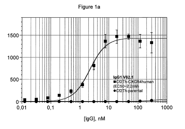
Figure 1 represents dose response curves of CXCR4 binding for antibodies of
the
present invention in IgG1 format, obtained as described under Example 4.
Figures 1a to
4
CA 03052070 2019-07-29
WO 2018/143938 PCT/US2017/015821
1e represent dose response curves for V62.1, V62.1-R108H, V62.1-H-m80, V62.1-H-
m43-m38 and V62.1-H-m47-m38, respectively.
Figure 2 represents in vivo luciferase activities in the breast region of mice
as measured
in the anti-metastatic model of Example 12.
DETAILED DESCRIPTION OF THE INVENTION
The present invention relates to anti-CXCR4 antibodies and uses thereof as
well as to
pharmaceutical compositions comprising anti-CXCR4 antibodies.
So that the invention may be more readily understood, certain terms are
specifically
defined below. Unless explicitly defined elsewhere in this document, all other
technical
and scientific terms used herein have the meaning that would be commonly
understood
by one of ordinary skill in the relevant art.
As used herein, including in the appended claims, the singular forms of words
such as
"a", "an", and "the", include their corresponding plural references unless the
context
clearly indicated otherwise.
The term "human CXCR4" refers to a protein whose amino acid sequence is at
least
90%, at least 95%, or at least 96%, 97%, 98%, or 99% identical to the complete
amino
acid sequence of human CXCR4 having Genbank accession number P61073, or to a
protein that has substantially the same biological function as CXCR4 but whose
sequence differs from the complete amino acid sequence of human CXCR4 by the
substitution, insertion or deletion of one or more amino acids.
The general structure of an "antibody" is well-known in the art. For an
antibody of the IgG
type, there are four amino acid chains (two "heavy" chains and two "light"
chains) that are
cross-linked via inter-chain disulfide bonds. Each of the heavy and light
chains has a
variable N-terminal region and a constant region. The constant regions of an
immunoglobulin antibody are called the Fc portion and are highly conserved in
humans.
The variable regions of each light/heavy chain pair form a variable domain
that
comprises the antibody's antigen binding site.
5
CA 03052070 2019-07-29
WO 2018/143938 PCT/US2017/015821
Each of the heavy and light chain variable regions can be further subdivided
into regions
of hypervariability, named complementarity determining regions (CDRs) that are
interspersed with regions that are more conserved, named framework regions
(FR).
Each variable region is composed of three CDRs and four FRs that are arranged
from
amino-terminus to carboxy-terminus in the following order: FR1, CDR1, FR2,
CDR2,
FR3, CDR3, FR4. Herein, the three CDRs of the heavy chain are referred to as
CDR1H,
CDR2H, and CDR3H and the three CDRs of the light chain are referred to as
CDR1L,
CDR2L and CDR3L. The CDRs contain most of the residues that form specific
interactions with the antigen. In the following, the heavy and light chain
variable regions
may be respectively referred to as HCVR and LCVR.
As used herein, the term "conservative modifications" of a given amino acid
sequence of
an antibody or a binding fragment, or of parts thereof, refers to amino acid
modifications
that do not significantly affect or alter the binding characteristics of the
antibody, binding
fragment, or parts thereof, containing the amino acid sequence. Such
conservative
modifications include amino acid substitutions, additions and deletions.
Modifications can
be introduced into an antibody of this disclosure by standard techniques known
in the art,
such as site-directed mutagenesis and PCR-mediated mutagenesis. Conservative
amino
acid substitutions are ones in which the amino acid residue is replaced with
an amino
acid residue having a side chain of related chemical character. Families of
amino acid
residues having side chains of related chemical character have been defined in
the art.
These families include amino acids with basic side chains (e.g., lysine,
arginine,
histidine), acidic side chains (e.g., aspartic acid, glutamic acid), uncharged
polar side
chains (e.g., glycine, asparagine, glutamine, serine, threonine, tyrosine,
cysteine,
tryptophan), nonpolar side chains (e.g., alanine, valine, leucine, isoleucine,
proline,
phenylalanine, methionine), beta-branched side chains (e.g., threonine,
valine,
isoleucine) and aromatic side chains (e.g., tyrosine, phenylalanine,
tryptophan,). Thus,
one or more amino acid residues within the CDR regions of an antibody of this
disclosure
can be replaced with other amino acid residues from the same side chain family
and the
altered antibody can be tested for retained antigen-binding properties using
the
functional assays described herein.
The sequence numbering used herein follows Kabat et al. (1991) Sequences of
proteins
of immunological interest. Public Health Service, National Institutes of
Health, Bethesda.
6
CA 03052070 2019-07-29
WO 2018/143938 PCT/US2017/015821
The CDR definitions used herein follow the method described in MacCallum et
al. (1996)
J. MoL Biol. 626:732-745.
An antibody according to the present invention can be intact, comprising
complete or full
.. length constant regions, including the Fc region, or a portion or fragment
of such an
antibody ("binding fragment") that comprises the antigen-binding portion and
retains
antigen-binding capability. Such a portion or fragment can include, e.g., a
Fab fragment
("fragment antigen binding"; i.e. the region of an antibody that binds to
antigens) that is
composed of a pair of heavy and light chain fragments each containing a
constant and a
variable region, or a Fab' or F(ab1)2 fragment that includes the CDRs or the
variable
regions of the anti-CXCR4 antibodies disclosed herein. Furthermore, such a
portion or
fragment can be a single chain Fv fragment that may be produced from a
polynucleotide
comprising nucleotide sequences encoding light and heavy chain variable
regions,
whereby the latter nucleotide sequences are separated by a linker sequence
(e.g.,
Pluckthun, The Pharmacology of Monoclonal Antibodies, vol. 113, Rosenburg and
Moore
eds., Springer-Verlag, New York, pp 269-315, 1994). Regardless of whether
fragments
or portions are specified, the term "binding fragment" as used herein includes
such
fragments or portions as well as single chain forms unless otherwise
indicated. As long
as a protein retains the ability to specifically or preferentially bind CXCR4
and includes a
CDR sequence(s) disclosed herein, it is included in the terms "antibody" and
"binding
fragment", respectively. It is understood that only full length antibodies may
perform
certain effector functions such as Antibody Dependent Cell Cytotoxicity
(ADCC).
Antibodies of the present invention may have a heavy chain constant region
selected
.. from any of the immunoglobulin classes (IgA, IgD, IgG, IgM, and IgE).
Preferably,
antibodies of the present invention are of the IgG type, more preferably the
IgG1 isotype.
It is to be understood that, unless there is an indication to the contrary,
the term "IgG1" in
the present text refers to human IgG1.
The term "human engineered antibody" refers to an antibody having frameworks,
hinge
regions, and constant regions of human origin that are identical with or
substantially
identical (substantially human) with frameworks, hinge regions and constant
regions
derived from human genomic sequences. Fully human frameworks, hinge regions,
and
constant regions encompass sequences expressed in the human germline as well
as
sequences containing spontaneous somatic mutations. A human engineered
antibody
7
CA 03052070 2019-07-29
WO 2018/143938 PCT/US2017/015821
may comprise framework, hinge, or constant regions derived from fully human
framework, hinge, or constant regions containing one or more amino acid
substitutions,
deletions, or additions therein, and/or glycosylation modifications. A "human
engineered
binding fragment" refers to a portion or fragment of a human engineered
antibody. Often,
a human engineered antibody is substantially non-immunogenic in humans.
A variety of different human framework sequences may be used singly or in
combination
as a basis for the human engineered antibodies of the present invention.
Preferably, the
framework regions of the antibodies of the invention are of human origin or
substantially
human (at least 95%, 97% or 99% of human origin). The sequences of framework
regions of human origin may be obtained from Current Trends in Monoclonal
Antibody
Development and Manufacturing by Shire et al., ISBN 978-0-387-76643-0.
Preferably, in
antibodies according to the present invention, the framework region of the
heavy chain
corresponds to the germline consensus sequence subgroup Ill. Preferably also,
in
antibodies according to the present invention, the framework region of the
light chain
corresponds to the germline kappa Ill consensus sequence.
As used herein, the terms "monospecific antibody" or "monospecific antibody
composition" refer to a preparation of antibody molecules having identical
protein
sequences (ionic or oxidation microvariants being included). A monospecific
antibody
composition displays a single binding specificity and affinity for a
particular epitope.
As used herein, an antibody that "specifically binds to human CXCR4" refers to
an
antibody that binds to human CXCR4 (and possibly CXCR4 from one or more non-
human species) with an EC50 of 50 nM or less, as measured in a Fluorescent
Flow
Cytometry-based assay as described in Example 4 herein below, but does not
substantially bind to other GPCRs such as, for example, CXCR7.
As used herein when referring to an antibody, the phrase "does not
substantially bind" to
non-CXCR4 proteins means that the antibody does not bind at all or exhibits
only weak
binding to non-CXCR4 proteins. The EC50 value for such weak binding can be
equal to
or greater than 100nM as measured in a Fluorescent Flow Cytometry-based assay
as
described in Example 4.
8
CA 03052070 2019-07-29
WO 2018/143938 PCT/US2017/015821
As used herein, ADCC refers to Antibody Dependent Cell Cytotoxicity, i.e.
antibody
mediated cell death, which is an antibody effector function mainly prompted by
the Fc
region. Antibodies of IgG isotypes, particulary IgG1, are known for having
good ADCC
properties.
When referring to SDF-1 or CXCL12 herein, unless otherwise specified or
exemplified, it
is meant to designate any and all human SDF-1 variants, including e.g. SDF-
1alpha or
CXCL12a and SDF-1beta or CXCL12b.
When referring to the binding properties, half maximal Effective Concentration
50 (EC50)
is the concentration which induces a response halfway between the baseline and
the
maximal binding of a given antibody. It is calculated via a dose response
curve, as
explained in Example 4 herein.
A "subject" is a mammal, preferably a human.
The term "treating" (or "treat" or "treatment") means slowing, stopping,
reducing, or
reversing the progression or severity of a symptom, disorder, condition or
disease.
The term "preventing" (or "prevent" or "prevention") means prohibiting,
restraining, or
inhibiting the incidence, occurrence or recurrence of a symptom, disorder,
condition, or
disease.
The term "therapeutically effective amount" refers to the amount or dose of an
antibody
of the present invention which, upon single or multiple dose administration to
a patient,
provides the desired treatment.
Particular antibodies of the present invention originate from a phage display
library, and
from affinity maturation processes as described herein.
Phage-display libraries are commonly used technologies for selection of
antibody
fragments that provide a starting point for generation and optimization of
human
engineered antibodies. See e.g. Hoogenboom (2005) Nat. Biotechnol. 23: 1105-
1116;
Bradbury & Marks (2004) J. Immunol. Methods 290: 29-49; and Fredericks et al.,
(2004)
Protein Eng. Des. Se!. 17: 95-106. Other types of display technologies useful
for the
9
CA 03052070 2019-07-29
WO 2018/143938 PCT/US2017/015821
generation and affinity maturation (optimization) including yeast-, mRNA- and
ribosome-
display libraries are gaining in popularity for selection and optimization of
antibodies (see
Hoogenboom, Bradbury & Marks, and Fredericks et al.).
Display libraries may display single-chain variable-domain antibody fragments
(scFvs) or
Fab fragments, and contain the encoding DNA or RNA. They have high genetic
diversity
or repertoire size (commonly 10^9-10"13). The genetic diversity in these
libraries is
commonly created by cloning the repertoire of the immunoglobulin heavy chain
and light
chain variable gene segments from naive or immunized individuals.
Alternatively, this
diversity can be achieved by randomization of CDR sequences, including using
chemically synthesized CDR fragments, or by a combination of these two
approaches.
The binding step (for selections from such a library ) can then be undertaken
with the
target (receptor) in solution, immobilized on a surface, on liposomes (such as
proteoliposomes described in US Patent 6,761,902), on cells, etc. After
extensive
washing, bound clones are recovered and amplified for a further round of
selection.
Affinity maturation processes may then be performed on initial best binder
antibody
candidates to try to obtain derivative candidates with improved properties,
such as better
stability and/or improved binding, etc. Several affinity maturation strategies
are available
to a person skilled in the art, such as, but not limited to, directed
comprehensive
mutagenesis, CDR or light/heavy chain shuffling, point insertion(s) or
deletion(s) in
CDRs, or any combination of these approaches.
Particular antibodies of the present invention include antibodies as disclosed
in
Examples 1 and 2 herein. It is to be understood that the present invention
also embraces
each and every possible exchange of CDRs between the variable regions provided
herein. Preferably, a heavy chain CDR may be exchanged with another heavy
chain
variable region CDR, and likewise, a light chain CDR may be exchanged with
another
light chain variable region CDR.
Antibody synthesis
Antibodies of the invention can be produced using techniques well known in the
art, e.g.,
recombinant technologies, in vitro protein expression technologies or
combinations of
such technologies or other technologies readily known in the art.
10
CA 03052070 2019-07-29
WO 2018/143938 PCT/US2017/015821
For example, Fab fragments obtained from a screen of a Fab display library
directly or
subsequent to affinity maturation can be converted into IgGs by commonly used
techniques such as cloning into appropriate expression vectors encoding the
desired
constant region.
For direct production of an IgG antibody, an appropriate host cell, such as
HEK 293 or
CHO cells, may be either transiently or stably transduced with an expression
system
suitable for producing and secreting IgG antibodies. The expression system
will comprise
heavy chain and light chain expression constructs that are transduced at an
optimized
ratio or a single vector system comprising expressible light chain as well as
heavy chain
genes. Secreted antibody can be purified using any of many commonly-used
techniques.
For example, culture medium containing antibody can be conveniently applied to
a
Protein A or G Sepharose FF column that has been equilibrated with a
compatible buffer,
e.g., phosphate-buffered saline (pH 7.4). The column is then washed to remove
non-
specifically binding components. Bound antibody is eluted, for example, by
application of
a pH gradient. Antibody-containing fractions are detected, e.g., by SDS-PAGE,
and are
pooled. Depending on the intended use, the antibody can be further purified.
The
antibody can be concentrated and/or sterile-filtered using common techniques.
Soluble
aggregates and multimers can be effectively removed by common techniques,
including
size exclusion, hydrophobic interaction, ion exchange, or hydroxyapatite
chromatography. Purified antibody typically can be stored refrigerated,
frozen, or
lyophilized.
The person of skill in the art will know that Fab antibodies can be similarly
produced
using cells such as bacterial, fungal (yeast) or insect cells.
Properties of antibodies of the invention
The antibodies of the present invention, in Fab format and/or in IgG format,
were
characterized in respect of several desirable biological properties.
Binding to CXCR4 is the first criterion for efficacy of the antibodies
according to the
present invention. Antibodies according to the present invention specifically
bind to
CXCR4 with an EC50 of below 50 nM, preferably below 10 nM, as revealed by
experiments using cells expressing CXCR4 from a transfected, expressible gene
and/or
tumor cell lines expressing CXCR4.
11
CA 03052070 2019-07-29
WO 2018/143938 PCT/US2017/015821
Conversion of Fab fragments into IgG antibodies generally improves receptor
binding
(EC50). This was also verified with antibodies of the present invention.
Preferably, when
in IgG1 format, antibodies according to the present invention specifically
bind CXCR4
with an EC50 of below 5 nM.
The antibodies of the present invention inhibit binding of SDF-1 to the CXCR4
receptor
and prevent receptor activation. Consequences of SDF-1 binding to its receptor
include,
for example, calcium flux induction and cell migration, which are important
parameters
for cancer cell invasion. The antibodies of the present invention inhibit
calcium flux
induction and/or migration of CXCR4-expressing cells.
As has been demonstrated for antibodies such as trastuzumab and rituximab,
ADCC can
be an important mechanism of action of therapeutic antibodies against tumors.
The
antibodies of the present invention were shown to be capable of ADCC. Studies
with
xenograft tumor models demonstrated the anti-tumor activity of the antibodies
of the
invention.
Pharmaceutical compositions and their administration
The present invention also concerns pharmaceutical compositions comprising an
antibody of the present invention. The latter compositions will be
preferentially
administered parenterally, but transnasal, transpulmonary or transdermal
delivery is also
envisaged. The pharmaceutical compositions may contain any conventional non-
toxic
pharmaceutically-acceptable excipient and, in the case of a liquid
formulation, diluent. In
some cases, the pH of the formulation may be adjusted with pharmaceutically
acceptable
acids, bases or buffers to enhance the stability of the formulated agent or
its delivery
form. The term parenteral as used herein includes subcutaneous,
intracutaneous,
intravenous, intramuscular, intraarticular, intraarterial, intrasynovial,
intrasternal,
intrathecal, intralesional and intracranial injection or infusion.
Antibody of the invention can be stored as a lyophilized formulation or as a
solution.
Injectable preparations, for example, sterile injectable aqueous or oleaginous
suspensions, may be formulated according to the known art using suitable
dispersing or
wetting agents and suspending agents. The sterile injectable preparation may
also be a
sterile injectable solution, suspension or emulsion in a nontoxic parenterally
acceptable
diluent or solvent. Among the acceptable diluents that may be employed are
water,
12
CA 03052070 2019-07-29
WO 2018/143938 PCT/US2017/015821
Ringer's solution, U.S.P. and isotonic sodium chloride solution. In addition,
sterile, fixed
oils are conventionally employed as a solvent or suspending medium. The
compositions
can further comprise "pharmaceutically-acceptable" excipients or stabilizers
typically
employed in the art (all of which are termed "excipients"). Excipients
comprise, e.g.,
buffering agents, stabilizing agents, preservatives, tonicity agents, non-
ionic detergents,
antioxidants and other miscellaneous additives. (See Remington's
Pharmaceutical
Sciences, 16th edition, A. Osol, Ed. (1980)). Such additives must be nontoxic
to the
recipients at the dosages and concentrations employed.
Buffering agents are preferably present at concentration ranging from about 2
mM to
about 50 mM. Suitable buffering agents include both organic and inorganic
acids and
salts thereof such as citrate buffers (e.g., monosodium citrate-disodium
citrate mixture,
citric acid-trisodium citrate mixture. citric acid-monosodium citrate mixture.
etc.),
succinate buffers (e.g., succinic acid-monosodium succinate mixture, succinic
acid-
sodium hydroxide mixture, succinic acid-disodium succinate mixture, etc.),
tartrate
buffers (e.g., tartaric acid-sodium tartrate mixture, tartaric acid-potassium
tartrate
mixture, tartaric acid-sodium hydroxide mixture, etc.), fumarate buffers
(e.g., fumaric
acid-monosodium fumarate mixture, etc.), fumarate buffers (e.g., fumaric acid-
monosodium fumarate mixture, fumaric acid-disodium fumarate mixture,
monosodium
fumarate-disodium fumarate mixture, etc.), gluconate buffers (e.g., gluconic
acid-sodium
glyconate mixture, gluconic acid-sodium hydroxide mixture, gluconic acid-
potassium
glyuconate mixture, etc.), oxalate buffer (e.g., oxalic acid-sodium oxalate
mixture, oxalic
acid-sodium hydroxide mixture, oxalic acid-potassium oxalate mixture, etc.),
lactate
buffers (e.g., lactic acid-sodium lactate mixture, lactic acid-sodium
hydroxide mixture,
lactic acid-potassium lactate mixture, etc.) and acetate buffers (e.g., acetic
acid-sodium
acetate mixture, acetic acid-sodium hydroxide mixture, etc.). Additionally,
there may be
the mentioned phosphate buffers, histidine buffers and trimethylamine salts
such as Tris.
Preservatives may be added to retard microbial growth, and may be added in
amounts
ranging from 0.2%-1% (w/v). Suitable preservatives for use with the present
invention
include phenol, benzyl alcohol, meta-cresol, methyl paraben, propyl paraben,
octadecyldimethylbenzyl ammonium chloride, benzalconium halides (e.g.,
chloride,
bromide, iodide), hexamethonium chloride, alkyl parabens such as methyl or
propyl
paraben, catechol, resorcinol, cyclohexanol, and 3-pentanol.
13
CA 03052070 2019-07-29
WO 2018/143938 PCT/US2017/015821
The osmolarity of the pharmaceutical compositions may be adjusted with
tonicity agents
to a value that is compatible with the intended use of the compositions. For
example, the
osnnolality of injectable solutions may be adjusted to approximately the
osmotic pressure
of blood, which is equivalent to about 0.9 w/v % of sodium chloride in water.
Examples of
suitable tonicity agents include chloride salts of sodium, potassium, calcium
and
magnesium, dextrose, glycerol, propylene glycol, mannitol, sorbitol,
erythritol, arabitol,
xylitol, and the like and mixtures thereof. Tonicity agents are typically used
in amounts
ranging from about 0.001 to about 1 % w/v. These amounts have been found to be
useful in providing a physiologically acceptable tonicity. Preferably, the
tonicity agent(s)
will be employed in an amount to provide a final osmotic value to the
composition of 150
to 450 mOsm/kg, more preferably between about 220 and about 350 mOsm/kg, and
most preferably between about 270 and about 300 mOsm/kg.
The compositions can further comprise a stablilizer. Typical stabilizers can
be polyhydric
sugar alcohols (enumerated above); amino acids such as arginine, lysine,
glycine,
glutamine, asparagine, histidine, alanine, ornithine, L-Ieucine, 2-
phenylalanine, glutamic
acid, threonine, etc., organic sugars or sugar alcohols, such as lactose,
trehalose,
stachyose, mannitol, sorbitol, xylitol, ribitol, myoinisitol, galactitol,
glycerol and the like,
including cyclitols such as inositol; polyethylene glycol; amino acid
polymers; sulfur
containing reducing agents, such as urea, glutathione, thioctic acid, sodium
thioglycolate,
thioglycerol, alpha-monothioglycerol and sodium thiosulfate; low molecular
weight
polypeptides (i.e. <10 residues); proteins such as human serum albumin, bovine
serum
albumin, gelatin or immunoglobulins; hydrophylic polymers such as
polyvinylpyrrolidone;
monosaccharides such as xylose, mannose, fructose, glucose; disaccharides such
as
lactose, maltose, sucrose and trisaccacharides such as raffinose;
polysaccharides such
as dextran. Stabilizers may be present in the weight range from 0.1 to 10,000
times the
weight of the antibody of the invention.
Wetting agents may be added to help solubilize the antibody of the invention
as well as
to protect it against agitation-induced aggregation. Suitable wetting agents
include non-
ionic surfactants such as polysorbates (20, 80, etc.), polyoxamers (184, 188
etc.),
Pluronic.RTM, polyols, polyoxyethylene sorbitan monoethers (Tween®-20,
Tween®-80, etc.). Non-ionic surfactants may be present in a range of about
0.05
14
CA 03052070 2019-07-29
WO 2018/143938 PCT/US2017/015821
mg/ml to about 1.0 mg/ml, preferably about 0.07 mg/ml to about 0.2 mg/ml.
The pharmaceutical compositions may also contain an additional active compound
as
necessary for the particular indication being treated, preferably a compound
with an
activity that does not adversely affect that of the antibody of the invention.
For example,
when a cancer is being treated, it may be desirable to further provide one or
more
chemotherapeutic agents. Such compounds are suitably present in combination in
amounts that are effective for the purpose intended.
The pharmaceutical compositions can be sterilized, for example, by filtration
through
sterile filtration membranes.
Antibody of the invention may also be entrapped in microcapsules prepared, for
example, by coacervation techniques or by interfacial polymerization, for
example,
hydroxymethylcellulose or gelatin-microcapsule and poly-(methylmethacylate)
microcapsules, respectively, in colloidal drug delivery systems (for example,
liposomes,
albumin micropheres, microemulsions, nano-particles and nanocapsules) or in
macroemulsions. Such techniques are disclosed in Remington's Pharmaceutical
Sciences, 16th edition, A. Osal, Ed. (1980).
Sustained-release preparations may be prepared. Suitable examples of sustained-
release preparations that may be adapted for the delivery of antibody of the
invention
include semi-permeable matrices of solid hydrophobic polymers containing the
antibody,
which matrices are in the form of shaped articles, e.g., films, or
microcapsules. Examples
of sustained-release matrices include polyesters, hydrogels (for example,
poly(2-
hydroxyethyl-methacrylate), or poly(vinylalcohol)), polylactides (U.S. Pat.
No. 3,773,919),
copolymers of L-glutamic acid and ethyl-L-glutamate, non-degradable ethylene-
vinyl
acetate, degradable lactic acid-glycolic acid copolymers and poly-D-(-)-3-
hydroxybutyric
acid. While polymers such as ethylene-vinyl acetate and lactic acid-glycolic
acid enable
release of molecules for over 100 days, certain hydrogels release proteins for
shorter
time periods. When encapsulated antibodies remain in the body for a long time,
they may
denature or aggregate as a result of exposure to moisture at 37 C, resulting
in a loss of
biological activity and possible changes in immunogenicity. Rational
strategies can be
devised for stabilization depending on the mechanism involved. For example, if
the
aggregation mechanism is discovered to be intermolecular S--S bond formation
through
CA 03052070 2019-07-29
WO 2018/143938 PCT/US2017/015821
thiol-disulfide interchange, stabilization may be achieved by modifying
sulfhydryl
residues, lyophilizing from acidic solutions, controlling moisture content,
using
appropriate additives, and developing specific polymer matrix compositions.
Administration methods can be appropriately selected in consideration of a
subject's age
and symptoms. The dose in a pharmaceutical composition of antibody or binding
fragment of the invention may be, for example, from about 0.0005 to about 100
mg/kg for
each administration. More preferably, the dose may be from about 0.1 to about
20 mg/kg
for each administration. Administration may be several times daily, daily,
every two days,
half-weekly or weekly. However, the present invention is not limited by the
numeric
values described above. The doses and administration methods vary depending on
the
subject's weight, age, symptoms, and such. Those skilled in the art can set
appropriate
doses and administration methods in consideration of the factors described
above.
Diagnostic uses for the antibodies of the invention
The antibodies and binding fragments of the present invention can be useful in
diagnostic assays, e.g., assays for detecting expression of CXCR4 on specific
cells,
tissues, or serum. For diagnostic applications, the antibody typically will be
labeled with a
detectable moiety. Numerous labels are available. Examples of enzymatic labels
include
luciferases (e.g., firefly luciferase and bacterial luciferase; U.S. Pat. No.
4,737,456),
malate dehydrogenase, urease, peroxidase such as horseradish peroxidase
(HRPO),
alkaline phosphatase, beta-galactosidase, glucoamylase, lysozyme, saccharide
oxidases
(e.g., glucose oxidase, galactose oxidase, and glucose-6-phosphate
dehydrogenase),
heterocyclic oxidases (such as uricase and xanthine oxidase), lactoperoxidase,
microperoxidase, and the like. Techniques for conjugating enzymes to
antibodies are
described in O'Sullivan et al., Methods for the Preparation of Enzyme-Antibody
Conjugates for Use in Enzyme Immunoassay, in Methods in Enzym. (Ed. Langone &
Van
Vunakis), Academic press, New York, 73: 147-166 (1981).
Sometimes, the label is indirectly conjugated with the antibody. The skilled
artisan will be
aware of various techniques for achieving this. For example, the antibody can
be
conjugated with biotin and any of the labels mentioned above can be conjugated
with
avidin, or vice versa. Biotin binds selectively to avidin and thus, the label
can be
conjugated with the antibody variant in this indirect manner. Alternatively,
to achieve
indirect conjugation of the label with the antibody, the antibody is
conjugated with a small
16
CA 03052070 2019-07-29
WO 2018/143938 PCT/US2017/015821
hapten (e.g. digoxin) and one of the different types of labels mentioned above
is
conjugated with an anti-hapten antibody (e.g. anti-digoxin antibody). Thus,
indirect
conjugation of the label with the antibody can be achieved.
In another embodiment of the invention, the antibody of the invention need not
be
labeled, and the presence thereof can be detected using a labeled antibody
which binds
to the antibody.
The antibodies or binding fragment of the present invention may be employed in
any
known immunochemical assay method, such as competitive binding assays, direct
and
indirect sandwich assays, and immunoprecipitation assays. Zola, Monoclonal
Antibodies:
A Manual of Techniques, pp. 147-158 (CRC Press, Inc. 1987). They can also be
used for
immunohistochemical detection of CXCR4 on cells and tissues. For
immunohistochemistry, a tissue sample, e.g., a tumor tissue sample, may be
fresh or
frozen or may be embedded in paraffin and fixed with a preservative such as
formalin, for
example.
The antibodies may also be used for in vivo diagnostic assays. Generally, the
antibody or
binding fragment is labeled with a radionucleotide (such as 1111n, 99-rc, 14C,
1311, 3H, 32p or
35S) so that CXCR4-over-expressing cells can be localized using
immunoscintiography.
The antibody or binding fragment of the present invention can be provided in a
kit, i.e., a
packaged combination of reagents in predetermined amounts with instructions
for
performing the diagnostic assay. Where the antibody is labeled with an enzyme,
the kit
may include substrates and cofactors required by the enzyme (e.g., a substrate
precursor which provides the detectable chromophore or fluorophore). In
addition, other
additives may be included such as stabilizers, buffers (e.g., a block buffer
or lysis buffer)
and the like. The relative amounts of the various reagents may be varied
widely to
provide for concentrations in solution of the reagents which substantially
optimize the
sensitivity of the assay. Particularly, the reagents may be provided as dry
powders,
usually lyophilized, including excipients which on dissolution will provide a
reagent
solution having the appropriate concentration.
17
CA 03052070 2019-07-29
WO 2018/143938 PCT/US2017/015821
Human therapeutic uses for the antibodies and binding fragments of the
invention
The antibodies of the invention can be used in stem cell and regenerative
medicine.
Interaction of CXCR4 with SDF-1alpha is important in holding hematopoietic
stem cells in
the bone marrow. Anti-CXCR4 antibodies can serve as antagonists that are
capable of
mobilizing hematopoietic stem cells into the bloodstream as peripheral blood
stem cells.
Peripheral blood stem cell mobilization can be important in hematopoietic stem
cell
transplantation (as an alternative to transplantation of surgically-harvested
bone marrow)
and is currently performed using drugs such as G-CSF. Antibodies and binding
fragments of the present invention can also be used to prevent late stage HIV
(X4
viruses) from interacting with the CXCR4 receptor and entering T cells.
The antibodies and binding fragments of the invention further can be used in
the
treatment of a variety of different cancers that express CXCR4. CXCR4 may be
the
chemokine receptor that is most commonly found on tumor cells, both in human
and
experimental murine cancers. The receptor has been found on at least the
following
tumor types: B-CLL, AML, B-lineage ALL (including Burkitt's lymphoma),
follicular center
myeloma, CML, multiple myeloma, pancreatic cancer, prostate cancer, breast
cancer,
ovarian cancer, thyroid cancer, colorectal cancer, oral squamous carcinoma,
cervical
cancer, neuroblastoma, kidney cancer, glioma, rhabdomyosarcoma, small lung
cancer
and melanoma. Balkwill (2004) Seminars in Cancer Biology 14: 171-9. Treatment
will
involve administration to the cancer patients of a pharmaceutical composition
comprising
an antibody or binding fragment of the invention. The composition may be
administered
by any suitable means, including parenteral, subcutaneous, intraperitoneal,
intrapulmonary, intranasal, and intralesional administration. Parenteral
infusions include
intramuscular, intravenous, intraarterial, intraperitoneal, or subcutaneous
administration.
In addition, the antibody or binding fragment is suitably administered by
pulse infusion,
particularly with declining doses of the antibody or binding fragment.
Preferably, the
dosing is given by injections, most preferably intravenous or subcutaneous
injections,
depending in part on whether the administration is brief or chronic.
Depending on the type and severity of the disease, about 0.1 mg/kg to about 20
mg/kg of
antibody or binding fragment is an initial candidate dosage for administration
to the
subject, whether, for example, by one or more separate administrations, or by
continuous
infusion. A typical daily dosage might range from about 1 mg/kg to 100 mg/kg
or more.
18
CA 03052070 2019-07-29
WO 2018/143938 PCT/US2017/015821
The pharmaceutical composition comprising antibody or binding fragment of the
invention will be formulated, dosed and administered in a manner consistent
with good
medical practice. Factors for consideration in this context include the type
and stage of
cancer, the clinical condition of the individual subject, the site of delivery
of the agent, the
method of administration, the scheduling of administration, and other factors
known to
medical practitioners. The "therapeutically effective amount" of the antibody
to be
administered will be governed by such considerations, and is the minimum
amount
necessary to treat the disease. The antibody need not be, but is optionally
formulated
with one or more agents currently used to treat the disease, e.g., one or more
chemotherapeutic agents. The effective amount of such other agents depends on
the
amount of antibody or binding fragment present in the formulation, the type
and stage of
cancer, and other factors discussed above. These are generally used in the
same
dosages and with administration routes as they are currently used (without
antibody of
the invention) or from about 1 to 99% of the currently employed dosages.
EXAMPLES
Example 1: preparation of Fab phage library and screening of phage antibodies
The antibodies of the present invention were originally derived from a Fab
library of the
size of 10'11 comprised of pSF1 phagennids carrying Fab E. coli codon-
optimized
synthetic genes encoding human Fab heavy and human Fab light chains with
randomized CDRs. For the heavy chain, the framework DP47 was employed, and for
the light chain, the framework DPK22 was employed.
The phage library was generated employing protocols and CDR randomization
schemes
as described in Knappik et al. (2000) J. Mol. Biol. 296:57-86; Lee et al.
(2004) J. MoL
Biol. 340:1073-93; Hoet et al. (2005) 23:344-8.
More specifically, in the Fab library the heavy chain CDR1, CDR2, and CDR3 and
the
light chain CDR3 were subjected to randomization. For the randomization of
heavy
chain CDR3, tri-nucleotide based oligonucleotides were employed as described
in
Knappik et al., whereas for other CDRs standard nucleotide mixtures were
employed to
generate CDR oligonucleotides.
The common light chain CDRs of the Fab library were as follows (MacCallum et
at.
(1996) J. MoL Biol. 626:732-745):
19
CA 03052070 2019-07-29
WO 2018/143938 PCT/US2017/015821
CDR1L--SSYLAWY-- (SEQ ID No.1)
CDR2L--LLIYGASSRA-- (SEQ ID No.2)
For the screening of the Fab library, a Magnetic ProteoLiposome technology was
used in
order to display CXCR4 in a liposome membrane in a conformation closely
resembling
its native conformation. See Mirzabekov et al. (2000) Nat Biotechnol. 18:649-
54 and US
Patent No. 6,761,902.
Screening of the Fab library was carried out using methods described by
Mirzabekov et
al. and yielded the following Fab candidate:
SEQ ID NO.
Antibody CDR3L CDR1H CDR2H CDR3H LCVR HCVR
V62.1 3 5 7 11 13 15
Example 2: affinity maturation
The initial candidate as described above was then submitted to affinity
maturation. Two
Affinity Maturation Libraries were generated by CDR2H or CDR3L randomization,
respectively. Each of these libraries was submitted to two rounds of high
stringency
selection. Selected Fabs were expressed individually, and clones with improved
binding
properties were retained. The best clones were reformatted as IgGs that were
characterized for best CXCR4 binding affinity and selectivity as well as for
best ability to
prevent ligand induction of Ca-flux. As a final step, heavy and light chains
of the most
promising IgGs were recombined, and the resulting IgGs were again
characterized as
before.
In addition, CDR3H was matured by introduction of point mutations. Resulting
mutated
Fab fragments were characterized to identify the best CXCR4 binders.
The following matured antibody candidates were pursued further:
SEQ ID NO.
Antibody* CDR3L CDR1H CDR2H CDR3H LCVR HCVR
V62.1-R108H 3 5 7 12 13 16
V62.1-R108H- 4 5 8 12 14 17
m43-m38
V62.1- R108H- 4 5 9 12 14 18
m47-m38
V62.1- R108H- 3 6 10 12 13 19
m80
CA 03052070 2019-07-29
WO 2018/143938 PCT/US2017/015821
*R108H refers to a point mutation in CDR3H (compare SEQ ID NO: 11 and 12), and
m38, m43, m47 and m80 refer to particular selected CDR2H or CDR3L sequences,
respectively.
As mentioned previously, all antibodies of the present invention share the
same CDR1L
(SEQ ID NO.1) and CDR2L (SEQ ID NO.2).
Example 3: synthesis of loG antibodies
The CHO/pTT Transient Transfection System from the Biotechnology Research
Institute
of the Canadian National Research Council (NRC-BRI) was used according to
protocols
provided by the NRC-BRI. See international patent application publication
W02009/137911 Al. More specifically, each IgG of interest was produced in CH0-
3E7
cells co-transfected with pTT vectors expressing the light chain and the heavy
chain of
the IgG, respectively, using polyethylenimine (PEI) as a transfection reagent.
Cell medium containing IgGs was collected, and IgGs were purified on Protein A
Plus
Agarose (Pierce) using standard methodology. All purification procedures were
performed using sterile, endotoxin-free solutions.
In the following examples, a Fab fragment of interest or an IgG antibody of
interest is
referred to as "test Fab", "test antibody" or "test IgG", as appropriate.
Example 4: binding to CXCR4-expressinq cells
Binding of Fabs or IgGs to CXCR4-expressing cells was measured by a
fluorescent flow
cytometry-based assay. The cells were stained with:
(A) for Fabs - anti-c-Myc mouse antibody 9E10 Mab that binds to a tag present
in the test
Fab and then with secondary anti-mouse IgG phycoerythrin (PE)-conjugated
antibody, or
(B) for IgGs - with anti-Human Fc PE-conjugated antibody.
As a control, cells that do not express CXCR4 or cells expressing other GPCR
were
used.
A typical protocol for the fluorescent flow cytometry-based assay was as
follows. Ten
microliter of a purified test IgG solution or buffer as a control were added
to 10 microliter
of a suspension containing approximately 30,000 Cf2-Th cells transfected to
express
human CXCR4. After incubation on ice for 40 min, cells were washed with FACS
buffer
21
CA 03052070 2019-07-29
WO 2018/143938 PCT/US2017/015821
(phosphate-buffered saline (PBS), pH7.4; 2% fetal calf serum, 0.1% sodium
azide) to remove unbound antibodies. Ten microliter of a solution of
phycoerythrin (PE)-
conjugated mouse anti-human Fc monoclonal antibody (1/20 dilution; catalog
number
12-4998-82, eBioscience Inc., San Diego, CA) were then added to the cells,
and, after a
30-min incubation on ice, cells were washed twice and then formalin-fixed (FIX
buffer:
PBS, pH7.4; 0.5% formaldehyde). Fixed samples were analysed by fluorescent
flow
cytometry using a Guava-PCA96 instrument (EMD Millipore Chemicals, Merck KGaA,
Darmstadt, Germany).
To determine an EC50 value, binding to the CXCR4-expressing cells was measured
at
different concentrations of test antibody. Duplicate or triplicate samples
were analysed
for each concentration. Titration curves were constructed based on the Mean
Fluorescence Intensity (MFI) values provided by the instrument using a
SoftMaxPro5
program (Molecular Devices Corp., Sunnyvale, CA).
In some experiments, non-transfected Cf2-Th parental cells (ATCC CRL-14307)
were
used as negative controls, thereby establishing the specificity of the
antibodies for
CXCR4. In some other experiments, several batches of the same antibody
candidate
were tested in parallel. The dose response curves and EC50 results obtained
with test
antibodies in IgG1 format are presented in Figs. la to e. All test IgG1
antibodies
exhibited an EC50 well below 10 nM.
Example 5: binding to CXCR4-expressing human lymphoma cells
Binding of test IgGs to CXCR4-expressing human lymphoma cells (Ramos; RA1,
ATCC
CRL-1596T) was measured by fluorescent flow cytometry-based assay. Tumor cell
staining was conducted as follows: human Fcy receptors of RA1 cells were
saturated by
incubation at 4 C for 30 minutes in PBS containing 2% human serum and 0.5mM
EDTA.
Cells were then incubated at 4 C for one hour with test IgGs or a human
isotypic
IgG1 kappa control (Coger Sari, Paris, France) (both antibody types at
10pg/mL). The
cells were washed with PBS and further incubated for one hour at 4 C with a
goat
F(abP)2 fragment anti-human IgG (H+L)-PE (Beckman Coulter). The cells were
washed
twice with PBS and fixed with 0.5% formaldehyde in PBS for analysis by flow
cytometry.
The data were acquired using an eleven-color flow cytometer (LSRII, BD
Biosciences),
and the analyses were performed with the FlowJo flow cytometry analysis
software (Tree
22
CA 03052070 2019-07-29
WO 2018/143938 PCT/US2017/015821
Star Inc., Ashland, OR). The living cells were selected using the side scatter
(SSC) and
the forward scatter (FSC); 10,000 events were acquired for each analysis. MFI
values
were recorded using the PE channel.
Antibody MFI
V62.1 595
V62.1-R108H 429
V62.1-R108H-m80 440
V62.1-R108H-m43-m38 488
V62.1-R108H-m47-m38 549
IgG1k isotypic control 42
MFI values well above that of the isotypic control indicate positive staining
of the RA1
cells, which was observed for all test IgGs.
Example 6: specificity for CXCR4
Cells over-expressing different GPCRs other than CXCR4 and several lines
transfected
to over-express CXCR4 (R1610-hCXCR4, Cf2Th-hCXCR4 and CHO-hCXCR4) were
compared. Cultures were incubated with a test IgG at 100 nM in FACS buffer for
40 min
at 4 C. Thereafter, cells were washed twice, stained with anti-human-Fc
antibody-PE
conjugate (Jackson Immunoresearch Laboratories Inc., West Grove, PA), washed
twice
in FACS buffer and then transferred to FIX buffer. Fluorescence intensities
were
measured by GUAVA PCA-96 at 425V (in triplicate).
The expression of GPCRs other than CXCR4 was confirmed using commercially
available antibodies (positive controls). For example, a commercial anti-CXCR1
antibody
was used as a positive control for confirming the expression of CXCR1 on the
CXCR1-
transfected CHO cells.
23
CA 03052070 2019-07-29
WO 2018/143938 PCT/US2017/015821
A summary of the MFI data obtained is presented in the Table below.
V62.1- V62.1
Antibody V62.1- R108H R108H Positive
Cell line V62'1 RI 08H - m47- m80 controls
m38
CHO-hCXCR1 4 4 N/A N/A 1325
hCXCR2 3 4 N/A N/A 1830
hCXCR3 3 3 2 2 500
hCXCR4 R1610-
812 513 N/A N/A 447
Cf2Th-
1267 861 2350 3870 1006
hCXCR4
CHO-hCXCR4 2053 1850 N/A N/A 994
hCXCR5 4 3 2 2 510
hCXCR6 3 3 2 7 1050
hCXCR7 3 4 2 2 300
hCCR3 4 4 40 2 213
hCCR4 3 3 2 2 506
hCCR5 4 3 2 8 724
hCCR6 3 3 2 10 1481
hCCR7 3 4 2 25 465
hCCR9 4 3 2 8 260
hCCR10 4 3 2 10 3000
Cf2Th 3 3 2 8 1
R1610 3 3 2 2 1
CHO 3 3 2 7 1
Example 7: inhibition of ligand binding to CXCR4
Inhibition of SDF-1alpha ligand binding was assayed by means of fluorescent
flow
cytometry using bacterially expressed SDF-1alpha containing an N-terminal FLAG
tag.
Cf2-Th cells transfected to express CXCR4 were incubated with a test IgG
antibody (100
nM) for 30 min on ice. Thereafter, the FLAG-tagged ligand was added to a final
concentration of 100 nM, and the cells were incubated for another 20 min.
Subsequently,
cells were washed, stained with an appropriate anti-FLAG tag PE-conjugated
antibody
and fixed with FIX buffer. In the control, no antibody was added. Then
fluorescence was
recorded. A decreased MFI value of cells that had been exposed to a test
antibody
(MFIwithAb), as compared to the MFI of cells that had not been exposed to the
IgG test
antibody (MFIN0Ab) indicated a competition between the antibody and the
ligand. Percent
inhibition was defined as (MFIwithAb / MFIN) x 100%. Similar MFIwithAb and
MFINoAb
values indicated that the pre-bound test antibody failed to prevent binding of
the tagged
ligand to CXCR4 on the cell surface. Antibodies tested, V62.1 and V62.1R108H,
were
able to inhibit ligand binding by up to 96%.
24
CA 03052070 2019-07-29
WO 2018/143938 PCT/US2017/015821
Example 8: inhibition of SDF-1-induced calcium flux
IC50 values were estimated based on data obtained from FLIPR calcium assays
(Calcium-5 kit, Molecular Devices LLC, Sunnyvale, CA) on CXCR4-transfected
Chem-1
cells (catalog no. HTS004C, EMD Millipore Chemicals). The cells were grown
overnight
at 37 C and 5% CO2. Before Ca-flux measurement, cells were starved in serum-
free
medium for 3h at 37 C and 5% CO2. Dye was added to the cells which were then
incubated for 30min at 37 C and 5% CO2 in the presence of different
concentrations of
test IgG1 antibody. Control samples were prepared similarly, but no antibody
was added.
Thereafter, SDF-1alpha (R&D Systems) in TBS was added to the dye-loaded cells
to a
final concentration of 30 nM. Inhibition of the chemokine-induced increase in
intracellular
calcium concentration (Ca-flux) was calculated as follows:
Inhibition = (1. ¨
LC .1) X 111 G96
where [I] ¨ means peak dye fluorescence
(n=4) in inhibited samples, [C] ¨ means peak dye fluorescence (n=18) in
control
samples.
Dose response curves were drawn and IC50 values calculated. IC50 represents
the
concentration of test IgG at which 50% inhibition of SDF-lalpha-induced
calcium flux is
observed. A summary of IC50 values determined for different test IgG is
presented in the
Table below:
Test antibody IC50 (nM):
V62.1 7.4
V62.1-R108H 6.8
V62.1-R108H-m43-m38 3.8
V62.1-R108H-m47-m38 5
V62.1-R108H-m80-Wt 7.5
All test antibodies significantly inhibited SDF-1alpha-induced calcium flux in
CXCR4-
expressing cells.
Example 9: inhibition of chemotaxis/cell miqration
Human U937 cells were grown in RPMI-1640 medium with 10% FCS, then washed
twice
and incubated in serum-free RPMI-1640 at 37 C for 3 hours (5% CO2). Starved
U937
CA 03052070 2019-07-29
WO 2018/143938 PCT/US2017/015821
cells were re-suspended in medium for chemotaxis (RPMI-1640 with 0.3% BSA) at
3*1 01'5 cells per ml and
a) incubated for 30 min at room temperature (not-pre-treated positive control
for
assay);
b) pre-treated with AMD 3100 at 1 pM concentration (positive control for
chemotaxis
inhibition) for 30 min at room temperature; or
c) pre-treated with a test IgG at 100 nM concentration for 30 min at room
temperature.
Respective not-pre-treated or pre-treated U937 cells were then placed into the
top wells
of a microchemotaxis chamber (15,000 cells per well). Bottom wells were
supplemented
as schematically presented in the table below.
(a) (al) (b) (c)
Top chamber Non-pretreated Non-pretreated AMD 3100 Pre- Test IgG pre-
cells cells treated cells treated
cells
Bottom Non- SDF-la (3 nM) SDF-la (3 nM) SDF-1 a (3 nM)
chamber supplemented + ADM 3100 (1 + test IgG (100
medium pM) nM)
Negative Positive control Positive control
control for for chemotaxis for chemotaxis
chemotaxis inhibition
A polycarbonate filter with a 8 pM pore diameter separated top and bottom
chambers.
After incubation for one hour at 37 C (5 % CO2), the cell suspensions were
removed
from the top wells, and the wells were washed once with PBS. Then the chamber
was
centrifuged for 4 min at 500 rpm, and migrated cells from bottom wells were
transferred
into wells of a V-shaped 96-well plate containing 50 pl PBS. The number of
migrated
cells in each well was determined using a Guava PCA-96 cytometer. All
measurements
were made in triplicates.
Maximal SDF-1 induced migration was calculated as the difference in the number
of
migrated cells between conditions (al) and (a) in the Table immediately above.
The
percentage of migration inhibition for a test antibody was then calculated by
reference to
this maximal migration.
26
CA 03052070 2019-07-29
WO 2018/143938 PCT/US2017/015821
The results for inhibition of SDF-1alpha-induced chemotaxis by different test
IgGs are
presented in the Table below.
Tested conditions (All IgGs at 100nM) % Inhibition
SDF-1alpha, 3 nM + AMD3100, 1 pM 98
SDF-1alpha, 3 nM +V62.1 76
SDF-1alpha, 3 nM + V62.1-R108H 69
SDF-1alpha, 3 nM + V62.1-R108H-m43-m38 77
SDF-1alpha, 3 nM + V62.1-R108H-m47-m38 78
SDF-1alpha, 3 nM + V62.1-R108H-m80 75
All test antibodies inhibited SDF-1alpha-induced chemotaxis by at least about
70%.
Example 10: Antibody-dependent cell-mediated cytotoxicity (ADCC)
ADCC was measured with RA1 cells as the target cells (T) and using the CytoTox
960
Non-Radioactive Cytotoxicity Assay (Promega Corp., Fitchburg, WI)), which
assay
measures lactate dehydrogenase (LDH) release. A round-bottom 96-well culture
plate
was set up with the following control and experimental wells (100 microliter
final
volumes):
a. RA1 cells (for target cell spontaneous LDH release control)
b. RA1 cells (for target cell maximum LDH release control)
c. Culture medium (RPM! 1640 Medium (1X) without phenol red) used for volume
correction control
d. Culture medium (for culture medium background control)
e. RA1 plus effector cells (E) (for target plus effector cell spontaneous LDH
release
control)
f. Experimental wells with effector and target cells (105) with serial
dilutions of each test
IgG or without test IgG.
The plates were centrifuged at 1600 rpm for 2 minutes and incubated at 37 C
for 4
hours. One hour prior to supernatant harvest, 20 microliters of Lysis Solution
(10X) were
added to the conditions b) and c). The plates were centrifuged at 1600 rpm for
2 minutes,
and 50 microliters of the supernatant from each well of the assay plates were
transferred
to corresponding wells of a flat-bottom 96-well enzymatic assay plate.
Substrate Mix (50
microliters) was added to each well of the latter plate, and the plate
(protected from light)
was incubated for 30 minutes at room temperature. Finally, 50 microliters of
Stop
27
CA 03052070 2019-07-29
WO 2018/143938 PCT/US2017/015821
Solution were added to each well, and absorbance (OD values) was measured at
490nm. Each condition was tested in triplicates.
Preliminary experiments had been conducted in order to determine the optimal
E:T ratio.
The data shown below were obtained at a 20:1 E:T ratio with 105 RA1 target
cells. The
effector cells used in this study were prepared as follows: human peripheral
blood
mononuclear cells (PBMCs) obtained from a healthy donor were isolated by means
of
density gradient centrifugation using Lymphocyte Separation Medium (ref. J15-
004,
Invitrogen Corp., Carlsbad, CA). PBMCs at 2 x 10⁶ per mL were cultured for two
days in
RPMI 1640 with 10% FCS, penicillin/streptomycin and 100 units per mL of human
recombinant IL-2 (obtained from Roussel-Uclaf) in a 37 C humidified incubator
(5%
CO2).
ADCC percentages obtained at different concentrations of test IgGs were
calculated with
the formula % ADCC = (f-e)/(b-a) x 100, i.e., % ADCC = (OD of Target +
Effector cells +/-
Test Mab - OD of Spontaneous Release of Target + Effector cells) / (OD Maximal
Release Target cells - OD Spontaneous Release of Target cells) x 100.
The data shown in the Table below demonstrate that all test antibodies induced
significant cell-mediated cytotoxicity.
ADCC percentage
IgGs 1pg/mL 0,1pg/mL 0,01 0,001 0,0001
pg/mL pg/mL pg/mL
Rituximab 60 46 27 8 3
IgG1 V62.1 R108H 59 57 23 15 8
IgG1 62.1- R108H -m80 55 59 45 16 9
IgG1 62.1- R108H -m47-
55 51 50 36 14
m38
IgG1 62.1- R108H -m43-
54 42 40 19 8
m38
IgG V62.1 45 44 45 28 8
IgG1k 5 0 -4 -5 -7
28
CA 03052070 2019-07-29
WO 2018/143938 PCT/US2017/015821
Example 11: Anti-tumor activity in a SCID/RA1 xenog raft model
The therapeutic effect of test antibodies was evaluated in an animal model of
Burkitt's
lymphoma. In the model used, systemic cancer in SCID mice causes hind limb
paralysis
and infiltrates all major organs.
Forty-eight hours before tumor cell injection, female SCID mice (7-9 weeks
old, weighing
17-22 g) were irradiated with a y-source (1.8 Gy, 60Co). At DO, one million
RA1 cells (B
lymphocyte-type cell line established from a patient with American-type
Burkitt
lymphoma) suspended in 200 pl of RPM! 1640 were intravenously injected into
the
caudal vein of the mice. The tumor bearing mice were distributed on D4 into 10
groups of
10 mice each based on body weight using Vivo manager software (Biosystemes,
Dijon,
France). Mean body weights were not statistically different from one group to
another
(mean body weight: 19.3 1.4 g). Treatment started on D4: mice were
administered
intravenously a 5 mg/kg or a 10 mg/kg dose of test antibody on days 4, 8, 12,
16, 20 and
24. The vehicle for injection was 10 mM Na-Citrate, 150 mM NaCI, 50 mM
Arginine (pH
5.5). Body weights were measured and recorded twice weekly. Mean survival time
was
calculated for each group as the mean of the day of death, and median survival
time was
calculated for each group as the median of the day of death. The efficacy of
each test
antibody was judged by the increased life span value (ILS). ILS% was expressed
as
follows: ILS%= [(T-C)/C] x 100. T was the median of the survival times of
animals treated
with each test antibody, and C was the median survival time of control animals
treated
with vehicle. The experiment was terminated 81 days after tumor injection.
All control mice treated with vehicle or Xolair (isotypic control) died of
disseminated
disease with severe weight loss or were terminated because they were moribund
within
four weeks after tumor cell inoculation.
Repeated treatment with antibodies V62.1 or V62.1-R108H led to a 2-fold
increase in
survival rate in comparison with control mice (p < 0.001). In addition, the
mice treated
with CD20 antibody Mabthera@, used as positive control, lived significantly
longer than
control mice (p < 0.001). These data indicated that test antibodies V62.1 and
V62.1-
R108H could effectively target and suppress RA1 cell proliferation in this
highly
aggressive xenograft model. Detailed results are shown in the Table below.
29
CA 03052070 2019-07-29
WO 2018/143938 PCT/US2017/015821
Mice alive at
the end of the Median Mean %
study Survival Survival Increased
Groups = day 81 (days) (days) life span
Vehicle (IV, Q4Dx6) 0/10 27 28 -
V62.1 (10 mg/kg, IV, Q4Dx6) 0/10 61 61 126
V62.1-R108H (10 mg/kg, IV,
Q4Dx6) 1/10 59 60 119
Mabthera (10 mg/kg, IV, Q4D
x6) 0/10 62 63 130
Xolair (10 mg/kg, IV, Q4Dx6) 0/10 27 29 0
Example 12: Anti-metastatic activity in a human breast cancer xenograft model
The aim of this experiment was to evaluate the efficacy of four test
antibodies in a breast
carcinoma metastasis model in which MDA-MB-231/Luc cells (cat. no. AKR-231,
Cell
Biolabs, Inc, San Diego, CA) were implanted intravenously in BALB/c nude mice.
The
MDA-MB-231/Luc cell line is a luciferase-expressing subline derived from the
MDA-MB-
231 human breast cancer cell line. MDA-MB-231/Luc cells produce experimental
metastasis in the lung. Transendothelial MDA-MB-231 cancer cell migration as
well as
vascular permeability was known to depend on SDF1/CXCR4 signaling (Lee et al.
(2004)
Mol. Cancer Res. 2: 327).
The study consisted of 7 experimental groups each containing 12 female BALB/c
nude
mice. On day -1, animals were randomized based on body weight. The mean body
weight of each group was not statistically different from the others by
variance analysis.
On day 0, 2x10⁶ MDA-MB-231/Luc cells in 100p1 0.9% NaCI were implanted
intravenously into all participating animals. Metastatic growth was assessed
on days 2, 9,
15, 24, 28, 31, 35 and 38 using in vivo bioluminescence imaging. Animal
weights were
measured every other day (Monday, Wednesday and Friday). Animals of Groups 2-5
were intravenously administered 10 mg/kg of test antibody on days -1, 3, 7,
11, 15, and
19 (Q4Dx6), and group 1 received vehicle (10 mM Na-Citrate, 150 mM NaCI, 50 mM
Arginine, pH 5.5). Antibody doses were calculated based on the latest body
weight
measurements.
Total luciferase activity in the chest region at the end of the study (at day
38) is shown in
Figure 2. All test antibodies significantly inhibited tumor cell growth in the
chest region.
CA 03052070 2019-07-29
WO 2018/143938 PCT/US2017/015821
Recitation of ranges of values herein is merely intended to serve as a
shorthand method
of referring individually to each separate value falling within the range,
unless otherwise
indicated herein, and each separate value is incorporated into the
specification as if it
were individually recited herein. Unless otherwise stated, all exact values
provided herein
are representative of corresponding approximate values (e. g., all exact
exemplary
values provided with respect to a particular factor or measurement can be
considered to
also provide a corresponding approximate measurement, modified by "about,"
where
appropriate).
The use of any and all examples, or exemplary language (e.g., "such as")
provided
herein is intended merely to better illuminate the invention and does not pose
a limitation
on the scope of the invention unless otherwise indicated.
The citation and incorporation of patent documents herein is done for
convenience only
and does not reflect any view of the validity, patentability and/or
enforceability of such
patent documents. The description herein of any aspect or embodiment of the
invention
using terms such as reference to an element or elements is intended to provide
support
for a similar aspect or embodiment of the invention that "consists of',"
"consists
essentially of" or "substantially comprises" that particular element or
elements, unless
otherwise stated or clearly contradicted by context (e. g., a composition
described herein
as comprising a particular element should be understood as also describing a
composition consisting of that element, unless otherwise stated or clearly
contradicted by
context).
This invention includes all modifications and equivalents of the subject
matter recited in
the aspects or claims presented herein to the maximum extent permitted by
applicable
law.
All publications and patent applications cited in this specification are
herein incorporated
by reference in their entireties as if each individual publication or patent
application were
specifically and individually indicated to be incorporated by reference.
Although the foregoing invention has been described in some detail by way of
illustration
and example for purposes of clarity of understanding, it will be readily
apparent to one of
ordinary skill in the art in light of the teachings of this invention that
certain changes and
31
CA 03052070 2019-07-29
WO 2018/143938 PCT/US2017/015821
modifications may be made thereto without departing from the essence or scope
of the
appended claims.
32