Note: Descriptions are shown in the official language in which they were submitted.
SYSTEMS AND METHODS FOR MASSIVELY PARALLEL COMBINATORIAL
ANALYSIS OF SINGLE CELLS
CROSS-REFERENCE TO RELATED APPLICATIONS
[0001] This application claims the benefit of U.S. Provisional Application No.
62/470,836, filed
March 13, 2017.
BACKGROUND OF THE INVENTION
[0003] Biological cells are extremely diverse and have an enormous variety of
biological
functions. Functional analysis of cells is therefore a fundamental requirement
in nearly any
biological experiment. Because even genetically homogeneous populations of
single cells have
heterogeneous biological functions, biological experiments are best performed
at the single cell
level. However, single cell functional analysis is difficult, or impossible,
using conventional
methods.
[0004] Conventionally, functional analysis of "target cells" in response to
exposure to "inducer
cells" is carried out in tissue culture plates, for example, 6-well or 96-well
plates. Target cells of
interest are incubated with an inducer cell type, and then responses of the
target cell are measured
by assessing proteins, transcripts, or other kinds of biomarkers. Such methods
are always carried
out on bulk populations, i.e., hundreds, thousands, or millions of target
cells are incubated with
hundreds, thousands, or millions of inducer cells in order to determine target
cell responses to the
inducer cells. However, the target and inducer cell populations are inherently
diverse genetically
and phenotypically. Even cells with indistinguishable genome sequences may
react differently to
inducer cells, because of epigenetic differences, environmental differences,
or reasons currently
unknown to science.
1
CA 3052490 2019-08-23
CA 03052490 2019-08-01
WO 2018/170013 PCT/US2018/022256
[0005] Furthermore, methods that are sufficiently sensitive to do functional
assays of a single
target or inducer cell have not been available. Typically, quantitative
differences in transcript
counts between induced and non-induced cells is only 2-, 5-, or 10-fold, so
highly sensitive
methods are required. Similarly, methods that are sufficiently high-throughput
to assay millions
of single target or inducer cells in parallel have not been available.
Additionally, functional
analysis often requires concurrent measurement of transcripts in both the
target and the inducer
cells, for example, by concurrently measuring and sequencing transcripts in
two cell types.
Without such sensitive, high-throughput and combinatorial screening methods,
it has been very
difficult to understand functional responses of single target cells exposed to
inducer cells, much
less millions of single target or inducer cells in parallel.
SUMMARY OF THE INVENTION
[0006] The present invention relates to a high-throughput technology that can
isolate single
target cells with single inducer cells or populations of inducer cells,
combined with a
methodology for detecting the response of target cells to inducer cells
(FIGURE I). In some
embodiments, target cells and inducer cells are additionally incubated with
"intermediary" cells,
which are a type of induced cell. The present invention provides a highly
sensitive method for
detecting quantitative differences in transcript counts between induced and
non-induced cells
that are only 2-, 5-, or 10-fold. The present invention further enables a
combinatorial
measurement, such that diverse populations of target and inducer cells can be
analyzed in
millions of possible pairwise combinations. Some methods of the present
invention involve
quantification of polynucleic acids generated by tethering or linking
polynucleic acids from more
than one cell type. The methods provide a novel way of single cell functional
screens that have
not been possible in well-plate methods. The methods further provide the
capability to trace
functional readout to genetic differences in single target, intermediary, or
inducer cells.
[0007] One aspect of the present invention relates to a method for functional
analysis of
biological cells, comprising the steps of (1) isolating into a monodisperse
emulsion microdroplet
a single target cell from a plurality of target cell clones of a first cell
type and one or more
inducer cells from a plurality of inducer cell clones of a second cell type;
(2) incubating islolated
cells in the monodisperse emulsion microdroplet, wherein the isolated cells
comprise the single
target cell and the one or more inducer cells; (3) introducing an aqueous
solution containing a
2
CA 03052490 2019-08-01
WO 2018/170013 PCT/US2018/022256
lysis reagent into said monodisperse emulsion microdroplets, thereby inducing
lysis of the
isolated cells; (4) capturing RNA released from the isolated cells on a solid
surface; and (5)
generating a library of hybridized polynucleic acids that comprise a
transcript from the isolated
cells, wherein the hybridized polynucleic acids are indicative of
transcriptional change in the
single target cell after the step of incubating the isolated cells.
[0008] In some embodiments, said hybridized polynucleic acids are further
indicative of
transcriptional change in the one or more inducer cells after the step of
incubating the isolated
cells. In some embodiments, said transcriptional change in the one or more
inducer cells
comprises increase of transcripts of a gene by less than tenfold.
[0009] In some embodiments, the plurality of target cell clones comprise more
than 10,000
unique cell clones, wherein each target cell clone of the plurality of target
cell clones is
genetically distinct from each other. In some embodiments, the plurality of
inducer cell clones
comprise more than 10,000 unique cell clones, wherein each inducer cell clone
of the plurality of
inducer cell clones is genetically distinct from each other. In some
embodiments, genetic
diversity of the target cell clones is created by introducing a library of
nucleic acid sequences
into a population of at least 100,000 cells. In some embodiments, genetic
diversity of the
inducer cell clones is created by introducing a library of nucleic acid
sequences into a population
of at least 100,000 cells.
[0010] In some embodiments, RNA capturing is performed using oligonucleotides
affixed to
bead, each bead has a diameter less than 10 ,m.
[0011] In some embodiments, the hybridized polynucleic acids are generated by
overlap
extension polymerase chain reaction. In some embodiments, the hybridized
polynucleic acids
are generated by first strand synthesis.
[0012] In some embodiments, the first cell type is a library of cells that
express T cell receptors.
In some embodiments, the first cell type is a library of cells that express
antibodies. In some
embodiments, the first cell type is a library of cells that express
peptide:MEC. In some
embodiments, the first cell type is a library of cells that express
polynucleic acid barcodes.
[0013] In some embodiments, cells are isolated into emulsions using
microfluidics.
3
CA 03052490 2019-08-01
WO 2018/170013 PCT/US2018/022256
[0014] Another aspect of the present invention relates to a composition
comprising the library of
hybridized polynucleic acids. In some embodiments, the composition comprises
hybridized
polynucleic acids of at least 10,000 unique sequences. In some embodiments,
the composition
comprises hybridized polynucleic acids of at least 1,000,000 unique sequences.
[0015] Another aspect of the present invention relates to a method for
functional analysis of a
population of cells comprising deep sequencing of the library of hybridized
polynucleic acids.
[0016] Another aspect of the present invention relates to a composition
comprising a library of
recombinant proteins, generated from the composition comprising the library of
hybridized
polynucleic acids. In some embodiments, the library of recombinant proteins
comprises T cell
receptors. In some embodiments, the library of recombinant proteins comprises
peptide:MHC.
In some embodiments, the library of recombinant proteins comprises antibodies.
[0017] Another aspect of the present invention relates to a composition
comprising a first probe
and a second probe, wherein (1) the first probe comprises a first subsequence
that is
complementary to a transcript of an inducer cell of a first cell type and a
second subsequence that
is complementary to at least a part of the second probe, wherein the
transcript is unique to the
first cell type, and (2) the second probe comprises a third subsequence that
is complementary to a
different transcript of a target cell of a second cell type and a fourth
subsequence that is
complementary to at least a part of the first probe, wherein the amount of the
different transcript
changes when the target cell is incubated with the inducer cell.
[0018] In some embodiments, the transcript unique to said first cell type
encodes a T cell
receptor. In some embodiments, the transcript unique to said first cell type
encodes an antibody.
In some embodiments, the transcript unique to said first cell type encodes a
peptide:MHC. In
some embodiments, the transcript unique to said first cell type encodes a
polynucleic acid
barcode. In some embodiments, the transcript unique to said first cell type
encodes a
recombinant protein.
[0019] Another aspect of the present invention relates to a method for for
functional analysis of
biological cells, comprising the steps of: (1) isolating into a monodisperse
emulsion microdroplet
a target cell from a plurality of target cell clones of a first cell type and
one or more inducer cells
from a plurality of inducer cell clones of a second cell type; (2) incubating
isolated cells in the
monodisperse emulsion microdroplet, wherein the isolated cells comprise the
single target cell
4
CA 03052490 2019-08-01
WO 2018/170013 PCT/US2018/022256
and the one or more inducer cells; (3) isolating RNA from the isolated cells;
(4) generating a
library of hybridized polynucleic acids using the composition comprising the
first probe and the
second probe, and (5) deep sequencing the library of hybridized polynucleic
acids.
[0020] Another aspect of the present invention relates to a method for
functional analysis of
biological cells, comprising the steps of (1) isolating into a monodisperse
emulsion microdroplet
a single target cell from a plurality of target cell clones of a first cell
type, one or more inducer
cells from a plurality of inducer cell clones of a second cell type, and one
or more intermediary
cells from a plurality of intermediary cell clones of a third cell type; (2)
incubating islolated cells
in the monodisperse emulsion microdroplet, wherein the isolated cells comprise
the single target
cell, the one or more inducer cells, and the one or more intermediary cells;
(3) introducing an
aqueous solution containing a lysis reagent into said monodisperse emulsion
microdroplets,
thereby inducing lysis of the isolated cells; (4) capturing RNA released from
the isolated cells on
a solid surface; and (5) generating a library of hybridized polynucleic acids
that comprise a
transcript from the isolated cells, wherein the hybridized polynucleic acids
are indicative of
transcriptional change in the intermediary cells after the step of incubating
the isolated cells.
[0021] In some embodiments, said hybridized polynucleic acids are indicative
of transcriptional
change in the one or more intermediary cells, after the step of incubating the
isolated cells. In
some embodiments, said transcriptional change in the one or more intermediary
cells comprises
increase of transcripts of a gene by less than tenfold.
[0022] In some embodiments, the plurality of target cell clones comprises more
than 10,000
unique cell clones, wherein each target cell clone of the plurality of target
cell clones is
genetically distinct from the other cell clone of the plurality of cell
clones. In some
embodiments, the plurality of inducer cell clones comprises more than 10,000
unique cell clones,
wherein each inducer cell clone of the plurality of inducer cell clones is
genetically distinct from
the other cell clone of the plurality of cell clones.
[0023] In some embodiments, genetic diversity of the target cell clones is
created by introducing
a library of nucleic acid sequences into a population of at least 100,000
cells. In some
embodiments, genetic diversity of the inducer cell clones is created by
introducing a library of
nucleic acid sequences into a population of at least 100,000 cells.
CA 03052490 2019-08-01
WO 2018/170013 PCT/US2018/022256
[0024] In some embodiments, RNA capturing is performed using oligonucleotides
affixed to
beads, wherein each bead has a diameter less than lOpm.
[0025] In some embodiments, the lysis reagent is a surfactant.
[0026] In some embodiments, the hybridized polynucleic acids are generated by
overlap
extension polymerase chain reaction. In some embodiments, the hybridized
polynucleic acids are
generated by first strand synthesis.
[0027] In some embodiments, the first cell type is a library of cells that
express T cell receptors.
In some embodiments, the first cell type is a library of cells that express
antibodies. In some
embodiments, the first cell type is a library of cells that express
peptide:MHC. In some
embodiments, the first cell type is a library of cells that transcriptionally
express polynucleic acid
barcodes.
[0028] In some embodiments, cells are isolated into emulsions using
microfluidics.
[0029] Another aspect of the present invention relates to a composition
comprising the library of
hybridized polynucleic acids generated by the method described herein. In some
embodiments,
the composition comprises hybridized polynucleic acids of at least 1,000,
10,000, 100,000, or
1,000,000 unique sequences.
[0030] Another aspect of the present invention relates to a method for
functional analysis of a
population of cells by deep sequencing the library of hybridized polynucleic
acids generated by
the method described herein.
[0031] Another aspect of the present invention relates to a composition
comprising a library of
recombinant proteins, generated from the composition comprising the library of
hybridized
polynucleic acids generated by the method described herein. In some
embodiments, the library of
recombinant proteins comprises T cell receptors. In some embodiments, the
library of
recombinant proteins comprises peptide:MHC. In some embodiments, the library
of recombinant
proteins comprises antibodies.
[0032] Another aspect of the present invention relates to a composition
comprising a first probe
and a second probe, wherein (1) the first probe comprises a first subsequence
that is
complementary to a transcript of an inducer cell of a first cell type and a
second subsequence that
is complementary to at least a part of the second probe, wherein the
transcript is unique to the
6
CA 03052490 2019-08-01
WO 2018/170013 PCT/US2018/022256
first cell type; and (2) the second probe comprises a third subsequence that
is complementary to a
different transcript of an intermediary cell of a second cell type and a
fourth subsequence that is
complementary to at least a part of the first probe, wherein the amount of the
different transcript
changes when the intermediary cell is incubated with the inducer cell and a
target cell.
[0033] In some embodiments, the transcript unique to said first cell type
encodes a T cell
receptor, an antibody, a peptide:MHC, a polynucleic acid barcode, or a
recombinant protein.
[0034] Another aspect of the present invention relates to a method for
functional analysis of
biological cells, comprising the steps of (1) isolating into a monodisperse
emulsion microdroplet
a target cell from a plurality of target cell clones of a first cell type, one
or more inducer cells
from a plurality of inducer cell clones of a second cell type and one or more
intermediary cells
from a plurality of intermediary cell clones of a third cell type; (2)
incubating isolated cells in the
monodisperse emulsion microdroplet, wherein the isolated cells comprise the
single target cell,
the one or more inducer cells, and the one or more intermediary cells; (3)
isolating RNA from the
isolated cells; (4) generating a library of hybridized polynucleic acids using
the composition
comprising the first probe and the second probe; and (5) deep sequencing the
library of
hybridized polynucleic acids.
BRIEF DESCRIPTION OF THE DRAWINGS
[0035] FIGURE 1 is a diagrammatic workflow illustrating methods of the present
invention for
parallel functional analysis of single cells.
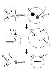
[0036] FIGURE 2 shows cell encapsulation in emulsion microdroplets. 1. Channel
constriction.
2. Glass into which microchannels are etched. 3. Cell input. 4. Lysis/RNA
capture bead mix
input. 5. Oil input. 6. Emulsion microdroplets.
[0037] FIGURE 3 shows droplet merging for cell lysis. 1. PDMS chip material.
2. Input
channel. 3. Cell mixture input. 4. Lysis/bead mixture droplet. 5. Widened
channel for droplet
fusion. 6. Outlet channel. 7. Electrodes. 8. Fused microdroplet.
[0038] FIGURE 4 is a diagrammatic workflow of the invention with at least two
different single
cells, with one clonal inducer cell and one target cell. 1. Cell mixture
encapsulation emulsion
microdroplet chip. 2. Clonal inducer cells. 3. Target cells. 4. Clonal inducer
cell. 5. Target cell. 6.
Cell culture media inside emulsion microdroplet. 7. Emulsion microdroplet
fusion chip. 8. Cell
7
CA 03052490 2019-08-01
WO 2018/170013 PCT/US2018/022256
mixture emulsion microdroplet. 9. Lysis/RNA capture bead mixture emulsion
microdroplet. 10.
Transcript traceable back to clonal inducer cell. 11. Emulsion microdroplet
for binding
transcripts to RNA capture beads. 12. Transcript from target cell, induced by
inducer cell. 13.
OE-RT-PCR emulsion microdroplet chip. 14. RNA-bound bead / OE-RT-PCR mix
input. 15.
RNA-bound bead / OE-RT-PCR mix input. 16. Amplicon comprising fusion between
cDNA
from transcript traceable back to clonal inducer cell and cDNA from transcript
from target cell,
induced by inducer cell. 17. OE-RT-PCR mix in emulsion microdroplet.
[0039] FIGURE 5 is a diagrammatic workflow of linking transcripts from at
least three different
single cells, with three cell types, with a target cell, an inducer cell, and
an intermediary cell. 1.
Cell mixture encapsulation emulsion microdroplet chip. 2. Clonal inducer
cells. 3. Target and
intermediary cells. 4. Clonal inducer cell. 5. Intermediary cell. 6. Target
cell. 7. Cell culture
media inside emulsion microdroplet. 8. Emulsion microdroplet fusion chip. 9.
Cell mixture
emulsion microdroplet. 10. Lysis,'RNA capture bead mixture emulsion
microdroplet, 11.
Transcript traceable back to clonal inducer cell. 12. Emulsion microdroplet
for binding
transcripts to RNA capture beads. 13. Transcript from target cell, induced by
inducer cell. 14.
OE-RT-PCR emulsion microdroplet chip. 15. RNA-bound bead / 0E-RT-F'CR mix
input. 16.
RNA-bound bead / 0E-RT-F'CR mix input. 17. Amplicon comprising fusion between
cDNA
from transcript traceable back to clonal inducer cell and cDNA from transcript
from target cell,
induced by inducer cell. 18. OE-RT-PCR mix in emulsion microdroplet.
[0040] FIGURE 6 is a diagrammatic workflow of linking transcripts from at
least two different
single cells, with a target cell and an inducer cell. 1. Inducer clone cell.
2. Target cell. 3. Inducer
clone cell transcript. 4. Target cell transcript (induced phenotype, or
indicative of induced
transcriptional change). 5. Inducer clone cell transcript cDNA. 6. OE-RT-PCR
linker sequence.
7. Target cell transcript (induced phenotype, or indicative of induced
transcriptional change)
cDNA. 8. OE-RT-PCR linker sequence. 9. OE-RT-PCR major, or linked, amplicon;
fusion
product of target and inducer cell transcript cDNAs. 10. Deep sequencing
analysis of 0E-RT-
PCR fusion product amplicons. 11. Identification or trace back of OE-RT-PCR
fusion product
amplicon sequence to original inducer cell clone.
[0041] FIGURE 7 is a diagrammatic workflow of linking transcripts from at
least three different
single cells, with a target cell, an inducer cell, and an intermediary cell.
1. Inducer clone cell. 2.
8
CA 03052490 2019-08-01
WO 2018/170013 PCT/US2018/022256
Target cell. 3. Intermediary cell. 4. Action (via a molecule, e.g., a secreted
antibody) of inducer
cell on intermediary cell. 5. Inducer clone cell transcript. 6. Target cell
transcript (induced
phenotype, or indicative of induced transcriptional change). 7. Inducer clone
cell transcript
cDNA. 8. OE-RT-PCR linker sequence. 9. Target cell transcript (induced
phenotype, or
indicative of induced transcriptional change) cDNA. 10. OE-RT-PCR linker
sequence. 11. OE-
RT-PCR major, or linked, amplicon; fusion product of target and inducer cell
transcript cDNAs.
12. Deep sequencing analysis of OE-RT-PCR fusion product amplicons 13.
Identification or
trace back of OE-RT-PCR fusion product amplicon sequence to original inducer
cell clone.
DETAILED DESCRIPTION OF THE INVENTION
Definitions
[0042] "Comprises." Consists at least of a list of components, i.e.,
encompasses all the elements
listed, but may also include additional, unnamed elements.
[0043] "Cell." The cell is the basic structural, functional, and biological
unit of all known living
organisms. A cell is the smallest unit of life that can replicate
independently.
[0044] "Transcriptome." Transcription is the first step of gene expression, in
which a particular
segment of DNA is copied into RNA (especially mRNA) by the enzyme RNA
polymerase, to
produce "transcripts". These transcripts have a variety of functions,
comprising in particular
providing the basis for translation of proteins inside cells. The
"transcriptome" is the complete
set of RNA transcripts present in a single cell or population of cells, or a
sampling of transcripts
that essentially comprises the complete set of RNA transcripts present in a
single cell or
population of cells.
[0045] "Transcriptional change." A change in the makeup of the transcriptome
of a single cell or
population of cells. Said transcriptional change may comprise a change in 1,
10, 100, 1,000,
10,000, or 100,000 transcripts. In some embodiments of this invention, a
transcriptional change
leads to changes in the function of the single cell or population of cells. In
some embodiments of
this invention, transcriptional change is induced in response to an external
stimulus. For
example, a T cell binding to its peptide:MHC antigen target may undergo
transcriptional changes
that produce proteins that lead to adaptive immune functions by the induced
cell. In some
embodiments of the invention, transcripts of interest are either up-regulated
or down-regulated.
9
CA 03052490 2019-08-01
WO 2018/170013 PCT/US2018/022256
[0046] "Cell phenotype." A phenotype, or "cell type", is the composite of a
cell's observable
characteristics or traits, such as its morphology, development, biochemical or
physiological
properties, behavior, and products of behavior. In complex multicellular
organisms, cells
specialize into different cell types that are adapted to particular
phenotypes. For avoidance of
doubt, phenotype is often synonymous with cell "function", though changes in
cell function do
not necessarily require a change in phenotype. In mammals, major cell
phenotypes include skin
cells, muscle cells, neurons, T cells, B cells, plasma cells, plasmablasts,
fibroblasts, stem cells,
and others. Cell types may differ both in appearance and function, yet may be
genetically
identical. Cells are able to be of the same genotype (i.e., they are "clonal")
but of different cell
type due to the differential expression of the genes they contain. Cellular
phenotype is the
conglomerate of multiple cellular processes involving gene and protein
expression that result in
the elaboration of a cell's particular morphology and function. Many kinds of
cells, such as
immune cells, undergo phenotypic (i.e., functional) changes in response to
external or internal
stimuli. For example, memory B cells mature into plasmablasts upon stimulation
with an antigen
that binds to a B cell receptor on the B cell surface. In certain embodiments,
RNA or protein
expressed by a cell are used as biomarkers to identify a cell's phenotype.
[0047] "Cell clone." A cell with a unique genetic sequence. For example, two T
cells that share a
T cell receptor comprise a cell clone. In other embodiments, two cells that
share an exogenous
polynucleic acid barcode comprise a cell clone. Cell clones may or may not
share a cell
phenotype. For example, a CD4+ T cell may share a T cell receptor sequence
with a CD8+ T
cell. In certain embodiments, cell clones comprise the same cell type.
[0048] "Cell population." A group of cells or cell clones, comprising either
multiple or single
cell phenotypes. In certain embodiments, a cell population comprises 10,000
cell clones of one
cell phenotype. In certain embodiments, a cell population comprises at least
10,000 single cells
of one cell phenotype, wherein thousands of cell clones are present. In
certain embodiments, a
cell population comprises 10,000 single cells of 10, 20, 50, or 100 different
cell types. For
example, a tumor comprises millions of cells and dozens of cell types. Cell
populations may
comprise recombinant cells or primary cells.
[0049] "Functional analysis." Functional analysis involves determination or
classification of a
cell's function (i.e., phenotype) classically through experimental methods
such as transcript
CA 03052490 2019-08-01
WO 2018/170013 PCT/US2018/022256
expression analysis (e.g., quantitative PCR, DNA microarrays, RNA-sequencing),
genome
sequencing or genotyping (e.g., immune repertoire sequencing, quantitative
PCR, whole genome
shotgun sequencing), protein expression analysis (e.g., flow cytometry,
ELISA), measurement of
glycans (e.g., mass spectrometry), or measurement of any molecule that is a
hallmark of the
function of the cell. Because cellular function can be plastic, i.e., cellular
phenotype can change
in response to external stimuli, measurement of cellular function is
particularly useful in
screening for drugs or molecules that induce a specific biological function
via cell functional
changes. For avoidance of doubt, functional analysis is generally synonymous
with phenotype
analysis, although changes in cell function do not necessarily require a
change in phenotype.
[0050] "Library." A pool of at least two polynucleic acids, cell clones,
molecules, or proteins. In
certain embodiments, a library is used to screen for biologically active
proteins. In other
embodiments, a library of cell clones is mixed with a drug, and then a
biological assay is used to
discern which cell clones are responsive to the drug. In other embodiments, a
library of drugs is
mixed with a single cell clone, and then a biological assay is used to discern
which drugs cause a
response in the cell clone. A library may comprise 100, 1,000, 10,000,
100,000, or 1 million
different peptide:MHC targets, either as a polynucleotide library that codes
for the peptide:MHC
targets, or as cells engineered to express the peptide:MHC. In other
embodiments, a library
comprises 100, 1,000, 10,000, 100,000, or 1 million polynucleic acid barcodes,
or cells
engineered to express the polynucleic acid barcodes as RNA.
[0051] "Combinatorial." Relating to combinations of libraries of cells,
proteins, polynucleic
acids, or other types of molecules. A combinatorial functional analysis
involves determining the
function of random combinatorial pairs of components from such libraries.
Because the
components of the libraries are paired randomly, the number of possible
combinations is the size
of the first library multiplied by the size of the second library. For
example, a library of 100
clones screened combinatorially against a library of 1,000 clones results in
100,000 theoretical
combinations. Combinatorial functional analysis is useful for discovery of
novel molecules or
cellular interactions that induce cell functions of interest. In certain
embodiments of the present
invention, a genetically diverse library of cell clones is combinatorially
screened against another
diverse (for example, 100, 1,000, 10,000, 100,000, or 1 million clones)
library of cell clones. In
certain embodiments of the invention, a diverse library of cell clones is
combinatorially screened
against an oligoclonal (for example, fewer than 10) library of cell clones.
11
CA 03052490 2019-08-01
WO 2018/170013 PCT/US2018/022256
[0052] "Polynucleic acid." A polynucleic acid is a double or single stranded
molecule of RNA or
DNA, typically comprising 5, 10, 20, 50, 100, 1,000, 10,000, or more base
pairs. Polynucleic
acids may be synthetic, i.e., manufactured chemically from individual
nucleotides, amplified,
i.e., generated enzymatically from template nucleic acids using a polymerase,
or purified from
biological systems, i.e., extracted from cells or other biological materials.
Polynucleic acids
derived from, or detected in, biological cells, often serve as "biomarkers"
that indicate functional
differences between cells or populations of cells. Polynucleic acids have many
sub-categories
familiar to those skilled in the art. Complementary DNA, or cDNA, is DNA
synthesized by using
an enzyme such as reverse transcriptase to make cDNA from an RNA template. An
"oligonucleotide" is a short (6-100 nucleotides) single stranded DNA or RNA
sequence,
typically manufactured synthetically by a commercial provider such as IDT DNA
or
ThermoFisher.
[0053] "Variable immune receptor." A variable immune receptor is any
glycoprotein or
glycoprotein complex that varies from cell to cell, or person to person.
Variable immune
receptors comprise critical innate and adaptive immune diversity required to
identify invasive (or
pathogenic) cells, viruses, bacteria, or other biologic material. In certain
embodiments, an
immune receptor that comprises the adaptive immune system, for example, an
antibody or a T
cell receptor. Most adult humans express billions of such variable receptors,
in billions of
different T cells or B cells. In other embodiments, an immune receptor that
comprises immune
system components that vary from individual to individual, for example, MHC or
killer cell
immunoglobulin-like (KIR) receptors.
[0054] "T cell receptor." The T cell receptor, or TCR, is a molecule found on
the surface of T
cells, or T lymphocytes, that are responsible for recognizing fragments of
antigen as peptides
bound to major histocompatibility complex (MHC) molecules. The TCR is a
disulfide-linked
membrane-anchored heterodimeric protein normally consisting of the highly
variable alpha (a)
and beta (0) chains expressed as part of a complex with the invariant CD3
chain molecules. T
cells expressing this receptor are referred to as a/I3 (or a13) T cells,
though a minority of T cells
express an alternate receptor, formed by variable gamma (y) and delta (8)
chains, referred as y6 T
cells. Each chain is composed of two extracellular domains: Variable (V)
region and a Constant
(C) region, both of Immunoglobulin superfamily domain forming antiparallel
beta-sheets. The
Constant region is proximal to the cell membrane, followed by a transmembrane
region and a
12
CA 03052490 2019-08-01
WO 2018/170013 PCT/US2018/022256
short cytoplasmic tail, while the Variable region binds to the peptide:MHC
complex. The
variable domain of both the TCR a-chain and I3-chain each have three
hypervariable or
complementarity determining regions (CDRs), whereas the variable region of the
13-chain has an
additional area of hypervariability (HV4) that does not normally contact
antigen and, therefore,
is not considered a CDR. The residues are located in two regions of the TCR,
at the interface of
the a- and I3-chains and in the 13-chain framework region that is thought to
be in proximity to the
CD3 signal-transduction complex. CDR3 is the main CDR responsible for
recognizing processed
antigen, although CDR1 of the alpha chain has also been shown to interact with
the N-terminal
part of the antigenic peptide, whereas CDR1 of the 13-chain interacts with the
C-terminal part of
the peptide. CDR2 is thought to recognize the MHC. CDR4 of the 13-chain is not
thought to
participate in antigen recognition, but has been shown to interact with
superantigens. The
constant domain of the TCR domain consists of short connecting sequences in
which a cysteine
residue forms disulfide bonds, which forms a link between the two chains. Each
recombined
TCR possess unique antigen specificity, determined by the structure of the
antigen-binding site
formed by the a and 13 chains in case of a13 T cells or 7 and 6 chains on case
of y6 T cells. It is
based mainly on genetic recombination of the DNA encoded segments in
individual somatic T
cells ¨ either somatic V(D)J recombination using RAG1 and RAG2 recombinases or
gene
conversion using cytidine deaminases. The intersection of these specific
regions (V and J for the
alpha or gamma chain; V, D, and J for the beta or delta chain) corresponds to
the CDR3 region
that is important for peptide:MTIC recognition. For avoidance of doubt, the
term "TCR"
throughout this disclosure embodies the full variety of possible recombinant
derivative formats,
and could be derived from any animal with an adaptive immune system, such as a
human,
mouse, camel, cow, bird, or fish. TCRs can be engineered into soluble form,
for example by
engineering chimeras with CD3 or Fe protein domains. These soluble TCRs then
act as drugs by
activating or antagonizing molecular targets of relevance to disease, for
example, cancer.
[0055] "T cell." A T cell is a lymphocyte of a type produced or processed by
the thymus gland
and actively participating in the immune response. T cells play a central role
in cell-mediated
immunity. T cells can be distinguished from other lymphocytes, such as B cells
and natural killer
cells, by the presence of a T-cell receptor on the cell surface. The several
subsets of T cells each
have a distinct function. T helper cells (TH cells) assist other white blood
cells in immunologic
processes, including maturation of B cells into plasma cells and memory B
cells, and activation
13
CA 03052490 2019-08-01
WO 2018/170013 PCT/US2018/022256
of cytotoxic T cells and macrophages. These cells are also known as CD4+ T
cells because they
express the CD4 glycoprotein on their surfaces. Helper T cells become
activated when they are
presented with peptide antigens by MHC class II molecules, which are expressed
on the surface
of antigen-presenting cells (APCs). Once activated, they divide rapidly and
secrete small proteins
called cytokines that regulate or assist in the active immune response. These
cells can
differentiate into one of several subtypes, including TH1, TH2, TH3, TH17,
TH9, or TFH, which
secrete different cytokines to facilitate different types of immune responses.
Signaling from the
APC directs T cells into particular subtypes. Cytotoxic T cells (Tc cells,
CTLs, T-killer cells,
killer T cells) destroy virus-infected cells and tumor cells, and are also
implicated in transplant
rejection. These cells are also known as CD8+ T cells since they express the
CD8 glycoprotein at
their surfaces. These cells recognize their targets by binding to antigen
associated with MHC
class I molecules, which are present on the surface of all nucleated cells.
Through IL-10,
adenosine, and other molecules secreted by regulatory T cells, the CD8+ cells
can be inactivated
to an anergic state, which prevents autoimmune diseases. Memory T cells are a
subset of
antigen-specific T cells that persist long-term after an infection has
resolved. They quickly
expand to large numbers of effector T cells upon re-exposure to their cognate
antigen, thus
providing the immune system with "memory" against past infections. Regulatory
T cells
(suppressor T cells) are crucial for the maintenance of immunological
tolerance. Their major role
is to shut down T cell-mediated immunity toward the end of an immune reaction
and to suppress
autoreactive T cells that escaped the process of negative selection in the
thymus. Suppressor T
cells along with Helper T cells can collectively be called Regulatory T cells
due to their
regulatory functions. Two major classes of CD4+ Treg cells have been described
¨ FOXP3+
Treg cells and FOXP3 ¨ Treg cells. The majority of human T cells rearrange
their alpha and beta
chains on the cell receptor and are termed alpha beta T cells (ab T cells) and
are part of the
adaptive immune system. Specialized gamma delta T cells, (a small minority of
T cells in the
human body, more frequent in ruminants), have invariant T cell receptors with
limited diversity,
that can effectively present antigens to other T cells and are considered to
be part of the innate
immune system. The genetic rearrangements and mutations that lead to TCR
expression
produces a T cell "clone". When the TCR engages with antigenic peptide and MEC
(peptide:MHC), the T lymphocyte is activated through signal transduction, that
is, a series of
biochemical events mediated by associated enzymes, co-receptors, specialized
adaptor
14
CA 03052490 2019-08-01
WO 2018/170013 PCT/US2018/022256
molecules, and activated or released transcription factors. Immortal cell
lines are often used
experimentally to study T cell function, for example, the Jurkat cell line. In
some embodiments
of the invention, the TCRab expressed by Jurkat is knocked out, or
deactivated, and a
recombinant TCRab is introduced into the genome or transiently expressed
through an
expression construct. T cells are engineered into "cellular therapeutics" by
introducing
recombinant TCR constructs, for example through lentivirus transduction. T
cell therapeutics are
allogeneic or autologous, and are used to treat cancer and other kinds of
serious disease. The
engineered TCR is therefore a kind of drug that acts via a T cell.
[0056] "Antigen." The other member of a cognate pair for an antibody or T cell
receptor. In
certain embodiments, antibodies or T cell receptors specifically bind to a
single antigen. In other
embodiments, antibodies or T cell receptors bind to multiple antigens.
Antibodies typically bind
to proteins or glycoproteins in their native conformation, whereas T cell
receptors require
processed peptide antigens presented on the surface of an antigen presenting
cell by an MFIC. In
certain embodiments, antigens are soluble, whereas in other embodiments,
antigens are tethered
to the surface of a cell.
[0057] "Antigen presenting cell." An antigen presenting cell (APC) displays an
antigen peptide
on its cell membrane. Antigen peptides are the product of proteolytic
processing inside the APC.
The antigenic peptides are then bound to a major histocompatibility complex
(MEW) protein on
the cell membrane of the APC. The bound complex is known as the peptide:MHC
complex. T
cell receptors do not bind antigen peptides directly, but instead require a
peptide:MHC complex.
In some embodiments, the peptide is derived from full proteins expressed by
the APC. In other
embodiments, the peptide is derived from viral proteins, and display of the
viral-derived peptide
is a hallmark of a cell infected by a virus. In certain embodiments, at least
one plasmid encoding
a full protein, partial protein, or polypeptide is introduced into a cell, and
the plasmid drives
expression of a recombinant peptide:MHC on the surface of the APC. In certain
embodiments,
APCs are incubated with peptides, peptide mixes, or proteins, resulting in a
peptide:MHC on the
APC membrane surface. In certain embodiments, cellular assays are performed
with APCs. In
certain embodiments, cellular assays are performed with APCs that are immortal
cell lines (e.g.,
T2 cells), or primary cells (e.g., B cells).
CA 03052490 2019-08-01
WO 2018/170013 PCT/US2018/022256
[0058] "Antibody." An antibody (Ab), also known as an immunoglobulin (Ig), is
a large, Y-
shaped protein produced mainly by plasma cells that is used by the immune
system to neutralize
pathogens such as bacteria and viruses. The antibody recognizes a unique
molecule of the
harmful agent, called an antigen, via the Fab's variable region. Each tip of
the "Y" of an antibody
contains a paratope (analogous to a lock) that is specific for one particular
epitope (similarly
analogous to a key) on an antigen, allowing these two structures to bind
together with precision.
Using this binding mechanism, an antibody can tag a microbe or an infected
cell for attack by
other parts of the immune system, or can neutralize its target directly (for
example, by blocking a
part of a microbe that is essential for its invasion and survival). Depending
on the antigen, the
binding may impede the biological process causing the disease or may activate
macrophages to
destroy the foreign substance. The ability of an antibody to communicate with
the other
components of the immune system is mediated via its Fc region (located at the
base of the "Y"),
which contains a conserved glycosylation site involved in these interactions.
The production of
antibodies is the main function of the humoral immune system. Antibodies can
occur in two
physical forms, a soluble form that is secreted from the cell to be free in
the blood plasma, and a
membrane-bound form that is attached to the surface of a B cell and is
referred to as the B-cell
receptor (BCR). The BCR is found only on the surface of B cells and
facilitates the activation of
these cells and their subsequent differentiation into either antibody
factories called plasma cells
or memory B cells that will survive in the body and remember that same antigen
so the B cells
can respond faster upon future exposure. In most cases, interaction of the B
cell with a T helper
cell is necessary to produce full activation of the B cell and, therefore,
antibody generation
following antigen binding. Soluble antibodies are released into the blood and
tissue fluids, as
well as many secretions to continue to survey for invading microorganisms.
They are typically
made of basic structural units¨each with two large heavy chains and two small
light chains.
There are several different types of antibody heavy chains that define the
five different types of
crystallisable fragments (Fc) that may be attached to the antigen-binding
fragments. The five
different types of Fc regions allow antibodies to be grouped into five
isotypes. Each Fc region of
a particular antibody isotype is able to bind to its specific Fc Receptor
(except for IgD, which is
essentially the BCR), thus allowing the antigen-antibody complex to mediate
different roles
depending on which FcR it binds. The ability of an antibody to bind to its
corresponding FcR is
further modulated by the structure of the glycan(s) present at conserved sites
within its Fe region.
16
CA 03052490 2019-08-01
WO 2018/170013 PCT/US2018/022256
The ability of antibodies to bind to FcRs helps to direct the appropriate
immune response for
each different type of foreign object they encounter. Though the general
structure of all
antibodies is very similar, a small region at the tip of the protein is
extremely variable, allowing
millions of antibodies with slightly different tip structures, or antigen-
binding sites, to exist. This
region is known as the hypervariable region. Each of these variants can bind
to a different
antigen. This enormous diversity of antibody paratopes on the antigen-binding
fragments allows
the immune system to recognize an equally wide variety of antigens. The large
and diverse
population of antibody paratope is generated by random recombination events of
a set of gene
segments that encode different antigen-binding sites (or paratopes), followed
by random
mutations in this area of the antibody gene, which create further diversity.
This recombinatorial
process that produces clonal antibody paratope diversity is called V(D)J or VJ
recombination.
Basically, the antibody paratope is polygenic, made up of three genes, V. D,
and J. Each paratope
locus is also polymorphic, such that during antibody production, one allele of
V, one of D, and
one of J is chosen. These gene segments are then joined together using random
genetic
recombination to produce the paratope. The regions where the genes are
randomly recombined
together is the hypervariable region used to recognize different antigens on a
clonal basis.
Soluble antibodies are commonly used as therapeutic drugs, for example,
rituximab,
adalimumab, pembrolizumab, or trastuzumab. Antibodies are sometimes
reformatted as Single
Chain Fragment Variable (scFv), comprising a heavy and light chain fused
together as a single
protein, via a peptide linker. In some scenarios, scFy are reformatted as
Chimeric Antigen
Receptors (CARs), which are then engineered into T cells to create cellular
therapeutics called
CAR-Ts. For avoidance of doubt, the term "antibodies" throughout this
disclosure embodies the
full variety of possible recombinant derivative formats, and could be derived
from any animal
with an adaptive immune system, such as a human, mouse, camel, cow, bird, or
fish.
[0059] "Natural killer cell." Natural killer cells (also known as NK cells, K
cells, and killer cells)
are a type of lymphocyte (a white blood cell) and a component of innate immune
system. NK
cells play a major role in the host-rejection of both tumors and virally
infected cells. Typically,
immune cells detect major histocompatibility complex (MHC) presented on
infected cell
surfaces, triggering cytokine release, causing lysis or apoptosis. NK cells
are unique, however, as
they have the ability to recognize stressed cells in the absence of antibodies
and MHC, allowing
for a much faster immune reaction. They were named "natural killers" because
of the initial
17
CA 03052490 2019-08-01
WO 2018/170013 PCT/US2018/022256
notion that they do not require activation to kill cells that are missing
"self markers of MHC
class 1. This role is especially important because harmful cells that are
missing MHC I markers
cannot be detected and destroyed by other immune cells, such as T lymphocyte
cells. NK cells
also kill cells by a mechanism called Antibody-Dependent Cell-mediated
Cytotoxicity (ADCC),
which starts with soluble antibodies binding to antigens on a target cell's
surface. Antibodies that
bind to antigens can be recognized by FcgRIII (CD16) receptors expressed on NK
cells, resulting
in NK activation, release of cytolytic granules and consequent cell apoptosis.
This is a major cell
killing mechanism of some monoclonal antibodies like rituximab, ofatumumab,
and others. In
certain embodiments, a cell line such as the NK-92 cell line is used in place
of primary NK cells.
[0060] "Target." A biological molecule to which a drug binds in order to
induce a
pharmacological function. In certain embodiments, the target is a protein
produced by a cell and
expressed on the cell membrane. Targets also comprise nucleic acids, lipids,
glycans, and
glycoproteins. In certain embodiments, the target is an antigen, for example,
a protein recognized
by an antibody or a peptide:MHC recognized by a TCR.
[0061] "Target cell." A biological cell that expresses an antigen or target.
In certain
embodiments of the invention, the target or antigen is bound to the cell
membrane of the target
cell, and therefore exposed to the extracellular space. In certain embodiments
of this invention,
the target cell undergoes quantifiable changes in 1, 10, 100, or 10,000 mRNA
transcripts as a
result of the inducer cell interacting with the antigen or target on the
surface of the target cell. In
some embodiments of the invention, the quantifiable changes in the target cell
are endogenous
transcripts. In some embodiments of the invention, the quantifiable changes in
the target cell are
transcripts arising from recombinantly engineered "reporter" constructs that
have been
introduced into the target cell. In some embodiments of the invention, the
reporter constructs
contain promoters, enhancers, or other regulatory elements that induce
transcription upon contact
with signals resulting from the inducer cell contacting the target cell. In
some embodiments of
the invention, transcripts of interest are either up-regulated or down-
regulated.
[0062] "Inducer cell." A biological cell that expresses a ligand or inducer
molecule that binds to
an antigen or target on the target cell. In certain embodiments of the present
invention, the
inducer cell secretes proteins or molecules that then bind to the target cell
to induce quantifiable
transcriptional changes. In other embodiments of the invention, proteins or
molecules on the
18
CA 03052490 2019-08-01
WO 2018/170013 PCT/US2018/022256
inducer cell surface bind to the target cell to induce quantifiable
transcriptional changes. In
certain embodiments, the inducing proteins or molecules comprise a single
species, whereas in
other embodiments of the invention, the inducing proteins or molecules
comprise 2, 5, 10, 100,
or 1,000 individual species. In certain embodiments of this invention, the
inducer cell undergoes
quantifiable changes in 1, 10, 100, or 10,000 mRNA transcripts as a result of
the inducer cell
interacting with the antigen or target on the surface of the target cell.
[0063] "Intermediary cell." A biological cell that responds functionally to
the interaction
between an inducer and a target cell, or to the interaction between a protein
secreted by an
inducer cell and proteins expressed by a target cell. In certain embodiments
of this invention, the
intermediary cell undergoes quantifiable changes in 1, 10, 100, or 10,000 mRNA
transcripts as a
result of the inducer cell interacting with the antigen or target on the
surface of the target cell. In
other embodiments of the invention, proteins or molecules secreted by the
inducer cell surface
bind to the target cell to induce quantifiable transcriptional changes in the
intermediary cells. In
some embodiments of the invention, the quantifiable changes in the
intermediary cell are
transcripts arising from recombinantly engineered "reporter" constructs that
have been
introduced into the intermediary cell.
[0064] "Synthetic polynucleic acid." Chemically or enzymatically synthesized
RNA or DNA. To
synthesize single-stranded RNA or DNA, or "oligonucleotides", the chemical
synthesis process
can be implemented as solid-phase synthesis using phosphoramidite method and
phosphoramidite building blocks derived from protected 2'-deoxynucleosides
(dA, dC, dG, and
dT), ribonucleosides (A, C, G, and U), or chemically modified nucleosides,
e.g. LNA or BNA.
To obtain the desired oligonucleotide, the chemical building blocks can be
sequentially coupled
to the growing oligonucleotide chain in the order required by the sequence of
the product.
Typically, synthetic oligonucleotides are single-stranded DNA or RNA molecules
around 15-25
bases in length. Synthetic polynucleic acids can be also generated by
enzymatic methods, such as
reverse transcription (RT), polymerase chain reaction (PCR), Gibson assembly,
overlap
extension PCR (OE-PCR), overlap extension RT-PCR (OE-RT-PCR), emulsion PCR,
emulsion
RT-PCR, emulsion OE-RT-PCR, emulsion 0E-PCR, ligase chain reaction (LCR),
hybridization,
in vitro transcription, or any other cell-free molecular biological method
that makes use of
purified enzymes.
19
CA 03052490 2019-08-01
WO 2018/170013 PCT/US2018/022256
[0065] "Polynucleic acid barcode." A polynucleic acid barcode comprises a
synthetic
polynucleic acid that enables an experimentalist to identify a cell clone,
i.e., a unique identifier.
In some embodiments, barcodes are engineered into the genome of a cell,
contained within an
expression plasmid, or encoded into a recombinant or synthetic RNA sequence.
In some
embodiments, a barcode is attached to a solid surface, such as a one micron
diameter magnetic
bead. In some embodiments, populations of clones contain 10, 100, 1,000,
10,000, 100,000, or 1
million different barcodes. The barcodes can be sequenced through bulk
sequencing, enabling
high throughput combinatorial analysis of cell function.
[0066] "Reverse transcription." The process by which a reverse transcriptase
(RT) enzyme is
used to generate complementary DNA (cDNA) from an RNA template. Reverse
transcriptase is
commonly used in research to apply the polymerase chain reaction technique to
RNA in a
technique called reverse transcription polymerase chain reaction (RT-PCR). The
classical PCR
technique can be applied only to DNA strands, but, with the help of reverse
transcriptase, RNA
can be reverse transcribed into DNA, thus making PCR analysis of RNA molecules
possible.
Reverse transcriptase is used also to create cDNA libraries from mRNA.
[0067] "Polymerase chain reaction." Polymerase chain reaction (PCR) is a
technique used in
molecular biology to amplify a single copy or a few copies of a piece of DNA
across several
orders of magnitude, generating thousands to millions of copies of a
particular DNA sequence.
The method relies on thermal cycling, consisting of cycles of repeated heating
and cooling of the
reaction for DNA melting and enzymatic replication of the DNA. Primers (short
DNA
fragments) containing sequences complementary to the target region along with
a DNA
polymerase, which the method is named after, are key components to enable
selective and
repeated amplification. As PCR progresses, the DNA generated is itself used as
a template for
replication, setting in motion a chain reaction in which the DNA template is
exponentially
amplified. PCR can be extensively modified to perform a wide array of genetic
manipulations.
PCR is not generally considered to be a recombinant DNA method, as it does not
involve cutting
and pasting DNA, only amplification of existing sequences. Almost all PCR
applications employ
a heat-stable DNA polymerase, such as Taq polymerase (an enzyme originally
isolated from the
bacterium Therm us aquaticus). This DNA polymerase enzymatically assembles a
new DNA
strand from DNA building-blocks, the nucleotides, by using single-stranded DNA
as a template
and DNA oligonucleotides (also called DNA primers), which are required for
initiation of DNA
CA 03052490 2019-08-01
WO 2018/170013 PCT/US2018/022256
synthesis. The vast majority of PCR methods use thermal cycling, i.e.,
alternately heating and
cooling the PCR sample through a defined series of temperature steps. In the
first step, the two
strands of the DNA double helix are physically separated at a high temperature
in a process
called DNA melting. In the second step, the temperature is lowered and the two
DNA strands
become templates for DNA polymerase to selectively amplify the target DNA. The
selectivity of
PCR results from the use of primers that are complementary to the DNA region
targeted for
amplification under specific thermal cycling conditions.
[0068] "Hybridization." Any process whereby two polynucleic acids are fused to
form a single
polynucleic acid molecule. Hybridization can occur by any process, natural or
artificial, that
results in two single-stranded polynucleic acids forming base pairing that
result in a molecule
that is at least partially double stranded. Base pairings conventionally occur
through reverse
complementarity, for example, guanine-cytosine, adenine-thymine, or adenine-
uracil. In some
embodiments, the hybridized base pairs are adjacent, for example, two single-
stranded
polynucleic acids that are each 100 nucleotides comprise 20 nucleotide
subsequences that are
reverse complements. Under the proper conditions, the two polynucleic acids
would hybridize
across these complementary nucleotide subsequences, forming a hybridized
molecule. The
amplification process called "overlap extension PCR" generates a plurality of
fused, double
stranded DNA products that result from the initial hybridization step between
two
polynucleotides that comprise complementary nucleotide subsequences.
[0069] "Microfluidics." Microfluidics is the science and technology of
manipulating and
controlling fluids, usually in the range of microliters (10-6) to picoliters
(10-12), in networks of
channels with lowest dimensions from tens to hundreds micrometers. Typically,
fluids are
moved, mixed, separated or otherwise processed. Numerous applications employ
passive fluid
control techniques like capillary forces. In some applications, external
actuation means are
additionally used for a directed transport of the media. Examples are rotary
drives applying
centrifugal forces for the fluid transport on the passive chips. Active
microfluidics refers to the
defined manipulation of the working fluid by active (micro) components such as
micropumps or
microvalves. Micropumps supply fluids in a continuous manner can be used for
dosing.
Microvalves can determine the flow direction or the mode of movement of pumped
liquids.
Processes which are normally carried out in a lab can be miniaturized on a
single chip in order to
enhance efficiency and mobility as well as to reduce sample and reagent
volumes. Droplet-based
21
CA 03052490 2019-08-01
WO 2018/170013 PCT/US2018/022256
microfluidics as a subcategory of microfluidics in contrast with continuous
microfluidics has the
distinction of manipulating discrete volumes of fluids in immiscible phases
with low Reynolds
number and laminar flow regimes. Two immiscible phases used for the droplet
generation are
termed as the continuous phase (medium in which droplets are generated) and
dispersed phase
(the droplet phase). The size of the generated droplets is mainly controlled
by the flow rates of
the continuous phase and dispersed phase, interfacial tension between two
phases and the
geometry used for the droplet generation.
[0070] "Microdroplet." A spherical, small volume of liquid, typically with
volume less than one
microliter. Microdroplets comprise aqueous-in-oil microdroplets and oil-in-
aqueous
microdroplets. A population of aqueous-in-oil microdroplets or oil-in-aqueous
microdroplets
comprise an "emulsion". Emulsions can be monodisperse, e.g., comprising
microdroplets
substantially the same volume, for example, varying by no more than 25% in
diameter, or
polydisperse, e.g., comprising microdroplets of a variety of volumes, for
example, varying by
>25% in diameter. Microdroplets are a means for performing high-throughput
molecular,
cellular, or biochemical experiments. Microdroplets serve to partition liquid
reactions and
therefore serve a similar function as a physical container. Millions or
billions of microdroplets
can be deposited in a small (for example, one milliliter) physical container,
enabling very large
combinatorial screening on single cells. In some embodiments of the present
invention,
monodisperse microdroplets are generated using microfluidics, i.e., "droplet
microfluidics". In
other embodiments of the invention, polydisperse microdroplets are generated
using a shaking or
mixing apparatus.
[0071] "Physical container." Physical containers used in molecular biology,
cell biology, or
biochemistry refer to tubes, plates, dishes, vials, or other formats
comprising solid plastic, glass,
polymer, or other solid material. In some embodiments, the physical container
is inert, i.e., the
container serves only to physically contain liquids for a molecular, cellular,
or biochemical
experiment. In some embodiments, reactive cells, molecules, proteins, drugs,
or biochemical
container are affixed to the physical container. Physical containers are a
means for performing
molecular, cellular, or biochemical experiments. To increase processing
throughput, physical
containers can be used together with robotic systems. In some embodiments,
throughput is
increased by using microfluidic chips that comprise physical containers, for
example, nanoliter
chambers on a glass, plastic, or PDMS microfluidic chip.
22
CA 03052490 2019-08-01
WO 2018/170013 PCT/US2018/022256
[0072] "Solid support." Solid supports used in molecular biology, cell
biology, or biochemistry
refer to beads or other geometric formats comprising solid plastic, glass,
polymer, or other solid
material. In some embodiments of the invention, reactive cells, polynucleic
acids, proteins, or
other molecules are affixed to solid supports. The solid supports are then
introduced into a
physical container or microdroplet, such that a biochemical, cellular, or
molecular function is
enabled. The solid supports can then be washed, or removed, simplifying multi-
step laboratory
processes. In some embodiments, the solid supports are magnetic beads of one,
ten, or one
hundred microns. In some embodiments, synthetic polynucleic acids are affixed
to the magnetic
beads, enabling purification of endogenous cellular polynucleic acids that are
complementary to
the synthetic polynucleic acids, also called "probes". In some embodiments,
solid supports are
beads coated with antibodies, which are then used to purify cells that express
antigens with
affinity for the antibodies.
[0073] "Bulk sequencing." Synonymous with deep sequencing, ultra-high
throughput
sequencing, massively parallel sequencing, and next-generation sequencing.
Bulk sequencing
comprises obtaining hundreds of thousands, millions, hundreds of millions, or
billions of DNA
sequence reads in parallel. In many embodiments, a diverse library of DNA is
generated using
methods such as PCR, RT-PCR, or hybridization and then a plurality of the
library is sequenced
using bulk sequencing. Methods can comprise sequencing by synthesis, nanopore
sequencing,
and pyrosequencing. As of 2017, commercial providers of bulk sequencing
comprise Illumina,
Pacific Biosciences, Oxford Nanopore, and Roche.
Overview of the invention
[0074] One aspect of the present invention relates to concurrent measurement
of polynucleic
acids derived from at least two different cell types. The measurement can be
performed in a
massively parallel fashion on a small number of cells, or combinatorial
screens can be performed
on millions of different cell type combinations. In som embodiments, cells are
combinatorially
isolated into reaction containers, incubated to induce a biological response,
and lysed to isolate
RNA while retaining the combinatorial context. Transcripts from at least two
different cell types
can be physically linked by hybridization, and then the linked clones can be
subject to deep
sequencing on a massively parallel scale (FIGURE 1).
23
CA 03052490 2019-08-01
WO 2018/170013 PCT/US2018/022256
[0075] The methods can involve isolation of single cells or subpopulations of
cells into
microemulsion droplets, gels, or microfluidic reaction containers. Millions of
cells can be
isolated or compartmentalized in a massively parallel manner to generate cell
mixtures that
represent genetically distinct pairwise combinations (FIGURE 2).
[0076] The cell mixtures can comprise one or more target cells, one or more
inducer cells, and/or
one or more intermediary cells. The target cells can comprise populations of
homogeneous cells
or genetically distinct clones (for example, B cells, T cells, cells
engineered with barcodes, cells
engineered to express peptide antigens, primary cancer cells in single cell
suspension). The
inducer cells can comprise populations of homogenous cells or genetically
distinct clones (for
example, B cells, T cells, cells engineered with barcodes, cells engineered to
express peptide
antigens, NK cells). In some embodiments, intermediary cells are used, and the
intermediary
cells can comprise populations of homogeneous cells or genetically distinct
clones (for example,
NK cells).
[0077] In some embodiments, the target cells and inducer cells are mixed with
a library of
polynucleic acid barcodes affixed to a solid support (for example, beads, or a
protein). In some
embodiments, the cell mixtures are additionally incubated in the same
microemulsion droplets,
gels, or reaction containers with a stimulus, for example, a homogeneous
population of cells, a
library of reagents, or a single reagent.
[0078] The mixtures of cells can be then lysed by introducing a reagent into
the microemulsion
droplets, gels, or microfluidic reaction containers. In some embodiments, this
step comprises
fusing microemulsion droplets containing the cells with microemulsion droplets
containing the
lysis reagent, thus preserving the compartmentalization of the cell mixtures
(FIGURE 3). After
lysis, transcripts from the cell mixtures can be purified, for example, using
beads coated with
oligo-dT oligonucleotides.
[0079] In some embodiments, two or more polynucleotide targets are hybridized,
such that
polynucleic acids that differentiate clones are linked to RNA transcripts that
indicate functional
changes (FIGURES 4-5). The key insight is to fuse transcripts derived from at
least two different
cell types, for example, antibody target encoding transcripts and antibody-
encoding transcripts
derived from antibody-producing cells (wherein antibody-producing cells are
the inducer cells).
24
CA 03052490 2019-08-01
WO 2018/170013 PCT/US2018/022256
The hybridized polynucleic acid molecules can be then sequenced by bulk, or
high-throughput,
sequencing. Any high-throughput sequencing method known in the art can be
employed.
[0080] The bulk sequencing data can be subsequently analyzed algorithmically
to determine
which clones from the initial clone library demonstrate a functional change in
response to the
inducer cell stimulus, or stimuli (FIGURES 6-7). Sequencing of hybridized
nucleic acid
molecules from multiple cell types enables concurrent measurement of at least
one transcript
from each of at least two cell types, for example, an antibody target
producing cell and an
antibody-producing cell. Because of the extreme sensitivity of deep
sequencing, transcript counts
that are only 2-, 5-, or 10-fold different between induced and non-induced
cells are detectable.
Therefore, the method of the present invention can provide insight into the
functional response of
single target cells exposed to inducer cells, across millions of single target
and inducer cells in
parallel, and enables combinatorial functional screens that have never before
been possible. In
some embodiments, the hybridized polynucleic acids are further used to make
libraries of
recombinant proteins, which can be subsequently further screened for binding
or function.
[0081] Provided herein are detailed descriptions of methods of the invention.
Also provided
herein are detailed descriptions of examples of embodiments of the invention,
with particular
application to immunology, drug discovery, drug development, and cancer
biology.
Other interpretational conventions
[0082] Ranges recited herein are understood to be shorthand for all of the
values within the
range, inclusive of the recited endpoints. For example, a range of 1 to 50 is
understood to
include any number, combination of numbers, or sub-range from the group
consisting of 1, 2, 3,
4, 5, 6, 7, 8, 9, 10, 11, 12, 13, 14, 15, 16, 17, 18, 19, 20, 21, 22, 23, 24,
25, 26, 27, 28, 29, 30, 31,
32, 33, 34, 35, 36, 37, 38, 39, 40, 41, 42, 43, 44, 45, 46, 47, 48, 49, and
50.
Methods of the invention
1) Generation of DNA libraries
[0083] Some embodiments of the present invention involves generation of
libraries of antibody
clones by isolating B cells from mammalian donors, and then fusing the primary
cells with
myeloma cells, using techniques such as electrofusion, which are well known to
those skilled in
the art (Smith & Crowe, Microbiol Spectr, 2015 3(1): AID-0027-2014). The
resulting cells,
CA 03052490 2019-08-01
WO 2018/170013 PCT/US2018/022256
known as hybridomas, can be easier to rear in culture than primary cells. A
variety of methods
have been used to make T and B cell hybridomas, using primary cells from
species comprising
mice and humans. Using these methods, libraries of tens of thousands, hundreds
of thousands, or
millions of clones of cells that each express a unique TCR or antibody can be
made.
[0084] Some embodiments of the present invention relate to methods of
generating DNA
libraries of a gene by isolating RNA from primary cells, for example, a tumor,
a liver, a brain,
blood, bone marrow, peripherial blood mononuclear cells, muscle tissue,
cerebrospinal fluid,
kidney tissue, lung lavage, lung tissue, immortal cell lines, skin tissue, or
any other tissue or cell
type. Reverse transcriptase can be used to synthesize cDNA from the RNA. For
example, RNA
is incubated with M-MuLV RT at 42 C with an oligo-dT primer for one hour. In
some
embodiments, the oligo-dT primer is fused with a nucleic acid barcode
sequence, flanked by
universal amplification primers, which enables specific amplification of the
barcode and trace
back of the barcode to a cDNA sequence. This enables de-multiplexing of
complex mixtures of
clones. RT-based methods have the advantage of cheaply and quickly generating
DNA libraries
comprised of tens of thousands, hundreds of thousands, or millions of DNA
clones in parallel. To
recover a plurality of cDNA clones of interest, the full cDNA library can be
subjected to PCR
using a reaction comprising gene-specific primers, a thermostable polymerase
such as Taq, and
thermocycling consisting of denaturation (95 C for 30 seconds), 30 cycles of
amplification
(95 C for 15 seconds, 62 C for 60 seconds, and 68 C for 3 minutes), followed
by a final
extension at 68 C for 5 minutes.
[0085] Some embodiments of the present invention relate to a method of
generating DNA
libraries of antibodies, TCRs, or any other kind of genetic sequence by DNA
synthesis. In some
embodiments, DNA sequencing data on TCR or antibody repertoires are obtained
using methods
known in the art, and then synthetic DNA libraries are engineered from
sequences identified
through bulk sequencing. In some embodiments, the synthesized DNA libraries
comprise TCRs
or antibodies known to bind to antigens of interest through methods comprising
yeast display,
mammalian display, or mammalian cell activation assays. DNA oligonucleotides
can be
designed such that they comprise libraries of overlapping, complementary
sequences that
hybridize when incubated together. Libraries of hundreds, thousands, tens of
thousands, or
hundreds of thousands of synthetic oligonucleotides can be manufactured by
microfluidic or
array-based methods, for example, by commercial providers such as Twist
Bioscience, Agilent
26
Technologies. or LC Biosciences. The libraries of oligonucleotides can be then
assembled into
DNA sequences of hundreds or thousands of nucleotides, using 5' exonuclease,
DNA
polymerase, and DNA ligase (e.g., "Gibson Assembly", Gibson et al. Nat
Methods. 2009
May;6(5):343-5). For example, T5 exonuclease, Taq polymerase, and Taq ligase
are mixed in a
reaction comprising overlapping oligonucleotides, nucleotides, DTT, MgCl2, and
buffer, and then
incubated at 50 C for 60 minutes. In some embodiments, Gibson Assembly is used
to synthesize
circular clones, for example, plasmid expression constructs. If the synthetic
DNA is circular, the
DNA can be transformed into bacteria to produce nanogram or more quantities of
plasmid.
Another method that generates linear synthetic DNA comprises mixing
overlapping
oligonucleotides and performing PCR using a thermostable polymerase. In order
to make circular
DNA, these linear PCR products can be then subcloned into plasmid expression
constructs using
methods comprising restriction enzymes and DNA ligase, Gibson Assembly, or
blunt end
cloning. Any of these DNA synthesis methods can be parallelized through 96-
well plate, 384-well
plate, microfluidic, or robotic processing systems.
[0086] DNA libraries of antibodies, TCRs, or any other target gene can be also
generated through
isolation and lysis of single cells, followed by nucleic acid amplification.
Single B cells can be
isolated into 96-well plates, and then heavy and light chain immunoglobulin
transcripts can be
linked using a method known in the art, for example, multiplexed "overlap
extension" RT-PCR
(Oleksiewicz EP1921144 B1). In overlap extension RT-PCR, or OE-RT-PCR, for
immunoglobulin amplification from single cells, a pool of primers can be
designed that bind and
amplify all possible heavy chain genes and all possible light chain genes. The
heavy chain
primers can also comprise subsequences with complementarity to the light chain
primers. During
OE-RT-PCR, the complementary subsequences can hybridize and a polymerase can
generate a
fused polynucleic acid from hybridized single stranded heavy and light chain
immunoglobulin. In
this fashion, the single cell context of the heavy and light chain
immunoglobulin can be
maintained.
[0087] DNA libraries of antibodies, TCRs, or any other target gene can be
generated by other
methods, for example, those involving OE-RT-PCR and microfluidics from
populations of more
than ten thousand cells. One exemplary method disclosed in Johnson EP2652155
involves use of
a droplet microfluidic device. The droplet microfluidic device inputs an
oil/surfactant mix, lysis
and RNA capture mix, and a cell
27
CA 3052490 2019-08-23
suspension and outputs single-cell emulsions into standard thermocycling
microtubes. The
oil/surfactant mix is based on mineral oil or fluorocarbon oil. The lysis and
RNA capture mixture
contains oligo-dT coated magnetic lum beads that capture messenger RNA (mRNA)
transcripts
from the single cells. The cell encapsulation device is comprised of three
pressure pumps, a
microfluidic droplet chip, and imaging apparatus. The microfluidic chip is
fabricated from glass
and channels are etched to 50um x 150um for most of the chip's length, and
narrow to 55 um at
the droplet junction. Droplet size depends on pressure, but typically droplets
of ¨40um are
optimally stable and appropriately sized for the single cell emulsions.
Droplet generation rates
also depend on pressure, but are typically up to 3kHz and capture 3 million
cells per hour. Cell
lysis methods comprise surfactant based methods, for example TritonTm X-100,
NP-40, TweenTm
20, TweenTm 80, or SDS. The emulsions are incubated at 50 C for 30 minutes,
and then the beads
are extracted from the emulsion using a solvent such as ethyl acetate.Next,
the mRNA-bound
beads are injected back into emulsions for OE-RT-PCR, using microfluidic chips
similar to the
cell encapsulation chips described above. For example, to generate TCRaf3
libraries, independent
TCRa and TCRI3 minor amplicons are generated in multiplex; these are then
fused to generate a
single major amplicon comprised of both TCRa and TCRI3. The TCRal3 primer pool
includes a
universal primer for 13 constant (C13) and a constant (Ca) regions. This
abrogates the need for a
large pool of J region primers. Additionally, the C region primers are
designed to either capture
the endogenous C region genotypes or isotypes, or the primers are designed to
ignore endogenous
C region genotypes or isotypes. The TCRal3 primer pool also includes 43
primers that bind to all
possible V segments for TCRal3 and TCR13. Thus, the primers amplify across the
full variable
region of each monomer to produce 450bp minor amplicons. Exemplary primers for
TCRI3 V
gene is provided herein as SEQ ID NO: 17-19, an exemplary primer for TCRf3 C
gene is provided
herein as SEQ ID NO: 20, exemplary primers for TCRa V gene are provided as SEQ
ID NO: 21-
23 and an exemplar primer for TCRa C gene is provided herein as SED ID NO: 24.
The bead
emulsions are then subjected to OE-RT-PCR using a reaction comprising an RT,
gene-specific
primers, a thermostable polymerase such as Taq, and thermocycling consisting
of reverse
transcription (42 C for 60 minutes), denaturation (95 C for 30 seconds), 30
cycles of
amplification (95 C for 15 seconds, 62 C for 60 seconds, and 68 C for 3
minutes), followed by a
final extension at 68 C for 5 minutes. Because a plurality of droplets
contains only a single
mRNA-bound bead, the native TCRaf3 pairing of the input T cell is maintained
in the TCRc43
28
CA 3052490 2020-02-28
linkage library. Similar methods can be used to generate linked heavy and
light chain
immunoglobulin DNA libraries. For example, immunoglobulin primer sets
comprising a
polynucleotide of any of SEQ ID NO: 1-8 can be used. SEQ ID NO: 1-3 provide
exemplary
primer sequences for IGG V gene, SEQ ID NO: 4 provides an exemplary primer
sequence for
IGG C gene, SEQ ID NO: 5-7 provide exemplary primer sequences for IGK V gene,
and SEQ ID
NO: 8 provides an exemplary primer sequence for IGK C gene. Primers for the
immunoglobulin
C regions are either isotype-specific, genotype specific, or are universal
primers designed amplify
any C region sequence. In some embodiments, the TCR or immunoglobulin subunits
are linked
with a polynucleic acid sequence encoding a porcine teschovirus-1 (P2A) amino
acid sequence.
In some embodiments, the TCR or immunoglobulin subunits are linked with a
polynucleic acid
sequence encoding a Gly-Ser peptide linker. In some embodiments, the TCR or
immunoglobulin
subunits are linked with a polynucleic acid sequence encoding an Internal
Ribosome Entry Site
(IRES). In other embodiments, the TCR or immunoglobulin subunits are linked
with artificial
linker sequences with no significant homology to any known endogenous
sequences.
100881 The DNA libraries of linked TCRap or heavy and light chain
immunoglobulin can be
converted to recombinant expression constructs using methods known in the art,
for example, the
method described in Johnson US9,422,547 Bl. The exemplary method described in
Johnson
US9,422,547 B1 uses nested outer PCR primers to add adapters with overhangs
for Gibson
Assembly to the 5. and 3' ends of the amplicon library. Primers for the C
regions can be either
isotype-specific, genotype specific, or are primers designed amplify any C
region sequence. T5
exonuclease and Taq ligase are mixed in a reaction comprising the TCRa0 or
immunoglobulin
insert, a linearized plasmid backbone with subsequences complementary with the
insert, DTI,
MgC12, and buffer, and then incubated at 50 C for 60 minutes. The plasmid
backbone comprises
a promoter, a poly(A) signal sequence, and C region sequence not amplified
through 0E-RT-
PCR. The C region matches the isotype or genotype of the linked amplicon, or
is designed to fuse
the amplicon with a non-native isotype or genotype. The library is then
transformed into E. coli
and spread on LB-ampicillin plates. The plasmid library is then purified with
a Maxi prep kit. The
purified Maxi prep library contains tens of thousands, hundreds of thousands,
or millions of
clones. Some workflows require a second round of Gibson Assembly. For example,
if the one or
both of the full C regions are not
29
CA 3052490 2019-08-23
. ,
amplified in the original OE-RT-PCR, it may be necessary to clone a C region
between the
TCRa0 or heavy and light chain immunoglobulin. In some embodiments, a
promoter, P2A, or
IRES sequence is cloned at the same time. The inserted sequences are
synthesized by assembling
a pool of oligonucleotides using Gibson Assembly or PCR, and then Gibson
Assembly can be
used to insert the polynucleic acid insert into the plasmid library. This
reaction can be performed
on tens of thousands, hundreds of thousands, or millions of clones in
parallel. The final result is a
library of tens of thousands, hundreds of thousands, or millions of TCRai3 or
heavy and light
chain immunoglobulin clones that express fully functional proteins, which
retain the native
pairing of the original single cell inputs.
[0089] In some embodiments, single cell amplification methods are used to
generate single cell
cDNA libraries for any transcript, set of transcripts, or full single cell
transcriptomes using
various methods for nucleic acid barcoding, for example, as described in
Johnson (U.S. App. No.
15/159,674 which was issued as U.S. Patent No. 9,695,474).
[0090] The exemplary method disclosed in Johnson (U.S. Patent No. 9,695,474),
comprises
delivering a clonal polynucleic acid barcode with a single cell into a
reaction vessel, microfluidic
chamber, or an emulsion microdroplet. One method is to affix polynucleic acids
that comprise
barcodes to solid supports comprising spherical beads with 111m, 5 m, or 101Am
diameter, made
of magnetic material to facilitate nucleic acid purification. Oligonucleotides
are modified with
NH2 and affixed to epoxy silane or isothiocyanate coated glass beads, or
oligonucleotides are
disulfide modified and attached to mercaptosilanized glass supports. For
droplet encapsulation,
bead solutions are mixed with cells, and then diluted such that a plurality
droplets contain a single
cell and a single bead. Because such methods result in a plurality of empty
droplets or droplets
with only a single bead or only a single cell, in some methods, cells and
beads are first
encapsulated into droplets in separate streams or separate devices, and then
the cell- and bead-
containing droplets are fused to generate a plurality of droplets that contain
a single cell and a
single bead. Depending on the application, a plurality of single cells can be
encapsulated with
multiple barcoded beads. Such methods enable trace back of individual barcodes
to single cells,
even if there are multiple barcodes for a plurality of single cells. Other
methods comprise biotin-
streptavidin and covalent conjugation chemistries. Another method is to affix
polynucleic acids
that comprise barcodes to antibodies, which are bound to cells prior to
delivering the cells to
reaction vessels, microfluidic chambers, or emulsion microdroplets. Methods
for conjugating
CA 3052490 2020-02-28
. ,
antibodies to nucleic acids available in the art can be employed, for example,
biotin-streptavidin
or covalent conjugation chemistries. Cell lysis methods can comprise
surfactant based methods,
for example Triton' X-100, NP-40, TweenTm 20, TweenT" 80, or SDS. In some
embodiments,
the emulsions are incubated at 50 C for 30 minutes, and then the beads are
extracted from the
emulsion using a solvent such as ethyl acetate. Next, the RNA-bound beads are
recovered from
the emulsion and then amplified in droplets or reaction vessels using the
methods described
above, with some modifications specific to nucleic acid barcoding. In nucleic
acid barcoding, the
first strand cDNA can be labeled with the nucleic acid barcode fused to the
transcript-specific
first strand primer. Universal primers 5' to the nucleic acid barcode can be
used in PCR to
amplify a plurality of barcoded RT-PCR amplicons. Alternatively, RNA can be
primed and
amplified separately from the barcode sequence, and then the barcode and cDNA
amplicons can
be fused in an overlap extension PCR inside of emulsion microdroplets.
Alternatively, first strand
cDNA barcoding can be effected with RT in the lysis mixture, without the
requirement to inject
RNA-bound beads for an RT-PCR amplification. In these methods, the cDNA-bound
beads can
be extracted from the emulsion and the barcoded cDNA can be subjected to
"bulk" PCR, i.e.,
PCR without an emulsion. The final result of any of these methods can be a
library of tens of
thousands, hundreds of thousands, or millions of barcoded cDNA clones that
express fully
functional proteins, which enable trace back of cDNAs with the same barcode
back to a single
originating cell. The cDNAs are not necessarily full length, for example,
peptide:MHC
complexes do not require full cDNA for functional analysis. In some
embodiments, a target
library comprises NY-ES0-1 target sequence (SEQ ID NO: 13), or MART-1 target
sequence
(SEQ ID NO: 16), engineered into two different mammalian clones.
[0091] Once reformatted as circular plasmids, libraries of cDNAs can be
introduced into
mammalian cells for protein production. For example, TCRaf3 expression
constructs can be
packaged into lentivirus or any other vector known in the art and then used to
transduce the Jurkat
J.RT3-T3.5 cell line (ATCC) or other cells, which lack TCR13 expression and
thus have no cell
surface TCR. In one specific embodiment, first, starting with the TCRaI3
plasmids Vesicular
Stomatitis Virus G (VSV-G) pseudotyped lentiviral particles are generated
using the 3rd
generation ViraSafe Lentiviral Packaging System (Cell Biolabs) and Lenti-Pac
293Ta cells
(GeneCopoeia). Lentiviral copy number can be determined using the Lenti-X qRT-
PCR Titration
31
CA 3052490 2020-02-28
CA 03052490 2019-08-01
WO 2018/170013 PCT/US2018/022256
Kit (Clontech) to normalize transduction. In the exemplary embodiment, 105 or
106 J.RT3-T3.5
cells are transduced with a library of lentiviral construct and then selected
with Puromycin for 14
days. In the exemplary embodiment, FACS analysis demonstrates 15-30%
transduction
efficiency. In other specific embodiment, CHO Flp-In (provided commercially by
Life
Technologies) cells are transfected for targeted genome integration of heavy
and light chain
immunoglobulin libraries. Whereas lentivirus integrate randomly into a
mammalian genome,
plasmids engineered for Flp-In will only integrate at an FRT site in a cell's
genome. CHO Flp-In
cells have been previously engineered to contain an FRT site at a single
location in the genome.
To engineer a library of antibody-expressing cells, a ratio of 2:1 Flp
recombinase vector to
antibody plasmid library is used to electroporate four million CHO Flp-In
cells in lngenio buffer
(Mirus Bio). After two days in growth medium without selection, the growth
medium is
supplemented with 600g/mL hygromycin, which selects against cells lacking
stable integrants.
After three weeks, colonies are counted, such that in a successful experiment,
approximately
¨1% of the electroporated cells result in stable integrants. CHO Flp-In cells
are engineered with
secreted or membrane-bound antibodies, depending on the requirements of
downstream
experiments. Other methods known in the art can be used to engineer protein
expression
constructs into the genomes of mammalian cells, for example, random
integration of
retroviruses, CRISPR/Cas9, Transcription Activator-Like Effector Nucleases
(TALENs), and
zinc finger nucleases. Any of the methods can be employed to obtain a library
of cell clones that
express thousands, tens of thousands, hundreds of thousands, millions, or
hundreds of millions of
different transcript and protein sequences of interest In some embodiments, an
example target
library comprises NY-ESO-1 target sequence (SEQ ID NO: 13), or MART-1 target
sequence
(SEQ ID NO: 16), engineered into two different mammalian clones.
2) Preparation of target cells, intermediary cells, and inducer cells for
functional assays
[0092] Some aspects of the present invention relate to a method of
partitioning single clonal cells
with their target cells, or single clonal cells with intermediary cells and
target cells. To facilitate
high-throughput analysis, partitioning of cells can be achieved by
encapsulation into aqueous-in-
oil droplets using droplet microfluidic chips. Any microfluidic chips known in
the art can be
employed. For example, microfluidic chips that can be used for various
embodiments of the
present invention include, but not limited to those fabricated from glass,
plastic, PDMS, or other
polymers. One specific embodiment employs a microfluidic chip fabricated from
glass, with
32
CA 03052490 2019-08-01
WO 2018/170013 PCT/US2018/022256
channels etched to 501.Lna x 150m for most of the chip's length, and which
narrow to 551.tm at
the droplet generation junction. Fluid is pumped through the microfluidic
chips using pressure
pumps or syringe pumps. Cells are injected into droplets in two streams. For
example, APCs are
injected in one stream and TCR-expressing cells are injected in a second
stream. Typically,
TCR-expressing cells are injected at 10,000-20,000 cells per microliter, and
APCs are injected at
a slightly lower concentration, for example, 2,000-5,000 cells per microliter,
such that most
droplets that contain an APC contain only a single APC. The droplets
containing the cell
mixtures are in the range of 20-200m. The ratio of inducer to target cells
varies from
application to application, but it is desirable for the partitions to contain
single inducer cells,
enabling detection of functional interaction between a clonal cell and its
target. In some
embodiments, cells are encapsulated into gels rather than aqueous solutions.
For example,
agarose gels are used to embed and encapsulate cells of interest. Reaction
vessels such as 96-well
plates, 384-well plates, or microfluidic chamber chips can be used if the size
of the clone library
does not exceed 10,000 genetically distinct clones. Flow cytometry or manual
pipetting can be
used to distribute cells into 96-well plates. Cells can be distributed into
microfluidic vessel chips
(for example, from vendors such as Fluidigm) using pressure pumps or syringe
pumps, and
microfluidic microwell valves are used to capture cells into microfluidic
chambers. Regardless of
whether droplets or reaction vessels are used to partition mixtures of cells,
the mixtures of cells
can be incubated in a way that enables the inducer cells to induce
transcriptional changes in the
target cells and/or intermediary cells, for example, RPMI, DMEM, or IMDM,
supplemented with
10% fetal calf serum (FCS), at 37 C in a tissue culture incubator.
[0093] In some embodiments, a glass microfluidic chip is used to inject CHO
cells into 35 m
radius droplets in RPMI with 10% FCS, with the oil phase comprising
fluorocarbon oil and
surfactant. Sytox Orange and Calcein-AM (ThermoFisher) are included in the
media to stain for
dead and live cells, respectively. We then overlay the emulsions with a layer
of mineral oil to
prevent fluorocarbon oil evaporation but enable gas exchange. The emulsions
are then incubated
in a microcentrifuge tube in a conventional tissue culture incubator at 37 C,
5% CO2. We then
use our fluorescent microscope to assess live/dead staining. In a typical
experiment, 49/50 cells
are still alive after 16 hours, and 45/50 cells are still alive after 24
hours. After 72 hours, >85%
of cells are still intact, but no longer fluoresce sufficiently for live/dead
determination.
33
. .
[0094] Target and inducer cells incubated in emulsion microdroplets can be
lysed to generate a
plurality of polynucleic acids that fuse clonal sequences from the inducer
cell with induced
transcripts from the target cell. Such protocol can retain proper pairing
between inducer clones
and target cells. Cell culture media which is optimal for functional studies
is not necessarily
optimal for cell lysis and enzymatic polynucleic acid amplification. To
address this issue, a
droplet microfluidic chip design that fuses cell-containing droplets with
lysis/bead mix can be
used.
[0095] In some embodiments, droplet fusion is driven by interfacial forces
where two droplets
have a larger interfacial area than a single droplet of the same volume. To
achieve this situation,
the continuous phase separating the two droplets can be removed. For example,
when the two
droplets have close contact with each other, a thin liquid bridge forms
between the two droplets
due to molecular attractions between the droplets. The curvature meniscus
formed around the
bridge creates an imbalance of surface tension which quickly merges the two
droplets. Fusion of
emulsion microdroplets is either passive (i.e., not requiring outside energy)
or active (i.e.,
requiring outside energy) (as summarized, for example, in Xu Micro and
Nanosystems 2011
3:131-136). Passive methods can rely on the structure of the microchannel or
surface properties
of the microchannel. On the other hand, active droplet coalescence can use
energy supplied by an
outside source, for example, by applying a magnetic, electric, or temperature
field.
[0096] In one exemplary embodiment, one chip design, manufactured in PDMS,
comprises two
aqueous input channels and two oil input channels. The aqueous/oil inputs are
in two pairs, i.e.,
one aqueous inlet is paired with one oil inlet. One aqueous/oil inlet pair is
approximately 100 m
in width or diameter, and the other is approximately 50 m in width or
diameter. Mixtures of cells
in ¨40 m emulsion microdroplets are injected into the 50 m channel using a
pressure pump set
at approximately 100mbar. A mixture of oligo-dT magnetic beads and TweenTm-20
surfactant in
an aqueous binding buffer, in ¨80 m droplets, is injected into the 100 m
channel using a
pressure pump set at approximately 100mbar. The droplets streams merge into a
single channel
such that they co-flow at periodicities controlled by the pressure or flow
rate of the inlet lines.
The two oil inlet lines are used to achieve droplet periodicity such that each
cell mixture droplet
is paired with a single lysis and bead mix droplet. Using a power supply
(Mastech) and an
inverter (TDK), a 7 V AC electrical current is applied to a 160 m stretch of
widened droplet co-
flow channel. The current is applied by injecting a 1M NaCl solution into a
channel
34
CA 3052490 2020-02-28
CA 03052490 2019-08-01
WO 2018/170013 PCT/US2018/022256
unconnected to the droplet co-flow channel, but close enough that the AC
current is conducted
into 1601.tm stretch of widened co-flow channel. The ¨80jim lysis/bead mix
droplet slows down
slightly and deforms in the widened channel. The ¨40jtm cell mixture droplets
do not slow down
in the widened channel, ensuring that each cell droplet is in contact with a
lysis/bead droplet.
Simultaneous application of electric current results in fused, diluted
droplets, which are then
incubated off-chip to bind poly(A) RNA to the oligo-dT beads. A typical
experiment in the
setting achieves >98% droplet fusion at a throughput of ¨500 droplets per
second, with 100%
cell lysis.
[0097] The interaction between a TCR and its cognate peptide:MHC target can
induce
transcriptional responses in both the TCR-expressing cell (e.g., primary T
cell or TCR-
engineered Jurkat cell) and the peptide:MHC-expressing cell (e.g., primary
APCs or engineered
APCs). Depending on which functional cellular interaction is of interest,
primer sets can be
designed to link peptide:MHC sequence with T cell transcriptional response, or
TCR sequence
with APC transcriptional response. In some embodiments, it is desirable to
investigate the
interaction comprehensively, e.g., link peptide:MEC, TCR, T cell response, and
APC response.
To link peptide:MHC sequence with T cell transcriptional response, APCs can be
incubated with
TCR-expressing cells in emulsion microdroplets in a combinatorial screen,
using the partitioning
methods described above. The recombinant APCs can be engineered to express a
library of
peptide:MHC targets, with a specific barcode indicating each peptide:MEC
target in the library.
After incubation for 6, 12, 18, 24, 36 hours, or more in emulsion
microdroplets, the cell mixture
emulsion microdroplets can be fused with lysis/bead emulsion microdroplets
using the methods
described above. The RNA-bound beads can be then injected into emulsion
microdroplets for
multiplex OE-RT-PCR. Primers can be introduced into the emulsion microdroplets
that amplify
at least one T cell activation marker, for example, Interferon Gamma (IFNg),
CD69, or
Interleukin-2 (IL-2). The primers can be designed to span across introns, such
that no
amplification from background genomic DNA takes place, and the amplicons are
100bp-300bp
in size. In some embodiments, one primer of each T cell activation primer pair
has a polynucleic
acid subsequence with complementarity to one primer of the barcode
amplification primer pair.
The complementary subsequences hybridize during OE-RT-PCR, so that a plurality
of linked
amplicons is generated. In this way, peptide:MHC target sequences are linked
to functional
responses in T cells. In an exemplary embodiment, a target library comprises
clones engineered
CA 03052490 2019-08-01
WO 2018/170013 PCT/US2018/022256
with a polynucleotide of NY-ESO-1 target sequence (SEQ ID NO: 13) and a
polynucleotide of
MART-1 target sequence (SEQ ID NO: 16), and a primer set that comprises target
barcode
primers (e.g., SEQ ID NO 14-15), primers for IFNG (SEQ ID NO: 25-26), and
primers for IL-2
(SEQ ID NO: 27-28). Sequencing adapters (e.g., Illumina sequencing adapters)
can be added to
the library of linked amplicons using nested, tailed-end PCR, as described
above. The
peptide:MHC and T cell activation marker pairings can be identified and
quantified by deep
sequencing the linked amplicons, for example, obtaining 100,000, one million,
or ten million
sequences from the library of linked peptide:MHC and T cell activation marker
complexes.
Bioinformatics can be then used to match the sequenced barcodes with
peptide:MUC by
searching a database of peptide:MHC barcodes, which was generated using any of
the methods
above. In some embodiments, it is beneficial to in parallel generate
hybridized amplicons that
link TCR sequences to peptide:MHC sequences, and TCR sequences with T cell
activation
markers. For such embodiments, the OE-RT-PCR amplification mixtures can also
include
primers that link TCRI3 polynucleic acids with peptide:MHC barcodes and/or T
cell activation
markers. The TCR primer set can amplify from the most 5' end of the TCRI3 V
region across to a
universal primer that sits in the CO region. The C13 primer can have a
polynucleic acid
subsequence with complementarity to one primer from the barcode amplification
primer pair,
and to one primer from each of the T cell activation marker primer pairs. The
TCR I3 amplicons
can be ¨400-500bp in size. The primer set that includes primers for
peptide:MHC barcodes, T
cell activation markers, and TCRI3 can generate the following amplicons:
peptide:MHC linked to
T cell activation markers, TORII linked to peptide:MHC, and/or TCR(3 linked to
T cell activation
markers. Sequencing adapters (e.g., Illumina adapters) can be added to these
amplicons using
nested, tailed-end PCR, as described above. The library (e.g., Illumina
library) can be then deep
sequenced to obtain 100,000, one million, or ten million sequences.
Bioinformatics can be then
used to process the raw sequences, and then match peptide:MHC to TCRI3, TCRi3
to T cell
activation markers, and/or peptide:MHC to T cell activation markers. In this
way, the
combinatorial screen yields a list of cognate pairs of peptide:MHC and TCRs
that bind and
activate cellular phenotypes of interest. An even more comprehensive mixture
can also generate
TCRO linkage amplicons, such that the interactions between APCs and T cells
can be used to
identify linked TCRaf3 of interest, which are then expressed as full length
recombinant TCRa13,
and further analyzed for in vitro and in vivo function.
36
CA 03052490 2019-08-01
WO 2018/170013 PCT/US2018/022256
[0098] Other primer mixes can be used if other T cell functional responses are
of interest. For
example, so-called immune "checkpoint" genes act as co-stimulatory or co-
inhibitory regulators
of T cell activity. Checkpoint molecules are typically expressed on the
surface of T cells or T cell
target cells, and interact with other co-stimulatory or co-inhibitory
molecules on the surface of
the same cell or another cell. Checkpoint molecules and the utility of
"checkpoint inhibition" in
cancer therapy are known in the art (e.g., Shin Current Opinion in Immunology
2015, 33:23-35).
These networks of co-stimulatory or co-inhibitory molecules are activated or
antagonized by a
variety of molecules, including monoclonal antibodies, and such modulatory
molecules effect
changes in T cell phenotype. Combinatorial screens can be performed on various
combinations
of activating or antagonizing molecules, or molecules with unknown function,
to induce
transcriptional changes in target T cells. This can be achieved, for example,
by partitioning a
library of antibody-secreting CHO cells (inducer cells) with checkpoint-
expressing cells (target
cells, e.g., T cells). In some embodiments, the checkpoint-expressing cells
are non-engineered
primary T cells, or primary T cells transduced to express a checkpoint
receptor protein. The
antibody-secreting CHO cells can comprise a library of antibodies with known
activities against
checkpoint molecules, or a library of antibodies with unknown function, for
example, a library
generated from antibody-expressing cells isolated from a mouse immunized with
a checkpoint
protein. In any scenario, antibody-expressing cells can be isolated into
emulsion microdroplet
partitions with checkpoint-expressing target cells. Ratios of antibody-
expressing cells to target
cells in this setting can be 1:1, 1:2, or 1:5, or any ratio in between if the
functions of the
antibodies are unknown. Optimal ratios of antibody-expressing cells to target
cells in this setting
are 1:1, 2:1, or 5:1 if the goal is to identify combinations of antibodies
that induce expression of
checkpoint molecules. After incubation for 6, 12, 18, 24, 36, or more hours in
emulsion
microdroplets, the cell mixture emulsion microdroplets can be fused with
lysis/bead emulsion
microdroplets using the methods described above. The RNA-bound beads can be
then injected
into emulsion microdroplets for multiplex OE-RT-PCR. In this application,
primers for 0E-RT-
PCR can comprise antibody-specific primers and checkpoint-molecule specific
primers. The
antibody primer pool can include a universal primer for the heavy chain
constant (C) region. This
abrogates the need for a large pool of J region primers. The primer pool can
also include primers
that bind to all possible V segments for IgG. The primers can amplify across
the full variable
region of each Ig monomer, i.e., FR1, CDR1, FR2, CDR2, FR3, CDR3, and FR4 for
heavy and
37
CA 03052490 2019-08-01
WO 2018/170013 PCT/US2018/022256
light chain Igs. Antibody heavy chain amplicons can be 400-450bp. At least one
checkpoint
transcript primer pair can be included, for example, a primer pair for LAG-3,
PD-1, TIM-3,
CEACAM-1, CD200R, CTLA-4, TIGIT, or BTLA. General proliferation or activation
markers
can also be included, such as IFNg or IL-2. Some primer pools include primers
for all of these
transcripts, or subsets of the list. The primer pool can also comprise the
full transcriptome of the
T cells. The primers can be designed to span across introns, such that
background genomic DNA
does not contaminate the amplification signal. Ampli cons for these
transcripts can be between
100-300bp, 200-500bp, 300-600bp or less than 1000bp. The antibody C region
primer can
comprise a subsequence with reverse complementarity with a subsequence of one
member of the
primer pair for each of the checkpoint transcripts. The complementary
polynucleic acid
subsequences enable OE-RT-PCR to generate major amplicons that link an
antibody sequence
from a CHO cell with checkpoint sequences from a target cell. Sequencing
adapters (e.g.,
Illumina sequencing adapters) can be added to the library of linked amplicons
using nested,
tailed-end PCR, as described above. The antibody and T cell checkpoint marker
pairings can be
identified and quantified by deep sequencing the linked amplicons, for
example, obtaining
100,000, one million, or ten million sequences from the library of linked
complexes.
Bioinformatics can be then used to quantify the checkpoint transcripts linked
to each antibody of
interest. In some embodiments, it is beneficial to also identify clonality of
the reactive T cell
clone. For example, if multiple antibody-expressing CHO cells are isolated
into emulsion
microdroplets with target cells, functional combinations of antibodies can be
of interest. In this
situation, T cell clones can be identified by including TCRI3 primers in the
OE-RT-PCR mix. In
some embodiments, the T cells can be engineered to express transcripts with
barcodes, such that
the barcodes are used to identify the T cell clones that are reactive to
antibody combinations. In
any experimental design, the bulk sequencing data can have utility for
identification of functional
relationships among co-stimulatory and co-inhibitory checkpoint molecules. For
example,
activation of 0X40 can result in down-regulation of PD-1 or CTLA4, inhibition
of PD-1 can
result in activation of 0X40, and so on. In another example, a mixture of two
antibodies
activates T cells more effectively than any other mixtures, as evidenced by a
large plurality of
bulk sequencing data that link the antibody sequences with IFNg and IL-2
proliferation and
activation markers. In some embodiments of the invention, transcripts of
interest are either up-
regulated or down-regulated.
38
CA 03052490 2019-08-01
WO 2018/170013 PCT/US2018/022256
[0099] In some embodiments, a primer set that links NK cell activity
(intermediary cells) with
antibody-expressing cells (inducer cells) can be used. For example, a
population of CHO cells is
engineered to express a library of secreted antibodies. Another population of
CHO cells is
engineered to express antigens of interest. Alternatively, tumor cells are
used as antigen-
expressing cells. In a typical combinatorial screen, a plurality of single
cells from a library of
tens, hundreds, thousands, hundreds of thousands, or millions of antibody-
expressing CHO
clones are partitioned with antigen-expressing cells. If the antigen-
expressing cells comprise a
diverse population of clones, the ratio of antibody-expressing cells to
antigen expressing cells
can be 1:2, 1:1, 2:1, or any ratio in between. If the antigen-expressing cells
comprise cancer
cells, the ratio of antibody-expressing cells to cancer cells can be 1:1, 1:5,
1:10, 1:100, or any
ratio in between. The mixtures of antibody-expressing cells and antigen-
expressing cells can be
partitioned into emulsion microdroplets with NK cells. We refer to the NK
cells as intermediary
cells because the the antibody-expressing cells induce changes in NK cell
expression via binding
of secreted antibody to the target cells, instead of through direct cell-to-
cell interactions between
the antibody-expressing cells and the target cells. After incubation for 6,
12, 18, 24, 36, or more
hours in emulsion microdroplets, the cell mixture emulsion microdroplets can
be fused with
lysis/bead emulsion microdroplets using the methods described above. The RNA-
bound beads
can be then injected into emulsion microdroplets for multiplex OE-RT-PCR. In
this application,
primers for OE-RT-PCR can comprise antibody-specific primers and NK activation
primers.
Antibody-specific OE-RT-PCR primers are described above. NK transcripts that
are up-
regulated upon activation can include effectors (IFNg; TNFa), proteases
(Granzyme A [Gzma];
Granzyme B [Gzmb]), transcription factors (T Box Transcription Factor 21
[Tbx21/T-bet];
Eomesodermin [Eomes]; PU Box Transcription Factor [PU.1]; Inhibitor of DNA
Binding 2
[Id2]), and signaling adaptor proteins (DAP12; Spleen Associated Tyrosine
Kinase [Syk]; Zeta-
Chain-Associated Protein Kinase 70 [Zap70]). Transcripts of interest can also
comprise targets
that are down-regulated on NK cell activation. The NK cell activation primer
set can comprise at
least one NK cell activation transcript target, for example, they can comprise
two, five, ten, 100,
or 1,000 targets, or the full transcriptome of NK cells. The primers can be
designed to span
across introns, such that background genomic DNA does not contaminate the
amplification
signal. Amplicons for the NK activation transcripts can be 100-300bp, 200-
500bp or less than
1000bp. The antibody C region primer can comprise a subsequence with reverse
39
CA 03052490 2019-08-01
WO 2018/170013 PCT/US2018/022256
complementarity with a subsequence of one member of the primer pair for each
of the NK cell
activation transcripts. The complementary polynucleic acid subsequences enable
OE-RT-PCR to
generate major amplicons that link an antibody sequence from a CHO cell with
NK cell
activation sequences. In some embodiments, an example primer set comprises
primers for IGG V
gene (e.g., SEQ ID NO:1, SEQ ID NO:2, SEQ ID NO:3, and SEQ ID NO:4), primers
for GZMB
(e.g., SEQ ID NO:9 and SEQ ID NO:10), and primers for TBX21 (e.g., SEQ ID
NO:11 and SEQ
ID NO:12). Sequencing adapters (e.g., Illumina sequencing adapters) can be
added to the library
of linked amplicons using nested, tailed-end PCR, as described above. The
antibody and NK cell
activation marker pairings can be identified and quantified by deep sequencing
the linked
amplicons, for example, by obtaining 100,000, one million, or ten million
sequences from the
library of linked complexes. Bioinformatics can be then used to quantify the
NK activation
transcripts linked to each antibody of interest. In this way, antibodies that
induce NK cells can be
identified through a functional assay that involves three cell types: NK cells
(intermediary cells),
antigen-expressing cells (target cells), and antibody-expressing cells
(inducer cells).
[0100] In some embodiments, the transcripts induced in the target cells are
uncharacterized, or
the transcriptional signature of the target response is complex, requiring
quantification of
hundreds or thousands of transcripts. In those cases, methods that quantify
the full transcriptome
of gene targets can be used. For example, unique polynucleic barcodes are
affixed to solid
supports, such as beads, using the methods described above, and are delivered
to emulsion
microdroplets with cell mixtures. Barcoded polynucleic acids from the beads,
also comprising
oligo-dT subsequences, can be used to barcode the full transcriptome of a
target cell. This can be
achieved through OE-RT-PCR or through first strand labeling. Then, OE-RT-PCR
or OE-PCR
can be used to generate major amplicons comprising polynucleic acid sequences
indicative of the
inducer clone. For example, peptide:MEC can be linked to the full
transcriptome of a TCR-
expressing cell, or an antibody sequence from an antibody-expressing cell can
be linked the full
transcriptome of a T cell. Such methods are also possible where the inducer
clone does not
directly interact with the target cells, for example, NK cells activated
through antibodies binding
to tumor cells, as described above. Nested, tailed-end PCR can be used to
attach sequencing
adapters (e.g., Illumina sequencing adapters) to a plurality of the major
amplicons. Then, bulk
sequencing can be performed to obtain hundreds of thousands, millions,
hundreds of millions, or
billions of sequences. Bioinformatic algorithms can be used to identify
transcripts in target cells
CA 03052490 2019-08-01
WO 2018/170013 PCT/US2018/022256
or intermediary cells that are up- or down-regulated in response to inducer
cells. Such methods
can be used to discover novel biomarkers for functional cellular interactions.
[0101] The methods described above are provided as examples, and any variants
thereof can be
adopted to achieve similar utility. For example, nucleic acid amplification
can be effected
through padlock probes or ligase chain reaction. Though most of the protocols
described above
use RNA sequences for clonal identification, it is also possible to use
genomic DNA sequences
for clonal identification. For example, a library of inducer clones can be
made by directed
CRISPR/Cas9 genome editing, or random insertion of a polynucleic acid of
interest into a library
of inducer clones. In such situations, the genomic DNA sequence of interest
can be amplified
and linked to transcripts in the target cells. In some applications, changes
other than
transcriptional changes can be induced in the target cells. For example,
inducer cells can induce
epigenetic changes in the target cell's genome. In some applications, inducer
cells can change
protein profiles of target cells. Such changes can be quantified by binding
nucleic-acid barcoded
antibodies to the target cells, such that the barcoded antibodies can be
amplified and linked to
polynucleic acid sequences for clonal identification in the inducer cells.
[0102] In some embodiments of the invention, a polynucleic acid barcode is
delivered to a
droplet or vessel that contains a mixture comprising target and inducer cells.
This polynucleic
acid barcode can be affixed to a solid support, such as a bead, antibody, or
cell. The cells can be
lysed and RNA from the mixture of cells is fused with polynucleic acid
barcode. Transcript
cDNAs from target and inducer cells can be then sequenced and traced back to
the droplet or
vessel using the polynucleic acid barcodes. Thus, in some embodiments,
transcript cDNAs from
target and inducer cells are never directly fused, but rather the combinations
are linked
bioinformatically through the polynucleic acid barcodes.
[0103] In some embodiments of the invention, cells, cell mixtures, or emulsion
microdroplets are
labeled with RFIDs, electronically indexed solid supports, light-triggered
microtransponders
(e.g., Mandecki US 20160175801), quantum dots, colorimetric indexes,
fluorescent markers, or
other identifying "barcodes" that are not based on polynucleic acids. These
identifiers can be
used to identify clones, memorialize laboratory protocols used to process
mixtures of cells, or
indicate the result of a biological assay. Such identifying barcodes can be
affixed to or comprise
solid supports, such as microchips or beads of less than 50 microns at the
widest dimension,
41
CA 03052490 2019-08-01
WO 2018/170013 PCT/US2018/022256
affixed to proteins, or engineered into cells as expression constructs
responsive to a stimulus. In
some embodiments, one population of TCR-expressing clones, for example, CD4+ T
cells, is
labeled with the same RFID barcode. A second population of TCR-expressing
clones, for
example, CD8+ T cells, is labeled with a second RFID barcode. Then, these two
populations of
cells are mixed. In some embodiments of the invention, the population of the
RFID-tagged TCR-
expressing clones are encapsulated into emulsion microdroplets with a library
of peptide:MHC-
expressing cells, as described above. The RFID tags can be then used to sort
microdroplets into
CD4+ and CD8+ emulsions In this way, the RFID barcode enables further de-
multiplexing
beyond a nucleic acid barcode or TCR clone. Two, ten, 100, 1,000, 100,000, or
millions of
different RFID particles can be used. In some embodiments, the identifying
index is a fluorescent
marker, and cell-containing droplets are sorted with flow cytometry, or FACS.
In some
embodiments, a biological assay taking place inside emulsion microdroplets
results in production
of a fluorescent marker, and then cell-containing droplets are sorted with
flow cytometry. In
some embodiments, a single fluorescent wavelength is used, and cell-containing
droplets are
sorted as positive or negative based on a fluorescence threshold that
indicates a positive readout
in the biological assay. Polynucleic acid barcodes can also be affixed to
particles with RFIDs, for
example, to link RFID with deep sequencing data. The particles with RFIDs can
also be soaked
in drugs, or coated with antibodies or proteins, which can then be used in
functional assays and
de-multiplexed with an RFID reader. In some embodiments, the RFID,
electronically indexed
solid supports, quantum dots, colorimetric indexes, fluorescent markers, or
other identifying
"barcodes" that are not based on polynucleic acids are used to trace an
incubation protocol. For
example, there is an interest in incubating TCR-expressing cells with
peptide:MHC-expressing
cells for 2hr, 6hr, 10 hr or more. RFID-tagged solid supports are delivered to
the emulsion
microdroplets with the cell mixtures. Then, emulsion microdroplets are sorted
into three different
incubation receptacles. The receptacles are incubated for 2hr, 6hr, or 10 hr.
During sorting, the
RFIDs are read by an RFID reader, and a computer is used to record the RFIDs
that are
associated with each protocol. The method enables combinatorial screens with
multiple protocols
run concurrently. Different protocols can comprise different media, incubation
temperatures,
interacting cells, drugs, proteins, or molecules, temperatures, or incubation
times.
[0104] In some embodiments of the invention, cells are used to both induce
responses in other
cells and to compartmentalize polynucleic acids unique to clones, for example,
a polynucleic
42
CA 03052490 2019-08-01
WO 2018/170013 PCT/US2018/022256
acid barcode or a variable immune receptor. In some embodiments, cell
responses are induced by
a molecular reagent affixed to a solid support, for example, a bead or a
microfluidic chamber. In
some embodiments, the molecular reagent and the solid support act as an
inducer, rather than a
cell. In some embodiments, the molecular reagent is expressed by filamentous
phage, or other
kind of virus or virus-like particle, rather than a cell or solid support. In
some embodiments, the
particle acts as an inducer, rather than a cell. In certain embodiments, said
molecular reagent is a
protein such as a cytokine, or an organic drug substance.
[0105] In some embodiments, microbial cells, such as recombinantly engineered
yeast are used
as inducer cells. For example, yeast display methods can be used for rapid and
cheap expression
of TCRs and antibody fragments (scFv). In some embodiments, tailed-end PCR is
used to add
polynucleic acid "adapters" to the heavy and light chain linkage amplicons,
for homologous
recombination in vivo. The modified DNA libraries can be then electroporated
into
Saccharornyces cerevisiae cells with a linearized vector (pYD) that contains a
GALI /10
promoter and an Aga2 cell wall tether. The GAL1 /10 promoter induces
expression of the scFv
protein in medium that contains galactose. The Aga2 cell wall tether can be
used to shuttle the
scFv to the yeast cell surface and tether the scFv to the extracellular space.
Transformed cells can
be then expanded and induced with galactose. The scFv-expressing yeast library
is then used as a
library of inducer clones.
3) High-throughput functional analysis
[0106] Libraries of clonal cells, prepared by any of the methods above, can be
characterized and
quantified through bulk sequencing. Prior to performing any kind of functional
assays, it can be
useful to characterize and quantify the contents of a population of clones.
For example, methods
that generate populations of clones can comprise several technical steps,
which can yield
inadequate results from time to time, and thus deep sequencing can be
performed as quality
control. RNA can be isolated from a population of clonal cells, and then
subjected to RT-PCR to
make libraries of DNA for bulk sequencing. If the library comprises antibodies
or TCRs, RT-
PCR can be performed using a pool of V-gene primers on the 5' end of the
transcripts, and C-
gene primers on the 3' end of the transcripts. In addition to the transcript-
specific sequences, the
RT-PCR primers can have subsequences that comprise polynucleic acid sequences
that enable
bulk sequencing (e.g., Illumina sequencing). These polynucleic acid sequences,
termed
43
CA 03052490 2019-08-01
WO 2018/170013 PCT/US2018/022256
sequencing adapters (e.g., Illumina sequencing adapters), enable hybridization
of the library to
bulk sequencing flow cells, such that bridge amplification and sequencing by
synthesis takes
place. Similar methods can be used for barcoded cDNA libraries, or any other
RNAs that enable
trace back to single cell clones. Sequencing methods offered by commercial
providers such as
Pacific Biosciences, Oxford Nanopore, and Roche have similar utility as
methods offered by
Illumina.
[0107] In bulk sequencing, read errors can be difficult to distinguish from
biological variation,
which complicates identification of clones. To reduce the frequency of base
call errors, the
expected error filtering method know in the art, e.g., methods of Edgar and
Flyvbj erg
(Bioinformatics 2015 Nov 1;31(21):3476-82), can be used. For example, the
expected number of
errors (E) for a read can be calculated from its Phred scores. Reads with E>1
can be discarded,
leaving reads for which the most probable number of base call errors is zero.
When greater
sensitivity to rare variants is needed, larger values of E may be used. As an
additional quality
filter, singleton reads (i.e., reads with a sequence found only once) can be
discarded, noting that
sequencing errors are unlikely to be reproduced by chance so that sequences
found two or more
times have a high probability of being correct.
[0108] Methods described above can be used interchangeably for biological
assays that measure
activation or inactivation, and for biological assays that measure up-
regulation or down-
regulation of transcripts. The biological assays can be used to measure both
up-regulation and
down-regulation of transcripts concurrently.
EXAMPLES
Example 1: Functional analysis of Fc variants or mutants
[0109] Therapeutic antibody drugs function by a variety of mechanisms. Two
common
mechanisms for therapeutic antibody drug function are Antibody-Dependent Cell-
mediated
Cytotoxicity (ADCC) and Complement Dependent Cytotoxicity (CDC). Both ADCC and
CDC
are mediated by the Fragment Crystallizable (Fc) region of antibodies. In
ADCC, the variable
domain of an antibody binds to an antigen exposed on the surface of a cell. If
enough antibody
molecules bind to the antigen, NK cells bind to the Fc domains via CD16, also
known as Fc
Receptor (FcR). In the classical pathway for CDC, antibodies bind an antigen
on a target cell's
surface. Then, the Cl complex of the complement cascade binds to the Fc domain
of the
44
CA 03052490 2019-08-01
WO 2018/170013 PCT/US2018/022256
antibody. Typically, at least six antibody molecules are required for Cl to
bind. Binding of Cl to
Fe then recruits remaining components of the classical complement pathway,
which form a
membrane attack complex that works to rupture the target cell's cell membrane.
The four major
IgG isotypes (IgGl, IgG2, IgG3, and IgG4) differ in their capacity for
mediating ADCC and
CDC. IgG3, IgGl, and IgG2 have the highest to lowest ability to activate
complement,
respectively. IgG4 does not activate complement. IgGl, IgG3, IgG4, and IgG2
have the highest
to lowest ability to bind FcR, respectively. Drug developers therefore have
interest in finding the
optimal Fe for antibody candidates. In certain situations, drug developers
fuse high-affinity
variable domains to the optimal wild type Fe sequences. In other situations,
drug developers
mutate wild type Fe sequences to generate libraries of Fe variants, or Fe
mutants.
Conventionally, drug developers choose optimal Fe variants by high-throughput
screens for
binders to FcR or CI, followed by functional analysis in 96-well plates. There
is a need in the
field for high-throughput methods that screen directly for functional Fe
variants, which removes
the requirement for 96-well plate functional analysis.
[0110] To screen functional Fe variants, a library of Fe mutants is generated
by methods known
in the art (e.g., synthetic generation of polynucleic acids that are then
assembled into protein-
coding polynucleic acids, site-directed mutagenesis, or error-prone PCR). The
library of Fe
mutants is expressed recombinantly in Chinese hamster ovary (CHO) cells. The
Fe mutants are
fused to a membrane tether protein domain. In this way, The Fe mutants are
able to bind directly
to FcR or Cl, and induce cellular functions, while still bound to the cell
membrane. The resulting
Fe mutant library comprises a population of clones, a plurality of which
express a single Fe
variant.
[0111] A plurality of clones from the library of variant Fe-expressing CHO
cells are isolated
with NK cells. between The ratio between Fe-expressing CHO cells and NK cells
ranges between
1:10, and 1:20. NK-92 cells or primary NK cells are used for the experiment.
Other kinds of
mammalian cell lines, for example CHO, FIEK293, or Jurkat, engineered to
express CD16
receptors, are also tested, substituting NK cells.
[0112] The Fe-expressing CHO cells and NK cells are partitioned into aqueous-
in-oil droplets,
and then incubated for 2, 4, 6, 12, 18, or 24 hours in a 37 C tissue culture
incubator, such that
functional Fe variants expressed by the CHO clones bind to CD16 molecules of
the NK cells,
CA 03052490 2019-08-01
WO 2018/170013 PCT/US2018/022256
which activates the NK cells. These droplets are 20-200um in diameter. The
droplets are then
injected into a second microfluidic chip that fuses the cell-containing
droplets with droplets that
contain lysis mix and oligo-dT microbeads. The lysis mix contains a surfactant
such as SDS, and
poly(A) RNA transcripts bind to the oligo-dT microbeads. Overlap extension
droplet PCR using
primers specific to immunoglobulin and NK cell activation markers, for
example, TNFa or IFNg,
such that the polynucleotides encoding the activation markers are linked
through hybridization to
polynucleotides encoding Fc variants. Universal primers are also added to
amplify any Fc variant
in the library of engineered CHO. The droplet overlap extension RT-PCR is
performed by
injecting beads into aqueous-in-oil reactors, and incubating in a tube in a
conventional thermal
cycler. The plurality of polynucleic acids generated by overlap extension RT-
PCR are then
subjected to bulk sequencing to identify and quantify Fc sequences linked to
NK cell activation
markers.
[0113] NK cell activation markers that can be used for these experiments are
endogenous
transcripts expressed by the NK cells or transcriptional reporters engineered
into NK cells. From
this experiment, Fc variants expressed by CHO cells that induce a functional
response in NK
cells are identified. Similar experiments are performed with neutrophils or
other cells that
phagocytose cells coated in complement, incubated with the Fc variant library.
The medium
encapsulated with the cells includes Cl and other components of complement.
Neutrophil
activation transcripts are linked by droplet overlap extension RT-PCR to Fc
variant sequences.
The resulting library of linked polynucleic acid molecules can be then
subjected to bulk
sequencing to identify and quantify Fc sequences linked to neutrophil
activation markers.
[0114] Similar experiments are also performed with recombinant cells
engineered to express
CDI6 or other receptors, incubated with the Fc variant library.
[0115] Variant Fc receptors that show optimal ADCC or CDC function are then
fused to an
antibody variable domain with affinity toward a therapeutic target of
interest. The methods for
cloning and purifying monoclonal antibodies are well known to those skilled in
the art. These
monoclonal antibodies are then further validated for ADCC or CDC by
conventional well plate
assays. The pharmacokinetic properties of the Fc variant are investigated. In
many therapeutic
modalities, increased antibody half-life is desired and is increased by
mutations in the Fe
domain. The Fe-variant fused antibodies are subjected to efficacy analysis
using mouse models
46
CA 03052490 2019-08-01
WO 2018/170013 PCT/US2018/022256
for cancer, efficacy analysis using opsonization studies or other types of
efficacy analysis. This
experiment provides highly efficient Fc-variant fused antibodies.
Example 2: Functional analysis of memory B cells
[0116] Many patients recover from severe disease for reasons currently unknown
to science. For
example, certain cancer patients respond better than other patients to medical
treatments. In
another example, certain patients respond better viral pathogens (e.g., Ebola,
Zika, or influenza
A) than other patients. Other examples include bacterial pathogens and
autoimmune disorders. In
some cases, patients successfully recover from severe disease because they
successfully mount
an immune response against the disease, e.g., T cell receptors or
immunoglobulins that are
present and active in good responding patients but not present in poor
responding patients might
function by binding to relevant disease targets.
[0117] Memory B cells, or Bmems, are particularly useful for the discovery of
antibodies that
helped an individual recover from serious disease. On initial stimulation by
an antigen, naive
follicular B cells differentiate into plasma cells and Bmems. Plasma cells
mount the primary
humoral immune response to the antigen. Persistent Bmems arise after affinity
maturation
(mutation and selection with the antigen) in germinal centers. A patient may
have millions to
billions of different Bmem clones from among which a drug developer may wish
to discover an
antibody that contributed to recovery from severe disease. Conventionally,
screening for reactive
Bmems involves incubating a population of Bmems with a fluorescently labeled
target of
interest, and then flow sorting for binders. Methods for flow sorting are
familiar to those skilled
in the art, and typically is performed using devices commercially manufactured
by suppliers such
as BD, Sony, or Beckman Coulter. However, such methods do not take Bmem
cellular function
into account. Additionally, flow sorting is easiest with a soluble target,
whereas many targets are
best studied as recombinant proteins embedded in cell membranes. Therefore,
there is a need in
the field for high-throughput cellular methods that could distinguish reactive
from non-reactive
Bmems, upon exposure to an antigen of interest.
[0118] To identify reactive Bmems, Bmems are extracted from the peripheral
blood of a patient
that has recovered from Ebola infection by flow cytometry or antibody-coated
magnetic beads.
The Bmems are then incubated ex vivo with the antigen of interest (e.g.,
recombinant inducer
cells that express a library of domains of the glycoprotein (GP) that
comprises surface
47
CA 03052490 2019-08-01
WO 2018/170013 PCT/US2018/022256
projections of the lipid envelope of the Ebola virus). The incubation takes
place inside aqueous-
in-oil microdroplets or in nanoliter wells in a microfluidic device. The B
cells are subjected to
emulsion overlap extension RT-PCR to generate a library of polynucleic acids
that link heavy
immunoglobulin sequences to transcripts indicative of Bmem cell activation.
The activation
transcript can be endogenous transcripts of Bmem cells such as Ki-67 or
transcripts of a reporter
engineered into the Bmem. From this experiment, antibodies expressed by Bmem
cells that
respond to the antigen are identified by the activation biomarkers, and that
these biomarker
transcripts are additionally hybridized to transcripts that discriminate the
presence of a GP
domain on a cell co-encapsulated with the target Bmem.
[0119] Antibody sequences linked to Bmem activation markers are then cloned
and purified as
monoclonal antibody protein. The methods are performed either on a single
antibody sequence,
or on a library of antibody sequences. If performed on a library of sequences
are cloned and
purified, recombinant proteins expressed from the library are then further
screened for binding or
function in vitro. The methods for cloning, purifying, and screening
recombinant antibodies are
well known to those skilled in the art. Isolated monoclonal antibodies are
then validated for
binding and function through conventional well-plate assays or mouse models.
This experiments
allow identification of antibodies that helped an individual recover from
Ebola infection.
[0120] Bmem response to antigens is also compared across many individuals, as
a method for
identifying appropriate polypeptide sequences for development of broadly
efficacious vaccines.
For example, the immunogenic domains of Ebola GP are discovered, associated
with good
outcomes in patients who have recovered from infection, and then those domains
form the basis
of a vaccine that generates a protective antibody response and Bmem population
for individuals
who receive the vaccine but have never been exposed to Ebola virus.
[0121] Similar methods are further used to find antigenic peptides for T
cells.
Example 3: Functional analysis for discovery of antibody tar2ets
[0122] Many patients recover from severe disease for reasons currently unknown
to science. For
example, certain cancer patients respond better than other patients to medical
treatments. In
another example, certain patients respond better viral pathogens (e.g., Ebola,
Zika, or influenza
A) than other patients. Other examples include bacterial pathogens and
autoimmune disorders. In
some cases, patients successfully recover from severe disease because they
successfully mount
48
CA 03052490 2019-08-01
WO 2018/170013 PCT/US2018/022256
an immune response against the disease, e.g., immunoglobulins that are present
and active in
good responding patients but not present in poor responding patients might
function by binding
to relevant disease targets.
[0123] However, because of the complexity of many diseases and the complexity
of immune
systems, it remains difficult to discover the immunoglobulins and their
respective targets. This
knowledge would be extremely useful to researchers studying the mechanism of
disease, the
mechanism of disease response, and methods for treating disease. For example,
an antibody
produced by a cancer patient binds to a tumor through specificity to a
glycoprotein target
expressed by the tumor and unknown to science. Binding of this antibody to the
tumor then
induces ADCC and CDC, which leads to complete remission of the cancer.
However, it is
difficult to find the sequence of the functional antibody as well as the
target of the functional
antibody. Drug developers may use the antibody as a drug, or develop closely
related sequences
once the endogenous sequence is known. Drug developers may also use the newly
discovered
target to immunize mice or screen phage display libraries, and develop novel
antibodies with
affinity toward the newly discovered target. Conventionally, it is difficult
and expensive to
obtain the complete complement of glycoprotein targets present in a tumor.
Therefore, the field
would benefit from a high-throughput method that identifies the antibody and
its target, using the
glycoprotein targets expressed by the tumor and the immune repertoire
sequences expressed by
the patient. The method is not limited to cancer, and can be applied to any
disease that involves
the immune system.
[0124] To identify an antibody and its target using the glycoprotein targets
expressed by the
tumor and the immune repertoire sequences expressed by the patient, B cells
are isolated from a
cancer patient, for example, peripheral blood, bone marrow, or tumor
infiltrating lymphocytes.
The cancer patient recently recovered from the cancer, is currently fighting
the cancer, or is
fighting the cancer and receiving immune modulating therapies. Methods for
separating B cells
from non-B cells include flow cytometry and antibody-coated magnetic beads. B
cells incubated
with an antigen, pool of antigens, cells, or tissues of interest (e.g., a
tumor or tumor cells) are
used for the purpose of activating or expanding B cells of interest to the
study. The B cells are
subjected to emulsion overlap extension RT-PCR to generate a library of
polynucleic acids with
natively linked heavy and light chain immunoglobulin pairings. These libraries
of
immunoglobulins are then used to engineer recombinant antibody-secreting
cells, for example,
49
CA 03052490 2019-08-01
WO 2018/170013 PCT/US2018/022256
Chinese hamster ovary cells. Methods for engineering cells are familiar to
those skilled in the art,
and may include electroporation of plasmids, lentiviral transduction, lipid-
based transfection or
transient transfection of a plasmid. Primary B cells are used to generate
antibody-secreting
hybridomas.
[0125] A library of cell clones secreting antibodies is screened against a
library of cell clones
expressing putative antibody targets. The antibody targets are encoded by
complementary DNA
cloned into an expression plasmid. The cDNAs are derived from RNA isolated
from a tumor, for
example, a tumor that was surgically removed from the patient that provided
the sample of B
cells, or from a different patient or patients. The tumor is the same tissue
of origin as the tumor
from the patient that provided the sample of B cells, or from a different
tissue of origin as the
tumor from the patient that provided the sample of B cells. cDNA derived from
tissues unrelated
to tumors, or human donors without cancer is used. For some experiments, the
library of putative
antibody targets generated by engineering recombinant cells with synthetic DNA
cloned into an
expression plasmid is used.
[0126] A plurality of clones from the library of antibody-secreting CHO cells
are then isolated
with cells that express cDNA from a matched tumor ("target clones"). A
plurality of NK cells
(intermediary cells) are also isolated with the antibody-expressing clones and
the cDNA-
expressing clones. A typical ratio of antibody-expressing cells to cDNA-
expressing cells to NK
cells is 1:1:10, or 1:1:20. NK cells comprise NK-92 cells or primary NK cells.
The cells are
partitioned into aqueous-in-oil droplets, and then incubated for 2, 4, 6, 12,
18, or 24 hours in a
37 C tissue culture incubator, such that antibodies secreted from CHO clones
bind to the cDNA-
expressing cells, which activates the NK cells. These droplets are 20-200jtm
in diameter. The
droplets are then injected into a second microfluidic chip that fuses the cell-
containing droplets
with droplets that contain lysis mix and oligo-dT microbeads. The cells are
lysed with a
surfactant such as SDS, and poly(A) RNA transcripts bind to the oligo-dT
microbeads. Overlap
extension droplet PCR using primers specific to immunoglobulin and NK cell
activation
markers, (e.g., endogenous transcripts of NK cells such as TNFa or IFNg, or
transcripts of
reporters engineered into NK cells), such that the polynucleotides encoding
the activation
markers are linked through hybridization to polynucleotides encoding
immunoglobulin.
Immunoglobulin is also linked through hybridization to specific identifying
sequences in the
putative target cDNA transcript. For example, the cDNA transcripts of the
putative targets may
CA 03052490 2019-08-01
WO 2018/170013 PCT/US2018/022256
contain synthetic polynucleic acid barcodes or unique non-synthetic sequences.
Droplet overlap
extension RT-PCR is performed by injecting the beads into aqueous-in-oil
reactors, and
incubating in a tube in a conventional thermal cycler. The plurality of
polynucleic acids
generated by overlap extension RT-PCR are then subjected to bulk sequencing to
identify and
quantify antibody sequences linked to NK cell activation markers, and then
link these antibody
sequences to putative cDNA target transcripts. Heavy chain immunoglobulin is
linked to
activations markers and light chain immunoglobulin, to form fusion complexes
of three, four, or
more transcripts such that polynucleic acid sequences sufficient to produce
antibody protein are
generated. Heavy chain immunoglobulin is linked to activations markers and
light chain
immunoglobulin, such that only two transcripts are linked, for example, heavy
chain
immunoglobulin and TNFa. From this experiment, antibodies secreted by antibody-
secreting
CHO cells that induce a functional response in NK cells are identified, and
these antibodies are
linked in parallel to putative target cDNA transcripts. In this way, an
antibody is paired with its
target through high-throughput functional analysis.
[0127] Similar experiments are performed with libraries of antibodies that are
not derived from
human repertoires. For example, antibody sequences randomly or synthetically
generated are
used. Cells that express such libraries comprise recombinant Chinese hamster
ovary cells
engineered with synthetically generated antibodies. The library of antibodies
is then screened
against a library of recombinant cells expressing tumor cDNAs. A single
monoclonal antibody is
screened against a library of recombinant cells expressing tumor cDNAs.
[0128] Similar experiments are performed with recombinant CD16-engineered
cells instead of
NK cells. Recombinant CD16-engineered cells also express a reporter
transcript, which is used
as an activation biomarker. Similarly, any cell reactive to antibodies binding
to a cell surface is
used instead of NK cells.
[0129] Antibody sequences linked to NK cell activation markers are then cloned
and purified as
monoclonal antibody protein. A cDNA target linked to NK cell activation and at
least one
antibody sequence from an immune repertoire is then used to discover novel
antibodies against
the cDNA target, for example, through mouse immunization, phage display, or
yeast display.
The methods for cloning and purifying monoclonal antibodies are well known to
those skilled in
51
CA 03052490 2019-08-01
WO 2018/170013 PCT/US2018/022256
the art. In parallel, the associated target cDNA is cloned and used to
validate the monoclonal
antibody by conventional well plate assays or mouse models for cancer.
Example 4: Functional screen of therapeutic antibody candidates
[0130] Therapeutic antibody drugs function by a variety of mechanisms, but
those skilled in the
art of antibody drug development would appreciate that the ability of an
antibody to bind to a
given target does not necessarily guarantee that the antibody induces the
required biological
function. For example, proteins expressed on the surface of immune cells that
modulate cancer
(e.g., PD-1, OX-40, or LAG3) may be immune activators or immune repressors. A
drug
developer looks for drugs that agonize or antagonize immune activators or
immune repressors.
For example, the putative therapeutic mechanism of an anti-0X40 antibody is to
act as an
agonist. 0X40 is expressed on the surface of T cells, and binding of OX4OL
activates T cells.
Activated T cells then can mount an immune response against the tumor, which
improves the
condition of the patient. In certain therapeutic modalities, activating 0X40
occurs by
crosslinking several molecules of 0X40, which then induces a signal
transduction cascade inside
of the cell. For example, TRAF2, 3, and 5, and PI3K are activated upon OX4OL
binding to an
0X40-expressing T cell. Certain antibodies that bind to 0X40 mimic the
functional effect of
OX4OL, however, other antibodies that bind to 0X40 do not mimic the functional
effect of
OX4OL. Though there are many high throughput methods that one skilled in the
art uses to
identify binders to the target of interest (e.g., phage display, yeast
display, hybridoma screening,
etc.), methods for identification of antibodies that induce a specific
biological functional remain
low-throughput, for example, practically limited to no more than 10-100 assays
per week per
laboratory technician. Therefore, there is a need for high-throughput methods
to identify binders
that induce a specific biological function. For example, high-throughput
methods provided
herein are used to identify immune agonists or antagonists, or to identify
activation of signal
transduction cascades.
[0131] To identify binders that induce a specific biological function, a mouse
is immunized with
a target protein of interest in the field of cancer biology. The target is a
protein that is
overexpressed on the surface of tumor cells (e.g., CD20, Her2, or EGFR), or a
protein expressed
on the surface of immune cells that modulate cancer (e.g., PD-1, 0X40, or
LAG3). Typical wild
type mouse strains include BL/6, SJ/L, and Balb/c. The genome of the mouse has
been
52
CA 03052490 2019-08-01
WO 2018/170013 PCT/US2018/022256
engineered to express fully human or chimeric antibodies, for example, the
Medarex or Trianni
mice. Before sacrificing the animal, serum is removed and assessed for titer
against the target of
interest. Lymph nodes are then removed from the mouse. Spleens and bone marrow
are removed
from the mouse. Single cell suspensions are then generated from the organs,
and B cells are
separated from non-B cells. Methods for generating single cell suspensions
from mouse organs
include enzymatic digestion and physical disaggregation. Methods for
separating B cells from
non-B cells include flow cytometry and antibody-coated magnetic beads.
[0132] Specifically, 0X40 is used as the immunogen for mouse immunization.
Mouse
immunization, overlap extension RT-PCR, and CHO cell engineering are used to
generate a
library of CHO cells that secrete antibody candidates against 0X40. These
antibodies are pre-
enriched for binders against 0X40, for example through scFv yeast or phage
display. A plurality
of clones from the library of antibody-secreting CHO cells are then isolated
with 0X40
expressing cells, for example, primary T cells or Jurkat cells engineered with
0X40. The cells
are partitioned into aqueous-in-oil droplets, and then incubated for 2, 4, 6,
12, 18, or 24 hours in
a 37 C tissue culture incubator. These droplets are 20-200)tm in diameter. The
droplets are then
injected into a second microfluidic chip that fuses the cell-containing
droplets with droplets that
contain lysis mix and oligo-dT microbeads. Cells are lysed with a surfactant
such as SDS, and
poly(A) RNA transcripts bind to the oligo-dT microbeads. Overlap extension
droplet PCR using
primers specific to immunoglobulin and T cell activation markers, (e.g.,
endogenous transcripts
of T cell such as CD69 and IFNg or transcripts of a reporter engineered into
target cells), such
that the polynucleotides encoding the activation markers are linked through
hybridization to
polynucleotides encoding immunoglobulin. Droplet overlap extension RT-PCR is
performed by
injecting the beads into aqueous-in-oil reactors, and incubating in a tube in
a conventional
thermal cycler. The plurality of polynucleic acids generated by overlap
extension RT-PCR are
then subjected to bulk sequencing to identify and quantify antibody sequences
linked to T cell
activation markers. Heavy chain immunoglobulin is linked to activations
markers and light chain
immunoglobulin, to form fusion complexes of three, four, or more transcripts
such that
polynucleic acid sequences sufficient to produce antibody protein are
generated. Heavy chain
immunoglobulin is linked to activations markers and light chain
immunoglobulin, such that two
transcripts are linked, for example, heavy chain immunoglobulin and CD69. The
antibody
sequence is linked to the full transcriptome, and then the transcriptome is
analyzed
53
CA 03052490 2019-08-01
WO 2018/170013 PCT/US2018/022256
bioinformatically to detect sequence changes indicative of changes in cell
function. From this
experiment, antibodies secreted by antibody-secreting CHO cells that induce a
functional
response in T cells are identified.
[0133] Aantibody sequences linked to T cell activation markers are then cloned
and purified as
monoclonal antibody protein. The methods for cloning and purifying monoclonal
antibodies are
well known to those skilled in the art. These monoclonal antibodies are then
validated for T cell
activation by conventional well plate assays or mouse models for cancer. For
example, NOD
SCID gamma (NSG) mice are grafted with human immune cell progenitors, which
give rise to
differentiated human T cells in the mice. NSG mice are provided by commercial
vendors such as
Jackson Labs. The mice are then grafted with tumor cells, and provided with
the candidate
monoclonal antibody. The response of the T cells in these conditions is then
compared to a
variety of controls, for example, NSG mice with differentiated human T cells
and tumor cells,
but no antibody.
Example 5: Epitope characterization using massively parallel functional
analysis
[0134] Antibodies can be discovered by screening for binders against a
complete protein, or a
domain of a protein that comprises at least 100 amino acids, for example,
through immunization
of a mouse or panning with a phage display library. A drug developer is often
interested to
characterize the specific binding epitope of an antibody of interest. This
information is useful for
government regulatory filings but also may be useful for choosing antibodies
with a desired
functional profile, for example, antagonism or agonism of a protein or
pathway. However,
epitope characterization is conventionally a slow and expensive process.
Additionally,
conventional methods for epitope characterization do not take cellular
function into account,
rather, the conventional methods only take binding affinity into account. The
field would benefit
from a high-throughput epitope screening method that is based on functional
analysis.
[0135] For a high-throughput epitope screening, an anti-Her2 antibody is
generated by
immunizing a mouse with the soluble, complete extracellular domain of Her2 and
a library of
putative Her2 epitopes is generated by engineering recombinant cells with
peptides or domains
from Her2, representing 10, 50, 100, 150, 200, or 250 amino acids, tethered to
the cell membrane
with a transmembrane domain. The library of Her2 epitopes comprises a set of
overlapping
peptides or domains that tile across the complete extracellular domain of the
Her2 protein. The
54
CA 03052490 2019-08-01
WO 2018/170013 PCT/US2018/022256
mRNA transcript encoding the epitope target also comprises a nucleic acid
barcode sequence
flanked by universal priming sites. The universal priming sites are used to
amplify the nucleic
acid barcode, which is used to identify the specific Her2 epitope clone. A
plurality of single cells
from a library of 5, 10, 50, 100, 150, 200, or 1000 epitope-expressing clones
are partitioned into
aqueous-in-oil droplets with NK cells and a CHO cell that secretes the anti-
Her2 antibody of
interest, and then the cell mixtures are incubated for 2, 4, 6, 12, 18, or 24
hours in a 37 C tissue
culture incubator. If the antibody binds to a given epitope, then the
antibodies coating the
epitope-expressing cell bind to CD16 molecules of the NK cells, which
activates the NK cells.
These droplets are 20-200p.m in diameter. The droplets are then injected into
a second
microfluidic chip that fuses the cell-containing droplets with droplets that
contain lysis mix and
oligo-dT microbeads. Cells are lysed with a surfactant such as SDS, and
poly(A) RNA
transcripts bind to the oligo-dT microbeads. Overlap extension droplet PCR
using primers
specific to the epitope clone and NK cell activation markers, for example,
TNFa or IFNg, such
that the polynucleotides encoding the activation markers are linked through
hybridization to
polynucleotides encoding the Her2 epitope. The NK cells can be NK-92 cells or
primary NK
cells or other kinds of mammalian cell lines, for example CHO, FIEK293, or
Jurkat, engineered
to express CD16 receptors, where the artificial reporter substitutes
endogenous NK activation
markers. Universal primers are also used to amplify an epitope in the library
of engineered
epitope target-expressing cells. Droplet overlap extension RT-PCR is performed
by injecting the
beads into aqueous-in-oil reactors, and incubating in a tube in a conventional
thermal cycler. The
plurality of polynucleic acids generated by overlap extension RT-PCR are then
subjected to bulk
sequencing to identify and quantify Her2 epitope clone sequences linked to NK
cell activation
markers. From this experiment,Her2 epitopes that induce a functional response
in NK cells are
identified. The method can be used for any antibody that functions via ADCC.
[0136] A soluble form of the extracellular domain of 0X40 is also used as an
immunogen for
mouse immunization. CHO cell engineering is used to generate a CHO clone that
secretes an
antibody against 0X40. A library of cell-expressed putative 0X40 epitopes is
generated by
engineering primary T cells or Jurkat cells with peptides or domains from
0X40, representing
10, 50, 100, 150, 200, or 250 amino acids, tethered to the cell membrane with
a transmembrane
domain. The library of 0X40 epitopes comprises a set of overlapping peptides
or domains that
tile across the complete extracellular domain of the 0X40 protein. The mRNA
transcript
CA 03052490 2019-08-01
WO 2018/170013 PCT/US2018/022256
encoding the epitope target also comprises a nucleic acid barcode sequence
flanked by universal
priming sites. The universal priming sites are used to amplify the nucleic
acid barcode, which is
used to identify the 0X40 epitope clone.A plurality of single cells from a
library of 5, 10, 50,
100, 150, 200, or 1000 epitope-expressing clones are partitioned into aqueous-
in-oil droplets
with NK cells and a CHO cell that secretes the anti-0X40 antibody of interest,
and then the cell
mixtures are incubated for 2, 4, 6, 12, 18, or 24 hours in a 37 C tissue
culture incubator. These
droplets are 20-20011m in diameter. The droplets are then injected into a
second microfluidic chip
that fuses the cell-containing droplets with droplets that contain lysis mix
and oligo-dT
microbeads. The cells are lysed with a surfactant such as SDS, and poly(A) RNA
transcripts bind
to the oligo-dl microbeads. Overlap extension droplet PCR using primers
specific to the 0X40
epitopes and T cell activation markers, for example, CD69 and IFNg, such that
the
polynucleotides encoding the activation markers are linked through
hybridization to
polynucleotides encoding an 0X40 epitope. When the target cells are engineered
to comprise a
reporter gene by introduction of a plasmid or genome engineering, the reporter
transcripts are
used as activation markers. Droplet overlap extension RT-PCR is performed by
injecting the
beads into aqueous-in-oil reactors, and incubating in a tube in a conventional
thermal cycler. The
plurality of polynucleic acids generated by overlap extension RT-PCR are then
subjected to bulk
sequencing to identify and quantify antibody sequences linked to T cell
activation markers. In
this way, epitopes necessary and/or sufficient for 0X40 activation are
discovered. The epitope
sequence is linked to the full transcriptome, and then the transcriptome is
analyzed
bioinformatically to detect sequence changes indicative of changes in cell
function. From this
experiment, the OX40 epitopes that induce a functional response in T cells, in
the presence of the
anti-0X40 antibody of interest, are identified. The method can be used for any
antibody drug
that functions via checkpoint inhibition.
[0137] Similar methods are used to characterize the functional binding
epitopes of an antibody
which is known to induce functional transcriptional changes in another type of
cell. Candidate
antibodies are cloned and purified as monoclonal antibody protein. The methods
for cloning and
purifying monoclonal antibodies are well known to those skilled in the art.
These monoclonal
antibodies are then validated for cell activation by conventional well plate
assays or mouse
models for cancer. For example, NOD SCID gamma (NSG) mice are grafted with
human
immune cell progenitors, which give rise to differentiated human T cells in
the mice. NSG mice
56
CA 03052490 2019-08-01
WO 2018/170013 PCT/US2018/022256
are provided by commercial vendors such as Jackson Labs. The mice are then
grafted with tumor
cells, and provided with the candidate monoclonal antibody. The response of
the T cells in these
conditions is then compared to a variety of controls, for example, NSG mice
with differentiated
human T cells and tumor cells, but no antibody.
[0138] Newly discovered epitopes that are necessary and sufficient to induce
cell function, when
paired with a given antibody, are then used to discover new antibodies that
comprise similar or
better functionality.
Example 6: Discovery of bispecific dru2s
[0139] In many therapeutic situations, it is desirable for a single molecule
to bind to two
different targets, thereby inducing two different therapeutic mechanisms
independently. For
example, one component of the drug is an antibody fragment that binds one
target, and another
component of the drug is an antibody fragment that binds a second target.
There are many
formats for such bispecific drugs, for example, "bis-scFv", wherein two
different scFy
sequences, with two different specificities, are fused together with a peptide
linker. For example,
one scFy binds to and agonizes CD3, and the second scFy binds to EGFR, which
is often over-
expressed on the surface of certain tumors. Agonism of CD3 activates T cells,
which then have
tumor killing activity. Bispecific drugs are not limited to antibodies, for
example, two TCRs can
be fused to generate a bispecific TCR, an antibody can be fused to a TCR, or a
recombinant
ligand can be fused to an antibody fragment (e.g., OX4OL fused to anti-CD3
antibody). A fusion
molecule whose individual parts generate individual activities may not
necessarily generate both
activities when the individual parts are fused. Conventionally, bispecific
activities are screened at
a throughput of no more than 10-100 candidates per week per laboratory
technician. Therefore,
there is a need in the field for high-throughput methods that screen for
multiple biological
functions simultaneously.
[0140] To screen multiple biological functions simultaneously, libraries of
bispecific drug
candidates are subjected to the screening procedures of the present invention.
Specifically, NK
cell activation screens are performed with two distinct antibody targets in
parallel (e.g., CD3 and
EpCAM). Furthermore, NK cell activation screens are performed in series with
TCR activation
screens. Various combination of combinatorial screens is possible with the
methods of the
present invention.
57
CA 03052490 2019-08-01
WO 2018/170013 PCT/US2018/022256
Example 7: Functional screen of therapeutic T cell receptor candidates
[0141] Therapeutic TCR drug discovery comprises mining of synthetic TCR
repertoires,
immunization and TCR recovery from mice, or mining of populations of human
lymphocytes.
Therapeutic T cell receptor drugs function by a variety of mechanisms, but the
ability of TCR to
bind to a given target does not necessarily guarantee that the TCR induces the
required biological
function.
[0142] However, it remains difficult to characterize the functional activity
of T cell receptors
that are known to bind to targets of interest. For example, a TCR is
discovered from a library
using MEC multimers, for example, MEC tetramers or MEC dextramers. When this
TCR is
expressed recombinantly in a T cell, the desired therapeutic mechanism of
action is for the TCR-
engineered T cell to bind to a peptide:MEC target on, for example, a target
cell in a disease state,
for example, a cancerous cell or a cell infected with a virus. However, proper
binding of a TCR
to a cognate peptide:MEC does not necessarily guarantee that the T cell will
be activated.
Therefore, the field would benefit from a method that screens libraries of
TCRs for functional
activity in the context of a target peptide:MEC of interest. Drug developers
may use the TCR as
a soluble drug or TCR-engineered T cell, or develop closely related, higher-
affinity, or higher-
activity, sequences once a functional sequence is known.
[0143] To screen a library of TCRs for functional activity, T cells are
isolated from a cancer
patient, for example, peripheral blood, bone marrow, or TILs. The cancer
patient recently
recovered from the cancer, is currently fighting the cancer, or is fighting
the cancer and receiving
immune modulating therapies. T cells are separated from non-T cells using
methods known in
the art such as flow cytometry and antibody-coated magnetic beads. The T cells
are incubated
with an antigen expressed in an APC, for the purpose of activating or
expanding T cells of
interest to the study. Primary T cells are subjected to emulsion overlap
extension RT-PCR to
generate a library of polynucleic acids with natively linked TCRal3 pairings.
These libraries of
TCRs are then used to engineer recombinant TCR-expressing cells, for example,
Jurkat cells.
Alternatively, the TCRal3 library is generated synthetically using molecular
biology, instead of
being derived from natural TCRc43 sequences expressed by primary T cells.
Methods for
engineering of recombinant cells can include electroporation of plasmids,
lentiviral transduction,
and lipid-based transfection. Cells transiently transfected with plasmids that
express TCRs, or
58
CA 03052490 2019-08-01
WO 2018/170013 PCT/US2018/022256
mRNAs that encode the TCRs of interest, primary T cells that express TCRs, or
primary T cells
engineered to express recombinant TCRs are used as the TCR-expressing cells.
[0144] A plurality of clones from the library of TCR-engineered cells are then
isolated with the
cells that express a cDNA, or cells from a tissue of interest, or cells
expressing a tandem
minigene ("target-expressing clones"). cDNAs are cloned into expression
vectors that include
polynucleotide sequences that encode for MHC expression, for example, HLA A*
02:01, HLA
A*24:02, or HLA DPB*04:01. This enables peptide target presentation in human
antigen
presenting cells that do not express the MHC of interest, or non-human antigen
presenting cells.
The APCs are cell lines, such as HEK293 or CHO cells, or primary cells, such
as dendritic cells
or B cells.
[0145] A plurality of clones from the library of TCR-engineered cells are then
isolated with the
target-expressing clones. The ratio of TCR-expressing cells to target-
expressing cells is 1:1,
10:1, or 1:10. The cells are partitioned into aqueous-in-oil droplets, and
then incubated for 2, 4,
6, 12, 18, or 24 hours in a 37 C tissue culture incubator, such that the TCR-
expressing clones
bind to the cDNA-expressing cells, which activates the T cells. These droplets
are 20-200 m in
diameter. The droplets are then injected into a second microfluidic chip that
fuses the cell-
containing droplets with droplets that contain lysis mix and oligo-dT
microbeads. The cells are
lysed with a surfactant such as SDS, and poly(A) RNA transcripts bind to the
oligo-dT
microbeads. Overlap extension droplet PCR using primers specific to the target
barcode or target
sequence, and T cell activation markers, for example, CD69 or IFNg, such that
the
polynucleotides encoding the activation markers are linked through
hybridization to
polynucleotides that identify the target clone. TCR sequences from the T cells
are also linked
through hybridization to specific identifying sequences in the target cDNA
transcript. The cDNA
transcripts of the putative targets may contain synthetic polynucleic acid
barcodes or unique non-
synthetic sequences. Droplet overlap extension RT-PCR is performed by
injecting the RNA-
bound beads into aqueous-in-oil reactors, and incubating in a tube in a
conventional thermal
cycler. The T cell activation markers are endogenous transcripts expressed by
the T cells, or
transcriptional reporters engineered into T cells. The plurality of
polynucleic acids generated by
overlap extension RT-PCR are then subjected to bulk sequencing to identify and
quantify TCR
sequences linked to T cell activation markers, and then link these TCRI3 to
putative cDNA target
transcripts. TCRI3 is linked to T cell activations markers and TCRa, to form
fusion complexes of
59
CA 03052490 2019-08-01
WO 2018/170013 PCT/US2018/022256
three, four, or more transcripts such that polynucleic acid sequences
sufficient to produce TCR
protein are generated. TCRI3 is linked to T cell activation markers and TCRct,
such that only two
transcripts are linked in a single molecule, for example, TCRI3 and CD69. If
the activation
biomarkers are not activated, fewer overlap extension RT-PCR products will be
generated, or no
products will be generated, depending on the background expression level of
the activation
biomarker. From this experiment, cognate pairings between the peptide:MHC of
interest and the
TCRs from the TCR library that induce a functional response in T cells are
identified. In this
way, thousands, tens of thousands, hundreds of thousands, or millions of TCRs
are discovered
through high-throughput functional analysis. Polynucleic acids comprising the
peptide:MHC
target are linked to the full transcriptome of the T cells, and then the
transcriptome is analyzed
bioinformatically to detect sequence changes indicative of changes in cell
function.
[0146] TCR sequences linked to T cell activation markers are then re-
engineered into soluble
format and purified as protein. The methods for cloning and purifying
monoclonal TCRs are well
known to those skilled in the art. In parallel, the associated target cDNA is
cloned and used to
validate the TCR by conventional well plate assays or mouse models for cancer.
The TCR is
engineered into T cells and used as a therapy, for example, adoptive T cell
cancer therapy. The
TCR-engineered T cells are validated non-clinically using in vitro methods,
such as cell killing
assays, for example by quantifying tumor cell killing by the TCR-engineered T
cells in vitro. The
TCR-engineered T cells are further validated with a mouse model, for example,
NSG mice
grafted with human lymphocytes, the TCR-engineered T cells, and tumor cells,
wherein tumor
cell killing is measured in vivo.
[0147] Libraries of TCRs not derived from human repertoires or randomly or
synthetically
generated can be used. When the target sequence is linked to the full
transcriptome, the
transcriptome is analyzed bioinformatically to detect sequence changes
indicative of changes in
cell function.
Example 8: Functional analysis for discovery of T cell receptor tar2ets
[0148] Because of the complexity of many diseases and the complexity of immune
systems, it
remains difficult to discover natural T cell receptors and their respective
targets. This knowledge
would be extremely useful to researchers studying the mechanism of disease,
the mechanism of
disease response, and methods for treating disease. For example, a TCR
produced by a cancer
CA 03052490 2019-08-01
WO 2018/170013 PCT/US2018/022256
patient binds to a tumor through specificity to a peptide:MHC target expressed
by the tumor and
unknown to science. Binding of the TCR to the tumor then induces cytotoxicity,
clone
propagation, and stimulation of other immune cells, which leads to complete
remission of the
cancer. One skilled in the art can appreciate the difficulty of finding the
sequence of the
functional TCRs well as the peptide:MHC target of the functional TCR. Drug
developers may
use the TCR as a soluble drug or TCR-engineered T cell, or develop closely
related sequences
once the endogenous sequence is known. Conventionally, it is difficult and
expensive to obtain
the complete complement of peptide:MHC targets present in a tumor. Therefore,
the field would
benefit from a high-throughput method that identifies the TCR and its
peptide:MHC target, using
the glycoprotein targets expressed by the tumor and the immune repertoire
sequences expressed
by the patient. The method is not limited to cancer, and can be applied to any
disease that
involves the immune system.
[0149] To identify TCR and its peptide:MHC target, T cells are isolated from a
cancer patient,
for example, peripheral blood, bone marrow, or TILs. In some embodiments of
the invention, the
cancer patient recently recovered from the cancer, is currently fighting the
cancer, or is fighting
the cancer and receiving immune modulating therapies. T cells are separated
from non-T cells by
methods such as flow cytometry and antibody-coated magnetic beads. The T cells
are incubated
with an antigen expressed in an APC, a pool of antigens expressed as a library
of APC clones,
cell lines, or primary tissues of interest (e.g., a tumor or tumor cells), for
the purpose of
activating or expanding T cells of interest to the study. The T cells are
subjected to emulsion
overlap extension RT-PCR to generate a library of polynucleic acids with
natively linked TCRab
pairings. These libraries of TCRs are then used to engineer recombinant TCR-
expressing cells,
for example, Jurkat cells. Cells are engineered using methods known in the
art, such as
electroporation of plasmids, lentiviral transduction, and lipid-based
transfection. Recombinant
cells transiently transfected with plasmids that express TCRs, or mRNAs that
encode the TCRs
of interest, The TCR-expressing cells are primary T cells that express TCRs,
or primary T cells
engineered to express recombinant TCRs.
[0150] A library of cell clones engineered to express surface TCRs is screened
against a library
of cell clones expressing putative TCR targets. Targets are encoded by
complementary DNA
cloned into an expression plasmid or a lentivirus. The cDNAs are derived from
RNA isolated
from a tumor, for example, a tumor that was surgically removed from the
patient that provided
61
CA 03052490 2019-08-01
WO 2018/170013 PCT/US2018/022256
the sample of T cells, or from a different patient or patients. The cDNAs are
cloned into
expression vectors that include polynucleotide sequences that encode for MHC
expression, for
example, HLA A*02:01, HLA A*24:02, or HLA DPB*04:01. This enables peptide
target
presentation in human antigen presenting cells that do not express the MEC of
interest, or non-
human antigen presenting cells. The APCs are cell lines, such as HEK293 or CHO
cells or
primary cells, such as dendritic cells or B cells. MHC and the target cDNA are
encoded on a
single mRNA molecule, which also comprises a nucleic acid barcode sequence
flanked by
universal priming sites. The universal priming sites are used to amplify the
nucleic acid barcode,
which is used to identify the cDNA clone. The tumor is the same tissue of
origin as the tumor
from the patient that provided the sample of T cells, or from a different
tissue of origin as the
tumor from the patient that provided the sample of T cells. The cDNA is
derived from tissues
unrelated to tumors, or human donors without cancer. The library of putative
TCR targets is
generated by engineering recombinant cells with synthetic DNA cloned into an
expression
plasmid.
[0151] A plurality of clones from the library of TCR-engineered cells are then
isolated with the
cells that express a library of cDNAs ("target-expressing clones"). A typical
ratio of TCR-
expressing cells to target-expressing cells 1:1, 10:1, or 1:10. The cells are
partitioned into
aqueous-in-oil droplets, and then incubated for 2, 4, 6, 12, 18, or 24 hours
in a 37 C tissue
culture incubator, such that the TCR-expressing clones bind to the cDNA-
expressing cells, which
activates the T cells. These droplets are 20-200p.m in diameter. The droplets
are then injected
into a second microfluidic chip that fuses the cell-containing droplets with
droplets that contain
lysis mix and oligo-dT microbeads. The cells are lysed with a surfactant such
as SDS, and
poly(A) RNA transcripts bind to the oligo-dT microbeads. Overlap extension
droplet PCR using
primers specific to the target barcode or target sequence, and T cell
activation markers, for
example, CD69 or IFNg, such that the polynucleotides encoding the activation
markers are
linked through hybridization to polynucleotides that identify the target
clone. TCR sequences
from the T cells are also linked through hybridization to specific identifying
sequences in the
putative target cDNA transcript. The cDNA transcripts of the putative targets
contain synthetic
polynucleic acid barcodes or unique non-synthetic sequences. Droplet overlap
extension RT-
PCR is performed by injecting the RNA-bound beads into aqueous-in-oil
reactors, and
incubating in a tube in a conventional thermal cycler. The T cell activation
markers used in these
62
CA 03052490 2019-08-01
WO 2018/170013 PCT/US2018/022256
experiments are endogenous transcripts expressed by the T cells or
transcriptional reporters
engineered into T cells. The plurality of polynucleic acids generated by
overlap extension RT-
PCR are then subjected to bulk sequencing to identify and quantify TCR
sequences linked to T
cell activation markers, and then link these TCR13 to putative cDNA target
transcripts. TCR13 is
linked to T cell activations markers and TCRa, to form fusion complexes of
three, four, or more
transcripts such that polynucleic acid sequences sufficient to produce TCR
protein are generated.
TCRfil is linked to T cell activations markers and TCR, such that only two
transcripts are linked
in a single molecule, for example, TCRI3 and CD69. From this experiment,
cognate pairings
between peptide:MHC and TCRs that induce a functional response in T cells are
identified, and
these TCRs are linked in parallel to putative target cDNA transcripts. In this
way, thousands, tens
of thousands, hundreds of thousands, or millions of TCRs are paired with their
target through
high-throughput functional analysis. When polynucleic acids comprising the
peptide:MEC target
are linked to the full transcriptome of the T cells, the transcriptome is
analyzed bioinformatically
to detect sequence changes indicative of changes in cell function.
[0152] Libraries of TCRs which are not derived from human repertoires or TCR
sequences
which are randomly or synthetically generated can be used. The library of TCRs
is screened
against a library of recombinant cells expressing tumor cDNAs. A single
monoclonal T cell
population is also screened against a library of recombinant cells expressing
tumor cDNAs.
[0153] TCR sequences linked to T cell activation markers are then re-
engineered into soluble
format and purified as protein. A cDNA target linked to T cell activation and
at least one TCR
sequence from an immune repertoire is then used to discover novel TCRs against
the cDNA
target, for example, through mouse immunization, phage display, or yeast
display. The methods
for cloning and purifying monoclonal TCRs are well known to those skilled in
the art. In parallel,
the associated target cDNA is cloned and used to validate the TCR by
conventional well plate
assays or mouse models for cancer. The TCR is engineered into autologous T
cells and used as a
therapy, for example, adoptive T cell cancer therapy. The TCR-engineered T
cells are validated
non-clinically using in vitro methods, such as cell killing assays, for
example by quantifying
tumor cell killing by the TCR-engineered T cells in vitro. The TCR-engineered
T cells are
further validated with a mouse model, for example, NSG mice grafted with human
lymphocytes,
the TCR-engineered T cells, and tumor cells, wherein tumor cell killing is
measured in vivo.
63
CA 03052490 2019-08-01
WO 2018/170013 PCT/US2018/022256
Example 9: Functional analysis of tumor infiltratin2 lymphocytes
[0154] Tumor infiltrating lymphocytes (TILs) are T cells that have infiltrated
a tumor in situ, and
therefore are considered a rich source of tumor-antigen reactive T cells. TILs
are expanded from
tumor samples ex vivo, to produce billions of TILs in culture. The TILs are
then infused back
into the patient as a cellular therapy for combatting cancer. Expansion
protocols involve culture
for several months with growth factors and cytokines, which sometimes leads to
efficacious cells
but at other times leads to cells without efficacy. Thus, it would be useful
to test the efficacy of
TILs prior to infusion into the patient.
[0155] To test the efficacy of TILs, TILs are co-cultureed, as the target
cells, with cells that
express peptide:MHC of clinical relevance, as the inducer cells. TILs are
screened against a
library of cell clones expressing tumor antigens of interest for quality
control. The target cells
include peptide:MHC sequence similarity with the therapeutically relevant
peptide:MHC target
or complementary DNA cloned into an expression plasmid or a lentivirus. The
cDNAs are
derived from RNA isolated from a tumor, for example, a tumor that was
surgically removed
from the patient that provided the sample of T cells, or from a different
patient or patients. The
cDNAs are cloned into expression vectors that include polynucleotide sequences
that encode for
MEC expression, for example, HLA A*02:01, HLA A*24:02, or HLA DPB*04:01. This
enables
peptide target presentation in human APCs that do not express the MEC of
interest, or non-
human APCs. Cell lines, such as HEK293 or CHO cells or primary cells, such as
dendritic cells
or B cells are used as the APCs. An MI-IC and a target cDNA are encoded on a
single mRNA
molecule, which also comprises a nucleic acid barcode sequence flanked by
universal priming
sites. The universal priming sites are used to amplify the nucleic acid
barcode, which is used to
identify the cDNA clone. The barcode amplicons are then linked through OE-RT-
PCR to
induced transcripts or TCRs.
[0156] TIL cultures that fail to demonstrate efficacy are not infused back
into the patient. Where
possible, the TIL cultures may be further cultured under different conditions,
for example, in the
presence of a stimulatory antigen of clinical relevance to the patient.
Example 10: Functional analysis of T cells in response to dru2s
[0157] Dysregulation of T cell immunity is a hallmark of many kinds of human
disease,
including cancer and autoimmunity. Stimulation and suppression of T cell
immunity involves a
64
CA 03052490 2019-08-01
WO 2018/170013 PCT/US2018/022256
complex interplay among a variety of proteins, for example, LAG-3, 0X40,
OX4OL, PD1,
PDL1, TIM3, CTLA4, CD47, 4-1BB, GITR, ICOS, and many others. One skilled in
the art can
appreciate that the field of immunology may not yet fully understand the
complex interplay that
results in stimulation and suppression of T cell immunity. It is likely that
there are many
components of this complex interplay that are unknown to science. Therefore,
there remains a
need for high-throughput single cell methods for further characterization of
the molecular
mechanisms of stimulation and suppression of T cell immunity.
[0158] To characterize the molecular mechanisms of stimulation and suppression
of T cell
immunity, recombinant DNA technology is used to engineer a library of cells
that express
molecules that are known to modulate immune regulatory pathways, such as
antibodies that act
as checkpoint inhibitors by antagonizing molecules such as PD-1, or endogenous
ligands in
immune regulatory pathways, for example, PD-L1, or secreted or membrane-
boundimmune
regulatory molecules. The library of immune modulatory cells comprises CHO,
HEK293, or
primary cells. Methods for engineering cells to express recombinant proteins
are well known to
those skilled in the art, for example, directed genome integration, transient
expression via a
plasmid, or lentivirus. The library of immune modulatory cells can comprise
microbes, for
example, engineered bacteria, yeast, or filamentous phage, instead of
mammalian cells. The
mRNA transcript encoding the immune modulator also comprises a nucleic acid
barcode
sequence flanked by universal priming sites. The universal priming sites are
used to amplify the
nucleic acid barcode, which is used to identify the immune modulator clone.
[0159] The library of cells expressing recombinant immune modulators is
partitioned into
aqueous-in-oil droplets with T cells, cells that express checkpoint molecules,
or T cells
engineered to express checkpoint molecules, and then the cell mixture
emulsions are incubated
for 2, 4, 6, 12, 18, or 24 hours in a 37 C tissue culture incubator. These
droplets are 20-200 m in
diameter. The droplets are then injected into a second microfluidic chip that
fuses the cell-
containing droplets with droplets that contain lysis mix and oligo-dT
microbeads. The cells are
lysed with a surfactant such as SDS, and poly(A) RNA transcripts bind to the
oligo-dT
microbeads. Overlap extension droplet PCR using primers specific to the immune
modulator
clone and T cell activation markers, for example, TNFa or IFNg, using methods
described above.
The T cell activation markers comprise co-stimulatory or co-inhibitory
checkpoint molecules,
such as LAG-3, 0X40, OX4OL, PD1, PDL1, TIM3, CTLA4, CD47, 4-1BB, GITR, or
ICOS.
CA 03052490 2019-08-01
WO 2018/170013 PCT/US2018/022256
Primers specific to the immune modulator clone are linked to primers that
amplify the full target
cell transcriptome as cDNA. Bioinformatics is then used to discover genes that
were not
previously implicated in immune co-stimulatory or co-inhibitory pathways, or,
to further clarify
the function of previously characterized immune co-stimulatory or co-
inhibitory pathways.
Bioinformatics can be used to process the full-transcriptome data to generate
transcript
expression panels of 10, 100, or 1,000 genes that are upregulated or
downregulated as part of co-
stimulatory or co-inhibitory pathways. These transcript expression panels are
used to test
whether non-clinical candidate checkpoint inhibitor drugs have the desired
effect on T cells or
other target cells. The transcript expression panels are also used to test
whether a given cancer
patient responds to clinical-stage checkpoint molecules.
[0160] The emulsion droplet screen is further combined with FACS. For example,
T cells are
engineered to express a fluorescent reporter molecule that is induced upon
incubation with a co-
stimulatory or co-inhibitory drug. Droplets that contain activated reporters
and are therefore
fluorescent are sorted using FACS. The sorted emulsion droplets that contain
reporter-positive
cell mixtures are then processed using the methods described above. In some
experiments, T
cells are engineered to secrete molecules, which bind to target proteins
linked to solid surfaces.
Said binding is then detected by a method such as fluorescence resonance
energy transfer
(FRET). Droplets that bind to the target protein are therefore fluorescent and
are sorted using
FACS. The sorted emulsion droplets that contain FRET-positive cell mixtures
are then processed
using the methods described above. For the experiment, a FACS
machineincorporated into
microfluidic chips, or a conventional FACS machine provided by commercial
vendors such as
BD or Beckman Coulter is used. Similar methods are used for identifying
droplets that contain
antibody-secreting cells that bind to target proteins, or any other kind of
cell that secretes a
protein that binds a target protein. This provides a population of droplets
that secrete proteins
that bind a target protein. This method increases the specificity of the assay
and enables to
perform large combinatorial screens.
[0161] The screen benefits from performing a variety of incubation protocols
in parallel. For
example, mixtures of cells are incubated for 2, 4, 6, 12, 18, or 24 hours in a
37 C tissue culture
incubator, followed by incubation for 2 hours at 20 C, 25 C, 30 C, 35 C, or 40
C, all in a single
experiment. Mixtures of cells expressing recombinant immune modulators mixed
with T cells
are partitioned, using the methods described above, into emulsion
microdroplets. Light-triggered
66
CA 03052490 2019-08-01
WO 2018/170013 PCT/US2018/022256
microtransponders, known in the art (e.g., Mandecki US 20160175801), are
delivered to the
microdroplets with the cell mixtures. Similar methods are employed using
"barcodes" encoded
by RFID, quantum dots, colorimetric, or other physical means. The light-
triggered
microtransponders are then used to track delivery of cell mixtures into six
chambers, which are
then incubated for 2, 4, 6, 12, 18, or 24 hours in a 37 C incubator. After
incubation, each
emulsion is then fed back into a microtransponder reader, which tracks
delivery of cell mixtures
to five chambers, at 20 C, 25 C, 30 C, 35 C, or 40 C. A microcomputer is used
to generate a
database of microtransponder barcodes and their associated protocols. In this
way, six different
first incubation protocols are tested combinatorially with five different
second incubation
protocols, for a total of 30 different combinations. This approach can be used
for any kind of
combinatorial screen.
Example 11: Functional validation of en2ineered adoptive cell therapies
[0162] TCR-engineered T cells and CAR-T cells are a newer class of therapies
that are primarily
being used for cancer and infectious disease. The engineered cells are either
autologous (i.e.,
derived from the patient) or allogeneic (i.e., derived from an individual
other than the patient).
All adoptive cell therapies must be characterized functionally prior to
infusion into patients.
Typically, such assays are limited to in vitro tumor cell killing assays.
However, conventional
assays fail to clearly identify specific killing of cells expressing
therapeutic targets, and any off-
target effects, i.e., killing of cells that should not be killed. Methods for
functional quality control
of adoptive cell therapy could make such therapies safer and more efficacious,
for example, by
demonstrating superiority of particular T cell transduction methods, or
showing the specificity of
a TCR or CAR-T in the context of different types of cells being used for
engraftment, or
different cell donors.
[0163] The method of present invention is used to screen cells engineered to
express a
therapeutic TCR against a library of cell clones expressing TCR targets of
interest for quality
control. Such targets include, for example, targets that are known to have
peptide:MHC sequence
similarity with the therapeutically relevant peptide:MHC target. Targets are
encoded by
complementary DNA cloned into an expression plasmid or a lentivirus. The cDNAs
are derived
from RNA isolated from a tumor, for example, a tumor that was surgically
removed from the
patient that provided the sample of T cells, or from a different patient or
patients. The cDNAs are
67
CA 03052490 2019-08-01
WO 2018/170013 PCT/US2018/022256
cloned into expression vectors that include polynucleotide sequences that
encode for MEC
expression, for example, HLA A*02:01, HLA A*24:02, or HLA DPB*04:01. This
enables
peptide target presentation in human antigen presenting cells that do not
express the MEW of
interest, or non-human antigen presenting cells. Cell lines, such as HEK293 or
CHO cells or
primary cells, such as dendritic cells or B cells are used as APCs. An MEW and
a target cDNA
are encoded on a single mRINA molecule, which also comprises a nucleic acid
barcode sequence
flanked by universal priming sites. The universal priming sites are used to
amplify the nucleic
acid barcode, which is used to identify the cDNA clone.
[0164] Cells engineered to express a therapeutic CAR-T are screened against a
library of cell
clone expressing antibody targets of interest for quality control. Such
targets include, for
example, surface protein targets that are known to have sequence similarity
with the
therapeutically relevant surface protein target. Targets are encoded by
complementary DNA
cloned into an expression plasmid or a lentivirus. The cDNAs are derived from
RNA isolated
from a tumor, for example, an autologous tumor that was surgically removed
from the patient
that provided the sample of T cells, or from a different patient or patients.
Cell lines, such as
HEK293 or CHO cells or primary cells, such as dendritic cells or B cells are
used as the APCs.
MHC and the target cDNA are encoded on a single mRNA molecule, which also
comprises a
nucleic acid barcode sequence flanked by universal priming sites. The
universal priming sites are
used to amplify the nucleic acid barcode, which is used to identify the cDNA
clone.
[0165] The ratio between TCR-expressing cells and target-expressing cells is
1:1, 10:1, or 1:10.
The cell mixtures are partitioned into aqueous-in-oil droplets, and then
incubated for 2, 4, 6, 12,
18, or 24 hours in a 37 C tissue culture incubator, such that the TCR-
expressing or CAR-T cells
bind to the cDNA-expressing cells, which activates the T cells. These droplets
are 20-200 m in
diameter. The droplets are then injected into a second microfluidic chip that
fuses the cell-
containing droplets with droplets that contain lysis mix and oligo-dT
microbeads. The cells are
lysed with a surfactant such as SDS, and poly(A) RNA transcripts bind to the
oligo-dT
microbeads. Overlap extension droplet PCR using primers specific to the target
barcode or target
sequence, and T cell activation markers, for example, CD69 or IFNg, such that
the
polynucleotides encoding the activation markers are linked through
hybridization to
polynucleotides that identify the target clone. TCR or CAR-T sequences from
the T cells are also
linked through hybridization to specific identifying sequences in the putative
target cDNA
68
CA 03052490 2019-08-01
WO 2018/170013 PCT/US2018/022256
transcript. The cDNA transcripts of the putative targets may contain synthetic
polynucleic acid
barcodes or unique non-synthetic sequences. Droplet overlap extension RT-PCR
is performed by
injecting the RNA-bound beads into aqueous-in-oil reactors, and incubating in
a tube in a
conventional thermal cycler. The T cell activation markers are endogenous
transcripts expressed
by the T cells. The plurality of poly-nucleic acids generated by overlap
extension RT-PCR are
then subjected to bulk sequencing to identify and quantify TCR or CAR-T
sequences linked to T
cell activation markers, and then link these TCRI3 to putative cDNA target
transcripts. TCRI3 is
linked to T cell activations markers and TCRa, to form fusion complexes of
three, four, or more
transcripts such that polynucleic acid sequences sufficient to produce
antibody protein are
generated. TCRI3 is linked to T cell activations markers and TCRa, such that
only two transcripts
are linked in a single molecule, for example, TCRI3 and CD69. From this
experiment, cognate
pairings between peptide:MHC and TCRs, or CAR-T and surface targets, that
induce a
functional response in T cells are identified, and these TCRs or CAR-T are
linked in parallel to
putative target cDNA transcripts. The target sequence is linked to the full
transcriptome, and then
the transcriptome is analyzed bioinformatically to detect sequence changes
indicative of changes
in cell function.
[0166] The efficacy and specificity of the adoptive TCR-engineered or CAR-T
cell therapy are
estimated by benchmarking the sequence counts of on-target and off-target
activation markers,
respectively. The engineered T cell activation assay is used to generate
control ranges for
manufacturing a clinical therapeutic. The assay is used during non-clinical
development of the
CAR-T or TCR-engineered adoptive T cell therapy. A transcriptome-wide
activation assay can
be used to discover transcripts that comprise novel biomarkers for engineered
T cell safety or
efficacy.
69