Note: Descriptions are shown in the official language in which they were submitted.
CA 03052622 2019-08-02
WO 2018/144864 PCT/US2018/016644
METHODS FOR MANIPULATING CELL FATE
CROSS-REFERENCE TO RELATED APPLICATION
[0001] This Application claims benefit under 35 U.S.C. 119(e) of the
U.S. Provisional
Application No. 62/454,254 filed February 3, 2017, the contents of which are
incorporated herein by
reference in their entirety.
FIELD OF THE INVENTION
[0002] The field of the invention relates to the field of regenerative
medicine.
GOVERNMENT SUPPORT
[0003] This invention was made with Government support under Grant Nos.
N5084869,
N5070577, and GM101420 awarded by the National Institutes of Health. The
Government has certain
rights in the invention.
BACKGROUND
[0004] In the early twentieth century, Otto Warburg observed a metabolic
switch in transformed
cells compared to normal cells from oxidative phosphorylation (OXPHOS) to
glycolysis, even in the
presence of high levels of oxygen 1. Interestingly, recent studies showed that
the metabolism of different
types of stem cells, in particular primed pluripotent stem cells (e.g., hESCs
and hiPSCs), is also biased
towards glycolysis rather than OXPHOS, exhibiting a Warburg-like effect'.
Indeed, more recent studies
showed that in primed hPSCs this metabolic switch from OXPHOS to glycolysis is
critical for
bioenergetics, biosynthetic capacity, and/or epigenetic regulation in hPSCs 8-
12, which was further
supported by metabolomics analyses 11'13. Unlike hESCs and hiPSCs that
represent a primed state, mouse
ESCs are known to be at a naive state and energetically bivalent, and can
dynamically switch from
glycolysis to OXPHOS on demand 9. Thus, these studies suggest that metabolic
reprogramming is
intimately linked to stem cell identity during induced pluripotency. However,
whether it is causative, or
merely reflective of identity is unknown.
[0005] Despite many efforts to optimize reprogramming techniques to
manipulate cell fate (e.g.,
induce pluripotency or produce highly differentiated cells in cluture), they
have nevertheless been
plagued by poor efficiency (often far less than 0.1%), irreproducibility, and
limited extensibility across
different target host cell types. Further, the great majority of iPSCs used
for disease mechanism studies
(-96%) are still generated by retroviral/lentiviral reprogramming methods.
Bellin et al., Nat Rev Mol
Cell Biol 13:713-726 (2012). While certain non-integrating reprogramming
methods (e.g., Adenovirus,
Sendai virus, episomal, mRNA, mature microRNA, and direct protein methods) do
exist, these methods
are so much less efficient than retro/lentiviral methods that their widespread
application has been
severely hampered.
1
CA 03052622 2019-08-02
WO 2018/144864 PCT/US2018/016644
[0006] Given the eventual therapeutic goal of generating patient-
specific, immunocompatible
biological material, there is a great need in the art to establish a robust
and reproducible means for
reprogramming cells that avoids use of viral components, while providing
effective reprogramming in
significant quantities. Such improved methods would ideally possess high
efficiency of reprogramming,
consistent reproducibility, and be readily extendible to a variety of cell
types.
SUMMARY
[0007] In one aspect of the invention described herein provides a method
to generate induced
human pluripotent stem cells comprising delivering to a somatic or non-
embryonic cell population an
effective amount of one or more reprogramming factors and also an agent that
downmodulates SIRT2,
and culturing the somatic or non-embryonic cell population for a period of
time sufficient to generate at
least one induced human pluripotent stem cell. In one embodiment of any
aspect, the method further
comprises delivering to the somatic or non-embryonic cell population an effect
amount of an agent that
upmodulates SIRT1. Exemplary agents that upmodulate SIRT1 include, but are not
limited to, a small
molecule, a peptide, or an expression vector encoding SIRT1.
[0008] In one embodiment of any aspect, the agent that downmodulates
SIRT2 is a small
molecule, an antibody, a peptide, an antisense oligo, or an inhibitory RNA
(RNAi). Exemplary RNAi
include, but are not limited to, microRNA, siRNA, or shRNA. In one embodiment
of any aspect, the
microRNA is a miR-200c-5p.
[0009] In one embodiment of any aspect, the method further comprises
delivering to the cells
one or more microRNAs selected from the miR-302/367 cluster.
[00010] In one embodiment of any aspect, the at least one reprogramming
factor is an agent that
increases the expression of c-Myc, 0ct4, Nanog, Lin-28, or Klf4 in the cells.
In another embodiment of
any aspect, the reprogramming factor is an agent that increases the expression
of 5V40 Large T Antigen
("SV4OLT"), or a short hairpin targeting p53 ("shRNA-p53").
[00011] In one embodiment of any aspect, delivery comprises contacting the
cell population with
an agent, or a vector that encodes the agent. Delivery can comprise
transduction, nucleofection,
electroporation, direct injection, and/or transfection.
[00012] In one embodiment or any aspect, the vector is not-integrative or
integrative. Exemplary
non-integrative vectors include, but are not limited to, an episomal vector,
EBNA1, a minicircle vector, a
non-integrative adenovirus, non-integrative RNA, or a Sendai virus. Exemplary
integrative vectors
include, but are not limited to a retrovirus, a lentivirus, and a herpe
simplex virus. In one embodiment or
any aspect, the vector is a lentivirus vector.
[00013] In one embodiment or any aspect, the culturing is for a period of
from 7 to 21 days.
[00014] In one embodiment or any aspect, SIRT2 is downmodulated by at
least about 50%, 60%,
70%, 80% or 90% as compared to an appropriate control. In one embodiment or
any aspect, SIRT1 is
upmodulated by at least about 2x, 5x, 6x, 7x, 8x, 9x, or 10x as compared to an
appropriate control. In one
2
CA 03052622 2019-08-02
WO 2018/144864 PCT/US2018/016644
embodiment of any aspect, an appropriate control can be a cell population that
an agent described herein
has been delivered to.
[00015] In one embodiment of any aspect, the methods described herein
result in at least a 2x
enhancement of the number of induced pluripotent stem cells is produced as
compared to an appropriate
control.
[00016] One aspect of the invention described herein provides a cell line
comprising induced
pluripotent stem cells generated by any methods described herein.
[00017] One aspect of the invention described herein provides a
pharmaceutical composition
comprising an induced pluripotent stem cell or population thereof generated by
any method described
herein, and a pharmaceutically acceptable carrier.
[00018] Another aspect of the invention described herein provides a method
to induce the
differentiation of human pluripotent stem cells or cancer cells into
differentiated somatic cells
comprising exposure of said human pluripotent stem cells or cancer cells to a
first agent that upregulates
the expression or levels of SIRT2 combined with exposure to a second agent
that downregulates the
expression or levels of SIRT1.
[00019] Yet another aspect of the invention described herein provides a
method to generate
differentiated cells comprising delivering to a pluripotent cell population an
agent that upmodulates
SIRT2, and culturing the cell population under differentiating conditions for
a period of time sufficient to
generate at least one differentiated cell. In one embodiment, the method
further comprises delivering to
the pluripotent cell population an agent that downmodulates SIRT1.
[00020] In one embodiment of any aspect, the pluripotent cell population
is an embryonic stem
cell population, an adult stem cell population, an induced pluripotent stem
cell population, or a cancer
stem cell population.
[00021] In one embodiment of any aspect, the agent that downmodulates
SIRT1 is a small
molecule, an antibody, a peptide, an antisense oligonucleotide, or an RNAi.
[00022] In one embodiment of any aspect, the agent that upmodulates SIRT2
is selected from the
group consisting of a small molecule, a peptide, and an expression vector
encoding SIRT2.
[00023] In one embodiment of any aspect, the culturing is for a period of
from 7 to 300 days.
[00024] In one embodiment of any aspect, SIRT1 is downmodulated by at
least about 50%, 60%,
70%, 80% or 90% as compared to an appropriate control. In one embodiment of
any aspect, SIRT2 is
upmodulated by at least about 2x, 5x, 6x, 7x, 8x, 9x, or 10x as compared to an
appropriate control.
[00025] In one embodiment of any aspect, the methods described herein
result in at least a 2x
enhancement of the number of differentiated cells is produced as compared to
an appropriate control.
[00026] In one embodiment of any aspect, the differentiated cells are
produced in a significantly
shorter period of time as compared to an appropriate control.
[00027] In one embodiment of any aspect, the differentiating conditions
are specific for neuronal
differentiation to thereby generate neuronal cells.
3
CA 03052622 2019-08-02
WO 2018/144864 PCT/US2018/016644
[00028] One aspect of the invention described herein provides a cell line
comprising
differentiated cells generated by any of the methods described herein.
[00029] One aspect of the invention described herein provides a method to
distinguish
the status or fate of a cell or a cell population comprising measuring the
levels and/or
regulation of SIRT1 and SIRT2 in said cell or cell population. In one
embodiment, a
measurement of upregulated SIRT1 and downregulated SIRT2 distinguishes or
defines a
pluripotent stem cell status. In one embodiment, a measurement of
downregulated SIRT1 and
upregulated SIRT2 distinguishes or defines a somatic differentiated cell
status.
[00030] Another aspect of the invention described herein provides a method
from selecting
pluripotent stem cells from an induced population comprising measuring the
level and/or activity of
SIRT1 and SIRT2 in a population of candidate cells, and selecting cells which
exhibit an increased level
and/or activity of SIRT1 and decreased level and/or activity of SIRT2. In one
embodiment, the candidate
cells are produced by any of the methods described herein.
[00031] Yet another aspect of the invention described herein provides a
method for selecting
differentiated cells from an induced population comprising measuring the level
and/or activity of SIRT1
and SIRT2 in a population of candidate cells, and selecting cells which
exhibit an increased level and/or
activity of SIRT2 and decreased level and/or activity of SIRT1. In one
embodiment, the candidate cells
are differentiated by any of the methods described herein.
[00032] In one embodiment of any aspect, the measuring is by
immunofluorescence.
Definitions
[00033] For convenience, the meaning of some terms and phrases used in the
specification,
examples, and appended claims, are provided below. Unless stated otherwise, or
implicit from context,
the following terms and phrases include the meanings provided below. The
definitions are provided to
aid in describing particular embodiments, and are not intended to limit the
claimed technology, because
the scope of the technology is limited only by the claims. Unless otherwise
defined, all technical and
scientific terms used herein have the same meaning as commonly understood by
one of ordinary skill in
the art to which this technology belongs. If there is an apparent discrepancy
between the usage of a term
in the art and its definition provided herein, the definition provided within
the specification shall prevail.
[00034] Definitions of common terms in immunology and molecular biology
can be found in The
Merck Manual of Diagnosis and Therapy, 19th Edition, published by Merck Sharp
& Dohme Corp., 2011
(ISBN 978-0-911910-19-3); Robert S. Porter etal. (eds.), The Encyclopedia of
Molecular Cell Biology
and Molecular Medicine, published by Blackwell Science Ltd., 1999-2012 (ISBN
9783527600908); and
Robert A. Meyers (ed.), Molecular Biology and Biotechnology: a Comprehensive
Desk Reference,
published by VCH Publishers, Inc., 1995 (ISBN 1-56081-569-8); Immunology by
Werner Luttmann,
published by Elsevier, 2006; Janeway's Immunobiology, Kenneth Murphy, Allan
Mowat, Casey Weaver
(eds.), Taylor & Francis Limited, 2014 (ISBN 0815345305, 9780815345305);
Lewin's Genes XI,
published by Jones & Bartlett Publishers, 2014 (ISBN-1449659055); Michael
Richard Green and Joseph
Sambrook, Molecular Cloning: A Laboratory Manual, 4th ed., Cold Spring Harbor
Laboratory Press, Cold
4
CA 03052622 2019-08-02
WO 2018/144864 PCT/US2018/016644
Spring Harbor, N.Y., USA (2012) (ISBN 1936113414); Davis etal., Basic Methods
in Molecular
Biology, Elsevier Science Publishing, Inc., New York, USA (2012) (ISBN
044460149X); Laboratory
Methods in Enzymology: DNA, Jon Lorsch (ed.) Elsevier, 2013 (ISBN 0124199542);
Current Protocols
in Molecular Biology (CPMB), Frederick M. Ausubel (ed.), John Wiley and Sons,
2014 (ISBN
047150338X, 9780471503385), Current Protocols in Protein Science (CPPS), John
E. Coligan (ed.),
John Wiley and Sons, Inc., 2005; and Current Protocols in Immunology (CPI)
(John E. Coligan, ADA M
Kruisbeek, David H Margulies, Ethan M Shevach, Warren Strobe, (eds.) John
Wiley and Sons, Inc., 2003
(ISBN 0471142735, 9780471142737), the contents of which are all incorporated
by reference herein in
their entireties.
[00035] The term "stem cell" as used herein, refers to an undifferentiated
cell which is capable of
proliferation and giving rise to more progenitor cells having the ability to
generate a large number of
mother cells that can in turn give rise to differentiated, or differentiable
daughter cells. The daughter
cells themselves can be induced to proliferate and produce progeny that
subsequently differentiate into
one or more mature cell types, while also retaining one or more cells with
parental developmental
potential. The term "stem cell" also refers to a subset of progenitors that
have the capacity or potential,
under particular circumstances, to differentiate to a more specialized or
differentiated phenotype, and
which retain the capacity, under certain circumstances, to proliferate without
substantially differentiating.
In one embodiment, the term stem cell refers generally to a naturally
occurring mother cell whose
descendants (progeny) specialize, often in different directions, by
differentiation, e.g., by acquiring
completely individual characters, as occurs in progressive diversification of
embryonic cells and tissues.
Cellular differentiation is a complex process typically occurring through many
cell divisions. A
differentiated cell may derive from a multipotent/pluripotent cell which
itself is derived from a
multipotent/pluripotent cell, and so on. While each of these cells may be
considered stem cells, the range
of cell types each can give rise to may vary considerably.
[00036] The term "pluripotent" as used herein refers to a cell with the
capacity, under appropriate
differentiation conditions, to differentiate into any type of cell in the
body. Embryonic stem cells are
considered `pluripotent'.
[00037] The term "multipotent" when used in reference to a "multipotent
cell" refers to a cell
that is able to differentiate into some but not all of the cells derived from
all three germ layers. Thus, a
multipotent cell is a partially differentiated cell. Multipotent cells are
well known in the art, and examples
of multipotent cells include adult stem cells, such as for example,
hematopoietic stem cells and neural
stem cells. Multipotent means a stem cell may form many types of cells in a
given lineage, but not cells
of other lineages. For example, a multipotent blood stem cell can form the
many different types of blood
cells (red, white, platelets, etc...), but it cannot naturally form neurons.
The term "multipotency" refers to
a cell with the degree of developmental versatility that is less than
totipotent and pluripotent.
[00038] The term "adult stem cell" is used to refer to any multipotent
stem cell derived from non-
embryonic tissue, including fetal, juvenile, and adult tissue. Stem cells have
been isolated from a wide
variety of adult tissues including blood, bone marrow, brain, olfactory
epithelium, skin, pancreas, skeletal
CA 03052622 2019-08-02
WO 2018/144864 PCT/US2018/016644
muscle, and cardiac muscle. Each of these stem cells can be characterized
based on gene expression,
factor responsiveness, and morphology in culture. Exemplary adult stem cells
include neural stem cells,
neural crest stem cells, mesenchymal stem cells, hematopoietic stem cells, and
pancreatic stem cells. As
indicated above, stem cells have been found resident in virtually every
tissue.
[00039] The term "differentiated cell" refers to a cell of a more
specialized cell type derived from
a cell of a less specialized cell type (e.g., a stem cell such as an induced
pluripotent stem cell) in a
cellular differentiation process. In the context of cell ontogeny, the
adjective "differentiated", or
"differentiating" is a relative term meaning a "differentiated cell" is a cell
that has progressed further
down the developmental pathway than the cell it is being compared with. Thus,
stem cells can
differentiate to lineage-restricted precursor cells (such as a mesodermal stem
cell), which in turn can
differentiate into other types of precursor cells further down the pathway
(such as an cardiomyocyte
precursor), and then to an end-stage differentiated cell, which plays a
characteristic role in a certain tissue
type, and may or may not retain the capacity to proliferate further.
[00040] It is possible that due to experimental manipulation cells that
begin as stem cells might
proceed toward a differentiated phenotype, but then (e.g., due to manipulation
such as by the methods
described herein) "reverse" and re-express the stem cell phenotype. This
reversal is often referred to as
"dedifferentiation" or "reprogramming" or "retrodifferentiation". Similarly,
cells that are de-
differentiated to become multipotent or pluripotent can then be differentiated
into a different
differentiated phenotype.
[00041] As used herein, the term "adult cell" refers to a cell found
throughout the body after
embryonic development.
[00042] As used herein, a "somatic cell" refers to a cell that is not a
germ line cell. A somatic cell
can be a fibroblast derived from various organs or tissues, e.g., dermus,
cardiac tissue, lung tissue, or the
periodontal ligament.
[00043] The cells used in the methods and compositions described herein
may be derived from
any subject. The term "subject" as used herein refers to human and non-human
animals. The term "non-
human animals" and includes all vertebrates, e.g., mammals, such as non-human
primates, (particularly
higher primates), sheep, dog, rodent (e.g. mouse or rat, guinea pig), goat,
pig, cat, rabbits, cows, and non-
mammals such as chickens, amphibians, reptiles etc. In one embodiment, the
subject is human. In
another embodiment, the subject is an experimental animal or animal substitute
as a disease model.
[00044] As used herein, "culturing" refers to maintaining a cell
population in conditions (e.g.,
type of culture medium, nutrient composition of culture medium, temperature,
pH, 02 and/or CO2
percentage, humidity level) suitable for growth.
[00045] As used herein, an "appropriate control" refers to an untreated,
otherwise identical cell or
population (e.g., a stem cell population or differentiated cell population
that was not contacted by an
agent described herein, or was contacted by only a subset of agents described
herein, as compared to a
non-control cell).
6
CA 03052622 2019-08-02
WO 2018/144864 PCT/US2018/016644
[00046] As used herein, "reprogramming factors" refers to factors used to
dedifferentiate a cell
population. A number of such factors are known in the art, for example, a set
of transcription factors that
have been identified to, e.g., promoting dedifferentitation. Exemplary
reprogramming factors include, but
are not limited to 0ct3, Soxl, Sox2, Sox3, Sox15, Klfl, Klf2, Klf4, Klf5, c-
Myc, L-Myc, N-Myc, Nanog,
Lin-28, SV4OLT, Glisl, and p53 shRNA. In one embodiment, a reprogramming
factor is an
environmental condition, such as serum starvation.
[00047] The term "downmodulate", "decrease", "reduce", or "inhibit" are
all used herein to mean
a decrease by a reproducible statistically significant amount. In some
embodiments, "downmodulate",
"decrease", "reduce" or "inhibit" typically means a decrease by at least 10%
as compared to a reference
level (e.g. the absence of a given treatment) and can include, for example, a
decrease by at least about
20%, at least about 25%, at least about 30%, at least about 35%, at least
about 40%, at least about 45%,
at least about 50%, at least about 55%, at least about 60%, at least about
65%, at least about 70%, at least
about 75%, at least about 80%, at least about 85%, at least about 90%, at
least about 95%, at least about
98%, at least about 99%, as well as a 100% decrease.
[00048] The terms "upmodulate", "increase", "enhance", or "activate" are
all used herein to mean
an increase by a reproducible statistically significant amount. In some
embodiments, the terms
µ`upmodulate", "increase", "enhance", or "activate" can mean an increase of at
least 10% as compared to
a reference level, for example an increase of at least about 20%, or at least
about 30%, or at least about
40%, or at least about 50%, or at least about 60%, or at least about 70%, or
at least about 80%, or at least
about 90% or up to and including a 100% increase or any increase between 10-
100% as compared to a
reference level, or at least about a 2-fold, or at least about a 3-fold, or at
least about a 4-fold, or at least
about a 5-fold or at least about a 10-fold increase, a 20 fold increase, a 30
fold increase, a 40 fold
increase, a 50 fold increase, a 6 fold increase, a 75 fold increase, a 100
fold increase, etc. or any increase
between 2-fold and 10-fold or greater as compared to a reference level. In the
context of a marker, an
"increase" is a reproducible statistically significant increase in such level.
[00049] As used herein, "Sirtuin 1 (SIRT1)" refers to a NAD (nicotinamide
adenine
dinucleotide)-dependent deacetylase enzyme that regulates proteins essential
for cellular regulation, e.g.,
via deacetylation. SIRT1 sequences are known for a number of species, e.g.,
human SIRT1, also known
as SIRrL1 and SIR2alpha, (NCBI Gene ID: 23411) polypeptide (e.g., NCBI Ref Seq
NP 001135970.1)
and mRNA (e.g., NCBI Ref Seq NM_001142498.1). SIRT1 can refer to human SIRT1,
including
naturally occurring variants, molecules, and alleles thereof SIRT1 refers to
the mammalian SIRT1 of,
e.g., mouse, rat, rabbit, dog, cat, cow, horse, pig, and the like.
[00050] As used herein, "Sirtuin 2 (SIRT2)" refers to a NAD-dependent
deacetylase enzyme that
functions as an intracellular regulatory protein with mono-ADP-
ribosyltransferase activity. Among other
roles, cytosolic SIRT2 has been shown to regulate processes such as
microtubule acetylation and
myelination, and nuclear SIRT2 facilitates methylation via deacetylation of
H4K16. SIRT2 sequences are
known for a number of species, e.g., human SIRT2, also known as SIR2, SIR2L,
and SIR2L2, (NCBI
Gene ID: 22933) polypeptide (e.g., NCBI Ref Seq NP 001180215.1) and mRNA
(e.g., NCBI Ref Seq
7
CA 03052622 2019-08-02
WO 2018/144864 PCT/US2018/016644
NM 001193286.1). SIRT2 can refer to human SIRT2, including naturally occurring
variants, molecules,
and alleles thereof. SIRT2 refers to the mammalian SIRT2 of, e.g., mouse, rat,
rabbit, dog, cat, cow,
horse, pig, and the like.
[00051] As used herein, the term "DNA" is defined as deoxyribonucleic
acid. The term
"polynucleotide" is used herein interchangeably with "nucleic acid" to
indicate a polymer of nucleosides.
Typically, a polynucleotide is composed of nucleosides that are naturally
found in DNA or RNA (e.g.,
adenosine, thymidine, guanosine, cytidine, uridine, deoxyadenosine,
deoxythymidine, deoxyguanosine,
and deoxycytidine) joined by phosphodiester bonds. However, the term
encompasses molecules
comprising nucleosides or nucleoside analogs containing chemically or
biologically modified bases,
modified backbones, etc., whether or not found in naturally occurring nucleic
acids, and such molecules
may be preferred for certain applications. Where this application refers to a
polynucleotide it is
understood that both DNA, RNA, and in each case both single- and double-
stranded forms (and
complements of each single-stranded molecule) are envisioned. The nucleic acid
can be either single-
stranded or double-stranded. A single-stranded nucleic acid can be one nucleic
acid strand of a denatured
double-stranded DNA. Alternatively, it can be a single-stranded nucleic acid
not derived from any
double-stranded DNA.
[00052] As used herein, the terms "protein" and "polypeptide" are used
interchangeably herein to
refer to a polymer of amino acids. A peptide is a relatively short
polypeptide, typically between about 2
and 60 amino acids in length. Polypeptides used herein typically contain amino
acids such as the 20 L-
amino acids that are most commonly found in proteins. However, other amino
acids and/or amino acid
analogs known in the art can be used. One or more of the amino acids in a
polypeptide may be modified,
for example, by the addition of a chemical entity such as a carbohydrate
group, a phosphate group, a fatty
acid group, a linker for conjugation, functionalization, etc. A polypeptide
that has a non-polypeptide
moiety covalently or noncovalently associated therewith is still considered a
"polypeptide." Exemplary
modifications include glycosylation and palmitoylation. Polypeptides can be
purified from natural
sources, produced using recombinant DNA technology or synthesized through
chemical means such as
conventional solid phase peptide synthesis, etc.
[00053] The term "RNAi" as used herein refers to interfering RNA or RNA
interference. RNAi
refers to a means of selective post-transcriptional gene silencing by
destruction of specific mRNA by
molecules that bind and inhibit the processing of mRNA, for example inhibit
mRNA translation or result
in mRNA degradation. As used herein, the term "RNAi" refers to any type of
interfering RNA, including
but are not limited to, siRNA, shRNA, endogenous microRNA and artificial
microRNA. For instance, it
includes sequences previously identified as siRNA, regardless of the mechanism
of down-stream
processing of the RNA (i.e. although siRNAs are believed to have a specific
method of in vivo
processing resulting in the cleavage of mRNA, such sequences can be
incorporated into the vectors in the
context of the flanking sequences described herein).
[00054] The term "short interfering RNA" (siRNA), also referred to as
"small interfering RNA"
is defined as an agent which functions to inhibit expression of a target gene,
for example SIRT1 or
8
CA 03052622 2019-08-02
WO 2018/144864 PCT/US2018/016644
SIRT2, e.g., by RNAi. As used herein an "siRNA" refers to a nucleic acid that
forms a double stranded
RNA, which double stranded RNA has the ability to reduce or inhibit expression
of a gene or target gene
when the siRNA is present or expressed in the same cell as the target gene.
The double stranded RNA
siRNA can be formed by the complementary strands. In one embodiment, a siRNA
refers to a nucleic
acid that can form a double stranded siRNA. The sequence of the siRNA can
correspond to the full
length target gene, or a subsequence thereof Typically, the siRNA is at least
about 15-50 nucleotides in
length (e.g., each complementary sequence of the double stranded siRNA is
about 15-50 nucleotides in
length, and the double stranded siRNA is about 15-50 base pairs in length,
preferably about 19-30 base
nucleotides, preferably about 20-25 nucleotides in length, e.g., 20, 21, 22,
23, 24, 25, 26, 27, 28, 29, or 30
nucleotides in length). An siRNA can contain a 3' and/or 5' overhang on each
strand having a length of
about 1, 2, 3, 4, or 5 nucleotides. The length of the overhang is independent
between the two strands,
i.e., the length of the over hang on one strand is not dependent on the length
of the overhang on the
second strand. Preferably the siRNA is capable of promoting RNA interference
through degradation or
specific post-transcriptional gene silencing (PTGS) of the target messenger
RNA (mRNA). An siRNA
can be chemically synthesized, it can be produced by in vitro transcription,
or it can be produced within a
host cell.
[00055] As used herein "shRNA" or "small hairpin RNA" (also called stem
loop) is a type of
siRNA. In one embodiment, these shRNAs are composed of a short, e.g. about 19
to about 25 nucleotide,
antisense strand, followed by a nucleotide loop of about 5 to about 9
nucleotides, and the analogous sense
strand. Alternatively, the sense strand can precede the nucleotide loop
structure and the antisense strand
can follow. shRNAs function as RNAi and/or siRNA species but differs in that
shRNA species are
double stranded hairpin-like structure for increased stability. These shRNAs
can be contained in
plasmids, retroviruses, or non-retroviruses such as lentiviruses and expressed
from, for example, the pol
III U6 promoter, or another promoter (see, e.g., Stewart, et al. (2003) RNA
Apr;9(4):493-501,
incorporated by reference herein in its entirety).
[00056] The terms "microRNA" or "miRNA" are used interchangeably and these
are endogenous
RNAs, some of which are known to regulate the expression of protein-coding
genes at the
posttranscriptional level. Endogenous microRNA are small RNAs naturally
present in the genome which
are capable of modulating the productive utilization of mRNA. The term
artificial microRNA includes
any type of RNA sequence, other than endogenous microRNA, which is capable of
modulating the
productive utilization of mRNA. MicroRNA sequences have been described in
publications such as Lim,
et al., Genes & Development, 17, p. 991-1008 (2003), Lim et al Science 299,
1540 (2003), Lee and
Ambros Science, 294, 862 (2001), Lau et al., Science 294, 858-861 (2001),
Lagos-Quintana et al, Current
Biology, 12, 735-739 (2002), Lagos Quintana et al, Science 294, 853-857
(2001), and Lagos-Quintana et
al, RNA, 9, 175-179 (2003), which are incorporated by reference. Multiple
microRNAs can also be
incorporated into a precursor molecule. Furthermore, miRNA-like stem-loops can
be expressed in cells
as a vehicle to deliver artificial miRNAs and short interfering RNAs (siRNAs)
for the purpose of
modulating the expression of endogenous genes through the miRNA and or RNAi
pathways.
9
CA 03052622 2019-08-02
WO 2018/144864 PCT/US2018/016644
[00057] The term "vector", as used herein, refers to a nucleic acid
construct designed for delivery
to a host cell or for transfer between different host cells. As used herein, a
vector can be viral or non-
viral. The term "vector" encompasses any genetic element that is capable of
replication when associated
with the proper control elements and that can transfer gene sequences to
cells. A vector can include, but
is not limited to, a cloning vector, an expression vector, a plasmid, phage,
transposon, cosmid, artificial
chromosome, virus, virion, etc.
[00058] As used herein, the term "viral vector" refers to a nucleic acid
vector construct that
includes at least one element of viral origin and has the capacity to be
packaged into a viral vector
particle. The viral vector can contain a nucleic acid encoding a polypeptide
as described herein in place
of non-essential viral genes. The vector and/or particle may be utilized for
the purpose of transferring
nucleic acids into cells either in vitro or in vivo. Numerous forms of viral
vectors are known in the art.
[00059] As used herein, the term "expression vector" refers to a vector
that directs expression of
an RNA or polypeptide (e.g., a polypeptide encoding SIRT1) from nucleic acid
sequences contained
therein linked to transcriptional regulatory sequences on the vector. The
sequences expressed will often,
but not necessarily, be heterologous to the cell. An expression vector may
comprise additional elements,
for example, the expression vector may have two replication systems, thus
allowing it to be maintained in
two organisms, for example in human cells for expression and in a prokaryotic
host for cloning and
amplification. The term "expression" refers to the cellular processes involved
in producing RNA and
proteins and as appropriate, secreting proteins, including where applicable,
but not limited to, for
example, transcription, transcript processing, translation and protein
folding, modification and
processing.
[00060] A vector can be integrating or non-integrating. "Integrating
vectors" have their delivered
RNA/DNA permanently incorporated into the host cell chromosomes. "Non-
integrating vectors" remain
episomal which means the nucleic acid contained therein is never integrated
into the host cell
chromosomes. Examples of integrating vectors include retrovirual vectors,
lentiviral vectors, hybrid
adenoviral vectors, and herpes simplex viral vector.
[00061] One example of a non-integrative vector is a non-integrative viral
vector. Non-
integrative viral vectors eliminate the risks posed by integrative
retroviruses, as they do not incorporate
their genome into the host DNA. One example is the Epstein Barr oriP/Nuclear
Antigen-1 ("EBNA1")
vector, which is capable of limited self-replication and known to function in
mammalian cells. As
containing two elements from Epstein-Barr virus, oriP and EBNA1, binding of
the EBNA1 protein to the
virus replicon region oriP maintains a relatively long-term episomal presence
of plasmids in mammalian
cells. This particular feature of the oriP/EBNA1 vector makes it ideal for
generation of integration-free
iPSCs. Another non-integrative viral vector is adenoviral vector and the adeno-
associated viral (AAV)
vector.
[00062] Another non-integrative viral vector is RNA Sendai viral vector,
which can produce
protein without entering the nucleus of an infected cell. The F-deficient
Sendai virus vector remains in
CA 03052622 2019-08-02
WO 2018/144864 PCT/US2018/016644
the cytoplasm of infected cells for a few passages, but is diluted out quickly
and completely lost after
several passages (e.g., 10 passages).
[00063] Another example of a non-integrative vector is a minicircle
vector. Minicircle vectors are
circularized vectors in which the plasmid backbone has been released leaving
only the eukaryotic
promoter and cDNA(s) that are to be expressed.
[00064] As used herein, the term "small molecule" refers to a chemical
agent which can include,
but is not limited to, a peptide, a peptidomimetic, an amino acid, an amino
acid analog, a polynucleotide,
a polynucleotide analog, an aptamer, a nucleotide, a nucleotide analog, an
organic or inorganic
compound (e.g., including heterorganic and organometallic compounds) having a
molecular weight less
than about 10,000 grams per mole, organic or inorganic compounds having a
molecular weight less than
about 5,000 grams per mole, organic or inorganic compounds having a molecular
weight less than about
1,000 grams per mole, organic or inorganic compounds having a molecular weight
less than about 500
grams per mole, and salts, esters, and other pharmaceutically acceptable forms
of such compounds.
[00065] The cells generated by the herein methods can be in a composition
comprising a
pharmaceutically acceptable carrier. The term "pharmaceutically acceptable
carrier" as used herein
means a pharmaceutically acceptable material, composition or vehicle, such as
a liquid or solid filler,
diluent, excipient, solvent or encapsulating material, involved in carrying or
transporting the active
ingredient (e.g., cells) to the targeting place in the body of a subject. Each
carrier must be "acceptable" in
the sense of being compatible with the other ingredients of the formulation
and is compatible with
administration to a subject, for example a human. In one embodiment, the
carrier is something other than
water or cell culture media.
[00066] The term "statistically significant" or "significantly" refers to
statistical significance and
generally means a two standard deviation (2SD) or greater difference.
[00067] As used herein the term "comprising" or "comprises" is used in
reference to
compositions, methods, and respective component(s) thereof, that are essential
to the method or
composition, yet open to the inclusion of unspecified elements, whether
essential or not.
[00068] The singular terms "a," "an," and "the" include plural referents
unless context clearly
indicates otherwise. Similarly, the word or is intended to include and unless
the context clearly
indicates otherwise. Although methods and materials similar or equivalent to
those described herein can
be used in the practice or testing of this disclosure, suitable methods and
materials are described below.
The abbreviation, "e.g." is derived from the Latin exempli gratia, and is used
herein to indicate a non-
limiting example. Thus, the abbreviation "e.g." is synonymous with the term
for example."
BRIEF DESCRIPTION OF THE DRAWINGS
[00069] FIGs 1A-H present results from experiments that indicate SIRT2
downregulation and
SIRT1 upregulation is a molecular signature of human pluripotency. (FIG. 1A)
Immunoprecipitation of hDF and hESCs proteins using antibodies against acetyl-
Lys, following LC-
MS/MS analyses to identify acetylated proteins. (FIG. 1B) Mean value scatter
plot of relative expression
11
CA 03052622 2019-08-02
WO 2018/144864 PCT/US2018/016644
levels of SIRT1 and SIRT2 in hESC lines (n = 25) and normal somatic cell lines
(n = 15) using results
from a database search (which can be found on the world wide web at
http://www.nextbio.com). All cell
line information is shown in Table 6. (Mean s.e.m., two-tailed unpaired
Student's t-test.) (FIG. IC)
SIRT1 and SIRT2 expression from hDFs, iPSCs and hESCs was determined by qRT-
PCR. (Mean
s.e.m., n = 3 biologically independent experiments, * P < 0.05; ** P < 0.01;
***P < 0.005, one-way
ANOVA with Newman¨Keuls post-test.) (FIG. 1D) Protein levels of SIRT1 and
SIRT2. (FIG. 1E)
Relative mRNA levels of SIRT1, SIRT2, 0ct4 and 50X2 during in vitro
differentiation of hESCs. (n = 2
biologically independent experiments.) (FIG. 1F) Immunofluorescence assays of
pluripotency markers
(0ct4 and Tra-1-60) and neuronal markers (TH and Tujl) before and after in
vitro DA differentiation,
respectively. Hoechst was used to show nucleus. Scale bar, 100 um. (FIG. 1G
and 1H) Gene expression
levels of DA neuronal markers (TH, Lmxlb, and Tujl) (FIG. 1G) and pluripotency
markers (FIG. 1H)
are shown along with those of SIRT1 and SIRT2. (Mean s.e.m., n=3
biologically independent
experiments, * P < 0.05; ***P < 0.005, two-tailed unpaired Student's t-test.)
(FIG. II) SIRT1 and SIRT2
protein levels during in vitro DA differentiation.
[00070] FIGs
2A-2G present results from experiments that indicate SIRT2 regulates
acetylation and enzymatic activity of glycolytic enzymes. (FIG. 2A) Left:
representative pictures of
inducible SIRT2-GFP H9 hESCs with or without doxycycline (Dox). Scale bar, 100
wn. Right: the
efficiency of SIRT2 overexpression was confirmed by western blotting with
SIRT2-specific antibody.
(FIGs 2B-2D) Total protein extracts from wild-type (mock) and inducible SIRT2-
GFP hESCs
(SIRT20E) with or without Dox were immunoprecipitated with anti-Aldolase A.
anti-PGI(1, anti-
Enolase or anti-GAPDH antibodies (FIG. 2B) or anti-acetyl-Lys (FIG. 2C).
Acetylation levels of each
enzyme were assessed by western blotting with an anti-acetyl-Lys antibody
(FIG. 213) or each specific
antibody (FIG. 2C). Enzymatic activities in each extracts are shown in FIG.
2D. Western blotting of
Aldolase A PGKI, Enolase, GAPDH, and 13-actin using equal amounts of extracts
are shown as the
control (input). (Mean s.d.,
biologically independent experiments, *** P<0.005, two ¨way
ANOVA with Bonferroni post-test). (FIG. 2E) Total proteins from mock and
SIRT2OE with or without
Dox were immunoprecipitated using anti-Alclolase A or anti-Enolase antibodies
and western blotting was
performed with anti-acetyl-Lys or anti-SIRT2 antibodies. Aldolase A, Enolase,
and 13-actin western
blotting of whole cell lysate (input) form wild-type and SIRT2-GFP hESCs were
used as control of equal
protein concentration for the IP. (FIGs 2F and 2G) Total protein extracts from
mock and SIRT2
knockdown (KD) hDFs were immunoprecipitated by anti-Aldolase A, anti-PGK1,
anti-Enolase or anti-
GAPDH antibodies. Acetylation levels and enzyme activity of Aldolase A, PGK1,
Enolase, or GAPDH
were determined by westemblotting with anti-acetyl-Lys antibody (FIG. 2F) and
enzymatic assays (FIG.
2G), respectively. Aldolase A, PGK1, Enolase, GAPDH, and b-actin western
blotting of whole cell
lysates (input) from WT and SIRT2KD hDFs were used as control of equal
concentration for the IP and
enzymatic activity assays. (Mean s.d. shown. n=3 biologically independent
experiments, *P<0.05, two-
way ANOVA with Bonferroni post-test.)
12
CA 03052622 2019-08-02
WO 2018/144864 PCT/US2018/016644
[00071] FIGs 3A-3F results from experiments that indicate acetylation
status of K322
regulates Aldo.A activity. (FIG. 3A) Western blotting shows that AldoA-Myc is
highly acetylated in
SIRT2KD 293T cells although total proteins are unchanged. (FIG. 3B) Sequence
alignment of putative
acetylation sites (Kill and K322) from different species. (FIG. 3C) Myc-tagged
AldoA, AldoAK111Q,
and A.IdoAK322Q were each expressed in li.DFs. AldoA. proteins were purified
by IP with a Myc
antibody, and specific activity for AldoA was determined. (Mean + s.d., n=3
biologically independent
experiments, ***P <0.005, one-way ANOVA with Bonferroni post-test.) (FIG. 3D)
Myc-tagged AldoA,
AldoAK111R, and A1doAK322R were each expressed in liDFs co-expressing SIRT2
shRNA
(SIRT2KD). AhloA proteins were purified by IP with Myc antibody, and specific
activity for AldoA was
determined. (Mean s.d., n=3 biologically independent experiments, ***P <
0.005, one-way ANOVA.
with Bonferroni post-test.) (FIG. 3E) Crystal structure model of human AldoA
(Protein Data Bank code:
1ALD). (FIG. 3F) Identified acetylated Lys in indicated sample.
[00072] FIGs 4A-4H present results from experiments that indicate SIRT2
influences
metabolism and cell survival of hPSCs. (FIG. 4A) Glycolytic bioenergetics of
wild-type (mock) and
inducible SIRT2-GFP H9 hESCs (SIRT2OE) with or without Dox were assessed using
the Seahorse XF
analyzer. Mean s.d. shown. n=3 biologically independent experiments. (FIG.
4B) Basal glycolytic rate,
glycolytic capacity and glycolytic reserve from mock and SIRT2OE with or
without Dox shown in FIG
4A. (Mean s.d., n=3 biologically independent experiments, *P < 0.05, one-way
ANOVA with
Bonferroni posttest.) (FIG. 4C) Cell proliferation of mock and SIRT2OE H9
hESCs with or without Dox
was analyzed by determining cell numbers every two days under ESC culture
condition. (Mean s.d., n =
3 biologically independent experiments, ***P < 0.005, two-way ANOVA with
Bonferroni post-test.) (FIG.
4D) GFP-positive (GFP+) WT and SIRT2 H9 hESCs with or without Dox were mixed
at a ratio of 1:1
with GFP-negative (GFP-) hESCs, respectively. The GFP+/GFP- ratios were
measured at each passage.
(Mean s.d., n=3 biologically independent experiments, "*P < 0.005, two-way
ANOVA with Bonferroni
post-test.) (FIG. 4E) Apoptotic population of mock and SIRT2OE H9 hESCs with
or without Dox for
three days under ESC culture conditions measured by Annexin V/7-AAD staining.
(FIG. 4F)
Quantification of Annexin V positive cells in mock and SIRT2OE hESC lines (H9
and H7) and two iPSC
lines (iPSC-1 and iPSC-2) with or without Dox. 1: Mock w/o Dox, 2: Mock with
Dox, 3: SIRT2OE w/o
Dox, 4: SIRT2OE with Dox. (Mean s.d., n=3 biologically independent
experiments, ***P < 0.005, one-
way ANOVA with Bonferroni post-test.) (FIG. 4G) Intracellular ROS levels of
mock and SIRT2OE
hPSCs (H9 and hiPSC-1) with or without Dox. 1: Mock w/o Dox, 2: Mock with Dox,
3: SIRT2OE w/o
Dox, 4: SIRT2OE with Dox. (Mean s.d., n = 5 biologically independent
experiments, ***P < 0.005, one-
way ANOVA with Bonferroni post-test.) (FIG. 4H) Effect of antioxidant on cell
death of hPSCs (H9 and
hiPSC-1) by SIRT2OE with or without Dox. 1: Veh only, 2: NAC, 3: Dox+Veh, 4:
Dox+NAC. (Mean
s.d., n=3 biologically independent experiments, "*.13 < 0.005, one-way ANOVA
with Bonferroni
posttest).
[00073] FIGs 5A-5G present results from experiments that indicate SIRT2
influences
metabolism during early in vitro differentiation of hESCs. (FIGs 5A and 5B)
Inducible SIRT2OE
13
CA 03052622 2019-08-02
WO 2018/144864 PCT/US2018/016644
11.9 hESCs were induced to differentiate spontaneously by culturing in serum-
free ITSFn medium for up
to 4 days, and gene expression levels of plaripotency markers (0ct4, .Nanog,
and Rexl) (FIG. 5A) and
early-differentiation markers (Pax6, Brachyury- (B-I), and Sox17) (FIG. 513)
were determined by gRT-
PCR. (Mean s.d., n=3 biologically independent experiments, *P < 0.05; **P
<0.01, one-way ANOVA.
with Bonferroni posttest.) (FIG. 5C) Expression level of SIRT2 in SIRT2OE 1-19
hESCs with or without
Dox during early differentiation. (Mean s.d., n-3 biologically independent
experiments, *P <0.05, one-
way ANOVA with Bonferroni posttest.) (FIG. 51)) Glycolytic bioenergetics of
mock and SIRT2OE H9
hESCs with or without Dox were assessed using the Seahorse XF analyzer, (Mean
s,d.., n=3
biologically independent experiments, *P <0.05, one-way ANOVA with Bonferroni
post-test.) (FIG. 5E)
Extracellular lactate production of mock and SIRT2OE 119 hESCs with or without
Dox. (Mean s.d.,
n=3 biologically independent experiments, *P < 0.05; **P <0.01; ***P < 0.005,
one-way ANOVA with
Bonferroni post-test.) (FIG. 5F) SIRT2OE H9 hESCs were induced to
differentiate spontaneously for 7
days, and differentiating cells were immunostained for the presence of lineage-
specific markers for
ectoderm (0tx2), endoderm (Sox17), and mesoderm (B-T). Scale bar, 100 urn.
(FIG. 5G) Heatmaps
depicting gene expression levels of markers representing ectoderm (Pax6, Map2,
GFAP and AADC),
endoderm (Foxa2, 5ox17, AFP, CK8 and CK18), and mesoderm (Msxl and B-T) in
wild-type (Mock)
and inducible 5IR12-GFP (SIRT2OE) H9 and H7 hESC lines with or without Dox
differentiated for up
to 12 days under differentiation condition. 1: Mock w/o Dox, 2: Mock with Dox,
3: SIRT2OE w/o Dox,
4: SIRT2OE with Dox. (n=2 biologically independent experiments).
[00074] FIGs 6A-6K present results from experiments that indicate SIRT2KD
facilitates
metabolic reprogramming in fibroblasts during the induced pluripotency-. (FIGs
6A and 6B)
Oxygen consumption rate (OCR) (FIG. 6A) and EC.AR (FIG. 68) of human
fibroblasts (haFs) infected
with control (siNS) or SIR,T2 siRNA (siSIRT2) at 3 days after transfection
were assessed by XF analyser.
(Mean s.d., n = 3 biologically independent experiments, *P <0.05, two-tailed
unpaired Studen(s t-test.)
(FIG. 6C) OXPI-IOS capacity of liDITs infected with siNS or siSIRT2 at 3 days
after transfection. (Mean
s.d., 11=3 biologically independent experiments.) (FIGs 61) and 6E) Basal
respiration, ATP turnover,
maximum respiration, oxidative reserve (FIG. 61)) or relative OCR changes
after FCCP injection (FIG.
6E) from siNS and siSIRT2 are shown in c. (Mean s.d., n = 3 biologically
independent experiments,
**P <0.01; ***P <0.005, two-tailed unpaired Student's t-test.) (FI.Gs 6F and
6G) OCR were shown for
liDFs infected with lentiviruses expressing four re,progranuning factors (Y4)
and/or SIRT2 knockdown
(SIRT2KD) at 3 (FIG. 6F) or 8 (FIG. 6G) days after transfection. (Mean sal.,
n=3 biologically
independent experiments.) (FIGs 611 and 61) Basal respiration, ATP turnover,
maximum respiration, and
oxidative reserve from Y4 and/or SIRT2KD at 3 (FIG. 611) or 8 (FIG. 61) days
after transfection are
shown in FIGs 6F and 6G (Mean s.d., n-3 biologically independent
experiments, * P <0.05; ** P
<0.01; ***P <0.005, one-way .ANOVA. with Bonferroni. posttest.) (FIGs 6J and
6K) OCR,IECAR ratio
(FIG. 6J) or relative OCR changes after FCCP injection (FIG. 6K) from Y4
and/or SIRT2KD are shown
in f,g. (Mean s.d.; n = 3 biologically independent experiments, * P <0.05;
P <0.01; ***P <0.005,
one-way AN with Bonferroni post-test).
14
CA 03052622 2019-08-02
WO 2018/144864 PCT/US2018/016644
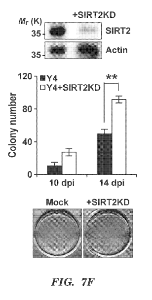
[00075] FIGs 7A-71 present results from experiments that indicate SIRT2
influences
somatic nuclear reprogramming through metabolic changes. (FIG. 7A) Time course
of expression
level of SIRT2 raRNA in hDFs infected with Y4 and/or SIIRT2KD. (Mean s.d., n
= 4 biologically
independent experiments, **P < 0.01; ***P < 0.005, two-way ANOVA with
Bonferroni post-test.) (FIGs
7B and 7C) OCR (FIG. 7B) and EC,NR (FIG. 7C) in hDFs infected with Y4 and/or
SIRT2KD were
assessed by XF analyzer. (Mean s.d., n=4 biologically independent
experiments, *P < 0.05; **P < 0.01;
***P <0.005, two-way ANOVA with Bonferroni post-test.) (FIG. 7D) Measurement
of lactate
production from hDFs infected with Y4 and/or SIRT2KD. (Mean s.d., n=3
biologically independent
experiments, ***P <0.005, two-way ANOVA with Bonferroni post-test.) (FIGs 7E
and 7F) Effects of
SIRT:20E or KD on iPSC generation. Upper: The efficiency of overexpression
(FIG. 7E) or knock.down
(FIG. 7F) was confirmed by western blotting with anti-SIRT2 antibody. Lower:
Representative pictures
of AP-positive colonies at 14 days post-infection (dpi). (Mean s.e.m., n = 3
biologically independent
experiments, **P <0.01, two-way ANOVA with Bonferroni post-test.) (FIGs 7G and
7H) Effects of
glycolytic inhibitor, 2-deoxyglucose (2DG) on iPSC generation by Y4 and/or
SIRT2KD at 8 days post-
transduction were assessed by OCR (FIG. 7G) and ECAR (FIG. 711). (Mean s.d.,
n = 4 biologically
independent experiments; **P < 0.01; ***P <0.005, two-way ANOVA with
Bonferroni post-test.) (FIG.
71) Effects of 2DG on iPSC generation by Y4 and/or SIRT2KD. Representative
pictures of AP-positive
colonies at 14 days post-transduction. (Mean s.d., n = 3 biologically
independent experiments, ***P
<0.005, two-way ANOVA with Bonferroni post-test.)
[00076] FIGs 8A-8G present results from experiments that indicate miR-200c
directly
targets SIRT2. (FIGs 8A and 8B) Altered expression levels of SIRT2 by pre-
miRNAs were analysed by
qRT-PCR (FIG. 8A) or western blotting with SIRT2-specific antibody (FIG. 8B).
(Mean s.d., n=3
biologically independent experiments, **P <0.01, one-way ANOVA with Bonferroni
posttest.) (FIG. 8C)
Sequences for stem loop of miR-200c (upper) and matured forms of miR-200c-5p
and -3p (lower). (FIGs
8D and 8E) Altered expression levels of SIRT2 by miRNA mimics for control
(Scr), miR-200c-5p (5p)
and -3p (3p) were analysed by qRT-PCR (FIG. 8D) or western blotting with SIRT2-
specific antibody
(FIG. 8E). (Mean s.d., n=3 biologically independent experiments, ***P
<0.005, one-way ANOVA with
Bonferroni post-test.) (FIG. 8F) Luciferase validation assays demonstrating
the effect of miR-200c-5p on
the CDS fragments of SIRT2 relative to control (Scr) in 293T cells. (Mean
s.d., n = 3 biologically
independent experiments, **P < 0.01, one-way ANOVA with Bonferroni post-test.)
(FIG. 8G) Proposed
model for miR-200c¨SIRT2-glycolytic enzymes (aldolase, GAPDH, enolase, and
PGK1) axis in
regulating metabolic switch and somatic reprogramming.
[00077] FIG. 9 presents results from experiments that indicate combined
effects of SIRTI
ov-erexpression (OE) and SIRT2 knock-down (KD) on human iPSC generation.
Fibroblasts were
treated with leiniviruses expressing four reprogramming factors with or
without SIRT-10E or SIRT2KD.
Representative pictures of AP-positive colonies at day 14 post lentiviral
transduction. Mean s.d., n = 3
biologically independent experiments, *** P<0.005, two-way ANOVA with
Bonferroni post-test.
CA 03052622 2019-08-02
WO 2018/144864 PCT/US2018/016644
[00078] FIG. 10 presents results from experiments that indicate SIRTI
expression is
variable in cancer. Although some cancer cells appear to express higher levels
of SIRT1, it is not
consistent like ESCs and iPSCs. It is however expected that SIRT1 is
consistently highly expressed in
cancer stem cells.
[00079] FIG. 11 presents results from experiments that indicate SIRT2
expression is
variable in cancer. Although some cancer cells appear to express lower levels
of SIRT2, it is not
consistent like ESCs and iPSCs. It is expected that SIRT2 is consistently down-
regulated in cancer stem
cells,
[00080] FIGs 12A-12G present results from experiments that indicate
Warburg-like effect
in hESCs and hiPSCs compared to hi-Ws. (FIG. 121) Human ESCs (1-19) and hiPSCs
cultured under
feeder-free condition were stained with specific antibodies against
pluripotency markers (e.g., 0ct4,
Nanog, SSEA4, and TRA1-60) along with Hoechst staining for nuclear staining.
Scale bar = 100 pm.
(FIG. 12B) Representative pictures of hESCs and hiPSCs. (FIG. 12C) In vitro
spontaneous
differentiation of hESCs and hiPSCs by culturing in serum-free ITSFn medium
for 7 days,
Immunostaining images (first and second row panels) show lineage specific
markers for ectoderm (Gtx2),
mesoderm (Brachyury; B-T), and endoderm (Sox17). Scale bar= 100 pm. (FIG. 12D)
intracellular ATP
levels were significantly lower in hiPSCs and hESCs than in the original
fibroblasts. Mean SEM (n--3)
are shown. ***p<0.005. (FIG. 12E) Mitochondria bioenergetics of parental haFs
and hiPSCs as well as
hESCs assessed by Seahorse XI' analyzer. (FIG. 12F) Expression levels of
glucose transporters (CLUTs)
including GLUT!. to GLUT7 in hDFs and hiPSCs as well as hESCs. Mean SEM (n=3)
are shown. *
p<0.05; ** p<0.01; ***p<0.005; ****p<0.001. (FIG. 12G) Immunoprecipitation of
hDF and hESCs
proteins using antibodies against acetyl-Lys, followed by LC-MS/MS analyses to
identify acetylated
proteins.
[00081] FIG. 13 presents results from experiments that indicate CID
spectra for the
acetylated proteins shown in FIG. 12 and Table 2. Peptides for tubulin,
Fructosc-biphosphate aldolase,
glyceraldehyde-3-phosphate dehydrogenase, phosphoglycerate kinase 1, enolase,
pyruvate kinase
isozymes Nll./M2 and ATP synthase were detected via combination of IP and LC-
MS/MS analysis. IP
was performed with anti-acetyl-Lys antibody.
[00082] FIGs 14A-14G present results from experiments that indicate meta-
analysis of
Sirtuin family expression in hESCs. (FIG. 14A) Compiled data used in this
study for Sirtuin family
gene expression in hESCs shown in Table 5. Expression levels of each Sirtuin
shown as up, down, and
N/A, which corresponds to upregulated, downregulated, and no significant
change, respectively.
Numbers in parenthesis represent expression changes from 5 different studies.
(FIG. 1411) Representative
data showing SIRT2 expression changes between different cells. SIRT2
downregulation was observed in
liPSCs compared to differentiated cells and original fibroblasts, (FIGs 14C-
14G) Expression levels
comparison of SIRT3 (FIG. 14C), SIRT4 (FIG. 14D), SIRT5, (FIG. 14E) SIRT6
(FIG. 14F), and
SIRT7 (FIG. 14G), across several hESC lines and normal non-cancer cell lines
based on Database
16
CA 03052622 2019-08-02
WO 2018/144864 PCT/US2018/016644
analyses (found on the world wide web at http://www.nextbio.com). The relative
expression levels are
presented as the mean value of scatter plot.
[00083] FIGs 15A-15D present results from experiments that indicate
characterization of
inducible SIRT2-GFP H9 hESCs. (FIG. 15A) Plasmid map of the EGFP SIRT2
doxycycline (Dox)
inducible overexpression vector. (FIG. 15B) Expression levels of glycolytic
enzymes in SIRT2-GFP
hESCs with or without Dox analyzed by qRT-PCR. Mean SEM (n=3) are shown. *
p<0.005. (FIGs 15C
and 15D) Expression levels of pluripotency markers in hESCs, hDFs, and SIRT2-
GFP hESCs with or
without Dox. Mean SEM (n=3) are shown. *p<0.005.
[00084] FIGs 1.6A-16F present results from experiments that indicate
effects of altered
SIRT2 expression on acetylation of AldoA. (FIGs 16A-16D) Detection of AldoA
Kill (FIGs 16A
and 16B) and K322 (FIGs 16C and 16D) acetylation by mass spectrometry
analysis. Symbol "@"
indicates the acetylation site. (FIG. 16E) Myc-tagged AIdoA, AldoAK1.11Q, and
AldoAK3224 were
each expressed in 293T cells. AldoA proteins were purified by IP with Myc
antibody, and specific
activity for AIdoA was determined. MeantSEM (n=3) are shown. *0* p<0.005.
(FIG. 16F) Myc-tagged
AldoA, AldoAK111R, and AldoAK322R were each expressed in 293T cells co-
expressing SIRT2
shRNA (SIRT2KD). AldoA proteins were purified by IP with Myc antibody and
specific activity for
AldoA was determined. MeantSEM (n=3) are shown. ***p<0.005.
[00085] FIGs 17A-17H present results from experiments that indicate
metabolic and
functional characterization of hPSC lines following SIRT2 oyerexpression.
(FIGs 17A, 17C, and
17E) Glycolytic bioenergetics of wild type (Mock) and inducible SIRT2-GFP cell
lines from H7 hESCs
(FIG. 17A) and two iPSC lines (FIG. 17C and 17E) with or without Dox were
assessed by XF analyzer.
(FIGs 17B, 17D, and 17F) Basal glycolytic rate, glycolytic capacity and
glycolytic reserve of mock and
SIRT2OE from. H7 hESCs (FIG. 17B) and two iPSC lines (FIG. 17D and 17F) with
or without Dox are
shown in FIGs 17A, 17C, and 17E, respectively. Mean :ESEM (n=3) are shown.
*p<0.05; "p<0.01. (FIG.
17G) OCR were shown for two hESC lines (H9 and H7) and hiPSC-1 line with or
without Dox. 1: Mock
w/o Dox, 2: Mock with Dox, 3: SIRT2OE w/o Dox, 4: SIRT2OE with Dox. Mean SEM
(n=3) are shown.
*p<0.05; ***p<0.005. (FIG. 17H) Cell proliferation of mock and SIRT2OE from H7
hESCs and two
independent iPSC lines (hiPSC-1 and hiPSC-2) with or without Dox were analyzed
by determining cell
numbers every 2 days under ESC culture conditions. Mean SEM (n=3) are shown.
"p<0.01;
***p<0.005.
[00086] FIGs 1.8A-18F present results from experiments that indicate SIRT2
influences
metabolic signatures of early differentiation potential of hiPSCs. (FIG. 18A
and 18B) Inducible
SIRT2OE hiPSC-1 cells were induced to differentiate spontaneously by culturing
serum-free ITSFn
medium for up to 4 days, and gene expression levels of pluripotency markers
(0ct4, Nanog, and Rex!)
(FIG. 18A) and early-differentiation markers (Pax6, Brachyury (B-T), and
Sox17) (FIG. 18B) were
determined by qRT-PCR. Mean SEM (n=3) are shown. * p<0.05; ** p<0.01. (FIG.
18C) Expression
level of SIRT2 in SIRT2OE hiPSC-1 cells with or without Dox during early
differentiation. Mean SEM
(n=3) are shown. * p<0.05. (FIG. 18D) Glycolvtic bioenergetics of mock and
SIRT2OE hiPSC-1 cells
17
CA 03052622 2019-08-02
WO 2018/144864 PCT/US2018/016644
with or without Dox were assessed using the Seahorse XF analyzer. Mean SEN1
(n-3) are shown. *
p<0.05. (FIG. 18E) Extracellular lactate production of mock and SIRT2OE hiPSC-
1 cells with or
without Dox. Mean SEM (n=3) are shown. * p<0.05; ** p<0.01. (FIG. 18F)
Heannaps depicting gene
expression levels of markers representing ectoderm (Pax6, Map2, Ci-FAP and
AADC), endoderm (Foxa2,
Sox17, AFP, CK8 and CK18), and mesoderm (Msxl and B-T) in wild type (Mock) and
inducible
SIRT2OE hiPSC lines including hiPSC-1 and hiPSC-2 with or without Dox for up
to 12 days under
differentiation condition. Mean SENI (rt=3) are shown. 1: Mock w/o Dox., 2:
Mock with Dox, 3:
SIRT2OE w/o Dox, 4: SIRT2OE with Dox.
[00087] FIGs I9A49H present results from experiments that indicate effects
of altered
SIRT1 expression on metabolic reprogramming and iPSC generation. (FIG. 191)
Plasmid map of
the EGFP SIRT I doxycycline inducible overexpression vector. (FIG. 1913) OCR
was shown for hDFs
infected with wild type (Mock) or inducible SIRTI-G-FP (SIRTIDE) with or
without Dox at 3 days after
transfection. (FIGs 19C and 19D) OCRIECAR ratio (FIG, 19C), and relative OCR
changes after FCCP
injection (FIG. 191)) from Mock and SIRT1OE with or without Dox are shown in
FIG. 1911, Mean
SEM (n=3) are shown. (FIGs 19E and 19F) Effects of SIRTIKD or OE on iPSC
generation. Upper:
Efficiency of SIRTIKD or OE was confirmed by western blotting with anti-SIRTI
antibody. Lower:
Representative pictures of AP-positive colonies at day 14 post lentiviral
transduction. Mean SEM (n=3)
are shown.. *p<0.005, G,H: OCR in 11Di:infected with Y4 and/or SIRT I OE at 3
(FIG. 19G) or 6 (FIG.
1911) days after tran.sfection.
DETAILED DESCRIPTION
[00088] Aspects of the invention are based on the discovery that the
metabolic pathway used by a
cell directly influences its state of differentiation. Although correlations
between cellular metabolism
and differentiation state have been previously observed, a causative effect of
metabolism on cell state
was not appreciated. The results presented herein indicate that the metabolic
pathway utilized drives a
cell either towards pluripotency or differentiation. As such metabolic
reprogramming (e.g., via
experimental manipulation) can directly influence the differentiated state of
a cell. Reprogramming cells
to increase utilization of glycolysis metabolism and decrease oxidative
phosphorylation (OXPHOS)
metabolism drives cells to a less differentiated state (to thereby increase
their -sternness"). Whereas,
reprogramming cells toward decrease utilization of glycolysis and increase
OXPHOS metabolism drives
cells towards a more differentiated state.
[00089] Aspects of the invention are further based on the finding that one
way in which a cell
regulates which metabolic pathway is utilized is through protein acetylation,
with acetylated glycolytic
enzymes being highly active compared to their deacetylated counterparts. This,
taken with the
recognition of the role of the different metabolic pathways in cell fate,
indicates that cell fate can be
manipulated by the appropriate manipulation of the acetylation state of
glycolytic enzymes.
[00090] As such, one aspect of the invention relates to the shifting of
cell fate by manipulation of
the acetylation state of the glycolytic enzymes. Deacetylation of the
glycolytic enzymes in otherwise
18
CA 03052622 2019-08-02
WO 2018/144864 PCT/US2018/016644
differentiated cells (e.g., somatic cells) to thereby reduce glycolysis in the
cells, shifts the cells towards
pluripotency. Alternatively, acetylation of the glycolytic enzymes in less
differentiated cells to thereby
increase glycolysis in the cells (e.g., pluripotent or multipotent) shifts the
cells towards differentiation.
[00091] One such method of reducing glycolysis is through manipulation of
the deacetylase
SIRT2. SIRT2 deacetylates glycolytic enzymes to thereby reduce their activity
and suppress glycolysis.
SIRT2 is highly active in differentiated cells. Reduction in SIRT2 activity
allows glycolysis to increase
thereby driving the cells toward de-differentiation. Alternatively, SIRT2
activity in less differentiated
cells (e.g., stem cells) is relatively low, as is glycolytic enzyme activity,
with OXPHOS being primarily
used for metabolism. Increasing SIRT2 activity in less differentiated cells
deacetylates the glycolytic
enzymes, suppressing glycolysis, and drives the cells toward a more
differentiated state.
[00092] Another acetylation modulating factor, SIRT1, has activity
reciprocal to that of SIRT2
with respect to cell fate. SIRT1 is active in less differentiated cells, with
activity decreasing in more
differentiated cells. Similar to SIRT2, SIRT1 alters acetylation of metabolic
enzymes to increase
utilization of glycolysis and decrease utilization of OXPHOS, thereby
contributing to the undifferentiated
state. SIRT1 manipulation can therefore be used in the methods described
herein to affect cell fate, with
an increase in SIRT1 driving a cell towards de-differentiation and a decrease
in SIRT1 driving a cell
towards further differentiation.
[00093] The ability to shift cell fate by manipulating the metabolic
pathways utilized is useful in
enhancing known methods of cell fate manipulation (e.g. to generate
pluripotent cells from differentiated
cells, and to generate differentiated cells from pluripotent cells). Methods
for de-differentiating cells
using reprogramming factors are well known in the art. Examples include the
induction of the Yamanaka
(reprogramming) factors: Oct-4, Sox-2, c-Myc (or 1-Myc) and Klf-4, and also
the induction of the
Thomson (reprogramming) factors: Oct-4, Sox-2, Nanog, and Lin-28.
Unfortunately, the current
methods for inducing de-differentiation of a cell (e.g., pluripotency) are
fairly inefficient, generating a
small percentage of the desired product. Modulation of cell metabolism, such
as by SIRT1
(upmodulation) and SIRT2 (downmodulation), as described herein, to shift a
cell towards a less
differentiated state can be used to enhance known methods for de-
differentiating cells (e.g., generating
induced pluripotent cells). As such, the methods involve SIRT1 and SIRT2
modulation in combination
with the full complement of reprogramming factors. It is expected however,
that SIRT1 and SIRT2
modulation, as described herein, will increase the number of de-differentiated
cells produced and/or
enable the omission of one or more of the reprogramming factors in the de-
differentiation process. The
ability to omit one or more reprogramming factors is considered an enhancement
of the known
procedures if it facilities a reduction in total manipulation of the cells
(e.g., delivery of less foreign matter
to the cells).
[00094] Various methods for differentiating cells (e.g., pluripotent or
multipotent stem cells) by
using various differentiation factors and/or culture procedures are known.
Many of these methods suffer
from low efficacy of induction and/or slow rate of induction. Modulation of
cell metabolism, wuch as
by SIRT1 (downmodulation) and SIRT2 (upmodulation), as described herein, to
shift a cell toward a
19
CA 03052622 2019-08-02
WO 2018/144864 PCT/US2018/016644
more differentiated state can be used to enhance known methods for
differentiating cells (e.g., generating
neuronal cells). As such, the methods involve SIRT1 and SIRT2 modulation in
combination with known
methods of differentiation. It is expected however, that SIRT1 and SIRT2
modulation will decrease the
time required to generate the differentiated cells and/or increase the number
of differentiated cells
produced. It is also expected that SIRT1 and SIRT2 modulation will also enable
the omission of one or
more steps or factors required for the differentiation process.
[00095] Moreover, the invention described herein provides methods for
selecting pluripotent
stem cells and differentiated cells based on the expression level and/or
activity of SIRT1 and/or SIRT2.
[00096] Methods and compositions described herein require that the levels
and/or activity of
SIRT1 and/or SIRT2 be modulated in order to more easily and readily alter the
cell fate. SIRT1 is a NAD
(nicotinamide adenine dinucleotide)-dependent deacetylase enzyme that
regulates proteins essential for
cellular regulation, e.g., via deacetylation. SIRT2 is a NAD-dependent
deacetylase enzyme that functions
as an intracellular regulatory protein with mono-ADP-ribosyltransferase
activity.
[00097] Downmodulate or downmodulation refers to reducing the function of
the protein (e.g.,
SIRT1 or SIRT2). This can be accomplished by directly inhibiting the
production of functional SIRT1 or
SIRT2 itself in the cell (e.g., by reducing gene expression or protein
synthesis), or alternatively by
reducing SIRT1 or SIRT2 function/activity. SIRT1 or SIRT2 function/activity
can be reduced, for
example by directly inhibiting the SIRT1 or SIRT2 protein itself or otherwise
targeting that protein for
degradation. As such, an agent useful in the present invention for
downmodulation is one that inhibits
SIRT1 or SIRT2 gene expression or protein synthesis, or inhibits SIRT1 or
SIRT2 function or activity.
Downmodulation of SIRT1 or SIRT2 can also be accomplished by inhibition of an
upstream factor that
induces or positively regulates SIRT1 or SIRT2 gene expression or SIRT1 or
SIRT2 function/activity.
As such, another useful agent for downmodulation is an agent that inhibits or
downmodulates such an
upstream factor by methods that correspond to those described for SIRT1 or
SIRT2.
[00098] Upmodulate or upmodulation refers to increasing the level of a
functional protein, and is
accomplished by methods described for downmodulation, but by instead
increasing or activating gene
expression or protein activity.
[00099] Induced pluripotent stem cells
[000100] Stem cells are undifferentiated cells defined by their ability at the
single cell level to both
self-renew and differentiate to produce progeny cells, including self-renewing
progenitors, non-renewing
progenitors, and terminally differentiated cells. Stem cells, depending on
their level of differentiation, are
also characterized by their ability to differentiate in vitro into functional
cells of various cell lineages
from multiple germ layers (endoderm, mesoderm and ectoderm), as well as to
give rise to tissues of
multiple germ layers following transplantation and to contribute substantially
to most, if not all, tissues
following injection into blastocysts. "Induced pluripotent stem cells" are
pluripotent stems cells that are
generated directly from adult cells, e.g., somatic or non-embryonic cells.
[000101] One aspect of the invention described herein provides a method to
generate induced
human pluripotent stem cells comprising delivering to a somatic or non-
embryonic cell population an
CA 03052622 2019-08-02
WO 2018/144864 PCT/US2018/016644
effective amount of one or more reprogramming factors (e.g., Yamanaka factors
or Thomson factors) and
also an agent that downmodulates SIRT2, and culturing the somatic or non-
embryonic cell population for
a period of time sufficient to generate at least one induced human pluripotent
stem cell. In one
embodiment, the method further comprises delivering to the somatic or non-
embryonic cell population an
effective amount of an agent that upmodulates SIRT1.
[000102] In one embodiment, the somatic or non-embryonic cell population is
cultured for a
period of time sufficient to generate at least one induced human pluripotent
stem cell. Culturing can
occur for a period of at least 7 days, at least 8 days, at least 9 days, at
least 10 days, at least 11 days, at
least 12 days, at least 13 days, at least 14 days, at least 15 days, at least
16 days, at least 17 days, at least
18 days, at least 19 days, at least 20 days, at least 21 days, or more.
[000103] In some instances, the chemical and/or atmospheric conditions are
altered for
reprogramming. For example, where the somatic and non-embryonic cells are not
vascularized and
hypoxic reprogramming under hypoxic conditions of 5% 02, instead of the
atmospheric 21% 02, may
further provide an opportunity to increase the reprogramming efficiency.
Similarly, chemical induction
techniques have been used in combination with reprogramming, particularly
histone deacetylase (HDAC)
inhibitor molecule, valproic acid (VPA), which has been found wide use in
different reprogramming
studies.
[000104] At the same time, other small molecules such as MAPK kinase (MEK)¨ERK
("MEK")
inhibitor PD0325901, transforming growth factor beta ("TGF-I3") type I
receptor ALK4, ALK5 and
ALK7 inhibitor SB431542 and the glycogen synthase kinase-3 ("GSK3") inhibitor
CHIR99021 have
been applied for activation of differentiation-inducing pathways (e.g. BMP
signaling), coupled with the
modulation of other pathways (e.g. inhibition of the MAPK kinase (MEK)¨ERK
pathway) in order to
sustain self-renewal. Other small molecules, such as Rho-associated coiled-
coil-containing protein
kinase ("ROCK") inhibitors, such as Y-27632 and thiazovivin ("Tzv") have been
applied in order to
promote survival and reduce vulnerability of cell death, particularly upon
single-cell dissociation. As
such, the inclusion of one or more of the factors in the herein described
methods is envisioned.
[000105] Efficiency of reprogramming
[000106] Efficiency of reprogramming, e.g., changing the cell fate of a cell,
is readily ascertained
by one of many techniques readily understood by the skilled practitioner. For
example, efficiency can be
described by the ratio between the number of donor cells receiving the
agent(s) and reprogramming
factors and the number of reprogrammed colonies (de-differentiated colonies)
generated. The number
donor cells receiving the agent(s) and reprogramming factors can be measured
directly, such as by use of
a reporter gene such as GFP included in a vector encoding an agent or
reprogramming factor.
Alternatively, indirect measurement of delivery efficiency can be accomplished
by transfecting a vector
encoding a reporter gene as a proxy to gauge delivery efficiency in paired
samples delivering agent(s)
and reprogramming factor vectors. Further, the number of reprogrammed colonies
generated can be
measured by, for example, observing the appearance of one or more multipotency
or pluripotency
characteristics such as alkaline phosphatase (AP)-positive clones, colonies
with endogenous expression
21
CA 03052622 2019-08-02
WO 2018/144864 PCT/US2018/016644
of transcription factors Oct-4 or Nanog, or antibody staining of surface
markers such as Tra-1-60.
Efficiency can alternatively be described by the time required for induced
pluripotent stem cell
generation. A combination of percentage of induced cells and the time of
induction can also be used.
[000107] In one embodiment, the methods described herein result in an
enhancement of the
number of induced pluripotent stem cells by at least 2-fold as compared to an
appropriate control. In
another embodiment, the methods described herein result in an enhancement of
the number of induced
pluripotent stem cells by at least 3-fold, at least 4-fold, at least 5-fold,
at least 6-fold, at least 7-fold, at
least 8-fold, at least 9-fold, at least 10-fold, or more as compared to an
appropriate control. As used
herein, an "appropriate control" refers to a comparably treated cell
population in the absence of the agent
(e.g., that downmodulates SIRT2 and/or that upmodulates SIRT1). The efficiency
of reprogramming can
be assessed as described above.
[000108] One aspect of the invention described herein provides a cell line
comprising induced
stem-like cells (e.g., pluripotent stem cells) generated by any of the methods
described herein.
[000109] Another aspect of the invention described herein provides a
pharmaceutical composition
comprising an induced stem-like cell (e.g., pluripotent stem cell) or
population thereof generated by any
of the methods described herein and a pharmaceutically acceptable carrier.
[000110] Reprogramming factors with downmodulation of SIRT2 and/or
upmodulation of
SIR Ti
[000111] The somatic or non-embryonic cell population is further contacted
with one or more
reprogramming factor. In one embodiment, the one or more reprogramming factor
is from one to four
reprogramming factors selected from the Yamanaka (reprogramming) factors, e.g,
Oct-4, Sox-2, c-Myc
(or 1-Myc) and Klf-4, or selected from the Thomson (reprogramming) factors,
e.g., Oct-4, Sox-2, Nanog,
and Lin-28. Reprogramming factors are traditionally understood to be normally
expressed early during
development and are involved in the maintenance of the pluripotent potential
of a subset of cells that
constitute the inner cell mass of the pre-implantation embryo and post-
implantation embryo proper.
Their ectopic expression is believed to allow the establishment of an
embryonic-like transcriptional
cascade that initiates and propagates an otherwise dormant endogenous core
pluripotency program within
a host cell.
[000112] In one embodiment, reprogramming factors are expressed in the cell
e.g., via an vector
such as those described herein, comprising a nucleic acid encoding a given
reprogramming factor. In
another embodiment, reprogramming factors are expressed in the cell e.g., via
expression of a nucleic
acid encoding a given reprogramming factor as naked DNA.
[000113] Additional reprogramming factors include, but are not limited to,
Tert, Klf-4, c-Myc,
SV40 Large T Antigen ("SV4OLT") and short hairpin RNAs targeting p53 ("shRNA-
p53"). One or more
of these factors can further be delivered to the cells to enhance the
reprogramming process using delivery
methods described herein.
[000114] The agent and reprogramming factors described herein may necessarily
be contained in
and thus further include a vector. Many such vectors useful for transferring
exogenous genes into target
22
CA 03052622 2019-08-02
WO 2018/144864 PCT/US2018/016644
mammalian cells are available. The vectors may be episomal, e.g. plasmids,
virus-derived vectors (e.g.,
viral vectors) such cytomegalovirus, adenovirus, etc., or may be integrated
into the target cell genome,
through homologous recombination or random integration, e.g. retrovirus-
derived vectors such as
MMLV, HIV-1, ALV, etc. For modification of stem cells, lentiviral vectors are
preferred. Lentiviral
vectors such as those based on HIV or FIV gag sequences can be used to
transfect non-dividing cells,
such as the resting phase of human stem cells (see Uchida et al. (1998)
P.N.A.S. 95(20): 11939-44). In
some embodiments, combinations of retroviruses and an appropriate packaging
cell line may also find
use, where the capsid proteins will be functional for infecting the target
cells. Usually, the cells and virus
will be incubated for at least about 24 hours in the culture medium. The cells
are then allowed to grow in
the culture medium for short intervals in some applications, e.g. 24-73 hours,
or for at least two weeks,
and may be allowed to grow for five weeks or more, before analysis. Commonly
used retroviral vectors
are "defective", i.e. unable to produce viral proteins required for productive
infection. Replication of the
vector requires growth in the packaging cell line.
[000115] The use of various combinations of vectors in the methods is
envisioned. While various
vectors and reprogramming factors in the art appear to present multiple
ingredients capable of
establishing reprogramming in cells, a high degree of complexity occurs when
taking into account the
stoichiometric expression levels necessary for successful reprogramming to
take hold. For example,
somatic cell reprogramming efficiency is reportedly fourfold higher when Oct-4
and Sox-2 are encoded
in a single transcript on a single vector in a 1:1 ratio, in contrast to
delivering the two factors on separate
vectors. The latter case results in a less controlled uptake ratio of the two
factors, providing a negative
impact on reprogramming efficiency. One approach towards addressing these
obstacles is the use of
polycistronic vectors, such as inclusion of an internal ribosome entry site
("IRES"), provided upstream of
transgene(s) that is distal from the transcriptional promoter. This
organization allows one or more
transgenes to be provided in a single reprogramming vector, and various
inducible or constitutive
promoters can be combined together as an expression cassette to impart a more
granular level of
transcriptional control for the plurality of transgenes. These more specific
levels of control can benefit
the reprogramming process considerably, and separate expression cassettes on a
vector can be designed
accordingly as under the control of separate promoters.
[000116] Although there are advantages to providing such factors via a
single, or small number of
vectors, upper limitations on vector size do exist, which can stymie attempts
to promote their delivery in
a host target cell. For example, early reports on the use of polycistronic
vectors were notable for
extremely poor efficiency of reprogramming, sometimes occurring in less than
1% of cells, more
typically less than 0.1%. These obstacles are due, in-part, to certain target
host cells possessing poor
tolerance for large constructs (e.g., fibroblasts), or inefficient processing
of IRES sites by the host cells.
Moreover, positioning of a factor in a vector expression cassette affects both
its stoichiometric and
temporal expression, providing an additional variable impacting reprogramming
efficiency. Thus, some
improved techniques can rely on multiple vectors each encoding one or more
reprogramming factors in
various expression cassettes. Under these designs, alteration of the amount of
a particular vector for
23
CA 03052622 2019-08-02
WO 2018/144864 PCT/US2018/016644
delivery provides a coarse, but relatively straightforward route for adjusting
expression levels in a target
cell.
[000117] In an alternate embodiment, the methods described herein do not
require the somatic or
non-embryonic cell to be contacted by a reprogramming factor.
[000118] Differentiation of an induced pluripotent stem cell
[000119] One aspect of the invention described herein provides a method to
generate differentiated
cells comprising delivering to a pluripotent cell population an agent that
upmodulates SIRT2 and
culturing the population under differentiating conditions for a period of time
sufficient to generate at least
one differentiated cell. In one embodiment, the method further comprises
delivering an agent that
downmodulates SIRT1
[000120] Pluripotent stem cells comprise the capacity to differentiate into
any cell type of the
organism. It should be understood that the methods and protocols for
differentiating a stem cell will vary
based on the cell type, e.g., differentiation into a neuron may require a
different protocol compared to
differentiation into a hepatocyte. Protocols for differentiating a stem cell
into a given cell type are known
in the art. The skilled practitioner is able to determine if a cell has
differentiated into a particular cell type
(e.g., a neuron) by assessing the differentiated cells for specific linage-
derived markers (e.g., Class III 13-
tubulin, neuron specific enolase (NSE), or calretinin). Markers for various
cell types are known and can
be determine by the skilled practitioner.
[000121] Specific differentiation conditions typically require cultureing in
specific differentiation
medium. As used herein, "differentiation media" refers to a medium containing
factors required for
differentiating a stem cell into a particular cell type. Differentiated media
useful for generating a
particular differentiated cell (e.g., a neuron, or other neuronal cell type)
are commercially available for
various cell types, e.g, at Cell Applications, Inc., San Diego, CA. The
skilled artisan can determine the
appropriate differentiation media and conditions for a desired cell type.
[000122] In one embodiment, differentiating conditions are specific for
neuronal differentiation
(e.g., differentiation in to a neuronal progenitor cell). Methods for
differentiation of a stem-like cell to a
neuronal cell include culturing an adherent population of stem-like cell in a
medium containing factors
that promote neural differentiation, such as retinoic acid, BMP inhibitors
(e.g., noggin), N2, B27, and
ITS. The adherent stem-like cells can be adherent to a matrix, e.g, laminin,
fibronection, or collagen, or
adherent to a population of feeder cells, e.g., a monolayer of fibroblast
cells. When cells in culture begin
to commit to neural fates, e.g., as observed by the presence of neural
rosettes, they are cultures in a
permissive medium, and neuronal rosettes are passaged in permissive medium
containing high levels of
basic FGF2. Methods for neuronal differentiation are further are reviewed in,
e.g., Dhara, SK., and Stice,
SL. J Cell Biochem. 2008 Oct 15; 105(3): 633-640, which is incorporated herein
by reference in its
entirety.
[000123] By way of another example, stem-like cells can be differentiated into
a hepatocyte by
culturing the stem-like cells in medium containing factors that promote
hepatocyte differentiation, e.g.,
FGF-4, and HGF. After 6 days, the cells are cultured in medium containing FGF-
4, HGF, and oncostatin
24
CA 03052622 2019-08-02
WO 2018/144864 PCT/US2018/016644
M to allow for differentiation. Complete hepatocyte differentiation can be
determined by assessing the
cells for hepatocyte markers, such as GATA4, HNF4a, and albumin. Methods for
hepatocyte
differentiation are further are reviewed in e.g., Agarwak, S., et al. Stem
Cells. 2008 Feb 21; 26(5): 1117-
1127, which is incorporated herein by reference in its entirety.
[000124] The stem cells for use with the methods and compositions described
herein can be
naturally occurring stem cells or "induced" stem cells, such as induced
pluripotent stem cells generated
using methods described herein. Induced pluripotent stem cells can be
generated using any methods
known in the art (e.g., as described herein). Stem cells can be obtained or
generated from any mammalian
subjects, e.g. human, primate, equine, bovine, porcine, canine, feline,
rodent, e.g. mice, rats, hamster, etc.
In one embodiment, the stem cell is a human stem cell. In one embodiment, the
stem cell is a non-human
stem cell.
[000125] In one embodiment, the pluripotent stem cell population is an
embryonic stem cell
population, an adult stem cell population, an induced pluripotent stem cell
population, or a cancer stem
cell population. In one embodiment, the stem cell is a non-embryonic stem
cell.
[000126] In one embodiment, a pluripotent cell population is cultured in,
e.g., differentiation
media, for a period of time sufficient to generate at least one differentiated
cell. Culturing can occur for a
period of from 1-5 days, at least 7 days, at least 8 days, at least 9 days, at
least 10 days, at least 11 days,
at least 12 days, at least 13 days, at least 14 days, at least 15 days, at
least 16 days, at least 17 days, at
least 18 days, at least 19 days, at least 20 days, at least 30 days, at least
40 days, at least 50 days, at least
60 days, at least 70 days, at least 80 days, at least 90 days, at least 100
days, at least 110 days, at least 120
days, at least 130 days, at least 140 days, at least 150 days, at least 160
days, at least 170 days, at least
180 days, at least 190 days, at least 200 days, at least 210 days, at least
220 days, at least 230 days, at
least 240 days, at least 250 days, at least 260 days, at least 1270 days, at
least 280 days, at least 290 days,
at least 300 days, or more. In one embodiment, culturing occurs for a period
of 7 to 100 days, 7 to 200
day, 7 to 300 days, 100 to 200 days, 200 to 300 days, 50 to 150 days, 150 to
250 days, or 150 to 300
days.
[000127] In one embodiment, the methods described herein produce an enhanced
number of
differentiated cells by at least 2-fold as compared to an appropriate control.
In another embodiment, the
methods described herein result in an enhancement of the number of
differentiated cells by at least 3-
fold, at least 4-fold, at least 5-fold, at least 6-fold, at least 7-fold, at
least 8-fold, at least 9-fold, at least
10-fold, or more as compared to an appropriate control. In one embodiment,
enhancement is by at least
100X, 250X, 500X, 750X, 100X or more, as compared to an appropriate control.
One such "appropriate
control" is a similar or identical cell subjected to an otherwise identical
method that does not
downmodulate SIRT1 and/or upmodulate SIRT2. The efficiency of de-
differentiation can be assessed as
described above for the efficiency of reprogramming.
[000128] In one embodiment, the differentiated cells are produced in a
significantly shorter period
of time than in appropriate control. In one embodiment, the period of time is
at least 10% shorter as
compared to an appropriate control. In one embodiment, period of time is at
least 20%, at least 30%, at
CA 03052622 2019-08-02
WO 2018/144864 PCT/US2018/016644
least 40%, at least 50%, at least 60%, at least 70%, at least 80%, at least
90%, at least 99%, or more,
shorter as compared to an appropriate control.
[000129] Another aspect of the invention relates to a cell line comprising
differentiated cells
generated by any of the methods described herein.
[000130] Agents
[000131] In various embodiment, agents are delivered to cells to modulate
(e.g., upmodulate, or
downmodulate) SIRT1 and SIRT2. The term "agent" as used herein means any
compound or substance
such as, but not limited to, a small molecule, nucleic acid, polypeptide,
peptide, drug, ion, etc. An
µ`agent" can be any chemical, entity or moiety, including without limitation
synthetic and naturally-
occurring proteinaceous and non-proteinaceous entities. In some embodiments,
an agent is nucleic acid,
nucleic acid analogues, proteins, antibodies, peptides, aptamers, oligomer of
nucleic acids, amino acids,
or carbohydrates including without limitation proteins, oligonucleotides,
ribozymes, DNAzymes,
glycoproteins, siRNAs, lipoproteins, aptamers, and modifications and
combinations thereof etc. In certain
embodiments, agents are small molecule having a chemical moiety. For example,
chemical moieties
included unsubstituted or substituted alkyl, aromatic, or heterocyclyl
moieties including macrolides,
leptomycins and related natural products or analogues thereof Compounds can be
known to have a
desired activity and/or property, or can be selected from a library of diverse
compounds.
[000132] Such an agent can take the form of any entity which is normally not
present or not
present at the levels being administered in the cell. Agents such as
chemicals; small molecules; nucleic
acid sequences; nucleic acid analogues; proteins; peptides; aptamers;
antibodies; or fragments thereof,
can be identified or generated for use to downmodulate or upmodulate SIRT1 or
SIRT2.
[000133] Agents in the form of nucleic acid sequences designed to specifically
inhibit gene
expression are particularly useful. Such a nucleic acid sequence can be RNA or
DNA, and can be single
or double stranded, and can be selected from a group comprising; nucleic acid
encoding a protein of
interest, oligonucleotides, nucleic acid analogues, for example peptide-
nucleic acid (PNA), pseudo-
complementary PNA (pc-PNA), locked nucleic acid (LNA) etc. Such nucleic acid
sequences include, for
example, but are not limited to, nucleic acid sequence encoding proteins, for
example that act as
transcriptional repressors, antisense molecules, ribozymes, small inhibitory
nucleic acid sequences, for
example but are not limited to RNAi, shRNAi, siRNA, micro RNAi (mRNAi),
antisense oligonucleotides
etc.
[000134] The agent can be a molecule from one or more chemical classes, e.g.,
organic molecules,
which may include organometallic molecules, inorganic molecules, genetic
sequences, etc. Agents may
also be fusion proteins from one or more proteins, chimeric proteins (for
example domain switching or
homologous recombination of functionally significant regions of related or
different molecules),
synthetic proteins or other protein variations including substitutions,
deletions, insertion and other
variants.
[000135] In one embodiment the agent is a catalytic antisense nucleic acid
constructs, such as
ribozymes, which is capable of cleaving RNA transcripts and thereby preventing
the production of the
26
CA 03052622 2019-08-02
WO 2018/144864 PCT/US2018/016644
encoded protein. Ribozymes are targeted to and anneal with a particular
sequence by virtue of two
regions of sequence complementary to the target flanking the ribozyme
catalytic site. After binding the
ribozyme cleaves the target in a site specific manner. The design and testing
of ribozymes which
specifically recognize and cleave sequences of the specific gene products is
commonly known to persons
of ordinary skill in the art.
[000136] In one embodiment, the agent inhibits gene expression (i.e. suppress
and/or repress the
expression of the gene). Such agents are referred to in the art as "gene
silencers" and are commonly
known in the art. Examples include, but are not limited to a nucleic acid
sequence, for an RNA, DNA or
nucleic acid analogue, and can be single or double stranded, and can be
selected from a group comprising
nucleic acid encoding a protein of interest, oligonucleotides, nucleic acids,
nucleic acid analogues, for
example but are not limited to peptide nucleic acid (PNA), pseudo-
complementary PNA (pc-PNA),
locked nucleic acids (LNA) and derivatives thereof etc. Nucleic acid agents
also include, for example,
but are not limited to nucleic acid sequences encoding proteins that act as
transcriptional repressors,
antisense molecules, ribozymes, small inhibitory nucleic acid sequences, for
example but are not limited
to RNAi, shRNA, siRNA, micro RNAi (miRNA), antisense oligonucleotides, etc.
[000137] The agent may function directly in the form in which it is
administered. Alternatively,
the agent can be modified or utilized intracellularly to produce something
which modulates SIRT1 or
SIRT2, such as introduction of a nucleic acid sequence into the cell and its
transcription resulting in the
production of the nucleic acid and/or protein inhibitor or activator of SIRT1
or SIRT2 within the cell. In
some embodiments, the agent is any chemical, entity or moiety, including
without limitation synthetic
and naturally-occurring non-proteinaceous entities. In certain embodiments the
agent is a small molecule
having a chemical moiety. For example, chemical moieties included
unsubstituted or substituted alkyl,
aromatic, or heterocyclyl moieties including macrolides, leptomycins and
related natural products or
analogues thereof Agents can be known to have a desired activity and/or
property, or can be selected
from a library of diverse compounds.
[000138] Agents in the form of a protein and/or peptide or fragment thereof
can also be designed
to downmodulate or upmodulate SIRT1 or SIRT2. Such agents encompass proteins
which are normally
absent or proteins that are normally endogenously expressed in the host cell.
Examples of useful proteins
are mutated proteins, genetically engineered proteins, peptides, synthetic
peptides, recombinant proteins,
chimeric proteins (any of which may take the form of a dominant negative
protein for SIRT1 or SIRT2),
antibodies, midibodies, minibodies, triabodies, humanized proteins, humanized
antibodies, chimeric
antibodies, modified proteins and fragments thereof Agents also include
antibodies (polyclonal or
monoclonal), neutralizing antibodies, antibody fragments, peptides, proteins,
peptide-mimetics, aptamers,
oligonucleotides, hormones, small molecules, nucleic acids, nucleic acid
analogues, carbohydrates or
variants thereof that function to inactivate the nucleic acid and/or protein
of the gene products identified
herein, and those as yet unidentified.
[000139] In one embodiment, an agent that downmodulates SIRT2 is delivered to
a differentiated
cell to a generate at least one induced pluripotent stem cell. In such
embodiment, the agent
27
CA 03052622 2019-08-02
WO 2018/144864 PCT/US2018/016644
downmodulates SIRT2 by at least 10%, by at least 20%, by at least 30%, by at
least 40%, by at least
50%, by at least 60%, by at least 70%, by at least 80%, by at least 90%, by at
least 100% or more as
compared to an appropriate control. In an alternate embodiment, an agent that
upmodulates SIRT2 is
delivered to a stem cell to generate at least one differentiated cell. In such
embodiment, the agent
upmodulates SIRT2 by at least 2-fold, by at least 3-fold, by at least 4-fold,
by at least 5-fold, by at least
6-fold, by at least 7-fold, by at least 8-fold, by at least 9-fold, by at
least 10-fold or more as compared to
an appropriate control, or by at least 10%, by at least 20%, by at least 30%,
by at least 40%, by at least
50%, by at least 60%, by at least 70%, by at least 80%, by at least 90%, by at
least 100% as compared to
an appropriate control. An "appropriate control" can be the same type of cell
or population thereof
similarly or identically treated to which an agent has not been delivered.
[000140] In another embodiment, an agent that downmodulates SIRT1 is delivered
to a stem cell
to generate at least one differentiated cell. In such embodiment, the agent
downmodulates SIRT1 by at
least 10%, by at least 20%, by at least 30%, by at least 40%, by at least 50%,
by at least 60%, by at least
70%, by at least 80%, by at least 90%, by at least 100% as compared to an
appropriate control. In an
alternate embodiment, an agent that upmodulates SIRT1 is delivered to a
differentiated cell to de-
differentiate the cell (e.g., generate at least one induced pluripotent stem
cell). In such embodiment, the
agent upmodulates SIRT1 by at least 2-fold, by at least 3-fold, by at least 4-
fold, by at least 5-fold, by at
least 6-fold, by at least 7-fold, by at least 8-fold, by at least 9-fold, by
at least 10-fold or more as
compared to an appropriate control, or by at least 10%, by at least 20%, by at
least 30%, by at least 40%,
by at least 50%, by at least 60%, by at least 70%, by at least 80%, by at
least 90%, by at least 100% or
more as compared to an appropriate control. An "appropriate control" can be a
cell or population thereof
similarly or identically treated to which an agent has not been delivered.
[000141] In one embodiment, SIRT1 is upmodulated by a nucleic acid encoding
SIRT1 expressed
in the cell e.g., via a vector comprising a nucleic acid encoding SIRT1. In
another embodiment, a nucleic
acid encoding SIRT1 is expressed in the cell e.g., via expression of a nucleic
acid encoding SIRT1 as
naked DNA. In one embodiment, the nucleic acid encoding SIRT1 has a sequence
corresponding to the
sequence of SEQ ID NO: 2; or comprises the sequence of SEQ ID NO: 2; or
comprises a sequence with
at least 80%, at least 85%, at least 90%, at least 91%, at least 92%, at least
93%, at least 94%, at least
95%, at least 96%, at least 97%, at least 98%, at least 99%, or at least 100%
sequence identity to the
sequence of SEQ ID NO: 2, and having the same activity as the sequence of SEQ
ID NO: 2 (e.g.,
acetylation of its substrates).
[000142] In one embodiment, SIRT2 is upmodulated by expression of a nucleic
acid encoding
SIRT1. The nucleic acid encoding SIRT2 can be expressed in the cell e.g., via
a vector comprising a
nucleic acid encoding SIRT2. In another embodiment, a nucleic acid encoding
SIRT2 is expressed in the
cell e.g., via expression of a nucleic acid encoding SIRT2 as naked DNA. In
one embodiment, the nucleic
acid encoding SIRT2 has a sequence corresponding to the sequence of SEQ ID NO:
3; or comprises the
sequence of SEQ ID NO: 3; or comprises a sequence with at least 80%, at least
85%, at least 90%, at
least 91%, at least 92%, at least 93%, at least 94%, at least 95%, at least
96%, at least 97%, at least 98%,
28
CA 03052622 2019-08-02
WO 2018/144864 PCT/US2018/016644
at least 99%, or at least 100% sequence identity to the sequence of SEQ ID NO:
3, and having the same
activity as the sequence of SEQ ID NO: 3 (e.g., acetylation of its
substrates).
[000143] In one embodiment, the agent is a small molecule that downmodulates
SIRT1 or SIRT2.
Such small molecules include, but are not limited, to the small molecules
listed in Table 1. Methods for
screening small molecules are known in the art and can be used to identify a
small molecule that is
efficient at, for example, inducing pluripotent stem cells or differentiated
cells, given the desired target
(e.g., SIRT1 or SIRT2).
Table 1. Small molecule compounds targeting Sirtuins
Molecular
Full Name Formula Information
Weight
SRT1720 HC1 is a selective SIRT1 activator with EC50 of
5RT1720 506.02 C25H23N705.HC1 0.16 M in a cell-free assay, but
is >230-fold less potent
for SIRT2 and SIRT3
EX 527 is a potent and selective SIRT1 inhibitor with IC50
EX527 248.71 C13H13C1N20 of 38 nM in a cell-free assay,
exhibits >200-fold selectivity
against SIRT2 and SIRT3. Phase 2.
Sirtinol is a specific SIRT1 and SIRT2 inhibitor with IC50
Sirtinol 394.47 C26H22N202
of 131 M and 38 M in cell-free assays, respectively.
Nicotinamide (Vitamin B3), a water-soluble vitamin, is an
Nicotinamide
122.12 C6H6N20 active component of coenzymes NAD and NADP, and also
(Vitamin B3)
act as an inhibitor of sirtuins.
SRT2183 is a small-molecule activator of the sirtuin
SRT2183 468.57 C27H24N4025 subtype SIRT1, currently being
developed by Sirtris
Pharmaceuticals.
Tenovin-6 acts through inhibition of the protein-
deacetylating activities of SirT1 and SirT2. Tenovin-6
Tenovin-6 454.63 C25H34N4025 inhibits the protein deacetylase
activities of purified human
SIRT1, SIRT2, and SIRT3 in vitro with IC50 of 21 M, 10
M, and 67 M, respectively.
SRT2104 516 . 64 C26H24N60252 SRT2104 (G5K2245840) is a selective
SIRT1 activator
.
(G5K2245840) involved in the regulation of energy
homeostasis. Phase 2.
Thiomyristoyl is a potent and specific SIRT2 inhibitor with
Thiomyristoyl 581.85 C34H51N3035 an IC50 of 28 nM. It inhibits SIRT1
with an IC50 value of
98 M and does not inhibit SIRT3 even at 200 M.
SirReal2 420.55 C22H20N4052 SirReal2 is a potent and selective
5irt2 inhibitor with IC50
of 140 nM.
Salermide is a reverse amide with a strong in vitro
Salermide 394.47 C26H22N202 inhibitory effect on Sirtl and 5irt2.
Compared with Sirtl,
Salermide is even more efficient at inhibiting 5irt2.
AGK2 is a potent, and selective SIRT2 inhibitor with IC50
AGK2 434.27 C23H13C12N302 of 3.5 M that minimally affects
either SIRT1 or SIRT3 at
10-fold higher levels.
5RT3025 606.2 C31H31N50252.HC1 5RT3025 is an orally available small
molecule activator of
the SIRT1 enzyme.
Fisetin 286.24 C15H1006 Fisetin (Fustel) is a potent sirtuin
activating compound
(STAC) and an agent that modulates sirtuins.
Quercetin, a natural flavonoid present in vegetables, fruit
Quercetin 302.24 C15H1007 and wine, is a stimulator of recombinant
SIRT1 and also a
PI3K inhibitor with IC50 of 2.4-5.4 M. Phase 4.
[000144] In one embodiment, the agent that downmodulates SIRT1 or SIRT2 is an
antibody or
antigen-binding fragment thereof, or an antibody reagent. As used herein, the
term "antibody reagent"
refers to a polypeptide that includes at least one immunoglobulin variable
domain or immunoglobulin
variable domain sequence and which specifically binds a given antigen. An
antibody reagent can
29
CA 03052622 2019-08-02
WO 2018/144864 PCT/US2018/016644
comprise an antibody or a polypeptide comprising an antigen-binding domain of
an antibody. In some
embodiments of any of the aspects, an antibody reagent can comprise a
monoclonal antibody or a
polypeptide comprising an antigen-binding domain of a monoclonal antibody. For
example, an antibody
can include a heavy (H) chain variable region (abbreviated herein as VH), and
a light (L) chain variable
region (abbreviated herein as VL). In another example, an antibody includes
two heavy (H) chain
variable regions and two light (L) chain variable regions. The term "antibody
reagent" encompasses
antigen-binding fragments of antibodies (e.g., single chain antibodies, Fab
and sFab fragments, F(ab')2,
Fd fragments, Fv fragments, scFv, CDRs, and domain antibody (dAb) fragments
(see, e.g. de Wildt et al.,
Eur J. Immunol. 1996; 26(3):629-39; which is incorporated by reference herein
in its entirety)) as well as
complete antibodies. An antibody can have the structural features of IgA, IgG,
IgE, IgD, or IgM (as well
as subtypes and combinations thereof). Antibodies can be from any source,
including mouse, rabbit, pig,
rat, and primate (human and non-human primate) and primatized antibodies.
Antibodies also include
midibodies, nanobodies, humanized antibodies, chimeric antibodies, and the
like.
[000145] The VH and VL regions can be further subdivided into regions of
hypervariability,
termed "complementarity determining regions" ("CDR"), interspersed with
regions that are more
conserved, termed "framework regions" ("FR"). The extent of the framework
region and CDRs has been
precisely defined (see, Kabat, E. A., et al. (1991) Sequences of Proteins of
Immunological Interest, Fifth
Edition, U.S. Department of Health and Human Services, NIH Publication No. 91-
3242, and Chothia, C.
et al. (1987) J. Mol. Biol. 196:901-917; which are incorporated by reference
herein in their entireties).
Each VH and VL is typically composed of three CDRs and four FRs, arranged from
amino-terminus to
carboxy-terminus in the following order: FR1, CDR1, FR2, CDR2, FR3, CDR3, FR4.
[000146] In some embodiments, a nucleic acid for use as an agent as described
herein (e.g.
SIRT1, or SIRT2) is contained in a vector for delivery and/or expression of
the nucleic acid.
[000147] In one embodiment, the agent that downmodulates SIRT1 or SIRT2 is an
antisense
oligonucleotide. As used herein, an "antisense oligonucleotide" refers to a
synthesized nucleic acid
sequence that is complementary to a DNA or mRNA sequence, such as that of a
microRNA. Antisense
oligonucleotides are typically designed to block expression of a DNA or RNA
target by binding to the
target and halting expression at the level of transcription, translation, or
splicing. Antisense
oligonucleotides of the present invention are complementary nucleic acid
sequences designed to
hybridize under cellular conditions to a gene, e.g., SIRT1 or SIRT2. Thus,
oligonucleotides are chosen
that are sufficiently complementary to the target, i.e., that hybridize
sufficiently well and with sufficient
specificity in the context of the cellular environment, to give the desired
effect.
[000148] In one embodiment the agent downmodulates SIRT1 or SIRT2 by RNA
inhibition.
Inhibitors of the expression of a given gene can be an inhibitory nucleic
acid. In oneembodiment, the
inhibitory nucleic acid is an inhibitory RNA (iRNA). The RNAi can be single
stranded or double
stranded.
[000149] The iRNA can be siRNA, shRNA, endogenous microRNA (miRNA), or
artificial
miRNA. In one embodiment, an iRNA as described herein effects inhibition of
the expression and/or
CA 03052622 2019-08-02
WO 2018/144864 PCT/US2018/016644
activity of a target, e.g. SIRT1 or SIRT2. In one embodiment, the agent is
siRNA that downmodulates
SIRT1 or SIRT2. In one embodiment, the agent is shRNA that downmodulates SIRT1
or SIRT2.
[000150] The skilled practitioner is able to design siRNA, shRNA, or miRNA to
target SIRT1 or
SIRT2, e.g., using publically available design tools. siRNA, shRNA, or miRNA
is commonly
commercially made by companies such as Dharmacon (Layfayette, CO) or Sigma
Aldrich (St. Louis,
MO). One skilled in the art will be able to readily assess whether the siRNA,
shRNA, or miRNA
effective target e.g., SIRT1 or SIRT2, for its downregulation, for example by
transfecting the siRNA,
shRNA, or miRNA into cells and detecting the expression levels of a gene
within the cell via western-
blotting for the encoded protein.
[000151] In one embodiment, the iRNA can be a dsRNA. A dsRNA includes two RNA
strands
that are sufficiently complementary to hybridize to form a duplex structure
under conditions in which the
dsRNA will be used. One strand of a dsRNA (the antisense strand) includes a
region of complementarity
that is substantially complementary, and generally fully complementary, to a
target sequence. The target
sequence can be derived from the sequence of an mRNA formed during the
expression of the target. The
other strand (the sense strand) includes a region that is complementary to the
antisense strand, such that
the two strands hybridize and form a duplex structure when combined under
suitable conditions
[000152] The RNA of an iRNA can be chemically modified to enhance stability or
other beneficial
characteristics. The nucleic acids featured in the invention may be
synthesized and/or modified by
methods well established in the art, such as those described in "Current
protocols in nucleic acid
chemistry," Beaucage, S.L. et al. (Edrs.), John Wiley & Sons, Inc., New York,
NY, USA, which is
hereby incorporated herein by reference.
[000153] microRNA
[000154] In one embodiment, the agent that downmodulates SIRT1 or SIRT2 is
miRNA.
microRNAs are small non-coding RNAs with an average length of 22 nucleotides.
These molecules act
by binding to complementary sequences within mRNA molecules, usually in the 3'
untranslated (3'UTR)
region, thereby promoting target mRNA degradation or inhibited mRNA
translation. The interaction
between microRNA and mRNAs is mediated by what is known as the "seed
sequence", a 6-8-nucleotide
region of the microRNA that directs sequence-specific binding to the mRNA
through imperfect Watson¨
Crick base pairing. More than 900 microRNAs are known to be expressed in
mammals. Many of these
can be grouped into families on the basis of their seed sequence, thereby
identifying a "cluster" of similar
microRNAs. A miRNA can be expressed in a cell, e.g., as naked DNA. A miRNA can
be encoded by a
nucleic acid that is expressed in the cell, e.g., as naked DNA or can be
encoded by a nucleic acid that is
contained within a vector.
[000155] In one embodiment, the agent that downmodulates SIRT2 is miRNA-200c-
5p. miRNA-
200c-5p is the mature product of miRNA-200c. miRNA-200c-5p sequences are known
for a number of
species, e.g., human miRNA-200c-5p, e.g., miRBase Accession number
MIMAT0004657. Human
miRNA-200c-5p comprises the sequence of CGUCULJACCCAGCAGUGLICTUGG (SEQ ID
NO:1). miRNA-
31
CA 03052622 2019-08-02
WO 2018/144864 PCT/US2018/016644
200c-5p can refer to human miRNA-200c-5p, including naturally occurring
variants, molecules, and
alleles thereof
[000156] In one embodiment, the agent, e.g., the miRNA, has a sequence
corresponding to the
sequence of SEQ ID NO: 1; or comprises the sequence of SEQ ID NO: 1; or
comprises a sequence with
at least 80%, at least 85%, at least 90%, at least 91%, at least 92%, at least
93%, at least 94%, at least
95%, at least 96%, at least 97%, at least 98%, at least 99%, or at least 100%
sequence identity to the
sequence of SEQ ID NO: 1, and having the same activity as the sequence of SEQ
ID NO: 1 (e.g.,
downmodulates SIRT2, and induces a pluripotent state).
[000157] Various other microRNAs (e.g., as miR-302, and -367) have been shown
synergize with
the reprogramming factors. One or more of these can also be delivered to the
cells to induce de-
differentiation in the methods described herein. The miR-302/367 cluster
contains eight microRNAs,
miR-367, 302d, 302c-5p, 302c-3p, 302a-5p, 302a-3p, 302b-5p and 302b-3p.
miR302a-d contain the same
seed sequence, AAGUGCU (SEQ ID NO: 200). The miR-302/367 cluster members have
been
demonstrated to play an important role in diverse biological processes, such
as the pluripotency of human
embryonic stem cells (hESCs), self-renewal and reprogramming. The miR-200
cluster is a family of
microRNAs that includes miR-200a, miR-200b, miR-200c, miR-141 and miR-429. In
one embodiment,
the methods described herein do not include/deliver the members of the miRNA-
200 cluster other than
miRNA-200c-5p.
[000158] In the various embodiments described herein, it is further
contemplated that variants
(naturally occurring or otherwise), alleles, homologs, conservatively modified
variants, and/or
conservative substitution variants of any of the particular polypeptides
described are encompassed. As to
amino acid sequences, one of ordinary skill will recognize that individual
substitutions, deletions or
additions to a nucleic acid, peptide, polypeptide, or protein sequence which
alters a single amino acid or a
small percentage of amino acids in the encoded sequence is a "conservatively
modified variant" where
the alteration results in the substitution of an amino acid with a chemically
similar amino acid and retains
the desired activity of the polypeptide. Such conservatively modified variants
are in addition to and do
not exclude polymorphic variants, interspecies homologs, and alleles
consistent with the disclosure.
[000159] A given amino acid can be replaced by a residue having similar
physiochemical
characteristics, e.g., substituting one aliphatic residue for another (such as
Ile, Val, Leu, or Ala for one
another), or substitution of one polar residue for another (such as between
Lys and Arg; Glu and Asp; or
Gln and Asn). Other such conservative substitutions, e.g., substitutions of
entire regions having similar
hydrophobicity characteristics, are well known. Polypeptides comprising
conservative amino acid
substitutions can be tested in any one of the assays described herein to
confirm that a desired activity, e.g.
ligan-mediated receptor activity and specificity of a native or reference
polypeptide is retained.
[000160] Amino acids can be grouped according to similarities in the
properties of their side
chains (in A. L. Lehninger, in Biochemistry, second ed., pp. 73-75, Worth
Publishers, New York (1975)):
(1) non-polar: Ala (A), Val (V), Leu (L), Ile (I), Pro (P), Phe (F), Trp (W),
Met (M); (2) uncharged polar:
Gly (G), Ser (S), Thr (T), Cys (C), Tyr (Y), Asn (N), Gln (Q); (3) acidic: Asp
(D), Glu (E); (4) basic: Lys
32
CA 03052622 2019-08-02
WO 2018/144864 PCT/US2018/016644
(K), Arg (R), His (H). Alternatively, naturally occurring residues can be
divided into groups based on
common side-chain properties: (1) hydrophobic: Norleucine, Met, Ala, Val, Leu,
Ile; (2) neutral
hydrophilic: Cys, Ser, Thr, Asn, Gln; (3) acidic: Asp, Glu; (4) basic: His,
Lys, Arg; (5) residues that
influence chain orientation: Gly, Pro; (6) aromatic: Trp, Tyr, Phe. Non-
conservative substitutions will
entail exchanging a member of one of these classes for another class.
Particular conservative
substitutions include, for example; Ala into Gly or into Ser; Arg into Lys;
Asn into Gln or into His; Asp
into Glu; Cys into Ser; Gln into Asn; Glu into Asp; Gly into Ala or into Pro;
His into Asn or into Gln; Ile
into Leu or into Val; Leu into Ile or into Val; Lys into Arg, into Gln or into
Glu; Met into Leu, into Tyr
or into Ile; Phe into Met, into Leu or into Tyr; Ser into Thr; Thr into Ser;
Trp into Tyr; Tyr into Trp;
and/or Phe into Val, into Ile or into Leu.
[000161] In some embodiments, a polypeptide described herein (or a nucleic
acid encoding such a
polypeptide) can be a functional fragment of one of the amino acid sequences
described herein. As used
herein, a "functional fragment" is a fragment or segment of a peptide which
retains at least 50% of the
wildtype reference polypeptide's activity according to an assay known in the
art or described below
herein. A functional fragment can comprise conservative substitutions of the
sequences disclosed herein.
[000162] In some embodiments, a polypeptide described herein can be a variant
of a polypeptide
or molecule as described herein. In some embodiments, the variant is a
conservatively modified variant.
Conservative substitution variants can be obtained by mutations of native
nucleotide sequences, for
example. A "variant," as referred to herein, is a polypeptide substantially
homologous to a native or
reference polypeptide, but which has an amino acid sequence different from
that of the native or
reference polypeptide because of one or a plurality of deletions, insertions
or substitutions. Variant
polypeptide-encoding DNA sequences encompass sequences that comprise one or
more additions,
deletions, or substitutions of nucleotides when compared to a native or
reference DNA sequence, but that
encode a variant protein or fragment thereof that retains activity of the non-
variant polypeptide. A wide
variety of PCR-based site-specific mutagenesis approaches are known in the art
and can be applied by the
ordinarily skilled artisan.
[000163] A variant amino acid or DNA sequence can be at least 80%, at least
90%, at least 91%,
at least 92%, at least 93%, at least 94%, at least 95%, at least 96%, at least
97%, at least 98%, at least
99%, identical to a native or reference sequence. The degree of homology
(percent identity) between a
native and a mutant sequence can be determined, for example, by comparing the
two sequences using
freely available computer programs commonly employed for this purpose on the
world wide web (e.g.
BLASTp or BLASTn with default settings).
[000164] Alterations of the native amino acid sequence can be accomplished by
any of a number
of techniques known in the art. Mutations can be introduced, for example, at
particular loci by
synthesizing oligonucleotides containing a mutant sequence, flanked by
restriction sites permitting
ligation to fragments of the native sequence. Following ligation, the
resulting reconstructed sequence
encodes an analog having the desired amino acid insertion, substitution, or
deletion. Alternatively,
oligonucleotide-directed site-specific mutagenesis procedures can be employed
to provide an altered
33
CA 03052622 2019-08-02
WO 2018/144864 PCT/US2018/016644
nucleotide sequence having particular codons altered according to the
substitution, deletion, or insertion
required. Techniques for making such alterations are well established and
include, for example, those
disclosed by Walder et al. (Gene 42:133, 1986); Bauer et al. (Gene 37:73,
1985); Craik (BioTechniques,
January 1985, 12-19); Smith et al. (Genetic Engineering: Principles and
Methods, Plenum Press, 1981);
and U.S. Pat. Nos. 4,518,584 and 4,737,462, which are herein incorporated by
reference in their
entireties. Any cysteine residue not involved in maintaining the proper
conformation of a polypeptide
also can be substituted, generally with serine, to improve the oxidative
stability of the molecule and
prevent aberrant crosslinking. Conversely, cysteine bond(s) can be added to a
polypeptide to improve its
stability or facilitate oligomerization.
[000165] Delivery of an agent
[000166] In the herein described methods and compositions, the agent is
contacted to the cell such
that it can exert its intended effect on the cell. In one embodiment, the
agent exerts its effects on cells
merely by interacting with the exterior of the cell (e.g., by binding to a
receptor). Agents that act on the
cell internally (e.g., RNAi or encoded protein) may be delivered in a form
readily taken up by the cell
when contacted to the cell (e.g., in a formulation which facilitates cellular
uptake and delivery to the
appropriate subcellular location). In one embodiment, the agent is in a
formulation in which it is readily
taken up by the cell so that it can exert it effect. In one embodiment, the
agent is applied to the media,
where it contacts the cell (such as the progenitor and/or feeder cells) and
produces its modulatory effects.
[000167] The agent may result in gene silencing of the target gene (e.g.,
SIRT1 or SIRT2), such as
with an RNAi molecule (e.g. siRNA or miRNA). This entails a decrease in the
mRNA level in a cell for
a target by at least about 5%, about 10%, about 20%, about 30%, about 40%,
about 50%, about 60%,
about 70%, about 80%, about 90%, about 95%, about 99%, about 100% of the mRNA
level found in the
cell without the presence of the agent. In one preferred embodiment, the mRNA
levels are decreased by
at least about 70%, about 80%, about 90%, about 95%, about 99%, about 100%.
[000168] As used herein, "delivery" refers to an effective amount of, e.g., an
agent, that enters a
cell or population thereof, and properly functions, e.g., delivery of
functional protein or a vector that
appropriately expresses the agent. Delivery can be done using any technique
known in the art. Exemplary
techniques include, but are not limited to transduction, nucleofection,
electroporation, direct injection, or
transfection. Effective delivery of an agent (e.g., a vector encoding SIRT1 or
SIRT2, or a small molecule
inhibitor of SIRT1 or SIRT2) can be assessed by measuring protein or mRNA
levels, e.g., via
Westerblotting or qRT-PCR, respectively. Effective delivery of an agent can
additionally be measured by
assessing the biological function of the intended target of the agent.
[000169] In one embodiment, an agent is delivered to a cell via culturing the
cell in a medium
comprising the agent. Culturing a population of cells with one or more agents
can be achieved in a
variety of ways. For instance, a population of cells, e.g., somatic or non-
embryonic cells, may be
contacted with one or more agents. Somatic or non-embryonic cells can be
cultured in the presence of
these agents for a period of time, such as for seven or more days. When more
than one agent (e.g., an
agent that downmodulates SIRT2, and an agent that upmodulates SIRT1) is in
contact with a population
34
CA 03052622 2019-08-02
WO 2018/144864 PCT/US2018/016644
of cells, the agents can be present in the cell culture medium together, such
that the cells are exposed to
the agents simultaneously. Alternatively, the agents may be added to the cell
culture medium
sequentially. For instance, the one or more agents may be added to a
population of cells in culture
according to a particular regimen, e.g., such that different agents are added
to the culture media at
different times during a culture period.
[000170] It is understood that the optimal method for delivery can vary based
on the type of agent,
and can be determined by a skilled practitioner.
[000171] Identifying cell populations of a particular cell fate
[000172] One aspect of the invention relates to a method for selecting
pluripotent stem cells from
an induced population comprising measuring the level and/or activity of SIRT1
and SIRT2 in a
population of candidate cells, and selecting cells that exhibit an increased
level and/or activity of SIRT1
and decreased level and/or activity of SIRT2. In one embodiment, the candidate
cells were induced using
any of the methods described herein. In another embodiment, the candidate
cells were induced using any
method known in the art.
[000173] In one embodiment, the level and/or activity of SIRT1 is increased
by at least 2-fold, by
at least 3-fold, by at least 4-fold, by at least 5-fold, by at least 6-fold,
by at least 7-fold, by at least 8-fold,
by at least 9-fold, by at least 10-fold or more as compared to an appropriate
control, or by at least 10%,
by at least 20%, by at least 30%, by at least 40%, by at least 50%, by at
least 60%, by at least 70%, by at
least 80%, by at least 90%, by at least 100% or more as compared to an
appropriate control, and the level
and/or activity of SIRT2 is decreased by at least 10%, by at least 20%, by at
least 30%, by at least 40%,
by at least 50%, by at least 60%, by at least 70%, by at least 80%, by at
least 90%, by at least 100% as
compared to an appropriate control. As used herein, an "appropriate control"
refers to a similarly or
identically treated cell or population thereof that is not an induced
pluripotent cell. An appropriate control
can be an identical cell population that was not induced to a pluripotent
state, e.g., a cell population that
was not contacted by an agent or reprogramming factor.
[000174] Another aspect of the invention described herein provides a method
for selecting
differentiated cells from an induced population comprising measuring the level
and/or activity of SIRT1
and SIRT2 in a population of candidate cells, and selecting cells that exhibit
an increased level and/or
activity of SIRT2 and decreased level and/or activity of SIRT1. In one
embodiment, the candidate cells
are induced using any of the methods described herein. In another embodiment,
the candidate cells are
induced using any method known in the art.
[000175] In one embodiment, the level and/or activity of SIRT2 is increased by
at least 2-fold, by
at least 3-fold, by at least 4-fold, by at least 5-fold, by at least 6-fold,
by at least 7-fold, by at least 8-fold,
by at least 9-fold, by at least 10-fold or more as compared to an appropriate
control, or by at least 10%,
by at least 20%, by at least 30%, by at least 40%, by at least 50%, by at
least 60%, by at least 70%, by at
least 80%, by at least 90%, by at least 100% or more as compared to an
appropriate control, and the level
and/or activity of SIRT1 is decreased by at least 10%, by at least 20%, by at
least 30%, by at least 40%,
by at least 50%, by at least 60%, by at least 70%, by at least 80%, by at
least 90%, by at least 100% as
CA 03052622 2019-08-02
WO 2018/144864 PCT/US2018/016644
compared to an appropriate control. As used herein, an "appropriate control"
can be a stem cell or
population thereof, either naturally occurring or induced. An appropriate
control can be an identical stem
cell population that was not induced to be differentiated, e.g., a cell
population that was not contacted by
an agent or differentiation factor, but otherwise identically treated.
[000176] In one embodiment, the levels of SIRT1 and/or SIRT2 is measured via
immunofluorescence using a reagent (e.g., an antibody reagent) that detects
SIRT1 or SIRT2 protein in
the cell. Fluorescence-activated cell sorting (FACS) can be used to select for
cells with a given SIRT1
and SIRT2 expression level. Alternatively, levels of SIRT1 and/or SIRT2 can be
measured, e.g., by
assessing the protein level or mRNA level in the cell via, e.g.,
Westernblotting or PCR-based screening
(e.g., qRT-PCR), respectively. Activity of SIRT1 and/orSIRT2 can be assessed
e.g., via functional
assays, e.g., by determining if SIRTlor SIRT2 substrates are acetylated.
[000177] In one respect, the present invention relates to the herein described
compositions,
methods, and respective component(s) thereof, as essential to the invention,
yet open to the inclusion of
unspecified elements, essential or not ("comprising). In some embodiments,
other elements to be
included in the description of the composition, method or respective component
thereof are limited to
those that do not materially affect the basic and novel characteristic(s) of
the invention ("consisting
essentially of'). This applies equally to steps within a described method as
well as compositions and
components therein. In other embodiments, the inventions, compositions,
methods, and respective
components thereof, described herein are intended to be exclusive of any
element not deemed an
essential element to the component, composition or method ("consisting of').
[000178] All patents, patent applications, and publications identified are
expressly incorporated
herein by reference for the purpose of describing and disclosing, for example,
the methodologies
described in such publications that might be used in connection with the
present invention. These
publications are provided solely for their disclosure prior to the filing date
of the present application.
Nothing in this regard should be construed as an admission that the inventors
are not entitled to antedate
such disclosure by virtue of prior invention or for any other reason. All
statements as to the date or
representation as to the contents of these documents is based on the
information available to the
applicants and does not constitute any admission as to the correctness of the
dates or contents of these
documents.
[000179] Some embodiments of the technology described herein can be defined
according to any
of the following numbered paragraphs:
1. A method to generate induced human pluripotent stem cells comprising
delivering to a somatic
or non-embryonic cell population an effective amount of one or more
reprogramming factors and
also an agent that downmodulates SIRT2, and culturing the somatic or non-
embryonic cell
population for a period of time sufficient to generate at least one induced
human pluripotent stem
cell.
2. The method of paragraph 1, further comprising delivering to the somatic
or non-embryonic cell
population an effective amount of an agent that upmodulates SIRT1.
36
CA 03052622 2019-08-02
WO 2018/144864 PCT/US2018/016644
3. The method of paragraph 1 or 2, wherein the reprogramming factor is an
agent that increases the
expression of c-Myc, 0ct4, Sox2, Nanog, Lin-28, or Klf4 in the cells.
4. The method of paragraph 1 -3, wherein the reprogramming factor is an
agent that increases the
expression of SV40 Large T Antigen ("SV4OLT"), or short hairpin RNAs targeting
p53 ("shRNA-
p53").
5. The method of any of paragraphs 1-3, wherein the agent that downmodulate
SIRT2 is selected
from the group consisting of a small molecule, an antibody, a peptide, an
antisense oligonucleotide,
and an RNAi.
6. The method of paragraph 5, wherein the RNAi is a microRNA, an siRNA, or
a shRNA.
7. The method of paragraph 6, wherein the microRNA is miR-200c-5p.
8. The method of any one of paragraphs 2-7, wherein the agent that
upmodulates SIRT1 is selected
from the group consisting of a small molecule, a peptide, and an expression
vector encoding SIRT1.
9. The method of any one of paragraphs 1-8, further comprising delivering
to the cells one or more
microRNAs selected from the miR-302/367 cluster.
10. The method of any one of paragraphs 1-9, wherein delivery comprises
contacting the cell
population with an agent or a vector that encodes the agent.
11. The method of any one of paragraphs 1-10, wherein delivery comprises
transduction,
nucleofection, electroporation, direct injection, and/or transfection.
12. The method of paragraph 10, wherein the vector is non-integrative or
integrative.
13. The method of paragraph12, wherein the non-integrative vector is selected
from the group
consisting of an episomal vector, an EBNA1 vector, a minicircle vector, a non-
integrative
adenovirus, a non-integrative RNA, and a Sendai virus.
14. The method of paragraph 10-12, wherein the vector is an episomal vector.
15. The method of paragraph 10, wherein the vector is a lentivirus vector.
16. The method of any one of paragraphs 1-15, wherein the culturing is for a
period of from 7 to 21
days.
17. The method of any one of paragraphs 1-16, wherein SIRT2 is downmodulated
by at least about
50%, 60%, 70%, 80% or 90% as compared to an appropriate control.
18. The method of any one of paragraphs 1-17, wherein SIRT1 is upmodulated by
at least about 2x,
5x, 6x, 7x, 8x, 9x, or 10x as compared to an appropriate control.
19. The method of any one of paragraphs 1-18, wherein at least a 2x
enhancement of the number of
induced pluripotent stem cells is produced as compared to an appropriate
control.
20. A cell line comprising induced pluripotent stem cells generated by the
method of any one of
paragraphs 1-19.
21. A pharmaceutical composition comprising an induced pluripotent stem cell
or population thereof
generated by the method of any one of paragraphs 1-19, and a pharmaceutically
acceptable carrier.
37
CA 03052622 2019-08-02
WO 2018/144864 PCT/US2018/016644
22. A method to generate differentiated cells comprising delivering to a
pluripotent cell population
an agent that upmodulates SIRT2 and culturing the population under
differentiating conditions for a
period of time sufficient to generate at least one differentiated cell.
23. The method of paragraph 22, further comprising delivering to the
pluripotent cell population an
agent that downmodulates SIRT1.
24. The method of paragraph 22 or 23, wherein the pluripotent cell population
is selected from the
group consisting of an embryonic stem population, an adult stem cell
population, an induced
pluripotent stem cell population, and a cancer stem cell population.
25. The method of paragraph 23 or 24, wherein the agent that downmodulates
SIRT1 is selected
from the group consisting of a small molecule, an antibody, a peptide, an
antisense oligonucleotide,
and an RNAi.
26. The method of paragraph 25, wherein the RNAi is a microRNA, an siRNA, or a
shRNA.
27. The method of any one of paragraphs 22-26, wherein the agent that
upmodulates SIRT2 is
selected from the group consisting of a small molecule, a peptide, and an
expression vector encoding
SIRT2.
28. The method of any one of paragraphs 22-27, wherein delivery comprises
contacting the cell
population with a vector that encodes the agent.
29. The method of paragraph 28, wherein delivery comprises transduction,
nucleofection,
electroporation, direct injection, and/or transfection.
30. The method of paragraph 28, wherein the vector is non-integrative or
integrative.
31. The method of paragraph 30, wherein the non-integrative vector is selected
from the group
consisting of an episomal vector, an EBNA1 vector, a minicircle vector, a non-
integrative
adenovirus, a non-integrative RNA, and a Sendai virus.
32. The method of any of paragraphs 28-30, wherein the vector is an episomal
vector.
33. The method of paragraph 28, wherein the vector is a lentivirus vector.
34. The method of any one of paragraphs 22-33, wherein the culturing is for a
period of from 7 to
300 days.
35. The method of any one of paragraphs 22-33, wherein SIRT1 is downmodulated
by at least about
50%, 60%, 70%, 80% or 90% as compared to an appropriate control.
36. The method of any one of paragraphs 23-35, wherein SIRT2 is upmodulated by
at least about 2x,
5x, 6x, 7x, 8x, 9x, or 10x as compared to an appropriate control.
37. The method of any one of paragraphs 23-36, wherein at least a 2x
enhancement of the number of
differentiated cells is produced as compared to an appropriate control.
38. The method of any one of paragraphs 23-37, wherein the differentiated
cells are produced in a
significantly shorter period of time as compared to an appropriate control.
39. The method of any of paragraphs 22-38, wherein the differentiating
conditions are specific for
neuronal differentiation to thereby generate neuronal cells.
38
CA 03052622 2019-08-02
WO 2018/144864 PCT/US2018/016644
40. A cell line comprising differentiated cells generated by the method of any
one of paragraphs 22-
39.
41. A method for selecting pluripotent stem cells from an induced population
comprising measuring
the level and/or activity of SIRT1 and SIRT2 in a population of candidate
cells, and selecting cells
which exhibit an increased level and/or activity of SIRT1 and decreased level
and/or activity of
SIRT2.
42. The method of paragraph 41, wherein the level and/or activity of SIRT1 is
increased by at least
about 2x, 5x, 6x, 7x, 8x, 9x, or 10x as compared to an appropriate control.
43. The method of paragraph 41, wherein the level and/or activity of SIRT2 is
decreased by at least
about 50%, 60%, 70%, 80% or 90% as compared to an appropriate control.
44. The method of paragraph 41, wherein the candidate cells are induced by the
method of any of
paragraphs 1-21.
45. A method for selecting differentiated cells from an induced population
comprising measuring the
level and/or activity of SIRT1 and SIRT2 in a population of candidate cells,
and selecting cells which
exhibit an increased level and/or activity of SIRT2 and decreased level and/or
activity of SIRT1.
46. The method of paragraph 45, wherein the level and/or activity of SIRT2 is
increased by at least
about 2x, 5x, 6x, 7x, 8x, 9x, or 10x as compared to an appropriate control.
47. The method of paragraph 45, wherein the level and/or activity of SIRT1 is
decreased by at least
about 50%, 60%, 70%, 80% or 90% as compared to an appropriate control.
48. The method of paragraph 45, wherein the candidate cells are differentiated
by the method of any
of paragraphs 50-53.
49. The method of paragraph 41 or 45, wherein measuring is by
immunofluorescence.
EXAMPLES
[000180] A hallmark of cancer cells is the metabolic switch from oxidative
phosphorylation
(OXPHOS) to glycolysis, a phenomenon referred to as the "Warburg effect",
which is also observed in
primed human pluripotent stem cells (hPSCs) such as human embryonic stem cells
(hESCs) and human
induced pluripotent stem cells (hiPSCs). It is reported herein that
downregulation of SIRT2 and
upregulation of SIRT1 is a molecular signature of primed hPSCs and critically
regulates induced
pluripotency. SIRT2 downregulation leads to hyperacetylation of enzymes of the
glycolytic pathway
(e.g., aldolase, glyceraldehyde-3-phosphate dehydrogenase, phosphoglycerate
kinase, and enolase) and to
their enhanced activities, indicating that SIRT2 critically regulates
metabolic reprogramming during
induced pluripotency. In support of this model, knockdown of SIRT2 in human
fibroblasts resulted in
significantly decreased OXPHOS and increased glycolysis, both in the absence
and presence of
reprogramming factors. Aldolase lysine residue 322 was identified herein as an
important acetylation site
whose deacetylation by SIRT2 robustly downregulates aldolase activity. In
addition, it was found that
miR-200c-5p specifically targets SIRT2, downregulating its expression through
two miRNA-response
elements that are identified to reside within the coding sequence.
Furthermore, doxycycline-induced
39
CA 03052622 2019-08-02
WO 2018/144864 PCT/US2018/016644
SIRT2 overexpression in hESCs significantly affected energy metabolism,
altering stem cell function
such as pluripotent differentiation properties. Taken together, experimental
data described herein identify
the miR-200c-SIRT2 axis as a key regulator of metabolic reprogramming (Warburg-
like effect), at a
minimum, in part via regulation of glycolytic enzymes acetylation and
activities, during human induced
pluripotency, as well as pluripotent stem cell function.
Introduction
[000181] Recent proteomics studies revealed that numerous proteins of the
nucleus, cytoplasm,
and mitochondria involved in diverse aspects of cellular metabolism are highly
acetylated in human,
mouse, and prokaryotic cells 1416. In particular, virtually all enzymes
involved in glycolysis and the
tricarboxylic acid (TCA) cycle were found to be acetylated in human liver
tissues 15, strongly suggesting
that protein acetylation is a key mechanism regulating metabolism 17, which
prompted the hypothesis that
protein acetylation regulates, at least in part, metabolic reprogramming.
Protein acetylation can be
modulated by histone acetyl transferase (HATs), as well as by class I, II, and
III histone deacetylases
(HDAC). Among these, class III HDACs, termed sirtuins, are NAD-dependent
protein deacetylases that
are highly conserved from bacteria to human 18,19. Since sirtuins are the only
HDACs whose activity is
dependent on NAD, a critical co-factor of cell metabolism, it was further
hypothesized that certain sirtuin
members play important roles in regulating metabolic reprogramming and are
likely linked to induced
pluripotency and stem cell fate control. Experimental data provided herein
indicate that altered
acetylation levels of glycolytic enzymes by SIRT2 downregulation critically
regulate metabolic
reprogramming during human induced pluripotency and influence stem cell
function and regulation in
primed hPSCs.
Results
[000182] Warburg-like effect in hESCs and hiPSCs.
[000183] To compare energy metabolism between human pluripotent stem cells
(hPSCs) and their
somatic counterpart, human iPSCs from were derived from newborn dermal
fibroblasts (hDFs) by
introducing four reprogramming genes (c-Myc, 0ct4, 5ox2, and Klf4) using
inducible lenti-viruses and
confirmed robust expression of the canonical pluripotency markers (0ct4,
Nanog, IRA1-60, and SSEA4)
in the resulting hiPSCs and in hESCs (FIG. 12A). In addition, these hiPSCs and
hESCs exhibited almost
identical morphology such as large nuclei and scant cytoplasm, and showed
pluripotent differentiation into
all 3 gem layers (FIGs 12B and 12C). Intracellular ATP levels were
significantly lower in hESCs and
hiPSCs compared to fibroblasts (FIG. 12D). Metabolic parameters were assayed
using the Seahorse Flux
analyzer by comparing mitochondrial respiration level defined as oxygen
consumption rate (OCR)20. When
cells were treated with oligomycin, an inhibitor of ATP synthase, OCR was
reduced more efficiently in
fibroblasts than in hESCs and hiPSCs (FIG. 12E). Adding
triflurocarbonylcyanide phenylhydrazone
(FCCP), an uncoupling reagent maximizing oxygen consumption, resulted in
significantly higher OCR in
fibroblasts than in hESCs and hiPSCs, indicating a higher maximal respiratory
capacity in fibroblasts (FIG.
12E), which was almost completely blocked by the addition of rotenone, an
inhibitor of complex I. Since
the Warburg effect is closely related to increased glucose uptake by
upregulation of glucose transporters
CA 03052622 2019-08-02
WO 2018/144864 PCT/US2018/016644
(GLUTs) in cancer cells 21, the expression levels of GLUT genes were compared.
As shown in FIG. 12F,
the levels of GLUT1-4 mRNAs were significantly upregulated in both iPSCs and
hESCs compared to
fibroblasts. Taken together, these results, in line with previous findings 11,
13, 22, 23, demonstrate that a
Warburg-like effect is operating in primed hPSCs.
[000184] Glycolytic enzymes are highly acetylated in hPSCs.
[000185] To address the hypothesis that regulation of acetylation affects the
metabolic switch,
protein acetylation in hESCs and dermal fibroblasts were compared. Acetylated
proteins were pulled
down by immunoprecipitation with acetyl-Lys antibody and subjected them to
liquid chromatography-
tandem mass spectrometry (LC-MS/MS) analyses following SDS-PAGE and in-gel
trypsin digestion
(FIG. 12G). This proteomic analysis identified >200 acetylated proteins in
both hDFs and hESCs. To
minimize non-specificity, proteins with less than 10 peptide hits were
excluded (FIG. 1A), which
represent highly stringent ID criteria (peptide or protein probability > 95%,
Exclusive spectrum count
option in Scaffold4; found on the world wide web at
http://www.proteomesoftware.com/). The graph in
FIG. lA illustrates this proteomic analysis where proteins with higher
acetylation (>1.5 fold) in hESCs or
in hDFs are shown. A total of 28 proteins were found to be highly acetylated
(Table 2), and a total of 15
proteins are highly deacetylated (Table 3), in hESCs compared to fibroblasts.
Two well-characterized
SIRT2 substrates, tubulin a/13 and 14-3-3 are among the highly acetylated
proteins in hESCs 24,25 In
agreement with these results, western blot analyses confirmed that hESCs and
hiPSCs contain higher
levels of acetylated a-tubulin than hDFs while they express similar levels of
total a-tubulin (FIG. 1A,
inlet). Notably, this analysis revealed that 5 out of 10 glycolytic enzymes
are highly acetylated in hESCs:
aldolase (encoded by ALDOA), glyceraldehyde-3-phosphate dehydrogenase (encoded
by GAPDH),
phosphoglycerate kinase (encoded by PGK1), enolase (encoded by EN01), and
pyruvate kinases (encoded
by PKM1 and (Table 2). Collision-induced dissociation (CID) spectra of the
acetylated peptides derived
from these glycolytic proteins are shown in FIG. 13.
[000186] Downregulation of SIRT2 and upregulation of SIRT1 is a molecular
signature of
primed hPSCs.
[000187] It was next determined if any acetylation-modulating factor(s) such
as HATs or HDACs
show a unique expression pattern in hPSCs compared to their counterpart
somatic tissues by meta-
analyses of web-based microarray databases. five independent studies (GSE28633
26, GSE18265 27,
GSE20013 28, GSE39144 (found on the world wide web at
http://www.ncbi.nlm.nih.gov/geo/query/acc.cgi?acc=GSE39144), and GSE9709 29)
of hESCs and/or
hiPSCs were analyzed against various sets of differentiated cell types (e.g.,
foreskin fibroblast, neuronal
differentiated cells from hESCs/hiPSCs, or endothelial cells). The microarray
dataset was analyzed using
GEO2R (found on the world wide web at https://www.ncbi.nlm.nih.gov/geo/geo2r/)
to identify
acetylation-modulating factor(s) whose expression is significantly different
in hPSCs compared to their
differentiated counterparts 11. Of 40,000 ¨ 50,000 primers, corresponding to
mRNA transcripts, only the
top 20% mRNA transcripts were selected as a cut-off range to validate
significance, based on p values.
Each gene expression in a given database was iirther ninrlithred across
multiple groups of hPSCs to
41
CA 03052622 2019-08-02
WO 2018/144864 PCT/US2018/016644
determine gene expression changes. It was first determined if the expression
of any acetyl transferase is
consistently altered in hPSCs, but failed to find any in all five meta-
analysis studies (Table 4). All known
deacetylases were next analyzed; 11 HDACs (belonging to HDAC I, II, and IV)
and 7 SIRTs (belonging
to HDAC III). Remarkably, SIRT2 was found to be uniquely and consistently
downregulated in all five
independent meta-analyses using multiple sets of hPSCs (FIGs 14A and 14B and
Table 5). In addition,
SIRT1 is upregulated in hPSCs in four meta-analyses. Furthermore, using
another web-based database
analysis tool (found on the world wide web at http://nextbio.com),
downregulation of SIRT2 gene
expression and upregulation of SIRT1 were observed without any exception in 25
hESCs compared to 15
human somatic cells (FIG. 1B and Table 6). In contrast, expression levels of
other sirtuins (SIRT3-7)
were variable between hESC lines and somatic cells (FIGs 14C-14G). Without
wishing to be bound by a
particular theory, these findings prompted the hypothesis that altered
acetylation of metabolic enzymes
by SIRT1 and/or 2 plays a critical role(s) in metabolic reprogramming and
pluripotent stem cell
functions. To test this, their gene expression was examined during somatic
reprogramming and in vitro
differentiation. As shown in FIGs 1C and 1D, SIRT2 expression (both mRNA and
protein level) was
prominently downregulated while SIRT1 expression was upregulated in hPSCs
compared to fibroblasts,
showing that induced pluripotency accompanies SIRT1 induction and SIRT2
suppression. In contrast,
during spontaneous in vitro differentiation, SIRT2 expression was highly
upregulated while SIRT1
expression was downregulated along with pluripotency markers 0ct4 and Sox2
(FIG. 1E). In addition,
SIRT2 was robustly up-regulated during lineage-specific in vitro
differentiation of hESCs into midbrain
dopamine neuron (FIGs 1G and 1I), as evidenced by dramatic increases in
expression of Tuj 1 (encoded
from TUBB3: Tubulin beta 3), tyrosine hydroxylase (TH), and transcription
factor Lmxlb (FIGs 1F and
1G), which was accompanied by a robust decrease in the expression of SIRT1,
0ct4 and Nanog (FIGs.
1H and 1I).
[000188] Functional effects of SIRT2 knockdown in hPSCs
[000189] Because glycolytic enzymes (e.g., aldolase (ALDOA), glyceraldehyde-3-
phosphate
dehydrogenase (GAPDH), phosphoglycerate kinase (PGK1), enolase (EN01), and
pyruvate kinase) are
highly acetylated and the deacetylase SIRT2 is robustly downregulated in
hESCs, it was hypothesized,
without wishing to be bound by theory, that SIRT2 downregulation is
responsible for their
hyperacetylation, directly contributing to the Warburg-like effect. To address
this, stable hESC lines were
first generated in which expression of SIRT2 and EGFP can be induced by
doxycycline (Dox) using a
lentiviral vector (FIGs 2A and 15A). Under normal hESC culture condition, this
hESC line (H9-
SIRT20E) exhibited the same morphology as wild type hESCs (H9) with or without
Dox treatment (FIG.
2A). However, their self-renewal and pluripotent differentiation function were
altered, as described herein
below. To investigate the effect of altered SIRT2 expression on acetylation
and enzymatic activities of
these glycolytic proteins, each glycolytic protein was pulled down by
immunoprecipitation with their
respective specific antibody and western blotting was performed using an anti-
acetyl-Lys antibody. As
shown in FIG. 2B, forced expression of SIRT2 in hESCs prominently deacetylated
all four enzymes
tested (aldolase, PGK1, enolase, and GAPDH). The same pattern was observed
when proteins were first
42
CA 03052622 2019-08-02
WO 2018/144864 PCT/US2018/016644
immunoprecipitated using acetyl-Lys antibody followed by western blotting
using specific antibodies
against each protein (FIG. 2C). In contrast to the altered acetylation levels
of these enzymes, expression
levels of their total proteins (see Input; FIGs 2B and 2C) and mRNAs (FIG.
15B) were unchanged.
PKM1 and 2 could not be analyzed here due to the lack of specific antibodies
that can distinguish these
isoforms. Whether altered acetylation affects their enzymatic activities was
next assessed. As shown in
FIG. 2D, deacetylation of glycolytic enzymes by SIRT2 overexpression (OE) in
hESCs caused a
significant decrease of enzymatic activities for all three enzymes tested
(aldolase, enolase, and GAPDH)
while the total proteins were unchanged (FIGs 2B and 2C). Remarkably, SIRT2
bound to aldolase and
enolase (FIG. 2E), but not to PGK1 or GAPDH (data not shown), likely due to
their weaker interaction
and/or to the lower affinity of the antibodies used herein.
[000190] Next, the effect of SIRT2 knockdown (KD) on glycolytic enzymes in
hDFs was
investigated using lentiviral SIRT2 shRNAs. Each protein was pulled down using
specific antibody and
detected by western blotting using anti-acetyl-Lys antibody. Acetylation
levels of aldolase, enolase,
PGK1 and GAPDH were substantially increased in SIRT2 KD fibroblasts, compared
to original
fibroblasts or mock control, while the expression levels of their total
proteins were similar (FIG. 2F).
Furthermore, their enzymatic activities were significantly increased,
indicating a direct correlation
between their acetylation levels and activities (FIG. 2G). In contrast to
SIRT2, SIRT1 OE in hDFs
affected neither acetylation levels nor activities of these enzymes (data not
shown).
[000191] The findings presented herein are surprising because acetylation is
generally known to
inhibit most metabolic enzymes'''. Thus, it was sought to identify specific
lysine residues and analyzed the
functional effects of their deacetylation by SIRT2, using aldolase (AldoA) as
an example. Using LTQ-
Orbitrap mass spectrometry, a total of 6 and 8 Lys residues are highly
acetylated in mock- and SIRT2 KD
cells, respectively, were found (FIGs 3A and 3B). Interestingly, 2 residues
(i.e., K111 and K322) are
enriched in SIRT2 KD cells, but not in control cells. Representative spectra
of acetylated peptide at K111
and 322 by LC-MS/MS analysis are shown in FIGs 16A, 16B, 16C, and 16D,
respectively. Acetylated and
non-acetylated forms of AldoA peptides were well separated and the acetylated
form of AldoA was shown
42 higher m/z value due to the acetyl groups. According to protein blast
searching (found on the world
wide web at http://blast.ncbi.nlm.nih.gov/Blast.cgi), the K111, but not the
K322, residue belongs to
catalytic domain/intersubunit interface (FIG. 3C)35. Thus, the K322 residue
represents an as-yet-
unidentified domain. In addition, sequence alignment of AldoA showed that K111
and K322 are highly
conserved among diverse species (FIG. 3C). To further determine whether K111
and/or K322 represent
SIRT2 target sites and play a role for regulating AldoA, each of them were
mutated to glutamine (Q;
acetylated mimetic) or arginine (R; deacetylated mimetic) and their activity
was examined. The
mutation of K322, but not K111 to Q, was found to robustly increase the
catalytic activity of AldoA
compared to wild type in both hDFs and 293T cells (FIGs 3D and 16E). Moreover,
SIRT2 KD
prominently activated wild-type AldoA and K111R mutant, but not K322R mutant
(FIGs 3E and 16F),
demonstrating that K322 is an important site of acetylation and that its
deacetylation by SIRT2
significantly downregulates its activity. This result further corroborates the
findings that SIRT2 levels
43
CA 03052622 2019-08-02
WO 2018/144864 PCT/US2018/016644
regulate acetylation and enzymatic activities of aldolase (FIGs 2B-2G).
Notably, AldoA structure
model showed that K322 is exposed to the outside surface of AldoA, indicating
its availability to bind
to SIRT2 (crystal structure model of human Aldolase A, Protein Data Bank code:
1 ALD) (FIG. 3F) 36.
Taken together, these finding indicate that SIRT2 directly controls the
acetylation levels and enzymatic
activities of glycolytic enzymes and contributes to metabolic reprogramming.
[000192] SIRT2 expression levels influence metabolism, cell survival, and
pluripotent
differentiation functions of hPSCs
[000193] It was next determined if altered SIRT2 levels directly influence
glycolytic metabolism in
hPSCs by measuring extracellular acidification rate (ECAR) 28. Indeed, Dox-
induced SIRT2 OE in hESC
cells resulted in a reduction of ECAR, basal glycolytic rate (0.77 0.07 versus
1.21 0.04 mpH/min/pg
protein) and glycolytic capacity (1.04 0.08 versus 1.84 0.11 mpH/min/vg
protein), compared to control
cells (FIGs 4A and 4B). Furthermore, OCR levels were increased by SIRT2 OE
compared to control cells
(FIG. 17G). The same pattern was observed with H7 hESCs and two independent
iPSC lines (the iPSC
line described above (hiPSC-1) and the iPS-DF19-9-11T line from the WiCell
Institute (hiPSC-2)) (FIGs
17A-17G). Interestingly, this Dox-induced SIRT2 OE did not change expression
levels of pluripotent
markers (e.g., 0ct4, Nanog, Esrrb, and Rexl) (FIG. 15C) or the morphology of
hESCs (FIG. 2A) under
nondifferentiating condition. However, the proliferation rate of SIRT2-
overexpressing hPSCs was
significantly reduced compared to control cells (FIGs 4A and 17H). a
fluorescence-based competition
assay was next performed 37' 38. When wild-type H9 hESCs (WT) were mixed at a
ratio of 1:1 with GFP-
overexpressing H9 cells (GFP), the ratios of GFP/ total cells remained 50% at
each passage up-to 5
passages. In contrast, when WT cells were mixed at a ratio of 1:1 with GFP-
overexpressing (GFP) and
SIRT2-overexpressing H9 cells (SIRT2), the ratio of GFP+ SIRT2-overexpressing
cells progressively
decreased (FIG. 4D). Since this compromised proliferation/self-renewal
capacity can be caused by
altered self-renewal per se, cellular senescence, and/or cell death, the cell
population was next
examined for the presence of the earliest marker of apoptosis, Annexin V.
Interestingly, it was found
that SIRT2 OE significantly increased the population of apoptotic cells in all
4 hPSC lines tested (FIGs
4E and 3F). In addition, it was found that intracellular levels of reactive
oxygen species (ROS) were
increased by SIRT2 OE (FIGs 4G and 4H). Furthermore, SIRT2-induced cell death
was rescued by
pretreatment with N-acetyl-L-Cysteine (NAC), a potent ROS scavenger,
indicating that induced SIRT2
levels can cause ROS-dependent apoptotic cell death, leading to compromised
proliferation/self-
renewal capacity.
[000194] Next, the effect of SIRT2 OE on metabolic reprogramming during the
early stage of
differentiation was investigated. mRNA expression patterns for pluripotency
and lineage-specific early
markers were examined. In addition, production of extracellular lactate, a key
metabolite of glycolysis,
was measured during in vitro differentiation of H9 hESCs. As shown in FIGs 5A-
5C, SIRT2 expression
was prominently upregulated within 2 days after differentiation along with
early differentiation markers
including Pax6, Brachyury (B-T), and 5ox17. Furthermore, ECAR levels in hPSCs
were decreased as
early as 3 days during in vitro differentiation, while lactate production was
significantly reduced at day 4
44
CA 03052622 2019-08-02
WO 2018/144864 PCT/US2018/016644
during in vitro differentiation (FIGs 5D and 5E). Remarkably, Dox-induced
SIRT2 OE in H9 hESCs
during in vitro differentiation resulted in a significant reduction of ECAR
and extracellular lactate
production compared to control cells (FIGs 5D and 5E). The same pattern was
observed with the hiPSC-
1 line (FIGs 18A-18E). These findings strongly support the hypothesis that
altered SIRT2 expression
directly influences metabolic reprogramming during the early differentiation
process of hPSCs followed
by a significant change of lactate production. To further determine whether
SIRT2 expression levels
affect the pluripotent differentiation potential of hESCs, mRNA or protein
expression patterns for various
lineage markers were examined at day 0, 3, 6, 9 or 12 (DO-D12) during
spontaneous in vitro
differentiation. Strikingly, SIRT2 overexpressing hESCs differentiated more
efficiently than WT and H9-
SIRT2 without Dox to all three germ layer lineages, as evidenced by staining
with antibodies against
0tx2 (ectodermal), 5ox17 (endodermal), and Brachyury (mesodermal marker) (FIG.
5F). Furthermore,
expression levels of diverse lineage marker genes of all three germ layers
were markedly increased in
SIRT2 OE hESC lines (H9 and H7) as well as hiPSC lines (hiPSC-1 and hiPSC-2)
compared to WT and
SIRT2 OE without Dox at all time points tested (D3-D12) (FIGS 5G and 18F).
Taken together, results
presented herein indicate that SIRT2 levels in hPSCs directly influence energy
metabolism and regulate
survival and pluripotent differentiation potential of hPSCs.
[000195] Expression levels of SIRT2 regulate energy metabolism in hDFs and
influence
the reprogramming process
[000196] Whether proper regulation of SIRT2 expression is critical for induced
pluripotency via
regulating metabolic reprogramming was next assessed. To this end, it was
first determined whether
altered SIRT2 expression induces a metabolic switch in fibroblasts. Indeed,
SIRT2 KD in fibroblasts
resulted in significant metabolic changes including decreased OCR and
increased ECAR compared to
control cells (FIGs 6A and 6B). Furthermore, compared to control, SIRT2 KD
cells showed significantly
decreased OXPHOS capacity, as evidenced by decreases in basal respiration, ATP
turnover, maximum
respiration, and oxidative reserve as well as OCR decrease after FCCP
treatment (FIGs 6C-6E). However,
SIRT2 KD in fibroblasts by itself was unable to generate any iPSC-like
colonies (data not shown). Thus,
hDFs were treated with reprogramming factors together with SIRT2 KD. Notably,
reprogramming cells
with SIRT2 KD showed significantly reduced oxidative metabolism at both day 3
and day 8, compared to
control reprogramming cells (FIGs 6F-6K).
[000197] The dynamics of metabolic change by altered SIRT2 expression were
also examined
during the reprogramming process. As shown in FIG. 7A, 6 days after
transfection of Y4, SIRT2
expression was prominently downregulated. Furthermore, decreased OCR and
increased ECAR levels
were also observed as early as 6 days after transfection, while lactate
production was significantly
induced at day 9 post-transfection (FIGs 7B-7D). Importantly, it was found
that reprogramming cells
with SIRT2 KD resulted in significantly enhanced changes in OCR and ECAR
levels and induction of
extracellular lactate production compared to control reprogramming cells (FIGs
7A-7D).
[000198] Whether altered SIRT2 expression influences the generation of iPSCs
from fibroblasts
was next tested. As shown in FIG. 7E, SIRT2 OE in hDFs interfered with the
generation of alkaline
CA 03052622 2019-08-02
WO 2018/144864 PCT/US2018/016644
phosphatase (AP)-positive iPSC colonies by approximately 80%. In contrast,
SIRT2 KD significantly
increased the generation of iPSC colonies (FIG. 7F). These results indicate
that downregulation of SIRT2
during the reprogramming process is critical for the generation of iPSCs, via
enhancing metabolic
reprogramming. In addition, it was found that SIRT1 KD prominently reduced the
number of iPSC
colonies while its overexpression significantly enhanced it (FIGs 19E and
19F), which is in agreement
with previous studies showing a critical role of SIRT1 for induced
pluripotency 32'39. However, altered
SIRT1 level in hDFs did not influence oxidative metabolism at day 3 (FIGs 19B-
19D). In addition, when
SIRT1 was overexpressed in the presence of reprogramming factors, no metabolic
change was detected at
day 3 during reprogramming (FIG. 19G). Notably, SIRT1 OE appears to enhance
metabolic switch at day
6 (FIG. 19H), which is likely due to an indirect effect by enhancing the
reprogramming process (FIGs
19E and 19F). To further test whether enhanced reprogramming by SIRT2 KD
depends on elevated
glycolysis, the effects of treatment with different concentrations of 2-deoxy-
glucose (2DG), a general
inhibitor of glycolysis, on metabolic changes and the generation of iPSC
colonies were tested. Notably,
treatment with 0.2 mM 2DG decreased the glycolytic flux in Y4+SIRT2 KD to the
level of Y4 only
without 2DG (FIG. 7H), resulting in the generation of iPSC-like colonies to
the level of Y4 only without
2DG (FIG. 71). In addition, when fibroblasts were treated with 0.5 mM 2DG,
metabolic changes and
increased generation of iPSC-like colonies by SIRT2 KD were abrogated (FIGs 7G-
7I). When fibroblasts
were treated with 1 mM or higher concentration of 2DG the generation of iPSC-
like colonies was
completely blocked. Taken together, these results indicate that enhanced
reprogramming by SIRT2 KD is
linked to SIRT2's effect on metabolic reprogramming.
[000199] miR-200c suppresses SIRT2 expression
[000200] Finally, it was sought to identify the molecular mechanism underlying
SIRT2
downregulation during induced pluripotency. In particular, it was speculated
that SIRT2 might be
regulated by a specific miRNA(s) that are induced by at least one of the
reprogramming factors. To
address this, miRNA target-prediction analyses using Rna224 was first
performed and 656 potential
miRNAs that can target the SIRT2 gene were identified. Among these, identified
four miRNAs (i.e.,
miR-25, -92b, -200c, and -367) that belong to the most highly enriched miRNAs
in hPSCs 41 were
further. Their potential target sites (miRNA-response elements; MREs) in the
5'-untranslated region
(UTR) and amino acid coding sequences (CDS) of the SIRT2 gene (Table 7) were
also identified.
Interestingly, one of these candidates (miR-200c), known to be induced by 0ct4
42, was found to
prominently downregulate SIRT2 expression at both the mRNA and protein levels
(FIGs 8A and 8B).
Because the prediction analysis used herein showed that SIRT2 could be
targeted by miR-200c-5p but
not miR-200c-3p (FIG. 8C and Table 7), fibroblasts were transfected with each
precursor miRNA (pre-
miRNA) oligomer and the effect on the expression levels of the endogenous
SIRT2 gene were measured
using qRT-PCR and western blot analyses. Transfection of pre-miR-200c-5p
significantly decreased the
expression level of SIRT2, whereas pre-miR-200c-3p or scrambled oligomers
(Scr) did not change
SIRT2 mRNA or protein expression (FIGs 8D and 8E). To validate if miR-200c-5p
suppresses SIRT2
expression through the identified MREs, luciferase reporter constructs
harboring each of these potential
46
CA 03052622 2019-08-02
WO 2018/144864 PCT/US2018/016644
sites were generated. It was found that transfection of pre-miR-200c-5p, but
not pre-miR-200c-3p or
scrambled sequences, significantly decreased the reporter expression of both
MREs (FIG. 8F). These
results indicate that 0ct4-induced miR-200c-5p downregulates SIRT2 expression
by targeting these two
MREs residing in the CDS. Taken together, the results presented herein
indicate that miR-200c
suppresses SIRT2 expression leading to metabolic reprogramming during human
induced pluripotency
(FIG 8G).
Discussion
[000201] Here, a molecular signature consisting of SIRT2 downregulation and
SIRT1
upregulation in primed hPSCs during the reprogramming process was uncovered,
which is critical for
induced pluripotency. It was found that SIRT2 KD in human fibroblasts
significantly increases the
generation of hiPSC colonies while its OE prominently inhibit it. Regulation
of SIRT1 expression is
also critical for induced pluripotency but in the opposite direction: SIRT1 OE
significantly increases
the generation of hiPSC colonies while its KD robustly interferes with it. In
line with their opposite
direction of expression, it appears that SIRT1 and SIRT2 regulate induced
pluripotency through
distinct mechanisms and targets. For instance, results presented herein
highlight that acetylation levels
and activities of glycolytic enzymes (e.g., aldolase, PGK1, enolase, and
GAPDH) are robustly
regulated by SIRT2, but not SIRT1. In agreement with results presented herein,
previous studies
showed upregulation of SIRT1 in hPSCs 31'32 and SIRT1's important roles for
generation of mouse
iPSCs 3239= In addition, the study by Si et al., 3' showed that SIRT2 is
upregulated during in vitro
differentiation of mouse ESCs and its KD promotes mesoderm and endoderm
lineages while
compromising ectoderm differentiation. In contrast, results presented herein
show that SIRT2 regulates
more fundamental stem cell functions such as metabolism, cell survival/death,
and pluripotent
differentiation potential in hPSCs. The different functional role(s) of SIRT2
between these two studies
possibly reflect species differences (mouse vs. human). Another possibility is
that SIRT2 has distinct
functional role(s) for different stem cell state. Unlike hESCs and hiPSCs,
which represent a primed
pluripotent state, mouse ESCs are known to be at a naïve pluripotent state and
are energetically
bivalent, dynamically switching from glycolysis to OXPHOS on demand 9.
[000202] Recent studies implicate that increased glycolysis is critical for
the maintenance or
induction of pluripotency 6, 7, 11-13. Especially, Moussaieff et al. found
that inhibition of glycolysis by BrPA
or 2DG causes a rapid loss of pluripotency 12. In contrast, results presented
herein showed that SIRT2 OE
hPSCs still can be maintained in the undifferentiated state using ESC culture
conditions, while they exhibit
decreased acetylation levels of glycolytic enzymes and reduced glycolytic
metabolism. When hPSCs were
exposed to differentiation condition, SIRT2 OE in hPSCs caused further
decreased glycolysis, leading to
reduced production of lactate, a key metabolite of glycolysis, during early
differentiation. It is to be noted
that culture conditions (in both ESC maintenance and differentiation) are
significantly different between
Moussaieff et al. 12 and findings presented herein. For instance, the
chemically defined culture medium
(E8TM) containing TGFI3 was used herein, which is known to support
undifferentiated proliferation of
hPSCs. Indeed, SIRT2 over expression in TGFO-free hPSCs culture condition
result in efficient loss of
pluripotency and spontaneous differentiation (data not shown). Furthermore,
during in vitro
47
CA 03052622 2019-08-02
WO 2018/144864 PCT/US2018/016644
differentiation, Moussaieff et al. detected a significant decrease of lactate
after 2 days of differentiation
while it was only evident after 4 days of differentiation in the experiments
presented herein.
[000203] Importantly, work presented herein found multiple lines of evidence
indicating that
SIRT2 is a key regulator of metabolic reprogramming (Warburg-like effect)
during human induced
pluripotency and critically regulates stem cell fates and functions. Firstly,
Dox-induced SIRT2 OE in
hESCs robustly altered the acetylation levels and enzymatic activities of
glycolytic enzymes,
significantly compromising glycolytic metabolism. Secondly, SIRT2 OE in hPSCs
caused enhanced
OXPHOS and reduced glycolysis, leading to reduction of lactate production. As
a result, SIRT2 OE
hPSCs exhibit significantly reduced cell proliferation, which may be caused,
at least in part, by
increased apoptotic cell death via enhanced production of ROS. In addition,
SIRT2 OE in hPSCs leads
to enhanced pluripotent differentiation potential. Thirdly, SIRT2 KD in human
fibroblasts robustly
increased acetylation levels and activities of glycolytic enzymes, leading to
prominent metabolic switch
from OXPHOS to glycolytic metabolism. Fourthly, SIRT2 KD together with the
introduction of
reprogramming factors into human fibroblasts more rapidly and effectively
induced metabolic switch
compared to reprogramming factors alone, resulting in more efficient
generation of hiPSC colonies. In
contrast, altered expression of SIRT1 did not directly influence the metabolic
status, further supporting
that SIRT1 and SIRT2 regulate the reprogramming process via distinct
mechanisms. Taken together,
data presented herein indicate that altered levels of SIRT2 during induced
pluripotency and
differentiation regulate OXPHOS and glycolysis in opposite directions, thus
facilitating the metabolic
switches. Notably, SIRT2 is the only sirtuin residing primarily in the
cytoplasm 18' 19, and this may
provide a unique advantage to directly control metabolic reprogramming by
regulating glycolytic
enzymes activities.
[000204] The finding that there is a direct correlation between acetylation
levels and enzymatic
activities is surprising because it was suggested that acetylation is
inhibitory to the activities of most
enzymes 34. For instance, two groups showed that deacetylation of a glycolytic
enzyme (phosphoglycerate
mutase) by SIRT1 or SIRT2 downregulates its activity 44' 45. However, another
study reported that the same
enzyme could be stimulated through deacetylation by SIRT2 46 and a recent
study showed that GAPDH
is activated by acetylation of its K254 residue 47. Furthermore, increasing
GapA acetylation in
Salmonella by Pat acetylase treatment increased its glycolysis activity 16.
Thus, the functional effect of
acetylation appears to be enzyme- and perhaps lysine-specific. To further
validate the findings presented
herein, LC-MS/MS analyses of Myc-tagged aldolase A (AldoA-Myc) was performed.
K111 and K322
were identified as specific SIRT2 target sites and found that K322 critically
regulates enzyme activity.
K322 resides on an outside surface of AldoA with unknown functional domain,
and the new functional
data presented herein will provide useful insight into this important enzyme
and its regulation in diseases
such as cancer.
[000205] Interestingly, it was found that SIRT2 is suppressed by miR-200c, a
miRNA induced in
pluripotent stem cells by 0ct4 42, via binding sites in the sirtuin gene
coding sequence. This miRNA
enhances metabolic reprogramming via SIRT2 suppression and this appears to be
a critical step of
induced pluripotency (FIG 8G). Indeed, enforced SIRT2 OE is highly inhibitory
to iPSC reprogramming
48
CA 03052622 2019-08-02
WO 2018/144864 PCT/US2018/016644
in human cells. It should be of interest to determine whether this regulation
of metabolism by the miR-
200c-SIRT2 axis is also important in stem cell function for other types of
stem cells (e.g., adult stem
cells, naïve pluripotent stem cells, and cancer stem cells). A defect in this
process could lead to
dysfunctional stem cells and compromised development in embryos or
dysfunctional tissues in adults.
Further, manipulation of the metabolic control of cell fate and function via
the miR-200c-SIRT2 axis
may aid translational approaches that use stem cells for regenerative medicine
and cell replacement
therapy.
Materials and methods
[000206] Cell culture. Human dermal fibroblasts (hDFs) were cultured in
Dulbecco's modified
Minimal Essential Medium (DMEM; Invitrogen, Carlsbad, CA) supplemented with 2
mM L-glutamine
(Invitrogen), 10% fetal bovine serum (FBS; Invitrogen), 100 U/ml penicillin
and 100 pg/m1 streptomycin
(Invitrogen). For iPSC induction, DMEM/F-12 medium supplemented with 2 mM L-
glutamine
(Invitrogen), 1 mM p-mercaptoethanol (Invitrogen), lx non-essential amino
acids (NEAA; Invitrogen),
20% knock-out serum replacement (KSR; Invitrogen), 100 U/ml penicillin, 100
pg/m1 streptomycin
(Invitrogen) and 10 ng/ml basic fibroblast growth factor (bFGF; Invitrogen)
was used as the
reprogramming medium. Human ESC lines and hiPSC lines were maintained in
Essential 8 medium
(Invitrogen) using Matrigel Matrix (Corning Life Sciences, Tewksbury, MA) and
passaged using 0.5
mM EDTA (Invitrogen) for gentle dissociation.
[000207] Plasmid construction and lentivirus production. Human SIRT1 or SIRT2
was PCR-
amplified from hESCs (H9) or hDFs, respectively, then cloned into the pGEM -T
Easy vector (Promega,
Madison, WI). The 2A sequence of the Thoseaasigna virus (T2A)-linked EGFP was
amplified from
pCXLE-EGFP plasmid (#27082; Addgene, Cambridge, MA) by RT-PCR, cloned into the
pGar-T Easy
vector. The SIRT1 and SIRT2 fragments were then cut off from the corresponding
vectors and inserted
into the pGEM-T-T2A-EGFP to generate pGEM-T-SIRT1-T2A-EGFP and pGEM-T-SIRT2-
T2A-
EGFP, respectively. The SIRT1-T2A-EGFP and SIRT2-T2A-EGFP constructs were
confirmed by
sequencing and then introduced into the EcoRI site of FUW-tet0 vector
(Addgene), respectively. Human
AldoA-Myc constructs, the AldoA fragment was PCR-amplified from hESCs (H9),
and then cloned into
the pcDNA3.1-Myc/His vector (Invitrogen). For the psicheck2 constructs, the
CDS fragments were
cloned in downstream of a Renilla luciferase open reading frame. Point
mutations of AldoA were
generated by site-directed mutagenesis using a QuickChange II XL Site-Directed
Mutagenesis kit
(Agilent Technologies, Santa Clara, CA). The primers are listed in Table 6.
FUW-tet0-based lentiviral
vectors containing the other individual reprogramming factors for 0ct4
(#20726), 5ox2 (#20724), K1f4
(#20725) or c-Myc (#20723) were purchased from Addgene. The polycistronic
human STEMCCA
lentiviral vector 48 was kindly provided by Dr. Gustavo Mostoslaysky (Boston
University). Genetic
knockdown of SIRT1 or SIRT2 was carried out using lentiviral shRNA plasmids
targeting human SIRT1
(RH53979-201750186, RH53979-201750188, RH53979-201750189, and RH53979-
201750190) or
human SIRT2 (RH53979-201797165, RH53979-201768981, RH53979-201768982, RH53979-
49
CA 03052622 2019-08-02
WO 2018/144864 PCT/US2018/016644
201768983, RHS3979-201768984, and RHS3979-201768985) that were obtained from
GE Healthcare
Dharmacon (Lafayette, CO).
[000208] For lentivirus production, lentiviral vectors were co-transfected by
packaging plasmids
into 293T cells which were maintained in DMEM supplemented with 10% FBS using
Lipofectamine
2000 (Invitrogen) according to the manufacturer's instruction. The viral
supernatant was harvested at 48
hours (h) after transfection and filtered using 0.45 pm Millex-HV (Millipore)
filters to remove cell
debris.
[000209] Human iPSC induction. Human iPSCs were generated using lentiviral
particles by
inducible lentiviral vectors or STEMCCA vectors to introduce the OSKM factors
(0ct4, Sox2, Klf4, and
c-Myc) into fibroblasts 49. ES-like colonies formed after 3 weeks of viral
infection and the observed ES-
like colonies were handpicked and transferred onto mouse feeder cells (MEF)-
plated or Matrigel-coated
tissue culture plates to generate iPSC lines. iPSC colonies were mechanically
picked until iPSC lines
were established.
[000210] Live cell metabolic analysis. Oxygen consumption rate (OCR) and
extracellular
acidification rates (ECAR) were measured using the XFp8 or XF24 analyzer
(Seahorse Bioscience, MA)
according to the manufacturer's instruction. Briefly, cells were plated into
wells of an XF cell culture
microplate and incubated at 37 C in a CO2 incubator for 24 h to ensure
attachment. The assay was
started after cells were equilibrated for 1 h in XF assay medium supplemented
with 10 mM glucose, 5
mM sodium pyruvate and 2 mM glutamine in a non-0O2 incubator. Mitochondrial
activity between
hDFs and hESCs/parental hDFs and iPSCs were monitored through sequential
injections of 1[IM
oligomycin, 0.3 pLM FCCP and 1 [IM rotenone/antimycin A to calculate basal
respiration rates
(baseline OCR ¨ rotenone/antimycin A OCR), ATP dependent (basal respiration
rate ¨oligomycin
OCR), maximum respiration (FCCP OCR¨ rotenone/antimycin A OCR), and oxidative
reserve
(maximum respiration rate ¨ basal respiration rate). Glycolytic processes were
measured by serial
injections of 10 mM glucose, 1[IM oligomycin, and 100 mM 2-deoxyglucose to
calculate basal
glycolytic rate, glycolytic capacity (in response to oligomycin), and
glycolytic reserve (glycolytic
capacity ¨ basal rate). Each plotted value was normalized to total protein
quantified using a Bradford
protein assay (Bio-Rad).
[000211] Immunoprecipitation. For immunoprecipitation assays, hESCs and hDFs
lysates were
incubated with specific antibodies against acetyl-Lys, aldolase, enolase, PGK1
or GAPDH at 4 C
overnight. After addition of protein A/G UltraLink resin, samples were
incubated at 4 C for 2 h. Beads
were washed three times with PBS and proteins were released from the beads by
boiling in SDS-sample
loading buffer and analyzed by SDS-PAGE.
[000212] Liquid chromatography mass spectrometry (LC-MS/MS). For
identification of acetylated
proteins, hESCs or hDFs (control) were plated in 100 mm dishes, grown in
STEMPRO hESC SFM up
to 60-70 % confluence. Cells were collected, washed with PBS and lysed (50 mM
Tris-HC1, pH 7.4, 150
mM NaC1, 1 mM EDTA, 1 mM EGTA, 0.5% NP-40, 1% SDS, and protease inhibitor
cocktail). Whole
cell lysate from hESCs and hDFs were incubated for 10 min on ice followed by
centrifugation at 14,000
CA 03052622 2019-08-02
WO 2018/144864 PCT/US2018/016644
x g for 15 min at 4 C. Supernatants were collected and pellets were
discarded. Protein concentrations
were determined using the BCA assay (Pierce, Rockford, IL) using bovine serum
albumin (BSA) as
standard. For immunoprecipitation assays, 500 lag of hESC and hDFs lysates
were incubated with anti-
acetyl-Lys antibody at 4 C for overnight. After addition of Protein A/G
UltraLink resin samples were
incubated at 4 C for 2 h. Beads were washed three times with PBS and proteins
were released from the
beads by addition of SDS-sample loading buffer. The eluted proteins were
analyzed by SDS-PAGE and
the gel stained with Coomassie Blue. For LC-MS/MS analyses, the gel was de-
stained and bands cut and
processed as follows. Briefly, acetylated proteins bands were divided into 10
mm sections and subjected
to in-gel digestion with trypsin. The tryptic digests were separated by on-
line reversed-phase
chromatography using a Thermo Scientific Eazy nano LC II UHPLC equipped with
an autosampler using
a reversed-phase peptide trap EASY-Column (100 [tm inner diameter, 2 cm
length) and a reversed-phase
analytical EASY-Column (75 gm inner diameter, 10 cm length, 3 pm particle
size), both from Thermo
Scientific, followed by electrospray ionization using a 30 gm (i.d.) nanobore
stainless steel online emitter
(Thermo Scientific) and a voltage set at 2.6 V., at a flow rate of 300 nl/min.
The chromatography system
was coupled on-line with an LTQ mass spectrometer. Spectra were searched
against the Human IPI v3.7
DB using the Sorcerer 2 IDA Sequest-based search algorithm, and comparative
analysis of proteins
identified in this study was performed using Scaffold 4. LC-MS/MS analysis was
performed at the
Biopolymers & Proteomics Core Facility of the David H. Koch Institute at MIT
and at the Medicinal
Bioconvergence Research Center at Seoul National University. To compare
protein acetylation between
hESCs and hDFs, the acetylated proteins in both samples were quantified based
on spectral counts. The
spectral counts were first normalized to ensure that average spectral counts
per protein was the same in
the two data sets 50. A G test was used to judge statistical significance of
protein abundance differences
51. Briefly, the G value of each protein was calculated as follows:
G = 2(Si x ln[Si/ ((Si + S2) / 2)] + S2/ ln[S2 /(S1 S2) 21));
wherein Si and S2 are the detected spectral counts of a given protein in any
of two samples for
comparison. Although the theoretical distribution of the G values is complex,
these values
approximately fit to the 72 distribution (1 degree of freedom), allowing the
calculation of related p
values 51. For identification of acetylation sites on AldoA, Myc-conjugated
AldoA proteins were
pulled down by immunoprecipitation via Myc antibody from 293T cells infected
with AldoA-Myc-
overexpressing plasmid together with empty or SIRT2 KD plasmid. The AldoA-Myc
band was
excised, digested with chymotrypsin, and analyzed by LTQ-Orbitrap ion-trap
mass spectrometer
from Thermo Scientific (Taplin Mass Spectrometry Facility, Harvard University,
Boston, MA;
found on the world wide web at https://taplin.med.harvard.edu/home).
[000213] Western blot analysis. Samples (50 lig) were loaded onto a 12% SDS-
PAGE and
separated by electrophoresis followed by transfer onto a piece of Immun-Blot
PVDF membrane (Bio-Rad,
Hercules, CA). After transfer, the membrane was blocked at room temperature
with Tris-buffered saline
(TBS) containing 0.1% Tween-20 and 5% (w/v) skim milk for 3-5 h and then
incubated overnight at 4 C
with primary antibody. The membrane was washed three times with TBS containing
0.05% Tween-20
51
CA 03052622 2019-08-02
WO 2018/144864 PCT/US2018/016644
(TBST) and then incubated for 2 h with the appropriate secondary antibody
(Pierce, Rockford, IL). After
washing twice with TBST and once with TBS, bound antibodies were detected by
chemiluminescence
using the SuperSignal West Pico kit (Pierce). Antibodies against acetyl-Lys
(#9441; 1:1000) and Enolase
(#3810; 1:1000) were purchased from Cell Signaling Technology (Danvers, MA),
actin (ab8227; 1:1000),
tubulin (ab4074; 1:1000), acetylated-tubulin (ab24610; 1:1000), total OXPHOS
cocktail (ab110413;
1:250), SIRT1 (ab32441; 1:1000), and SIRT2 (ab51023; 1:1000) from Abcam
(Cambridge, MA),
Aldolase A (sc-12059; 1:1000), PGK1 (sc-130335; 1:1000), GAPDH (sc-32233;
1:1000) from Santa Cruz
Biotechnologies (Santa Cruz, CA). horseradish peroxidase-conjugated Veriblot
for 1P secondary antibody
(ab131366; Abcam) were used to facilitate detection of immunoprecipitated
proteins without co-detecting
the IgG heavy and light chains. The PVDF membrane was stripped by washing
three times with TBST
followed by incubation at 50 C for 30 min with shaking in stripping buffer
(62.5 mM Tris-HC1, pH 6.7,
100 mM13-mercaptoethanol, and 2% SDS). After incubation, the membrane was
washed several times
with TBST. Stripped membranes were blocked and probed with primary and
secondary antibodies as
previously described.
[000214] Immunofluorescence. For immunofluorescence assay, cells were
immediately fixed (2%
formaldehyde, 100 mM KC1, 200 mM sucrose, 1 mM EGTA, 1 mM MgC12, 10 mM PIPES,
pH 6.8) for
min, washed with PBS and then treated with permeabilization buffer (0.2%
Triton X-100, 100 mM
KC1, 200 mM sucrose, 1 mM EGTA, 1 mM MgCl2, 10 mM PIPES, pH 6.8) for 10 min.
Cells were
washed with PBS three times and incubated with blocking solution containing 3%
BSA in PBS for 15
min. Cells were washed with PBS three times and incubated with primary
antibodies in blocking solution
at 4 C overnight. 0ct4 (sc-5279; 1:500) and Nanog (sc-33759; 1:500) antibodies
were obtained from
Santa Cruz Biotechnologies, SSEA4 (MAB4304; 1:500) and TRA-1-60 (MAB4360;
1:500) antibodies
from EMD Millipore (Billerica, MA), 0tx2 (AF1979; 1:500), 5ox17 (AF1924;
1:500) and Brachyury
(AF2085; 1:500) antibodies from R&D Systems, Inc. (Minneapolis, MN). Cells
were washed with PBS
three times and incubated with Alexa Fluor conjugated secondary antibodies
(Alexa fluor 488 goat anti-
mouse (A11001; Invitrogen) and Alexa fluor 568 donkey anti-rabbit (A10042;
Invitrogen)) in blocking
solution. After washing with PBS, nuclei were stained with Hoechst33342
(H3570; Invitrogen). Each
image was examined using a confocal laser-scanning microscope (Olympus America
Inc., Melville, NY).
[000215] Quantitative reverse transcription polymerase chain reaction (qRT-
PCR). Total RNA
was extracted from cells by using the Direct-zol RNA purification Kit (Zymo
research, Irvine, CA) and
cDNA was synthesized using the ThermoScriptTm RT-PCR system (Invitrogen). For
quantitative
analysis, qRT-PCR (Bio-Rad) was performed using SsoAdvanced SYBR Green
supermix (Bio-Rad) with
target genes specific primers. The expression level of each gene was shown as
relative value following
normalization against that of the 13-actin gene. Primers used in this study
are listed in Table 8.
[000216] ATP determination assay. Cellular ATP concentration was measured by
using an ATP
determination kit (Molecular Probe, Carlsbad, CA). Cells (iPSCs and parental
hDFs/hESCs and hDFs)
were washed three times with PBS and lysed by addition of water and boiled for
5 min. Cell lysates were
collected by centrifugation for 15 min at 4 C. ATP chemiluminescent detection
was performed using
52
CA 03052622 2019-08-02
WO 2018/144864 PCT/US2018/016644
firefly luciferase and luciferin and measured by a SpectraMax L (Molecular
Devices, Sunnyvale, CA).
Cell lysates protein concentrations were determined using the BCA assay (Bio-
Rad) and RLU (relative
luminescent unit) were normalized according to protein concentrations.
[000217] Neuronal and spontaneous differentiation. Neuronal differentiation
was performed as
described previously with slight modifications 52. Briefly, hESCs were
dissociated and embryoid bodies
(EB) were allowed to form for 1 week after plating on bacterial dishes in hESC
medium without bFGF.
EBs were allowed to attach to tissue culture dish and neuronal precursors were
selected by incubation in
serum-free ITSFn (Insulin-Transferrin-Selenium-Fibronectin) medium for 30
days. hESCs and hiPSCs in
vitro spontaneous differentiation was performed by culturing in serum-free
ITSFn medium for different
periods up to 12 days without EB formation.
[000218] Fluorescence-based competition assay. Fluorescence-based competition
assay was
performed as described previously with slight modifications 37'38. Briefly,
GFP expressing hESCs (GFP)
or SIRT2 (and GFP)-inducible hESCs (SIRT2) were mixed with wild type hESCs
(GFP-) and cultured in
matrigel-coated 6 well plates. Every 5 days (one passage) cells were
dissociated using accutase (A6964;
Sigma-Aldrich, St. Louis, MO) and replated. At each passage, the proportion of
GFP/GFP- cells was
measured by flow cytometry on a BD Accuri flow cytometer using the Accuri C6
data analysis software
(Ann Arbor, MI). Analyses were carried out for six consecutive passages.
[000219] Enzyme activity assay. Enzyme activity of aldolase (#K665-100),
enolase (#K691-100),
and GAPDH (#K640-100) was measured using an enzymatic colorimetric assay kit
(Biovision, Milpitas,
CA) according to the manufacturer's instruction. All samples were assayed in
triplicate wells, and data
are presented as mean SEM.
[000220] Proliferation assay. Cells were detached using accutase for 10 min
and suspended in ESC
medium and counted using a hemocytometer. An equal number of cells (1x10⁴
cells/well) were seeded on
matrigel-coated 12 well plates. The total number of cells per well was
determined at 2, 4, 6 days post-
seeding using a hemocytometer.
[000221] Annexin stainin. For apoptosis analysis, cells were washed twice with
cold PBS, and
then stained with annexin V-PE and 7-AAD (559763; BD Biosciences), and
analyzed by flow cytometer.
[000222] Luciferase reporter assay. The Promega dual luciferase assay kit was
used to perform the
luciferase assay according to the manufacturer's instruction. In brief, cell
lysates were analyzed for
luciferase activity using the dual luciferase system in which two luciferase
enzymes, one (from Renilla
reniformis) containing the experimental target sequence and another (from
firefly) containing the control.
The Renilla/firefly luciferase ratios were normalized against the empty
psicheck-2 vector and averaged
over 6 replicates.
[000223] Cellular ROS measurements. Intracellular ROS levels were determined
using a
CeliROX0 Deep Red Oxidative Stress Reagent (C10422; Life technologies)
according to the
manufacturer's instruction.
[000224] Lactate assay. Extracellular lactate production was measured using L-
Lactate assay kit
(700510; Cayman Chemical, Ann Arbor, MI) according to the manufacturer's
instruction.
53
CA 03052622 2019-08-02
WO 2018/144864 PCT/US2018/016644
[000225] Statistical analysis. The graphical data are presented as mean SEM.
For multiple group
comparisons one-way analysis of variance (ANOVA) was used followed by
Bonferroni post-test
analysis. For two groups comparisons Student's t test was used. Statistically
significant differences are
indicated as follows: *p<0.05; "p<0.01; ***p<0.005; ****p<0.001.
[000226] Nucleic acid sequence encoding SIRT1 (SEQ ID NO: 2)
atgtttga tattgaatat ttcagaaaag atccaagacc attcttcaag
tttgcaaagg aaatatatcc tggacaattc cagccatctc tctgtcacaa attcatagcc
ttgtcagata aggaaggaaa actacttcgc aactataccc agaacataga cacgctggaa
caggttgcgg gaatccaaag gataattcag tgtcatggtt cctttgcaac agcatcttgc
ctgatttgta aatacaaagt tgactgtgaa gctgtacgag gagatatttt taatcaggta
gttcctcgat gtcctaggtg cccagctgat gaaccgcttg ctatcatgaa accagagatt
gtgttttttg gtgaaaattt accagaacag tttcatagag ccatgaagta tgacaaagat
gaagttgacc tcctcattgt tattgggtct tccctcaaag taagaccagt agcactaatt
ccaagttcca taccccatga agtgcctcag atattaatta atagagaacc tttgcctcat
ctgcattttg atgtagagct tcttggagac tgtgatgtca taattaatga attgtgtcat
aggttaggtg gtgaatatgc caaactttgc tgtaaccctg taaagctttc agaaattact
gaaaaacctc cacgaacaca aaaagaattg gcttatttgt cagagttgcc acccacacct
cttcatgttt cagaagactc aagttcacca gaaagaactt caccaccaga t (SEQIDNO:
2)
[000227] Nucleic acid sequence encoding SIRT2 (SEQ ID NO: 3)
gcagacatgg acttcctgcg
gaacttattc tcccagacgc tcagcctggg cagccagaag gagcgtctgc tggacgagct
gaccttggaa ggggtggccc ggtacatgca gagcgaacgc tgtcgcagag tcatctgttt
ggtgggagct ggaatctcca catccgcagg catccccgac tttcgctctc catccaccgg
cctctatgac aacctagaga agtaccatct tccctaccca gaggccatct ttgagatcag
ctatttcaag aaacatccgg aacccttctt cgccctcgcc aaggaactct atcctgggca
gttcaagcca accatctgtc actacttcat gcgcctgctg aaggacaagg ggctactcct
gcgctgctac acgcagaaca tagataccct ggagcgaata gccgggctgg aacaggagga
cttggtggag gcgcacggca ccttctacac atcacactgc gtcagcgcca gctgccggca
cgaatacccg ctaagctgga tgaaagagaa gatcttctct gaggtgacgc ccaagtgtga
agactgtcag agcctggtga agcctgatat cgtctttttt ggtgagagcc tcccagcgcg
tttcttctcc tgtatgcagt cagacttcct gaaggtggac ctcctcctgg tcatgggtac
ctccttgcag gtgcagccct ttgcctccct catcagcaag gcacccctct ccacccctcg
cctgctcatc aacaaggaga aagctggcca gtcggaccct ttcctgggga tgattatggg
cctcggagga ggcatggact ttgactccaa gaaggcctac agggacgtgg cctggctggg
tgaatgcgac cagggctgcc tggcccttgc tgagctcctt ggatggaaga aggagctgga
ggaccttgtc cggagggagc acgccagcat agatgcccag tcgggggcgg gggtccccaa
54
CA 03052622 2019-08-02
WO 2018/144864 PCT/US2018/016644
ccccagcact tcagcttccc ccaagaagtc cccgccacct gccaaggacg aggccaggac
aacagagagg gagaaacccc agtgacagct (SEQ ID NO: 3)
[000228] Nucleic acid sequence encodig pCXLE-miR-302s/200c (SEQ ID NO: 199)
tcgacattgattattgactagttattaatagtaatcaattacggggtcattagttcatagcccatat
atggagttccgcgttacataacttacggtaaatggcccgcctggctgaccgcccaacgacccccgcc
cattgacgtcaataatgacgtatgttcccatagtaacgccaatagggactttccattgacgtcaatg
ggtggagtatttacggtaaactgcccacttggcagtacatcaagtgtatcatatgccaagtacgccc
cctattgacgtcaatgacggtaaatggcccgcctggcattatgcccagtacatgaccttatgggact
ttcctacttggcagtacatctacgtattagtcatcgctattaccatggtcgaggtgagccccacgtt
ctgcttcactctccccatctcccccccctccccacccccaattttgtatttatttattttttaatta
ttttgtgcagcgatgggggcggggggggggggggggcgcgcgccaggcggggcggggcggggcgagg
ggcggggcgggcgaggcggagaggtgcggcggcagccaatcagagcggcgcgctccgaaagtttcct
tttatggcgaggcggcggcggcggcggccctataaaaagcgaagcgcgcggcgggcgggagtcgctg
cgcgctgccttcgccccgtgccccgctccgccgccgcctcgcgccgcccgccccggctctgactgac
cgcgttactcccacaggtgagcgggcgggacggcccttctcctccgggctgtaattagcgcttggtt
taatgacggcttgtttcttttctgtggctgcgtgaaagccttgaggggctccgggagggccctttgt
gcggggggagcggctcggggggtgcgtgcgtgtgtgtgtgcgtggggagcgccgcgtgcggctccgc
gctgcccggcggctgtgagcgctgcgggcgcggcgcggggctttgtgcgctccgcagtgtgcgcgag
gggagcgcggccgggggcggtgccccgcggtgcggggggggctgcgaggggaacaaaggctgcgtgc
ggggtgtgtgcgtgggggggtgagcagggggtgtgggcgcgtcggtcgggctgcaaccccccctgca
cccccctccccgagttgctgagcacggcccggcttcgggtgcggggctccgtacggggcgtggcgcg
gggctcgccgtgccgggcggggggtggcggcaggtgggggtgccgggcggggcggggccgcctcggg
ccggggagggctcgggggaggggcgcggcggcccccggagcgccggcggctgtcgaggcgcggcgag
ccgcagccattgccttttatggtaatcgtgcgagagggcgcagggacttcctttgtcccaaatctgt
gcggagccgaaatctgggaggcgccgccgcaccccctctagcgggcgcggggcgaagcggtgcggcg
ccggcaggaaggaaatgggcggggagggccttcgtgcgtcgccgcgccgccgtccccttctccctct
ccagcctcggggctgtccgcggggggacggctgccttcgggggggacggggcagggcggggttcggc
ttctggcgtgtgaccggcggctctagagcctctgctaaccatgttcatgccttcttctttttcctac
agctcctgggcaacgtgctggttattgtgctgtctcatcattttggcaaagaattcgctctctcttg
aaaacaaaaaqgaaacataagaaaaaacataaagagagacataaaatcrqgagaagaagttataccat
taagagtgctatcaaagtaagtctgtqgtttaaattctgtcattqgcttaacaatccatcaccattg
ctaaagtgcaattccaatttatattcaacagagttgcatattagcaacagtaatqgcctgtagccaa
gaactgcacacagtgtqggcgttaacgcaattgctgattaqgtaqgaaccaccacactcaaacatqg
aagcacttatttttgtcatgtcacagcaagtgcctccatgttaaagtagaqgqggccccttaacaga
tgtaaaaatacaaaataaagcttaaatatatgagctgcqgtcaatacaataaagttattttctaaaa
aaataataaaaatgtaaaacrgaagacttaccatcaccaaaacatqgaagcacttacttctttagttt
caaagcaagtacatccacgtttaagtqgtcrqggagcccagtcttqgaaaaagttagaatcctttaac
CA 03052622 2019-08-02
WO 2018/144864 PCT/US2018/016644
ctgtaacaagcttagtaaccttaaaacacaataacaaaaaaattttgtaacaaaaqgtgtgctqgct
tcrgagacacctccactgaaacatcrgaagcacttacttttgtttcacacagcaqgtacccccatgtta
aacrcaaacrqggatc cc tt caaatgaqgt tagcgtgt tc tatt ttqgagaagtaaat tcrgatt
cactc
ctactaaaacatqgaagcacttacttttaaagtcacagaaagcacttccatgttaaagttgaacrqga
gcccacccaacatacaacttctttqgacttcagagtatttagagctgaqgagaaagaaaacaaaatg
gcataactttagaagaaaaaaatttttttaccttcctgaactagttcccaaagattcgtgttctcct
ccagaaqggtaaaaqgcaqggacttcagccacttctatttatactattcttaactcttt ctagaagg
gctcaccaggaagtgtccccagggactcgggtggtggggggatgggagccagggatctgcagctttt
ccgcagggat cc tgggcc tgaagc tgcc tgac ccaaggtgggcgggctgggcgggggc cc tcgt ctt
ac ccagcagtgt ttgggtgcggttgggagt ct ctaatactgc cgggtaatgatggaggcc cc tgtcc
ctgtgtcagcaacatccatcgcctcaggtccccagcccttagctggctgcagccccctccccacttc
ccacgcaccccggaagcccctcgtcttgagctgagagcgttgcacaaggggtggttcttgttggctg
gctgccactaagggacacaatgggccccagcccctcctcccacccagtgcgatttgtcacctggtgg
atccagaacccacagtcgaccttgagcttggggttggctcgccccctctcaagagacctcacctggc
ctgtggccagggtcccctgtagcaactggtgagcgcgcaccgtagttctctgtcggccggccctggg
tccatcttccagtacagtgttggatggtctaattgtgaagctcctaacactgtctggtaaagatggc
tcccgggtgggttctctcggcagtaaccttcagggagccctgaagaccatggaggactactgaccaa
caacctctgaccttcacccctctggatgggggacgaatcactaggcaaaggggaacaatgggaagga
gacagaattcaagcttcggggactagtcatatgataatcaacctctggattacaaaatttgtgaaag
attgactggtattcttaactatgttgctccttttacgctatgtggatacgctgctttaatgcctttg
tatcatgctattgcttcccgtatggctttcattttctcctccttgtataaatcctggttgctgtctc
tttatgaggagttgtggcccgttgtcaggcaacgtggcgtggtgtgcactgtgtttgctgacgcaac
ccccactggttggggcattgccaccacctgtcagctcctttccgggactttcgctttccccctccct
attgccacggcggaactcatcgccgcctgccttgcccgctgctggacaggggctcggctgttgggca
ctgacaattccgtggtgttgtcggggaagctgacgtcctttccatggctgctcgcctgtgttgccac
ctggattctgcgcgggacgtccttctgctacgtcccttcggccctcaatccagcggaccttccttcc
cgcggcctgctgccggctctgcggcctcttccgcgtcttcgccttcgccctcagacgagtcggatct
ccctttgggccgcctccccgcatcggtaaattcactcctcaggtgcaggctgcctatcagaaggtgg
tggctggtgtggccaatgccctggctcacaaataccactgagatctttttccctctgccaaaaatta
tggggacatcatgaagccccttgagcatctgacttctggctaataaaggaaatttattttcattgca
atagtgtgttggaattttttgtgtctctcactcggaaggacatatgggagggcaaatcatttaaaac
atcagaatgagtatttggtttagagtttggcaacatatgcccatatgctggctgccatgaacaaagg
ttggctataaagaggtcatcagtatatgaaacagccccctgctgtccattccttattccatagaaaa
gccttgacttgaggttagattttttttatattttgttttgtgttatttttttctttaacatccctaa
aattttccttacatgttttactagccagatttttcctcctctcctgactactcccagtcatagctgt
ccctcttctcttatggagatccctcgacctgcagcccaagcttggcgtaatcatggtcatagctgtt
tcctgtgtgaaattgttatccgctcacaattccacacaacatacgagccggaagcataaagtgtaaa
gcctggggtgcctaatgagtgagctaactcacattaattgcgttgcgctcactgcccgctttccagt
56
CA 03052622 2019-08-02
WO 2018/144864 PCT/US2018/016644
cgggaaacctgtcgtgccagcggatctcaattccgatcatattcaataacccttaatataacttcgt
ataatgtatgctatacgaagttattaggtctgaagaggagtttacgtccagccaagcttaggatcaa
ttctcatgtttgacagcttatcatcgataagctgatcctcacaggccgcacccagcttttcttccgt
tgccccagtagcatctctgtctggtgaccttgaagaggaagaggaggggtcccgagaatccccatcc
ctaccgtccagcaaaaagggggacgaggaatttgaggcctggcttgaggctcaggacgcaaatcttg
aggatgttcagcgggagttttccgggctgcgagtaattggtgatgaggacgaggatggttcggagga
tggggaattttcagacctggatctgtctgacagcgaccatgaaggggatgagggtgggggggctgtt
ggagggggcaggagtctgcactccctgtattcactgagcgtcgtctaataaagatgtctattgatct
cttttagtgtgaatcatgtctgacgaggggccaggtacaggacctggaaatggcctaggagagaagg
gagacacatctggaccagaaggctccggcggcagtggacctcaaagaagagggggtgataaccatgg
acgaggacggggaagaggacgaggacgaggaggcggaagaccaggagccccgggcggctcaggatca
gggccaagacatagagatggtgtccggagaccccaaaaacgtccaagttgcattggctgcaaaggga
cccacggtggaacaggagcaggagcaggagcgggaggggcaggagcaggaggggcaggagcaggagg
aggggcaggagcaggaggaggggcaggaggggcaggaggggcaggaggggcaggagcaggaggaggg
gcaggagcaggaggaggggcaggaggggcaggaggggcaggagcaggaggaggggcaggagcaggag
gaggggcaggaggggcaggagcaggaggaggggcaggaggggcaggaggggcaggagcaggaggagg
ggcaggagcaggaggaggggcaggaggggcaggagcaggaggaggggcaggaggggcaggaggggca
ggagcaggaggaggggcaggagcaggaggggcaggaggggcaggaggggcaggagcaggaggggcag
gagcaggaggaggggcaggaggggcaggaggggcaggagcaggaggggcaggagcaggaggggcagg
agcaggaggggcaggagcaggaggggcaggaggggcaggagcaggaggggcaggaggggcaggagca
ggaggggcaggaggggcaggagcaggaggaggggcaggaggggcaggagcaggaggaggggcaggag
gggcaggagcaggaggggcaggaggggcaggagcaggaggggcaggaggggcaggagcaggaggggc
aggaggggcaggagcaggaggaggggcaggagcaggaggggcaggagcaggaggtggaggccggggt
cgaggaggcagtggaggccggggtcgaggaggtagtggaggccggggtcgaggaggtagtggaggcc
gccggggtagaggacgtgaaagagccagggggggaagtcgtgaaagagccagggggagaggtcgtgg
acgtggagaaaagaggcccaggagtcccagtagtcagtcatcatcatccgggtctccaccgcgcagg
ccccctccaggtagaaggccatttttccaccctgtaggggaagccgattattttgaataccaccaag
aaggtggcccagatggtgagcctgacgtgcccccgggagcgatagagcagggccccgcagatgaccc
aggagaaggcccaagcactggaccccggggtcagggtgatggaggcaggcgcaaaaaaggagggtgg
tttggaaagcatcgtggtcaaggaggttccaacccgaaatttgagaacattgcagaaggtttaagag
ctctcctggctaggagtcacgtagaaaggactaccgacgaaggaacttgggtcgccggtgtgttcgt
atatggaggtagtaagacctccctttacaacctaaggcgaggaactgcccttgctattccacaatgt
cgtcttacaccattgagtcgtctcccctttggaatggcccctggacccggcccacaacctggcccgc
taagggagtccattgtctgttatttcatggtctttttacaaactcatatatttgctgaggttttgaa
ggatgcgattaaggaccttgttatgacaaagcccgctcctacctgcaatatcagggtgactgtgtgc
agctttgacgatggagtagatttgcctccctggtttccacctatggtggaaggggctgccgcggagg
gtgatgacggagatgacggagatgaaggaggtgatggagatgagggtgaggaagggcaggagtgatg
taacttgttaggagacgccctcaatcgtattaaaagccgtgtattcccccgcactaaagaataaatc
57
CA 03052622 2019-08-02
WO 2018/144864 PCT/US2018/016644
cccagtagacatcatgcgtgctgttggtgtatttctggccatctgtcttgtcaccattttcgtcctc
ccaacatggggcaattgccggaacccttaatataacttcgtataatgtatgctatacgaagttatta
ggtccctcgaagaggttcactagcggatctcaattgggcatacccatgttgtcacgtcactcagctc
cgcgctcaacaccttctcgcgttggaaaacattagcgacatttacctggtgagcaatcagacatgcg
acggctttagcctggcctccttaaattcacctaagaatgggagcaaccagcaggaaaaggacaagca
gcgaaaattcacgcccccttgggaggtggcggcatatgcaaaggatagcactcccactctactactg
ggtatcatatgctgactgtatatgcatgaggatagcatatgctacccggatacagattaggatagca
tatactacccagatatagattaggatagcatatgctacccagatatagattaggatagcctatgcta
cccagatataaattaggatagcatatactacccagatatagattaggatagcatatgctacccagat
atagattaggatagcctatgctacccagatatagattaggatagcatatgctacccagatatagatt
aggatagcatatgctatccagatatttgggtagtatatgctacccagatataaattaggatagcata
tactaccctaatctctattaggatagcatatgctacccggatacagattaggatagcatatactacc
cagatatagattaggatagcatatgctacccagatatagattaggatagcctatgctacccagatat
aaattaggatagcatatactacccagatatagattaggatagcatatgctacccagatatagattag
gatagcctatgctacccagatatagattaggatagcatatgctatccagatatttgggtagtatatg
ctacccatggcaacattagcccaccgtgctctcagcgacctcgtgaatatgaggaccaacaaccctg
tgcttggcgctcaggcgcaagtgtgtgtaatttgtcctccagatcgcagcaatcgcgcccctatctt
ggcccgcccacctacttatgcaggtattcccoggggtgccattagtggttttgtgggcaagtggttt
gaccgcagtggttagcggggttacaatcagccaagttattacacccttattttacagtccaaaaccg
cagggcggcgtgtgggggctgacgcgtgcccccactccacaatttcaaaaaaaagagtggccacttg
tctttgtttatgggccccattggcgtggagccccgtttaattttcgggggtgttagagacaaccagt
ggagtccgctgctgtcggcgtccactctctttccccttgttacaaatagagtgtaacaacatggttc
acctgtcttggtccctgcctgggacacatcttaataaccccagtatcatattgcactaggattatgt
gttgcccatagccataaattcgtgtgagatggacatccagtctttacggcttgtccccaccccatgg
atttctattgttaaagatattcagaatgtttcattcctacactagtatttattgcccaaggggtttg
tgagggttatattggtgtcatagcacaatgccaccactgaaccccccgtccaaattttattctgggg
gcgtcacctgaaaccttgttttcgagcacctcacatacaccttactgttcacaactcagcagttatt
ctattagctaaacgaaggagaatgaagaagcaggcgaagattcaggagagttcactgcccgctcctt
gatcttcagccactgcccttgtgactaaaatggttcactaccctcgtggaatcctgaccccatgtaa
ataaaaccgtgacagctcatggggtgggagatatcgctgttccttaggacccttttactaaccctaa
ttcgatagcatatgcttcccgttgggtaacatatgctattgaattagggttagtctggatagtatat
actactacccgggaagcatatgctacccgtttagggttaacaagggggccttataaacactattgct
aatgccctcttgagggtccgcttatcggtagctacacaggcccctctgattgacgttggtgtagcct
cccgtagtcttcctgggcccctgggaggtacatgtcccccagcattggtgtaagagcttcagccaag
agttacacataaaggcaatgttgtgttgcagtccacagactgcaaagtctgctccaggatgaaagcc
actcagtgttggcaaatgtgcacatccatttataaggatgtcaactacagtcagagaacccctttgt
gtttggtccccccccgtgtcacatgtggaacagggcccagttggcaagttgtaccaaccaactgaag
ggattacatgcactgccccgcgaagaaggggcagagatgtcgtagtcaggtttagttcgtccggggc
58
CA 03052622 2019-08-02
WO 2018/144864 PCT/US2018/016644
ggggcatcgatcctctagagtcgacgctagcggatccgctgcattaatgaatcggccaacgcgcggg
gagaggcggtttgcgtattgggcgctcttccgcttcctcgctcactgactcgctgcgctcggtcgtt
cggctgcggcgagcggtatcagctcactcaaaggcggtaatacggttatccacagaatcaggggata
acgcaggaaagaacatgtgagcaaaaggccagcaaaaggccaggaaccgtaaaaaggccgcgttgct
ggcgtttttccataggctccgcccccctgacgagcatcacaaaaatcgacgctcaagtcagaggtgg
cgaaacccgacaggactataaagataccaggcgtttccccctggaagctccctcgtgcgctctcctg
ttccgaccctgccgcttaccggatacctgtccgcctttctcccttcgggaagcgtggcgctttctca
tagctcacgctgtaggtatctcagttcggtgtaggtcgttcgctccaagctgggctgtgtgcacgaa
ccccccgttcagcccgaccgctgcgccttatccggtaactatcgtcttgagtccaacccggtaagac
acgacttatcgccactggcagcagccactggtaacaggattagcagagcgaggtatgtaggcggtgc
tacagagttcttgaagtggtggcctaactacggctacactagaagaacagtatttggtatctgcgct
ctgctgaagccagttaccttcggaaaaagagttggtagctcttgatccggcaaacaaaccaccgctg
gtagcggtggtttttttgtttgcaagcagcagattacgcgcagaaaaaaaggatctcaagaagatcc
tttgatcttttctacggggtctgacgctcagtggaacgaaaactcacgttaagggattttggtcatg
agattatcaaaaaggatcttcacctagatccttttaaattaaaaatgaagttttaaatcaatctaaa
gtatatatgagtaaacttggtctgacagttaccaatgcttaatcagtgaggcacctatctcagcgat
ctgtctatttcgttcatccatagttgcctgactccccgtcgtgtagataactacgatacgggagggc
ttaccatctggccccagtgctgcaatgataccgcgagacccacgctcaccggctccagatttatcag
caataaaccagccagccggaagggccgagcgcagaagtggtcctgcaactttatccgcctccatcca
gtctattaattgttgccgggaagctagagtaagtagttcgccagttaatagtttgcgcaacgttgtt
gccattgctacaggcatcgtggtgtcacgctcgtcgtttggtatggcttcattcagctccggttccc
aacgatcaaggcgagttacatgatcccccatgttgtgcaaaaaagcggttagctccttcggtcctcc
gatcgttgtcagaagtaagttggccgcagtgttatcactcatggttatggcagcactgcataattct
cttactgtcatgccatccgtaagatgcttttctgtgactggtgagtactcaaccaagtcattctgag
aatagtgtatgcggcgaccgagttgctcttgcccggcgtcaatacgggataataccgcgccacatag
cagaactttaaaagtgctcatcattggaaaacgttcttcggggcgaaaactctcaaggatcttaccg
ctgttgagatccagttcgatgtaacccactcgtgcacccaactgatcttcagcatcttttactttca
ccagcgtttctgggtgagcaaaaacaggaaggcaaaatgccgcaaaaaagggaataagggcgacacg
gaaatgttgaatactcatactcttcctttttcaatattattgaagcatttatcagggttattgtctc
atgagcggatacatatttgaatgtatttagaaaaataaacaaataggggttccgcgcacatttcccc
gaaaagtgccacctggg (SEQ ID NO: 199)
[000229] In SEQ ID NO: 199, the bolded, double underlined text represents the
sequence of
miR-302s, and the bolded, underlined text represents the sequence of miRNA-
200c.
59
CA 03052622 2019-08-02
WO 2018/144864 PCT/US2018/016644
References
1. Warburg, 0., Wind, F. & Negelein, E. The Metabolism of Tumors in the
Body. J Gen Physiol 8,
519-530 (1927).
2. Warburg, 0. On the origin of cancer cells. Science 123, 309-314 (1956).
3. Rafalski, V.A., Mancini, E. & Brunet, A. Energy metabolism and energy-
sensing pathways in
mammalian embryonic and adult stem cell fate. Journal of cell science 125,
5597-5608 (2012).
4. Ito, K. & Suda, T. Metabolic requirements for the maintenance of self-
renewing stem cells.
Nature reviews. Molecular cell biology 15, 243-256 (2014).
5. Burgess, R.J., Agathocleous, M. & Morrison, S.J. Metabolic regulation of
stem cell function.
J Intern Med 276, 12-24 (2014).
6. Zhang, J., Nuebel, E., Daley, G.Q., Koehler, C.M. & Teitell, M.A.
Metabolic regulation in
pluripotent stem cells during reprogramming and self-renewal. Cell stem cell
11, 589-595
(2012).
7. Folmes, C.D., Dzeja, P.P., Nelson, T.J. & Terzic, A. Metabolic
plasticity in stem cell
homeostasis and differentiation. Cell stem cell 11, 596-606 (2012).
8. Zhang, J. et al. UCP2 regulates energy metabolism and differentiation
potential of human
pluripotent stem cells. The EiVIBO journal 30, 4860-4873 (2011).
9. Zhou, W. et al. HIF 1 alpha induced switch from bivalent to exclusively
glycolytic metabolism
during ESC-to-EpiSC/hESC transition. EiVIBO J31, 2103-2116 (2012).
10. Varum, S. et al. Energy metabolism in human pluripotent stem cells and
their differentiated
counterparts. PLoS One 6, e20914 (2011).
11. Folmes, C.D. et al. Somatic oxidative bioenergetics transitions into
pluripotency-dependent
glycolysis to facilitate nuclear reprogramming. Cell metabolism 14, 264-271
(2011).
12. Moussaieff, A. et al. Glycolysis-Mediated Changes in Acetyl-CoA and
Histone Acetylation
Control the Early Differentiation of Embryonic Stem Cells. Cell metabolism 21,
392-402
(2015).
13. Panopoulos, A.D. et al. The metabolome of induced pluripotent stem
cells reveals metabolic
changes occurring in somatic cell reprogramming. Cell research 22, 168-177
(2012).
14. Kim, S.C. et al. Substrate and functional diversity of lysine
acetylation revealed by a
proteomics survey. Mol Cell 23, 607-618 (2006).
15. Zhao, S. et al. Regulation of cellular metabolism by protein lysine
acetylation. Science 327,
1000-1004 (2010).
16. Wang, Q. et al. Acetylation of metabolic enzymes coordinates carbon
source utilization and
metabolic flux. Science 327, 1004-1007 (2010).
17. Guarente, L. The logic linking protein acetylation and metabolism. Cell
metabolism
14, 151-153 (2011).
CA 03052622 2019-08-02
WO 2018/144864 PCT/US2018/016644
18. Guarente, L. Franklin H. Epstein Lecture: Sirtuins, aging, and
medicine. The New England
journal of medicine 364, 2235-2244 (2011).
19. Finkel, T., Deng, C.X. & Mostoslaysky, R. Recent progress in the
biology and physiology of
sirtuins. Nature 460, 587-591 (2009).
20. Zhang, J. et al. Measuring energy metabolism in cultured cells,
including human pluripotent
stem cells and differentiated cells. Nature protocols 7, 1068-1085 (2012).
21. Gatenby, R.A. & Gillies, R.J. Why do cancers have high aerobic
glycolysis? Nat Rev Cancer 4,
891-899 (2004).
22. Mathieu, J. et al. Hypoxia-Inducible Factors Have Distinct and Stage-
Specific Roles during
Reprogramming of Human Cells to Pluripotency. Cell stem cell 14, 592-605
(2014).
23. Prigione, A. et al. HIF 1 alpha modulates cell fate reprogramming
through early glycolytic shift
and upregulation of PDK1-3 and PKM2. Stem Cells 32, 364-376 (2014).
24. North, B.J., Marshall, B.L., Borra, M.T., Denu, J.M. & Verdin, E. The
human 5ir2 ortholog,
SIRT2, is an NAD+-dependent tubulin deacetylase. Mol Cell 11, 437-444 (2003).
25. Lynn, E.G., McLeod, C.J., Gordon, J.P., Bao, J. & Sack, M.N. SIRT2 is a
negative regulator of
anoxia-reoxygenation tolerance via regulation of 14-3-3 zeta and BAD in H9c2
cells. FEBS Lett
582, 2857-2862 (2008).
26. Fathi, A. et al. Comprehensive gene expression analysis of human
embryonic stem cells
during differentiation into neural cells. PLoS One 6, e22856 (2011).
27. Assou, S. et al. A meta-analysis of human embryonic stem cells
transcriptome integrated into
a web-based expression atlas. Stem Cells 25, 961-973 (2007).
28. Li, Z. et al. Functional and transcriptional characterization of human
embryonic stem cell-
derived endothelial cells for treatment of myocardial infarction. PLoS One 4,
e8443 (2009).
29. Masaki, H. et al. Heterogeneity of pluripotent marker gene expression
in colonies generated in
human iPS cell induction culture. Stem Cell Res 1, 105-115 (2007).
30. Barrett, T. et al. NCBI GEO: archive for functional genomics data sets--
update. Nucleic Acids
Res 41, D991-995 (2013).
31. Calvanese, V. et al. Sirtuin 1 regulation of developmental genes during
differentiation of stem
cells. Proc Nall Acad Sci USA 107, 13736-13741 (2010).
32. Lee, Y.L. et al. Sirtuin 1 facilitates generation of induced
pluripotent stem cells from mouse
embryonic fibroblasts through the miR-34a and p53 pathways. PloS one 7, e45633
(2012).
33. Si, X. et al. Activation of GSK3beta by 5irt2 is required for early
lineage commitment of
mouse embryonic stem cell. PloS one 8, e76699 (2013).
34. Xiong, Y. & Guan, K.L. Mechanistic insights into the regulation of
metabolic enzymes by
acetylation. J Cell Biol 198, 155-164 (2012).
35. Marchler-Bauer, A. et al. CDD: NCBI's conserved domain database.
Nucleic acids
research 43, D222-226 (2015).
61
CA 03052622 2019-08-02
WO 2018/144864 PCT/US2018/016644
36. Gamblin, S.J. etal. Activity and specificity of human aldolases.
Journal of molecular
biology 219, 573-576 (1991).
37. Park, K. S. et al. Transcription elongation factor Tcea3 regulates the
pluripotent differentiation
potential of mouse embryonic stem cells via the Leftyl-Nodal-Smad2 pathway.
Stem Cells 31,
282-292 (2013).
38. Ivanova, N et al. Dissecting self-renewal in stem cells with RNA
interference. Nature 442,
533-538 (2006).
39. Mu, W.L. et al. Sox2 Deacetylation by Sirtl Is Involved in Mouse
Somatic
Reprogramming. Stem Cells 33, 2135-2147 (2015).
40. Miranda, K.C. et al. A pattern-based method for the identification of
MicroRNA binding sites
and their corresponding heteroduplexes. Cell 126, 1203-1217 (2006).
41. Suh, M.R. et al. Human embryonic stem cells express a unique set of
microRNAs.
Developmental biology 270, 488-498 (2004).
42. Wang, G. et al. Critical regulation of miR-200/ZEB2 pathway in
0ct4/5ox2-induced
mesenchymal-to-epithelial transition and induced pluripotent stem cell
generation. Proceedings
of the National Academy of Sciences of the United States of America 110, 2858-
2863 (2013).
43. Gomes, P., Outeiro, T.F. & Cavadas, C. Emerging Role of Sirtuin 2 in
the Regulation of
Mammalian Metabolism. Trends in pharmacological sciences 36, 756-768 (2015).
44. Hallows, W.C., Yu, W. & Denu, J.M. Regulation of glycolytic enzyme
phosphoglycerate
mutase-1 by Sirtl protein-mediated deacetylation. The Journal of biological
chemistry 287,
3850-3858 (2012).
45. Tsusaka, T. et al. Deacetylation of phosphoglycerate mutase in its
distinct central region by
SIRT2 down-regulates its enzymatic activity. Genes Cells 19, 766-777 (2014).
46. Xu, Y. et al. Oxidative stress activates SIRT2 to deacetylate and
stimulate
phosphoglycerate mutase. Cancer Res 74, 3630-3642 (2014).
47. Li, T. et al. Glyceraldehyde-3-phosphate dehydrogenase is activated by
lysine 254 acetylation in
response to glucose signal. J Biol Chem 289, 3775-3785 (2014).
48. Somers, A. et al. Generation of transgene-free lung disease-specific
human induced
pluripotent stem cells using a single excisable lentiviral stem cell cassette.
Stem Cells 28,
1728-1740 (2010).
49. Takahashi, K. et al. Induction of pluripotent stem cells from adult
human fibroblasts by
defined factors. Cell 131, 861-872 (2007).
50. Kislinger, T. et al. Global survey of organ and organelle protein
expression in mouse:
combined proteomic and transcriptomic profiling. Cell 125, 173-186 (2006).
51. Zhou, J.Y. et al. Galectin-3 is a candidate biomarker for amyotrophic
lateral sclerosis:
discovery by a proteomics approach. Journal of proteome research 9, 5133-5141
(2010).
62
CA 03052622 2019-08-02
WO 2018/144864
PCT/US2018/016644
52. Kim, D. etal. Generation of human induced pluripotent stem cells by
direct delivery of
reprogramming proteins. Cell stem cell 4, 472-476 (2009).
63
Table 2: List of hyperacetylated proteins in hESCs includes five glycolytic
enzymes.
Description Acession Molec. hDF
hESC hESC/ G-stat p-value Representative peptide
Seq
No. Weight peptide peptide hDF
ID 0
tµ.)
Fatty acid synthase IPI00026781 273 3 34
11.33 30.46916 3.39E-08 (R)FPQLDSTSFANSR(D) SEQ o
1¨,
oe
kDa
ID
.6.
NO: .6.
oe
30 c:
.6.
Fructose-bisphosphate M00418262 48 kDa 1 10
10 8.547244 0.003460458 (R)YASIQQGIVPIVEP SEQ
aldolase
EILPDGDHDLK(R) ID
NO:
31
Ubiquitin-like modifier- IPI00645078
118 1 10 10 8.547244 0.003460458 (R)YDGQVAVFGSDLQEK(L) SEQ
activating enzyme 1
kDa ID
NO:
32 p
ATP synthase subunit IPI00303476 57 kDa 2 18
9 14.72257 0.000124547 (K)TVLIELINVAK(A) SEQ .
beta, mitochondria'
ID (.2
r.,
c:
2
.6.
NO: "
r.,
33
.
,
Isoform alpha-enolase IPI00465248 47 kDa 2 13
6.5 9.014181 0.002678928 (K)VNQIGSVTESIQAK(L) SEQ ,
o
.3
,
ID 2
NO:
34
Phosphoglycerate M00169383 45 kDa 4 21
5.25 12.67387 0.000370802 (R)AHSSVGVNLPQK(A) SEQ
kinase 1
ID
NO:
Actin, cytoplasmic 1 IPI00021439 42 kDa 4 17
4.25 8.661848 0.003249416 (K)DSYVGDEAQSK(R) SEQ Iv
n
ID
NO:
cp
o
1¨
Transitional IPI00022774 89 kDa 5 20
4 9.637238 0.001906718 (R)IVSQLLTLMDGLK(Q)
SEQ oe
'a
endoplasmic reticulum
ID
c:
ATPase
NO: c:
.6.
.6.
37
14-3-3 protein IPI00021263 28 kDa 4
15 3.75 6.782773 0.009204181
(R)YLAEVAAGDDK(K) SEQ
zeta/delta
ID
NO: 0
38
oe
Isoform 1 of Heat shock IPI00003865 71 kDa 7
26 3.71 11.64198 0.0006448 (K)NQVAMNPTNTVFDAK(R) SEQ
cognate 71 kDa protein
ID
oe
NO:
39
Protein disulfide- IPI00009904 73 kDa 4
11 2.75 3.39696 0.065316682
(K)VEGFPTIYFAPSGDK(K) SEQ
isomerase A4
ID
NO:
Heat shock protein HSP M00414676 83 kDa 15 40
2.67 11.7914 0.000595049 (R)TLTIVDTGIGMTK(A) SEQ
90-beta
ID
NO: p
Isoform 1 of Heat shock IPI00784295 85 kDa 7 16 --
2.29 3.617618 0.057170688 -- (K)HSQFIGYPITLFVEK(E) -- SEQ
u4 protein HSP 90-alpha
ID
NO:
42
Isoform Long of IPI00883857 91 kDa 6
13 2.17 2.640709 0.104157079 --
(R)GYFEYIEENK(Y) -- SEQ
Heterogeneous nuclear
ID
ribonucleoprotein U
NO:
43
Tubulin alpha-lA chain M00180675 50 kDa 9 19
2.11 3.651513 0.056018296 (K)TIGGGDDSFNTFFSET SEQ
GAGK(H)
ID
NO:
44
Tubulin beta-2C chain IPI00007752 50 kDa 13 27
2.08 5.005292 0.025269938 (R)IMNTFSVVPSPK(V) SEQ
ID
NO:
oe
60 kDa heat shock M00784154 61 kDa 5 10
2 1.69899 0.192420068 (K)VGGTSDVEVNEK(K)
SEQ
protein, mitochondria'
ID
NO:
46
Pyruvate kinase M1/M2 IPI00220644 57 kDa 5
10 2 1.69899 0.192420068 (R)LNFSHGTHEYHAETIK(N) SEQ
ID
0
t.)
NO: o
1¨,
oe
.6.
Heat shock 70 kDa IPI00304925 70 kDa
7 13 1.86 1.828022 0.176361396
(K)NQVALNPQNTVFDAK(R) SEQ .6.
oe
protein 1A/1B
ID c:
.6.
NO:
48
Fructose-bisphosphate IPI00465439 39 kDa 9 16
1.78 1.986449 0.158712633 (K)GILAADESTGSIAK(R) SEQ
aldolase A
ID
NO:
49
Glyceraldehyde-3- M00219018 36 kDa
9 16 1.78 1.986449 0.158712633
(R)GALQNIIPASTGAAK(A) SEQ
phosphate
ID p
dehydrogenase
NO: 2
50
2
r.,
c:
2
c: 78 kDa glucose- IPI00003362 72 kDa 15
24 1.6 2.095762 0.147708093
(R)IINEPTAAAIAYGLDK(R) SEQ "
r.,
regulated protein
ID ,2
NO: ,
.3
,
51
2
Hydroxymethylglutaryl- IPI00008475 57 kDa 0 14
19.40812 1.06E-05 (K)VTQDATPGSALDK(I) SEQ
CoA synthase,
ID
cytoplasmic
NO:
52
THO complex subunit 4 IPI00328840 28 kDa 0 13
18.02183 2.18E-05 (R)SLGTADVHFER(K) SEQ
ID
NO: Iv
n
53
Nuclease-sensitive IPI00031812 36 kDa 0 13
18.02183 2.18E-05 (K)EDVFVHQTAIK(K) SEQ
cp
element-binding protein
ID t.)
o
1¨,
1
NO: oe
'a
54
c:
Insulin-like growth IPI00008557 63 kDa 0 11
15.24924 9.42E-05 (R)MVIITGPPEAQFK(A) SEQ
c:
.6.
.6.
factor 2 RNAbinding
ID
protein 1
NO:
Isoform 1 of M00219526 61 kDa 0 11
15.24924 9.42E-05 (K)FNISNGGPAPEAITDK(I) SEQ 0
t.)
Phosphoglucomutase-1
ID o
1-,
oe
NO:
.6.
56 .6.
oe
Isocitrate IPI00027223 47 kDa 0 10
13.86294 0.000196638 (K)VEITYTPSDGTQK(V) SEQ c:
.6.
dehydrogenase [NADP1
ID
cytoplasmic
NO:
57
Table 3: List of hypoacetylated proteins in hESCs.
Description Acession Molec. hDF hESC hESC
G-stat p-value Representative peptide
No. Weigh peptid peptid /
t e e hDF
p
Talin-1 IPI0029899 270 11 17 0.65
1.29573 0.254993 (R)ILAQATSDLVNAIK(A) SEQ ID .
u9
4 kDa 9
NO: 58 "
c:
2
--4 Actinin alpha 1 IPI0092111 107 7
11 0.64 0.89635 0.343761 (K)VLAVNQENEQLMED
SEQ ID "
r.,
isoform 3 8 kDa 3
YEK(L) NO: 59 .
,
,
Isoform 1 of IPI0002241 263 13 23
0.57 2.81465 0.093407 (K)WCGTTQNYDADQK(F) SEQ
ID .3
,
Fibronectin 8 kDa
1 NO: 60
Isoform 2 of Annexin IPI0041816 40 kDa 6 11 0.55
1.49256 0.22182 (K)LSLEGDHSTPPSAYGS SEQ ID
A2 9
VK(A) NO: 61
Non-POU domain- IPI0030459 54 kDa 6 12
0.5 2.03878 0.153332 (R)PVTVEPMDQLDDEE SEQ ID
containing octamer- 6 8
GLPEK(L) NO: 62
binding protein
Isoform Al-A of IPI0046536 34 kDa
3 9 0.33 3.13948 0.076418 (R)EDSQRPGAHLTVK(K) SEQ ID
Heterogeneous 5
9 NO: 63 1-d
n
nuclear
ribonucleoprotein Al
cp
t.)
Isoform 1 of Myosin- IPI0001950 227 6 25
0.24 12.5128 0.000404 (R)LTEMETLQSQLMAEK(L SEQ
ID o
1-,
9 2 kDa 2
) NO: 64
'a
Annexin A6 IPI0022122 76 kDa 2 9
0.22 4.81817 0.028161 (R)PANDFNPDADAK(A) SEQ ID
c:
c:
6 3
NO: 65 .6.
.6.
Isoform B1 of IPI0039637 37 kDa 2
9 0.22 4.81817 0.028161 (R)EESGKPGAHVTVK(K) SEQ ID
Heterogeneous 8 3
NO: 66
nuclear
0
ribonucleoproteins
t..)
A2/B1
o
1¨
oe
p180/ribosome IPI0085609 166 4 19 0.21
10.6310 0.001112 (K)LLATEQEDAAVAK(S) SEQ ID 1¨
.6.
receptor 8 kDa 7
NO: 67 .6.
oe
Cytoskeleton- IPI0014131 66 kDa 5 24 0.21
13.5403 0.000233 (K)SINDNIAIFTEVQK(R) SEQ ID o
.6.
associated protein 4 8 4
NO: 68
Isoform A of IPI0002140 74 kDa 4 32
0.13 24.7906 6.39E-07 (K)AAYEAELGDAR(K) SEQ ID
Prelamin-A/C 5 9
NO: 69
Isoform 1 of IPI0002220 344 1 13 0.08
12.2032 0.000477 (K)SDDEVDDPAVELK(Q) SEQ ID
Collagen alpha-3(VI) 0 kDa
NO: 70
chain
Isoform 2 of Filamin- IPI0041395 287 1 29
0.03 32.8201 1.01E-08 (K)GAGTGGLGLTVEGP SEQ ID
C 8 kDa 5
cEAK(I) NO: 71 Q
Neuroblast IPI0002181 629 0 21
29.1121 6.83E-08 (R)FPOLDSTSFANSR(D) SEQ ID .
u,
differentiation- 2 kDa 8
NO: 72 "
c7,
N,
oe associated protein
N,
N,
AHNAK
.
,
Talin-1 IPI0029899 270 11 17 0.65
1.29573 0.254993 (R)ILAQATSDLVNAIK(A) SEQ ID ,
.3
,
4 kDa 9
NO: 73 2
Table 4a: Meta-analyses of hPSCs and their differentiated cells. hESCs,
hiPSCs, and their differentiated cells were grouped and meta-
analysis for HAT family was performed using GEO2R. The meta-analysis did not
reveal any change in HAT expression pattern in hESC and
hiPSC. GEO accession numbers GSE28633, GSE18265, GSE20013, GSE39 lzkl, and
GSE9709 were used for the analysis. Adj.P.Val
indicates P-value adjustment for multiple comparisons.
GSE# ID adj P Value Gene symbol Gene title
Expressio Samples Ref.
P.Val
n 1-d
n
G5E28633 ILMN 2095840 4.24E- 4.37E-04 KAT6A K(lysine)
Down 3 hESCs and 3 30
03
acetyltransferase 6A Neural cells
cp
t..)
ILMN 1725244 5.27E- 9.20E-03 HAT1 histone
Up =
1-
02
acetyltransferase 1 oe
ILMN 2293692 8.25E- 1.59E-02 CREBBP
CREB binding Up 1¨
o
o
02
protein (CBP) .6.
.6.
ILMN 1782247 9.32E- 1.85E-02 KAT2A K(lysine)
Up
02
acetyltransferase 2A
(GCN5)
GSE18265 None
4 hESCs, 3 hiPSCs 31 0
t..)
and 1 hFF
o
1¨
oe
GSE20013 A 23 P3394 5.73E-07 7.12E-08 HAT1 histone
Up 4 hESCs, 4 ECs 32 1-
80
acetyltransferase 1 (hESCs) and 4 .6.
.6.
oe
A 32 P1596 3.39E-06 6.09E-07 KAT2B K(lysine)
Down HUVECs o
.6.
51
acetyltransferase 2B
A 24 P9415 5.38E-06 1.05E-06 KAT6B K(lysine)
Up
86
acetyltransferase 6B
GSE39144 226547_at 2.53E-08 1.97E-10 KAT6A K(lysine)
Down 3 hESCs, 6 Un-
acetyltransferase 6A hiPSCs,4 Neurons pub!
203845 at 1.18E-07 1.89E-09 KAT2B K(lysine)
Down (hESCs), 7 ishe
acetyltransferase 2B Neurons (hiPSCs) d
202423_at 2.00E-06 8.79E-08 KAT6A K(lysine)
Down and 1 hDF p
acetyltransferase 6A
0
u,
239585 at 3.02E-06 1.54E-07 KAT2B K(lysine)
Down " 0
o r.,
o acetyltransferase 2B
r.,
GSE9709 203845_at 0.0139 2.98E-04 KAT2B K(lysine)
Down 6 hiPSCs and 2 33 0
,
,
acetyltransferase 2B hDFs 0
,
1559142 at 0.23203 4.59E-02 KAT6A K(lysine)
Down 0
r.,
acetyltransferase 6A
Table 4b: Meta-analyses of hPSCs and their differentiated cells. Compiled HAT
family data used in this study. Expression levels of each
HAT family member shown as up, down, and N/A indicate up-regulated, down-
regulated, and no significant change respectively in hESCs.
Numbers in parentheses indicate the number of changed expression among the 5
different studies.
Gene Gene Title Expression in
hESCs (# of studies)
Symbol
Iv
n
HAT1 histone acetyltransferase 1
Up (1)
KAT2A K(lysine) acetyltransferase 2A
Up (1)
cp
t..)
(GCN5)
=
1¨
KAT2B K(lysine) acetyltransferase 2B Down
(3) oe
-c-,--,
KAT5 K(lysine) acetyltransferase 5
N/A 1¨
o
o
KAT6A K(lysine) acetyltransferase 6A Down
(3) .6.
.6.
KAT6B K(lysine) acetyltransferase 6B
Up (1)
KAT7 K(lysine) acetyltransferase 7 N/A
KAT8 K(lysine) acetyltransferase 8 N/A
0
t..)
o
Table 5a: Meta-analyses of HDAC family gene expression. hESCs, hiPSCs, and
their differentiated cells were grouped and meta-analyses 1-
oe
performed by GEO2R for HDAC family gene expression.
1-
.6.
GSE# ID adj P Value Gene
symbol Gene title Expressio Samples Ref. .6.
P.Val
n c7,
.6.
GSE28633 ILMN 1727458 2.62E- 5.03E-08 HDAC1
histone deacetylase 1 Up 3 hESCs and 3 30
06
Neural cells
ILMN 2398711 2.36E- 7.88E-07 SIRT2 sirtuin 2
Down
05
ILMN 2291644 1.03E- 4.82E-06 SIRT5 sirtuin 5
Down
04
ILMN 1657868 1.05E- 4.94E-06 SIRT4 sirtuin 4
Down
04
P
ILMN 1810856 1.48E- 7.55E-06 HDAC5
histone deacetylase 5 Down
u,
04
."
o ILMN 1739083 2.71E- 1.55E-05
SIRT1 sirtuin 1 Up
r.,
04
,
,
ILMN 1683059 1.06E- 8.15E-05 SIRT5 sirtuin 5
Up 2
,
03
.
r.,
ILMN 1723494 1.17E- 1.49E-03 SIRT2 sirtuin 2
Down
02
ILMN 1798546 2.96E- 4.55E-03 HDAC6
histone deacetylase 6 Down
02
ILMN 1799598 6.10E- 1.10E-02 SIRT5 sirtuin 5
Up
02
ILMN 1772455 7.43E- 1.40E-02 HDAC3
histone deacetylase 3 Up 1-d
n
02
GSE18265 218878 s_at 0.03419 1.31E-03 SIRT1
sirtuin 1 Up 4 hESCs, 3 hiPSCs 31 cp
t..)
220047 at 0.11623 1.18E-02 SIRT4 sirtuin 4
Up and 1 hFF
1-
oe
205659_at 0.1889 3.00E-02 HDAC9
histone deacetylase 9 Up -a-,
220605 sat 0.20971 3.70E-02 SIRT2
sirtuin 2 Down o
o
GSE20013 A 23 P122304 2.38E- 1.22E-09 HDAC2
histone deacetylase 2 Up 4 hESCs, 4 ECs 32 .6.
.6.
08
(hESCs) and 4
A 24 P125283 7.40E- 9.75E-08 HDAC5
histone deacetylase 5 Up HUVECs
07
A 23 P98022 6.75E- 2.11E-10 SIRT1
sirtuin 1 Up -- 0
t..)
09
o
1-
oe
A 23 P142455 1.99E- 3.24E-07 SIRT2 sirtuin 2
Down 1-
.6.
06
.6.
oe
GSE39144 228813_at 1.62E- 1.38E-06 HDAC4
histone deacetylase 4 Down 3 hESCs, 6 Un- o
.6.
05
hiPSCs,4 Neurons pub!
223908_at 3.25E- 1.70E-07 HDAC8
histone deacetylase 8 Up (hESCs), 7 ishe
06
Neurons (hiPSCs) d
218878_s_at 7.66E- 5.29E-07 SIRT1
sirtuin 1 Up -- and 1 hDF
06
1558331_at 2.23E- 1.02E-07 SIRT2 sirtuin 2
Down
06
219185_at 1.19E- 9.30E-07 SIRT5
sirtuin 5 Up P
05
0
0
u,
GSE9709 232870 at 0.08736 7.98E-03 HDAC10
histone deacetylase Down 6 hiPSCs and 2 33 0"
--.1
r.,
1- 10
hDFs "
r.,
229408_at 0.13408 1.70E-02 HDAC5
histone deacetylase 5 Down 0
,
,
223908 at 0.21785 4.08E-02 HDAC8
histone deacetylase 8 Up 0
,
1558331 at 0.06372 4.61E-03 SIRT2
sirtuin 2 Down 0
r.,
220605_s_at 0.12525 1.51E-02 SIRT2 sirtuin 2
Down
222080 s_at 0.15282 2.15E-02 SIRT5
sirtuin 5 N/A
229112_at 0.18893 3.15E-02 SIRT5
sirtuin 5 N/A
Table 5b: Compiled data used in this study for HDAC family. Expression levels
of each family member shown as up, down, and N/A indicate up-
regulated, down-regulated, and no significant change respectively in hESCs.
Numbers in parentheses indicate the number of changed expression
1-d
among the 5 different studies.
n
Gene Gene Title Expression in
hESCs (# of studies)
Symbol
cp
t..)
SIRT1 sirtuinl Up (4/5)
1-
oe
SIRT3 sirtuin3 N/A
o
o
SIRT4 sirtuin4 Up (1)/ Down (1)
.6.
.6.
SIRT5 sirtuin5 Up (2)/ Down (1)
SIRT6 sirtuin6 N/A
SIRT7 sirtuin7 N/A
0
Table 5c: Compiled data used in this study for Sirtuin family. Expression
levels of each family member shown as up, down, and MA indicate up-
regulated, down-regulated, and no significant change respectively in hESCs.
Numbers in parentheses indicate the number of changed expression
among the 5 different studies.
oe
Gene Gene Title Expression in hESCs
(# of studies)
Symbol
HDAC1 histone deacetylase 1 Up (1)
HDAC2 histone deacetylase 2 Up (1)
HDAC3 histone deacetylase 3 Up (1)
HDAC4 histone deacetylase 4 Down (1)
HDAC5 histone deacetylase 5 Up (1)/ Down (2)
HDAC6 histone deacetylase 6 Down (1)
HDAC7 histone deacetylase 7 N/A
HDAC8 histone deacetylase 8 Up (2)
HDAC9 histone deacetylase 9 Up (1)
HDAC10 histone deacetylase 10 Down (1)
HDAC11 histone deacetylase 11 N/A
Table 6a: List of hESC lines and normal somatic cell lines used for web-based
data analyses of FIG. 1B.
Embryonic stem cell lines Normal cells
Human embryonic stem cell (H9) Lung fibroblast cell line WI-38
Human embryonic stem cell (T3) Embryonic skin fibroblast D551 cell line
Human embryonic stem cell (SA01) Extravillous trophoblast cell line SGHPL-5
Human embryonic stem cell (HD90) Neonatal foreskin keratinocyte NHEK cell
line
Human embryonic stem cell (VUB01) Extravillous trophoblast cell line HTR-
8_SVneo
Human embryonic stem cell (HS181) Neonatal melanocyte cell line HEM-N
Human embryonic stem cell (WIBR3) Fibroblast of skin cell line GM-5659
Human embryonic stem cell (HS235) Umbilical vein cell line HUVEC
oe
Human embryonic stem cell (HD129) Melanocyte cell line Hermes 1
Human embryonic stem cell (HD83) Melanocyte cell line HEM-LP
Human embryonic stem cell (HUES6) Melanocyte cell line Hermes 2B
Human embryonic stem cell (WIBR1) Breast epithelial cell line HMEC
Human embryonic stem cell (Cythera) Testis fibroblast cell line Hs 1.Tes
0
Human embryonic stem cell (HUES 8) Kidney epithelial cell line HEK-293
t.)
o
Human embryonic stem cell (WIBR2) Skin keratinocyte HaCaT cell line
oc,
Human embryonic stem cell (BG01)
.6.
.6.
Human embryonic stem cell (H7)
oe
c:
.6.
Human embryonic stem cell (H14)
Human embryonic stem cell (CSES4)
Human embryonic stem cell (H14A)
Human embryonic stem cell (H13)
Human embryonic stem cell (H13B)
Human embryonic stem cell (E54)
Human embryonic stem cell (H1)
Human embryonic stem cell (E52)
P
2
2
Table 6b: List of originally published data sets for all cell lines used for
web-based data analyses. "
¨.1
2
GSE# Description
Platform Ref. "
N,
G5E1822 Kidney epithelial cell line HEK-293
Affymetrix Human Genome U133A Array 52 ,9
I
G5E2638 Breast epithelial cell line HMEC
Affymetrix Human Genome U133A Array 53 m ,
2
G5E4975 Skin keratinocyte cell line HaCaT
Affymetrix Human Genome U133A Array 54
Affymetrix Human Genome U133B Array
Affymetrix Human Genome U133 Plus 2.0 Array
G5E7214 hESCs (SA01, VUB01) Affymetrix
Human Genome U133 Plus 2.0 Array 55
G5E7216 Neonatal foreskin keratinocyte cell line NHEK
Affymetrix Human Genome U133 Plus 2.0 Array 56
G5E9196 hESCs (H9) Affymetrix
Human Genome U133 Plus 2.0 Array 57
Iv
G5E9440 hESCs (T3) Affymetrix
Human Genome U133 Plus 2.0 Array 58 n
GSE1191 Fibroblast of skin cell line GM05659 Affymetrix
Human Genome U133 Plus 2.0 Array 59
9
cp
t.)
o
G5E1239 hESCs (HUES8) Affymetrix
Human Genome U133 Plus 2.0 Array 60
oc,
0
G5E1258 hESCs (E52, E54) Affymetrix
Human Genome U133 Plus 2.0 Array 61 c:
c:
.6.
3
.6.
G5E1471 hESCs (BG01) Affymetrix
Human Genome U133 Plus 2.0 Array 62
1
GSE1514 hESCs (H1, H7, H13B, H14A) Affymetrix
Human Genome U133 Plus 2.0 Array 63
0
8
t.)
o
1-,
GSE1522 Testis fibroblast cell line Hs 1.Tes Affymetrix
Human Genome U133 Plus 2.0 Array 64 oc,
1-,
0
Affymetrix Human Tiling 2.0R Set, Array 1 .6.
.6.
Affymetrix Human Tiling 2.0R Set, Array 2
oe
c:
.6.
G5E1540 Embryonic skin fibroblast cell line D551 Affymetrix
Human Genome U133 Plus 2.0 Array 65
0 Lung fibroblast cell line WI-38
GSE1665 hESCs (CSES4) Affymetrix
Human Genome U133 Plus 2.0 Array 66
4
OSUCCC Human miRNA Expression custom
Bioarray
G5E1668 Umbilical vein cell line HUVEC Affymetrix
Human Genome U133 Plus 2.0 Array 67
3
G5E1826 hESCs (HD83, HD90, HD129, H5181, H5235) Affymetrix
Human Genome U133 Plus 2.0 Array Unpu P
blishe 2
d
:12 G5E1861 hESCs (Cythera, HUES6) Affymetrix
Human Genome U133 Plus 2.0 Array 68 2
r.,
8
,2
G5E2003 hESCs (H7, H13, H14) Affymetrix
Human Genome U133 Plus 2.0 Array 69 I
.3
3
,
2
GSE2051 Extravillous
trophoblast cell lines (SGHPL-5, HTR-8_Svneo) Affymetrix Human Genome U133A
Array 70
0
G5E2122 hESCs (BG01, WIBR1, WIBR2, WIBR3) Affymetrix
Human Genome U133 Plus 2.0 Array 71
2
G5E2216 hESCs (H1) Affymetrix
Human Genome U133 Plus 2.0 Array 72
7
G5E2230 Melanocyte cell lines (HEM-LP, HEM-N, Hermes 1, Hennes 2B)
Affymetrix Human Genome U133A 2.0 Array 73 Iv
1
n
,-i
cp
Table 7: Summary of peptide fragments from acetylated lysine residues
identified from control and SIRT2KD 293T cells. Symbol r:q.; t.)
o
indicated the the site of acetylation detected by LTQ-Orbitrap mass
spectrometry. oe
-c-:--,
Sample XCorr Start End ModScore Peptide
Acetylated Lys SEQ ID
c7,
Name Position Position
c7,
.6.
.6.
AldoA 4.841 23 42 R.IVAPGK@GILAADESTGSIAK.R
Lys28 SEQ ID NO: 74
3.014 23 43 R.IVAPGKGILAADESTGSIAK@R.L Lys42 SEQ
ID NO: 75
2.364 88 101 K.ADDGRPFPQVIK@SK.G Lys99 SEQ
ID NO: 76
0
2.961 100 111 K.SK@GGVVGIKVDK.G Lys101 SEQ
ID NO: 77 t.)
o
3.205 102 111 K.GGVVGIKAVDK.G Lys108 SEQ
ID NO: 78
oe
2.064 141 149 K.DGADFAK@WR.0 Lys 147
SEQ ID NO: 79
.6.
AldoA+S 3.305 23 42
R.IVAPGK@GILAADESTGSIAK.R Lys28 SEQ ID NO: 80 .6.
oe
e:
IRT2KD 4.605 29 43
K.GILAADESTGSIAKAR.L Lys42 SEQ ID NO: 81 .6.
2.881 88 101 K.ADDGRPFPQVIKASK.G Lys99 SEQ
ID NO: 82
2.326 100 111 K.SKAGGVVGIKVDK.G Lys101 SEQ
ID NO: 83
2.324 100 111 K.SKGGVVGIK@VDK.G Lys 108
SEQ ID NO: 84
2.782 102 111 K.GGVVGIK@VDK.G Lys108 SEQ
ID NO: 85
5.024 109 134 K.VDK@GVVPLAGTNGETTTQGLDGLS Lys111 SEQ
ID NO: 86
ER.0
3.101 141 149 K.DGADFAK@WR.0 Lys 147
SEQ ID NO: 87
3.189 319 330 K.ENLK@AAQEEYVK.R Lys322 SEQ
ID NO: 88 P
(.9
-4 Table 8: List of the predicted MREs on the SIRT2 mRNAs.
2
vi
MiRNA leftmost cDNA Folding Predicted target site
Targeting miRNA sequence Base span ,
position region energy (in (SEQ ID)
(SEQ ID) pairs of ,
.3
,
of Kcal/mol),
in target
predicte includes
putati
d target contribution
ve
site from linker
heter
odupl
ex
hsa_miR_25 114 5'UTR -24.9 AAGCGCGTCTGCGGCCGCAA CATTGCACTTGTCTCGGTCT 13 22
TG
GA Iv
(SEQ ID NO: 89)
(SEQ ID NO: 96) n
,-i
hsamiR92b 114 5 'UTR -23.799999 AAGCGCGTCTGCGGCCGCAA
TATTGCACTCGTCCCGGCCT 14 22
_ _
cp
TG
CC t.)
o
(SEQ ID NO: 90)
(SEQ ID NO: 97)
oe
'a
hsa_miR_200 1416 CDS -24.799999 TCCCCGCCACCTGCCAAGGA CGTCTTACCCAGCAGTGTTT 15
22
e:
c* CG
GG e:
.6.
(SEQ ID NO: 91)
(SEQ ID NO: 98) .6.
839 CDS -26.6
ACAGGAGGACTTGGTGGAGG CGTCTTACCCAGCAGTGTTT 15 22
CG
GG
(SEQ ID NO: 92)
(SEQ ID NO: 99)
0
hsa_miR_367 133 5'UTR -24.1 ATGTCTGCTGAGAGTTGTAG
AATTGCACTTTAGCAATGGT 15 22 t.)
TT
GA o
oe
(SEQ ID NO: 93)
(SEQ ID NO: 100)
.6.
337 CDS -23.1 CCCAGGCAGGGAAGGTGCAG
AATTGCACTTTAGCAATGGT 14 22 .6.
oe
GA
GA c:
.6.
(SEQ ID NO: 94)
(SEQ ID NO: 101)
1084 CDS -24 GTACCTCCTTGCAGGTGCAG
AATTGCACTTTAGCAATGGT 16 22
CC
GA
(SEQ ID NO: 95)
(SEQ ID NO: 102)
Table 9: Sequences of primer used for qIZT-PCR analyses and cloning.
Gene PCR Primer Sequences (5' to
3')
P
Forward SEQ ID:
Reverse SEQ ID 2
SIRT1
TAGACACGCTGGAACAGGTTGC (SEQ ID NO: 103)
CTCCTCGTACAGCTTCACAGTC (SEQ ID NO: 151) 09
V, SIRT2
CTGCGGAACTTATTCTCCCAGAC (SEQ ID NO: 104) CCACCAAACAGATGACTCTGCG (SEQ ID
NO: 152)
SIRT3 CATTCCAGACTTCAGATCGC (SEQ ID NO: 105)
AGCAGCCGGAGAAAGTAGT (SEQ ID NO: 153)
,
SIRT4 TGGGATCATCCTTGCAGGTAT (SEQ ID NO: 106)
TGGTCAGCATGGGTCTATCA (SEQ ID NO: 154) .
,
2
SIRT5 GCCAAGTTCAAGTATGGCAGA (SEQ ID NO: 107)
CGCCGGTAGTGGTAGAA (SEQ ID NO: 155) ,L
SIRT6
TGGCAGTCTTCCAGTGTGGTGT (SEQ ID NO: 108) CGCTCTCAAAGGTGGTGTCGAA (SEQ ID
NO: 156)
SIRT7
TGGAGTGTGGACACTGCTTCAG (SEQ ID NO: 109) CCGTCACAGTTCTGAGACACCA (SEQ ID
NO: 157)
Lmx lb CAAGGCATCCTTTGAGGTCTC (SEQ ID NO: 110)
TCCATGCGGCTTGACAGAAC (SEQ ID NO: 158)
Tuj 1 CAACAGCACGGCCATCCAGG (SEQ ID NO: 111)
CTTGGGGCCCTGGGCCTCCGA (SEQ ID NO: 159)
TH GAGTAC
ACCGCCGAGGAGATTG (SEQ ID NO: 112) GCGGATATACTGGGTGCACTGG (SEQ ID NO:
160)
0ct4 GCTCGAGAAGGATGTGGTCC (SEQ ID NO: 113)
CGTTGTGCATAGTCGCTGCT (SEQ ID NO: 161)
5ox2 AACCCCAAGATGCACAACTC (SEQ ID NO: 114)
CGGGGCCGGTATTTATAATC (SEQ ID NO: 162) 00
n
Nanog CAAAGGCAAACAACCCACTT (SEQ ID NO: 115)
TCTGCTGGAGGCTGAGGTAT (SEQ ID NO: 163)
Esrrb TGTCAAGCCATGATGGAAAA (SEQ ID NO: 116)
GGTGAGCCAGAGATGCTTTC (SEQ ID NO: 164) cp
t.)
Rexl GGCGGAAATAGAACCTGTCA (SEQ ID NO: 117)
CTTCCAGGATGGGTTGAGAA (SEQ ID NO: 165) o
oe
Utfl GTCCCCACCGAAGTCTGC (SEQ ID NO: 118)
GGACACTGTCTGGTCGAAGG (SEQ ID NO: 166) 'a
GDF3 AAATGTTTGTGTTGCGGTCA (SEQ ID NO: 119)
TCTGGCACAGGTGTCTTCAG (SEQ ID NO: 167) c:
c:
Tell GCCTGGGAGAAGTTCGTGTA (SEQ ID NO: 120)
ACTAAGCGCCAGAAACTGGA (SEQ ID NO: 168) .6.
.6.
Ecatl CGAAGGTAGTTCGCCTTGAG (SEQ ID NO: 121)
CGGTGATAGTCAGCCAGGTT (SEQ ID NO: 169)
Gbx2 GGTGCAGGTGAAAATCTGGT (SEQ ID NO: 122)
GCTGCTGATGCTGACTTCTG (SEQ ID NO: 170)
Pax6 ACCCATTATCCAGATGTGTTTGC (SEQ ID NO: 123)
ATGGTGAAGCTGGGCATAGGCGG (SEQ ID NO: 171)
0
CCGAG CAG
t.)
Map2 CAGGTGGCGGACGTGTGAAAAT (SEQ ID NO: 124)
CACGCTGGATCTGCCTGGGGACTG (SEQ ID NO: 172) o
oe
TGAGAGTG TG
4=,
GFAP GGCCCGCCACTTGCAGGAGTACC (SEQ ID NO: 125)
CTTCTGCTCGGGCCCCTCATGAGA (SEQ ID NO: 173) .6.
oe
AGG CG
c:
.6.
AADC CGCCAGGATCCCCGCTTTGAAAT (SEQ ID NO: 126)
TCGGCCGCCAGCTCTTTGATGTGT (SEQ ID NO: 174)
CTG TC
Foxa2 TGGGAGCGGTGAAGATGGAAGG (SEQ ID NO: 127)
TCATGCCAGCGCCCACGTACGACG (SEQ ID NO: 175)
GCAC AC
Sox17 CGCTTTCATGGTGTGGGCTAAGG (SEQ ID NO: 128)
TAGTTGGGGTGGTCCTGCATGTGC (SEQ ID NO: 176)
ACG TG
AFP GAATGCTGCAAACTGACCACGCT (SEQ ID NO: 129)
TGGCATTCAAGAGGGTTTTCAGTC (SEQ ID NO: 177)
GGAAC TGGA
P
CK8 CCTGGAAGGGCTGACCGACGAG (SEQ ID NO: 130)
CTTCCCAGCCAGGCTCTGCAGCTC (SEQ ID NO: 178) 2
ATCAA C
--4
2
' CK18 AGCTCAACGGGATCCTGCTGCAC (SEQ ID NO: 131)
CACTATCCGGCGGGTGGTGGTCTT (SEQ ID NO: 179)
CTTG TTG
."
,
Msxl CGAGAGGACCCCGTGGATGCAG (SEQ ID NO: 132)
GGCGGCCATCTTCAGCTTCTCCAG (SEQ ID NO: 180) 2
,
AG
2
B-T GCCCTCTCCCTCCCCTCCACGCA (SEQ ID NO: 133)
CGGCGCCGTTGCTCACAGACCACA (SEQ ID NO: 181)
CAG GG
Glutl TGGCATCAACGCTGTCTTCT (SEQ ID NO: 134)
AACAGCGACACGACAGTGAA (SEQ ID NO: 182)
Glut2 GCTGCGAATAAACAGGCAGG (SEQ ID NO: 135)
AGGGTCCCAGTGACCTTATCT (SEQ ID NO: 183)
Glut3 GACCCAGAGATGCTGTAATGGT (SEQ ID NO: 136)
GGGGTGACCTTCTGTGTCCC (SEQ ID NO: 184)
Glut4 ATTGCTCATGCCCCTACTCA (SEQ ID NO: 137)
CCTGGTGAAGAGTGCCCCTA (SEQ ID NO: 185)
Glut5 GCATGAAGGAAGGGAGGCTG (SEQ ID NO: 138)
ACAGACCACAGCAACGTCAA (SEQ ID NO: 186) 00
n
Glut6 TCTCAGCGGCCATCATGTTT (SEQ ID NO: 139)
GGCGTAGCCCATGATGAAGA (SEQ ID NO: 187)
Glut7 CATTCCATTGGGCCCAGTCCT (SEQ ID NO: 140)
TGAAACTGTAGGCACCGATGG (SEQ ID NO: 188) cp
t.)
AldoA CAGGGACAAATGGCGAGACTA (SEQ ID NO: 141)
GGGGTGTGTTCCCCAATCTT (SEQ ID NO: 189) o
oe
AldoB TGTCTGGTGGCATGAGTGAAG (SEQ ID NO: 142)
GGCCCGTCCATAAGAGAAACTT (SEQ ID NO: 190) 'a
AldoC GCCAAATTGGGGTGGAAAACA (SEQ ID NO: 143)
TTCACACGGTCATCAGCACTG (SEQ ID NO: 191) c:
c:
.6.
EN01 GC CGTGAA CGAGAAGTCCTG (SEQ ID NO: 144)
ACGCCTGAAGAGACTCGGT (SEQ ID NO: 192) .6.
EN02 CCGGGAACTCAGACCTCATC (SEQ ID NO: 145)
CTCTGCACCTAGTCGCATGG (SEQ ID NO: 193)
EN03 TATCGCAATGGGAAGTACGATCT (SEQ ID NO: 146)
AAGCTCTTATACAGCTCTCCGA (SEQ ID NO: 194)
PGK1 GAACAAGGTTAAAGCCGAGCC (SEQ ID NO: 147)
GTGGCAGATTGACTCCTACCA (SEQ ID NO: 195)
0
PGK2 AAACTGGATGTTAGAGGGAAGC (SEQ ID NO: 148)
GGCCGACCTAGATGACTCATAAG (SEQ ID NO: 196) tµ.)
G
o
oe
GAPDH GGGTGTGAACCATGAGAA (SEQ ID NO: 149)
GTCTTCTGGGTGGCAGTGAT (SEQ ID NO: 197)
.6.
I3-actin CATGTACGTTGCTATCCAGGC (SEQ ID NO: 150)
CTCCTTAATGTCACGCACGAT (SEQ ID NO: 198) .6.
oe
o
.6.
P
2
2 ,
N)
2
,,
2
I
.3
,
2
00
n
1-i
cp
tµ.)
o
oe
'a
o
o
.1-
.1-