Note: Descriptions are shown in the official language in which they were submitted.
CA 03052971 2019-08-07
WO 2018/148593 PCT/US2018/017691
METHODS FOR MEASURING CONCENTRATIONS OF BIOMOLECULES IN
BIOFLUIDS
CROSS-REFERENCE TO RELATED APPLICATIONS
100011 This application claims benefit of priority under 35 U.S.C. 119(e) of
U.S. Serial No.
62/457,715, filed February 10, 2017, of U.S. Serial No. 62/473,212, filed
March 17, 2017 and
U.S. Serial No. 62/500,344, filed May 2, 2017, the entire contents of which
are all
incorporated herein by reference in its entirety.
INCORPORATION OF SEQUENCE LISTING
100021 The material in the accompanying sequence listing is hereby
incorporated by
reference into this application. The accompanying sequence listing text file,
name
C2N1140 3W0 Sequence Listing, was created on February 9, 2018, and is 36 kb.
The file
_ _
can be assessed using Microsoft Word on a computer that uses Windows OS.
BACKGROUND OF THE INVENTION
FIELD OF THE INVENTION
100031 The invention relates generally to analytical methods for the diagnosis
and treatment
of neurological and neurodegenerative diseases, disorders, and associated
processes.
BACKGROUND INFORMATION
100041 Alzheimer's Disease (AD) is the most common cause of dementia and is an
increasing public health problem. It is currently estimated to afflict 5
million people in the
United States, with an expected increase to 13 million by the year 2050
(Herbert et al, 2001,
Alzheimer Dis. Assoc. Disord. 15(4): 169-173). AD, like other central nervous
system (CNS)
degenerative diseases, is characterized by disturbances in protein production,
accumulation,
and clearance. In AD, dysregulation in the metabolism of the protein, amyloid-
beta (AP), is
indicated by a massive buildup of this protein in the form of amyloid plaques
in the brains of
those with the disease. In addition, the protein Tau builds up in the brain in
the form of Tau
tangles. AD leads to loss of memory, cognitive function, and ultimately
independence and
death. The disease takes a heavy personal and financial toll on the patient,
the family, and
society. Because of the severity and increasing prevalence of this disease in
the population, it
is urgent that better diagnostics and treatments be developed.
100051 Currently, there are some medications that modify symptoms, however,
there are no
disease-modifying treatments. Disease-modifying treatments will likely be most
effective
when given before the onset of irreversible brain damage. However, by the time
clinical
diagnosis of AD is made, extensive neuronal loss has already occurred (Price
et al. 2001,
1
CA 03052971 2019-08-07
WO 2018/148593 PCT/US2018/017691
Arch. Neurol. 58(9): 1395-1402). Therefore, a way to identify those at risk of
developing AD
would be most helpful in preventing or delaying the onset of AD. Currently,
there are no
means of identifying the pathophysiologic changes that occur in AD before the
onset of
clinical symptoms or of effectively measuring the effects of treatments that
may prevent the
onset or slow the progression of the disease.
100061 A need therefore exists for a sensitive, accurate, and reproducible
method for
quantifying diagnostic biomolecules in the blood as well as the cerebral
spinal fluid (CSF) of
humans. Previous technologies used for absolute quantitation include enzyme
linked
immunosorbent assays (EL ISAs), which use antibodies to capture and measure
the
concentrations. However, ELISAs quantitate total concentration or rely on
isoform specific
antibodies for quantitation and can, for the most part, be used to measure the
concentration of
only one species per assay. Antibodies used for ELISA assays must be highly
specific for the
protein species and the conformations of the proteins they bind and the
reliance upon two
antibodies binding to the protein of interest can lead to high inter- and
intra-assay variability
in the reported protein concentrations from ELISA assays. As such, a method is
needed for
measuring the absolute concentrations of one or more neurodegenerative
disorder-specific
proteins or protein forms in blood plasma or serum, or CSF obtained from
humans, where
the proteins or biomolecules are associated with the diagnosis and/or
progression of diseases.
SUMMARY OF THE INVENTION
100071 Among the various aspects of the present invention is the provision of
a method for
calculating the concentration of one or more biomolecules in a subject's
sample. The method
includes contacting a sample from the subject with a Quantitation Internal
Standard, where
the Quantitation Internal Standard is a known concentration of a labeled
biomolecule of
interest. The Quantitation Standard can be contacting the sample from the
subject either
before isolation of the endogenous biomolecules of interest present in the
sample or after
isolation of the endogenous biomolecule from the sample. The method further
includes
isolating the endogenous biomolecule of interest from the plasma, serum, or
CSF sample and
determining a ratio of labeled Quantitation Internal Standard to unlabeled
endogenous
biomolecules in the sample, which is thereby used to calculate the
concentration of the
endogenous biomolecule in the sample. In one embodiment, the method further
includes
normalizing the calculated concentration to a standard curve, wherein the
standard curve is
generated by determining two or more ratios of endogenous biomolecules to
Quantitation
Standard, where the concentration of the endogenous biomolecule is known.
2
CA 03052971 2019-08-07
WO 2018/148593 PCT/US2018/017691
[0008] In another aspect, the present invention provides a method of
quantifying the
concentration of one or more peptide fragments that derive from the endogenous
protein or
proteoforms of interest and present in the plasma, serum, or CSF of a human.
The method
further includes obtaining a plasma, serum, or CSF sample or tissue from the
subject prior to
and/or after the subject has been exposed to any therapeutic intervention
(drug, chemical,
behavioral) that may change the endogenous protein or peptide fragment or
proteoform
concentration. The sample is then contacted with a Quantitation Internal
Standard, where the
Quantitation Internal Standard is a known concentration of a labeled
biomolecule. As above,
calculating the intervention-induced change in the concentration of the
endogenous protein or
peptide fragment, or proteoform of interest is determined by the ratio of
labeled Quantitation
Internal Standard to unlabeled endogenous biomolecules in the sample. Another
provision of
the process comprises comparing the change in the endogenous protein, peptide
fragment, or
proteoforms concentration in the subject to a suitable control value or
subject, wherein a
change from the control value or subject indicates how the therapeutic
intervention affects the
protein, peptide fragment, or proteoforms concentration in the plasma, serum
or CSF of the
subject.
[0009] In yet another embodiment, the calculated concentrations of endogenous
and labeled
proteins, peptide fragments, or proteoforms are normalized to each of their
individual
standard curves, wherein the standard curve is generated by determining the
ratio of
unlabeled endogenous and labeled biomolecules to Quantitation Internal
Standard, where the
concentration of unlabeled endogenous and labeled biomolecule is known.
[0010] In
another aspect, the invention provides a kit for performing the methods of the
invention. In one embodiment, a kit is provided for diagnosing and/or
monitoring the
progression or treatment of a neurological or neurodegenerative disease in a
subject. The kit
includes a means for obtaining a biological sample at regular time intervals
from the subject.
In certain embodiments, the kit will also include instructions for processing
the biological
samples to make them suitable for freezing and shipping to a suitable
analytical laboratory. In
certain embodiments, the kit will also include instructions for preparing the
samples for
detecting and determining the ratio of labeled to unlabeled endogenous
proteins, peptide
fragments, or proteoforms of interest over time and for calculating the
concentration of the
endogenous proteins, peptide fragments, or proteoforms. In
one embodiment, the
instructions will disclose methods for comparing the calculated concentration
to certain
standards and/or controls as disclosed herein.
3
CA 03052971 2019-08-07
WO 2018/148593 PCT/US2018/017691
100111 In some aspects, the Quantitation Internal Standard contains a non-
radioactive
isotope that is selected from the group consisting of 2H, '3C, 15N5 1705 180,
"S, 4, and 36S.
In one embodiment, the labeled moiety is uniformly labeled 15N-Tau with 441
amino acid
residues. In some aspects, the endogenous biomolecule may be a protein,
peptide fragment,
or proteoform present in the central nervous system such as Tau. In aspects of
the invention
where two or more biomolecules are assayed, the biomolecules may be isoforms
of the same
protein. As such, in one embodiment, the biomolecule may be one or more of Tau-
4R2N,
Tau-4R1N, Tau-4RON, Tau-3R2N, Tau-3R1N, Tau-3RON.
[0012] Other aspects and features of the invention are described in more
detail below.
BRIEF DESCRIPTION OF THE DRAWINGS
[0013] Figure 1A shows an overview of how 15N-Tau-441 is used as the
Quantitation
Internal Standard, added to the biological sample at a known concentration,
the Quantitation
Internal Standard and endogenous proteins are digested with trypsin, or
another proteolytic
enzyme, to form peptide fragments that are specific to the protein from which
they derive.
The mass, amino acid sequence, and relative abundance of the peptides derived
from
endogenous proteins and 15N-Tau-441 protein are determined using a mass
spectrometer.
The concentration of each peptide derived from endogenous Tau is determined by
the mass
spectrometer signal intensity for each peptide expressed as a ratio to the
signal intensity for
the corresponding 15N-labeled peptide derived from 15N-Tau-441. A series of
standards that
contain different but known quantities of endogenous Tau and 15N-Tau-441 and
that are
processed exactly the same as the biological samples are used to generate a
quantitation
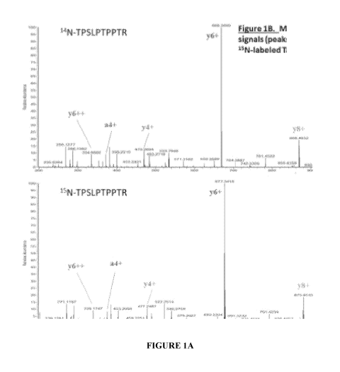
standard curve. To those persons skilled in the art, Figure 1B details the
mass fragmentation
pattern obtained from a select endogenous Tau peptide, the mass fragmentation
pattern
obtained from the corresponding peptide present in the 15N-Tau-441
Quantitation Internal
Standard, and the relative abundance (Gaussian peaks or signals) obtained by
mass
spectrometry for several endogenous and 15N-labeled fragments (y- and a-ions).
In this
aspect, the mass fragmentation pattern specifically identifies the amino acid
sequence for this
Tau peptide, and the relative abundance signals for the known concentration of
the 15N-
labeled fragments are used to calculate the concentration of the endogenous
14N-Tau peptide.
[0014] Figure 2 shows the entire sample and standard preparation,
processing, and
analysis workflow.
DETAILED DESCRIPTION OF THE INVENTION
[0015] The present invention is based, in part, on the principle that
stable isotope labeled
proteins and peptides have a known, slightly greater molecular weight than
their
4
CA 03052971 2019-08-07
WO 2018/148593 PCT/US2018/017691
corresponding endogenous proteins and peptides, but they have identical
physical or chemical
properties, behave the same way in a mass spectrometer, except for their
greater mass, which
makes them ideal quantitative internal standards. Using the techniques
provided herein,
endogenous proteins, peptide fragments, and proteoforms are quantified and can
be used to
diagnose and/or treat a subject having or at risk of developing a neurological
or
neurodegenerative disorder. Accordingly, the present invention provides
methods and kits
useful for calculating the concentration of one or more proteins, peptide
fragments, and
proteoforms of interest in a subject.
[00161 The invention also provides a method to assess whether a therapeutic
intervention
affects the concentration of proteins, peptide fragments, and proteoforms in
the subject,
where the biomolecules are relevant to neurological or neurodegenerative
diseases.
Accordingly, the method may be used to determine the optimal doses and/or
optimal dosing
regimens of the therapeutic intervention. Additionally, the method may be used
to determine
which subjects respond better to a particular therapeutic intervention. For
example, subjects
with high protein, peptide fragment, proteoform concentrations may respond
better to one
therapeutic agent, whereas subjects with normal concentrations may be at lower
risk for
developing a neurodegenerative disorder and are not eligible to enroll in
clinical trials of
experimental therapeutic agents or interventions. Alternatively, subjects with
one particular
genotype or proteotype may respond better to a particular therapeutic agent
than those with a
different genotype or proteotype. Finally, by allowing isoform specific
quantitation, the
method may be used to determine whether a therapeutic agent can modulate the
relative
concentration of one isoform to another isoform of the same protein.
100171 As used in this specification and the appended claims, the singular
forms "a", "an",
and "the" include plural references unless the context clearly dictates
otherwise. Thus, for
example, references to "the method" includes one or more methods, and/or steps
of the type
described herein which will become apparent to those persons skilled in the
art upon reading
this disclosure and so forth.
100181 Unless defined otherwise, all technical and scientific terms used
herein have the
same meaning as commonly understood by one of ordinary skill in the art to
which this
invention belongs. Although any methods and materials similar or equivalent to
those
described herein can be used in the practice or testing of the invention, the
preferred methods
and materials are now described.
100191 The term "subject" as used herein refers to any individual or
patient to which the
subject methods are performed. Generally, the subject is human, although as
will be
CA 03052971 2019-08-07
WO 2018/148593 PCT/US2018/017691
appreciated by those in the art, the subject may be an animal. Thus, other
animals, including
mammals such as rodents (including mice, rats, hamsters and guinea pigs),
cats, dogs, rabbits,
farm animals including cows, horses, goats, sheep, pigs, etc., and primates
(including
monkeys, chimpanzees, orangutans and gorillas) are included within the
definition of subject.
In addition, the term "subject" may refer to a culture of cells, where the
methods of the
invention are performed in vitro to assess, for example, efficacy of a
therapeutic agent.
[0020] As used herein, the terms "sample" and "biological sample" refer to
any sample
suitable for the methods provided by the present invention. A sample of cells
used in the
present method can be obtained from tissue samples or bodily fluid from a
subject, or tissue
obtained by a biopsy procedure (e.g., a needle biopsy) or a surgical
procedure. In certain
embodiments, the biological sample of the present invention is a sample of
bodily fluid, e.g.,
cerebral spinal fluid (CSF), blood, plasma, urine, saliva, and tears.
[0021] As disclosed herein, stable isotope labeled Quantitation Internal
Standards have a
slightly higher molecular weight than their endogenous counterparts, but does
not alter the
physical or chemical properties of the proteins, peptide fragments, and
proteoforms. Thus,
these biomolecules and their stable isotope labeled counterparts will bind to
antibodies and
elute off a liquid chromatography column in an identical fashion. Sensitive
instruments, such
as mass spectrometers, provide the ability to measure small differences in
mass between
labeled and unlabeled biomolecules.
[0022] Accordingly, in one aspect, the invention provides a method of
calculating the
concentration of a biomolecule in a subject. In one embodiment, the method
includes
contacting a sample from the subject with a Quantitation Internal Standard. As
used herein, a
"Quantitation Internal Standard" refers to a known concentration of a stable
isotope labeled
biomolecule, which has a distinct molecular weight from other labeled or
unlabeled
biomolecules that may exist in the sample. Thereafter, a sensitive measuring
device, such as
a mass spectrometer, a tandem mass spectrometer, or a combination of both, is
used to
measure the ratio of labeled to unlabeled biomolecules. Since the physical
properties of the
labeled and unlabeled biomolecules are identical, the ratio measured by the
mass
spectrometer is identical to the ratio in the original sample. Thus, by adding
a known amount
of one or more biomolecules, each labeled with a unique isotopic label, the
invention
provides the ability to quantitate the amount of those biomolecules that have
different
isotopic composition.
[0023] As used herein, the term "biomolecule" refers to any organic
molecule in a living
organism. Exemplary biomolecules include, but are not limited to proteins,
peptides,
6
CA 03052971 2019-08-07
WO 2018/148593 PCT/US2018/017691
proteoforms. In one embodiment, the biomolecule is a peptide, such as a
protein, that is
synthesized in the central nervous system (CNS) of the subject. Exemplary
proteins that can
be measured by the methods of the invention include, but are not limited to,
Tau and post-
translationally modified such as phospho-Tau (associated with Alzheimer's
Disease). In one
embodiment, the protein whose in vivo concentration is measured may be Tau or
its variants
or isoforms. Exemplary isoforms of Tau whose concentrations may be measured
include, but
are not limited to, the following phosphorylated or unphosphorylated isoforms
of Tau: Tau-
4R2N, Tau-4R1N, Tau-4RON, Tau-3R2N, Tau-3R1N, Tau-3RON. The following shows a
multiple sequence alignment of the 6 different isoforms of Tau.
7
SEQ ID Nos.
1 441aa 4R/2N
MAEPRQEFEVMEDHAGTYGLGDRICDQGGYTMHQDQEGDTDAGLICESPLQTPTEDGSEEPG 60
0
2 412aa 4R/1N
MAEPRQEFEVMEDHAGTYGLGDRICDQGGYTMTIQDQEGDTDAGLICESPLQTPTEDGSEEPG 60
b.)
o
3 383aa 4R/ON MAEPRQEFEVMEDHAGTYGLGDRICDQGGYTMHQDQEGDTDAGLK
44
co
-...
4 410aa 3R/2N
MAEPRQEFEVMEDHAGTYGLGDRICDQGGYTMTIQDQEGDTDAGLICESPLQTPTEDGSEEPG 60
4.
00
381aa 3R/1N MAEPRQEFEVMEDHAGTYGLGDRICDQGGYTMHQDQEGDTDAGLICESPLQTPTEDGSEEPG
60 en
c..)
6 352aa 3R/ON MAEPRQEFEVMEDH AGTYGLGDRICDQGGYTMFIQDQEGDTDAGLK
44
********************************************
7 441aa
SETSDAKS'TPTAEDVTAPLVDEGAPGKQAAAQPH'TEIPEGTTAEEAGIGDTPSLEDEAAG 120
8 412aa SETSDAKSTPTAE --------------- AEEAGIGDTPSLEDEAAG
91
9 383aa ----------------------------- AEEAGIGDTPSLEDEAAG
62
410aa SETSDAKSTPTAEDVTAPLVDEGAPGKQAAAQPHTEIPEGTTAEE AGIGDTPSLEDE AAG
120
11 381aa SETSDAKS'TPTAE -------------- AEEAGIGDTPSLEDEAAG
91
0
12 352aa ----------------------------- AEEAGIGDTPSLEDEAAG
62 e
0
******************
tx
ce 13 441aa
HVTQARMVSKSKDGTGSDDICKAKGADGKTKIATPRGAAPPGQKGQANATRIPAKTPPAPK 180
.4
F.
14
14 412aa
HVTQARMVSKSICDGTGSDDKKAKGADGKTKIATPRGAAPPGQKGQANA'TRIPAKTPPAPK 151
0
p.
,
383aa HVTQARMVSKSKDGTGSDDICKAKGADGKTK IATPRGAAPPGQKGQANATRIPAKTPPAPK
122 ,
,
0
16 410aa
HVTQARMVSKSICDGTGSDDICKAKGADGKTKIATPRGAAPPGQKGQANATRIPAKTPPAPK 180
.4
17 381aa HVTQARMVSKSKDGTGSDDICKAKGADGKTKIATPRGAAPPGQKGQANATR
IPAKTPPAPK 151
18 352aa
HVTQARMVSKSICDGTGSDDICKAKGADGKTKIATPRGAAPPGQKGQANATRIPAKTPPAPK 122
************************************************************
19 441aa
TPPSSGEPPKSGDRSGYSSPGSPGTPGSRSRTPSLPTPPTREPKKVAVVRTPPKSPSSAK 240
412aa TPPSSGEPPKSGDRSGYSSPGSPGTPGSRSRTPSLPTPPTREPKKVAVVRTPPKSPSSAK
211
21 383aa
TPPSSGEPPKSGDRSGYSSPGSPGTPGSRSRTPSLPTPPTREPKKVAVVRTPPKSPSSAK 182
v
en
22 410aa
TPPSSGEPPKSGDRSGYSSPGSPGTPGSRSRTPSLPTPPTREPKKVAVVRTPPKSPSSAK 240
13
cil 23 381aa
TPPSSGEPPKSGDRSGYSSPGSPGTPGSRSRTPSLPTPPTREPICKVAVVRTPPKSPSSAIC 211
24 352aa
TPPSSGEPPKSGDRSGYSSPGSPGTPGSRSRTPSLPTPPTREPKKVAVVRTPPKSPSSAK 182
o
I¨.
ce
o
I¨.
441aa SRLQTAPVPMPDLKNVKSKIGSTENLKHQPGGGKVQIINICKLDLSNVQSKCGSICDNIICHV
300 --.1
cr.
%.,
26 4 I.2aa
SRLQTAPVPMPDLKNVKSKIGS'TENI,KHQPGGGKVQIINICKLDLSNVQSKCGSICDNIICHV 271
I¨.
27 383aa
SRLQTAPVPMPDLKNVKSKIGS'TENLKHQPGGGKVQIINICKLDLSNVQSKCGSICDNIKHV 242
28 410aa SRLQTAPVPMPDLKNVKSKIGSTENLKHQPGGGK
274
0
29 381aa SRLQTAPVPMPDLKNVKSKIGSTENLKHQPGGGK
245 k..)
o
30 352aa SRLQTAPVPMPDLKNVKSKIGSTENLKHQPGGGK
216
co
--.
4.
00
31 441aa
PGGGSVQIVYKPVDLSKVTSKCGSLGN1HRKPGGGQVEVKSEKLDFICDRVQSKIGSLDNI 360
en
ca
32 412aa
PGGGSVQIVYKPVDLSKVTSKCGSLGNIHIIKPGGGQVEVKSEKLDFKDRVQSKIGSLDNI 331
33 383aa
PGGGSVQIVITKPVDLSKVTSKCGSLGNIFIHKPGGGQVEVKSEKLDFICDRVQSKIGSLDNI 302
34 410aa
VQIVYKPVDLSKVTSKCGSLGNIFIIIKPGGGQVEVKSEKLDFICDRVQSKIGSLDNI 329
35 38 1 aa
VQIVYKPVDLSKVTSKCGSLGNIFIHKPGGGQVEVKSEKLDFKDRVQSKIGSLDNI 300
36 352aa
VQIVYKPVDLSKVTSKCGSLGNIFIIIKPGGGQVEVKSEKLDFICDRVQSKIGSLDNI 271
*******************************************************
37 441aa
THVPGGGNICKIETHKLTFRENAKAKTDHGAEIVYKSPVVSGDTSPRHLSNVSSTGSIDMV 420
0
38 412aa
THVPGGGNICK1ETHKLTFRENAKAKTDHGAEIVYKSPVVSGDTSPRHLSNVSSTGSIDMV 391
e
0,
0
39 383aa
THVPGGGNKKIETHKLTFRENAKAKTDHGAEIVYKSPVVSGDTSPRHLSNVSSTGSIDMV 362
ui
h,
µ,0 40 410aa THVPGGGNKK
IETHKLTFRENAKAKTDHGAEIVYKSPVVSGDTSPRHLSNVSSTGSIDMV 389
.1
F.
h,
41 381aa
THVPGGGNKKIETHKLTFRENAKAKTDHGAEIVYKSPVVSGDTSPRHLSNVSSTGSIDMV 360
0
p.
,
42 352aa THVPGGGNICK1ETHKLTFRENAKAKTDHGAEIVYKSPVVSGDTSPR1-
IISNVSSTGSIDMV 331 0,
,
0
43 441aa DSPQLATLADEVSASLAKQGL
441
44 412aa DSPQLATLADEVSASLAKQGL
412
45 383aa DSPQLATLADEVSASLAKQGL
383
46 410aa DSPQLATLADEVSASLAKQGL
410
47 381aa DSPQLATLADEVSASLAKQGL
381
48 352aa DSPQLATLADEVSASLAKQGL
352 v
en
*********************
13
r)
o
I¨.
ce
--.
o
I¨.
--.1
cr.
µ,0
I¨.
CA 03052971 2019-08-07
WO 2018/148593 PCT/US2018/017691
100241 By way of example and not limitation, it is noted that several unique
isoforms of
Tau exist in CSF and plasma, and that these isoforms can be post-
translationally modified in
several ways including phosphorylation. Trypsin digestion of Tau yields
several peptides
which may or may not be unique to each isoform, see Table 1. Thus,
quantitation of some of
these peptides allows for calculation of the concentration of these isoforms
in the original
biological fluid.
includes Plasm
Tau 441 CSF CSF
SEQ a
Plasma
Residues Isofor LLOQ Std
ID Table 1. Peptide Sequence LLOQ
Std
With ms (ng/ Curve
NO. (ng/
Curve R2
Cleavage mt.) R2
Residues mt.}
49 R.QEFEVMEDHAGTYGLGDR.K 6-23 Total 0.08 0.9987 ad
50 R. KIX)GGYTMIKMEG DTDAGLKE 24-44 Total 0.08 0.9999 ad
51 K.ESPLQTPTEDGSEEPGSETSDAK.S 45-67 I N/2N 0.03 0.9932
0.05 0.9932
52 K.STPTAED VTAPL %%DEG APGK.Q 68-87 2N
0.03 0.9956 0.05 1 0.9956
53 K. QAAAQP1frE} PEGTTAEEAGIGDIPSLEDE AA G VrQA R.M 88-126 2N
0.03 0.9929 0.05 0.9397
54 K. lATPR..G 151-155 Total 0.03
0.9979 2.5 0.8603
55 R.GAAPPGQK.G 156-163 Total ad --
2.5 0.9937
56 KIPPAPK.T 175-180 Total 0.25 0.9982 ad
--
57 K.TPPSSGEPPK.S 181-190 Total 0.03
0.9948 I tid --
58 R.SGYSSPGSPGTPGSR.S 195-209 Total 0.003 0.9974
0.05 0.9937
59 R.TPSLPTPPTR.E 212-221 Total 0.003 0.9994
0.05 0.9994
60 R.IPSI,FIPPTREPK.K 212-224 Total 0.5
09988 I 2.5 0.9941
61 K.IGSTENLK.11 260-267 Total 0.03 0.9886 0.05
0.9988
62 K.VQ1ENK.K 275-280 4R 0.01 0.9831 1
0.5 0.9967
63 K. VQ1VYKPVI)LSK.V 275-286 3R 10 ad 10
ad
64 K.LDLSNVQSK.C. 282-290 4R
0.05 0.9910 0.05 0.9969
65 K.H VPGGGSVQ1VYKPVDLSK. 299-317 4R 0.05 0.9999 0.05
0.9883
66 K .1G SID Null VPGGG NK.K 354-369 Total 0.05
0.999 0.05 0.9977
67 K.TDI-IGAEIVYK.S 386-395 Total 0.1
0.9985 0.05 I 0.9710
68 K.SPVVSGDTSPR.H 396-406 Total 0.05 0.9952 0.5
0.9819
[00251 As such, the methods provide the ability to measure concentrations
of various
isoforms of Tau, such as fragments produced after digestion with an
endoprotease (e.g.,
tfypsin, 1.,ysN, or V8 protease). Exemplary fragments of Tau isoforms include
but are not
limited to regions of Tau that are different between the different isoforms
and their
boundaries, such as the N-terminal region (2N/1N/ON) and the C-terminal repeat
region
(4R/3 R).
[00261 As used herein, the term "nucleic acid" refers to DNA, RNA, single-
stranded,
double-stranded or triple stranded and any chemical modifications thereof
Virtually any
modification of the nucleic acid is contemplated. A "nucleic acid molecule"
can be of almost
any length, from 1.0, 20, 30, 40, 50, 60, 75, 100, 125, 150, 175, 200, 225,
250, 275, 300, 400,
500, 600, 700, 800, 900, 1000, 1500, 2000, 2500, 3000, 3500, 4000, 4500, 5000,
6000, 7000,
8000, 9000, 10,000, 15,000, 20,000, 30,000, 40,000, 50,000, 75,000, 100,000,
150,000,
CA 03052971 2019-08-07
WO 2018/148593 PCT/US2018/017691
200,000, 500,000, 1,000,000, 1,500,000, 2,000,000, 5,000,000 or even more
bases in length,
up to a full-length chromosomal DNA molecule.
[0027] The terms "polypeptide," "peptide" and "protein" are used
interchangeably herein
to refer to two or more amino acid residues joined to each other by peptide
bonds or modified
peptide bonds, i.e., peptide isosteres. The terms apply to amino acid polymers
in which one
or more amino acid residue is an artificial chemical mimetic of a
corresponding naturally
occurring amino acid, as well as to naturally occurring amino acid polymers,
those containing
modified residues, and non-naturally occurring amino acid polymer.
"Polyeptide" refers to
both short chains, commonly referred to as peptides, oligopeptides or
oligomers, and to
longer chains, generally referred to as proteins. Polypeptides may contain
amino acids other
than the 20 gene-encoded amino acids. Likewise, "protein" refers to at least
two covalently
attached amino acids, which includes proteins, polypeptides, oligopeptides and
peptides. A
protein may be made up of naturally occurring amino acids and peptide bonds,
or synthetic
peptidomimetic structures. Thus "amino acid", or "peptide residue", as used
herein means
both naturally occurring and synthetic amino acids. For example, homo-
phenylalanine,
citrulline and noreleucine are considered amino acids for the purposes of the
invention.
"Amino acid" also includes imino acid residues such as proline and
hydroxyproline. The side
chains may be in either the (R) or the (S) configuration.
[0028] Several different moieties may be used to label the biomolecule of
interest.
Generally speaking, the two types of labeling moieties utilized in the method
of the invention
are radioactive isotopes and non-radioactive (stable) isotopes. In one
embodiment, non-
radioactive isotopes may be used and measured by mass spectrometry. Preferred
stable
isotopes include deuterium (2H), 1.3c, 15N, 17 or 180, and 33, 34. or 36",
but it is recognized that a
number of other stable isotopes that change the mass of an atom by more or
less neutrons
than is seen in the prevalent native form would also be effective. A suitable
label generally
will change the mass of the biomolecule under study such that it can be
detected in a mass
spectrometer. In one embodiment, the biomolecule to be measured may be a
peptide or
protein, and the labeled moiety may be an amino acid comprising a non-
radioactive isotope
(e.g., 13C). In another embodiment, the biomolecule to be measured may be a
nucleic acid,
and the labeled moiety may be a nucleoside triphosphate comprising a non-
radioactive
isotope (e.g., 15N). Alternatively, a radioactive isotope may be used, and the
labeled
biomolecules may be measured with a scintillation counter (or via nuclear
scintigraphy) as
well as by a mass spectrometer. One or more labeled moieties may be used
simultaneously
or in sequence.
11
CA 03052971 2019-08-07
WO 2018/148593 PCT/US2018/017691
[0029] Thus, in one embodiment, when the method is employed to measure the
concentration of proteins, the labeled moiety typically will be an amino acid.
Those of skill
in the art will appreciate that several amino acids may be used to provide the
label of
biomolecules. Generally, the choice of amino acid is based on a variety of
factors such as: (1)
The amino acid generally is present in at least one residue of the protein or
peptide of interest.
(2) The amino acid is generally able to reach the site of protein production
and rapidly
equilibrate tissue or cellular barriers. And (3) commercial availability of
the desired amino
acid (i.e., some amino acids are much more expensive or harder to manufacture
than others).
100301 In one embodiment, the amino acid is an essential amino acid (not
produced by the
body), so that a higher percent of labeling may be achieved. In another
embodiment, the
amino acid is a non-essential amino acid. Exemplary amino acids include, but
are not limited
to, leucine, isoleucine, and phenylalanine. As such, in one embodiment, the
labeled amino
acid is one or more of a 15N-labeled amino acid, a '3C-labeled phenylalanine,
where x = 1 to
9, a '3C-labeled isoleucine, where x = 1 to 6. For example, 13C6-
phenylalanine, which
contains six 13C atoms, may be used to label a biomolecule of interest (e.g.,
a CNS derived
protein). In another embodiment, 13C6-leucine may be used to label a
biomolecule of interest
(e.g., a CNS derived protein). In yet another embodiment, 13C6-leucine is used
to label
amyloid-beta (AP).
[0031] There are numerous commercial sources of labeled amino acids, both non-
radioactive isotopes and radioactive isotopes. Generally, the labeled amino
acids may be
produced either biologically or synthetically. Biologically produced amino
acids may be
obtained from an organism (e.g., kelp/seaweed) grown in an enriched mixture of
13C, 15N, or
another isotope that is incorporated into amino acids as the organism produces
proteins. The
amino acids are then separated and purified. Alternatively, amino acids may be
made with
known synthetic chemical processes.
[0032] Once disease is established and a treatment protocol is initiated,
the methods of the
invention may be repeated on a regular basis to monitor the concentration(s)
of
biomolecule(s) of interest in the subject. The results obtained from
successive assays may be
used to show the efficacy of treatment over a period ranging from several days
to months.
Accordingly, another aspect of the invention is directed to methods for
monitoring a
therapeutic regimen for treating a subject having a neurological or
neurodegenerative
disorder. A comparison of the concentration(s) of biomolecule(s) of interest
prior to and
during therapy will be indicative of the efficacy of the therapy. Therefore,
one skilled in the
art will be able to recognize and adjust the therapeutic approach as needed.
12
CA 03052971 2019-08-07
WO 2018/148593 PCT/US2018/017691
100331 The method of the invention provides that a sample be obtained from the
subject
such that the in vivo concentration of one or more biomolecules of interest
can be determined.
In one embodiment, the sample is a body fluid. Suitable body fluids include,
but are not
limited to, cerebral spinal fluid (CSF), blood plasma, blood serum, urine,
saliva, perspiration,
and tears. It should be understood that biological fluids typically contain a
multitude of
quantifiable biomolecules. For example, where the sample is CSF, exemplary
biomolecules
that can be quantified include, but are not limited to, Tau, variants of Tau,
amyloid-beta
protein, variants of amyloid-beta protein (AP), digestion products of amyloid-
beta protein,
amyloid precursor protein (APP), apolipoprotein E, apolipoprotein J, alpha-
synuclein, or any
combination thereof. In another embodiment, the sample is a tissue sample,
such as a sample
of tissue from the central nervous system (CNS). The sample generally will be
collected
using standard procedures well known to those of skill in the art.
100341 In one embodiment, the sample is a CNS sample, which includes, but
is not limited
to, tissue from the central nervous system, which comprises brain tissue and
spinal cord
tissue. In one embodiment of the invention, the CNS sample may be taken from
brain tissue,
including, but not limited to, tissue from the forebrain (e.g., cerebral
cortex, basal ganglia,
hippocampus), the interbrain (e.g., thalamus, hypothalamus, subthalamus), the
midbrain (e.g.,
tectum, tegmentum), or the hindbrain (e.g., pons, cerebellum, medulla
oblongata). In another
embodiment, the CNS sample may be collected from spinal cord tissue. In still
other
embodiments, CNS samples from more than one CNS region may be taken.
Accordingly, the
concentration of a biomolecule of interest may be measured in different CNS
samples, e.g., in
the cortex and the hippocampus, simultaneously.
100351 CNS samples may be obtained by known techniques. For instance, brain
tissue or
spinal cord tissue may be obtained via dissection or resection. Alternatively,
CNS samples
may be obtained using laser microdissection. The subject may or may not have
to be
sacrificed to obtain the sample, depending on the CNS sample desired and the
subject
utilized.
100361 In general, when the biomolecule under study is a peptide or
protein, the invention
provides that a first sample may be taken from a subject prior to
administration of the
therapeutic agent to provide a baseline concentration. After administration of
the therapeutic
agent, one or more samples are obtained from the subject. As will be
appreciated by those of
skill in the art, the number of samples and when the samples are taken
generally will depend
upon a number of factors such as: the type of analysis, type of
administration, the protein of
interest, the rate of metabolism, the type of detection, and the type of
subject.
13
CA 03052971 2019-08-07
WO 2018/148593 PCT/US2018/017691
100371 It
should be understood that if samples at different time-points are desired,
more
than one subject may be used. For instance, one subject may be used for a
baseline sample,
another subject for a time-point of one-hour post administration of the
therapeutic agent,
another subject for a time-point six hours post administration of the
therapeutic agent.
100381
Accordingly, the present invention provides that detection of the amount of
labeled
biomolecule and the amount of unlabeled biomolecule in the sample may be used
to
determine the ratio of labeled biomolecule to unlabeled biomolecule, which in
turn, may be
used to calculate the concentration of the biomolecule of interest in the
subject. In one
embodiment, the ratio is determined by means of detecting changes in mass of
the labeled
biomolecule (e.g., peptide or protein) with respect to the unlabeled
biomolecule. Exemplary
means for detecting differences in mass between the labeled and unlabeled
biomolecules
include, but are not limited to, liquid chromatography mass spectrometry, gas
chromatography mass spectrometry, MALDI-TOF mass spectrometry, and tandem mass
spectrometry.
100391
However, prior to detecting the ratio of labeled biomolecule to unlabeled
biomolecule, it may be desirable to isolate and/or separate the biomolecule of
interest from
other biomolecules in the sample. Thus, in one embodiment, immunoprecipitation
may be
used to isolate and purify the biomolecule (e.g., peptide or protein) of
interest before it is
analyzed. In another embodiment, the biomolecule of interest may be isolated
or purified by
affinity chromatography or immunoaffinity chromatography.
Alternatively, mass
spectrometers having chromatography setups may be used to separate
biomolecules without
immunoprecipitation, and then the biomolecule of interest may be measured
directly. In an
exemplary embodiment, the protein of interest may be immunoprecipitated and
then analyzed
by a liquid chromatography system interfaced with a tandem MS unit equipped
with an
electrospray ionization source (LC-ESE-tandem MS). One example of using 3
different
antibodies to immunoprecipitate Tau protein from 3 aliquots of the same human
CSF sample,
followed by digestion of Tau protein, and quantitation of the relative
abundance of different
Tau peptides is shown in Table 2. This aspect includes the observation that
different
antibodies have different affinity for different portions of the Tau protein,
and the selection of
immunoprecipitation antibody can be used to focus the quantitative analysis on
different
14
CA 03052971 2019-08-07
WO 2018/148593
PCT/US2018/017691
portions or proteoforrns of the Tau biomolecule.
SEQ
ID Table 2. Peptide Sequence (Residue) HJ8.5 HJ8.7 Tau12
Nos. __
69 QEFEVMEDHAGTYHLI1DR (6-23) \t\\,
0.68 \ MIO
70 KDQGGYTMHQDQEGDTDAGLK (24-44) 0.84 t
71 ESPLQTPTEDFSEEPGSETSDAK (45-67) 0.74
11111111111101i921111111111111
72 STPTAEDVTAPLVDEGAPGK (68-87)
73
QAAAQPHTEIPEGTTAEEAGIGDTPSLEDEAAGIWTQAR \, 0.59
74 1ATPR (151-155) 0.55
75 TPPAPK (175-180) = _______
0.97 0.99
76 TPPSSGEPPK (181-190) 0.52
iial,t,,tmAnt!:;.X.
77 SGYSSPGSPGTPGSR (195-209) 0.83 0.80
78 TPSLPTPPTR (212-221) aµ
79 TF'SLPTPPTREPK (212-224) 0.94 0.94 \
&A
80 IGSTENLK (260-267) 0. 86
81 VQIINK (275-280) 016
82 LDLSNVWSK (282-290)
83 FIVPGGGSVQICYKPVDLSK (299-316) 0.83
84 1GSLDNITHVPGGGNK (354-369) 0.61 0.86
85 TDHGAEIVYK (386-395) 0.68
86 SPINSGDTSPR (396-406) )42
Peptide Intensity
*Values are reported as a fraction of the abundance for the immune-
precipitation
antibody that provided the highest abundance (1.0) for that specife Tau
peptide
100401 In
another aspect, the invention provides that multiple biomolecules in the same
sample may be measured simultaneously. That is, both the amount of unlabeled
and labeled
biomolecule may be detected and measured separately or at the same time for
multiple
biomolecules. As such, the invention provides a useful method for screening
changes in
concentration, of one or more biomolecules on a large scale
proteomics/metabolomics)
and provides a sensitive means to detect and measure biomolecules involved in
the
underlying pathophysiology. In this aspect, the invention also provides a
means to measure
multiple types of biomolecules. In this context, for example, a protein and a
lipid may be
measured simultaneously or sequentially.
100411
Once the amount of labeled and unlabeled biomolecule has been detected in a
sample, the ratio or percent of labeled biomolecule to unlabeled biomolecule
may be
determined. Thereafter, the concentration of the unlabeled biomolecule in the
sample can be
CA 03052971 2019-08-07
WO 2018/148593 PCT/US2018/017691
determined. In other words, since a known amount of labeled biomolecule is
added to an
unknown amount of biomolecules and the ratio of labeled to unlabeled is
measured, the
concentration of the unlabeled biomolecules can be calculated from the ratio
as follows:
(i) Concentration of unlabeled = (ratio of unlabeled to labeled) x
(concentration of labeled).
The equation may be simplified as:
(ii) Concentration of unlabeled = (ratio of unlabeied.Quantitation Internal
Standard) x
(concentration of Quantitation Internal Standard).
[0042] Conversely, if a known amount of unlabeled is added to an unknown
amount
labeled the concentration of the labeled can be calculated as follows:
(iii) Concentration of labeled = (ratio of labeled to unlabeled) x
(concentration of unlabeled).
[0043] In addition, if a known amount of biomolecule 1, labeled with label
1, is added to
an unknown amount of biomolecule 2, labeled with label 2, the concentration of
the
biomolecule 2 can be calculated as follows:
(iv) Concentration of label 2 = (ratio of label 2 to label 1) x (concentration
of label 1).
[0044] Similarly, if a known amount of biomolecule 1, labeled with label 1,
is added to an
unknown amount of biomolecule 2, labeled with label 2, and biomolecule 3,
labeled with
label 3, the concentration of the biomolecule 2 and biomolecule 3 can be
calculated as
follows:
(v) Concentration of label 2 = (ratio of label 2 to label 1) x (concentration
of label 1)
(vi) Concentration of label 3 = (ratio of label 3 to label 1) x (concentration
of label 1).
[0045] Finally, if a known amount of biomolecule 1, labeled with label 1,
is added to an
unknown amount of biomolecule 2, labeled with label 2, and an unknown amount
of
unlabeled biomolecule 3, the concentration of the biomolecule 2 and unlabeled
biomolecule
can be calculated as follows:
(vii) Concentration of label 2 = (ratio of label 2 to label 1) x
(concentration of label 1)
(viii) Concentration of unlabeled = (ratio of unlabeled to label 1) x
(concentration of label 1).
[0046] In another embodiment, the methods further include the step of
normalizing the
calculated concentration to a standard curve based on the curve fitting
equation generated by
the standard curve. The standard curve used herein is generated by determining
two or more
ratios of unlabeled biomolecules to their respective Quantitation Internal
Standards, where
the concentration of the unlabeled biomolecule of interest is known.
[0047] In another aspect, the invention allows measurement of the labeled
and unlabeled
protein at the same time, so that the ratio of labeled to unlabeled protein,
as well as other
calculations, may be made.
16
CA 03052971 2019-08-07
WO 2018/148593 PCT/US2018/017691
[0048] In one aspect, Tau is isolated from the biologic samples by
immunoprecipitation
using an antibody that recognizes Tau. In this embodiment, the isolated
peptides are eluted
from the antibody, for example by using formic acid and then digested with
trypsin or another
protease. This invention measures the concentration of Tau peptides.
[0049] The term "antibody" as used in this invention is meant to include
intact molecules
of polyclonal or monoclonal antibodies, as well as fragments thereof, such as
Fab and F(ab)2,
Fv and SCA fragments which are capable of binding an epitopic determinant. The
term
"specifically binds" or "specifically interacts," when used in reference to an
antibody means
that an interaction of the antibody and a particular epitope has a
dissociation constant of at
least about 1 x 1O, generally at least about 1 x 10⁴, usually at least about 1
x 10-8, and
particularly at least about 1 x 10.9 or 1 x 10⁻¹⁰ or less.
[0050] The method of the invention may be used to diagnose or monitor the
progression
of a neurological or neurodegenerative disease by measuring the in vivo
concentration of one
or more biomolecules of interest in a subject. Additionally, the methods of
the invention may
be used to monitor the treatment of a neurological or neurodegenerative
disease by measuring
the in vivo concentration of a biomolecule of interest in a subject. The
concentration of the
biomolecule may be linked to a neurological or neurodegenerative disease such
that any
increase or decrease may be indicative of the presence or progression of the
disease. Thus,
the calculated concentration of one or more biomolecules of interest may be
compared to the
concentration of the same biomolecules in a corresponding normal sample, to
the
concentration of the same biomolecules in a subject of known neurological or
neurodegenerative disease state, to the concentration of the same biomolecules
from the same
subject determined at an earlier time, or any combination thereof.
[0051] In addition, such methods may help identify an individual as having
a
predisposition for the development of the disease or may provide a means for
detecting the
disease prior to the appearance of actual clinical symptoms. A more definitive
diagnosis of
this type may allow health professionals to employ preventative measures or
aggressive
treatment earlier thereby preventing the development or further progression of
the disease.
[0052] As used herein a "corresponding normal sample" refers to a sample from
the same
organ and/or of the same type as the sample being examined. In one aspect, the
corresponding normal sample comprises a sample of cells obtained from a
healthy individual.
Such a corresponding normal sample can, but need not be, from an individual
that is age-
matched and/or of the same sex as the individual providing the sample being
examined. In
another aspect, the corresponding normal sample comprises a sample of cells
obtained from
17
CA 03052971 2019-08-07
WO 2018/148593 PCT/US2018/017691
an otherwise healthy portion of tissue of the subject from which the sample
being tested is
obtained.
[0053] Reference to the concentration of biomolecules in a subject of known
neurological
or neurodegenerative disease state includes a predetermined concentration of a
biomolecule
linked to a neurological or neurodegenerative disease. Thus, the concentration
may be
compared to a known concentration of biomolecules obtained from a sample of a
single
individual or may be from an established cell line of the same type as that of
the subject. In
one aspect, the established cell line can be one of a panel of such cell
lines, wherein the panel
can include different cell lines of the same type of disease and/or different
cell lines of
different diseases associated with the same biomolecule. Such a panel of cell
lines can be
useful, for example, to practice the present method when only a small number
of cells can be
obtained from the subject to be treated, thus providing a surrogate sample of
the subject's
cells, and also can be useful to include as control samples in practicing the
present methods.
[0054] Exemplary neurological or neurodegenerative diseases that may be
linked to the
concentration ranges of biomolecules of interest include, but are not limited
to, Alzheimer's
Disease, Pick's Disease, Parkinson's Disease, stroke, frontal temporal
dementias (FTDs),
Huntington's Disease, progressive supranuclear palsy (PSP), corticobasal
degeneration
(CBD), aging-related disorders and dementias, Multiple Sclerosis, Prion
Diseases (e.g.,
Creutzfeldt-Jakob Disease, bovine spongiform encephalopathy or Mad Cow
Disease, and
scrapie), Lewy Body Disease, schizophrenia, Amyotrophic Lateral Sclerosis (ALS
or Lou
Gehrig's Disease) or other motor neuron diseases, restless legs syndrome,
epilepsy or other
seizure disorders, tremors, depression, mania, anxiety disorders, brain trauma
or injury,
narcolepsy, insomnia or other sleep disorders, autism, normal pressure
hydrocephalus, pain
disorders or syndromes, migraines, cluster headaches or other forms of
headache,
spinocerebellar disorders, muscular dystrophies, myasthenia gravis, retinitis
pigmentosa or
other forms of retinal degeneration. It is also envisioned that the method of
the invention may
be used to study the normal physiology, metabolism, and function of the CNS.
[0055] In another aspect, the present invention provides a method for
assessing whether a
therapeutic agent used to treat a neurological or neurodegenerative disease
affects the
concentration of a biomolecule of interest in the subject. For example, the
concentration of
the biomolecule may be measured to determine if a given therapeutic agent
results in an
increase, or a decrease in the concentration of the biomolecule. In one
embodiment, the
method is performed in vivo, as herein described. In another embodiment, the
method is
performed in vitro utilizing a culture of cells, where the culture of cells is
the "subject" in the
18
CA 03052971 2019-08-07
WO 2018/148593 PCT/US2018/017691
methods described herein. Accordingly, use of the methods provided herein will
allow those
of skill in the art to accurately determine the degree of change in the
concentration of the
biomolecule of interest, and correlate these measurements with the clinical
outcome of the
disease modifying treatment. Results from this aspect of the invention,
therefore, may help
determine the optimal doses and frequency of doses of a therapeutic agent, may
assist in the
decision-making regarding the design of clinical trials, and may ultimately
accelerate
validation of effective therapeutic agents for the treatment of neurological
or
neurodegenerative diseases.
[0056]
Thus, the method of the invention may be used to predict which subjects will
respond to a particular therapeutic agent. For
example, subjects with increased
concentrations of a particular biomolecule may respond to a particular
therapeutic agent
differently than subjects with decreased concentrations of the biomolecule. In
particular,
results from the method may be used to select the appropriate treatment (e.g.,
an agent that
blocks the production of the biomolecule or an agent that increases the
clearance of the
biomolecule) for a particular subject. Similarly, results from the method may
be used to
select the appropriate treatment for a subject having a particular genotype.
[0057]
Those of skill in the art will appreciate that the therapeutic agent can and
will vary
depending upon the neurological or neurodegenerative disease or disorder to be
treated and/or
the biomolecule whose metabolism is being analyzed. In embodiments in which
the
biomolecule is Tau, non-limiting examples of suitable therapeutic agents
include Tau
metabolism modulators, Tau kinase inhibitors, cathepsin D inhibitors, Tau
autophagy
activators, and Tau aggregation inhibitors. Other suitable AD therapeutic
agents include
hormones, neuroprotective agents, and cell death inhibitors. Many of the above
mentioned
therapeutic agents may also affect the in vivo metabolism of other proteins
implicated in
neurodegenerative disorders. Furthermore, therapeutic agents that may affect
the in vivo
metabolism of synuclein include sirtuin 2 inhibitors, synuclein aggregation
inhibitors,
proteasome inhibitors, etc.
[0058] The
therapeutic agent may be administered to the subject in accordance with
known methods. Typically, the therapeutic agent will be administered orally,
but other routes
of administration such as parenteral or topical may also be used. The amount
of therapeutic
agent that is administered to the subject can and will vary depending upon the
type of agent,
the subject, and the particular mode of administration. Those skilled in the
art will appreciate
that dosages may be determined with guidance from Goodman & Goldman's The
19
CA 03052971 2019-08-07
WO 2018/148593 PCT/US2018/017691
Pharmacological Basis of Therapeutics, Tenth Edition (2001), Appendix II, pp.
475-493, and
the Physicians' Desk Reference.
[0059] It should be understood that the methods of the invention described
herein can be
adapted to a high throughput format, thus allowing the examination of a
plurality (i.e., 2, 3, 4,
or more) of samples and/or biomolecules, which independently can be the same
or different,
in parallel. A high throughput format provides numerous advantages. For
example, a high
throughput format allows for the examination/quantitation of two, three, four,
etc., different
biomolecules, alone or in combination, of a subject. Finally, a high
throughput format
allows, for example, control samples (positive controls and or negative
controls) to be run in
parallel with test samples. In addition, a high throughput method may allow
immunoprecipitation of multiple proteins at the same time using multiple
antibodies.
100601 In another aspect, the invention provides a kit for performing the
methods of the
invention. In one embodiment, a kit is provided for diagnosing and/or
monitoring the
progression or treatment of a neurological or neurodegenerative disease in a
subject. The kit
may further include an appropriate Quantitation Internal Standard and means
for obtaining a
biological sample at regular time intervals from the subject. In certain
embodiments, the kit
will also include instructions for detecting and determining the ratio of
labeled to unlabeled
biomolecules of interest over time and for calculating the concentration of
the endogenous
unlabeled biomolecule. In one embodiment, the instructions will disclose
methods for
comparing the calculated concentration to certain standards and/or controls as
disclosed
herein.
[0061] In another embodiment, the kit of the invention provides a
compartmentalized
carrier including one or more containers containing the Quantitation Internal
Standard and the
various means for performing the methods of the invention.
[0062] The following examples are provided to further illustrate the
advantages and
features of the present invention but are not intended to limit the scope of
the invention.
While they are typical of those that might be used, other procedures,
methodologies, or
techniques known to those skilled in the art may alternatively be used.
EXAMPLE
Quantitation of Tau by SISAQ
100631 15N labeled Tau was used as Quantitation Internal Standard and
spiked into a
standard curve of samples containing concentrations of Tau ranging from 0.05
pg/mL to 100
pg/mL. In addition, the Quantitation Internal Standard was spiked into CSF and
plasma from
two different individuals. Tau was isolated from the samples using
immunoprecipitation and
CA 03052971 2019-08-07
WO 2018/148593
PCT/US2018/017691
then digested with trypsin and analyzed by mass spectrometry. The ratio of
unlabeled
endogenous Tau to Quantitation Internal Standard was calculated for all
samples and a
standard curve generated. The standard curve was linear in the range tested
(0.05 pg/mL to
100 pg/mL) and was used to calculate the concentration of Tau in the CSF and
plasma
samples. The concentration of Tau was approximately 5 pg/mL in the CSF samples
and
around 0.1 pg/mL in plasma, and the intra- and inter-assay CV for triplicate
measures of CSF
and plasma Tau concentrations was less than 25%. Our data show that regardless
of the
concentration, Tau can be reliably and reproducibly measured using the
invention methods.
Examples of the intra-assay and inter-assay reliability (%CV) for quantifying
several Tau
peptides present in human CSF are shown in Tables 3 & 4. The intra-assay
reliability (Table
3) was calculated by triplicate analysis of a single pooled human CSF sample
where these
injection replicate analyses were dispersed among many other standard curve
samples and
unknown samples included in the same analytical run. The inter-assay
reliability (Table 4)
was calculated by triplicate analysis of a single human CSF pool that was
prepared and
analyzed on 3 separate days (process replicates) in different analytical runs
that included
many other samples and standards.
SEQ.
ID. Table 3. Incra-Assay Analysis Calculated Concentration (ng/mL)
Average
Concentration Standard (1..)CV
Nos.
Deviation
#s) .:Rdfillate 2 ReiiIi61-6-Y "g/11114
87 QUEVMEDIIAGTY111,111)R (6-23) 3.174 3.22() 3.111
3.168 0.055 1.7
88 KDQGGYTMHQDQEGDTDAGLK 04-44) 3.516 3.653 3.845 3.672
==::4.1g
89 ESPLQTPTEDFSEEPGSETSDAK (45-67) 1.738 1.885 1.852 1.825
0.078 4.2
90 ...STPTAEDVTAPLVD.EGAMERW) (.223 ft21202 15
niiNOOL 2.6
QAAAQPHTEIPEGTFAEEAGIGDTPSLE
91 0 174 0158 0. .
199 0177
0.020 11.5
DEAAGITVTQAR (88-126) . .
92 IATPR (13.1A415.)1 211.74:.: 2.190
1.315WRininirminiinA7S 256
93 TPPAPK (175-180) 1.317 1.499 1.598 1.471
0.142 9.7
94 Eng 1.551 .10153miniimiloo7isini
95 SGY SSPG'SPOTPGSR (195-209) 1.240
1.289 1.047 1.192 0.128 10.7
96 TPSLPTPPTR (212----:221"'" "1.191 1..259
SEQ.
ID. Table 3. Incra-Assay Analysis Calculated Concentration (ngind..)
Av erage Standard
Concentration
%CV
Nos. Do
iation
. . . . . . . . . . . . . . . ...= . . . ....
= - . . . . . .. . (ntni)
inEPOtitleSetwaliterftWitif0-40: =Itik 2 :MilM1)..:o =gi
97 QEFEVNIEDHAGTYELIIDR (6-23) 2.935 3.898 3.174 3.336
0.501 15.0
8 IUQGGYTMHQDQEGDThAGL& 2,419 3,W70 3,51&
2.935 0.35/ 18.8
4.4).Mgggggggggggggggggggggggg
99 1-3.SPLQTPTEDFSEI-3.K1SETSDAK (45-67) 1.488 2.001 1.718
1.743 0 25S I 4.S
ggIti(M OsitiVi4:A1EDVIMP1VDECIAPC5K. (68417 gggatM MD..274 0..221
0,238 0.031
QAAAQPHTEIPEGTTAEEAGIGDTPSLE
101 0 184 0218 0.174 0. .
192 0023 I 12.2
DEAAG1-IVTQAR (88-126) . .
* 102 11A.Mit151-l5) U0Z v58 7
2.074 1,654 0390 23
103 TPPAPK (175-180) 1.423 1.310 1 1.350
0.063 4.7
21
CA 03052971 2019-08-07
WO 2018/148593 PCT/US2018/017691
104 1.177 1.686 :::1:agg::1418.
1,427 0,255
105 SGYSSPGSPGTPGSR (195-209) I. 195 1.435 1.240
1.290 0.125 9.9
loo TPSDPITMat242Z10 1,004
0,015MRSAM
100641 This illustrates the feasibility of using stable isotope labeled Tau as
a quantitation
internal standard and relating the ratio of unlabeled to labeled Tau to a
standard curve to
allow for measurement of concentrations of Tau in unknown samples.
100651 Although the invention has been described with reference to the above
example, it
will be understood that modifications and variations are encompassed within
the spirit and
scope of the invention. Accordingly, the invention is limited only by the
following claims.
22