Note: Descriptions are shown in the official language in which they were submitted.
CA 03053002 2019-08-07
WO 2018/148469
PCT/US2018/017501
BIO/CHEMICAL MATERIAL EXTRACTION AND ASSAY
CROSS-REFERENCE TO RELATED APPLICATIONS
[0001] This application claims the benefit of U.S. Provisional
Application No. 62/456,552,
filed on February 8, 2017, U.S. Provisional Application No. 62/459,232, filed
on February 15,
.. 2017, and U.S. Provisional Application No. 62/463,578, filed on February
24, 2017, U.S.
Provisional Application No. 62/456,504, filed on February 8, 2017, U.S.
Provisional Application
No. 62/460,062, filed on February 16, 2017, and U.S. Provisional Application
No. 62/457,133,
filed on February 9, 2017, all of which are herein incorporated by reference
in their entireties for
all purposes.
FIELD OF THE DISCLOSURE
[0002] This disclosure relates generally to systems and methods of
performing bio/chemical
material extraction and assay.
BACKGROUND OF THE DISCLOSURE
[0003] In many chemical and/or biological assays and testing (e.g.
immunoassay, nucleotide
assay, blood panel analysis, etc.), there are needs for methods and devices
that can accelerate the
process and quantify the parameters (e.g. analyte concentration, the sample
volume, etc.),
simplify the sample collection and measurement processes, handle samples with
small volumes,
perform entire assays in a short amount of time (e.g. less than a minute),
allow results to be
analyzed automatically (e.g. by a mobile phone), and allow non-professionals
to perform the
assay her/himself. The present disclosure relates to the methods, devices, and
systems that uses
detection of electronic signals (electrical measurement) to address these
needs.
[0004] Among other things, early identification of coagulopathy has
important clinical
implications for managing patients who are critically ill, severely injured,
or on anticoagulation
therapy. Rapid and accurate assessments are essential to ensure that patients
prone to blood
clots¨as well as those who have difficulty clotting¨receive appropriate care
to their conditions.
1
CA 03053002 2019-08-07
WO 2018/148469
PCT/US2018/017501
Traditional tests (prothrombin time (PT) and activated partial thromboplastin
time (aPTT) test)
need to be conducted in a professional testing facility and require up to 10
mL blood.
Consequently, a simple and portable assay that is fast, easy to use, and/or
inexpensive is
desirable.
[0005] Further, in biological and chemical assays (e.g. diagnostic
testing), often a small chip
(e.g., 3mm x 3mm x0.5mm, Length x Height x Thickness) is used. In many
situations, it is
desirable to use hands to handle a sensing chip and a fluid sample to be
analyzed by the sensing
chip. When a sensing chip has a dimension small compared with the fingers of
hands, the sensing
chip can be difficult to be handled by the hands. Furthermore, when a fluid
sample is dropped on
a sensing chip that is small in dimension (e.g. a few millimeters in size),
the fluid sample can
overflow, making a mess. Moreover, when the fluid sample is deposited on a
chip, there are
needs to measure the volume, change the shape, and/or detect analytes of a
sample or a part of
the sample, quickly and simply.
SUMMARY OF THE DISCLOSURE
[0006] As discussed above, there are needs for methods and devices that can
accelerate the
process and quantify the parameters for bio/chemical material samples.
According to some
embodiments, the present disclosure describe a QMAX (Q: quantification; M:
magnifying; A:
adding reagents; X: acceleration) device having two or more electrodes that
accelerates the
electrical measurement process. In addition, among other things, the
electrical measurement
technology of the current disclosure can also be used for the extraction,
separation, and
purification of sample components, such as but not limited to nucleic acids.
For example, while
traditional nucleic acid extraction assays (e.g. ethanol precipitation and
phenol¨chloroform
extraction) in plates or tubes are complex, time-consuming, laborious and
requires lab setups and
significant amount of sample (typically > 100uL), extraction with the devices
and methods herein
discussed can overcome the shortcomings discussed above.
[0007] In some embodiments, the present disclosure describes devices,
systems, and methods
of a QMAX device having a plate for hosting a small sensing chip to facilitate
a bio/chemical
2
CA 03053002 2019-08-07
WO 2018/148469
PCT/US2018/017501
sensing of the sensing chip. The QMAX device of the present disclosure can
allow easy, fast,
operation of using the small chip and can enable a person to handle samples
with his or her hands
and without the need for additional sample volume measuring device.
[0008] In some embodiments, the exemplary embodiments disclosed herein
are applicable to
embodiments including but not limited to: bio/chemical assays, QMAX cards and
systems,
QMAX with hinges, notches, recessed edges and sliders, assays and devices with
uniform sample
thickness, smartphone detection systems, cloud computing designs, various
detection methods,
labels, capture agents and detection agents, analytes, diseases, applications,
and samples; the
various embodiments are disclosed, described, and/or referred to in PCT
Application No.
PCT/US2016/045437, which was filed on August 10, 2016, PCT Application No.
PCT/US2016/051775, which was filed on September 14, 2016, and PCT Application
No.
PCT/US2016/051794, which was filed on September 14, 2016, all of which are
hereby
incorporated by reference in their entireties and for all purposes.
BRIEF DESCRIPTION OF THE DRAWINGS
[0009] The foregoing summary, as well as the following detailed description
of
embodiments, is better understood when read in conjunction with the appended
drawings. For the
purpose of illustrating the present disclosure, the drawings show example
embodiments of the
disclosure; the disclosure, however, is not limited to the specific methods
and instrumentalities
disclosed. In the figures that present experimental data points, the lines
that connect the data
points are for guiding a viewing of the data only and have no other means. In
the drawings:
[0010] FIGS. 1A-B illustrate a QMAX (Q: quantification; M: magnifying;
A: adding
reagents; X: acceleration) device configured to permit bio/chemical material
extraction and
assay, according to some embodiments;
[0011] FIGS. 2A-C illustrate QMAX devices having electrodes not
positioned at either of the
inner surfaces of a first plate and a second plate, according to some
embodiments;
[0012] FIGS. 3A-B illustrates a QMAX device that is functional with
current flow, according
to some embodiments;
3
CA 03053002 2019-08-07
WO 2018/148469
PCT/US2018/017501
[0013] FIGS. 4A-B illustrates a QMAX device that is functional with
current flow, according
to some embodiments;
[0014] FIGS. 5A-B illustrates a QMAX device configured to measure
analyte concentration
in a fluid sample, according to some embodiments;
[0015] FIG. 6 illustrates a diagram showing how a QMAX device can be
configured to
measure analyte concentration in a fluid sample, according to some
embodiments;
[0016] FIGS. 7A-F illustrate diagrams showing how a QMAX is configured
to perform
bio/chemical material extraction, according to some embodiments;
[0017] FIGS. 8A-C illustrate a QMAX device configured to carry a sensing
chip for
performing bio/chemical assay of a fluid sample, according to some
embodiments;
[0018] FIGS 9A-C illustrate various perspectives of a QMAX device
including a plate having
a plurality of wells for hosting a corresponding plurality of sensing chips,
according to some
embodiments;
[0019] FIG. 10A illustrates a method for performing bio/chemical
material assay of a fluid
sample using a QMAX device, according to some embodiments;
[0020] FIG. 10B illustrates a method for measuring permittivity of a
fluid sample using a
QMAX device, according to some embodiments;
[0021] FIG. 11 illustrates a graph that shows representative measurement
results of
permittivity that was experimentally obtained using a QMAX device, according
to some
embodiments;
[0022] FIGS. 12A-B illustrate graphs that show representative
measurement results of
electrolyte concentrations that were experimentally obtained using a QMAX
device, according to
some embodiments;
[0023] FIG. 13 illustrates a method for extracting charged bio/chemical
material from a fluid
sample using a QMAX device, according to some embodiments;
4
CA 03053002 2019-08-07
WO 2018/148469
PCT/US2018/017501
[0024] FIG. 14 illustrates a method for hosting a sensing chip on a QMAX
device, according
to some embodiments;
[0025] FIG. 15 illustrates a system for analyzing a fluid sample using a
computing device,
according to some embodiments; and
[0026] FIG. 16 illustrates an example of a computer, in accordance with one
embodiment.
DETAILED DESCRIPTION
[0027] Described herein are computer-readable storage mediums, systems,
and methods for
performing bio/chemical material extraction and assay. The following detailed
description
illustrates some embodiments of the present disclosure by way of example and
not by way of
limitation. The section headings and any subtitles used herein are for
organizational purposes
only and are not to be construed as limiting the subject matter described in
any way. The contents
under a section heading and/or subtitle are not limited to the section heading
and/or subtitle, but
apply to the entire description of the present invention.
[0028] The citation of any publication is for its disclosure prior to
the filing date and should
not be construed as an admission that the present claims are not entitled to
antedate such
publication by virtue of prior invention. Further, the dates of publication
provided can be
different from the actual publication dates which can need to be independently
confirmed.
[0029] The terms "CROF Card (or card)", "COF Card", "QMAX-Card", "Q-
Card", "CROF
device", "COF device", "QMAX-device", "CROF plates", "COF plates", and "QMAX-
plates"
are interchangeable, except that in some embodiments, the COF card does not
include spacers;
and the terms refer to a device that includes a first plate and a second plate
that are movable
relative to each other into different configurations (including an open
configuration and a closed
configuration), and that includes spacers (except some embodiments of the COF
card) that
regulate the spacing between the first and second plates. The term "X-plate"
refers to one of the
first and second plates in a CROF card, where the spacers are fixed to this
plate. More
descriptions of the COF Card, CROF Card, and X-plate are given in the
provisional application
5
CA 03053002 2019-08-07
WO 2018/148469
PCT/US2018/017501
serial no. 62/456065, filed on February 7, 2017, which is incorporated herein
by reference in its
entirety for all purposes.
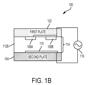
[0030] FIGS. 1A-B illustrate a QMAX (Q: quantification; M: magnifying;
A: adding
reagents; X: acceleration; also known as compressed regulated open flow
(CROF)) device 100
configured to permit bio/chemical material extraction and assay, according to
some
embodiments. In some embodiments, QMAX device 100 includes first plate 102
(also referred to
as "substrate" in the present disclosure), second plate 104 (also referred to
as "X-plate" in the
present disclosure), spacers 106A-C (also referred to as "pillars" in the
present disclosure), and
electrodes 108A-B and 110. In some embodiments, to enable bio/chemical
material extraction
.. and assay, first plate 102 and second plate 104 are movable relative to
each other to configure
QMAX device 100 into a plurality of different configurations including an open
configuration, as
shown in FIG. 1A, and a closed configuration, as shown in FIG. 1B.
[0031] FIG. lA illustrates a sectional view of QMAX device 100 in the
open configuration,
according to some embodiments. In the open configuration, first plate 102 and
second plate 104
are partially or entirely separated apart, allowing a fluid sample 112A, i.e.,
a bio/chemical
material, to be deposited on either one or both of first plate 102 and second
plate 104. In some
embodiments, the surface of first plate 102 facing second plate 104 is defined
as inner surface
118 of first plate 102; the surface of second plate 104 that faces first plate
102 is similarly
defined as inner surface 120 of second plate 104. Each of inner surfaces 118
and 120 include a
sample contact area for contacting fluid sample 112A, such as but not limited
to blood. The
sample contact area of each of inner surfaces 118 and 120 occupies a part of
the entirety of each
inner surfaces 118 and 120, respectively. In some embodiments, fluid sample
112A can be
deposited on first plate 102, second plate 104, or both first plate 102 and
second plate 104. In
some embodiments, liquid sample 112A deposited on first plate 102 or second
plate 104 has an
unknown or unmeasured volume.
[0032] In some embodiments, as shown in FIG. 1A, first plate 102 can
include spacers 106A-
C that are fixed on inner surface 118 of first plate 102 and that allow QMAX
device 100 to be
configured into a closed configuration, as will be described below with
respect to FIG. 1B.
6
CA 03053002 2019-08-07
WO 2018/148469
PCT/US2018/017501
Alternatively, spacers 106A-C can be fixed on inner surface 120 of second
plate 104. In some
embodiments, spacers 106A-C can be fixed on both inner surfaces 118 and 120.
In some
embodiments, spacers 106A-C are fixed on one or both inner surfaces 118 and
120 by directly
embossing or injection molding of first plate 102 or second plate 104. In some
embodiments,
spacers 106A-C can be composed of materials selected from one of polystyrene,
PMMA, PC,
COC, COP, or another plastic.
[0033] In some embodiments, spacers 106A-C can each have a predetermined
substantially
uniform height. In some embodiments, in the open configuration of FIG. 1A, the
gap between
first plate 102 and second plate 104 is not regulated by spacers 106A-C, which
allows fluid
sample 112A to be easily deposited on one or both of inner surfaces 118 and
120. In some
embodiments, at least one of spacers 106A-C is positioned inside the sample
contact area of one
or both of inner surfaces 118 and 120. In some embodiments, all of spacers
106A-C are
positioned inside the sample contact area. In some embodiments, spacers 106A-C
are not fixed to
either first plate 102 or second plate 104, and are instead mixed into fluid
sample 112A.
[0034] In some embodiments, each of spacers 106A-C may have a pillar shape
with a cross-
sectional shape selected from round, polygonal, circular, square, rectangular,
oval, elliptical, or a
combination thereof. In some embodiments, each of spacers 106A-C may have a
pillar shape
with a substantially flat top surface. In some embodiments, the sidewall
corners of spacers 106A-
C have a round shape with a radius of curvature of at least lum.
[0035] In some embodiments, the ratio of the lateral dimension to height of
each of spacers
106A-C is at least about 1. In some embodiments, spacers 106A-C have a density
of at least
100/mm2 or at least 1000/mm2.
[0036] In some embodiments, the minimum lateral dimension of spacers
106A-C is less than
or substantially equal to the minimum dimension of an analyte in fluid sample
112A. In some
embodiments, the minimum lateral dimension of spacers 106A-C is between about
0.5um-100um
or about 0.5um-10um.
7
CA 03053002 2019-08-07
WO 2018/148469
PCT/US2018/017501
[0037] In some embodiments, the inter-spacer distance of spacers 106A-C
is substantially
periodic. In some embodiments, the inter-spacer distance of spacers 106A-C is
between about
5um-200um, about 7um-50um, about 50um-120um, or 120um-200um.
[0038] In some embodiments, to configure QMAX device 100 for use in
bio/chemical assay
and extraction, each of first plate 102 and second plate 104 can include one
or more electrodes
that are positioned at inner surface 118 of first plate 102 and inner surface
120 of second plate
104. In some embodiments, the electrodes are attached to inner surface 118 of
first plate 102 and
inner surface 120 of second plate 104. For example, as shown in FIG. 1A,
electrodes 108A-B are
attached to inner surface 118 of first plate 102 and electrode 110 is attached
to inner surface 120
of second plate 104. In some embodiments, there is only one electrode attached
to each of first
plate 102 and second plate 104. In some embodiments, there are a plurality of
electrodes attached
to each of first plate 102 and second plate 104. In some embodiments, at least
one of spacers
106A-C include the electrode, e.g., electrode 108A. In some embodiments, one
or more of
electrodes 108A-B and 110 are placed on the outside surface of one or both of
first plate 102 or
second plate 104. In some embodiments, all of electrodes 108A-B and 110 are
placed on the
outside surface of one or both of first plate 102 and second plate 104. In
some embodiment, at
least one of electrodes 108A-C is placed on the outer surface of first plate
102 and at least one of
electrode 110 is placed on inner surface 120 of second plate 104, or vice
versa. In some
embodiments, electrodes 108A-B and 110 are made from conductive material.
[0039] In some embodiments, the conductive material can be metals such as
but not limited
to: gold, copper, silver, aluminum, alloys thereof, or mixtures thereof. In
some embodiments, the
conductive material can be conductive metallic oxide or metallic compound that
is selected from
the group consisting of: indium tin oxide (ITO), zinc oxide (Zn0), titanium
oxide (TiOx),
molybdenum dioxide (Mo02), lithium fluoride (LiF), and a combination thereof.
[0040] In some embodiments, the conductive material that make up the
electrodes can be
conductive small molecule and conductive polymer that is selected from
poly(3,4-
ethylenedioxythiophene) poly(styrenesulfonate) (PECOT:PSS), fullerene
derivatives (as C60),
aluminum tris (8-hydroxyquinoline)(Alq3), and a combination thereof.
8
CA 03053002 2019-08-07
WO 2018/148469
PCT/US2018/017501
[0041] FIG. 1B illustrates a sectional view of QMAX device 100 in the
closed configuration,
according to some embodiments. In some embodiments, one or more of the outer
surfaces of first
plate 102 and second plate 104 in the open configuration of FIG. lA can be
pressed towards each
other such that inner surfaces 118 and 120 of first plate 102 and second plate
104, respectively,
are pressed against each other in the closed configuration of FIG. 1B. In some
embodiments, gap
114 between first plate 102 and second plate 104 are regulated by at least one
spacer 106A-C
(not shown in FIG. 1B).
[0042] In some embodiments, first plate 102 and second plate 104 can be
pressed together
after fluid sample 112A is deposited to compress at least part of fluid sample
112A into a layer of
fluid sample 112B having a substantially uniform thickness and being stagnant
relative to first
plate 102 and second plate 104. In some embodiments, the layer of fluid sample
112B is confined
by inner surfaces 118 and 120, and uniform thickness of the layer is regulated
by the
substantially uniform height of spacers 106A-C and the first and second plates
102 and 104. In
some embodiments, the uniform thickness of layer of fluid sample 112B is the
same as gap 114;
in some embodiments, the thickness of layer of fluid sample 112B and gap 114
are the same as
the height of spacers 106A-C. In some embodiments, layer fluid sample 112B has
a uniform
thickness over a lateral area that is at least lmm2. In some embodiments,
liquid sample 112A has
an unknown volume and QMAX 102 as configured in the closed configuration of
FIG. 1B can
compress liquid sample 112A in layer 112B having a uniform height, which may
correspond to a
known volume over a sample contact area.
[0043] In some embodiments, the height of spacers 106A-C is less than
about lcm, about
200um, 100um, about 10um, about 5um, about lum, or about 0.1um. In some
embodiments, the
height of spacers 106A-C is greater than about 0.01um, about 0.1um, about lum,
about 5um,
about 10um, about 100um, about 200um, or about lcm. In some embodiments, the
height of
spacers is between about 0.01um and lcm such as between about 0.01um-200um,
about 0.01um-
Sum, about 5um-10um, about 10um-100um, or about 100um-lcm.
[0044] As discussed above, layer of fluid sample 112B is a layer of
having a substantially
uniform thickness regulated by spacers 106A-C, in some embodiments. Therefore,
the average
9
CA 03053002 2019-08-07
WO 2018/148469
PCT/US2018/017501
thickness of the substantially uniform thickness can be the height of spacers
106A-C, as
discussed above. In some embodiments, the average thickness of layer of fluid
sample 112B is
less than about lcm, about 200um, 100um, about 10um, about 5um, about lum, or
about 0. lum.
In some embodiments, the average thickness of layer of fluid sample 112B can
is greater than
about 0.01um, about 0.1um, about lum, about 5um, about 10um, about 100um,
about 200um, or
about lcm. In some embodiments, the average thickness of layer of fluid sample
112B is
between about 0.01um and lcm such as between about 0.01um-200um, about 0.01um-
5um,
about 5um-10um, about 10um-100um, or about 100um-lcm. In some embodiments, the
average
thickness of the layer of uniform thickness is about equal to a minimum
dimension of an analyte
in fluid sample 112A.
[0045] In some embodiments, when fluid sample 112A is a blood sample
(e.g., whole blood),
the average thickness of layer of fluid sample 112B is about 1.8um-3.8um,
about 1.8um-2um,
about 2um-2.2um, about 2.2um-2.6um, or about 2.6um-3.8um.
[0046] As shown in FIG. 1B, in the closed configuration, electrodes 108A-
B and 110 are in
contact with at least part of layer of fluid sample 112B. For the sake of
simplicity, first plate 102
is shown as having two electrodes 108A and 108B and second plate 104 is shown
as having one
electrode 110. In some embodiments, however, electrodes 108A-B can be
representative of only
one electrode or a plurality of electrodes (e.g., three or four electrodes or
more); similarly,
electrode 110 can be representative of only one electrode or a plurality of
electrodes. In some
embodiments, at least one of first plate 102 and second plate 104 is flexible
such that one or both
of first plate 102 and second plate 104 can bend slightly while compressing
layer of fluid sample
112B in the closed configuration of FIG. 1B.
[0047] In some embodiments, QMAX device 100 includes a power source 116,
such as but
not limited to an electricity source that provides alternative current (AC) or
direct current (DC).
In some embodiments, power source 116 is operably connected to electrodes 108A-
B and 110,
which are in contact with the layer of the fluid sample 112B that is pressed
into a layer of
substantially uniform thickness. In some embodiments, power source 116 can
provide a first
electric potential at electrodes 108A-B and a second electric potential at
electrode 110 to induce a
CA 03053002 2019-08-07
WO 2018/148469
PCT/US2018/017501
voltage between electrodes 108A-B and 110 such that electrodes 108A-B and 110
are in ionic
communication with layer of fluid sample 112B compressed into a layer of
uniform thickness.
[0048] In some embodiments, the electrical potentials applied by power
source 116 can be
less than about 1000V, about 500V, about 220V, about 200V, about 150V, about
110V, about
100V, about 50V, about 10V, about 5V, about 1V, about 0.5V, about 0.2V, or
about 0.1V. In
some embodiments, the electrical potentials to be applied by power source 116
can be selected
based on a type of electric property being measured or electrical
characteristics of layer of fluid
sample 112B being measured.
[0049] In some embodiments, when power source 116 provides AC, the
frequency of the AC
is less than about 1GHz, about 1MHz, about 100kHz, about 10kHz, about 1000Hz,
about 100Hz,
or about 10Hz. In some embodiments, the frequency of the AC can be varied
between any two of
the frequencies listed above, including between 10kHz and 1MHz.
[0050] In some embodiments, once powered by power source 116, electrodes
108A-B and
110 are in ionic communication with layer of fluid sample 112B compressed into
a layer of
uniform thickness. As such, electros pass between electrodes 108A-B and
electrode 110 to enable
electrodes 108A-B and electrode 110 to detect one or more electric properties
of fluid sample
112B to enable bio/chemical assay and extraction. For example, electrodes 108A-
B and 110 can
be configured to detect electric properties such as one or more of
conductivity, current, potential,
resistance, impedance, and capacitance as well as permittivity of fluid sample
112B in the layer
of uniform thickness.
[0051] In some embodiments, the width of each of electrodes 108A-B and
110 can be at least
about 2 times, about 5 times, about 10 times, about 50 times, about 100 times,
about 500 time, or
about 1000 times larger than the height of each of electrodes 108A-B and 110,
respectively. In
some embodiments, the width of each of electrodes 108A-B and 110 can be less
than about 2000
times, about 1000 times, about 500 times, about 100 times, about 50 times,
about 10 time, or
about 5 times larger than the height of each of electrodes 108A-B and 110,
respectively. In some
embodiments, the width of each of electrodes 108A-B and 110 can be about 2-
1000 times, about
11
CA 03053002 2019-08-07
WO 2018/148469
PCT/US2018/017501
5-500 times, or about 50-100 times larger than the height of each of
electrodes 108A-B and 110,
respectively.
[0052] In some embodiments, the width of each of electrodes 108A-B and
110 can be at least
about mm, about lOnm, about 50nm, about 100nm, about 500nm, about lum, about
10um, about
50um, about 100um, about 500um, about lmm, about 5mm, about lOmm, about 50mm,
or about
100mm. In some embodiments, the width of each of electrodes 108A-B and 110 can
be less than
about 100mm, about 50mm, about lOmm, about 5mm, about lmm ,about 500um, about
50um,
about 10um, about lum, about 500nm, about 100nm, about 50nm, about lOnm, or
about mm. In
some embodiments, the width of each of electrodes 108A-B and 110 can be
between about mm-
100mm, about lnm-100um, about 50um-100um, about 100um-500um, about 500um-1mm,
about
lmm-5mm, about 5mm-10mm, or about 10mm-100mm.
[0053] In some embodiments, the height of each of electrodes 108A-B and
110 can be at
least about mm, about lOnm, about 50nm, about 100nm, about 500nm, about lum,
about 10um,
about 50um, about 100um, about 500um, about lmm, about 5mm, or about lOmm. In
some
embodiments, the height of each of electrodes 108A-B and 110 can be less than
about lOmm,
about 5mm, about lmm ,about 500um, about 50um, about 10um, about lum, about
500nm, about
100nm, about 50nm, about lOnm, or about mm. In some embodiments, the height of
each of
electrodes 108A-B and 110 can be between about mm-lOmm, about 1 nm-100um,
about 50um-
100um, about 100um-500um, about 500um-1mm, or about lmm-5mm.
[0054] In some embodiments, the width of each of electrodes 108A-B and 110
can be at least
about 2 times, about 5 times, about 10 times, about 50 times, about 100 times,
about 500 time, or
about 1000 times larger than the gap between any two adjacent electrodes of
electrodes 108A-B
and 110, such as between electrodes 108A-B. In some embodiments, the width of
each of
electrodes 108A-B and 110 can be less than about 2000 times, about 1000 times,
about 500
times, about 100 times, about 50 times, about 10 time, or about 5 times larger
than the gap
between any two adjacent electrodes of electrodes 108A-B and 110. In some
embodiments, the
width of each of electrodes 108A-B and 110 can be about 2-1000 times, about 5-
500 times, or
12
CA 03053002 2019-08-07
WO 2018/148469
PCT/US2018/017501
about 50-100 times larger than the gap between any two adjacent electrodes of
electrodes 108A-
B and 110.
[0055] In some embodiments, the gap between any two adjacent electrodes
of electrodes
108A-B and 110 can be at least about mm, about lOnm, about 50nm, about 100nm,
about
500nm, about lum, about 10um, about 50um, about 100um, about 500um, about lmm,
about
5mm, about lOmm, about 50mm, or about 100mm. In some embodiments, the gap
between any
two adjacent electrodes of electrodes 108A-B and 110 can be less than about
100mm, about
50mm, about lOmm, about 5mm, about lmm ,about 500um, about 50um, about 10um,
about
lum, about 500nm, about 100nm, about 50nm, about lOnm, or about mm. In some
embodiments, the gap between any two adjacent electrodes of electrodes 108A-B
and 110 can be
between about lnm-100mm, about lnm-100um, about 50um-100um, about 100um-500um,
about
500um-1mm, about 1mm-5mm, about 5mm-10mm, or about 10mm-100mm.
[0056] In some embodiments, QMAX device 100 includes a measuring unit
electrically
coupled to at least one of electrodes 108A-B and 110 to measure one or more
electric properties
(e.g., electrical conductance or capacitance or both) being detected by
electrodes 108A-B and
110. In some embodiments, the measuring unit can be an electric circuit
electrically coupled to at
least two of electrodes 108A-B and 110 to measure a permittivity of layer of
fluid sample 112B.
For example, to measure the permittivity of fluid sample 112B, the measuring
unit can be
configured to measure capacitance between electrodes 108A-B and electrode 110
to derive the
permittivity of fluid sample 112B because capacitance is proportional to the
permittivity.
[0057] In some embodiments, the measuring unit can be configured to
measure the one or
more electric properties of fluid sample 112B (being compressed into the
uniform thickness) for
a predetermined number of times at predetermined time periods. In some
embodiments, the
predetermined time periods include at times of two or more of about 10s, about
30s, about 60s,
about 2min, about 3min, about 5min, about 8min, about 10min, about 15min,
about 20min, about
30min, etc., after fluid sample 112A is compressed into a layer of fluid
sample 112B as shown in
FIG. 1B.
13
CA 03053002 2019-08-07
WO 2018/148469
PCT/US2018/017501
[0058] In some embodiments where fluid samples 112A-B is a blood sample,
QMAX device
100 can be configured to measure and assess coagulation properties of the
blood sample. For
example, QMAX device 100 may include a calculation unit configured to
calculate one or both
of a prothrombin time (PT) and an activated partial thromboplastin time (aPTT)
based on the
permittivity of fluid sample 112B being measured. In some embodiments, fluid
sample 112A
includes a blood sample such as whole blood or blood serum. In some
embodiments, fluid
sample is whole blood without dilution by liquid. In some embodiments, the
blood sample
includes added Ca2+. In some embodiments, the blood sample with added Ca2+
and/or citrate salt
or acid can be used as controls.
[0059] In some embodiments, to measure coagulation properties of the blood
sample, a
coagulation regulator can be added to the blood sample. For example, the blood
sample may
include citrate salt or acid for anti-coagulation purposes. In some
embodiments, the blood sample
includes added anticoagulant corn trypsin inhibitor (CTI). In some
embodiments, the blood
sample further includes added anticoagulant penicillins. In some embodiments,
the blood sample
includes added Activator cephalin. In some embodiments, the blood sample
includes added
Activator Tissue Factors (ATF). In some embodiments, the coagulation regulator
can be pre-
deposited and dried on one or both of inner surfaces 118 and 120. Such a
coagulation regulator
can be any of the regulators discussed above including, without limitation,
peptides, proteins
(e.g., Tissue Factors), or small molecules (e.g., ions, antibiotics, and other
drugs). In some
embodiments, to enable QMAX device 100 to more accurately measure coagulation
properties of
the blood sample, QMAX device 100 includes a temperature controller unit that
is added outside
of first plate 102 and second plate 104. The temperature controller can be
configured to control
the temperature during coagulation process in the range of 0 C to 100 C, with
a preferred
temperature of 37 C.
[0060] In some embodiments, fluid sample 112A can be a biological sample
selected from
amniotic fluid, aqueous humour, vitreous humour, blood (e.g., whole blood,
fractionated blood,
plasma, or serum), breast milk, cerebrospinal fluid (CSF), cerumen (earwax),
chyle, chime,
endolymph, perilymph, feces, breath, gastric acid, gastric juice, lymph, mucus
(including nasal
14
CA 03053002 2019-08-07
WO 2018/148469
PCT/US2018/017501
drainage and phlegm), pericardial fluid, peritoneal fluid, pleural fluid, pus,
rheum, sebum, semen,
sputum, sweat, synovial fluid, tears, vomit, urine or exhaled condensate.
[0061] In some embodiments, fluid sample 112A can be a biological
sample, an
environmental sample, a chemical sample, or a clinical sample. In some
embodiments, fluid
sample 112A can be bodily fluid such as blood, saliva, or urine.
[0062] In some embodiments, fluid sample 112A includes at least one
analyte. In some
embodiments, fluid sample 112A includes a plurality of analytes. In some
embodiments, the at
least one analyte can be a protein, nucleic acid, a cell, or a metabolite.
[0063] In some embodiments, the plurality of analytes are analytes
selected from the group
consisting of sodium (Na+), potassium (K+), calcium (Ca2+), bicarbonate (HCO3-
), magnesium
(Mg2+), chloride (Cl-), and hydrogen phosphate (HP042-).9. In some
embodiments, the plurality
of analytes are macromolecules selected from the group consisting of
carbohydrates
(monosaccharides, disaccharides, and polysaccharides), glucose, and sucrose.
[0064] In some embodiments, the plurality of analytes are nucleic acid
including a polymer
of any length composed of nucleotides, e.g., deoxyribonucleotides or
ribonucleotides, or
compounds produced synthetically (e.g., PNA as described in U.S. Pat. No.
5,948,902) which can
hybridize with naturally occurring nucleic acids in a sequence specific manner
analogous to that
of two naturally occurring nucleic acids, e.g., can participate in Watson-
Crick base pairing
interactions.
[0065] In some embodiments, the plurality of analytes are proteins
including a polymeric
form of amino acids of any length. In some embodiments, the length of amino
acids can be more
than about 2, about 5, about 10, about 20, about 50, about 100, about 200,
about 500, about 1000,
or about 2000. In some embodiments, the length of amino acids can be less than
about 2000,
about 1000, about 500, about 200, about 100, about 50, about 20, about 10, or
about 5. hi some
embodiments, the length of amino acids can be between about 2 and 2000.
[0066] In some embodiments, the plurality of analytes are proteins that
can include coded
and non-coded amino acids, chemically or bio/chemically modified or
derivatized amino acids,
CA 03053002 2019-08-07
WO 2018/148469
PCT/US2018/017501
and polypeptides having modified peptide backbones. In some embodiments, the
term protein
can refer to fusion proteins, including, but not limited to, fusion proteins
with a heterologous
amino acid sequence, fusions with heterologous and homologous leader
sequences, with or
without N-terminal methionine residues; immunologically tagged proteins;
fusion proteins with
detectable fusion partners, e.g., fusion proteins including as a fusion
partner a fluorescent protein,
(3-galactosidase, luciferase, etc.; and the like.
[0067] In some embodiments, the plurality of analytes are polypeptides
that are post-
translationally modified in a cell, e.g., glycosylated, cleaved, secreted,
prenylated, carboxylated,
phosphorylated, etc., and polypeptides with secondary or tertiary structure,
and polypeptides that
are strongly bound, e.g., covalently or non-covalently, to other moieties,
e.g., other polypeptides,
atoms, cofactors, etc.
[0068] In some embodiments, the plurality of analytes are cells
including prokaryotes and
eukaryotes, including bone cells, cartilage cells, nerve cells, epithelial
cell, muscle cells,
secretory cell, adipose cells, blood cells, conductive cells, connective
cells, glandular cells,
storage cells, supportive cells, etc.
[0069] In some embodiments, the plurality of analytes can include
bacteria such as coccus,
bacillus, vibrio, spirillum, spirochete, etc.
[0070] In some embodiments, QMAX device 100 can be configured to include
a location
marker included on a surface of or inside of first plate 802 or second plate
804 to provide
information of the location of the location marker. For example, the location
may indicate a
sample area. In some embodiments, one or more of first plate 102 and second
plate 104 can
include a scale marker that provides information of a lateral dimension of the
respective plate or
fluid sample 112B. In some embodiments, the scale marker can be positioned on
either an inner
surface or an outer surface of the first plate 102 or the second plate 104. In
some embodiments,
one or more of first plate 102 and second plate 104 can include an imaging
marker, on an inner
surface or inside of first plate 102 or second plate 104, that can be
configured to aid in imaging of
fluid sample 112B, as will be further described with respect to FIGS. 8A-C. In
some
embodiments, one or more of spacers 106A-C functions as a location marker, a
scale marker, an
16
CA 03053002 2019-08-07
WO 2018/148469
PCT/US2018/017501
imaging marker, or a combination thereof. For example, spacers 106A and 106B
may be placed
at an inter-spacer distance to indicate scale, a location of a particular part
of first plate 102 or
second plate 104, and/or function as a guide post for imaging purposes.
[0071] In some embodiments, to enable first plate 102 and second plate
104 to be capable of
being configured in the open configuration of FIG. 1A and the closed
configuration of FIG. 1B,
first plate 102 can be connected to second plate 104 to enable first plate 102
to fold over second
plate 104. In some embodiments, first plate 102 and second plate 104 can be
made from a single
piece of material that is configured to be changed from the open configuration
to the closed
configuration by folding first plate 102 and second plates 104.
[0072] In some embodiments, first plate 102 and second plate 104 are
connected by a hinge
configured to allow folding along the hinge to configure the first plate 102
and second plate 104
in the open and closed configurations. In some embodiments, the hinge is a
separate material
from first plate 102 and second plate 104.
[0073] FIGS. 2-6 illustrate QMAX devices similar to QMAX device 100 of
FIGS. 1A-B, but
with varying placement and amount of electrodes, according to some
embodiments. It should be
noted, that for clarity purposes, not all the components as described with
respect to FIGS. 1A-B
are shown in all of FIGS. 2-6. For example, spacers 106A-C, as described with
respect to FIGS.
1A-B, are not shown in FIGS. 2-6. The specific design of the QMAX devices of
FIGS. 2-6 and
their components can vary and the presence or absence of the certain
components, such as
spacers 106A-C, can be inferred from the design of the experiments and the
descriptions for the
specific QMAX devices.
[0074] FIGS. 2A-C illustrate QMAX devices 200A-C having electrodes not
positioned at
either of the inner surfaces of first plate 202A-C and second plate 204A-C,
according to some
embodiments. Therefore, FIGS. 2A-C show QMAX devices 200A-C that are
functional without
current flow. In some embodiments, a power source 206A-C (e.g., a voltage
source (DC or AC)
or a current source (DC or AC)) applies power to at least two electrodes of
each of QMAX
devices 200A-C, respectively, with each electrode being outside of QMAX
devices 200A-C.
17
CA 03053002 2019-08-07
WO 2018/148469
PCT/US2018/017501
Here, the term "outside" refers to space outside QMAX devices 200A-C when
respective first
plate 202A-C and second plate 204A-C are in the closed configuration.
[0075] In FIG. 2A, QMAX device 200A includes two electrodes 208 and 210
positioned
outside of and in contact with the outer surfaces of first plate 202A and
second plate 204A,
respectively. In FIG. 2B, QMAX device 200B includes a plurality of electrodes
212A-C and a
plurality of electrodes 214A-D that are each dis-continuous (e.g. an array),
outside, and in contact
with the outer surfaces of first plate 202B and second plate 204B,
respectively. As shown in
FIGS. 2A-B, each of first plate 202A-B and second plate 204A-B can include
only one electrode
or a plurality of electrodes, according to some embodiments. In FIG. 2C, QMAX
device 200C
includes electrodes 216 and 218 that are separate, outside first plate 202C
and second plate 204C,
and not in contact with either of first plate 202C and second plate 204C. In
some embodiments,
such as that depicted in FIG. 2C, none of the electrodes of the QMAX device is
in physical
contact with either of the first or second plates of the QMAX device.
[0076] FIGS. 3A-B illustrate a QMAX device 300 that is functional with
current flow,
according to some embodiments. As described with respect to FIGS. 1A-B, QMAX
device 300
can include similarly named components: first plate 302, second plate 304,
power source 306,
and electrodes 308 and 310. In some embodiments, FIG. 3A shows a sectional
view of QMAX
device 300 where first plate 302 has at least one electrode 308 on its inner
surface and second
plate 304 has at least one electrode 310 on its inner surface. In some
embodiments, when power
source 306 is DC, at least one of electrodes 308 and 310 connects to the anode
of power source
306 and at least one of electrodes 308 and 310 connects to the cathode of
power source 306.
[0077] As shown in a FIG. 3B, each of electrodes 308 and 310 have two
pads: contact pad
308A and measurement pad 308B for electrode 308; and contact pad 310A and
measurement pad
310B for electrode 310. In some embodiments, as shown in FIG. 3A, each of
contact pads 308A
and 310A are electrically connected to the outside power source 306. In some
embodiments,
when QMAX device 300 is configured into the closed configuration after a
sample liquid is
deposited, measurement pads 308B and 310B are in contact with the fluid
sample. In some
embodiments, the entirety of measurement pads 308B and 310B are in contact
with the fluid
18
CA 03053002 2019-08-07
WO 2018/148469
PCT/US2018/017501
sample. In some embodiments, only a portion of each of measurement pads 308B
and 310B is in
contact with the fluid sample. In some embodiments, each of electrodes 308 and
310 has only
respective measurement pads 308B and 310B, each of which is directly connected
to the power
source 306 with wires. As described above with respect to FIGS. 1A-B,
electrodes 308 and 310,
(e.g., measurement pads 308B and 310B), can be configured to measure one or
more electric
properties of the fluid sample compressed into a layer of substantially
uniform thickness once
power source 306 is applied to electrodes 308 and 310 via, for example,
contact pads 308A and
310A. Therefore, QMAX device 300 can be configured to perform bio/chemical
material assay
via electrical measurements.
[0078] FIGS. 4A-B illustrate a QMAX device 400 that is functional with
current flow,
according to some embodiments. As described with respect to FIGS. 3A-B, QMAX
device 400
can include similarly named components: first plate 402, second plate 404,
power source 406,
and electrodes 408 and 410. Further, like QMAX device 300 of FIGS. 3A-B, each
of electrodes
408 and 410 can include respective contact pads 408A and 410A and respective
measurement
pads 408B and 410B. In some embodiments, in contrast to FIGS. 3A-B, both of
electrodes 408
and 410 can be placed inside, on the inner surface, of only one of first plate
402 or second plate
404. For example, as shown in FIGS. 4A-B, both of electrodes 408 and 410 are
placed on the
inner surface of second plate 404. In some embodiments, all of the electrodes
(e.g., electrodes
408 and 410) of QMAX device 400 are placed on only one of the first plate 402
or second plate
404.
[0079] In some embodiments, the width of each of electrodes 408 and 410
is much larger
than the height of each electrode 408 and 410 and much larger than the gap
between two adjacent
electrodes 408 and 410. In some embodiments, the length and width of each of
electrodes 408
and 410 are substantially larger than the gap between two adjacent electrodes
(e.g., electrodes
408 and 410) when first plate 402 and second plate 404 are pressed together in
the closed
configuration of QMAX device 400. In some embodiments, the length and/or the
width of each
of the electrodes 408 and 410 (e.g. the measurement pads 408B and 410B) are at
least about 2
times, about 5 times, about 10 times, about 20 times, about 30 times, about 40
times, about 50
19
CA 03053002 2019-08-07
WO 2018/148469
PCT/US2018/017501
times, about 75 times, about 100 times, about 150 times, about 200 times,
about 300 times, about
400 times, about 600 times, about 600 times, about 700 times, about 800 times,
about 900 times,
about 1000 times, about 5000 times, about 10000 times, about 5000 times, about
100000 times,
about 500000 times, or about 1000000 times larger than the gap between the two
electrodes 408
.. and 410.
[0080]
FIGS. 5A-B illustrates a QMAX device configured to measure analyte
concentration
of a fluid sample, according to some embodiments. As described with respect to
FIGS. 4A-B,
QMAX device 500 can include similarly named components: first plate 502,
second plate 504,
power source 506, electrode 508 (e.g., contact pad 508A and measurement pad
508B), and
.. electrode 510 (e.g., contact pad 510A and measurement pad 510B). While the
specific placement
and number of electrodes 508 and 510 of QMAX device 500 are shown to be
similar to the
design shown in FIGS. 4A-B, the placement and number of electrodes 508-510 may
instead be in
one of the configurations as described or shown in any of FIGS. 1A-B or 3A-B,
according to
some embodiments. In contrast to previously described embodiments, however,
QMAX device
500 includes a barrier membrane 512 (e.g., an ion-selective membrane) that
covers one of
electrodes 508 or 510. For example, as shown in FIGS. 5A-B, barrier membrane
512 may cover
electrode 510, specifically, measurement pad 510B of electrode 510. In some
embodiments, to
cover electrode 510, barrier membrane 512 can be coated on top of electrode
510. While example
dimensions of first plate 502 and second plate 504 are shown in FIG. 5B, the
dimensions may be
adjusted according to specific design of the experiments, such as but not
limited to the sample
amount and the analyte within the fluid sample to be measured. In some
embodiments, barrier
membrane 512 has a contacting surface for contacting the fluid sample. In some
embodiments,
one of electrodes 508 and 510 (e.g., electrode 510) includes a perforated
conductive sheet, which
provides the function of a contacting surface of barrier membrane 512.
[0081] In some embodiments, as described with respect to FIGS. 1A-B, QMAX
device 500
can include a measurement device to measure electric properties of the fluid
sample when
QMAX device 500 is in a closed configuration. For example, the measurement
device can
measure a current flowing through the fluid sample in a layer of substantially
uniform thickness
CA 03053002 2019-08-07
WO 2018/148469
PCT/US2018/017501
and flowing between electrodes 508 and 510. In some embodiments, barrier
membrane 512 can
be composed to have ion selecting effects such that the current measured by
the measurement
device can reflect the concentration/amount of certain analytes, such as but
not limited to ions,
within the fluid sample. In particular, barrier membrane 512 can be configured
to permit a
selected analyte in the fluid sample from passing through barrier membrane 512
to be in
electrical communication with at least one electrode (e.g., electrode 510)
being covered by
bather membrane 512 as a result of power source 506 supplying a power. In some
embodiments,
bather membrane 512 can be configured to allow the passing through of one or
more selected
analytes in the fluid sample and block the passing through of one or more
different selected
analytes in the fluid sample.
[0082] In some embodiments, to enable the selective effects of barrier
membrane 512, barrier
membrane 512 can be made of insoluble, infusible synthetic organic polymer
matrix which is
bound with chemicals that selectively allow certain analytes in the fluid
sample to pass through
bather membrane 512.In some embodiments, barrier membrane 512 can be made of
organic
polymer matrix selected from the group consisting of poly(vinyl chloride)
(PVC),
polyvinylpyrrolidone, polydimethylsiloxane, perfluoropolyether, etc. In some
embodiments, the
chemicals that selectively allow passage of certain analytes can be chemicals
selected from ETH
157 carrier, ETh 227 carrier, ETH 2120 carrier, a bis(12-crown-4) compound ,
hemispherand,
valinomycin, BBPA, KTpC1PB, and '70 o-nitrophenyl octyl ether, etc.
[0083] In some embodiments, to measure the analyte concentrations of the
fluid sample, the
measurement device can measure current flowing between electrodes 508 and 510
at a plurality
of different voltages applied by power source 506 between electrodes 508 and
510. In some
embodiments, the measurement device can be configured to measure an electron
amount and
current density passing between at least two electrodes (e.g., electrodes 508
and 510) to
determine an analyte concentration of the fluid sample because the electron
amount and current
density is correlated to the concentration of the selected analyte in the
sample liquid.
[0084] In some embodiments, the measurement device can be configured to
measure an
electrical impedance between at least two electrodes (e.g., electrodes 508 and
510) when
21
CA 03053002 2019-08-07
WO 2018/148469
PCT/US2018/017501
powered by power source 506 to determine an analyte concentration of the fluid
sample. In some
embodiments, the electrical impedance measured between about frequencies 10kHz
to 1MHz is
correlated to the concentration of the selected analyte in the sample liquid.
[0085] In some embodiments, the inner surface of at least one of first
plate 502 and second
plate 504 can be coated with chemicals, which generate electrons in
communication with a
selected analyte in the fluid sample when electrodes 508 and 510 are powered
by power source
506. In some embodiments, the measurement device can be configured to measure
an electron
amount and current density passing between at least two electrodes (e.g.,
electrodes 508 and 510)
to determine an analyte concentration of the fluid sample because the electron
amount and
current density (as a result of the added chemical) is correlated to the
concentration of the
selected analyte in the sample liquid.
[0086] In some embodiments, the measurement device can be configured to
measure one or
more of the current, potential, conductance, and/or capacitance of the fluid
sample compressed
into a layer of uniform thickness. Accordingly, including barrier membrane 512
enables QMAX
device 500 to measure analyte concentrations in the fluid sample.
[0087] FIG. 6 illustrates a diagram showing how a QMAX device 600 can be
configured to
measure analyte concentration in a fluid sample, according to some
embodiments. For ease of
illustration, QMAX device 600 has similarly named and placed components as
QMAX device
500 of FIGS. 5A-B: first plate 602, second plate 604, power source 606,
electrode 608, electrode
610, and barrier membrane 612. In some embodiments, as described with respect
to FIGS. 1A-B,
a fluid sample can be deposited on, for example, the inner surface of second
plate 604. Then, first
plate 602 can be pressed over second plate 604 to compress the fluid sample
into a layer of fluid
sample 614 having a uniform thickness. In some embodiments, layer of fluid
sample 614 covers
both of electrodes 608 and 610. In some embodiments, the current (as shown by
the arrows)
flowing between and electrodes 608 and 610 can be measured at a plurality of
voltages induced
by power source 606 between electrodes 608 and 610. Barrier membrane 612 can
be selected to
enable the analyte concentration of fluid sample 614 to be measured, according
to some
embodiments.
22
CA 03053002 2019-08-07
WO 2018/148469
PCT/US2018/017501
[0088] FIGS. 7A-F illustrate diagrams of a QMAX device 700 configured to
perform
bio/chemical material 710 (e.g., nucleic acid) extraction from a fluid sample
708, according to
some embodiments. In some embodiments, QMAX device 700 can be implemented as a
QMAX
device of any of FIGS. 1A-B, 2A-C, 3A-B, or 4A-B. In some embodiments, QMAX
device 700
can be as a QMAX device where first and second electrodes are positioned
outside of the first
and second plates, as described with respect to FIGS. 2A-C. For ease of
illustration, the first and
second electrodes are not depicted in FIGS. 7A-F.
[0089] In FIG. 7A, QMAX device 700 includes first plate 702 and second
plate 704 in an
open configuration. In some embodiments, before bio/chemical material 710 can
be extracted,
fluid sample 708 needs to be lysed. In some embodiments, the lysing can be
chemical,
mechanical, or both. Some embodiments of such lysing have been described US
Provisional
Application No. 62/456,528, which was filed on February 8, 2017, and US
Provisional
Application No. 62/456,596, which was filed on February 8, 2017, all of which
applications are
incorporated herein by reference in their entireties for all purposes. In some
embodiments, cell
lysing reagent 706 can be attached to and dried on first plate 702 or second
plate 704 to enable
chemical lysing.
[0090] The term "cell lysing reagent" as used herein can include salts,
detergents, enzymes,
and other additives. In some embodiments, the term "salt" herein include but
not limited to
lithium salt (e.g. lithium chloride), sodium salt (e.g. sodium chloride), or
potassium (e.g.
potassium chloride) or any combination thereof. In some embodiments, the term
"detergent"
herein serves not only as cell lysing reagents, but also as nucleic acid
binding reagents that
facilitate released nucleic acids and cell-free nucleic acids to bind to the
plate surface. The
detergent can be any detergent, and a vast range are known and described in
the literature. The
detergent can be ionic, including anionic and cationic, non-ionic or
zwitterionic. The term "ionic
detergent" as used herein includes any detergent which is partly or wholly in
ionic form when
dissolved in water. Suitable anionic detergents include but are not limited to
sodium dodecyl
sulphate (SDS) or other alkali metal alkylsulphate salts or similar
detergents, sarkosyl, or
combinations thereof. In some embodiments, the detergent can be in a
concentration of 0.2 to
23
CA 03053002 2019-08-07
WO 2018/148469
PCT/US2018/017501
30% (w/v), preferably 0.5 to 15%, or more preferably 1 to 10%. In some
embodiments, the term
"enzyme" herein includes but is not limited to lysozyme, cellulase, and
proteinase. In some
embodiments, the tern "additive" can include chelating agents and buffer
components, including
but not limited to EDTA, EGTA and other polyamino carboxylic acids, and some
reducing
agents, such as dithiotreitol (dTT), Tris, Bicine, Tricine, and phosphate
buffers.
[0091] In FIG. 7B, fluid sample 708 can be deposited on either first
plate 702 or second plate
704 such that fluid sample 708 comes in contact with the inner surface of
first plate 702 or
second plate 704. In some embodiments, fluid sample 708 can include
bio/chemical material 710
(e.g., nucleic acids or cell-free nucleic acids) to be extracted and other
components (e.g., cellular
structures) 712.
[0092] In FIG. 7C, similar to QMAX devices as described above with
respect to FIGS. 1A-B,
QMAX device 700 can be configured in the closed configuration by pressing
first plate 702 and
second plate 704 together. In some embodiments, a target component 712 (e.g.,
cellular
structures) in fluid sample 708 can be chemically lysed by having cell lysing
reagent 706 come
into contact with fluid sample 708. In some embodiments, when in contact with
fluid sample 708,
cell lysing reagent 706 can be dissolved within fluid sample 708.
[0093] In some embodiments, when pressing first plate 702 and second
plate 704 together,
the spacers (not shown) on the inner surface of first plate 702 faces to the
inner surface of second
plate 704, and the spacers are sufficient to mechanically break target
components 712 (e.g.,
cellular structures) in fluid sample 708 to release bio/chemical material 710.
Therefore, cell
lysing reagent 706 may not be used, in some embodiments.
[0094] In FIG. 7D, an electric field can be applied between first plate
702 and second plate
704 via a first and a second electrode, respectively, coupled to first plate
702 and second plate
704. In some embodiments, second plate 704 can be rendered positively charged
to capture the
negatively charged bio/chemical material 710 on the inner surface of second
plate 704. In some
embodiments, once target components 712 are disrupted (i.e., lysed), the
released bio/chemical
material 710 can be captured nearly instantly.
24
CA 03053002 2019-08-07
WO 2018/148469
PCT/US2018/017501
[0095] In some embodiments, first plate 702 and second plate 704 can be
pressed together to
reduce a thickness of fluid sample 708 to 250um or less, which greatly reduce
the average
diffusion time of bio/chemical material 710 from a location in fluid sample
708 to the electrically
charge extraction surface of second plate 704, and hence greatly increases the
extraction speed
(and thus, reduces the extraction time)
[0096] In some embodiments where a power source that applies the
electric field is DC, the
anode of the power source connects to second plate 704 or a second electrode
connected to
second plate 704; and the cathode of the power source connects to first plate
702 or a first
electrode connected to first plate 702.
[0097] In some embodiments, first plate 702 and second plate 704 need not
be electrically
charged by the power source. In some embodiments, second plate 704 can be
chemically
modified to exhibit electropositivity to allow binding of bio/chemical
material 710 (having a
negative charge) on the inner surface of second plate 704. Similarly, for
bio/chemical material
710 that is positively charged, second plate 704 can be chemically modified to
exhibit
electronegativity to bind positively-charged bio/chemical material 710.
[0098] In some embodiments, to further enhance the ability of second
plate 704 to capture
bio/chemical material 710 in fluid sample 708, capture probes specific to
bio/chemical material
710 to be extracted can be immobilized on the inner surface of second plate
704. In some
embodiments where bio/chemical material 710 is nucleic acid, following the
lysing process,
bio/chemical material 710 can sequence dependently hybridize to the capture
probes on the inner
surface of second plate 704.
[0099] In some embodiments, "capture probe" as used herein can refer to
oligonucleotides
having a length between 1-200bp, preferably between 5-50bp, and more
preferably between 10-
20bp. In some embodiments, capture probes have complementary sequence to
nucleic acid
.. sequences of interest in fluid sample 708. In some embodiments, identical
capture probes can be
immobilized on the inner surface of first plate 702. In some other
embodiments, different capture
probes having different base pair compositions are immobilized on the surface
of first plate 702.
CA 03053002 2019-08-07
WO 2018/148469
PCT/US2018/017501
In some embodiments, capture probes can be DNA, or RNA, or both, but
preferably to be single
strand DNA.
[0100] In some embodiments, "immobilize" as used herein refers to a
process of anchoring
the capture probe on the inner surface of a plate, such as second plate 704.
In some embodiments,
capture probes are anchored through covalent bond, where, for example, either
5' or 3' end of the
capture probe is modified to facilitate coating on the inner surface of second
plate 704.
Commonly used 3' end modifications may include but are not limited to thiol,
dithiol, amine,
biotin, etc. In some other embodiments, capture probes can be passively
absorbed on the inner
surface of second plate 704.
[0101] In some embodiments, salts, including but not limited to sodium
chloride and sodium
citrate, and molecular crowding reagents, including but not limited to ficoll,
dextran, or
polyethylene glycol, can also be dried on the inner surface of second plate
704 to facilitate
capturing nucleic acids from the sample.
[0102] In FIG. 7E, first plate 702 can be peeled off from second plate
704 to enable the inner
surface of second plate 704 to be cleaned by a sponge 716. In some
embodiments, a "sponge"
refers to a class of flexible porous materials that change pore sizes under
different pressures. IN
some embodiments, sponge 716 contains a washing reagent 714 that come in
contact with the
inner surface of second plate 704 to remove contaminates. In some embodiments,
sponge 716 can
come in contact with second plate 704 only one time, only two times, or more
than two times to
clean the inner surface of second plate 704 of the contaminants.
[0103] In some embodiments, "contaminate" as used herein can refer to
compounds
including but not limited to cell debris, proteins, non-specific nucleic acid,
etc. that are
detrimental to amplification reaction of bio/chemical material 710 captured in
FIG. 7D.
[0104] In some embodiments, the washing is conducted by squeezing sponge
716 to release
washing reagent 714 onto the inner surface of second plate 704 and releasing
sponge 716 to
reabsorb washing reagent 714. In some embodiments, "washing reagent" as used
herein can refer
to a solution that can take away contaminates without affecting the bounded
bio/chemical
26
CA 03053002 2019-08-07
WO 2018/148469
PCT/US2018/017501
material 710 on the inner surface of second plate 704. In some embodiments,
washing reagent
714 includes low to moderate ionic strength buffers, including but not limited
to 10mM Tris-HC1
or 40mM sodium chloride.
[0105] FIG. 7F shows bio/chemical material 710 captured by second plate
704 and washed of
contaminants. In some embodiments, bio/chemical material 710 can be used in
further biological
applications, including but not limited to, nucleic acid amplification,
nucleic acid hybridization
and sequencing procedures.
[0106] In some embodiments, first plate 702 or second plate 704 of FIGS.
7A-F can include a
storage site configured to store a reagent, which when contacting fluid sample
708, can diffuse in
fluid sample 708. In some embodiments, the reagent can be cell lysing reagent
706 or washing
reagent 714.
[0107] In some embodiments, sponge 716, cell lysing reagent 706, and
washing reagent 714
are further described in US Provisional Application No. 62/394,753, which was
filed on
September 15, 2016, US Provisional Application No. 62/456,488, which was filed
on February 8,
2017, and US Provisional Application No. 62/456,287, which was filed on
February 8, 2017, all
of which applications are incorporated by reference herein in their entireties
for all purposes.
[0108] FIGS. 8A-C illustrate a QMAX device 800 configured to carry a
sensing chip 806 for
performing bio/chemical assay of a fluid sample 812, according to some
embodiments. In some
embodiments, QMAX device 800 includes: a first plate 802, a second plate 804,
and sensing chip
.. 806. Like QMAX device 100 as described with respect to FIGS. 1A-B, QMAX
device 800 can
be configured in an open configuration, as shown in FIG. 8A, and a closed
configuration, as
shown in FIG. 8C. In some embodiments, at least one of first plate 802 and
second plate 804 is
made from transparent materials. In some embodiments, like QMAX device 100
described
above, first plate 802 or second plate 804 can include one or more location
markers, one or more
scale markers, or one or more imaging markers.
[0109] In some embodiments, first plate 802 includes a plurality of
spacers 808, which may
correspond to and have similar properties as spacers 106A-C as described with
respect to FIGS.
27
CA 03053002 2019-08-07
WO 2018/148469
PCT/US2018/017501
1A-B. In some embodiments, spacers 808 can be positioned on only sensing chip
806 and not on
the first plate 802 or second plate 804. In some embodiments, spacers 808 can
be positioned on
only the sample contact area of first plate 802 and not on second plate 804.
In some
embodiments, one or more spacers 808 can function as a location marker, a
scale marker, or an
imaging marker.
[0110] In some embodiments, second plate 804 includes a well 816
configured to host
sensing chip 806 inside well 816. In some embodiments, sensing chip 806 can be
composed of
material that is dielectric, a metal, or a combination thereof. In some
embodiments, sensing chip
806 can be composed of plastics.
[0111] In some embodiments, first plate 802 and second plate 804 can each
have an average
length, width, and thickness. In some embodiments, the thickness of first
plate 802 and second
plate 804 is at least about 50nm, about 100nm, about 200nm, about 500nm, about
lum, about
2um, about 5um, about 10um, about 20um, about 30um, about 50um, about 70um,
about 100um,
about 120um, about 150um, about 200um, about 300um, about 400um, about 500um,
or about
lmm. In some embodiments, the thickness of first plate 802 and second plate
804 is less than
about 3 mm, about lmm, about 500um, about 400um, about 300um, about 200um,
about 150um,
about 120um, about 100, about 70um, about 50um, about 30um, about 20um, about
10um, about
Sum, about 2um, about lum, about 500um, about 200nm, or about 100nm. In some
embodiments, the thickness of first plate 802 and second plate 804 is between
about 50nm-3mm,
about 500nm-700um, about lum-500um, about 10um-300um, or about 20um-250um.
[0112] In some embodiments, the length or width of first plate 802 and
second plate 804 is at
least about lmm, about 3mm, about 5mm, about 6mm, about 7mm, about 8mm, about
lOmm,
about 15mm, about 20mm, about 30mm, about 40mm, about 50mm, about 60mm, about
70mm,
about 80mm, about 90mm, about 100mm, or about 150mm. In some embodiments, the
length or
width of first plate 802 and second plate 804 is less than about 200mm, about
150mm, about 100
mm, about lmm, about 500um, about 400um, about 300um, about 200um, about
150um, about
90um, about 80mm, about 70um, about 60um, about 50um, about 40um, about 30um,
about
20um, about 15um, about 10um, about 8mm, about 7mm, about 6mm, about 5mm, or
about
28
CA 03053002 2019-08-07
WO 2018/148469
PCT/US2018/017501
3mm. In some embodiments, the length or width of first plate 802 and second
plate 804 is
between about lmm-200m or about 3mm-80mm.
[0113] In some embodiments, first plate 802 and second plate 804 can be
connected by a
hinge and/or have a recess notch for easy separation. In some embodiments, one
plate has a
dimension such that at least two of the edges are recessed from the
corresponding edges of the
other plate. In some embodiments, the plate with recessed edge is much thinner
than the other
plate. In some embodiments, the plate with recessed edge has a thickness from
about 10um-
250um, while the other plate has a thickness from about 300um-1.5mm.
[0114] FIG. 8A illustrates that when sensing chip 806 is hosted in well
816 of second plate
804, sensing chip has sensing surface 807 and surface offset 810. In some
embodiments, sensing
surface 807 is oriented and faces the same direction as the inner surface of
second plate 804.
[0115] In some embodiments, surface offset 810 is the average distance
between sensing
surface 807 to the nearest surface of second plate 804. In some embodiments,
the nearest sample
contact surface of second plate 804 is the surface that is closest to sensing
chip 806. If sensing
surface 807 of sensing chip 806 is higher than that of the nearest surface of
second plate 804,
surface offset 810 may be positive, otherwise surface offset 810 may be
negative.
[0116] In some embodiments, sensing surface 807 can be planar or
nonplanar. In some
embodiments, sensing surface 807 can be smooth or non-smooth. In some
embodiments, sensing
surface 807 includes a binding site that binds target analytes in fluid sample
812. In some
embodiments, the binding site includes a capture agent to capture the target
analytes. In some
embodiments, the binding site includes an antibody or nucleic acid.
[0117] In some embodiments, sensing surface 807 has an amplification
surface. In some
embodiments, the amplification surface can selected from local surface
plasmonic structures (e.g.
D2PA), surface plasmonic surface, metallic surfaces, and a blend of metallic
and dielectric layers
or structures. In some embodiments, including local surface plasmonic
structures (e.g. D2PA), an
example amplification surface, can amplify the signals of light emitted and/or
absorbed on
sensing surface 807 to enable better sensing performance of sensing chip 806.
In some
29
CA 03053002 2019-08-07
WO 2018/148469
PCT/US2018/017501
embodiments, a capture agent can be immobilized on the amplification surface
to enable sensing
surface 807 to capture target analytes.
[0118] In some embodiments, surface offset 810 is substantially close to
zero. In some
embodiments, surface offset 810 is a positive or a negative value of at least
about lOnm, about
100nm, about 500nm, about lum, about 2um, about 5um, about 10um, about 20um,
about 30um,
about 50um, about 70um, about 100um, about 120um, about 150um, about 200um,
about 300um,
about 400um, or about 500um. In some embodiments, surface offset 810 is a
positive or a
negative value of less than about lmm, about 500um, about 400um, about 300um,
about 200um,
about 150um, about 120um, about 100um, about 70um, about 50um, about 30um,
about 20um,
about 10um, about 5um, about 2um, about lum, about 500nm, about 100nm, or
about 50nm. In
some embodiments, surface offset 810 is a positive or a negative value of
about lOnm-lmm,
about l0nm-50um, about or about 100nm-5um.
[0119] In some embodiments, sensing chip 806 can have an average
thickness of at least
about 50nm, about 100nm, about 200nm, about 500nm, about lum, about 2um, about
5um, about
10um, about 20um, about 30um, about 50um, about 70um, about 100um, about
120um, about
150um, about 200um, about 300um, about 400um, about 500um, about 600um, about
700um, or
about 1 mm. In some embodiments, sensing chip 806 can have an average
thickness of less than
about 3 mm, about lmm, about 700um, about 600um, about 500um, about 400um,
about 300um,
about 200um, about 150um, about 120um, about 100, about 70um, about 50um,
about 30um,
about 20um, about 10um, about 5um, about 2um, about lum, about 500um, about
200nm, about
100nm, or about 50nm. In some embodiments, sensing chip 806 can have an
average thickness of
between about 50nm-3mm, about 500nm-700um, about lum-500um, about lum-20um, or
about
20um-100um.
[0120] In some embodiments, sensing chip 806 can have a length or width
of at least about
50nm, about 100nm, about 200nm, about 500nm, about lum, about 2um, about 5um,
about
10um, about 20um, about 30um, about 50um, about 70um, about 100um, about
120um, about
150um, about 200um, about 300um, about 400 um, about 500um, about lmm, about
3mm, about
4mm, about 5mm, about 6mm, about 7mm, about 8mm, about lOmm, about 15mm, or
about
CA 03053002 2019-08-07
WO 2018/148469
PCT/US2018/017501
20mm. In some embodiments, sensing chip 806 can have a length or width of less
than about
30mm, about 20mm, about 15mm, about lOmm, about 8mm, about 7mm, about 6mm,
about
5mm, about 4mm, about 3mm, about lmm, about 500um, about 400um, about 300um,
about
200um, about 150um, about 120um, about 100um, about 70um, about 50um, about
30um, about
20um, about 10um, about 5um, about 2um, about lum, about 500nm, about 200nm,
or about
100nm. In some embodiments, sensing chip 806 can have a length or width of
between about
50nm-30mm, about lum-10mm, about lum-8mm, about lum-150um, 150um-lmm, or 1mm-
5mm.
[0121] In some embodiments, well 816 has a lateral dimension: a length
and a width, and a
vertical dimension: a well depth. In some embodiments, the lateral dimension
of well 816 is
configured to be larger than the lateral dimension of sensing chip 806 such
that, when sensing
chip 806 is placed inside well 816, there exists a lateral distance between
sensing chip 806 and
well wall. In some embodiments, the well depth is configured to have a desired
surface offset 810
between second plate 804 and sensing surface 807 of sensing chip 806.
[0122] Accordingly, in some embodiments, by including well 816 on QMAX 802
for hosting
sensing chip 806, QMAX 802 can enable a user, e.g., a doctor or a researcher,
to more easily
assay liquid sample 812 and handle sensing chip 806 because QMAX 802 is much
larger
compared to sensing chip 806.
[0123] FIG. 8B illustrates fluid sample 812 can be deposited (e.g.,
dropped) between first
plate 802 and second plate 804 when QMAX device 800 is configured in the open
configuration,
according to some embodiments. In some embodiments, in the open configuration,
first plate 802
and second plate 804 are partially or completely separated apart and the
average distance
between the sample contact areas of first plate 802 and second plate 804 is
greater than about
300um. In some embodiments, fluid sample 812 can be deposited over sensing
surface 807 of
sensing chip 806. In some embodiments, second plate 804 includes an overflow
barrier (e.g., a
trench, a wall, or both a trench and a wall) configured to prevent fluid
sample 812 from spilling
out or off of second plate 804. In some embodiments, after fluid sample 812 is
deposited between
first plate 802 and second plate 804, first plate 802 can be placed over
second plate 804 to cover
31
CA 03053002 2019-08-07
WO 2018/148469
PCT/US2018/017501
fluid sample 812. In some embodiments, fluid sample 812 has a viscosity
between about 0.1-4
mPa s.
[0124] FIG. 8C illustrates that first plate 802 and second plate 804 can
be pressed together in
the closed configuration to deform fluid sample 812 of FIG. 8B into a liquid
film 814 between
first plate 802 and second plate 804, according to some embodiments. In some
embodiments,
liquid film 814 is at least a portion of fluid sample 812 being compressed
into a layer having a
substantially uniform thickness. In some embodiments, in the closed
configuration, the average
distance between the sample contact areas of first plate 802 and second plate
804 is less than
about 300um. In some embodiments, the uniform thickness of liquid film 814 can
be confined by
first plate 802 and sensing surface 807, and regulated by spacers 808. In some
embodiments,
sensing chip 806 includes one or more spacers 808 that regulate the average
spacing between the
inner surface of first plate 802 and sensing surface 807 of sensing chip 806
in the closed
configuration. In some embodiments, second plate 804 includes one or more
spacers 808 that
regulate the average spacing between the inner surface of first plate 802 and
sensing surface 807
of sensing chip 806 in the closed configuration. In some embodiments, first
plate 802 and second
plate 804 can be pressed together using imprecision forces and/or is pressed
together by hand or
machine.
[0125] In some embodiments, QMAX device 800 includes a device adaptor
that includes a
housing, an attachment on the housing that allows the device adaptor to
attached to a mobile
phone with a camera, a slot in the housing that allows first plate 802 and
second plate 804 in the
closed configuration to slide into the slot and when the first and second
plates are in the slot, an
optical system in the housing is configured to have at least a part of the
sample contact area be
imaged by the camera.
[0126] In some embodiments, liquid film 814 has a uniform thickness
sample area of at least
about 5mm2 (millimeter square), about 10mm2, about 20mm2, about 40mm2, about
60mm2, or
about 80mm2. In some embodiments, the uniform thickness sample area is less
than about
150mm2, about 80mm2, about 60mm2, about 40mm2, about 20mm2, or about 10mm2. In
some
embodiments, the uniform thickness sample area is between about 5mm2-150mm2,
about 5mm2-
32
CA 03053002 2019-08-07
WO 2018/148469
PCT/US2018/017501
10mm2, about 10mm2-20mm2, about 20mm2-40mm2, about 40mm2-60mm2, about 60mm2-
80mm2, or about 80mm2-150mm2.
[0127] In some embodiments, by operating QMAX 802 having first plate
802, second plate
804, and spacers 808 to compress liquid sample 812 as shown in FIG. 8C, QMAX
802 can be
configured to produce a liquid film 814 of liquid sample 812 above sensing
chip 806.
Accordingly, the present disclosure, through the use of QMAX 808 as shown in
FIGS. 8A-C,
enables liquid film 814 having a thin layer of substantially uniform thickness
to be generated
over sensing chip 806. Further, a user, e.g., a researcher, may use QMAX 800
to more easily
operate liquid sample 812 with sensing chip 806 having small dimensions,
according to some
embodiments.
[0128] In some embodiments, the thickness of liquid film 814 in the
closed configuration is
much thinner than the lateral dimension (e.g., an area) of sensing surface 807
of sensing chip
806, and the thickness is configured so that in the testing period, the
saturation binding time is
limited by the vertical diffusion across thickness of liquid film 814 such
that the analytes that are
many multiples of the thickness away from the binding sites on sensing surface
807 will have
nearly no effects on the local binding.
[0129] In some embodiments, sensing surface 807 can be non-flat and
first plate 802 can be
configured to conform to a non-flat surface of sensing surface 807 to make the
thickness of fluid
sample 812 above sensing surface 807 have a substantially uniform thickness.
In some
embodiments, to enable sensing surface 807 to be non-flat, a flexibility of
first plate 802, an
inter-spacer distance of spacers 808, a geometry of spacers 808, or a
combination thereof may
need to be configured according to specific properties, as will be described
below.
[0130] In some embodiments, to enable fluid sample 812 to be compressed
into liquid film
814 having a uniform thickness, first plate 802 can be made from flexible
material capable of
bending 816. In some embodiments, first plate 802 can be composed of a
flexible polymer such
as one of polystyrene, PMMA, PC, COC, COP, or another plastic. In some
embodiments, first
plate 802 can be made from a flexible material having a thickness between
about 10um-200um.
33
CA 03053002 2019-08-07
WO 2018/148469
PCT/US2018/017501
[0131] In some embodiments, QMAX device 800 can include first plate 802
being made
from a flexible material such that QMAX device 800 has the property that a
fourth power of the
inter-spacer-distance (IDS) divided by the thickness (h) and the Young's
modulus (E) of flexible
first plate 802 (ISDA4/(hE)) is less than about 5x10^6um^3/GPa, about
1x10^6um^3/GPa, about
5x10^5um^3/GPa, about lx10^5um^3/GPa, or about 1x10^4um^3/GPa. In some
embodiments,
QMAX device 800 has the property that a fourth power of the inter-spacer-
distance (IDS)
divided by the thickness (h) and the Young's modulus (E) of the flexible plate
(ISDA4/(hE)) is at
least about 0.5x10^4um^3/GPa, about lx10^5um^3/GPa, about5x10^5um^3/GPa, or
about
1x10^6um^3/GPa. In some embodiments, first plate 802 or second plate 804 each
have a
thickness in the range of 20 um to 250 um and Young's modulus of the plates in
the range 0.1 to
5 GPa. In some embodiments, first plate 802 or second plate 804 have the
property such that each
a thickness of the flexible plate times the Young's modulus of the flexible
plate is in the range 60
to 750 GPa-um.
[0132] In some embodiments, QMAX device 800 can include first plate 802
being made
from a flexible material such that QMAX device 800 has the property that the
thickness (h) times
the Young's modulus (E) of flexible first plate 802 is in the range of about
60-750 GPa-um.
[0133] In some embodiments, a filling factor of spacers 808 can be
defined as the ratio of the
area of spacers 808 in contact with the layer of liquid film 814 (i.e., fluid
sample 812 compressed
into a layer of substantially uniform thickness) to the total plate area in
contact with liquid film
814. In some embodiments, the total plate area includes the area of first
plate 802 in contact with
liquid film 814. In some embodiments, the total plate area includes the area
of first plate 802 and
second plate 804 in contact with liquid film 814.
[0134] In some embodiments, spacers 808 that regulate the uniform
thickness of liquid film
814 have the property such that the Young's modulus of spacers 808 multiplied
by the filling
factor of spacers 808 is at least about 2MPa, about 4MPa, about 5MPa, about
8MPa, or about
lOMPa. In some embodiments, spacers 808 have the property such that the
Young's modulus of
spacers 808 multiplied by the filling factor of spacers 808 is less than about
15MPa, about
lOMPa, about 8MPa, about 5MPa, or about 4MPa.
34
CA 03053002 2019-08-07
WO 2018/148469
PCT/US2018/017501
[0135] In some embodiments, spacers 808 have a height that is between
about 1.8um-4um,
about 1.8um-3.6um, about 1.8um-2.2um, about 2um-2.5um, or about 2um-3um. In
some
embodiments, the height of spacers 808 is about 2um, about 2.2um, about 2.4um,
about 2.6um,
about 2.8um, about 3um, about 3.2um, about 3.4um, or about 3.6um.
[0136] In some embodiments, the inter-spacer distance of spacers 808 is at
least about two
times larger than the size of an analyte in liquid film 814 where the size of
the analyte is less than
about 200um.
[0137] In some embodiments, a ratio of the inter-spacer-distance to the
spacer width of
spacers 808 is at least about 1.5. In some embodiments, a ratio of the width
to the height of
spacers 808 is at least about 1, about 1.5, about 2, about 3, about 5, about
10, about 20, about 30,
or about 50. In some embodiments, a ratio of the width to the height of
spacers 808 is less than
about 100, about 50, about 30, about 20, about 10, about 5, about 3, about 2,
or about 1.5.
[0138] In some embodiments, spacers 808 are configured such that the
filling factor of
spacers 808 is at least about 1%, about 5%, about 10%, about 20%, about 30%,
about 40%, about
50%, about 60%, or about 70%. In some embodiments, spacers 808 are configured
such that the
filling factor of spacers 808 is less than about 80%, about 70%, about 60%,
about 50%, about
40%, about 30%, about 20%, about 10%, or about 5%. In some embodiments,
spacers 808 are
configured such that the filling factor of spacers 808 is between about 1%-
80%, about 1%-5%,
about 5%-50%, about 5%-10%, about 10%-20%, about 20%-30%, or about 50%-80%.
[0139] In some embodiments, spacers 808 are configured such that the
filling factor of
spacers 808 multiplied by the Young's modulus of spacers 808 is at least about
2MPa, about
lOMPa, about 20MPa, about 40MPa, about 80MPa, or about 120MPa. In some
embodiments,
spacers 808 are configured such that the filling factor of spacers 808
multiplied by the Young's
modulus of spacers 808 is less than about 150MPa, about 120MPa, about 80MPa,
about 40MPa,
about 20MPa, or about lOMPa. In some embodiments, spacers 808 are configured
such that the
filling factor of spacers 808 multiplied by the Young's modulus of spacers 808
is between about
2MPa-150MPa, about 2MPa-10MPa, about 10MPa-20MPa, about 20MPa-40MPa, about
40MPa-
80MPa, about 80MPa-120MPa, or about 120MPa-150MPa.
CA 03053002 2019-08-07
WO 2018/148469
PCT/US2018/017501
[0140] In some embodiments, when a pressure is applied to first plate
802 in FIG. 8C, first
plate 802 is flexible and spacers 808 are compressible. In other embodiments,
first plate 802 is
not flexible and spacers 808 are compressible. In further embodiments, first
plate 802 is flexible
and spacers 808 are not compressible.
[0141] In some embodiments, QMAX device 800 can include a first well for
hosting a first
sensing chip and a second well for hosting a second sensing chip. In these
embodiments, the first
sensing chip and the second sensing chip can have different dimensions.
Further, in some
embodiments, the first sensing chip can include a first reagent on its sensing
surface and the
second sensing chip can include a second reagent on its sensing surface. By
doing so, QMAX
device 800 can be configured to measure a two different characteristics of
fluid sample 812.
[0142] FIGS. 9A-C illustrate various perspectives of a QMAX device 900
including a plate
904 having a plurality of wells 908A-D for hosting a corresponding plurality
of sensing chips
906A-D, according to some embodiments. In some embodiments, plate 904 may
correspond to
second plate 804 of FIGS. 8A-C. In some embodiments, two or more of sensing
chips 906A-D
may have different dimensions. Similarly, two or more of wells 908A-D may have
different
dimensions. In some embodiments, as depicted in FIG. 9B, each of sensing chips
906A-D may
have corresponding surface offsets 910A-910D that may differ.
[0143] FIG. 15 illustrates a system 1500 for analyzing a fluid sample
1504A-C using a
computing device 1506, according to some embodiments. In some embodiments,
system 1500
includes a QMAX device 1502A-D at various stages of interaction with fluid
sample 1504A-C.
In some embodiments, QMAX device 1502A-E may be any of the QMAX devices
discussed
above with respect to FIGS. 1-9.
[0144] In some embodiments, QMAX device 1502A-D includes a first plate
and a second
movable relative to each other to enter an open configuration (e.g., as shown
by QMAX device
1502A-B) and a closed configuration (e.g., as shown by QMAX device 1502C-D).
In some
embodiments, QMAX device 1502A can be foldable. In some embodiments, the fluid
sample
1504A can be deposited on the first plate or the second plate of QMAX device
1502B. As shown,
fluid sample 1504A being deposited may be a blood sample from a pricked finger
of a human. In
36
CA 03053002 2019-08-07
WO 2018/148469
PCT/US2018/017501
some embodiments, the first and second plates can be pressed together into the
closed
configuration of QMAX device 1502C. In this closed configuration, fluid sample
1504A can start
to become deformed or compressed as shown by fluid sample 1504B. In some
embodiments, a
pressing force can be exerted on the first plate or the second plate of QMAX
device 1502D to
compress fluid sample 1504B into fluid sample 1504C having a layer of
substantially uniform
thickness.
[0145] In some embodiments, QMAX device 1502D can be configured to sense
a target
bio/chemical material of fluid sample 1504C at the layer of substantially
uniform thickness. For
example, QMAX device 1502D may include an electrode configured to extract the
bio/chemical
material having an opposite polarity charge as the electrode, as discussed
above with respect to
FIGS. 7. In other embodiments, the second plate of QMAX device 1502D may
include a well for
hosting a sensing chip configured to detect or bind to specific bio/chemical
material, as discussed
above with respect to FIGS. 8A-C and 14. In some embodiments, the first or
second plate of
QMAX device 1502D includes a binding site that binds a specific analyte or
bio/chemical
material where at least a part of fluid sample 1504C at the uniform thickness
is over the binding
site and is substantially less than the average lateral linear dimension of
the binding site.
[0146] In some embodiments, system 1500 includes computing device 1506
configured to
analyze fluid sample 1504C at the substantially uniform thickness. In some
embodiments,
computing device 1506 can be a mobile communication device such as a
smartphone, a tablet, a
smartwatch, among other portable devices.
[0147] In some embodiments, computing device 1506 includes a camera for
capturing an
image of a portion of fluid sample 1504C as compressed by QMAX device 1502D.
In some
embodiments, computing device 1506 includes one or more processors configured
to process the
captured image to detect an analyte in the closed configuration of QMAX device
1502D. For
example, computing device can be configured to count a number of a specific
analyte captured in
the image, as discussed below with respect to FIG. 15.
[0148] In some embodiments, to enable the camera to image fluid sample
1504C, one or both
of the first and second plates of QMAX device 1502D can be transparent. In
some embodiments,
37
CA 03053002 2019-08-07
WO 2018/148469
PCT/US2018/017501
a light source from either computing device 1506 or an external source can be
directed at QMAX
device 1502D to enable the camera to detect or image fluid sample 1504C.
[0149] In some embodiments, system 1500 includes adaptor device 1520
configured to assist
computing device 1506 in imaging fluid sample 1504C. In some embodiments,
adaptor device
1502 includes a housing 1522 having a slot 1524 to receive QMAX device 1502D
in the closed
configuration. Additionally, housing 1522 can be mounted to computing device
1506. In some
embodiments, adaptor device 1520 includes optics for facilitating the imaging
and/or signal
processing of fluid sample 1504C by computing device 1506. In some
embodiments, housing
1522 includes a mount configured to hold optics on computing device 1506. In
some
embodiments, an element of the optics in housing 1522 can be movable relative
to housing 1522.
In some embodiments, with or without the assistance of adaptor device 1520,
computing device
1506 can be configured to receive an image 1508 of the imaging of fluid sample
1504C.
[0150] In some embodiments, computing device 1506 is configured to
wirelessly
communicate information associated with image 1508 (e.g., the actual image
1508) to server
1530. In some embodiments, computing device 1506 can be configured to
communicate with
server 1530 via WiFi or a cellular network. In some embodiments, server 1530
can be located in
a cloud and accessed by computing device 1506 via a cloud network. In some
embodiments,
server 1530 can be configured to further analyze image 1508 or associated
results. For example,
server 1530 can include greater processing capability refine the analysis
performed at computing
device 1506. In some embodiments, server 1530 can be associated with a medical
professional, a
medical facility, or an insurance company. In some embodiments, server 1530
can be refine the
results provided by computing device 1506 and provide information (e.g., the
refined results)
back to computing device 1506 via a wireless communication network. In some
embodiments,
the information received by the computing device 1506 can be a prescription, a
diagnosis, or a
recommendation from a medical processional.
[0151] FIG. 10A illustrates a method 1000A for performing bio/chemical
material assay of a
fluid sample using a QMAX device, according to some embodiments.
38
CA 03053002 2019-08-07
WO 2018/148469
PCT/US2018/017501
[0152] In step 1002, the QMAX device having an open configuration and a
closed
configuration is obtained. In some embodiments, the QMAX device includes a
first plate, a
second plate, and spacers fixed on one or both of the first and second plates.
The spacers have a
predetermined substantially uniform height. In some embodiments, the first and
second plates are
movable relative to each other into different configurations, including the
open configuration and
the closed configuration. The first and second plate each has a respective
inner surface that has a
sample contact area for contacting the fluid sample. In some embodiments, the
first plate includes
a first electrode and the second plate includes a second electrode, as shown
in FIGS. 3A-B. In
some embodiments, the first plate includes one or more first electrodes and
the second plate
includes one or more second electrodes, as shown in FIGS. 1A-B. In some
embodiments, the
second plate includes the first electrode and the second electrode, as shown
in FIGS. 4A-B and
5A-B.
[0153] In step 1004, the fluid sample is deposited on the second plate
when the QMAX
device is in the open configuration. In some embodiments, in the open
configuration, the first and
second plates are partially or entirely separated apart and the spacing
between the first and
second plates is not regulated by the spacers. In some embodiments, the fluid
sample can be
deposited on both the first and second plates. In some embodiments, the fluid
sample can be
deposited at the center of the second plate.
[0154] In step 1006, the first and second plates are pressed together
into the closed
configuration to compress at least part of the fluid sample into a layer of
substantially uniform
thickness. In some embodiments, in the closed configuration, the layer of the
at least part of the
fluid sample is confined by the inner surfaces of the first and second plates
and is regulated by
the spacers. In some embodiments, by compressing the fluid sample, the first
and second
electrodes are in contact with the fluid sample at the layer of uniform
thickness.
[0155] In step 1008, one or more electric properties of the fluid sample at
the layer of
uniform thickness are measured through the first and second electrodes. In
some embodiments,
the one or more electric properties include one or more of a current, a
capacitance, a potential, a
conductance, impedance, a permittivity, or combination thereof. In some
embodiments, a voltage
39
CA 03053002 2019-08-07
WO 2018/148469
PCT/US2018/017501
is induced between the first and second electrodes to enable the one or more
electric properties of
the fluid sample to be measured. In some embodiments, by applying electric
potential to the first
and second electrodes to induce the voltage, electrons in the sample liquid
can be permitted to
pass between the first and second electrodes. In some embodiments, the QMAX
device includes
a measuring device to measure the one or more electric properties such as
permittivity of the
fluid sample at the layer of substantially uniform thickness.
[0156] In some embodiments, the QMAX device includes a barrier membrane,
such as
barrier membrane 512 as described with respect to FIGS. 5A-B, to enable the
measuring device
to measure an electrolyte concentration of the fluid sample.
[0157] FIG. 10B illustrates a method 1000B for measuring permittivity, an
example electric
property, of a fluid sample using a QMAX device, according to some
embodiments. In some
embodiments, the fluid sample can be a blood sample, as described with respect
to FIGS. 1A-B,
and the QMAX device can be configured to determine coagulation characteristics
of the fluid
sample based on the measured permittivity. In some embodiments, the QMAX
device, as
described with respect to method 1000A, can perform method 1000B. For example,
the QMAX
device includes a first plate, a second plate, and spacers fixed on one or
both of the first and
second plates. The spacers have a predetermined substantially uniform height.
In some
embodiments, the first and second plates are movable relative to each other
into different
configurations, including the open configuration and the closed configuration.
The first and
second plate each has a respective inner surface that has a sample contact
area for contacting the
fluid sample. In some embodiments, the first plate includes a first electrode
and the second plate
includes a second electrode. As discussed above with respect to FIG. 10A,
other placement and
numbers of the first and second electrodes are possible according to some
embodiments.
[0158] In step 1010, the fluid sample is deposited on the second plate
of the QMAX device in
the open configuration. In some embodiments, the fluid sample is a blood
sample that is
deposited at the center of the second plate.
[0159] In step 1012, the first plate covers the second plate and the
first and second plates are
pressed together to configure the QMAX device in the closed configuration. In
some
CA 03053002 2019-08-07
WO 2018/148469
PCT/US2018/017501
embodiments, the fluid sample is compressed into a layer of substantially
uniform thickness in
the closed configuration.
[0160] In step 1014, a high frequency AC is applied by a power source to
the first and second
electrodes.
[0161] In step 1016, permittivity values of the fluid sample at the layer
of substantially
uniform thickness are measured in real time across a plurality of times. In
some embodiments,
the QMAX device includes a measurement device configured to measure the
permittivity values
at predetermined times, e.g., at 15s, 30s, 45s, lmin, etc.
[0162] In step 1018, a time needed to reach a peak of permittivity
(Tpeak) is determined. In
.. some embodiments, to determine the peak, the measurement device can be
configured to
determine the maximum permittivity value of the permittivity values being
measured. In some
embodiments, the QMAX device can be configured to determine coagulation
characteristics of
the fluid sample based on the peak of permittivity. In some embodiments, where
the fluid sample
is a blood sample, the permittivity parameters (e.g., the peak of
permittivity) can be used to
.. calculate PT or aPPT.
[0163] FIG. 11 illustrates a graph 1100 that shows representative
measurement results of
permittivity that was experimentally obtained using a QMAX device, according
to some
embodiments. In some embodiments, the measurement results were obtained using
QMAX
device 400 of FIGS. 4A-B. In some embodiments, to obtain the permittivity
results, method
1000B was performed. In the experimental setup of FIG. 11, a blood sample was
deposited on
QMAX device 400 and a 1M Hz AC was applied to electrodes 408 and 410 of QMAX
device
400 once first plate 402 and second plate 404 were pressed together to
configure QMAX device
400 in a closed configuration. Then, as shown in graph 1100, permittivity was
measured and
normalized at a plurality of time periods ranging from Omin to 12min. In some
embodiments, the
plurality of permittivity values can be used as an indicator for blood
coagulation properties.
Further, in this experimental setup, the peak of normalized permittivity
(Tpeak) is determined to
be around 2minutes, which may be used as an indicator for a coagulation
property of the blood
sample.
41
CA 03053002 2019-08-07
WO 2018/148469
PCT/US2018/017501
[0164] FIGS. 12A-B illustrate graphs 1200A-B that show representative
measurement results
of electrolyte concentrations of a fluid sample that were experimentally
obtained using a QMAX
device, according to some embodiments. In these examples, method 1000A of FIG.
10A was
performed using QMAX device 500 of FIGS. 5A-B.
[0165] To obtain the representative results of graphs 1200A-B, QMAX device
500 having
the following properties were used: (1) second plate 404 had dimensions of
32mm x 24mm x
lmm, and includes electrode 508 and electrode 510 each made with gold; (2)
each of electrode
508 and electrode 510 had two areas, one contact pad area (3mm x 3mm x 50nm
gold) and one
measurement pad (20mm x 5mm x 50nm gold); (3) a measurement area of electrode
510 was
coated with an ion-selective barrier membrane 512 for different application
(selective for ions
such as but not limited to Na+, K+, etc.) with a thickness around lum; (4)
barrier membrane 512
in FIG. 12A was Na+ selective membrane that contained 1 wt % ionophore ET H
2120,33 wt 5%
PVC, and 66 wt % DOS; (5) barrier membrane 512 in FIG. 12B was K+ selective
membrane that
contained valinomycin 2.0 mg, BBPA 65.5 mg, KTpC1PB 0.5 mg, PVC 33.0 mg. This
mixture
was dissolved in ca. 1 ml tetrahydrofuran; (6) first plate 502 included
spacers made from a
micro-pillar array with 30 x 40um pillar size, 80um inter spacing distance,
and 30um pillar
height, made on 175um thick PMMA film; (7) power source 506 was a DC
electrical source with
OV to 10V range applied on electrodes 508 and 510; and (8) a current meter
serially connected
with one of electrodes 508 and 510 to monitor current flow.
[0166] To obtain the representative results of graphs 1200A-B, method 1000A
was
performed. In particular, the experiment setup included the following steps:
(1) a drop of 12uL
NaCl and KC1 solution fluid sample with concentrations between 0.1uM to 1M was
deposited on
second plate 504; (2) first plate 502 was pressed onto second plate 504; (3)
and current flow
through electrodes 508 and 510 was measured at 1V. As shown in FIGS. 12A-B, a
current value
was measured and plotted at 1V bias versus different concentration of Na+
(graph 1200A) and
K+ (graph 1200B), showing LOD of Na+ 1.1pM and K+ 0.9311M, while the
selectivity of sodium
membrane (Na+:K+) is 14 and the selectivity of potassium membrane (K+:Na+) is
12.
42
CA 03053002 2019-08-07
WO 2018/148469
PCT/US2018/017501
[0167] FIG. 13 illustrates a method 1300 for extracting charged
bio/chemical material from a
fluid sample using a QMAX device, according to some embodiments. In some
embodiments, the
charged material that can be extracted from the fluid sample can include, for
example, nucleic
acids such as but not limited to DNA and RNA.
[0168] In step 1302, the QMAX device having an open configuration and a
closed
configuration is obtained. In some embodiments, the QMAX device includes a
first plate, a
second plate, and spacers fixed on one or both of the first and second plates.
The spacers have a
predetermined substantially uniform height. In some embodiments, the first and
second plates are
movable relative to each other into different configurations, including the
open configuration and
the closed configuration. The first and second plate each has a respective
inner surface that has a
sample contact area for contacting the fluid sample. In some embodiments, the
first plate includes
a first electrode positioned on an outer surface of the first plate and the
second plate includes a
second electrode positioned on an outer surface of the second plate, as shown
in FIG. 2A. In
some embodiments, the first plate includes one or more first electrodes
positioned on an outer
surface of the first plate and the second plate includes one or more second
electrodes positioned
on an outer surface of the second plate, as shown in FIG. 2B. In some
embodiments, the first
electrode is positioned outside of and not in contact with the first plate and
the second electrode
is positioned outside of and not in contact with the second plate, as shown in
FIG. 2C.
[0169] In step 1304, the fluid sample is deposited on the second plate
when the QMAX
device is in the open configuration. In some embodiments, in the open
configuration, the first and
second plates are partially or entirely separated apart and the spacing
between the first and
second plates is not regulated by the spacers. In some embodiments, the fluid
sample can be
deposited on one of or both of the first and second plates. In some
embodiments, the fluid sample
includes the charged bio/chemical material having a first charge polarity. In
some embodiments,
the charged bio/chemical material can be one of nucleic acid, a protein, a
molecule, a virus,
bacteria, cell, or nanoparticles.
[0170] In step 1306, the first and second plates are pressed together
into the closed
configuration to compress at least part of the fluid sample into a layer of
substantially uniform
43
CA 03053002 2019-08-07
WO 2018/148469
PCT/US2018/017501
thickness. In some embodiments, in the closed configuration, the layer of the
at least part of the
fluid sample is confined by the inner surfaces of the first and second plates
and is regulated by
the spacers. Further, the layer of substantially uniform thickness can be
substantially stagnant
relative to the first and second plates, according to some embodiments.
[0171] In some embodiments, the fluid sample can be a biological sample
such as, for
example, blood. In these embodiments, the charged bio/chemical material in the
fluid sample
may be nucleic acids, which have a negative charge. In some embodiments, a
target component
of the fluid sample is lysed to release the nucleic acids. In some
embodiments, the uniform height
of the spacers is configured to allow the first and second plates to lyse the
target component in
the fluid sample while the fluid sample is compressed into the layer of
uniform thickness in the
closed configuration of the QMAX. In some embodiments, the target component is
cells in the
fluid sample. In some embodiments, the target component is blood cells in the
fluid sample. As
described with respect to FIGS. 1A-B, the fluid sample may be whole blood or
serum blood.
Further, the fluid sample may be a blood sample having one or more added
coagulation regulates,
according to some embodiments.
[0172] In step 1308, an electric field is applied by a power source
between the first and
second electrodes for a period of time during the closed configuration to
configure the second
plate to a second charge polarity opposite the first charge polarity of the
bio/chemical material. In
some embodiments, the electric field is controllable by the power source. In
some embodiments,
the second plate becomes conductive with a voltage bias applied by the first
and second
electrodes, which enables the second plate to become charged to the polarity
opposite that of the
charged bio/chemical material.
[0173] In some embodiments, the bio/chemical material can be a
negatively-charged
component such as nucleic acids. In these embodiments, the power source can be
configured to
adjust the electric filed to render the second plate positively charged to
enable the second plate to
capture the negatively-charged components of the nucleic acids at the inner
surface of the second
plate.
44
CA 03053002 2019-08-07
WO 2018/148469
PCT/US2018/017501
[0174] In some embodiments, the electrostatic interaction between the
charged bio/chemical
material and the opposite charged surface of the second plate makes the
charged bio/chemical
material immobilized at the opposite charged surface. In some embodiments,
after a period of
time has elapsed, most of the charged bio/chemical material in the fluid
sample would have
diffused to the sample contact area of the opposite charged surface and become
immobilized at
that opposite charged surface until a saturation extraction is reached.
[0175] In step 1310, the first and second plates are opened into the
open configuration.
[0176] In step 1312, the inner surface of the second plate is washed. In
some embodiments,
the inner surface is washed of contaminates and other un-captured components.
In some
embodiments, the inner surface is washed with a sponge. In some embodiments,
the sponge
includes flexible porous material having pores that are deformable and having
a size and surface
properties configured to absorb the washing medium into the material or
release the washing
medium out of the material, when the shape of the pores are changed. In some
embodiments,
washing the inner surface with the sponge includes pressing the sponge with a
force (and
changing the shape of the pores) to release a washing medium in the sponge
onto the inner
surface and then removing the force (and changing the shapes of the pores) to
allow the sponge
to reabsorb the washing medium.
[0177] In step 1314, the bio/chemical material captured on the inner
surface of the second
plate washed of contaminates is extracted for further analysis.
[0178] In some embodiments, a detection agent can be added to the charged
bio/chemical
material and the detection agent can be configured to bind to an analyte of
the bio/chemical
material to produce a detectable signal. In some embodiments where the charged
bio/chemical
material is nucleic acids, the detection agent can be a polymerase chain
reaction (PCR) medium
added to the captured nucleic acids to conduct a PCR reaction. In some
embodiments, the PCR
reaction is performed by changing a temperature of the second plate by
electromagnetic signals.
In some embodiments, the temperature can be changed by electric signals from
the second
electrode and induced by the power source. In some embodiments, the PCR
reaction is performed
by changing a light emitted on the second plate.
CA 03053002 2019-08-07
WO 2018/148469
PCT/US2018/017501
[0179] FIG. 14 illustrates a method 1400 for hosting a sensing chip
using a QMAX device,
according to some embodiments. In some embodiments, the QMAX device can be
QMAX
device 800, as described with respect to FIGS. 8A-B.
[0180] In step 1402, a first plate and a second plate of the QMAX device
are obtained that
are movable relative to each other into a plurality of configurations,
including an open
configuration and a closed configuration. In some embodiments, one or both of
the first and
second plates are flexible. In some embodiments, the first plate includes, on
a first inner surface,
a sample contacting area for contacting a fluid sample. In some embodiments,
the second plate
includes, on a second inner surface, a well and the sensing chip placed inside
the well. The
sensing chip has a sensing surface for contacting the fluid sample. In some
embodiments, one or
more of the first plate, the second plate, and the sensing chip include
spacers that are permanently
fixed on a respective sample contact area. In some embodiments, only the
sensing chip includes
the spacers.
[0181] In step 1404, the fluid sample is deposited on the first plate,
the second plate, or the
sensing chip when the first and second plates are in the open configuration.
In some
embodiments, in the open configuration, the first and second plates are
partially or entirely
separated apart and the spacing between the first and second plates is not
regulated by the
spacers. In some embodiments, the fluid sample can be deposited on the sensing
surface of the
sensing chip.
[0182] In step 1406, the first and second plates are pressed together into
the closed
configuration to compress at least part of the fluid sample into a layer of
substantially uniform
thickness. In some embodiments, pressing together the first and second plates
cause the first plate
and the sensing chip to compress the fluid sample into the layer of
substantially uniform
thickness. In some embodiments, in the closed configuration, the layer of
substantially uniform
thickness is confined by the first inner surface (e.g., the sample contact
area) of the first plate and
the sensing surface of the sensing chip and is regulated by the first plate,
the sensing chip, and the
spacers. Further, the layer of substantially uniform thickness can be
substantially stagnant
relative to the first and second plates, according to some embodiments.
46
CA 03053002 2019-08-07
WO 2018/148469
PCT/US2018/017501
[0183] In step 1408, the sensing chip can be configured to analyze the
fluid sample
compressed into the layer of substantially uniform thickness. In some
embodiments, sensing chip
can be configured to analyze the fluid sample compressed into the layer of
substantially uniform
thickness in less than about 60 seconds or about 10 seconds. In some
embodiments, the sensing
chip can include a dry binding site of a predetermined area to bind and
immobilize specific
analytes in the layer. In some embodiments, analyzing the fluid sample
includes detecting a
predetermined analyte in the fluid sample. In some embodiments, the
predetermined analyzte can
be a biomarker. In some embodiments, the analyze can be nucleic acid, a
protein, a cell type, or
metabolite. In some embodiments, analyzing the fluid sample includes counting
an amount of a
specific analyte. For example, analyzing the fluid sample can include counting
a number of any
of the following analyzed in the fluid sample: red blood cells, white blood
cells, neutrophils,
lymphocytes, monocytes, eosoniphils, and basophils.
[0184] In some embodiments, the QMAX device can be configured to be
operable with an
adaptor device to enable at least a part of the contact area to be further
analyzed by a computing
device (e.g., a mobile device) including an imaging device (e.g., a camera).
In some
embodiments, the part of the contact area to be further analyzed can be the
sensing surface of the
sensing chip. In some embodiments, the adaptor further includes a slot in the
housing that allows
the first and second plates in the closed configuration to slide into the slot
to enable the imaging
device of the computing device to image the at least part of the contact area.
In some
embodiments, the computing device can be configured to analyze the image of
the at least part of
the contact area to count a number of a specific type of analyze captured by
the sensing chip.
[0185] In some embodiments, to aid in further analysis such as analysis
of images taking by
the imaging device, the QMAX device can include a dry reagent coated on the
first inner surface
of the first plate or the second inner surface of the second plate. In some
embodiments, the dry
reagent can be a releasable agent that is released into the fluid sample upon
coming into contact
with the fluid sample. In some embodiments, the QMAX device can include a
release time
control material on the first inner surface or the second inner surface that
delays the time at
47
CA 03053002 2019-08-07
WO 2018/148469
PCT/US2018/017501
which the releasable dry reagent is released into the fluid sample. In some
embodiments, the
release time control material delays the release of the dry reagent by at
least about 3 seconds.
[0186] In some embodiments, the dry reagent can include an anticoagulant
or a staining
reagent (e.g., a cell stain) for interacting with the target analyte (e.g.,
nucleic acids) in the fluid
sample. In some embodiments, the dry reagent can include a labeled reagent or
a fluorescently-
labeled reagent configured to diffuse in the fluid sample to stain the target
analyte. In some
embodiments, by staining the target analyte via the dry reagent, the image of
the at least part of
the contact area can be more easily analyzed.
[0187] FIG. 16 illustrates an example of a computer in accordance with
one embodiment.
Computer 1600 can be a component of a system for analyzing a fluid sample such
as computing
device 1512 or server 1530 from FIG. 15, according to some embodiments.
[0188] Computer 1600 can be a host computer connected to a network.
Computer 1600 can
be a client computer or a server. As shown in FIG. 16, computer 1600 can be
any suitable type of
microprocessor-based device, such as a personal computer, workstation, server,
Internet Of
Things device, or handheld computing device, such as a phone or tablet. The
computer can
include, for example, one or more of processor 1610, input device 1620, output
device 1630,
storage 1640, and communication device 1660. Input device 1620 and output
device 1630 can
generally correspond to those described above and can either be connectable or
integrated with
the computer.
[0189] Input device 1620 can be any suitable device that provides input,
such as a touch
screen or monitor, keyboard, mouse, or voice-recognition device. Output device
1630 can be any
suitable device that provides output, such as a touch screen, monitor,
printer, disk drive, or
speaker.
[0190] Storage 1640 can be any suitable device that provides storage,
such as an electrical,
magnetic, or optical memory, including a RAM, cache, hard drive, CD-ROM drive,
tape drive, or
removable storage disk. Communication device 1660 can include any suitable
device capable of
transmitting and receiving signals over a network, such as a network interface
chip or card. The
48
CA 03053002 2019-08-07
WO 2018/148469
PCT/US2018/017501
components of the computer can be connected in any suitable manner, such as
via a physical bus
or wirelessly. Storage 1640 can be a non-transitory computer-readable storage
medium
comprising one or more programs, which, when executed by one or more
processors, such as
processor 1610, cause the one or more processors to execute one or more steps
of methods
described herein.
[0191] Software 1650, which can be stored in storage 1640 and executed
by processor 1616,
can include, for example, the programming that embodies the functionality of
the present
disclosure (e.g., as embodied in the systems, computers, servers, and/or
devices as described
above). In some embodiments, software 1650 can include a combination of
servers such as
application servers and database servers.
[0192] Software 1650 can also be stored and/or transported within any
computer-readable
storage medium for use by or in connection with an instruction execution
system, apparatus, or
device, such as those described above, that can fetch and execute instructions
associated with the
software from the instruction execution system, apparatus, or device. In the
context of this
disclosure, a computer-readable storage medium can be any medium, such as
storage 1640, that
can contain or store programming for use by or in connection with an
instruction execution
system, apparatus, or device.
[0193] Software 1650 can also be propagated within any transport medium
for use by or in
connection with an instruction execution system, apparatus, or device, such as
those described
above, that can fetch and execute instructions associated with the software
from the instruction
execution system, apparatus, or device. In the context of this disclosure, a
transport medium can
be any medium that can communicate, propagate, or transport programming for
use by or in
connection with an instruction execution system, apparatus, or device. The
transport-readable
medium can include, but is not limited to, an electronic, magnetic, optical,
electromagnetic, or
infrared wired or wireless propagation medium.
[0194] Computer 1600 may be connected to a network, which can be any
suitable type of
interconnected communication system. The network can implement any suitable
communications
protocol and can be secured by any suitable security protocol. The network can
comprise
49
CA 03053002 2019-08-07
WO 2018/148469
PCT/US2018/017501
network links of any suitable arrangement that can implement the transmission
and reception of
network signals, such as wireless network connections, Ti or T3 lines, cable
networks, DSL, or
telephone lines.
[0195] Computer 1600 can implement any operating system suitable for
operating on the
network. Software 1650 can be written in any suitable programming language,
such as C, C++,
Java, or Python. In various embodiments, application software embodying the
functionality of
the present disclosure can be deployed in different configurations, such as in
a client/server
arrangement or through a Web browser as a Web-based application or Web
service, for example.
Embodiments and Related Disclosures
[0196] The present disclosure includes a variety of embodiments, which can
be combined in
multiple ways as long as the various components do not contradict one another.
The
embodiments should be regarded as a single invention file: each filing has
other filing as the
references and is also referenced in its entirety and for all purpose, rather
than as a discrete
independent. These embodiments include not only the disclosures in the current
file, but also the
documents that are herein referenced, incorporated, or to which priority is
claimed.
Definitions
[0197] The terms used in describing the devices, systems, and methods
herein are defined in
the current application, or in PCT Application (designating U.S.) Nos.
PCT/U52016/046437 and
PCT/US2016/051775, which were respectively filed on August 10, 2016 and
September 14,
2016, US Provisional Application No. 62/456,065, which was filed on February
7, 2017, US
Provisional Application No. 62/456,287, which was filed on February 8, 2017,
and US
Provisional Application No. 62/456,504, which was filed on February 8, 2017,
all of which are
incorporated herein by reference in their entireties for all purposes.
[0198] The terms "CROF Card (or card)", "COF Card", "QMAX-Card", "Q-
Card", "CROF
device", "COF device", "QMAX-device", "CROF plates", "COF plates", and "QMAX-
plates"
are interchangeable, except that in some embodiments, the COF card may not
include spacers;
and the terms refer to a device that comprises a first plate and a second
plate that are movable
CA 03053002 2019-08-07
WO 2018/148469
PCT/US2018/017501
relative to each other into different configurations (including an open
configuration and a closed
configuration), and that includes spacers (except some embodiments of the COF
card) that
regulate the spacing between the plates. The term "X-plate" refers t=o one of
the two plates in a
CROF card, wherein the spacers are fixed to this plate. More descriptions of
the COF Card,
CROF Card, and X-plate are given in the provisional application serial nos.
62/456,065, filed on
February 7, 2017, which is incorporated herein by reference in its entirety
for all purposes.
Q-Card, Spacer and Uniform Sample thickness, and Amplification Surfaces
[0199] The devices, systems, and methods herein disclosed can include or
use Q-cards,
spacers, and uniform sample thickness embodiments for sample detection,
analysis, and
quantification. In some embodiments, the Q-card includes spacers, which help
to render at least
part of the sample into a layer of high uniformity. The structure, material,
function, variation and
dimension of the spacers, as well as the uniformity of the spacers and the
sample layer, are herein
disclosed, or listed, described, and summarized in PCT Application
(designating U.S.) Nos.
PCT/US2016/046437 and PCT/US2016/051775, which were respectively filed on
August 10,
2016 and September 14, 2016, US Provisional Application No. 62/456,065, which
was filed on
February 7, 2017, US Provisional Application No. 62/456,287, which was filed
on February 8,
2017, and US Provisional Application No. 62/456,504, which was filed on
February 8, 2017, all
of which applications are incorporated herein by reference in their entireties
for all purposes.
Hinges, Opening Notches, Recessed Edge and Sliders
[0200] The devices, systems, and methods herein disclosed can include or
use Q-cards for
sample detection, analysis, and quantification. In some embodiments, the Q-
card comprises
hinges, notches, recesses, and sliders, which help to facilitate the
manipulation of the Q card and
the measurement of the samples. The structure, material, function, variation
and dimension of the
hinges, notches, recesses, and sliders are herein disclosed, or listed,
described, and summarized
in PCT Application (designating U.S.) Nos. PCT/U52016/046437 and
PCT/US2016/051775,
which were respectively filed on August 10, 2016 and September 14, 2016, US
Provisional
Application No. 62/456,065, which was filed on February 7, 2017, US
Provisional Application
No. 62/456,287, which was filed on February 8, 2017, and US Provisional
Application No.
51
CA 03053002 2019-08-07
WO 2018/148469
PCT/US2018/017501
62/456,504, which was filed on February 8, 2017, all of which applications are
incorporated
herein by reference in their entireties for all purposes.
[0201] In some embodiments of QMAX, the sample contact area of one or
both of the plates
comprises a compressed open flow monitoring surface structures (MSS) that are
configured to
monitoring how much flow has occurred after COF. For examples, the MSS
comprises, in some
embodiments, shallow square array, which will cause friction to the components
(e.g. blood cells
in a blood) in a sample. By checking the distributions of some components of a
sample, one can
obtain information related to a flow, under a COF, of the sample and its
components.
[0202] The depth of the MSS can be 1/1000, 1/100, 1/100, 1/5, 1/2 of the
spacer height or in
a range of any two values, and in either protrusion or well form.
Q-Card, sliders, and smartphone detection system
[0203] The devices, systems, and methods herein disclosed can include or
use Q-cards for
sample detection, analysis, and quantification. In some embodiments, the Q-
cards are used
together with sliders that allow the card to be read by a smartphone detection
system. The
structure, material, function, variation, dimension and connection of the Q-
card, the sliders, and
the smartphone detection system are herein disclosed, or listed, described,
and summarized in
PCT Application (designating U.S.) Nos. PCT/US2016/046437 and
PCT/US2016/051775, which
were respectively filed on August 10, 2016 and September 14, 2016, US
Provisional Application
No. 62/456,065, which was filed on February 7, 2017, US Provisional
Application No.
62/456,287, which was filed on February 8, 2017, and US Provisional
Application No.
62/456,504, which was filed on February 8, 2017, all of which applications are
incorporated
herein by reference in their entireties for all purposes.
Dimensions
The devices, apparatus, systems, and methods herein disclosed can include or
use a
QMAX device, which can comprise plates and spacers, as discussed above with
respect to FIGS.
1-9 and 15 In some embodiments, the dimension of the individual components of
the QMAX
device and its adaptor are listed, described and/or summarized in PCT
Application (designating
U.S.) No. PCT/US2016/046437 filed on August 10, 2016, and U.S. Provisional
Application Nos.
52
CA 03053002 2019-08-07
WO 2018/148469
PCT/US2018/017501
62,431,639 filed on December 9, 2016, and 62/456,287 filed on February 8,
2017, which are all
hereby incorporated by reference by their entireties.
In some embodiments, further to the dimensions of the various components of
QMAX
device described above, the dimensions can be as listed in the Tables below:
Plates:
Para- Embodiments Preferred Embodiments
meters
Shape round, ellipse, rectangle, triangle, polygonal, ring- at least
one of the two (or
shaped, or any superposition of these shapes; the more) plates of the QMAX
two (or more) plates of the QMAX card can have card has round corners
for
the same size and/or shape, or different size and/or user safety concerns,
shape; wherein the round
corners
have a diameter of 100um or
less, 200um or less, 500um
or less, lmm or less, 2mm
or less, 5mm or less, lOmm
or less, 50 mm or less, or in
a range between any two of
the values.
Thickness the average thickness for at least one of the plates is For at least
one of the plates
2 nm or less, 10 nm or less, 100 nm or less, 200 nm is in the range of 0.5 to
1.5
or less, 500 nm or less, 1000 nm or less, 2 gm mm; around 1 mm; in the
(micron) or less, 5 gm or less, 10 gm or less, 20 gm range of 0.15 to 0.2 mm;
or
or less, 50 gm or less, 100 gm or less, 150 gm or around 0.175 mm
less, 200 gm or less, 300 gm or less, 500 gm or
less, 800 gm or less, 1 mm (millimeter) or less, 2
mm or less, 3 mm or less, 5 mm or less, 10 mm or
less, 20 mm or less, 50 mm or less, 100 mm or less,
500 mm or less, or in a range between any two of
these values
Lateral For at least one of the plate is 1 mm2 (square For at least
one plate of the
53
CA 03053002 2019-08-07
WO 2018/148469
PCT/US2018/017501
Area millimeter) or less, 10 mm2 or less, 25 mm2 or QMAX card is in the
range
less, 50 mm2 or less, 75 mm2 or less, 1 cm2 of 500 to 1000 mm2; or
(square centimeter) or less, 2 cm2 or less, 3 cm2 or around 750 mm2.
less, 4 cm2 or less, 5 cm2 or less, 10 cm2 or less,
100 cm2 or less, 500 cm2 or less, 1000 cm2 or less,
5000 cm2 or less, 10,000 cm2 or less, 10,000 cm2
or less, or in a range between any two of these
values
Lateral For at least one of the plates of the QMAX card is 1 For at least
one plate of the
Linear mm or less, 5 mm or less, 10 mm or less, 15 mm or QMAX card is in
the range
Dimensio less, 20 mm or less, 25 mm or less, 30 mm or less, of 20 to 30 mm; or
around
n (width, 35 mm or less, 40 mm or less, 45 mm or less, 50 24 mm
length, or mm or less, 100 mm or less, 200 mm or less, 500
diameter, mm or less, 1000 mm or less, 5000 mm or less, or
etc.) in a range between any two of these values
Recess 1 urn or less, 10 um or less, 20 um or less, 30 urn or In the range
of 1 mm to 10
width less, 40 urn or less, 50 urn or less, 100 urn or less, mm; Or
200 um or less, 300 um or less, 400 um or less, 500 About 5 mm
urn or less, 7500 um or less, 1 mm or less, 5 mm or
less, 10 mm or less, 100 mm or less, or 1000 mm or
less, or in a range between any two of these values.
Hinge:
Parameters Embodiments Preferred Embodiments
Number 1, 2, 3, 4, 5, or more 1 or 2
Length of 1 mm or less, 2 mm or less, 3 mm or less, 4 mm In the range of
5 mm to 30
Hinge Joint or less, 5 mm or less, 10 mm or less, 15 mm or mm.
less, 20 mm or less, 25 mm or less, 30 mm or less,
40 mm or less, 50 mm or less, 100 mm or less,
200 mm or less, or 500 mm or less, or in a range
between any two of these values
54
CA 03053002 2019-08-07
WO 2018/148469
PCT/US2018/017501
Ratio (hinge 1.5 or less, 1 or less, 0.9 or less, 0.8 or less, 0.7 or In the
range of 0.2 to 1; or
joint length less, 0.6 or less, 0.5 or less, 0.4 or less, 0.3 or less,
about 1
vs. aligning 0.2 or less, 0.1 or less, 0.05 or less or in a range
plate edge between any two of these values.
length
Area 1 mm2 or less, 5 mm2 or less, 10 mm2 or less, 20 In the range of 20
to 200
mm2 or less, 30 mm2 or less, 40 mm2 or less, 50 mm2; or about 120 mm2
mm2 or less, 100 mm2 or less, 200 mm2 or less,
500 mm2 or less, or in a range between any of the
two values
Ratio (hinge 1 or less, 0.9 or less, 0.8 or less, 0.7 or less, 0.6 or In the
range of 0.05 to 0.2,
area vs. less, 0.5 or less, 0.4 or less, 0.3 or less, 0.2 or less, around
0.15
plate area) 0.1 or less, 0.05 or less, 0.01 or less or in a range
between any two of these values
Max. Open 15 or less, 30 or less, 45 or less, 60 or less, 75 or In the
range of 90 to 180
Degree less, 90 or less, 105 or less, 120 or less, 135 or degrees
less, 150 or less, 165 or less, 180 or less, 195 or
less, 210 or less, 225 or less, 240 or less, 255 or
less, 270 or less, 285 or less, 300 or less, 315 or
less, 330 or less, 345 or less or 360 or less
degrees, or in a range between any two of these
values
No. of 1, 2, 3, 4, 5, or more 1 or 2
Layers
Layer 0.1 urn or less, 1 urn or less, 2um or less, 3um or In the range of
20 urn to 1
thickness less, 5 urn or less, 10 urn or less, 20 um or less, 30 mm; or
urn or less, 50 urn or less, 100 urn or less, 200 urn Around 50 urn
or less, 300 urn or less, 500 urn or less, lmm or
less, 2 mm or less, and a range between any two
of these values
Angle- Limiting the angle adjustment with no more than No more than 2
maintaining 90, 45, 30, 25, 20, 15, 10, 8, 6, 5,
CA 03053002 2019-08-07
WO 2018/148469
PCT/US2018/017501
4, 3, 2, or 1, or in a range between any two
of these values
Notch:
Parameters Embodiments Preferred Embodiments
Number 1, 2, 3, 4, 5, or more 1 or 2
Shape round, ellipse, rectangle, triangle, polygon, ring- Part of a
circle
shaped, or any superposition or portion of these
shapes.
Positioning Any location along any edge except the hinge
edge, or any corner joint by non-hinge edges
Lateral 1 mm or less, 2.5mm or less, 5 mm or less, 10 In the range of 5
mm to 15
Linear mm or less, 15 mm or less, 20 mm or less, 25 mm mm; or about 10 mm
Dimension or less, 30 mm or less, 40 mm or less, 50 mm or
(Length less, or in a range between any two of these
along the values
edge,
radius, etc.)
Area 1 mm2 (square millimeter) or less, 10 mm2 or In the range of 10 to
150
less, 25 mm2 or less, 50 mm2 or less, 75 mm2 or mm2; or about 50 mm2
less or in a range between any two of these
values.
Trench:
Parameters Embodiments Preferred Embodiments
Number 1, 2, 3, 4, 5, or more 1 or 2
Shape Closed (round, ellipse, rectangle, triangle, polygon,
ring-shaped, or any superposition or portion of
these shapes) or open-ended (straight line, curved
line, arc, branched tree, or any other shape with
open endings);
56
CA 03053002 2019-08-07
WO 2018/148469
PCT/US2018/017501
Length 0.001 mm or less, 0.005 mm or less, 0.01 mm or
less, 0.05 mm or less, 0.1 mm or less, 0.5 mm or
less, 1 mm or less, 2 mm or less, 5 mm or less, 10
mm or less, 20 mm or less, 50 mm or less, 100 mm
or less, or in a range between any two of these
values
Cross- 0.001 mm2 or less, 0.005 mm2 or less, 0.01 mm2
sectional or less, 0.05 mm2 or less, 0.1 mm2 or less, 0.5
Area mm2 or less, 1 mm2 or less, 2 mm2 or less, 5 mm2
or less, 10 mm2 or less, 20 mm2 or less, or in a
range between any two of these values.
Volume 0.1 uL or more, 0.5 uL or more, 1 uL or more, 2 In the range of 1 uL
to 20
uL or more, 5 uL or more, 10 uL or more, 30 uL or uL; or
more, 50 uL or more, 100 uL or more, 500 uL or About 5 uL
more, 1 mL or more, or in a range between any
two of these values
Receptacle Slot
Parameters Embodiments Preferred Embodiments
Shape of round, ellipse, rectangle, triangle, polygon, ring-
receiving shaped, or any superposition of these shapes;
area
Difference 100nm, 500nm, 1 urn, 2 um, 5 urn, 10 urn, 50 urn, In the
range of 50 to 300
between 100 um, 300 urn, 500 um, 1 mm, 2 mm, 5 mm, 1 urn; or about 75
urn
sliding track cm, or in a range between any two of the values.
gap size and
card
thickness
[0204]
Spacer Dimensions
57
CA 03053002 2019-08-07
WO 2018/148469
PCT/US2018/017501
[0205] The term "spacer filling factor" or "filling factor" refers to
the ratio of the spacer
contact area to the total plate area", wherein the spacer contact area refers,
at a closed
configuration, the contact area that the spacer's top surface contacts to the
inner surface of a
plate, and the total plate area refers the total area of the inner surface of
the plate that the flat top
of the spacers contact. Since there are two plates and each spacer has two
contact surfaces each
contacting one plate, the filling fact is the filling factor of the smallest.
[0206] For example, if the spacers are pillars with a flat top of a
square shape (10 urn x 10
urn), a nearly uniform cross-section and 2 um tall, and the spacers are
periodic with a period of
100 um, then the filing factor of the spacer is 1%. If in the above example,
the foot of the pillar
.. spacer is a square shape of 15 urn x 15 urn, then the filling factor is
still 1% by the definition.
[0207] The method or device of any prior embodiment, wherein the spacers
have pillar shape
and nearly uniform cross-section.
[0208] The method or device of any prior embodiment, wherein the inter
spacer distance
(SD) is equal or less than about 120 um (micrometer).
[0209] The method or device of any prior embodiment, wherein the inter
spacer distance
(SD) is equal or less than about 100 um (micrometer).
[0210] The method or device of any prior embodiment, wherein the fourth
power of the inter-
spacer-distance (ISD) divided by the thickness (h) and the Young's modulus (E)
of the flexible
plate (ISD4/(hE)) is 5x10⁶ um3/GPa or less.
[0211] The method or device of any prior embodiment, wherein the fourth
power of the inter-
spacer-distance (ISD) divided by the thickness (h) and the Young's modulus (E)
of the flexible
plate (ISD4/(hE)) is 5x10⁵ um3/GPa or less.
[0212] The method or device of any prior embodiment, wherein the spacers
have pillar
shape, a substantially flat top surface, a predetermined substantially uniform
height, and a
predetermined constant inter-spacer distance that is at least about 2 times
larger than the size of
the analyte, wherein the Young's modulus of the spacers times the filling
factor of the spacers is
equal or larger than 2 MPa, wherein the filling factor is the ratio of the
spacer contact area to the
58
CA 03053002 2019-08-07
WO 2018/148469
PCT/US2018/017501
total plate area, and wherein, for each spacer, the ratio of the lateral
dimension of the spacer to its
height is at least 1 (one).
[0213] The method or device of any prior embodiment, wherein the spacers
have pillar
shape, a substantially flat top surface, a predetermined substantially uniform
height, and a
predetermined constant inter-spacer distance that is at least about 2 times
larger than the size of
the analyte, wherein the Young's modulus of the spacers times the filling
factor of the spacers is
equal or larger than 2 MPa, wherein the filling factor is the ratio of the
spacer contact area to the
total plate area, and wherein, for each spacer, the ratio of the lateral
dimension of the spacer to its
height is at least 1 (one), wherein the fourth power of the inter-spacer-
distance (ISD) divided by
the thickness (h) and the Young's modulus (E) of the flexible plate
(ISD4/(hE)) is 5x10⁶ um3/GPa
or less.
[0214] The device of any prior device embodiment, wherein the ratio of
the inter-spacing
distance of the spacers to the average width of the spacer is 2 or larger, and
the filling factor of
the spacers multiplied by the Young's modulus of the spacers is 2 MPa or
larger.
[0215] The method or device of any prior embodiment, wherein the spacers
have a shape of
pillars and a ratio of the width to the height of the pillar is equal or
larger than one. The method
or device of any prior embodiment, wherein the spacers have a shape of pillar,
and the pillar has
substantially uniform cross-section.
Dimensions of Sample Compressed into a Layer
[0216] Further, in addition to the embodiments described above with respect
to the sample
compressed into a layer of substantially uniform thickness (e.g., layer 112B,
614, 708, or liquid
film 814), the layer of uniform thickness may have the following dimensions,
according to some
embodiments:
The device of any prior device embodiment, wherein the layer of uniform
thickness
sample is uniform over a lateral area that is at least 1 mm2.
The device of any prior device embodiment, wherein the layer of uniform
thickness
sample is uniform over a lateral area that is at least 3 mm2.
59
CA 03053002 2019-08-07
WO 2018/148469
PCT/US2018/017501
The device of any prior device embodiment, wherein the layer of uniform
thickness
sample is uniform over a lateral area that is at least 5 mm2.
The device of any prior device embodiment, wherein the layer of uniform
thickness
sample is uniform over a lateral area that is at least 10 mm2.
The device of any prior device embodiment, wherein the layer of uniform
thickness
sample is uniform over a lateral area that is at least 20 mm2.
The device of any prior device embodiment, wherein the layer of uniform
thickness
sample is uniform over a lateral area that is in a range of 20 mm2 to 100 mm2.
The device of any prior device embodiment, wherein the layer of uniform
thickness
sample has a thickness uniformity of up to +/-5% or better.
The device of any prior device embodiment, wherein the layer of uniform
thickness
sample has a thickness uniformity of up to +/-10% or better.
The device of any prior device embodiment, wherein the layer of uniform
thickness
sample has a thickness uniformity of up to +/-20% or better.
[0217] The device of any prior device embodiment, wherein the layer of
uniform thickness
sample has a thickness uniformity of up to +/-30% or better.
Detection methods
[0218] The devices, systems, and methods herein disclosed can include or
be used in various
types of detection methods. The detection methods are herein disclosed, or
listed, described, and
summarized in PCT Application (designating U.S.) Nos. PCT/U52016/046437 and
PCT/US2016/051775, which were respectively filed on August 10, 2016 and
September 14,
2016, US Provisional Application No. 62/456,065, which was filed on February
7, 2017, US
Provisional Application No. 62/456,287, which was filed on February 8, 2017,
and US
Provisional Application No. 62/456,504, which was filed on February 8, 2017,
all of which
applications are incorporated herein by reference in their entireties for all
purposes.
Labels, Capture Agent and Detection Agent
[0219] The devices, systems, and methods herein disclosed can employ
various types of
labels, capture agents, and detection agents that are used for analytes
detection. The labels are
herein disclosed, or listed, described, and summarized in PCT Application
(designating U.S.)
CA 03053002 2019-08-07
WO 2018/148469
PCT/US2018/017501
Nos. PCT/US2016/046437 and PCT/US2016/051775, which were respectively filed on
August
10, 2016 and September 14, 2016, US Provisional Application No. 62/456,065,
which was filed
on February 7, 2017, US Provisional Application No. 62/456,287, which was
filed on February 8,
2017, and US Provisional Application No. 62/456,504, which was filed on
February 8, 2017, all
of which applications are incorporated herein by reference in their entireties
for all purposes.
Analytes
[0220] The devices, systems, and methods herein disclosed can be applied
to manipulation
and detection of various types of analytes (including biomarkers). The
analytes are herein
disclosed, or listed, described, and summarized in PCT Application
(designating U.S.) Nos.
PCT/U52016/046437 and PCT/US2016/051775, which were respectively filed on
August 10,
2016 and September 14, 2016, US Provisional Application No. 62/456,065, which
was filed on
February 7, 2017, US Provisional Application No. 62/456,287, which was filed
on February 8,
2017, and US Provisional Application No. 62/456,504, which was filed on
February 8, 2017, all
of which applications are incorporated herein by reference in their entireties
for all purposes. For
example, analytes can include molecules (e.g., a protein, peptides, DNA, RNA,
nucleic acid, or
other molecule), cells, tissues, viruses, and nanoparticles with different
shapes, according to
some embodiments. In some embodiments, the analytes can include white blood
cells, red blood
cells, or platelets. In some embodiments, the analytes can be proteins,
peptides, nucleic acids,
synthetic compounds, or inorganic compounds.
Sample
[0221] As described in the disclosure herein, the term "sample" can
refer to samples obtained
in the fields of biology (e.g., human biology), veterinary, agriculture,
foods, environments, or
drug testing. The sample can be freshly obtained, or stored or treated in any
desired or convenient
way, for example by dilution or adding buffers, or other solutions or
solvents. Cellular structures
can exist in the sample, such as human cells, animal cells, plant cells,
bacteria cells, fungus cells,
and virus particle.
61
CA 03053002 2019-08-07
WO 2018/148469
PCT/US2018/017501
[0222] In some embodiments, the sample is a biological sample from a
human that is selected
from hair, finger nail, ear wax, breath, connective tissue, muscle tissue,
nervous tissue, epithelial
tissue, cartilage, cancerous sample, or bone. In some embodiments, the sample
may include cells,
tissues, bodily fluids, or stool. In some embodiments, the sample refer to a
biological sample that
includes but not limited to human bodily fluids, such as whole blood, plasma,
serum, urine,
saliva, and sweat, and cell cultures (mammalian, plant, bacteria, or fungi).
In some embodiments,
the sample is a biological sample selected from amniotic fluid, aqueous
humour, vitreous
humour, blood (e.g., whole blood, fractionated blood, plasma or serum), breast
milk,
cerebrospinal fluid (CSF), cerumen (earwax), chyle, chime, endolymph,
perilymph, feces, breath,
gastric acid, gastric juice, lymph, mucus (including nasal drainage and
phlegm), pericardial fluid,
peritoneal fluid, pleural fluid, pus, rheum, saliva, exhaled breath
condensates, sebum, semen,
sputum, sweat, synovial fluid, tears, vomit, and urine.
[0223] In some embodiments, the samples relates to the detection,
purification and
quantification of chemical compounds or biomolecules that correlates with the
stage of certain
diseases.
[0224] In some embodiments, the samples is related to infectious and
parasitic disease,
injuries, cardiovascular disease, cancer, mental disorders, neuropsychiatric
disorders, pulmonary
diseases, renal diseases, and other and organic diseases.
[0225] In some embodiments, the samples is related to the detection,
purification and
quantification of microorganism.
[0226] In some embodiments, the samples is related to virus, fungus and
bacteria from
environment, e.g., water, soil, or biological samples.
[0227] In some embodiments, the samples is related to the detection,
quantification of
chemical compounds or biological samples that pose hazard to food safety or
national security,
e.g. toxic waste, anthrax.
[0228] In some embodiments, the samples is related to quantification of
vital parameters in
medical or physiological monitor.
62
CA 03053002 2019-08-07
WO 2018/148469
PCT/US2018/017501
[0229] In some embodiments, the samples is related to glucose, blood,
oxygen level, total
blood count.
[0230] In some embodiments, the samples is related to the detection and
quantification of
specific DNA or RNA from biosamples.
[0231] In some embodiments, the samples is related to the sequencing and
comparing of
genetic sequences in DNA in the chromosomes and mitochondria for genome
analysis.
[0232] In some embodiments, the samples is related to detect reaction
products, e.g., during
synthesis or purification of pharmaceuticals.
Nucleic Acid
[0233] As described in the disclosure herein, the term "nucleic acid" can
refer to any DNA or
RNA molecule, or a DNA/RNA hybrid, or mixtures of DNA and/or RNA. The term
"nucleic
acid" therefore is intended to include but is not limited to genomic or
chromosomal DNA,
plasmid DNA, amplified DNA, cDNA, total RNA, mRNA and small RNA. The term
"nucleic
acid" is also intended to include natural DNA and/or RNA molecule, or
synthetic DNA and/or
RNA molecule. In some embodiments, cell-free nucleic acids are present in the
sample, as used
herein "cell-free" indicates nucleic acids are not contained in any cellular
structures. In some
other embodiments, nucleic acids are contained within cellular structures,
which include but not
limited to human cells, animal cells, plant cells, bacterial cells, fungi
cells, and/or viral particles.
In some embodiments, nucleic acids can be in a form of cell-free nucleic
acids, within cellular
structures, or a combination thereof. In some further embodiments, nucleic
acids are purified
before being introduced onto the inner surface of the first plate or the
second plate. In yet further
embodiments, nucleic acids can be within a complex associated with other
molecules, such as
proteins and lipids.
Applications (field and samples)
[0234] The devices, systems, and methods herein disclosed can be used for
various
applications (fields and samples). The applications are herein disclosed, or
listed, described, and
summarized in PCT Application (designating U.S.) Nos. PCT/U52016/046437 and
63
CA 03053002 2019-08-07
WO 2018/148469
PCT/US2018/017501
PCT/US2016/051775, which were respectively filed on August 10, 2016 and
September 14,
2016, US Provisional Application No. 62/456,065, which was filed on February
7, 2017, US
Provisional Application No. 62/456,287, which was filed on February 8, 2017,
and US
Provisional Application No. 62/456,504, which was filed on February 8, 2017,
all of which
applications are incorporated herein by reference in their entireties for all
purposes.
[0235] In some embodiments, the devices, apparatus, systems, and methods
herein disclosed
are used in a variety of different application in various field, wherein
determination of the
presence or absence, quantification, and/or amplification of one or more
analytes in a sample are
desired. For example, in certain embodiments the subject devices, apparatus,
systems, and
methods are used in the detection of proteins, peptides, nucleic acids,
synthetic compounds,
inorganic compounds, organic compounds, bacteria, virus, cells, tissues,
nanoparticles, and other
molecules, compounds, mixtures and substances thereof. The various fields in
which the subject
devices, apparatus, systems, and methods can be used include, but are not
limited to: diagnostics,
management, and/or prevention of human diseases and conditions, diagnostics,
management,
and/or prevention of veterinary diseases and conditions, diagnostics,
management, and/or
prevention of plant diseases and conditions, agricultural uses, veterinary
uses, food testing,
environments testing and decontamination, drug testing and prevention, and
others.
[0236] The applications of the present invention include, but are not
limited to: (a) the
detection, purification, quantification, and/or amplification of chemical
compounds or
biomolecules that correlates with certain diseases, or certain stages of the
diseases, e.g.,
infectious and parasitic disease, injuries, cardiovascular disease, cancer,
mental disorders,
neuropsychiatric disorders and organic diseases, e.g., pulmonary diseases,
renal diseases, (b) the
detection, purification, quantification, and/or amplification of cells and/or
microorganism, e.g.,
virus, fungus and bacteria from the environment, e.g., water, soil, or
biological samples, e.g.,
tissues, bodily fluids, (c) the detection, quantification of chemical
compounds or biological
samples that pose hazard to food safety, human health, or national security,
e.g. toxic waste,
anthrax, (d) the detection and quantification of vital parameters in medical
or physiological
monitor, e.g., glucose, blood oxygen level, total blood count, (e) the
detection and quantification
64
CA 03053002 2019-08-07
WO 2018/148469
PCT/US2018/017501
of specific DNA or RNA from biological samples, e.g., cells, viruses, bodily
fluids, (f) the
sequencing and comparing of genetic sequences in DNA in the chromosomes and
mitochondria
for genome analysis or (g) the detection and quantification of reaction
products, e.g., during
synthesis or purification of pharmaceuticals.
[0237] In some embodiments, the subject devices, apparatus, systems, and
methods are used
in the detection of nucleic acids, proteins, or other molecules or compounds
in a sample. In
certain embodiments, the devices, apparatus, systems, and methods are used in
the rapid, clinical
detection and/or quantification of one or more, two or more, or three or more
disease biomarkers
in a biological sample, e.g., as being employed in the diagnosis, prevention,
and/or management
of a disease condition in a subject. In certain embodiments, the devices,
apparatus, systems, and
methods are used in the detection and/or quantification of one or more, two or
more, or three or
more environmental markers in an environmental sample, e.g. sample obtained
from a river,
ocean, lake, rain, snow, sewage, sewage processing runoff, agricultural
runoff, industrial runoff,
tap water or drinking water. In certain embodiments, the devices, apparatus,
systems, and
methods are used in the detection and/or quantification of one or more, two or
more, or three or
more foodstuff marks from a food sample obtained from tap water, drinking
water, prepared
food, processed food or raw food.
[0238] In some embodiments, the subject device is part of a microfluidic
device. In some
embodiments, the subject devices, apparatus, systems, and methods are used to
detect a
fluorescence or luminescence signal. In some embodiments, the subject devices,
apparatus,
systems, and methods include, or are used together with, a communication
device, such as but
not limited to: mobile phones, tablet computers and laptop computers. In some
embodiments, the
subject devices, apparatus, systems, and methods include, or are used together
with, an identifier,
such as but not limited to an optical barcode, a radio frequency ID tag, or
combinations thereof.
In some embodiments, the sample is a diagnostic sample obtained from a
subject, the
analyte is a biomarker, and the measured amount of the analyte in the sample
is diagnostic of a
disease or a condition. In some embodiments, the subject devices, systems and
methods further
include receiving or providing to the subject a report that indicates the
measured amount of the
CA 03053002 2019-08-07
WO 2018/148469
PCT/US2018/017501
biomarker and a range of measured values for the biomarker in an individual
free of or at low
risk of having the disease or condition, wherein the measured amount of the
biomarker relative to
the range of measured values is diagnostic of a disease or condition.
In some embodiments, the sample is an environmental sample, and wherein the
analyte is
an environmental marker. In some embodiments, the subject devices, systems and
methods
includes receiving or providing a report that indicates the safety or
harmfulness for a subject to
be exposed to the environment from which the sample was obtained. In some
embodiments, the
subject devices, systems and methods include sending data containing the
measured amount of
the environmental marker to a remote location and receiving a report that
indicates the safety or
harmfulness for a subject to be exposed to the environment from which the
sample was obtained.
[0239] In some embodiments, the sample is a foodstuff sample, wherein
the analyte is a
foodstuff marker, and wherein the amount of the foodstuff marker in the sample
correlate with
safety of the foodstuff for consumption. In some embodiments, the subject
devices, systems and
methods include receiving or providing a report that indicates the safety or
harmfulness for a
subject to consume the foodstuff from which the sample is obtained. In some
embodiments, the
subject devices, systems and methods include sending data containing the
measured amount of
the foodstuff marker to a remote location and receiving a report that
indicates the safety or
harmfulness for a subject to consume the foodstuff from which the sample is
obtained.
[0240] The present disclosure find use in a variety of different
applications in various
fields, where determination of the presence or absence, and/or quantification
of one or more
analytes in a sample are desired. For example, the present inventions finds
use in the detection of
atoms, molecules, proteins, peptides, nucleic acids, synthetic compounds,
inorganic compounds,
organic compounds, bacteria, virus, cells, tissues, nanoparticles, and the
like. The sample can be
a sample in various fields, that include, but not limited to, human,
veterinary, agriculture, foods,
environments, health, wellness, beauty, and others.
Cloud
[0241] The devices, systems, and methods herein disclosed can employ
cloud technology for
data transfer, storage, and/or analysis. The related cloud technologies are
herein disclosed, or
listed, described, and summarized in PCT Application (designating U.S.) Nos.
66
CA 03053002 2019-08-07
WO 2018/148469
PCT/US2018/017501
PCT/US2016/046437 and PCT/US2016/051775, which were respectively filed on
August 10,
2016 and September 14, 2016, US Provisional Application No. 62/456,065, which
was filed on
February 7, 2017, US Provisional Application No. 62/456,287, which was filed
on February 8,
2017, and US Provisional Application No. 62/456,504, which was filed on
February 8, 2017, all
of which applications are incorporated herein by reference in their entireties
for all purposes.
Additional Descriptions
[0242] The foregoing description sets forth exemplary methods,
parameters and the like. It
should be recognized, however, that such description is not intended as a
limitation on the scope
of the present disclosure but is instead provided as a description of
exemplary embodiments. The
illustrative embodiments described above are not intended to be exhaustive or
to limit the
disclosure to the precise forms disclosed. Many modifications and variations
are possible in view
of the above teachings. The embodiments were chosen and described to best
explain the
principles of the disclosed techniques and their practical applications.
Others skilled in the art are
thereby enabled to best utilize the techniques and various embodiments with
various
.. modifications as are suited to the particular use contemplated.
[0243] Although the disclosure and examples have been fully described
with reference to the
accompanying figures, it is to be noted that various changes and modifications
will become
apparent to those skilled in the art. Such changes and modifications are to be
understood as being
included within the scope of the disclosure and examples as defined by the
claims. In the
foregoing description of the disclosure and embodiments, reference is made to
the accompanying
drawings, in which are shown, by way of illustration, specific embodiments
that can be practiced.
It is to be understood that other embodiments and examples can be practiced,
and changes can be
made without departing from the scope of the present disclosure.
[0244] Although the foregoing description uses terms first, second, etc.
to describe various
.. elements, these elements should not be limited by the terms. These terms
are only used to
distinguish one element from another.
67
CA 03053002 2019-08-07
WO 2018/148469
PCT/US2018/017501
[0245] It must be noted that as used herein and in the appended
embodiments, the singular
forms "a", "an", and "the" include plural referents unless the context clearly
dictates otherwise,
e.g., when the word "single" is used. For example, reference to "an analyte"
includes a single
analyte and multiple analytes, reference to "a capture agent" includes a
single capture agent and
multiple capture agents, reference to "a detection agent" includes a single
detection agent and
multiple detection agents, and reference to "an agent" includes a single agent
and multiple
agents.
[0246] As used herein, the terms "adapted" and "configured" mean that
the element,
component, or other subject matter is designed and/or intended to perform a
given function.
Thus, the use of the terms "adapted" and "configured" should not be construed
to mean that a
given element, component, or other subject matter is simply "capable of'
performing a given
function. Similarly, subject matter that is recited as being configured to
perform a particular
function may additionally or alternatively be described as being operative to
perform that
function.
[0247] As used herein, the phrase, "for example," the phrase, "as an
example," and/or simply
the terms "example" and "exemplary" when used with reference to one or more
components,
features, details, structures, embodiments, and/or methods according to the
present disclosure, are
intended to convey that the described component, feature, detail, structure,
embodiment, and/or
method is an illustrative, non-exclusive example of components, features,
details, structures,
embodiments, and/or methods according to the present disclosure. Thus, the
described
component, feature, detail, structure, embodiment, and/or method is not
intended to be limiting,
required, or exclusive/exhaustive; and other components, features, details,
structures,
embodiments, and/or methods, including structurally and/or functionally
similar and/or
equivalent components, features, details, structures, embodiments, and/or
methods, are also
.. within the scope of the present disclosure.
[0248] The term "if' may be construed to mean "when" or "upon" or "in
response to
determining" or "in response to detecting," depending on the context.
Similarly, the phrase "if it
is determined" or "if [a stated condition or event] is detected" may be
construed to mean "upon
68
CA 03053002 2019-08-07
WO 2018/148469
PCT/US2018/017501
determining" or "in response to determining" or "upon detecting [the stated
condition or event]"
or "in response to detecting [the stated condition or event]," depending on
the context.
[0249] As used herein, the term "and/or" placed between a first entity
and a second entity
means one of (1) the first entity, (2) the second entity, and (3) the first
entity and the second
entity. Multiple entity listed with "and/or" should be construed in the same
manner, i.e., "one or
more" of the entity so conjoined. Other entity may optionally be present other
than the entity
specifically identified by the "and/or" clause, whether related or unrelated
to those entities
specifically identified.
[0250] Reference to "about" a value or parameter herein includes (and
describes) variations
that are directed to that value or parameter per se. For example, description
referring to "about
X" includes description of "X". In addition, reference to phrases "less than",
"greater than", "at
most", "at least", "less than or equal to", "greater than or equal to", or
other similar phrases
followed by a string of values or parameters is meant to apply the phrase to
each value or
parameter in the string of values or parameters. For example, a statement that
the protein has at
least about 2 amino acids, about 10 amino acids, or about 50 amino acids means
that the protein
has at least about 2 amino acids, at least about 10 amino acids, or at least
about 50 amino acids.
[0251] This application discloses several numerical ranges in the text
and figures. The
numerical ranges disclosed inherently support any range or value within the
disclosed numerical
ranges, including the endpoints, even though a precise range limitation is not
stated verbatim in
the specification because this disclosure can be practiced throughout the
disclosed numerical
ranges.
[0252] Reference to "substantially" herein can mean that the variation
is less than about 30%,
about 20%, about 10%, about 5%, about 1%, about 0.5%, about 0.25%, about 0.1%,
about
0.05%, about 0.01%, or about 0.005%.
[0253] Where numerical ranges are mentioned herein, this disclosure
includes embodiments
in which the endpoints are included, embodiments in which both endpoints are
excluded, and
embodiments in which one endpoint is included and the other is excluded. It
should be assumed
69
CA 03053002 2019-08-07
WO 2018/148469
PCT/US2018/017501
that both endpoints are included unless indicated otherwise. Furthermore,
unless otherwise
indicated or otherwise evident from the context and understanding of one of
ordinary skill in the
art.
[0254] In
the event that any patents, patent applications, or other references are
incorporated
by reference herein and (1) define a term in a manner that is inconsistent
with and/or (2) are
otherwise inconsistent with, either the non-incorporated portion of the
present disclosure or any
of the other incorporated references, the non-incorporated portion of the
present disclosure shall
control, and the term or incorporated disclosure therein shall only control
with respect to the
reference in which the term is defined and/or the incorporated disclosure was
present originally.