Note: Descriptions are shown in the official language in which they were submitted.
, .
,
MULTI LUMEN ACCESS DEVICE
TECHNICAL FIELD
[0001] The present disclosure relates to a surgical access device.
More particularly,
the present disclosure relates to a surgical access device having multiple
lumens.
BACKGROUND OF RELATED ART
[0002] Minimally invasive surgery has become increasingly popular
in recent years.
Minimally invasive surgery eliminates the need to cut a large incision in a
patient, thereby
reducing discomfort, recovery time, and many of the deleterious side effects
associated with
traditional open surgery. Minimally invasive viewing instruments (e.g.,
laparoscopes and
endoscopes) are optical instruments to facilitate the viewing of internal
tissues and/or organs.
[0003] Laparoscopic surgery involves the placement of a laparoscope
in a small
incision in the abdominal wall of a patient to view the surgical site.
Endoscopic surgery
involves the placement of an endoscope in a naturally occurring orifice (e.g.,
mouth, nose,
anus, urethra, or vagina) to view the surgical site. Other minimally invasive
surgical
procedures include video assisted thoracic surgery and cardiovascular surgery
conducted
through small incisions between the ribs. These procedures also utilize scopes
to view the
surgical site.
[0004] A typical minimally invasive viewing instrument (e.g., a
laparoscope or an
endoscope) includes a housing, an elongated shaft extending from one end of
the housing,
and a lens that is provided in the distal end of the shaft. A camera
viewfinder extends from
the other end of the housing. A camera is connected to the housing and
transmits images of
the surgical field viewed through the lens to a monitor on which the images
are displayed.
During a surgical procedure, the distal end portion of the shaft is extended
into the patient,
while the proximal end portion of the shaft, the housing, and the camera
viewfinder remain
1
CA 3053138 2019-08-27
outside the patient. In this manner, the laparoscope/endoscope is positioned
and adjusted to
view particular anatomical structures in the surgical field on the monitor.
[0005] During insertion of an endoscope or a laparoscope into the body
and during
the surgical procedure, debris (e.g., organic matter and moisture) may be
deposited on the
lens of the endoscope. The buildup of debris and condensation on the lens
impairs
visualization of the surgical site, and often necessitates cleaning of the
lens. This may require
the surgeon to remove, clean, and re-insert the endoscope one or more times
during a surgical
procedure to maintain a clear image of the surgical site. Cleaning of the
instruments often
necessitates removal of the instruments from the surgical site, thereby
increasing the time
required to perform the surgical procedure.
[0006] Systems for cleaning viewing devices such as endoscopes and
laparoscopes
are known in the art. Examples of known systems and techniques are described
in U.S. Patent
Application Publication No. 2009/0234193 to Weisenburgh, II et al., U.S.
Patent No.
8,047,215 to Sasaki, and U.S. Patent No. 8,888,689 to Poll et al.
SUMMARY
[0007] According to one embodiment of the present disclosure, a surgical
access
device includes a housing including a seal, a tubular member extending from
the housing, the
tubular member including a plurality of lumens extending therethrough, a valve
disposed on
the housing and fluidly coupled with a first lumen of the plurality of lumens,
and a tip
member disposed at a distal end of the tubular member, the tip member
including a first port
that is aligned and fluidly coupled with the first lumen of the plurality of
lumens, the first port
configured to direct a fluid towards a predetermined location.
[0008] The surgical access device may include the tubular member with an
inner tube
and an outer tube defining an annular chamber therebetween. The annular
chamber may be
2
CA 3053138 2019-08-27
, . .
,
fluidly coupled to the valve and the first lumen of the plurality of lumens is
disposed within
the annular chamber.
[0009] The surgical access device may include the annular chamber
having the
second lumen of the plurality of lumens extending therethrough. The second
lumen of the
plurality of lumens may be fluidly coupled to a second port located in the tip
member. The
second port may be configured to direct a fluid towards the predetermined
location.
[0010] The surgical access device of may include the inner tubular
member defining a
third lumen of the plurality of lumens extending therethrough.
[0011] The surgical access device may include the first and second
lumens of the
plurality of lumens being radially spaced apart.
[0012] The surgical access device may include the predetermined
location lying along
a central longitudinal axis of the tubular member.
[0013] The surgical access device may include the valve fluidly
coupling a source of
fluid to the first and second lumens of the plurality of lumens.
[0014] The surgical access device may include the first port being
offset from the
second port by 180 .
[0015] The surgical access device may include each of the first and
second ports
having a spray pattern that covers 1800 of the predetermined location.
[0016] The surgical access device may include the first port and
the second port being
radially offset in a range between about 60 and about 120 .
[0017] The surgical access device may include the channel being
configured to
receive a viewing instrument therethrough.
[0018] The surgical access device may be insertable through an
opening in tissue.
[0019] According to an embodiment of the present disclosure, a
method for cleaning
a viewing instrument includes moving a lens of a viewing instrument towards a
target area
3
CA 3053138 2019-08-27
=
defined in a channel of a tubular member, the tubular member including an
inner tube
disposed in an outer tube defining an annular chamber therebetween, and
dispensing a
cleaning fluid from a first port towards the target area, the first port
located on a tip member,
the tip member located at a distal end of the tubular member, the first port
fluidly coupled to
a first lumen of a plurality of lumens that is disposed in the annular
chamber, the first lumen
of the plurality of lumens fluidly coupled to a valve for controlling flow of
the cleaning fluid.
[0020] The method may include dispensing the cleaning fluid from a second
port
towards the target area. The second port may be located on the tip member and
fluidly
coupled to a second lumen of the plurality of lumens that is disposed in the
annular chamber.
The second lumen of the plurality of lumens may be fluidly coupled to the
valve for
controlling flow of the cleaning fluid.
[0021] The method may include moving the optical portion into a third
lumen of the
plurality of lumens defined by the inner tube.
[0022] The method may further include positioning the tubular member
through
tissue of a patient. The tubular member may extend from a housing with a seal
member.
[0023] The method may further include repositioning the lens of the
viewing
instrument along a longitudinal axis of the tubular member such that the lens
moves into and
out of the predetermined region.
[0024] The method may further include viewing an image on a monitor
coupled to the
viewing instrument during repositioning of the lens.
BRIEF DESCRIPTION OF THE DRAWINGS
[0025] Various embodiments of the present disclosure are illustrated
herein with
reference to the accompanying drawings, wherein:
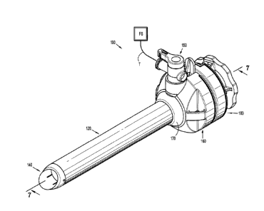
[0026] FIG. 1 is a perspective view of a surgical access device coupled
to a source of
fluid according to an embodiment of the present disclosure;
4
CA 3053138 2019-08-27
[0027] FIG. 2 is an exploded, perspective view of the surgical access
device of FIG. 1
with parts separated;
[0028] FIG. 3 is a perspective view of a tubular member of the surgical
access device
of FIG. 1 shown in phantom;
[0029] FIG. 4 is an enlarged view of the indicated area of detail of FIG.
2;
[0030] FIG. 5 is an enlarged view of the indicated area of detail of FIG.
2;
[0031] FIG. 6 is a perspective view of a distal tip of the surgical
access device of FIG.
1;
[0032] FIG. 7 is a side cross-sectional view of the surgical access
device of FIG. 1
taken along section line 7-7 in FIG. 1;
[0033] FIG. 8 is an enlarged view of the indicated area of detail of FIG.
7;
[0034] FIG. 9 is an enlarged view of the indicated area of detail of FIG.
7;
[0035] FIG. 10 is an end, cross-sectional view of the surgical access
device of FIG. 7
taken along section line 10-10 in FIG. 7;
[0036] FIG. 11 is a cross-sectional view of the surgical access device of
FIG. 7 taken
along section line 11-11 in FIG. 7;
[0037] FIG. 12 is an end cross-sectional view of the distal tip of the
surgical access
device of FIG. 11 taken along section line 12-12 in FIG. 11; and
[0038] FIG. 13 is a perspective view of an endoscope.
DETAILED DESCRIPTION
[0039] Embodiments of the presently disclosed surgical access device are
described
in detail with reference to the drawings, wherein like reference numerals
designate
corresponding elements in each of the several views. As used herein, the term
"distal" refers
to that portion of the instrument, or component thereof which is farther from
the user while
CA 3053138 2019-08-27
t ,
the term "proximal" refers to that portion of the instrument or component
thereof which is
closer to the user.
[0040] Various embodiments of a surgical access device are described
herein. With
initial reference to FIGS. 1 and 2, a surgical access device 100 is
illustrated. The components
of the surgical access device 100 may be formed from suitable biocompatible
materials such
as medical grade metals (e.g., stainless steel), polymeric materials (e.g.,
polycarbonate), or
combinations thereof. The surgical access device 100 includes a housing 160. A
collar 170 is
insertable through the housing 160 and a tubular member 120 extends from a
distal end of the
collar 170. A tip member 140 is located at a distal end of the tubular member
120. A seal
assembly 180 is releasably coupled to a proximal end of the housing 160. An
example of a
suitable seal assembly usable with the presently disclosed surgical access
device 100 is
described in U.S. Patent No. 10,022,149, issued on July 17, 2018, the entire
contents of
which are hereby incorporated by reference. It is contemplated that the
tubular member 120
may include a plurality of spaced annular ribs along a portion of a length of
the tubular
member to improve retention of the surgical access device 100 in an opening
through body
tissue. An example of a cannula with annular ribs is disclosed in U.S. Patent
No. 8,740,925,
issued on June 3, 2014, the entire contents of which are hereby incorporated
by reference.
Additionally, the surgical access device 100 may include a fixation device
such as a balloon,
an umbrella, a foam collar, etc. An example of a surgical access device with a
foam collar
and an anchoring balloon is disclosed in U.S. Patent No. 7,963,975, issued on
June 21, 2011,
the entire contents of which are hereby incorporated by reference. The
surgical access device
100 may include a combination of ribs, balloons, foam collars, or other known
structures for
securing an access device in body tissue. Access devices with other fixation
features are
disclosed in U.S. application no. 62/631,540, U.S. application no. 16/043,279,
U.S.
6
CA 3053138 2019-08-27
application no. 62/653,859, and U.S. application no. 62/568,497, the entire
disclosures of
which are hereby incorporated by reference herein.
[0041] The housing 160 has open proximal and distal ends defining a
cavity 166
therein. The proximal opening has a larger diameter than the distal opening. A
duck bill or
zero-closure seal 162 is positioned in the cavity 166 of the housing 160 (FIG.
7). The zero-
closure seal 162 is formed from a suitable resilient material (e.g., silicone)
and is configured
to prevent fluids from exiting proximally through the housing 160 in the
absence of a surgical
instrument (e.g., an endoscope) inserted therethrough. The zero-closure seal
162 is
sandwiched between the housing 160 and a proximally positioned cap 164. The
cap 164 is
attached to the housing 160 to retain the zero-closure seal 162 in position
and provide a fluid-
tight boundary for the housing 160. The cap 164 may be attached to the housing
160 using
ultrasonic or RF welding, adhesives, or any other suitable technique for the
materials
involved. The housing 160 further includes a port 168 having an opening 169
therethrough
with a valve 150 positioned therein. The valve 150 has a lever 152 that is
rotatable about an
axis of the valve 150 allowing the user to open and close the valve 150. The
lever 152 is
rotatable between an open position of the valve 150 and a closed position of
the valve 150.
The lever 152 may be positioned in one of a plurality of intermediate
positions allowing the
user to adjust the flow rate of a fluid through the valve 150. With additional
reference to
FIGS. 4, 7, and 10, the valve 150 is fluidly coupled to an annular conduit 174
in the collar
170. In particular, the valve 150 is positioned in the opening 169 of port 168
and is aligned
with an orifice 172 of the collar 170. This alignment allows fluid to flow
through the valve
150, the orifice 172, and into the annular conduit 174. In turn, the annular
conduit 174 is open
at the proximal end of the collar 170 for fluidly coupling with lumens 126a-f
in the tubular
member 120 (FIGS. 7 and 10) as will be described in detail hereinbelow.
7
CA 3053138 2019-08-27
. = =
[0042] Referring now to FIGS. 1-4, 7, and 11, the tubular member 120
extends
distally from the collar 170 and is formed of a suitable biocompatible
material. The tubular
member 120 is attached to the collar 170 using known techniques such as RF
welding,
ultrasonic welding, adhesives, etc. The tubular member 120 may be partially or
completely
transparent, translucent, or opaque. A passage or channel 118 extends between
open proximal
and distal ends of the tubular member 120. As illustrated, the tubular member
120 has
substantially uniform inner and outer diameters. It is contemplated that
either the inner
diameter or the outer diameter may vary along a length of the tubular member
120 such that
the tubular member 120 is tapered with one of the proximal or distal ends
having different
diameters from the other of the proximal or distal ends. It is further
contemplated that the
outer diameter of the tubular member 120 may be tapered such that the distal
end has a
smaller outer diameter than the proximal end while the inner diameter of the
tubular member
120 does not vary along the length of the tubular member 120.
[0043] Further, the tubular member 120 has lumens 126a-f defined between
an inner
wall 122 of the tubular member 120 and an outer wall 124 of the tubular member
120. Each
lumen 126 extends longitudinally along a length of the tubular member 120. The
inner and
outer walls 122, 124 have substantially the same length, but are axially
staggered such that a
recess 132 is defined in the distal region of the tubular member 120 (FIG. 5)
and an extension
134 is defined in the proximal region of the tubular member 120 (FIG. 4). The
number of
lumens 126 disposed between the inner and outer walls 122, 124 of the tubular
member 120
may vary. In embodiments, there may be as few as one or two lumens 126 and in
other
embodiments, there may be as many as six lumens 126 as illustrated in FIG. 3.
However, this
does not preclude a greater number of lumens 126 being defined between the
inner and outer
walls 122, 124 of the tubular member 120.
8
CA 3053138 2019-08-27
, = ,
[0044] Each lumen 126 is fluidly coupled to the annular conduit 174
of the collar 170
such that fluid may be supplied to the lumens from a source of fluid FS (FIG.
1) that is
coupled to the valve 150 using tubing T. The outlet 156 of the valve 150 is
fluidly coupled to
the annular conduit 174 via the orifice 172. The fluid may be a cleaning fluid
including, but
not limited to, an insufflation fluid (e.g., CO2), sterile saline, a
surfactant solution, etc. The
fluid flow may be through the valve 150 towards the lumens 126a-f or through
the valve 150
towards the source of fluid FS as determined by the differential pressure
between the lumens
126a-f and an inlet 154 of the valve 150.
[0045] The tip member 140 is located at the distal end of the
tubular member 120.
With additional reference to FIGS. 5, 6, and 12, the tip member 140 includes a
number of
ports 142a-f equal to the number of lumens 126a-f of the tubular member 120.
Each port 142
includes a duct 144 that is fluidly coupled to a corresponding lumen 126 of
the tubular
member 120. Each duct 144 extends longitudinally through the tip member 140
and fluidly
couples one of the lumens 126 with an outlet 146 of the port 142. Each outlet
146 is
configured to direct fluid to a predetermined or target region in the tip
member 140 such that
the output from each port 142 is directed to the same predetermined region
resulting in an
increase in the volume of fluid in the predetermined region. One or more of
the outlets 146
may be configured to generate turbulent fluid flow. As shown in FIG. 8, a
surface of the duct
144 of each port 142 is angled with respect to a longitudinal axis of the
tubular member 120
which functions to direct the fluid from the duct 144 to the outlet 146 of the
port 142 towards
the predetermined region. The tip member 140 has a proximally extending
portion 147 with
an outer diameter is less than an outer diameter of a body 145 of tip member
140 and the
proximally extending portion 147 is receivable in the recess 132 of the
tubular member 120
(FIGS. 5 and 6). A distal portion of the tip member 140 is angled such that
one location
extends further distally than another location (FIG. 5). The tip member 140 is
attached to the
9
CA 3053138 2019-08-27
, .
tubular member 120 using known techniques such as RF welding, ultrasonic
welding,
adhesives, etc. It is envisioned that one lumen 126 may be fluidly coupled to
a plurality of
ports 142. In one non-limiting example, the tubular member 120 may include
three lumens
126a-c that are fluidly coupled to six ports 142a-f where each lumen 126 is
coupled to two
ports 142. Other combinations of lumens 126 and ports 142 are also possible.
[0046] In the illustrated embodiment with six ports, each port 142 is
radially offset by
60 from the adjacent ports 142. In instances where greater or fewer than six
ports are
disposed in the tip member 140, the amount of radial offset of each port 142
from an adjacent
port 142 may be defined by dividing 360 by the number of ports 142 in the
distal tip (e.g.,
four ports would be radially offset by 90 and three ports would be radially
offset by 120 ). It
is contemplated that the radial offset between ports 142 may not be uniform to
create a
different spray pattern of fluid (e.g., four ports that are radially offset by
30 ).
100471 It is contemplated that the ports 142 may not be in the same
plane. In
particular, one port 142 may be closer to the outer wall 122 while an adjacent
port 142 may
be closer to the inner wall 124 such that the ports 142 are not on the same
plane. It is also
contemplated that this staggered arrangement may be repeated for all the ports
142 where one
or more ports are on on plane while other ports 142 are on different planes
(e.g., three ports
located on three different planes). Other combinations of non-planar ports are
also
envisioned. Further, the ports 142 may be arranged in a helical pattern and
the ports 142 may
be angled with respect to the longitudinal axis of the surgical access device
100 to provide a
desired spray pattern. Additionally, the ports 142 may be staggered
longitudinally.
[0048] The fluid flow in the predetermined region is usable to remove
debris from an
outer surface of a lens of a minimally invasive viewing instrument or an
endoscope 200 (FIG.
13). The endoscope 200 has a housing 220 with a shaft 210 extending therefrom.
A viewing
element or lens 212 is located at the distal end of the shaft 210. A monitor M
is coupled to the
CA 3053138 2019-08-27
= .
housing 220 of the endoscope 200 using cable C. The monitor M allows the
clinician to see
what is within the field of view of the lens 212 of the endoscope 200. This
allows the
clinician to observe the surgical site. During a surgical procedure, the
endoscope 200 extends
through the surgical access device 100 such that the lens 212 is in position
in the surgical site
providing the clinician with a view of the surgical site on the monitor M.
When the lens 212
of the endoscope 200 is to be cleaned, the clinician moves the lens 212 of the
endoscope 200
from the surgical site into the chamber 118 of the tubular member 120 such
that an outer
surface of the lens 212 is in the predetermined region such that the fluid
directed into the
predetermined region by the outlets 146a-f of the ports 142a-f impinges upon
the outer
surface of the lens 212 to gently dislodge particulate debris without damaging
the outer
surface of the lens 212. Additionally or alternatively, the clinician may move
the endoscope
200 distally and proximally into and out of the predetermined region to assist
removing
debris from the lens 212. During the movement of the endoscope 200, the
clinician may
check the monitor to locate the position of the lens 212 relative to the
predetermined region.
This allows the clinician to more accurately position the lens 212 of the
endoscope 200 for
cleaning and also determine when the lens 212 of the endoscope is sufficiently
cleaned. This
may be performed with or without a change in the flow rate of fluid into the
predetermined
region to assist in cleaning debris from the lens 212. This cleans the outer
surface of the lens
212 such that the clinician has an unobstructed view through the lens 212 of
the endoscope
200. This arrangement allows the clinician to clean the lens 212 of the
endoscope 200 without
removing the endoscope 200 from the surgical site. As cleaning the lens 212 of
the endoscope
200 may occur dozens of times during a surgical procedure, being able to clean
the lens 212
without removing the endoscope 200 from the access device will streamline the
surgical
procedure allowing the clinician to perform the surgical procedure more
efficiently and in
less time as compared to removing the endoscope 200 multiple times during a
procedure to
11
CA 3053138 2019-08-27
µ =
clean it. Additionally, allowing the endoscope 200 to remain in the access
device for cleaning
reduces the risk of damaging the zero closure seal during repeated removals
and insertions of
the endoscope 200 for cleaning.
[0049] As assembled for use, fluid travels from the source of fluid FS
through tubing
to the inlet of the valve 150. Repositioning the lever 152 of the valve 150
controls the rate of
fluid flow through the valve 150 from zero flow (i.e., valve 150 is fully
shut) to full flow (i.e.,
valve 150 is fully open). With the valve 150 either partially or fully open,
the fluid flows
through the body of the valve 150 and exits the outlet 156 of the valve 150
where it enters the
annular conduit 174 of the collar 170. The annular conduit 174 is fluidly
coupled to the
lumens 126a-f defined between the inner and outer walls 122, 124 of the
tubular member 120
such that fluid exiting the outlet 156 of the valve 150 is directed by the
annular conduit 174 to
the lumens 126a-f and ultimately to the outlets 146a-f of the ports 142a-f.
Although fluid
flow is described as traveling from the source of fluid FS to the outlets 146a-
f of the ports
142a-f, it is contemplated that fluid may flow from the outlets 146a-f of the
ports 142a-f
towards the valve 150 and an associated vacuum source or fluid source FS with
a lower
pressure than the pressure at the outlets 146a-f of the ports 142a-f.
[0050] Persons skilled in the art will understand that the devices and
methods
specifically described herein and illustrated in the accompanying drawings are
non-limiting
exemplary embodiments. It is envisioned that the elements and features
illustrated or
described in connection with one exemplary embodiment may be combined with the
elements
and features of another without departing from the scope of the present
disclosure. As well,
one skilled in the art will appreciate further features and advantages of the
disclosure based
on the above-described embodiments. Accordingly, the disclosure is not to be
limited by
what has been particularly shown and described, except as indicated by the
appended claims.
12
CA 3053138 2019-08-27