Note: Descriptions are shown in the official language in which they were submitted.
CA 03053196 2019-08-08
WO 2018/157067
PCT/US2018/019743
IROA METABOLOMICS WORKFLOW FOR IMPROVED
ACCURACY, IDENTIFICATION AND QUANTITATION
Description
CROSS-REFERENCE TO RELATED APPLICATION
This application claims benefit of
Provisional Patent application No. 62/463153,
entitled "Implementing INS-assisted IROA for
Metabolomics" filed on February 24, 2017, whose
disclosures are incorporated by reference.
BACKGROUND ART
Metabolites are small molecular weight
compounds (less than about 2000 Da and more usually
less than about 1000 Da) that are employed as
building blocks or produced as end products in
various metabolic pathways and cellular regulatory
processes in a biological system. The entire
collection of metabolites in a biological system,
whether at the cellular, pathway or organism level,
is known as a "metabolome". Levels of these
metabolites in a metabolome are either dictated by
the genome, proteome, and/or transcriptome of the
biological system or imposed by environmental
perturbations and results in changes in phenotype.
Thus, metabolomics can be applied to map or identify
the cause of alteration in phenotype and understand
correlations between "omics". Dwivedi, et al., Int J
Mass Spectrom 2010 298:78-90.
Isotopic Ratio Outlier Analysis (IROA) has
been developed to enable the characterization of
carbon information in a given metabolites or a
-1-
CA 03053196 2019-08-08
WO 2018/157067
PCT/US2018/019743
fragment. Unlike other stable isotope labeling
methods, rather than utilizing substrates with
natural abundance (1.1% of 130 isotopomer seen in
carbon atoms in nature) and 98-99% enrichment for the
control and experimental populations, respectively,
IRO with prototrophic yeast uses randomized 95% 120
glucose (5% 13C), and 95% randomized 130 glucose (5%
120) as carbon sources. This strategy leads to more
predictable and diagnostic patterns for the
observable isotopic peaks in the mass spectra. [Qiu
et al., Metabolites 2018 8:9].
The promise of IROA for metabolic
phenotyping has been demonstrated in model organism
studies. Saccharomyces cerevisiae, a prototrophic
wild-type strain in the CEN.PK background [Brauer et
al., Mb/. Biol. Cell 2005, 16:2503-2517] was grown in
minimal yeast nitrogen base (YNB) media, containing
either randomized 95% 120, or 95% 130 glucose as the
main carbon source, in order that the isotopomer
pattern of all metabolites would mirror the labeled
glucose [Qiu et al., Anal. Chem. 2016, 88:2747-2754],
a protocol that can easily be adapted for microbial
species studies.
The abundance of the light isotopologues in
the 95% 130 samples (m Mn_2, etc., the 130
envelope) or the heavy isotopologues in the 95% 120
samples (M0+1, M0+2, etc., the 120 envelope), follows
the binomial distribution for 130, based on the
initial substrate enrichment, in the metabolite
products generated. The mass difference between the
120 (M0) isotopic peak and the 130 (Mn) isotopic peak
indicates the number of carbons (n) in the
metabolite's carbon backbone. This narrows
possibilities for chemical formula generation (CFG)
-2-
CA 03053196 2019-08-08
WO 2018/157067
PCT/US2018/019743
and for normalization between control (130) and
treated (120) groups. [Qiu et al., Metabolites 2018
8:9].
It is possible to use metabolomic
techniques, such as the IRO A basic, or IROA
phenotypic protocols (optimally)[de Jong and Beecher,
C. Bioanalysis 2012, 4 (18):2303-2314], or standard
metabolomic techniques to identify and crudely
quantify several hundred or even thousands of
compounds in a biological sample. However, to make
such measurements and to compare the measurements
from any two or more samples, all the samples need to
be analyzed in a single batch, ideally during a
single day because day-to-day variances are too great
to otherwise overcome, and absolute quantitation;
i.e., relative to a known standard, cannot be
assured.
It is currently not quantitatively
acceptable to compare samples run on the same
instrumentation several days apart, and impossible to
compare data generated on different instruments, or
based on different methods. Instrument drift,
chromatographic drift, and even environmental
conditions can alter results sufficiently so that
reproducibility is hard to obtain even on the same
instrument. In addition to these problems of
quantitation, the identification of any compound
across many mass spectral techniques alone is
unlikely to be successful unless very careful
calibrations have been made and authentic standards
are run. This is because, not only are there
multiple biological compounds that can be confused
because they have the same exact mass but, even more
problematic, there are often more artefactual or
-3-
CA 03053196 2019-08-08
WO 2018/157067
PCT/US2018/019743
fragmentary compounds that are structurally different
from, but can share the correct mass, or even
formulae, as biological isobaric equivalents.
The invention disclosed hereinafter extends
methods described in the following U. S. Patents: No.
7,820,963, the basic IROA patent, issued October 26,
2010, referred to hereinafter as IROA963; No.
7,820,964, issued October 26, 2010, and referred to
hereinafter as IROA964; No. 8,168,945, issued May 1,
2012, referred to hereinafter as IROA945; No.
8,536,520, issued September 17, 2013, referred to
hereinafter as IROA520 ; and No. 8,969,251 that
issued March 3, 2015, and is referred to hereinafter
as IROA251. These patents and the art cited therein
are incorporated herein by reference.
The IROA protocols rely on the creation of
isotopic patterns that are mathematically informative
to insert information into biological samples to
provide better identification and quantitation of the
individual chemical components when the samples are
subjected to mass spectral analysis. Traditional
methods required chromatographically clean; i.e.,
"baseline", separation to achieve the best
quantitative accuracy, the IROA protocols do not and
hence can be used in the quantitation of very
chemically complex samples where such separation is
not consistently possible.
The exemplary samples studied were
uniformly and universally labeled with appropriate
isotopes. An element in which there are two stable
isotopes that are not significantly distinguished by
enzymes or living systems is preferably used. Carbon
(specifically, 12 C and 13C) is used for purposes of
illustration herein because of its universal
-4-
CA 03053196 2019-08-08
WO 2018/157067
PCT/US2018/019743
applicability. However, additional examples are well
known to a worker of ordinary skill.
The use of isotopes that exhibit minimal
biological isotope effect is of import. For
instance, the use of the isotopes of hydrogen such as
deuterium (D) is not suitable because it frequently
causes an observable effect on metabolism due to the
fact that the deuterium isotope has a mass that is
twice that of hydrogen, and thus causes a reduction
in the kinetics of some enzyme mechanisms. Tritium
(T) is radioactive and thus not stable to decay.
In many of these protocols the production
of the IROA patterns relies on the creation of
molecules where the probability of all carbons in a
molecule is carefully constrained to a close range of
isotopic probabilities. Illustratively, for a system
using stable isotopes of carbon [carbon-12 (120) and
carbon-13 (130)], the isotopic ratios in this example
specifically include a dilution of five to ten
percent of one carbon isotope in another; i.e., one
sample is grown on a carbon source (nutrient in a
medium) that can be 95% carbon-12 (120) and 5%
carbon-13 (130), hereinafter called "0-12 medium",
and in such a situation the other sample is grown in
mirrored medium that contains a nutrient that
contains 95% carbon-13 and 5% carbon-12 in a medium,
hereinafter called "0-13 medium". In each of these
cases the biological system takes up the nutrient in
the medium and grows upon it in such a way as to
transform itself so that all of its parts are
distinctively identifiable as to their origin.
Further information can sometimes be obtained by
incorporating a second set of two isotopes of a
-5-
CA 03053196 2019-08-08
WO 2018/157067
PCT/US2018/019743
second atom present at two different predetermined
isotopic ratios into the nutrient compositions.
When the two samples are mixed,
intermingled or otherwise composited, the composite
sample contains molecules from both the "control"
(that are made up of a substantial majority, e.g.,
90% to 95%, of 12C) and the "experimental" (that are
made up of a substantial majority, e.g., 90% to 95%,
of 13C). Deviating significantly from the 90% to 95%
ratio taught by this method reduces the potential for
interpretation as is taught in IROA963, although 98%
and 2% of the carbon isotopes have been successfully
used.
More specifically still, the probability
can be set to 95% C-13 in an illustrative IROA
standard sample. In such a standard all the molecules
contained in it exhibit the property that the
probability for of its carbons will be as close to 95%
13C as is achievable. Such IROA molecules have many
special properties, namely:
1) The isotopic balance of 12C to 13C is so
much larger than the natural abundance probability of
approximately 1.1% and yet is specifically not
approaching 100%, therefore each molecule presents
itself as a collection of isotopomeric sets of that
molecule with the mass of each set differing by the
mass of exactly one carbon neutron, or approximately
1.00335 AMU. These sets are significantly larger and
more complex than natural abundance equivalents and
can be easily identified.
2) The distribution of isotopomers across
the above sets is a function of the number of carbons
in the molecule and the probability of a 130 in each
such position. The presence of isotopomeric sets
-6-
CA 03053196 2019-08-08
WO 2018/157067
PCT/US2018/019743
contributed from other natural abundance sources of
hydrogen, oxygen, nitrogen, etc. are so small that
their patterns are equally distributed into and
insignificant to the 013 isotopomeric sets.
3) The amount of isotopomers for each IROA
molecule can be deduced in a mass spectrometer as the
height of a peak, and therefore the relative
concentration of all isotopomeric sets creates a
pattern of peaks for each molecule. This pattern is
effectively defined as a binomial distribution the
percent (x), and the number of carbons (n), and
therefore can be calculated as probabilities, ((1-x) +
(x))1-1.
4) These IROA patterns are dominant
features of any mass spectral analysis of an IROA
sample. Because the patterns themselves can be quite
complex, their occurrence due to random peak noise is
effectively non-existent. Software was developed that
identifies these patterns with great accuracy.
5) The 012 and 013 monoisotopic masses of
such a molecule cannot be seen but can be determined
by inspection of the shape of the patterns seen. The
monoisotopic mass constrained by the number of carbons
effectively is an unique determinant of the molecular
formula of the molecule, significantly more accurate
than attempting to solve the polynomial equations
required for natural abundance molecules.
6) Aside from the mass differences of
their isotopomeric sets, the molecules are otherwise
indistinguishable and thus perform very similarly
through almost all treatments and generally have the
same physical characteristics. This characteristic of
IROA peaks is a basis of the IROA Identification
Techniques.
-7-
CA 03053196 2019-08-08
WO 2018/157067
PCT/US2018/019743
There are many IROA protocols based on
these properties. The following two IROA protocols
are relevant to this invention.
The Basic IROA Protocol
The basic IROA protocol (which was
described in IR0A963, and continued in IR0A945, and
IR0A520) creates two populations of IROA molecules
containing widely different amounts, typically 90-95%
and 10-5% of the first and second isotopes,
respectively, and 10-5% of the first isotope with 90-
95% of the second isotope. Isotopes other than
hydrogen and deuterium are preferred such as the
particularly preferred approximately 5% 013 and
approximately 95% 012 used with approximately 5% 012
and approximately 95% C13.
In both populations, the distribution of
013 in every compound is random and universal and the
probability of a carbon being either a 012 or a 013
is the stated value, here either approximately 5% or
95%. The experimental "base" population of molecules
(012-B) with approximately' 5% 013 and the remaining
carbons (95%) are 012. The control "Internal
Standard" population (013-IS) sample made up of
approximately 95% C13 and 5% 012.
Because both the 012-B and 013-IS are made
up of IROA molecules:
1) For any given molecule their respective
peak patterns are different, but both solve to the
same molecular formula. The 012-B monoisotopic peak
has a distinct M+1, M+2 peaks, and possibly
additional M+n peaks. The 013-IS monoisotopic peak
has a distinct M-1, M-2 peaks, and likely additional
M-n peaks.
-8-
CA 03053196 2019-08-08
WO 2018/157067
PCT/US2018/019743
2) Unlike isotopomers based on deuterium,
these isotopomers co-chromatograph and exhibit very
similar physical properties except for mass.
3) IROA compound peaks can only be created
in most experimental systems through biological means
(IRO1520), but intentional synthetic IROA compounds
(IROA964) can also be prepared and added. In this
workflow, the presence of an IROA signal assures that
all IROA patterns can come only from the C13-IS or
the C12-B, and that they are immediately
distinguishable from artefact, electronic noise, or
any spurious signals that are always be based on
natural abundance isotopic signatures.
4) When the patterns from the same
molecule from both the C12-B sample and the C13-IS
are found in the same sample, the paired signal is a
triply redundant information system in which:
a) the number of carbons in the
molecule can be determined by the ratio of the height
of the M+1 to the C12-B monoisotopic for the IROA
molecules coming from the experimental samples,
b) the number of carbons in the
molecule can be determined by the ratio of the height
of the M-1 to the C13-IS monoisotopic peak for the
IROA molecules coming from the C13-IS samples, and
c) the number of carbons in the
molecule can be determined by the mass difference
between the monoisotopic mass of the molecules coming
from the experimental sample, and the mass of the
monoisotopic from the 013-IS.
When all three of these calculations
indicate the same number of carbons, it is extremely
likely that the pattern has been correctly found, and
that the probability of error is extremely low.
-9-
CA 03053196 2019-08-08
WO 2018/157067
PCT/US2018/019743
Because discovery of these patterns can be entirely
software- driven, the discovery of such peaks is a
completely automatable task (IROA945).
The basic IROA protocol permits for a
completely unbiased (or non-targeted) analysis of an
experimental sample in which the 012-B can be made to
vary according to an experimental design for purposes
of discovery of the biological effect of such
experimental design. In such a sample the C12-B
population is derived from an experimental sample,
and if a molecule does not happen to be in either the
013-IS or the 012-B sample, the presence and probable
identification of the molecule is still possible, and
the absence of the molecule in the other is an
establishable fact.
Although not triply redundant, the presence
of a randomly created (i.e. artefactual) IROA peak is
so low that a single IROA peak is easily identified
as such and can be quantified. This basic IROA
protocol is therefore suited to experimental
situations in which the ability to find and
characterize all the peaks of biological origin in
either the 012-B1 or 013-IS, thereby identifying
those situations in which a molecule is present in
one but missing from the other.
The triple redundancy of the basic IROA
protocol is such a strong algorithm that it is
possible to find very weak signals even in the
presence of very strong noise by simply enforcing the
peak shape requirements.
In the case of Matrix, where the 012-B and
013-IS sides are both of equal chemical composition
and matching isotopic balance, by design, the
requirement for symmetry makes it easy to find many
-10-
CA 03053196 2019-08-08
WO 2018/157067
PCT/US2018/019743
very small peaks in deep noise situations with little
chance of error. Thus, Matrix represents a special
case of the IROA Basic protocol in which its
characteristics are so predictable as to make the
information derived from it especially reproducible
and capable of being found at extremely low levels of
detection.
The IROA Workflow is based on this unique
property. The source of material for Matrix can be
either biological or synthetic. The IROA
Identification Techniques can be applied to any IROA
peak to further strengthen the identification of the
underlying compound.
The Phenotypic IROA Protocol
The Phenotypic IROA protocol is a protocol
for situations in which it is not feasible or
practical to label the experimental sample itself but
a common and consistent 95% (+/- 3%) IROA internal
standard, such as the above described C13-IS, is used
to assure accurate identification of a molecule and
accurate quantitation. The Phenotypic Protocol is
useful for the analysis of human (clinical) samples,
agricultural samples, industrial samples, or other
situations where the size or the source of the
experimental samples is such that it is simply not
feasible to label them. However, the Phenotypic
protocol, by providing a common rigorous IROA
internal standard, provides a more accurate route for
the identification and quantification of a large
number of compounds that are found in the sample
natural abundance isolates.
Unlike the "unbiased" or "non-targeted"
analysis of basic IROA, Phenotypic IROA is a targeted
-11-
CA 03053196 2019-08-08
WO 2018/157067
PCT/US2018/019743
quantitative analysis of a very large number of
compounds based on a very chemically complex IROA
internal standard (IS). A C13-IS can contain well
over 1000 compounds (potentially unlimited), but the
IROA properties outlined earlier do not require
complete chromatographic separation to assure both
the identity and quantitation of all the compounds
contained in the IS.
The Phenotypic protocol puts an IROA
internal standard into every natural abundance sample
and uses the dual pieces of information from the C13-
IS, 13C-monoisotopic mass and number of carbons, to
locate the natural-abundance isotopomer of the same
compound. Correlation of the natural abundance time-
resolved chromatographic profile of the found peak,
and it's natural-abundance isotopic form are then
used to support the IROA-based identification.
Because the IROA peaks are informatically
self-contained, it is possible to correctly identify
and quantify multiple co-eluting peaks. In the case
of the Phenotypic Protocol, the IS can be created by
a worker to provide support for the unique
quantitation needs of the experimental system. Thus,
a wheat researcher, can create a wheat 013-IS that
can be used because it contains a chemical profile
more reflective of wheat biochemistry, but this 013-
IS is used primarily to find and identify IROA peaks
in wheat and quantify their natural abundance
counterparts. Although the triple redundancy of the
Basic IROA protocol does not exist in the Phenotypic
protocol, the signal is still redundant in that the
95% C13-IS provides a mass and number of carbons to
determine exactly where the natural abundance
monoisotopic signal is found (see Fig. 10F).
-12-
CA 03053196 2019-08-08
WO 2018/157067
PCT/US2018/019743
In the IROA workflow, the same 013-IS is
used in both the Matrix and the Clinical or
experimental samples and the chemical information
derived from the Matrix sample is used to verify and
validate the compounds found in the clinical or
experimental (Phenotypic) samples. The Phenotypic
samples can be analyzed for chemical information to
the same extent as the Matrix samples but this is not
required. For instance, whereas the Matrix samples
need to be analyzed to completely characterize every
compound present in it, it can be sufficient to use
the mass and retention information derived from the
analysis of the Matrix to find the same compounds in
the experimental or clinical samples, and use a
higher acquisition rate than would be possible in the
Matrix samples to achieve a higher quantitative
accuracy.
BRIEF SUMMARY OF THE INVENTION
In one aspect, the present invention
contemplates an IROA Matrix composition of
biologically-produced metabolite compounds. Each of
those metabolite compounds has a molecular weight of
about 2000 AMU or less. Each of the metabolite
compounds is present as first and second isotopomers
that are equally present at two predetermined
isotopomeric balances. The first isotopomers contain
about 2 to about 10% of a first isotope, and the
second isotopomers contain about 90 to about 98% of a
second isotope of the same atom. The first and
second isotopes are stable to radioactive decay and
are other than hydrogen and deuterium.
The biologically-produced metabolite
compounds are obtained from a cell lysate preparation
-13-
CA 03053196 2019-08-08
WO 2018/157067
PCT/US2018/019743
obtained from culture of single-celled or multi-
celled organisms, and the molecules are randomly and
universally labeled with isotope pairs of one or more
elements selected from the group consisting of
isotopes of carbon (12C and 13C), nitrogen (14N and
15N), oxygen (160, 170, or 180), sulfur (32S, 33S,
34S, or 36S), chlorine (3501 and 37C1), magnesium
(24Mg, 25Mg and 26Mg), silicon (27Si, 28Si and 29Si),
calcium (40Ca, 42Ca, 43Ca, and 44Ca), and bromine
(79Br and 81Br).
Another contemplated aspect of the
invention is a method of creating a reference library
of identity data of compounds in an IROA Matrix as
described above, and comprises the steps of 1) mass
spectrally determining the identity of the compounds
of an IROA Matrix that are within the resolution and
sensitivity of the apparatus to provide its
symmetrical IROA peak pattern, and additionally
determining one or more of: a) the gas and/or liquid
chromatographic properties of the compounds present,
b) the collisional cross section of the compounds
present, and c) the fragmentation pattern of the
compounds present. The compound identity data so
determined is maintained for use in identifying one
or more of the same compounds in a later-analyzed
sample. The reference library of identity data of
compounds in an IROA Matrix is itself also
contemplated. The use of one or both of compound
collisional cross sections and fragmentation patterns
are preferred in conjunction with mass spectral
identification.
A further contemplated invention is a
method of quantifying and identifying compounds in a
natural abundance sample using an Internal Standard
-14-
CA 03053196 2019-08-08
WO 2018/157067
PCT/US2018/019743
that is of the same chemical composition as
isotopomers containing the about 90 to about 98% of
the heavier molecular weight isotope-containing
compounds of an IROA Matrix composition and is
inserted into that natural abundance sample. Each
compound in the Internal Standard is itself
identified in a before-described reference library of
identity data. It is preferred that the quantity of
each identifiable compound of the natural abundance
sample is determined, and more preferably, the
quantity of each natural abundance sample compound is
determined relative to the Internal Standard.
Yet another aspect of the invention is a
method of measuring quality assurance and/or a
quality control on the operational constancy of a mass
spectral apparatus and associated ion mobility channel
and chromatographic apparatus, when present. That
method comprises the steps of assaying the sample of
an IROA Matrix composition as described above, and
determining whether the same sets and amplitudes of
symmetric IROA mass spectral peaks are present in each
analysis. The preferences noted above in regard to an
IROA Matrix composition are repeated here and in each
time a Matrix composition or its components are used
herein.
A still further aspect of the present
invention contemplates a reagent pair capable of
transforming a natural abundance mass spectral
analysis metabolite sample into an IROA sample. That
reagent pair comprises two reactively identical
reagents that constitute first and second
isotopomers. The first isotopomers contain about 2
to about 10% of a first isotope, and the second
isotopomers contain about 90 to about 98% of a second
-15-
CA 03053196 2019-08-08
WO 2018/157067
PCT/US2018/019743
isotope of the same atom. The first and second
isotopes are stable to radioactive decay and are
other than hydrogen and deuterium. Each of the
reagent pair contains the same reactive group that
reacts with and bonds to a functional group of one or
more compounds present in a composition of
biologically-produced metabolite compounds. Each of
the biologically-produced metabolite compounds of the
natural abundance mass spectral analysis sample has a
molecular weight of about 2000 AMU or less.
A reagent pair reactive group reacts with
and bonds to a biologically-produced metabolite
functional group selected from the group consisting
of one or more of an amine, aldehyde or ketone,
hydroxyl, thiol and carboxylic acid. Preferably, a
reactive group reacts with and bonds to an amine
functional group. A preferred reactive group is a
isothiocyanate reactive group, and the reagent pair
are isotopomers of phenylisothiocyanate whose first
isotopomers contain about 2 to about 10% of a first
isotope, and whose second isotopomers contain about
90 to about 98% of a second isotope. An alternative
pair of reagents are IROA isotopomers of a hydrazine
or a semicarbazide that react and bind to carbonyl
groups of aldehyde and ketone groups present in a
natural abundance mass spectral analysis metabolite
sample.
BRIEF DESCRIPTION OF THE DRAWINGS
In the drawings forming a portion of this
disclosure,
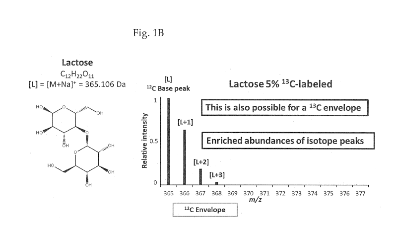
Fig. 1A shows the mass spectral peaks
obtained on the analysis of lactose that contains
naturally abundant amounts of 120 and 130, Fig. 1B
-16-
CA 03053196 2019-08-08
WO 2018/157067
PCT/US2018/019743
shows the mass spectral peaks obtained on the
analysis of lactose that contains 95% 120 and 5% 130,
Fig. 10 shows the mass spectral peaks obtained on the
analysis of lactose that contains 5% 120 and 95% 130,
and Fig. 10 shows the mass spectral peaks obtained on
the analysis of lactose that contains equal amounts
of lactose that contains 5% 130 with 95% 120 along
with 95% 130 and 5% 120;
Fig. 2 illustrates the spectra of lactose
containing natural abundance of both 120 and 130 as
well as peaks obtained from lactose containing 95%
130, and illustrates the number of carbon atoms in
the assayed molecule by the difference in m/z value
of the two base peaks being 12 AMU;
Fig. 3 illustrates the symmetrical
arrangement of IROA peaks with the difference between
the m/z values for the two base peaks defining the
number of carbon atoms present in the assayed
compound. See, e.g., de Jong, F. A.; Beecher, C.
Bioanalysis 2012, 4 (18), 2303-2314;
Fig. 4 broadly illustrates steps in the
preparation of an IROA sample prepared separately
from Saccharomyces cerevisiae grown in a medium that
contains 5% 130 or 95% 130 as the main source from
which a solvent-soluble (usually water) cell lysate
is prepared, providing two solutions that contain the
same amount of each yeast metabolite compound present
and containing either 5% 130 or 95% 130, which are
then combined to form a pooled extract that is
freeze-dried to form a reconstitutable IROA standard
referred to herein as "Matrix".. See, e.g., Qiu, et
al., J. Anal. Chem.2016, 88 (5), 2747-2754;
Fig. 5 whose upper portion shows a
schematic of a IM apparatus drift tube with the
-17-
CA 03053196 2019-08-08
WO 2018/157067
PCT/US2018/019743
analyte ions within the dashed rectangle and ions of
unknown origin outside of that dashed rectangle and a
simulated ion mobility spectrometric- (1MS-) Assisted
IROA mass spectrum containing peaks with added X's
above them to indicate the peaks due to those ions of
unknown origin, and in which ions of unknown origin
that interfere with detected IROA masses but have
been separated by ion mobility are represented with
black circles;
Fig. 6 is similar to Fig. 5, except that
this figure is a simulation in which two isomers are
detected and as seen in the upper schematic IM drift
tubes whose separated analytes are shown within the
two dashed rectangles and ions of unknown origin
outside of those dashed rectangles with the same
meanings as in Fig. 5, and because there are IROA
internal standards for them, they can be
independently quantitated, which would not be
possible without both the IROA internal standards and
ion mobility. See, e.g., Dwivedi et al., Int. Jounal
Mass Spectrom. 2010, 298 (1-3):78-90 that discusses
use of IMS-assisted mass spectroscopy;
Fig. 7A illustrates a LC-IM-MS analysis of a
portion of the pooled yeast extract (IROA Matrix)
prepared as discussed in Fig. 4 and in greater detail
hereinafter, in which the portion of the LC
separation within the box is the portion analyzed
mass spectrally and the line beneath the boxed line
illustrates the elution solvent gradient, Fig. 7B
illustrates a portion of the LC separation that was
analyzed (boxed peak) with the resulting mass
spectrum for an 11 carbon compound adjacent to the LC
trace, and Fig. 7C illustrates further details of the
separation and MS analysis in the upper portion such
-18-
CA 03053196 2019-08-08
WO 2018/157067
PCT/US2018/019743
as retention time, mass range and drift time, as
well as IM analysis readily showing the IROA peak
pattern, specific drift time and number of carbons in
the analyzed compound;
Fig. 8A illustrates a LC-IM-MS analysis of
another peak (boxed) from a LC separation in which a
carbon compound was the analyte, and Fig. 8B
illustrates one IROA recognized pattern using LC-MS
separation with further details that indicate that
two compounds are present from the IM data;
Fig. 9A illustrates an overlapping MS peak
pattern in compounds obtained from the boxed LC peak
on the left side of the figure, Fig. 913 illustrates
the deconvoluted spectra in which the IM data
indicate that two nine carbon compounds have the same
chemical formula, and drift times matched with
identity data reference libraries and that a ten
carbon compound was also present, and Fig. 9C shows
the mass spectra for each of the three compounds
identified;
Fig. 10A is a mass spectrum of arginine in
which C12 and C13 are present in natural abundance,
whereas Fig. 10B shows the similar spectrum using
arginine that contains 95% C12 and 5% C13, Fig. 10C
shows the mass spectrum for arginine that contains
95% C13 and 5% C12, Fig. 10D shows the basic IROA
spectrum of arginine when equal amounts of the
compound containing 95% C12 and 5% C13 and the
compound 95% C13 and 5% C12 are present, Fig. 10E
illustrates triply redundant IROA peak patterns for
arginine natural abundance noise in which
relationship between the monoisotopic peaks is
assured when the height of the C12 M+1, the C13 M-1,
and the mass difference between the two monoisotopic
-19-
CA 03053196 2019-08-08
WO 2018/157067
PCT/US2018/019743
peaks all indicate the same number of carbons are
present in the molecule, and Fig 105 illustrates
Phenotypic "redundancy", in which the identity of the
natural abundance peak is confirmed by both the
molecular formula of the 130 monoisotopic peak and
the number of carbons indicated by the height of it's
M-1 provided by admixture of the 013-IS sample to the
sample for analysis. Peaks associated with arginine
are starred (*) in each spectrum of Figs. 10A-D;
Fig. 11A shows mass spectral peaks present
when adenosine is assayed using a sample containing
equal amounts equal amounts of the compound
containing 95% 012 and 5% 013 and the compound 95%
013 and 5% 012, and Fig. 11B shows mass spectral
peaks present when phenylalanine is assayed using a
sample containing equal amounts equal amounts of the
compound containing 95% 012 and 5% 013 and the
compound 95% 013 and 5% 012. It is noted that the
number of carbon atoms present is provided by the
difference in m/z values for the base peaks in each
spectrum;
Fig. 12A illustrates the mass spectral IROA
peak pattern for phenylalanine based on use of a
mixture of equal amounts of 5% 012 and 95% 013
phenylalanine and 95% 012 and 5% 013 phenylalanine;
Fig. 12B illustrates the mass spectral IROA peak
pattern for that same phenylalanine sample after
SWATH fragmentation; and Fig. 120 provides IROA
diagnostic structural information via fragment
interpretations from the peaks of Fig. 12B;
Fig. 13A and Fig. 13B illustrate that
derivatized peaks of arginine maintain their IROA
character in ion mobility [with and without
differential mobility spectrometry (DMS)] when
-20-
CA 03053196 2019-08-08
WO 2018/157067
PCT/US2018/019743
derivatized using either isotopically labeled IROA
compounds (Fig. 13A) or with natural abundance
compounds derivatized with isotopically labeled
reagent such as a 95% 013 phenylthiocarbamyl (PITC)
group (Fig. 13B). The collection of isotopomers
appear as a unit in Ion Mobility, here Sciex
SelexIon'.
Fig. 14A and Fig. 14B illustrate a similar
maintenance of IROA character for similarly prepared
and assayed tyrosine derivatives;
Fig. 15A, Fig. 15B, Fig. 150 and Fig 15D
illustrate the power of IROA, particularly in
conjunction with ion mobility to separate complex
spectra into their component individual spectra.
Thus, the isotopomeric collections of IROA peaks
remain IROA peaks in IM, here using an Agilent 6560
(ion mobility time of flight) IM-QTOF machine, that
uses Drift Tube IM (DT-IM). Although the
ClusterFinderTM software separates out overlapping
IROA peaks based on mass differences in the pre-IM
Mass spectrum (Fig. 15A) here two co-eluting IROA
peaks are separated cleanly in the IM (Fig. 15B) for
complete compound spectral identification (Fig. 150
and Fig. 15D) based on their IROA characteristics.
Definitions:
As used herein, the abbreviations "130",
"013" and "13C" all refer to the isotope of the
element carbon that has an atomic weight of 13 AMU.
Similarly, the abbreviations "120", "012" and w120÷
all refer to the isotope of the element carbon that
has an atomic weight of 12 AMU.
-21-
CA 03053196 2019-08-08
WO 2018/157067
PCT/US2018/019743
Chromatography can mean any form of a
chemical separation, including but not limited to all
forms of liquid chromatography (LC), gas
chromatography (GC), capillary electrophoresis (CE),
ion mobility (IM), solid phase extraction (SEE), etc.
Compound identification means any method of
determining the physical characteristics of a
chemical compound, including but not limited to mass
spectroscopy (ms), fragmentation (msms), charge and
electronic properties (ms, IN, etc.), shape (IM,
drift, etc.), bond and vibrational properties
(various spectroscopic methods), and it's IROA form
(base mass and number of carbons).
A Matrix is a standard well-defined Basic
IROA mixture of compounds such as metabolites,
including anabolite and catabolite molecules, or
other compounds utilized or present in a given study
and contains at least one compound a pair of stable
isotopes of the same element that differ in molecular
weight (AMU) by at least one AMU. The two isotopes
are present in the molecules of that at least one
compound in a predetermined ratio that is other than
the naturally occurring ratio of those isotopes.
Various Matrices exist, but each matrix
supports a specific analytical system, such as
plasma, human biopsies, wheat, urine, etc. In
addition, a plurality of Matrices can be prepared for
the same specific analytical system.
A library is a group of compounds known to
be present in a Matrix.
An Internal Standard (IS) is a chemical
mixture of compounds that can represent either the
lighter or heavier set of IROA compounds such as
metabolites, subset thereof of a Matrix sample, or
-22-
CA 03053196 2019-08-08 2018/157067
PCT/US2018/019743
other compounds present or utilized in a Matrix
sample of a given study, and is inserted exogenously
into every sample that is to be analyzed. Like a
Matrix, the IS is a standard well-defined mixture of
compounds. The chemical compositions of both the
Matrix and IS are ideally identical.
As used herein, predetermined first and
second stable isotope amounts are preferably present
in "inverted ratios" of each other such as those
discussed immediately above in which the number of
the numerator of the first ratio is the number of the
denominator of the second ratio, and the number of
the denominator of the first ratio is the number of
the numerator of the second ratio.
Taking the above ratios of 95% and 5%, a
first ratio would be 95/5 12C/13C in the C-12 medium,
whereas the second, inverted ratio, would be 5/95
12C/13C in the C-13 medium. It is to be understood
that a contemplated set of ratios need not be 95/5
and 5/95, and although those amounts are particularly
preferred, they are used herein for convenience.
It is to be understood that the first and
second stable isotopes present in a Matrix or any
other exogenously provided composition such as an
internal standard are predetermined and as are their
respective amounts of each isotope. As a
consequence, the words "predetermined" and "stable"
are rarely used herein with their presence implied to
minimize verbosity.
DETAILED DESCRIPTION OF PREFERRED EMBODIMENTS
A first aspect of this invention
contemplates an IROA Matrix composition of
biologically-produced metabolites, including
-23-
CA 03053196 2019-08-08
WO 2018/157067
PCT/US2018/019743
anabolite and catabolite molecules, that is typically
a room temperature solid that is dispersible or
soluble in an aqueous medium (as defined
hereinafter). The individual metabolites have a
molecular weight of less than about 2000 AMU,
preferably about 1500 AMU or less, and more preferably
less than about 1000 AMU. The lower weight limit for
a contemplated metabolite is about 60-75 AMU as in
acetic acid and glycine.
Every compound is equally present at both
of two predetermined isotopomeric balances such that
each of the isotopomers is present at about 2 to
about 10% of isotope one and at about 90 to about 98%
of isotope two. Illustrative useful first and second
isotopes of the same atom are one or more elements
that include the isotopes of carbon (120 and 130),
nitrogen (14N and 15N), oxygen (160, 170, or 180),
sulfur (32S, 33S, 34S, or 36S), chlorine (3501 and
3701), magnesium (24Mg, 25Mg and 26Mg), silicon
(27Si, 28Si and 29Si), calcium (400a, 42Ca, 430a, and
440a), and bromine (79Br and 81Br). The first and
second isotopes are stable to radioactive decay (can
be used in a laboratory without added protection from
possible radiation injury), and are other than
hydrogen and deuterium.
Put more explicitly in terms of the
particularly preferred isotopes, 012 and 013, one
group of isotopomers contains about 2 to about 10%
013 and the other group contains about 90 to about
98% 013. Preferably, a first group contains about 5
to about 10% 013 and the second group contains about
90 to about 95% 013, with the remaining carbon atoms
being 012 in each instance. It is particularly
preferred that the first group contains about 5% 013
-24-
CA 03053196 2019-08-08
WO 2018/157067
PCT/US2018/019743
and the second group contains about 95% 013, with the
remaining carbon atoms being C12. This means that
the IROA peak shape for each compound ideally is
comprised as a perfectly balanced, symmetrical
collection of peaks, with each half a mirror image of
the other.
It is to be understood that the above-
stated percentages are intended to be identifiably
different from the natural abundance amounts of the
two isotopes used. Thus, in the case of carbon
isotopes, whose natural abundances are 98.89% for 012
and 1.11% for 013, use of about 90 to about 98% for
one isotope and about 2 to about 10% of the other
isotope permits the analytical equipment to readily
distinguish between natural abundance peaks and those
provided by an IROA Matrix. Use of the term "about"
for the percentage of one or the other isotopomers
present is meant to be within 3% of the stated
amount. Thus, the above isotope percentages are
known and predetermined, but use of specific amounts
within the ranges stated is mostly a matter of
convenience.
It is also to be understood that trace,
impurity amounts of the element used for an IROA
study, here carbon, can also be present among the
atoms of that element. Such trace amounts are
typically of no consequence to a study. For example,
the Handbook of Chemistry and Physics, 54th ed., CRC
Press, Cleveland, OH, page B251, 1973-1974, lists the
natural abundance of 012 and 013 as being 98.89 and
1.11 percents, respectively, with the presence of 014
being reported, but not its percent amount.
It is still further to be understood that
use of the words "first" and "second" in regard to
-25-
CA 03053196 2019-08-08
WO 2018/157067
PCT/US2018/019743
the isotopes and the several compositions that can
contain them is only for purposes of clarity to
distinguish the isotopes, and is not meant to imply
anything concerning the order of carrying out any
manipulations.
It is preferred that IROA matrices be
prepared in relatively large quantities, such as
about 10 to about 100 g for an industrial scale and
about 10 to about 1000 mg on a laboratory scale so
that each batch can be utilized over many spectral
analyses. The obtained Matrix composition is
preferably kept frozen such as at -80 C until used
to maximize its chemical stability and analytical
reproducibility.
Another contemplated aspect of the
invention is a method of creating a reference library
of identity data of compounds in an IROA Matrix as
described above, and comprises the steps of 1) mass
spectrally determining the identity of the compounds
of an IROA Matrix that are within the resolution and
sensitivity of the apparatus to provide its
symmetrical IROA peak pattern, and additionally
determining one or more of: a) the gas and/or liquid
chromatographic properties of the compounds present,
b) the collisional cross section of the compounds
present, and c) the fragmentation pattern of the
compounds present. The compound identity data so
determined is maintained for use in identifying one
or more of the same compounds in a later-analyzed
sample. The reference library of identity data of
compounds in an IROA Matrix is itself also
contemplated. The use of one or both of compound
collisional cross sections and fragmentation patterns
-26-
CA 03053196 2019-08-08
WO 2018/157067
PCT/US2018/019743
are preferred in conjunction with mass spectral
identification.
A further contemplated invention is a
method of quantifying and identifying compounds in a
natural abundance sample using an Internal Standard
that is of the same chemical composition as
isotopomers containing the about 90 to about 98% of
the heavier molecular weight isotope-containing
compounds of an IROA Matrix composition and is
inserted into that natural abundance sample. Each
compound in the Internal Standard is itself
identified in a before-described reference library of
identity data. It is preferred that the quantity of
each identifiable compound of the natural abundance
sample is determined, and more preferably, the
quantity of each natural abundance sample compound is
determined relative to the Internal Standard.
Another aspect of the invention
contemplates a method of quality assurance and/or a
quality control on the operational constancy of a mass
spectral apparatus and associated ion mobility channel
and chromatographic apparatus, when present. This
method contemplates carrying out a mass spectral
analysis on multiple Matrix samples during the course
of carrying out analyses of different samples, and
determining whether the same sets of symmetric IROA
mass spectral peaks are present in each analysis.
Illustratively, a Matrix sample can by analyzed before
an experimental sample is analyzed, after an
experimental sample is analyzed, after the next
experimental sample is analyzed. Interpretation of
these Matrix analyses is discussed elsewhere herein.
Use of the above technique permits the user
to simultaneously validate and quantitate a compound
-27-
CA 03053196 2019-08-08
WO 2018/157067
PCT/US2018/019743
that is present in a complex mixture without the need
for a prior baseline separation. This technique
benefits from the fact that a collection of
isotopomeric ions of any compound, e.g., a C-13 based
Isotopic Ratio Outlier Analysis (IROA) peak, an IROA
pooled peak, or any other combination of isotopomeric
forms of the same molecule, down to and including the
dual collection of a 0-12 monoisotopic isotopomer
paired with the 0-13 monoisotopic peak, or even
isotopomers based on isotopomers of other elements,
such as nitrogen, oxygen, sulfur, or others, share
the same collisional cross section (CCS). As a
consequence, the isotopomer ions pass through an ion
mobility (IM) channel, e.g., high-field asymmetric
waveform ion mobility spectrometry (FAIMS),
differential mobility spectrometry (DMS), structures
for lossless ion manipulation (SLIM), trapped ion
mobility spectrometry (TINS), and other drift tube
and/or ion mobility spectrometric (INS) technologies
to emerge at the same time as the same collection
that entered when it exits, or in a predictable
fashion therefrom.
The entire such collection of ions can then
be subjected to a fragmentation, which yields
fragment ions, all of which bear the same number of
isotopomers as the original collection. The identity
of the original compound is confirmed by the
fragmentation patterns resulting, and its acquired
mobility information can contribute to this
confirmation.
The absolute quantity of any compound can
be determined by comparison if the quantity of any of
the subsets of the collection is known. The use of a
liquid chromatographic (LC) separation prior to the
-28-
CA 03053196 2019-08-08
WO 2018/157067
PCT/US2018/019743
entrance of the ion collection reduces the number of
ion collections that enter the IM channel at any
given time, which can be helpful but is not needed.
This technique can be used in the quantitative
analysis of extremely complex mixtures, for instance,
a tissue, cell, biopsy, or biofluid, human or non-
human.
In such a case, an appropriate isotopomeric
internal standard (IS), IROA or otherwise, that
contains a multitude of the same compounds at a fixed
or known concentration can be added to the biological
material. The resulting pooled mixture can be
analyzed and quantitated with complete confirmation
of identities without the need for chromatographic
baseline separation of the material as current
practice requires.
A preferred embodiment of this method can
include the preparation of the biological sample,
addition of the isotopomeric mixture, separation of
the pooled material; first by an high-performance LC
(HPLC) separation, generally LC coupled to mass
spectrometry (LC/MS), followed by an IM channel, and
finally fragmentation by MS/MS. Other separation
methods such as gas chromatography (GC),
supercritical fluid chromatography (SFC), capillary
electrophoresis (CE), or similar, or non-
chromatographic systems such as Solid Phase Extraction
(SPE) or on-line methods can also be used to help but
are not needed. Any compound that is present in the
IS can be quantitated at the level of the MS, or
MS/MS. Identity is confirmed by MS/MS. See, e.g.,
Stupp, et al., Anal. Chem. 2013, 85 (24), 11858-
11865.
-29-
CA 03053196 2019-08-08
WO 2018/157067
PCT/US2018/019743
Another aspect of the contemplated
invention provides a new aspect to the previously
discussed Phenotypic IROA Protocol. This aspect
contemplates a reagent pair that is capable of
transforming the biologically-produced metabolite
compounds of a natural abundance mass spectral
analysis sample into an IROA sample. This reagent
pair comprises two reactively identical reagents that
constitute first and second isotopomers.
The first isotopomers contain about 2 to
about 10% of a first isotope, and second isotopomers
contain about 90 to about 98% of a second isotope of
the same atom. The first and second isotopes are
stable to radioactive decay and are other than
hydrogen and deuterium.
It is preferred that the reagent molecule
contain 4 or more atoms that can be one or the other
of the isotopes of choice. The upper limit of such
atoms is typically a matter of convenience, with
reagents that can contain 6 to 10 atoms of possibly
variant isotopes of choice being preferred.
Each of the reagent pair contains the same
reactive group that reacts with and bonds to a
functional group of one or more compounds present in
a composition of biologically-produced natural
abundance metabolite compounds. Each of those
metabolite compounds has a molecular weight of about
2000 AMU or less, preferably about 1500 AMU or less,
and more preferably about 1000 AMU or less.
It is noted that the phrase "reactive group
that reacts with and bonds to a functional group" is
not chemically accurate in that once reacted with
each other, the reactive group and the functional
group are no longer in existence so they cannot bond
-30-
CA 03053196 2019-08-08
WO 2018/157067
PCT/US2018/019743
to each other. Rather it is residues of each group
that bond to each other. The latter phrase is
thought to be cumbersome and therefore, the former,
quoted, phrase is used with the understanding that
the latter phrase more chemically accurate is
intended.
The reactive group of the reagent pair
reacts with and bonds to a functional group selected
from the group consisting of one or more of an amine,
aldehyde or ketone, hydroxyl, thiol and carboxylic
acid. Those reactive functionalities are present in
proteinaceous metabolites and also compounds
containing sugars, as well as mostly oxidized
carbonaceous condensation products such as the
terpenoids such as limonene, carvone and geraniol.
A particularly preferred reactive group
reacts with and bonds to an amine group as is present
as the amino-terminus of oligopeptides, amino acids
and compounds with exocyclic nitrogen atoms such as
mescaline, serotonin, and dopamine.
One such particularly preferred reactive
groups is an isothiocyanate group. Isothiocyanate
synthesis is well known in the art such that an
isothiocyanato group containing a desired percentage
of 13C can be linked to a carbonaceous group that
itself can be prepared to contain a desired
percentage of 13C so that desired pares of
isotopomers can be readily prepared. A particularly
preferred isothiocyanate is phenylisothiocyanate
(PITC).
In another preferred reagent pair, the
reactive group reacts with and bonds to a ketone or
aldehyde group. Here, reactive group is a hydrazine
or a semicarbazine that forms a hydrazone or
-31-
CA 03053196 2019-08-08
WO 2018/157067
PCT/US2018/019743
semicarbazide with a ketone or aldehyde of a
metabolite. Syntheses of these reactive group-
containing compounds is also well known so that they
too can be linked to carbonaceous moieties that
contain a desired amount of 13C.
The Problem and Problem Solved
It is possible to use metabolomic
techniques, such as the IROA basic, or IROA
phenotypic protocols (optimally), or standard
metabolomic techniques to identify and crudely
quantify several hundred or even thousands of
compounds in a biological sample. However, until the
present invention, in order to make such measurements
and to compare the measurements from any two or more
samples, all the samples needed to be analyzed in a
single batch, ideally during a single day because
day-to-day variances are too great to otherwise
overcome, and absolute quantitation; i.e., relative
to a known standard, cannot be assured.
It is currently not quantitatively
acceptable to compare samples assayed on the same
instrumentation several days apart, and impossible to
compare data generated on different instruments, or
based on different methods. Instrument drift,
chromatographic drift, and even environmental
conditions can alter results sufficiently so that
reproducibility is hard to obtain even on the same
instrument.
In addition to these problems of
quantitation, the identification of any compound
across many mass spectral techniques alone is
unlikely to be successful unless very careful
calibrations have been made and authentic standards
-32-
CA 03053196 2019-08-08
WO 2018/157067
PCT/US2018/019743
are run. This is because, not only are there
multiple biological compounds that can be confused
because they have the same exact mass but, even more
problematic, there are often more artefactual or
fragmentary compounds that are structurally different
from, but can share the correct mass, or even
formulae, as biological isobaric equivalents. The
IROA workflow directly addresses these issues, and
others, on many levels and overcomes them.
The IROA workflow provides a "standard
sample", referred to as "the Matrix sample", that is
deeply analyzed multiple times during the analytical
session. The Matrix sample is randomly intermingled
with experimental or clinical samples. The identity
and behavior of the compounds in this Matrix sample
are used to identify all of the same compounds in the
experimental samples based on their shared IROA
patterns. There can be different Matrix sample types
for different analytical situations; i.e., a "Matrix"
for Blood plasma, a "Matrix" for human liver, or even
a "Matrix" for wheat. The Matrix sample can contain
synthetic IROA patterns in situations as described in
IROA963, IROA964, and IROA251.
A Matrix sample is always constituted as
the same carefully controlled mixture of compounds.
Different compounds can be present at different
concentrations; however in any given "Matrix" batch,
each individual compound is always present at the
same concentration in all aliquoted Matrix samples.
All the compounds present in the Matrix sample are
highly defined.
The Matrix sample is a Basic IROA sample,
and thus every compound is equally present at both
predetermined isotopomeric balances, such as the
-33-
CA 03053196 2019-08-08
WO 2018/157067
PCT/US2018/019743
preferred 5% C13 and 95% C13. This means that the
IROA peak shape for each compound ideally is
comprised as a perfectly balanced, symmetrical
collection of peaks, with each half a mirror image of
the other.
Because of the symmetry of the IROA peaks
in the Matrix, Matrix samples can be completely
catalogued; even peaks deep into mass spectral noise
at extremely low levels well below what would
otherwise be possible to discern can be identified
and characterized. The triple-redundancy of the
Basic IROA peak guarantees the consistent
interpretation, and identification in every analysis.
Because all the compounds present in the
Matrix samples can be catalogued, and because they
are consistent in a given Matrix, their
chromatographic behavior, ionization efficiency, ion
mobility (IM) characteristics, fragmentation
behavior, and the like can be evaluated and these
values are used to correct for any day-to-day
variances, when the analytical system is similar, or
even if it is very dissimilar.
Because the majority of these compounds are
found even across very different analytical
platforms; i.e., with different chromatographic,
ionization, or detection systems, the IROA
characteristics of the IROA primary scan and the IROA
secondary chemical characteristics, as seen in ion
mobility, SWATH [see, Gillet et al., Mbl Cell
Proteomics, 11:011.016717 (June 01, 2012)] , or other
fragmentation systems assure that every compound in
Matrix can be mapped from any analytical system to
any other analytical system, thereby providing a
mechanism for directly comparing the complete Matrix
-34-
CA 03053196 2019-08-08
WO 2018/157067
PCT/US2018/019743
chemical composition of any two matrix samples, and
through them any clinical or experimental samples
they support.
A Table illustrating windowing widths for
SWATH set to pass all of the desired IROA compound
peaks through them is shown below.
Max # carbons
below center center window overlap count min max
3 59 10 5 1 49 69
4 74 15 8 2 59 89
89 15 8 3 74 104
7 119 30 15 4 89 149
9 149 30 15 5 119 179
13 209 60 30 6 149 269
17 269 60 30 7 209 329
27 389 120 60 8 269 509
36 509 120 60 9 389 629
54 749 240 120 10 509 989
Windowing schemes such as SWATH windows set
as shown above permits all IROA peaks to passage
through them to provide for the isolation of a
complete IROA peak set pattern as is shown in Fig.
12A. It is based on the maximum number of carbons in
any known metabolite with a mass of the center mass,
and sets windows (minima and maxima), and overlaps
accordingly. Fig. 12B illustrates the phenylalanine
fragmentation pattern that provides diagnostic
structural information as seen from Fig. 12C.
Because the chemical makeup and therefore
chromatographic behavior of the Matrix sample is
identical to the Internal Standard applied to the
Experimental samples and analyzed within the same
batch, it is possible to use the in-depth,
informationally-strong, triply redundant chemical
-35-
CA 03053196 2019-08-08
WO 2018/157067
PCT/US2018/019743
identification information obtained from the Matrix
sample and apply it to the Experimental samples.
The Matrix samples can be analyzed to find,
identify, and collect all identifying physical
characteristics for all of the compounds contained
within it with extreme accuracy and sensitivity. For
every triply redundant IROA peak, the physical
information can include but is not limited to
information from the primary ms scans:
the retention time (RT), 120 monoisotopic
mass, 130 monoisotopic mass, number of carbons
contained in the molecule;
in-source and post-source fragmentation
characteristics;
any physical characteristics gleaned from
other methods applied to the effluent stream, for
instance, IR, UV;
various post source fragmentation
methodologies, including for instance, collision-
induced dissociation (CID), electron-capture
dissociation (SOD), SWATH, etc., whether Directed
(data dependent acquisition - DDA), Independent (data
independent acquisition (DIA), such as MSe, SWATH,
etc., ion mobility (IM);
or any other technique that can provide
information to support the identification of this
IROA peak.
The experimental or clinical samples are
biochemically complex and contain a diverse
assortment of compounds; however, the carbon isotopic
balance for these compounds is present only at
natural abundance 013 levels; i.e., approximately
1.1% Cl.
-36-
CA 03053196 2019-08-08
WO 2018/157067
PCT/US2018/019743
An internal standard (IS) that is identical
in concentration and chemical composition to the 95%
C13 (or other suitable) isotopomeric portion of the
Matrix samples is added to each clinical sample.
This addition means that each experimental sample can
be analyzed as a Phenotypic IROA sample because it
now conforms to the Phenotypic IROA protocol.
Because the same C13 isotopomeric IROA
signal is present in both the Matrix and Experimental
samples, and the chromatography is consistent across
both, the chemical compound identification and
physical characteristics seen, and verified, in the
Matrix can be mapped directly to the experimental
samples. Because of the uniqueness of the IROA
signal in the IS placed into a redundant Phenotypic
sample, the mapping does not require that the
experimental samples also have the secondary physical
characteristics, but rather the user can infer those
secondary physical characteristics based on reference
to a co-incidentally analyzed Matrix sample.
The Matrix and experimental samples are
randomly interspersed into a single sample set (for
instance, such that there is one Matrix injection for
every approximately 10 experimental injections), and
the entire sample-set analyzed.
Because the samples have been completely
and randomly intermixed during the analysis, the
catalog of all peak pairs, their RT, number of
carbons, IM and fragmentation characteristics provide
information where each of these same IROA peaks is
found in the experimental samples. The Natural
abundance peak is easily located and quantitated as
it collocates with it's IROA peak at a mass that is
the mass of the IROA 1-3C monoisotopic peak less the
-37-
CA 03053196 2019-08-08
WO 2018/157067
PCT/US2018/019743
number of carbons it contains times the mass of a
neutron.
Quality Control, reproducibility, and
accuracy for all samples analyzed according to the
IROA workflow are assured because:
the Matrix sample is a "standard" sample,
that is always the same, the catalog of all IROA
peaks found in each daily Matrix analysis provides a
way to quantitate the performance characteristics for
the instrumentation for every day's analysis and
provides a mechanism for correcting any instrumental
error or determining that the error on a given day
was un-acceptable;
the amount of IS introduced to every sample
is identical to that in the Matrix and is the same
across all samples, the sum of all signals in the IS
is a constant and can be used to normalize samples if
they are not otherwise normalized.
With the inclusion of an orthogonal,
second-stage analysis and the collection of data
detailing additional physical characteristics, such
as an ion mobility, fragmentation, such as SWATH, UV,
or IR, etc., the compounds found in two sets of
Matrix samples that have been analyzed under very
different analytical conditions can be unequivocally
mapped from one to the other and therefore provide
for the quantitative comparison of the clinical or
experimental samples associated with their respective
Matrix samples.
This workflow can be automated in its
entirety due to the triple-redundancy of the
compounds in Matrix samples, and the redundancy and
equivalency of the clinical samples.
-38-
CA 03053196 2019-08-08 2018/157067
PCT/US2018/019743
Thus, the IROA workflow combines the
strengths of two IROA-based protocols to 1) provide a
method for the quantitation of a very large number of
compounds to be measured in a single analytical run,
2) provide a mechanism to correct any errors in
quantitation irrespective of the analytical systems
used, and 3) provide a mechanism to assure that the
identification of all compounds is consistent across
time and analytical platforms.
The Phenotypic IROA Protocol
The Phenotypic IROA protocol is a protocol
for situations in which it is not feasible or
practical to label the experimental sample itself but
a common and consistent 95% (+/- 3%) IROA internal
standard, such as the above described 013-IS, is used
to assure accurate identification of a molecule and
accurate quantitation. The Phenotypic Protocol is
useful for the analysis of human (clinical) samples,
agricultural samples, industrial samples, or other
situations where the size or the source of the
experimental samples is such that it is simply not
feasible to label them. However, the Phenotypic
protocol, by providing a common rigorous IROA
internal standard, provides a more accurate route for
the identification and quantification of a large
number of compounds that are found in the sample
natural abundance isolates.
Unlike the "unbiased" or "non-targeted"
analysis of basic IROA, Phenotypic IROA is a targeted
quantitative analysis of a very large number of
compounds based on a very chemically complex IROA
internal standard (IS). A 013-IS can contain well
over 1000 compounds (potentially unlimited), but the
-39-
CA 03053196 2019-08-08
WO 2018/157067
PCT/US2018/019743
IROA properties outlined earlier do not require
complete chromatographic separation to assure both
the identity and quantitation of all the compounds
contained in the IS.
The Phenotypic protocol puts an IROA
internal standard into every natural abundance sample
and uses the dual pieces of information from the C13-
IS, 13C-monoisotopic mass and number of carbons, to
locate the natural-abundance isotopomer of the same
compound. Correlation of the natural abundance time-
resolved chromatographic profile of the found peak,
and it's natural-abundance isotopic form are then
used to support the IROA-based identification.
Because the IROA peaks are informatically
self-contained, it is possible to correctly identify
and quantify multiple co-eluting peaks. In the case
of the Phenotypic Protocol, the IS can be created by
a worker to provide support for the unique
quantitation needs of the experimental system. Thus,
a wheat researcher, can create a wheat C13-IS that
can be used because it contains a chemical profile
more reflective of wheat biochemistry, but this C13-
IS is used primarily to find and identify IROA peaks
in wheat and quantify their natural abundance
counterparts. Although the triple redundancy of the
Basic IROA protocol does not exist in the Phenotypic
protocol, the signal is still redundant in that the
95% C13-IS provides a mass and number of carbons to
determine exactly where the natural abundance
monoisotopic signal is found (see Figs 5 and 6).
In the IROA workflow the same C13-IS is
used in both the Matrix and the Clinical or
experimental samples and the chemical information
derived from the Matrix sample is used to verify and
-40-
CA 03053196 2019-08-08
WO 2018/157067
PCT/US2018/019743
validate the compounds found in the clinical or
experimental (Phenotypic) samples. The Phenotypic
samples can be analyzed for chemical information to
the same extent as the Matrix samples but this is not
required. For instance, whereas the Matrix samples
need to be analyzed to completely characterize every
compound present in it, it can be sufficient to use
the mass and retention information derived from the
analysis of the Matrix to find the same compounds in
the experimental or clinical samples, and use a
higher acquisition rate than would be possible in the
Matrix samples to achieve a higher quantitative
accuracy.
The IROA Workflow
The IROA workflow combines and leverages
the strengths of two previous IROA protocols, Basic
IROA and Phenotypic IROA, and adds additional
abilities to resolve chemical identity, normalize
data, and enhance reproducibility between samples and
across platforms whether similar or dissimilar.
IROA workflow makes the best use of the
Basic IROA signal to catalog, validate, and
characterize all of the compounds in the Matrix and
thus C13-IS (which is common, and consistent to both
the Basic and Phenotypic samples). By using the same
C13-IS in the Phenotypic clinical or experimental
samples, all the chemical identification and
validation of the Matrix sample, a Basic IROA sample,
can be applied to the experimental or clinical
samples, which are Phenotypic samples.
In addition, the IROA Workflow applies an
additional orthogonal identification second stage,
such as Ion mobility, in-source or post source
-41-
CA 03053196 2019-08-08
WO 2018/157067
PCT/US2018/019743
fragmentation, UV, IR, or the collection of other
chemical characteristics, for each IROA peak in the
Matrix, to provide additional unique physical
attributes for every compound in the Matrix sample.
If every compound in the Matrix is uniquely
identified and is mappable to every clinical or
experimental sample, this system supports completely
reproducible compound identification irrespective of
the analytical platform.
Therefore, the C13-IS in the experimental
samples is capable of both providing a complete
identification and quantitation solution without the
need for a base-line chromatographic solution, and
without the need for using the same orthogonal
identification system on these samples. This is of
import because the secondary systems can lower the
temporal resolution and thereby lower the precision
of the analytical measurement, but the measurement in
Matrix is required for the mapping of chemical
attributes and for identification purposes. The
quantification of the Matrix is needed only at a
lower level of precision.
On the other hand, for the clinical or
experimental samples the quantification precision
should be as high as possible. This overall solution
has:
1) very high-level accuracy in
identification and quantitation of compounds found in
in the experimental sample due to the presence of the
IS and mapped to the Corresponding compounds in
Matrix,
2) a highly accurate and precise
identification of all of the compounds in the Matrix
samples, and
-42-
CA 03053196 2019-08-08
WO 2018/157067
PCT/US2018/019743
3) a rich and continuous quality assurance/
quality control (QA/QC) for all instrumentation
parameters (again derived from the Matrix sample)
that is applied to the clinical or experimental
sample, which is required for making human-relevant
clinical biochemical measurements.
The chemical identification of the
compounds in each Matrix injection derived from the
secondary analytical streams, such as ion mobility,
fragmentation, or other UV/vis, etc. provides
sufficient characterization so that each compound can
be uniquely identified based on these secondary
features. Therefore, the combination of IROA
pattern plus this secondary data, much of which is
also IROA-based, provides a method to provide the
reproducibility (quantitative and qualitative) needed
to compare samples across wildly differing analytical
platforms, or to adjust for day-to-day variances of
instrumentation, and to assure both compound identity
and quantification across differing platforms.
This workflow uses aspects of the Basic
IROA protocol and a consistent Matrix sample to
provide a (new) (QA/QC) that is independent of the
instrument or the chromatographic systems.
When the benefits of each of these two
protocols are combined into a single protocol they
bring the strength of the triply-redundant Basic IROA
protocol to build targeted libraries from highly
standardized "Matrix" samples that are then used in
the doubly redundant Phenotypic analysis of clinical
samples.
The Matrix and clinical samples both
contain the same concentration of the same 95% C13-
IS. Because the Matrix samples additionally contain
-43-
CA 03053196 2019-08-08
WO 2018/157067
PCT/US2018/019743
a matched C12-B that has the same chemical profile,
their combined isotopic signals are symmetrical,
mathematically balanced and unambiguously found.
These libraries (catalogs of all compounds found) are
created in Basic IROA samples at a higher level of
stringency can be used, enabling a much broader
assortment of compounds to be found even deep in mass
spectral noise. Because the Phenotypic samples rely
on the same 95% C13 internal standard, these
libraries can be applied to coincidentally run
samples with perfect matching expected and can be
compared to non-coincidental samples through their
common Matrix references.
The novelty of this approach derives from a
previously mentioned attribute of the IROA peaks,
namely that all of the isotopomers of a particular
compound will share virtually identical chemical
physical attributes except for mass, including UV,
and to a limited extent the IR, as the additional
neutrons have little influence on the electronic
fields, charge distributions, or electron
" configurations. Thus, the IROA peaks will co-
chromatograph and be seen as IROA peaks in the MS
scans, and will also move through both ion mobility,
SWATH fragmentations, as well as other processes, as
complete units. The ability to find them beyond the
MS level is a novel observation in this regard that
permits the IROA workflow to use these IROA
attributes to qualify and interpret IROA peaks at all
stages of the analytical and identification process
and make the IROA workflow possible.
Matrix and the C13-IS are always the same
chemical mixture, at the same concentration,
therefore these libraries provide a basis for a new
-44-
CA 03053196 2019-08-08
WO 2018/157067
PCT/US2018/019743
cross-platform, cross-instrument, time independent
QA/QC that makes it possible compare samples prepared
in different laboratories, using different methods.
The Matrix and C13-IS can be either biologically or
chemically produced.
The Identification of Matrix compounds
Each Matrix has a library associated with
it when it is first prepared. The libraries are the
compounds that can be seen reproducibly when a Matrix
sample is chromatographically separated and the Basic
IROA peaks in it are examined. Given the extreme
diversity of possible chemical structures, the mass
spectral data generated from chromatographic
separation alone is not sufficient to identify most
compounds, and is not even sufficient to identify a
unique molecular formula for most molecules.
The Basic IROA peaks add to the
monoisotopic mass the exact number of carbons in the
molecule, and for most utilized libraries such as
metabolite libraries this is sufficient to provide a
unique molecular formula. However, for many
molecular formulae, a given formula can be shared by
a large number of compounds, hence, although IROA
provides an assured formula it does not, in and of
itself provide assured identification.
The IROA Workflow analyzes Matrix on a
regular basis. In addition to the molecular formula
for each IROA peak, if we can add collisional cross-
section (CCS from IM), fragmentation data (ms/ms from
SWATH or other techniques), UV, IR or any other
physical characteristic of each compound in the
Matrix and the library of compounds known to be
contained in it, then the combination of assured
-45-
CA 03053196 2019-08-08
WO 2018/157067
PCT/US2018/019743
molecular formula and these physical attributes
become unique identifiers for each compound.
The IROA workflow analyzes the Matrix
sample to determine the chromatographic behavior of
all library compounds in the Matrix and IS on a daily
basis. Because the concentration of the compounds in
Matrix and IS, and their chromatographic behavior are
identical, any identification made in Matrix can be
mapped to IS. The key to the use of Matrix is that
the clear IROA-formatted peaks maintain their
integrity through msms where all fragments will show
as IROA fragmentation, and similarly through IM where
all the IROA peaks share a common CCS.
Illustrative Preparation of Matrix and IS
Whereas a Matrix (and IS) can be created to
be a perfect match to any sample, most living things
share a common core metabolism and therefore a
"generic" matrix can be produced that is suitable for
identifying and quantifying a wide variety of
compounds in a wide variety of sample types. For
instance, almost all living things use the same 20
amino acids and the same nucleotides, and share most
of the same biochemical paths. Therefore, for good
economic reasons one can opt to not create a specific
Matrix (and IS) for a given sample type, but rather
use a generic Matrix (and IS). A reasonable Matrix
(and IS) can be created from single-celled or multi-
celled organisms. Single-celled organisms such as
fermentable yeasts, bacteria or alga where the
efficiency of the fermentation process can be ,
carefully controlled are preferred.
The preparation of a particularly preferred
Matrix (and IS) is illustrated here, but a similar
-46-
CA 03053196 2019-08-08
WO 2018/157067
PCT/US2018/019743
process could be followed to create a more specific
Matrix:
1) A strain of S. cerevisiae that grows
well on minimal media, such as S2880, or similar, is
biochemically most competent, is selected and is
tested to assure that it grows on a 95% 130 U-glucose
as a main carbon source. If it passes this test and
approximates normal growth habit to the eye of a
fermentation expert it is deemed suitable;
2) The selected strain can be serially
gown in sequentially larger containers until enough
cells are available to initiate a large-scale
fermentation, as for instance, a 20 Liter fermenter
or larger. The media for these early fermentations
is isotopically enriched glucose, with added
minerals, including a nitrate or other nitrogen
source, and vitamins. When sufficient cells are
achieved, such as a 50 ml late-stage growth, this
material is transferred into a 20 L fermenter in
which the main carbon source for growth is
isotopically labeled glucose, a nitrate or other
nitrogen source, minerals and vitamins. The
fermenter is aerobically sparged with carbon-dioxide
free air for the duration of the fermentation to
lower ethanol production and assure that the only CO2
available is that produced from the isotopically
labeled glucose. During the fermentation, additional
isotopically labeled glucose is continuously added to
replace that consumed. Throughout the course of the
fermentation aliquots of fermentation fluids are
removed for analysis, the cell density is determined,
and media chemistry controlled. When the
fermentation achieves an optimal density, late log-
phase growth, but before it proceeds to senescence,
-47-
CA 03053196 2019-08-08
WO 2018/157067
PCT/US2018/019743
the entire cell mass is harvested. The filtered
cellular (yeast) mass is recovered from the media and
frozen at -80 C.
3) The frozen yeast mass is removed from
the freezer, resuspended in doubly distilled ion free
water, and extruded through a French press, or other
method of cellular disruption, at least three times;
i.e., until it appears that substantially all of the
yeast cells are ruptured. This ruptured lysate is
permitted to autolyze; i.e, be digested by its own
enzymes, for 24 hours at 45 C to form a "yeast
extract preparation".
4) The solid portions of the resulting
yeast extract preparation are centrifugally separated
as one fraction. The supernatant is filtered to
remove fine particles, and then it is lyophilized as
"yeast extract". The resulting yeast extract is a
very rich biochemical mixture containing most of the
stable biochemicals and their intermediates. Because
this is meant to be a generic extract, the presence
of unstable biochemical intermediates is minimized.
If these relatively unstable biochemicals, such as
ATP, etc., are sought then a specialized Matrix is
needed.
For a Matrix, the above process is run to
produce a 95% yeast extract (produced from yeast
grown with 95% 13C U-glucose as a main carbon source)
and, in a separate run, to produce a 5% yeast extract
(produced from yeast grown with 5% 13C U-glucose as a
main carbon source). The 95% yeast extract is used
as the C13 half of the Matrix, and the Matrix is also
the IS; i.e., these two compositions are obtained
from exactly the same, homogeneous material in order
to be chemically identical. (Note: they are both
-48-
CA 03053196 2019-08-08
WO 2018/157067
PCT/US2018/019743
present in exactly the same concentration (20 mg per
40 ml) in both the Matrix and experimental samples.
The 5% yeast extract (5% YE) is added in an equal
proportion to the 95% yeast extract (95% YE) to
provide the Matrix. Therefore, the Matrix, on mass
spectral analysis, provides perfectly symmetrical
sets of peaks. The Internal Standard is identical
chemically and at its components are present at the
same concentration as those in the Matrix, but the IS
contains only 5% C12 material. The addition of the
experimental sample provides the source of the C12
material which is to be measured.
The method for making up the Matrix and IS
are as follows.
1) Weigh out an exact quantity of 95% YE,
for instance, 90 mg. This is dissolved to create a
mg per ml solution by the addition of the
appropriate amount of 50/50 water/ethanol, for this
example exactly 9 ml of 95% YE is made.
2) Weigh out an exact quantity of 5% YE,
for instance, 30 mg. This is dissolved to create a
10 mg per ml solution by the addition of the
appropriate amount of 50/50 water/ethanol, for this
example exactly 3 ml of 5% YE are made.
3) To make Matrix add equal volumes of the
95% YE solution, and the 5% YE solution. Thus, in
this example add 3 ml of 95% YE to 3 ml of 5% YE, to
form Matrix precursor.
a) Aliquot 4 ml of the Matrix
precursor into each injection vial, dry and seal
under nitrogen and store at -80 C.
4) To make IS aliquot 50 1 of the 95% YE
into a 2 ml vial, dry and seal under nitrogen, and
store at -80 C.
-49-
CA 03053196 2019-08-08
WO 2018/157067
PCT/US2018/019743
In the case of Matrix, the dried Matrix
injection vial contains 20 1 of 95% YE and 20 1 of
5% YE. When it is dissolved for injection, for
instance by addition of 40 1 of dH20, and mass
spectrally analyzed, the resulting spectrum has very
symmetrical, and very identifiable IROA peaks.
In the case of IS, the 2 ml vial contains
0.5 mg (or 0.500 mg) of the 95% YE. When this is
dissolved in 1.2 ml (1200 1) and 40 1 of this
solution is added to a dried prepared experimental
sample, each resulting experimental sample contains
20 mg of 95% YE, the same amount as is in the Matrix.
The experimental samples will also contain the same
compounds as the Matrix and IS, but they are present
at natural abundance C13 levels, approximately 1.1%.
Because mass spectral analysis of Matrix provided the
identity of all of the Matrix compounds and thus
those of the IS, the exact placement of the natural
abundance peak is known for every compound. The
height or area of the natural abundance compound is
measured with complete knowledge of its identity and
relative to the standard quantity of it 95%
isotopomer.
Illustrated case 1
Blood work-up in a hospital/clinical lab
In the last 10 years mass spectrometry has
moved forcefully into the field of clinical
measurements because the flexibility, sensitivity,
and cost are generally more favorable than
traditional methods. However, in order to make a
measurement, it is usually required to use an
internal standard and to get a clean baseline
separation between the compound(s) to be measured and
-50-
CA 03053196 2019-08-08
WO 2018/157067
PCT/US2018/019743
other compounds that could affect the ionization
efficiency, or cause confusion by accidently
appearing where they are not expected, namely during
the measurement period.
The reasons for this are simple. The
internal standard is critical because the mass
spectral ion source is potentially variable, the mass
spectral signals are sensitive to tuning, ion
suppression, solvent variability, or even atmospheric
conditions. An internal standard that has a single
peak, such as almost all non-IROA standards, can have
its single peak confused with, or contaminated with
an artefact that has a very similar mass.
When measuring natural abundance analytes
this risk is normally mitigated by the dual approach
of a) separating the analyte from all other compounds
chromatographically and b) including an internal
standard. As long as the internal standard co-
chromatographs with the analyte and it is separated
from potential confounders, the risk of a false
measurement is considered low enough to accept the
result. Although these steps can lower the risk to
an acceptable level when a single compound is being
measured, the risk is multiplied enormously when
hundreds of compounds need to be measured
simultaneously, and baseline separation cannot be
assured.
A more secure system is thus needed:
if an internal standard (IS) bore one or
more unique identifying characteristics, that could
assure it was the internal standard for a particular
compound and it could not be mistaken for an
artefact;
-51-
CA 03053196 2019-08-08
WO 2018/157067
PCT/US2018/019743
if the IS co-chromatographed with the
target compound, it would suffer all the source, and
analytical variance of the target compound and would
provide a perfect point of quantitative comparison;
and
if the IS co-chromatographed, shared all
analytical variance, and could be uniquely matched to
its target, a clean base-line separation would no
longer be required to make a good analytical
measurement.
The IRO A C13-IS meets all of these
criteria, 1) for each compound the shape of the IROA
cluster is determined completely by its formula, 2)
the compounds in the 013-IS co-chromatograph with
their natural abundance (in the case of clinical or
experimental samples) or their 012-B (in the case of
matrix) isotopomers, and 3) are chemically otherwise
identical.
Discovery Example 1
Illustratively, assume that the 013 - 1S
and the Matrix are distributed as dried powder. The
013-IS is a dry powder in a 2 ml vial containing 500
pg 013-IS, and the Matrix is a dried powder in a
glass injection vial with a glass insert that
contains 20 ug 013-IS and 20 ug 012-B.
Further assume that there are 50 plasma
samples to be analyzed, using a standard plasma
preparation protocol such as:
50 pL of each of 50 samples of plasma are
put into 50 1.8 ml Eppendorf tubes. These constitute
the 50 samples to be analyzed;
addition of 400 pL of cold precipitation
solution (8:1:1 acetonitrile:methanol:acetone) with
-52-
CA 03053196 2019-08-08
WO 2018/157067
PCT/US2018/019743
repeater pipette to make a solution of 1:8
(sample:solvent) ratio;
vortex sample to ensure mixing, cool sample
@ 4 C for 30 minutes to further precipitate
proteins; centrifuge at 20,000 rcf for 10 minutes at
<10 C to create a pellet of proteins; transfer 375
pL of supernatant to new, labeled tube making sure to
leave behind protein pellet;
dry the liquid sample using nitrogen, argon
or other gas inert to reaction under the utilized
conditions, in an Organomation Associates MultiVa0
or similar apparatus, and store dried capped samples
@ -80 until ready to reconstitute.
Assume that the 013 - 1S and the Matrix are
distributed as dried powder. The 013-IS is a dry
powder in a 2 ml vial containing 500 pg 013-IS, and
the Matrix is a dried powder in a glass injection
vial with a glass insert that contains 20 pg 013-IS
and 20 pg 012-5.
The 013-IS is reconstituted by addition of
1.25 ml cold 80% aqueous methanol; i.e., 20 pg per 50
pl, and vortexed to ensure mixing, then allowed to
rest @ 4 C for 10 minutes.
50 pl of the reconstituted 013-IS is used
to reconstitute each dried capped sample. The sample
is vortexed, allowed to rest 1 minute, and 40 pl is
transferred to a glass injection vial with a glass
insert.
The Matrix is reconstituted in 50 pl of 80%
aqueous methanol, vortexed, and allowed to rest 1
minute.
The Matrix and 50 experimental samples are
transferred to a mass spectrometer for
chromatographic separation and MS analysis. All
-53-
CA 03053196 2019-08-08
WO 2018/157067
PCT/US2018/019743
sample injections will be 4 ul injections, and thus
contain the same concentration of C13-IS. The Matrix
sample will be injected at least 5 times, randomly
within the sequence of the experimental sample.
The data sets from these analyses are
analyzed as follows:
Software such as the ClusterFinderTM
software (IROA Technologies LLC, Ann Arbor, MI) can
be used to find and characterize all of the IROA
peaks that can be found across the multiple matrix
injections. It accumulates all associated
identifying characteristics for all IROA peaks found,
including retention time (RT), C12 monoisotopic mass,
013 monoisotopic mass, number of carbons in the
molecule, Ion mobility characteristics, fragmentation
characteristics (in source, and post source), the
amplitude of each peak in every IROA peak, the
relationships between all IROA peaks, and any
additional physical characteristics that were
recorded. The software uses all of the information
found to identify each peak. Most peaks are well-
known and previously well characterized, but possibly
the software needs to create a new identifier. The
molecular formula for each peak is derived from its
IROA characteristics.
The software provides a file (typically
written) that summarizes its finding with regard to
each compound found. It can include RT (average and
range, start of peak to end of peak), 012
monoisotopic mass (average and range), 013
monoisotopic mass (average and range), formula, and
identity. On the standard high-resolution
instruments available today, such as those made by
-54-
CA 03053196 2019-08-08
WO 2018/157067
PCT/US2018/019743
Agilent, Thermo-Fisher, Sciex, and the like, the
ranges found are quite tight.
This file is the basis of a targeted
analysis of each experimental file, such that for
every compound found in the Matrix sample, a detailed
targeted analysis is run. Because the 013-IS is the
same in every sample it is found in every sample.
Because the mass of its natural abundance 013
monoisotopic is known it is possible to accurately
quantify its presence.
If the 013-IS is seen but no natural
abundance is seen, it can be labeled as absent.
If a 013-IS is absent but was seen in
matrix, it can indicate a quality control issue.
The sum of all peaks within either the 013-
IS and the natural abundance clusters is the numeric
output.
Because the absolute amount of 013-IS in
each vial is identical, the sum of all 013-IS peaks
in each vial is fairly close if not identical.
Deviations in this sum indicate problems either in
the injection (if only one file shows it, or in the
instrument if it shows a trend.
If normalization is needed, the assumption
can be made that the sum of area on the natural
abundance side is approximately equal, and if the
013-IS sums are relatively constant, then the natural
abundance sum can be normalized to the 013-IS sum.
(Note: This is only be needed in extreme cases.)
The outcome of this analysis is a
standardized measure of all of the compounds present
in the experimental (or clinical) sample. For each
sample there is an associated quality measure. The
standardization to a consistent Matrix sample assures
-55-
CA 03053196 2019-08-08
WO 2018/157067
PCT/US2018/019743
nomenclature, within the sample set, across days
within similar instruments, or even across widely
divergent instruments, although the performance
differences across different instruments will likely
cause some consistent differences that can be well
characterized due to the instruments source, lensing,
detectors, etc. Nonetheless, it is expected that the
majority of compounds will be mappable across widely
differing platforms.
Illustrated Case 2
Urine work-up in a hospital/clinical lab
Although there are some similarities, the
chemistry of urine is very different from that of
plasma so urine is used to illustrate a variation of
the IROA Workflow one in which the Matrix is custom-
made for urine.
Consider the following as one possible
variation:
500 (or more) liters of urine are acquired
and dried. The powdered natural abundance urine is
then split into two aliquots one that represents 90%
of the total amount obtained (aliquot A), and the
other that represents 10% of that total amount
(aliquot B). Each aliquot is derivatized using an
IROA-based derivatization reagent, such as
phenylisothiocyanate (PITC), although many others
could be used. There are seven carbons in the
resulting phenylthiocarbamyl reaction product, so if
derivatization is done using both a 5% 13C PITC and a
95% 13C PITC, the resulting products can be mixed
together to yield seven carbon IROA peak patterns.
In a similar manner, 5% 13C phenylhydrazine
and a 95% 13C phenylhydrazine that each contain six
-56-
CA 03053196 2019-08-08
WO 2018/157067
PCT/US2018/019743
carbon atoms can be used to derivatize aldehydes and
ketones in a sample such as urine as discussed above
for PITC. In the presence of excess phenylhydrazine,
some sugars add two phenylhydrazine groups to form a
diphenylosazone. Phenylsemicarbazide that contains
seven carbon atoms can similarly be used to react
with ketones and aldehydes present in a sample to be
analyzed.
The above-discussed reagents react with and
bond to primary amines and aldehydes/ketones,
respectively. A worker of ordinary skill can readily
consult common texts such as Green et al., Protective
Groups in Organic Synthesis, 3rd ed., John Wiley &
Sons, Inc. New York, 1999, or Lundblad, Techniques in
Protein Modification, CRC Press, Boca Raton, 1995, to
identify further reagents that can be used to convert
other functional groups such as hydroxyl groups,
thiols and carboxylic acids present in compounds of
an all natural abundance sample into an7 IROA sample
as discussed herein.
Therefore, with a view to the amounts
needed reacting aliquot A with 95% 13C PITC and
aliquot B with 5% "C PITC yields two mixed products
in which all amine-containing compounds in urine are
converted to their phenylthiocarbamyl equivalents.
The material from aliquot A and aliquot B are mixed
in equal quantities to form a Urine-specific Matrix,
whereas the additional material from aliquot A
provides a comparable C13-IS when added to the
experimental samples.
Once these reagents are at hand a protocol
similar to that of Illustrated case 1 can be followed
to quantitate all of the amine-containing compounds
in urine, where they are plentiful. Different
-57-
CA 03053196 2019-08-08
WO 2018/157067
PCT/US2018/019743
derivatization reagents can be used to highlight
other chemical functionalities. In fact, if the
products are as stable and relatively non-reactive to
one another, two or more can be individually created
and then pooled to create a more complex Matrix.
Each of the patents, patent applications and
articles cited herein is incorporated by reference. The
use of the article "a" or "an" is intended to include
one or more.
The foregoing description and the examples are
intended as illustrative and are not to be taken as
limiting. Still other variations within the spirit and
scope of this invention are possible and will readily
present themselves to those skilled in the art.
-58-