Note: Descriptions are shown in the official language in which they were submitted.
CA 03053326 2019-08-12
WO 2017/205446
PCT/US2017/034126
PORTABLE DEVICE WITH DISPOSABLE RESERVOIR FOR COLLECTION OF
INTERNAL FLUID AFTER SURGERY
This application claims benefit of and priority to U.S. Provisional
Applications Nos.
62/340,853, filed May 24, 2016, and 62/409,400, filed Oct. 18, 2016, and is
entitled to priority to
those filing dates. The specifications, drawings, appendices, and complete
disclosures of U.S.
Provisional Applications Nos. 62/340,853 and 62/409,400 are incorporated
herein by specific
reference for all purposes.
FIELD OF INVENTION
This invention relates to medical devices, particularly those used to drain
serous or
serosanguinous fluid from the percutaneous site after surgery.
BACKGROUND OF THE INVENTION
In order to drain the fluid which naturally builds up after surgeries such as
mastectomies,
abdominoplasties, panniculectomies, hernia repair, and the like, surgeons
place drains attached to
reservoirs which collect the bodily fluids for a period of time ranging from
several days to several
months. Once the bulbs are filled, the patient or an aide empties the contents
into a measuring
cup, measures and reports the amounts of collected fluid to the healthcare
provider. The daily
collected amount is the determinant of the clinical decision, i.e., the
removal of the drains.
Patients strongly dislike the drains due to quality of life issues, but yet it
is their self-reported
values that determine the clinical course. This conflict of interest
jeopardizes the optimal care of
the patient.
Despite prior attempts to reduce the risk of postoperative seromas due to
large flap
forming surgeries, no single technique has been shown to eliminate the risk
completely. Current
solutions are passive, tend to clog, are ineffective in removing fluid, are
much disliked by patients
and healthcare providers, and lack any diagnostic capability.
One of the major issues with post-surgical fluid management is the storage of
the
collected fluids. There are large amounts of fluid that is collected in
patients who undergo large
void-forming surgeries. This results in large volumes to be collected,
measured, and emptied.
The patients wear graduated bulbs in which fluid is collected and measured by
patients
themselves. Multiple issues relate to this: (1) fluid is collected only after
all the air is removed
from the abdomen; (2) patients have to pour the fluid in a measuring cup,
measure, record, and
report to their healthcare provider, (3) maintenance of sterility is
difficult. In order to effectively
remove the fluid in a continuous manner, air must be removed from the
collection reservoir.
1
CA 03053326 2019-08-12
WO 2017/205446
PCT/US2017/034126
Otherwise, either the reservoir is filled quickly with air/liquid mixture and
emptying must take
place to remove fluid often, or the reservoir overfills leading to high
pressure levels and possibly
backflow.
Devices which are designed to remove serous or serosanguinous fluid from the
internal
percutaneous space of a patient after surgery are cumbersome for patients to
manage, and apply
severely limited pressure to the internal space resulting in ineffective
drainage and the
development of blockages in the drainage lines.
Accordingly, there is a need for a device that that addresses these problems
and issues
with a comprehensive approach.
SUMMARY OF INVENTION
In various exemplary embodiments, the present invention comprises a system and
apparatus for the collection of serous or serosanguinous fluid from the
percutaneous site after
surgery. There are large amounts of fluid that collect in patients who undergo
large void-forming
surgeries. This results in large volumes to be collected, measured, and
emptied. In order to
effectively remove the fluid in a continuous manner, air must be removed from
the collection
reservoir. Otherwise, either the reservoir is filled quickly with air/liquid
mixture and emptying
must take place to remove fluid often, or the reservoir overfills leading to
high pressure levels and
possibly backflow.
In several embodiments, the present invention makes use of a powered source of
negative
pressure which helps overcome clogging observed in prior art devices, and one
or more
reservoirs which allow excess air to be removed. The invention comprises
disposable reservoirs
with one-way valves that are easy to handle while maintaining sterility and a
seal to prevent the
loss of vacuum. The present invention further provides continuous negative
pressure suction
which assists in providing constant drainage. Prior art devices do not provide
a means of
applying continuous negative pressure to the percutaneous wound site.
In addition, the measurements of the output can be performed automatically,
relieving the
need for the patient to perform measurements directly (and thus resolving the
potential conflict of
interest in self-measuring so that the best clinical decisions can be made).
The measurements of
output can be relayed to the caregiver, doctor, or the nurse via wired or
wireless communications,
and enables patients who do not have companions to manage their drain care.
There is a potential
diagnostic value in taking various measurements associated with the collected
fluid.
Measurements can include and are not limited to collected fluid amount, pH,
certain known
harmful mediators (cytokines, chemokines, reactive oxygen species), protein
levels, blood
2
CA 03053326 2019-08-12
WO 2017/205446
PCT/US2017/034126
content, etc. For example, amount of fluid collected can be an indicator of
possible seroma
development in some hernia surgeries. Additionally, pH has also been shown to
act as an
indicator of possible seroma formation. The present invention thus allows for
the detection of
infectious materials, and any other chemicals or substances which may indicate
infection, or the
presence of some medical condition which may naturally arise in response to
the surgical
procedure, initial pathology, or additional complications (of either the
surgical procedure or the
initial pathology) in the fluid collected from percutaneous (internal) wounds.
BRIEF DESCRIPTION OF THE DRAWINGS
FIG. 1 is a perspective view of an exemplary embodiment of the device and
patient.
FIG. 2 is a perspective view of one embodiment of the device incorporated into
an
abdominal binder.
FIG. 3 is a perspective view of one embodiment of the device incorporated into
a bra for
use after mastectomy.
FIGS. 4A-B are views of exemplary embodiments of drainage structures which may
be
connected to the source of negative pressure.
FIG. 5 is a perspective view of one embodiment of the pump device.
FIG. 6 is a perspective view of one embodiment of the fluid reservoir.
FIG. 7 is a schematic of one embodiment of the device communication features.
FIG. 8 is a schematic of a different embodiment of the device communication
features.
FIG. 9 is a perspective view of one embodiment of a device used to fasten the
device to
the patient (e.g., abdominal binder, mastectomy bra, and the like).
FIG. 10 is a cutaway view of one embodiment of the multiple tubing input
manifold.
FIG. 11 is a view of one embodiment of a mechanism to prevent excess pressure
for
building up against the outlet one-way valve.
FIG. 12 is a view of one embodiment of a mechanism to allow the preservation
of the
"stripping" or "milking procedure", and also allows for the collection of
large materials which
may be problematic for the pumps in the device.
FIG. 13 is an exploded view of one embodiment of the device described in this
document.
FIG. 14 is an assembled view of one embodiment of a pump unit in accordance
with an
exemplary embodiment of the present invention.
FIG. 15 is an assembled view of the pump housing of FIG. 14.
FIGS. 16-23 show views of an alternative embodiment of a pump unit and
reservoir.
3
CA 03053326 2019-08-12
WO 2017/205446
PCT/US2017/034126
FIG. 24 shows a view of inlet ports with mesh.
FIGS. 25A-C show views of inlet ports with a rotary blade.
FIGS. 26A-D show views of a reservoir connection unit with integrated filters.
DETAILED DESCRIPTION OF EXEMPLARY EMBODIMENTS
In various exemplary embodiments, the present invention comprises a system and
apparatus for the collection of serous or serosanguinous fluid from the
percutaneous site after
surgery. There are large amounts of fluid that collect in patients who undergo
large void-forming
surgeries. This results in large volumes to be collected, measured, and
emptied. In order to
effectively remove the fluid in a continuous manner, air must be removed from
the collection
reservoir. Otherwise, either the reservoir is filled quickly with air/liquid
mixture and emptying
must take place to remove fluid often, or the reservoir overfills leading to
high pressure levels and
possibly backflow.
In several embodiments, the present invention makes use of a powered source of
negative
pressure which helps overcome clogging observed in prior art devices, and one
or more reservoirs
which allow excess air to be removed. The invention comprises disposable
reservoirs with one-
way valves that are easy to handle while maintaining sterility and a seal to
prevent the loss of
vacuum. The present invention further provides continuous negative pressure
suction which
assists in providing constant drainage. Prior art devices do not provide a
means of applying
continuous negative pressure to the percutaneous wound site.
In addition, the measurements of the output can be performed automatically,
relieving the
need for the patient to perform measurements directly (and thus resolving the
potential conflict of
interest in self-measuring so that the best clinical decisions can be made).
The measurements of
output can be relayed to the caregiver, doctor, or the nurse via wired or
wireless communications,
and enables patients who do not have companions to manage their drain care.
There is a potential
diagnostic value in taking various measurements associated with the collected
fluid.
Measurements can include and are not limited to collected fluid amount, pH,
certain known
harmful mediators (cytokines, chemokines, reactive oxygen species), protein
levels, blood
content, etc. For example, amount of fluid collected can be an indicator of
possible seroma
development in some hernia surgeries. Additionally, pH has also been shown to
act as an
indicator of possible seroma formation. The present invention thus allows for
the detection of
infectious materials, and any other chemicals or substances which may indicate
infection, or the
presence of some medical condition which may naturally arise in response to
the surgical
4
CA 03053326 2019-08-12
WO 2017/205446
PCT/US2017/034126
procedure, initial pathology, or additional complications (of either the
surgical procedure or the
initial pathology) in the fluid collected from percutaneous (internal) wounds.
Figure 1 shows an exemplary embodiment of the present system. Drainage
structures 3
begin in the percutaneous space, extend through the percutaneous tissue at an
exit site 2 and
terminate in the multiple-drainage-structure manifold 4. A pump 6 creates a
negative pressure in
the connection 5 between the pump 6 and manifold 4 and imparts a negative
pressure to the single
or multiple drainage structures 3. The pump 6, by employing either a
peristaltic mechanism,
positive displacement, or some other source conveys positive pressure to the
collected fluid after
it enters the pump unit, which causes the fluid to be transported to the
disposable reservoir 8. A
series of one way valves which may be placed at either one or all of the
following locations
ensure the prevention of backflow: at the manifold entrance, pump entrance and
exit, and
reservoir entrance.
The pump 6 is controlled by means of an onboard processor which may take as
inputs
from the user the following: on/off; desired pump pressure; and device
communication
parameters (i.e., mobile device connectivity and the selection of default
mobile device).
Additionally, the onboard processor may take as inputs from the device the
following: pump
pressure differential (between exit 2 and pump entrance); flow rate at
manifold (for each
individual drainage structure or for all drainage structures combined); motor
current draw; device
orientation with respect to force of gravity (from accelerometer); presence of
bacterial or
pathogenic substances; immune system indicators; battery charge level; or any
value relevant to
the operation of the device.
The device may communicate via Bluetooth or some other communication protocol
(e.g.,
BLE, NFC) to a mobile device or to a larger cellular network in order to
provide information
regarding the performance of the device (e.g., battery charge level, need to
change reservoir,
device temperature, current magnitude of negative pressure, presence of
blockage in tubing, or
any other relevant information which may be of benefit to either the patient,
their nurse, their
doctor, their caregivers, their family, or any interested party) and the
characteristics of the
collected fluids. These characteristics may include, but are not limited to,
the following: total
collected amount (either total or per drainage structure); rate of fluid
collection (total or per
drainage structure) over one or more time scales (e.g., hours, days, or
weeks); presence of
infectious materials; and the presence of any other chemicals or substances
which may indicate
infection or the presence of some medical condition which may naturally arise
in response to the
surgical procedure, initial pathology, or additional complications (of either
the surgical procedure
or the initial pathology) in the fluid collected from percutaneous (internal)
wounds. This
5
CA 03053326 2019-08-12
WO 2017/205446
PCT/US2017/034126
information may be relayed to a mobile computing device, personal computer, or
any computer or
database system which may be accessed by the staff of an inpatient or
outpatient medical center,
the patient, their nurse, their doctor, their caregivers, their family, or any
interested party as
allowed by law. This information may be accessed by a purposefully designed
mobile application
on the mobile computing devices of the patient, their nurse, their doctor,
their caregivers, their
family, or any interested party as allowed by law.
Figure 2 shows a perspective view of a pump 9, manifold 10, and disposable
reservoir 11
placed onto an abdominal binder 8. This arrangement comprises a single unit
generally placed at
the end of the surgical procedure. The device components may connect to the
binder by means of
a removable fastening system so that it may be removed from the binder to
facilitate patient
comfort. Additionally, the binder may incorporate some means to secure the
drainage structure
(drainage tubing) at the surgical exit site, and along its path to the pump
unit. Furthermore the
binder may fasten to itself (forming a continuous loop) by means of hook-and-
loop fabric
connection, buckle connector, or button snap connector(s). The location at
which the pump unit
attaches to the abdominal binder may incorporate some means of heat
mitigation, such as, but not
limited to, an open-cell foam pad, or gel-filled plastic pouch type pad.
In an alternative embodiment, Figure 3 shows a perspective view of a combined
pump,
manifold, and disposable reservoir unit 14 placed on a bra 13 or mastectomy
binder, which is
commonly used following a mastectomy procedure. This allows a single unit
which is generally
to be placed at the end of the surgical procedure. The device 14 may connect
or be attached to
the bra 13 by means of a removable fastener (as described above) so that it
may be removed from
the binder to facilitate patient comfort. Additionally, the bra may
incorporate some means to
secure the drainage structure (drainage tubing) at the surgical exit site, and
along its path to the
pump unit.
Figures 4A-B shows views of possible internal drainage structures placed
inside of the
percutaneous space at the time of surgery. A hollow flexible tube 16, 18 may
be perforated, or
may incorporate some cross section which facilitates the drainage of fluid and
prevents tissue
ingrowth into the tubing. Scaffolding 15, 19 holds the drainage structure in
the conformation
which increases surface area. The scaffolding units may be biodegradable or
resorbable, and may
incorporate different geometry, number, or conformation than shown in the
figures. Additionally,
these scaffolding units may incorporate antibacterial substances, or any
substance which may aid
in the tissue apposition of the wound space, healing, infection prevention,
blood clot formation, or
any other medically useful property. The scaffolding may adhere to the surface
of the drainage
tubing, or may incorporate such geometry as is necessary to allow the
scaffolding to completely
6
CA 03053326 2019-08-12
WO 2017/205446
PCT/US2017/034126
encapsulate the drainage tubing at the points of intersection. The drainage
structure continues 17
through the percutaneous tissue through the exit site and terminates at the
fluid collection unit or
drainage bulb. While the drainage structures shown in Figures 4A-B are
embodiments of the
unique drainage structure, many other possible configurations are possible
which utilize
resorbable or biodegradable scaffolds to form the geometry of the drainage
suture.
Figure 5 shows a perspective view of one embodiment of the pump mechanism. The
top
housing (front 28 and rear 23) provides the main structural support for the
device, and may also
provide the contact path necessary for the peristaltic action or positive
displacement to occur.
Furthermore, it may house all necessary electronic components which include,
but are not limited
to, the microprocessor/microcontroller, the battery charging components, the
user interface
components (buttons, switches, displays), the communication components and
circuitry, and all
necessary wiring and small components. The peristaltic action is accomplished
by the central
rolling mechanism 21 sequentially compressing the internal tubing 22 which may
consist of
silicone rubber or any similar flexible material which may have desirable
properties for this
application. The driving force needed to rotate the central rolling mechanism
is provided by an
electric motor 20 which may be powered by either a rechargeable or a non-
rechargeable battery
source. In one embodiment, the motor is a 6V DC motor with a 90 degree output
shaft in order to
reduce the overall device profile. The majority of the electrical components
are contained within
the rear device housing 23. This also provides some storage space for
batteries.
Sterile, one-way valves 27 prevent backflow of the fluid at both the pump
entrance, and
also at the pump exit (reservoir entrance). Fluid is transferred from the pump
to the reservoir 25
through either direct connection or via additional tubing 24 to allow the
reservoir to be placed at a
distance away from the pump. The reservoir may be either soft flexible plastic
or a hard, rigid
container, or a combination of both in which a flexible plastic pouch is
placed within a rigid outer
container. As the reservoir 24 is placed downstream from the pump unit, it
must provide for the
release of excess air which may otherwise become trapped in the reservoir. Air-
permeable, liquid-
impermeable membranes may be incorporated into the reservoir in order to allow
this air to
escape. Furthermore the entire reservoir may be comprised of an air-permeable,
liquid-
impermeable material.
The pump unit may have features which allows it to be easily attached to an
abdominal
binder, mastectomy binder, or other means of securing the device to the
patient. Additionally, an
insulator (not illustrated) may be attached to the external surface of the
rear device housing 23 to
protect the patient/user from any excess heat generated by the device itself
during operation. In a
further exemplary embodiment, a sound insulator/reduction component or
structure to reduce the
7
CA 03053326 2019-08-12
WO 2017/205446
PCT/US2017/034126
sound waves generated by the unit may also be attached to the external surface
of the rear device
housing. The sound insulator/reduction component may reduce both actual sound
volume as well
as amplitude thereof, in order to provide a more comfortable situation for the
patient/user.
Figure 6 shows a cutaway view of an embodiment of the reservoir. The reservoir
is
.. compartmentalized by segmenting structures 34 (in the case of a rigid
reservoir) or by the heat-
sealed or pressed structure (in the case of a flexible pouch-like reservoir).
These segmenting
structures prevent the splashing or excessive or irregular movement of fluid
200 in the reservoir,
and provide a sequential filling order of the reservoir to limit the amount of
fluid present in the
final segment, in which gas-permeable, liquid-impermeable membranes 32 allow
the escape of
air. Fluid 200 is transferred from the pump unit into the reservoir through a
quick-release
connection 28. A one-way valve 29 prevents the backflow of fluid when
disconnecting the
reservoir from the pump unit. A significant distinction between this reservoir
and prior art devices
is that the reservoir of the present invention is designed to be disposed of
and replaced by a new,
clean reservoir each time the fluid fills a reservoir. This significantly
improves the patient
experience in that they no longer must empty the drain reservoir and replace
it.
At the end of the reservoir furthest from the intake connection 28 is a
chamber which
may contain some compound 33, such as activated carbon, which both hinders the
flow of fluid
should it gain entry to the chamber, but also removes any odor from the air
which is to be
released from the reservoir. A mesh (foam or otherwise) filter 31 prevents
excess fluid from
backing up against the first gas-permeable, liquid-impermeable membrane 32.
The end segment is
constructed in such a way as to maximize gas release, and minimize the leakage
of fluid. In the
embodiment shown, three sequential membranes 32 are utilized in order to
prevent the escape of
fluid from the reservoir.
Additionally, the reservoir may make use of an onboard system (electronic or
otherwise)
for measuring certain characteristics of the collected fluid. These
characteristics may include, but
are not limited to, the following: total collected amount; rate of fluid
collection on the time scales
of hours, days, or weeks; presence of infectious materials; and any other
chemicals or substances
which may indicate infection; or the presence of some medical condition which
may naturally
arise in response to the surgical procedure, initial pathology, or additional
complications (of
either the surgical procedure or the initial pathology) in the fluid collected
from percutaneous
(internal) wounds.
For example, in one embodiment the reservoir may make use of a fluorescent-
based assay
for detecting the presence of bacteria, by using a photosensitive sensor to
detect the light emitted
by excitation of the fluorescent compound in the presence of bacteria. The
reservoir may also
8
CA 03053326 2019-08-12
WO 2017/205446
PCT/US2017/034126
make use of external graduation markings in combination with a transparent
material to allow
easy monitoring of fluid collection. Furthermore, in the case of a flexible
reservoir design, the
reservoir may comprise an internal pouch and an external rigid structure. As
the pouch expands
and reaches its maximum fill level, it may actuate a limit switch or proximity
switch indicating
the reservoir is nearing total capacity.
Figure 7 shows a schematic of one embodiment of a communication channel
between the
pump device 35 and the devices 35, 36 of the staff of an inpatient or
outpatient medical center,
the patient, their nurse, their doctor, their caregivers, their family, or any
interested party as
allowed by law. This communication is designed to relay information regarding
the function of
the device, or the characteristics of the collected fluid, as described
previously. The pump device
35 communicates wirelessly with the patient's mobile device 36, tablet
computer, or personal
computer by either device-to-device communication or by utilizing a local
wireless local area
network or a cell network. The information received by the patient's device is
then relayed in a
like fashion (device-to-device, wireless local area network, cell network) to
the mobile devices
37, tablet computers, or personal computers of the staff of an inpatient or
outpatient medical
center, the patient's nurse, their doctor, their caregivers, their family, or
any interested party as
allowed by law.
Any of these devices, or the pump device itself, may make treatment
recommendations or diagnoses based on the information gained from the
collected fluid.
Figure 8 shows another embodiment of a communication channel between the pump
device 38 and the devices 39, 40, 41, 42 of the staff of an inpatient or
outpatient medical center,
the patient, their nurse, their doctor, their caregivers, their family, or any
interested party as
allowed by law. This communication is designed to relay information regarding
the function of
the device, or the characteristics of the collected fluid as described
previously. The pump device
38 communicates wirelessly with the mobile devices, tablet computers, or
personal computers of
the staff of an inpatient or outpatient medical center, the patient 39, the
patient's nurse, their
doctor, their caregivers, their family, or any interested party as allowed by
law 40, 41, 42. Any of
these devices, or the pump device itself may make treatment recommendations or
diagnoses
based on the information gained from the collected fluid.
Figure 9 is a perspective view of an exemplary embodiment of an apparatus 43
which
may function as an abdominal binder, mastectomy bra, or any other means of
attaching the pump
device and reservoir to the patient. The apparatus 43 is constructed from
fabric or other suitable
material, and is backed with a padding or other material 43a which increases
the comfort to the
patient, such as, but not limited to, foam or gel padding. A series of ports
46 which allow the
drainage tubing to pass through the apparatus are provided at various
locations around the
9
CA 03053326 2019-08-12
WO 2017/205446
PCT/US2017/034126
apparatus, and may be present in a repeating pattern or spacing. The apparatus
may incorporate a
greater or lesser number of these ports than shown in Figure 9.
A tubing channel 44 is provided in the apparatus to allow convenient routing
of the
drainage tubing. This channel may secure the tubing by means of folding a
section hook-and-
loop fastener fabric over the tubing along the length of device or portions
thereof. The channel
also may comprise several snap-fit clamps along the length of the apparatus.
A magnified view 48 of the pass-through ports 46 shows in detail the
construction of the
port. The port comprises a foam portion which has been pre-punched or pre-cut
47 in such a way
as to allow easy removal of the section of foam which has a diameter close to
the diameter of the
desired drainage tubing. By incorporating this feature, surgeons may make use
of any diameter
drainage tubing, or may utilize several different sizes of tubing at different
locations.
A fastening feature 45 allows the apparatus to be removed easily. The feature
may
function by means of hook-and-loop fabric, button snaps, buckle fasteners, or
clasps. The
apparatus may also include some feature for mounting the pump and reservoir,
or any other
desired peripheral devices. This feature will match a corresponding feature on
the pump and
reservoir to allow quick and easy removal, in a manner similar to that
described above. The
device also may feature some other means of securing the drainage tubing.
Figure 10 is a cutaway view of one embodiment of the manifold at the pump
entrance.
This manifold allows the connection of one or many drainage inputs. In this
embodiment, four
input connections are shown; however the manifold may comprise fewer inputs or
greater inputs.
Each drainage tubing line 49 is secured to the manifold by a connector 50
(which may be a
barbed fitting and quick-disconnect combination). This connector 50 may allow
for the input of
many different sizes of drainage tubing via adaptor fittings or through
inherent design.
Downstream of the connector is a one-way valve 51 which prevents backflow of
the fluid.
Within the body 54 of the manifold are channels 52 which accept the fluid
after the one-
way valve 51. These channels 52 direct the fluid into separate measurement
units 53 which
collect information about the characteristics of the collected fluid. These
characteristics may
include, but are not limited to, the following: total collected amount; rate
of fluid collection on
the time scales of hours, days, or weeks; presence of infectious materials;
and any other
chemicals or substances which may indicate infection, or the presence of some
medical condition
which may naturally arise in response to the surgical procedure, initial
pathology, or additional
complications (of either the surgical procedure or the initial pathology) in
the fluid collected from
percutaneous (internal) wounds. This information may then be relayed to an
onboard processor 58
for additional processing before being forwarded on to the processor in the
pump device. A
CA 03053326 2019-08-12
WO 2017/205446
PCT/US2017/034126
collection unit 55 channels all fluid into single channel. The manifold may
include another one-
way valve 56 at the exit 57 which may make use of a quick-disconnect connector
or may transfer
the fluid directly the pump unit. In this embodiment, the manifold, itself,
does not possess any
means of moving the collected fluid but rather relies on the action of the
downstream pump
device. The manifold may be separable from the pump device or may be a
continuous molded
unit with the body of the pump device.
Figure 11 shows an overview of a unique mechanism within the peristaltic pump
device
which prevents a high pressure in the system downstream from the central
rolling unit 63. This is
useful particularly when the reservoir is removed from its connection to the
upstream collected
fluid. A one-way valve or valves may be positioned both before and after the
reservoir
connection, and the upstream valve likely will have some residual pressure
against it which may
cause an amount of fluid to leak when the reservoir is disconnected. This
mechanism allows the
central rolling unit to automatically reverse, i.e., turn in a direction
opposite the direction it must
turn to normally pump the fluid. This is achieved via a spring 65 at the
attachment between the
motor output shaft 64 and the body of the central rolling unit 63. When the
motor is stopped the
spring naturally unwinds or uncoils, causing the central rolling unit to turn
with it some amount.
This causes the point at which the rollers contact the tubing 66 to shift,
causing the fluid to be
pushed backwards opposite its normal flow direction. A section of compliant
tubing 60 allows the
influx of excess fluid without causing a higher than optimal pressure to
develop in the tubing. A
one-way valve 59 prevents the fluid from back flowing through the pump
entrance 58. The
arrows 58 and 67 show the normal direction of fluid transport. The direction
of fluid transport
caused by this mechanism (when the motor is stopped) is opposite the direction
denoted by the
arrows. Not shown is the pump housing, which holds all components and allows
the peristaltic
action of the pump.
Figure 12 is an overview of one embodiment of a mechanism to allow the
preservation of
a "stripping" or "milking procedure", and also allow for the collection of
large materials, which
can cause problems in the pump(s). The "milking" or "stripping" procedure is
currently
prescribed as a method to clear blockage in the drainage structure, and calls
for the user to apply
pressure using their fingers to the tubing above the blockage and, in a
peristaltic nature, moving
their fingers down the tubing past the blockage, promoting a restored flow. As
several exemplary
embodiments of the present utilize a peristaltic pump, which occludes flow if
stopped, some
mechanism is needed to accommodate the "stripping" or "milking" procedure.
This mechanism
consists of one or more one-way valves 68 (generally one per drainage
connection) immediately
after the connection to the drainage structure, which prevents backflow of
fluid or particles into
11
CA 03053326 2019-08-12
WO 2017/205446 PCT/US2017/034126
the tubing. Immediately downstream of the one-way valve is a chamber (or
chambers) 70 to
receive fluid and particles, the latter of which may potentially block
downstream components in
the device and inhibit flow. At the exit of this chamber is a filter or screen
69, which prevents
larger particles from moving further downstream. This entire chamber may be
removable, in
which case seals 74 are incorporated to prevent fluid leakage by occluding the
gap necessary to
facilitate removal of the chamber. Downstream of this chamber, the tubing
bifurcates with one
channel facilitating fluid transport to the pump(s) 71 and a second "bypass"
channel facilitating
fluid transport around the pump when the "milking" or "stripping" procedure is
performed. A
one-way valve 73 is placed in the second channel to prevent backflow of fluid
during normal
pump operations. The valve remains
closed, and the bypass channel thus is shut-off to fluid
flow during normal operations. The two tubing channels converge to a single
channel
downstream from the pump and one-way valve, facilitating fluid transport to
the remainder of the
device 72 or to the output reservoir.
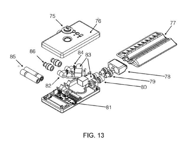
Figure 13 is an exploded view of an exemplary embodiment of an assembled
device
incorporating the elements described above. Fluid inlet connectors 86 (either
barbed or otherwise)
allow for the connection of one or multiple drainage structures or tubes (as
described above) to
the pump unit of the device. In this embodiment, two drainage structures are
accommodated;
however, additional structures may be provided for by including additional
assemblies of the
relevant components. For each fluid inlet, immediately downstream of the
connector is a one-
way valve, as described above, to prevent the backflow of material into the
drainage structure.
Downstream of the one-way valve is a fluid chamber 82, which includes a
pressure sensor 84 to
monitor the pressure developed in the device. Tubing allows fluid from this
chamber to flow into
a peristaltic, or positive displacement, pump 83, which applies negative
pressure on the upstream
side of the pump, and positive pressure on the downstream side. This positive
pressure
downstream of the pump causes fluid to be transported through the remainder of
the pump
housing body and connection elements to a reservoir unit 77.
A set of one-way valves 79, 80 may be incorporated at the connection between
the pump
housing body and the reservoir to prevent fluid leakage during change of
reservoirs. The
reservoirs may be collapsible in nature which are much more comfortable to the
patient, and may
be made in a more economic, and environmentally conscious, way as the
collapsible reservoir
will necessitate a smaller volume of plastic to produce. The reservoir
incorporates some means of
removably attaching to the pump body, which allows the reservoir to be
conveniently detached
and replaced by the patient. In this embodiment, a connector 78 is attached to
the reservoir, which
mates to a counterpart receptor on the pump housing body.
12
CA 03053326 2019-08-12
WO 2017/205446
PCT/US2017/034126
As seen in Figure 13, the reservoir unit comprises a pair of independent
reservoirs as
described above. The reservoir thus may contain several channels to allow the
fluid from multiple
drainage structures to be independently collected. These may be necessary if
the healthcare
professional desires to independently record the collected fluid amounts.
Furthermore, the
reservoir may be graduated, either by adhering a label or paint to the
reservoir, or by embossing
the plastic. These graduations allow the fluid collected fluid amount to be
easily assessed.
The reservoir may also contain a substance intended to sterilize the collected
fluid, and
may also cause the fluid to congeal. This is necessary for the reservoir to be
disposed of as "white
bag" waste, or waste which may be disposed of in landfill without additional
treatment. This
.. substance may be contained in a pouch or container within the reservoir or
may be freely
distributed inside of the reservoir. This pouch or container may be ruptured
by the patient in order
to disburse the contents, or may simply dissolve within a convenient period of
time.
The reservoir or manifold, or both, may further comprise one or more sensors
or
measurement devices 400, 402, 404, internally or externally, or both. These
sensors provide
diagnostic value in taking various measurements associated with the collected
fluid.
Measurements can include and are not limited to collected fluid amount, pH,
certain known
harmful mediators (cytokines, chemokines, reactive oxygen species), protein
levels, blood
content, etc. For example, amount of fluid collected can be an indicator of
possible seroma
development in some hernia surgeries. Additionally, pH has also been shown to
act as an
indicator of possible seroma formation. The present invention thus allows for
the detection of
infectious materials, and any other chemicals or substances which may indicate
infection, or the
presence of some medical condition which may naturally arise in response to
the surgical
procedure, initial pathology, or additional complications (of either the
surgical procedure or the
initial pathology) in the fluid collected from percutaneous (internal) wounds.
Sensors may also
.. be located in the pump unit.
Detection of a full reservoir may be accomplished by counting the revolutions
of the
peristaltic pump, or cycles of the positive displacement pump, and then
calculating the total
displaced fluid. This is made possible because the peristaltic, or positive
displacement pump
moves a nearly constant amount of fluid or gas with each revolution of its
motor. The device may
.. be powered by either consumable or rechargeable batteries 85 which are held
in a battery holder.
A circuit control board 81 comprising some or all required electrical
components controls
the operation of the device. The control board may take as inputs, and make
decisions regarding,
the following: user inputs via interface buttons; battery charge level; need
to change reservoir;
device temperature; current magnitude of negative pressure; presence of
blockage in tubing; or
13
CA 03053326 2019-08-12
WO 2017/205446
PCT/US2017/034126
the characteristics of the collected fluids. These characteristics may
include, but are not limited
to, the following: total collected amount (either total or per drainage
structure); rate of fluid
collection (total or per drainage structure) on the time scales of hours,
days, or weeks; presence of
infectious materials; and any other chemicals or substances which may indicate
infection, or the
presence of some medical condition which may naturally arise in response to
the surgical
procedure, initial pathology, or additional complications (of either the
surgical procedure or the
initial pathology) in the fluid collected from percutaneous (internal) wounds.
The user interface may comprise a single push-button 75, which controls an
on/off or
pause function, as well as any other functions which are desirable for the
operation of the device.
.. One operation may be the selection of desired level of negative pressure.
The interface may also
consist of a series of lights or a screen which alerts the user to various
conditions including, but
not limited to, device power state (off/on/paused), selected pressure level,
battery charge level,
need to change battery, reservoir fill level, need to change reservoir,
insufficient vacuum seal at
any point in the system, or presence of infections materials, and any other
chemicals or
substances which may indicate infection, or the presence of some medical
condition. The device
may apply a negative pressure in the range of 50 mmHg to 700 mmHg below
ambient pressure
either continuously or intermittently, or operate solely in range from 200
mmHg and 700 mmHg
below ambient pressure, either continuously or intermittently. The device may
create a constant
negative pressure of a desired amount and then allow the motors to momentarily
stop, until a time
when the onboard pressure sensors detect that the applied pressure has fallen
below some desired
threshold. Alternatively, the pumps may apply pressure based on a time
increment rather than a
pressure level.
Figure 14 shows a perspective view of the assembled pump unit device 87 and
reservoir
88 as detailed above in the description of Figure 13. In this embodiment, the
pump unit is
relatively flat and rectangular, with rounded edges and corners.
Figures 15A-C show several views of another exemplary embodiment of the
assembled
pump unit device. In this embodiment, the edges and corners may be more
rounded and the
entire unit may be curved, as shown. The front or top of the device provides a
user interface
comprising a single push-button 90, and lights 96 which indicate the status of
the unit (which may
.. include but not be limited to on/off, device paused, reservoir full, or
faulty tubing connection).
The unit may comprise two (or more, as described above) fluid inlets 91, which
provide the
connection for two drainage structures, and two (or more, to correspond to the
fluid inlets) fluid
outlets 92, which allow the fluid to be transported to the collection
reservoir. As seen in Figure
15C, the housing 93 is curved in order to conform to the shape of the human
abdomen on which
14
CA 03053326 2019-08-12
WO 2017/205446
PCT/US2017/034126
the device will be worn. The device may curves not only along the horizontal
axis (i.e., length-
wise), but also along the vertical axis (i.e., width-wise).
Figures 16-23 yet another embodiment of the pump unit 120 and reservoir 140 of
the
present invention, unconnected and connected. Figures 20-23 show the interior
of the pump unit
(i.e., with the front half of the pump unit housing removed. In this
embodiment, the pump unit
120 is curved in a similar manner to the pump unit shown in Figures 15A-C. A
pair of inlet
ports/connectors 122 with one-way valves and inlet fluid chambers 134 are
located near one end.
A push-button interface 124 is located on the top or front of the pump
housing, and a series of
lights 126 are located on the side near the inlet ports (which is generally
the top side, when the
unit is worn). A battery cover 128 allows access to the batteries 130, which
provide power to the
circuit control board 132 and the pump motors 138. Pumps 136 move fluid to the
outlet
ports/connectors 154, which are contained in the reservoir holder 150. Pairs
of one-way valves
142 extend from one end of the reservoir unit 140 (which contains two
independent reservoirs in
the embodiment shown), and are inserted into the outlet ports/connectors 154
to attach the
reservoir unit to the pump unit. One or more rigid or semi-rigid guides 146
may be provided to
fit into corresponding slots or holes in the reservoir holder 150. This
establishes connection with
a sensor or switch, which enables the control board in the pump unit to
determine whether the
reservoir unit is attached, as described below. The guides also may help
ensure accurate
connection and prevent damage to the one-way valves or other connection
elements. One or
more quick-release tabs or buttons 152 may be provided to allow the reservoir
unit to be
disengaged and easily removed when pressed.
In several embodiments, as seen in Figure 25, the fluid inlet ports 310 may
further
comprise a grate or mesh 312, which breaks-up or divides any particles or
similar larger material
in the fluid which may have been collected during operation. Alternatively, as
seen in Figures
26A-C, the fluid inlet ports 310 may comprise a rotating blade 320 or rotary
apparatus for the
same purpose. The blade 320 may spin within the fixed port by either a powered
motor or by the
power provided by movement of the fluid. In the latter case, the blade has
geometry to transform
the fluid flow to rotational force, as well as separate geometry to break-up
or divide particles or
similar larger material as described above.
In yet another exemplary embodiment, filters 340 are provided on a reservoir
connection
unit 330 which is attached to one end of the reservoir unit 140. When the
reservoir connection
unit is used to attach the reservoir unit 140 to the pump unit 120, the filter
arm 342 with filters
340 is inserted into a slot in the end of the pump unit, so the filters 340
are inserted into the fluid
flow lines in the body of the pump unit 120. When the reservoir unit is
removed (such as by
CA 03053326 2019-08-12
WO 2017/205446
PCT/US2017/034126
pressing the quick release latch 348), the reservoir connection unit and
filters are also removed.
The reservoir connection unit and filters can be disposed of with the
reservoir. In one
embodiment, the filters or reservoir connection unit, or both, may be
removable from the
reservoir unit, and cleaned for re-use.
In yet another embodiment of the invention, the reservoir unit prevents re-
connection to
the pump unit after an initial connection to the pump unit (or other suction
apparatus). This
prevents re-attachment of a presumably full reservoir unit, and the attempted
movement of fluid
into a full fluid collection reservoir.
In a further embodiment of the invention, the pump control unit can detect
whether a
reservoir unit is connected to the outlet ports/connections, and prevents
normal operation (i.e., the
pumping of fluid) without a reservoir present to contain the fluid. The
detection mechanism may
comprise a mechanical switch or latch, the formation or breaking of an optical
pathway, or
similar mechanism appropriate for determining or confirming proximity.
The various embodiments of the present invention thus provide substantial
improvements
and advantages over the prior art. First, the present invention allows
multiple drainage tubes to
be connected to the same source of negative pressure. Prior art devices lack
the functionality to
allow the combination of multiple drainage tubes into a common source of
negative pressure, thus
requiring patients in surgeries necessitating multiple drains to wear multiple
instances of the
previously described device. Second, the present invention also places the
reservoir after the
negative pressure source. Prior art systems require the reservoir to be placed
between the tubing
leading from the internal wound site and the source of the negative pressure,
which impairs
functioning of the device. For example, gravity's action on the fluid to
provide an air space on
which the source of negative pressure may act prevents prior art devices from
functioning
optimally while the patient is in the prone or supine position. Furthermore,
the placement of the
reservoir in prior art devices increases the working distance between the
source of negative
pressure and the internal wound, necessitating that it act on a larger volume,
reducing the
efficiency of the device, and creating a source of oscillating pressure in the
case of a temporary
blockage which is suddenly freed. Third, prior art devices make use of a
perforated internal drain
which allows the collection of fluid. The present system comprises a manifold
which allows the
use of the unique internal drain described herein or the use of one or more of
the many
conventional internal drainage structures which the surgeon may prefer.
Further, the present
invention incorporates adaptor fittings which allow any size or sizes (in the
case of multiple drain
lines) to be utilized.
16
CA 03053326 2019-08-12
WO 2017/205446
PCT/US2017/034126
Additionally, prior art devices prescribe the application of a pressure regime
from 125
mmHg to 200 mmHg below atmospheric. At this range, it is unlikely that the
device will impart
sufficient force on any impediment to flow which may become lodged in the
drainage tubing such
as a mass of clotted blood, fibrous material, or small portion of tissue. The
present invention may
operate at a pressure above 200 mmHg for certain periods of operation, such as
the initial
drawing together of the separated (surgically or otherwise) tissue and the
clearing of a blockage.
At other times, the present invention may operate at lower pressures in order
to allow a more
passive means of suctioning.
Further, prior art devices do not incorporate a disposable
reservoir, and do not allow neutralizing any odor from the collected fluid.
The present invention
comprises a fluid reservoir inherently designed to be disposable, and is
placed downstream from
the source of negative pressure, negating the previously described problems
with prior art
devices.
Prior art devices do not allow for the accurate measurement of collection
fluid, or
derivative measurements. The present invention allows for the measurement of
the amount of
collected fluid in either the input manifold or the reservoir, and further
calculates the calculation
of the percentage of collected fluid to air which would allow for the
prediction of poor suturing
and possibly surgical site infection (SSI). To accomplish this, the present
invention carries out
the following steps:
1. Calculating the amount of total volume (air plus liquid) collected via
counting
revolutions of peristaltic rotor.
2. Calculating the collected fluid amount by positioning a series of electrode
pairs acting
as graduations in the reservoir or by making use of the fluid measurement
units in the
manifold.
3. Calculating the ratio of total collected volume to total collected fluid.
In order to provide a context for the various computer-implemented aspects of
the
invention, the following discussion provides a brief, general description of a
suitable computing
environment in which the various aspects of the present invention may be
implemented. A
computing system environment is one example of a suitable computing
environment, but is not
intended to suggest any limitation as to the scope of use or functionality of
the invention. A
computing environment may contain any one or combination of components
discussed below,
and may contain additional components, or some of the illustrated components
may be absent.
Various embodiments of the invention are operational with numerous general
purpose or special
purpose computing systems, environments or configurations. Examples of
computing systems,
environments, or configurations that may be suitable for use with various
embodiments of the
17
CA 03053326 2019-08-12
WO 2017/205446
PCT/US2017/034126
invention include, but are not limited to, personal computers, laptop
computers, computer servers,
computer notebooks, hand-held devices, microprocessor-based systems,
multiprocessor systems,
TV set-top boxes and devices, programmable consumer electronics, cell phones,
personal digital
assistants (PDAs), tablets, smart phones, touch screen devices, smart TV,
internet enabled
appliances, internet enabled security systems, internet enabled gaming
systems, internet enabled
watches; internet enabled cars (or transportation), network PCs,
minicomputers, mainframe
computers, embedded systems, virtual systems, distributed computing
environments, streaming
environments, volatile environments, and the like.
Embodiments of the invention may be implemented in the form of computer-
executable
instructions, such as program code or program modules, being executed by a
computer, virtual
computer, or computing device. Program code or modules may include programs,
objects,
components, data elements and structures, routines, subroutines, functions and
the like. These are
used to perform or implement particular tasks or functions. Embodiments of the
invention also
may be implemented in distributed computing environments. In such
environments, tasks are
performed by remote processing devices linked via a communications network or
other data
transmission medium, and data and program code or modules may be located in
both local and
remote computer storage media including memory storage devices such as, but
not limited to,
hard drives, solid state drives (SSD), flash drives, USB drives, optical
drives, and internet-based
storage (e.g., "cloud" storage).
In one embodiment, a computer system comprises multiple client devices in
communication with one or more server devices through or over a network,
although in some
cases no server device is used. In various embodiments, the network may
comprise the Internet,
an intranet, Wide Area Network (WAN), or Local Area Network (LAN). It should
be noted that
many of the methods of the present invention are operable within a single
computing device.
A client device may be any type of processor-based platform that is connected
to a
network and that interacts with one or more application programs. The client
devices each
comprise a computer-readable medium in the form of volatile and/or nonvolatile
memory such as
read only memory (ROM) and random access memory (RAM) in communication with a
processor. The processor executes computer-executable program instructions
stored in memory.
Examples of such processors include, but are not limited to, microprocessors,
ASICs, and the
like.
Client devices may further comprise computer-readable media in communication
with the
processor, said media storing program code, modules and instructions that,
when executed by the
processor, cause the processor to execute the program and perform the steps
described herein.
18
CA 03053326 2019-08-12
WO 2017/205446
PCT/US2017/034126
Computer readable media can be any available media that can be accessed by
computer or
computing device and includes both volatile and nonvolatile media, and
removable and non-
removable media. Computer-readable media may further comprise computer storage
media and
communication media. Computer storage media comprises media for storage of
information, such
as computer readable instructions, data, data structures, or program code or
modules. Examples
of computer-readable media include, but are not limited to, any electronic,
optical, magnetic, or
other storage or transmission device, a floppy disk, hard disk drive, CD-ROM,
DVD, magnetic
disk, memory chip, ROM, RAM, EEPROM, flash memory or other memory technology,
an
ASIC, a configured processor, CDROM, DVD or other optical disk storage,
magnetic cassettes,
magnetic tape, magnetic disk storage or other magnetic storage devices, or any
other medium
from which a computer processor can read instructions or that can store
desired information.
Communication media comprises media that may transmit or carry instructions to
a computer,
including, but not limited to, a router, private or public network, wired
network, direct wired
connection, wireless network, other wireless media (such as acoustic, RF,
infrared, or the like) or
other transmission device or channel. This may include computer readable
instructions, data
structures, program modules or other data in a modulated data signal such as a
carrier wave or
other transport mechanism. Said transmission may be wired, wireless, or both.
Combinations of
any of the above should also be included within the scope of computer readable
media. The
instructions may comprise code from any computer-programming language,
including, for
example, C, C++, C#, Visual Basic, Java, and the like.
Components of a general purpose client or computing device may further include
a
system bus that connects various system components, including the memory and
processor. A
system bus may be any of several types of bus structures, including, but not
limited to, a memory
bus or memory controller, a peripheral bus, and a local bus using any of a
variety of bus
architectures. Such architectures include, but are not limited to, Industry
Standard Architecture
(ISA) bus, Micro Channel Architecture (MCA) bus, Enhanced ISA (EISA) bus,
Video
Electronics Standards Association (VESA) local bus, and Peripheral Component
Interconnect
(PCI) bus.
Computing and client devices also may include a basic input/output system
(BIOS),
which contains the basic routines that help to transfer information between
elements within a
computer, such as during start-up. BIOS typically is stored in ROM. In
contrast, RAM typically
contains data or program code or modules that are accessible to or presently
being operated on by
processor, such as, but not limited to, the operating system, application
program, and data.
19
CA 03053326 2019-08-12
WO 2017/205446
PCT/US2017/034126
Client devices also may comprise a variety of other internal or external
components, such
as a monitor or display, a keyboard, a mouse, a trackball, a pointing device,
touch pad,
microphone, joystick, satellite dish, scanner, a disk drive, a CD-ROM or DVD
drive, or other
input or output devices. These and other devices are typically connected to
the processor through
a user input interface coupled to the system bus, but may be connected by
other interface and bus
structures, such as a parallel port, serial port, game port or a universal
serial bus (USB). A
monitor or other type of display device is typically connected to the system
bus via a video
interface. In addition to the monitor, client devices may also include other
peripheral output
devices such as speakers and printer, which may be connected through an output
peripheral
interface.
Client devices may operate on any operating system capable of supporting an
application
of the type disclosed herein. Client devices also may support a browser or
browser-enabled
application. Examples of client devices include, but are not limited to,
personal computers, laptop
computers, personal digital assistants, computer notebooks, hand-held devices,
cellular phones,
mobile phones, smart phones, pagers, digital tablets, Internet appliances, and
other processor-
based devices. Users may communicate with each other, and with other systems,
networks, and
devices, over the network through the respective client devices.
Thus, it should be understood that the embodiments and examples described
herein have
been chosen and described in order to best illustrate the principles of the
invention and its
practical applications to thereby enable one of ordinary skill in the art to
best utilize the invention
in various embodiments and with various modifications as are suited for
particular uses
contemplated. Even though specific embodiments of this invention have been
described, they are
not to be taken as exhaustive. There are several variations that will be
apparent to those skilled in
the art.
20