Note: Descriptions are shown in the official language in which they were submitted.
CA 03053812 2019-08-15
WO 2018/170023 PCT/US2018/022267
PD-L2 VARIANT IMMUNOMODULATORY PROTEINS AND USES THEREOF
CROSS-REFERENCE TO RELATED APPLICATIONS
[0001] This application claims priority from U.S. provisional application No.
62/472,572
filed March 16 2017, entitled "PD-L2 Variant Immunomodulatory Proteins and
Uses Thereof,"
U.S. provisional application No. 62/475,156 filed March 22, 2017, entitled "PD-
L2 Variant
Immunomodulatory Proteins and Uses Thereof," and U.S. provisional application
No.
62/537,928 filed July 27, 2017, entitled "PD-L2 Variant Immunomodulatory
Proteins and Uses
Thereof," the contents of each of which are incorporated by reference in their
entirety.
INCORPORATION BY REFERENCE OF SEQUENCE LISTING
[0002] The present application is being filed along with a Sequence Listing in
electronic
format. The Sequence Listing is provided as a file entitled
761612001540SeqList.TXT, created
March 12, 2018 which is 3,353,517 bytes in size. The information in the
electronic format of the
Sequence Listing is incorporated by reference in its entirety.
FIELD
[0003] The present disclosure relates to therapeutic compositions for
modulating immune
response in the treatment of cancer and immunological diseases. In some
aspects, the present
disclosure relates to particular variants of PD-L2 that exhibit improved
binding, such as
improved binding affinity or selectivity, for one or more of the cognate
binding partner proteins
PD-1 and RGMb.
BACKGROUND
[0004] Modulation of the immune response by intervening in the processes that
occur in the
immunological synapse (IS) formed by and between antigen-presenting cells
(APCs) or target
cells and lymphocytes is of increasing medical interest. Mechanistically, cell
surface proteins in
the IS can involve the coordinated and often simultaneous interaction of
multiple protein targets
with a single protein to which they bind. IS interactions occur in close
association with the
junction of two cells, and a single protein in this structure can interact
with both a protein on the
same cell (cis) as well as a protein on the associated cell (trans), likely at
the same time.
Although therapeutics are known that can modulate the IS, improved
therapeutics are needed.
1
CA 03053812 2019-08-15
WO 2018/170023 PCT/US2018/022267
Provided are immunomodulatory proteins, including soluble proteins or
transmembrane
immunomodulatory proteins capable of being expressed on cells, that meet such
needs.
SUMMARY
[0005] Provided herein is a variant PD-L2 polypeptide, containing an IgV
domain or a
specific binding fragment thereof, an IgC domain or a specific binding
fragment thereof, or both,
wherein the variant PD-L2 polypeptide contains one or more amino acid
modifications in an
unmodified PD-L2 or a specific binding fragment thereof corresponding to
position(s) selected
from 2, 12, 13, 15, 18, 20, 23, 24, 28, 31, 32, 33, 36, 37, 39, 44, 45, 46,
47, 48, 58, 59, 65, 67, 69,
71, 72, 73, 74, 75, 76, 77, 82, 85, 86, 89, or 91 with reference to numbering
of SEQ ID NO:31.
In some embodiments, the amino acid modification is an amino acid
substitution, insertion or
deletion.
[0006] In some embodiments, the unmodified PD-L2 is a mammalian PD-L2 or a
specific
binding fragment thereof. In some of any such embodiments, the unmodified PD-
L2 is a human
PD-L2 or a specific binding fragment thereof In some of any such embodiments,
the
unmodified PD-L2 contains (i) the sequence of amino acids set forth in SEQ ID
NO:31, (ii) a
sequence of amino acids that has at least 95% sequence identity to SEQ ID
NO:31; or (iii) a
portion thereof containing an IgV domain or IgC domain or specific binding
fragments thereof or
both.
[0007] In some of any such embodiments, the specific binding fragment of the
IgV domain
or IgC domain has a length of at least 50, 60, 70, 80, 90, 100, 110 or more
amino acids; or the
specific binding fragment of the IgV domain contains a length that is at least
80% of the length of
the IgV domain set forth as amino acids 24-130 of SEQ ID NO:4 and/or the
specific binding
fragment of the IgC domain contains a length that is at least 80% of the
length of the IgC domain
set forth as amino acids 122-203 of SEQ ID NO:4. In some of any such
embodiments, the
variant PD-L2 contains up to 1, 2, 3, 4, 5, 6, 7, 8, 9, 10, 11, 12, 13, 14,
15, 16, 17, 18, 19 or 20
amino acid modifications, optionally amino acid substitutions, insertions
and/or deletions. In
some of any such embodiments, the variant PD-L2 polypeptide contains a
sequence of amino
acids that exhibits at least 85%, 86%, 87%, 88%, 89%, 90%, 91%, 92%, 93%, 94%,
95%, 96%,
97%, 98% or 99% sequence identity to SEQ ID NO:31 or a specific binding
fragment thereof.
[0008] In some of any such embodiments, the variant PD-L2 polypeptide exhibits
altered
binding to the ectodomain of PD-lor RGMb compared to the binding of the
unmodified PD-L2.
In some of any such embodiments, the variant PD-L2 polypeptide exhibits
altered binding to the
2
CA 03053812 2019-08-15
WO 2018/170023 PCT/US2018/022267
ectodomain of PD-1 compared to the unmodified PD-L2. In some aspects, the
altered binding is
altered binding affinity and/or altered binding selectivity.
[0009] In some of any such embodiments, the one or more amino acid
modifications are
selected from F2L, 112V, 113V, H15Q, N18D, N24S, C23S, G28V, N24D,V31A,V31M,
N32D,
L33PõL33H, L33F, I36V, T37A, S48C, S39I, E44D, N45S, D46E, T47A, E58G, E59G,
K65R,
S67L, H69L, P71S, Q72H, V73A, Q74R, V75G, R76G, D77N, Q82R, I85F, I86T, V89D,
W91R, or a conservative amino acid substitution thereof. In some of any such
embodiments, the
one or more amino acid modifications are selected from among H15Q, N24D, E44D,
V89D,
Q82R/V89D,
E59G/Q82R, S39I/V89D, S67L/V89D, S67L/I85F, S67L/I86T, H15Q/K65R,
H15Q/Q72H/V89D, H15Q/S67L/R76G, H15Q/R76G/185F, H15Q/T47A/Q82R,
H15Q/Q82R/V89D, H15Q/C23S/186T, H15Q/S391/186T, E44D/V89D/W91R,
113V/S67L/V89D, H15Q/S67L/186T, Il3V/H15Q/S67L/186T, 113V/H15Q/E44D/V89D,
Il3V/S391/E44D/Q82R/V89D, Il3V/E44D/Q82R/V89D, Il3V/Q72H/R76G/186T,
1 3V/H15Q/R76G/185F, H15Q/S391/R76G/V89D, H15Q/S67L/R76G/185F,
H15Q/T47A/Q72H/R76G/186T, H15Q/T47A/Q72H/R76G, Il3V/H15Q/T47A/Q72H/R76G,
H15Q/E44D/R76G/185F, H15Q/S391/S67L/V89D, H15Q/N32D/S67L/V89D,
N32D/S67L/V89D, H15Q/S67L/Q72H/R76G/V89D, H15Q/Q72H/Q74R/R76G/186T,
G28V/Q72H/R76G/I86T, Il3V/H15Q/S391/E44D/S67L, E44D/S67L/Q72H/Q82R/V89D,
H15Q/V89D, H15Q/T47A, 113V/H15Q/Q82R, 113V/H15Q/V89D, 113V/S67L/Q82R/V89D,
113V/H15Q/Q82R/V89D, H15Q/V31M/S67L/Q82R/V89D, 113V/H15Q/T47A/Q82R,
Il3V/H15Q/V31A/N45S/Q82R/V89D, H15Q/T47A/H69L/Q82R/V89D,
Il3V/H15Q/T47A/H69L/R76G/V89D, Il2V/I13V/H15Q/T47A/Q82R/V89D,
Il3V/H15Q/R76G/D77N/Q82R/V89D,
Il3V/H15Q/T47A/R76G/V89D, Il3V/H15Q/T47A/Q82R/V89D,
Il3V/H15Q/N24D/Q82R/V89D, Il3V/H15Q/136V/T47A/S67L/V89D,
H15Q/T47A/K65R/S67L/Q82R/V89D, H15Q/L33P/T47A/S67L/P71S/V89D,
Il3V/H15Q/Q72H/R76G/186T, H15Q/T47A/S67L/Q82R/V89D,
F2L/H15Q/D46E/T47A/Q72H/R76G/Q82R/V89D, Il3V/H15Q/L33F/T47A/Q82R/V89D,
Il3V/H15Q/T47A/E58G/S67L/Q82R/V89D, H15Q/N24S/T47A/Q72H/R76G/V89D,
Il3V/H15Q/E44V/T47A/Q82R/V89D, H15Q/N18D/T47A/Q72H/V73A/R76G/186TN89D,
Il3V/H15Q/T37A/E44D/S48C/S67L/Q82R/V89D, H15Q/L33H/S67L/R76G/Q82R/V89D,
3
CA 03053812 2019-08-15
WO 2018/170023 PCT/US2018/022267
113 V/H15Q/T47A/Q72H/R76G/186T, H15Q/S391/E44D/Q72H/V75G/R76G/Q82R/V89D,
H15Q/T47A/S67L/R76G/Q82R/V89D, or Il3V/H15Q/T47A/S67L/Q72H/R76G/Q82R/V89D.
[0010] In some of any such embodiments, the one or more amino acid
substitutions
correspond to position(s) selected from 13,15, 39, 44, 47 67, 72, 76, 82, 85,
86, or 89. In some of
any such embodiments, the one or more amino acid substitutions are selected
from 113V, H15Q,
E44D, T47A, S67L, Q72H, R76G, Q82R, I85F, I86T, V89D, or a conservative amino
acid
substitution thereof. In some of any such embodiments, the one or more amino
acid substitutions
correspond to position(s) selected from 13, 15, 44, 47, 67, 72, 76, 82, 86, or
89. In some of any
such embodiments, the one or more amino acid substitutions are selected from
113V, H15Q,
E44D, T47A, S67L, Q72H, R76G, Q82R, I86T, V89D, or a conservative amino acid
substitution
thereof.
[0011] In some embodiments, the variant PD-L2 polypeptide contains amino acid
modifications Il3V/H15Q, 113V/T47A, 113V/S67L, 113V/Q72H, 113V/Q72H,
113V/R76G,
113V/Q82R, 113V/I86T, 113V/V89D, H15Q/T47A, H1 5Q /S67L, H15Q/Q72H, H15Q/Q72H,
H15Q/R76G, H15Q/Q82R, H15Q/186T, H15Q/V89D, T47A/S67L, T47A/Q72H, T47A/Q72H,
T47A/R76G, T47A/Q82R, T47A/I86T, T47A/V89D, S67L/Q72H, S67L/Q72H, S67L/R76G,
S67L/Q82R, S67L/I86T, S67L/V89D, Q72H/R76G, Q72H/Q82R, Q72H/I86T, Q72H/V89D,
R76G/Q82R, R76G/I86T, R76G/V89D, Q82R/I86T, Q82R/V89D or I86T/V89D. In some
examples, the variant PD-L2 polypeptide contains amino acid modifications
H15Q/S62L/Q82R,
H15Q/S62L/V89D, H15Q/Q82R/V89D, or S62L/Q82R/V89D. In some instances, the
variant
PD-L2 polypeptide contains amino acid modifications
H15Q/T47A/K65R/S67L/Q82R/V89D.
[0012] In some of any such embodiments, the variant PD-L2 polypeptide contains
the IgV
domain or a specific fragment thereof and the IgC domain or a specific
fragment thereof. In
some of any such embodiments, the variant PD-L2 polypeptide includes the
sequence of amino
acids set forth in any of SEQ ID NOS: 56-106, 108-114, 116-132 or a specific
binding fragment
thereof, or a sequence of amino acids that exhibits at least 95% sequence
identity to any of SEQ
ID NOS: 56-106, 108-114, 116-132 or a specific binding fragment thereof and
that contains the
one or more of the amino acid substitutions.
[0013] In some of any such embodiments, the variant PD-L2 polypeptide contains
the IgV
domain or a specific binding fragment thereof In some of any such embodiments,
the IgV
domain or specific binding fragment thereof is the only PD-L2 portion of the
variant PD-L2
4
CA 03053812 2019-08-15
WO 2018/170023 PCT/US2018/022267
polypeptide. In some of any such embodiments, the IgC domain or specific
binding fragment
thereof is the only PD-L2 portion of the variant PD-L2 polypeptide.
[0014] In some of any such embodiments, the variant PD-L2 polypeptide contains
the
sequence of amino acids set forth in any of SEQ ID NOS: 133-183, 185-191, 193-
209, 268-318,
320-343 or a specific binding fragment thereof, a sequence of amino acids that
exhibits at least
95% sequence identity to any of SEQ ID NOS: 133-183, 185-191, 193-209, 268-
318, 320-343 or
a specific binding fragment thereof and that contains the one or more of the
amino acid
substitutions. In some embodiments, the IgC domain or specific binding
fragment thereof is the
only PD-L2 portion of the variant PD-L2 polypeptide.
[0015] In some of any such embodiments, the variant PD-L2 polypeptide
specifically binds
to the ectodomain of PD-1 or RGMb with increased affinity compared to the
binding of the
unmodified PD-L2 to the ectodomain of PD-1 or RGMb. In some of any such
embodiments, the
variant PD-L2 polypeptide specifically binds to the ectodomain of PD-1 with
increased affinity
compared to binding of the unmodified PD-L2 to the ectodomain of PD-1. In some
of any such
embodiments, the variant PD-L2 polypeptide specifically binds to the
ectodomain of PD-1 and
the ectodomain of RGMb each with increased affinity compared to the binding of
the unmodified
PD-L2 to the ectodomain of PD-1 and the ectodomain of RGMb. In some of any
such
embodiments, the variant PD-L2 polypeptide specifically binds to the
ectodomain of PD-1 with
increased affinity and specifically binds to the ectodomain of the other of
RGMb with decreased
affinity compared to the binding of the unmodified PD-L2 to the ectodomain of
the PD-1 or
RGMb.
[0016] In some of any such embodiments, the increased affinity to the
ectodomain of PD-1 is
increased more than 1.2-fold, 1.5-fold, 2-fold, 3-fold, 4-fold, 5-fold, 6-
fold, 7-fold, 8-fold, 9-fold,
10-fold, 20-fold, 30-fold, 40-fold, 50-fold or 60-fold compared to the
unmodified PD-L2. In
some embodiments, the increased affinity to the ectodomain of RGMb is
increased more than
1.2-fold, 1.5-fold, 2-fold, 3-fold, 4-fold, 5-fold, 6-fold, 7-fold, 8-fold, 9-
fold, 10-fold, 20-fold,
30-fold, 40-fold, 50-fold or 60-fold compared to the unmodified PD-L2. In some
aspects, the
decreased affinity to the ectodomain of RGMb is decreased more than 1.2-fold,
1.5-fold, 2-fold,
3-fold, 4-fold, 5-fold, 6-fold, 7-fold, 8-fold, 9-fold, 10-fold, 20-fold, 30-
fold, 40-fold, 50-fold or
60-fold compared to the unmodified PD-L2.
[0017] In some of any such embodiments, the variant polypeptide specifically
binds to the
ectodomain of PD-1 with increased selectivity compared to the unmodified PD-
L2. In some
CA 03053812 2019-08-15
WO 2018/170023 PCT/US2018/022267
instances, the increased selectivity includes a greater ratio of binding of
the variant polypeptide
for PD-1 versus RGMb compared to the ratio of binding of the unmodified PD-L2
polypeptide
for PD-1 versus RGMb. In some instances, the ratio is greater by at least or
at least about 1.5-
fold, 2.0-fold, 3.0-fold, 4.0-fold, 5.0-fold, 10-fold, 15-fold, 20-fold, 30-
fold, 40-fold, 50-fold or
more.
[0018] In some of any such embodiments, the PD-1 is a human PD-1. In some of
any such
embodiments, the RGMb is a human RGMb.
[0019] In some of any such embodiments, the binding activity is altered
(increased or
decreased) more than 1.2-fold, 1.5-fold, 2-fold, 3-fold, 4-fold, 5-fold, 6-
fold, 7-fold, 8-fold, 9-
fold, 10-fold, 20-fold, 30-fold 40-fold or 50-fold compared to the unmodified
PD-L2.
[0020] In some of any such embodiments, the variant PD-L2 polypeptide is a
soluble protein.
In some embodiments, the variant PD-L2 polypeptide lacks the PD-L2
transmembrane domain
and intracellular signaling domain; and/or the variant PD-L2 polypeptide is
not capable of being
expressed on the surface of a cell.
[0021] In some of any such embodiments, the variant PD-L2 polypeptide is
linked to a
multimerization domain. In some of any such embodiments, the variant PD-L2
polypeptide is a
multimeric polypeptide, optionally a dimeric polypeptide, containing a first
variant PD-L2
polypeptide linked to a multimerization domain and a second variant PD-L2
polypeptide linked
to a multimerization domain. In some embodiments, the first variant PD-L2
polypeptide and the
second variant PD-L2 polypeptide are the same or different.
[0022] In some of any such embodiments, the multimerization domain is an Fc
domain or a
variant thereof with reduced effector function. In some of any such
embodiments, the variant
PD-L2 polypeptide is linked to a moiety that increases biological half-life of
the polypeptide. In
some of any such embodiments, the variant PD-L2 polypeptide is linked to an Fc
domain or a
variant thereof with reduced effector function.
[0023] In some embodiments, the Fc domain is mammalian, optionally human; or
the variant
Fc domain contains one or more amino acid modifications compared to an
umodified Fc domain
that is mammalian, optionally human. In some embodiments, the Fc domain or
variant thereof
contains the sequence of amino acids set forth in SEQ ID NO:211 or SEQ ID
NO:212 or a
sequence of amino acids that exhibits at least 85% sequence identity to SEQ ID
NO:211 or SEQ
ID NO:212. In some of any such embodiments, the Fc domain contains one or more
amino acid
modifications selected from among E233P, L234A, L234V, L235A, L235E, G236del,
G237A,
6
CA 03053812 2019-08-15
WO 2018/170023 PCT/US2018/022267
S267K, N297G, V302C, and K447del each by EU numbering. In some embodiments,
the Fc
domain contains the amino acid modification C220S by EU numbering. In some
embodiments,
the Fc domain contains the sequence of amino acids set forth in any of SEQ ID
NOS: 1189,
1205, 1206, 1207, 1739, 1738, 1739, 1740 or a sequence of amino acids that
exhibits at least
85% sequence identity to any of SEQ ID NOS: 1189, 1205, 1206, 1207, 1739,
1738, 1739, 1740
and exhibits reduced effector function.
[0024] In some of any such embodiments, the variant PD-L2 polypeptide is
linked indirectly
via a linker, optionally a G45 linker. In some of any such embodiments, the
variant PD-L2
polypeptide is a transmembrane immunomodulatory protein further containing a
transmembrane
domain linked to the extracellular domain (ECD) or specific binding fragment
thereof of the
variant PD-L2 polypeptide. In some instances, the transmembrane domain
contains the sequence
of amino acids set forth as residues 221-241 of SEQ ID NO:4 or a functional
variant thereof that
exhibits at least 85% sequence identity to residues 221-241 of SEQ ID NO:4. In
some
embodiments, the variant PD-L2 polypeptide further contains a cytoplasmic
signaling domain
linked to the transmembrane domain. In some aspects, the cytoplasmic signaling
domain
includes the sequence of amino acids set forth as residues 242-273 of SEQ ID
NO:4 or a
functional variant thereof that exhibits at least 85% sequence identity to
residues 242-273 of SEQ
ID NO:4.
[0025] In some of any of the provided embodiments, the variant PD-L2
polypeptide
modulates a response of an immune cell, such as a T cell. In some embodiments,
the response,
e.g. T cell response, is increased or is decreased. In some of any such
embodiments, the variant
PD-L2 increases IFN-gamma (interferon-gamma) expression relative to the
unmodified PD-L2 in
an in vitro T-cell assay. In some of any such embodiments, the variant PD-L2
decreases IFN-
gamma (interferon-gamma) expression relative to the unmodified PD-L2 in an in
vitro T-cell
assay.
[0026] In some embodiments of any one of the variant PD-L2 polypeptides
described herein,
the variant PD-L2 polypeptide increases T cell signaling relative to the
unmodified PD-L2 such
as determined using a reporter assay involving a T cell (e.g. Jurkat)
engineered with a reporter
(e.g. luciferase) operably connected to an IL-2 promoter. In some embodiments
of any one of the
variant PD-L2 polypeptides described herein, the variant PD-L2 polypeptide
decreases T cell
signaling relative to the unmodified PD-L2, such as determined using a
reporter assay involving
a T cell (e.g. Jurkat) engineered with a reporter (e.g. luciferase) operably
connected to an IL-2
7
CA 03053812 2019-08-15
WO 2018/170023 PCT/US2018/022267
promoter. In some of any such embodiments, the variant PD-L2 polypeptide is
provided in any
of a variety of formats, such as soluble or immobilized (e.g. plate-bound).
[0027] In some of any such embodiments, the variant PD-L2 polypeptide is
deglycosylated.
[0028] Also provided is an immunomodulatory polypeptide containing the variant
PD-L2
according to any of the provided embodiments linked, directly of indirectly
via a linker, to a
second polypeptide containing an immunoglobulin superfamily (IgSF) domain. In
some cases,
the IgSF domain is affinity modified and exhibits altered binding to one or
more of its cognate
binding partner(s) compared to the unmodified or wild-type IgSF domain. In
some instances, the
IgSF domain exhibits increased binding to one or more of its cognate binding
partner(s)
compared to the unmodified or wild-type IgSF domain to the same one or more
cognate binding
partner(s).
[0029] In some of any such embodiments, the variant PD-L2 is a first PD-L2
variant and the
IgSF domain of the second polypeptide is an IgSF domain from a second variant
PD-L2 of any of
the provided embodiments, wherein the first and second PD-L2 variant are the
same or different.
In some embodiments, the variant PD-L2 polypeptide is capable of specifically
binding to PD-1
or RGMb and the IgSF domain of the second polypeptide is capable of binding to
a cognate
binding partner other than one specifically bound by the PD-L2 variant
polypeptide.
[0030] In some of any such embodiments, the IgSF domain is from a member of
the B7
family. In some embodiments, the IgSF domain is a tumor-localizing moiety that
binds to a
ligand expressed on a tumor. In some cases, the ligand is B7H6. In some
aspects, the IgSF
domain is from NKp30.
[0031] In some of any such embodiments, the IgSF domain or affinity-modified
IgSF domain
thereof, optionally of the second or third polypeptide, is or contains an IgV
domain. In some
embodiments, the variant PD-L2 polypeptide is or contains an IgV domain. In
some of any such
embodiments, the immunomodulatory protein contains a multimerization domain
linked to one or
both of the variant PD-L2 polypeptide of the IgSF domain.
[0032] In some embodiments, the multimerization domain is an Fc domain or a
variant
thereof with reduced effector function. In some embodiments, the
immunomodulatory protein is
dimeric. In some cases, the immunomodulatory protein is homodimeric. In some
instances, the
immunomodulatory protein is heterodimeric.
[0033] In some of any such embodiments, the IgSF domain of the second
polypeptide is an
IgSF domain of a ligand that binds to an inhibitory receptor, or is an
affinity-modified IgSF
8
CA 03053812 2019-08-15
WO 2018/170023 PCT/US2018/022267
domain thereof. In some instances, the affinity-modified IgSF domain exhibits
increased binding
affinity and/or binding selectivity for the inhibitory receptor compared to
binding of the
unmodified IgSF domain to the same inhibitory receptor. In some embodiments,
the inhibitory
receptor is TIGIT or CTLA-4; or the ligand of the inhibitory receptor is
CD155, CD112 or CD80.
[0034] In some of any such embodiments, the IgSF domain of the second
polypeptide is an
affinity-modified IgSF domain containing (i) a wildtype CD155 comprising an
IgSF set forth in
any of SEQ ID NOS: 47, 344, or 387, or a variant CD155 polypeptide comprising
an IgSF
domain of any of SEQ ID NOS set forth in Table 5, optionally any of the SEQ ID
NOs: 345-386,
388-699, 1527-1736; (ii) a wildtype CD112 comprising an IgSF domain set forth
in any of SEQ
ID NOS: 48, 700, or 795, or a variant CD112 polypeptide comprising an IgSF
domain of any of
SEQ ID NOS set forth in Table 4, optionally any of the SEQ ID NOs: 701-794,
796-965, 1455-
1526; (iii) a wildtype CD80 comprising an IgSF set forth in any of SEQ ID
NOS:28, 1039, or
2039, or a variant CD80 polypeptide comprising an IgSF of any of SEQ ID NOS
set forth in
Table 3, optionally any of the SEQ ID NOs: 28, 966-998, 1000-1072, 1074-1146,
1147-1186;
(iv) a wildtype PD-Li comprising an IgSF set forth in any of SEQ ID NOS: 30,
1812, 1258, or
1454, or a variant PD-Li polypeptide comprising an IgSF of any of SEQ ID NOS
set forth in
Table 8, optionally any of SEQ ID NOS: 1259-1453, 1743-1811, 1813-2021; (v) a
sequence of
amino acids that exhibits at least 95% sequence identity to any of the SEQ ID
NOSs in (i)-(iv)
and that contains the amino acid substitution; or (vi) a specific binding
fragment of any of (i)-
(v).
[0035] In some embodiments, the immunomodulatory protein further contains a
third
polypeptide containing a wild-type IgSF domain or a variant or affinity-
modified IgSF domain
thereof, said affinity-modified IgSF domain comprising one or more amino acid
modifications
compared to the unmodified or wild-type IgSF domain of the IgSF family member,
wherein the
third polypeptide is the same as the first and/or second polypeptide; or the
third polypeptide is
different from the first and/or second polypeptide.
[0036] In some examples, the IgSF domain of the third polypeptide is an
affinity-modified
IgSF domain containing: (i) a wildtype CD155 comprising an IgSF set forth in
any of SEQ ID
NOS: 47, 344, or 387, or a variant CD155 polypeptide comprising an IgSF domain
set forth in
any of SEQ ID NOS: 345-386, 388-699, 1527-1736; (ii) a wildtype CD112
comprising an IgSF
domain set forth in any of SEQ ID NOS: 48, 700, or 795, or a variant CD112
polypeptide
comprising an IgSF domain set forth in any of SEQ ID NOS: 701-794, 796-965,
1455-1526; (iii)
9
CA 03053812 2019-08-15
WO 2018/170023 PCT/US2018/022267
a wildtype CD80 comprising an IgSF set forth in any of SEQ ID NOS:28, 1039, or
2039, or a
variant CD80 polypeptide comprising an IgSF domain set forth in any of SEQ ID
NOS: 966-998,
1000-1038, 1040-1072, 1074-1112, 1114-1146, 1147-1186; (iv) a wildtype PD-Li
comprising an
IgSF set forth in any of SEQ ID NOS: 30, 1812, 1258, or 1454, or a variant PD-
Li polypeptide
comprising an IgSF domain set forth in any of SEQ ID NOS:1259-1453, 1743-1811,
1813-2021;
(v) a sequence of amino acids that exhibits at least 95% sequence identity to
any of the SEQ ID
NOS in (i)-(iv) and that comprises the amino acid modifications, optionally
amino acid
substitutions, insertions and/or deletions thereof; or (vi) a specific binding
fragment of any of (i)-
(v).
[0037] In some of any such embodiments, the immunomodulatory protein further
contains at
least one additional polypeptide containing an IgSF domain of an IgSF family
member or an
affinity-modified IgSF domain thereof, said affinity-modified IgSF domain
comprising one or
more amino acid modifications compared to the binding of the unmodified or
wild-type IgSF
domain of the IgSF family member.
[0038] In some embodiments, the immunomodulatory protein further contains a
multimerization domain linked to at least one of the variant PD-L2
polypeptide, the second
polypeptide and/or the third polypeptide, optionally wherein the
multimerization domain is an Fc
domain or a variant thereof with reduced effector function.
[0039] In some embodiments, the multimerization domain promotes heterodimer
formation.
[0040] Provided is an immunomodulatory protein containing a first variant PD-
L2
polypeptide in which the multimerization domain is a first multimerization
domain and a second
variant PD-L2 polypeptide in which the multimerization domain is a second
multimerization
domain, wherein the first and second multimerization domains interact to form
a multimer
containing the first and second variant PD-L2 polypeptide, optionally wherein
the first and
second variant PD-L2 polypeptides are the same. Also provided is an
immunomodulatory
protein containing any of the provided immunomodulatory proteins, wherein the
multimerization
domain is a first multimerization domain and interacts with a second
multimerization domain to
form a multimer containing the immunomodulatory protein.
[0041] In some embodiments, the immunomodulatory protein is a first
immunomodulatory
protein and a second immunomodulatory protein is linked directly or indirectly
via a linker to the
second multimerization domain, wherein the multimer contains the first and
second
immunomodulatory protein. In some cases, the second immunomodulatory protein
is an
CA 03053812 2019-08-15
WO 2018/170023 PCT/US2018/022267
immunomodulatory protein as described herein and the multimerization domain is
the second
multimerization domain. In some embodiments, the multimer is a dimer. In some
aspects, the
immunomodulatory protein is a homodimer, optionally wherein the first and
second
multimerization domain is the same.
[0042] In some embodiments, the second polypeptide is a variant CD155
polypeptide and the
first and/or second immunomodulatory protein includes the sequence set forth
in any of SEQ ID
NOS: 1191-1196, or a sequence of amino acids that exhibits at least 85%, 86%,
87%, 88%, 89%,
90%, 91%, 92%, 93%, 94%, 95%, 96%, 97%, 98% or 99% sequence identity to any of
SEQ ID
NOS: 1191-1196. In some cases, the immunomodulatory protein is a heterodimer,
optionally
wherein the first and second multimerization domain are different and/or are
capable of
interacting to mediate heterodimer formation. In some examples, the second
polypeptide is a
variant CD155 polypeptide and: the first or second immunomodulatory protein
contains the
sequence set forth in any of SEQ ID NOS: 1197, 1198, 1199, 1200, 1201, 1203 or
a sequence of
amino acids that exhibits at least 85%, 86%, 87%, 88%, 89%, 90%, 91%, 92%,
93%, 94%, 95%,
96%, 97%, 98% or 99% sequence identity to any of SEQ ID NOS: 1197, 1198, 1199,
1200, 1201
or 1203; and the other of the first or second immunomodulatory protein
includes the sequence set
forth in any of SEQ ID NOS: 1188, 1190, 1202 or 1204, or a sequence of amino
acids that
exhibits at least 85%, 86%, 87%, 88%, 89%, 90%, 91%, 92%, 93%, 94%, 95%, 96%,
97%, 98%
or 99% sequence identity to any of SEQ ID NOS: 1188, 1190, 1202 or 1204.
[0043] In some embodiments, the first and/or second multimerization domain is
an Fc
domain or a variant thereof with reduced effector function, optionally wherein
the Fc domain is
of an immunoglobulin protein that is human and/or the Fc region is human,
optionally wherein
the Fc region is of an immunoglobulin G1 (IgG1) or an immunoglobulin G2
(IgG2), optionally
set forth in SEQ ID NO:211 or SEQ ID NO:212; or the variant Fc domain contains
one or more
amino acid substitutions in a wildtype Fc region, optionally wherein the
reduced effector function
is reduced compared to a wildtype Fc region, optionally wherein the wildtype
human Fc is of
human IgGl.
[0044] In some of any such embodiments, the first and second multimerization
domain is the
same or different. In some examples, the variant Fc region contains the amino
acid substitutions
E233P, L234A, L234V, L235A, L235E, G236del, G237A, S267K, or N297G, with
residue
numbering according to the EU index of Kabat; or the amino acid substitutions
R292C/N297G/V302C or L234A/L235E/G237A, with residue numbering according to
the EU
11
CA 03053812 2019-08-15
WO 2018/170023 PCT/US2018/022267
index of Kabat. In some cases, the Fe region or variant Fe region contains the
amino acid
substitution C220S, with residue numbering according to the EU index of Kabat.
In some
examples, the Fe region or variant Fe region contains K447del, with residue
numbering
according to the EU index of Kabat.
[0045] In some of any such embodiments, the sequence of amino acids set forth
in any of
SEQ ID NOS: 1191-1204 or a sequence of amino acids that exhibits at least 85%,
86%, 87%,
88%, 89%, 90%, 91%, 92%, 93%, 94%, 95%, 96%, 97%, 98% or 99% sequence identity
to any
of SEQ ID NOS: 1191-1204.
[0046] Also provided is a conjugate containing a variant PD-L2 according to
any of the
provided embodiments or an immunomodulatory polypeptide according to any of
the provided
embodiments linked to a moiety. In some cases, the moiety is a targeting
moiety that specifically
binds to a molecule on the surface of a cell. In some instances, the targeting
moiety specifically
binds to a molecule on the surface of an immune cell. In some embodiments, the
immune cell is
an antigen presenting cell or a lymphocyte.
[0047] In some cases, the targeting moiety is a tumor-localizing moiety that
binds to a
molecule on the surface of a tumor. In some of any such embodiments, the
moiety is a protein, a
peptide, nucleic acid, small molecule or nanoparticle. In some of any such
embodiments, the
moiety is an antibody or antigen-binding fragment. In some of any such
embodiments, the
conjugate is divalent, tetravalent, hexavalent or octavalent. In some cases,
the conjugate is a
fusion protein.
[0048] Also provided is a nucleic acid molecule(s) encoding a variant PD-L2
polypeptide
according to any of the provided embodiments, a conjugate that is a fusion
protein according to
any of the provided embodiments, or an immunomodulatory polypeptide according
to any of the
provided embodiments. In some embodiments, the nucleic acid molecule is
synthetic nucleic
acid. In some instances, the nucleic acid molecule is cDNA.
[0049] Also provided is a vector containing the nucleic acid molecule
according to any of the
provided embodiments. In some cases, the vector is an expression vector. In
some embodiments,
the vector is a mammalian expression vector or a viral vector.
[0050] Also provided is a cell containing the vector of any of the provided
embodiments. In
some cases, the cell is a mammalian cell. In some aspect, the cell is a human
cell.
[0051] Also provided is a method of producing a variant PD-L2 polypeptide or
an
immunomodulatory protein, including introducing the nucleic acid molecule
according to any of
12
CA 03053812 2019-08-15
WO 2018/170023 PCT/US2018/022267
the provided embodiments or vector according to any of the provided
embodiments into a host
cell under conditions to express the protein in the cell. In some cases, the
method further
includes isolating or purifying the variant PD-L2 polypeptide or
immunomodulatory protein from
the cell. Also provided is a method of engineering a cell expressing a variant
PD-L2 variant
polypeptide, including introducing a nucleic acid molecule encoding the
variant PD-L2
polypeptide according to any of the provided embodiments into a host cell
under conditions in
which the polypeptide is expressed in the cell.
[0052] Also provided is an engineered cell, expressing the variant PD-L2
polypeptide
according to any of the provided embodiments, a conjugate that is a fusion
protein according to
any of the provided embodiments, the immunomodulatory protein according to any
of the
provided embodiments, the nucleic acid molecule according to any of the
provided embodiments,
or the vector according to any of the embodiments.
[0053] In some cases the variant PD-L2 polypeptide or immunomodulatory protein
contains
a signal peptide. In some embodiments, the variant PD-L2 polypeptide or
immunomodulatory
protein does not contain a transmembrane domain and/or is not expressed on the
surface of the
cell. In some embodiments, the variant PD-L2 polypeptide or immunomodulatory
protein is
secreted or is capable of being secreted from the engineered cell. In some
embodiments, the
engineered cell contains a variant PD-L2 polypeptide that contains a
transmembrane domain
and/or is the transmembrane immunomodulatory protein according to any of the
provided
embodiments. In some embodiments, the variant PD-L2 polypeptide is expressed
on the surface
of the cell.
[0054] In some of any such embodiments, the cell is an immune cell. In some
cases, the
immune cell is an antigen presenting cell (APC) or a lymphocyte. In some
embodiments, the
engineered cell is a primary cell. In some embodiments, the cell is a
mammalian cell. In some
embodiments, the cell is a human cell. In some embodiments, the cell is a
lymphocyte and the
lymphocyte is a T cell. In some cases, the cell is an APC and the APC is an
artificial APC. In
some examples, the engineered cell is a primary cell. In some cases, the cell
is a mammalian
cell. In some instances, the cell is a human cell. In some embodiments, the
engineered cell
further contains a chimeric antigen receptor (CAR) or an engineered T-cell
receptor.
[0055] Also provided is an infectious agent, containing a nucleic acid
molecule encoding a
variant PD-L2 polypeptide according to any of the provided embodiments or an
immunomodulatory polypeptide according to any of the provided embodiments. In
some cases,
13
CA 03053812 2019-08-15
WO 2018/170023 PCT/US2018/022267
the encoded variant PD-L2 polypeptide or immunomodulatory polypeptide does not
contain a
transmembrane domain and/or is not expressed on the surface of a cell in which
it is expressed.
In some embodiments, the encoded variant PD-L2 polypeptide or immunomodulatory
polypeptide is secreted from a cell in which it is expressed. In some cases,
the encoded variant
PD-L2 polypeptide contains a transmembrane domain. In some embodiments, the
encoded
variant PD-L2 polypeptide is expressed on the surface of a cell in which it is
expressed.
[0056] In some of any such embodiments, the infectious agent is a bacterium or
a virus. In
some embodiments, the virus is a lentiviral or retroviral construct or a
hybrid thereof. In some
instances, the virus is an oncolytic virus. In some examples, the oncolytic
virus is an adenovirus,
adeno-associated virus, herpes virus, Herpes Simplex Virus, Vesticular
Stomatic virus, Reovirus,
Newcastle Disease virus, parvovirus, measles virus, vesticular stomatitis
virus (VSV), Coxsackie
virus or a Vaccinia virus. In some aspects, the virus specifically targets
dendritic cells (DCs)
and/or is dendritic cell-tropic. In some embodiments, the virus is a
lentiviral vector that is
pseudotyped with a modified Sindbis virus envelope product.
[0057] In some embodiments, the infectious agent further contains a nucleic
acid molecule
encoding a further gene product that results in death of a target cell or that
can augment or boost
an immune response. In some cases, the further gene product is selected from
an anticancer
agent, anti-metastatic agent, an antiangiogenic agent, an immunomodulatory
molecule, an
immune checkpoint inhibitor, an antibody, a cytokine, a growth factor, an
antigen, a cytotoxic
gene product, a pro-apoptotic gene product, an anti-apoptotic gene product, a
cell matrix
degradative gene, genes for tissue regeneration or a reprogramming human
somatic cells to
pluripotency.
[0058] Also provided is a pharmaceutical composition containing the variant PD-
L2
polypeptide according to any of the provided embodiments, an immunomodulatory
protein
according to any of the provided embodiments, a conjugate according to any of
the provided
embodiments, or an engineered cell according to any of the provided
embodiments. In some
instances, the pharmaceutical composition contains a pharmaceutically
acceptable excipient. In
some embodiments, the pharmaceutical composition is sterile.
[0059] Also provided is an article of manufacture containing the
pharmaceutical composition
of according to any of the provided embodiments in a vial. In some cases, the
vial is sealed.
14
CA 03053812 2019-08-15
WO 2018/170023 PCT/US2018/022267
[0060] Also provided is a kit containing the pharmaceutical composition
according to any of
the provided embodiments and instructions for use. Also provided is a kit
containing the article
of manufacture according to any of the provided embodiments and instructions
for use.
[0061] Also provided is a method of modulating an immune response, such as
increasing or
decreasing an immune response in a subject including administering the
pharmaceutical
composition according to any of the provided embodiments to the subject. In
some cases the
method includes administering the engineered cells according to any of the
provided
embodiments. In some examples, the engineered cells are autologous to the
subject. In some
cases, the engineered cells are allogenic to the subject. In some embodiments,
the method
comprises administering to the subject a soluble variant PD-L2 polypeptide
according to any one
of the embodiments described herein, an immunomodulatory protein according to
any one of the
embodiments described herein or a conjugate according to any one of the
embodiments described
herein. In some embodiments, the method comprises administering to the subject
an infectious
agent encoding a variant PD-L2 polypeptide according to any one of the
embodiments described
herein.
[0062] In some embodiments, the method modulating the immune response treats a
disease
or condition in the subject.
[0063] In some of any such embodiments, the immune response is increased.
Various
formats of a variant PD-L2 polypeptide are contemplated for administration to
a subject to
increase an immune response, such as antagonist formats of a variant PD-L2. In
some cases,
such methods are carried out under conditions in which signaling by the
inhibitory receptor PD-1
is blocked or attenuated by the administration. In some of any such
embodiments, the method
includes a variant PD-L2 polypeptide or immunomodulatory protein that is
soluble is
administered to the subject. In some instances, the soluble immunomodulatory
protein is an
immunomodulatory Fc fusion protein. In some embodiments of the method, a
variant PD-L2
polypeptide according to any of the provided embodiments, or the
immunomodulatory protein
according to any of the provided embodiments is administered to the subject.
[0064] In some embodiments, an engineered cell containing a secretable variant
PD-L2
polypeptide is administered to the subject. In some embodiments, an engineered
cell according
to any of the provided embodiments is administered to the subject. In some
embodiments, an
infectious agent encoding a variant PD-L2 polypeptide that is a secretable
immunomodulatory
protein is administered to the subject, optionally under conditions in which
the infectious agent
CA 03053812 2019-08-15
WO 2018/170023 PCT/US2018/022267
infects a tumor cell or immune cell and the secretable immunomodulatory
protein is secreted
from the infected cell.
[0065] In some of any such embodiments, the disease or condition is a tumor or
cancer. In
some embodiments, the disease or condition is selected from melanoma, lung
cancer, bladder
cancer, a hematological malignancy, liver cancer, brain cancer, renal cancer,
breast cancer,
pancreatic cancer, colorectal cancer, spleen cancer, prostate cancer,
testicular cancer, ovarian
cancer, uterine cancer, gastric carcinoma, a musculoskeletal cancer, a head
and neck cancer, a
gastrointestinal cancer, a germ cell cancer, or an endocrine and
neuroendocrine cancer. In some
of any such embodiments, the variant PD-L2 is administered in a format that
increases an
immune response in the subject.
[0066] In some of any such embodiments, the immune response is decreased.
Various
formats of a variant PD-L2 polypeptide are contemplated for administration to
a subject to
decrease an immune response, such as agonist formats of a variant PD-L2. In
some cases, such
methods are carried out under conditions in which signaling by the inhibitory
receptor PD-1 is
activated or stimulated or induced by the administration. In some embodiments,
an
immunomodulatory protein or conjugate comprising a variant PD-L2 polypeptide
linked to an
IgSF domain or a moiety that localizes to a cell or tissue of an inflammatory
environment is
administered to the subject. In some instances, the binding molecule contains
an antibody or an
antigen-binding fragment thereof or contains a wild-type IgSF domain or
variant thereof.
[0067] In some embodiments, the immunomodulatory protein according to any of
the
provided embodiments or the conjugate according to any of the provided
embodiments is
administered to the subject. In some embodiments, a variant PD-L2 polypeptide
that is a
transmembrane immunomodulatory protein is administered to the subject. In some
of any such
embodiments, the engineered cell containing a variant PD-L2 polypeptide that
is a
transmembrane immunomodulatory protein according to any of the provided
embodiments is
administered to the subject. In some embodiments, an infectious agent encoding
a variant PD-L2
polypeptide that is a transmembrane immunomodulatory protein is administered
to the subject,
optionally under conditions in which the infectious agent infects a tumor cell
or immune cell and
the transmembrane immunomodulatory protein is expressed on the surface of the
infected cell.
[0068] In some of any such embodiments, the disease or condition is an
inflammatory or
autoimmune disease or condition. In some embodiments, the disease or condition
is an
antineutrophil cytoplasmic antibodies (ANCA)-associated vasculitis, a
vasculitis, an autoimmune
16
CA 03053812 2019-08-15
WO 2018/170023 PCT/US2018/022267
skin disease, transplantation, a Rheumatic disease, an inflammatory
gastrointestinal disease, an
inflammatory eye disease, an inflammatory neurological disease, an
inflammatory pulmonary
disease, an inflammatory endocrine disease, or an autoimmune hematological
disease. In some
embodiments, the disease or condition is selected from inflammatory bowel
disease, transplant,
Crohn's disease, ulcerative colitis, multiple sclerosis, asthma, rheumatoid
arthritis, or psoriasis. In
some of any such embodiments, the variant PD-L2 is administered in a format
that decreases an
immune response in the subject.
BRIEF DESCRIPTION OF THE DRAWINGS
[0069] FIG. 1A-1C depicts various formats of the provided variant IgSF domain
molecules.
FIG. 1A depicts soluble molecules, including: (1) a variant IgSF domain (vIgD)
fused to an Fc
chain; (2) a stack molecule containing a first variant IgSF domain (first
vIgD) and a second IgSF
domain, such as a second variant IgSF domain (second vIgD); (3) a tumor
targeting IgSF
molecule containing a first variant IgSF domain (vIgD) and an IgSF domain that
targets to a
tumor antigen, such as an NKp30 IgSF domain; and (4) a variant IgSF domain
(vIgD) linked to
an antibody (V-mAb). FIG. 1B depicts a transmembrane immunomodulatory protein
(TIP)
containing a variant IgSF domain (vIgD) expressed on the surface of a cell. In
an exemplary
embodiment, the cognate binding partner of the transmembrane bound vIgD is an
inhibitory
receptor (e.g. PD-1), and the TIP containing the vIgD (e.g. PD-L2 vIgD)
antagonizes or blocks
the negative signaling of the inhibitory receptor, thereby resulting in an
activated T cell or
effector T cell. In some cases, if clustering of the inhibitory receptor (PD-
1) is proximal to an
activating receptor (e.g. CD28) then agonizing activity by the TIP may be
realized. FIG. 1C
depicts a secreted immunomodulatory protein (SIP) in which a variant IgSF
domain (vIgD) is
secreted from a cell, such as a first T cell (e.g. CAR T cell). In an
exemplary embodiment, the
cognate binding partner of the secreted vIgD is an inhibitory receptor (e.g.
PD-1), which can be
expressed by the first cell (e.g. T cell, such as a CAR T cell) and/or on a
second cell (e.g. T cell;
either endogenous or engineered, such as a CAR T cell). Upon binding of the
SIP with its
cognate binding partner, the SIP antagonizes or blocks the negative signaling
via the inhibitory
receptor, thereby resulting in an activated T cell or effector T cell. In all
cases, the vIgD can be a
V-domain (IgV) only, the combination of the V-domain (IgV) and C-domain (IgC),
including the
entire extracellular domain (ECD), or any combination of Ig domains of the
IgSF superfamily
member.
17
CA 03053812 2019-08-15
WO 2018/170023 PCT/US2018/022267
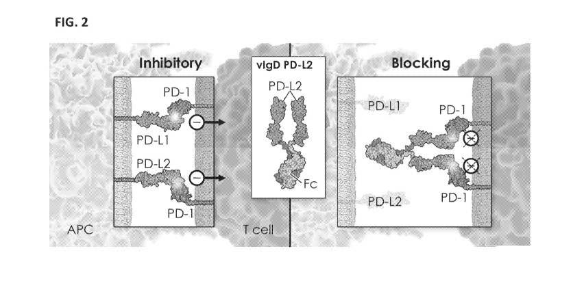
[0070] FIG. 2 depicts an exemplary schematic of the activity of a variant IgSF
domain
(vIgD) fused to an Fc (vIgD-Fc) in which the vIgD is a variant of an IgSF
domain of PD-L2. As
shown, a soluble vIgD of PD-L2 interacts with its cognate binding partners to
block interactions
of PD-Li or PD-L2 with PD-1, thereby blocking the PD-1 inhibitory receptor,
and, in some
cases, allowing the T cell to differentiate into an effector phenotype.
[0071] FIG. 3 depicts an exemplary schematic of a stack molecule that is a
multi-target
checkpoint antagonist containing a first variant IgSF domain (first vIgD) that
is a PD-Li or PD-
L2 vIgD and a second IgSF domain (e.g. a second vIgD) that binds to a second
inhibitory
receptor. In the exemplary schematic, the second IgSF domain (e.g. second
vIgD) is a CD112 or
CD155 vIgD. As shown, the first vIgD and second vIgD interact with their
cognate binding
partners to block interactions of PD-Li or PD-L2 with PD-1 and block
interactions of CD155 or
CD112 with TIGIT and/or CD112R, respectively, thereby blocking multiple
inhibitory receptors.
[0072] FIG. 4 depicts an exemplary schematic of a stack molecule for
localizing the variant
IgSF (vIgD) to a tumor cell. In this format, the stack molecule contains a
first variant IgSF
domain (first vIgD) and a second IgSF domain (e.g. a second vIgD) in which the
second IgSF
domain (e.g a second vIgD) is a tumor-targeted IgSF domain that binds to a
tumor antigen. An
exemplary tumor-targeted IgSF domain is an IgSF domain of NKp30, which binds
to the tumor
antigen B7-H6. In this depiction, the first variant IgSF domain (vIgD) is a
variant of an IgSF
domain of PD-L2. As shown, binding of tumor-targeted IgSF domain to the
surface of the tumor
cell localizes the first variant IgSF domain on the tumor cell surface where
it can interact with
one or more of its cognate binding partners expressed on the surface of an
adjacent immune cell
(e.g. T cell) to antagonize inhibitory receptor signaling.
[0073] FIG. 5A depicts various exemplary configurations of a stack molecule
containing a
first variant IgSF domain (first vIgD) and a second IgSF domain, such as a
second variant IgSF
domain (second vIgD). As shown, the first vIgD and second IgSF domain are
independently
linked, directly or indirectly, to the N- or C-terminus of an Fc region. For
generating a
homodimeric Fc molecule, the Fc region is one that is capable of forming a
homodimer with a
matched Fc region by co-expression of the individual Fc regions in a cell. For
generating a
heterodimeric Fc molecule, the individual Fc regions contain mutations (e.g.
"knob-into-hole"
mutations in the CH3 domain), such that formation of the heterodimer is
favored compared to
homodimers when the individual Fc regions are co-expressed in a cell.
18
CA 03053812 2019-08-15
WO 2018/170023 PCT/US2018/022267
[0074] FIG. 5B depicts various exemplary configurations of a stack molecule
containing a
first variant IgSF domain (first vIgD), a second IgSF domain, such as a second
variant IgSF
domain (second vIgD), and a third IgSF domain, such as a third variant IgSF
domain (third
vIgD). As shown, the first vIgD, second IgSF, and third IgSF domains are
independently linked,
directly or indirectly, to the N- or C-terminus of an Fc region. For
generating a homodimeric Fc
molecule, the Fc region is one that is capable of forming a homodimer with a
matched Fc region
by co-expression of the individual Fc regions in a cell.
[0075] FIG. 6 depicts an exemplary schematic of the activity of a variant IgSF
domain
(vIgD) ¨conjugated to an antibody (V-Mab) in which the antibody (e.g. anti-
HER2 antibody)
binds to an antigen on the surface of the tumor cell to localize the vIgD to
the cell. As shown,
binding of the antibody to the surface of the tumor cell localizes the vIgD on
the tumor cell
surface where it can interact with one or more of its cognate binding partners
expressed on the
surface of an adjacent immune cell (e.g. T cell) to agonize or antagonize
receptor signaling. In an
exemplary embodiment as shown, the variant IgSF domain (vIgD) is a variant of
an IgSF domain
of PD-L2 that binds, such as has increased affinity for, the inhibitory
receptor PD-1. Binding of
the PD-L2 vIgD to the PD-1 inhibitory receptor antagonizes or blocks the
negative signaling of
the inhibitory receptor, thereby resulting in an activated T cell or effector
T cell. In some cases, if
clustering of the inhibitory receptor (PD-1) is proximal to an activating
receptor (e.g. CD28) then
agonizing of the inhibitory receptor activity by the TIP may be realized.
[0076] FIG. 7A-7C depicts various exemplary configurations of a variant IgSF-
antibody
conjugate (V-Mab). FIG. 7A shows various configurations in which a variant
IgSF domain is
linked, directly or indirectly, to the N- and/or C-terminus of the light chain
of an antibody. FIG.
7B shows various configurations in which a variant IgSF domain is linked,
directly or indirectly,
to the N- and/or C-terminus of the heavy chain of an antibody. FIG. 7C depicts
the results V-
Mab configurations when a light chain of FIG. 7A and a heavy chain of FIG. 7B
are co-
expressed in a cell.
[0077] FIG. 8 depicts the MFI for binding to Jurakt/PD-1 cells at various
concentrations of
PD-L2 variant Fc fusion protein. Binding studies were carried out using
Jurkat/IL-2 reporter
cells that were then transduced to stably express PD-1 (Jurkat/PD-1). Cells
were incubated with
indicated concentrations of each candidate PD-L2 variant Fc fusion protein. As
controls, a full
extracellular domain of wild-type PD-Li ("PDL1-FL") and an IgV domain of wild-
type PD-L2
("wild type PD-L2 IgV") or, anti-PD-1 monoclonal antibody (nivolumab), were
tested.
19
CA 03053812 2019-08-15
WO 2018/170023 PCT/US2018/022267
Unbound antibody was removed, bound antibody detected with fluorescently
conjugated anti-
human IgG, and the cells were analyzed by flow cytometry for MFI.
[0078] FIG. 9 and FIG. 10 depict the results for soluble variant PD-L2 IgV-Fc
bioactivity
tested in a human Mixed Lymphocyte Reaction (MLR). Approximately, 10,000
matured DC and
100,000 purified allogeneic CD4+ T cells were co-cultured with various
increasing
concentrations of variant PD-L2 IgV-Fc fusion proteins in 96 well round-bottom
plates.
Irrelevant human IgG or media only (designated "No Add") were used as negative
controls. As
controls, either wildtype PDL2-Fc (full PD-L2 extracellular domain), wildtype
PD-L2 IgV-Fc
and or positive control anti-PD-1 monoclonal antibody (nivolumab) was
assessed. IFN-gamma
secretion in culture supernatants was analyzed and shown in FIG. 9 and FIG.
10.
[0079] FIG. 11A and FIG. 11B depicts the detection of PD-L2 SIP in supernatant
of
transduced CD19 CAR T cells and in HEK-293 cells, respectively.
[0080] FIG. 12A depicts the proliferation studies for T cells transduced with
exemplary
tested variant PD-L2 SIP. FIG. 12B depicts levels of IFN-gamma in the
supernatant released by
T cells transduced with exemplary tested variant PD-L2 SIP as measured by
ELISA on day 5
after re-stimulation.
DETAILED DESCRIPTION
[0081] Provided herein are immunomodulatory proteins that are or comprise
variants or
mutants of Programmed cell death 1 ligand 2 (also known as PD-L2, PDCD1L2,
PDCD1LG2,
cluster of differentiation 273, CD273, or B7-DC) or specific binding fragments
thereof that
exhibit activity to bind to at least one target ligand cognate binding partner
(also called counter-
structure protein). In some embodiments, the variant PD-L2 polypeptides
contain one or more
amino acid modifications (e.g. amino acid substitutions, deletions or
additions) compared to an
unmodified or wild-type PD-L2 polypeptide. In some embodiments, the one or
more amino acid
modifications (e.g. substitutions) are in an IgSF domain (e.g. IgV) of an
unmodified or wild-type
PD-L2 polypeptide. In some embodiments, the variant PD-L2 polypeptide and
immunomodulatory proteins exhibits altered, such as increased or decreased,
binding activity or
affinity for at least one cognate binding partner, such as at least one of PD-
1 or RGMb. In some
embodiments, the immunomodulatory proteins are soluble. In some embodiments,
the
immunomodulatory proteins are transmembrane immunomodulatory proteins capable
of being
expressed on the surface of cells. In some embodiments, also provided herein
are one or more
CA 03053812 2019-08-15
WO 2018/170023 PCT/US2018/022267
other immunomodulatory proteins that are conjugates or fusions containing a
variant PD-L2
polypeptide provided herein and one or more other moiety or polypeptide.
[0082] In some embodiments, the variant PD-L2 polypeptides and
immunomodulatory
proteins modulate an immunological immune response, such as increase or
decrease an immune
response. In some embodiments, the variant PD-L2 polypeptides and
immunomodulatory
proteins provided herein can be used for the treatment of diseases or
conditions that are
associated with a dysregulated immune response.
[0083] In some embodiments, the provided variant PD-L2 polypeptides modulate T
cell
activation via interactions with costimulatory and/or coinhibitory signaling
molecules. In
general, antigen specific T-cell activation generally requires two distinct
signals. The first signal
is provided by the interaction of the T-cell receptor (TCR) with major
histocompatibility
complex (MHC) associated antigens present on antigen presenting cells (APCs).
The second
signal is costimulatory to TCR engagement and is necessary for T cell
proliferation,
differentiation and/or survival, including, in some cases, to avoid T-cell
apoptosis or anergy.
[0084] In some embodiments, under normal physiological conditions, the T cell-
mediated
immune response is initiated by antigen recognition by the T cell receptor
(TCR) and is regulated
by a balance of co-stimulatory and inhibitory signals (e.g., immune checkpoint
proteins). The
immune system relies on immune checkpoints to prevent autoimmunity (i.e., self-
tolerance) and
to protect tissues from excessive damage during an immune response, for
example during an
attack against a pathogenic infection. In some cases, however, these
immunomodulatory proteins
can be dysregulated in diseases and conditions, including tumors, as a
mechanism for evading the
immune system.
[0085] In some embodiments, among known T-cell costimulatory receptors is
Programmed
cell death protein 1 or PD-1, which is the T-cell costimulatory receptor for
the ligands PD-Li
(also known as cluster of differentiation 274, CD274. B7 homolog 1 or B7-H1)
and PD-L2 (also
known as PDCD1L2, PDCD1LG2, cluster of differentiation 273, CD273. or B7-DC).
PD-Li and
PD-L2 are normally expressed on the surface of T cells, B cells, and myeloid
cells. PD-Li and
PD-L2 are negative regulators of immune activation and are capable of down-
modulating the
immune response via interactions with programmed death 1 (PD-1) receptor. In
some aspects,
PD-1 is expressed on NK cells and T cells, including CD4+ and CD8+ T cells,
whereby
engagement of PD-1 can inhibit activation cell activation, proliferation,
and/or expansion.
However, PD-L2 ligands can also bind to Repulsive guidance molecule B or RGMb
(also known
21
CA 03053812 2019-08-15
WO 2018/170023 PCT/US2018/022267
as DRAGON or DRG11-responsive axonal guidance and outgrowth of neurite). The
binding of
PD-L2 to RGMb can block the interaction between PD-L2 and PD-1, and thereby
potentiate or
enhance the immune response. Thus, in some cases, interaction of PD-L2 with
RGMb and PD-
L2 with PD-1 yields opposing effects in modulating immune responses. Thus, PD-
1 and RGMb
may play opposing roles in immune responses to modulate pro-inflammatory or
anti-
inflammatory response, which, in some cases, are associated with a number of
diseases and
conditions.
[0086] In some embodiments, PD-1 and RGBb may play complementary roles in
modeling
an immune response. In some embodiments, enhancement or suppression of the
activity of PD-
1 receptor has clinical significance for treatment of inflammatory and
autoimmune disorders,
cancer, and viral infections. In some cases, however, therapies to intervene
and alter the
immunomodulatory effects of such receptors are constrained by the spatial
orientation
requirements as well as size limitations imposed by the confines of the
immunological synapse.
In some aspects, existing therapeutic drugs, including antibody drugs, may not
be able to interact
simultaneously with the multiple target proteins involved in modulating these
interactions. In
addition, in some cases, existing therapeutic drugs may only have the ability
to antagonize but
not agonize an immune response. Additionally, pharmacokinetic differences
between drugs that
independently target one of these receptors can create difficulties in
properly maintaining a
desired blood concentration of such drug combinations throughout the course of
treatment.
[0087] In some embodiments, the provided variant PD-L2 polypeptides or
immunomodulatory proteins modulate (e.g. increase or decrease) immunological
activity
associated PD-1. Thus, in some embodiments, the provided polypeptides overcome
these
constraints by providing variant PD-L2 with altered (e.g. increased or
decreased) binding
affinities to PD-1, thereby agonizing or antagonizing the effects of the
receptor. In some
embodiments, the provided polypeptides overcome these constraints by providing
variant PD-L2
with altered (e.g. increased or decreased) binding affinities to RGMb, thereby
modulating the
effects of the interaction between PD-1 and PD-L2. Methods of making and using
these variant
PD-L2 are also provided.
[0088] All publications, including patents, patent applications scientific
articles and
databases, mentioned in this specification are herein incorporated by
reference in their entirety
for all purposes to the same extent as if each individual publication,
including patent, patent
application, scientific article or database, were specifically and
individually indicated to be
22
CA 03053812 2019-08-15
WO 2018/170023 PCT/US2018/022267
incorporated by reference. If a definition set forth herein is contrary to or
otherwise inconsistent
with a definition set forth in the patents, applications, published
applications and other
publications that are herein incorporated by reference, the definition set
forth herein prevails over
the definition that is incorporated herein by reference.
[0089] The section headings used herein are for organizational purposes only
and are not to
be construed as limiting the subject matter described.
I. DEFINITIONS
[0090] Unless defined otherwise, all terms of art, notations and other
technical and scientific
terms or terminology used herein are intended to have the same meaning as is
commonly
understood by one of ordinary skill in the art to which the claimed subject
matter pertains. In
some cases, terms with commonly understood meanings are defined herein for
clarity and/or for
ready reference, and the inclusion of such definitions herein should not
necessarily be construed
to represent a substantial difference over what is generally understood in the
art.
[0091] The terms used throughout this specification are defined as follows
unless otherwise
limited in specific instances. As used in the specification and the appended
claims, the singular
forms "a," "an," and "the" include plural referents unless the context clearly
dictates otherwise.
Unless defined otherwise, all technical and scientific terms, acronyms, and
abbreviations used
herein have the same meaning as commonly understood by one of ordinary skill
in the art to
which the invention pertains. Unless indicated otherwise, abbreviations and
symbols for chemical
and biochemical names is per IUPAC-IUB nomenclature. Unless indicated
otherwise, all
numerical ranges are inclusive of the values defining the range as well as all
integer values in-
between.
[0092] The term "affinity modified" as used in the context of an
immunoglobulin
superfamily domain, means a mammalian immunoglobulin superfamily (IgSF) domain
having an
altered amino acid sequence (relative to the corresponding wild-type parental
or unmodified IgSF
domain) such that it has an increased or decreased binding affinity or avidity
to at least one of its
cognate binding partners (alternatively "counter-structures") compared to the
parental wild-type
or unmodified (i.e., non-affinity modified) IgSF control domain. Included in
this context is an
affinity modified PD-L2 IgSF domain. In some embodiments, the affinity-
modified IgSF
domain can contain 1, 2, 3, 4, 5, 6, 7, 8,9, 10, 11, 12, 13, 14, 15, 16, 17,
18, 19, 20, 21, 22, 23,
24, 25, 26, 27, 28, 29, 30 or more amino acid differences, such as amino acid
substitutions, in a
23
CA 03053812 2019-08-15
WO 2018/170023 PCT/US2018/022267
wildtype or unmodified IgSF domain. An increase or decrease in binding
affinity or avidity can
be determined using well known binding assays such as flow cytometry. Larsen
et al., American
Journal of Transplantation, Vol 5: 443-453 (2005). See also, Linsley et al.,
Immunity, Vol 1(9):
793-801 (1994). An increase in a protein's binding affinity or avidity to its
cognate binding
partner(s) is to a value at least 10% greater than that of the wild-type IgSF
domain control and in
some embodiments, at least 20%, 30%, 40%, 50%, 100%, 200%, 300%, 500%, 1000%,
5000%,
or 10000% greater than that of the wild-type IgSF domain control value. A
decrease in a
protein's binding affinity or avidity to at least one of its cognate binding
partner is to a value no
greater than 90% of the control but no less than 10% of the wild-type IgSF
domain control value,
and in some embodiments no greater than 80%, 70% 60%, 50%, 40%, 30%, or 20%
but no less
than 10% of the wild-type IgSF domain control value. An affinity-modified
protein is altered in
primary amino acid sequence by substitution, addition, or deletion of amino
acid residues. The
term "affinity modified IgSF domain" is not to be construed as imposing any
condition for any
particular starting composition or method by which the affinity-modified IgSF
domain was
created. Thus, the affinity modified IgSF domains of the present invention are
not limited to wild
type IgSF domains that are then transformed to an affinity modified IgSF
domain by any
particular process of affinity modification. An affinity modified IgSF domain
polypeptide can,
for example, be generated starting from wild type mammalian IgSF domain
sequence
information, then modeled in silico for binding to its cognate binding
partner, and finally
recombinantly or chemically synthesized to yield the affinity modified IgSF
domain composition
of matter. In but one alternative example, an affinity modified IgSF domain
can be created by
site-directed mutagenesis of a wild-type IgSF domain. Thus, affinity modified
IgSF domain
denotes a product and not necessarily a product produced by any given process.
A variety of
techniques including recombinant methods, chemical synthesis, or combinations
thereof, may be
employed.
[0093] The term "allogeneic" as used herein means a cell or tissue that is
removed from one
organism and then infused or adoptively transferred into a genetically
dissimilar organism of the
same species. In some embodiments of the invention, the species is murine or
human.
[0094] The term "autologous" as used herein means a cell or tissue that is
removed from the
same organism to which it is later infused or adoptively transferred. An
autologous cell or tissue
can be altered by, for example, recombinant DNA methodologies, such that it is
no longer
genetically identical to the native cell or native tissue which is removed
from the organism. For
24
CA 03053812 2019-08-15
WO 2018/170023 PCT/US2018/022267
example, a native autologous T-cell can be genetically engineered by
recombinant DNA
techniques to become an autologous engineered cell expressing a transmembrane
immunomodulatory protein and/or chimeric antigen receptor (CAR), which in some
cases
involves engineering a T-cell or TIL (tumor infiltrating lymphocyte). The
engineered cells are
then infused into a patient from which the native T-cell was isolated. In some
embodiments, the
organism is human or murine.
[0095] The terms "binding affinity," and "binding avidity" as used herein
means the specific
binding affinity and specific binding avidity, respectively, of a protein for
its counter-structure
under specific binding conditions. In biochemical kinetics avidity refers to
the accumulated
strength of multiple affinities of individual non-covalent binding
interactions, such as between
PD-L2 and its counter-structures PD-1 and/or RGMb. As such, avidity is
distinct from affinity,
which describes the strength of a single interaction. An increase or
attenuation in binding affinity
of a variant PD-L2 containing an affinity modified PD-L2 IgSF domain to its
counter-structure is
determined relative to the binding affinity of the unmodified PD-L2, such as
an unmodified PD-
L2 containing the native or wild-type IgSF domain, such as IgV domain. Methods
for
determining binding affinity or avidity are known in art. See, for example,
Larsen et al.,
American Journal of Transplantation, Vol 5: 443-453 (2005). In some
embodiments, a variant
PD-L2 of the invention (i.e. a PD-L2 protein containing an affinity modified
IgSF domain)
specifically binds to PD-1 and/or RGMb measured by flow cytometry with a
binding affinity that
yields a Mean Fluorescence Intensity (MFI) value at least 10%, 20%, 30%, 40%,
50%, 60%,
70%, 80%, 90%, or 100% greater than a wild-type PD-L2 control in a binding
assay.
[0096] The term "biological half-life" refers to the amount of time it takes
for a substance,
such as an immunomodulatory polypeptide comprising a variant PD-L2 of the
present invention,
to lose half of its pharmacologic or physiologic activity or concentration.
Biological half-life can
be affected by elimination, excretion, degradation (e.g., enzymatic) of the
substance, or
absorption and concentration in certain organs or tissues of the body. In some
embodiments,
biological half-life can be assessed by determining the time it takes for the
blood plasma
concentration of the substance to reach half its steady state level ("plasma
half-life"). Conjugates
that can be used to derivatize and increase the biological half-life of
polypeptides of the invention
are known in the art and include, but are not limited to, polyethylene glycol
(PEG), hydroxyethyl
starch (HES), XTEN (extended recombinant peptides; see, W02013130683), human
serum
CA 03053812 2019-08-15
WO 2018/170023 PCT/US2018/022267
albumin (HSA), bovine serum albumin (BSA), lipids (acylation), and poly-Pro-
Ala-Ser (PAS),
polyglutamic acid (glutamylation).
[0097] The term "chimeric antigen receptor" or "CAR" as used herein refers to
an artificial
(i.e., man-made) transmembrane protein expressed on a mammalian cell
comprising at least an
ectodomain, a transmembrane, and an endodomain. Optionally, the CAR protein
includes a
"spacer" which covalently links the ectodomain to the transmembrane domain. A
spacer is often
a polypeptide linking the ectodomain to the transmembrane domain via peptide
bonds. The CAR
is typically expressed on a mammalian lymphocyte. In some embodiments, the CAR
is
expressed on a mammalian cell such as a T-cell or a tumor infiltrating
lymphocyte (TIL). A
CAR expressed on a T-cell is referred to herein as a "CAR T-cell" or "CAR-T."
In some
embodiments the CAR-T is a T helper cell, a cytotoxic T-cell, a natural killer
T-cell, a memory
T-cell, a regulatory T-cell, or a gamma delta T-cell. When used clinically in,
e.g. adoptive cell
transfer, a CAR-T with antigen binding specificity to the patient's tumor is
typically engineered
to express on a T-cell obtained from the patient. The engineered T-cell
expressing the CAR is
then infused back into the patient. The CAR-T is thus often an autologous CAR-
T although
allogeneic CAR-T are included within the scope of the invention. The
ectodomain of a CAR
comprises an antigen binding region, such as an antibody or antigen binding
fragment thereof
(e.g. scFv), that specifically binds under physiological conditions with a
target antigen, such as a
tumor specific antigen. Upon specific binding a biochemical chain of events
(i.e., signal
transduction) results in modulation of the immunological activity of the CAR-
T. Thus, for
example, upon specific binding by the antigen binding region of the CAR-T to
its target antigen
can lead to changes in the immunological activity of the T-cell activity as
reflected by changes in
cytotoxicity, proliferation or cytokine production. Signal transduction upon
CAR-T activation is
achieved in some embodiments by the CD3-zeta chain ("CD3-z") which is involved
in signal
transduction in native mammalian T-cells. CAR-Ts can further comprise multiple
signaling
domains such as CD28, 41BB or 0X40, to further modulate immunomodulatory
response of the
T-cell CD3-z comprises a conserved motif known as an immunoreceptor tyrosine-
based
activation motif (ITAM) which is involved in T-cell receptor signal
transduction.
[0098] The term "collectively" or "collective" when used in reference to
cytokine production
induced by the presence of two or more variant PD-L2 of the invention in an in
vitro assay,
means the overall cytokine expression level irrespective of the cytokine
production induced by
26
CA 03053812 2019-08-15
WO 2018/170023 PCT/US2018/022267
individual variant PD-L2. In some embodiments, the cytokine being assayed is
IFN-gamma,
such as in an in vitro primary T-cell assay.
[0099] The term "cognate binding partner" (used interchangeably with "counter-
structure")
in reference to a polypeptide, such as in reference to an IgSF domain of a
variant PD-L2, refers to
at least one molecule (typically a native mammalian protein) to which the
referenced polypeptide
specifically binds under specific binding conditions. In some aspects, a
variant PD-L2
containing an affinity modified IgSF domain specifically binds to the counter-
structure of the
corresponding native or wildtype PD-L2 but with increased or attenuated
affinity. A species of
ligand recognized and specifically binding to its cognate receptor under
specific binding
conditions is an example of a counter-structure or cognate binding partner of
that receptor. A
"cognate cell surface binding partner" is a cognate binding partner expressed
on a mammalian
cell surface. A "cell surface molecular species" is a cognate binding partner
of ligands of the
immunological synapse (IS), expressed on and by cells, such as mammalian
cells, forming the
immunological synapse.
[0100] As used herein, "conjugate," "conjugation" or grammatical variations
thereof refers
the joining or linking together of two or more compounds resulting in the
formation of another
compound, by any joining or linking methods known in the art. It can also
refer to a compound
which is generated by the joining or linking together two or more compounds.
For example, a
variant PD-L2 polypeptide linked directly or indirectly to one or more
chemical moieties or
polypeptide is an exemplary conjugate. Such conjugates include fusion
proteins, those produced
by chemical conjugates and those produced by any other methods.
[0101] The term "competitive binding" as used herein means that a protein is
capable of
specifically binding to at least two cognate binding partners but that
specific binding of one
cognate binding partner inhibits, such as prevents or precludes, simultaneous
binding of the
second cognate binding partner. Thus, in some cases, it is not possible for a
protein to bind the
two cognate binding partners at the same time. Generally, competitive binders
contain the same
or overlapping binding site for specific binding but this is not a
requirement. In some
embodiments, competitive binding causes a measurable inhibition (partial or
complete) of
specific binding of a protein to one of its cognate binding partner due to
specific binding of a
second cognate binding partner. A variety of methods are known to quantify
competitive binding
such as ELISA (enzyme linked immunosorbent assay) assays.
27
CA 03053812 2019-08-15
WO 2018/170023 PCT/US2018/022267
[0102] The term "conservative amino acid substitution" as used herein means an
amino acid
substitution in which an amino acid residue is substituted by another amino
acid residue having a
side chain R group with similar chemical properties (e.g., charge or
hydrophobicity). Examples
of groups of amino acids that have side chains with similar chemical
properties include 1)
aliphatic side chains: glycine, alanine, valine, leucine, and isoleucine; 2)
aliphatic-hydroxyl side
chains: serine and threonine; 3) amide-containing side chains: asparagine and
glutamine; 4)
aromatic side chains: phenylalanine, tyrosine, and tryptophan; 5) basic side
chains: lysine,
arginine, and histidine; 6) acidic side chains: aspartic acid and glutamic
acid; and 7) sulfur-
containing side chains: cysteine and methionine. Conservative amino acids
substitution groups
are: valine-leucine-isoleucine, phenylalanine-tyrosine, lysine-arginine,
alanine-valine, glutamate-
aspartate, and asparagine-glutamine.
[0103] The term, "corresponding to" with reference to positions of a protein,
such as
recitation that nucleotides or amino acid positions "correspond to"
nucleotides or amino acid
positions in a disclosed sequence, such as set forth in the Sequence listing,
refers to nucleotides
or amino acid positions identified upon alignment with the disclosed sequence
based on
structural sequence alignment or using a standard alignment algorithm, such as
the GAP
algorithm. For example, corresponding residues can be determined by alignment
of a reference
sequence with the sequence of wild-type PD-L2 set forth in SEQ ID NO:31 (ECD
domain) or set
forth in SEQ ID NO: 55 (IgV domain) by structural alignment methods as
described herein. By
aligning the sequences, one skilled in the art can identify corresponding
residues, for example,
using conserved and identical amino acid residues as guides.
[0104] The terms "decrease" or "attenuate" "or suppress" as used herein means
to decrease
by a statistically significant amount. A decrease can be at least 10%, 20%,
30%, 40%, 50%,
60%, 70%, 80%, 90%, or 100%.
[0105] The terms "derivatives" or "derivatized" refer to modification of a
protein by
covalently linking it, directly or indirectly, to a composition so as to alter
such characteristics as
biological half-life, bioavailability, immunogenicity, solubility, toxicity,
potency, or efficacy
while retaining or enhancing its therapeutic benefit. Derivatives of
immunomodulatory
polypeptides of the invention are within the scope of the invention and can be
made by, for
example, glycosylation, pegylation, lipidation, or Fc-fusion.
[0106] As used herein, "domain" (typically a sequence of three or more,
generally 5 or 7 or
more amino acids, such as 10 to 200 amino acid residues) refers to a portion
of a molecule, such
28
CA 03053812 2019-08-15
WO 2018/170023 PCT/US2018/022267
as a protein or encoding nucleic acid, that is structurally and/or
functionally distinct from other
portions of the molecule and is identifiable. For example, domains include
those portions of a
polypeptide chain that can form an independently folded structure within a
protein made up of
one or more structural motifs and/or that is recognized by virtue of a
functional activity, such as
binding activity. A protein can have one, or more than one, distinct domains.
For example, a
domain can be identified, defined or distinguished by homology of the primary
sequence or
structure to related family members, such as homology to motifs. In another
example, a domain
can be distinguished by its function, such as an ability to interact with a
biomolecule, such as a
cognate binding partner. A domain independently can exhibit a biological
function or activity
such that the domain independently or fused to another molecule can perform an
activity, such
as, for example binding. A domain can be a linear sequence of amino acids or a
non-linear
sequence of amino acids. Many polypeptides contain a plurality of domains.
Such domains are
known, and can be identified by those of skill in the art. For exemplification
herein, definitions
are provided, but it is understood that it is well within the skill in the art
to recognize particular
domains by name. If needed appropriate software can be employed to identify
domains.
[0107] The term "ectodomain" as used herein refers to the region of a membrane
protein,
such as a transmembrane protein, that lies outside the vesicular membrane.
Ectodomains often
comprise binding domains that specifically bind to ligands or cell surface
receptors, such as via a
binding domain that specifically binds to the ligand or cell surface receptor.
The ectodomain of a
cellular transmembrane protein is alternately referred to as an extracellular
domain.
[0108] The terms "effective amount" or "therapeutically effective amount"
refer to a quantity
and/or concentration of a therapeutic composition of the invention, including
a protein
composition or cell composition, that when administered ex vivo (by contact
with a cell from a
patient) or in vivo (by administration into a patient) either alone (i.e., as
a monotherapy) or in
combination with additional therapeutic agents, yields a statistically
significant decrease in
disease progression as, for example, by ameliorating or eliminating symptoms
and/or the cause of
the disease. An effective amount may be an amount that relieves, lessens, or
alleviates at least
one symptom or biological response or effect associated with a disease or
disorder, prevents
progression of the disease or disorder, or improves physical functioning of
the patient. In the case
of cell therapy, the effective amount is an effective dose or number of cells
administered to a
patient by adoptive cell therapy. In some embodiments the patient is a mammal
such as a non-
human primate or human patient.
29
CA 03053812 2019-08-15
WO 2018/170023 PCT/US2018/022267
[0109] The term "endodomain" as used herein refers to the region found in some
membrane
proteins, such as transmembrane proteins, that extends into the interior space
defined by the cell
surface membrane. In mammalian cells, the endodomain is the cytoplasmic region
of the
membrane protein. In cells, the endodomain interacts with intracellular
constituents and can be
play a role in signal transduction and thus, in some cases, can be an
intracellular signaling
domain. The endodomain of a cellular transmembrane protein is alternately
referred to as a
cytoplasmic domain, which, in some cases, can be a cytoplasmic signaling
domain.
[0110] The terms "enhanced" or "increased" as used herein in the context of
increasing
immunological activity of a mammalian lymphocyte means to increase one or more
activities the
lymphocyte. An increased activity can be one or more of increase cell
survival, cell proliferation,
cytokine production, or T-cell cytotoxicity, such as by a statistically
significant amount. In some
embodiments, reference to increased immunological activity means to increase
interferon gamma
(IFN-gamma) production, such as by a statistically significant amount. In some
embodiments,
the immunological activity can be assessed in a mixed lymphocyte reaction
(MLR) assay.
Methods of conducting MLR assays are known in the art. Wang et al., Cancer
Immunol Res.
2014 Sep: 2(9):846-56. Other methods of assessing activities of lymphocytes
are known in the
art, including any assay as described herein. In some embodiments an
enhancement can be an
increase of at least 10%, 20%, 30%, 40%, 50%, 75%,100%, 200%, 300%, 400%, or
500%
greater than a non-zero control value.
[0111] The term "engineered cell" as used herein refers to a mammalian cell
that has been
genetically modified by human intervention such as by recombinant DNA methods
or viral
transduction. In some embodiments, the cell is an immune cell, such as a
lymphocyte (e.g. T
cell, B cell, NK cell) or an antigen presenting cell (e.g. dendritic cell).
The cell can be a primary
cell from a patient or can be a cell line. in some embodiments, an engineered
cell of the
invention comprises a variant PD-L2 of the invention engineered to modulate
immunological
activity of a T-cell expressing PD-1 and/or RGMb to which the variant PD-L2
specifically binds.
In some embodiments, the variant PD-L2 is a transmembrane immunomodulatory
protein
(hereinafter referred to as "TIP") containing the extracellular domain or a
portion thereof
containing the IgV domain linked to a transmembrane domain (e.g. a PD-L2
transmembrane
domain) and, optionally, an intracellular signaling domain. In some cases, the
TIP is formatted
as a chimeric receptor containing a heterologous cytoplasmic signaling domain
or endodomain.
In some embodiments, an engineered cell is capable of expressing and secreting
an
CA 03053812 2019-08-15
WO 2018/170023 PCT/US2018/022267
immunomodulatory protein as described herein. Among provided engineered cells
also are cells
further containing an engineered T-cell receptor (TCR) or chimeric antigen
receptor (CAR).
[0112] The term "engineered T-cell" as used herein refers to a T-cell such as
a T helper cell,
cytotoxic T-cell (alternatively, cytotoxic T lymphocyte or CTL), natural
killer T-cell, regulatory
T-cell, memory T-cell, or gamma delta T-cell, that has been genetically
modified by human
intervention such as by recombinant DNA methods or viral transduction methods.
An
engineered T-cell comprises a variant PD-L2 transmembrane immunomodulatory
protein (TIP)
or secreted immunodulatory protein (SIP) of the present invention that is
expressed on the T-cell
and is engineered to modulate immunological activity of the engineered T-cell
itself, or a
mammalian cell to which the variant PD-L2 expressed on the T-cell specifically
binds.
[0113] The term "engineered T-cell receptor" or "engineered TCR" refers to a T-
cell receptor
(TCR) engineered to specifically bind with a desired affinity to a major
histocompatibility
complex (MHC)/peptide target antigen that is selected, cloned, and/or
subsequently introduced
into a population of T-cells, often used for adoptive immunotherapy. In
contrast to engineered
TCRs, CARs are engineered to bind target antigens in a MHC independent manner.
[0114] The term "expressed on" as used herein is used in reference to a
protein expressed on
the surface of a cell, such as a mammalian cell. Thus, the protein is
expressed as a membrane
protein. In some embodiments, the expressed protein is a transmembrane
protein. In some
embodiments, the protein is conjugated to a small molecule moiety such as a
drug or detectable
label. Proteins expressed on the surface of a cell can include cell-surface
proteins such as cell
surface receptors that are expressed on mammalian cells.
[0115] The term "half-life extending moiety" refers to a moiety of a
polypeptide fusion or
chemical conjugate that extends the half-life of a protein circulating in
mammalian blood serum
compared to the half-life of the protein that is not so conjugated to the
moiety. In some
embodiments, half-life is extended by greater than or greater than about 1.2-
fold, 1.5-fold, 2.0-
fold, 3.0-fold, 4.0-fold, 5.0-fold, or 6.0-fold. In some embodiments, half-
life is extended by more
than 6 hours, more than 12 hours, more than 24 hours, more than 48 hours, more
than 72 hours,
more than 96 hours or more than 1 week after in vivo administration compared
to the protein
without the half-life extending moiety. The half-life refers to the amount of
time it takes for the
protein to lose half of its concentration, amount, or activity. Half-life can
be determined for
example, by using an ELISA assay or an activity assay. Exemplary half-life
extending moieties
include an Fc domain, a multimerization domain, polyethylene glycol (PEG),
hydroxyethyl
31
CA 03053812 2019-08-15
WO 2018/170023 PCT/US2018/022267
starch (HES), XTEN (extended recombinant peptides; see, W02013130683), human
serum
albumin (HSA), bovine serum albumin (BSA), lipids (acylation), and poly-Pro-
Ala-Ser (PAS),
and polyglutamic acid (glutamylation).
[0116] The term "immunological synapse" or "immune synapse" as used herein
means the
interface between a mammalian cell that expresses MHC I (major
histocompatibility complex) or
MHC II, such as an antigen-presenting cell or tumor cell, and a mammalian
lymphocyte such as
an effector T cell or Natural Killer (NK) cell.
[0117] An Fc (fragment crystallizable) region or domain of an immunoglobulin
molecule
(also termed an Fc polypeptide) corresponds largely to the constant region of
the
immunoglobulin heavy chain, and is responsible for various functions,
including the antibody's
effector function(s). The Fc domain contains part or all of a hinge domain of
an immunoglobulin
molecule plus a CH2 and a CH3 domain. The Fc domain can form a dimer of two
polypeptide
chains joined by one or more disulfide bonds. In some embodiments, the Fc is a
variant Fc that
exhibits reduced (e.g. reduced greater than 30%, 40%, 50%, 60%, 70%, 80%, 90%
or more)
activity to facilitate an effector function. In some embodiments, reference to
amino acid
substitutions in an Fc region is by EU numbering system unless described with
reference to a
specific SEQ ID NO. EU numbering is known and is according to the most
recently updated
IMGT Scientific Chart (IMGT , the international ImMunoGeneTics information
system ,
http://www.imgt.org/IMGTScientificChart/Numbering/Hu IGHGnber. html (created:
17 May
2001 , last updated: 10 Jan 2013) and the EU index as reported in Kabat, E.A.
et al. Sequences of
Proteins of Immunological interest. 5th ed. US Department of Health and Human
Services, NIH
publication No. 91-3242 (1991).
[0118] An immunoglobulin Fc fusion ("Fc-fusion"), such as an immunomodulatory
Fc fusion
protein, is a molecule comprising one or more polypeptides (or one or more
small molecules)
operably linked to an Fc region of an immunoglobulin. An Fc-fusion may
comprise, for
example, the Fc region of an antibody (which facilitates effector functions
and pharmacokinetics)
and a variant PD-L2. An immunoglobulin Fc region may be linked indirectly or
directly to one or
more variant PD-L2 or small molecules (fusion partners). Various linkers are
known in the art
and can optionally be used to link an Fc to a fusion partner to generate an Fc-
fusion. Fc-fusions
of identical species can be dimerized to form Fc-fusion homodimers, or using
non-identical
species to form Fc-fusion heterodimers. In some embodiments, the Fc is a
mammalian Fc such
as a murine or human Fc.
32
CA 03053812 2019-08-15
WO 2018/170023 PCT/US2018/022267
[0119] The term "host cell" refers to a cell that can be used to express a
protein encoded by a
recombinant expression vector. A host cell can be a prokaryote, for example,
E. coli, or it can be
a eukaryote, for example, a single-celled eukaryote (e.g., a yeast or other
fungus), a plant cell
(e.g., a tobacco or tomato plant cell), an animal cell (e.g., a human cell, a
monkey cell, a hamster
cell, a rat cell, a mouse cell, or an insect cell) or a hybridoma. Examples of
host cells include
Chinese hamster ovary (CHO) cells or their derivatives such as Veggie CHO and
related cell
lines which grow in serum-free media or CHO strain DX-B11, which is deficient
in DHFR.
Another example is Human Endothelial Kidney 293 cells or their derivatives. In
some
embodiments, a host cell is a mammalian cell (e.g., a human cell, a monkey
cell, a hamster cell, a
rat cell, a mouse cell, or an insect cell).
[0120] The term "immunoglobulin" (abbreviated "Ig") as used herein refers to a
mammalian
immunoglobulin protein including any of the five human classes of antibody:
IgA (which
includes subclasses IgAl and IgA2), IgD, IgE, IgG (which includes subclasses
IgGl, IgG2,
IgG3, and IgG4), and IgM. The term is also inclusive of immunoglobulins that
are less than full-
length, whether wholly or partially synthetic (e.g., recombinant or chemical
synthesis) or
naturally produced, such as antigen binding fragment (Fab), variable fragment
(Fv) containing
VH and VL, the single chain variable fragment (scFv) containing VH and VL
linked together in
one chain, as well as other antibody V region fragments, such as Fab', F(ab)2,
F(a1302, dsFy
diabody, Fc, and Fd polypeptide fragments. Bispecific antibodies,
homobispecific and
heterobispecific, are included within the meaning of the term.
[0121] The term "immunoglobulin superfamily" or "IgSF" as used herein means
the group of
cell surface and soluble proteins that are involved in the recognition,
binding, or adhesion
processes of cells. Molecules are categorized as members of this superfamily
based on shared
structural features with immunoglobulins (i.e., antibodies); they all possess
a domain known as
an immunoglobulin domain or fold. Members of the IgSF include cell surface
antigen receptors,
co-receptors and co-stimulatory molecules of the immune system, molecules
involved in antigen
presentation to lymphocytes, cell adhesion molecules, certain cytokine
receptors and intracellular
muscle proteins. They are commonly associated with roles in the immune system.
Proteins in the
immunological synapse are often members of the IgSF. IgSF can also be
classified into
"subfamilies" based on shared properties such as function. Such subfamilies
typically consist of
from 4 to 30 IgSF members.
33
CA 03053812 2019-08-15
WO 2018/170023 PCT/US2018/022267
[0122] The terms "IgSF domain" or "immunoglobulin domain" or "Ig domain" as
used
herein refers to a structural domain of IgSF proteins. Ig domains are named
after the
immunoglobulin molecules. They contain about 70-110 amino acids and are
categorized
according to their size and function. Ig-domains possess a characteristic Ig-
fold, which has a
sandwich-like structure formed by two sheets of antiparallel beta strands.
Interactions between
hydrophobic amino acids on the inner side of the sandwich and highly conserved
disulfide bonds
formed between cysteine residues in the B and F strands, stabilize the Ig-
fold. One end of the Ig
domain has a section called the complementarity determining region that is
important for the
specificity of antibodies for their ligands. The Ig like domains can be
classified (into classes) as:
IgV, IgC (which either can be an IgC1 or IgC2), or IgI. Most Ig domains are
either variable
(IgV) or constant (IgC). IgV domains with 9 beta strands are generally longer
than IgC domains
with 7 beta strands. Ig domains of some members of the IgSF resemble IgV
domains in the
amino acid sequence, yet are similar in size to IgC domains. These are called
IgC2 domains,
while standard IgC domains are called IgC1 domains. T-cell receptor (TCR)
chains contain two
Ig domains in the extracellular portion; one IgV domain at the N-terminus and
one IgC1 domain
adjacent to the cell membrane. PD-L2 contains two Ig domains: one IgV and one
IgC domain.
[0123] The term "IgSF species" as used herein means an ensemble of IgSF member
proteins
with identical or substantially identical primary amino acid sequence. Each
mammalian
immunoglobulin superfamily (IgSF) member defines a unique identity of all IgSF
species that
belong to that IgSF member. Thus, each IgSF family member is unique from other
IgSF family
members and, accordingly, each species of a particular IgSF family member is
unique from the
species of another IgSF family member. Nevertheless, variation between
molecules that are of
the same IgSF species may occur owing to differences in post-translational
modification such as
glycosylation, phosphorylation, ubiquitination, nitrosylation, methylation,
acetylation, and
lipidation. Additionally, minor sequence differences within a single IgSF
species owing to gene
polymorphisms constitute another form of variation within a single IgSF
species as do wild type
truncated forms of IgSF species owing to, for example, proteolytic cleavage. A
"cell surface
IgSF species" is an IgSF species expressed on the surface of a cell, generally
a mammalian cell.
[0124] The term "immunological activity" as used herein in the context of
mammalian
lymphocytes such as T-cells refers to one or more cell survival, cell
proliferation, cytokine
production (e.g. interferon-gamma), or T-cell cytotoxicity activities. In some
cases, an
immunological activity can mean the cell expression of cytokines, such as
chemokines or
34
CA 03053812 2019-08-15
WO 2018/170023 PCT/US2018/022267
interleukins. Assays for determining enhancement or suppression of
immunological activity
include the MLR (mixed lymphocyte reaction) assays measuring interferon-gamma
cytokine
levels in culture supernatants (Wang et al., Cancer Immunol Res. 2014 Sep:
2(9):846-56), SEB
(staphylococcal enterotoxin B) T cell stimulation assay (Wang et al., Cancer
Immunol Res. 2014
Sep: 2(9):846-56), and anti-CD3 T cell stimulation assays (Li and Kurlander, J
Transl Med.
2010: 8: 104). Since T cell activation is associated with secretion of IFN-
gamma cytokine,
detecting IFN-gamma levels in culture supernatants from these in vitro human T
cell assays can
be assayed using commercial ELISA kits (Wu et al, Immunol Lett 2008 Apr 15;
117(1): 57-62).
Induction of an immune response results in an increase in immunological
activity relative to
quiescent lymphocytes. An immunomodulatory protein, such as a variant PD-L2
polypeptide
containing an affinity modified IgSF domain, as provided herein can in some
embodiments
increase or, in alternative embodiments, decrease IFN-gamma (interferon-gamma)
expression in
a primary T-cell assay relative to a wild-type IgSF member or IgSF domain
control. Those of
skill will recognize that the format of the primary T-cell assay used to
determine an increase in
IFN-gamma expression can differ from that employed to assay for a decrease in
IFN-gamma
expression. In assaying for the ability of an immunomodulatory protein or
affinity modified
IgSF domain of the invention to alter IFN-gamma expression in a primary T-cell
assay, a Mixed
Lymphocyte Reaction (MLR) assay can be used. Conveniently, in some cases, a
soluble form of
an affinity modified IgSF domain of the invention can be employed to determine
its ability to
increase or decrease the IFN-gamma expression in a MLR. Alternatively, a co-
immobilization
assay can be used. In a co-immobilization assay, a T-cell receptor signal,
provided in some
embodiments by anti-CD3 antibody, is used in conjunction with a co-immobilized
affinity
modified IgSF domain, such as variant PD-L2, to determine the ability to
increase or decrease
IFN-gamma expression relative to a wild-type IgSF domain control. Methods to
assay the
immunological activity of engineered cells, including to evaluate the activity
of a variant PD-L2
transmembrane immunomodulatory protein, are known in the art and include, but
are not limited
to, the ability to expand T cells following antigen stimulation, sustain T
cell expansion in the
absence of re- stimulation, and anti-cancer activities in appropriate animal
models. Assays also
include assays to assess cytotoxicity, including a standard 51Cr-release assay
(see e.g. Milone et
al., (2009) Molecular Therapy 17: 1453-1464) or flow based cytotoxicity
assays, or an
impedance based cytotoxicity assay (Peper et al. (2014) Journal of
Immunological Methods,
405:192-198).
CA 03053812 2019-08-15
WO 2018/170023 PCT/US2018/022267
[0125] An "immunomodulatory polypeptide" or "immunomodulatory protein" is a
polypeptide or protein molecule that modulates immunological activity. By
"modulation" or
"modulating" an immune response is meant that immunological activity is either
increased or
decreased. An immunomodulatory protein can be a single polypeptide chain or a
multimer
(dimers or higher order multimers) of at least two polypeptide chains
covalently bonded to each
other by, for example, interchain disulfide bonds. Thus, monomeric, dimeric,
and higher order
multimeric polypeptides are within the scope of the defined term. Multimeric
polypeptides can
be homomultimeric (of identical polypeptide chains) or heteromultimeric (of
non-identical
polypeptide chains). An immunomodulatory protein of the invention comprises a
variant PD-L2.
[0126] The term "increase" as used herein means to increase by a statistically
significant
amount. An increase can be at least 5%, 10%, 20%, 30%, 40%, 50%, 75%, 100%, or
greater
than a non-zero control value.
[0127] An "isoform" of PD-L2 is one of a plurality naturally occurring PD-L2
polypeptides
that differ in amino acid sequence. Isoforms can be the product of splice
variants of an RNA
transcript expressed by a single gene, or the expression product of highly
similar but different
genes yielding a functionally similar protein such as may occur from gene
duplication. As used
herein, the term "isoform" of PD-L2 also refers to the product of different
alleles of a PD-L2
gene.
[0128] The term "lymphocyte" as used herein means any of three subtypes of
white blood
cell in a mammalian immune system. They include natural killer cells (NK
cells) (which
function in cell-mediated, cytotoxic innate immunity), T cells (for cell-
mediated, cytotoxic
adaptive immunity), and B cells (for humoral, antibody-driven adaptive
immunity). T cells
include: T helper cells, cytotoxic T-cells, natural killer T-cells, memory T-
cells, regulatory T-
cells, or gamma delta T-cells. Innate lymphoid cells (ILC) are also included
within the definition
of lymphocyte.
[0129] The terms "mammal," or "patient" specifically includes reference to at
least one of a:
human, chimpanzee, rhesus monkey, cynomolgus monkey, dog, cat, mouse, or rat.
[0130] The term "membrane protein" as used herein means a protein that, under
physiological conditions, is attached directly or indirectly to a lipid
bilayer. A lipid bilayer that
forms a membrane can be a biological membrane such as a eukaryotic (e.g.,
mammalian) cell
membrane or an artificial (i.e., man-made) membrane such as that found on a
liposome.
Attachment of a membrane protein to the lipid bilayer can be by way of
covalent attachment, or
36
CA 03053812 2019-08-15
WO 2018/170023 PCT/US2018/022267
by way of non-covalent interactions such as hydrophobic or electrostatic
interactions. A
membrane protein can be an integral membrane protein or a peripheral membrane
protein.
Membrane proteins that are peripheral membrane proteins are non-covalently
attached to the
lipid bilayer or non-covalently attached to an integral membrane protein. A
peripheral membrane
protein forms a temporary attachment to the lipid bilayer such that under the
range of conditions
that are physiological in a mammal, peripheral membrane protein can associate
and/or
disassociate from the lipid bilayer. In contrast to peripheral membrane
proteins, integral
membrane proteins form a substantially permanent attachment to the membrane's
lipid bilayer
such that under the range of conditions that are physiological in a mammal,
integral membrane
proteins do not disassociate from their attachment to the lipid bilayer. A
membrane protein can
form an attachment to the membrane by way of one layer of the lipid bilayer
(monotopic), or
attached by way of both layers of the membrane (polytopic). An integral
membrane protein that
interacts with only one lipid bilayer is an "integral monotopic protein". An
integral membrane
protein that interacts with both lipid bilayers is an "integral polytopic
protein" alternatively
referred to herein as a "transmembrane protein".
[0131] The terms "modulating" or "modulate" as used herein in the context of
an immune
response, such as a mammalian immune response, refer to any alteration, such
as an increase or a
decrease, of existing or potential immune responses that occurs as a result of
administration of an
immunomodulatory polypeptide comprising a variant PD-L2 of the present
invention or as a
result of administration of engineered cells expresses an immunomodulatory
protein, such as a
variant PD-L2 transmembrane immunomodulatory protein of the present invention.
Thus, it
refers to an alteration, such as an increase or decrease, of an immune
response as compared to the
immune response that occurs or is present in the absence of the administration
of the
immunomodulatory protein comprising the variant PD-L2 or cells expressing such
an
immunomodulatory polypeptide. Such modulation includes any induction,
activation,
suppression or alteration in degree or extent of immunological activity of an
immune cell.
Immune cells include B cells, T cells, NK (natural killer) cells, NK T cells,
professional antigen-
presenting cells (APCs), and non-professional antigen-presenting cells, and
inflammatory cells
(neutrophils, macrophages, monocytes, eosinophils, and basophils). Modulation
includes any
change imparted on an existing immune response, a developing immune response,
a potential
immune response, or the capacity to induce, regulate, influence, or respond to
an immune
response. Modulation includes any alteration in the expression and/or function
of genes, proteins
37
CA 03053812 2019-08-15
WO 2018/170023 PCT/US2018/022267
and/or other molecules in immune cells as part of an immune response.
Modulation of an
immune response or modulation of immunological activity includes, for example,
the following:
elimination, deletion, or sequestration of immune cells; induction or
generation of immune cells
that can modulate the functional capacity of other cells such as autoreactive
lymphocytes, antigen
presenting cells, or inflammatory cells; induction of an unresponsive state in
immune cells (i.e.,
anergy); enhancing or suppressing the activity or function of immune cells,
including but not
limited to altering the pattern of proteins expressed by these cells. Examples
include altered
production and/or secretion of certain classes of molecules such as cytokines,
chemokines,
growth factors, transcription factors, kinases, costimulatory molecules, or
other cell surface
receptors or any combination of these modulatory events. Modulation can be
assessed, for
example, by an alteration in IFN-gamma (interferon gamma) expression relative
to the wild-type
PD-L2 control in a primary T cell assay (see, Zhao and Ji, Exp Cell Res. 2016
Janl; 340(1) 132-
138). Modulation can be assessed, for example, by an alteration of an
immunological activity of
engineered cells, such as an alteration in in cytotoxic activity of engineered
cells or an alteration
in cytokine secretion of engineered cells relative to cells engineered with a
wild-type PD-L2
transmembrane protein
[0132] The term "molecular species" as used herein means an ensemble of
proteins with
identical or substantially identical primary amino acid sequence. Each
mammalian
immunoglobulin superfamily (IgSF) member defines a collection of identical or
substantially
identical molecular species. Thus, for example, human PD-L2 is an IgSF member
and each
human PD-L2 molecule is a molecular species of PD-L2. Variation between
molecules that are
of the same molecular species may occur owing to differences in post-
translational modification
such as glycosylation, phosphorylation, ubiquitination, nitrosylation,
methylation, acetylation,
and lipidation. Additionally, minor sequence differences within a single
molecular species owing
to gene polymorphisms constitute another form of variation within a single
molecular species as
do wild type truncated forms of a single molecular species owing to, for
example, proteolytic
cleavage. A "cell surface molecular species" is a molecular species expressed
on the surface of a
mammalian cell. Two or more different species of protein, each of which is
present exclusively
on one or exclusively the other (but not both) of the two mammalian cells
forming the IS, are
said to be in "cis" or "cis configuration" with each other. Two different
species of protein, the
first of which is exclusively present on one of the two mammalian cells
forming the IS and the
second of which is present exclusively on the second of the two mammalian
cells forming the IS,
38
CA 03053812 2019-08-15
WO 2018/170023 PCT/US2018/022267
are said to be in "trans" or "trans configuration." Two different species of
protein each of which
is present on both of the two mammalian cells forming the IS are in both cis
and trans
configurations on these cells.
[0133] The term, a "multimerization domain" refers to a sequence of amino
acids that
promotes stable interaction of a polypeptide molecule with one or more
additional polypeptide
molecules, each containing a complementary multimerization domain (e.g. a
first
multimerization domain and a second multimerization domain), which can be the
same or a
different multimerization domain. The interactions between complementary
multimerization
domains, e.g. interaction between a first multimerization domain and a second
multimerization
domain, form a stable protein-protein interaction to produce a multimer of the
polypeptide
molecule with the additional polypeptide molecule. In some cases, the
multimerization domain
is the same and interacts with itself to form a stable protein-protein
interaction between two
polypeptide chains. Generally, a polypeptide is joined directly or indirectly
to the
multimerization domain. Exemplary multimerization domains include the
immunoglobulin
sequences or portions thereof, leucine zippers, hydrophobic regions,
hydrophilic regions, and
compatible protein-protein interaction domains. The multimerization domain,
for example, can
be an immunoglobulin constant region or domain, such as, for example, the Fc
domain or
portions thereof from IgG, including IgGl, IgG2, IgG3 or IgG4 subtypes, IgA,
IgE, IgD and IgM
and modified forms thereof
[0134] The terms "nucleic acid" and "polynucleotide" are used interchangeably
to refer to a
polymer of nucleic acid residues (e.g., deoxyribonucleotides or
ribonucleotides) in either single-
or double-stranded form. Unless specifically limited, the terms encompass
nucleic acids
containing known analogues of natural nucleotides and that have similar
binding properties to it
and are metabolized in a manner similar to naturally-occurring nucleotides.
Unless otherwise
indicated, a particular nucleic acid sequence also implicitly encompasses
conservatively modified
variants thereof (e.g., degenerate codon substitutions) and complementary
nucleotide sequences
as well as the sequence explicitly indicated (a "reference sequence").
Specifically, degenerate
codon substitutions may be achieved by generating sequences in which the third
position of one
or more selected (or all) codons is substituted with mixed-base and/or
deoxyinosine residues.
The term nucleic acid or polynucleotide encompasses cDNA or mRNA encoded by a
gene.
[0135] The term "non-competitive binding" as used herein means the ability of
a protein to
specifically bind simultaneously to at least two cognate binding partners.
Thus, the protein is
39
CA 03053812 2019-08-15
WO 2018/170023 PCT/US2018/022267
able to bind to at least two different cognate binding partners at the same
time, although the
binding interaction need not be for the same duration such that, in some
cases, the protein is
specifically bound to only one of the cognate binding partners. In some
embodiments, the
binding occurs under specific binding conditions. In some embodiments, the
simultaneous
binding is such that binding of one cognate binding partner does not
substantially inhibit
simultaneous binding to a second cognate binding partner. In some embodiments,
non-
competitive binding means that binding a second cognate binding partner to its
binding site on
the protein does not displace the binding of a first cognate binding partner
to its binding site on
the protein. Methods of assessing non-competitive binding are well known in
the art such as the
method described in Perez de La Lastra et al., Immunology, 1999 Apr: 96(4):
663-670. In some
cases, in non-competitive interactions, the first cognate binding partner
specifically binds at an
interaction site that does not overlap with the interaction site of the second
cognate binding
partner such that binding of the second cognate binding partner does not
directly interfere with
the binding of the first cognate binding partner. Thus, any effect on binding
of the cognate
binding partner by the binding of the second cognate binding partner is
through a mechanism
other than direct interference with the binding of the first cognate binding
partner. For example,
in the context of enzyme-substrate interactions, a non-competitive inhibitor
binds to a site other
than the active site of the enzyme. Non-competitive binding encompasses
uncompetitive binding
interactions in which a second cognate binding partner specifically binds at
an interaction site
that does not overlap with the binding of the first cognate binding partner
but binds to the second
interaction site only when the first interaction site is occupied by the first
cognate binding
partner.
[0136] The term "pharmaceutical composition" refers to a composition suitable
for
pharmaceutical use in a mammalian subject, often a human. A pharmaceutical
composition
typically comprises an effective amount of an active agent (e.g., an
immunomodulatory
polypeptide comprising a variant PD-L2 or engineered cells expressing a
variant PD-L2
transmembrane immunomodulatory protein) and a carrier, excipient, or diluent.
The carrier,
excipient, or diluent is typically a pharmaceutically acceptable carrier,
excipient or diluent,
respectively.
[0137] The terms "polypeptide" and "protein" are used interchangeably herein
and refer to a
molecular chain of two or more amino acids linked through peptide bonds. The
terms do not refer
to a specific length of the product. Thus, "peptides," and "oligopeptides,"
are included within the
CA 03053812 2019-08-15
WO 2018/170023 PCT/US2018/022267
definition of polypeptide. The terms include post-translational modifications
of the polypeptide,
for example, glycosylations, acetylations, phosphorylations and the like. The
terms also include
molecules in which one or more amino acid analogs or non-canonical or
unnatural amino acids
are included as can be synthesized, or expressed recombinantly using known
protein engineering
techniques. In addition, proteins can be derivatized.
[0138] The term "primary T-cell assay" as used herein refers to an in vitro
assay to measure
interferon-gamma ("IFN-gamma") expression. A variety of such primary T-cell
assays are
known in the art. In a preferred embodiment, the assay used is an anti-CD3
coimmobilizaton
assay. In this assay, primary T cells are stimulated by anti-CD3 immobilized
with or without
additional recombinant proteins. Culture supernatants are harvested at
timepoints, usually 24-72
hours. In another embodiment, the assay used is the MLR. In this assay,
primary T cells are
stimulated with allogeneic APC. Culture supernatants are harvested at
timepoints, usually 24-72
hours. Human IFN-gamma levels are measured in culture supernatants by standard
ELISA
techniques. Commercial kits are available from vendors and the assay is
performed according to
manufacturer's recommendation.
[0139] The term "purified" as applied to nucleic acids, such as encoding
immunomodulatory
proteins of the invention, generally denotes a nucleic acid or polypeptide
that is substantially free
from other components as determined by analytical techniques well known in the
art (e.g., a
purified polypeptide or polynucleotide forms a discrete band in an
electrophoretic gel,
chromatographic eluate, and/or a media subjected to density gradient
centrifugation). For
example, a nucleic acid or polypeptide that gives rise to essentially one band
in an electrophoretic
gel is "purified." A purified nucleic acid or protein of the invention is at
least about 50% pure,
usually at least about 75%, 80%, 85%, 90%, 95%, 96%, 99% or more pure (e.g.,
percent by
weight or on a molar basis).
[0140] The term "recombinant" indicates that the material (e.g., a nucleic
acid or a
polypeptide) has been artificially (i.e., non-naturally) altered by human
intervention. The
alteration can be performed on the material within, or removed from, its
natural environment or
state. For example, a "recombinant nucleic acid" is one that is made by
recombining nucleic
acids, e.g., during cloning, affinity modification, DNA shuffling or other
well-known molecular
biological procedures. A "recombinant DNA molecule," is comprised of segments
of DNA
joined together by means of such molecular biological techniques. The term
"recombinant
protein" or "recombinant polypeptide" as used herein refers to a protein
molecule which is
41
CA 03053812 2019-08-15
WO 2018/170023 PCT/US2018/022267
expressed using a recombinant DNA molecule. A "recombinant host cell" is a
cell that contains
and/or expresses a recombinant nucleic acid or that is otherwise altered by
genetic engineering,
such as by introducing into the cell a nucleic acid molecule encoding a
recombinant protein, such
as a transmembrane immunomodulatory protein provided herein. Transcriptional
control signals
in eukaryotes comprise "promoter" and "enhancer" elements. Promoters and
enhancers consist of
short arrays of DNA sequences that interact specifically with cellular
proteins involved in
transcription. Promoter and enhancer elements have been isolated from a
variety of eukaryotic
sources including genes in yeast, insect and mammalian cells and viruses
(analogous control
elements, i.e., promoters, are also found in prokaryotes). The selection of a
particular promoter
and enhancer depends on what cell type is to be used to express the protein of
interest. The terms
"in operable combination," "in operable order" and "operably linked" as used
herein refer to the
linkage of nucleic acid sequences in such a manner or orientation that a
nucleic acid molecule
capable of directing the transcription of a given gene and/or the synthesis of
a desired protein
molecule is produced.
[0141] The term "recombinant expression vector" as used herein refers to a DNA
molecule
containing a desired coding sequence and appropriate nucleic acid sequences
necessary for the
expression of the operably linked coding sequence in a particular host cell.
Nucleic acid
sequences necessary for expression in prokaryotes include a promoter,
optionally an operator
sequence, a ribosome binding site and possibly other sequences. Eukaryotic
cells are known to
utilize promoters, enhancers, and termination and polyadenylation signals. A
secretory signal
peptide sequence can also, optionally, be encoded by the recombinant
expression vector,
operably linked to the coding sequence for the recombinant protein, such as a
recombinant fusion
protein, so that the expressed fusion protein can be secreted by the
recombinant host cell, for
easier isolation of the fusion protein from the cell, if desired. The term
includes the vector as a
self-replicating nucleic acid structure as well as the vector incorporated
into the genome of a host
cell into which it has been introduced. Among the vectors are viral vectors,
such as lentiviral
vectors.
[0142] The term "selectivity" refers to the preference of a subject protein,
or polypeptide, for
specific binding of one substrate, such as one cognate binding partner,
compared to specific
binding for another substrate, such as a different cognate binding partner of
the subject protein.
Selectivity can be reflected as a ratio of the binding activity (e.g. binding
affinity) of a subject
protein and a first substrate, such as a first cognate binding partner, (e.g.,
Kdi) and the binding
42
CA 03053812 2019-08-15
WO 2018/170023 PCT/US2018/022267
activity (e.g. binding affinity) of the same subject protein with a second
cognate binding partner
(e.g., KdA
[0143] The term "sequence identity" as used herein refers to the sequence
identity between
genes or proteins at the nucleotide or amino acid level, respectively.
"Sequence identity" is a
measure of identity between proteins at the amino acid level and a measure of
identity between
nucleic acids at nucleotide level. The protein sequence identity may be
determined by comparing
the amino acid sequence in a given position in each sequence when the
sequences are aligned.
Similarly, the nucleic acid sequence identity may be determined by comparing
the nucleotide
sequence in a given position in each sequence when the sequences are aligned.
Methods for the
alignment of sequences for comparison are well known in the art, such methods
include GAP,
BESTFIT, BLAST, FA STA and TFASTA. The BLAST algori thin calculates percent
sequence
identity and performs a statistical analysis of the similarity between the two
sequences. The
software for performing BLAST analysis is publicly available through the
National Center for
Biotechnology Information (NCB I) web site
[0144] The term "soluble" as used herein in reference to proteins, means that
the protein is
not a membrane protein. In general, a soluble protein contains only the
extracellular domain of
an IgSF family member receptor, or a portion thereof containing an IgSF domain
or domains or
specific-binding fragments thereof, but does not contain the transmembrane
domain. In some
cases, solubility of a protein can be improved by linkage or attachment,
directly or indirectly via
a linker, to an Fc domain, which, in some cases, also can improve the
stability and/or half-life of
the protein. In some aspects, a soluble protein is an Fc fusion protein.
[0145] The term "species" as used herein with respect to polypeptides or
nucleic acids means
an ensemble of molecules with identical or substantially identical sequences.
Variation between
polypeptides that are of the same species may occur owing to differences in
post-translational
modification such as glycosylation, phosphorylation, ubiquitination,
nitrosylation, methylation,
acetylation, and lipidation. Slightly truncated sequences of polypeptides that
differ (or encode a
difference) from the full length species at the amino-terminus or carboxy-
terminus by no more
than I, 2, or 3 amino acid residues are considered to be of a single species.
Such
microheterogeneities are a common feature of manufactured proteins,
[0146] The term "specific binding fragment" as used herein in reference to a
full-length wild-
type mammalian PD-L2 polypeptide or an IgV or an IgC (e.g. IgC2) domain
thereof, means a
polypeptide having a subsequence of the full-length polypeptide or an IgV
and/or IgC domain
43
CA 03053812 2019-08-15
WO 2018/170023 PCT/US2018/022267
and that specifically binds in vitro and/or in vivo to a mammalian PD-1 and/or
mammalian
RGMb such as a human or murine PD-1 or RGMb. In some embodiments, the specific
binding
fragment comprises an PD-L2 IgV or a PD-L2 IgC2 subsequence that is at least
60%, 70%, 75%,
80%, 85%, 90%, 95%, 96%, 97%, 98%, or 99% the sequence length of the full-
length wild-type
sequence or an IgV or a an IgC (e.g. IgC2) sequence thereof. The specific
binding fragment can
be altered in sequence to form a variant PD-L2 of the invention.
[0147] The term "specifically binds" as used herein means the ability of a
protein, under
specific binding conditions, to bind to a target protein such that its
affinity or avidity is at least 5
times as great, but optionally at least 10, 20, 30, 40, 50, 100, 250 or 500
times as great, or even at
least 1000 times as great as the average affinity or avidity of the same
protein to a collection of
random peptides or polypeptides of sufficient statistical size. A specifically
binding protein need
not bind exclusively to a single target molecule but may specifically bind to
a non-target
molecule due to similarity in structural conformation between the target and
non-target (e.g.,
paralogs or orthologs). Those of skill will recognize that specific binding to
a molecule having
the same function in a different species of animal (i.e., ortholog) or to a
non-target molecule
having a substantially similar epitope as the target molecule (e.g., paralog)
is possible and does
not detract from the specificity of binding which is determined relative to a
statistically valid
collection of unique non-targets (e.g., random polypeptides). Thus, a
polypeptide of the invention
may specifically bind to more than one distinct species of target molecule due
to cross-reactivity.
Solid-phase ELISA immunoassays, ForteBio Octet, or Biacore measurements can be
used to
determine specific binding between two proteins. Generally, interactions
between two binding
proteins have dissociation constants (Kd) less than lx10-5M, and often as low
as 1 x 10-12 M. In
certain embodiments of the present disclosure, interactions between two
binding proteins have
dissociation constants of less than or less than about lx10-6M, lx10-7M, lx10-
8 M, lx10-9M,
lx10-1 M or lx10-11M or less.
[0148] The terms "surface expresses" or "surface expression" in reference to a
mammalian
cell expressing a polypeptide means that the polypeptide is expressed as a
membrane protein. In
some embodiments, the membrane protein is a transmembrane protein.
[0149] As used herein, "synthetic," with reference to, for example, a
synthetic nucleic acid
molecule or a synthetic gene or a synthetic peptide refers to a nucleic acid
molecule or
polypeptide molecule that is produced by recombinant methods and/or by
chemical synthesis
methods.
44
CA 03053812 2019-08-15
WO 2018/170023 PCT/US2018/022267
[0150] The term "targeting moiety" as used herein refers to a composition that
is covalently
or non-covalently attached to, or physically encapsulates, a polypeptide
comprising a variant PD-
L2 of the present invention. The targeting moiety has specific binding
affinity for a desired
counter-structure such as a cell surface receptor (e.g., PD-1), or a tumor
antigen such as tumor
specific antigen (TSA) or a tumor associated antigen (TAA) such as B7-H6.
Typically, the
desired counter-structure is localized on a specific tissue or cell-type.
Targeting moieties
include: antibodies, antigen binding fragment (Fab), variable fragment (Fv)
containing VH and
VL, the single chain variable fragment (scFv) containing VH and VL linked
together in one chain,
as well as other antibody V region fragments, such as Fab', F(ab)2, F(a1302,
dsFy diabody,
nanobodies, soluble receptors, receptor ligands, affinity matured receptors or
ligands, as well as
small molecule (<500 dalton) compositions (e.g., specific binding receptor
compositions).
Targeting moieties can also be attached covalently or non-covalently to the
lipid membrane of
liposomes that encapsulate a polypeptide of the present invention.
[0151] The term "transmembrane protein" as used herein means a membrane
protein that
substantially or completely spans a lipid bilayer such as those lipid bilayers
found in a biological
membrane such as a mammalian cell, or in an artificial construct such as a
liposome. The
transmembrane protein comprises a transmembrane domain ("transmembrane
domain") by which
it is integrated into the lipid bilayer and by which the integration is
thermodynamically stable
under physiological conditions. Transmembrane domains are generally
predictable from their
amino acid sequence via any number of commercially available bioinformatics
software
applications on the basis of their elevated hydrophobicity relative to regions
of the protein that
interact with aqueous environments (e.g., cytosol, extracellular fluid). A
transmembrane domain
is often a hydrophobic alpha helix that spans the membrane. A transmembrane
protein can pass
through the both layers of the lipid bilayer once or multiple times. A
transmembrane protein
includes the provided transmembrane immunomodulatory proteins described
herein. In addition
to the transmembrane domain, a transmembrane immunomodulatory protein of the
invention
further comprises an ectodomain and, in some embodiments, an endodomain.
[0152] The terms "treating," "treatment," or "therapy" of a disease or
disorder as used herein
mean slowing, stopping or reversing the disease or disorders progression, as
evidenced by
decreasing, cessation or elimination of either clinical or diagnostic
symptoms, by administration
of a therapeutic composition (e.g. containing an immunomodulatory protein or
engineered cells)
of the invention either alone or in combination with another compound as
described herein.
CA 03053812 2019-08-15
WO 2018/170023 PCT/US2018/022267
"Treating," "treatment," or "therapy" also means a decrease in the severity of
symptoms in an
acute or chronic disease or disorder or a decrease in the relapse rate as for
example in the case of
a relapsing or remitting autoimmune disease course or a decrease in
inflammation in the case of
an inflammatory aspect of an autoimmune disease. As used herein in the context
of cancer, the
terms "treatment" or, "inhibit," "inhibiting" or "inhibition" of cancer refers
to at least one of: a
statistically significant decrease in the rate of tumor growth, a cessation of
tumor growth, or a
reduction in the size, mass, metabolic activity, or volume of the tumor, as
measured by standard
criteria such as, but not limited to, the Response Evaluation Criteria for
Solid Tumors (RECIST),
or a statistically significant increase in progression free survival (PFS) or
overall survival (OS).
"Preventing," "prophylaxis," or "prevention" of a disease or disorder as used
in the context of
this invention refers to the administration of an immunomodulatory polypeptide
or engineered
cells of the invention, either alone or in combination with another compound,
to prevent the
occurrence or onset of a disease or disorder or some or all of the symptoms of
a disease or
disorder or to lessen the likelihood of the onset of a disease or disorder.
[0153] The term "tumor specific antigen" or "TSA" as used herein refers to a
counter-
structure that is present primarily on tumor cells of a mammalian subject but
generally not found
on normal cells of the mammalian subject. A tumor specific antigen need not be
exclusive to
tumor cells but the percentage of cells of a particular mammal that have the
tumor specific
antigen is sufficiently high or the levels of the tumor specific antigen on
the surface of the tumor
are sufficiently high such that it can be targeted by anti-tumor therapeutics,
such as
immunomodulatory polypeptides of the invention, and provide prevention or
treatment of the
mammal from the effects of the tumor. In some embodiments, in a random
statistical sample of
cells from a mammal with a tumor, at least 50% of the cells displaying a TSA
are cancerous. In
other embodiments, at least 60%, 70%, 80%, 85%, 90%, 95%, or 99% of the cells
displaying a
TSA are cancerous.
[0154] The term "variant" (also "modified" or mutant") as used in reference to
a variant PD-
L2 means a PD-L2, such as a mammalian (e.g., human or murine) PD-L2 created by
human
intervention. The variant PD-L2 is a polypeptide having an altered amino acid
sequence, relative
to an unmodified or wild-type PD-L2. The variant PD-L2 is a polypeptide which
differs from a
wild-type PD-L2 isoform sequence by one or more amino acid substitutions,
deletions, additions,
or combinations thereof. For purposes herein, the variant PD-L2 contains at
least one affinity
modified domain, whereby one or more of the amino acid differences occurs in
an IgSF domain
46
CA 03053812 2019-08-15
WO 2018/170023 PCT/US2018/022267
(e.g. IgV domain). A variant PD-L2 can contain 1, 2, 3, 4, 5, 6, 7, 8, 9, 10,
11, 12, 13, 14, 15, 16,
17, 18, 19, 20, 21, 22, 23, 24, 25, 26, 27, 28, 29, 30 or more amino acid
differences, such as
amino acid substitutions. A variant PD-L2 polypeptide generally exhibits at
least 50%, 60%,
70%, 80%, 85%, 86%, 87%, 88%, 89%, 90%, 91%, 92%, 93%, 94%, 95%, 96%, 97%,
98%,
99% or more sequence identity to a corresponding wild-type or unmodified PD-
L2, such as to the
sequence of SEQ ID NO:3, a mature sequence thereof (lacking the signal
sequence) or a portion
thereof containing the extracellular domain or an IgSF domain thereof. In some
embodiments, a
variant PD-L2 polypeptide exhibits at least 50%, 60%, 70%, 80%, 85%, 86%, 87%,
88%, 89%,
90%, 91%, 92%, 93%, 94%, 95%, 96%, 97%, 98%, 99% or more sequence identity to
a
corresponding wild-type or unmodified PD-L2 comprising the sequence set forth
in SEQ ID
NO:31 or SEQ ID NO: 55. Non-naturally occurring amino acids as well as
naturally occurring
amino acids are included within the scope of permissible substitutions or
additions. A variant
PD-L2 is not limited to any particular method of making and includes, for
example, de novo
chemical synthesis, de novo recombinant DNA techniques, or combinations
thereof. A variant
PD-L2 of the invention specifically binds to at least one or more of PD-1 or
RGMb of a
mammalian species. In some embodiments, the altered amino acid sequence
results in an altered
(i.e., increased or decreased) binding affinity or avidity to PD-1 and/or RGMb
compared to the
wild-type or unmodified PD-L2 protein. An increase or decrease in binding
affinity or avidity
can be determined using well known binding assays such as flow cytometry.
Larsen et al.,
American Journal of Transplantation, Vol 5: 443-453 (2005). See also, Linsley
et al., Immunity,
Vol 1(9): 793-801 (1994). An increase in variant PD-L2 binding affinity or
avidity to PD-1
and/or RGMb is to a value at least 5% greater than that of the wild-type or
unmodified PD-L2
and in some embodiments, at least 10%, 15%, 20%, 30%, 40%, 50%, 100% greater
than that of
the wild-type or unmodified PD-L2 control value. A decrease in PD-L2 binding
affinity or
avidity to PD-1 and/or RGMb is to a value no greater than 95% of the wild-type
or unmodified
control values, and in some embodiments no greater than 80%, 70% 60%, 50%,
40%, 30%, 20%,
10%, 5%, or no detectable binding affinity or avidity of the wild-type or
unmodified control
values. A variant PD-L2 is altered in primary amino acid sequence by
substitution, addition, or
deletion of amino acid residues. The term "variant" in the context of variant
PD-L2 is not to be
construed as imposing any condition for any particular starting composition or
method by which
the variant PD-L2 is created. A variant PD-L2 can, for example, be generated
starting from wild
type mammalian PD-L2 sequence information, then modeled in silico for binding
to PD-1 and/or
47
CA 03053812 2019-08-15
WO 2018/170023 PCT/US2018/022267
RGMb, and finally recombinantly or chemically synthesized to yield a variant
PD-L2 of the
present invention. In but one alternative example, a variant PD-L2 can be
created by site-
directed mutagenesis of a wild-type PD-L2. Thus, variant PD-L2 denotes a
composition and not
necessarily a product produced by any given process. A variety of techniques
including
recombinant methods, chemical synthesis, or combinations thereof, may be
employed.
[0155] The term "wild-type" or "natural" or "native" as used herein is used in
connection
with biological materials such as nucleic acid molecules, proteins (e.g., PD-
L2), IgSF members,
host cells, and the like, refers to those which are found in nature and not
modified by human
intervention.
II. VARIANT PD-L2 POLYPEPTIDES
[0156] Provided herein are variant PD-L2 polypeptides that exhibit altered
(increased or
decreased) binding activity or affinity for one or more of a PD-L2 cognate
binding partner. In
some embodiments, the PD-L2 cognate binding partner is PD-1 or RGMb. In some
embodiments, the PD-L2 cognate binding partner is PD-1. In some embodiments,
the variant
PD-L2 polypeptide contains one or more amino acids modifications, such as one
or more
substitutions (alternatively, "mutations" or "replacements"), deletions or
addition, in an
immunoglobulin superfamily (IgSF) domain (IgD) relative to a wild-type or
unmodified PD-L2
polypeptide or a portion of a wild-type or unmodified PD-L2 containing an
immunoglobulin
superfamily (IgSF) domain or a specific binding fragment thereof. Thus, a
provided variant PD-
Li polypeptide is or comprises a variant IgD (hereinafter called "vIgD") in
which the one or
more amino acid modifications (e.g. substitutions) is in an IgD.
[0157] In some embodiments, the IgD comprises an IgV domain or an IgC (e.g.
IgC2)
domain or specific binding fragment of the IgV domain or the IgC (e.g. IgC2)
domain. , or
combinations thereof In some embodiments, the IgD can be an IgV only, the
combination of the
IgV and IgC, including the entire extracellular domain (ECD), or any
combination of Ig domains
of PD-L2. Table 2 provides exemplary residues that correspond to IgV or IgC
regions of PD-L2.
In some embodiments, the variant PD-L2 polypeptide contains an IgV domain or
an IgC domain
or specific binding fragments thereof in which the at least one of the amino
acid modifications
(e.g. substitutions) is in the IgV domain or IgC domain or a specific binding
fragment thereof. In
some embodiments, the variant PD-L2 polypeptide contains an IgV domain or
specific binding
fragments thereof in which the at least one of the amino acid modifications
(e.g. substitutions) is
in the IgV domain or a specific binding fragment thereof In some embodiments,
by virtue of the
48
CA 03053812 2019-08-15
WO 2018/170023 PCT/US2018/022267
altered binding activity or affinity, the altered IgV domain or IgC (e.g.
IgC2) domain is an
affinity-modified IgSF domain.
[0158] In some embodiments, the variant is modified in one more IgSF domains
relative to
the sequence of an unmodified PD-L2 sequence. In some embodiments, the
unmodified PD-L2
sequence is a wild-type PD-L2. In some embodiments, the unmodified or wild-
type PD-L2 has
the sequence of a native PD-L2 or an ortholog thereof. In some embodiments,
the unmodified
PD-L2 is or comprises the extracellular domain (ECD) of PD-L2 or a portion
thereof containing
one or more IgSF domain (see Table 2). In some embodiments, the extracellular
domain of an
unmodified or wild-type PD-L2 polypeptide comprises an IgV domain and an IgC
(e.g. IgC2)
domain or domains. However, the variant PD-L2 polypeptide need not comprise
both the IgV
domain and the IgC (e.g. IgC2) domain or domains. In some embodiments, the
variant PD-L2
polypeptide comprises or consists essentially of the IgV domain or a specific
binding fragment
thereof. In some embodiments, the variant PD-L2 polypeptide comprises or
consists essentially
of one or both of the IgC (e.g. IgC2) domain or specific binding fragments
thereof. In some
embodiments, the variant PD-L2 polypeptide comprises or consists essentially
of only one of the
IgC (e.g. IgC2) domain or a specific binding fragment thereof In some
embodiments, the
variant PD-L2 polypeptide comprises the IgV domain or a specific binding
fragment thereof, and
the first and second IgC (e.g. IgC2) domains or specific binding fragment
thereof In some
embodiments, the variant PD-L2 is soluble and lacks a transmembrane domain. In
some
embodiments, the variant PD-L2 further comprises a transmembrane domain and,
in some cases,
also a cytoplasmic domain.
[0159] In some embodiments, the wild-type or unmodified PD-L2 sequence is a
mammalian
PD-L2 sequence. In some embodiments, the wild-type or unmodified PD-L2
sequence can be a
mammalian PD-L2 that includes, but is not limited to, human, mouse, cynomolgus
monkey, or
rat. In some embodiments, the wild-type or unmodified PD-L2 sequence is human.
[0160] In some embodiments, the wild-type or unmodified PD-L2 sequence has (i)
the
sequence of amino acids set forth in SEQ ID NO:3 or a mature form thereof
lacking the signal
sequence, (ii) a sequence of amino acids that exhibits at least 85%, 86%, 87%,
88%, 89%, 90%,
91%, 92%, 93%, 94%, 95%, 96%, 97%, 98%, 99% or more sequence identity to SEQ
ID NO:3
or the mature form thereof, or (iii) is a portion of (i) or (ii) containing an
IgV domain or IgC (e.g.
IgC2) domain or specific binding fragments thereof.
49
CA 03053812 2019-08-15
WO 2018/170023 PCT/US2018/022267
[0161] In some embodiments, the wild-type or unmodified PD-L2 sequence is or
comprises
an extracellular domain of the PD-L2 or a portion thereof In some embodiments,
the
unmodified or wild-type PD-L2 polypeptide comprises the amino acid sequence
set forth in SEQ
ID NO: 31, or an ortholog thereof. In some cases, the unmodified or wild-type
PD-L2
polypeptide can comprise (i) the sequence of amino acids set forth in SEQ ID
NO: 31, (ii) a
sequence of amino acids that has at least about 85%, 86%, 87%, 88%, 89%, 90%,
91%, 92%,
93%, 94%, 95%, 96%, 97%, 98%, 99% sequence identity to SEQ ID NO: 31, or (iii)
is a specific
binding fragment of the sequence of (i) or (ii) comprising an IgV domain or an
IgC (e.g. IgC2)
domain.
[0162] In some embodiments, the wild-type or unmodified PD-L2 polypeptide
comprises an
IgV domain or an IgC (e.g. IgC2) domain or domains, or a specific binding
fragment thereof In
some embodiments, the IgV domain of the wild-type or unmodified PD-L2
polypeptide
comprises the amino acid sequence set forth in SEQ ID NO: 55 (corresponding to
amino acid
residues 21-118 of SEQ ID NO: 4), or an ortholog thereof. For example, the IgV
domain of the
unmodified or wild-type PD-L2 polypeptide can contain (i) the sequence of
amino acids set forth
in SEQ ID NO: 55, (ii) a sequence of amino acids that has at least about 85%,
86%, 87%, 88%,
89%, 90%, 91%, 92%, 93%, 94%, 95%, 96%, 97%, 98%, 99% sequence identity to SEQ
ID NO:
55, or (iii) a specific binding fragment of the sequence of (i) or (ii). In
some embodiments, the
wild-type or unmodified IgV domain is capable of binding one or more PD-L2
cognate binding
proteins, such as one or more of PD-1 or RGMb.
[0163] In some embodiments, a first IgC2 domain of the wild-type or unmodified
PD-L2
polypeptide comprises the amino acid sequence set forth as residues 122-203 of
SEQ ID NO: 4,
or an ortholog thereof For example, an IgC2 domain of the unmodified or wild-
type PD-L2
polypeptide can contain (i) the sequence of amino acids set forth residues 122-
203 of SEQ ID
NO: 4, (ii) a sequence of amino acids that has at least about 85%, 86%, 87%,
88%, 89%, 90%,
91%, 92%, 93%, 94%, 95%, 96%, 97%, 98%, 99% sequence identity to residues 122-
203 of SEQ
ID NO: 4, or (iii) a specific binding fragment of (i) or (ii). In some
embodiments, the wild-type
or unmodified IgC domain is capable of binding one or more PD-L2 cognate
binding proteins.
[0164] In some embodiments, the wild-type or unmodified PD-L2 polypeptide
contains a
specific binding fragment of PD-L2, such as a specific binding fragment of the
IgV domain or
the IgC (e.g. IgC2) domain. In some embodiments the specific binding fragment
can bind PD-1
and/or RGMb. The specific binding fragment can have an amino acid length of at
least 50 amino
CA 03053812 2019-08-15
WO 2018/170023 PCT/US2018/022267
acids, such as at least 60, 70, 80, 90, 100, or 110 amino acids. In some
embodiments, a specific
binding fragment of the IgV domain contains an amino acid sequence that is at
least about 85%,
86%, 87%, 88%, 89%, 90%, 91%, 92%, 93%, 94%, 95%, 96%, 97%, 98%, 99% of the
length of
the IgV domain set forth as amino acids 21-118 of SEQ ID NO: 4. In some
embodiments, a
specific binding fragment of an IgC (e.g. IgC2) domain comprises an amino acid
sequence that is
at least about 85%, 86%, 87%, 88%, 89%, 90%, 91%, 92%, 93%, 94%, 95%, 96%,
97%, 98%,
99% of the length of the IgC domain set forth as amino acids 122-203 of SEQ ID
NO: 3.
[0165] In some embodiments, the variant PD-L2 polypeptide comprises the ECD
domain or a
portion thereof comprising one or more affinity modified IgSF domains. In some
embodiments,
the variant PD-L2 polypeptides can comprise an IgV domain or an IgC (e.g.
IgC2) domain or
domains, or a specific binding fragment of the IgV domain or a specific
binding fragment of the
IgC (e.g. IgC2) domain or domains in which one or more of the IgSF domains
(IgV or IgC)
contains the one or more amino acid modifications (e.g. substitutions). In
some embodiments,
the variant PD-L2 polypeptides can comprise an IgV domain and an IgC (e.g.
IgC2) domain or
domains, or a specific binding fragment of the IgV domain and a specific
binding fragment of the
IgC (e.g. IgC2) domain or domains, in which at least one of the IgV or IgC
domain contains the
amino acid modifications (e.g. substitutions). In some embodiments, the
variant PD-L2
polypeptide comprises a full-length IgV domain. In some embodiments, the
variant PD-L2
polypeptide comprises a full-length IgC (e.g. IgC2) domain or domains. In some
embodiments,
the variant PD-L2 polypeptide comprises a specific binding fragment of the IgV
domain. In
some embodiments, the variant PD-L2 polypeptide comprises a specific binding
fragment of the
IgC (e.g. IgC2) domain or domains. In some embodiments, the variant PD-L2
polypeptide
comprises a full-length IgV domain and a full-length IgC (e.g. IgC2) domain or
domains. In
some embodiments, the variant PD-L2 polypeptide comprises a full-length IgV
domain and a
specific binding fragment of an IgC (e.g. IgC2) domain or domains. In some
embodiments, the
variant PD-L2 polypeptide comprises a specific binding fragment of an IgV
domain and a full-
length IgC (e.g. IgC2) domain or domains. In some embodiments, the variant PD-
L2 polypeptide
comprises a specific binding fragment of an IgV domain and a specific binding
fragment of an
IgC (e.g. IgC2) domain or domains.
[0166] In any of such embodiments, the one or more amino acid modifications
(e.g.
substitutions) of the variant PD-L2 polypeptides can be located in any one or
more of the PD-L2
polypeptide IgSF domains. For example, in some embodiments, one or more amino
acid
51
CA 03053812 2019-08-15
WO 2018/170023 PCT/US2018/022267
modifications (e.g. substitutions) are located in the extracellular domain of
the variant PD-L2
polypeptide. In some embodiments, one or more amino acid modifications (e.g.
substitutions)
are located in the IgV domain or specific binding fragment of the IgV domain.
In some
embodiments, one or more amino acid modifications (e.g. substitutions) are
located in an IgC
(e.g. IgC2) domain or specific binding fragment of an IgC (e.g. IgC2) domain.
[0167] Generally, each of the various attributes of polypeptides are
separately disclosed
below (e.g., soluble and membrane bound polypeptides, affinity of PD-L2 for PD-
1 and RGMb,
number of variations per polypeptide chain, number of linked polypeptide
chains, the number
and nature of amino acid alterations per variant PD-L2, etc.). However, as
will be clear to the
skilled artisan, any particular polypeptide can comprise a combination of
these independent
attributes. It is understood that reference to amino acids, including to a
specific sequence set
forth as a SEQ ID NO used to describe domain organization of an IgSF domain
are for
illustrative purposes and are not meant to limit the scope of the embodiments
provided. It is
understood that polypeptides and the description of domains thereof are
theoretically derived
based on homology analysis and alignments with similar molecules. Thus, the
exact locus can
vary, and is not necessarily the same for each protein. Hence, the specific
IgSF domain, such as
specific IgV domain or IgC domain, can be several amino acids (such as one,
two, three or four)
longer or shorter.
[0168] Further, various embodiments of the invention as discussed below are
frequently
provided within the meaning of a defined term as disclosed above. The
embodiments described
in a particular definition are therefore to be interpreted as being
incorporated by reference when
the defined term is utilized in discussing the various aspects and attributes
described herein.
Thus, the headings, the order of presentation of the various aspects and
embodiments, and the
separate disclosure of each independent attribute is not meant to be a
limitation to the scope of
the present disclosure.
Exemplary Modifications
[0169] Provided herein are variant PD-L2 polypeptides containing at least one
affinity-
modified IgSF domain (e.g. IgV or IgC) or a specific binding fragment thereof
relative to an IgSF
domain contained in a wild-type or unmodified PD-L2 polypeptide such that the
variant PD-L2
polypeptide exhibits altered (increased or decreased) binding activity or
affinity for one or more
ligands PD-1 or RGMb compared to a wild-type or unmodified PD-L2 polypeptide.
In some
embodiments, a variant PD-L2 polypeptide has a binding affinity for PD-1
and/or RGMb that
52
CA 03053812 2019-08-15
WO 2018/170023 PCT/US2018/022267
differs from that of a wild-type or unmodified PD-L2 polypeptide control
sequence as
determined by, for example, solid-phase ELISA immunoassays, flow cytometry,
ForteBio Octet
or Biacore assays. In some embodiments, the variant PD-L2 polypeptide has an
increased
binding affinity for PD-1 and/or RGMb. In some embodiments, the variant PD-L2
polypeptide
has a decreased binding affinity for PD-1 and/or RGMb, relative to a wild-type
or unmodified
PD-L2 polypeptide. The PD-1 and/or the RGMb can be a mammalian protein, such
as a human
protein or a murine protein.
[0170] Binding affinities for each of the cognate binding partners are
independent; that is, in
some embodiments, a variant PD-L2 polypeptide has an increased binding
affinity for one or
both of PD-1 and/or RGMb, and a decreased binding affinity for one or both of
PD-1 and RGMb,
relative to a wild-type or unmodified PD-L2 polypeptide.
[0171] In some embodiments, the variant PD-L2 polypeptide has an increased
binding
affinity for PD-1, relative to a wild-type or unmodified PD-L2 polypeptide. In
some
embodiments, the variant PD-L2 polypeptide has an increased binding affinity
for RGMb,
relative to a wild-type or unmodified PD-L2 polypeptide. In some embodiments,
the variant PD-
L2 polypeptide has a decreased binding affinity for PD-1, relative to a wild-
type or unmodified
PD-L2 polypeptide. In some embodiments, the variant PD-L2 polypeptide has a
decreased
binding affinity for RGMb, relative to a wild-type or unmodified PD-
L2polypeptide.
[0172] In some embodiments, the variant PD-L2 polypeptide has an increased
binding
affinity for PD-1 and RGMb, relative to a wild-type or unmodified PD-L2
polypeptide. In some
embodiments, the variant PD-L2 polypeptide has an increased binding affinity
for PD-1 and a
decreased binding affinity for RGMb, relative to a wild-type or unmodified PD-
L2 polypeptide.
In some embodiments, the variant PD-L2 polypeptide has a decreased binding
affinity for PD-1
and RGMb, relative to a wild-type or unmodified PD-L2 polypeptide. In some
embodiments, the
variant PD-L2 polypeptide has a decreased binding affinity for PD-1 and an
increased binding
affinity for RGMb, relative to a wild-type or unmodified PD-L2 polypeptide.
[0173] In some embodiments, a variant PD-L2 polypeptide with increased or
greater binding
affinity to PD-1 and/or RGMb will have an increase in binding affinity
relative to the wild-type
or unmodified PD-L2 polypeptide control of at least about 5%, such as at least
about 10%, 15%,
20%, 25%, 35%, or 50% for the PD-1 and/or RGMb. In some embodiments, the
increase in
binding affinity relative to the wild-type or unmodified PD-L2 polypeptide is
more than 1.2-fold,
1.5-fold, 2-fold, 3-fold, 4-fold, 5-fold, 6-fold, 7-fold, 8-fold, 9-fold, 10-
fold, 20-fold, 30-fold 40-
53
CA 03053812 2019-08-15
WO 2018/170023 PCT/US2018/022267
fold or 50-fold. In such examples, the wild-type or unmodified PD-L2
polypeptide has the same
sequence as the variant PD-L2 polypeptide except that it does not contain the
one or more amino
acid modifications (e.g. substitutions).
[0174] In some embodiments, a variant PD-L2 polypeptide with reduced or
decreased
binding affinity to PD-1 and/or RGMb will have decrease in binding affinity
relative to the wild-
type or unmodified PD-L2 polypeptide control of at least 5%, such as at least
about 10%, 15%,
20%, 30%, 40%, 50%, 60%, 70%, 80%, 90% or more for the PD-1 and/or RGMb. In
some
embodiments, the decrease in binding affinity relative to the wild-type or
unmodified PD-L2
polypeptide is more than 1.2-fold, 1.5-fold, 2-fold, 3-fold, 4-fold, 5-fold, 6-
fold, 7-fold, 8-fold, 9-
fold, 10-fold, 20-fold, 30-fold 40-fold or 50-fold. In such examples, the wild-
type or unmodified
PD-L2 polypeptide has the same sequence as the variant PD-L2 polypeptide
except that it does
not contain the one or more amino acid modifications (e.g. substitutions).
[0175] In some embodiments, the equilibrium dissociation constant (Kd) of any
of the
foregoing embodiments to PD-1 and/or RGMb can be less than lx10-5M, 1x10⁻⁶ M,
lx10-7M,
lx10-8M, lx10-9M, lx101 M or lx1011M, or lx1042 M or less.
[0176] The wild-type or unmodified PD-L2 sequence does not necessarily have to
be used as
a starting composition to generate variant PD-L2 polypeptides described
herein. Therefore, use
of the term "modification", such as "substitution" does not imply that the
present embodiments
are limited to a particular method of making variant PD-L2 polypeptides.
Variant PD-L2
polypeptides can be made, for example, by de novo peptide synthesis and thus
does not
necessarily require a modification, such as a "substitution" in the sense of
altering a codon to
encode for the modification, e.g. substitution. This principle also extends to
the terms "addition"
and "deletion" of an amino acid residue which likewise do not imply a
particular method of
making. The means by which the variant PD-L2 polypeptides are designed or
created is not
limited to any particular method. In some embodiments, however, a wild-type or
unmodified
PD-L2 encoding nucleic acid is mutagenized from wild-type or unmodified PD-L2
genetic
material and screened for desired specific binding affinity and/or induction
of IFN-gamma
expression or other functional activity. In some embodiments, a variant PD-L2
polypeptide is
synthesized de novo utilizing protein or nucleic acid sequences available at
any number of
publicly available databases and then subsequently screened. The National
Center for
Biotechnology Information provides such information and its website is
publicly accessible via
the internet as is the UniProtKB database.
54
CA 03053812 2019-08-15
WO 2018/170023 PCT/US2018/022267
[0177] Unless stated otherwise, as indicated throughout the present
disclosure, the amino
acid modification(s) are designated by amino acid position number
corresponding to the
numbering of positions of the unmodified ECD sequence set forth in SEQ ID
NO:31 or also,
where applicable, the unmodified IgV sequence set forth in SEQ ID NO:55 or SEQ
ID NO:115
(containing residues 1-102 of SEQ ID NO:31) as follows:
LFTVTVPKELYIIEHGSNVTLECNFDTGSHVNLGAITASLQKVENDTSPHRERATLLEE
QLPLGKASFHIPQVQVRDEGQYQCIIIYGVAWDYKYLTLKVKASYRKINTHILKVPET
DEVELTCQATGYPLAEVSWPNVSVPANTSHSRTPEGLYQVTSVLRLKPPPGRNFSCVF
WNTHVRELTLASIDLQSQMEPRTHPT (SEQ ID NO:31)
FTVTVPKELYIIEHGSNVTLECNFDTGSHVNLGAITASLQKVENDTSPHRERATLLEEQ
LPLGKASFHIPQVQVRDEGQY QCIIIYGVAW DYKYLTLK (SEQ ID NO:55)
LFTVTVPKELYIIEHGSNVTLECNFDTGSHVNLGAITASLQKVENDTSPHRERATLLEE
QLPLGKASFHIPQVQVRDEGQY QCIIIYGVAW DYKYLTLKVKA (SEQ ID NO:115)
[0178] It is within the level of a skilled artisan to identify the
corresponding position of a
modification, e.g. amino acid substitution, in a PD-L2 polypeptide, including
portion thereof
containing an IgSF domain (e.g. IgV) thereof, such as by alignment of a
reference sequence with
SEQ ID NO:31, SEQ ID NO:55 or SEQ ID NO:115. In the listing of modifications
throughout
this disclosure, the amino acid position is indicated in the middle, with the
corresponding
unmodified (e.g. wild-type) amino acid listed before the number and the
identified variant amino
acid substitution listed after the number. If the modification is a deletion
of the position a "del"
is indicated and if the modification is an insertion at the position an "ins"
is indicated. In some
cases, an insertion is listed with the amino acid position indicated in the
middle, with the
corresponding unmodified (e.g. wild-type) amino acid listed before and after
the number and the
identified variant amino acid insertion listed after the unmodified (e.g. wild-
type) amino acid.
[0179] In some embodiments, the variant PD-L2 polypeptide has one or more
amino acid
modifications, e.g. substitutions in a wild-type or unmodified PD-L2 sequence.
The one or more
amino acid modifications, e.g. substitutions can be in the ectodomain
(extracellular domain) of
the wild-type or unmodified PD-L2 sequence. In some embodiments, the one or
more amino
CA 03053812 2019-08-15
WO 2018/170023 PCT/US2018/022267
acid modifications, e.g. substitutions are in the IgV domain or specific
binding fragment thereof.
In some embodiments, the one or more amino acid modifications, e.g.
substitutions are in an IgC
(e.g. IgC2) domain or specific binding fragment thereof. In some embodiments
of the variant
PD-L2 polypeptide, some of the one or more amino acid modifications, e.g.
substitutions are in
the IgV domain or a specific binding fragment thereof, and some of the one or
more amino acid
modifications, e.g. substitutions are in an IgC domain or domains (e.g. IgC2)
or a specific
binding fragment thereof.
[0180] In some embodiments, the variant PD-L2 polypeptide has up to 1, 2, 3,
4, 5, 6, 7, 8, 9,
10, 11, 12, 13, 14, 15, 16, 17, 18, 19, or 20 amino acid modifications, e.g.
substitutions. The
modifications, e.g. substitutions can be in the IgV domain or the IgC (e.g.
IgC2) domain or
domains. In some embodiments, the variant PD-L2 polypeptide has up to 1, 2, 3,
4, 5, 6, 7, 8, 9,
10, 11, 12, 13, 14, 15, 16, 17, 18, 19, or 20 amino acid modifications, e.g.
substitutions in the IgV
domain or specific binding fragment thereof In some embodiments, the variant
PD-L2
polypeptide has up to 1, 2, 3, 4, 5, 6, 7, 8, 9, 10, 11, 12, 13, 14, 15, 16,
17, 18, 19, or 20 amino
acid modifications, e.g. substitutions in the IgC (e.g. IgC2) domain or
domains or specific
binding fragment thereof. In some embodiments, the variant PD-L2 polypeptide
has at least
about 85%, 86%, 86%, 88%, 89%, 90%, 91%, 92%, 93%, 94%, 95%, 96%, 97%, 98%, or
99%
sequence identity with the wild-type or unmodified PD-L2 polypeptide or
specific binding
fragment thereof, such as with the amino acid sequence of SEQ ID NO: 31, 55 or
115.
[0181] In some embodiments, the variant PD-L2 polypeptide has one or more
amino acid
modifications, e.g. substitutions in an unmodified PD-L2 or specific binding
fragment thereof
corresponding to position(s) 2, 12, 13, 15, 18, 20, 23, 24, 28, 31, 32, 33,
36, 37, 39, 44, 45, 46,
47, 48, 58, 59, 65, 67, 69, 71, 72, 73, 74, 75, 76, 77, 82, 85, 86, 89, or 91
with reference to
positions set forth in SEQ ID NO: 31. In some embodiments, such variant PD-L2
polypeptides
exhibit altered binding affinity to one or more of PD-1 and/or RGMb compared
to the wild-type
or unmodified PD-L2 polypeptide. For example, in some embodiments, the variant
PD-L2
polypeptide exhibits increased binding affinity to PD-1 and/or RGMb compared
to a wild-type or
unmodified PD-L2 polypeptide. In some embodiments, the variant PD-L2
polypeptide exhibits
decreased binding affinity to PD-1 or RGMb compared to a wild-type or
unmodified PD-L2
polypeptide.
[0182] In some embodiments, the variant PD-L2 polypeptide has one or more
amino acid
modifications, e.g. amino acid substitutions, selected from F2L, 112V, 113V,
H15Q, N18D,
56
CA 03053812 2019-08-15
WO 2018/170023 PCT/US2018/022267
T20A, N24S, C23S, G28V, N24D,V31A,V31M, N32D, L33PõL33H, L33F, I36V, T37A,
S48C,
S39I, E44D, E44V, N45S, D46E, T47A, E58G, E59G, K65R, S67L, H69L, P71S, Q72H,
V73A,
Q74R, V75G, R76G, D77N, Q82R, I85F, I86T, V89D, or W91R , or a conservative
amino acid
substitution thereof. A conservative amino acid substitution is any amino acid
that falls in the
same class of amino acids as the substituted amino acids, other than the wild-
type or unmodified
amino acid. The classes of amino acids are aliphatic (glycine, alanine,
valine, leucine, and
isoleucine), hydroxyl or sulfur-containing (serine, cysteine, threonine, and
methionine), cyclic
(proline), aromatic (phenylalanine, tyrosine, tryptophan), basic (histidine,
lysine, and arginine),
and acidic/amide (aspartate, glutamate, asparagine, and glutamine).
[0183] In some embodiments, the variant PD-L2 polypeptide has two or more
amino acid
modifications, e.g. amino acid substitutions, selected from F2L, 112V, 113V,
H15Q, N18D,
T20A, N24S, C23S, G28V, N24D,V31A,V31M, N32D, L33PõL33H, L33F, I36V, T37A,
S48C,
S39I, E44D, E44V, N45S, D46E, T47A, E58G, E59G, K65R, S67L, H69L, P71S, Q72H,
V73A,
Q74R, V75G, R76G, D77N, Q82R, I85F, I86T, V89D, or W91R .
[0184] In some embodiments, the amino acid modifications, e.g. amino acid
substitutions, of
a variant PD-L2 polypeptide include H15Q, N24D, E44D, V89D, Q82R/V89D,
E59G/Q82R,
S391/V89D, S67L/V89D, S67L/I85F, S67L/I86T, H15Q/K65R, H15Q/Q72H/V89D,
H15Q/S67L/R76G, H15Q/R76G/185F, H15Q/T47A/Q82R, H15Q/Q82R/V89D,
H15Q/C23S/186T, H15Q/S391/186T, E44D/V89D/W91R, 113V/S67L/V89D,
H15Q/S67L/186T,
113V/H15Q/S67L/186T, 113V/H15Q/E44D/V89D, 113V/S391/E44D/Q82R/V89D,
113V/E44D/Q82R/V89D, Il3V/Q72H/R76G/186T, Il3V/H15Q/R76G/185F,
H15Q/S391/R76G/V89D, H15Q/S67L/R76G/185F, H15Q/T47A/Q72H/R76G/186T,
H15Q/T47A/Q72H/R76G, Il3V/H15Q/T47A/Q72H/R76G, H15Q/E44D/R76G/185F,
H15Q/S391/S67L/V89D, H15Q/N32D/S67LN89D, N32D/S67L/V89D,
H15Q/S67L/Q72H/R76G/V89D, H15Q/Q72H/Q74R/R76G/186T, G28V/Q72H/R76G/I86T,
Il3V/H15Q/S391/E44D/S67L, E44D/S67L/Q72H/Q82R/V89D, H15Q/V89D, H15Q/T47A,
113V/H15Q/Q82R, 113V/H15Q/V89D, 113V/S67L/Q82R/V89D, 113V/H15Q/Q82R/V89D,
H15Q/V31M/S67L/Q82R/V89D, Il3V/H15Q/T47A/Q82R,
Il3V/H15Q/V31A/N45S/Q82R/V89D, H15Q/T47A/H69L/Q82R/V89D,
Il3V/H15Q/T47A/H69L/R76G/V89D, Il2V/I13V/H15Q/T47A/Q82R/V89D,
Il3V/H15Q/R76G/D77N/Q82R/V89D, Il3V/H15Q/T47A/R76G/V89D,
Il3V/H15Q/T47A/Q82R/V89D, Il3V/H15Q/N24D/Q82R/V89D,
57
CA 03053812 2019-08-15
WO 2018/170023 PCT/US2018/022267
Il3V/H15Q/136V/T47A/S67L/V89D, H15Q/T47A/K65R/S67L/Q82R/V89D,
H15Q/L33P/T47A/S67L/P71S/V89D, Il3V/H15Q/Q72H/R76G/186T,
H15Q/T47A/S67L/Q82R/V89D, F2L/H15Q/D46E/T47A/Q72H/R76G/Q82R/V89D,
Il3V/H15Q/L33F/T47A/Q82R/V89D, Il3V/H15Q/T47A/E58G/S67L/Q82R/V89D,
H15Q/N24S/T47A/Q72H/R76G/V89D, Il3V/H15Q/E44V/T47A/Q82R/V89D,
H15Q/N18D/T47A/Q72H/V73A/R76G/186T/V89D,
Il3V/H15Q/T37A/E44D/S48C/S67L/Q82R/V89D, H15Q/L33H/S67L/R76G/Q82R/V89D,
Il3V/H15Q/T47A/Q72H/R76G/186T, H15Q/S391/E44D/Q72H/V75G/R76G/Q82R/V89D,
H15Q/T47A/S67L/R76G/Q82R/V89D, or Il3V/H15Q/T47A/S67L/Q72H/R76G/Q82R/V89D.
[0185] In some embodiments, the variant PD-L2 polypeptide comprises amino acid
modifications in an unmodified PD-L2 or specific binding fragment thereof at a
position
corresponding to position 13, with reference to positions set forth in SEQ ID
NO: 31. In some
embodiments, the amino acid modification is the amino acid substitution 113V
or a conservative
amino acid substitution thereof. In some embodiments, the variant PD-L2
polypeptide further
contains one or more amino acid modifications, e.g. amino acid substitutions,
at one or more
positions is, 44, 47, 67, 72, 72, 76, 82, 86 or 89. In some embodiments, the
one or more amino
acid modification is one or more amino acid substitutions H15Q, E44D, T47A,
567L, Q72H,
R76G, Q82R, I86T, V89D, or a conservative amino acid substitution thereof In
some
embodiments, the variant PD-L2 polypeptide comprises the amino acid
modifications
113V/H15Q, 113V/E44D, 113V/T47A, 113V/567L, 113V/Q72H, 113V/R76G, 113V/Q82R,
113V/I86T or 113V/V89D.
[0186] In some embodiments, the variant PD-L2 polypeptide comprises amino acid
modifications in an unmodified PD-L2 or specific binding fragment thereof at a
position
corresponding to position 15, with reference to positions set forth in SEQ ID
NO: 31. In some
embodiments, the amino acid modification is the amino acid substitution H15Q
or a conservative
amino acid substitution thereof. In some embodiments, the variant PD-L2
polypeptide further
contains one or more amino acid modifications, e.g. amino acid substitutions,
at one or more
positions 13, 44, 47, 67, 72, 72, 76, 82, 86 or 89. In some embodiments, the
one or more amino
acid modification is one or more amino acid substitutions 113V, E44D, T47A,
567L, Q72H,
R76G, Q82R, I86T, V89D, or a conservative amino acid substitution thereof In
some
embodiments, the variant PD-L2 polypeptide comprises the amino acid
modifications
113V/H15Q, H15Q /E44D, H15Q /T47A, H15Q /567L, H15Q /Q72H, H15Q /R76G, H15Q
58
CA 03053812 2019-08-15
WO 2018/170023 PCT/US2018/022267
/Q82R, H15Q /I86T or H15Q/V89D. In some embodiments, the variant PD-L2
polypeptide
comprises the amino acid modifications H15Q/S62L/Q82R, H15Q/S62LN89D, or
H15Q/Q82R/V89D.
[0187] In some embodiments, the variant PD-L2 polypeptide comprises amino acid
modifications in an unmodified PD-L2 or specific binding fragment thereof at a
position
corresponding to position 47, with reference to positions set forth in SEQ ID
NO: 31. In some
embodiments, the amino acid modification is the amino acid substitution T47A
or a conservative
amino acid substitution thereof. In some embodiments, the variant PD-L2
polypeptide further
contains one or more amino acid modifications, e.g. amino acid substitutions,
at one or more
positions 13, 15, 44, 67, 72, 72, 76, 82, 86 or 89. In some embodiments, the
one or more amino
acid modification is one or more amino acid substitutions 113V, H15Q, E44D,
567L, Q72H,
R76G, Q82R, I86T, V89D, or a conservative amino acid substitution thereof In
some
embodiments, the variant PD-L2 polypeptide comprises the amino acid
modifications
113V/T47A, H15Q/T47A, T47A/E44D, T47A/567L, T47A/Q72H, T47A/R76G, T47A/Q82R,
T47A/I86T or T47A/V89D.
[0188] In some embodiments, the variant PD-L2 polypeptide comprises amino acid
modifications in an unmodified PD-L2 or specific binding fragment thereof at a
position
corresponding to position 67, with reference to positions set forth in SEQ ID
NO: 31. In some
embodiments, the amino acid modification is the amino acid substitution 567L
or a conservative
amino acid substitution thereof. In some embodiments, the variant PD-L2
polypeptide further
contains one or more amino acid modifications, e.g. amino acid substitutions,
at one or more
positions 13, 15, 44, 47, 72, 72, 76, 82, 86 or 89. In some embodiments, the
one or more amino
acid modification is one or more amino acid substitutions 113V, H15Q, E44D,
T47A, Q72H,
R76G, Q82R, I86T, V89D, or a conservative amino acid substitution thereof In
some
embodiments, the variant PD-L2 polypeptide comprises the amino acid
modifications
113V/567L, H15Q/567L, 567L/E44D, T47A/567L, 567L /Q72H, 567L /R76G, 567L
/Q82R,
567L /I86T or 567L/V89D. In some embodiments, the variant PD-L2 polypeptide
comprises the
amino acid modifications H15Q/562L/Q82R, H15Q/562L/V89D, or 562L/Q82R/V89D.
[0189] In some embodiments, the variant PD-L2 polypeptide comprises amino acid
modifications in an unmodified PD-L2 or specific binding fragment thereof at a
position
corresponding to position 72, with reference to positions set forth in SEQ ID
NO: 31. In some
embodiments, the amino acid modification is the amino acid substitution Q72H
or a conservative
59
CA 03053812 2019-08-15
WO 2018/170023 PCT/US2018/022267
amino acid substitution thereof. In some embodiments, the variant PD-L2
polypeptide further
contains one or more amino acid modifications, e.g. amino acid substitutions,
at one or more
positions 13, 15, 44, 47, 67, 76, 82, 86 or 89. In some embodiments, the one
or more amino acid
modification is one or more amino acid substitutions 113V, H15Q, E44D, T47A,
S67L, R76G,
Q82R, I86T, V89D, or a conservative amino acid substitution thereof. In some
embodiments, the
variant PD-L2 polypeptide comprises the amino acid modifications 113V/Q72H,
H15Q/ Q72H,
Q72H /E44D, T47A/Q72H, S67L /Q72H, Q72H /R76G, Q72H /Q82R, Q72H /I86T or
Q72H/V89D.
[0190] In some embodiments, the variant PD-L2 polypeptide comprises amino acid
modifications in an unmodified PD-L2 or specific binding fragment thereof at a
position
corresponding to position 76, with reference to positions set forth in SEQ ID
NO: 31. In some
embodiments, the amino acid modification is the amino acid substitution R76G
or a conservative
amino acid substitution thereof. In some embodiments, the variant PD-L2
polypeptide further
contains one or more amino acid modifications, e.g. amino acid substitutions,
at one or more
positions 13, 15, 44, 47, 67, 72, 82, 86 or 89. In some embodiments, the one
or more amino acid
modification is one or more amino acid substitutions 113V, H15Q, E44D, T47A,
567L, Q72H,
Q82R, I86T, V89D, or a conservative amino acid substitution thereof. In some
embodiments, the
variant PD-L2 polypeptide comprises the amino acid modifications 113V/R76G,
H15Q/R76G,
E44D/R76G, T47A/R76G, 567L/R76G, Q72H/R76G, R76G/Q82R, R76G/I86T or R76G/V89D.
[0191] In some embodiments, the variant PD-L2 polypeptide comprises amino acid
modifications in an unmodified PD-L2 or specific binding fragment thereof at a
position
corresponding to position 82, with reference to positions set forth in SEQ ID
NO: 31. In some
embodiments, the amino acid modification is the amino acid substitution Q82R
or a conservative
amino acid substitution thereof. In some embodiments, the variant PD-L2
polypeptide further
contains one or more amino acid modifications, e.g. amino acid substitutions,
at one or more
positions 13, 15, 44, 47, 67, 72, 76, 86 or 89. In some embodiments, the one
or more amino acid
modification is one or more amino acid substitutions 113V, H15Q, E44D, T47A,
567L, Q72H,
R76G, I86T, V89D, or a conservative amino acid substitution thereof. In some
embodiments, the
variant PD-L2 polypeptide comprises the amino acid modifications 113V/Q82R,
H15Q/Q82R,
E44D/Q82R, T47A/Q82R, 567L/Q82A, Q72H/Q82R, R76G/Q82R, Q82R/I86T or Q86R/V89D.
[0192] In some embodiments, the variant PD-L2 polypeptide comprises amino acid
modifications in an unmodified PD-L2 or specific binding fragment thereof at a
position
CA 03053812 2019-08-15
WO 2018/170023 PCT/US2018/022267
corresponding to position 86, with reference to positions set forth in SEQ ID
NO: 31. In some
embodiments, the amino acid modification is the amino acid substitution I86T
or a conservative
amino acid substitution thereof. In some embodiments, the variant PD-L2
polypeptide further
contains one or more amino acid modifications, e.g. amino acid substitutions,
at one or more
positions 13, 15, 44, 47, 67, 72, 76, 82 or 89. In some embodiments, the one
or more amino acid
modification is one or more amino acid substitutions 113V, H15Q, E44D, T47A,
567L, Q72H,
R76G, Q82R, V89D, or a conservative amino acid substitution thereof In some
embodiments,
the variant PD-L2 polypeptide comprises the amino acid modifications
113V/I86T, H15Q/186T,
E44D/I86T, T47A/I86T, 567L/I86T, Q72H/I86T, R76G/I86T, Q82R/I86T or I86T/V89D.
[0193] In some embodiments, the variant PD-L2 polypeptide comprises amino acid
modifications in an unmodified PD-L2 or specific binding fragment thereof at a
position
corresponding to position 89, with reference to positions set forth in SEQ ID
NO: 31. In some
embodiments, the amino acid modification is the amino acid substitution V89D
or a conservative
amino acid substitution thereof. In some embodiments, the variant PD-L2
polypeptide further
contains one or more amino acid modifications, e.g. amino acid substitutions,
at one or more
positions 13, 15, 44, 47, 67, 72, 76, 82 or 86. In some embodiments, the one
or more amino acid
modification is one or more amino acid substitutions 113V, H15Q, E44D, T47A,
567L, Q72H,
R76G, Q82R, I86T, or a conservative amino acid substitution thereof In some
embodiments, the
variant PD-L2 polypeptide comprises the amino acid modifications 113V/V89D,
H15Q/V89D,
E44D/V89D, T47A/V89D, 567L/V89D, Q72H/V89D, R76H/V89D, I86T/V89D. In some
embodiments, the variant PD-L2 polypeptide comprises the amino acid
modifications
H15Q/S62L/V89D, H15Q/Q82R/V89D, or 562L/Q82R/V89D.
[0194] In some embodiments, the variant PD-L2 polypeptide comprises any of the
substitutions (mutations) listed in Table 1. Table 1 also provides exemplary
sequences by
reference to SEQ ID NO for the extracellular domain (ECD) or IgV domain of
wild-type PD-L2
or exemplary variant PD-L2 polypeptides. As indicated, the exact locus or
residues
corresponding to a given domain can vary, such as depending on the methods
used to identify or
classify the domain. Also, in some cases, adjacent N- and/or C-terminal amino
acids of a given
domain (e.g. IgV) also can be included in a sequence of a variant IgSF
polypeptide, such as to
ensure proper folding of the domain when expressed. Thus, it is understood
that the
exemplification of the SEQ ID NOSs in Table 1 is not to be construed as
limiting. For example,
the particular domain, such as the IgV domain, of a variant PD-L2 polypeptide
can be several
61
CA 03053812 2019-08-15
WO 2018/170023 PCT/US2018/022267
amino acids longer or shorter, such as 1-10, e.g. 1, 2, 3, 4, 5, 6 or 7 amino
acids longer or
shorter, than the sequence of amino acids set forth in the respective SEQ ID
NO.
[0195] In some embodiments, the variant PD-L2 polypeptide comprises any of the
mutations
listed in Table 1. Table 1 also provides exemplary sequences by reference to
SEQ ID NO for the
extracellular domain (ECD) or IgV domain of wild-type PD-L2 or exemplary
variant PD-L2
polypeptides. As indicated, the exact locus or residues corresponding to a
given domain can
vary, such as depending on the methods used to identify or classify the
domain. Also, in some
cases, adjacent N- and/or C-terminal amino acids of a given domain (e.g. IgV)
also can be
included in a sequence of a variant IgSF polypeptide, such as to ensure proper
folding of the
domain when expressed. Thus, it is understood that the exemplification of the
SEQ ID NOs in
Table 1 is not to be construed as limiting. For example, the particular
domain, such as the IgV
domain, of a variant PD-L2 polypeptide can be several amino acids longer or
shorter, such as 1-
10, e.g. 1, 2, 3, 4, 5, 6 or 7 amino acids longer or shorter, than the
sequence of amino acids set
forth in the respective SEQ ID NO.
[0196] In some embodiments, the variant PD-L2 polypeptide comprises any of the
mutations
listed in Table 1.
[0197] In some embodiments, the variant PD-L2 polypeptide comprises or
consists of any of
the extracellular domain (ECD) sequences listed in Table 1 (i.e., any one of
SEQ ID NOS: 56-
106, 108-114 and 116-132). In some embodiments, the variant PD-L2 polypeptide
comprises or
consists of a polypeptide sequence that exhibits at least 90% identity, at
least 91% identity, at
least 92% identity, at least 93% identity, at least 94% identity, at least 95%
identity, such as at
least 96% identity, 97% identity, 98% identity, or 99% identity to any of the
extracellular domain
(ECD) sequences listed in Table 1 (i.e., any one of SEQ ID NOS: 56-106, 108-
114 and 116-132)
and contains the amino acid modification(s), e.g. substitution(s) not present
in the wild-type or
unmodified PD-L2. In some embodiments, the variant PD-L2 polypeptide comprises
or consists
of a specific binding fragment of any of the extracellular domain (ECD)
sequences listed in Table
1 (i.e., any one of SEQ ID NOS: 56-106, 108-114 and 116-132) and contains the
amino acid
modification(s), e.g. substitution(s) not present in the wild-type or
unmodified PD-L2.
[0198] In some embodiments, the variant PD-L2 polypeptide comprises or
consists of any of
the IgV sequences listed in Table 1 (i.e., any one of SEQ ID NOS: 133-183, 185-
191, 193-209,
268-318, 320-343). In some embodiments, the variant PD-L2 polypeptide
comprises or consists
of a polypeptide sequence that exhibits at least 90% identity, at least 91%
identity, at least 92%
62
CA 03053812 2019-08-15
WO 2018/170023 PCT/US2018/022267
identity, at least 93% identity, at least 94% identity, at least 95% identity,
such as at least 96%
identity, 97% identity, 98% identity, or 99% identity to any of the IgV
sequences listed in Table
1 (i.e., any one of SEQ ID NOS: 133-183, 185-191, 193-209, 268-318, 320-343)
and contains the
amino acid substitution(s) not present in the wild-type or unmodified PD-L2.
In some
embodiments, the variant PD-L2 polypeptide comprises or consists of a specific
binding
fragment of any of the IgV sequences listed in Table 1 (i.e., any one of SEQ
ID NOS: 133-183,
185-191, 193-209, 268-318, 320-343) and contains the amino acid
modification(s), e.g.
substitution(s) not present in the wild-type or unmodified PD-L2.
TABLE 1: Exemplary variant PD-L2 polypeptides
Mutation(s) ECD SEQ ID IgV SEQ ID
NO NO
Wild-type 31 55, 115
H15Q 56 133,268
N24D 57 134,269
E44D 58 135,270
V89D 59 136,271
Q82R/V89D 60 137,272
E59G/Q82R 61 138, 273
539I/V89D 62 139, 274
567L/V89D 63 140, 275
567L/I85F 64 141, 276
567L/I86T 65 142, 277
H15Q/K65R 66 143,278
H15Q/Q72H/V89D 67 144,279
H15Q/567L/R76G 68 145,280
H15Q/R76G/I85F 69, 74 146, 281, 151,
186
H15Q/T47A/Q82R 70 147, 282
H15Q/Q82R/V89D 71 148,283
H15Q/C235/I86T 72 149,284
H15Q/5391/I86T 73 150,285
E44D/V89D/W91R 75 152, 287
I13V/567L/V89D 76 153,288
H15Q/567L/I86T 77 154, 289
I13V/H15Q/567L/I86T 78 155, 290
113 V/H15Q/E44D/V89D 79 156,291
113 V/S39I/E44D/Q82R/V89D 80 157, 292
113 V/E44D/Q82R/V89D 81 158,293
I13V/Q72H/R76G/I86T 82 159, 294
I13V/H15Q/R76G/I85F 83 160,295
H15Q/5391/R76G/V89D 84 161,296
63
CA 03053812 2019-08-15
WO 2018/170023
PCT/US2018/022267
H15Q/S67L/R76G/185F 85 162, 297
H15Q/T47A/Q72H/R76G/186T 86 163, 298
H15Q/T47A/Q72H/R76G 87 164, 299
I13V/H15Q/T47A/Q72H/R76G 88 165, 300
H15Q/E44D/R76G/185F 89 166, 301
H15Q/S391/S67L/V89D 90 167, 302
H15Q/N32D/S67L/V89D 91 168, 303
N32D/S67L/V89D 92 169, 304
H15Q/S67L/Q72H/R76G/V89D 93 170, 305
H15Q/Q72H/Q74R/R76G/186T 94 171, 306
G28V/Q72H/R76G/I86T 95 172, 307
113V/H15Q/S391/E44D/S67L 96 173, 308
E44D/S67L/Q72H/Q82R/V89D 97 174, 309
H15Q/V89D 98 175,310
H15Q/T47A 99 176,311
113V/H15Q/Q82R 100 177,312
113V/H15Q/V89D 101 178,313
113V/S67L/Q82R/V89D 102 179,314
113V/H15Q/Q82R/V89D 103 180, 315
H15Q/V31M/S67L/Q82R/V89D 104 181, 316
113V/H15Q/T47A/Q82R 105 182,317
I13V/H15Q/V31A/N45S/Q82R/V89D 106 183, 318
H15Q/T47A/H69L/Q82R/V89D 108 185, 320
I13V/H15Q/T47A/H69L/R76G/V89D 109 186, 321
I12V/113V/H15Q/T47A/Q82R/V89D 110 187, 322
113V/H15Q/R76G/D77N/Q82R/V89D 111 188, 323
I13V/H15Q/T47A/R76G/V89D 112 189,324
113V/H15Q/T47A/Q82R/V89D 113 190,325
I13V/H15Q/N24D/Q82R/V89D 114 191,326
I13V/H15Q/136V/T47A/S67L/V89D 116 193, 327
H15Q/T47A/K65R/S67L/Q82R/V89D 117 194, 328
H15Q/L33P/T47A/S67L/P71S/V89D 118 195, 329
113V/H15Q/Q72H/R76G/186T 119 196,330
H15Q/T47A/S67L/Q82R/V89D 120 197, 331
F2L/H15Q/D46E/T47A/Q72H/R76G/Q82R/V89D 121 198, 332
Il3V/H15Q/L33F/T47A/Q82R/V89D 122 199, 333
113V/H15Q/T47A/E58G/S67L/Q82R/V89D 123 200, 334
H15Q/N24S/T47A/Q72H/R76G/V89D 124 201, 335
Il3V/H15Q/E44V/T47A/Q82R/V89D 125 202, 336
H15Q/N18D/T47A/Q72H/V73A/R76G/186TN89D 126 203, 337
Il3V/H15Q/T37A/E44D/S48C/S67L/Q82R/V89D 127 204, 338
H15Q/L33H/S67L/R76G/Q82R/V89D 128 205, 339
Il3V/H15Q/T47A/Q72H/R76G/186T 129 206, 340
H15Q/S391/E44D/Q72H/V75G/R76G/Q82R/V89D 130 207, 341
H15Q/T47A/S67L/R76G/Q82R/V89D 131 208, 342
Il3V/H15Q/T47A/S67L/Q72H/R76G/Q82R/V89D 132 209, 343
64
CA 03053812 2019-08-15
WO 2018/170023 PCT/US2018/022267
[0199] In some embodiments, the variant PD-L2 polypeptide exhibits increased
affinity for
the ectodomain of PD-1 compared to the wild-type or unmodified PD-L2
polypeptide, such as
comprising the sequence set forth in SEQ ID NO: 31,55 or 115. In some
embodiments, the PD-
L2 polypeptide exhibits increased affinity for the ectodomain of RGMb compared
to the wild-
type or unmodified PD-L2, such as comprising the sequence set forth in SEQ ID
NO: 31, 55 or
115. In some embodiments, the PD-L2 polypeptide exhibits increased affinity
for the
ectodomain of PD-1 and the ectodomain of RGMb compared to the wild-type or
unmodified PD-
L2, such as comprising the sequence set forth in SEQ ID NO: 31, 55 or 115.
[0200] In some embodiments, the variant PD-L2 polypeptide exhibits increased
binding
affinity for binding one of the ectodomains of PD-1 or RGMb and exhibits
decreased binding
affinity for binding to the other of the ectodomains of PD-1 or RGMb compared
to the wild-type
or unmodified PD-L2 polypeptide, such as comprising the sequence set forth in
SEQ ID NO: 31,
55 or 115. In some embodiments, the variant PD-L2 polypeptide exhibits
increased affinity for
the ectodomain of PD-1, and decreased affinity for the ectodomain of RGMb,
compared to wild-
type or unmodified PD-L2 polypeptide, such as comprising the sequence set
forth in SEQ ID
NO: 31, 55 or 115. In some embodiments, the variant PD-L2 polypeptide exhibits
increased
affinity for the ectodomain of RGMb and decreased affinity for the ectodomain
of PD-1,
compared to wild-type or unmodified PD-L2 polypeptide, such as comprising the
sequence set
forth in SEQ ID NO: 31, 55 or 115.
[0201] In some embodiments, a variant PD-L2 polypeptide exhibits increased
selectivity for
PD-1 versus RGMb or other cognate binding partner of PD-L2 compared to the
ratio of binding
of the unmodified PD-L2 polypeptide, e.g. set forth in SEQ ID NO: 31, 55 or
115, for binding
RGMb or other cognate binding partner. In some embodiments, the ratio of PD-1
binding to
RGMb binding (PD-1 :RGMb binding ratio) is greater than 1. In some
embodiments, the variant
PD-L2 polypeptide exhibits a ratio of binding PD-1 versus RGMb or other
cognate binding
partner that is greater than or greater than about or 1.1, 1.2, 1.3, 1.4, 1.5,
2, 3, 4, 5, 6, 7, 8, 9, 10,
11, 12, 13, 14, 15, 16, 17, 18, 19, 20, 21, 22, 23, 24, 25, 26, 27, 28, 29,
30, 35, 40, 45, 50, 55, 60,
65, 70, or more.
CA 03053812 2019-08-15
WO 2018/170023 PCT/US2018/022267
III. FORMAT OF VARIANT POLYPEPTIDES
[0202] The immunomodulatory polypeptide comprising a variant PD-L2 provided
herein in
which is contained a vIgD can be formatted in a variety of ways, including as
a soluble protein,
membrane bound protein or secreted protein. In some embodiments, the
particular format can be
chosen for the desired therapeutic application. In some cases, an
immunomodulatory polypeptide
comprising a variant PD-L2 polypeptide is provided in a format to antagonize
or block activity of
its cognate binding partner, e.g. PD-1. In some embodiments, antagonism of PD-
1 may be useful
to promote immunity in oncology. In some cases, an immunomodulatory
polypeptide
comprising a variant PD-L2 polypeptide is provided in a format to agonize or
stimulate activity
of its cognate binding partner, e.g. PD-1. In some embodiments, agonism of PD-
1 may be useful
for treating inflammation or autoimmunity. A skilled artisan can readily
determine the activity of
a particular format, such as for antagonizing or agonizing one or more
specific cognate binding
partner. Exemplary methods for assessing such activities are provided herein,
including in the
examples.
[0203] In some aspects, provided are immunomodulatory proteins comprising a
vIgD of PD-
L2 in which such proteins are soluble, e.g. fused to an Fc chain. In some
aspects, one or more
additional IgSF domain, such as one or more additional vIgD, may be linked to
a vIgD of PD-L2
as provided herein (hereinafter called a "stack" or "stacked" immunomodulatory
protein). In
some embodiments, the modular format of the provided immunomodulatory proteins
provides
flexibility for engineering or generating immunomodulatory proteins for
modulating activity of
multiple counter structures (multiple cognate binding partners). In some
embodiments, such
"stack" molecules can be provided in a soluble format or, in some cases, may
be provided as
membrane bound or secreted proteins. In some embodiments, a variant PD-L2
immunomodulatory protein is provided as a conjugate in which is contained a
vIgD of PD-L2
linked, directly or indirectly, to a targeting agent or moiety, e.g. to an
antibody or other binding
molecules that specifically binds to a ligand, e.g. an antigen, for example,
for targeting or
localizing the vIgD to a specific environment or cell, such as when
administered to a subject. In
some embodiments, the targeting agent, e.g. antibody or other binding
molecule, binds to a tumor
antigen, thereby localizing the variant PD-L2 containing the vIgD to the tumor
microenvironment, for example, to modulate activity of tumor infiltrating
lymphocytes (TILs)
specific to the tumor microenvironment.
66
CA 03053812 2019-08-15
WO 2018/170023 PCT/US2018/022267
[0204] In some embodiments, provided immunomodulatory proteins are expressed
in cells
and provided as part of an engineered cellular therapy (ECT). In some
embodiments, the variant
PD-L2 polypeptide is expressed in a cell, such as an immune cell (e.g. T cell
or antigen
presenting cell), in membrane-bound form, thereby providing a transmembrane
immunomodulatory protein (hereinafter also called a "TIP"). In some
embodiments, depending
on the cognate binding partner recognized by the TIP, engineered cells
expressing a TIP can
agonize a cognate binding partner by providing a costimulatory signal, either
positive to
negative, to other engineered cells and/or to endogenous T cells. In some
aspects, the variant
PD-L2 polypeptide is expressed in a cell, such as an immune cell (e.g. T cell
or antigen
presenting cell), in secretable form to thereby produce a secreted or soluble
form of the variant
PD-L2 polypeptide (hereinafter also called a "SIP"), such as when the cells
are administered to a
subject. In some aspects, a SIP can antagonize a cognate binding partner in
the environment (e.g.
tumor microenvironment) in which it is secreted. In some embodiments, a
variant PD-L2
polypeptide is expressed in an infectious agent (e.g. viral or bacterial
agent) which, upon
administration to a subject, is able to infect a cell in vivo, such as an
immune cell (e.g. T cell or
antigen presenting cell), for delivery or expression of the variant
polypeptide as a TIP or a SIP in
the cell.
[0205] In some embodiments, a soluble immunomodulatory polypeptide, such as a
variant
PD-L2 containing a vIgD, can be encapsulated within a liposome which itself
can be conjugated
to any one of or any combination of the provided conjugates (e.g., a targeting
moiety). In some
embodiments, the soluble or membrane bound immunomodulatory polypeptides of
the invention
are deglycosylated. In more specific embodiments, the variant PD-L2 sequence
is
deglycosylated. In even more specific embodiments, the IgV and/or IgC (e.g.
IgC2) domain or
domains of the variant PD-L2 is deglycosylated.
[0206] Non-limiting examples of provided formats are described in FIGS. 1A-1C
and further
described below.
A. Soluble Protein
[0207] In some embodiments, the immunomodulatory protein containing a variant
PD-L2
polypeptide is a soluble protein. Those of skill will appreciate that cell
surface proteins typically
have an intracellular, transmembrane, and extracellular domain (ECD) and that
a soluble form of
such proteins can be made using the extracellular domain or an immunologically
active
subsequence thereof. Thus, in some embodiments, the immunomodulatory protein
containing a
67
CA 03053812 2019-08-15
WO 2018/170023 PCT/US2018/022267
variant PD-L2 polypeptide lacks a transmembrane domain or a portion of the
transmembrane
domain. In some embodiments, the immunomodulatory protein containing a variant
PD-L2 lacks
the intracellular (cytoplasmic) domain or a portion of the intracellular
domain. In some
embodiments, the immunomodulatory protein containing the variant PD-L2
polypeptide only
contains the vIgD portion containing the ECD domain or a portion thereof
containing an IgV
domain and/or IgC (e.g. IgC2) domain or domains or specific binding fragments
thereof
containing the amino acid modification(s).
[0208] In some embodiments, the immunomodulatory protein is or contains a
variant PD-L2
polypeptide that is in monomer form and/or that exhibits monovalent binding to
its binding
partner. In some aspects, a variant PD-L2 polypeptide as described, such as a
variant PD-L2 that
is soluble and/or that lacks a transmembrane domain and intracellular
signaling domain, is
linked, directly or indirectly, to a further moiety. In some embodiments, the
further moiety is a
protein, peptide, small molecule or nucleic acid. In some embodiments, the
monovalent
immunomodulatory protein is a fusion protein. In some embodiments, the moiety
is a half-life
extending molecule. Exemplary of such half-life extending molecules include,
but are not
limited to, albumin, an albumin-binding polypeptide, Pro/Ala/Ser (PAS), a C-
terminal peptide
(CTP) of the beta subunit of human chorionic gonadotropin, polyethylene glycol
(PEG), long
unstructured hydrophilic sequences of amino acids (XTEN), hydroxyethyl starch
(HES), an
albumin-binding small molecule, or a combination thereof.
[0209] In some embodiments, the immunomodulatory polypeptide comprising a
variant PD-
L2 can be linked to a moiety that includes conformationally disordered
polypeptide sequences
composed of the amino acids Pro, Ala, and Ser (See e.g., W02008/155134, SEQ ID
NO: 2033).
In some cases, the amino acid repeat is at least 3, 4, 5, 6, 7, 8, 9, 10, 11,
12, 13, 14, 15, 16, 17, 18,
19, 20, 21, 22, 23, 24, 25, 26, 27, 28, 29, 30 or more amino acid residues,
wherein each repeat
comprises (an) Ala, Ser, and Pro residue(s). Thus, provided herein is an
immunomodulatory
protein is a PASylated protein wherein the variant PD-Li polypeptide is
linked, directly or
indirectly via a linker, to Pro/Ala/Ser (PAS). In some embodiments, one or
more additional
linker structures may be used.
[0210] In some embodiments, the moiety facilitates detection or purification
of the variant
PD-L2 polypeptide. In some cases, the immunomodulatory polypeptide comprises a
tag or
fusion domain, e.g. affinity or purification tag, linked, directly or
indirectly, to the N- and/or c-
terminus of the PD-L2 polypeptide. Various suitable polypeptide tags and/or
fusion domains are
68
CA 03053812 2019-08-15
WO 2018/170023 PCT/US2018/022267
known, and include but are not limited to, a poly-histidine (His) tag, a FLAG-
tag (SEQ ID NO:
2034), a Myc-tag, and fluorescent protein-tags (e.g., EGFP, set forth in SEQ
ID NOs: 2035-
2037). In some cases, the immunomodulatory polypeptide comprising a variant PD-
L2
comprises at least six histidine residues (set forth in SEQ ID NO: 1253). In
some cases, the
immunomodulatory polypeptide comprising a variant PD-L2 further comprises
various
combinations of moieties. For example, the immunomodulatory polypeptide
comprising a
variant PD-L2 further comprises one or more polyhistidine-tag and FLAG tag.
[0211] In some embodiments, the PD-L2 polypeptide is linked to a modified
immunoglobulin heavy chain constant region (Fc) that remains in monovalent
form such as set
forth in SEQ ID NO: 1187.
[0212] In some embodiments, the immunomodulatory protein contains a variant PD-
L2
polypeptide that is linked, directly or indirectly via a linker to a
multimerization domain. In
some aspects, the multimerization domain increases half-life of the molecule.
Interaction of two
or more variant PD-L2 polypeptides can be facilitated by their linkage, either
directly or
indirectly, to any moiety or other polypeptide that are themselves able to
interact to form a stable
structure. For example, separate encoded variant PD-L2 polypeptide chains can
be joined by
multimerization, whereby multimerization of the polypeptides is mediated by a
multimerization
domain. Typically, the multimerization domain provides for the formation of a
stable protein-
protein interaction between a first variant PD-L2 polypeptide and a second
variant PD-L2
polypeptide.
[0213] Homo- or heteromultimeric polypeptides can be generated from co-
expression of
separate variant PD-L2 polypeptides. The first and second variant PD-L2
polypeptides can be the
same or different. In particular embodiments, the first and second variant PD-
L2 polypeptide
are the same in a homodimer, and each are linked to a multimerization domain
that is the same.
In other embodiments, heterodimers can be formed by linking first and second
variant PD-L2
polypeptides that are different. In such embodiments, in some aspects the
first and second
variant PD-L2 polypeptide are linked to different multimerization domains
capable of promoting
heterodimer formation.
[0214] In some embodiments, a multimerization domain includes any capable of
forming a
stable protein-protein interaction. The multimerization domains can interact
via an
immunoglobulin sequence (e.g. Fc domain; see e.g., International Patent Pub.
Nos. WO 93/10151
and WO 2005/063816 US; U.S. Pub. No. 2006/0024298; U.S. Pat. No. 5,457,035);
leucine zipper
69
CA 03053812 2019-08-15
WO 2018/170023 PCT/US2018/022267
(e.g. from nuclear transforming proteins fos and jun or the proto-oncogene c-
myc or from
General Control of Nitrogen (GCN4)) (e.g., Busch and Sassone-Corsi (1990)
Trends
Genetics, 6:36-40; Gentz et al., (1989) Science, 243:1695-1699); a hydrophobic
region; a
hydrophilic region; or a free thiol which forms an intermolecular disulfide
bond between the
chimeric molecules of a homo- or heteromultimer. In addition, a
multimerization domain can
include an amino acid sequence comprising a protuberance complementary to an
amino acid
sequence comprising a hole, such as is described, for example, in U.S. Pat.
No. 5,731,168;
International Patent Pub. Nos. WO 98/50431 and WO 2005/063816; Ridgway et al.
(1996)
Protein Engineering, 9:617-621. Such a multimerization region can be
engineered such that steric
interactions not only promote stable interaction, but further promote the
formation of
heterodimers over homodimers from a mixture of chimeric monomers. Generally,
protuberances
are constructed by replacing small amino acid side chains from the interface
of the first
polypeptide with larger side chains (e.g., tyrosine or tryptophan).
Compensatory cavities of
identical or similar size to the protuberances are optionally created on the
interface of the second
polypeptide by replacing large amino acid side chains with smaller ones (e.g.,
alanine or
threonine). Exemplary multimerization domains are described below.
[0215] The variant PD-L2 polypeptide can be joined anywhere, but typically via
its N- or C-
terminus, to the N- or C-terminus of a multimerization domain to form a
chimeric polypeptide.
The linkage can be direct or indirect via a linker. The chimeric polypeptide
can be a fusion
protein or can be formed by chemical linkage, such as through covalent or non-
covalent
interactions. For example, when preparing a chimeric polypeptide containing a
multimerization
domain, nucleic acid encoding all or part of a variant PD-L2 polypeptide can
be operably linked
to nucleic acid encoding the multimerization domain sequence, directly or
indirectly or
optionally via a linker domain. In some cases, the construct encodes a
chimeric protein where the
C-terminus of the variant PD-L2 polypeptide is joined to the N-terminus of the
multimerization
domain. In some instances, a construct can encode a chimeric protein where the
N-terminus of
the variant PD-L2 polypeptide is joined to the N- or C-terminus of the
multimerization domain.
[0216] A polypeptide multimer contains multiple, such as two, chimeric
proteins created by
linking, directly or indirectly, two of the same or different variant PD-L2
polypeptides directly or
indirectly to a multimerization domain. In some examples, where the
multimerization domain is a
polypeptide, a gene fusion encoding the variant PD-L2 polypeptide and
multimerization domain
is inserted into an appropriate expression vector. The resulting chimeric or
fusion protein can be
CA 03053812 2019-08-15
WO 2018/170023 PCT/US2018/022267
expressed in host cells transformed with the recombinant expression vector,
and allowed to
assemble into multimers, where the multimerization domains interact to form
multivalent
polypeptides. Chemical linkage of multimerization domains to variant PD-L2
polypeptides can
be effected using heterobifunctional linkers.
[0217] The resulting chimeric polypeptides, such as fusion proteins, and
multimers formed
therefrom, can be purified by any suitable method such as, for example, by
affinity
chromatography over Protein A or Protein G columns. Where two nucleic acid
molecules
encoding different polypeptides are transformed into cells, formation of homo-
and heterodimers
will occur. Conditions for expression can be adjusted so that heterodimer
formation is favored
over homodimer formation.
[0218] In some embodiments, the multimerization domain is an Fc domain or
portions
thereof from an immunoglobulin. In some embodiments, the immunomodulatory
protein
comprises a variant PD-L2 polypeptide attached to an immunoglobulin Fc
(yielding an
"immunomodulatory Fc fusion," such as a "PD-L2-Fc variant fusion," also termed
a PD-L2
vIgD-Fc fusion). In some embodiments, the attachment of the variant PD-L2
polypeptide is at
the N-terminus of the Fc. In some embodiments, the attachment of the variant
PD-L2
polypeptide is at the C-terminus of the Fc. In some embodiments, two or more
PD-L2 variant
polypeptides (the same or different) are independently attached at the N-
terminus and at the C-
terminus.
[0219] In some embodiments, the Fc is murine or human Fc. In some embodiments,
the Fc is
a mammalian or human IgGl, lgG2, lgG3, or lgG4 Fc regions. In some
embodiments, the Fc is
derived from IgGl, such as human IgG1 . In some embodiments, the Fc comprises
the amino acid
sequence set forth in SEQ ID NO: 211 or a sequence of amino acids that
exhibits at least 85%,
86%, 87%, 88%, 89%, 90%, 91%, 92%, 93%, 94%, 95%, 96%, 97%, 98%, 99% or more
sequence identity to SEQ ID NO: 211.
[0220] In some embodiments, the Fc region contains one more modifications to
alter (e.g.
reduce) one or more of its normal functions. In general, the Fc region is
responsible for effector
functions, such as complement-dependent cytotoxicity (CDC) and antibody-
dependent cell
cytotoxicity (ADCC), in addition to the antigen-binding capacity, which is the
main function of
immunoglobulins. In some cases, effector functions of an Fc region can include
programmed cell
death and cellular phagocytosis. Additionally, the FcRn sequence present in
the Fc region plays
the role of regulating the IgG level in serum by increasing the in vivo half-
life by conjugation to
71
CA 03053812 2019-08-15
WO 2018/170023 PCT/US2018/022267
an in vivo FcRn receptor. In some embodiments, such functions can be reduced
or altered in an
Fc for use with the provided Fc fusion proteins.
[0221] In some embodiments, one or more amino acid modifications may be
introduced into
the Fc region of a PD-L2-Fc variant fusion provided herein, thereby generating
an Fc region
variant. In some embodiments, the Fc region variant has decreased effector
function. There are
many examples of changes or mutations to Fc sequences that can alter effector
function. For
example, WO 00/42072, W02006019447, W02012125850, W02015/107026,
US2016/0017041
and Shields et al. J Biol. Chem. 9(2): 6591-6604 (2001) describe exemplary Fc
variants with
improved or diminished binding to FcRs. The contents of those publications are
specifically
incorporated herein by reference.
[0222] In some embodiments, the provided variant PD-L2-Fc fusions comprise an
Fc region
that exhibits reduced effector functions, which makes it a desirable candidate
for applications in
which the half-life of the PD-L2-Fc variant fusion in vivo is important yet
certain effector
functions (such as CDC and ADCC) are unnecessary or deleterious. In vitro
and/or in vivo
cytotoxicity assays can be conducted to confirm the reduction/depletion of CDC
and/or ADCC
activities. For example, Fc receptor (FcR) binding assays can be conducted to
ensure that the PD-
L2-Fc variant fusion lacks FcyR binding (hence likely lacking ADCC activity),
but retains FcRn
binding ability. The primary cells for mediating ADCC, NK cells, express
FcyRIII only, whereas
monocytes express FcyRI, FcyRII and FcyRIII. FcR expression on hematopoietic
cells is
summarized in Table 3 on page 464 of Ravetch and Kinet, Annu. Rev. Immunol.
9:457-492
(1991). Non-limiting examples of in vitro assays to assess ADCC activity of a
molecule of
interest is described in U.S. Pat. No. 5,500,362 (see, e.g. Hellstrom, I. et
al. Proc. Nat'l Acad. Sci.
USA 83:7059-7063 (1986)) and Hellstrom, let at., Proc. Nat'l Acad. Sci. USA
82:1499-1502
(1985); U.S. Pat. No. 5,821,337 (see Bruggemann, M. et al.,i Exp. Med.
166:1351-1361
(1987)). Alternatively, non-radioactive assay methods may be employed (see,
for example,
ACTITm non-radioactive cytotoxicity assay for flow cytometry (CellTechnology,
Inc. Mountain
View, Calif; and CytoTox 96TM non-radioactive cytotoxicity assay (Promega,
Madison, Wis.).
Useful effector cells for such assays include peripheral blood mononuclear
cells (PBMC) and
Natural Killer (NK) cells. Alternatively, or additionally, ADCC activity of
the molecule of
interest may be assessed in vivo, e.g., in an animal model such as that
disclosed in Clynes et al.
Proc. Nat'l Acad. Sci. USA 95:652-656 (1998). Clq binding assays may also be
carried out to
confirm that the PD-L2-Fc variant fusion is unable to bind Clq and hence lacks
CDC activity.
72
CA 03053812 2019-08-15
WO 2018/170023 PCT/US2018/022267
See, e.g., Clq and C3c binding ELISA in WO 2006/029879 and WO 2005/100402. To
assess
complement activation, a CDC assay may be performed (see, for example, Gazzano-
Santoro et
al., I Immunol. Methods 202:163 (1996); Cragg, M. S. etal., Blood 101:1045-
1052 (2003); and
Cragg, M. S. and M. J. Glennie, Blood 103:2738-2743 (2004)). FcRn binding and
in vivo
clearance/half life determinations can also be performed using methods known
in the art (see,
e.g., Petkova, S. B. etal., Intl. Immunol. 18(12):1759-1769 (2006)).
[0223] PD-L2-Fc variant fusions with reduced effector function include those
with
substitution of one or more of Fc region residues 238, 265, 269, 270, 297, 327
and 329 by EU
numbering (U.S. Pat. No. 6,737,056). Such Fc mutants include Fc mutants with
substitutions at
two or more of amino acid positions 265, 269, 270, 297 and 327 by EU
numbering, including the
so-called "DANA" Fc mutant with substitution of residues 265 and 297 to
alanine (U.S. Pat. No.
7,332,581).
[0224] In some embodiments, the Fc region of PD-L2-Fc variant fusions has an
Fc region in
which any one or more of amino acids at positions 234, 235, 236, 237, 238,
239, 270, 297, 298,
325, and 329 (indicated by EU numbering) are substituted with different amino
acids compared
to the native Fc region. Such alterations of Fc region are not limited to the
above-described
alterations, and include, for example, alterations such as deglycosylated
chains (N297A and
N297Q), IgG1-N297G, IgG1-L234A/L235A, IgG1-L234A/L235E/G237A, IgGl-
A325A/A330S/P331S, IgG1-C2265/C2295, IgG1-C2265/C2295/E233P/L234V/L235A, IgGl-
E233P/L234V/L235A/G236de1/ S267K, IgG1-L234F/L235E/P331S, IgG1-5267E/L328F,
IgG2-
V234A/G237A, IgG2-H268Q/V309L/A330S/A331S, IgG4-L235A/G237A/E318A, and IgG4-
L236E described in Current Opinion in Biotechnology (2009) 20 (6), 685-691;
alterations such as
G236R/L328R, L235G/G236R, N325A/L328R, and N325LL328R described in WO
2008/092117; amino acid insertions at positions 233, 234, 235, and 237
(indicated by EU
numbering); and alterations at the sites described in WO 2000/042072.
[0225] Certain Fc variants with improved or diminished binding to FcRs are
described. (See,
e.g.,U U.S. Pat. No. 6,737,056; WO 2004/056312, W02006019447 and Shields
etal., I Biol.
Chem. 9(2): 6591-6604 (2001).)
[0226] In some embodiments, there is provided a PD-L2-Fc variant fusion
comprising a
variant Fc region comprising one or more amino acid substitutions which
increase half-life
and/or improve binding to the neonatal Fc receptor (FcRn). Antibodies with
increased half-lives
and improved binding to FcRn are described in U52005/0014934A1 (Hinton etal.)
or
73
CA 03053812 2019-08-15
WO 2018/170023 PCT/US2018/022267
W02015107026. Those antibodies comprise an Fe region with one or more
substitutions therein
which improve binding of the Fe region to FcRn. Such Fe variants include those
with
substitutions at one or more of Fe region residues: 238, 256, 265, 272, 286,
303, 305, 307, 311,
312, 317, 340, 356, 360, 362, 376, 378, 380, 382, 413, 424 or 434 by EU
numbering, e.g.,
substitution of Fe region residue 434 (U.S. Pat. No. 7,371,826).
[0227] In some embodiments, the Fe region of a PD-L2-Fe variant fusion
comprises one or
more amino acid substitution E356D and M358L by EU numbering. In some
embodiments, the
Fe region of a PD-L2-Fe variant fusion comprises one or more amino acid
substitutions C2205,
C2265 and/or C2295 by EU numbering. In some embodiments, the Fe region of a PD-
L2 variant
fusion comprises one or more amino acid substitutions R292C and V302C. See
also Duncan &
Winter, Nature 322:738-40 (1988); U.S. Pat. No. 5,648,260; U.S. Pat. No.
5,624,821; and WO
94/29351 concerning other examples of Fe region variants.
[0228] In some embodiments, alterations are made in the Fe region that result
in diminished
Clq binding and/or Complement Dependent Cytotoxicity (CDC), e.g., as described
in U.S. Pat.
No. 6,194,551, WO 99/51642, and Idusogie et al., I Immunol. 164: 4178-4184
(2000).
[0229] In some embodiments, there is provided a PD-L2-Fe variant fusion
comprising a
variant Fe region comprising one or more amino acid modifications, wherein the
variant Fe
region is derived from IgGl, such as human IgGl. In some embodiments, the
variant Fe region is
derived from the amino acid sequence set forth in SEQ ID NO: 211. In some
embodiments, the
Fe contains at least one amino acid substitution that is N82G by numbering of
SEQ ID NO: 211
(corresponding to N297G by EU numbering). In some embodiments, the Fe further
contains at
least one amino acid substitution that is R77C or V87C by numbering of SEQ ID
NO: 211
(corresponding to R292C or V302C by EU numbering). In some embodiments, the
variant Fe
region further comprises a C55 amino acid modification by numbering of SEQ ID
NO: 211
(corresponding to C2205 by EU numbering). For example, in some embodiments,
the variant Fe
region comprises the following amino acid modifications: V297G and one or more
of the
following amino acid modifications C2205, R292C or V302C by EU numbering
(corresponding
to N82G and one or more of the following amino acid modifications C55, R77C or
V87C with
reference to SEQ ID NO:211), e.g. the Fe region comprises the sequence set
forth in SEQ ID
NO:1205. In some embodiments, the variant Fe region comprises one or more of
the amino acid
modifications C2205, L234A, L235E or G237A, e.g. the Fe region comprises the
sequence set
forth in SEQ ID NO:1206. In some embodiments, the variant Fe region comprises
one or more
74
CA 03053812 2019-08-15
WO 2018/170023 PCT/US2018/022267
of the amino acid modifications C220S, L235P, L234V, L235A, G236del or S267K,
e.g. the Fc
region comprises the sequence set forth in SEQ ID NO:1207. In some
embodiments, the variant
Fc comprises one or more of the amino acid modifications C2205, L234A, L235E,
G237A,
E356D or M358L, e.g. the Fc region comprises the sequence set forth in SEQ ID
NO:1189.
[0230] In some embodiments, the Fc region lacks the C-terminal lysine
corresponding to
position 232 of the wild-type or unmodified Fc set forth in SEQ ID NO: 211
(corresponding to
K447del by EU numbering).. In some aspects, such an Fc region can additionally
include one or
more additional modifications, e.g. amino acid substitutions, such as any as
described.
Exemplary of such an Fc region is set forth in SEQ ID NO: 1737, 1738, 1739 or
1740.
[0231] In some embodiments, there is provided a PD-L2-Fc variant fusion
comprising a
variant Fc region in which the variant Fc comprises the sequence of amino
acids set forth in any
of SEQ ID NOS:1189, 1205, 1206, 1207, 1737, 1738, 1739 or 1740 or a sequence
of amino
acids that exhibits at least 85%, 86%, 87%, 88%, 89%, 90%, 91%, 92%, 93%, 94%,
95%, 96%,
97%, 98%, 99% or more sequence identity to any of SEQ ID NOS: 1189, 1205,
1206, 1207,
1737, 1738, 1739 or 1740.
[0232] In some embodiments, the Fc is derived from IgG2, such as human IgG2.
In some
embodiments, the Fc comprises the amino acid sequence set forth in SEQ ID NO:
212 or a
sequence of amino acids that exhibits at least 85%, 86%, 87%, 88%, 89%, 90%,
91%, 92%, 93%,
94%, 95%, 96%, 97%, 98%, 99% or more sequence identity to SEQ ID NO: 212.
[0233] In some embodiments, the IgG4 Fc is a stabilized Fc in which the CH3
domain of
human IgG4 is substituted with the CH3 domain of human IgG1 and which exhibits
inhibited
aggregate formation, an antibody in which the CH3 and CH2 domains of human
IgG4 are
substituted with the CH3 and CH2 domains of human IgGl, respectively, or an
antibody in
which arginine at position 409 indicated in the EU index proposed by Kabat et
al. of human IgG4
is substituted with lysine and which exhibits inhibited aggregate formation
(see e.g. U.S. Patent
No. 8,911,726. In some embodiments, the Fc is an IgG4 containing the 5228P
mutation, which
has been shown to prevent recombination between a therapeutic antibody and an
endogenous
IgG4 by Fab-arm exchange (see e.g. Labrijin et al. (2009) Nat. Biotechnol.,
27(8)767-71.) In
some embodiments, the Fc comprises the amino acid sequence of hIgG4.
[0234] In some embodiments, the variant PD-L2 polypeptide is directly linked
to the Fc
sequence. In some embodiments, the variant PD-L2 polypeptide is indirectly
linked to the Fc
sequence, such as via a linker. In some embodiments, one or more "peptide
linkers" link the
CA 03053812 2019-08-15
WO 2018/170023 PCT/US2018/022267
variant PD-L2 polypeptide and the Fe domain. In some embodiments, a peptide
linker can be a
single amino acid residue or greater in length. In some embodiments, the
peptide linker has at
least one amino acid residue but is no more than 20, 19, 18, 17, 16, 15, 14,
13, 12, 11, 10, 9, 8, 7,
6, 5, 4, 3, 2, or 1 amino acid residues in length. In some embodiments, the
linker is three
alanines (AAA). In some embodiments, the linker is a flexible linker. In some
embodiments, the
linker is (in one-letter amino acid code): GGGGS ("4GS" or "G4S"; SEQ ID
NO:1742) or
multimers of the 4G5 linker, such as repeats of 2, 3, 4, or 5 4G5 linkers,
such as set forth in SEQ
ID NO: 264 (2xGGGGS) or SEQ ID NO: 263 (3xGGGGS). In some embodiments, the
linker (in
one-letter amino acid code) is GSGGGGS (SEQ ID NO:1741). In some embodiments,
the linker
also can include a series of alanine residues alone or in addition to another
peptide linker (such as
a 4G5 linker or multimer thereof). In some embodiments, the number of alanine
residues in each
series is: 2, 3, 4, 5, or 6 alanines. In some embodiments, the linker is a
rigid linker. For example,
the linker is an a-helical linker. In some embodiments, the linker is (in one-
letter amino acid
code): EAAAK or multimers of the EAAAK linker, such as repeats of 2, 3, 4, or
5 EAAAK
linkers, such as set forth in SEQ ID NO: 2030 (1xEAAAK), SEQ ID NO: 2031
(3xEAAAK) or
SEQ ID NO: 2032 (5xEAAAK). In some embodiments, the linker can further include
amino
acids introduced by cloning and/or from a restriction site, for example the
linker can include the
amino acids GS (in one-letter amino acid code) as introduced by use of the
restriction site
BANIFII. For example, in some embodiments, the linker (in one-letter amino
acid code) is
GSGGGGS (SEQ ID NO:1741), GS(G45)3 (SEQ ID NO: 2040), or GS(G45)5 (SEQ ID
NO: 2041). In some examples, the linker is a 2xGGGGS followed by three
alanines
(GGGGSGGGGSAAA; SEQ ID NO: 265). In some cases, the immunomodulatory
polypeptide
comprising a variant PD-L2 comprises various combinations of peptide linkers.
[0235] In some embodiments, the variant PD-L2-Fc fusion protein is a dimer
formed by two
variant PD-L2 Fe polypeptides linked to an Fe domain. In some embodiments, the
dimer is a
homodimer in which the two variant PD-L2 Fe polypeptides are the same. In some
embodiments, the dimer is a heterodimer in which the two variant PD-L2 Fe
polypeptides are
different.
[0236] Also provided are nucleic acid molecules encoding the variant PD-L2-Fc
fusion
protein. In some embodiments, for production of an Fe fusion protein, a
nucleic acid molecule
encoding a variant PD-L2-Fc fusion protein is inserted into an appropriate
expression vector.
The resulting variant PD-L2-Fc fusion protein can be expressed in host cells
transformed with the
76
CA 03053812 2019-08-15
WO 2018/170023 PCT/US2018/022267
expression where assembly between Fe domains occurs by interchain disulfide
bonds formed
between the Fe moieties to yield dimeric, such as divalent, variant PD-L2-Fc
fusion proteins.
[0237] The resulting Fe fusion proteins can be easily purified by affinity
chromatography
over Protein A or Protein G columns. For the generation of heterodimers,
additional steps for
purification can be necessary. For example, where two nucleic acids encoding
different variant
PD-L2 polypeptides are transformed into cells, the formation of heterodimers
must be
biochemically achieved since variant PD-L2 molecules carrying the Fe-domain
will be expressed
as disulfide-linked homodimers as well. Thus, homodimers can be reduced under
conditions that
favor the disruption of interchain disulfides, but do no effect intra-chain
disulfides. In some
cases, different variant-PD-L2 Fe monomers are mixed in equimolar amounts and
oxidized to
form a mixture of homo- and heterodimers. The components of this mixture are
separated by
chromatographic techniques. Alternatively, the formation of this type of
heterodimer can be
biased by genetically engineering and expressing Fe fusion molecules that
contain a variant PD-
L2 polypeptide using knob-into-hole methods described below.
B. Stack Molecules with Additional IgSF Domains
[0238] In some embodiments, the immunomodulatory proteins can contain any of
the variant
PD-L2 polypeptides provided herein linked, directly or indirectly, to one or
more other
immunoglobulin superfamily (IgSF) domain ("stacked" immunomodulatory protein
construct
and also called a "Type II" immunomodulatory protein). In some aspects, this
can create unique
multi-domain immunomodulatory proteins that bind two or more, such as three or
more, cognate
binding partners, thereby providing a multi-targeting modulation of the immune
synapse.
[0239] In some embodiments, an immunomodulatory protein comprises a
combination (a
"non-wild-type combination") and/or arrangement (a "non-wild type arrangement"
or "non-wild-
type permutation") of a variant PD-L2 domain with one or more other affinity
modified and/or
non-affinity modified IgSF domain sequences of another IgSF family member
(e.g. a mammalian
IgSF family member) that are not found in wild-type IgSF family members. In
some
embodiments, the immunomodulatory protein contains 2, 3, 4, 5 or 6
immunoglobulin
superfamily (IgSF) domains, where at least one of the IgSF domain is a variant
PD-L2 IgSF
domain (vIgD of PD-L2) according to the provided description.
[0240] In some embodiments, the sequences of the additional IgSF domains can
be a
modified IgSF domain that contains one or more amino acid modifications, e.g.
substitutions,
77
CA 03053812 2019-08-15
WO 2018/170023 PCT/US2018/022267
compared to a wildtype or unmodified IgSF domain. In some embodiments, the
IgSF domain
can be non-affinity modified (e.g., wild-type) or have been affinity modified.
In some
embodiments, the unmodified or wild-type IgSF domain can be from mouse, rat,
cynomolgus
monkey, or human origin, or combinations thereof. In some embodiments, the
additional IgSF
domains can be an IgSF domain of an IgSF family member set forth in Table 2.
In some
embodiments, the additional IgSF domain can be an affinity-modified IgSF
domain containing
one or more amino acid modifications, e.g. substitutions, compared to an IgSF
domain contained
in an IgSF family member set forth in Table 2.
[0241] In some embodiments, the additional IgSF domain is an affinity or non-
affinity
modified IgSF domain contained in an IgSF family member of a family selected
from Signal-
Regulatory Protein (SIRP) Family, Triggering Receptor Expressed On Myeloid
Cells Like
(TREML) Family, Carcinoembryonic Antigen-related Cell Adhesion Molecule
(CEACAM)
Family, Sialic Acid Binding Ig-Like Lectin (SIGLEC) Family, Butyrophilin
Family, B7 family,
CD28 family, V-set and Immunoglobulin Domain Containing (VSIG) family, V-set
transmembrane Domain (VSTM) family, Major Histocompatibility Complex (MHC)
family,
Signaling lymphocytic activation molecule (SLAM) family, Leukocyte
immunoglobulin-like
receptor (LIR), Nectin (Nec) family, Nectin-like (NECL) family, Poliovirus
receptor related
(PVR) family, Natural cytotoxicity triggering receptor (NCR) family, T cell
immunoglobulin
and mucin (TIM) family or Killer-cell immunoglobulin-like receptors (KIR)
family. In some
embodiments, the additional IgSF domains are independently derived from an
IgSF protein
selected from the group consisting of CD80(B7-1), CD86(B7-2), CD274 (PD-L2, B7-
H1),
PDCD1LG2(PD-L2, CD273), ICOSLG(B7RP1, CD275, ICOSL, B7-H2), CD276(B7-H3),
VTCN1(B7-H4), CD28, CTLA4, PDCD1(PD-1), ICOS, BTLA(CD272), CD4, CD8A(CD8-
alpha), CD8B(CD8-beta), LAG3, HAVCR2(TIM-3), CEACAM1, TIGIT, PVR(CD155),
PVRL2(CD112), CD226, CD2, CD160, CD200, CD200R1(CD200R), and NC R3 (NKp30).
[0242] The first column of Table 2 provides the name and, optionally, the name
of some
possible synonyms for that particular IgSF member. The second column provides
the protein
identifier of the UniProtKB database, a publicly available database accessible
via the internet at
uniprot.org or, in some cases, the GenBank Number. The Universal Protein
Resource (UniProt)
is a comprehensive resource for protein sequence and annotation data. The
UniProt databases
include the UniProt Knowledgebase (UniProtKB). UniProt is a collaboration
between the
European Bioinformatics Institute (EMBL-EBI), the SIB Swiss Institute of
Bioinformatics and
78
CA 03053812 2019-08-15
WO 2018/170023 PCT/US2018/022267
the Protein Information Resource (PIR) and supported mainly by a grant from
the U.S. National
Institutes of Health (NIH). GenBank is the NIH genetic sequence database, an
annotated
collection of all publicly available DNA sequences (Nucleic Acids Research,
2013
Jan;41(D1):D36-42). The third column provides the region where the indicated
IgSF domain is
located. The region is specified as a range where the domain is inclusive of
the residues defining
the range. Column 3 also indicates the IgSF domain class for the specified
IgSF region. Column
4 provides the region where the indicated additional domains are located
(signal peptide, S;
extracellular domain, E; transmembrane domain, T; cytoplasmic domain, C). It
is understood
that description of domains can vary depending on the methods used to identify
or classify the
domain, and may be identified differently from different sources. The
description of residues
corresponding to a domain in Table 2 is for exemplification only and can be
several amino acids
(such as one, two, three or four) longer or shorter. Column 5 indicates for
some of the listed IgSF
members, some of its cognate cell surface binding partners.
TABLE 2. IgSF members according to the present disclosure.
NCBI IgSF Member
Amino Acid Sequence
Protein (SEQ ID NO)
IgSF Cognate Cell
Accession IgSF Region Other
Member Surface
Number/ & Domain Domains
(Synonyms Binding Precursor
UniProtKB Class
Partners (mature Mature ECD
Protein residues)
Identifier
CD80 NP_005182. 35-135,35- CD28,
CTLA4, SEQ ID NO: 1 SEQ ID SEQ ID
(B7-1) PD-Li 1 138, 37-
138 or S: 1-34, (35-288) NO: 213 NO: 28
35-141 IgV, E: 35-242,
P33681 145-230 or T:243-263,
154-232 IgC C: 264-288
CD86 P42081.2
33-131 IgV, S: 1-23, CD28, CTLA4 SEQ ID NO: 2 SEQ ID SEQ ID
(B7-2) 150-225 IgC2 E: 24-247,
(24-329) NO: 214 NO: 29
T: 248-268,
C: 269-329
CD274 Q9NZQ7.1 24-130 IgV, S: 1-18, PD-1, B7-1 SEQ ID NO: 3 SEQ ID
SEQ ID
(PD-L1, 133-225 IgC2 E: 19-238, (19-290)
NO: 215 NO: 30
B7-H1) T: 239-259,
C: 260-290
79
CA 03053812 2019-08-15
WO 2018/170023 PCT/US2018/022267
TABLE 2. IgSF members according to the present disclosure.
NCBI
IgSF Member Amino Acid Sequence
Protein (SEQ ID NO)
IgSF Cognate Cell
Accession IgSF Region Other
Member Surface
Number/ & Domain Domains
(Synonyms
UniProtKB Class Binding Precursor
Partners (mature Mature ECD
Protein residues)
Identifier
PDCD1LG Q9BQ51.2 21-118 IgV, S: 1-19, PD-1, RGMb SEQ ID NO: 4 SEQ ID SEQ ID
2 122-203 IgC2 E: 20-220,
(20-273) NO: 216 NO: 31
(PD-L2, T: 221-241,
CD273) C: 242-273
ICOSLG 075144.2 19-129 IgV, ICOS,
CD28, SEQ ID NO: 5 SEQ ID SEQ ID
S: 1-18, CTLA4
(B7RP1, 141-227 IgC2 (19-302)
NO: 217 NO: 32
E: 19-256,
CD275,
T: 257-277,
ICOSL, B7-
C: 278-302
H2)
CD276 Q5ZPR3.1 29-139 IgV, SEQ ID NO: 6 SEQ ID SEQ ID
(B7-H3) 145-238 IgC2, S: 1-28, (29-534)
NO: 218 NO: 33
243-357 IgV2, E: 29-466,
363-456, 367- T: 467-487,
453 IgC2 C: 488-534
VTCN1 Q7Z7D3.1 35-146 IgV, S:1-24, SEQ ID NO:
7 SEQ ID SEQ ID
(B7-H4) 153-241 IgV E: 25-259,
(25-282) NO: 219 NO: 34
T: 260-280,
C: 281-282
CD28 P10747.1 28-137 IgV S:1-18, B7-1,B7-2,
SEQ ID NO: 8 SEQ ID SEQ ID
B7RP1
E: 19-152, (19-220) NO: 220 NO: 35
T: 153-179,
C: 180-220
CTLA4 P16410.3 39-140 IgV S: 1-35, B7-1, B7-
2, SEQ ID NO: 9 SEQ ID SEQ ID
B7RP1
E: 36-161, (36-223) NO: 221 NO: 36
T: 162-182,
C: 183-223
CA 03053812 2019-08-15
WO 2018/170023 PCT/US2018/022267
TABLE 2. IgSF members according to the present disclosure.
NCBI
IgSF Member Amino Acid Sequence
Protein (SEQ ID NO)
IgSF Cognate Cell
Accession IgSF Region Other
Member Surface
Number/ & Domain Domains Precursor
(Synonyms
UniProtKB Class Binding
) Partners (mature Mature
ECD
Protein residues)
Identifier
PDCD1 Q15116.3 35-145 IgV S: 1-20,
PD-L2, PD-L2 SEQ ID NO: SEQ ID SEQ ID
(PD-1) E: 21-170, 10
NO: 222 NO: 37
T: 171-191, (21-288)
C: 192-288
ICOS Q9Y6W8.1 30-132 IgV S: 1-20, B7RP1
SEQ ID NO: SEQ ID SEQ ID
E:21-140, 11 NO: 223
NO: 38
T: 141-161, (21-199)
C: 162-199
BTLA Q7Z6A9.3 31-132 IgV S:1-30, HVEM
SEQ ID NO: SEQ ID SEQ ID
(CD272) E:31-157, 12
NO: 224 NO: 39
T: 158-178, (31-289)
C: 179-289
CD4 P01730.1 26-125 IgV, MHC class II SEQ ID NO:
SEQ ID SEQ ID
126-203 IgC2, S: 1-25, 13 NO: 225 NO: 40
204-317 IgC2, E: 26-396, (26-458)
317-389, 318- T: 397-418,
374 IgC2 C:419-458
CD8A P01732.1 22-135 IgV S: 1-21,E: MHC
class I SEQ ID NO: SEQ ID SEQ ID
(CD8- 22-182,T: 14
NO: 226 NO: 41
alpha) 183-203, (22-235)
C: 204-235
CD8B P10966.1 22-132 IgV S:1-21, MHC class
I SEQ ID NO: SEQ ID SEQ ID
(CD8-beta) E: 22-170, 15
NO: 227 NO: 42
T: 171-191, (22-210)
C: 192-210
81
CA 03053812 2019-08-15
WO 2018/170023 PCT/US2018/022267
TABLE 2. IgSF members according to the present disclosure.
NCBI IgSF Member
Amino Acid Sequence
Protein (SEQ ID NO)
IgSF Cognate Cell
Accession IgSF Region Other
Member Surface
Number/ & Domain Domains
(Synonyms UniProtKB Class Binding Precursor
Partners (mature Mature ECD
Protein residues)
Identifier
LAG3 P18627.5 37-167 IgV, MHC class II SEQ ID NO:
SEQ ID SEQ ID
168-252 IgC2, S: 1-28, 16 NO: 228 NO: 43
265-343 IgC2, E: 29-450, (29-525)
349-419 IgC2 T: 451-471,
C: 472-525
HAVCR2 Q8TDQ0.3 22-124 IgV S: 1-21, CEACAM-1, SEQ ID NO:
SEQ ID SEQ ID
phosphatidylser
(TIM-3) E: 22-202, 17
NO: 229 NO: 44
ine, Galectin-9,
T: 203-223, HMGB1 (22-301)
C: 224-301
CEACAM1 P13688.2 35-142 IgV, S: 1-34, TIM-3 SEQ ID NO: SEQ ID SEQ ID
145-232 IgC2, E: 35-428, 18 NO: 230 NO: 45
237-317 IgC2, T: 429-452, (35-526)
323-413 IgC2 C: 453-526
TIGIT Q495A1.1 22-124 IgV S:1-21,
CD155, CD112 SEQ ID NO: SEQ ID SEQ ID
E:22-141, 19 NO: 231 NO: 46
T: 142-162, (22-244)
C: 163-244
PVR P15151.2 24-139 IgV, S: 1-20,
TIGIT, CD226, SEQ ID NO: SEQ ID SEQ ID
(CD155) 145-237 IgC2, E: 21-343, p
oC1 iDo v9i6r s 20 NO: 232 NO: 47
244-328 IgC2 T: 344_367, (21-417)
C: 368-417
PVRL2 Q92692.1
32-156 IgV, S: 1-31, TIGIT, CD226, SEQ ID NO: SEQ ID SEQ ID
CD112R
(CD112) 162-256 IgC2, E: 32-360, 21
NO: 233 NO: 48
261-345 IgC2 T: 361-381, (32-538)
C: 382-538
82
CA 03053812 2019-08-15
WO 2018/170023 PCT/US2018/022267
TABLE 2. IgSF members according to the present disclosure.
NCBI IgSF Member
Amino Acid Sequence
Protein (SEQ ID NO)
IgSF Cognate Cell
Accession IgSF Region Other
Member Surface
Number/ & Domain Domains
(Synonyms UniProtKB Class Binding Precursor
) Partners (mature
Mature ECD
Protein residues)
Identifier
CD226 Q15762.2
19-126 IgC2, S: 1-18, CD155, CD112 SEQ ID NO: SEQ ID __ SEQ ID
135-239 IgC2 E: 19-254, 22 NO: 234 NO: 49
T: 255-275, (19-336)
C: 276-336
CD2 P06729.2 25-128 IgV, S: 1-24, CD58
SEQ ID NO: SEQ ID SEQ ID
129-209 IgC2 E: 25-209, 23 NO: 235 NO: 50
T: 210-235, (25-351)
C: 236-351
CD160 095971.1 27-122 IgV
HVEM, MHC SEQ ID NO: SEQ ID SEQ ID
family of
24 NO: 236 NO: 51
proteins
N/A (27-159)
CD200 P41217.4 31-141 IgV, S: 1-30, CD200R
SEQ ID NO: SEQ ID SEQ ID
142-232 IgC2 E: 31-232, 25 NO: 237 NO: 52
T: 233-259, (31-278)
C: 260-278
CD200R1 Q8TD46.2 53-139 IgV, S: 1-28, CD200 SEQ ID NO: SEQ ID SEQ ID
(CD200R) 140-228 IgC2 E: 29-243,
26 NO: 238 NO: 53
T: 244-264, (29-325)
C: 265-325
NCR3 014931.1 19-126 IgC- S: 1-18, B7-H6
SEQ ID NO:27 SEQ ID SEQ ID
(NKp30) like E: 19-135,
(19-201) NO: 239 NO: 54
T: 136-156,
C: 157-201
VSIG8 Q5VU13 22-141 IgV,1 S: 1-21 VISTA
SEQ ID NO: SEQ ID SEQ ID
146-257 E: 22-263 240
NO: 241 NO: 242
IgV2 T: 264-284 (22-414)
C: 285-414
83
CA 03053812 2019-08-15
WO 2018/170023 PCT/US2018/022267
[0243] In some embodiments, the provided immunomodulatory proteins, in
addition to
containing a variant PD-L2 polypeptide, also contains at least 2, 3, 4, 5 or 6
additional
immunoglobulin superfamily (IgSF) domains, such as an IgD domain of an IgSF
family member
set forth in Table 2. In some embodiments, the provided immunomodulatory
protein contains at
least one additional IgSF domain (e.g. second IgSF domain). In some
embodiments, the
provided immunomodulatory protein contains at least two additional IgSF
domains (e.g. second
and third IgSF domain). In some embodiments, the provided immunomodulatory
protein
contains at least three additional IgSF domains (e.g. second, third and
fourth). In some
embodiments, the provided immunomodulatory protein contains at least four
additional IgSF
domains (e.g. second, third, fourth and fifth). In some embodiments, the
provided
immunomodulatory protein contains at least five additional IgSF domains (e.g.
second, third,
fourth, fifth and sixth). In some embodiments, the provided immunomodulatory
protein contains
at least six additional IgSF domains (e.g. second, third, fourth, fifth, sixth
and seventh). In some
embodiments, each of the IgSF domains in the immunomodulatory protein are
different. In
some embodiments, at least one of the additional IgSF domain is the same as at
least one other
IgSF domain in the immunomodulatory protein. In some embodiments, each of the
IgSF domains
is from or derived from a different IgSF family member. In some embodiments,
at least two of
the IgSF domains is from or derived from the same IgSF family member.
[0244] In some embodiments, the additional IgSF domain comprises an IgV domain
or an
IgC (e.g., IgC2) domain or domains, or a specific binding fragment of the IgV
domain or a
specific binding fragment of the IgC (e.g., IgC2) domain or domains. In some
embodiments, the
additional IgSF domain is or comprises a full-length IgV domain. In some
embodiments, the
additional IgSF domain is or comprises a full-length IgC (e.g., IgC2) domain
or domains. In
some embodiments, the additional IgSF domain is or comprises a specific
binding fragment of
the IgV domain. In some embodiments, the additional IgSF domain is or
comprises a specific
binding fragment of the IgC (e.g., IgC2) domain or domains. In some
embodiments, the
immunomodulatory protein contains at least two additional IgSF domains from a
single (same)
IgSF member. For example, in some aspects, the immunomodulatory protein
contains an ECD
or portion thereof of an IgSF member containing a full-length IgV domain and a
full-length IgC
(e.g., IgC2) domain or domains or specific binding fragments thereof In some
embodiments, the
provided immunomodulatory proteins contains at least one additional IgSF
domain (e.g. a second
or, in some cases, also a third IgSF domain) in which at least one additional
or second IgSF
84
CA 03053812 2019-08-15
WO 2018/170023 PCT/US2018/022267
domain is an IgSF domain set forth in a wild-type or unmodified IgSF domain or
a specific
binding fragment thereof contained in the sequence of amino acids set forth in
any of SEQ ID
NOS: 1-27 and 240. In some embodiments, the wild-type or unmodified IgSF
domain is an IgV
domain or an IgC domain, such as an IgC1 or IgC2 domain.
[0245] In some embodiments, the provided immunomodulatory proteins, in
addition to
containing a variant PD-L2 polypeptide, also contains at least one additional
affinity-modified
IgSF domain (e.g. a second or, in some cases, also a third affinity-modified
IgSF domain and so
on) in which at least one additional IgSF domain is a vIgD that contains one
or more amino acid
modifications (e.g. substitution, deletion or mutation) compared to an IgSF
domain in a wild-type
or unmodified IgSF domain, such as an IgSF domain in an IgSF family member set
forth in Table
2. In some embodiments, the additional or second affinity-modified IgSF domain
comprises at
least 85%, 86%, 87%, 88%, 89%, 90%, 91%, 92%, 93%, 94%, 95%, 96%, 97%, 98%,
99% or
more sequence identity to a wild-type or unmodified IgSF domain or a specific
binding fragment
thereof contained in the sequence of amino acids set forth in any of SEQ ID
NOS: 1-27 and 240.
In some embodiments, the wild-type or unmodified IgSF domain is an IgV domain
or an IgC
domain, such as an IgC1 or IgC2 domain. In some embodiments, the additional,
e.g., or second
or third IgSF domain is an affinity-modified IgV domain and/or IgC domain. In
some
embodiments, the one or more additional IgSF domain is an affinity-modified
IgSF domain that
contains an IgV domain and/or an IgC (e.g., IgC2) domain or domains, or a
specific binding
fragment of the IgV domain and/or a specific binding fragment of the IgC
(e.g., IgC2) domain or
domains, in which the IgV and/or IgC domain contains the amino acid
modification(s) (e.g.,
substitution(s)). In some embodiments, the one or more additional affinity-
modified IgSF
domain contains an IgV domain containing the amino acid modification(s) (e.g.
substitution(s)).
In some embodiments, the one or more additional affinity-modified IgSF domain
include IgSF
domains present in the ECD or a portion of the ECD of the corresponding
unmodified IgSF
family member, such as a full-length IgV domain and a full-length IgC (e.g.,
IgC2) domain or
domains, or specific binding fragments thereof, in which one or both of the
IgV and IgC contain
the amino acid modification(s) (e.g. substitution(s)).
[0246] In some embodiments, the immunomodulatory polypeptide comprising a
variant PD-
L2 can include one or more vIgD of PD-L2 provided herein. In some embodiments,
a variant
PD-L2 immunomodulatory protein provided herein will comprise exactly 1, 2, 3,
4, 5 or more
CA 03053812 2019-08-15
WO 2018/170023 PCT/US2018/022267
variant PD-L2 sequences. In some embodiments, at least two of variant PD-L2
sequences are
identical variant IgSF domains.
[0247] In some embodiments, the provided immunomodulatory polypeptide
comprises two
or more vIgD sequences of PD-L2. Multiple variant PD-L2 within the polypeptide
chain can be
identical (i.e., the same species) to each other or be non-identical (i.e.,
different species) variant
PD-L2 sequences. In addition to single polypeptide chain embodiments, in some
embodiments
two, three, four, or more of the polypeptides of the invention can be
covalently or non-covalently
attached to each other. Thus, monomeric, dimeric, and higher order (e.g., 3,
4, 5, or more)
multimeric proteins are provided herein. For example, in some embodiments
exactly two
polypeptides of the invention can be covalently or non-covalently attached to
each other to form
a dimer. In some embodiments, attachment is made via interchain cysteine
disulfide bonds.
Compositions comprising two or more polypeptides of the invention can be of an
identical
species or substantially identical species of polypeptide (e.g, a homodimer)
or of non-identical
species of polypeptides (e.g., a heterodimer). A composition having a
plurality of linked
polypeptides of the invention can, as noted above, have one or more identical
or non-identical
variant PD-L2 of the invention in each polypeptide chain. In some specific
embodiments,
identical or substantially identical species (allowing for 3 or fewer N-
terminus or C-terminus
amino acid sequence differences) of PD-L2-Fc variant fusion polypeptides will
be dimerized to
create a homodimer. Alternatively, different species of PD-L2-Fc variant
fusion polypeptides
can be dimerized to yield a heterodimer.
[0248] In some embodiments, the provided immunomodulatory protein contains at
least one
additional (e.g., second or, in some cases, also a third IgSF domain and so
on)IgSF domain that is
a vIgD that contains one or more amino acid substitutions compared to an IgSF
domain (e.g.
IgV) of a wild-type or unmodified IgSF domain other than PD-L2.
[0249] In some embodiments, the one or more additional IgSF domain (e.g.
second or third
IgSF) domain is an IgSF domain (e.g. IgV) of another IgSF family member that
itself also binds
to an inhibitory receptor. In some aspects, the one or more additional IgSF
domain (e.g. second
or third IgSF) domain is an affinity-modified IgSF domain that is a variant
IgSF domain (vIgD)
of an IgSF family member that bind to an inhibitory receptor and that contains
or is a vIgD
thereof that contains one or more amino acid substitutions in an IgSF domain
(e.g. IgV), in
which, in some cases, the one or more amino acid modifications result in
increased binding to the
inhibitory receptor. In some embodiments, the vIgD contains one or more amino
acid
86
CA 03053812 2019-08-15
WO 2018/170023 PCT/US2018/022267
modifications (e.g. substitutions, deletions or additions) in a wild-type or
unmodified IgSF
domain (e.g. IgV) of an IgSF family member that binds to an inhibitory
receptor. In addition to
PD-1, exemplary of such inhibitory receptors are CTLA-4, LAG3, TIGIT, TIM-3,
or BTLA. In
some embodiments, the one or more additional IgSF domain is from an IgSF
family member
selected from CD112, CD155, PD-L1, CD80 or CEACAM1. Thus, in some aspects,
provided
are multi-target checkpoint antagonists that target or block activity of more
than one inhibitory
receptor. In some embodiments, the immunomodulatory protein in a multi-target
checkpoint
antagonist that targets or blocks activity of at least two, three, four or
more inhibitory receptors.
[0250] In some embodiments, there is provided an immunomodulatory protein
containing
any one of the variant PD-L2 polypeptides and one or more IgSF domain of an
inhibitory
receptor, such as a wild-type or unmodified inhibitory receptor. In some
embodiments, there is
provided an immunomodulatory protein containing any one of the variant PD-L2
polypeptides
and one or more IgSF domain of CD80, e.g. wild-type or unmodified CD80, such
as an IgV
domain set forth in SEQ ID NO: i039, 1113, 2039 or an ECD or a portion thereof
(containing the
IgV and IgC domain or specific binding fragments thereof) set forth in SEQ ID
NO:28 or a
portion thereof. In some embodiments, there is provided an immunomodulatory
protein
containing any one of the variant PD-L2 polypeptides and one or more IgSF
domain of PD-L1,
e.g. wild-type or unmodified PD-L1, such as an IgV domain set forth in SEQ ID
NO: 1258 or
1454 or an ECD or a portion thereof (containing the IgV and IgC domain or
specific binding
fragments thereof) set forth in SEQ ID NO: 30 or 1812 or a portion thereof In
some
embodiments, there is provided an immunomodulatory protein containing any one
of the variant
PD-L2 polypeptides and one or more IgSF domain of CD112, e.g. wild-type or
unmodified
CD112, such as an IgV domain set forth in SEQ ID NO: 700 or 795 or an ECD or a
portion
thereof (containing the IgV and IgC domain or specific binding fragments
thereof) set forth in
SEQ ID NO:48 or a portion thereof In some embodiments, there is provided an an
immunomodulatory protein containing any one of the variant PD-L2 polypeptides
and one or
more IgSF domain of CD155, e.g. wild-type or unmodified CD155, such as an IgV
domain set
forth in SEQ ID NO:344 or 387 or an ECD or a portion thereof (containing the
IgV and IgC
domain or specific binding fragments thereof) set forth in SEQ ID NO:47 or a
portion thereof.
[0251] In some embodiments, there is provided an immunomodulatory protein
containing
one or more additional IgSF domain (e.g., second or third IgSF) that is a vIgD
of an IgSF family
member that binds to an inhibitory receptor in which the one or more amino
acid modifications in
87
CA 03053812 2019-08-15
WO 2018/170023 PCT/US2018/022267
an IgSF domain (e.g. IgV) results in increased binding affinity of the vIgD,
or a fusion or
immunomodulatory protein containing the vIgD, for its inhibitory receptor
cognate binding
partner compared to the unmodified IgSF domain, such as binding affinity that
is increased more
than 1.2-fold, 1.5-fold, 2-fold, 3-fold, 4-fold, 5-fold, 6-fold, 7-fold, 8-
fold, 9-fold, 10-fold, 20-
fold, 30-fold 40-fold or 50-fold. In some embodiments, the one or more amino
acid
modifications in an IgSF domain (e.g. IgV) results in increased selectivity of
the vIgD, or a
fusion or immunomodulatory protein containing the vIgD for its inhibitory
receptor compared to
the unmodified IgSF domain. In some embodiments, the increased selectivity is
a greater ratio of
binding of the vIgD for the inhibitory receptor versus another cognate binding
partner, such as a
cognate binding partner that is not an inhibitory receptor, compared to the
ratio of binding of the
unmodified IgSF for the inhibitory receptor versus the another cognate binding
partner. In some
embodiments, the ratio is greater by at least or at least about 1.2-fold, 1.5-
fold, 2-fold, 3-fold, 4-
fold, 5-fold, 6-fold, 7-fold, 8-fold, 9-fold, 10-fold, 20-fold, 30-fold 40-
fold or 50-fold.
[0252] In some embodiments, the at least one additional (e.g. second or third)
vIgD is an
IgSF domain (e.g. IgV) of a variant CD80 polypeptide that contains one or more
amino acid
modifications (e.g. substitutions, deletions or additions) in the IgSF domain
(e.g. IgV) compared
to unmodified or wild-type of CD80, which, in some aspects, result in
increased, binding to the
inhibitory receptor CTLA-4. Exemplary amino acid modifications, such as
substitutions,
deletions or additions, in an IgSF domain (e.g. IgV or ECD containing IgV and
IgC) of a variant
CD80 polypeptide are set forth in Table 3. In some embodiments, there is
provided an
immunomodulatory protein containing any of the provided variant PD-L2
polypeptides and a
variant CD80 polypeptide containing an IgV domain including any of the amino
acid
modifications set forth in Table 3, such as the IgV domain set forth in any of
SEQ ID NOS:
1040-1072, 1074-1112, 1114-1146, 1148-1186 or an IgV domain that has at least
85%, 86%,
87%, 88%, 89%, 90%, 91%, 92%, 93%, 94%, 95%, 96%, 97%, 98%, 99% to any of SEQ
ID
NOS: 1040-1072, 1074-1112, 1114-1146, 1148-1186 and contains the one or more
amino acid
modifications. In some embodiments, there is provided an immunomodulatory
protein
containing any of the provided variant PD-L2 polypeptides and a variant CD80
polypeptide
containing an ECD or a portion thereof containing the IgV and/or IgC domains,
in which is
contained any of the amino acid modifications set forth in Table 3, such as
the ECD set forth in
any of SEQ ID NOS: 966-998, 1000-1038 or an ECD that contains at least 85%,
86%, 87%,
88
CA 03053812 2019-08-15
WO 2018/170023 PCT/US2018/022267
88%, 89%, 900 o, 91%, 92%, 9300, 9400, 9500, 960 0, 970, 98%, 9900 to any of
SEQ ID NOS:
966-998, 1000-1038 and contains the one or more amino acid modifications.
[0253] In some embodiments, the at least one additional (e.g., second or
third) vIgD is an
IgSF domain (e.g. IgV) of a variant CD112 polypeptide that contains one or
more amino acid
modifications (e.g., substitutions, deletions or additions) in the IgSF domain
(e.g., IgV) compared
to unmodified or wild-type CD112, which, in some aspects, result in increased
binding to the
inhibitory receptor TIGIT. Exemplary amino acid modifications, such as
substitutions, deletions
or additions, in an IgSF domain (e.g. IgV or ECD containing IgV and IgC) of a
variant CD112
polypeptide are set forth in Table 4. In some embodiments, there is provided
an
immunomodulatory protein containing any of the provided variant PD-L2
polypeptides and a
variant CD112 polypeptide containing an IgV domain including any of the amino
acid
modifications set forth in Table 4, such as the IgV domain set forth in any of
SEQ ID NOS: 748-
794, 796-842, 884-965, 1479-1526 or an IgV domain that has at least 85%, 86%,
87%, 88%,
89%, 900o, 910o, 920o, 930, 940, 950, 960o, 970, 980o, 990 to any of SEQ ID
NOS: 748-794,
796-842, 884-965, 1479-1526, and contains the one more amino acid
modifications. In some
embodiments, there is provided an immunomodulatory protein containing any of
the provided
variant PD-L2 polypeptides and a variant CD112 polypeptide containing an ECD
or a portion
thereof containing the IgV and/or IgC domains, in which is contained any of
the amino acid
modifications set forth in Table 4, such as the ECD set forth in any of SEQ ID
NOS: 701-747,
843-883, 1455-1478 or an ECD that contains at least 85%, 86%, 87%, 88%, 89%,
90%, 91%,
92%, 930, 940, 950, 960o, 970, 980o, 990 to any of SEQ ID NOS: 701-747, 843-
883, 1455-
1478 and contains the one or more amino acid modifications.
[0254] In some embodiments, the at least one additional (e.g., second or
third) vIgD is an
IgSF domain (e.g. IgV) of a variant CD155 polypeptide that contains one or
more amino acid
modifications (e.g., substitutions, deletions or additions) in the IgSF domain
(e.g., IgV) compared
to unmodified or wild-type CD155, which, in some aspects, result in increased
binding to the
inhibitory receptor TIGIT. Exemplary amino acid modifications, such as
substitutions, deletions
or additions, in an IgSF domain (e.g. IgV or ECD containing IgV and IgC) of a
variant CD155
polypeptide are set forth in Table 5. In some embodiments, there is provided
an
immunomodulatory protein containing any of the provided variant PD-L2
polypeptides and a
variant CD155 polypeptide containing an IgV domain including any of the amino
acid
modifications set forth in Table 5, such as the IgV domain set forth in any of
SEQ ID NOS: 366-
89
CA 03053812 2019-08-15
WO 2018/170023 PCT/US2018/022267
386, 388-408, 506-699, 1527-1595, 1597-1598, 1645-1736 or an IgV domain that
has at least
85%, 86%, 8'7%, 88%, 89%, 90%, 91%, 92%, 9300, 94%, 950, 96%, 970, 98%, 99 A
to any of
SEQ ID NOS: 366-386, 388-408, 506-699, 1527-1595, 1597-1598, 1645-1736 and
contains the
one more amino acid modifications. In some embodiments, there is provided an
immunomodulatory protein containing any of the provided variant PD-L2
polypeptides and a
variant CD155 polypeptide containing an ECD or a portion thereof containing
the IgV and/or IgC
domains, in which is contained any of the amino acid modifications set forth
in Table 5, such as
the ECD set forth in any of SEQ ID NOS: 1573-1596, 1599-1644 or an ECD that
contains at
least 85%, 86%, 87%, 88%, 89%, 90%, 91%, 92%, 9300, 9400, 9500, 9600, 970,
98%, 9900 to
any of SEQ ID NOS: 1573-1596, 1599-1644 and contains the one or more amino
acid
modifications.
[0255] In some embodiments, the at least one additional (e.g., second or
third) vIgD is an
IgSF domain (e.g. IgV) of a variant PD-Li polypeptide that contains one or
more amino acid
modifications (e.g., substitutions, deletions or additions) in the IgSF domain
(e.g., IgV or ECD)
compared to unmodified or wild-type PD-L1, which, in some aspects, result in
increased binding
to the inhibitory receptor PD-1. Exemplary amino acid modifications, such as
substitutions,
deletions or additions, in an IgSF domain (e.g. IgV or ECD containing IgV and
IgC) of a variant
PD-Li polypeptide are set forth in Table 8. In some embodiments, there is
provided an
immunomodulatory protein containing any of the provided variant PD-L2
polypeptides and a
variant PD-Li polypeptide containing an IgV domain including any of the amino
acid
modifications set forth in Table 8, such as the IgV domain set forth in any of
SEQ ID NOS:
1324-1453, 1810-1811, 1992-2021, or an IgV domain that has at least 85%, 86%,
87%, 88%,
89%, 90%, 91%, 92%, 93%, 94%, 95%, 96%, 97%, 98%, 99 A to any of SEQ ID NOS:
1324-
1453, 1810-1811, 1992-2021, and contains the one more amino acid
modifications. In some
embodiments, there is provided an immunomodulatory protein containing any of
the provided
variant PD-L2 polypeptides and a variant PD-Li polypeptide containing an ECD
or a portion
thereof containing the IgV and/or IgC domains, in which is contained any of
the amino acid
modifications set forth in Table 8, such as the ECD set forth in any of SEQ ID
NOS: 1259-1323,
1743-1809, 1813-1991 or an ECD that contains at least 85%, 86%, 87%, 88%, 89%,
90%, 91%,
92%, 93%, 94%, 95%, 96%, 97%, 98%, 99% to any of SEQ ID NOS: 1259-1323, 1743-
1809,
1813-1991, and contains the one or more amino acid modifications.
CA 03053812 2019-08-15
WO 2018/170023 PCT/US2018/022267
[0256] In some embodiments, the one or more additional IgSF domain (e.g.
second or third
IgSF) domain is an IgSF domain (e.g. IgV) of another IgSF family member that
binds or
recognizes a tumor antigen. In such embodiments, the IgSF family member serves
as a tumor-
localizing moiety, thereby bringing the vIgD of PD-L2 in close proximity to
immune cells in the
tumor microenvironment. In some embodiments, the additional IgSF domain (e.g.
second IgSF)
domain is an IgSF domain of NKp30, which binds or recognizes B7-H6 expressed
on a tumor
cell. In some embodiments, the at least one additional (e.g. second) IgSF
domain, e.g. NKp30, is
an affinity-modified IgSF domain or vIgD that contains one or more amino acid
modifications
(e.g. substitutions, deletions or additions). In some embodiments, the one or
more amino acid
modifications increase binding affinity and/or selectivity to B7-H6 compared
to unmodified IgSF
domain, e.g. NKp30, such as by at least or at least about 1.2-fold, 1.5-fold,
2-fold, 3-fold, 4-fold,
5-fold, 6-fold, 7-fold, 8-fold, 9-fold, 10-fold, 20-fold, 30-fold 40-fold or
50-fold. Exemplary
amino acid modifications, such as substitutions, deletions or additions, in an
IgSF domain (e.g.
IgC-like or full ECD) of a variant NKp30 polypeptide are set forth in Table 6.
Among the
exemplary polypeptides is an NKp30 variant that contains the mutations
L30V/A60V/S64P/S86G with reference to positions in the NKp30 extracellular
domain
corresponding to positions set forth in SEQ ID NO: 54. In some embodiments,
there is provided
an immunomodulatory protein containing any of the provided variant PD-L2
polypeptides and a
variant NKp30 polypeptide containing an IgC-like domain including any of the
amino acid
modifications set forth in Table 6, such as the IgC-like domain set forth in
any of SEQ ID NOS:
1232-1236 or an IgC-like domain that has at least 85%, 86%, 87%, 88%, 89%,
90%, 91%, 92%,
93%, 94%, 95%, 96%, 97%, 98%, 99% to any of SEQ ID NOS: 1232-1236 and contains
the one
more amino acid modifications. In some embodiments, there is provided an
immunomodulatory
protein containing any of the provided variant PD-L2 polypeptides and a
variant NKp30
polypeptide containing an ECD or a portion thereof containing an IgSF domain
or domains, in
which is contained any of the amino acid modifications set forth in Table 6,
such as the ECD set
forth in any of SEQ ID NOS: 1226-1230 or an ECD that contains at least 85%,
86%, 87%, 88%,
89%, 90%, 91%, 92%, 93%, 94%, 95%, 96%, 97%, 98%, 99% to any of SEQ ID NOS:
1226-
1230 and contains the one or more amino acid modifications.
[0257] In some embodiments, the at least one additional (e.g., second or
third) vIgD is an
IgSF domain (e.g. IgV) of a variant CD86 polypeptide that contains one or more
amino acid
modifications (e.g., substitutions, deletions or additions) in the IgSF domain
(e.g., IgV) compared
91
CA 03053812 2019-08-15
WO 2018/170023 PCT/US2018/022267
to unmodified or wild-type CD86, which, in some aspects, result in increased
binding to its
cognate binding partner. Exemplary amino acid modifications, such as
substitutions, deletions
or additions, in an IgSF domain (e.g. IgV or ECD containing IgV and IgC) of a
variant CD86
polypeptide are set forth in Table 7. Among exemplary polypeptides include
CD86 variants that
contain the mutations Q35H/H9OL/Q102H with reference to positions in the CD86
extracellular
domain corresponding to positions set forth in SEQ ID NO: 29. In some
embodiments, there is
provided an immunomodulatory protein containing any of the provided variant PD-
L2
polypeptides and a variant CD86 polypeptide containing an IgV domain including
any of the
amino acid modifications set forth in Table 7, such as the IgV domain set
forth in any of SEQ ID
NOS: 1244-1247 or an IgV domain that has at least 85%, 86%, 87%, 88%, 89%,
90%, 91%,
92%, 93%, 94%, 95%, 96%, 97%, 98%, 99% to any of SEQ ID NOS: 1244-1247 and
contains
the one more amino acid modifications. In some embodiments, there is provided
an
immunomodulatory protein containing any of the provided variant PD-L2
polypeptides and a
variant CD86 polypeptide containing an ECD or a portion thereof containing the
IgV and/or IgC
domains, in which is contained any of the amino acid modifications set forth
in Table 7, such as
the ECD set forth in any of SEQ ID NOS: 1239-1242 or an ECD that contains at
least 85%, 86%,
87%, 88%, 89%, 90%, 91%, 92%, 93%, 94%, 95%, 96%, 97%, 98%, 99% to any of SEQ
ID
NOS: 1239-1242 and contains the one or more amino acid modifications.
[0258] Tables 3-8 provide exemplary polypeptides containing one or more
affinity-modified
IgSF domains that can be used in stack constructs provided herein.
TABLE 3: Exemplary variant CD80 polypeptides
Mutation(s) ECD IgV SEQ
SEQ ID ID NO
NO
Wild-type 28 1039, 2039
L7OP 966 1040, 1114
I30F/L7OP 967 1041, 1115
Q27H/T41S/A71D 968 1042, 1116
I30T/L7OR 969 1043, 1117
T13R/C16R/L70Q/A71D 970 1044, 1118
T571 971 1045, 1119
M431/C82R 972 1046, 1120
V22L/M38V/M47T/A71D/L85M 973 1047, 1121
130V/T571/L70P/A71D/A91T 974 1048, 1122
V221/L70M/A71D 975 1049, 1123
N55D/L70P/E77G 976 1050, 1124
92
CA 03053812 2019-08-15
WO 2018/170023 PCT/US2018/022267
TABLE 3: Exemplary variant CD80 polypeptides
Mutation(s) ECD IgV
SEQ
SEQ ID ID NO
NO
157A/I69T 977 1051,
1125
N55D/K86M 978 1052,
1126
L72P/1791 979 1053,
1127
L70P/F92S 980 1054,
1128
179P 981 1055,
1129
E35D/M471/L65P/D90N 982 1056,
1130
L25S/E35D/M471/D9ON 983 1057,
1131
A71D 985 1059,
1133
E81K/A91S 987 1061,
1135
Al2V/M47V/L7OM 988 1062,
1136
K34E/T41A/L72V 989 1063,
1137
T41S/A71DN84A 990 1064,
1138
E35D/A71D 991 1065,
1139
E35D/M471 992 1066,
1140
K36R/G78A 993 1067,
1141
Q33E/T41A 994 1068,
1142
M47V/N48H 995 1069,
1143
M47LN68A 996 1070,
1144
S44P/A71D 997 1071,
1145
Q27H/M431/A71D/R73S 998 1072,
1146
E35D/1571/L70Q/A71D 1000 1074,
1148
M471/E88D 1001 1075,
1149
M421/I61V/A71D 1002 1076,
1150
P51A/A71D 1003 1077,
1151
H18Y/M471/1571/A71G 1004 1078,
1152
V201/M47V/T571N841 1005 1079,
1153
V201/M47V/A71D 1006 1080,
1154
A71D/L72V/E95K 1007 1081,
1155
V22L/E35G/A71D/L72P 1008 1082,
1156
E35D/A71D 1009 1083,
1157
E35D/I67L/A71D 1010 1084,
1158
Q27H/E35G/A71D/L72P/1791 1011 1085,
1159
T13R/M42V/M471/A71D 1012 1086,
1160
E35D 1013 1087,
1161
E35D/M471/L7OM 1014 1088,
1162
E35D/A71D/L72V 1015 1089,
1163
E35D/M43L/L7OM 1016 1090,
1164
A26P/E35D/M431/L85Q/E88D 1017 1091,
1165
E35D/D46V/L85Q 1018 1092,
1166
Q27L/E35D/M471/T571/L70Q/E88D 1019 1093,
1167
M47V/I69F/A71DN831 1020 1094,
1168
E35D/157A/A71D/L85Q 1021 1095,
1169
93
CA 03053812 2019-08-15
WO 2018/170023 PCT/US2018/022267
TABLE 3: Exemplary variant CD80 polypeptides
Mutation(s) ECD IgV SEQ
SEQ ID ID NO
NO
H18Y/A26T/E35D/A71D/L85Q 1022 1096,
1170
E35D/M47L 1023 1097,
1171
E23D/M42V/M431/I58V/L7OR 1024 1098,
1172
V68M/L70M/A71D/E95K 1025 1099,
1173
N551/T571/I69F 1026 1100,
1174
E35D/M431/A71D 1027 1101,
1175
T41S/1571/L7OR 1028 1102,
1176
H18Y/A71D/L72P/E88V 1029 1103,
1177
V201/A71D 1030 1104,
1178
E23G/A26S/E35D/T62N/A71D/L72V/L85M 1031 1105,
1179
Al2T/E24D/E35D/D46V/I61V/L72P/E95V 1032 1106,
1180
V22L/E35D/M43L/A71G/D76H 1033 1107,
1181
E35G/K54E/A71D/L72P 1034 1108,
1182
L70Q/A71D 1035 1109,
1183
A26E/E35D/M47L/L85Q 1036 1110,
1184
D46E/A71D 1037 1111,
1185
Y31H/E35D/T41SN68L/K93R/R94W 1038 1112,
1186
TABLE 4: Exemplary variant CD112 polypeptides
Mutation(s) ECD IgV SEQ
SEQ ID NO
ID
NO
Wild-type 48 700, 795
Y33H, All2V, G117D 701 748,796
V19A, Y33H, S64G, S80G, G98S, N106Y, Al 12V 702 749, 797
L32P, All2V 703 750,798
A95V, A112I 704 751,799
P28S, All2V 705 752,800
P27A, T38N, V101A, All2V 706 753,801
S118F 707 754, 802
R12W, H48Y, F54S, S118F 708 755,803
R12W, Q79R, S118F 709 756, 804
T113S, S118Y 710 757,805
S118Y 711 758,806
N1061, S118Y 712 759, 807
N1061, S118F 713 760, 808
A95T, L96P, S118Y 714 761,809
Y33H, P67S, N106Y, All2V 715 762,810
94
CA 03053812 2019-08-15
WO 2018/170023
PCT/US2018/022267
TABLE 4: Exemplary variant CD112 polypeptides
Mutation(s) ECD IgV SEQ
SEQ ID NO
ID
NO
N106Y, All2V 716 763,811
T18S, Y33H, All2V 717 764,812
P9S, Y33H, N47S, All2V 718 765,813
P42S, P67H, All2V 719 766,814
P27L, L32P, P42S, All2V 720 767,815
G98D, All2V 721 768,816
Y33H, S35P, N106Y, All2V 722 769,817
L32P, P42S, T100A, All2V 723 770,818
P27S, P45S, N1061, Al 12V 724 771,819
Y33H, N47K, All2V 725 772,820
Y33H, N106Y, All2V 726 773,821
K78R, D84G, All2V, F114S 727 774,822
Y33H, N47K, F54L, All2V 728 775,823
Y33H, All2V 729 776,824
A95V, All2V 730 777,825
R12W, All2V 731 778,826
R12W, P27S, A112V 732 779, 827
Y33H, V51M, All2V 733 780,828
Y33H, All2V, S118T 734 781,829
Y33H, V101A, A112V, P115S 735 782, 830
H24R, T38N, D43G, All2V 736 783,831
Al 12V 737 784,832
P27A, All2V 738 785,833
A112V, S118T 739 786,834
R12W, Al 12V, M1221 740 787,835
Q83K, N106Y, All2V 741 788,836
R12W, P27S, All2V, S118T 742 789,837
P28S, Y33H, All2V 743 790,838
P27S, Q90R, All2V 744 791,839
L15V, P27A, Al 12V, S118T 745 792, 840
Y33H, N106Y, T1081, A112V 746 793, 841
Y33H, P56L, V75M, V101M, Al 12V 747 794, 842
N47K, Q79R, S118F 843 884,925
Q40R, P6OT, All2V, S118T 844 885,926
F114Y, S118F 845 886,927
Y33H, K78R, S118Y 846 887,928
R12W, A46T, K66M, Q79R, N106I, T113A, S118F 847 888, 929
Y33H, All2V, S118F 848 889,930
R12W, Y33H, N1061, S118F 849 890, 931
CA 03053812 2019-08-15
WO 2018/170023
PCT/US2018/022267
TABLE 4: Exemplary variant CD112 polypeptides
Mutation(s) ECD IgV SEQ
SEQ ID NO
ID
NO
L15V, Q90R, S118F 850 891,932
N47K, D84G, N106I, S118Y 851 892, 933
L32P, S118F 852 893,934
Y33H, Q79R, All2V, S118Y 853 894,935
T18A,N1061, S118T 854 895,936
L15V, Y33H, N106Y, All2V, S118F 855 896, 937
V37M, S118F 856 897,938
N47K, All2V, S118Y 857 898,939
A46T, All2V 858 899,940
P28S, Y33H, N106I, S118Y 859 900,941
P3OS, Y33H, N47K, V75M, Q79R, N106I, S1 18Y 860 901, 942
V19A, N47K, N106Y, K116E, S118Y 861 902, 943
Q79R, T85A, All2V, 5118Y 862 903, 944
V101M,N106I, S118Y 863 904,945
Y33H, Q79R, N106I, All2V, 5118T 864 905, 946
Q79R, A112V 865 906, 947
Y33H, A46T, Q79R, N1061, 5118F 866 907, 948
A112V, G121S 867 908,949
Y33H, Q79R,N1061, 5118Y 868 909, 950
Y33H, N106I, All2V 869 910,951
Y33H, A46T, V101M, All2V, 5118T 870 911, 952
L32P, L99M, N106I, S118F 871 912,953
L32P, T108A, S118F 872 913,954
R12W, Q79R, All2V 873 914,955
Y33H, N106Y, EllOG, All2V 874 915, 956
Y33H, N106I, S118Y 875 916,957
Q79R, 5118F 876 917,958
Y33H, Q79R, G98D, V101M, Al 12V 877 918, 959
N47K, 181S, V101M, A112V, 5118F 878 919, 960
G825, 5118Y 879 920,961
Y33H, All2V, S118Y 880 921,962
Y33H, N47K, Q79R, N106Y, A112V 881 922, 963
Y33H, 5118T 882 923,964
R12W, Y33H, Q79R, V101M, A112V 883 924, 965
Y33H, Q83K, All2V, S118T 1455 1479,
1503
V29M, Y33H, N106I, S118F 1456 1480,
1504
Y33H, A46T, All2V 1457 1481,
1505
Y33H, Q79R, S118F 1458 1482,
1506
Y33H, N47K, F74L, S118F 1459 1483,
1507
96
CA 03053812 2019-08-15
WO 2018/170023
PCT/US2018/022267
TABLE 4: Exemplary variant CD112 polypeptides
Mutation(s) ECD IgV SEQ
SEQ ID NO
ID
NO
R12W, V101M, N106I, S118Y 1460 1484,
1508
A46T, V101A, N106I, S118Y 1461 1485,
1509
N106Y, All2V, S118T 1462 1486,
1510
S76P, T81I, V101M, N106Y, All2V, S118F 1463 1487,
1511
P9R, L21V, P22L, I34M, S69F, F74L, A87V, A112V, L125A 1464 1488,
1512
Y33H, V101M, All2V 1465 1489,
1513
V29A, L32P, S118F 1466 1490,
1514
Y33H, V101M, N106I, All2V 1467 1491,
1515
R12W, Y33H, N47K, Q79R, S118Y 1468 1492,
1516
Y33H, A46T, All2V, S118T 1469 1493,
1517
Y33H, All2V, F114L, S118T 1470 1494,
1518
Y33H, T38A, A46T, V101M, All2V 1471 1495,
1519
P28S, Y33H, S69P, N106I, All2V, S118Y 1472 1496,
1520
Y33H, P42L, N47K, V101M, All2V 1473 1497,
1521
Y33H, N47K, F74S, Q83K, N106I, F111L, All2V, S118T 1474 1498,
1522
Y33H, All2V, S118T, V119A 1475 1499,
1523
Y33H, N106I, All2V, S118F 1476 1500,
1524
Y33H, K66M, S118F, W124L 1477 1501,
1525
N1061, All2V 1478 1502,
1526
TABLE 5: Exemplary variant CD155 polypeptides
Mutation(s) ECD IgV SEQ
SEQ ID NO
ID NO
Wild-type 47 344, 387
P18S, P64S, F91S 345 366,388
P18S, F91S, L104P 346 367,389
L44P 347 368, 390
A56V 348 369, 391
P18L, L79V, F91S 349 370,392
P18S, F91S 350 371,393
P18T, F91S 351 372,394
P18T, S42P, F91S 352 373,395
G7E, Pl8T, Y30C, F91S 353 374,396
P18T, F91S, G111D 354 375,397
P18S, F91P 355 376,398
97
CA 03053812 2019-08-15
WO 2018/170023 PCT/US2018/022267
TABLE 5: Exemplary variant CD155 polypeptides
Mutation(s) ECD IgV SEQ
SEQ ID NO
ID NO
P18T, F91S, F108L 356 377,399
P18T, T45A, F91S 357 378,400
P18T, F91S, R94H 358 379,401
P18S, Y30C, F91S 359 380,402
A81V, L83P 360 381,403
L88P 361 382, 404
R94H 362 383,405
A13E, Pl8S, A56V, F91S 363 384,406
P18T,F91S, V115A 364 385,407
P18T, Q60K 365 386,408
S52M 409 506, 603
T45Q, S52L, L104E, G111R 410 507,604
S42G 411 508,605
Q62F 412 509, 606
S52Q 413 510,607
S42A, L104Q, G111R 414 511,608
S42A, S52Q, L104Q, G111R 415 512,609
S52W, L104E 416 513,610
S42C 417 514,611
S52W 418 515,612
S52M, L104Q 419 516,613
S42L, S52L, Q62F, L104Q 420 517,614
S42W 421 518,615
S42Q 422 519,616
S52L 423 520, 617
S52R 424 521,618
L104E 425 522,619
G111R 426 523,620
S52E 427 524, 621
Q62Y 428 525, 622
T45Q, S52M, L104E 429 526, 623
S42N, L104Q, G111R 430 527,624
S52M, V57L 431 528,625
S42N, S52Q, Q62F 432 529, 626
S42A, S52L, L104E, G111R 433 530,627
S42W, S52Q, V57L, Q62Y 434 531, 628
L104Q 435 532, 629
S42L, S52Q, L104E 436 533, 630
S42C, S52L 437 534,631
S42W, S52R, Q62Y, L104Q 438 535, 632
98
CA 03053812 2019-08-15
WO 2018/170023 PCT/US2018/022267
TABLE 5: Exemplary variant CD155 polypeptides
Mutation(s) ECD IgV SEQ
SEQ ID NO
ID NO
T45Q, S52R, L104E 439 536, 633
S52R, Q62F, L104Q, G111R 440 537,634
T45Q, S52L, V57L, L104E 441 538, 635
S52M, Q62Y 442 539, 636
Q62F, L104E, G111R 443 540,637
T45Q, S52Q 444 541, 638
S52L, L104E 445 542,639
S42V, S52E 446 543, 640
T45Q, S52R, G111R 447 544,641
S42G, S52Q, L104E, G111R 448 545,642
S42N, S52E, V57L, L104E 449 546, 643
S42C, S52M, Q62F 450 547, 644
S42L 451 548, 645
S42A 452 549, 646
S42G, S52L, Q62F, L104Q 453 550, 647
S42N 454 551,648
P18T, S65A, S67V, F91S 455 552, 649
P18F, T39A, T45Q, T61R, S65N, S67L, E73G, R78G 456 553, 650
P18T, T45Q, T61R, S65N, S67L 457 554, 651
P18F, S65A, S67V, F91S 458 555,652
P18F, T45Q, T61R, S65N, S67L, F91S, L104P 459 556, 653
P18S, L79P, L104M 460 557,654
P18S, L104M 461 558,655
L79P, L104M 462 559,656
P18T, T45Q, L79P 463 560,657
P18T, T45Q, T61R, S65H, S67H 464 561, 658
P18T, A81E 465 562,659
P18S, D23Y, E37P, S52G, Q62M, G80S, A81P, G99Y, S112N 466 563, 660
A13R, D23Y, E37P, S42P, Q62Y, A81E 467 564, 661
A13R, D23Y, E37P, G99Y, S112N 468 565, 662
A13R, D23Y, E37P, Q62M, A77V, G80S, A81P, G99Y 469 566, 663
P18L, E37S, Q62M, G80S, A81P, G99Y, S112N 470 567, 664
P18S, L104T 471 568,665
P18S, Q62H, L79Q, F91S 472 569,666
T45Q, S52K, Q62F, L104Q, G111R 473 570, 667
T45Q, S52Q, Q62Y, L104Q, G111R 474 571, 668
T45Q, S52Q, Q62Y, L104E, G111R 475 572, 669
V57A, T61M, S65W, S67A, E96D, L104T 476 573, 670
P18L, V57T, T61S, S65Y, S67A, L104T 477 574, 671
P18T, T45Q 478 575, 672
99
CA 03053812 2019-08-15
WO 2018/170023 PCT/US2018/022267
TABLE 5: Exemplary variant CD155 polypeptides
Mutation(s) ECD IgV SEQ
SEQ ID NO
ID NO
P18L, V57A, T61M, S65W, S67A, L104T 479 576, 673
T61M, S65W, S67A, L104T 480 577, 674
P18S, V41A, S42G, T45G, L104N 481 578, 675
P18H, S42G, T451, S52T, G53R, S54H, V57L, H59E, T61S, S65D, 482 579, 676
E68G, L104N
P18S, S42G, T45V, F58L, S67W, L104N 483 580, 677
P18S, T45I, L104N 484 581,678
P18S, S42G, T45G, L104N, V106A 485 582, 679
P18H, H4OR, S42G, T451, S52T, G53R, S54H, V57L, H59E, T61S, 486 583, 680
S65D, E68G, L104Y, V106L, F108H
E37V, S42G, T45G, L104N 487 584, 681
P18S, T45Q, L79P, L104T 488 585,682
P18L, Q62R 489 586, 683
A13R, D23Y, E37P, S42L, S52G, Q62Y, A81E 490 587, 684
P18L, H49R, L104T, D116N 491 588,685
A13R, D23Y, E37P, Q62M, G80S, A81P, L104T 492 589, 686
S65T, L104T 493 590,687
A13R, D23Y, E37P, S52G, V57A, Q62M, K70E, L104T 494 591, 688
P18L, A47V, Q62Y, E73D, L104T 495 592, 689
H40T, V41M, A47V, S52Q, Q62L, S65T, E73R, D97G, E98S, L104T, 496 593,
690
D1 16N
P18L, S42P, T45Q, T61G, S65H, S67E, L104T, D116N 497 594, 691
P18S, H40T, V41M, A47V, S52Q, Q62L, S65T, E73R, L104M, V106A 498 595, 692
H40T, V41M, A47V, S52Q, Q62L, S65T, E68G, E73R, D97G, E98S, 499 596, 693
Li 04T
T45Q, S52E, L104E 500 597, 694
T45Q, S52E, Q62F, L104E 501 598, 695
P18F, T26M, L44V, Q62K, L79P, F91S, L104M, G111D 502 599, 696
P18S, T45S, T61K, S65W, S67A, F91S, G111R 503 600, 697
P18S, L79P, L104M, T107M 504 601,698
P18S, S65W, S67A, M90V, V95A, L104Q, G111R 505 602, 699
P18S, A47G, L79P, F91S, L104M, T107A, R113W 1573 1527, 1550
P18T, D23G, S24A, N35D, H49L, L79P, F91S, L104M, G111R 1574 1528, 1551
V9L, P18S, Q60R, V75L, L79P, R89K, F91S, L104E, G111R 1575 1529, 1552
P18S, H49R, E73D, L79P, N85D, F91S, V95A, L104M, G111R 1576 1530, 1553
V11A, Pl8S, L79P, F91S, L104M, G111R 1577 1531, 1554
V11A, Pl8S, S54R, Q60P, Q62K, L79P, N85D, F91S, T107M 1578 1532, 1555
P18T, S52P, S65A, S67V, L79P, F91S, L104M, G111R 1579 1533, 1556
P18T, M36T, L79P, F91S, G111R 1580 1534, 1557
D8G, P18S, M361, V38A, H49Q, A76E, F91S, L104M, T107A, R113W 1581 1535,
1558
100
CA 03053812 2019-08-15
WO 2018/170023 PCT/US2018/022267
TABLE 5: Exemplary variant CD155 polypeptides
Mutation(s) ECD IgV SEQ
SEQ ID NO
ID NO
P18S, S52P, S65A, S67V, L79P, F91S, L104M, T107S, R113W 1582 1536, 1559
T15I, Pl8T, L79P, F91S, L104M, G111R 1583 1537, 1560
P18F, T26M, L44V, Q62K, L79P, E82D, F91S, L104M, G111D 1584 1538, 1561
P18T, E37G, G53R, Q62K, L79P, F91S, E98D, L104M, T107M 1585 1539, 1562
P18L, K70E, L79P, F91S, V95A, G111R 1586 1540, 1563
V9I, Q12K, P18F, S65A, S67V, L79P, L104T, G111R, S112I 1587 1541, 1564
P18F, S65A, S67V, F91S, L104M, G111R 1588 1542, 1565
V9I, V10I, P18S, F20S, T45A, L79P, F91S, L104M, F108Y, G111R' 1589
1543, 1566
S112V
V9L, P18L, L79P, M90I, F91S, T102S, L104M, G111R 1590 1544, 1567
P18C, T26M, L44V, M55I, Q62K, L79P, F91S, L104M, T107M 1591 1545, 1568
V9I, P18T, D23G, L79P, F91S, G111R 1592 1546, 1569
P18F, L79P, M9OL, F91S, V95A, L104M, G111R 1593 1547, 1570
P18T, M36T, S65A, S67E, L79Q, A81T, F91S, G111R 1594 1548, 1571
V9L, P18T, Q62R, L79P, F91S, L104M, G111R 1595 1549, 1572
P18S, S65W, S67A, L104Q, G111R 1596 1597, 1598
P18T, G19D, M36T, S54N, L79P, L83Q, F91S, T107M, F108Y 1599 1645, 1691
V9L, P18L, M55V, S69L, L79P, A81E, F91S, T107M 1600 1646, 1692
P18F, H40Q, T61K, Q62K, L79P, F91S, L104M, T107V 1601 1647, 1693
P18S, Q32R, Q62K, R78G, L79P, F91S, T107A, R113W 1602 1648, 1694
Q12H, P18T, L21S, G22S, V57A, Q62R, L79P, F91S, T107M 1603 1649, 1695
V9I, P18S, S24P, H49Q, F58Y, Q60R, Q62K, L79P, F91S, T107M 1604 1650,
1696
P18T, W46C, H49R, S65A, S67V, A76T, L79P, S87T, L104M 1605 1651, 1697
P18S, S42T, E51G, L79P, F91S, G92W, T107M 1606 1652, 1698
V10F, T15S, Pl8L, R48Q, L79P, F91S, T107M, V115M 1607 1653, 1699
Pl8S, L21M, Y30F, N35D, R84W, F91S, T107M, D116G 1608 1654, 1700
P18F, E51V, S54G, Q60R, L79Q, E82G, S87T, M90I, F91S, G92R,
T107M 1609 1655, 1701
Q16H, P18F, F91S, T107M 1610 1656, 1702
P18T, D23G, Q60R, S67L, L79P, F91S, T107M, V115A 1611 1657, 1703
D8G, V9I, V11A, P18T, T26M, S52P, L79P, F91S, G92A, T107L,
V115A 1612 1658, 1704
V9I, P18F, A47E, G50S, E68G, L79P, F91S, T107M 1613 1659, 1705
P18S, M55I, Q62K, S69P, L79P, F91S, T107M 1614 1660, 1706
P18T, T39S, S52P, S54R, L79P, F91S, T107M 1615 1661, 1707
P18S, D23N, L79P, F91S, T107M, S114N 1616 1662, 1708
Pl8S, P34S, E51V, L79P, F91S, G111R 1617 1663, 1709
P18S, H59N, V75A, L79P, A81T, F91S, L104M, T107M 1618 1664, 1710
P18S, W46R, E68D, L79P, F91S, T107M, R113G 1619 1665, 1711
V9L, P18F, T45A, S65A, S67V, R78K, L79V, F91S, T107M, S114T 1620 1666,
1712
101
CA 03053812 2019-08-15
WO 2018/170023 PCT/US2018/022267
TABLE 5: Exemplary variant CD155 polypeptides
Mutation(s) ECD IgV SEQ
SEQ ID NO
ID NO
P18T, M55L, T61R, L79P, F91S, V1061, T107M 1621 1667, 1713
T151, Pl8S, V33M, N35F, T39S, M55L, R78S, L79P, F91S, T107M 1622 1668,
1714
P18S, Q62K, K70E, L79P, F91S, G92E, R113W 1623 1669, 1715
P18F, F201, T26M, A47V, E51K, L79P, F91S 1624 1670, 1716
P18T, D23A, Q60H, L79P, M90V, F91S, T107M 1625 1671, 1717
P18S, D23G, C29R, N35D, E37G, M551, Q62K, S65A, S67G, R78G,
1626 1672, 1718
L79P, F91S, L104M, T107M, Q11OR
A13E, Pl8S, M36R, Q62K, S67T, L79P, N85D, F91S, T107M 1627 1673, 1719
V91, P18T, H49R, L79P, N85D, F91S, L104T, T107M 1628 1674, 1720
V9A, P18F, T61S, Q62L, L79P, F91S, G111R 1629 1675, 1721
D8E, P18T, T61A, L79P, F91S, T107M 1630 1676, 1722
P18S, V41A, H49R, S54C, L79S, N85Y, L88P, F91S, L104M, T107M 1631 1677,
1723
V11E, P18H, F20Y, V25E, N35S, H49R, L79P, F91S, T107M, G111R 1632 1678,
1724
V11A, P18F, D23A, L79P, G80D, V95A, T107M 1633 1679, 1725
P18S, K7OR, L79P, F91S, G111R 1634 1680, 1726
V9L, V11M, Pl8S, N35S, S54G, Q62K, L79P, L104M, T107M, 1635 1681, 1727
V9L, P18Y, V25A, V38G, M55V, A77T, L79P, M901, F91S, L104M 1636 1682,
1728
VlOG, P18T, L72Q, L79P, F91S, T107M 1637 1683, 1729
P18S, H59R, A76G, R78S, L79P 1638 1684, 1730
V9A, Pl8S, M36T, S65G, L79P, F91S, L104T, G111R, S1121 1639 1685, 1731
P18T, S52A, V57A, Q60R, Q62K, S65C, L79P, F91T, N100Y, T107M 1640 1686,
1732
V11A, Pl8F, N35D, A47E, Q62K, L79P, F91S, G99D, T107M, S114N 1641 1687,
1733
V11A, Pl8T, N35S, L79P, S87T, F91S 1642 1688, 1734
V9D, V11M, Q12L, P18S, E37V, M551, Q60R, K70Q, L79P, F91S,
L104M, T107M 1643 1689, 1735
T15S, P18S, Y3OH, Q32L, Q62R, L79P, F91S, T107M 1644 1690, 1736
TABLE 6: Exemplary variant NKp30 polypeptides
Mutation(s) ECD
IgC-like
SEQ ID domain
NO
SEQ ID
NO
Wild-type 54 1231,
1238
L30V/A60V/S64P/S86G 1226 1232
L3OV 1227 1233
A60V 1228 1234
S64P 1229 1235
S86G 1230 1236
102
CA 03053812 2019-08-15
WO 2018/170023 PCT/US2018/022267
TABLE 7: Exemplary variant CD86 polypeptides
Mutation(s) ECD IgV
SEQ ID SEQ ID
NO NO
Wild-type 29 1243
Q35H/H9OL/Q102H 1239 1244
Q35H 1240 1245
H9OL 1241 1246
Q102H 1242 1247
TABLE 8: Exemplary variant PD-Li polypeptides
Mutation(s) ECD SEQ ID IgV SEQ
NO ID NO
Wild-type 30, 1812 1258,
1454
K28N/M41V/N45T/H51N/K57E 1259, 1743 1324,
1389
120L/136T/N45D/147T 1260, 1744 1325,
1390
120L/M41K/K44E 1261, 1745 1326,
1391
P6S/N45T/N781/183T 1262, 1746 1327,
1392
N781 1263, 1747 1328,
1393
M41KN781 1264, 1748 1329,
1394
N45T/N781 1265, 1749 1330,
1395
120L/N45T 1266, 1750 1331,
1396
N45T 1267, 1751 1332,
1397
M41K 1268, 1752 1333,
1398
120L/136T/N45D 1269, 1753 1334,
1399
N17D/N45T/V50A/D72G 1270, 1754 1335,
1400
120L/F49S 1271, 1755 1336,
1401
N45T/V50A 1272, 1756 1337,
1402
120L/N45T/N781 1273, 1757 1338,
1403
120L/N45T/V50A 1274, 1758 1339,
1404
M41VN45T 1275, 1759 1340,
1405
M41KN45T 1276, 1760 1341,
1406
A33D/S75P/D85E 1277, 1761 1342,
1407
M181/M41K/D43G/H51R/N781 1278, 1762 1343,
1408
V11E/120L/136T/N45D/H6OR/S75P 1279, 1763 1344,
1409
A33D/V50A 1280, 1764 1345,
1410
S 1 6G/A33D/K71E/S75P 1281, 1765 1346,
1411
E27G/N45T/M971 1282, 1766 1347,
1412
E27G/N45T/K57R 1283, 1767 1348,
1413
A33D/E53V 1284, 1768 1349,
1414
D43G/N45D/V58A 1285, 1769 1350,
1415
E40G/D43V/N45T/V50A 1286, 1770 1351,
1416
Y14S/K28E/N45T 1287, 1771 1352,
1417
103
CA 03053812 2019-08-15
WO 2018/170023 PCT/US2018/022267
TABLE 8: Exemplary variant PD-Li polypeptides
Mutation(s) ECD SEQ ID IgV SEQ
NO ID NO
A33D/N78S 1288, 1772 1353,
1418
A33D/N781 1289, 1773 1354,
1419
A33D/N45T 1290, 1774 1355,
1420
A33D/N45T/N781 1291, 1775 1356,
1421
E27G/N45T/V50A 1292, 1776 1357,
1422
N45 T/V50A/N78 S 1293, 1777 1358,
1423
120L/N45T/V110M 1294, 1778 1359,
1424
120L/136T/N45T/V50A 1295, 1779 1360,
1425
N45T/L74P/S75P 1296, 1780 1361,
1426
N45T/S75P 1297, 1781 1362,
1427
S75P/K106R 1298, 1782 1363,
1428
S75P 1299, 1783 1364,
1429
A33D/S75P 1300, 1784 1365,
1430
A33D/S75P/D104G 1301, 1785 1366,
1431
A33D/S75P 1302, 1786 1367,
1432
120L/E27G/N45T/V50A 1303, 1787 1368,
1433
120L/E27G/D43G/N45D/V58A/N781 1304, 1788 1369,
1434
120L/D43G/N45D/V58A/N781 1305, 1789 1370,
1435
120L/A33D/D43G/N45DN58A/N781 1306, 1790 1371,
1436
120L/D43G/N45D/N781 1307, 1791 1372,
1437
E27G/N45T/V50A/N781 1308, 1792 1373,
1438
N45T/V50A/N781 1309, 1793 1374,
1439
V11A/120L/E27G/D43G/N45D/H51Y/S99G 1310, 1794 1375,
1440
120L/E27G/D43G/N45T/V50A 1311, 1795 1376,
1441
120L/K28E/D43G/N45D/V58A/Q89R 1312, 1796 1377,
1442
120L/136T/N45D 1313, 1797 1378,
1443
120L/K28E/D43G/N45D/E53G/V58AN781 1314, 1798 1379,
1444
A33D/D43G/N45D/V58A/S75P 1315, 1799 1380,
1445
K23R/D43GN45D 1316, 1800 1381,
1446
120L/D43G/N45D/V58A/N781/D90G/G101D 1317, 1801 1382,
1447
D43 G/N45D/L56Q/V58A/G101G-ins(G101GG) 1318, 1802 1383,
1448
120L/K23E/D43G/N45D/V58A/N781 1319, 1803 1384,
1449
120L/K23E/D43G/N45D/V50A/N781 1320, 1804 1385,
1450
T191/E27G/N451/V50A/N781/M97K 1321, 1805 1386,
1451
120L/M41K/D43G/N45D 1322, 1806 1387,
1452
K23R/N45T/N781 1323, 1807 1388,
1453
120L/K28E/D43G/N45D/V58A/Q89R/G101G-ins (G101GG) 1808, 1809 1810,
1811
K57R/S99G 1813, 1903 1992,
2007
K57R/S99G/F189L 1814, 1904
M18V/M97L/F 193 S/R195G/E200K/H202Q 1815, 1905
136 S/M41K/M97L/K144Q/R195G/E200K/H202Q/L206F 1816, 1906
C22R/Q65L/L124S/K144Q/R195G/E200N/H202Q/T221L 1817
104
CA 03053812 2019-08-15
WO 2018/170023 PCT/US2018/022267
TABLE 8: Exemplary variant PD-Li polypeptides
Mutation(s) ECD SEQ ID IgV SEQ
NO ID NO
M18V/198L/L124 S/P198T/L206F 1818, 1907
S99G/N117S/1148V/K171R/R180S 1819, 1908
136T/M97L/A103V/Q155H 1820, 1909
K281/S99G 1821, 1910 1993,
2008
R195S 1822, 1911
A79T/S99G/T185A/R195G/E200K/H202Q/L206F 1823, 1912
K57R/S99G/L124S/K144Q 1824, 1913
K57R/S99G/R195G 1825, 1914
D55V/M97L/S99G 1826, 1915 1994,
2009
E27G/136T/D55N/M97L/K111E 1827, 1916 1995,
2010
E54G/M97L/S99G 1828, 1917 1996,
2011
G15A/136T/M97L/K111E/H202Q 1829, 1918
G15A/136T/V129D 1830, 1919
G15A/136T/V129D/R195G 1831, 1920
G15A/V129D 1832, 1921
I36S/M97L 1833, 1922 1997,
2012
136T/D55N/M97L/K111E/A204T 1834, 1923
136T/D55N/M97L/K111E/V129A/F173L 1835, 1924
136T/D55S/M97L/K111E/1148V/R180S 1836, 1925
136T/G52R/M97L/V112A/K144E/V175A/P198T 1837, 1926
136T/146V/D55G/M97L/K106E/K144E/T185A/R195G 1838, 1927
136T/I83T/M97L/K144E/P198T 1839, 1928
136T/M97L/K111E 1840, 1929 1998,
2013
136T/M97L/K144E/P198T 1841, 1930
I36T/M97L/Q155H/F 193 S/N201Y 1842, 1931
I36T/M97L/V129D 1843, 1932
L35P/136S/M97L/K111E 1844, 1933 1999,
2014
M181/136T/E53G/M97L/K144E/E199GN207A 1845, 1934
M18T/136T/D55N/M97L/K111E 1846, 1935 2000,
2015
M18V/M97L/T176N/R195G 1847, 1936
M97L/S99G 1848, 1937 2001,
2016
N17D/M97L/S99G 1849, 1938 2002,
2017
S99G/T185A/R195G/P198T 1850, 1939
V129D/H202Q 1851, 1940
V129D/P198T 1852, 1941
V129D/T150A 1853, 1942
V93E/V129D 1854, 1943
Y10F/M18V/S99G/Q138R/T203A 1855, 1944
N45D 1856, 1945 2003,
2018
K160M/R195G 1857, 1946
N45D/K144E 1858, 1947
N45D/P198S 1859, 1948
105
CA 03053812 2019-08-15
WO 2018/170023 PCT/US2018/022267
TABLE 8: Exemplary variant PD-Li polypeptides
Mutation(s) ECD SEQ ID IgV SEQ
NO ID NO
N45D/P198T 1860, 1949
N45D/R195G 1861, 1950
N45D/R195S 1862, 1951
N45D/S 131F 1863, 1952
N45D/V58D 1864, 1953 2004, 2019
V129D/R195S 1865, 1954
I98T/F173Y/L196S 1866, 1955
N45D/E134G/L213P 1867, 1956
N45D/F1731/S177C 1868, 1957
N45D/I148V/R195G 1869, 1958
N45D/K111T/R195G 1870, 1959
N45D/N113Y/R195S 1871, 1960
N45D/N165Y/E170G 1872, 1961
N45D/Q89R/I98V 1873, 1962 2005, 2020
N45D/S131F/P198S 1874, 1963
N45D/S75P/P198S 1875, 1964
N45D/V50A/R195T 1876, 1965
E27D/N45D/T183A/I188V 1877, 1966
F173Y/T1831/L196S/T203A 1878, 1967
K23N/N45D/S75P/N120S 1879, 1968
N45D/G102D/R194W/R195G 1880, 1969
N45D/G52V/Q121L/P198S 1881, 1970
N45D/I148V/R195G/N201D 1882, 1971
N45D/K111T/T183A/I188V 1883, 1972
N45D/Q89R/F189S/P198S 1884, 1973
N45D/S99G/C137R/V207A 1885, 1974
N45D/T1631/K167R/R195G 1886, 1975
N45D/T183A/T192S/R194G 1887, 1976
N45D/V50A/I119T/K144E 1888, 1977
T19A/N45D/K144E/R195G 1889, 1978
V11E/N45D/T130A/P198T 1890, 1979
V26A/N45D/T1631/T185A 1891, 1980
K23N/N45D/L124S/K167T/R195G 1892, 1981
K23N/N45D/Q73R/T1631 1893, 1982
K28E/N45D/W149R/S158G/P198T 1894, 1983
K28R/N45D/K57E/I98V/R195S 1895, 1984
K28R/N45DN129D/T163N/R195T 1896, 1985
M41K/D43G/N45D/R64S/R195G 1897, 1986
M41K/D43G/N45D/R64S/S99G 1898, 1987 2006, 2021
N45D/R68L/F173L/D197G/P198S 1899, 1988
N45D/V50A/I148V/R195G/N201D 1900, 1989
M41K/D43G/K44E/N45D/R195G/N201D 1901, 1990
106
CA 03053812 2019-08-15
WO 2018/170023 PCT/US2018/022267
TABLE 8: Exemplary variant PD-Li polypeptides
Mutation(s) ECD SEQ ID IgV SEQ
NO ID NO
N45D/V50A/L124S/K144E/L179P/R195G 1902, 1991
[0259] The number of such non-affinity modified or affinity modified IgSF
domains present
in a "stacked" immunomodulatory protein construct (whether non-wild type
combinations or
non-wild type arrangements) is at least 2, 3, 4, or 5 and in some embodiments
exactly 2, 3, 4, or 5
IgSF domains (whereby determination of the number of affinity modified IgSF
domains
disregards any non-specific binding fractional sequences thereof and/or
substantially
immunologically inactive fractional sequences thereof).
[0260] In some embodiments of a stacked immunomodulatory protein provided
herein, the
number of IgSF domains is at least 2 wherein the number of affinity modified
and the number of
non-affinity modified IgSF domains is each independently at least: 0, 1, 2, 3,
4, 5, or 6. Thus, the
number of affinity modified IgSF domains and the number of non-affinity
modified IgSF
domains, respectively, (affinity modified IgSF domain: non-affinity modified
IgSF domain), can
be exactly or at least: 2:0 (affinity modified: wild-type), 0:2, 2:1, 1:2,
2:2, 2:3, 3:2, 2:4, 4:2, 1:1,
1:3, 3:1, 1:4, 4:1, 1:5, or 5:1.
[0261] In some embodiments of a stacked immunomodulatory protein, at least two
of the
non-affinity modified and/or affinity modified IgSF domains are identical IgSF
domains.
[0262] In some embodiments, a stacked immunomodulatory protein provided herein
comprises at least two affinity modified and/or non-affinity modified IgSF
domains from a single
IgSF member but in a non-wild-type arrangement (alternatively, "permutation").
One illustrative
example of a non-wild type arrangement or permutation is an immunomodulatory
protein
comprising a non-wild-type order of affinity modified and/or non-affinity
modified IgSF domain
sequences relative to those found in the wild-type PD-L2 whose IgSF domain
sequences served
as the source of the variant IgSF domains as provided herein. Thus, in one
example, the
immunomodulatory protein can comprise an IgV proximal and an IgC distal to the
transmembrane domain albeit in a non-affinity modified and/or affinity
modified form. The
presence, in an immunomodulatory protein provided herein, of both non-wild-
type combinations
and non-wild-type arrangements of non-affinity modified and/or affinity
modified IgSF domain
is also within the scope of the provided subject matter.
107
CA 03053812 2019-08-15
WO 2018/170023 PCT/US2018/022267
[0263] In some embodiments of a stacked immunomodulatory protein, the non-
affinity
modified and/or affinity modified IgSF domains are non-identical (i.e.,
different) IgSF domains.
Non-identical affinity modified IgSF domains specifically bind, under specific
binding
conditions, different cognate binding partners and are "non-identical"
irrespective of whether or
not the wild-type or unmodified IgSF domains from which they are engineered
was the same.
Thus, for example, a non-wild-type combination of at least two non-identical
IgSF domains in an
immunomodulatory protein can comprise at least one IgSF domain sequence whose
origin is
from and unique to one PD-L2, and at least one of a second IgSF domain
sequence whose origin
is from and unique to another IgSF family member that is not PD-L2, wherein
the IgSF domains
of the immunomodulatory protein are in non-affinity modified and/or affinity
modified form.
However, in alternative embodiments, the two non-identical IgSF domains
originate from the
same IgSF domain sequence but at least one is affinity modified such that they
specifically bind
to different cognate binding partners.
[0264] A plurality of non-affinity modified and/or affinity modified IgSF
domains in a
stacked immunomodulatory protein polypeptide chain need not be covalently
linked directly to
one another. In some embodiments, an intervening span of one or more amino
acid residues
indirectly covalently bonds the non-affinity modified and/or affinity modified
IgSF domains to
each other. The linkage can be via the N-terminal to C-terminal residues.
[0265] In some embodiments, the two or more IgSF domain, including a vIgD of
PD-L2 and
one or more additional IgSF domain (e.g. second or third variant IgSF domain)
from another
IgSF family member, are covalently or non-covalently linked. In some
embodiments, the two or
more IgSF domains are linked directly or indirectly, such as via a linker. In
some embodiments,
an intervening span of one or more amino acid residues indirectly covalently
bonds IgSF
domains to each other. The linkage can be via the N-terminal to C-terminal
residues. In some
embodiments, the linkage can be made via side chains of amino acid residues
that are not located
at the N-terminus or C-terminus of the IgSF domain(s). Thus, linkages can be
made via terminal
or internal amino acid residues or combinations thereof
[0266] In some embodiments, the immunomodulatory protein contains at least two
IgSF
domains, each linked directly or indirectly via a linker. In some embodiments,
the
immunomodulatory protein contains at least three immunomodulatory proteins,
each linked
directly or indirectly via a linker. Various configurations are shown in FIG.
5A and 5B.
108
CA 03053812 2019-08-15
WO 2018/170023 PCT/US2018/022267
[0267] In some embodiments, one or more "peptide linkers" link the vIgD of PD-
L2 and one
or more additional IgSF domain (e.g. second or third variant IgSF domain). In
some
embodiments, a peptide linker can be a single amino acid residue or greater in
length. In some
embodiments, the peptide linker has at least one amino acid residue but is no
more than 20, 19,
18, 17, 16, 15, 14, 13, 12, 11, 10,9, 8, 7, 6, 5, 4, 3, 2, or 1 amino acid
residues in length. In some
embodiments, the linker is a flexible linker. In some embodiments, the linker
is (in one-letter
amino acid code): GGGGS ("4GS") or multimers of the 4GS linker, such as
repeats of 2, 3, 4, or
4GS linkers. In some embodiments, the peptide linker is (GGGGS)2(SEQ ID
NO:264) or
(GGGGS)3(SEQ ID NO:263). In some embodiments, the linker also can include a
series of
alanine residues alone or in addition to another peptide linker (such as a 4GS
linker or multimer
thereof). In some embodiments, the number of alanine residues in each series
is: 2, 3, 4, 5, or 6
alanines. In some embodiments, the linker also can include a series of alanine
residues alone or in
addition to another peptide linker (such as a 4GS linker or multimer thereof).
In some
embodiments, the number of alanine residues in each series is: 2, 3, 4, 5, or
6 alanines. In some
embodiments, the linker is a rigid linker. For example, the linker is an a-
helical linker. In some
embodiments, the linker is (in one-letter amino acid code): EAAAK or multimers
of the EAAAK
linker, such as repeats of 2, 3, 4, or 5 EAAAK linkers, such as set forth in
SEQ ID NO: 2030
(1xEAAAK), SEQ ID NO: 2031 (3xEAAAK) or SEQ ID NO: 2032 (5xEAAAK). In some
embodiments, the linker can further include amino acids introduced by cloning
and/or from a
restriction site, for example the linker can include the amino acids GS (in
one-letter amino acid
code) as introduced by use of the restriction site BAMIII. For example, in
some embodiments,
the linker (in one-letter amino acid code) is GSGGGGS (SEQ ID NO:1741),
GS(G45)3 (SEQ ID
NO: 2040), or GS(G45)5 (SEQ ID NO: 2041). In some examples, the linker is a
2xGGGGS
followed by three alanines (GGGGSGGGGSAAA; SEQ ID NO: 265). In some cases,
various
combinations of peptide linkers are used.
[0268] In some embodiments, the non-affinity modified and/or affinity modified
IgSF
domains are linked by "wild-type peptide linkers" inserted at the N-terminus
and/or C-terminus
of a non-affinity modified and/or affinity modified IgSF domains. These
linkers are also called
leading sequences (N-terminal to non-affinity modified or affinity modified
IgSF domain) or
trailing sequences (C-terminal to non-affinity modified or affinity modified
IgSF domain), and
sequences that exist in the wild-type protein that span immediately outside
the structural
prediction of the Ig fold of the IgSF. In some embodiments, the "wild-type
linker" is an amino
109
CA 03053812 2019-08-15
WO 2018/170023 PCT/US2018/022267
acid sequence that exists after the signal sequence, but before in the IgSF
domain, such as the
defined IgV domain, in the amino acid sequence of the wild-type protein. In
some embodiments,
the "wild-type" linker is an amino acid sequence that exists immediately after
the IgSF domain,
such as immediately after the defined IgV domain but before the IgC domain, in
the amino acid
sequence of the wild-type protein. These linker sequences can contribute to
the proper folding
and function of the neighboring IgSF domain(s). In some embodiments, there is
present a leading
peptide linker inserted at the N-terminus of the first IgSF domain and/or a
trailing sequence
inserted at the C-terminus of the first non-affinity modified and/or affinity
modified IgSF
domain. In some embodiments, there is present a second leading peptide linker
inserted at the N-
terminus of the second IgSF domain and/or a second trailing sequence inserted
at the C-terminus
of the second non-affinity modified and/or affinity modified IgSF domain. When
the first and
second non-affinity modified and/or affinity modified IgSF domains are derived
from the same
parental protein and are connected in the same orientation, wild-type peptide
linkers between the
first and second non-affinity modified and/or affinity modified IgSF domains
are not duplicated.
For example, when the first trailing wild-type peptide linker and the second
leading wild-type
peptide linker are the same, the Type II immunomodulatory protein does not
comprise either the
first trailing wild-type peptide linker or the second leading wild-type
peptide linker.
[0269] In some embodiments, the Type II immunomodulatory protein comprises a
first
leading wild-type peptide linker inserted at the N-terminus of the first non-
affinity modified
and/or affinity modified IgSF domain, wherein the first leading wild-type
peptide linker
comprises at least 5 (such as at least about any of 6, 7, 8, 9, 10, 11, 12,
13, 14, 15, or more)
consecutive amino acids from the intervening sequence in the wild-type protein
from which the
first non-affinity modified and/or affinity modified IgSF domain is derived
between the parental
IgSF domain and the immediately preceding domain (such as a signal peptide or
an IgSF
domain). In some embodiments, the first leading wild-type peptide linker
comprises the entire
intervening sequence in the wild-type protein from which the first non-
affinity modified and/or
affinity modified IgSF domain is derived between the parental IgSF domain and
the immediately
preceding domain (such as a signal peptide or an IgSF domain).
[0270] In some embodiments, the Type II immunomodulatory protein further
comprises a
first trailing wild-type peptide linker inserted at the C-terminus of the
first non-affinity modified
and/or affinity modified IgSF domain, wherein the first trailing wild-type
peptide linker
comprises at least 5 (such as at least about any of 6, 7, 8, 9, 10, 11, 12,
13, 14, 15, or more)
110
CA 03053812 2019-08-15
WO 2018/170023 PCT/US2018/022267
consecutive amino acids from the intervening sequence in the wild-type protein
from which the
first non-affinity modified and/or affinity modified IgSF domain is derived
between the parental
IgSF domain and the immediately following domain (such as an IgSF domain or a
transmembrane domain). In some embodiments, the first trailing wild-type
peptide linker
comprises the entire intervening sequence in the wild-type protein from which
the first non-
affinity modified and/or affinity modified IgSF domain is derived between the
parental IgSF
domain and the immediately following domain (such as an IgSF domain or a
transmembrane
domain).
[0271] In some embodiments, the Type II immunomodulatory protein further
comprises a
second leading wild-type peptide linker inserted at the N-terminus of the
second non-affinity
modified and/or affinity modified IgSF domain, wherein the second leading wild-
type peptide
linker comprises at least 5 (such as at least about any of 6, 7, 8, 9, 10, 11,
12, 13, 14, 15, or more)
consecutive amino acids from the intervening sequence in the wild-type protein
from which the
second non-affinity modified and/or affinity modified IgSF domain is derived
between the
parental IgSF domain and the immediately preceding domain (such as a signal
peptide or an IgSF
domain). In some embodiments, the second leading wild-type peptide linker
comprises the entire
intervening sequence in the wild-type protein from which the second non-
affinity modified
and/or affinity modified IgSF domain is derived between the parental IgSF
domain and the
immediately preceding domain (such as a signal peptide or an IgSF domain).
[0272] In some embodiments, the Type II immunomodulatory protein further
comprises a
second trailing wild-type peptide linker inserted at the C-terminus of the
second non-affinity
modified and/or affinity modified IgSF domain, wherein the second trailing
wild-type peptide
linker comprises at least 5 (such as at least about any of 6, 7, 8, 9, 10, 11,
12, 13, 14, 15, or more)
consecutive amino acids from the intervening sequence in the wild-type protein
from which the
second non-affinity modified and/or affinity modified IgSF domain is derived
between the
parental IgSF domain and the immediately following domain (such as an IgSF
domain or a
transmembrane domain). In some embodiments, the second trailing wild-type
peptide linker
comprises the entire intervening sequence in the wild-type protein from which
the second non-
affinity modified and/or affinity modified IgSF domain is derived between the
parental IgSF
domain and the immediately following domain (such as an IgSF domain or a
transmembrane
domain).
111
CA 03053812 2019-08-15
WO 2018/170023 PCT/US2018/022267
[0273] In some embodiments, the two or more IgSF domain, including a vIgD of
PD-L2 and
one or more additional IgSF domain (e.g. second and/or third variant IgSF
domain) from another
IgSF family member, are linked or attached to a multimerization domain, such
as to an Fc to
form an Fc fusion, which, upon expression in a cell can, in some aspects,
produce a dimeric
multi-domain stack immunomodulatory protein. Thus, also provided are dimeric
multi-domain
immunomodulatory proteins.
[0274] In some embodiments, the variant PD-L2 polypeptide and one or more
additional
IgSF domain are independently linked, directly or indirectly, to the N- or C-
terminus of a
multimerization domain, such as an Fc region. In some embodiments, the variant
PD-L2
polypeptide and at least one of the one or more additional IgSF domain are
linked, directly or
indirectly, and one of the variant PD-L2 and one of the one or more additional
IgSF domain is
also linked, directly or indirectly, to the N- or C-terminus of the
multimerization domain, such as
an Fc region. In some embodiments, the N- or C-terminus of the multimerization
domain, such as
an Fc region, is linked to the variant PD-L2 polypeptide or the one or more
additional IgSF
domain and the other of the N- or C-terminus of the Fc region is linked to the
other of the PD-L2
variant or another of the one or more additional IgSF domain. In some
embodiments, linkage to
the multimerization domain, such as to an Fc, is via a peptide linker, e.g. a
peptide linker, such as
described above. In some embodiments, linkage between the variant PD-L2 and
the one or more
additional second IgSF domain is via a peptide linker, e.g. a peptide linker,
such as described
above. In some embodiments, the vIgD of PD-L2, the one or more additional IgSF
domains, and
the multimerization domain, such as an Fc domain, can be linked together in
any of numerous
configurations as depicted in FIG. 5A and 5B. Exemplary configurations are
described in the
Examples.
[0275] In some embodiments, the stacked immunomodulatory protein is a dimer
formed by
two immunomodulatory Fc fusion polypeptides. Also provided are nucleic acid
molecules
encoding any of the stacked immunomodulatory proteins. In some embodiments,
the dimeric
multi-domain stack immunomodulatory protein can be produced in cells by
expression, or in
some cases co-expression, of stack immunomodulatory Fc fusion polypeptides,
such as described
above in accord with generating dimeric Fc fusion proteins.
[0276] In some embodiments, the dimeric multi-domain stack immunomodulatory
protein is
divalent for each Fc region, monovalent for each subunit, or divalent for one
subunit and
tetravalent for the other.
112
CA 03053812 2019-08-15
WO 2018/170023 PCT/US2018/022267
[0277] In some embodiments, the dimeric multi-domain stack immunomodulatory
protein is
a homodimeric multi-domain stack Fc protein. In some embodiments, the dimeric
multi-domain
stack immunomodulatory protein comprises a first stack immunomodulatory Fc
fusion
polypeptide and a second stack immunomodulatory Fc fusion polypeptide in which
the first and
second polypeptide are the same. In some embodiments, the multi-domain stack
molecule
contains a first Fc fusion polypeptide containing a variant PD-L2 and a second
IgSF domain and
a second Fc fusion polypeptide containing the variant PD-L2 and the second
IgSF domain. In
some embodiments, the multi-domain stack molecule contains a first Fc fusion
polypeptide
containing a variant PD-L2, a second IgSF domain, and a third IgSF domain and
a second Fc
fusion polypeptide containing the variant PD-L2, the second IgSF domain, and
the third IgSF
domain. In some embodiments, the Fc portion of the first and/or second fusion
polypeptide can
be any Fc as described above. In some embodiments, the Fc portion or region of
the first and
second fusion polypeptide is the same.
[0278] In some embodiments, the multi-domain stack molecule is heterodimeric,
comprising
two different Fc fusion polypeptides, e.g. a first and a second Fc
polypeptide, wherein at least
one is an Fc polypeptide containing a is an Fc fusion polypeptide containing
at least one variant
PD-L2 polypeptide and/or at least one second IgSF domain (e.g. second variant
IgSF domain).
In some embodiments, the first or second Fc fusion polypeptide further
contains a third IgSF
domain (e.g. third variant IgSF domain). In some embodiments, the multi-domain
stack molecule
contains a first Fc fusion polypeptide containing a variant PD-L2 and a second
Fc fusion
polypeptide containing at a second IgSF domain, in which, in some cases, the
first or second Fc
fusion polypeptide additionally contains a third IgSF domain. In some
embodiments, the multi-
domain stack molecule contains a first Fc fusion polypeptide containing a
variant PD-L2, a
second IgSF domain, and in some cases, a third IgSF domain and a second Fc
fusion polypeptide
that is not linked to either a variant PD-L2 polypeptide or an additional IgSF
domain. In some
embodiments, the Fc portion or region of the first and second fusion
polypeptide is the same. In
some embodiments, the Fc portion or region of the first and second fusion
polypeptide is
different.
[0279] In some embodiments, the multi-domain stack molecule contains a first
Fc fusion
polypeptide containing 1, 2, 3, 4 or more variant PD-L2 polypeptides and 1, 2,
3, 4 or more
additional IgSF domains, wherein the total number of IgSF domains in the first
stack Fc fusion
polypeptide is greater than 2, 3, 4, 5, 6 or more. In one example of such an
embodiment, the
113
CA 03053812 2019-08-15
WO 2018/170023 PCT/US2018/022267
second stack Fc polypeptide contains 1, 2, 3, 4 or more additional variant PD-
L2 polypeptides
and 1, 2, 3, 4 or more second IgSF domains, wherein the total number of IgSF
domains in the
second stack Fc fusion polypeptide is greater than 2, 3, 4, 5, 6 or more. In
another example of
such embodiments, the second Fc fusion polypeptide is not linked to either a
variant PD-L2
polypeptide or additional IgSF domain.
[0280] In some embodiments, the heterodimeric stack molecule contains a first
stack
immunomodulatory Fc fusion polypeptide and a second stack immunomodulatory Fc
fusion
polypeptide in which the first and second polypeptide are different. In some
embodiments, a
heterodimeric stack molecule contains a first Fc polypeptide fusion containing
an Fc region and a
first variant PD-L2 polypeptide and/or second IgSF domain (e.g. second variant
IgSF domain)
and a second Fc polypeptide fusion containing an Fc region and the other of
the first variant PD-
L2 polypeptide or the second IgSF domain. In some embodiments, a heterodimeric
stack
molecule contains a first Fc polypeptide fusion containing an Fc region and a
first variant PD-L2
polypeptide and/or second IgSF domain (e.g. second variant IgSF domain) and a
second Fc
subunit containing both the first variant PD-L2 polypeptide and second IgSF
domain (e.g. second
variant IgSF domain) but in a different orientation or configuration from the
first Fc region. In
some embodiments, the first and/or second Fc fusion polypeptide also contains
a third IgSF
domain (e.g. third variant IgSF domain).
[0281] In some embodiments, the Fc domain of one or both of the first and
second stacked
immunomodulatory Fc fusion polypeptide comprises a modification (e.g.
substitution) such that
the interface of the Fc molecule is modified to facilitate and/or promote
heterodimerization. In
some embodiments, modifications include introduction of a protuberance (knob)
into a first Fc
polypeptide and a cavity (hole) into a second Fc polypeptide such that the
protuberance is
positionable in the cavity to promote complexing of the first and second Fc-
containing
polypeptides. Amino acids targeted for replacement and/or modification to
create protuberances
or cavities in a polypeptide are typically interface amino acids that interact
or contact with one or
more amino acids in the interface of a second polypeptide.
[0282] In some embodiments, a sequence of amino acids is added preceding the
Fc sequence
for constructs in which the Fc sequence was the N-terminal portion of the
sequence. In some
cases, the sequence of amino acids HMSSVSAQ (SEQ ID NO:1190) is added
immediately
preceding the Fc sequence for constructs in which the Fc sequence was the N-
terminal portion of
the sequence. In some embodiments, a heterodimeric stack molecule contains a
first Fc
114
CA 03053812 2019-08-15
WO 2018/170023 PCT/US2018/022267
polypeptide fusion containing an Fe region (knob) and a first variant
polypeptide and/or second
IgSF domain (e.g. second variant IgSF domain) and a second Fe polypeptide
fusion containing an
Fe region (hole) and a stuffer sequence HMSSVSAQ (SEQ ID NO:1190) was added
immediately
preceding both Fe regions of the first and second Fe polypeptide fusion.
[0283] In some embodiments, a first polypeptide that is modified to contain
protuberance
(hole) amino acids include replacement of a native or original amino acid with
an amino acid that
has at least one side chain which projects from the interface of the first
polypeptide and is
therefore positionable in a compensatory cavity (hole) in an adjacent
interface of a second
polypeptide. Most often, the replacement amino acid is one which has a larger
side chain volume
than the original amino acid residue. One of skill in the art knows how to
determine and/or assess
the properties of amino acid residues to identify those that are ideal
replacement amino acids to
create a protuberance. In some embodiments, the replacement residues for the
formation of a
protuberance are naturally occurring amino acid residues and include, for
example, arginine (R),
phenylalanine (F), tyrosine (Y), or tryptophan (W). In some examples, the
original residue
identified for replacement is an amino acid residue that has a small side
chain such as, for
example, alanine, asparagine, aspartic acid, glycine, serine, threonine, or
valine.
[0284] In some embodiments, a second polypeptide that is modified to contain a
cavity
(hole) is one that includes replacement of a native or original amino acid
with an amino acid that
has at least one side chain that is recessed from the interface of the second
polypeptide and thus
is able to accommodate a corresponding protuberance from the interface of a
first polypeptide.
Most often, the replacement amino acid is one which has a smaller side chain
volume than the
original amino acid residue. One of skill in the art knows how to determine
and/or assess the
properties of amino acid residues to identify those that are ideal replacement
residues for the
formation of a cavity. Generally, the replacement residues for the formation
of a cavity are
naturally occurring amino acids and include, for example, alanine (A), serine
(S), threonine (T)
and valine (V). In some examples, the original amino acid identified for
replacement is an amino
acid that has a large side chain such as, for example, tyrosine, arginine,
phenylalanine, or
tryptophan.
[0285] The CH3 interface of human IgGl, for example, involves sixteen residues
on each
domain located on four anti-parallel 13-strands which buries 1090 A2 from each
surface (see e.g.,
Deisenhofer et al. (1981) Biochemistry, 20:2361-2370; Miller et al., (1990) J
Mol. Biol., 216,
965-973; Ridgway et al., (1996) Prot. Engin., 9: 617-621; U.S. Pat. No.
5,731,168).
115
CA 03053812 2019-08-15
WO 2018/170023 PCT/US2018/022267
Modifications of a CH3 domain to create protuberances or cavities are
described, for example, in
U.S. Pat. No. 5,731,168; International Patent Applications W098/50431 and WO
2005/063816;
and Ridgway et al., (1996) Prot. Engin., 9: 617-621. In some examples,
modifications of a CH3
domain to create protuberances or cavities are typically targeted to residues
located on the two
central anti-parallel 13-strands. The aim is to minimize the risk that the
protuberances which are
created can be accommodated by protruding into the surrounding solvent rather
than being
accommodated by a compensatory cavity in the partner CH3 domain.
[0286] In some embodiments, the heterodimeric molecule contains a T366W
mutation in the
CH3 domain of the "knobs chain" and T366S, L368A, Y407V mutations in the CH3
domain of
the "hole chain". In some cases, an additional interchain disulfide bridge
between the CH3
domains can also be used (Merchant, A. M., et al., Nature Biotech. 16 (1998)
677-681) e.g. by
introducing a Y349C mutation into the CH3 domain of the "knobs" or "hole"
chain and a E356C
mutation or a S354C mutation into the CH3 domain of the other chain. In some
embodiments, the
heterodimeric molecule contains S354C, T366W mutations in one of the two CH3
domains and
Y349C, T366S, L368A, Y407V mutations in the other of the two CH3 domains. In
some
embodiments, the heterodimeric molecule comprises E356C, T366W mutations in
one of the two
CH3 domains and Y349C, T366S, L368A, Y407V mutations in the other of the two
CH3
domains. In some embodiments, the heterodimeric molecule comprises Y349C,
T366W
mutations in one of the two CH3 domains and E356C, T366S, L368A, Y407V
mutations in the
other of the two CH3 domains. In some embodiments, the heterodimeric molecule
comprises
Y349C, T366W mutations in one of the two CH3 domains and S354C, T366S, L368A,
Y407V
mutations in the other of the two CH3 domains. Examples of other knobs-in-
holes technologies
are known in the art, e.g. as described by EP 1 870 459 Al.
[0287] In some embodiments, the Fc regions of the heterodimeric molecule
additionally can
contain one or more other Fc mutation, such as any described above. In some
embodiments, the
heterodimer molecule contains an Fc region with a mutation that reduces
effector function.
[0288] In some embodiments, an Fc variant containing CH3 protuberance (knob)
or cavity
(hole) modifications can be joined to a stacked immunomodulatory polypeptide
anywhere, but
typically via its N- or C-terminus, to the N- or C-terminus of a first and/or
second stacked
immunomodulatory polypeptide, such as to form a fusion polypeptide. The
linkage can be direct
or indirect via a linker. Typically, a knob and hole molecule is generated by
co-expression of a
first stacked immunomodulatory polypeptide linked to an Fc variant containing
CH3
116
CA 03053812 2019-08-15
WO 2018/170023 PCT/US2018/022267
protuberance modification(s) with a second stacked immunomodulatory
polypeptide linked to an
Fc variant containing CH3 cavity modification(s).
[0289] There is provided herein a homodimeric multi-domain stack molecule
produced from
a stack immunomodulatory Fc fusion polypeptide containing an IgSF domain, e.g.
IgV domain,
of a variant CD155 polypeptide and a second IgSF domain, e.g. IgV, of a
variant PD-L2
polypeptide. In some embodiments, the first and second immunomodulatory Fc
fusion
polypeptide of the multi-domain stack molecule has the sequence set forth in
any of SEQ ID
NOS: 1191, 1192, 1193, 1194, 1195, or 1196 or a sequence of amino acids that
has at least 85%,
86%, 87%, 88%, 89%, 90%, 91%, 92%, 93%, 94%, 95%, 96%, 97%, 98%, 99% or more
sequence identity to any of SEQ ID NOS: 1191, 1192, 1193, 1194, 1195, or 1196
and contains
the one more amino acid modifications in the variant PD-L2 and/or CD155 IgSF
domain. In
some embodiments, the resulting multi-domain stack molecules bind to both
TIGIT and PD-1. In
some aspects, the binding to TIGIT is to the same or similar degree or, in
some cases, is
increased, compared to the binding to TIGIT of the corresponding IgSF domain
of unmodified or
wild-type CD155. In some aspects, the binding to PD-1 is to the same or
similar degree or, in
some cases, is increased, compared to the binding to PD-1 of the corresponding
IgSF domain of
unmodified or wild-type PD-Li. In some embodiments, the binding to TIGIT or PD-
1 is at least
30%, 40%, 50%, 60%, 70%, 80%, 90%, or more of the binding to TIGIT or PD-1 of
the non-
stacked form of the variant CD155 IgSF-Fc. In some embodiments, the binding to
TIGIT is at
least 30%, 40%, 50%, 60%, 70%, 80%, 90% or more of the binding to TIGIT of the
non-stacked
form of the variant CD155 IgSF-Fc. In some embodiments, the resulting multi-
domain stack
molecule increases T cell immune responses compared to the non-stack variant
PD-L2 IgSF-Fc
and/or variant CD155-IgSF-Fc, such as determined in a reporter assay. In some
embodiments,
the increase is greater than 1.2-fold, 1.3-fold, 1.4-fold, 1.5-fold, 2.0-fold,
3.0-fold, 4.0-fold, 5.0-
fold or more.
[0290] There is provided herein a heterodimeric multi-domain stack molecule
produced from
a stack immunomodulatory Fc fusion polypeptide containing an IgSF domain, e.g.
IgV domain,
of a variant CD155 polypeptide and a second IgSF domain, e.g. IgV, of a
variant PD-L2
polypeptide. In some embodiments, one of the first and second immunomodulatory
Fc fusion
polypeptide of the multi-domain stack molecule comprises a knob molecule that
has the sequence
set forth in any of SEQ ID NOS: 1197, 1198, 1199, 1200, 1201, or 1203, or a
sequence of amino
acids that has at least 85%, 86%, 87%, 88%, 89%, 90%, 91%, 92%, 93%, 94%, 95%,
96%, 97%,
117
CA 03053812 2019-08-15
WO 2018/170023 PCT/US2018/022267
98%, 99% or more sequence identity to any of SEQ ID NOS: 1197, 1198, 1199,
1200, 1201, or
1203, and contains the one more amino acid modifications in the variant PD-L2
and/or CD155
IgSF domain. In some embodiments, the other of the first and second
immunomodulatory Fc
fusion polypeptide of the multi-domain stack molecule comprises a hole
molecule that has the
sequence set forth in any of SEQ ID NOS: 1188, 1202, or 1204, or a sequence of
amino acids
that has at least 85%, 86%, 87%, 88%, 89%, 90%, 91%, 92%, 93%, 94%, 95%, 96%,
97%, 98%,
99% or more sequence identity to any of SEQ ID NOS: 1188, 1202, or 1204, and
contains the
one more amino acid modifications in the variant PD-L2 and/or CD155 IgSF
domain. In some
embodiments, the resulting multi-domain stack molecules bind to both TIGIT and
PD-1. In some
embodiments, the knob and hole molecules are expressed in various combinations
and are
generated by co-expression of a first stacked immunomodulatory polypeptide
linked to an Fc
variant containing CH3 protuberance modification(s) with a second stacked
immunomodulatory
polypeptide linked to an Fc variant containing CH3 cavity modification(s). For
example, the first
and second immunomodulatory Fc fusion polypeptide of the multi-domain stack
molecule
comprises a knob and hole molecule that has the pair of sequences set forth in
any of SEQ ID
NOS:1197+1188, 1198+1188, 1199+1188, 1200+1188, 1201+1202, 1203+1204,
1199+1204,
1199+1202, 1200+1202, 1200+1204, or a sequence of amino acids that has at
least 85%, 86%,
87%, 88%, 89%, 90%, 91%, 92%, 93%, 94%, 95%, 96%, 97%, 98%, 99% or more
sequence
identity to any of the pairs of SEQ ID NOS: 1197+1188, 1198+1188, 1199+1188,
1200+1188,
1201+1202, 1203+1204, 1199+1204, 1199+1202, 1200+1202, 1200+1204, and contains
the one
more amino acid modifications in the variant PD-L2 and/or CD155 IgSF domain.
In some cases,
the knob or hole molecule includes a N-terminal HMSSVSAQ set forth in SEQ ID
NO:1190. In
some aspects, the binding to TIGIT is to the same or similar degree or, in
some cases, is
increased, compared to the binding to TIGIT of the corresponding IgSF domain
of unmodified or
wild-type CD155. In some aspects, the binding to PD-1 is to the same or
similar degree or, in
some cases, is increased, compared to the binding to PD-1 of the corresponding
IgSF domain of
unmodified or wild-type PD-L2. In some embodiments, the binding to TIGIT or PD-
1 is at least
30%, 40%, 50%, 60%, 70%, 80%, 90%, or more of the binding to TIGIT or PD-1 of
the non-
stacked form of the variant CD155 IgSF-Fc. In some embodiments, the binding to
TIGIT is at
least 30%, 40%, 50%, 60%, 70%, 80%, 90% or more of the binding to TIGIT of the
non-stacked
form of the variant CD155 IgSF-Fc. In some embodiments, the resulting multi-
domain stack
molecule increases T cell immune responses compared to the non-stack variant
PD-L2 IgSF-Fc
118
CA 03053812 2019-08-15
WO 2018/170023 PCT/US2018/022267
and/or variant CD155-IgSF-Fc, such as determined in a reporter assay. In some
embodiments,
the increase is greater than 1.2-fold, 1.3-fold, 1.4-fold, 1.5-fold, 2.0-fold,
3.0-fold, 4.0-fold, 5.0-
fold or more.
C. Conjugates and Fusions of Variant Polypeptides and Immunomodulatory
Proteins
[0291] In some embodiments, the variant polypeptides provided herein, which
are
immunomodulatory proteins comprising variants of an Ig domain of the IgSF
family (vIgD), can
be conjugated with or fused with a moiety, such as an effector moiety, such as
another protein,
directly or indirectly, to form a conjugate ("IgSF conjugate"). In some
embodiments, the
attachment can be covalent or non-covalent, e.g., via a biotin-streptavidin
non-covalent
interaction. In some embodiments, the moiety can be a targeting moiety, a
small molecule drug
(non-polypeptide drug of less than 500 daltons molar mass), a toxin, a
cytostatic agent, a
cytotoxic agent, an immunosuppressive agent, a radioactive agent suitable for
diagnostic
purposes, a radioactive metal ion for therapeutic purposes, a prodrug-
activating enzyme, an agent
that increases biological half-life, or a diagnostic or detectable agent.
[0292] In some embodiments the effector moiety is a therapeutic agent, such as
a cancer
therapeutic agent, which is either cytotoxic, cytostatic or otherwise provides
some therapeutic
benefit. In some embodiments, the effector moiety is a targeting moiety or
agent, such as an
agent that targets a cell surface antigen, e.g., an antigen on the surface of
a tumor cell. In some
embodiments, the effector moiety is a label, which can generate a detectable
signal, either
directly or indirectly. In some embodiments, the effector moiety is a toxin.
In some
embodiments, the effector moiety is a protein, peptide, nucleic acid, small
molecule or
nanoparticle.
[0293] In some embodiments, 1, 2, 3, 4, 5 or more effector moieties, which can
be the same
or different, are conjugated, linked or fused to the variant polypeptide or
protein to form an IgSF
conjugate. In some embodiments, such effector moieties can be attached to the
variant
polypeptide or immunomodulatory protein using various molecular biological or
chemical
conjugation and linkage methods known in the art and described below. In some
embodiments,
linkers such as peptide linkers, cleavable linkers, non-cleavable linkers or
linkers that aid in the
conjugation reaction, can be used to link or conjugate the effector moieties
to the variant
polypeptide or immunomodulatory protein.
119
CA 03053812 2019-08-15
WO 2018/170023 PCT/US2018/022267
[0294] In some embodiments, the IgSF conjugate comprises the following
components:
(protein or polypeptide), (L)q and (effector moiety)., wherein the protein or
polypeptide is any of
the described variant polypeptides or immunomodulatory proteins capable of
binding one or
more cognate counter structure ligands as described; L is a linker for linking
the protein or
polypeptide to the moiety; m is at least 1; q is 0 or more; and the resulting
IgSF conjugate binds
to the one or more counter structure ligands. In particular embodiments, m is
1 to 4 and q is 0 to
8.
[0295] In some embodiments, there is provided an IgSF conjugate comprising a
variant
polypeptide or immunomodulatory protein provided herein conjugated with a
targeting agent that
binds to a cell surface molecule, for example, for targeted delivery of the
variant polypeptide or
immunomodulatory protein to a specific cell. In some embodiments, the
targeting agent is a
molecule(s) that has the ability to localize and bind to a molecule present on
a normal cell/tissue
and/or tumor cell/tumor in a subject. In other words, IgSF conjugates
comprising a targeting
agent can bind to a ligand (directly or indirectly), which is present on a
cell, such as a tumor cell.
The targeting agents of the invention contemplated for use include antibodies,
polypeptides,
peptides, aptamers, other ligands, or any combination thereof, that can bind a
component of a
target cell or molecule.
[0296] In some embodiments, the targeting agent binds a tumor cell(s) or can
bind in the
vicinity of a tumor cell(s) (e.g., tumor vasculature or tumor
microenvironment) following
administration to the subject. The targeting agent may bind to a receptor or
ligand on the surface
of the cancer cell. In another aspect of the invention, a targeting agent is
selected which is
specific for a noncancerous cells or tissue. For example, a targeting agent
can be specific for a
molecule present normally on a particular cell or tissue. Furthermore, in some
embodiments, the
same molecule can be present on normal and cancer cells. Various cellular
components and
molecules are known. For example, if a targeting agent is specific for EGFR,
the resulting IgSF
conjugate can target cancer cells expressing EGFR as well as normal skin
epidermal cells
expressing EGFR. Therefore, in some embodiments, an IgSF conjugate of the
invention can
operate by two separate mechanisms (targeting cancer and non-cancer cells).
[0297] In various aspects of the invention disclosed herein an IgSF conjugate
of the
invention comprises a targeting agent which can bind/target a cellular
component, such as a
tumor antigen, a bacterial antigen, a viral antigen, a mycoplasm antigen, a
fungal antigen, a prion
antigen, an antigen from a parasite. In some aspects, a cellular component,
antigen or molecule
120
CA 03053812 2019-08-15
WO 2018/170023 PCT/US2018/022267
can each be used to mean, a desired target for a targeting agent. For example,
in various
embodiments, a targeting agent is specific for or binds to a component, which
includes but is not
limited to, epidermal growth factor receptor (EGFR, ErbB-1, HER1), ErbB-2
(HER2/neu), ErbB-
3/HER3, ErbB-4/HER4, EGFR ligand family; insulin-like growth factor receptor
(IGFR) family,
IGF-binding proteins (IGFBPs), IGFR ligand family; platelet derived growth
factor receptor
(PDGFR) family, PDGFR ligand family; fibroblast growth factor receptor (FGFR)
family, FGFR
ligand family, vascular endothelial growth factor receptor (VEGFR) family,
VEGF family; HGF
receptor family; TRK receptor family; ephrin (EPH) receptor family; AXL
receptor family;
leukocyte tyrosine kinase (LTK) receptor family; TIE receptor family,
angiopoietin 1,2; receptor
tyrosine kinase-like orphan receptor (ROR) receptor family, e.g. ROR1; CD171
(L1CAM); B7-
H6 (NCR3LG1); PD-L2, tumor glycosylation antigen, e.g. sTn or Tn, such as sTn
Ag of MUCl;
LHR (LHCGR); phosphatidylserine, discoidin domain receptor (DDR) family; RET
receptor
family; KLG receptor family; RYK receptor family; MuSK receptor family;
Transforming
growth factor-a (TGF-a) receptors, TGF-f3; Cytokine receptors, Class I
(hematopoietin family)
and Class II (interferon/IL-10 family) receptors, tumor necrosis factor (TNF)
receptor
superfamily (TNFRSF), death receptor family; cancer-testis (CT) antigens,
lineage-specific
antigens, differentiation antigens, alpha-actinin-4, ARTC1, breakpoint cluster
region-Abelson
(Bcr-abl) fusion products, B-RAF, caspase-5 (CASP-5), caspase-8 (CASP-8), 13-
catenin
(CTNNB1), cell division cycle 27 (CDC27), cyclin-dependent kinase 4 (CDK4),
CDKN2A,
COA-I, dek-can fusion protein, EFTUD-2, Elongation factor 2 (ELF2), Ets
variant gene 6/acute
myeloid leukemia 1 gene ETS (ETC6-AML1) fusion protein, fibronectin (FN), e.g.
the
extradomain A (EDA) of fibronectin, GPNMB, low density lipid receptor/GDP-L
fucose: f3-D-
galactose 2-a-L-fucosyltransferase (LDLR/FUT) fusion protein, HLA-A2. arginine
to isoleucine
exchange at residue 170 of the a-helix of the a2-domain in the HLA-A2gene (HLA-
A*201-
R170I), HLA-Al 1, heat shock protein 70-2 mutated (HSP70-2M), K1AA0205, MART2,
melanoma ubiquitous mutated 1, 2, 3 (MUM-I, 2, 3), prostatic acid phosphatase
(PAP), neo-PAP,
Myosin class I, NFYC, OGT, OS-9, pml-RARa fusion protein, PRDX5, PTPRK, K-ras
(KRAS2), N-ras (NRAS), HRAS, RBAF600, SIRT2, SNRPD1, SYT-SSX1 or -SSX2 fusion
protein, Triosephosphate Isomerase, BAGE, BAGK- 1, BAGE-2,3,4,5, GAGE-
1,2,3,4,5,6,7,8,
GnT-V (aberrant N-acetyl glucosaminyl transferase V, MGAT5), HERV-K-MEL, KK-
LC, KM-
HN-I, LAGE, LAGE-I, CTL-recognized antigen on melanoma (CAMEL), MAGE-Al (MAGE-
I),
MAGE-A2, MAGE-A3, MAGE-A4, MAGE-A5, MAGE-A6, MAGE-A8, MAGE-A9, MAGE-
121
CA 03053812 2019-08-15
WO 2018/170023 PCT/US2018/022267
A10, MAGE-AIl, MAGE-Al2, MAGE-3, MAGE-B1, MAGE-B2, MAGE-B5, MAGE-B6,
MAGE- Cl, MAGE-C2, mucin 1 (MUC1), MART-1/Melan-A (MLANA), gp100, gp100/
Pme117 (SILV), tyrosinase (TYR), TRP-I, HAGE, NA-88, NY-ESO-I, NY-ES0-1/LAGE-
2,
SAGE, Sp17, SSX-1,2,3,4, TRP2-INT2, carcino-embryonic antigen (CEA),
Kallikrein 4,
mammaglobin-A, 0A1, prostate specific antigen (PSA), TRP- 1/ gp75, TRP-2,
adipophilin,
interferon inducible protein absent in melanoma 2 (AIM-2), BING-4, CPSF,
cyclin D1, epithelial
cell adhesion molecule (Ep-CAM), EphA3, fibroblast growth factor-5 (FGF-5),
glycoprotein 250
(gp250), EGFR (ERBB1), HER-2/neu (ERBB2), interleukin 13 receptor a2 chain
(IL13Ra2), IL-
6 receptor, intestinal carboxyl esterase (iCE), alpha-feto protein (AFP), M-
CSF, mdm-2, MUC1,
p53 (TP53), PBF, PRAME, PSMA, RAGE-I, RNF43, RU2AS, SOX10, STEAP1, survivin
(BIRC5), human telomerase reverse transcriptase (hTERT), telomerase, Wilms'
tumor gene
(WT1), SYCP1, BRDT, SPANX, XAGE, ADAM2, PAGE-5, LIP1, CTAGE-I, CSAGE,
CAGE, BORIS, HOM-TES-85, AF15q14, HCA661, LDHC, MORC, SGY-I, SPO1 1, TPX1, NY-
SAR-35, FTHL17, NXF2, TDRD1, TEX15, FATE, TPTE, immunoglobulin idiotypes,
Bence-
Jones protein, estrogen receptors (ER), androgen receptors (AR), CD40, CD30,
CD20, CD 19,
CD33, cancer antigen 72-4 (CA 72-4), cancer antigen 15-3 (CA 15-3), cancer
antigen 27- 29 (CA
27-29), cancer antigen 125 (CA 125), cancer antigen 19-9 (CA 19-9), 13-human
chorionic
gonadotropin, (3-2 microglobulin, squamous cell carcinoma antigen, neuron-
specific enolase, heat
shock protein gp96, GM2, sargramostim, CTLA-4, 707 alanine proline (707-AP),
adenocarcinoma antigen recognized by T cells 4 (ART- 4), carcinoembryogenic
antigen peptide-
1 (CAP-I), calcium-activated chloride channel-2 (CLCA2), cyclophilin B (Cyp-
B), human signet
ring tumor-2 (HST-2), Human papilloma virus (HPV) proteins (HPV-E6, HPV-E7,
major or
minor capsid antigens, others), Epstein-Barr virus (EBV) proteins (EBV latent
membrane
proteins - LMP1, Ll\fP2; others), Hepatitis B or C virus proteins, and HIV
proteins.
[0298] In some embodiments, an IgSF conjugate, through its targeting agent,
will bind a
cellular component of a tumor cell, tumor vasculature or tumor
microenvironment, thereby
promoting killing of targeted cells via modulation of the immune response,
(e.g., by activation of
co-stimulatory molecules or inhibition of negative regulatory molecules of
immune cell
activation), inhibition of survival signals (e.g., growth factor or cytokine
or hormone receptor
antagonists), activation of death signals, and/or immune-mediated
cytotoxicity, such as through
antibody dependent cellular cytotoxicity. Such IgSF conjugates can function
through several
mechanisms to prevent, reduce or eliminate tumor cells, such as to facilitate
delivery of
122
CA 03053812 2019-08-15
WO 2018/170023 PCT/US2018/022267
conjugated effector moieties to the tumor target, such as through receptor-
mediated endocytosis
of the IgSF conjugate; or such conjugates can recruit, bind, and/or activate
immune cells (e.g.
NK cells, monocytes/macrophages, dendritic cells, T cells, B cells). Moreover,
in some instances
one or more of the foregoing pathways may operate upon administration of one
or more IgSF
conjugates of the invention.
[0299] In some embodiments, an IgSF conjugate, through its targeting agent,
will be
localized to, such as bind to, a cellular component of a tumor cell, tumor
vasculature or tumor
microenvironment, thereby modulating cells of the immune response in the
vicinity of the tumor.
In some embodiments, the targeting agent facilitates delivery of the
conjugated IgSF (e.g. vIgD)
to the tumor target, such as to interact with its cognate binding partner to
alter signaling of
immune cells (e.g. NK cells, monocytes/macrophages, dendritic cells, T cells,
B cells) bearing
the cognate binding partner. In some embodiments, localized delivery mediates
an antagonizing
or blocking activity of the PD-1 inhibitory receptor. In some embodiments,
localized delivery
agonizes the PD-1 inhibitory receptor, which, in some cases, can occur where
there is proximal
clustering of an activating receptor.
[0300] In some embodiments, the targeting agent is an immunoglobulin. As used
herein, the
term "immunoglobulin" includes natural or artificial mono- or polyvalent
antibodies including,
but not limited to, polyclonal, monoclonal, multispecific, human, humanized or
chimeric
antibodies, single chain antibodies, Fab fragments, F(ab') fragments,
fragments produced by a
Fab expression library, single chain Fv (scFv); anti-idiotypic (anti-Id)
antibodies (including, e.g.,
anti-Id antibodies to antibodies of the invention), and epitope-binding
fragments of any of the
above. The term "antibody," as used herein, refers to immunoglobulin molecules
and
immunologically active portions of immunoglobulin molecules, e.g., molecules
that contain an
antigen binding site that immunospecifically binds an antigen. The
immunoglobulin molecules of
the invention can be of any type (e.g., IgG, IgE, IgM, IgD, IgA, and IgY),
class (e.g., IgGl, IgG2,
IgG3, IgG4, IgAl , and IgA2) or subclass of immunoglobulin molecule.
[0301] In some embodiments, an IgSF conjugate, through its antibody targeting
moiety, will
bind a cellular component of a tumor cell, tumor vasculature or tumor
microenvironment, thereby
promoting apoptosis of targeted cells via modulation of the immune response,
(e.g., by activation
of co-stimulatory molecules or inhibition of negative regulatory molecules of
immune cell
activation), inhibition of survival signals (e.g., growth factor or cytokine
or hormone receptor
antagonists), activation of death signals, and/or immune-mediated
cytotoxicity, such as through
123
CA 03053812 2019-08-15
WO 2018/170023 PCT/US2018/022267
antibody dependent cellular cytotoxicity. Such IgSF conjugates can function
through several
mechanisms to prevent, reduce or eliminate tumor cells, such as to facilitate
delivery of
conjugated effector moieties to the tumor target, such as through receptor-
mediated endocytosis
of the IgSF conjugate; or such conjugates can recruit, bind, and/or activate
immune cells (e.g.
NK cells, monocytes/macrophages, dendritic cells, T cells, B cells).
[0302] In some embodiments, an IgSF conjugate, through its antibody targeting
moiety, will
bind a cellular component of a tumor cell, tumor vasculature or tumor
microenvironment, thereby
modulating the immune response (e.g., by activation of co-stimulatory
molecules or inhibition of
negative regulatory molecules of immune cell activation). In some embodiments,
such
conjugates can recognize, bind, and/or modulate (e.g. inhibit or activate)
immune cells (e.g. NK
cells, monocytes/macrophages, dendritic cells, T cells, B cells).
[0303] Antibody targeting moieties of the invention include antibody fragments
that include,
but are not limited to, Fab, Fab' and F(ab')2, Fd, single-chain Fvs (scFv),
single-chain antibodies,
disulfide-linked Fvs (sdFv) and fragments comprising either a VL or VH domain.
Antigen-
binding antibody fragments, including single-chain antibodies, may comprise
the variable
region(s) alone or in combination with the entirety or a portion of the
following: hinge region,
CH1, CH2, and CH3 domains. Also included in the invention are antigen-binding
fragments also
comprising any combination of variable region(s) with a hinge region, CH1,
CH2, and CH3
domains. Also included in the invention are Fc fragments, antigen-Fc fusion
proteins, and Fc-
targeting moiety conjugates or fusion products (Fc-peptide, Fc-aptamer). The
antibody targeting
moieties of the invention may be from any animal origin including birds and
mammals. In one
aspect, the antibody targeting moieties are human, murine (e.g., mouse and
rat), donkey, sheep,
rabbit, goat, guinea pig, camel, horse, or chicken. Further, such antibodies
may be humanized
versions of animal antibodies. The antibody targeting moieties of the
invention may be
monospecific, bispecific, trispecific, or of greater multispecificity.
[0304] In various embodiments, an antibody/targeting moiety recruits, binds,
and/or
activates immune cells (e.g. NK cells, monocytes/macrophages, dendritic cells)
via interactions
between Fc (in antibodies) and Fc receptors (on immune cells) and via the
conjugated variant
polypeptides or immunomodulatory proteins provided herein. In some
embodiments, an
antibody/targeting moiety recognizes or binds a tumor agent via and localizes
to the tumor cell
the conjugated variant polypeptides or immunomodulatory proteins provided
herein to facilitate
modulation of immune cells in the vicinity of the tumor.
124
CA 03053812 2019-08-15
WO 2018/170023 PCT/US2018/022267
[0305] Examples of antibodies which can be incorporated into IgSF conjugates
include but
are not limited to antibodies such as Cetuximab (IMC-C225; Erbituxg),
Trastuzumab
(Hercepting), Rituximab (Rituxang; MabTherag), Bevacizumab (Avasting),
Alemtuzumab
(Campathg; Campath-1Hg; Mabcampathg), Panitumumab (ABX-EGF; Vectibixg),
Ranibizumab (Lucentisg), Ibritumomab, Ibritumomab tiuxetan, (Zevalin (ID),
Tositumomab,
Iodine 1131 Tositumomab (BEXXARg), Catumaxomab (Removabg), Gemtuzumab,
Gemtuzumab ozogamicine (Mylotargg), Abatacept (CTLA4-Ig; Orenciag), Belatacept
(L104EA29YIg; LEA29Y; LEA), Ipilimumab (MDX-010; MDX-101), Tremelimumab
(ticilimumab; CP-675,206), PRS-010, PRS-050, Aflibercept (VEGF Trap, AVE005),
Volociximab (M200), F200, MORAb-009, SS1P (CAT-5001), Cixutumumab (IMC-Al2),
Matuzumab (EMD72000), Nimotuzumab (h-R3), Zalutumumab (HuMax-EGFR),
Necitumumab
IMC-11F8, mAb806 / ch806, Sym004, mAb-425, Panorex @ (17-1A) (murine
monoclonal
antibody); Panorex @ (17-1A) (chimeric murine monoclonal antibody); IDEC- Y2B8
(murine,
anti- CD20 MAb) ; BEC2 (anti-idiotypic MAb, mimics the GD epitope) (with BCG);
Oncolym
(Lym-1 monoclonal antibody); SMART MI95 Ab, humanized 13' I LYM-I (Oncolym),
Ovarex
(B43.13, anti-idiotypic mouse MAb); MDX-210 (humanized anti-HER-2 bispecific
antibody);
3622W94 MAb that binds to EGP40 (17-1A) pancarcinoma antigen on
adenocarcinomas; Anti-
VEGF, Zenapax (SMART Anti-Tac (IL-2 receptor); SMART MI95 Ab, humanized Ab,
humanized); MDX-210 (humanized anti- HER-2 bispecific antibody); MDX-447
(humanized
anti-EGF receptor bispecific antibody); NovoMAb-G2 (pancarcinoma specific Ab);
TNT
(chimeric MAb to histone antigens); TNT (chimeric MAb to histone antigens);
Gliomab-H
(Monoclon s - Humanized Abs); GNI-250 Mab; EMD-72000 (chimeric-EGF
antagonist);
LymphoCide (humanized LL2 antibody); and MDX-260 bispecific, targets GD-2, ANA
Ab,
SMART ID10 Ab, SMART ABL 364 Ab or ImmuRAIT-CEA. As illustrated by the
forgoing list,
it is conventional to make antibodies to a particular target epitope.
[0306] In some embodiments, the antibody targeting moiety is a full length
antibody, or
antigen-binding fragment thereof, containing an Fc domain. In some
embodiments, the variant
polypeptide or immunomodulatory protein is conjugated to the Fc portion of the
antibody
targeting moiety, such as by conjugation to the N-terminus of the Fc portion
of the antibody.
[0307] In some embodiments, the vIgD is linked, directly or indirectly, to the
N- or C-
terminus of the light and/or heavy chain of the antibody. In some embodiments,
linkage can be
via a peptide linker, such as any described above. Various configurations can
be constructed.
125
CA 03053812 2019-08-15
WO 2018/170023 PCT/US2018/022267
Fig. 7A-7C depict exemplary configurations. In some embodiments, the antibody
conjugate can
be produced by co-expression of the heavy and light chain of the antibody in a
cell.
[0308] In one aspect of the invention, the targeting agent is an aptamer
molecule. For
example, in some embodiments, the aptamer is comprised of nucleic acids that
function as a
targeting agent. In various embodiments, an IgSF conjugate of the invention
comprises an
aptamer that is specific for a molecule on a tumor cell, tumor vasculature,
and/or a tumor
microenvironment. In some embodiments, the aptamer itself can comprise a
biologically active
sequence, in addition to the targeting module (sequence), wherein the
biologically active
sequence can induce an immune response to the target cell. In other words,
such an aptamer
molecule is a dual use agent. In some embodiments, an IgSF conjugate of the
invention
comprises conjugation of an aptamer to an antibody, wherein the aptamer and
the antibody are
specific for binding to separate molecules on a tumor cell, tumor vasculature,
tumor
microenvironment, and/or immune cells.
[0309] The term "aptamer" includes DNA, RNA or peptides that are selected
based on
specific binding properties to a particular molecule. For example, an
aptamer(s) can be selected
for binding a particular gene or gene product in a tumor cell, tumor
vasculature, tumor
microenvironment, and/or an immune cell, as disclosed herein, where selection
is made by
methods known in the art and familiar to one of skill in the art.
[0310] In some aspects of the invention the targeting agent is a peptide. For
example, the
variant polypeptides or immunomodulatory proteins provided herein can be
conjugated to a
peptide which can bind with a component of a cancer or tumor cells. Therefore,
such IgSF
conjugates of the invention comprise peptide targeting agents which binds to a
cellular
component of a tumor cell, tumor vasculature, and/or a component of a tumor
microenvironment.
In some embodiments, targeting agent peptides can be an antagonist or agonist
of an integrin.
Integrins, which comprise an alpha and a beta subunit, include numerous types
well known to a
skilled artisan.
[0311] In one embodiment, the targeting agent is Vvf33. Integrin Vvf33 is
expressed on a
variety of cells and has been shown to mediate several biologically relevant
processes, including
adhesion of osteoclasts to bone matrix, migration of vascular smooth muscle
cells, and
angiogenesis. Suitable targeting molecules for integrins include RGD peptides
or
peptidomimetics as well as non-RGD peptides or peptidomimetics (see, e.g.,
U.S. Pat. Nos.
5,767,071 and 5,780,426) for other integrins such as V4.0i (VLA-4), V4-P7
(see, e.g., U.S. Pat.
126
CA 03053812 2019-08-15
WO 2018/170023 PCT/US2018/022267
No. 6,365,619; Chang eta!, Bioorganic & Medicinal Chem Lett, 12:159-163
(2002); Lin etal.,
Bioorganic & Medicinal Chem Lett, 12:133-136 (2002)), and the like.
[0312] In some embodiments, there is provided an IgSF conjugate comprising a
variant
polypeptide or immunomodulatory protein provided herein conjugated with a
therapeutic agent.
In some embodiments, the therapeutic agent includes, for example, daunomycin,
doxorubicin,
methotrexate, and vindesine (Rowland et al., Cancer Immunol. Immunother.
21:183-187, 1986).
In some embodiments, the therapeutic agent has an intracellular activity. In
some embodiments,
the IgSF conjugate is internalized and the therapeutic agent is a cytotoxin
that blocks the protein
synthesis of the cell, therein leading to cell death. In some embodiments, the
therapeutic agent is
a cytotoxin comprising a polypeptide having ribosome-inactivating activity
including, for
example, gelonin, bouganin, saporin, ricin, ricin A chain, bryodin, diphtheria
toxin, restrictocin,
Pseudomonas exotoxin A and variants thereof. In some embodiments, where the
therapeutic
agent is a cytotoxin comprising a polypeptide having a ribosome-inactivating
activity, the IgSF
conjugate must be internalized upon binding to the target cell in order for
the protein to be
cytotoxic to the cells.
[0313] In some embodiments, there is provided an IgSF conjugate comprising a
variant
polypeptide or immunomodulatory protein provided herein conjugated with a
toxin. In some
embodiments, the toxin includes, for example, bacterial toxins such as
diphtheria toxin, plant
toxins such as ricin, small molecule toxins such as geldanamycin (Mandler et
al., J.Nat. Cancer
Inst. 92(19):1573-1581 (2000); Mandler etal., Bioorganic & Med. Chem. Letters
10:1025- 1028
(2000); Mandler etal., Bioconjugate Chem. 13:786-791 (2002)), maytansinoids
(EP 1391213;
Liu et al., Proc. Natl. Acad. Sci. USA 93:8618-8623 (1996)), and calicheamicin
(Lode etal.,
Cancer Res. 58:2928 (1998); Hinman etal., Cancer Res. 53:3336-3342 (1993)).
The toxins may
exert their cytotoxic and cytostatic effects by mechanisms including tubulin
binding, DNA
binding, or topoisomerase inhibition.
[0314] In some embodiments, there is provided an IgSF conjugate comprising a
variant
polypeptide or immunomodulatory protein provided herein conjugated with a
label, which can
generate a detectable signal, indirectly or directly. These IgSF conjugates
can be used for
research or diagnostic applications, such as for the in vivo detection of
cancer. The label is
preferably capable of producing, either directly or indirectly, a detectable
signal. For example,
the label may be radio-opaque or a radioisotope, such as 3H, 14C, 32P, 35S,
1231, 1251, 1311; a
fluorescent (fluorophore) or chemiluminescent (chromophore) compound, such as
fluorescein
127
CA 03053812 2019-08-15
WO 2018/170023 PCT/US2018/022267
isothiocyanate, rhodamine or luciferin; an enzyme, such as alkaline
phosphatase,f3-galactosidase
or horseradish peroxidase; an imaging agent; or a metal ion. In some
embodiments, the label is a
radioactive atom for scintigraphic studies, for example 99Tc or 1231, or a
spin label for nuclear
magnetic resonance (NMR) imaging (also known as magnetic resonance imaging,
MRI), such as
zirconium-89, iodine-123, iodine-131, indium-111, fluorine-19, carbon-13,
nitrogen-15, oxygen-
17, gadolinium, manganese or iron. Zirconium-89 may be complexed to various
metal chelating
agents and conjugated to antibodies, e.g., for PET imaging (WO 2011/056983).
In some
embodiments, the IgSF conjugate is detectable indirectly. For example, a
secondary antibody that
is specific for the IgSF conjugate and contains a detectable label can be used
to detect the IgSF
conjugate.
[0315] The IgSF conjugates may be prepared using any methods known in the art.
See, e.g.,
WO 2009/067800, WO 2011/133886, and U.S. Patent Application Publication No.
2014322129,
incorporated by reference herein in their entirety.
[0316] The variant polypeptides or immunomodulatory proteins of an IgSF
conjugate may
be "attached to" the effector moiety by any means by which the variant
polypeptides or
immunomodulatory proteins can be associated with, or linked to, the effector
moiety. For
example, the variant polypeptides or immunomodulatory proteins of an IgSF
conjugate may be
attached to the effector moiety by chemical or recombinant means. Chemical
means for preparing
fusions or conjugates are known in the art and can be used to prepare the IgSF
conjugate. The
method used to conjugate the variant polypeptides or immunomodulatory proteins
and effector
moiety must be capable of joining the variant polypeptides or immunomodulatory
proteins with
the effector moiety without interfering with the ability of the variant
polypeptides or
immunomodulatory proteins to bind to their one or more counter structure
ligands.
[0317] The variant polypeptides or immunomodulatory proteins of an IgSF
conjugate may
be linked indirectly to the effector moiety. For example, the variant
polypeptides or
immunomodulatory proteins of an IgSF conjugate may be directly linked to a
liposome
containing the effector moiety of one of several types. The effector moiety(s)
and/or the variant
polypeptides or immunomodulatory proteins may also be bound to a solid
surface.
[0318] In some embodiments, the variant polypeptides or immunomodulatory
proteins of an
IgSF conjugate and the effector moiety are both proteins and can be conjugated
using techniques
well known in the art. There are several hundred crosslinkers available that
can conjugate two
proteins. (See for example "Chemistry of Protein Conjugation and
Crosslinking," 1991, Shans
128
CA 03053812 2019-08-15
WO 2018/170023 PCT/US2018/022267
Wong, CRC Press, Ann Arbor). The crosslinker is generally chosen based on the
reactive
functional groups available or inserted on the variant polypeptides or
immunomodulatory
proteins and/or effector moiety. In addition, if there are no reactive groups,
a photoactivatible
crosslinker can be used. In certain instances, it may be desirable to include
a spacer between the
variant polypeptides or immunomodulatory proteins and the effector moiety.
Crosslinking agents
known to the art include the homobifunctional agents: glutaraldehyde,
dimethyladipimidate and
Bis(diazobenzidine) and the heterobifunctional agents: m Maleimidobenzoyl-N-
Hydroxysuccinimide and Sulfo-m Maleimidobenzoyl-N-Hydroxysuccinimide.
[0319] In some embodiments, the variant polypeptides or immunomodulatory
proteins of an
IgSF conjugate may be engineered with specific residues for chemical
attachment of the effector
moiety. Specific residues used for chemical attachment of molecule known to
the art include
lysine and cysteine. The crosslinker is chosen based on the reactive
functional groups inserted on
the variant polypeptides or immunomodulatory proteins, and available on the
effector moiety.
[0320] An IgSF conjugate may also be prepared using recombinant DNA
techniques. In such
a case a DNA sequence encoding the variant polypeptides or immunomodulatory
proteins is
fused to a DNA sequence encoding the effector moiety, resulting in a chimeric
DNA molecule.
The chimeric DNA sequence is transfected into a host cell that expresses the
fusion protein. The
fusion protein can be recovered from the cell culture and purified using
techniques known in the
art.
[0321] Examples of attaching an effector moiety, which is a label, to the
variant
polypeptides or immunomodulatory proteins include the methods described in
Hunter, et al.,
Nature 144:945 (1962); David, et al., Biochemistry 13:1014 (1974); Pain, et
al., J. Immunol.
Meth. 40:219 (1981); Nygren, J. Histochem. and Cytochem. 30:407 (1982); Wensel
and Meares,
Radioimmunoimaging And Radioimmunotherapy, Elsevier, N.Y. (1983); and Colcher
et al.,
"Use Of Monoclonal Antibodies As Radiopharmaceuticals For The Localization Of
Human
Carcinoma Xenografts In Athymic Mice", Meth. Enzymol., 121:802-16 (1986).
[0322] The radio- or other labels may be incorporated in the conjugate in
known ways. For
example, the peptide may be biosynthesized or may be synthesized by chemical
amino acid
synthesis using suitable amino acid precursors involving, for example,
fluorine-19 in place of
hydrogen. Labels such as 99Tc or 1231, 186Re, 188Re and 111In can be attached
via a cysteine
residue in the peptide. Yttrium-90 can be attached via a lysine residue. The
IODOGEN method
(Fraker et al., Biochem. Biophys. Res. Commun. 80:49-57 (1978)) can be used to
incorporate
129
CA 03053812 2019-08-15
WO 2018/170023 PCT/US2018/022267
iodine-123. "Monoclonal Antibodies in Immunoscintigraphy" (Chatal, CRC Press
1989)
describes other methods in detail.
[0323] Conjugates of the variant polypeptides or immunomodulatory proteins and
a
cytotoxic agent may be made using a variety of bifunctional protein coupling
agents such as N-
succinimidy1-3-(2-pyridyldithio) propionate (SPDP), succinimidy1-4-(N-
maleimidomethyl)
cyclohexane-1 -carboxylate (SMCC), iminothiolane (IT), bifunctional
derivatives of imidoesters
(such as dimethyl adipimidate HCI), active esters (such as disuccinimidyl
suberate), aldehydes
(such as glutaraldehyde), bis-azido compounds (such as bis (p-azidobenzoyl)
hexanediamine),
bis-diazonium derivatives (such as bis-(p-diazoniumbenzoy1)- ethylenediamine),
diisocyanates
(such as toluene 2,6-diisocyanate), and bis-active fluorine compounds (such as
1 ,5-difluoro-2,4-
dinitrobenzene). For example, a ricin immunotoxin can be prepared as described
in Vitetta et al.,
Science 238:1098 (1987). Carbon-14-labeled 1-p-isothiocyanatobenzy1-3-
methyldiethylenetriaminepentaacetic acid (MX-DTPA) is an exemplary chelating
agent for
conjugation of radionucleotide to the antibody. See, e.g., W094/11026. The
linker may be a
"cleavable linker" facilitating release of the cytotoxic drug in the cell. For
example, an acid-labile
linker, peptidase-sensitive linker, photolabile linker, dimethyl linker or
disulfide-containing
linker (Chari et al., Cancer Research 52:127-131(1992); U.S. Patent No.
5,208,020) may be
used.
[0324] The IgSF conjugates of the invention expressly contemplate, but are not
limited to,
drug conjugates prepared with cross-linker reagents: BMPS, EMCS, GMBS, HBVS,
LC-SMCC,
MBS, MPBH, SBAP, SIA, STAB, SMCC, SMPB, SMPH, sulfo-EMCS, sulfo-GMBS, sulfo-
KMUS, sulfo-MBS, sulfo-SIAB, sulfo-SMCC, and sulfo-SMPB, and SVSB
(succinimidy1-(4-
vinylsulfone)benzoate) which are commercially available (e.g., from Pierce
Biotechnology, Inc.,
Rockford, IL, USA). See pages 467-498, 2003-2004 Applications Handbook and
Catalog.
D. Transmembrane and Secretable Immunomodulatory Proteins and Engineered
Cells
[0325] Provided herein are engineered cells which express the immunomodulatory
variant
PD-L2 polypeptides (alternatively, "engineered cells"). In some embodiments,
the expressed
immunomodulatory variant PD-L2 polypeptide is a transmembrane protein and is
surface
expressed. In some embodiments, the immunomodulatory variant PD-L2 polypeptide
is
expressed and secreted from the cell.
130
CA 03053812 2019-08-15
WO 2018/170023 PCT/US2018/022267
1. Transmembrane Immunomodulatory Proteins
[0326] In some embodiments, an immunomodulatory polypeptide comprising a
variant PD-
L2 can be a membrane bound protein. As described in more detail below, the
immunomodulatory polypeptide can be a transmembrane immunomodulatory
polypeptide
comprising a variant PD-L2 in which is contained: an ectodomain containing at
least one affinity
modified IgSF domain (IgV or IgC), a transmembrane domain and, optionally, a
cytoplasmic
domain. In some embodiments, the transmembrane immunomodulatory protein can be
expressed
on the surface of an immune cell, such as a mammalian cell, including on the
surface of a
lymphocyte (e.g. T cell or NK cell) or antigen presenting cell. In some
embodiments, the
transmembrane immunomodulatory protein is expressed on the surface of a
mammalian T-cell,
including such T-cells as: a T helper cell, a cytotoxic T-cell (alternatively,
cytotoxic T
lymphocyte or CTL), a natural killer T-cell, a regulatory T-cell, a memory T-
cell, or a gamma
delta T-cell. In some embodiments, the mammalian cell is an antigen presenting
cell (APC).
Typically, but not exclusively, the ectodomain (alternatively, "extracellular
domain") of
comprises the one or more amino acid variations (e.g. amino acid
substitutions) of the variant
PD-L2 of the invention. Thus, for example, in some embodiments a transmembrane
protein will
comprise an ectodomain that comprises one or more amino acid substitutions of
a variant PD-L2
of the invention.
[0327] In some embodiments, the engineered cells express a variant PD-L2
polypeptides are
transmembrane immunomodulatory polypeptides (TIPs) that can be a membrane
protein such as
a transmembrane protein. In typical embodiments, the ectodomain of a membrane
protein
comprises an extracellular domain or IgSF domain thereof of a variant PD-L2
provided herein in
which is contained one or more amino acid substitutions in at least one IgSF
domain as
described. The transmembrane immunomodulatory proteins provided herein further
contain a
transmembrane domain linked to the ectodomain. In some embodiments, the
transmembrane
domain results in an encoded protein for cell surface expression on a cell. In
some embodiments,
the transmembrane domain is linked directly to the ectodomain. In some
embodiments, the
transmembrane domain is linked indirectly to the ectodomain via one or more
linkers or spacers.
In some embodiments, the transmembrane domain contains predominantly
hydrophobic amino
acid residues, such as leucine and valine.
[0328] In some embodiments, a full length transmembrane anchor domain can be
used to
ensure that the TIPs will be expressed on the surface of the engineered cell,
such as engineered T
131
CA 03053812 2019-08-15
WO 2018/170023 PCT/US2018/022267
cell. Conveniently, this could be from a particular native protein that is
being affinity modified
(e.g. PD-L2 or other native IgSF protein), and simply fused to the sequence of
the first membrane
proximal domain in a similar fashion as the native IgSF protein (e.g. PD-L2).
In some
embodiments, the transmembrane immunomodulatory protein comprises a
transmembrane
domain of the corresponding wild-type or unmodified IgSF member, such as a
transmembrane
domain contained in the sequence of amino acids set forth in SEQ ID NO:4
(Table 2). In some
embodiments, the membrane bound form comprises a transmembrane domain of the
corresponding wild-type or unmodified polypeptide, such as corresponding to
residues 221-241
of SEQ ID NO:4.
[0329] In some embodiments, the transmembrane domain is a non-native
transmembrane
domain that is not the transmembrane domain of native PD-L2. In some
embodiments, the
transmembrane domain is derived from a transmembrane domain from another non-
PD-L2
family member polypeptide that is a membrane-bound or is a transmembrane
protein. In some
embodiments, a transmembrane anchor domain from another protein on T cells can
be used. In
some embodiments, the transmembrane domain is derived from CD8. In some
embodiments, the
transmembrane domain can further contain an extracellular portion of CD8 that
serves as a spacer
domain. An exemplary CD8 derived transmembrane domain is set forth in SEQ ID
NO: 266 or
1212 or a portion thereof containing the CD8 transmembrane domain. In some
embodiments, the
transmembrane domain is a synthetic transmembrane domain.
[0330] In some embodiments, the transmembrane immunomodulatory protein further
contains an endodomain, such as a cytoplasmic signaling domain, linked to the
transmembrane
domain. In some embodiments, the cytoplasmic signaling domain induces cell
signaling. In
some embodiments, the endodomain of the transmembrane immunomodulatory protein
comprises the cytoplasmic domain of the corresponding wild-type or unmodified
polypeptide,
such as a cytoplasmic domain contained in the sequence of amino acids set
forth in SEQ ID
NO:4 (see Table 2).
[0331] In some embodiments, a provided transmembrane immunomodulatory protein
that is
or comprises a variant PD-L2 comprises a sequence of amino acids that exhibits
at least 85%,
86%, 87%, 88%, 89%, 90%, 91%, 92%, 93%, 94%, 95%, 96%, 97%, 98% or 99%
sequence
identity to SEQ ID NO: 216 and contains an ectodomain comprising at least one
affinity-
modified PD-L2 IgSF domain as described and a transmembrane domain. In some
embodiments,
the transmembrane immunomodulatory protein contains any one or more amino acid
132
CA 03053812 2019-08-15
WO 2018/170023 PCT/US2018/022267
substitutions in an IgSF domain (e.g. IgV domain) as described, including any
set forth in Table
1. In some embodiments, the transmembrane immunomodulatory protein can further
comprise a
cytoplasmic domain as described. In some embodiments, the transmembrane
immunomodulatory protein can further contain a signal peptide. In some
embodiments, the
signal peptide is the native signal peptide of wild-type IgSF member, such as
contained in the
sequence of amino acids set forth in SEQ ID NO:4 (see e.g. Table 2).
[0332] Also provided is a nucleic acid molecule encoding such transmembrane
immunomodulatory proteins. In some embodiments, a nucleic acid molecule
encoding a
transmembrane immunomodulatory protein comprises a nucleotide sequence that
encodes a
sequence of amino acids that exhibits at least 85%, 86%, 87%, 88%, 89%, 90%,
91%, 92%, 93%,
94%, 95%, 96%, 97%, 98% or 99% sequence identity to SEQ ID NOS: 216 and
contains an
ectodomain comprising at least one affinity-modified IgSF domain as described,
a
transmembrane domain and, optionally, a cytoplasmic domain. In some
embodiments, the
nucleic acid molecule can further comprise a sequence of nucleotides encoding
a signal peptide.
In some embodiments, the signal peptide is the native signal peptide of the
corresponding wild-
type IgSF member (see e.g. Table 2).
[0333] In some embodiments, provided are CAR-related transmembrane
immunomodulatory
proteins in which the endodomain of a transmembrane immunomodulatory protein
comprises a
cytoplasmic signaling domain that comprises at least one ITAM (immunoreceptor
tyrosine-based
activation motif)-containing signaling domain. ITAM is a conserved motif found
in a number of
protein signaling domains involved in signal transduction of immune cells,
including in the CD3-
zeta chain ("CD3-z") involved in T-cell receptor signal transduction. In some
embodiments, the
endodomain comprises at CD3-zeta signaling domain. In some embodiments, the
CD3-zeta
signaling domain comprises the sequence of amino acids set forth in SEQ ID NO:
243 or a
sequence of amino acids that exhibits at least 85%, 86%, 87%, 88%, 89%, 90%,
91%, 92%, 93%,
94%, 95%, 96%, 97%, 98% or 99% to SEQ ID NO:243 and retains the activity of T
cell
signaling. In some embodiments, the endodomain of a CAR-related transmembrane
immunomodulatory protein can further comprise a costimulatory signaling domain
to further
modulate immunomodulatory responses of the T-cell. In some embodiments, the
costimulatory
signaling domain is CD28, ICOS, 41BB or 0X40. In some embodiments, the
costimulatory
signaling domain is a derived from CD28 or 4-1BB and comprises the sequence of
amino acids
set forth in any of SEQ ID NOS: 1213-1216 or a sequence of amino acids that
exhibits at least
133
CA 03053812 2019-08-15
WO 2018/170023 PCT/US2018/022267
85%, 86%, 8'7%, 88%, 89%, 90%, 91%, 92%, 9300, 9400, 950, 96%, 970, 98% or 99
A to SEQ
ID NO:1213-1216 and retains the activity of T cell costimulatory signaling. In
some
embodiments, the provided CAR-related transmembrane immunomodulatory proteins
have
features of CARs to stimulate T cell signaling upon binding of an affinity
modified IgSF domain
to a cognate binding partner or counter structure. In some embodiments, upon
specific binding
by the affinity-modified IgSF domain to its counter structure can lead to
changes in the
immunological activity of the T-cell activity as reflected by changes in
cytotoxicity, proliferation
or cytokine production.
[0334] In some embodiments, the transmembrane immunomodulatory protein does
not
contain an endodomain capable of mediating cytoplasmic signaling. In some
embodiments, the
transmembrane immunomodulatory protein lacks the signal transduction mechanism
of the wild-
type or unmodified polypeptide and therefore does not itself induce cell
signaling. In some
embodiments, the transmembrane immunomodulatory protein lacks an intracellular
(cytoplasmic) domain or a portion of the intracellular domain of the
corresponding wild-type or
unmodified polypeptide, such as a cytoplasmic signaling domain contained in
the sequence of
amino acids set forth in SEQ ID NO:4 (see Table 2). In some embodiments, the
transmembrane
immunomodulatory protein does not contain an ITIM (immunoreceptor tyrosine-
based inhibition
motif), such as contained in certain inhibitory receptors, including
inhibitory receptors of the
IgSF family (e.g. PD-1 or TIGIT). Thus, in some embodiments, the transmembrane
immunomodulatory protein only contains the ectodomain and the transmembrane
domain, such
as any as described.
2. Secreted Immunomodulatory Proteins and Engineered Cells
[0335] In some embodiments, the PD-L2 variant immunomodulatory polypeptide
containing
any one or more of the amino acid mutations as described herein, is
secretable, such as when
expressed from a cell. Such a variant PD-L2 immunomodulatory protein does not
comprise a
transmembrane domain. In some embodiments, the variant PD-L2 immunomodulatory
protein is
not conjugated to a half-life extending moiety (such as an Fc domain or a
multimerization
domain). In some embodiments, the variant PD-L2 immunomodulatory protein
comprises a
signal peptide, e.g. an antibody signal peptide or other efficient signal
sequence to get domains
outside of cell. When the immunomodulatory protein comprises a signal peptide
and is
expressed by an engineered cell, the signal peptide causes the
immunomodulatory protein to be
134
CA 03053812 2019-08-15
WO 2018/170023 PCT/US2018/022267
secreted by the engineered cell. Generally, the signal peptide, or a portion
of the signal peptide,
is cleaved from the immunomodulatory protein with secretion. The
immunomodulatory protein
can be encoded by a nucleic acid (which can be part of an expression vector).
In some
embodiments, the immunomodulatory protein is expressed and secreted by a cell
(such as an
immune cell, for example a primary immune cell).
[0336] Thus, in some embodiments, there are provided variant PD-L2
immunomodulatory
proteins that further comprises a signal peptide. In some embodiments,
provided herein is a
nucleic acid molecule encoding the variant PD-L2 immunomodulatory protein
operably
connected to a secretion sequence encoding the signal peptide.
[0337] A signal peptide is a sequence on the N-terminus of an immunomodulatory
protein
that signals secretion of the immunomodulatory protein from a cell. In some
embodiments, the
signal peptide is about 5 to about 40 amino acids in length (such as about 5
to about 7, about 7 to
about 10, about 10 to about 15, about 15 to about 20, about 20 to about 25, or
about 25 to about
30, about 30 to about 35, or about 35 to about 40 amino acids in length).
[0338] In some embodiments, the signal peptide is a native signal peptide from
the
corresponding wild-type PD-L2 (see Table 2). In some embodiments, the signal
peptide is a
non-native signal peptide. For example, in some embodiments, the non-native
signal peptide is a
mutant native signal peptide from the corresponding wild-type PD-L2, and can
include one or
more (such as 2, 3, 4, 5, 6, 7, 8, 9, or 10 or more) substitutions insertions
or deletions. In some
embodiments, the non-native signal peptide is a signal peptide or mutant
thereof of a family
member from the same IgSF family as the wild-type IgSF family member. In some
embodiments, the non-native signal peptide is a signal peptide or mutant
thereof from an IgSF
family member from a different IgSF family that the wild-type IgSF family
member. In some
embodiments, the signal peptide is a signal peptide or mutant thereof from a
non-IgSF protein
family, such as a signal peptide from an immunoglobulin (such as IgG heavy
chain or IgG-kappa
light chain), a cytokine (such as interleukin-2 (IL-2), or CD33), a serum
albumin protein (e.g.
HSA or albumin), a human azurocidin preprotein signal sequence, a luciferase,
a trypsinogen
(e.g. chymotrypsinogen or trypsinogen) or other signal peptide able to
efficiently secrete a
protein from a cell. Exemplary signal peptides include any described in the
Table 9.
TABLE 9. Exemplary Signal Peptides
SEQ ID NO Signal Peptide Peptide Sequence
135
CA 03053812 2019-08-15
WO 2018/170023 PCT/US2018/022267
SEQ ID NO: 245 HSA signal peptide MKWVTFISLLFLFSSAYS
SEQ ID NO: 246 Ig kappa light chain MDMRAPAGIFGFLLVLFPGYRS
SEQ ID NO: 247 human azurocidin preprotein signal sequence MTRLTVLALLAGLLASSRA
SEQ ID NO: 248 IgG heavy chain signal peptide MELGLSWIFLLAILKGVQC
SEQ ID NO: 249 IgG heavy chain signal peptide MELGLRWVFLVAILEGVQC
SEQ ID NO: 250 IgG heavy chain signal peptide MKHLWFFLLLVAAPRWVLS
SEQ ID NO: 251 IgG heavy chain signal peptide MDWTWRILFLVAAATGAHS
SEQ ID NO: 252 IgG heavy chain signal peptide MDWTWRFLFVVAAATGVQS
SEQ ID NO: 253 IgG heavy chain signal peptide MEFGLSWLFLVAILKGVQC
SEQ ID NO: 254 IgG heavy chain signal peptide MEFGLSWVFLVALFRGVQC
SEQ ID NO: 255 IgG heavy chain signal peptide MDLLHKNMKHLWFFLLLVAAPRWVLS
SEQ ID NO: 256 IgG Kappa light chain signal sequences:
MDMRVPAQLLGLLLLWLSGARC
SEQ ID NO: 257 IgG Kappa light chain signal sequences:
MKYLLPTAAAGLLLLAAQPAMA
SEQ ID NO: 258 Gaussia luciferase MGVKVLFALICIAVAEA
SEQ ID NO: 259 Human albumin MKWVTFISLLFLFSSAYS
SEQ ID NO: 260 Human chymotlypsinogen MAFLWLLSCWALLGTTFG
SEQ ID NO: 261 Human interleukin-2 MQLLSCIALILALV
SEQ ED NO: 262 Human trypsinogen-2 MNLLLILTFVAAAVA
[0339] In some embodiments of a secretable variant PD-L2 immunomodulatory
protein, the
immunomodulatory protein comprises a signal peptide when expressed, and the
signal peptide
(or a portion thereof) is cleaved from the immunomodulatory protein upon
secretion.
[0340] In some embodiments, the engineered cells express a variant PD-L2
polypeptides that
are secreted from the cell. In some embodiments, such a variant PD-L2
polypeptide is encoded
by a nucleic acid molecule encoding an immunomodulatory protein under the
operable control of
a signal sequence for secretion. In some embodiments, the encoded
immunomodulatory protein
is secreted when expressed from a cell. In some embodiments, the
immunomodulatory protein
encoded by the nucleic acid molecule does not comprise a transmembrane domain.
In some
embodiments, the immunomodulatory protein encoded by the nucleic acid molecule
does not
comprise a half-life extending moiety (such as an Fc domain or a
multimerization domain). In
some embodiments, the immunomodulatory protein encoded by the nucleic acid
molecule
comprises a signal peptide. In some embodiments, a nucleic acid of the
invention further
comprises nucleotide sequence that encodes a secretory or signal peptide
operably linked to the
nucleic acid encoding the immunomodulatory protein, thereby allowing for
secretion of the
immunomodulatory protein
136
CA 03053812 2019-08-15
WO 2018/170023 PCT/US2018/022267
3. Cells and Engineering Cells
[0341] Provided herein are engineered cells expressing any of the provided
immunomodulatory polypeptides. In some embodiments, the engineered cells
express on their
surface any of the provided transmembrane immunomodulatory polypeptides. In
some
embodiments, the engineered cells express and are capable of or are able to
secrete the
immunomodulatory protein from the cells under conditions suitable for
secretion of the protein.
In some embodiments, the immunomodulatory protein is expressed on or in a
lymphocyte such
as a tumor infiltrating lymphocyte (TIL), T-cell or NK cell, or on a myeloid
cell. In some
embodiments, the engineered cells are antigen presenting cells (APCs). In some
embodiments,
the engineered cells are engineered mammalian T-cells or engineered mammalian
antigen
presenting cells (APCs). In some embodiments, the engineered T-cells or APCs
are human or
murine cells.
[0342] In some embodiments, engineered T-cells include, but are not limited
to, T helper
cell, cytotoxic T-cell (alternatively, cytotoxic T lymphocyte or CTL), natural
killer T-cell,
regulatory T-cell, memory T-cell, or gamma delta T-cell. In some embodiments,
the engineered
T cells are CD4+ or CD8+.
[0343] In some embodiments, the engineered APCs include, for example, MHC II
expressing APCs such as macrophages, B cells, and dendritic cells, as well as
artificial APCs
(aAPCs) including both cellular and acellular (e.g., biodegradable polymeric
microparticles)
aAPCs. Artificial APCs (aAPCs) are synthetic versions of APCs that can act in
a similar manner
to APCs in that they present antigens to T-cells as well as activate them.
Antigen presentation is
performed by the MHC (Class I or Class II). In some embodiments, in engineered
APCs such as
aAPCs, the antigen that is loaded onto the MHC is, in some embodiments, a
tumor specific
antigen or a tumor associated antigen. The antigen loaded onto the MHC is
recognized by a T-
cell receptor (TCR) of a T cell, which, in some cases, can express PD-1 or
other molecule
recognized by the variant PD-L2 polypeptides provided herein. Materials which
can be used to
engineer an aAPC include: poly (glycolic acid), poly(lactic-co-glycolic acid),
iron-oxide,
liposomes, lipid bilayers, sepharose, and polystyrene.
[0344] In some embodiments a cellular aAPC can be engineered to contain a TIP
and TCR
agonist which is used in adoptive cellular therapy. In some embodiments, a
cellular aAPC can be
engineered to contain a TIP and TCR agonist which is used in ex vivo expansion
of human T
cells, such as prior to administration, e.g. for reintroduction into the
patient. In some aspects, the
137
CA 03053812 2019-08-15
WO 2018/170023 PCT/US2018/022267
aAPC may include expression of at least one anti-CD3 antibody clone, e.g. such
as, for example,
OKT3 and/or UCHT1. In some aspects, the aAPCs may be inactivated (e.g.
irradiated). In some
embodiment, the TIP can include any variant IgSF domain that exhibits binding
affinity for a
cognate binding partner on a T cell.
[0345] In some embodiments, an immunomodulatory protein provided herein, such
as a
transmembrane immunomodulatory protein or a secretable immunomodulatory
protein, is co-
expressed or engineered into a cell that expresses an antigen-binding
receptor, such as a
recombinant receptor, such as a chimeric antigen receptor (CAR) or T cell
receptor (TCR). In
some embodiments, the engineered cell, such as an engineered T cell,
recognizes a desired
antigen associated with cancer, inflammatory and autoimmune disorders, or a
viral infection. In
specific embodiments, the antigen-binding receptor contains an antigen-binding
moiety that
specifically binds a tumor specific antigen or a tumor associated antigen. In
some embodiments,
the engineered T-cell is a CAR (chimeric antigen receptor) T-cell that
contains an antigen-
binding domain (e.g. scFv) that specifically binds to an antigen, such as a
tumor specific antigen
or tumor associated antigen. In some embodiments, the TIP protein is expressed
in an
engineered T-cell receptor cell or an engineered chimeric antigen receptor
cell. In such
embodiments, the engineered cell co-expresses the TIP and the CAR or TCR. In
some
embodiments, the SIP protein is expressed in an engineered T-cell receptor
cell or an engineered
chimeric antigen receptor cell. In such embodiments, the engineered cell co-
expresses the SIP
and the CAR or TCR.
[0346] In some embodiments, the SIP protein is expressed in an engineered T-
cell receptor
cell or an engineered chimeric antigen receptor cell. In such embodiments, the
engineered cell
co-expresses the SIP and the CAR or TCR.
[0347] Chimeric antigen receptors (CARs) are recombinant receptors that
include an
antigen-binding domain (ectodomain), a transmembrane domain and an
intracellular signaling
region (endodomain) that is capable of inducing or mediating an activation
signal to the T cell
after the antigen is bound. In some example, CAR-expressing cells are
engineered to express an
extracellular single chain variable fragment (scFv) with specificity for a
particular tumor antigen
linked to an intracellular signaling part comprising an activating domain and,
in some cases, a
costimulatory domain. The costimulatory domain can be derived from, e.g.,
CD28, OX-40, 4-
1BB/CD137, inducible T cell costimulator (ICOS), The activating domain can be
derived from,
e.g., CD3, such as CD3 zeta, epsilon, delta, gamma, or the like. In certain
embodiments, the CAR
138
CA 03053812 2019-08-15
WO 2018/170023 PCT/US2018/022267
is designed to have two, three, four, or more costimulatory domains. The CAR
scFv can be
designed to target an antigen exressed on a cell associated with a disease or
condition, e.g. a
tumor antigen, such as, for example, CD19, which is a transmembrane protein
expressed by cells
in the B cell lineage, including all normal B cells and B cell malignances,
including but not
limited to NHL, CLL, and non-T cell ALL. Example CAR+ T cell therapies and
constructs are
described in U.S. Patent Publication Nos. 2013/0287748, 2014/0227237,
2014/0099309, and
2014/0050708, and these references are incorporated by reference in their
entirety.
[0348] In some aspects, the antigen-binding domain is an antibody or antigen-
binding
fragment thereof, such as a single chain fragment (scFv). In some embodiments,
the antigen is
expressed on a tumor or cancer cell. Exemplary of an antigen is CD19.
Exemplary of a CAR is
an anti-CD19 CAR, such as a CAR containing an anti-CD19 scFv set forth in SEQ
ID NO:1163
or SEQ ID NO:1174. In some embodiments, the CAR further contains a spacer, a
transmembrane domain, and an intracellular signaling domain or region
comprising an ITAM
signaling domain, such as a CD3zeta signaling domain. In some embodiments, the
CAR further
includes a costimulatory signaling domain. In some embodiments, the spacer and
transmembrane domain are the hinge and transmembrane domain derived from CD8,
such as
having an exemplary sequence set forth in SEQ ID NO: 266, 1212, 2022 or a
sequence of amino
acids that exhibits at least 85%, 86%, 87%, 88%, 89%, 90%, 91%, 92%, 93%, 94%,
95%, 96%,
97%, 98%, 99% or more sequence identity to SEQ ID NO:266, 1212, 2022. In some
embodiments, the endodomain comprises at CD3-zeta signaling domain. In some
embodiments,
the CD3-zeta signaling domain comprises the sequence of amino acids set forth
in SEQ ID NO:
243 or a sequence of amino acids that exhibits at least 85%, 86%, 87%, 88%,
89%, 90%, 91%,
92%, 93%, 94%, 95%, 96%, 97%, 98% or 99% or more sequence identity to SEQ ID
NO:243
and retains the activity of T cell signaling. In some embodiments, the
endodomain of a CAR can
further comprise a costimulatory signaling domain or region to further
modulate
immunomodulatory responses of the T-cell. In some embodiments, the
costimulatory signaling
domain is or comprises a costimulatory region, or is derived from a
costimulatory region, of
CD28, ICOS, 41BB or 0X40. In some embodiments, the costimulatory signaling
domain is a
derived from CD28 or 4-1BB and comprises the sequence of amino acids set forth
in any of SEQ
ID NOS: 1213-1216 or a sequence of amino acids that exhibits at least 85%,
86%, 87%, 88%,
89%, 90%, 91%, 92%, 93%, 94%, 95%, 96%, 97%, 98% or 99% or more sequence
identity to
SEQ ID NO: 1213-1216 and retains the activity of T cell costimulatory
signaling.
139
CA 03053812 2019-08-15
WO 2018/170023 PCT/US2018/022267
[0349] In some embodiments, the construct encoding the CAR further encodes a
second
protein, such as a marker, e.g. detectable protein, separated from the CAR by
a self-cleaving
peptide sequence. In some embodiments, the self-cleaving peptide sequence is
an F2A, T2A,
E2A or P2A self-cleaving peptide. Exemplary sequences of a T2A self-cleaving
peptide are set
for the in any one of SEQ ID NOS: 1217, 1225 or 2029 or a sequence of amino
acids that
exhibits at least 85%, 86%, 87%, 88%, 89%, 90%, 91%, 92%, 93%, 94%, 95%, 96%,
97%, 98%
or 99% or more sequence identity to any of SEQ ID NOS: 1217, 1225 or 2029. In
some
embodiments, the T2A is encoded by the sequence of nucleotides set forth in
SEQ ID NO:1255
or a sequence that exhibits at least 85%, 86%, 87%, 88%, 89%, 90%, 91%, 92%,
93%, 94%,
95%, 96%, 97%, 98% or 99% or more sequence identity to any of SEQ ID NO: 1255.
An
exemplary sequence of a P2A self-cleaving peptide is set in SEQ ID NO: 2038 or
a sequence of
amino acids that exhibits at least 85%, 86%, 87%, 88%, 89%, 90%, 91%, 92%,
93%, 94%, 95%,
96%, 97%, 98% or 99% or more sequence identity to SEQ ID NOS: 2038. In some
cases, a
nucleic acid construct that encodes more than one P2A self-cleaving peptide
(such as a P2A1 and
P2A2), in which the nucleotide sequence P2A1 and P2A2 each encode the P2A set
forth in SEQ
ID NO:2038, the nucleotide sequence may be different to avoid recombination
between
sequences.
[0350] In some embodiments, the marker is a detectable protein, such as a
fluorescent
protein, e.g. a green fluorescent protein (GFP) or blue fluorescent protein
(BFP). Exemplary
sequences of a fluorescent protein marker are set forth in SEQ ID NO:1218,
2028, or 2035-2037
or a sequence of amino acids that exhibits at least 85%, 86%, 87%, 88%, 89%,
90%, 91%, 92%,
93%, 94%, 95%, 96%, 97%, 98% or 99% or more sequence identity to SEQ ID NO:
1218, 2028,
or 2035-2037.
[0351] In some embodiments, the CAR has the sequence of amino acids set forth
in any of
SEQ ID NOS: 1208, 1219, 1220, 1221, 2023, 2024, 2026 or 2027 or a sequence of
amino acids
that exhibits at least 85%, 86%, 87%, 88%, 89%, 90%, 91%, 92%, 93%, 94%, 95%,
96%, 97%,
98% or 99% or more sequence identity to any one of SEQ ID NOS: 1208, 1219,
1220, 1221,
2023, 2024, 2026 or 2027. In some embodiments, the CAR is encoded by a
sequence of
nucleotides set forth in SEQ ID NO: 1223 or 2025 or a sequence of amino acids
that exhibits at
least 85%, 86%, 87%, 88%, 89%, 90%, 91%, 92%, 93%, 94%, 95%, 96%, 97%, 98% or
99% or
more sequence identity to any one of SEQ ID NO: 1223 or 2025.
140
CA 03053812 2019-08-15
WO 2018/170023 PCT/US2018/022267
[0352] In another embodiment, the engineered T-cell possesses a TCR, including
a
recombinant or engineered TCR. In some embodiments, the TCR can be a native
TCR. Those of
skill in the art will recognize that generally native mammalian T-cell
receptors comprise an alpha
and a beta chain (or a gamma and a delta chain) involved in antigen specific
recognition and
binding. In some embodiments, the TCR is an engineered TCR that is modified.
In some
embodiments, the TCR of an engineered T-cell specifically binds to a tumor
associated or tumor
specific antigen presented by an APC.
[0353] In some embodiments, the immunomodulatory polypeptides, such as
transmembrane
immunomodulatory polypeptides or secretable immunomodulatory polypeptides, can
be
incorporated into engineered cells, such as engineered T cells or engineered
APCs, by a variety
of strategies such as those employed for recombinant host cells. A variety of
methods to
introduce a DNA construct into primary T cells are known in the art. In some
embodiments,
viral transduction or plasmid electroporation are employed. In typical
embodiments, the nucleic
acid molecule encoding the immunomodulatory protein, or the expression vector,
comprises a
signal peptide that localizes the expressed transmembrane immunomodulatory
proteins to the
cellular membrane or for secretion. In some embodiments, a nucleic acid
encoding a
transmembrane immunomodulatory proteins of the invention is sub-cloned into a
viral vector,
such as a retroviral vector, which allows expression in the host mammalian
cell. The expression
vector can be introduced into a mammalian host cell and, under host cell
culture conditions, the
immunomodulatory protein is expressed on the surface or is secreted.
[0354] In an exemplary example, primary T-cells can be purified ex vivo (CD4
cells or CD8
cells or both) and stimulated with an activation protocol consisting of
various TCR/CD28
agonists, such as anti-CD3/anti-CD28 coated beads. After a 2 or 3 day
activation process, a
recombinant expression vector containing an immunomodulatory polypeptide can
be stably
introduced into the primary T cells through art standard lentiviral or
retroviral transduction
protocols or plasmid electroporation strategies. Cells can be monitored for
immunomodulatory
polypeptide expression by, for example, flow cytometry using anti-epitope tag
or antibodies that
cross-react with native parental molecule and polypeptides comprising variant
PD-L2. T-cells
that express the immunomodulatory polypeptide can be enriched through sorting
with anti-
epitope tag antibodies or enriched for high or low expression depending on the
application.
[0355] Upon immunomodulatory polypeptide expression the engineered T-cell can
be
assayed for appropriate function by a variety of means. The engineered CAR or
TCR co-
141
CA 03053812 2019-08-15
WO 2018/170023 PCT/US2018/022267
expression can be validated to show that this part of the engineered T cell
was not significantly
impacted by the expression of the immunomodulatory protein. Once validated,
standard in vitro
cytotoxicity, proliferation, or cytokine assays (e.g., IFN-gamma expression)
can be used to assess
the function of engineered T-cells. Exemplary standard endpoints are percent
lysis of the tumor
line, proliferation of the engineered T-cell, or IFN-gamma protein expression
in culture
supernatants. An engineered construct which results in statistically
significant increased lysis of
tumor line, increased proliferation of the engineered T-cell, or increased IFN-
gamma expression
over the control construct can be selected for. Additionally, non-engineered,
such as native
primary or endogenous T-cells could also be incorporated into the same in
vitro assay to measure
the ability of the immunomodulatory polypeptide construct expressed on the
engineered cells,
such as engineered T-cells, to modulate activity, including, in some cases, to
activate and
generate effector function in bystander, native T-cells. Increased expression
of activation
markers such as CD69, CD44, or CD62L could be monitored on endogenous T cells,
and
increased proliferation and/or cytokine production could indicate desired
activity of the
immunomodulatory protein expressed on the engineered T cells.
[0356] In some embodiments, the similar assays can be used to compare the
function of
engineered T cells containing the CAR or TCR alone to those containing the CAR
or TCR and a
TIP construct. Typically, these in vitro assays are performed by plating
various ratios of the
engineered T cell and a "tumor" cell line containing the cognate CAR or TCR
antigen together in
culture. Standard endpoints are percent lysis of the tumor line, proliferation
of the engineered T
cell, or IFN-gamma production in culture supernatants. An engineered
immunomodulatory
protein which resulted in statistically significant increased lysis of tumor
line, increased
proliferation of the engineered T cell, or increased IFN-gamma production over
the same TCR or
CAR construct alone can be selected for. Engineered human T cells can be
analyzed in
immunocompromised mice, like the NSG strain, which lacks mouse T, NK and B
cells.
Engineered human T cells in which the CAR or TCR binds a target counter-
structure on the
xenograft and is co-expressed with the TIP affinity modified IgSF domain can
be adoptively
transferred in vivo at different cell numbers and ratios compared to the
xenograft. For example,
engraftment of CD19+ leukemia tumor lines containing a luciferase/GFP vector
can be
monitored through bioluminescence or ex vivo by flow cytometry. In a common
embodiment,
the xenograft is introduced into the murine model, followed by the engineered
T cells several
days later. Engineered T cells containing the immunomodulatory protein can be
assayed for
142
CA 03053812 2019-08-15
WO 2018/170023 PCT/US2018/022267
increased survival, tumor clearance, or expanded engineered T cells numbers
relative to
engineered T cells containing the CAR or TCR alone. As in the in vitro assay,
endogenous,
native (i.e., non-engineered) human T cells could be co-adoptively transferred
to look for
successful epitope spreading in that population, resulting in better survival
or tumor clearance.
E. Infectious Agents Expressing Variant Polypeptides and
Immunomodulatory
Proteins
[0357] Also provided are infectious agents that contain nucleic acids encoding
any of the
variant polypeptides, such as PD-L2 vIgD polypeptides, including secretable or
transmembrane
immunomodulatory proteins described herein. In some embodiments, such
infectious agents can
deliver the nucleic acids encoding the variant immunomodulatory polypeptides
described herein,
such as PD-L2 vIgD polypeptides, to a target cell in a subject, e.g., immune
cell and/or antigen-
presenting cell (APC) or tumor cell in a subject. Also provided are nucleic
acids contained in
such infectious agents, and/or nucleic acids for generation or modification of
such infectious
agents, such as vectors and/or plasmids, and compositions containing such
infectious agents.
[0358] In some embodiments, the infectious agent is a microorganism or a
microbe. In some
embodiments, the infectious agent is a virus or a bacterium. In some
embodiments, the infectious
agent is a virus. In some embodiments, the infectious agent is a bacterium. In
some
embodiments, such infectious agents can deliver nucleic acid sequences
encoding any of the
variant polypeptides, such as PD-L2 vIgD polypeptides, including secretable or
transmembrane
immunomodulatory proteins, described herein. Thus, in some embodiments, the
cell in a subject
that is infected or contacted by the infectious agents can be rendered to
express on the cell
surface or secrete, the variant immunomodulatory polypeptides. In some
embodiments, the
infectious agent can also deliver one or more other therapeutics or nucleic
acids encoding other
therapeutics to the cell and/or to an environment within the subject. In some
embodiments, other
therapeutics that can be delivered by the infectious agents include cytokines
or other
immunomodulatory molecules.
[0359] In some embodiments, the infectious agent, e.g., virus or bacteria,
contains nucleic
acid sequences that encode any of the variant polypeptides, such as PD-L2 vIgD
polypeptides,
including secretable or transmembrane immunomodulatory proteins, described
herein, and by
virtue of contact and/or infection of a cell in the subject, the cell
expresses the variant
polypeptides, such as PD-L2 vIgD polypeptides, including secretable or
transmembrane
immunomodulatory proteins, encoded by the nucleic acid sequences contained in
the infectious
143
CA 03053812 2019-08-15
WO 2018/170023 PCT/US2018/022267
agent. In some embodiments, the infectious agent can be administered to the
subject. In some
embodiments, the infectious agent can be contacted with cells from the subject
ex vivo.
[0360] In some embodiments, the variant polypeptides, such as PD-L2 vIgD
polypeptides,
including transmembrane immunomodulatory proteins, expressed by the cell
infected by the
infectious agent is a transmembrane protein and is surface expressed. In some
embodiments, the
variant polypeptides, such as PD-L2 vIgD polypeptides, including secretable
immunomodulatory
proteins, expressed by the cell infected by the infectious agent is expressed
and secreted from the
cell. The transmembrane immunomodulatory protein or secreted immunomodulatory
protein can
be any described herein.
[0361] In some embodiments, the cells in the subject that are targeted by the
infectious agent
include a tumor cell, an immune cell, and/or an antigen-presenting cell (APC).
In some
embodiments, the infectious agent targets a cell in the tumor microenvironment
(TME). In some
embodiments, the infectious agent delivers the nucleic acids encoding the
variant polypeptides,
such as PD-L2 vIgD polypeptides, including secretable or transmembrane
immunomodulatory
proteins, to an appropriate cell (for example, an APC, such as a cell that
displays a peptide/WIC
complex on its cell surface, such as a dendritic cell) or tissue (e.g.,
lymphoid tissue) that will
induce and/or augment the desired effect, e.g., immunomodulation and/or a
specific cell-
medicated immune response, e.g., CD4 and/or CD8 T cell response, which CD8 T
cell response
may include a cytotoxic T cell (CTL) response. In some embodiments, the
infectious agent
targets an APC, such as a dendritic cell (DC). In some embodiments, the
nucleic acid molecule
delivered by the infectious agents described herein include appropriate
nucleic acid sequences
necessary for the expression of the operably linked coding sequences encoding
the variant
immunomodulatory polypeptides, in a particular target cell, e.g., regulatory
elements such as
promoters.
[0362] In some embodiments, the infectious agent that contains nucleic acid
sequences
encoding the immunomodulatory polypeptides can also contain nucleic acid
sequences that
encode one or more additional gene products, e.g., cytokines, prodrug
converting enzymes,
cytotoxins and/or detectable gene products. For example, in some embodiments,
the infectious
agent is an oncolytic virus and the virus can include nucleic acid sequences
encoding additional
therapeutic gene products (see, e.g., Kim et al., (2009) Nat Rev Cancer 9:64-
71; Garcia-
Aragoncillo et al., (2010) Curr Opin Mol Ther 12:403-411; see U.S. Pat. Nos.
7,588,767,
7,588,771, 7,662,398 and 7,754,221 and U.S. Pat. Publ. Nos. 2007/0202572,
2007/0212727,
144
CA 03053812 2019-08-15
WO 2018/170023 PCT/US2018/022267
2010/0062016, 2009/0098529, 2009/0053244, 2009/0155287, 2009/0117034,
2010/0233078,
2009/0162288, 2010/0196325, 2009/0136917 and 2011/0064650. In some
embodiments, the
additional gene product can be a therapeutic gene product that can result in
death of the target
cell (e.g., tumor cell) or gene products that can augment or boost or regulate
an immune response
(e.g., cytokine). Exemplary gene products also include among an anticancer
agent, an anti-
metastatic agent, an antiangiogenic agent, an immunomodulatory molecule, an
immune
checkpoint inhibitor, an antibody, a cytokine, a growth factor, an antigen, a
cytotoxic gene
product, a pro-apoptotic gene product, an anti-apoptotic gene product, a cell
matrix degradative
gene, genes for tissue regeneration and reprogramming human somatic cells to
pluripotency, and
other genes described herein or known to one of skill in the art. In some
embodiments, the
additional gene product is Granulocyte-macrophage colony-stimulating factor
(GM-CSF).
1. Viruses
[0363] In some embodiments, the infectious agent is a virus. In some
embodiments, the
infectious agent is an oncolytic virus, or a virus that targets particular
cells, e.g., immune cells.
In some embodiments, the infectious agent targets a tumor cell and/or cancer
cell in the subject.
In some embodiments, the infectious agent targets an immune cell or an antigen-
presenting cell
(APC).
[0364] In some embodiments, the infectious agent is an oncolytic virus.
Oncolytic viruses are
viruses that accumulate in tumor cells and replicate in tumor cells. By virtue
of replication in the
cells, and optional delivery of nucleic acids encoding variant PD-L2
polypeptides or
immunomodulatory polypeptides described herein, tumor cells are lysed, and the
tumor shrinks
and can be eliminated. Oncolytic viruses can also have a broad host and cell
type range. For
example, oncolytic viruses can accumulate in immunoprivileged cells or
immunoprivileged
tissues, including tumors and/or metastases, and also including wounded
tissues and cells, thus
allowing the delivery and expression of nucleic acids encoding the variant
immunomodulatory
polypeptides described herein in a broad range of cell types. Oncolytic
viruses can also replicate
in a tumor cell specific manner, resulting in tumor cell lysis and efficient
tumor regression.
[0365] Exemplary oncolytic viruses include adenoviruses, adeno-associated
viruses, herpes
viruses, Herpes Simplex Virus, vesticular stomatic virus, Reovirus, Newcastle
Disease virus,
parvovirus, measles virus, vesticular stomatitis virus (VSV), Coxsackie virus
and Vaccinia virus.
In some embodiments, oncolytic viruses can specifically colonize solid tumors,
while not
145
CA 03053812 2019-08-15
WO 2018/170023 PCT/US2018/022267
infecting other organs, and can be used as an infectious agent to deliver the
nucleic acids
encoding the variant immunomodulatory polypeptides described herein to such
solid tumors.
[0366] Oncolytic viruses for use in delivering the nucleic acids encoding
variant PD-L2
polypeptides or immunomodulatory polypeptides described herein, can be any of
those known to
one of skill in the art and include, for example, vesicular stomatitis virus,
see, e.g., U.S. Pat. Nos.
7,731,974, 7,153,510, 6,653,103 and U.S. Pat. Pub. Nos. 2010/0178684,
2010/0172877,
2010/0113567, 2007/0098743, 20050260601, 20050220818 and EP Pat. Nos. 1385466,
1606411
and 1520175; herpes simplex virus, see, e.g., U.S. Pat. Nos. 7,897,146,
7,731,952, 7,550,296,
7,537,924, 6,723,316, 6,428,968 and U.S. Pat. Pub. Nos., 2014/0154216,
2011/0177032,
2011/0158948, 2010/0092515, 2009/0274728, 2009/0285860, 2009/0215147,
2009/0010889,
2007/0110720, 2006/0039894, 2004/0009604, 2004/0063094, International Patent
Pub. Nos.,
WO 2007/052029, WO 1999/038955; retroviruses, see, e.g., U.S. Pat. Nos.
6,689,871, 6,635,472,
5,851,529, 5,716,826, 5,716,613 and U.S. Pat. Pub. No. 20110212530; vaccinia
viruses, see, e.g.,
2016/0339066, and adeno-associated viruses, see, e.g., U.S. Pat. Nos.
8,007,780, 7,968,340,
7,943,374, 7,906,111, 7,927,585, 7,811,814, 7,662,627, 7,241,447, 7,238,526,
7,172,893,
7,033,826, 7,001,765, 6,897,045, and 6,632,670.
[0367] Oncolytic viruses also include viruses that have been genetically
altered to attenuate
their virulence, to improve their safety profile, enhance their tumor
specificity, and they have
also been equipped with additional genes, for example cytotoxins, cytokines,
prodrug converting
enzymes to improve the overall efficacy of the viruses (see, e.g., Kim et al.,
(2009) Nat Rev
Cancer 9:64-71; Garcia-Aragoncillo et al., (2010) Curr Opin Mol Ther 12:403-
411; see U.S. Pat.
Nos. 7,588,767, 7,588,771, 7,662,398 and 7,754,221 and U.S. Pat. Publ. Nos.
2007/0202572,
2007/0212727, 2010/0062016, 2009/0098529, 2009/0053244, 2009/0155287,
2009/0117034,
2010/0233078, 2009/0162288, 2010/0196325, 2009/0136917 and 2011/0064650). In
some
embodiments, the oncolytic viruses can be those that have been modified so
that they selectively
replicate in cancerous cells, and, thus, are oncolytic. For example, the
oncolytic virus is an
adenovirus that has been engineered to have modified tropism for tumor therapy
and also as gene
therapy vectors. Exemplary of such is ONYX-015, H101 and Ad5ACR (Hallden and
Portella
(2012) Expert Opin Ther Targets, 16:945-58) and TNFerade (McLoughlin et al.
(2005) Ann.
Surg. Oncol., 12:825-30), or a conditionally replicative adenovirus Oncorineg.
[0368] In some embodiments, the infectious agent is a modified herpes simplex
virus. In
some embodiments, the infectious agent is a modified version of Talimogene
laherparepvec (also
146
CA 03053812 2019-08-15
WO 2018/170023 PCT/US2018/022267
known as T-Vec, Imlygic or OncoVex GM-CSF), that is modified to contain
nucleic acids
encoding any of the variant immunomodulatory polypeptides described herein,
such as variant
PD-L2 or immunomodulatory polypeptides described herein. In some embodiments,
the
infectious agent is a modified herpes simplex virus that is described, e.g.,
in WO 2007/052029,
WO 1999/038955, US 2004/0063094, US 2014/0154216, or, variants thereof.
[0369] In some embodiments, the infectious agent is a virus that targets a
particular type of
cells in a subject that is administered the virus, e.g., a virus that targets
immune cells or antigen-
presenting cells (APCs). Dendritic cells (DCs) are essential APCs for the
initiation and control
of immune responses. DCs can capture and process antigens, migrate from the
periphery to a
lymphoid organ, and present the antigens to resting T cells in a major
histocompatibility complex
(MEIC)-restricted fashion. In some embodiments, the infectious agent is a
virus that specifically
can target DCs to deliver nucleic acids encoding the variant PD-L2
polypeptides or
immunomodulatory polypeptides for expression in DCs. In some embodiments, the
virus is a
lentivirus or a variant or derivative thereof, such as an integration-
deficient lentiviral vector. In
some embodiments, the virus is a lentivirus that is pseudotyped to efficiently
bind to and
productively infect cells expressing the cell surface marker dendritic cell-
specific intercellular
adhesion molecule-3-grabbing non-integrin (DC-SIGN), such as DCs. In some
embodiments, the
virus is a lentivirus pseudotyped with a Sindbis virus E2 glycoprotein or
modified form thereof,
such as those described in WO 2013/149167. In some embodiments, the virus
allows for
delivery and expression of a sequence of interest (e.g., a nucleic acid
encoding any of the variant
PD-L2 polypeptides or immunomodulatory polypeptides described herein) to a DC.
In some
embodiments, the virus includes those described in WO 2008/011636 or US
2011/0064763,
Tareen et al. (2014) Mol. Ther., 22:575-587, or variants thereof. Exemplary of
a dendritic cell-
tropic vector platform is ZVexTM.
2. Bacteria
[0370] In some embodiments, the infectious agent is a bacterium. For example,
in some
embodiments, the bacteria can deliver nucleic acids encoding any of the
variant PD-L2
polypeptides or immunomodulatory polypeptides described herein to a target
cell in the subject,
such as a tumor cell, an immune cell, an antigen-presenting cell and/or a
phagocytic cell. In
some embodiments, the bacterium can be preferentially targeted to a specific
environment within
a subject, such as a tumor microenvironment (TME), for expression and/or
secretion of the
147
CA 03053812 2019-08-15
WO 2018/170023 PCT/US2018/022267
variant immunomodulatory polypeptides and/or to target specific cells in the
environment for
expression of the variant immunomodulatory polypeptides.
[0371] In some embodiments, the bacterium delivers the nucleic acids to the
cells via
bacterial-mediated transfer of plasmid DNA to mammalian cells (also referred
to as
"bactofection"). For example, in some embodiments, delivery of genetic
material is achieved
through entry of the entire bacterium into target cells. In some embodiments,
spontaneous or
induced bacterial lysis can lead to the release of plasmid for subsequent
eukaryotic cell
expression. In some embodiments, the bacterium can deliver nucleic acids to
non-phagocytic
mammalian cells (e.g., tumor cells) and/or to phagocytic cells, e.g., certain
immune cells and/or
APCs. In some embodiments, the nucleic acids delivered by the bacterium can be
transferred to
the nucleus of the cell in the subject for expression. In some embodiments,
the nucleic acids also
include appropriate nucleic acid sequences necessary for the expression of the
operably linked
sequences encoding the variant immunomodulatory polypeptides in a particular
host cell, e.g.,
regulatory elements such as promoters or enhancers. In some embodiments, the
infectious agent
that is a bacterium can deliver nucleic acids encoding the immunomodulatory
proteins in the
form of an RNA, such as a pre-made translation-competent RNA delivered to the
cytoplasm of
the target cell for translation by the target cell's machinery.
[0372] In some embodiments, the bacterium can replicate and lyse the target
cells, e.g,. tumor
cells. In some embodiments, the bacterium can contain and/or release nucleic
acid sequences
and/or gene products in the cytoplasm of the target cells, thereby killing the
target cell, e.g.,
tumor cell. In some embodiments, the infectious agent is bacterium that can
replicate specifically
in a particular environment in the subject, e.g., tumor microenvironment
(TME). For example, in
some embodiments, the bacterium can replicate specifically in anaerobic or
hypoxic
microenvironments. In some embodiments, conditions or factors present in
particular
environments, e.g., aspartate, serine, citrate, ribose or galactose produced
by cells in the TME,
can act as chemoattractants to attract the bacterium to the environment. In
some embodiments,
the bacterium can express and/or secrete the immunomodulatory proteins
described herein in the
environment, e.g., TME.
[0373] In some embodiments, the infectious agent is a bacterium that is a
Listeria sp., a
Bifidobacterium sp., an Escherichia sp., a Closteridium sp., a Salmonella sp.,
a Shigella sp., a
Vibrio sp. or a Yersinia sp. In some embodiments, the bacterium is selected
from among one or
more of Listeria monocytogenes, Salmonella typhimurium, Salmonella
choleraesuis, Escherichia
148
CA 03053812 2019-08-15
WO 2018/170023 PCT/US2018/022267
coil, Vibrio cholera, Clostridium perfringens, Clostridium butyricum,
Clostridium novyi,
Clostridium acetobutylicum, Bifidobacterium infantis, Bifidobacterium longum
and
Bifidobacterium adolescent/s. In some embodiments, the bacterium is an
engineered bacterium.
In some embodiments, the bacterium is an engineered bacterium such as those
described in, e.g.,
Seow and Wood (2009) Molecular Therapy 17(5):767-777; Baban et al. (2010)
Bioengineered
Bugs 1:6, 385-394; Patyar et al. (2010) J Biomed Sci 17:21; Tangney et al.
(2010) Bioengineered
Bugs 1:4, 284-287; van Pijkeren et al. (2010) Hum Gene Ther. 21(4):405-416; WO
2012/149364;
WO 2014/198002; US 9103831; US 9453227; US 2014/0186401; US 2004/0146488; US
2011/0293705; US 2015/0359909 and EP 3020816. The bacterium can be modified to
deliver
nucleic acid sequences encoding any of the variant immunomodulatory
polypeptides, conjugates
and/or fusions provided herein, and/or to express such variant
immunomodulatory polypeptides
in the subject.
F. Nucleic Acids, Vectors and Methods for Producing the Polypeptides or
Cells
[0374] Provided herein are isolated or recombinant nucleic acids collectively
referred to as
"nucleic acids" which encode any of the various provided embodiments of the
variant PD-L2
polypeptides or immunomodulatory polypeptides provided herein. In some
embodiments, nucleic
acids provided herein, including all described below, are useful in
recombinant production (e.g.,
expression) of variant PD-L2 polypeptides or immunomodulatory polypeptides
provided herein.
In some embodiments, nucleic acids provided herein, including all described
below, are useful in
expression of variant PD-L2 polypeptides or immunomodulatory polypeptides
provided herein in
cells, such as in engineered cells, e.g. immune cells, or infectious agent
cells. The nucleic acids
provided herein can be in the form of RNA or in the form of DNA, and include
mRNA, cRNA,
recombinant or synthetic RNA and DNA, and cDNA. The nucleic acids provided
herein are
typically DNA molecules, and usually double-stranded DNA molecules. However,
single-
stranded DNA, single-stranded RNA, double-stranded RNA, and hybrid DNA/RNA
nucleic
acids or combinations thereof comprising any of the nucleotide sequences of
the invention also
are provided.
[0375] Also provided herein are recombinant expression vectors and recombinant
host cells
useful in producing the variant PD-L2 polypeptides or immunomodulatory
polypeptides provided
herein.
[0376] Also provided herein are engineered cells, such as engineered immune
cells,
containing any of the provided nucleic acid molecules or encoded variant PD-L2
polypeptides or
149
CA 03053812 2019-08-15
WO 2018/170023 PCT/US2018/022267
immunomodulatory polypeptides, such as any of the transmembrane
immunomodulatory
polypeptides or secretable immunomodulatory polypeptides.
[0377] Also provided herein are infectious agents, such as bacterial or viral
cells, containing
any of the provided nucleic acid molecules or encoded variant PD-L2
polypeptides or
immunomodulatory polypeptides, such as any of the transmembrane
immunomodulatory
polypeptides or secretable immunomodulatory polypeptides.
[0378] In any of the above provided embodiments, the nucleic acids encoding
the
immunomodulatory polypeptides provided herein can be introduced into cells
using recombinant
DNA and cloning techniques. To do so, a recombinant DNA molecule encoding a
immunomodulatory polypeptide is prepared. Methods of preparing such DNA
molecules are
well known in the art. For instance, sequences coding for the peptides could
be excised from
DNA using suitable restriction enzymes. Alternatively, the DNA molecule could
be synthesized
using chemical synthesis techniques, such as the phosphoramidite method. Also,
a combination
of these techniques could be used. In some instances, a recombinant or
synthetic nucleic acid
may be generated through polymerase chain reaction (PCR). In some embodiments,
a DNA
insert can be generated encoding one or more variant PD-L2 polypeptides
containing at least one
affinity-modified IgSF domain and, in some embodiments, a signal peptide, a
transmembrane
domain and/or an endodomain in accord with the provided description. This DNA
insert can be
cloned into an appropriate transduction/transfection vector as is known to
those of skill in the art.
Also provided are expression vectors containing the nucleic acid molecules.
[0379] In some embodiments, the expression vectors are capable of expressing
the
immunomodulatory proteins in an appropriate cell under conditions suited to
expression of the
protein. In some aspects, nucleic acid molecule or an expression vector
comprises the DNA
molecule that encodes the immunomodulatory protein operatively linked to
appropriate
expression control sequences. Methods of effecting this operative linking,
either before or after
the DNA molecule is inserted into the vector, are well known. Expression
control sequences
include promoters, activators, enhancers, operators, ribosomal binding sites,
start signals, stop
signals, cap signals, polyadenylation signals, and other signals involved with
the control of
transcription or translation.
[0380] In some embodiments, expression of the immunomodulatory protein is
controlled by
a promoter or enhancer to control or regulate expression. The promoter is
operably linked to the
portion of the nucleic acid molecule encoding the variant polypeptide or
immunomodulatory
150
CA 03053812 2019-08-15
WO 2018/170023 PCT/US2018/022267
protein. In some embodiments, the promotor is a constitutively active promotor
(such as a tissue-
specific constitutively active promotor or other constitutive promotor). In
some embodiments,
the promotor is an inducible promotor, which may be responsive to an inducing
agent (such as a
T cell activation signal).
[0381] In some embodiments, a constitutive promoter is operatively linked to
the nucleic
acid molecule encoding the variant polypeptide or immunomodulatory protein.
Exemplary
constitutive promoters include the Simian vacuolating virus 40 (5V40)
promoter, the
cytomegalovirus (CMV) promoter, the ubiquitin C (UbC) promoter, and the EF-1
alpha (EF1a)
promoter. In some embodiments, the constitutive promoter is tissue specific.
For example, in
some embodiments, the promoter allows for constitutive expression of the
immunomodulatory
protein in specific tissues, such as immune cells, lymphocytes, or T cells.
Exemplary tissue-
specific promoters are described in U.S. Patent No. 5,998,205, including, for
example, a
fetoprotein, DF3, tyrosinase, CEA, surfactant protein, and ErbB2 promoters.
[0382] In some embodiments, an inducible promoter is operatively linked to the
nucleic acid
molecule encoding the variant polypeptide or immunomodulatory protein such
that expression of
the nucleic acid is controllable by controlling the presence or absence of the
appropriate inducer
of transcription. For example, the promoter can be a regulated promoter and
transcription factor
expression system, such as the published tetracycline-regulated systems or
other regulatable
systems (see, e.g. published International PCT Appl. No. WO 01/30843), to
allow regulated
expression of the encoded polypeptide. An exemplary regulatable promoter
system is the Tet-On
(and Tet-Off) system available, for example, from Clontech (Palo Alto, CA).
This promoter
system allows the regulated expression of the transgene controlled by
tetracycline or tetracycline
derivatives, such as doxycycline. Other regulatable promoter systems are known
(see e.g.,
published U.S. Application No. 2002-0168714, entitled "Regulation of Gene
Expression Using
Single-Chain, Monomeric, Ligand Dependent Polypeptide Switches," which
describes gene
switches that contain ligand binding domains and transcriptional regulating
domains, such as
those from hormone receptors).
[0383] In some embodiments, the promotor is responsive to an element
responsive to T-cell
activation signaling. Solely by way of example, in some embodiments, an
engineered T cell
comprises an expression vector encoding the immunomodulatory protein and a
promotor
operatively linked to control expression of the immunomodulatory protein. The
engineered
T cell can be activated, for example by signaling through an engineered T cell
receptor (TCR) or
151
CA 03053812 2019-08-15
WO 2018/170023 PCT/US2018/022267
a chimeric antigen rector (CAR), and thereby triggering expression and
secretion of the
immunomodulatory protein through the responsive promotor.
[0384] In some embodiments, an inducible promoter is operatively linked to the
nucleic acid
molecule encoding the immunomodulatory protein such that the immunomodulatory
protein is
expressed in response to a nuclear factor of activated T-cells (NFAT) or
nuclear factor kappa-
light-chain enhancer of activated B cells (NF--03). For example, in some
embodiments, the
inducible promoter comprises a binding site for NFAT or NF-x13. For example,
in some
embodiments, the promoter is an NFAT or NF-x13 promoter or a functional
variant thereof.
Thus, in some embodiments, the nucleic acids make it possible to control the
expression of
immunomodulatory protein while also reducing or eliminating the toxicity of
the
immunomodulatory protein. In particular, engineered immune cells comprising
the nucleic acids
of the invention express and secrete the immunomodulatory protein only when
the cell (e.g., a T-
cell receptor (TCR) or a chimeric antigen receptor (CAR) expressed by the
cell) is specifically
stimulated by an antigen and/or the cell (e.g., the calcium signaling pathway
of the cell) is non-
specifically stimulated by, e.g., phorbol myristate acetate (PMA)/Ionomycin.
Accordingly, the
expression and, in some cases, secretion, of immunomodulatory protein can be
controlled to
occur only when and where it is needed (e.g., in the presence of an infectious
disease-causing
agent, cancer, or at a tumor site), which can decrease or avoid undesired
immunomodulatory
protein interactions.
[0385] In some embodiments, the nucleic acid encoding an immunomodulatory
protein
described herein comprises a suitable nucleotide sequence that encodes a NFAT
promoter,
NF-x13 promoter, or a functional variant thereof. "NFAT promoter" as used
herein means one or
more NFAT responsive elements linked to a minimal promoter. "NF-x13 promoter"
refers to one
or more NF-x13 responsive elements linked to a minimal promoter. In some
embodiments, the
minimal promoter of a gene is a minimal human IL-2 promoter or a CMV promoter.
The NFAT responsive elements may comprise, e.g., NFAT1, NFAT2, NFAT3, and/or
NFAT4
responsive elements. The NFAT promoter, NF--03 promoter, or a functional
variant thereof may
comprise any number of binding motifs, e.g., at least two, at least three, at
least four, at least five,
or at least six, at least seven, at least eight, at least nine, at least ten,
at least eleven, or up to
twelve binding motifs.
[0386] The resulting recombinant expression vector having the DNA molecule
thereon is
used to transform an appropriate host. This transformation can be performed
using methods well
152
CA 03053812 2019-08-15
WO 2018/170023 PCT/US2018/022267
known in the art. In some embodiments, a nucleic acid provided herein further
comprises
nucleotide sequence that encodes a secretory or signal peptide operably linked
to the nucleic acid
encoding an immunomodulatory polypeptide such that a resultant soluble
immunomodulatory
polypeptide is recovered from the culture medium, host cell, or host cell
periplasm. In other
embodiments, the appropriate expression control signals are chosen to allow
for membrane
expression of an immunomodulatory polypeptide. Furthermore, commercially
available kits as
well as contract manufacturing companies can also be utilized to make
engineered cells or
recombinant host cells provided herein.
[0387] In some embodiments, the resulting expression vector having the DNA
molecule
thereon is used to transform, such as transduce, an appropriate cell. The
introduction can be
performed using methods well known in the art. Exemplary methods include those
for transfer of
nucleic acids encoding the receptors, including via viral, e.g., retroviral or
lentiviral,
transduction, transposons, and electroporation. In some embodiments, the
expression vector is a
viral vector. In some embodiments, the nucleic acid is transferred into cells
by lentiviral or
retroviral transduction methods.
[0388] Any of a large number of publicly available and well-known mammalian
host cells,
including mammalian T-cells or APCs, can be used in the preparing the
polypeptides or
engineered cells. The selection of a cell is dependent upon a number of
factors recognized by the
art. These include, for example, compatibility with the chosen expression
vector, toxicity of the
peptides encoded by the DNA molecule, rate of transformation, ease of recovery
of the peptides,
expression characteristics, bio-safety and costs. A balance of these factors
must be struck with
the understanding that not all cells can be equally effective for the
expression of a particular
DNA sequence.
[0389] In some embodiments, the host cells can be a variety of eukaryotic
cells, such as in
yeast cells, or with mammalian cells such as Chinese hamster ovary (CHO) or
HEK293 cells. In
some embodiments, the host cell is a suspension cell and the polypeptide is
engineered or
produced in cultured suspension, such as in cultured suspension CHO cells,
e.g. CHO-S cells. In
some examples, the cell line is a CHO cell line that is deficient in DHFR
(DHFR-), such as DG44
and DUXB11. In some embodiments, the cell is deficient in glutamine synthase
(GS), e.g.
CHO-S cells, CHOK1 SV cells, and CHOZN((R)) GS-/- cells. In some embodiments,
the CHO
cells, such as suspension CHO cells, may be CHO-S-2H2 cells, CHO-S-clone 14
cells, or
ExpiCHO-S cells.
153
CA 03053812 2019-08-15
WO 2018/170023 PCT/US2018/022267
[0390] In some embodiments, host cells can also be prokaryotic cells, such as
with E. coil.
The transformed recombinant host is cultured under polypeptide expressing
conditions, and then
purified to obtain a soluble protein. Recombinant host cells can be cultured
under conventional
fermentation conditions so that the desired polypeptides are expressed. Such
fermentation
conditions are well known in the art. Finally, the polypeptides provided
herein can be recovered
and purified from recombinant cell cultures by any of a number of methods well
known in the
art, including ammonium sulfate or ethanol precipitation, acid extraction,
anion or cation
exchange chromatography, phosphocellulose chromatography, hydrophobic
interaction
chromatography, and affinity chromatography. Protein refolding steps can be
used, as desired, in
completing configuration of the mature protein. Finally, high performance
liquid chromatography
(HPLC) can be employed in the final purification steps.
[0391] In some embodiments, the cell is an immune cells, such as any described
above in
connection with preparing engineered cells. In some embodiments, such
engineered cells are
primary cells. In some embodiments, the engineered cells are autologous to the
subject. In some
embodiment, the engineered cells are allogeneic to the subject. In some
embodiments, the
engineered cells are obtained from a subject, such as by leukapheresis, and
transformed ex vivo
for expression of the immunomodulatory polypeptide, e.g. transmembrane
immunomodulatory
polypeptide or secretable immunomodulatory polypeptide.
[0392] Also provided are nucleic acids encoding any of the variant
immunomodulatory
polypeptides contained in infectious agents described herein. In some
embodiments, the
infectious agents deliver the nucleic acids to a cell in the subject, and/or
permit expression of the
encoded variant polypeptides in the cell. Also provided are nucleic acids that
are used to
generate, produce or modify such infectious agents. For example, in some
embodiments,
provided are vectors and/or plasmids that contain nucleic acids encoding the
variant
immunomodulatory polypeptides, for generation of the infectious agents,
delivery to the cells in a
subject and/or expression of the variant immunomodulatory polypeptides in the
cells in the
subject.
[0393] In some embodiments, the provided nucleic acids are recombinant viral
or bacterial
vectors containing nucleic acid sequences encoding the variant
immunomodulatory polypeptides.
In some embodiments, the recombinant vectors can be used to produce an
infectious agent that
contains nucleic acid sequences encoding the variant immunomodulatory
polypeptides and/or to
be delivered to a target cell in the subject for expression by the target
cell. In some
154
CA 03053812 2019-08-15
WO 2018/170023 PCT/US2018/022267
embodiments, the recombinant vector is an expression vector. In some
embodiments, the
recombinant vector includes appropriate sequences necessary for generation
and/or production of
the infectious agent and expression in the target cell.
[0394] In some embodiments, the recombinant vector is a plasmid or cosmid.
Plasmid or
cosmid containing nucleic acid sequences encoding the variant immunomodulatory
polypeptides,
as described herein, is readily constructed using standard techniques well
known in the art. For
generation of the infectious agent, the vector or genome can be constructed in
a plasmid form
that can then be transfected into a packaging or producer cell line or a host
bacterium. The
recombinant vectors can be generated using any of the recombinant techniques
known in the art.
In some embodiments, the vectors can include a prokaryotic origin of
replication and/or a gene
whose expression confers a detectable or selectable marker such as a drug
resistance for
propagation and/or selection in prokaryotic systems.
[0395] In some embodiments, the recombinant vector is a viral vector.
Exemplary
recombinant viral vectors include a lentiviral vector genome, poxvirus vector
genome, vaccinia
virus vector genome, adenovirus vector genome, adenovirus-associated virus
vector genome,
herpes virus vector genome, and alpha virus vector genome. Viral vectors can
be live, attenuated,
replication conditional or replication deficient, non-pathogenic (defective),
replication competent
viral vector, and/or is modified to express a heterologous gene product, e.g.,
the variant
immunomodulatory polypeptides provided herein. Vectors for generation of
viruses also can be
modified to alter attenuation of the virus, which includes any method of
increasing or decreasing
the transcriptional or translational load.
[0396] Exemplary viral vectors that can be used include modified vaccinia
virus vectors (see,
e.g., Guerra et al., J. Virol. 80:985-98 (2006); Tartaglia et al., AIDS
Research and Human
Retroviruses 8: 1445-47 (1992); Gheradi et al., J. Gen. Virol. 86:2925-36
(2005); Mayr et al.,
Infection 3:6-14 (1975); Hu et al., J. Virol. 75: 10300-308 (2001); U.S.
Patent Nos. 5,698,530,
6,998,252, 5,443,964, 7,247,615 and 7,368,116); adenovirus vector or
adenovirus-associated
virus vectors (see., e.g., Molin et al., J. Virol. 72:8358-61 (1998); Narumi
et al., Am J. Respir.
Cell Mol. Biol. 19:936-41 (1998); Mercier et al., Proc. Natl. Acad. Sci. USA
101:6188-93
(2004); U.S. Patent Nos. 6,143,290; 6,596,535; 6,855,317; 6,936,257;
7,125,717; 7,378,087;
7,550,296); retroviral vectors including those based upon murine leukemia
virus (MuLV), gibbon
ape leukemia virus (GaLV), ecotropic retroviruses, simian immunodeficiency
virus (SIV), human
immunodeficiency virus (HIV), and combinations (see, e.g., Buchscher et al.,
J. Virol. 66:2731-
155
CA 03053812 2019-08-15
WO 2018/170023 PCT/US2018/022267
39 (1992); Johann et al., J. Virol. 66: 1635-40 (1992); Sommerfelt et al.,
Virology 176:58-59
(1990); Wilson et al., J. Virol. 63:2374-78 (1989) ; Miller et al., J. Virol.
65:2220-24 (1991);
Miller et al., Mol. Cell Biol. 10:4239 (1990) ; Kolberg, NIH Res. 4:43 1992;
Cornetta et al.,
Hum. Gene Ther. 2:215 (1991)); lentiviral vectors including those based upon
Human
Immunodeficiency Virus (HIV-1), HIV-2, feline immunodeficiency virus (FIV),
equine
infectious anemia virus, Simian Immunodeficiency Virus (SIV), and maedi/visna
virus (see, e.g.,
Pfeifer et al., Annu. Rev. Genomics Hum. Genet. 2: 177-211 (2001); Zufferey et
al., J. Virol. 72:
9873, 1998; Miyoshi et al., J. Virol. 72:8150, 1998; Philpott and Thrasher,
Human Gene Therapy
18:483, 2007; Engelman et al., J. Virol. 69: 2729, 1995; Nightingale et al.,
Mol. Therapy, 13:
1121, 2006; Brown et al., J. Virol. 73:9011 (1999); WO 2009/076524; WO
2012/141984; WO
2016/011083; McWilliams et al., J. Virol. 77: 11150, 2003; Powell et al., J.
Virol. 70:5288,
1996) or any, variants thereof, and/or vectors that can be used to generate
any of the viruses
described above. In some embodiments, the recombinant vector can include
regulatory
sequences, such as promoter or enhancer sequences, that can regulate the
expression of the viral
genome, such as in the case for RNA viruses, in the packaging cell line (see,
e.g., U.S. Patent
Nos.5,385,839 and 5,168,062).
[0397] In some embodiments, the recombinant vector is an expression vector,
e.g., an
expression vector that permits expression of the encoded gene product when
delivered into the
target cell, e.g., a cell in the subject, e.g., a tumor cell, an immune cell
and/or an APC. In some
embodiments, the recombinant expression vectors contained in the infectious
agent are capable
of expressing the immunomodulatory proteins in the target cell in the subject,
under conditions
suited to expression of the protein.
[0398] In some aspects, nucleic acids or an expression vector comprises a
nucleic acid
sequence that encodes the immunomodulatory protein operatively linked to
appropriate
expression control sequences. Methods of affecting this operative linking,
either before or after
the nucleic acid sequence encoding the immunomodulatory protein is inserted
into the vector, are
well known. Expression control sequences include promoters, activators,
enhancers, operators,
ribosomal binding sites, start signals, stop signals, cap signals,
polyadenylation signals, and other
signals involved with the control of transcription or translation. The
promoter can be operably
linked to the portion of the nucleic acid sequence encoding the
immunomodulatory protein. In
some embodiments, the promotor is a constitutively active promotor in the
target cell (such as a
tissue-specific constitutively active promotor or other constitutive
promotor). For example, the
156
CA 03053812 2019-08-15
WO 2018/170023 PCT/US2018/022267
recombinant expression vector may also include, lymphoid tissue-specific
transcriptional
regulatory elements (TRE) such as a B lymphocyte, T lymphocyte, or dendritic
cell specific
TRE. Lymphoid tissue specific TRE are known in the art (see, e.g., Thompson et
al., Mol. Cell.
Biol. 12:1043-53 (1992); Todd et al., J. Exp. Med. 177:1663-74 (1993); Penix
et al., J. Exp. Med.
178:1483-96 (1993)). In some embodiments, the promotor is an inducible
promotor, which may
be responsive to an inducing agent (such as a T cell activation signal). In
some embodiments,
nucleic acids delivered to the target cell in the subject, e.g., tumor cell,
immune cell and/or APC,
can be operably linked to any of the regulatory elements described above.
[0399] In some embodiments, the vector is a bacterial vector, e.g,. a
bacterial plasmid or
cosmid. In some embodiments, the bacterial vector is delivered to the target
cell, e.g., tumor
cells, immune cells and/or APCs, via bacterial-mediated transfer of plasmid
DNA to mammalian
cells (also referred to as "bactofection"). In some embodiments, the delivered
bacterial vector
also contains appropriate expression control sequences for expression in the
target cells, such as a
promoter sequence and/or enhancer sequences, or any regulatory or control
sequences described
above. In some embodiments, the bacterial vector contains appropriate
expression control
sequences for expression and/or secretion of the encoded variant polypeptides
in the infectious
agent, e.g., the bacterium.
[0400] In some embodiments, polypeptides provided herein can also be made by
synthetic
methods. Solid phase synthesis is the preferred technique of making individual
peptides since it
is the most cost-effective method of making small peptides. For example, well
known solid phase
synthesis techniques include the use of protecting groups, linkers, and solid
phase supports, as
well as specific protection and deprotection reaction conditions, linker
cleavage conditions, use
of scavengers, and other aspects of solid phase peptide synthesis. Peptides
can then be
assembled into the polypeptides as provided herein.
IV. METHODS OF ASSESSING ACTIVITY IMMUNE MODULATION OF
VARIANT PD-L2 POLYPEPTIDES AND IMMUNOMODULATORY PROTEINS
[0401] In some embodiments, the variant PD-L2 polypeptides provided herein
(e.g. full-
length and/or specific binding fragments or conjugates, stack constructs or
fusion thereof,
engineered cells or infectious agents) exhibit immunomodulatory activity to
modulate T cell
activation. In some embodiments, PD-L2 polypeptides modulate IFN-gamma
expression in a T
cell assay relative to a wild-type or unmodified PD-L2 control. In some cases,
modulation of
157
CA 03053812 2019-08-15
WO 2018/170023 PCT/US2018/022267
IFN-gamma expression can increase or decrease IFN-gamma expression relative to
the control.
Assays to determine specific binding and IFN-gamma expression are well-known
in the art and
include the MLR (mixed lymphocyte reaction) assays measuring interferon-gamma
cytokine
levels in culture supernatants (Wang et al., Cancer Immunol Res. 2014 Sep:
2(9):846-56), SEB
(staphylococcal enterotoxin B) T cell stimulation assay (Wang et al., Cancer
Immunol Res. 2014
Sep: 2(9):846-56), and anti-CD3 T cell stimulation assays (Li and Kurlander, J
Transl Med.
2010: 8: 104).
[0402] In some embodiments, a variant PD-L2 polypeptide can in some
embodiments
increase or, in alternative embodiments, decrease IFN-gamma (interferon-gamma)
expression in
a primary T-cell assay relative to a wild-type PD-L2 control. In some
embodiments, such
activity may depend on whether the variant PD-L2 polypeptide is provided in a
form for
antagonist activity or in a form for agonist activity. In some embodiments, a
variant PD-L2
polypeptide or immunomodulatory protein is an antagonist of the inhibitory
receptor, such as
blocks an inhibitory signal in the cell that may occur to decrease response to
activating stimuli,
e.g. CD3 and/or CD28 costimulatory signal or a mitogenic signal. Those of
skill will recognize
that different formats of the primary T-cell assay used to determine an
increase or decrease in
IFN-gamma expression exist.
[0403] In assaying for the ability of a variant PD-L2 to increase or decrease
IFN-gamma
expression in a primary T-cell assay, a Mixed Lymphocyte Reaction (MLR) assay
can be used.
In some embodiments, a variant PD-L2 polypeptide or immunomodulatory protein
provided in
antagonist form, such as soluble form, e.g. variant PD-L2-Fc or secretable
immunomodulatory
protein, block activity of the PD-1 inhibitory receptor and thereby increase
MLR activity in the
assay, such as observed by increased production of IFN-gamma in the assay. In
some
embodiments, a variant PD-L2 polypeptide or immunomodulatory protein provided
in agonist
form, such as a localizing vIgD stack or conjugate containing a tumor-
localizing moiety or an
engineered cell expressing a transmembrane immunomodulatory protein as
provided, may
stimulate activity of the PD-1 inhibitory receptor and thereby decrease MLR
activity, such as
evidenced by decreased IFN-gamma production. In some embodiments, a variant PD-
L2
polypeptide or immunomodulatory protein provided in agonist form, such as a
localizing vIgD
stack or conjugate containing a tumor-localizing moiety or an engineered cell
expressing a
transmembrane immunomodulatory protein as provided, may block activity of the
PD-1
inhibitory receptor and thereby increase MLR activity, such as increase IFN-
gamma production.
158
CA 03053812 2019-08-15
WO 2018/170023 PCT/US2018/022267
[0404] Alternatively, in assaying for the ability of a variant PD-L2 to
modulate an increase
or decrease IFN-gamma expression in a primary T-cell assay, a co-
immobilization assay can be
used. In a co-immobilization assay, a TCR signal, provided in some embodiments
by anti-CD3
antibody, is used in conjunction with a co-immobilized variant PD-L2 to
determine the ability to
increase or decrease IFN-gamma expression relative to a PD-L2 unmodified or
wild-type control.
In some embodiments, a variant PD-L2 polypeptide or immunomodulatory protein,
e.g. a co-
immobilized variant PD-L2-Fc, decreases IFN-gamma production in a co-
immobilization assay.
[0405] In some embodiments, in assaying for the ability of a variant PD-L2 to
modulate an
increase or decrease IFN-gamma expression a T cell reporter assay can be used.
In some
embodiments, the T cell is a Jurkat T cell line or is derived from Jurkat T
cell lines. In reporter
assays, the reporter cell line (e.g. Jurkat reporter cell) also is generated
to overexpress an
inhibitory receptor that is the cognate binding partner of the variant IgSF
domain polypeptide.
For example, in the case of a variant PD-L2, the reporter cell line (e.g.
Jurkat reporter cell) is
generated to overexpress PD-1. In some embodiments, the reporter T cells also
contain a
reporter construct containing an inducible promoter responsive to T cell
activation operably
linked to a reporter. In some embodiments, the reporter is a fluorescent or
luminescent reporter.
In some embodiments, the reporter is luciferase. In some embodiments, the
promoter is
responsive to CD3 signaling. In some embodiments, the promoter is an NFAT
promoter. In
some embodiments, the promoter is responsive to costimulatory signaling, e.g.
CD28
costimulatory signaling. In some embodiments, the promoter is an IL-2
promoter.
[0406] In aspects of a reporter assay, a reporter cell line is stimulated,
such as by co-
incubation with antigen presenting cells (APCs) expressing the wild-type
ligand of the inhibitory
receptor, e.g. PD-L2. In some embodiments, the APCs are artificial APCs.
Artificial APCs are
well known to a skilled artisan. In some embodiments, artificial APCs are
derived from one or
more mammalian cell line, such as K562, CHO or 293 cells.
[0407] In some embodiments, the Jurkat reporter cells are co-incubated with
artificial APCs
overexpressing the inhibitory ligand in the presence of the variant IgSF
domain molecule or
immunomodulatory protein, e.g., variant PD-L2 polypeptide or immunomodulatory
protein. In
some embodiments, reporter expression is monitored, such as by determining the
luminescence
or fluorescence of the cells. In some embodiments, normal interactions between
its inhibitory
receptor and ligand result in a repression of or decrease in the reporter
signal, such as compared
to control, e.g. reporter expression by co-incubation of control T cells and
APCs in which the
159
CA 03053812 2019-08-15
WO 2018/170023 PCT/US2018/022267
inhibitory receptor and ligand interaction is not present, e.g. APCs that do
not overexpress PD-
L2. In some embodiments, a variant PD-L2 polypeptide or immunomodulatory
protein provided
herein antagonizes the interaction, e.g. when provided in soluble form as a
variant PD-L2-Fc or
when expressed from the APC as a secretable immunomodulatory protein, thereby
resulting in an
increase in the reporter signal compared to the absence of the variant PD-L2
polypeptide or
immunomodulatory protein. In some cases, certain formats of a variant PD-L2
polypeptide or
immunomodulatory protein as provided herein may provide an agonist activity,
thereby
decreasing reporter expression compared to the absence of the variant PD-L2
polypeptide or
immunomodulatory protein.,
[0408] Use of proper controls is known to those of skill in the art, however,
in the
aforementioned embodiments, a control typically involves use of the unmodified
PD-L2, such as
a wild-type of native PD-L2 isoform from the same mammalian species from which
the variant
PD-L2 was derived or developed. In some embodiments, the wild-type or native
PD-L2 is of the
same form or corresponding form as the variant. For example, if the variant PD-
L2 is a soluble
form containing a variant ECD fused to an Fc protein, then the control is a
soluble form
containing the wild-type or native ECD of PD-L2 fused to the Fc protein.
Irrespective of
whether the binding affinity and/or selectivity to PD-1 is increased or
decreased, a variant PD-L2
in some embodiments will increase IFN-gamma expression and, in alternative
embodiments,
decrease IFN-gamma expression in a T-cell assay relative to a wild-type PD-L2
control.
[0409] In some embodiments, a variant PD-L2 polypeptide or immunomodulatory
protein,
increases IFN-gamma expression (i.e., protein expression) relative to a wild-
type or unmodified
PD-L2 control by at least: 5%, 10%, 20%, 30%, 40%, 50%, 60%, 70%, 80%, 90%, or
higher. In
other embodiments, a variant PD-L2 or immunomodulatory protein decreases IFN-
gamma
expression (i.e. protein expression) relative to a wild-type or unmodified PD-
L2 control by at
least: 5%, 10%, 20%, 30%, 40%, 50%, 60%, 70%, 80%, 90%, or higher. In some
embodiments,
the wild-type PD-L2 control is murine PD-L2, such as would typically be used
for a variant PD-
L2 altered in sequence from that of a wild-type murine PD-L2 sequence. In some
embodiments,
the wild-type PD-L2 control is human PD-L2, such as would typically be used
for a variant PD-
L2 altered in sequence from that of a wild-type human PD-L2 sequence.
V. PHARMACEUTICAL FORMULATIONS
[0410] Provided herein are compositions containing any of the variant PD-L2
polypeptides,
immunomodulatory proteins, conjugates, engineered cells or infectious agents
described herein.
160
CA 03053812 2019-08-15
WO 2018/170023 PCT/US2018/022267
The pharmaceutical composition can further comprise a pharmaceutically
acceptable excipient.
For example, the pharmaceutical composition can contain one or more excipients
for modifying,
maintaining or preserving, for example, the pH, osmolarity, viscosity,
clarity, color, isotonicity,
odor, sterility, stability, rate of dissolution or release, adsorption, or
penetration of the
composition. In some aspects, a skilled artisan understands that a
pharmaceutical composition
containing cells may differ from a pharmaceutical composition containing a
protein.
[0411] In some embodiments, the pharmaceutical composition is a solid, such as
a powder,
capsule, or tablet. For example, the components of the pharmaceutical
composition can be
lyophilized. In some embodiments, the solid pharmaceutical composition is
reconstituted or
dissolved in a liquid prior to administration.
[0412] In some embodiments, the pharmaceutical composition is a liquid, for
example
variant PD-L2 polypeptides dissolved in an aqueous solution (such as
physiological saline or
Ringer's solution). In some embodiments, the pH of the pharmaceutical
composition is between
about 4.0 and about 8.5 (such as between about 4.0 and about 5.0, between
about 4.5 and about
5.5, between about 5.0 and about 6.0, between about 5.5 and about 6.5, between
about 6.0 and
about 7.0, between about 6.5 and about 7.5, between about 7.0 and about 8.0,
or between about
7.5 and about 8.5).
[0413] In some embodiments, the pharmaceutical composition comprises a
pharmaceutically-
acceptable excipient, for example a filler, binder, coating, preservative,
lubricant, flavoring agent,
sweetening agent, coloring agent, a solvent, a buffering agent, a chelating
agent, or stabilizer.
Examples of pharmaceutically-acceptable fillers include cellulose, dibasic
calcium phosphate,
calcium carbonate, microcrystalline cellulose, sucrose, lactose, glucose,
mannitol, sorbitol,
maltol, pregelatinized starch, corn starch, or potato starch. Examples of
pharmaceutically-
acceptable binders include polyvinylpyrrolidone, starch, lactose, xylitol,
sorbitol, maltitol,
gelatin, sucrose, polyethylene glycol, methyl cellulose, or cellulose.
Examples of
pharmaceutically-acceptable coatings include hydroxypropyl methylcellulose
(HPMC), shellac,
corn protein zein, or gelatin. Examples of pharmaceutically-acceptable
disintegrants include
polyvinylpyrrolidone, carboxymethyl cellulose, or sodium starch glycolate.
Examples of
pharmaceutically-acceptable lubricants include polyethylene glycol, magnesium
stearate, or
stearic acid. Examples of pharmaceutically-acceptable preservatives include
methyl parabens,
ethyl parabens, propyl paraben, benzoic acid, or sorbic acid. Examples of
pharmaceutically-
acceptable sweetening agents include sucrose, saccharine, aspartame, or
sorbitol. Examples of
161
CA 03053812 2019-08-15
WO 2018/170023 PCT/US2018/022267
pharmaceutically-acceptable buffering agents include carbonates, citrates,
gluconates, acetates,
phosphates, or tartrates.
[0414] In some embodiments, the pharmaceutical composition further comprises
an agent for
the controlled or sustained release of the product, such as injectable
microspheres, bio-erodible
particles, polymeric compounds (polylactic acid, polyglycolic acid), beads, or
liposomes.
[0415] In some embodiments, the pharmaceutical composition is sterile.
Sterilization may be
accomplished by filtration through sterile filtration membranes or radiation.
Where the
composition is lyophilized, sterilization using this method may be conducted
either prior to or
following lyophilization and reconstitution. The composition for parenteral
administration may
be stored in lyophilized form or in solution. In addition, parenteral
compositions generally are
placed into a container having a sterile access port, for example, an
intravenous solution bag or
vial having a stopper pierceable by a hypodermic injection needle.
[0416] In some embodiments, provided are pharmaceutical compositions
containing the
transmembrane immunomodulatory proteins, including engineered cells expressing
such
transmembrane immunomodulatory proteins. In some embodiments, the
pharmaceutical
compositions and formulations include one or more optional pharmaceutically
acceptable carrier
or excipient. Such compositions may comprise buffers such as neutral buffered
saline, phosphate
buffered saline and the like; carbohydrates such as glucose, mannose, sucrose
or dextrans,
mannitol; proteins; polypeptides or amino acids such as glycine; antioxidants;
chelating agents
such as EDTA or glutathione; adjuvants (e.g., aluminum hydroxide); and
preservatives.
Compositions of the present invention are preferably formulated for
intravenous administration.
[0417] Such a formulation may, for example, be in a form suitable for
intravenous infusion.
A pharmaceutically acceptable carrier may be a pharmaceutically acceptable
material,
composition, or vehicle that is involved in carrying or transporting cells of
interest from one
tissue, organ, or portion of the body to another tissue, organ, or portion of
the body. For example,
the carrier may be a liquid or solid filler, diluent, excipient, solvent, or
encapsulating material, or
some combination thereof. Each component of the carrier must be
"pharmaceutically acceptable"
in that it must be compatible with the other ingredients of the formulation.
It also must be
suitable for contact with any tissue, organ, or portion of the body that it
may encounter, meaning
that it must not carry a risk of toxicity, irritation, allergic response,
immunogenicity, or any other
complication that excessively outweighs its therapeutic benefits.
[0418]
162
CA 03053812 2019-08-15
WO 2018/170023 PCT/US2018/022267
VI. ARTICLES OF MANUFACTURE AND KITS
[0419] Also provided herein are articles of manufacture comprising the
pharmaceutical
compositions described herein in suitable packaging. Suitable packaging for
compositions (such
as ophthalmic compositions) described herein are known in the art, and
include, for example,
vials (such as sealed vials), vessels, ampules, bottles, jars, flexible
packaging (e.g., sealed Mylar
or plastic bags), and the like. These articles of manufacture may further be
sterilized and/or
sealed.
[0420] Further provided are kits comprising the pharmaceutical compositions
(or articles of
manufacture) described herein, which may further comprise instruction(s) on
methods of using
the composition, such as uses described herein. The kits described herein may
also include other
materials desirable from a commercial and user standpoint, including other
buffers, diluents,
filters, needles, syringes, and package inserts with instructions for
performing any methods
described herein.
VII. THERAPEUTIC APPLICATIONS
[0421] Provided herein are methods using the provided pharmaceutical
compositions
containing a variant PD-L2 polypeptide, immunomodulatory protein, conjugate,
engineered cell
or infectious agent described herein, for modulating an immune response,
including in
connection with treating a disease or condition in a subject, such as in a
human patient. The
pharmaceutical compositions described herein (including pharmaceutical
composition
comprising the variant PD-L2 polypeptides, the immunomodulatory proteins, the
conjugates, the
engineered cells or the infectious agents described herein) can be used in a
variety of therapeutic
applications, such as the treatment of a disease. For example, in some
embodiments the
pharmaceutical composition is used to treat inflammatory or autoimmune
disorders, cancer,
organ transplantation, viral infections, and/or bacterial infections in a
mammal. The
pharmaceutical composition can modulate (e.g. increase or decrease) an immune
response to treat
the disease.
[0422] In some embodiments, the provided methods are applicable to therapeutic
administration of variant PD-L2 polypeptides, the immunomodulatory proteins,
the conjugates,
the engineered cells and infectious agents described herein. It is within the
level of a skilled
artisan, in view of the provided disclosure, to choose a format for the
indication depending on the
type of modulation of the immune response, e.g. increase or decrease that is
desired.
163
CA 03053812 2019-08-15
WO 2018/170023 PCT/US2018/022267
[0423] In some embodiments, a pharmaceutical composition provided herein that
stimulates
the immune response is administered, which can be useful, for example, in the
treatment of
cancer, viral infections, or bacterial infections. In some embodiments, the
pharmaceutical
composition contains a variant PD-L2 polypeptide in a format that exhibits
antagonist activity of
its cognate binding partner PD-1 and/or that inhibits costimulatory signaling
via PD-1.
Exemplary formats of PD-L2 polypeptide for use in connection with such
therapeutic
applications include, for example, a variant PD-L2 polypeptide that is soluble
(e.g. variant PD-
L2-Fc fusion protein), an immunomodulatory protein or "stack" of a variant PD-
L2 polypeptide
and another IgSF domain, including soluble forms thereof that are Fc fusions,
an engineered cell
expressing a secretable immunomodulatory protein, or an infectious agent
comprising a nucleic
acid molecule encoding a secretable immunomodulatory protein, such as for
expression and
secretion of the secretable immunomodulatory protein in an infected cell (e.g.
tumor cell or APC,
e.g. dendritic cell). Among such methods are methods carried out by delivery
of a variant PD-L2
polypeptide in a soluble format. Exemplary soluble formats are described
herein, including
formats in which an extracellular portion of a variant PD-L2 polypeptide
containing an affinity
modified IgSF domain (e.g. IgV) is linked, directly or indirectly, to a
multimerization domain,
e.g. an Fc domain or region. In some embodiments, such a therapeutic agent is
a variant PD-L2-
Fc fusion protein.
[0424] The provided methods to modulate an immune response can be used to
treat a disease
or condition, such as a tumor or cancer. In some embodiments, the
pharmaceutical composition
can be used to inhibit growth of mammalian cancer cells (such as human cancer
cells). A method
of treating cancer can include administering an effective amount of any of the
pharmaceutical
compositions described herein to a subject with cancer. The effective amount
of the
pharmaceutical composition can be administered to inhibit, halt, or reverse
progression of
cancers, including cancers that are sensitive to modulation of immunological
activity, such as by
the provided variants or immunomodulatory proteins. Human cancer cells can be
treated in vivo,
or ex vivo. In ex vivo treatment of a human patient, tissue or fluids
containing cancer cells are
treated outside the body and then the tissue or fluids are reintroduced back
into the patient. In
some embodiments, the cancer is treated in a human patient in vivo by
administration of the
therapeutic composition into the patient. Thus, the present invention provides
ex vivo and in vivo
methods to inhibit, halt, or reverse progression of the tumor, or otherwise
result in a statistically
significant increase in progression-free survival (i.e., the length of time
during and after treatment
164
CA 03053812 2019-08-15
WO 2018/170023 PCT/US2018/022267
in which a patient is living with cancer that does not get worse), or overall
survival (also called
"survival rate," i.e., the percentage of people in a study or treatment group
who are alive for a
certain period of time after they were diagnosed with or treated for cancer)
relative to treatment
with a control.
[0425] The cancers that can be treated by the pharmaceutical compositions and
the treatment
methods described herein include, but are not limited to, melanoma, bladder
cancer,
hematological malignancies (leukemia, lymphoma, myeloma), liver cancer, brain
cancer, renal
cancer, breast cancer, pancreatic cancer (adenocarcinoma), colorectal cancer,
lung cancer (small
cell lung cancer and non-small-cell lung cancer), spleen cancer, cancer of the
thymus or blood
cells (i.e., leukemia), prostate cancer, testicular cancer, ovarian cancer,
uterine cancer, gastric
carcinoma, a musculoskeletal cancer, a head and neck cancer, a
gastrointestinal cancer, a germ
cell cancer, or an endocrine and neuroendocrine cancer. In some embodiments,
the cancer is
Ewing's sarcoma. In some embodiments, the cancer is a lymphoma, lymphoid
leukemia,
myeloid leukemia, cervical cancer, neuroblastoma, or multiple myeloma.
[0426] In some embodiments, the pharmaceutical composition is administered as
a
monotherapy (i.e., as a single agent) or as a combination therapy (i.e., in
combination with one or
more additional anticancer agents, such as a chemotherapeutic drug, a cancer
vaccine, or an
immune checkpoint inhibitor. In some embodiments, the pharmaceutical
composition can also
be administered with radiation therapy. In some aspects of the present
disclosure, the immune
checkpoint inhibitor is nivolumab, tremelimumab, pembrolizumab, ipilimumab, or
the like.
[0427] In some embodiments, the pharmaceutical composition suppresses an
immune
response, which can be useful in the treatment of inflammatory or autoimmune
disorders, or
organ transplantation. In some embodiments, the pharmaceutical composition
contains a variant
PD-L2 polypeptide in a format that exhibits agonist activity of its cognate
binding partner PD-1
and/or that stimulates inhibitory signaling via PD-1. Exemplary formats of a
PD-L2 polypeptide
for use in connection with such therapeutic applications include, for example,
an
immunomodulatory protein or "stack" of a variant PD-L2 polypeptide and an IgSF
domain or
variant thereof that localizes to a cell or tissue of an inflammatory
environment, a conjugate
containing a variant PD-L2 polypeptide linked to a moiety that localizes to a
cell or tissue of an
inflammatory environmentõ an engineered cell expressing a transmembrane
immunomodulatory
protein, or an infectious agent comprising a nucleic acid molecule encoding a
transmembrane
165
CA 03053812 2019-08-15
WO 2018/170023 PCT/US2018/022267
immunomodulatory protein, such as for expression of the transmembrane
immunomodulatory
protein in an infected cell.
[0428] The provided methods to modulate an immune response can be used to
treat a disease
or condition, such as an inflammatory or autoimmune disorder. In some
embodiments, the
inflammatory or autoimmune disorder is Antineutrophil cytoplasmic antibodies
(ANCA)-
associated vasculitis, a vasculitis, an autoimmune skin disease,
transplantation, a Rheumatic
disease, an inflammatory gastrointestinal disease, an inflammatory eye
disease, an inflammatory
neurological disease, an inflammatory pulmonary disease, an inflammatory
endocrine disease, or
an autoimmune hematological disease.
[0429] In some embodiments, the inflammatory and autoimmune disorders that can
be
treated by the pharmaceutical composition described herein is Addison's
Disease, allergies,
alopecia areata, Alzheimer's, antineutrophil cytoplasmic antibodies (ANCA)-
associated
vasculitis, ankylosing spondylitis, antiphospholipid syndrome (Hughes
Syndrome), asthma,
atherosclerosis, rheumatoid arthritis, autoimmune hemolytic anemia, autoimmune
hepatitis,
autoimmune inner ear disease, autoimmune lymphoproliferative syndrome,
autoimmune
myocarditis, autoimmune oophoritis, autoimmune orchitis, azoospermia, Behcet's
Disease,
Berger's Disease, bullous pemphigoid, cardiomyopathy, cardiovascular disease,
celiac
Sprue/coeliac disease, chronic fatigue immune dysfunction syndrome (CFIDS),
chronic
idiopathic polyneuritis, chronic inflammatory demyelinating,
polyradicalneuropathy (CIDP),
chronic relapsing polyneuropathy (Guillain-Barre syndrome), Churg- Strauss
Syndrome (C SS),
cicatricial pemphigoid, cold agglutinin disease (CAD), COPD (chronic
obstructive pulmonary
disease), CREST syndrome, Crohn's disease, dermatitis, herpetiformus,
dermatomyositis,
diabetes, discoid lupus, eczema, epidermolysis bullosa acquisita, essential
mixed
cryoglobulinemia, Evan's Syndrome, exopthalmos, fibromyalgia, Goodpasture's
Syndrome,
Graves' Disease, Hashimoto's thyroiditis, idiopathic pulmonary fibrosis,
idiopathic
thrombocytopenia purpura (ITP), IgA nephropathy, immunoproliferative disease
or disorder,
inflammatory bowel disease (IBD), interstitial lung disease, juvenile
arthritis, juvenile idiopathic
arthritis (JIA), Kawasaki's Disease, Lambert-Eaton Myasthenic Syndrome, lichen
planus, lupus
nephritis, lymphocytic hypophysitis, Meniere's Disease, Miller Fish
Syndrome/acute
disseminated encephalomyeloradiculopathy, mixed connective tissue disease,
multiple sclerosis
(MS), muscular rheumatism, myalgic encephalomyelitis (ME), myasthenia gravis,
ocular
inflammation, pemphigus foliaceus, pemphigus vulgaris, pernicious anaemia,
polyarteritis
166
CA 03053812 2019-08-15
WO 2018/170023 PCT/US2018/022267
nodosa, polychondritis, polyglandular syndromes (Whitaker's syndrome),
polymyalgia
rheumatica, polymyositis, primary agammaglobulinemia, primary biliary
cirrhosis/autoimmune
cholangiopathy, psoriasis, psoriatic arthritis, Raynaud's Phenomenon, Reiter's
Syndrome/reactive arthritis, restenosis, rheumatic fever, rheumatic disease,
sarcoidosis,
Schmidt's syndrome, scleroderma, Sjorgen's Syndrome, stiff-man syndrome,
systemic lupus
erythematosus (SLE), systemic scleroderma, Takayasu arteritis, temporal
arteritis/giant cell
arteritis, thyroiditis, Type 1 diabetes, ulcerative colitis, uveitis,
vasculitis, vitiligo, interstitial
bowel disease or Wegener's Granulomatosis. In some embodiments, the
inflammatory or
autoimmune disorder is selected from interstitial bowel disease, transplant,
Crohn's disease,
ulcerative colitis, multiple sclerosis, asthma, rheumatoid arthritis, and
psoriasis.
[0430] In some embodiments, the pharmaceutical composition is administered to
modulate
an autoimmune condition. For example, suppressing an immune response can be
beneficial in
methods for inhibiting rejection of a tissue, cell, or organ transplant from a
donor by a recipient.
Accordingly, in some embodiments, the pharmaceutical compositions described
herein are used
to limit or prevent graft-related or transplant related diseases or disorders,
such as graft versus
host disease (GVHD). In some embodiments, the pharmaceutical compositions are
used to
suppress autoimmune rejection of transplanted or grafted bone marrow, organs,
skin, muscle,
neurons, islets, or parenchymal cells.
[0431] Pharmaceutical compositions comprising engineered cells and the methods
described
herein can be used in adoptive cell transfer applications. In some
embodiments, cell
compositions comprising engineered cells can be used in associated methods to,
for example,
modulate immunological activity in an immunotherapy approach to the treatment
of, for
example, a mammalian cancer or, in other embodiments the treatment of
autoimmune disorders.
The methods employed generally comprise a method of contacting a TIP of the
present invention
with a mammalian cell under conditions that are permissive to specific binding
of the affinity
modified IgSF domain and modulation of the immunological activity of the
mammalian cell. In
some embodiments, immune cells (such as tumor infiltrating lymphocytes (TILs),
T-cells
(including CD8+ or CD4+ T-cells), or APCs) are engineered to express as a
membrane protein
and/or as a soluble variant PD-L2 polypeptide, immunomodulatory protein, or
conjugate as
described herein. The engineered cells can then be contact a mammalian cell,
such as an APC, a
second lymphocyte or tumor cell in which modulation of immunological activity
is desired and
under conditions that are permissive of specific binding of the affinity
modified IgSF domain to a
167
CA 03053812 2019-08-15
WO 2018/170023 PCT/US2018/022267
counter-structure on the mammalian cell such that immunological activity can
be modulated in
the mammalian cell. Cells can be contacted in vivo or ex vivo.
[0432] In some embodiments, the engineered cells are autologous cells. In
other
embodiments, the cells are allogeneic. In some embodiments, the cells are
autologous
engineered cells reinfused into the mammal from which it was isolated. In some
embodiments,
the cells are allogenic engineered cells infused into the mammal. In some
embodiments, the cells
are harvested from a patient's blood or tumor, engineered to express a
polypeptide (such as the
variant PD-L2 polypeptide, immunomodulatory protein, or conjugate as described
herein),
expanded in an in vitro culture system (for example, by stimulating the
cells), and reinfused into
the patient to mediate tumor destruction. In some embodiments, the methods are
conducted by
adoptive cell transfer wherein cells expressing the TIP (e.g., a T-cell) are
infused back into the
patient. In some embodiments, the therapeutic compositions and methods of the
invention are
used in the treatment of a mammalian patient of cancers such as lymphoma,
lymphoid leukemia,
myeloid leukemia, cervical cancer, neuroblastoma, or multiple myeloma.
[0433] In some embodiments, the pharmaceutical composition is administered to
a subject.
Generally, dosages and routes of administration of the pharmaceutical
composition are
determined according to the size and condition of the subject, according to
standard
pharmaceutical practice. For example, the therapeutically effective dose can
be estimated
initially either in cell culture assays or in animal models such as mice,
rats, rabbits, dogs, pigs, or
monkeys. An animal model may also be used to determine the appropriate
concentration range
and route of administration. Such information can then be used to determine
useful doses and
routes for administration in humans. The exact dosage will be determined in
light of factors
related to the subject requiring treatment. Dosage and administration are
adjusted to provide
sufficient levels of the active compound or to maintain the desired effect.
Factors that may be
taken into account include the severity of the disease state, the general
health of the subject, the
age, weight, and gender of the subject, time and frequency of administration,
drug
combination(s), reaction sensitivities, and response to therapy.
[0434] Long-acting pharmaceutical compositions may be administered every 3 to
4 days,
every week, or biweekly depending on the half-life and clearance rate of the
particular
formulation. The frequency of dosing will depend upon the pharmacokinetic
parameters of the
molecule in the formulation used. Typically, a composition is administered
until a dosage is
reached that achieves the desired effect. The composition may therefore be
administered as a
168
CA 03053812 2019-08-15
WO 2018/170023 PCT/US2018/022267
single dose, or as multiple doses (at the same or different
concentrations/dosages) over time, or
as a continuous infusion. Further refinement of the appropriate dosage is
routinely made.
Appropriate dosages may be ascertained through use of appropriate dose-
response data. A
number of biomarkers or physiological markers for therapeutic effect can be
monitored including
T cell activation or proliferation, cytokine synthesis or production (e.g.,
production of TNF-a,
IFN-y, IL-2), induction of various activation markers (e.g., CD25, IL-2
receptor), inflammation,
joint swelling or tenderness, serum level of C-reactive protein, anti-collagen
antibody production,
and/or T cell-dependent antibody response(s).
[0435] In some embodiments, the pharmaceutical composition is administered to
a subject
through any route, including orally, transdermally, by inhalation,
intravenously, intra-arterially,
intramuscularly, direct application to a wound site, application to a surgical
site,
intraperitoneally, by suppository, subcutaneously, intradermally,
transcutaneously, by
nebulization, intrapleurally, intraventricularly, intra-articularly,
intraocularly, or intraspinally.
[0436] In some embodiments, the dosage of the pharmaceutical composition is a
single dose
or a repeated dose. In some embodiments, the doses are given to a subject once
per day, twice
per day, three times per day, or four or more times per day. In some
embodiments, about 1 or
more (such as about 2 or more, about 3 or more, about 4 or more, about 5 or
more, about 6 or
more, or about 7 or more) doses are given in a week. In some embodiments,
multiple doses are
given over the course of days, weeks, months, or years. In some embodiments, a
course of
treatment is about 1 or more doses (such as about 2 or more does, about 3 or
more doses, about 4
or more doses, about 5 or more doses, about 7 or more doses, about 10 or more
doses, about 15
or more doses, about 25 or more doses, about 40 or more doses, about 50 or
more doses, or about
100 or more doses).
[0437] In some embodiments, an administered dose of the pharmaceutical
composition is
about 1 tg of protein per kg subject body mass or more (such as about 2 tg of
protein per kg
subject body mass or more, about 5 tg of protein per kg subject body mass or
more, about 10 tg
of protein per kg subject body mass or more, about 25 tg of protein per kg
subject body mass or
more, about 50 tg of protein per kg subject body mass or more, about 100 tg of
protein per kg
subject body mass or more, about 250 tg of protein per kg subject body mass or
more, about 500
tg of protein per kg subject body mass or more, about 1 mg of protein per kg
subject body mass
or more, about 2 mg of protein per kg subject body mass or more, or about 5 mg
of protein per kg
subject body mass or more).
169
CA 03053812 2019-08-15
WO 2018/170023 PCT/US2018/022267
[0438] In some embodiments, a therapeutic amount of a cell composition is
administered.
Typically, precise amount of the compositions of the present invention to be
administered can be
determined by a physician with consideration of individual differences in age,
weight, tumor size,
extent of infection or metastasis, and condition of the patient (subject). It
can generally be stated
that a pharmaceutical composition comprising engineered cells, e.g. T cells,
as described herein
may be administered at a dosage of 104 to 109 cells/kg body weight, such as
105 to 106 cells/kg
body weight, including all integer values within those ranges. Engineered cell
compositions, such
as T cell compositions, may also be administered multiple times at these
dosages. The cells can
be administered by using infusion techniques that are commonly known in
immunotherapy (see,
e.g., Rosenberg et al, New Eng. J. of Med. 319: 1676, 1988). The optimal
dosage and treatment
regime for a particular patient can readily be determined by one skilled in
the art of medicine by
monitoring the patient for signs of disease and adjusting the treatment
accordingly.
[0439] In some embodiments, the pharmaceutical composition contains infectious
agents
containing nucleic acid sequences encoding the immunomodulatory variant
polypeptides. In
some embodiments, the pharmaceutical composition contains a dose of infectious
agents suitable
for administration to a subject that is suitable for treatment. In some
embodiments, the
pharmaceutical composition contains an infectious agent that is a virus, at a
single or multiple
dosage amount, of between about between or between about lx 105 and about lx
1012 plaque-
forming units (pfu), lx 106 and lx 1010 pfu, or lx 107 and lx 1010 pfu, each
inclusive, such as at
least or at least about or at about 1 x 10⁶, 1 x 10⁷, 1 x 10⁸, 1 x 10⁹, 2 x
109, 3 x10⁹, 4 x 10⁹, 5 x 10⁹pfu or
about lx 1010 pfu. In some embodiments, the pharmaceutical composition can
contain a virus
concentration of from or from about 105 to about 1010 pfu/mL, for example, 5 x
106 to 5x 10⁹ or
lx 107 to lx 109 pfu/mL, such as at least or at least about or at about 106
pfu/mL, 107 pfu/mL, 108
pfu/mL or 109 pfu/mL. In some embodiments, the pharmaceutical composition
contains an
infectious agent that is a bacterium, at a single or multiple dosage amount,
of between about
between or between about lx 103 and about lx 109 colony-forming units (cfu),
lx 104 and lx 109
cfu, or lx 105 and lx 107 cfu, each inclusive, such as at least or at least
about or at about lx 104,
1 x10⁵, 1 x10⁶, 1 x10⁷, 1 x10⁸ or 1 x10⁹ cfu. In some embodiments, the
pharmaceutical
composition can contain a bacterial concentration of from or from about 103 to
about 108 cfu/mL,
for example, 5 x 10⁵ to 5 x 10⁷ or lx 106 to lx 107 cfu/mL, such as at least
or at least about or at
about 105 cfu/mL, 106 cfu/mL, 107 cfu/mL or 108 cfu/mL.
170
CA 03053812 2019-08-15
WO 2018/170023 PCT/US2018/022267
[0440] A variety of means are known for determining if administration of a
therapeutic
composition of the invention sufficiently modulates immunological activity by
eliminating,
sequestering, or inactivating immune cells mediating or capable of mediating
an undesired
immune response; inducing, generating, or turning on immune cells that mediate
or are capable
of mediating a protective immune response; changing the physical or functional
properties of
immune cells; or a combination of these effects. Examples of measurements of
the modulation of
immunological activity include, but are not limited to, examination of the
presence or absence of
immune cell populations (using flow cytometry, immunohistochemistry,
histology, electron
microscopy, polymerase chain reaction (PCR)); measurement of the functional
capacity of
immune cells including ability or resistance to proliferate or divide in
response to a signal (such
as using T-cell proliferation assays and pepscan analysis based on 3H-
thymidine incorporation
following stimulation with anti-CD3 antibody, anti-T-cell receptor antibody,
anti-CD28 antibody,
calcium ionophores, PMA (phorbol 12-myristate 13-acetate) antigen presenting
cells loaded with
a peptide or protein antigen; B cell proliferation assays); measurement of the
ability to kill or lyse
other cells (such as cytotoxic T cell assays); measurements of the cytokines,
chemokines, cell
surface molecules, antibodies and other products of the cells (e.g., by flow
cytometry, enzyme-
linked immunosorbent assays, Western blot analysis, protein microarray
analysis,
immunoprecipitation analysis); measurement of biochemical markers of
activation of immune
cells or signaling pathways within immune cells (e.g., Western blot and
immunoprecipitation
analysis of tyrosine, serine or threonine phosphorylation, polypeptide
cleavage, and formation or
dissociation of protein complexes; protein array analysis; DNA
transcriptional, profiling using
DNA arrays or subtractive hybridization); measurements of cell death by
apoptosis, necrosis, or
other mechanisms (e.g., annexin V staining, TUNEL assays, gel electrophoresis
to measure DNA
laddering, histology; fluorogenic caspase assays, Western blot analysis of
caspase substrates);
measurement of the genes, proteins, and other molecules produced by immune
cells (e.g.,
Northern blot analysis, polymerase chain reaction, DNA microarrays, protein
microarrays, 2-
dimensional gel electrophoresis, Western blot analysis, enzyme linked
immunosorbent assays,
flow cytometry); and measurement of clinical symptoms or outcomes such as
improvement of
autoimmune, neurodegenerative, and other diseases involving self-proteins or
self-polypeptides
(clinical scores, requirements for use of additional therapies, functional
status, imaging studies)
for example, by measuring relapse rate or disease severity (using clinical
scores known to the
171
CA 03053812 2019-08-15
WO 2018/170023 PCT/US2018/022267
ordinarily skilled artisan) in the case of multiple sclerosis, measuring blood
glucose in the case of
type I diabetes, or joint inflammation in the case of rheumatoid arthritis.
VIII. EXEMPLARY EMBODIMENTS
[0441] Among the provided embodiments are:
1. A variant PD-L2 polypeptide, comprising an IgV domain or a specific
binding
fragment thereof, an IgC domain or a specific binding fragment thereof, or
both, wherein the
variant PD-L2 polypeptide comprises one or more amino acid modifications at
one or more
positions in an unmodified PD-L2 or a specific binding fragment thereof
corresponding to
position(s) selected from 2, 12, 13, 15, 18, 23, 24, 28, 31, 32, 33, 36, 37,
39, 44, 45, 46, 47, 48,
58, 59, 65, 67, 69, 71, 72, 73, 74, 75, 76, 77, 82, 85, 86, 89, or 91 with
reference to numbering of
SEQ ID NO:31.
2. The variant PD-L2 polypeptide of embodiment 1, wherein the amino acid
modification is an amino acid substitution, insertion or deletion.
3. The variant PD-L2 polypeptide of embodiment 1 or embodiment 2, wherein
the
unmodified PD-L2 is a mammalian PD-L2 or a specific binding fragment thereof.
4. The variant PD-L2 polypeptide of any of embodiments 1-3, wherein the
unmodified PD-L2 is a human PD-L2 or a specific binding fragment thereof
5. The variant PD-L2 polypeptide of any one of embodiments 1-4, wherein the
unmodified PD-L2 comprises (i) the sequence of amino acids set forth in SEQ ID
NO:31, (ii) a
sequence of amino acids that has at least 95% sequence identity to SEQ ID
NO:31; or (iii) a
portion thereof comprising an IgV domain or IgC domain or specific binding
fragments thereof
or both.
6. The variant PD-L2 polypeptide of any one of embodiments 1-5, wherein:
the specific binding fragment of the IgV domain or IgC domain has a length of
at least 50,
60, 70, 80, 90, 100, 110 or more amino acids; or
the specific binding fragment of the IgV domain comprises a length that is at
least 80% of
the length of the IgV domain set forth as amino acids 24-130 of SEQ ID NO:4
and/or the specific
binding fragment of the IgC domain comprises a length that is at least 80% of
the length of the
IgC domain set forth as amino acids 122-203 of SEQ ID NO:4.
7. The variant PD-L2 polypeptide of any one of embodiments 1-6, wherein:
172
CA 03053812 2019-08-15
WO 2018/170023 PCT/US2018/022267
the variant PD-L2 comprises up to 1, 2, 3, 4, 5, 6, 7, 8, 9, 10, 11, 12, 13,
14, 15, 16, 17,
18, 19 or 20 amino acid modifications, optionally amino acid substitutions,
insertions and/or
deletions; or
the variant PD-L2 polypeptide comprises a sequence of amino acids that
exhibits at least
85%, 86%, 87%, 88%, 89%, 90%, 91%, 92%, 93%, 94%, 95%, 96%, 97%, 98% or 99%
sequence identity to SEQ ID NO:31 or a specific binding fragment thereof.
8. The variant PD-L2 polypeptide of any of embodiments 1-7, wherein the
variant
PD-L2 polypeptide exhibits altered binding to the ectodomain of PD-lor RGMb
compared to the
unmodified PD-L2.
9. The variant PD-L2 polypeptide of any of embodiments 1-8, wherein the
variant
PD-L2 polypeptide exhibits altered binding to the ectodomain of PD-1 compared
to the
unmodified PD-L2.
10. The variant PD-L2 polypeptide of embodiment 8 or embodiment 9, wherein
the
altered binding is altered binding affinity and/or altered binding
selectivity.
11. The variant PD-L2 polypeptide of any of embodiments 1-10, wherein the
one or
more amino acid modifications are selected from F2L, 112V, 113V, H15Q, N18D,
N245, C235,
G28V, N24D,V31A,V31M, N32D, L33PõL33H, L33F, I36V, T37A, 548C, S39I, E44D,
N455,
D46E, T47A, E58G, E59G, K65R, 567L, H69L, P71S, Q72H, V73A, Q74R, R76G, D77N,
Q82R, 185F, I86T, V89D, W91R, or a conservative amino acid substitution
thereof.
12. The variant PD-L2 polypeptide of any of embodiments 1-11, wherein the
one or
more amino acid modifications are selected from among H15Q, N24D, E44D, V89D,
Q82R/V89D,
E59G/Q82R, S39I/V89D, 567L/V89D, 567L/I85F, 567L/I86T, H15Q/K65R,
H15Q/Q72H/V89D, H15Q/S67L/R76G, H15Q/R76G/185F, H15Q/T47A/Q82R,
H15Q/Q82R/V89D, H15Q/C23S/186T, H15Q/S391/186T, E44D/V89D/W91R,
113V/S67L/V89D, H15Q/S67L/186T, Il3V/H15Q/S67L/186T, 113V/H15Q/E44D/V89D,
Il3V/5391/E44D/Q82R/V89D, Il3V/E44D/Q82R/V89D, Il3V/Q72H/R76G/186T,
1 3V/H15Q/R76G/185F, H15Q/S391/R76G/V89D, H15Q/S67L/R76G/185F,
H15Q/T47A/Q72H/R76G/186T, H15Q/T47A/Q72H/R76G, Il3V/H15Q/T47A/Q72H/R76G,
H15Q/E44D/R76G/185F, H15Q/S391/S67L/V89D, H15Q/N32D/S67L/V89D,
N32D/567L/V89D, H15Q/S67L/Q72H/R76G/V89D, H15Q/Q72H/Q74R/R76G/186T,
G28V/Q72H/R76G/I86T, Il3V/H15Q/S391/E44D/S67L, E44D/567L/Q72H/Q82R/V89D,
173
CA 03053812 2019-08-15
WO 2018/170023 PCT/US2018/022267
H15Q/V89D, H15Q/T47A, 113V/H15Q/Q82R, 113V/H15Q/V89D, 113V/S67L/Q82R/V89D,
113V/H15Q/Q82R/V89D, H15Q/V31M/S67L/Q82R/V89D, 113V/H15Q/T47A/Q82R,
Il3V/H15Q/V31A/N45S/Q82R/V89D, H15Q/T47A/H69L/Q82R/V89D,
Il3V/H15Q/T47A/H69L/R76G/V89D, Il2V/I13V/H15Q/T47A/Q82R/V89D,
Il3V/H15Q/R76G/D77N/Q82R/V89D,
Il3V/H15Q/T47A/R76G/V89D, Il3V/H15Q/T47A/Q82R/V89D,
Il3V/H15Q/N24D/Q82R/V89D, Il3V/H15Q/136V/T47A/S67L/V89D,
H15Q/T47A/K65R/S67L/Q82R/V89D, H15Q/L33P/T47A/S67L/P71S/V89D,
Il3V/H15Q/Q72H/R76G/186T, H15Q/T47A/S67L/Q82R/V89D,
F2L/H15Q/D46E/T47A/Q72H/R76G/Q82R/V89D, Il3V/H15Q/L33F/T47A/Q82R/V89D,
Il3V/H15Q/T47A/E58G/S67L/Q82R/V89D, H15Q/N24S/T47A/Q72H/R76G/V89D,
Il3V/H15Q/E44V/T47A/Q82R/V89D, H15Q/N18D/T47A/Q72H/V73A/R76G/186TN89D,
Il3V/H15Q/T37A/E44D/S48C/S67L/Q82R/V89D, H15Q/L33H/S67L/R76G/Q82R/V89D,
Il3V/H15Q/T47A/Q72H/R76G/186T, H15Q/S391/E44D/Q72H/V75G/R76G/Q82R/V89D,
H15Q/T47A/S67L/R76G/Q82R/V89D, or Il3V/H15Q/T47A/S67L/Q72H/R76G/Q82R/V89D.
13. The variant PD-L2 polypeptide of any of embodiments 1-12, wherein the
one or
more amino acid substitutions correspond to position(s) selected from 13,15,
39, 44, 47 67, 72,
76, 82, 85, 86, or 89, optionally wherein the one or more amino acid
substitutions are selected
from 113V, H15Q, E44D, T47A, S67L, Q72H, R76G, Q82R, I85F, I86T, V89D, or a
conservative amino acid substitution thereof.
14. The variant PD-L2 polypeptide of any of embodiments 1-13, wherein the
one or
more amino acid substitutions correspond to position(s) selected from 13, 15,
44, 47, 67, 72, 76,
82, 86, or 89, optionally wherein the one or more amino acid substitutions are
selected from
113V, H15Q, E44D, T47A, S67L, Q72H, R76G, Q82R, I86T, V89D, or a conservative
amino
acid substitution thereof
15. The variant PD-L2 polypeptide of any of embodiments 1-14, wherein the
variant
PD-L2 polypeptide comprises amino acid modifications 113V/H15Q, 113V/T47A,
113V/S67L,
113V/Q72H, 113V/Q72H, 113V/R76G, 113V/Q82R, 113V/I86T, 113V/V89D, H15Q/T47A,
H15Q
/S67L, H15Q/Q72H, H15Q/Q72H, H15Q/R76G, H15Q/Q82R, H15Q/186T, H15Q/V89D,
T47A/S67L, T47A/Q72H, T47A/Q72H, T47A/R76G, T47A/Q82R, T47A/I86T, T47A/V89D,
S67L/Q72H, S67L/Q72H, S67L/R76G, S67L/Q82R, S67L/I86T, S67L/V89D, Q72H/R76G,
174
CA 03053812 2019-08-15
WO 2018/170023 PCT/US2018/022267
Q72H/Q82R, Q72H/I86T, Q72H/V89D, R76G/Q82R, R76G/I86T, R76G/V89D, Q82R/I86T,
Q82R/V89D or I86T/V89D.
16. The variant PD-L2 polypeptide of any of embodiments 1-11, wherein the
variant
PD-L2 polypeptide comprises amino acid modifications H15Q/S62L/Q82R,
H15Q/S62LN89D,
H15Q/Q82R/V89D, or S62L/Q82R/V89D.
17. The variant PD-L2 polypeptide of any of embodiments 1-16, wherein the
variant
PD-L2 polypeptide comprises amino acid modifications
H15Q/T47A/K65R/S67L/Q82R/V89D.
18. The variant PD-L2 polypeptide of any of embodiments 1-17, wherein the
variant
PD-L2 polypeptide comprises or consists of the IgV domain or a specific
fragment thereof and
the IgC domain or a specific fragment thereof.
19. The variant PD-L2 polypeptide of any of embodiments 1-18, comprising or
consisting of the sequence of amino acids set forth in any of SEQ ID NOS: 56-
106, 108-114,
116-132 or a specific binding fragment thereof, or a sequence of amino acids
that exhibits at least
95% sequence identity to any of SEQ ID NOS: 56-106, 108-114, 116-132 or a
specific binding
fragment thereof and that contains the one or more of the amino acid
substitutions thereof.
20. The variant PD-L2 polypeptide of any of embodiments 1-17, wherein the
variant
PD-L2 polypeptide comprises or consists of the IgV domain or a specific
binding fragment
thereof.
21. The variant PD-L2 polypeptide of any of embodiments 1-17 and 20,
wherein the
IgV domain or specific binding fragment thereof is the only PD-L2 portion of
the variant PD-L2
polypeptide.
22. The variant PD-L2 polypeptide of any of embodiments 1-21, comprising or
consisting of the sequence of amino acids set forth in any of SEQ ID NOS: 133-
183, 185-191,
193-209, 268-318, 320-343 or a specific binding fragment thereof, or a
sequence of amino acids
that exhibits at least 95% sequence identity to any of SEQ ID NOS: 133-183,
185-191, 193-209,
268-318, 320-343 or a specific binding fragment thereof and that comprises the
one or more of
the amino acid modifications thereof.
23. The variant PD-L2 polypeptide of any of embodiments 1-17, wherein the
IgC
domain or specific binding fragment thereof is the only PD-L2 portion of the
variant PD-L2
polypeptide.
175
CA 03053812 2019-08-15
WO 2018/170023 PCT/US2018/022267
24. The variant PD-L2 polypeptide of any of embodiments 1-23, wherein the
variant
PD-L2 polypeptide specifically binds to the ectodomain of PD-1 or RGMb with
increased
affinity compared to the binding of the unmodified PD-L2 to the ectodomain of
PD-1 or RGMb.
25. The variant PD-L2 polypeptide of any of embodiments 1-24, wherein the
variant
PD-L2 polypeptide specifically binds to the ectodomain of PD-1 with increased
affinity
compared to the binding of the unmodified PD-L2 to the ectodomain of PD-1.
26. The variant PD-L2 polypeptide of any of embodiments 1-25, wherein the
variant
PD-L2 polypeptide specifically binds to the ectodomain of PD-1 and the
ectodomain of RGMb
each with increased affinity compared to the binding of the unmodified PD-L2
to the ectodomain
of PD-1 and the ectodomain of RGMb.
27. The variant PD-L2 polypeptide of any of embodiments 1-26, wherein the
variant
PD-L2 polypeptide specifically binds to the ectodomain of PD-1 with increased
affinity and
specifically binds to the ectodomain of the other of RGMb with decreased
affinity compared to
the binding of the unmodified PD-L2 to the ectodomain of the other of RGMb.
28. The variant PD-L2 polypeptide of any of embodiments 24-27, wherein the
increased affinity to the ectodomain of PD-1 is increased more than 1.2-fold,
1.5-fold, 2-fold, 3-
fold, 4-fold, 5-fold, 6-fold, 7-fold, 8-fold, 9-fold, 10-fold, 20-fold, 30-
fold, 40-fold, 50-fold or
60-fold compared to the unmodified PD-L2.
29. The variant PD-L2 polypeptide of embodiment 24 or embodiment 26,
wherein the
increased affinity to the ectodomain of RGMb is increased more than 1.2-fold,
1.5-fold, 2-fold,
3-fold, 4-fold, 5-fold, 6-fold, 7-fold, 8-fold, 9-fold, 10-fold, 20-fold, 30-
fold, 40-fold, 50-fold or
60-fold compared to the unmodified PD-L2.
30. The variant PD-L2 polypeptide of embodiment 27, wherein the decreased
affinity
to the ectodomain of RGMb is decreased more than 1.2-fold, 1.5-fold, 2-fold, 3-
fold, 4-fold, 5-
fold, 6-fold, 7-fold, 8-fold, 9-fold, 10-fold, 20-fold, 30-fold, 40-fold, 50-
fold or 60-fold
compared to the unmodified PD-L2.
31. The variant PD-L2 polypeptide of any of embodiments 1-30, wherein the
variant
polypeptide specifically binds to the ectodomain of PD-1 with increased
selectivity compared to
the unmodified PD-L2.
32. The variant PD-L2 polypeptide of embodiment 31, wherein the increased
selectivity comprises a greater ratio of binding of the variant polypeptide
for PD-1 versus RGMb
compared to the ratio of binding of the unmodified PD-L2 polypeptide for PD-1
versus RGMb.
176
CA 03053812 2019-08-15
WO 2018/170023 PCT/US2018/022267
33. The variant PD-L2 polypeptide of embodiment 32, wherein the ratio is
greater by
at least or at least about 1.5-fold, 2.0-fold, 3.0-fold, 4.0-fold, 5.0-fold,
10-fold, 15-fold, 20-fold,
30-fold, 40-fold, 50-fold or more.
34. The variant PD-L2 polypeptide of any of embodiments 9-33, wherein the
PD-1 is
a human PD-1.
35. The variant PD-L2 polypeptide of any of embodiments 9-34, wherein the
RGMb
is a human RGMb.
36. The variant PD-L2 polypeptide of any of embodiments 1-35, wherein the
binding
activity is altered (increased or decreased) more than 1.2-fold, 1.5-fold, 2-
fold, 3-fold, 4-fold, 5-
fold, 6-fold, 7-fold, 8-fold, 9-fold, 10-fold, 20-fold, 30-fold 40-fold or 50-
fold compared to the
unmodified PD-L2.
37. The variant PD-L2 polypeptide of any of embodiments 1-34 that is a
soluble
protein.
38. The variant PD-L2 polypeptide of any of embodiments 1-37, wherein:
the variant PD-L2 polypeptide lacks the PD-L2 transmembrane domain and
intracellular
signaling domain; and/or
the variant PD-L2 polypeptide is not capable of being expressed on the surface
of a cell.
39. The variant PD-L2 polypeptide of any of embodiments 1-38, wherein the
variant
PD-L2 polypeptide is linked to a moiety that increases biological half-life of
the polypeptide.
40. The variant PD-L2 polypeptide of any of embodiments 1-39, wherein the
variant
PD-L2 polypeptide is linked to a multimerization domain. 41. The variant PD-
L2
polypeptide of embodiment 40, wherein the variant PD-L2 polypeptide is linked
to an Fc domain
or a variant Fc domain with reduced effector function.
42. The variant PD-L2 polypeptide of any of embodiments 39-41, wherein:
the Fc domain is mammalian, optionally human; or
the variant Fc domain comprises one or more amino acid modifications compared
to an
unmodified Fc domain that is mammalian, optionally human.
43. The variant PD-L2 polypeptide of any one of embodiments 41 and
embodiment
42, wherein the Fc domain or variant thereof comprises the sequence of amino
acids set forth in
SEQ ID NO:211 or SEQ ID NO:212 or a sequence of amino acids that exhibits at
least 85%
sequence identity to SEQ ID NO:211 or SEQ ID NO:212.
177
CA 03053812 2019-08-15
WO 2018/170023 PCT/US2018/022267
44. The variant PD-L2 polypeptide of any of embodiments 41-43, wherein the
Fe
domain comprises one or more amino acid modifications selected from among
E233P, L234A,
L234V, L235A, L235E, G236del, G237A, S267K, N297G, V302C, and K447del each by
EU
numbering.
45. The variant PD-L2 polypeptide of any of embodiments 40-44, wherein the
Fe
domain comprises the amino acid modification C220S by EU numbering.
46. The variant PD-L2 polypeptide of any of embodiments 40-45, wherein the
Fe
domain comprises the sequence of amino acids set forth in any of SEQ ID NOS:
1189, 1205,
1206, 1207, 1739, 1738, 1739, 1740 or a sequence of amino acids that exhibits
at least 85%
sequence identity to any of SEQ ID NOS: 1189, 1205, 1206, 1207, 1739, 1738,
1739, 1740 and
exhibits reduced effector function.
47. The variant PD-L2 polypeptide of any of embodiments 41-46, wherein the
variant
PD-L2 polypeptide is linked indirectly via a linker, optionally a G45 linker.
48. The variant PD-L2 polypeptide of any of embodiments 1-47, wherein the
variant
PD-L2 polypeptide is a transmembrane immunomodulatory protein further
comprising a
transmembrane domain, optionally wherein the transmembrane domain is linked,
directly or
indirectly, to the extracellular domain (ECD) or specific binding fragment
thereof of the variant
PD-L2 polypeptide.
49. The variant PD-L2 polypeptide of embodiment 48, wherein the
transmembrane
domain comprises the sequence of amino acids set forth as residues 221-241 of
SEQ ID NO:4 or
a functional variant thereof that exhibits at least 85% sequence identity to
residues 221-241 of
SEQ ID NO:4.
50. The variant PD-L2 polypeptide of embodiment 48 or embodiment 49,
further
comprising a cytoplasmic signaling domain, optionally wherein the cytoplasmic
domain is
linked, directly or indirectly, to the transmembrane domain.
51. The variant PD-L2 polypeptide of embodiment 50, wherein the cytoplasmic
signaling domain comprises the sequence of amino acids set forth as residues
242-273 of SEQ ID
NO:4 or a functional variant thereof that exhibits at least 85% sequence
identity to residues 242-
273 of SEQ ID NO:4.
52. The variant PD-L2 polypeptide of any of embodiments 1-51, wherein the
variant
PD-L2 increases IFN-gamma (interferon-gamma) expression relative to the
unmodified PD-L2 in
an in vitro T-cell assay.
178
CA 03053812 2019-08-15
WO 2018/170023 PCT/US2018/022267
53. The variant PD-L2 polypeptide of any of embodiments 1-51, wherein the
variant
PD-L2 decreases IFN-gamma (interferon-gamma) expression relative to the
unmodified PD-L2
in an in vitro T-cell assay.
54. The variant PD-L2 polypeptide of any of embodiments 1-53 that is
deglycosylated.
55. An immunomodulatory protein, comprising the variant PD-L2 polypeptide
of any
of embodiments 1-54 linked, directly or indirectly via a linker, to a second
polypeptide
comprising an immunoglobulin superfamily (IgSF) domain of an IgSF family
member.
56. The immunomodulatory protein of embodiment 55, wherein the IgSF domain
is
an affinity-modified IgSF domain, said affinity-modified IgSF domain
comprising one or more
amino acid modifications compared to the unmodified or wild-type IgSF domain
of the IgSF
family member.
57. The immunomodulatory protein of embodiment 56, wherein the affinity
modified
IgSF domain exhibits altered binding to one or more of its cognate binding
partner(s) compared
to the binding of the unmodified or wild-type IgSF domain of the IgSF family
member to the
same one or more cognate binding partner(s).
58. The immunomodulatory protein of embodiment 57, wherein the IgSF domain
exhibits increased binding to one or more of its cognate binding partner(s)
compared to the
binding of the unmodified or wild-type IgSF domain to the same one or more
cognate binding
partner(s).
59. The immunomodulatory polypeptide of any one of embodiments 56-58,
wherein
the variant PD-L2 is a first PD-L2 variant and the IgSF domain of the second
polypeptide is an
IgSF domain from a second variant PD-L2 of any of embodiments 1-58, wherein
the first and
second PD-L2 variant are the same or different.
60. The immunomodulatory protein of any one of embodiments 55-59, wherein
the
variant PD-L2 polypeptide is capable of specifically binding to PD-1 or RGMb
and the IgSF
domain of the second polypeptide is capable of binding to a cognate binding
partner other than
one specifically bound by the PD-L2 variant polypeptide.
61. The immunomodulatory protein of any of embodiments 55-60, wherein the
IgSF
domain is from a member of the B7 family.
62. The immunomodulatory protein of any of embodiments 55-60, wherein the
IgSF
domain is a tumor-localizing moiety that binds to a ligand expressed on a
tumor or is an
179
CA 03053812 2019-08-15
WO 2018/170023 PCT/US2018/022267
inflammatory-localizing moiety that binds to a ligand expressed on a cell or
tissue of an
inflammatory environment.
63. The immunomodulatory protein of embodiment 62, wherein the ligand is
B7H6.
64. The immunomodulatory protein of embodiment 62 or embodiment 63, wherein
the IgSF domain is from NKp30.
65. The immunomodulatory protein of any of embodiments 55-64, wherein the
IgSF
domain or affinity-modified IgSF domain thereof, optionally of the second or
third polypeptide,
is or comprises an IgV domain.
66. The immunomodulatory protein of any of embodiments 55-65, wherein the
variant PD-L2 polypeptide is or comprises an IgV domain.
67. The immunomodulatory protein of any of embodiments 55-66, wherein the
immunomodulatory protein further comprises a multimerization domain linked to
at least one of
the variant PD-L2 polypeptide or the second polypeptide, optionally wherein
the multimerization
domain is an Fc domain or a variant thereof with reduced effector function.
68. The immunomodulatory protein of any embodiments 55-67, wherein the IgSF
domain of the second polypeptide is an IgSF domain of a ligand that binds to
an inhibitory
receptor, or is an affinity-modified IgSF domain thereof, optionally wherein
the affinity-modified
IgSF domain exhibits increased binding affinity and/or binding selectivity for
the inhibitory
receptor compared to binding of the unmodified IgSF domain to the same
inhibitory receptor.
69. The immunomodulatory protein of embodiment 68, wherein:
the inhibitory receptor is TIGIT or CTLA-4; or
the ligand of the inhibitory receptor is CD155, CD112, or CD80.
70. The immunomodulatory protein of any of embodiments 55-69, wherein the
second polypeptide is selected from:
(i) a wildtype CD155 comprising an IgSF set forth in any of SEQ ID NOS: 47,
344, or
387, or a variant CD155 polypeptide comprising an IgSF domain set forth in any
of SEQ ID
NOS: 345-386, 388-699, 1527-1736;
(ii) a wildtype CD112 comprising an IgSF domain set forth in any of SEQ ID
NOS: 48,
700, or 795, or a variant CD112 polypeptide comprising an IgSF domain set
forth in any of SEQ
ID NOS: 701-794, 796-965, 1455-1526;
180
CA 03053812 2019-08-15
WO 2018/170023 PCT/US2018/022267
(iii) a wildtype CD80 comprising an IgSF set forth in any of SEQ ID NOS:28,
1039, or
2039, or a variant CD80 polypeptide comprising an IgSF domain set forth in any
of SEQ ID
NOS: 966-998, 1000-1038, 1040-1072, 1074-1112, 1114-1146, 1147-1186;
(iv) a wildtype PD-Li comprising an IgSF set forth in any of SEQ ID NOS: 30,
1812,
1258, or 1454, or a variant PD-Li polypeptide comprising an IgSF domain set
forth in any of
SEQ ID NOS:1259-1453, 1743-1811, 1813-2021;
(v) a sequence of amino acids that exhibits at least 95% sequence identity to
any of the
SEQ ID NOS in (i)-(iv) and that comprises the amino acid modifications,
optionally amino acid
substitutions, insertions and/or deletions thereof; or
(vi) a specific binding fragment of any of (i)-(v).
71. The immunomodulatory protein of any of embodiments 55-70, further
comprising
a third polypeptide comprising a wild-type IgSF domain or a variant or
affinity-modified IgSF
domain thereof, said affinity-modified IgSF domain comprising one or more
amino acid
modifications compared to the unmodified or wild-type IgSF domain of the IgSF
family member,
wherein:
the third polypeptide is the same as the first and/or second polypeptide; or
the third polypeptide is different from the first and/or second polypeptide.
72. The immunomodulatory protein of embodiment 71, wherein the IgSF domain
of
the third polypeptide is an affinity-modified IgSF domain comprising:
(i) a wildtype CD155 comprising an IgSF set forth in any of SEQ ID NOS: 47,
344, or
387, or a variant CD155 polypeptide comprising an IgSF domain set forth in any
of SEQ ID
NOS: 345-386, 388-699, 1527-1736;
(ii) a wildtype CD112 comprising an IgSF domain set forth in any of SEQ ID
NOS: 48,
700, or 795, or a variant CD112 polypeptide comprising an IgSF domain set
forth in any of SEQ
ID NOS: 701-794, 796-965, 1455-1526;
(iii) a wildtype CD80 comprising an IgSF set forth in any of SEQ ID NOS:28,
1039, or
2039, or a variant CD80 polypeptide comprising an IgSF domain set forth in any
of SEQ ID
NOS: 966-998, 1000-1038, 1040-1072, 1074-1112, 1114-1146, 1147-1186;
(iv) a wildtype PD-Li comprising an IgSF set forth in any of SEQ ID NOS: 30,
1812,
1258, or 1454, or a variant PD-Li polypeptide comprising an IgSF domain set
forth in any of
SEQ ID NOS:1259-1453, 1743-1811, 1813-2021;
181
CA 03053812 2019-08-15
WO 2018/170023 PCT/US2018/022267
(v) a sequence of amino acids that exhibits at least 95% sequence identity to
any of the
SEQ ID NOS in (i)-(iv) and that comprises the amino acid modifications,
optionally amino acid
substitutions, insertions and/or deletions thereof; or
(vi) a specific binding fragment of any of (i)-(v).
73. The immunomodulatory protein of 70-72, further comprising at least one
additional polypeptide comprising an IgSF domain of an IgSF family member or
an affinity-
modified IgSF domain thereof, said affinity-modified IgSF domain comprising
one or more
amino acid modifications compared to the binding of the unmodified or wild-
type IgSF domain
of the IgSF family member.
74. The immunomodulatory protein of any of embodiments 71-73, wherein the
immunomodulatory protein further comprises a multimerization domain linked to
at least one of
the variant PD-L2 polypeptide, the second polypeptide and/or the third
polypeptide, optionally
wherein the multimerization domain is an Fc domain or a variant thereof with
reduced effector
function..
75. The immunomodulatory protein of any of embodiments 67-74, wherein the
multimerization domain promotes heterodimer formation.
76. An immunomodulatory protein comprising a first variant PD-L2
polypeptide of
any of embodiments 40-47 in which the multimerization domain is a first
multimerization
domain and a second variant PD-L2 polypeptide of any of embodiments 40-47 in
which the
multimerization domain is a second multimerization domain, wherein the first
and second
multimerization domains interact to form a multimer comprising the first and
second variant PD-
L2 polypeptide, optionally wherein the first and second variant PD-L2
polypeptides are the same.
77. An immunomodulatory protein comprising the immunomodulatory protein of
any
of embodiments 67-75, wherein the multimerization domain is a first
multimerization domain
and interacts with a second multimerization domain to form a multimer
comprising the
immunomodulatory protein.
78. The immunomodulatory protein of embodiment 77, wherein the
immunomodulatory protein is a first immunomodulatory protein and a second
immunomodulatory protein is linked directly or indirectly via a linker to the
second
multimerization domain, wherein the multimer comprises the first and second
immunomodulatory protein.
182
CA 03053812 2019-08-15
WO 2018/170023 PCT/US2018/022267
79. The immunomodulatory protein of embodiment 78, wherein the second
immunomodulatory protein is an immunomodulatory protein of any of caims 67-75,
wherein the
multimerization domain is the second multimerization domain.
80. The immunomodulatory protein of any of embodiments 76-79, wherein the
multimer is a dimer.
81. The immunomodulatory protein of any of embodiments 76-80 that is a
homodimer, optionally wherein the first and second multimerization domain is
the same.
82. The immunomodulatory protein of any of embodiments 77-81, wherein the
second polypeptide is a variant CD155 polypeptide and the first and/or second
immunomodulatory protein comprises the sequence set forth in any of SEQ ID
NOS: 1191-1196,
or a sequence of amino acids that exhibits at least 85%, 86%, 87%, 88%, 89%,
90%, 91%, 92%,
93%, 94%, 95%, 96%, 97%, 98% or 99% sequence identity to any of SEQ ID NOS:
1191-1196.
83. The immunomodulatory protein of any of embodiments 76-80 that is a
heterodimer, optionally wherein the first and second multimerization domain
are different and/or
are capable of interacting to mediate heterodimer formation.
84. The immunomodulatory protein of any of embodiments 77-80 and 83,
wherein the
second polypeptide is a variant CD155 polypeptide and:
the first or second immunomodulatory protein comprises the sequence set forth
in any of
SEQ ID NOS: 1197, 1198, 1199, 1200, 1201, 1203 or a sequence of amino acids
that exhibits at
least 85%, 86%, 87%, 88%, 89%, 90%, 91%, 92%, 93%, 94%, 95%, 96%, 97%, 98% or
99%
sequence identity to any of SEQ ID NOS: 1197, 1198, 1199, 1200, 1201 or 1203;
and
the other of the first or second immunomodulatory protein comprises the
sequence set
forth in any of SEQ ID NOS: 1188, 1190, 1202 or 1204, or a sequence of amino
acids that
exhibits at least 85%, 86%, 87%, 88%, 89%, 90%, 91%, 92%, 93%, 94%, 95%, 96%,
97%, 98%
or 99% sequence identity to any of SEQ ID NOS: 1188, 1190, 1202 or 1204.
85. The immunomodulatory protein of any of embodiments 76-84, wherein the
first
and/or second multimerization domain is an Fc domain or a variant thereof with
reduced effector
function, optionally wherein:
the Fc domain is of an immunoglobulin protein that is human and/or the Fc
region is
human, optionally wherein the Fc region is of an immunoglobulin G1 (IgG1) or
an
immunoglobulin G2 (IgG2), optionally set forth in SEQ ID NO:211 or SEQ ID
NO:212; or
183
CA 03053812 2019-08-15
WO 2018/170023 PCT/US2018/022267
the variant Fe domain comprises one or more amino acid substitutions in a
wildtype Fe
region, optionally wherein the reduced effector function is reduced compared
to a wildtype Fe
region, optionally wherein the wildtype human Fe is of human IgG1 .
86. The immunomodulatory protein of any of embodiments 76-85, wherein the
first
and second multimerization domain is the same or different.
87. The immunomodulatory protein of any of embodiments 65-75, 85 and 86,
wherein
the variant Fe region comprises:
the amino acid substitutions E233P, L234A, L234V, L235A, L235E, G236del,
G237A,
S267K, or N297G, with residue numbering according to the EU index of Kabat; or
the amino acid substitutions R292C/N297G/V302C or L234A/L235E/G237A, with
residue numbering according to the EU index of Kabat.
88. The immunomodulatory protein of any of embodiments 65-75 and 85-87,
wherein
the Fe region or variant Fe region comprises the amino acid substitution
C220S, with residue
numbering according to the EU index of Kabat.
89. The immunomodulatory protein of any of embodiments 65-75 and 85-88,
wherein
the Fe region or variant Fe region comprises K447del, with residue numbering
according to the
EU index of Kabat.
90. A conjugate, comprising a variant PD-L2 of any of embodiments 1-54 or
an
immunomodulatory protein of any of embodiments 55-89 linked to a moiety.
91. The conjugate of embodiment 90, wherein the moiety is a targeting
moiety that
specifically binds to a molecule on the surface of a cell.
92. The conjugate of embodiment 91, wherein the targeting moiety
specifically binds
to a molecule on the surface of an immune cell, optionally wherein the immune
cell is an antigen
presenting cell or a lymphocyte.
93. The conjugate of embodiment 91, wherein the targeting moiety is a tumor-
localizing moiety that binds to a molecule on the surface of a tumor.
94. The conjugate of any of embodiments 90-93, wherein the moiety is a
protein, a
peptide, nucleic acid, small molecule or nanoparticle.
95. The conjugate of any of embodiments 90-94, wherein the moiety is an
antibody or
antigen-binding fragment.
96. The conjugate of any of embodiments 90-95, wherein the conjugate is
divalent,
tetravalent, hexavalent or octavalent.
184
CA 03053812 2019-08-15
WO 2018/170023 PCT/US2018/022267
97. The conjugate of any of embodiments 90-96 that is a fusion protein.
98. A nucleic acid molecule(s), encoding a variant PD-L2 polypeptide of any
of
embodiments 1-59, an immunomodulatory protein of any of embodiments 55-89 or a
conjugate
that is a fusion protein of any of embodiments 90-97.
99. The nucleic acid molecule of embodiment 98 that is a synthetic nucleic
acid.
100. The nucleic acid molecule of embodiment 98 or embodiment 99 that is cDNA.
101. A vector, comprising the nucleic acid molecule of any of embodiments 98-
100.
102. The vector of embodiment 101 that is an expression vector.
103. The vector of embodiment 101 or embodiment 102, wherein the vector is a
mammalian expression vector or a viral vector.
104. A cell, comprising the vector of any of embodiments 101-103.
105. The cell of embodiment 104 that is a mammalian cell.
106. The cell of embodiment 104 or embodiment 105 that is a human cell.
107. A method of producing a variant PD-L2 polypeptide or an immunomodulatory
protein, comprising introducing the nucleic acid molecule of any of
embodiments 98-100 or
vector of any of embodiments 101-103 into a host cell under conditions to
express the protein in
the cell.
108. The method of embodiment 107, further comprising isolating or purifying
the
variant PD-L2 polypeptide or immunomodulatory protein from the cell.
109. A method of engineering a cell expressing a variant PD-L2 variant
polypeptide,
comprising introducing a nucleic acid molecule encoding the variant PD-L2
polypeptide of any
of embodiments 1-54 into a host cell under conditions in which the polypeptide
is expressed in
the cell.
110. An engineered cell, expressing the variant PD-L2 polypeptide of any of
embodiments 1-54, the immunomodulatory protein of any of embodiments 55-89, a
conjugate
that is a fusion protein of any of embodiments 90-97, the nucleic acid
molecule of any of
embodiments 98-100 or the vector of any of embodiments 101-102.
111. The engineered cell of embodiment 110, wherein the variant PD-L2
polypeptide
or immunomodulatory protein is encoded by a nucleic acid comprising a sequence
of nucleotides
encoding a signal peptide.
185
CA 03053812 2019-08-15
WO 2018/170023 PCT/US2018/022267
112. The engineered cell of embodiment 110 or embodiment 111, wherein the
variant
PD-L2 polypeptide or immunomodulatory protein does not comprise a
transmembrane domain
and/or is not expressed on the surface of the cell.
113. The engineered cell of any of embodiments 110-112, wherein the variant PD-
L2
polypeptide or immunomodulatory protein is secreted or is capable of being
secreted from the
engineered cell.
114. The engineered cell of embodiment 110 or embodiment 111, wherein the
engineered cell comprises a variant PD-L2 polypeptide that comprises a
transmembrane domain
and/or is the transmembrane immunomodulatory protein of any of embodiments 48-
54.
115. The engineered cell of embodiment 110, embodiment 111 or embodiment 114,
wherein the variant PD-L2 polypeptide is expressed on the surface of the cell.
116. The engineered cell of any of embodiments 110-115, wherein the cell is an
immune cell.
117. The engineered cell of embodiment 116, wherein the immune cell is an
antigen
presenting cell (APC) or a lymphocyte.
118. The engineered cell of any of embodiments 110-117, wherein the cell is a
lymphocyte and the lymphocyte is a T cell.
119. The engineered cell of embodiment 118, wherein the cell is an APC and
the APC is an artificial APC.
120. The engineered cell of any of embodiments 110-119 that is a primary cell.
121. The engineered cell of any of embodiments 110-120, wherein the cell is a
mammalian cell.
122. The engineered cell of any of embodiments 110-121, wherein the cell is a
human
cell.
123. The engineered cell of any of embodiments 110-122, wherein the cell
further
comprises a chimeric antigen receptor (CAR) or an engineered T-cell receptor.
124. An infectious agent, comprising a nucleic acid molecule encoding a
variant PD-L2
polypeptide of any of embodiments 1-54, an immunomodulatory polypeptide of any
of
embodiments 55-89 or a conjugate that is a fusion protein of any of claims 90-
97.
125. The infectious agent of embodiment 124, wherein the encoded variant PD-L2
polypeptide or immunomodulatory polypeptide does not comprise a transmembrane
domain
and/or is not expressed on the surface of a cell in which it is expressed.
186
CA 03053812 2019-08-15
WO 2018/170023 PCT/US2018/022267
126. The infectious agent of embodiment 124 or embodiment 125, wherein the
encoded
variant PD-L2 polypeptide or immunomodulatory polypeptide is secreted or is
capable of being
secreted from a cell in which it is expressed.
127. The infectious agent of embodiment 124, wherein the encoded variant PD-L2
polypeptide comprises a transmembrane domain.
128. The infectious agent of embodiment 124 or embodiment 127, wherein the
encoded
variant PD-L2 polypeptide is expressed on the surface of a cell in which it is
expressed.
129. The infectious agent of any of embodiments 124-128, wherein the
infectious agent
is a bacterium or a virus.
130. The infectious agent of embodiment 129, wherein the infectious agent is a
virus
and the virus is an oncolytic virus.
131. The infectious agent of embodiment 130, wherein the oncolytic virus is an
adenovirus, adeno-associated virus, herpes virus, Herpes Simplex Virus,
Vesticular Stomatic
virus, Reovirus, Newcastle Disease virus, parvovirus, measles virus,
vesticular stomatitis virus
(VSV), Coxsackie virus or a Vaccinia virus.
132. The infectious agent of embodiment 130 or embodiment 131, wherein the
virus
specifically targets dendritic cells (DCs) and/or is dendritic cell-tropic.
133. The infectious agent of embodiment 132, wherein the virus is a lentiviral
vector
that is pseudotyped with a modified Sindbis virus envelope product.
134. The infectious agent of any of embodiments 124-133, further comprising a
nucleic
acid molecule encoding a further gene product that results in death of a
target cell or that can
augment or boost an immune response.
135. The infectious agent of embodiment 134, wherein the further gene product
is
selected from an anticancer agent, anti-metastatic agent, an antiangiogenic
agent, an
immunomodulatory molecule, an immune checkpoint inhibitor, an antibody, a
cytokine, a growth
factor, an antigen, a cytotoxic gene product, a pro-apoptotic gene product, an
anti-apoptotic gene
product, a cell matrix degradative gene, genes for tissue regeneration or a
reprogramming human
somatic cells to pluripotency.
136. A pharmaceutical composition, comprising the variant PD-L2 polypeptide of
any
of embodiments 1-54, an immunomodulatory protein of any of embodiments 55-89,
a conjugate
of any of embodiments 90-97 or an engineered cell of any of embodiments 110-
123 or an
infectious agent of any of embodiments 124-135.
187
CA 03053812 2019-08-15
WO 2018/170023 PCT/US2018/022267
137. The pharmaceutical composition of embodiment 136, comprising a
pharmaceutically acceptable excipient.
138. The pharmaceutical composition of embodiment 136 or 137, wherein the
pharmaceutical composition is sterile.
139. An article of manufacture comprising the pharmaceutical composition of
any of
embodiments 136-138 in a vial.
140. The article of manufacture of embodiment 139, wherein the vial is sealed.
141. A kit comprising the pharmaceutical composition of any of embodiments 136-
138,
and instructions for use.
142. A kit comprising the article of manufacture according to embodiment 139
and 140,
and instructions for use.
143. A method of modulating an immune response in a subject, comprising
administering the pharmaceutical composition of any of embodiments 136-138 to
the subject.
144. A method of modulating an immune response in a subject, comprising
administering the engineered cells of any of embodiments 110-123.
145. The method of embodiment 144, wherein the engineered cells are autologous
to
the subject.
146. The method of embodiment 144, wherein the engineered cells are allogenic
to the
subject.
147. The method of any of embodiments 143-146, wherein modulating the immune
response treats a disease or condition in the subject.
148. The method of any of embodiments 143-147, wherein the immune response is
increased.
149. The method of any of embodiments 143-148, wherein a variant PD-L2
polypeptide or immunomodulatory protein that is soluble, optionally that lacks
a PD-L2
transmembrane and intracellular signaling domain, is administered to the
subject.
150. The method of any of embodiments 143-149, wherein the variant polypeptide
or
immunomodulatory protein is an Fc fusion protein.
151. The method of any of embodiments 143 and 148-150, wherein a variant PD-L2
polypeptide of any of embodiments 1-54, or the immunomodulatory protein of any
of
embodiments 55-89 is administered to the subject.
188
CA 03053812 2019-08-15
WO 2018/170023 PCT/US2018/022267
152. The method of any of embodiments 143-149 wherein an engineered cell
comprising a secretable variant PD-L2 polypeptide is administered to the
subject.
153. The method of any of embodiments 143 and 148-152, wherein an engineered
cell
of any of embodiments 98-101 and 104-111 is administered to the subject.
154. The method of any of embodiments 143, 147 and 148, wherein an infectious
agent
encoding a variant PD-L2 polypeptide that is a secretable immunomodulatory
protein is
administered to the subject, optionally under conditions in which the
infectious agent infects a
tumor cell or immune cell and the secretable immunomodulatory protein is
secreted from the
infected cell.
155. The method of any of embodiments 143-154, wherein the disease or
condition is a
tumor or cancer.
156. The method of any one of embodiments 143-155, wherein the disease or
condition
is selected from melanoma, lung cancer, bladder cancer, a hematological
malignancy, liver
cancer, brain cancer, renal cancer, breast cancer, pancreatic cancer,
colorectal cancer, spleen
cancer, prostate cancer, testicular cancer, ovarian cancer, uterine cancer,
gastric carcinoma, a
musculoskeletal cancer, a head and neck cancer, a gastrointestinal cancer, a
germ cell cancer, or
an endocrine and neuroendocrine cancer.
157. The method of any of embodiments 143-147, wherein the immune response is
decreased.
158. The method of any of embodiments 143-147 and 157, wherein an
immunomodulatory protein or conjugate comprising a variant PD-L2 polypeptide
linked to an
IgSF domain or a moiety that localizes to a cell or tissue of an inflammatory
environment is
administered to the subject.
159. The method of embodiment 158, wherein the binding molecule comprises an
antibody or an antigen-binding fragment thereof or comprises a wild-type IgSF
domain or variant
thereof.
160. The method of any of embodiments 143-147 and 157-159, wherein the
immunomodulatory protein of any of embodiments 57-77 or the conjugate of any
of
embodiments 90-97 is administered to the subject.
161. The method of any of embodiments 143-147 and 157, wherein a variant PD-L2
polypeptide that is a transmembrane immunomodulatory protein is administered
to the subject.
189
CA 03053812 2019-08-15
WO 2018/170023 PCT/US2018/022267
162. The method of any of embodiments 144-147 and 157-161, wherein the
engineered
cell comprising a variant PD-L2 polypeptide that is a transmembrane
immunomodulatory protein
of any of embodiments 50-59 is administered to the subject.
163. The method of any of embodiments 143, 147 and 157, wherein an infectious
agent
encoding a variant PD-L2 polypeptide that is a transmembrane immunomodulatory
protein is
administered to the subject, optionally under conditions in which the
infectious agent infects a
tumor cell or immune cell and the transmembrane immunomodulatory protein is
expressed on the
surface of the infected cell.
164. The method of any of embodiments 143-147 and 157-163, wherein the disease
or
condition is an inflammatory or autoimmune disease or condition.
165. The method of any of embodiments 143-147 and 157-164 wherein the disease
or
condition is an Antineutrophil cytoplasmic antibodies (ANCA)-associated
vasculitis, a vasculitis,
an autoimmune skin disease, transplantation, a Rheumatic disease, an
inflammatory
gastrointestinal disease, an inflammatory eye disease, an inflammatory
neurological disease, an
inflammatory pulmonary disease, an inflammatory endocrine disease, or an
autoimmune
hematological disease.
166. The method of embodiment 164 or embodiment 165, wherein the disease or
condition is selected from inflammatory bowel disease, transplant, Crohn's
disease, ulcerative
colitis, multiple sclerosis, asthma, rheumatoid arthritis, or psoriasis.
IX. EXAMPLES
[0442] The following examples are included for illustrative purposes only and
are not
intended to limit the scope of the invention.
EXAMPLE 1
Generation of Mutant DNA Constructs of IgSF Domains
[0443] Example 1 describes the generation of mutant DNA constructs of human PD-
L2 IgSF
domains for translation and expression on the surface of yeast as yeast
display libraries.
A. Degenerate Libraries
[0444] Constructs were generated based on a wildtype human PD-L2 sequence set
forth in
SEQ ID NO:115 containing the immunoglobulin-like V-type (IgV) domain as
follows:
190
CA 03053812 2019-08-15
WO 2018/170023 PCT/US2018/022267
LFTVTVPKELYIIEHGSNVTLECNFDTGSHVNLGAITASLQKVENDTSPHRERATLLEE
QLPLGKASFHIPQVQVRDEGQYQCIIIYGVAWDYKYLTLKVKA (SEQ ID NO:115)
[0445] For libraries that target specific residues for complete or partial
randomization with
degenerate codons, degenerate codons, such as specific mixed base sets to code
for various
amino acid substitutions, are generated using an algorithm at the
URL:rosettadesign.med.unc.edu/SwiftLib/. In general, positions to mutate were
chosen from
direct crystal structure information for PDL2::PD1 complex (e.g. PDB: 3BP5) .
Alternatively, a
homology model may be generated if structures of homologous protein complexes
are
unavailable. Using the structural information, a structure viewer (available
at the URL:
spdbv.vital-it.ch) can be used to identify contact or non-contact interface
residues for
mutagenesis with degenerate codons
[0446] The next step in library design can be the alignment of human, mouse,
rat, and
monkey PD-L2 sequences to identify which of the residues chosen for
mutagenesis are conserved
residues. Based on this analysis, conserved target residues can be mutated
with degenerate
codons that only specify conservative amino acid changes plus the wild-type
residue. Residues
that are not conserved can be mutated more aggressively, but also included the
wild-type residue.
Degenerate codons that also encoded the wild-type residue are deployed to
avoid excessive
mutagenesis of target protein. For the same reason, only up to 20 positions
are targeted for
mutagenesis for each library. Selection of residues for mutagenesis is focused
on contact, such
as target side chain residues that interact with the ligand, and non-contact
interfacial residues that
are within 6 A of the binding surface with their side chains directed toward
the ligand/counter
structure.
[0447] To generate DNA encoding the targeted library, overlapping oligos of up
to 80
nucleotides in length and containing degenerate codons at the residue
positions targeted for
mutagenesis, can be ordered from Integrated DNA Technologies (Coralville,
USA). The
oligonucleotides are dissolved in sterile water, mixed in equimolar ratios,
heated to 95 C for five
minutes and slowly cooled to room temperature for annealing. IgV domain-
specific
oligonucleotide primers that anneal to the start and end of the IgV domain
gene sequence are then
used to generate a first PCR product. IgV domain-specific oligonucleotides
which overlap by
40bp with pBYDS03 cloning vector (Life Technologies USA), beyond and including
the BamHI
and KpnI cloning sites, are then used to amplify 10Ong of PCR product from the
prior step to
191
CA 03053812 2019-08-15
WO 2018/170023 PCT/US2018/022267
generate a total of at least 12 [.t.g of DNA for every electroporation. Both
PCR's were by
polymerase chain reaction (PCR) using OneTaq 2x PCR Master Mix (New England
Biolabs,
USA). The second PCR products are purified using a PCR purification kit
(Qiagen, Germany)
and resuspended in sterile deionized water. Alternatively, Ultramers
(Integrated DNA
Technologies) of up to 200 bp in length can be used in conjunction with
megaprimer PCR (URL:
http://www.ncbi.nlm.nih.gov/pmc/articles/PMC146891/pdf/253371.pdf) to generate
larger
stretches of degenerate codons that could not be as easily generated using
multiple small
overlapping primers. Following the generation of full length product using
megaprimer PCR, the
mutant IgV domain library is PCR amplified again using DNA primers containing
40 bp overlap
region with the pBYDS03 cloning vector for homologous recombination into
yeast.
[0448] To prepare for library insertion, pBYDS03 vector is digested with BamHI
and KpnI
restriction enzymes (New England Biolabs, USA) and the large vector fragment
is gel-purified
and dissolved in sterile, deionized water. Electroporation-ready DNA for the
next step is
generated by mixing 12 [ig of library DNA insert with 4 [ig of linearized
vector in a total volume
of 50 [EL deionized and sterile water. An alternative method to generate
targeted libraries, is to
carry out site-directed mutagenesis (Multisite kit, Agilent, USA) of the
target IgV domain with
oligonucleotides containing degenerate codons. This approach can be used to
generate
sublibraries that only target a few specific stretches of DNA for mutagenesis.
In these cases,
sublibraries can be mixed before proceeding to the selection steps. In
general, library sizes are in
the range of 10E7 to 10E8 clones, except that sublibraries are only in the
range of 10E4 to 10E5.
B. Random Libraries
[0449] Random libraries were also constructed to identify variants of the IgV
domain of PD-
L2 set forth in SEQ ID NO:115. DNA encoding the wild-type IgV domain was
cloned between
the BamHI and KpnI sites of the yeast display vector pBYDS03. The DNA was then
mutagenized with the Genemorph II Kit (Agilent, USA) to generate an average of
three to five
amino acid changes per library variant. Mutagenized DNA was then amplified by
the two-step
PCR and further processed as described above for targeted libraries.
EXAMPLE 2
Introduction of DNA Libraries into Yeast
192
CA 03053812 2019-08-15
WO 2018/170023 PCT/US2018/022267
[0450] Example 2 describes the introduction of PD-L2 DNA libraries into yeast.
[0451] To introduce library DNA into yeast, electroporation-competent cells of
yeast strain
BJ5464 (ATCC.org; ATCC number 208288) were prepared and electroporated on a
Gene Pulser
II (Biorad, USA) with the electroporation-ready DNA from the steps above
essentially as
described (Colby, D.W. et al. 2004 Methods Enzymology 388, 348-358). The only
exception is
that transformed cells were grown in non-inducing minimal selective SCD-Leu
medium to
accommodate the LEU2 selectable marker carried by modified plasmid pBYDS03.
One liter of
SCD-Leu media consists of 14.7 grams sodium citrate, 4.29 grams citric acid
monohydrate, 20
grams dextrose, 6.7 grams yeast nitrogen base, and 1.6 grams yeast synthetic
drop-out media
supplement without leucine. The Medium was filter sterilized before use using
a 0.22 jim
vacuum filter device.
[0452] Library size was determined by plating serial dilutions of freshly
recovered cells on
SCD-Leu agar plates and then extrapolating library size from the number of
single colonies from
plating that generated at least 50 colonies per plate. In general, library
sizes ranged from 10E8 to
10E9 transformants based on this dilution assay. The remainder of the
electroporated culture was
grown to saturation in SCD-Leu and cells from this culture were subcultured
1/100 into fresh
SCD-Leu once more and grown to saturation to minimize the fraction of
untransformed cells, and
grown overnight and to allow for segregation of plasmid from cells that may
contain two or more
library variants. To maintain library diversity, this subculturing step was
carried out using an
inoculum that contained at least 10x more cells than the calculated library
size. Cells from the
second saturated culture were resuspended in fresh medium containing sterile
25%
(weight/volume) glycerol to a density of 10E10/mL and frozen and stored at -80
C (frozen
library stock).
EXAMPLE 3
Yeast Selection
[0453] Example 3 describes the selection of yeast expressing affinity modified
variants of
PD-L2.
[0454] A number of cells equal to at least 10 times the estimated library size
were thawed
from individual library stocks, suspended to 0.1 x 10E6 cells/mL in non-
inducing SCD-Leu
medium, and grown overnight. The next day, a number of cells equal to 10 times
the library size
193
CA 03053812 2019-08-15
WO 2018/170023 PCT/US2018/022267
were centrifuged at 2000 RPM for two minutes and resuspended to 0.5 x 10E6
cells/mL in
inducing SCDG-Leu media. One liter of the SCDG-Leu induction media consists of
5.4 grams
Na2HPO4, 8.56 grams of NaH2P044120, 20 grams galactose, 2.0 grams dextrose,
6.7 grams yeast
nitrogen base, and 1.6 grams of yeast synthetic drop out media supplement
without leucine
dissolved in water and sterilized through a 0.22 i_tm membrane filter device.
The culture was
grown in induction medium for 1 day at room temperature to induce expression
of library
proteins on the yeast cell surface.
[0455] Cells were sorted two to three times using magnetic beads loaded with
cognate ligand
to reduce non-binders and enrich for all variant PD-L2 with the ability to
bind their exogenous
recombinant counter-structure proteins. This was then followed by one to two
rounds of
fluorescence activated cell sorting (FACS) using decreasing concentrations of
exogenous
counter-structure protein staining in each round to enrich the fraction of
yeast cells that displays
improved binders. Magnetic bead enrichment and selections by flow cytometry
were carried out
essentially as described in Miller, K.D. et al., Current Protocols in
Cytometry 4.7.1-4.7.30, July
2008.
[0456] With PD-L2 libraries, target ligand proteins was rhPD-1.Fc (i.e.,
recombinant human
PD-1-Fc fusion protein from R&D Systems, USA). Magnetic Protein A beads were
obtained
from New England Biolabs, USA. For two-color, flow cytometric sorting, a Bio-
Rad 53e sorter
was used. PD-L2 display levels were monitored with an anti-hemagglutinin
antibody labeled
with Alexafluor 488 (Life Technologies, USA). Ligand binding of Fc fusion
proteins to rhPD-
1.Fc was detected with rPhycoerythrin (PE) conjugated anti-human Ig specific
goat Fab (Jackson
ImmunoResearch, USA). Doublet yeast were gated out using forward scatter (F
SC) / side scatter
(SSC) parameters, and sort gates were based upon higher ligand binding
detected in FL2 that
possessed more limited tag expression binding in FL1.
[0457] Yeast outputs from the flow cytometric sorts were assayed for higher
specific binding
affinity. Sort output yeast were expanded and re-induced to express the
particular IgSF affinity
modified domain variants they encode. This population then can be compared to
the parental,
wild-type yeast strain, or any other selected outputs, such as the bead output
yeast population, by
flow cytometry.
[0458] For PD-L2, the second round FACS outputs (F2) were compared to parental
for
binding rPD-1.Fc by double staining each population with anti-HA
(hemagglutinin) tag
expression and the anti-human Fc-PE secondary to detect ligand binding.
194
CA 03053812 2019-08-15
WO 2018/170023 PCT/US2018/022267
[0459] Selected variant PD-L2 IgV domains were further formatted as fusion
proteins and
tested for binding and functional activity as described below.
EXAMPLE 4
Reformatting Selection Outputs as Fc-Fusions and in Various Immunomodulatory
Protein Types
[0460] Example 4 describes reformatting of selection outputs identified in
Example 3 as
immunomodulatory proteins containing an affinity modified (variant)
immunoglobulin-like V-
type (IgV) domain of PD-L2 fused to an Fc molecule (variant IgV domain -Fc
fusion molecules).
[0461] Output cell pools from final flow cytometric PD-L2 sorts were grown to
terminal
density in SCD-Leu medium. Plasmid DNA from at least 10X the number of cells
of each sort
output was isolated using a yeast plasmid DNA isolation kit (Zymo Research,
USA). For Fc
fusions, PCR primers with added restriction sites suitable for cloning into
the Fc fusion vector of
choice were used to batch-amplify from the plasmid DNA preps the coding DNA
for the mutant
target IgV domains After restriction digestion, the PCR products were ligated
into Fc fusion
vector followed by heat shock transformation into E. coil strain XL1 Blue
(Agilent, USA) or
NEB5alpha (New England Biolabs) as directed by supplier. Alternatively, the
outputs were PCR
amplified with primers containing 40 bp overlap regions on either end with Fc
fusion vector to
carry out in vitro recombination using Gibson Assembly Mastermix (New England
Biolabs,
USA), which was subsequently used for heat shock transformation into E. coil
strain NEB5alpha.
Exemplary of an Fc fusion vector is pFUSE-hIgGl-Fc2 (InvivoGen, USA).
[0462] Dilutions of transformation reactions were plated on LB-agar containing
10011g/mL
carbenicillin (Teknova, USA) to isolate single colonies for selection. Up to
96 colonies from
each transformation were then grown in 96 well plates to saturation overnight
at 37 C in LB-
broth (Teknova cat # L8112) and a small aliquot from each well was submitted
for DNA
sequencing of the IgV domain insert in order to identify mutation(s) in all
clones. Sample
preparation for DNA sequencing was carried out using protocols provided by the
service
provider (Genewiz; South Plainfield, NJ). After removal of sample for DNA
sequencing,
glycerol was then added to the remaining cultures for a final glycerol content
of 25% and plates
were stored at -20 C for future use as master plates (see below).
Alternatively, samples for DNA
sequencing were generated by replica plating from grown liquid cultures onto
solid agar plates
195
CA 03053812 2019-08-15
WO 2018/170023 PCT/US2018/022267
using a disposable 96 well replicator (VWR, USA). These plates were incubated
overnight to
generate growth patches and the plates were submitted to Genewiz as specified
by Genewiz.
[0463] After analysis of the Genewiz-generated DNA sequencing data, clones of
interest
were recovered from master plates and individually grown to saturation in
liquid LB-broth
containing 100m/mL carbenicillin (Teknova, USA) and cultures were then used
for preparation
of plasmid DNA of each clone using a standard kit such as the PureYield
Plasmid Miniprep
System (Promega, USA) or the MidiPlus kit (Qiagen). Identification of clones
of interest from
Genewiz sequencing data generally involved the following steps. First, DNA
sequence data files
were downloaded from the Genewiz web site. All sequences were then manually
curated so that
they start at the beginning of the IgV domain coding region. The curated
sequences were then
batch-translated using a suitable program available at the URL:
www.ebi.ac.uk!Tools/st/embosstranseq/. The translated sequences were then
aligned using a
suitable program available at the URL:
multalin.toulouse.inra.fr/multalin/multalin.html.
Alternatively, Genewiz sequenced were processed to generate alignments using
Ugene software
(http://ugene.net).
[0464] Clones of interest were then identified from alignments using the
following criteria:
1.) identical clone occurs at least two times in the alignment and 2.) a
mutation occurs at least
two times in the alignment and preferably in distinct clones. Clones that meet
at least one of these
criteria were assumed to be clones that have been enriched by the sorting
process most likely due
to improved binding.
[0465] To generate recombinant immunomodulatory proteins that are Fc fusion
proteins
containing an IgV domain of PD-L2 with at least one affinity-modified domain
(e.g. variant PD-
L2 IgV-Fc), the encoding DNA was generated to encode a protein as follows:
variant (mutant)
PD-L2 IgV domain followed by a linker of three alanines (AAA) followed by a
human IgG1 Fc
set forth in SEQ ID NO:1205 containing the mutations R292C, N297G and V302C by
EU
numbering (corresponding to R77C, N82G and V87C with reference to wild-type
human IgG1
Fc set forth in SEQ ID NO: 211). Since the construct does not include any
antibody light chains
that can form a covalent bond with a cysteine, the human IgG1 Fc also contains
replacement of
the cysteine residues to a serine residue at position 220 (C2205) by EU
numbering
(corresponding to position 5 (C55) with reference to the wild-type or
unmodified Fc set forth in
SEQ ID NO: 211).
196
CA 03053812 2019-08-15
WO 2018/170023 PCT/US2018/022267
EXAMPLE 5
Expression and Purification of Fc-Fusions
[0466] Example 5 describes the high throughput expression and purification of
Fe-fusion
proteins containing variant IgV PD-L2 as described in the above Examples.
[0467] Recombinant variant Fe fusion proteins were produced from suspension-
adapted
human embryonic kidney (HEK) 293 cells with Expi293 expression system
(Invitrogen, USA).
41..tg of each plasmid DNA from the previous step was added to 2004, Opti-MEM
(Invitrogen,
USA) at the same time as 10.84, ExpiFectamine was separately added to another
2004, Opti-
MEM. After 5 minutes, the 2004, of plasmid DNA was mixed with the 2004, of
ExpiFectamine and was further incubated for an additional 20 minutes before
adding this mixture
to cells. Ten million Expi293 cells were dispensed into separate wells of a
sterile 10mL, conical
bottom, deep 24 well growth plate (Thomson Instrument Company, USA) in a
volume 4mL
Expi293 media (Invitrogen, USA). Plates were shaken for 5 days at 120 RPM in a
mammalian
cell culture incubator set to 95% humidity and 8% CO2. Following a 5-day
incubation, cells
were pelleted and culture supernatants were retained.
[0468] Protein was purified from supernatants using a high throughput 96 well
Filter Plate
(Thomson Catalog number931919), each well loaded with 604, of Mab SelectSure
settled bead
(GE Healthcare cat. no.17543801). Protein was eluted with four consecutive
2004, fractions of
50mM Acetate pH3.3. Each fraction's pH was adjusted to above pH 5.0 with 44,
2M Tris pH
8Ø Fractions were pooled and quantitated using 280nm absorbance measured by
Nanodrop
instrument (Thermo Fisher Scientific, USA), and protein purity was assessed by
loading 5 j_tg of
protein on NUPAGE pre-cast, polyacrylamide gels (Life Technologies, USA) under
denaturing
and non-reducing conditions and subsequent gel electrophoresis. Proteins were
visualized in gel
using standard Coomassie staining.
EXAMPLE 6
Assessment of Binding of Affinity-Matured IgSF Domain-Containing Molecules
A. Binding to Cell-Expressed Counter Structures
[0469] This Example describes Fe-fusion binding studies of purified proteins
from the above
Examples to assess specificity and affinity of PD-L2 domain variant
immunomodulatory proteins
for the cognate binding partner.
197
CA 03053812 2019-08-15
WO 2018/170023 PCT/US2018/022267
[0470] Binding studies were carried out on transfected HEK293 cells generated
to express
the full-length mammalian surface ligands using Jurkat/IL-2 reporter cells
(purchased from
Promega Corp. USA) that were then transduced to stably express PD-1 (Jurkat/PD-
1). For
staining by flow cytometry, 100,000 Jurkat/PD-1 cells or negative control
(Jurkat only) were
plated in 96 well round bottom plates. Cells were spun down and resuspended in
staining buffer
(PBS (phosphate buffered saline), 1% BSA (bovine serum albumin), and 0.1%
sodium azide) for
20 minutes to block non-specific binding. Afterwards, cells were centrifuged
again and
resuspended in 50 tL staining buffer containing 100nM to 46 pM of each
candidate PD-L2
variant Fc fusion protein. As controls, a full extracellular domain of wild-
type PD-L2 (composed
of one IgV and one IgC domain) fused to Fc ("Full length ECD of PD-L2") and an
IgV domain
of wild-type PD-L2 ("wild type PD-L2 IgV") or, in some cases, anti-PD-1
monoclonal antibody
(nivolumab), were tested. Primary staining was performed on ice for 45
minutes, before washing
cells twice in 150 staining buffer. PE-conjugated anti-human Fc (Jackson
ImmunoResearch,
USA) was diluted 1:150 in 50 pi staining buffer and added to cells and
incubated another 30
minutes on ice. Secondary antibody was washed out twice, cells were fixed in
4%
formaldehyde/PBS, and samples were analyzed on Intellicyt flow cytometer
(Intellicyt Corp,
USA).
[0471] Mean Fluorescence Intensity (MFI) was calculated and compared to wild
type PD-L2
IgV with FlowJo Version 10 (FlowJo LLC, USA). Specifically, the binding
activity as measured
by the Mean Fluorescence Intensity (MFI) value for binding of 50 nM of each
variant Fc-fusion
molecule to Jurkat/PD-1 cells and the ratio of the MFI compared to the binding
of the
corresponding wild type (unmodified) PD-L2 IgV fusion molecule not containing
the amino acid
sub stitution(s) to PD-1 was determined.
B. ForteBio Octet Binding Assay
[0472] Protein-protein interactions between PD-1 and PD-L2 domain variant
immunomodulatory proteins were further assessed using Fortebio binding assays.
PD-1 was
loaded individually onto anti-human capture sensors (ForteBio Octet AHC) and
Fc fusions of full
length wildtype (unmodified) PD-L2 IgV, full length ECD of PD-L2, full length
ECD of PD-L1,
or variant PD-L2 Fc-fusion molecules were bound to the PD-1 receptor at a
single concentration
of 100 nM. As a positive control, anti-PD-1 monoclonal antibody (nivolumab)
was also
assessed.
198
CA 03053812 2019-08-15
WO 2018/170023 PCT/US2018/022267
[0473] Loading response of anti-human capture sensors of each binding protein
being tested
with the variant IgV-Fc fusion molecule was determined.
C. Results
[0474] Results for the binding studies for exemplary tested variant PD-L2 IgV-
Fc fusion
molecules are shown in Table 10. Table 10 indicates amino acid substitutions
in the IgV of the
variant PD-L2 selected in the screen described above. In the Table and Figure,
the exemplary
amino acid substitutions and insertions in the IgV domain are designated by
amino acid position
number corresponding to amino acid positions in the respective reference
unmodified PD-L2
extracellular domain (ECD) sequence set forth in SEQ ID NO:31. The amino acid
position is
indicated in the middle, with the corresponding unmodified (e.g. wild-type)
amino acid listed
before the number and the identified variant amino acid substitution (or
inserted designated by
"ins") listed after the number. Column 2 sets forth the SEQ ID NO identifier
for each variant
IgV domain contained in the variant IgV-Fc fusion molecule.
[0475] As shown in Table 10, the selections resulted in the identification of
a number of PD-
L2 IgSF (e.g. IgV) domain variants that were affinity-modified to exhibit
increased binding for
PD-1 as determined by flow cytometry binding studies to Jurkat/PD-1 cells or
by Fortebio
binding analysis. FIG. 8 depicts the MFI for binding to Jurakt/PD-1 cells at
various
concentrations of PD-L2 variant Fc fusion protein. As shown in FIG. 8, the
tested variants
exhibit improved binding to Jurkat/PD-1 compared to PDL2-IgV, and to levels
that were similar
to anti-PD-1 monoclonal antibody (nivolumab).
TABLE 10: Variant PD-L2 selected against PD-1. Molecule sequence and binding
data.
Binding to Jurkat/
Fortebio
PD-1 Cells binding
PD-L2 mutation(s) SEQ ID MFI Fold increase to PD-1-
NO at over wildtype Fc
(IgV) 50nM PD-L2 IgV- Response
Fc Units
H15Q 268 15998 1.63
0.007
N24D 269 1414 0.14 -
0.039
E44D 270 2928 0.3 -
0.006
V89D 271 3361 0.34
0.005
Q82R,V89D 272 44977 4.57
1.111
E59G,Q82R 273 12667 1.29 -
0.028
199
CA 03053812 2019-08-15
WO 2018/170023 PCT/US2018/022267
TABLE 10: Variant PD-L2 selected against PD-1. Molecule sequence and binding
data.
Binding to Jurkat/
Fortebio
PD-1 Cells binding
PD-L2 mutation(s) SEQ ID MFI Fold increase to PD-1-
NO at over wildtype Fc
(IgV) 50nM PD-L2 IgV- Response
Fc Units
274
S391,V89D 26130 2.65
0.26
275
S67L,V89D 15991 1.62
0.608
276
S67L,I85F 529 0.05 -
0.005
277
S67L,I86T 6833 0.69
0.141
278 13497 1.37 -
0.001
H15Q,K65R
279 12629 1.28
0.718
H15Q,Q72H,V89D
280 47201 4.8
0.418
H15Q,S67L,R76G
281 2941 0.3 -
0.038
H15Q,R76G,185F
282 65174 6.62
0.194
H15Q,T47A,Q82R
283 49652 5.04
1.198
H15Q,Q82R,V89D
284 830 0.08 -
0.026
H15Q,C23S,186T
285 1027 0.1
0.309
H15Q,S391,186T
286 1894 0.19 -
0.006
H15Q,R76G,185F
287 614 0.06 -
0.048
E44D,V89D,W91R
288 26200 2.66
1.42
113V,S67L,V89D
289 15952 1.62
0.988
H15Q,S67L,186T
290 21570 2.19
1.391
113V,H15Q,S67L,I86T
291 23958 2.43
1.399
113V,H15Q,E44D,V89D
292 71423 7.26
0.697
Il3V,S391,E44D,Q82R,V89D
293 45191 4.59
1.283
Il3V,E44D,Q82R,V89D
294 10429 1.06
0.733
Il3V,Q72H,R76G,I86T
295 4736 0.48 -
0.04
Il3V,H15Q,R76G,I85F
297 2869 0.29
0.025
H15Q,S67L,R76G,185F
298
H15Q,S39I,R76G,V89D
Little or no protein produced
298 32103 3.26
0.512
H15Q,T47A,Q72H,R76G,186T
299 16500 1.68
0.327
H15Q,T47A,Q72H,R76G
300 73412 7.46
0.896
Il3V,H15Q,T47A,Q72H,R76G
301 2885 0.29 -
0.013
H15Q,E44D,R76G,185F
302 45502 4.62
1.174
H15Q,S391,S67L,V89D
200
CA 03053812 2019-08-15
WO 2018/170023 PCT/US2018/022267
TABLE 10: Variant PD-L2 selected against PD-1. Molecule sequence and binding
data.
Binding to Jurkat/
Fortebio
PD-1 Cells binding
PD-L2 mutation(s) SEQ ID MFI Fold increase to PD-1-
NO at over wildtype Fc
(IgV) 50nM PD-L2 IgV- Response
Fc Units
303 25880 2.63
1.407
H15Q,N32D,S67L,V89D
304
N32D,S67L,V89D 31753 3.23
1.155
305 40180 4.08
1.464
H15Q,S67L,Q72H,R76G,V89D
306 4049 0.41
0.093
H15Q,Q72H,Q74R,R76G,I86T
307
G28V,Q72H,R76G,I86T 5563 0.57
0.003
308
Il3V,H15Q,S391,E44D,S67L 63508 6.45
0.889
309 51467 5.23
1.061
E44D,S67L,Q72H,Q82R,V89D
310 17672 1.8 0.31
H15Q,V89D
311 26578 2.7
0.016
H15Q,T47A
312 76146 7.74
0.655
Il3V,H15Q,Q82R
313 28745 2.92
1.331
Il3V,H15Q,V89D
314 58992 5.99
1.391
Il3V,S67L,Q82R,V89D
315 49523 5.03
1.419
Il3V,H15Q,Q82R,V89D
316 67401 6.85
1.37
H15Q,V31M,S67L,Q82R,V89D
317 89126 9.05
0.652
Il3V,H15Q,T47A,Q82R
318 68016 6.91
1.327
Il3V,H15Q,V31A,N45S,Q82R,V89D
319
Il3V, T20A, T47A, K65X*, Q82R, V89D 36743 3.73
0.092
320 65598 6.66
1.44
H15Q,T47A,H69L,Q82R,V89D
321
Il3V,H15Q,T47A,H69L,R76G,V89D 54340 5.52
1.719
322 61207 6.22
1.453
Il2V,I13V,H15Q,T47A,Q82R,V89D
323 33079 3.36
0.065
Il3V,H15Q,R76G,D77N,Q82R,V89D
324
Il3V,H15Q,T47A,R76G,V89D 53668 5.45
1.596
325 63320 6.43
1.418
Il3V,H15Q,T47A,Q82R,V89D
Il3V,H15Q,T47A,Q82R,V89D 325 60980 6.2
1.448
327
Il3V,H15Q,I36V,T47A,S67L,V89D 52835 5.37
1.627
328 79692 8.1
1.453
H15Q,T47A,K65R,S67L,Q82R,V89D
329
H15Q,L33P,T47A,S67L,P71S,V89D 45726 4.65
1.467
330 24450 2.48
1.355
Il3V,H15Q,Q72H,R76G,I86T
331 67962 6.9
1.479
H15Q,T47A,S67L,Q82R,V89D
201
CA 03053812 2019-08-15
WO 2018/170023 PCT/US2018/022267
TABLE 10: Variant PD-L2 selected against PD-1. Molecule sequence and binding
data.
Binding to Jurkat/
Fortebio
PD-1 Cells binding
PD-L2 mutation(s) SEQ ID MFI Fold increase to PD-1-
NO at over wildtype Fc
(IgV) 50nM PD-L2 IgV- Response
Fc Units
F2L,H15Q,D46E,T47A,Q72H,R76G,Q82R,V89D 332 23039 2.34
1.045
333 Il3V,H15Q,L33F,T47A,Q82R,V89D 62254 6.32
1.379
335 H15Q,N24S,T47A,Q72H,R76G,V89D 32077 3.26 0.4
I13V,H15Q,E44V,T47A,Q82R,V89D 336 61005 6.2
1.329
337 H15Q,N18D,T47A,Q72H,V73A,R76G,I86T,V89D 48317 4.91
0.475
Il3V,H15Q,T37A,E44D,S48C,S67L,Q82R,V89D 338 47605 4.84
1.255
H15Q,L33H,S67L,R76G,Q82R,V89D 339 62326 6.33
1.507
Il3V,H15Q,T47A,Q72H,R76G,I86T 340 49016 4.98
1.477
H15Q,S391,E44D,Q72H,V75G,R76G,Q82R,V89D 341 43713 4.44
0.646
H15Q,T47A,S67L,R76G,Q82R,V89D 342 71897 7.3
1.539
343 Il3V,H15Q,T47A,S67L,Q72H,R76G,Q82R,V89D 71755 7.29
1.536
Wild Type PD-L2 IgV 115 9843 1 -
0.024
Full length ECD of PD-L2 31 2145 0.22
0.071
Full length ECD of PD-Li (R&D Systems) 30 23769 2.41
1.263
Anti-PD-1 monoclonal antibody (nivolumab) 87002 8.84
0.899
*stop codon at indicated position
EXAMPLE 7
Assessment of Activity of Affinity-Matured IgSF Domain-Containing Molecules
with Mixed
Lymphocyte Reaction (MLR)
[0476] This Example describes Fc-fusion variant protein bioactivity
characterization in
human primary T cell in vitro assays.
[0477] Soluble variant PD-L2 IgV-Fc bioactivity was tested in a human Mixed
Lymphocyte
Reaction (MLR). Human primary dendritic cells (DC) were generated by culturing
monocytes
isolated from PBMC (BenTech Bio, USA) in vitro for 7 days with 50 ng/mL rIL-4
(R&D
Systems, USA) and 80ng/mL rGM-CSF (R&D Systems, USA) in Ex-Vivo 15 media
(Lonza,
Switzerland). To fully induce DC maturation, lipopolysaccharide (LPS)
(InvivoGen Corp.,
USA) was added at 100 ng/mL to the DC cultures on day 6 and cells were
incubated for an
additional 24 hours. Approximately, 10,000 matured DC and 100,000 purified
allogeneic CD4+
T cells (BenTech Bio, USA) were co-cultured with several concentrations of
variant PD-L2 IgV-
202
CA 03053812 2019-08-15
WO 2018/170023 PCT/US2018/022267
Fe fusion proteins in 96 well round-bottom plates in 200 tL final volume of Ex-
Vivo 15 media.
Irrelevant human IgG or media only (designated "No Add") were used as negative
controls. As
positive controls, either wildtype PDL2-Fc (full PD-L2 extracellular domain),
wildtype PD-L2
IgV-Fc and or positive control anti-PD-1 monoclonal antibody (nivolumab) was
assessed. On
day 5, IFN-gamma secretion in culture supernatants was analyzed using the
Human IFN-gamma
Duoset ELISA kit (R&D Systems, USA). Optical density was measured on a BioTek
Cytation
Multimode Microplate Reader (BioTek Corp., USA) and quantitated against
titrated rIFN-gamma
standard included in the IFN-gamma Duo-set kit (R&D Systems, USA).
[0478] Results for the bioactivity studies for exemplary tested variant PD-L2
IgV-Fc are
shown in Table 11, which sets forth the calculated levels of IFN-gamma in
culture supernatants
(pg/mL) at the tested concentration of variant IgV-Fc fusion molecule. Also
shown in Table 11
is the fold increase in IFN-gamma levels of each variant IgV-Fc fusion
molecule compared to the
IFN-gamma levels of the corresponding unmodified (wildtype) IgV-Fc fusion
molecule not
containing the amino acid substitution(s). The variants are identified with
reference to the amino
acid substitutions in the IgV of PD-L2 with reference to positions
corresponding to positions of
the unmodified (wildtype) PD-L2 ECD sequence set forth in SEQ ID NO:31.
[0479] Exemplary variant PD-L2 IgV-Fc fusion proteins in Table 11 were further
tested in an
MLR at various increasing concentrations as indicated in FIG. 9 and FIG. 10.
As shown in FIG.
9, exemplary variant PD-L2 IgV-Fc molecules exhibited improved activities to
increase IFN-
gamma production in an MLR assay. As shown in FIG. 9 and 10, the exemplary
variant PD-L2
IgV-Fc fusion molecules tested show comparable activity to nivolumab in the
MLR assay.
TABLE 11: Bioactivity Data of PD-L2 variants selected against PD-1 in MLR.
Fold
SEQ ID IFN increase
PD-L2 mutation(s) NO gamma over
(IgV) levels wildtype
pg/mL PD-L2
IgV-Fc
H15Q 268 1817.1 1.32
N24D 269 1976.3 1.44
E44D 270 1499.4 1.09
V89D 271 1168.1 0.85
Q82R,V89D 272 1617 1.17
E59G,Q82R 273 1511.3 1.1
S391,V89D 274 1314.5 0.95
S67L,V89D 275 1230.1 0.89
203
CA 03053812 2019-08-15
WO 2018/170023
PCT/US2018/022267
TABLE 11: Bioactivity Data of PD-L2 variants selected against PD-1 in MLR.
Fold
SEQ ID IFN increase
PD-L2 mutation(s) NO gamma over
(IgV) levels wildtype
pg/mL PD-L2
IgV-Fc
S67L,I85F 276 1281.9 0.93
S67L,I86T 277 1020.4 0.74
H15Q,K65R 278 1510.8 1.1
H15Q,Q72H,V89D 279 1272.2 0.92
H15Q,S67L,R76G 280 1426.2 1.04
H15Q,R76G,185F 281 1725.7 1.25
H15Q,T47A,Q82R 282 1317.9 0.96
H15Q,Q82R,V89D 283 1081.2 0.79
H15Q,C23S,186T 284 1847.2 1.34
H15Q,S391,186T 285 1415.2 1.03
H15Q,R76G,185F 286 1437.8 1.04
E44D,V89D,W91R 287 1560.1 1.13
113V,S67L,V89D 288 867.5 0.63
H15Q,S67L,186T 289 1034.2 0.75
113V,H15Q,S67L,I86T 290 1014.4 0.74
113V,H15Q,E44D,V89D 291 1384.2 1.01
113V,S391,E44D,Q82R,V89D 292 935.6 0.68
Il3V,E44D,Q82R,V89D 293 1009.5 0.73
113V,Q72H,R76G,I86T 294 1953 1.42
113V,H15Q,R76G,I85F 295 1528.5 1.11
H15Q,S67L,R76G,185F 297 1318.7 0.96
H15Q,T47A,Q72H,R76G,186T 298 1599.6 1.16
H15Q,T47A,Q72H,R76G 299 1462.5 1.06
113V,H15Q,T47A,Q72H,R76G 300 1469.8 1.07
H15Q,E44D,R76G,185F 301 1391.6 1.01
H15Q,S391,S67L,V89D 302 1227 0.89
H15Q,N32D,S67L,V89D 303 1285.7 0.93
N32D,S67L,V89D 304 1194 0.87
H15Q,S67L,Q72H,R76G,V89D 305 1061.2 0.77
H15Q,Q72H,Q74R,R76G,186T 306 933.8 0.68
G28V,Q72H,R76G,I86T 307 1781.6 1.29
113V,H15Q,S391,E44D,S67L 308 1256.9 0.91
E44D,S67L,Q72H,Q82R,V89D 309 1281.4 0.93
H15Q,V89D 310 1495.4 1.09
H15Q,T47A 311 1637.2 1.19
113V,H15Q,Q82R 312 1432.9 1.04
113V,H15Q,V89D 313 1123 0.82
113V,S67L,Q82R,V89D 314 1372.8 1
204
CA 03053812 2019-08-15
WO 2018/170023
PCT/US2018/022267
TABLE 11: Bioactivity Data of PD-L2 variants selected against PD-1 in MLR.
Fold
SEQ ID IFN increase
PD-L2 mutation(s) NO gamma over
(IgV) levels wildtype
pg/mL PD-L2
IgV-Fc
113V,H15Q,Q82R,V89D 315 1596.6 1.16
H15Q,V31M,S67L,Q82R,V89D 316 1206.5 0.88
113V,H15Q,T47A,Q82R 317 1703.3 1.24
113V,H15Q,V31A,N45S,Q82R,V89D 318 1723.1 1.25
113V, T20A, T47A, K65X, Q82R, V89D 319 1378.8 1
H15Q,T47A,H69L,Q82R,V89D 320 1732.5 1.26
113V,H15Q,T47A,H69L,R76G,V89D 321 1075.5 0.78
112V,I13V,H15Q,T47A,Q82R,V89D 322 1533.2 1.11
113V,H15Q,R76G,D77N,Q82R,V89D 323 1187.9 0.86
113V,H15Q,147A,R76G,V89D 324 1253.7 0.91
113V,H15Q,T47A,Q82R,V89D 325 1445.5 1.05
113V,H15Q,T47A,Q82R,V89D 325 1737 1.26
113V,H15Q,I36V,T47A,S67L,V89D 327 1357.4 0.99
H15Q,T47A,K65R,S67L,Q82R,V89D 328 1335.3 0.97
H15Q,L33P,T47A,S67L,P71S,V89D 329 1289.1 0.94
113V,H15Q,Q72H,R76G,I86T 330 1221 0.89
H15Q,T47A,S67L,Q82R,V89D 331 1197.1 0.87
F2L,H15Q,D46E,T47A,Q72H,R76G,Q82R,V89D 332 1170.7 0.85
113V,H15Q,L33F,T47A,Q82R,V89D 333 1468.4 1.07
113V,H15Q,T47A,E58G,S67L,Q82R,V89D 334 836.1 0.61
H15Q,N24S,T47A,Q72H,R76G,V89D 335 1091.8 0.79
Il3V,H15Q,E44V,T47A,Q82R,V89D 336 1270.5 0.92
H15Q,N18D,T47A,Q72H,V73A,R76G,186T,V89D 337 1065.8 0.77
113V,H15Q,T37A,E44D,S48C,S67L,Q82R,V89D 338 1751.7 1.27
H15Q,L33H,S67L,R76G,Q82R,V89D 339 1502 1.09
113V,H15Q,T47A,Q72H,R76G,I86T 340 1088.1 0.79
H15Q,S391,E44D,Q72H,V75G,R76G,Q82R,V89D 341 940.9 0.68
H15Q,T47A,S67L,R76G,Q82R,V89D 342 1097.8 0.8
Il3V,H15Q,T47A,S67L,Q72H,R76G,Q82R,V89D 343 1559.6 1.13
Wild Type PD-L2 IgV 115 1376.8 1
Full length ECD of PD-L2 31 1173.2 0.85
Full length ECD of PD-Ll 30 2190.9 1.59
Nivolumab (anti-PD-1) - 418.9 0.3
EXAMPLE 8
Additional Affinity Modified IgSF Domains
205
CA 03053812 2019-08-15
WO 2018/170023 PCT/US2018/022267
[0480] This examples describe the design, creation, and screening of
additional affinity
modified CD80 (B7-1), PD-L1, CD155 and CD112 and CD86 (B7-2) immunomodulatory
proteins, which are other components of the immune synapse (IS) that have a
demonstrated dual
role in both immune activation and inhibition. Affinity-modified NKp30
variants also were
generated and screened. These examples demonstrate that affinity modification
of IgSF domains
yields proteins that can act to both increase and decrease immunological
activity. Various
combinations of those domains fused in pairs (i.e., stacked) with a variant
affinity modified PD-
L2 to form a Type II immunomodulatory protein to achieve immunomodulatory
activity.
[0481] Mutant DNA constructs of encoding a variant of the ECD domain of human
CD80, or
IgV domains of PD-L1, CD155 and CD112 for translation and expression as yeast
display
libraries were generated substantially as described in Example 1. Target
libraries that target
specific residues for complete or partial randomization with degenerate codons
and/or random
libraries were constructed to identify variants of the IgV of CD80 (SEQ ID
NO:2039), variants of
the ECD or IgV of PD-Li (SEQ ID NO: 1454), variants of the IgV of CD155 (SEQ
ID NO:387),
and variants of the IgV of CD112 (SEQ ID NO:795) substantially as described in
Example 1.
Similar methods also were used to generate libraries of the IgC-like domain of
NKp30 (SEQ ID
NO: i238).
[0482] The degenerate or random library DNA was introduced into yeast
substantially as
described in Example 2 to generate yeast libraries. The libraries were used to
select yeast
expressing affinity modified variants of CD80, PD-L1, CD155, CD112, CD86 (B7-
2), and
NKp30 substantially as described in Example 3. Cells were processed to reduce
non-binders and
to enrich for CD80, PD-L1, CD155 or CD112, CD86 (B7-2), and NKp30 variants
with the ability
to bind their exogenous recombinant counter-structure proteins substantially
as described in
Example 3.
[0483] With CD80, CD86 and NKp30 libraries, target ligand proteins were
sourced from
R&D Systems (USA) as follows: human rCD28.Fc (i.e., recombinant CD28-Fc fusion
protein),
rPDLl.Fc, rCTLA4.Fc, and rB7H6.Fc. Two-color flow cytometry was performed
substantially
as described in Example 3. Yeast outputs from the flow cytometric sorts were
assayed for higher
specific binding affinity. Sort output yeast were expanded and re-induced to
express the
particular IgSF affinity modified domain variants they encode. This population
then can be
compared to the parental, wild-type yeast strain, or any other selected
outputs, such as the bead
output yeast population, by flow cytometry.
206
CA 03053812 2019-08-15
WO 2018/170023 PCT/US2018/022267
[0484] In the case of NKp30 yeast variants selected for binding to B7-H6, the
F2 sort outputs
gave MFI values of 533 when stained with 16.6nM rB7H6.Fc, whereas the parental
NKp30 strain
MFI was measured at 90 when stained with the same concentration of rB7H6.Fc (6-
fold
improvement).
[0485] Among the NKp30 variants that were identified, was a variant that
contained
mutations L30V/A60V/S64P/S86G with reference to positions in the NKp30
extracellular
domain corresponding to positions set forth in SEQ ID NO:54.
[0486] For CD80 variants provided in Table 12A-B, CD80 libraries consisted of
positive
selection with the desired counter structure CTLA4 and negative selection with
the counter
structure CD28.
[0487] For CD155 variants provided in Table 13A, CD155 libraries were selected
against
each of TIGIT, CD96, and CD226, separately. For CD155 variants provided in
Table 13B-F,
selection involved two positive selections with the desired counter structures
TIGIT and CD96
followed by one negative selection with the counter structure CD226 to select
away from CD226
and improve binding specificity of the variant CD155. Selection was performed
essentially as
described in Example 3 above except the concentrations of the counter
structures (TIGIT/CD96)
and selection stringency of the positive sorts were varied to optimize lead
identification. The
concentration of CD226 for the negative selection was kept at 100 nM.
[0488] For CD112 variants provided in Table 14A, CD112 libraries were selected
against
each of TIGIT, CD112R, and CD226, separately. For additional CD112 variants
provided in
Table 14B-14C, selection involved two positive selections with the desired
counter structures
TIGIT and CD112R followed by one negative selection with the counter structure
CD226 to
select away from CD226 and improve binding specificity of the variant CD112.
Selection was
performed essentially as described in Example 3 above except the
concentrations of the counter
structures (TIGIT/CD112R) and selection stringency of the positive sorts were
varied to optimize
lead identification. The concentration of CD226 for the negative selection was
kept at 100 nM.
[0489] For PD-Li variants provided in Table 15, yeast display targeted or
random PD-Li
libraries were selected against PD-1. This was then followed by two to three
rounds of flow
cytometry sorting using exogenous counter-structure protein staining to enrich
the fraction of
yeast cells that displays improved binders. Alternatively, for PD-L1,
selections were performed
with human rCD80.Fc (i.e., human recombinant CD80 Fc fusion protein from R&D
Systems,
USA). Selections were carried out largely as described for PD-1. Magnetic bead
enrichment and
207
CA 03053812 2019-08-15
WO 2018/170023 PCT/US2018/022267
selections by flow cytometry are essentially as described in Miller K.D.,
Current Protocols in
Cytometry 4.7.1-4.7.30, July 2008. PD-Li variants in Table 15A-B were assessed
for binding to
cell-expressed counter structures. Additional PD-Li variants identified in the
screen as described
above are set forth in Table 15C.
[0490] Exemplary selection outputs were reformatted as immunomodulatory
proteins
containing an affinity modified (variant) IgV of CD80, variant IgV of PD-L1,
variant IgV of
CD155, variant IgV of CD112, each fused to an Fc molecule (variant ECD-Fc
fusion molecules
or variant IgV-Fc fusion molecules) substantially as described in Example 4
and the Fc-fusion
protein was expressed and purified substantially as described in Example 5.
[0491] Binding of exemplary IgSF domain variants to cell-expressed counter
structures was
then assessed substantially as described in Example 6. Cells expressing
cognate binding partners
were produced and binding studies and flow cytometry were carried out
substantially as
described in Example 6. In addition, the bioactivity of the Fc-fusion variant
protein was
characterized by either mixed lymphocyte reaction (MLR) or anti-CD3
coimmobilization assay
substantially as described in Example 6.
[0492] As above, for each Table, the exemplary amino acid substitutions are
designated by
amino acid position number corresponding to the respective reference
unmodified ECD sequence
(Table 2). The amino acid position is indicated in the middle, with the
corresponding unmodified
(e.g. wild-type) amino acid listed before the number and the identified
variant amino acid
substitution listed (or inserted designated by a) after the number.
[0493] Also shown is the binding activity as measured by the Mean Fluorescence
Intensity
(MFI) value for binding of each variant Fc-fusion molecule to cells engineered
to express the
cognate counter structure ligand and the ratio of the MFI compared to the
binding of the
corresponding unmodified Fc fusion molecule not containing the amino acid
substitution(s) to the
same cell-expressed counter structure ligand. The functional activity of the
variant Fc-fusion
molecules to modulate the activity of T cells also is shown based on the
calculated levels of IFN-
gamma in culture supernatants (pg/mL) generated either i) with the indicated
variant Fc fusion
molecule coimmoblized with anti-CD3 or ii) with the indicated variant Fc
fusion molecule in an
MLR assay. The Tables also depict the ratio of IFN-gamma produced by each
variant ECD-Fc
or IgV-Fc compared to the corresponding unmodified ECD-Fc or IgV-Fc in the
functional
assays.
208
CA 03053812 2019-08-15
WO 2018/170023 PCT/US2018/022267
[0494] As shown in Tables 12A-15, the selections resulted in the
identification of a number
of PD-L1, CD155, CD112, and CD80 IgSF domain variants that were affinity-
modified to
exhibit increased binding for at least one, and in some cases more than one,
cognate counter
structure ligand. In addition, the results showed that affinity modification
of the variant
molecules also exhibited improved activities to both increase and decrease
immunological
activity depending on the format of the molecule.
TABLE 12A: Variant CD80 Binding to HEK293 Cells Transfected with CTLA4, CD28
or PD-Li
SEQ ID
CTLA4 CD28 PD-Li
NO
(IgV) MFI at Fold MFI Fold MFI at
Fold Ratio of
66.6 change at 66.6 change 22.2
change CTLA4:
CD80 mutation(s) nM to WT nM to WT nM to WT
CD28
L7OP 1114 Not tested
130F/L7OP 1115 Not tested
Q27H/T41S/A71D 1116 368176 2.3 25051 1.01 24181
N/A 14.7
130T/L7OR 1117 2234 0.0 2596 0.10 5163 N/A
0.9
T13R/C16R/L70Q/A71D 1118 197357 1.2 16082 0.65 9516
N/A 12.3
T571 1119 393810 2.4 23569 0.95 3375
N/A 16.7
M431/C82R 1120 3638 0.0 3078 0.12 7405 N/A
1.2
V22L/M38V/M471/A71D/ 1121
175235 1.1 3027 0.12 6144 N/A 57.9
L85M
130V/T571/L70P/A71D/ 1122
116085 0.7 10129 0.41 5886 N/A 11.5
A91T
V221/L70M/A71D 1123 163825 1.0 22843 0.92 33404
N/A 7.2
N55D/L70P/E77G 1124 Not tested
T57A/I69T 1125 Not tested
N55D/K86M 1126 3539 0.0 3119 0.13 5091 N/A
1.1
L72P/1791 1127 50176 0.3 3397 0.14 6023
N/A 14.8
L70P/F92S 1128 4035 0.0 2948 0.12 6173 N/A
1.4
T79P 1129 2005 0.0 2665 0.11 4412 N/A
0.8
E35D/M471/L65P/D9ON 1130 4411 0.0 2526 0.10 4034 N/A
1.7
L25S/E35D/M471/D9ON 1131 61265 0.4 4845 0.20 20902
N/A 12.6
Q27X*/S44P/I67T/P74S/ 1132
195637 1.2 17524 0.71 17509 N/A 11.2
E81G/E95D
A71D 1133 220090 1.4 16785 0.68 29642
N/A 13.1
T13A/Q27X*461N/A71D 1134 195061 1.2 17519 0.71 21717
N/A 11.1
E81K/A91S 1135 98467 0.6 3309 0.13 44557
N/A 29.8
Al2V/M47V/L7OM 1136 81616 0.5 7400 0.30 31077
N/A 11.0
K34E/T41A/L72V 1137 88982 0.6 3755 0.15 35293
N/A 23.7
T41S/A71DN84A 1138 103010 0.6 5573 0.22 83541
N/A 18.5
E35D/A71D 1139 106069 0.7 18206 0.73 40151
N/A 5.8
E35D/M471 1140 353590 2.2 14350 0.58 149916
N/A 24.6
K36R/G78A 1141 11937 0.1 2611 0.11 5715
N/A 4.6
Q33E/T41A 1142 8292 0.1 2442 0.10 3958 N/A
3.4
M47V/N48H 1143 207012 1.3 14623 0.59 145529
N/A 14.2
209
CA 03053812 2019-08-15
WO 2018/170023 PCT/US2018/022267
TABLE 12A: Variant CD80 Binding to HEK293 Cells Transfected with CTLA4, CD28
or PD-Li
SEQ ID
CTLA4 CD28 PD-Li
NO
(IgV) MFI at Fold MFI Fold MFI at Fold
Ratio of
66.6 change at 66.6 change 22.2
change CTLA4:
CD80 mutation(s) nM to WT nM to WT nM to WT
CD28
M47L/V68A 1144 74238 0.5 13259 0.53 11223 N/A
5.6
S44P/A71D 1145 8839 0.1 2744 0.11 6309 N/A
3.2
Q27H/M431/A71D/R73S 1146 136251 0.8 12391 0.50 8242
N/A 11.0
E35D/1571/L70Q/A71D 1148 121901 0.8 21284 0.86 2419
N/A 5.7
M471/E88D 1149 105192 0.7 7337 0.30 97695 N/A
14.3
M421/I61V/A71D 1150 54478 0.3 6074 0.24 4226 N/A
9.0
P51A/A71D 1151 67256 0.4 4262 0.17 5532 N/A
15.8
H18Y/M471/T571/A71G 1152 136455 0.8 20081 0.81 13749
N/A 6.8
V201/M47V/T571N841 1153 183516 1.1 26922 1.08 3583
N/A 6.8
2039 Not
WT 161423 1.0 24836 1.00 N/A
6.5
tested
*Stop codon at indicated position
TABLE 12B: Variant CD80 Binding to HEK293 Cells Transfected with CTLA4, CD28
or PD-Li
SEQ ID
CTLA4 CD28 PD-Li
NO
(Igv) MFI at Fold MFI Fold MFI at Fold
Ratio of
66.6 change at 66.6 change 22.2
change CTLA4:
CD80 mutation(s) nM to WT nM to WT nM to WT
CD28
V201/M47V/A71D 1154 149937 7.23 15090 9.33 9710
5.48 9.9
A71D/L72V/E95K 1155 140306 6.77 6314 3.90 8417
4.75 22.2
V22L/E35G/A71D/L72P 1156 152588 7.36 8150 5.04 1403
0.79 18.7
E35D/A71D 1157 150330 7.25 14982 9.26 13781
7.77 10.0
E35D/I67L/A71D 1158 146087 7.04 11175 6.91 9354
5.28 13.1
T13R/M42V/M471/A71D 1160 108900 5.25 16713 10.33 1869
1.05 6.5
E35D 1161 116494 5.62 3453 2.13 25492
14.38 33.7
E35D/M471/L7OM 1162 116531 5.62 14395 8.90 49131
27.71 8.1
E35D/A71/L72V 1163 134252 6.47 11634 7.19 13125
7.40 11.5
E35D/M43L/L7OM 1164 102499 4.94 3112 1.92 40632
22.92 32.9
A26P/E35D/M43I/L85Q/ 1165
83139 4.01 5406 3.34 9506 5.36
15.4
E88D
E35D/D46V/L85Q 1166 85989 4.15 7510 4.64 38133 21.51
11.4
Q27L/E35D/M47I/T57I/ 1167
59793 2.88 14011 8.66 1050 0.59
4.3
L70Q/E88D
Q27H/E35G/A71D/L72P/ 1159
85117 4.10 10317 6.38 1452 0.82
8.3
T79I
M47V/I69F/A71DN831 1168 76944 3.71 15906 9.83 3399
1.92 4.8
E35D/T57A/A71D/L85Q 1169 85724 4.13 3383 2.09 1764
0.99 25.3
H18Y/A26T/E35D/A71D/ 1170
70878 3.42 6487 4.01 8026 4.53
10.9
L85Q
E35D/M47L 1171 82410 3.97 11508 7.11 58645
33.08 7.2
E23D/M42V/M43I/I58V/ 1172
37331 1.80 10910 6.74 2251 1.27
3.4
L7OR
V68M/L70M/A71D/E95K 1173 56479 2.72 10541 6.51 38182
21.53 5.4
210
CA 03053812 2019-08-15
WO 2018/170023 PCT/US2018/022267
TABLE 12B: Variant CD80 Binding to HEK293 Cells Transfected with CTLA4, CD28
or PD-Li
SEQ ID
CTLA4 CD28 PD-Li
NO
(Igv) MFI at Fold MFI Fold MFI at Fold Ratio of
66.6 change at 66.6 change 22.2
change CTLA4:
CD80 mutation(s) nM to WT nM to WT nM to WT
CD28
N551/T571/I69F 1174 2855 0.14 1901 1.17 14759
8.32 1.5
E35D/M43I/A71D 1175 63789 3.08 6369 3.94 27290
15.39 10.0
T41S/T571/L7OR 1176 59844 2.89 4902 3.03 19527
11.01 12.2
H18Y/A71D/L72P/E88V 1177 68391 3.30 8862 5.48 1085
0.61 7.7
V201/A71D 1178 60323 2.91 10500 6.49
3551 2.00 5.7
E23G/A26S/E35D/T62N/ 1179
59025 2.85 5484 3.39 10662 6.01
10.8
A71D/L72V/L85M
Al2T/E24D/E35D/D46V/ 1180
I61V/L72P/E95V 63738 3.07 7411 4.58 1221 0.69
8.6
V22L/E35D/M43L/A71G/ 1181
2970 0.14 1498 0.93 1851 1.04
2.0
D76H
E35G/K54E/A71D/L72P 1182 71899 3.47 3697 2.29 1575
0.89 19.4
L70Q/A71D 1183 45012 2.17 18615 11.50
1692 0.95 2.4
A26E/E35D/M47L/L85Q 1184 40325 1.94 2266 1.40 55548
31.33 17.8
D46E/A71D 1185 69674 3.36 16770 10.36
22777 12.85 4.2
Y31H/E35D/T41SN68L/ 1186
3379 0.16 2446 1.51 18863 10.64
1.4
K93R/R94W
WT CD80 IgV Fc 2039 20739 1.00 1618 1.00 1773
1.00 12.8
WT CD80 ECD Fc - 72506 3.50 3072 1.90 4418 2.49
23.6
TABLE 13A: Variant CD155 selected against cognate binding partners. Molecule
sequences, binding data,
and costimulatory bioactivity data.
SEQ ID CD226 TIGIT tfxn CD96 Mock
Anti-CD3
NO tfxn MFI MFI MFI Expi293 IFN-gamma
CD155 mutations (IgV) (CD226 (TIGIT MFI (CD96 MFI (pg/mL)
MFI parental
MFI (Mock MFI (Anti-CD3
parental ratio) parental parental IFN-gamma
ratio) ratio) ratio)
parental
ratio)
P18S, P64S, F91S 388 497825 247219 140065 3528
270.1
(133.7) (91.1) (45.4) (1.2)
(0.7)
P18S, F91S, L104P 389 26210 75176 10867 2130
364.2
(7.0) (27.7) (3.5) (0.7)
(0.9)
L44P 390 581289 261931 152252 3414
277.6
(156.1) (96.5) (49.4) (1.2)
(0.7)
A56V 391 455297 280265 161162 2601
548.2
(122.3) (103.2) (52.2)
(0.9) (1.4)
P18L, L79V, F91S 392 5135 4073 3279 2719 1241.5
(1.4) (1.5) (1.1) (0.9)
(3.2)
P18S, F91S 393 408623 284190 147463 3348
760.6
(109.8) (104.7) (47.8)
(1.1) (2.0)
P18T, F91S 394 401283 223985 157644 3065
814.7
(107.8) (82.5) (51.1) (1.1)
(2.1)
P18T, S42P, F91S 395 554105 223887 135395 3796
539.7
(148.8) (82.5) (43.9) (1.3)
(1.4)
G7E, Pl8T, Y30C, F91S 396 12903 12984 7906 2671
275.9
(3.5) (4.8) (2.6) (0.9)
(0.7)
211
CA 03053812 2019-08-15
WO 2018/170023 PCT/US2018/022267
TABLE 13A: Variant CD155 selected against cognate binding partners. Molecule
sequences, binding data,
and costimulatory bioactivity data.
SEQ ID CD226 TIGIT tfxn CD96 Mock
Anti-CD3
NO tfxn MFI MFI MFI Expi293
IFN-gamma
CD155 mutations (IgV) (CD226 (TIGIT MFI (CD96 MFI
(pg/mL)
MFI parental MFI
(Mock MFI (Anti-CD3
parental ratio) parental parental IFN-gamma
ratio) ratio) ratio)
parental
ratio)
P18T, F91S, G111D 397 438327 287315 167583 4012
307.2
(117.7) (105.8) (54.3) (1.4)
(0.8)
P18S, F91P 398 4154 3220 2678 2816 365.7
(1.1) (1.2) (0.9) (1.0)
(0.9)
P18T, F91S, F108L 399 394546 298680 193122 2926
775.4
(106.0) (110.0) (62.6) (1.0)
(2.0)
P181 145A F91S 400 435847 222044 191026 2948 1546.8
, ,
(117.1) (81.8) (61.9) (1.0)
(4.0)
P181 F91S R94H 401 3589 2942 2509 2390 1273.2
, ,
(1.0) (1.1) (0.8) (0.8)
(3.3)
P18S Y30C F91S 402 382352 276358 56934 3540 426.5
, ,
(102.7) (101.8) (18.5) (1.2)
(1.1)
A81 L83P 403 4169 2912 2616 2993 339.7
V ,
(1.1) (1.1) (0.8) (1.0)
(0.9)
L88P 404 65120 74845 35280 2140 969.2
(17.5) (27.6) (11.4) (0.7)
(2.5)
Wild type 387 3723 2715 3085 2913
389.6
(1.0) (1.0) (1.0) (1.0)
(1.0)
R94H 405 18905 104013 11727 1663 372.6
(5.1) (38.3) (3.8) (0.6)
(1.0)
A13E, Pl8S, A56V, 406 357808 179060 118570 2844
349.2
F91S (96.1) (66.0) (38.4) (1.0)
(0.9)
P181, F91S, V115A 407 38487 46313 22718 2070
1574.5
(10.3) (17.1) (7.4) (0.7)
(4.0)
P18T, Q60K 408 238266 173730 154448 4778 427.2
(64.0) (64.0) (50.1) (1.6)
(1.1)
TABLE 13B: Additional CD155 Variants and Binding Data.
SEQ TIGIT CD226 CD112R
CD96
ID NO
(IgV) Fold
CD155 Mutation(s) MFI at Ito MFI at Fold I Fold I MFI at MFI
at Fold Ito
100nM WT 100nM to WT to WT 100nM 100nM
WT ECD
ECD ECD
ECD
S52M 603 1865.3 0.00 1901.0 0.01 1553.4
0.87 1609.8 0.02
145Q, S52L, L104E, 604
2287.0 0.01 2390.4 0.01 1735.1
0.97 1575.1 0.02
G111R
S42G 605 4837.5 0.01 2448.1 0.01 1815.4
1.02 1699.6 0.02
Q62F 606 2209.5 0.01 2572.1 0.01 2706.5 1.52 2760.7 0.03
S52Q 607 2288.1 0.01 2022.3 0.01 1790.1
1.00 1822.3 0.02
S42A, L104Q, G111R 608 1923.7 0.00 1901.7 0.01 1815.1
1.02 1703.8 0.02
S42A, S52Q, L104Q, 609
1807.5 0.00 2157.2 0.01 1894.4
1.06 1644.0 0.02
G111R
S52W, L104E 610 1938.2 0.00 1905.6 0.01 2070.6 1.16
1629.5 0.02
212
CA 03053812 2019-08-15
WO 2018/170023 PCT/US2018/022267
TABLE 13B: Additional CD155 Variants and Binding Data.
SEQ TIGIT CD226 CD112R CD96
ID NO
(IgV) Fold
Fold I Fold I
CD155 Mutation(s) MFI at Ito MFI at MFI at MFI at Fold
Ito
to WT to WT
100nM WT 100nM 100nM 100nM WT ECD
ECD ECD
ECD
S42C 611 1914.0 0.00 2096.1 0.01 1685.0 0.95 1592.4 0.02
S52W 612 1991.6 0.00 2037.3 0.01 1612.8 0.90 1712.9 0.02
S52M, L104Q 613 2666.6 0.01 2252.2 0.01 1706.0
0.96 1633.1 0.02
S42L, S52L, Q62F, 614
2021.4 0.00 2643.8 0.02 1730.1
0.97 2318.7 0.02
L104Q
S42W 615 2434.5 0.01 2133.4 0.01 2325.7 1.30
2555.4 0.03
S42Q 616 2073.5 0.00 2225.9 0.01 1905.1 1.07 2143.1 0.02
S52L 617 2224.8 0.01 2676.3 0.02 2038.6 1.14 2043.2 0.02
S52R 618 4395.4 0.01 3964.4 0.02 2741.7 1.54 4846.9 0.05
L104E 619 3135.4 0.01 2264.2 0.01 1803.5 1.01
1556.7 0.02
G111R 620 2082.7 0.00 2791.3 0.02 2470.9 1.39 3317.1 0.03
S52E 621 2655.4 0.01 2599.8 0.02 1904.9 1.07 1799.0 0.02
Q62Y 622 2528.6 0.01 2621.4 0.02 1918.4 1.08 1827.5 0.02
623 79498 143238
T45Q, S52M, L104E ' 0.19 0.83 2600.6 1.46
6310.4 0.06
2 .5
S42N, L104Q, G111R 624 2432.1 0.01 2311.3 0.01 1847.4
1.04 1958.3 0.02
S52M, V57L 625 1760.7 0.00 2431.6 0.01 2006.9
1.13 1858.7 0.02
S42N, S52Q, Q62F 626 2402.7 0.01 2152.0 0.01 1855.0
1.04 1737.6 0.02
S42A, S52L, L104E, 627
2262.7 0.01 1889.4 0.01 1783.2
1.00 1606.2 0.02
G111R
S42W, S52Q, V57L, 628
1961.4 0.00 2138.3 0.01 1844.9
1.03 1699.6 0.02
Q62Y
L104Q 629 10314' 4 0.02 3791.4 0.02 2119.9 1.19 1542.6 0.02
S42L, S52Q, L104E 630 1946.9 0.00 6474.3 0.04 1749.0
0.98 1702.2 0.02
S42C, S52L 631 1762.5 0.00 2147.3 0.01 1663.4
0.93 1484.7 0.01
S42W, S52R, Q62Y, 632
1918.8 0.00 2300.1 0.01 1824.6
1.02 1756.0 0.02
L104Q
633 121636 142381
T45Q, S52R, L104E 0.29 0.82 2617.9 1.47
3748.2 0.04
.9 .2
S52R, Q62F, L104Q, 634
2969.2 0.01 3171.6 0.02 1725.4
0.97 2362.3 0.02
G111R
T45Q, S52L, V57L, 635
2857.7 0.01 5943.5 0.03 1496.8
0.84 1533.3 0.02
L104E
S52M, Q62Y 636 1926.6 0.00 2000.3 0.01 1771.6
0.99 1651.1 0.02
Q62F, L104E, G111R 637 1966.4 0.00 2043.5 0.01 1701.9
0.95 1524.8 0.02
T45Q, S52Q 638 4812.8 0.01 5787.5 0.03 1765.6
0.99 2451.3 0.02
S52L, L104E 639 4317.8 0.01 2213.9 0.01 1756.9
0.99 1829.3 0.02
S42V, S52E 640 2055.0 0.00 2272.6 0.01 1808.0
1.01 2530.2 0.03
T45Q, S52R, G111R 641 4092.3 0.01 2075.2 0.01 1793.6
1.01 2336.6 0.02
S42G, S52Q, L104E, 642
2010.1 0.00 2019.2 0.01 1706.4
0.96 1707.6 0.02
G111R
213
CA 03053812 2019-08-15
WO 2018/170023 PCT/US2018/022267
TABLE 13B: Additional CD155 Variants and Binding Data.
SEQ TIGIT CD226 CD112R CD96
ID NO
(IgV) Fold
Fold I Fold I
CD155 Mutation(s) MFI at Ito MFI at MFI at MFI at
Fold Ito
to WT to WT
100nM WT 100nM 100nM 100nM WT ECD
ECD ECD
ECD
S42N, S52E, V57L, 643
1784.2 0.00 1743.6 0.01 1690.1
0.95 1538.7 0.02
L104E
Wildtype 387 1964.7 0.00 2317.1 0.01 2169.6 1.22 1893.4 0.02
S42C, S52M, Q62F 644 1861.0 0.00 2084.2 0.01 1592.3
0.89 1481.3 0.01
S42L 645 1930.4 0.00 2187.2 0.01 1743.2 0.98 1618.4 0.02
Wildtype 387 2182.6 0.01 2374.5 0.01 1743.1 0.98 1680.4 0.02
S42A 646 1929.2 0.00 2188.6 0.01 1733.7 0.97 1623.6 0.02
S42G, S52L, Q62F, 647
1924.3 0.00 2157.6 0.01 1661.3
0.93 1642.1 0.02
L104Q
S42N 648 1817.4 0.00 1910.9 0.01 1699.7 0.95 1691.5 0.02
CD155 IgV Fc 387 4690 0.01 4690 0.03 2941 1.65
3272 0.03
Wildtype CD155 ECD- 47
Fc (ECD) 423797 1.00 172839 1.00 1783 1.00 99037 1.00
_
Anti-human Fc PE 1506.3 0.00 3774 0.02 1587 0.89
1618 0.02
TABLE 13C: Additional CD155 Variants and Binding Data.
SEQ TIGIT CD226 CD96
ID NO
(IgV)
Fold Fold Fold
CD155 Mutation(s) MFI at MFI at MFI at
Increase Increase
Increase
100nM 100nM 100nM
to WT to WT to
WT
ECD ECD ECD
P18T, S65A, S67V, F91S 649 297843 1.99 351195 3.22 128180
1.68
P18F, T39A, T45Q, T61R, S65N, 650 Little
to no protein produced
S67L, E73G, R78G
P18T, T45Q, T61R, S65N, S67L 651 224682 1.50 270175 2.48
22820 0.30
P18F, S65A, S67V, F91S 652 534106 3.57 350410 3.21 144069
1.89
P18F, T45Q, T61R, S65N, S67L, 653 Little
to no protein produced
F91S, L104P
P18S, L79P, L104M 654 342549 2.29 320823 2.94 107532
1.41
P18S, L104M 655 449066 3.00 295126 2.70 121266
1.59
L79P, L104M 656 3210 0.02 8323 0.08 2894 0.04
P18T, T45Q, L79P 657 542878 3.63 371498 3.40 193719
2.55
P18T, T45Q, T61R, S65H, S67H 658 312337 2.09 225439 2.07
152903 2.01
P18T, A81E 659 Little to no
protein produced
P18S, D23Y, E37P, S52G, Q62M, 660 Little
to no protein produced
G80S, A81P, G99Y, S112N
A13R, D23Y, E37P, S42P, Q62Y, 661
A81E 4161 0.03 11673 0.11 5762
0.08
A13R, D23Y, E37P, G99Y, 662 Little to no
protein produced
S112N
214
CA 03053812 2019-08-15
WO 2018/170023 PCT/US2018/022267
TABLE 13C: Additional CD155 Variants and Binding Data.
SEQ TIGIT CD226 CD96
ID NO
(IgV) Fold Fold Fold
CD155 Mutation(s) MFI at MFI at MFI at
Increase Increase
Increase
100nM 100nM 100nM
to WT to WT to
WT
ECD ECD ECD
A13R, D23Y, E37P, Q62M, 663 Little to no protein produced
A77V, G80S, A81P, G99Y
P18L, E37S, Q62M, G80S, A81P, 664
G99Y, S112N 5900 0.04 14642 0.13 3345 0.04
P18S, L104T 665 321741 2.15 367470 3.37
108569 1.43
P18S, Q62H, L79Q, F91S 666 283357 1.89 324877 2.98
125541 1.65
P18S, F91S 393 222780 1.49 300049 2.75
48542 0.64
T45Q, S52K, Q62F, L104Q, 667
Little to no protein produced
G111R
T45Q, S52Q, Q62Y, L104Q, 668
Little to no protein produced
G111R
T45Q, S52Q, Q62Y, L104E, 669
Little to no protein produced
G111R
V57A, T61M, S65W, S67A, 670
Little to no protein produced
E96D, L104T
P18L, V57T, T61S, S65Y, S67A, 671
L104T 278178 1.86 276870 2.54
121499 1.60
P18T, T45Q 672 326769 2.18 357515 3.28
92389 1.21
P18L, V57A, T61M, S65W, 673 Little to no protein produced
S67A, L104T
T61M, S65W, S67A, L104T 674 360915 2.41 417897 3.83
148954 1.96
P18S, V41A, S42G, T45G, 675
L104N 3821 0.03 11449 0.10 3087 0.04
P18H, S42G, T451, S52T, G53R, 676
S54H, V57L, H59E, T61S, S65D,
E68G, L104N 5066 0.03 177351 1.63 3700
0.05
P18S, S42G, T45V, F58L, S67W, 677
L104N 14137 0.09 15175 0.14
15324 0.20
P18S, T451, L104N 678 141745 0.95 298011 2.73
97246 1.28
P18S, S42G, T45G, L104N, 679
V106A 29387 0.20 117965 1.08
15884 0.21
P18H, H4OR, S42G, T451, S52T, 680
G53R, S54H, V57L, H59E, T61S,
S65D, E68G, L104Y, V106L,
F108H 12335 0.08 14657 0.13
15779 0.21
E37V, S42G, T45G, L104N 681 Little to no protein produced
P18S, T45Q, L79P, L104T 682 206674 1.38 285512 2.62
87790 1.15
P18L, Q62R 683 66939 0.45 25063 0.23
10928 0.14
A13R, D23Y, E37P, S42L, S52G, 684 Little to no protein
produced
Q62Y, A81E
P18L, H49R, L104T, D116N 685 167980 1.12 214677 1.97
62451 0.82
A13R, D23Y, E37P, Q62M, 686 Little to no protein produced
G80S, A81P, L104T
S65T, L104T 687 205942 1.38 187147 1.71
65207 0.86
A13R, D23Y, E37P, S52G, 688 Little to no protein produced
V57A, Q62M, K70E, L104T
215
CA 03053812 2019-08-15
WO 2018/170023
PCT/US2018/022267
TABLE 13C: Additional CD155 Variants and Binding Data.
SEQ TIGIT CD226 CD96
ID NO
(IgV) Fold Fold Fold
CD155 Mutation(s) MFI at MFI at MFI at
Increase Increase
Increase
100nM 100nM 100nM
to WT to WT to WT
ECD ECD
ECD
P18L, A47V, Q62Y, E73D, 689
L104T 146142 0.98 248926 2.28
73956 -- 0.97
H40T, V41M, A47V, S52Q, 690
Q62L, S65T, E73R, D97G, E98S, Little to no protein produced
L104T,D116N
P18L, S42P, T45Q, T61G, S65H, 691
S67E, L104T, D116N 153536 1.03 402503 3.69
53044 -- 0.70
P18S, H40T, V41M, A47V, 692
S52Q, Q62L, S65T, E73R, Little to no protein produced
L104M, V106A
H40T, V41M, A47V, S52Q, 693
Q62L, S65T, E68G, E73R, D97G, Little to no protein produced
E98S, L104T
T45Q, S52E, L104E 694 Little to no protein produced
T45Q, S52E, Q62F, L104E 695 132850 0.89 276434 2.53
14558 0.19
47
Wildtype CD155 ECD-Fc (ECD) 149692 1.00 109137
1.00 -- 76083 -- 1.00
Anti-human Fc PE - 2287 0.02 4799 0.04 2061
0.03
TABLE 13D: Additional CD155 Variants and Binding Data.
SEQ ID
TIGIT CD226
NO CD96
(IgV)
Fold
CD155 Mutations MFI at Increase
MFI at Fold Increase to MFI at Fold
100nM to WT 100nM WT IgV 100nM Increase to
WT IgV
IgV
P18F, T26M, L44V, 696
Q62K, L79P, F91S, 117327 1.2 1613 0.1 1629 0.1
L104M, G111D
P18S, 145S, 161K, 697
S65W, S67A, F91S, 124936 1.3 2114 0.1 2223
0.1
G111R
P18S, L79P, 698
110512 1.1 18337 0.9 22793
1.3
L104M, T107M
P18S, S65W, S67A, 696
M90V, V95A, 101726 1.0 1605 0.1 2571
0.1
L104Q, G111R
Wildtype CD155- 47
98935 1.0 20029 1.0 17410 1.0
ECD (ECD)
TABLE 13E: Additional CD155 Variants and Binding Data.
216
CA 03053812 2019-08-15
WO 2018/170023
PCT/US2018/022267
SEQ ID TIGIT CD226 CD96
NO (IgV) _________________________________________
Fold Fold
Fold
CD155 Mutations WTI at Change
WTI at Change
WTI at Change
from from
from
111M 11.1M 11.1nM
CD155- CD155- CD155-
ECD ECD
ECD
P18S, A47G, L79P, F91S, 1550
56,409 1.19 1,191 0.08 25,362
1.49
L104M, T107A, R113W
P18T, D23G, S24A, N35D, 1551
H49L, L79P, F91S, L104M, 128,536 2.72 987 0.06 3,497
0.20
G111R
V9L, P18S, Q60R, V75L, 1552
L79P, R89K, F91S, L104E, 125,329 2.65 986 0.06 959
0.06
G111R
P18S, H49R, E73D, L79P, 1553
N85D, F91S, V95A, Little to no protein produced
L104M, G111R
V11A, P18S, L79P, F91S, 1554
48,246 1.02 974 0.06 923
0.05
L104M, G111R
V11A, P18S, S54R, Q60P, 1555
Q62K, L79P, N85D, F91S, 190,392 4.02 1,019 0.07 1,129
0.07
T107M
P18T, S52P, S65A, S67V, 1556
L79P, F91S, L104M, 121,611 2.57 986 0.06 16,507
0.97
G111R
P18T, M36T, L79P, F91S, 1557
150,015 3.17 1,029 0.07 2,514
0.15
G111R
D8G, P18S, M36I, V38A, 1558
H49Q, A76E, F91S, 79,333 1.68 1,026 0.07 2,313
0.14
L104M, T107A, R113W
P18S, S52P, S65A, S67V, 1559
L79P, F91S, L104M, 23,766 0.50 1,004 0.07 1,080
0.06
T107S, R113W
T151, P18T, L79P, F91S, 1560
55,498 1.17 1,516 0.10 1,030
0.06
L104M, G111R
P18F, T26M, L44V, Q62K, 1561
L79P, E82D, F91S, L104M, 213,640 4.51 991 0.06 1,276
0.07
G111D
P18T, E37G, G53R, Q62K, 1562
L79P, F91S, E98D, L104M, 251,288 5.31 2,001 0.13 45,878
2.69
T107M
P18L, K70E, L79P, F91S, 1563
62,608 1.32 1,117 0.07 973
0.06
V95A, G111R
1564
V9I, Q12K, P18F, S65A,
S67V, L79P, L104T, 81,932 1.73 803 0.05 68,295
4.00
G111R, 81121
P18F, S65A, S67V, F91S, 1565
30,661 0.65 901 0.06 3,193
0.19
L104M, G111R
217
CA 03053812 2019-08-15
WO 2018/170023
PCT/US2018/022267
TABLE 13E: Additional CD155 Variants and Binding Data.
SEQ ID TIGIT CD226 CD96
NO (IgV)
Fold Fold Fold
CD155 Mutations WTI at Change
WTI at Change
WTI at Change
from from from
11.1nM 11.1nM 11.1nM
CD155- CD155- CD155-
ECD ECD ECD
1566
V9I, V10I, P18S, F20S,
T45A, L79P, F91S, L104M, 151,489 3.20 973 0.06 974
0.06
F108Y, G111R, S112V
V9L, P18L, L79P, M90I, 1567
F91S, T102S, L104M, 155,279 3.28 910 0.06 10,568
0.62
G111R
P18C, T26M, L44V, M55I, 1568
Q62K, L79P, F91S, L104M, 137,521 2.91 973 0.06 111,085
6.51
T107M
V9I, P18T, D23G, L79P, 1569
151,426 3.20 897 0.06 2,725
0.16
F91S, G111R
P18F, L79P, M9OL, F91S, 1570
125,639 2.66 917 0.06 3,939
0.23
V95A, L104M, G111R
P18F, L79P, M9OL, F91S, 1570
115,156 2.43 1,073 0.07 2,464
0.14
V95A, L104M, G111R
P18T, M36T, S65A, S67E, 1571
10,616 0.22 1,130 0.07 963
0.06
L79Q, A81T, F91S, G111R
V9L, P18T, Q62R, L79P, 1572
195,111 4.12 835 0.05 1,497
0.09
F91S, L104M, G111R
CD155-ECD-Fc 47 (ECD) 47,319 1.00 15,421 1.00
17,067 1.00
Fc Control 1189 2,298 0.05 1,133 0.07 996
0.06
TABLE 13F: Additional CD155 Variants and Binding Data.
SEQ TIGIT CD226 CD112R CD96
ID NO Fold Fold Fold Fold
(IgV) Change Change Change Change
CD155 Mutations WTI at MFI at WTI at WTI at
from from from
from
25nM 25nM CD155 CD155 25nM 25nM
CD155
CD155
-ECD -ECD -ECD
-ECD
P18T, G19D, M36T, S54N, 1691
L79P, L83Q, F91S, T107M, 905 0.02 748 0.02 1276 1.56
726 0.01
F108Y
1692
V9L, P18L, M55V, S69L,
58656 1.34 11166 0.29 920 1.13 67364 1.39
L79P, A81E, F91S, T107M
1693
P18F, H40Q, T61K, Q62K,
L79P, F91S, L104M, 108441 2.48 853 0.02 918
1.13 8035 0.17
T107V
1694
P18S, Q32R, Q62K, R78G,
L79P, F91S, T107A, 5772 0.13 701 0.02 843
1.03 831 0.02
R113W
218
CA 03053812 2019-08-15
WO 2018/170023 PCT/US2018/022267
TABLE 13F: Additional CD155 Variants and Binding Data.
SEQ TIGIT CD226 CD112R CD96
ID NO Fold Fold Fold
Fold
(IgV) Change Change Change
Change
CD155 Mutations WTI at MFI at WTI at WTI at
from from from
from
25nM 25nM 25nM 25nM
CD155 CD155 CD155
CD155
-ECD -ECD -ECD
-ECD
Q12H, P18T, L21S, G22S, 1695
V57A, Q62R, L79P, F91S, 1084 0.02 687 0.02 876 1.07 818
0.02
T107M
V9I, P18S, S24P, H49Q, 1696
F58Y, Q60R, Q62K, L79P, 69926 1.60 1089 0.03 1026 1.26
43856 0.90
F91S, T107M
P18T, W46C, H49R, S65A, 1697
S67V, A76T, L79P, S87T, 918 0.02 640 0.02 803 0.98 717
0.01
L104M
P18S, S42T, E51G, L79P, 1698
12630 0.29 707 0.02 857 1.05
1050 0.02
F91S, G92W, T107M
1698
P18S, S42T, E51G, L79P,
7476 0.17 851 0.02 935 1.15 924
0.02
F91S, G92W, T107M
V10F, T15S, P18L, R48Q, 1699
L79P, F91S, T107M, 1168 0.03 792 0.02 901 1.10 998
0.02
V115M
P18S, L21M, Y30F, N35D, 1700
R84W, F91S, T107M, 1377 0.03 743 0.02 946 1.16
1033 0.02
D116G
1701
P18F, E5 1V, S54G, Q60R,
L79Q, E82G, S87T, M901, 46090 1.05 15701 0.41 1012
1.24 61814 1.27
F91S, G92R, T107M
1702
Q16H, P18F, F91S, T107M Little to no protein produced
P18T, D23G, Q60R, S67L, 1703
L79P, F91S, T107M, 64091 1.47 30931 0.81 874
1.07 108875 2.24
V115A
1704
D8G, V9I, V11A, P18T,
T26M, S52P, L79P, F91S, 52508 1.20 9483 0.25 817 1.00
97770 2.01
G92A, T107L, V115A
V9I, P18F, A47E, G50S, 1705
55167 1.26 54341 1.43 752 0.92
102115 2.10
E68G, L79P, F91S, T107M
P18S, M55I, Q62K, S69P, 1706
Little to no protein produced
L79P, F91S, T107M
P18T, T39S, S52P, S54R, 1707
45927 1.05 744 0.02 1038 1.27
1225 0.03
L79P, F91S, T107M
1708
P18S, D23N, L79P, F91S,
Little to no protein produced
T107M, S114N
P18S, P34S, E51V, L79P, 1709
7917 0.18 769 0.02 853 1.04 892
0.02
F91S, G111R
219
CA 03053812 2019-08-15
WO 2018/170023 PCT/US2018/022267
TABLE 13F: Additional CD155 Variants and Binding Data.
SEQ TIGIT CD226 CD112R CD96
ID NO Fold Fold Fold
Fold
(IgV) Change Change Change
Change
CD155 Mutations WTI at WTI at WTI at WTI at
from from from
from
25nM 25nM 25nM 25nM
CD155 CD155 CD155
CD155
-ECD -ECD -ECD
-ECD
P18S, H59N, V75A, L79P, 1710
A81T, F91S, L104M, 800 0.02 676 0.02 915 1.12
759 0.02
T107M
1711
P18S, W46R, E68D, L79P,
1359 0.03 717 0.02 798 0.98
737 0.02
F91S, T107M, R113G
V9L, P18F, T45A, S65A, 1712 130274
S67V, R78K, L79V, F91S, 2.98 153569 4.04 812
1.00 85605 1.76
T107M, S114T
P18T, M55L, T61R, L79P, 1713
133399 3.05 1906 0.05 827 1.01
57927 1.19
F91S, V1061, T107M
T151, P18S, V33M, N35F, 1714
T39S, M55L, R78S, L79P, 7550 0.17 1015 0.03 789 0.97
2709 0.06
F91S, T107M
P18S, Q62K, K70E, L79P, 1715
1951
11173 0.26 691 0.02 735 0.90
0.04
F91S, G92E, R113W
P18F, F20I, T26M, A47V, 1716
136088 3.11 54026 1.42 1401 1.72
96629 1.99
E51K, L79P, F91S
P18T, D23A, Q60H, L79P, 1717
43795 1.00 98241 2.58 888 1.09
70891 1.46
M90V, F91S, T107M
P18S, D23G, C29R, N35D, 1718
E37G, M55I, Q62K, S65A,
1599 0.04 1030 0.03 1115 1.37
1944 0.04
S67G, R78G, L79P, F91S,
L104M, T107M, Q11OR
A13E, P18S, M36R, Q62K, 1719
S67T, L79P, N85D, F91S, Little to no protein produced
T107M
V9I, P18T, H49R, L79P, 1720
N85D, F91S, L104T, 46375 1.06 76851 2.02 794
0.97 80210 1.65
T107M
V9A, Pl8F, T61S, Q62L, 1721
26109 0.60 891 0.02 825 1.01
2633 0.05
L79P, F91S, G111R
D8E, P18T, T61A, L79P, 1722
Little to no protein produced
F91S, T107M
P18S, V41A, H49R, S54C, 1723
L79S, N85Y, L88P, F91S, 1098 0.03 830 0.02 876 1.07
1678 0.03
L104M, T107M
V11E, P18H, F20Y, V25E, 1724
N35S, H49R, L79P, F91S, 979 0.02 846 0.02 844 1.03
928 0.02
T107M, G111R
V11A, P18F, D23A, L79P, 1725
45249 1.04 913 0.02 830 1.02
33883 0.70
G80D, V95A, T107M
P18S, K7OR, L79P, F91S, 1726
16180 0.37 793 0.02 854 1.05
1182 0.02
G111R
P18T, D23A, Q60H, L79P, 1717
175673 4.02 161958 4.26 879
1.08 50981 1.05
M90V, F91S, T107M
V9L, V11M, P18S, N35S, 1727
2999 0.07 2315 0.06 893 1.09
925 0.02
S54G, Q62K, L79P,
220
CA 03053812 2019-08-15
WO 2018/170023 PCT/US2018/022267
TABLE 13F: Additional CD155 Variants and Binding Data.
SEQ TIGIT CD226 CD112R
CD96
ID NO Fold Fold Fold Fold
(IgV) Change Change Change
Change
CD155 Mutations WTI at MFI at WTI at WTI at
from from from
from
25nM 25nM 25nM 25nM
CD CD155 CD155
CD155
-ECD -ECD -ECD
-ECD
L104M, T107M, V115M
V9L, P18Y, V25A, V38G, 1728
M55V, A77T, L79P, M90I, 138011 3.16 26015 0.68 919
1.13 17970 0.37
F91S, L104M
VlOG, Pl8T, L72Q, L79P, 1729
4253 0.10 1584 0.04 863 1.06
3643 0.07
F91S, T107M
P18S, H59R, A76G, R78S, 1730
130622 2.99 79435 2.09 1009 1.24
44493 0.91
L79P
V9A, P18S, M36T, S65G, 1731
L79P, F91S, L104T, 92503 2.12 989 0.03 886 1.09
7850 0.16
G111R, S1121
P18T, S52A, V57A, Q60R, 1732
Q62K, S65C, L79P, F91T, 187338 4.29 10579 0.28 908
1.11 3791 0.08
N100Y, T107M
V11A, P18F, N35D, A47E, 1733
Q62K, L79P, F91S, G99D, Little to no protein produced
T107M, 5114N
V11A, P18T, N355, L79P, 1734
218660 5.00 273825 7.20 1269 1.56
69871 1.44
S87T, F91S
V9D, V11M, Q12L, P18S, 1735
E37V, M55I, Q60R, K70Q,
8693 0.20 790 0.02 852 1.04
1991 0.04
L79P, F915, L104M,
T107M
T155, Pl8S, Y3OH, Q32L, 1736
16213 0.37 2092 0.06 1056 1.29
6994 0.14
Q62R, L79P, F915, T107M
47
CD155-ECD-Fc 43704 1.00 38032 1.00 816 1.00
48638 1.00
(ECD)
CD112-IgV 795 1289 824 17819 1172
0.02
TABLE 14A: Variant CD112 selected against cognate binding partners. Molecule
sequences, binding data, and
costimulatory bioactivity data.
SEQ ID TIGIT CD112R CD226 Mock Expi293 Anti-
CD3
NO (IgV) tfxn MFI tfxn MFI MFI MFI IFN-
gamma
CD112 mutation(s) (TIGIT (CD112R (CD226 (Mock MFI
(pg/mL)
MFI MFI MFI parental
(Anti-CD3
parental parental parental ratio) IFN-gamma
ratio) ratio) ratio)
parental
ratio)
WT CD112 795 210829 1452 265392 1112
676.6
(1.00) (1.00) (1.00) (1.00)
(1.00)
Y33H, All2V, G117D 796 12948 1552 1368 1241
164.8
(0.06) (1.07) (0.01) (1.12)
(0.24)
V19A, Y33H, S64G, S80G, 797 48356 1709 2831 1098
G985, N106Y, Al 12V (0.23) (1.18) (0.01) (0.99)
L32P, All2V 798 191432 1557 11095 1259 390.4
(0.91) (1.07) (0.04) (1.13)
(0.58)
A95V, A1121 799 238418 1706 51944 1215 282.5
221
CA 03053812 2019-08-15
WO 2018/170023 PCT/US2018/022267
TABLE 14A: Variant CD112 selected against cognate binding partners. Molecule
sequences, binding data, and
costimulatory bioactivity data.
SEQ ID TIGIT CD112R
CD226 Mock Expi293 Anti-CD3
NO (IgV) tfxn MFI tfxn MFI MFI MFI IFN-gamma
CD112 mutation(s) (TIGIT (CD112R (CD226
(Mock MFI (pg/mL)
MFI MFI MFI parental
(Anti-CD3
parental parental parental ratio) IFN-gamma
ratio) ratio) ratio)
parental
ratio)
(1.13) (1.17) (0.20) (1.09)
(0.42)
P28S, All2V 800 251116 1985 153382 1189 503.4
(1.19) (1.37) (0.58) (1.07)
(0.74)
P27A, T38N, V101A, 801 255803 2138 222822 1399
240.7
A112V (1.21) (1.47) (0.84) (1.26)
(0.36)
S118F 802 11356 5857 6938 1270
271.7
(0.05) (4.03) (0.03) (1.14)
(0.40)
R12W, H48Y, F54S, S118F 803 10940 3474 5161 1069
(0.05) (2.39) (0.02) (0.96)
R12W, Q79R, S118F 804 2339 7370 1880 1338
447.4
(0.01) (5.08) (0.01) (1.20)
(0.66)
T113S, S118Y 805 6212 6823 1554 1214 225.1
(0.03) (4.70) (0.01) (1.09)
(0.33)
S118Y 806 2921 6535 2003 1463
190.4
(0.01) (4.50) (0.01) (1.32)
(0.28)
N1061, S118Y 807 2750 7729 1815 1222 265.8
(0.01) (5.32) (0.01) (1.10)
(0.39)
N1061, S118F 808 1841 9944 1529 1308 437.9
(0.01) (6.85) (0.01) (1.18)
(0.65)
A95T, L96P, S118Y 809 2352 4493 1412 1329
292.4
(0.01) (3.09) (0.01) (1.19)
(0.43)
Y33H, P67S, N106Y, 810 225015 3259 204434 1296
618.8
A112V (1.07) (2.24) (0.77) (1.17)
(0.91)
N106Y, All2V 811 6036 1974 15334 1108 409.9
(0.03) (1.36) (0.06) (1.00)
(0.61)
T18S, Y33H, All2V 812 252647 1347 183181 1412
601.8
(1.20) (0.93) (0.69) (1.27)
(0.89)
P9S, Y33H, N47S, All2V 813 240467 1418 203608 1361
449.1
(1.14) (0.98) (0.77) (1.22)
(0.66)
P42S, P67H, All2V 814 204484 1610 188647 1174
530.6
(0.97) (1.11) (0.71) (1.06)
(0.78)
P27L, L32P, P42S, All2V 815 219883 1963 84319 1900
251.6
(1.04) (1.35) (0.32) (1.71)
(0.37)
G98D, All2V 816 4879 2369 6100 1729 387.0
(0.02) (1.63) (0.02) (1.55)
(0.57)
Y33H, S35P, N106Y, 817 250724 1715 94373 1495
516.2
A112V (1.19) (1.18) (0.36) (1.34)
(0.76)
L32P, P42S, T100A, 818 242675 1742 202567 1748
435.3
A112V (1.15) (1.20) (0.76) (1.57)
(0.64)
P27S, P45S, N106I, All2V 819 223557 1799 84836
1574 277.5
(1.06) (1.24) (0.32) (1.42)
(0.41)
Y33H, N47K, All2V 820 251339 1525 199601 1325
483.2
(1.19) (1.05) (0.75) (1.19)
(0.71)
Y33H, N106Y, All2V 821 297169 1782 258315 1440
485.4
(1.41) (1.23) (0.97) (1.30)
(0.72)
K78R, D84G, All2V, 822 236662 1638 24850 1345
142.5
F114S (1.12) (1.13) (0.09) (1.21)
(0.21)
222
CA 03053812 2019-08-15
WO 2018/170023 PCT/US2018/022267
TABLE 14A: Variant CD112 selected against cognate binding partners. Molecule
sequences, binding data, and
costimulatory bioactivity data.
SEQ ID TIGIT CD112R
CD226 Mock Expi293 Anti-CD3
NO (IgV) tfxn MFI tfxn MFI MFI MFI IFN-gamma
CD112 mutation(s) (TIGIT (CD112R (CD226
(Mock MFI (pg/mL)
MFI MFI MFI parental
(Anti-CD3
parental parental parental ratio) IFN-gamma
ratio) ratio) ratio)
parental
ratio)
Y33H, N47K, F54L, All2V 823 14483 1617 2371 1353
352.8
(0.07) (1.11) (0.01) (1.22)
(0.52)
Y33H, All2V 824 98954 1216 1726 1298
(0.47) (0.84) (0.01) (1.17)
A95V, Al 12V 825 168521 2021 200789 1459
412.9
(0.80) (1.39) (0.76) (1.31)
(0.61)
R12W, All2V 826 135635 1582 23378 1412
165.8
(0.64) (1.09) (0.09) (1.27)
(0.24)
A112V 832 213576 1986 151900 1409
211.4
(1.01) (1.37) (0.57) (1.27)
(0.31)
Y33H, All2V 824 250667 1628 230578 1216
612.7
(1.19) (1.12) (0.87) (1.09)
(0.91)
R12W, P27S, All2V 827 3653 1308 9105 1051
(0.02) (0.90) (0.03) (0.94)
Y33H, V51M, All2V 828 218698 1384 195450 1170
709.4
(1.04) (0.95) (0.74) (1.05)
(1.05)
Y33H, All2V, S118T 829 219384 1566 192645 1313
396.3
(1.04) (1.08) (0.73) (1.18)
(0.59)
Y33H, V101A, Al 12V, 830 5605 1582 5079 1197
P115S (0.03) (1.09) (0.02) (1.08)
H24R, T38N, D43G, 831 227095 1537 229311 1336
858.6
A112V (1.08) (1.06) (0.86) (1.20)
(1.27)
A112V 832 4056 1356 10365 986
(0.02) (0.93) (0.04) (0.89)
P27A, All2V 833 193537 1531 230708 3084
355.1
(0.92) (1.05) (0.87) (2.77)
(0.52)
A112V, S118T 834 233173 1659 121817 845
533.3
(1.11) (1.14) (0.46) (0.76)
(0.79)
R12W, Al 12V, M1221 835 235935 1463 217748 1350
528.0
(1.12) (1.01) (0.82) (1.21)
(0.78)
Q83K, N106Y, All2V 836 205948 2042 234958 1551
481.4
(0.98) (1.41) (0.89) (1.39)
(0.71)
R12W, P27S, All2V, 837 11985 2667 12756 1257
334.4
S118T (0.06) (1.84) (0.05) (1.13)
(0.49)
P28S, Y33H, All2V 838 4711 1412 3968 955
(0.02) (0.97) (0.01) (0.86)
P27S, Q90R, All2V 839 3295 1338 6755 1048
(0.02) (0.92) (0.03) (0.94)
L15V, P27A, All2V, 840 209888 1489 84224 1251
512.3
S118T (1.00) (1.03) (0.32) (1.13)
0.76)
Y33H, N106Y, T1081, 841 Not tested
All2V
Y33H, P56L, V75M, 842 Not tested
V101M, A112V
TABLE 14B: Additional CD112 Variants and Binding Data.
223
CA 03053812 2019-08-15
WO 2018/170023 PCT/US2018/022267
SEQ TIGIT CD226 CD112R CD96
ID NO
(IgV) Fold Fold Fold
Fold
CD112 MFI Increase MFI at Increase MFI at Increase MFI at
Increase
Mutation(s) 100nM to WT 100nM to WT 100nM to WT 100nM to WT
IgV IgV IgV
IgV
S118F 802 1763 0.02 1645 0.08 2974 0.61 1659
0.19
N47K, Q79R, 925
1738 0.02 1689 0.09 2637 0.54 1647
0.19
S118F
Q40R, P60T, 926
4980 0.06 1608 0.08 2399 0.50 2724
0.32
Al 12V, 5118T
F114Y, S118F 927 110506 1.34 7325 0.37 1502 0.31
1553 0.18
N1061, 5118Y 807 1981 0.02 1700 0.09 2394 0.49 1582
0.19
5118Y 806 101296 1.23 9990 0.50 1429 0.30 1551
0.18
Y33H, K78R, 928
2276 0.03 2115 0.11 3429 0.71 2082
0.24
S118Y
N1061, 5118F 808 1875 0.02 1675 0.08 2365 0.49 1662
0.19
R12W, A46T, 929
K66M, Q79R,
3357 0.04 1808 0.09 1664 0.34 4057
0.48
N106I, T113A,
S118F
Y33H, All2V, 930
3376 0.04 2886 0.15 3574 0.74 3685
0.43
S118F
R12W, Y33H, 931
100624 1.22 24513 1.24 1490 0.31 2060
0.24
N106I, 5118F
L15V, Q90R, 932
5791 0.07 4169 0.21 2752 0.57 4458
0.52
S118F
N47K, D84G, 933
3334 0.04 2819 0.14 2528 0.52 3498
0.41
N106I, 5118Y
L32P, 5118F 934 3881 0.05 2506 0.13 2659 0.55 2518
0.29
Y33H, Q79R, 935
Low to no protein produced
Al 12V, 5118Y
T18A, N106I, 936
84035 1.02 10208 0.52 1585 0.33 1590
0.19
S118T
L15V, Y33H, 937
N106Y, Al 12V, Low to no protein produced
S118F
V37M, 5118F 938 96986 1.18 2523 0.13 1985 0.41 1849
0.22
N47K, All2V, 939
1980 0.02 1859 0.09 2733 0.56 1825
0.21
S118Y
A46T, Al 12V 940 4224 0.05 4685 0.24 3288 0.68 4273
0.50
P285, Y33H, 941
6094 0.07 2181 0.11 1891 0.39 3021
0.35
N106I, 5118Y
P305, Y33H, 942
N47K, V75M,
2247 0.03 2044 0.10 1796 0.37 2658
0.31
Q79R, N106I,
S1 18Y
V19A, N47K, 943
N106Y, K116E, 2504 0.03 2395 0.12 2174 0.45 2852
0.33
S1 18Y
Q79R, T85A, 944
2192 0.03 1741 0.09 2367 0.49 1620
0.19
All2V, S118Y
824
Y33H, All2V 0.25 1465 0.07 1794 0.37 2589
0.30
20646
224
CA 03053812 2019-08-15
WO 2018/170023 PCT/US2018/022267
TABLE 14B: Additional CD112 Variants and Binding Data.
SEQ TIGIT CD226 CD112R CD96
ID NO
(IgV) Fold Fold Fold
Fold
CD112 MFI Increase MFI at Increase MFI at Increase MFI at
Increase
Mutation(s) 100nM to WT 100nM to WT 100nM to WT 100nM to WT
IgV IgV IgV
IgV
V101M, N1061, 945
55274 0.67 6625 0.33 1357 0.28 1494
0.17
S118Y
Y33H, Q79R, 946
N1061, All2V, 6095 0.07 1760 0.09 2393 0.49 3033
0.36
S118T
Q79R, All2V 947 1571 0.02 1490 0.08 2284 0.47 1326
0.16
Y33H, A46T, 948
Q79R, N1061, 90813 1.10 15626 0.79 1298 0.27 3571
0.42
S118F
A112V, G121S 949 95674 1.16 19992 1.01 1252 0.26
4005 0.47
Y33H, Q79R, 950
36246 0.44 2118 0.11 1970 0.41 3250
0.38
N1061, 5118Y
Y33H, N1061, 951
47352 0.57 4217 0.21 2641 0.55 1488
0.17
All2V
Y33H, A46T, 952
V101M, 14413 0.17 1596 0.08 2335 0.48 1441
0.17
Al 12V, 5118T
L32P, L99M, 953
3056 0.04 1791 0.09 2210 0.46 2000
0.23
N1061, 5118F
L32P, T108A, 954
104685 1.27 4531 0.23 2308 0.48 1518
0.18
S118F
A112V 832 4937 0.06 1903 0.10 1646 0.34 3011
0.35
R12W, Q79R, 955
55539 0.67 6918 0.35 1386 0.29 1740
0.20
All2V
Y33H, N106Y, 956
2786 0.03 2517 0.13 1787 0.37 2023
0.24
EllOG, A112V
Y33H, N1061, 957
1967 0.02 1579 0.08 2601 0.54 1517
0.18
S118Y
Q79R, 5118F 958 82055 1.00 7582 0.38 1298 0.27
1970 0.23
Y33H, Q79R, 959
G98D, V101M, 21940 0.27 1632 0.08 1141 0.24 18423
2.16
All2V
N47K, T815, 960
V101M, 6889 0.08 1311 0.07 1303 0.27 1145
0.13
Al 12V, 5118F
G825, 5118Y 961 4267 0.05 1938 0.10 2140 0.44 2812
0.33
Y33H, All2V, 962
14450 0.18 1532 0.08 2353 0.49 3004
0.35
S118Y
Y33H, N47K, 963
Q79R, N106Y, 70440 0.85 3557 0.18 1447 0.30 1679
0.20
All2V
Y33H, 5118T 964 113896 1.38 17724 0.89 1252 0.26
5001 0.59
R12W, Y33H, 965
Q79R, V101M, 3376 0.04 2727 0.14 2047 0.42 2339
0.27
All2V
5118F 802 2685 0.03 1864 0.09 2520 0.52 1566
0.18
225
CA 03053812 2019-08-15
WO 2018/170023 PCT/US2018/022267
TABLE 14B: Additional CD112 Variants and Binding Data.
SEQ TIGIT CD226 CD112R CD96
ID NO
(IgV) Fold Fold Fold
Fold
CD112 MFI Increase MFI at Increase MFI at Increase MFI at
Increase
Mutation(s) 100nM to WT 100nM to WT 100nM to WT 100nM to WT
IgV IgV IgV
IgV
Wildtype 795
82414 1.00 19803 1.00 4842 1.00 8541
1.00
CD112-IgV Fc
CD112 ECD-Fc 48
29157 0.35 8755 0.44 1107 0.23 1103
0.13
(ECD)
Anti-hFc PE - 1383 0.02 1461 0.07 1358 0.28 1468
0.17
TABLE 14C: Additional CD112 Variants and Binding Data.
SEQ
ID NO TIGIT CD226 CD112R CD96
(IgV)
CD112 Fold Fold Fold Fold
Mutation(s) MFI Increase MFI at Increase MFI at Increase MFI at Increase
20nM to WT 20nM to WT 20nM to WT 20nM
to WT
IgV IgV IgV
IgV
N1061, S118Y 807 1288 0.04 1334 0.12 6920 4.16
1102 0.44
Y33H, Q83K, 1503
115690 3.31 10046 0.93 1128 0.68 2053
0.82
A112V, S118T
R12W, Q79R, 804
1436 0.04 1296 0.12 6546 3.93 1046
0.42
S118F
V29M, Y33H, 1504
Not tested
N106I, 5118F
Y33H, A46T, 1505
111256 3.18 14974 1.39 1148 0.69 3333
1.34
All2V
Y33H, Q79R, 1506
1483 0.04 1326 0.12 7425 4.46 1138
0.46
S118F
Y33H, N47K, 1507
1338 0.04 1159 0.11 1516 0.91 1140
0.46
F74L, 5118F
R12W, V101M, 1508
1378 0.04 1249 0.12 5980 3.59 1182
0.47
N106I, 5118Y
A46T, V101A, 1509
1359 0.04 1199 0.11 6729 4.04 1173
0.47
N106I, 5118Y
Y33H, N106Y, 821
113580 3.25 17771 1.65 1207 0.72 2476
0.99
All2V
N106Y, All2V, 1510 Not tested
S118T
576P, T81I, 1511 Not tested
V101M,
N106Y, Al 12V,
S118F
N106Y, All2V 811 29015 0.83 2760 0.26 1159 0.70
1639 0.66
P9R, L21V, 5112
P22L, I34M,
569F, F74L, 1920 0.05 1218 0.11 1107 0.66 1074
0.43
A87V, Al 12V,
L125A
226
CA 03053812 2019-08-15
WO 2018/170023 PCT/US2018/022267
TABLE 14C: Additional CD112 Variants and Binding Data.
SEQ
ID NO TIGIT CD226 CD112R CD96
(IgV)
CD112 Fold Fold Fold
Fold
Mutation(s) MFI Increase MFI at Increase MFI at Increase MFI at
Increase
20nM to WT 20nM to WT 20nM to WT 20nM to WT
IgV IgV IgV
IgV
Y33H, V101M, 1513
126266 3.61 24408 2.27 1150 0.69
4535 1.82
All2V
N1061, S118F 808 1776 0.05 1385 0.13 9058 5.44
1370 0.55
V29A, L32P, 1514
1265 0.04 1148 0.11 5057 3.04
1194 0.48
S118F
A112V 832 69673 1.99 6387 0.59 1140 0.68
1214 0.49
Y33H, V101M, 1513
133815 3.83 24992 2.32 1184 0.71
6338 2.54
All2V
P285, Y33H, 941
2745 0.08 1689 0.16 6625 3.98
1978 0.79
N1061, 5118Y
Y33H, V101M, 1515
118654 3.40 21828 2.03 1253 0.75
3871 1.55
N1061, A112V
R12W, Y33H, 1516
N47K, Q79R, 171390 4.91 5077 0.47 1124 0.68
2636 1.06
S118Y
Al 12V, S118T 834 103203 2.95 15076 1.40 1155
0.69 1426 0.57
Y33H, A46T, 1517
141859 4.06 29436 2.74 1184 0.71
5760 2.31
Al 12V, 5118T
Y33H, All2V, 1518
5161 0.15 1734 0.16 1184 0.71
1249 0.50
F114L, S1181
A112V 832 78902 2.26 6224 0.58 1114 0.67
1181 0.47
Y33H, T38A, 1519
A46T, V101M, 111293 3.19 25702 2.39 1192 0.72
99015 39.69
All2V
Q79R, All2V 047 96674 2.77 7264 0.67 1130 0.68
1216 0.49
Y33H, N1061, 957
5720 0.16 1453 0.14 6543 3.93
1248 0.50
S118Y
P285, Y33H, 1520
569P, N1061, 22393 0.64 1378 0.13 1550 0.93
19174 7.68
Al 12V, 5118Y
Y33H, P42L, 1521
N47K, V101M, 214116 6.13 13878 1.29 1315 0.79
4753 1.91
All2V
Y33H, N47K, 1522
F745, Q83K,
6719 0.19 1319 0.12 1305 0.78
1278 0.51
N1061, F111L,
Al 12V, 5118T
Y33H, All2V, 1523
184794 5.29 10204 0.95 1269 0.76
4321 1.73
5118T, V119A
Y33H, N1061, 1524
6872 0.20 1591 0.15 2308 1.39
2796 1.12
Al 12V, 5118F
Y33H, K66M, 1525
1724 0.05 1259 0.12 6782 4.07
1197 0.48
5118F, W124L
5118F 802 1325 0.04 1213 0.11 7029 4.22
1135 0.46
227
CA 03053812 2019-08-15
WO 2018/170023
PCT/US2018/022267
TABLE 14C: Additional CD112 Variants and Binding Data.
SEQ
ID NO TIGIT CD226 CD112R CD96
(IgV)
CD112 Fold Fold Fold
Fold
Mutation(s) MFI Increase MFI at Increase MFI at Increase MFI at
Increase
20nM to WT 20nM to WT 20nM to WT 20nM to WT
IgV IgV IgV
IgV
N1061, All2V 811 111342 3.19 4241 0.39 1546 0.93
1178 0.47
Y33H, All2V 824 177926 5.09 13761 1.28 1152 0.69
3117 1.25
WT CD112 IgV 795 34932 1.00 10762 1.00 1665 1.00
2495 1.00
WT CD112-Fc 48
(ECD) 28277 0.81 8023 0.75 1253 0.75 1064 0.43
ECD
Anti-huFc PE - 1138 0.03 1006 0.09 1010 0.61
1062 0.43
TABLE 15A: Selected PD-Li variants and binding data.
SEQ ID Binding to Jurkat/PD-1 Cells
PD-Li Mutation(s) NO (IgV) Fold
increase
MFI at 50nM over
wildtype
PD-Li IgV-Fc
K28N, M41V, N45T, H51N, K57E 1389 12585 2.4
120L, I36T, N45D, I47T 1390 3119 0.6
120L, M41K, K44E 1391 9206 1.8
P6S, N4ST, N78I, I83T 1392 419 0.1
N781 1393 2249 0.4
M41K, N781 1394
Little or no protein produced
N17D, N45T, V50A, D72G 1400
Little or no protein produced
120L, F49S 1401
Little or no protein produced
N45T, VSOA 1402 23887 4.6
120L, N45T, N781 1403 29104 5.6
N45T, N781 1395 24865 4.7
120L, N4ST 1396 24279 4.6
120L, N45T, VSOA 1404 34158 6.5
N45T 1397 6687 1.3
M41K 1398 5079 1.0
M41V, N45T 1405
Little or no protein produced
M41K, N45T 1406
Little or no protein produced
A33D, S75P, D85E 1407 685 0.1
M181, M41K, D43G, H51R, N781 1408 20731 4.0
V11E, 120L, I36T, N45D, H6OR, S75P 1409 3313 0.6
A33D, V50A 1410
Little or no protein produced
S16G, A33D, K71E, S75P 1411
Little or no protein produced
E27G, N4ST, M97I 1412 881 0.2
E27G, N45T, K57R 1413 5022 1.0
A33D, E53V 1414 650 0.1
228
CA 03053812 2019-08-15
WO 2018/170023
PCT/US2018/022267
TABLE 15A: Selected PD-Li variants and binding data.
SEQ ID
Binding to Jurkat/PD-1 Cells
PD-Li Mutation(s) NO (IgV) Fold
increase
MFI at 50nM over
wildtype
PD-Li IgV-Fc
D43G, N45D, V58A 1415 63960 12.2
E40G, D43V, N45T, V50A 1416 809 0.2
Y14S, K28E, N45T 1417 16232 3.1
A33D, N78S 1418 1725 0.3
A33D, N781 1419 8482 1.6
A33D, N45T 1420 17220 3.3
A33D,N45T, N78I 1421 Little or no protein
produced
E27G, N45T, V50A 1422 25267 4.8
N45T, V50A, N78S 1423 28572 5.4
N451, V50A 1402 18717 3.6
I20L, N45T, V110M 1424 464 0.1
120L, I36T, N45T, V50A 1425 7658 1.5
N45T, L74P, S75P 1426 5251 1.0
N45T, S75P 1427 12200 2.3
S75P, K106R 1428 388 0.1
S75P 1429 1230 0.2
A33D, S75P 1430 306 0.1
A33D, S75P, D104G 1431 251 0.0
A33D, S75P 1430 1786 0.3
120L, E27G, N45T, V50A 1433 29843 5.7
120L, E27G, D43G, N45D, V58A, N781 1434 69486 13.3
120L, D43G, N45D, V58A, N781 1435 72738 13.9
120L, A33D, D43G, N45D, V58A, N781 1436 80205 15.3
120L, D43G, N45D, N781 1437 67018 12.8
E27G, N45T, V50A, N781 1438 30677 5.9
N45T, V50A, N78I 1439 32165 6.1
V11A, 120L, E27G, D43G, N45D, H51Y, 1440
S99G 73727 14.1
120L, E27G, D43G, N45T, V50A 1441 36739 7.0
I2OL, K28E, D43G, N45D, V58A, Q89R, 1442 80549 15.4
120L, I36T, N45D 1443 16870 3.2
120L, K28E, D43G, N45D, E53G, V58A, 1444
N78I 139 0.0
A33D, D43G, N45D, V58A, S75P 1445 58484 11.2
K23R, D43G, N45D 1446 67559 12.9
120L, D43G, N45D, V58A, N78I, D90G, 1447
G101D 259 0.0
D43G, N45D, L56Q, V58A, G101G- 1448
ins(G101GG) 88277 16.8
120L, K23E, D43G, N45D, V58A, N781 1449 89608 17.1
229
CA 03053812 2019-08-15
WO 2018/170023 PCT/US2018/022267
TABLE 15A: Selected PD-Li variants and binding data.
SEQ ID Binding to Jurkat/PD-1
Cells
NO (IgV)
Fold increase
PD-Li Mutation(s)
MFI at 50nM
over wildtype
PD-Li IgV-Fc
120L, K23E, D43G, N45D, V50A, N781 1450 88829 16.9
T191, E27G, N45I, V50A, N78I, M97K 1451 25496 4.9
120L, M41K, D43G, N45D 1452 599 0.1
K23R, N45T, N781 1453 84980 16.2
Full length PD-Li Fc - 18465 3.5
Wild type PD-Li IgV 1454 5243 1.0
Anti-PD-1 monoclonal antibody (niyolumab) -
79787 15.2
Human IgG - 198 0.0
Table 15B: Flow Binding to Cells Expressing PD-1 or CD80
PD-1 CD80
Fold Fold
SEQ ID NO Change Change
PD-Li Mutation(s) at MFI MFI at
(ECD) Compared Compared
20nM 20nM
to WT
to WT
PD-Li
PD-Li
K57R, S99G 1813 2953 0.9 16253
121.3
K57R, S99G, F189L 1814 1930 0.6 12906
96.3
M18V, M97L, F193S, R195G, E200K, 1815
69 0.0 241 1.8
H202Q
I36S, M41K, M97L, K144Q, R195G, 1816
3498 1.1 68715 512.8
E200K, H202Q, L206F
C22R, Q65L, L124S, K144Q, R195G, 1817
Little or no protein produced
E200N, H202Q, T221L
M18V, I98L, L124S, P198T, L206F 1818 2187 0.7 143
1.1
S99G, N117S, I148V, K171R, R180S 1819 Little or no protein
produced
I36T, M97L, A103V, Q155H 1820 120 0.0 128
1.0
K281, S99G 1821 830 0.3 693
5.2
R195S 1822 3191 1.0 138
1.0
A79T, S99G, T185A, R195G, E200K, 1823
1963 0.6 643 4.8
H202Q, L206F
K57R, S99G, L124S, K144Q 1824 2081 0.7 14106
105.3
K57R, S99G, R195G 1825 2479 0.8 10955
81.8
D55V, M97L, S99G 1826 11907 3.8 71242
531.7
E27G, I36T, D55N, M97L, K111E 1827 1904 0.6 88724
662.1
E54G, M97L, S99G 1828 8414 2.7 51905
387.4
Gl5A, I36T, M97L, K111E, H202Q 1829 112 0.0 13530
101.0
Gl5A, I36T, V129D 1830 114 0.0 136
1.0
Gl5A, I36T, V129D, R195G 1831 125 0.0 134
1.0
Gl5A, V129D 1832 2075 0.7 128
1.0
I36S, M97L 1833 3459 1.1 44551
332.5
230
CA 03053812 2019-08-15
WO 2018/170023 PCT/US2018/022267
Table 15B: Flow Binding to Cells Expressing PD-1 or CD80
PD-1 CD80
Fold Fold
SEQ ID NO Change
Change
PD-Li Mutation(s) MFI at MFI at
(ECD)
20nM Compared
20nM Compared
to WT to WT
PD-Li PD-Li
I36T, D55N, M97L, K111E, A204T 1834 265 0.1 62697 467.9
I36T, D55N, M97L, K111E, V129A, 1835
393 0.1 72641 542.1
F173L
I36T, D55S, M97L, K111E, I148V, 1836
94 0.0 30704 229.1
R180S
I36T, G52R, M97L, V112A, K144E, 1837
81 0.0 149
1.1
V175A, P198T
I36T, I46V, D55G, M97L, K106E, 1838
69 0.0 190
1.4
K144E, T185A, R195G
I36T, I83T, M97L, K144E, P198T 1839 62 0.0 6216 46.4
I36T, M97L, K111E 1840 Little or no protein
produced
I36T, M97L, K144E, P198T 1841 197 0.1 40989 305.9
I36T, M97L, Q155H, F193S, N201Y 1842 69 0.0 1251
9.3
I36T, M97L, V129D 1843 523 0.2 50905 379.9
L35P, I36S, M97L, K111E 1844 190 0.1 155
1.2
M181, I36T, E53G, M97L, K144E, 1845
104 0.0 47358 353.4
E199G, V207A
M18T, I36T, D55N, M97L, K111E 1846 138 0.0 71440 533.1
M18V, M97L, T176N, R195G 1847 1301 0.4 45300 338.1
M97L, S99G 1848 12906 4.1 81630
609.2
N17D, M97L, S99G 1849 10079 3.2 73249
546.6
S99G, T185A, R195G, P198T 1850 2606 0.8 22062 164.6
V129D, H202Q 1851 2001 0.6 219 1.6
V129D, P198T 1852 3245 1.0 152 1.1
V129D, T150A 1853 1941 0.6 142 1.1
V93E, V129D 1854 1221 0.4 150
1.1
Y10F, M18V, S99G, Q138R, T203A 1855 70 0.0 412
3.1
WT PD-Ll (IgV+IgC) Fc 1812 3121 1.0 134
1.0
CTLA4-Fc - 59 N/A 199670 N/A
Anti-PD1 mAb - 31482 N/A 134 N/A
Fc Control 1189 59 N/A 132 N/A
TABLE 15C. Additional Affinity-Matured IgSF Domain-Containing Molecules
SEQ ID
SEQ ID
PD-Li Mutation(s) NO PD-Li Mutation(s) NO
(ECD) (ECD)
N45D 1945 N45D, G102D, R194W, R195G
1969
K160M, R195G 1946 N45D, G52V, Q121L, P198S
1970
N45D, K144E 1947 N45D, I148V, R195G, N201D
1971
231
CA 03053812 2019-08-15
WO 2018/170023 PCT/US2018/022267
N45D, P198S 1948 N45D, K111T, T183A, I188V 1972
N45D, P1981 1949 N45D, Q89R, F189S, P198S 1973
N45D, R195G 1950 N45D, S99G, C137R, V207A 1974
N45D, R195S 1951 N45D, 11631, K167R, R195G 1975
N45D, S131F 1952 N45D, 1183A, 1192S, R194G 1976
N45D, V58D 1953 N45D, V50A, 11191, K144E 1977
V129D, R195S 1954 119A, N45D, K144E, R195G 1978
1981, F173Y, L196S 1955 V11E, N45D, T130A, P198T 1979
N45D, E134G, L213P 1956 V26A, N45D, 11631, 1185A 1980
N45D, F1731, S177C 1957 K23N, N45D, L124S, K1671, R195G 1981
N45D, I148V, R195G 1958 K23N, N45D, Q73R,1163I 1982
N45D, K111T, R195G 1959 K28E, N45D, W149R, S158G, P1981 1983
N45D, N113Y, R195S 1960 K28R, N45D, K57E, I98V, R195S 1984
N45D, N165Y, E170G 1961 K28R, N45D, V129D, 1163N, R1951 1985
N45D, Q89R, I98V 1962 M41K, D43G, N45D, R64S, R195G 1986
N45D, S131F, P198S 1963 M41K, D43G, N45D, R64S, S99G 1987
N45D, S75P, P198S 1964 N45D, R68L, F173L, D197G, P198S 1988
N45D, V50A, R1951 1965 N45D, V50A, I148V, R195G, N201D 1989
M41K, D43G, K44E, N45D, R195G,
E27D, N45D, 1183A, I188V
1966 N201D 1990
F173Y,11831, L196S, 1203A N45D, V50A, L124S, K144E, L179P,
1967 R195G 1991
K23N, N45D, S75P, N120S 1968
EXAMPLE 9
Generation of Stacked Molecules Containing Different Affinity-Modified Domains
[0495] This Example describes further immunomodulatory proteins that were
generated as
multi-domain stack constructs containing at least two different affinity
modified IgV domains
from identified variant PD-L2 polypeptides and identified variant CD155
polypeptides described
above. Specifically, the exemplary variant PD-L2 IgV
H15Q/147A/K65R/567L/Q82R/V89D
(SEQ ID NO:328) and the exemplary variant CD155 IgV molecule
P185/565W/567A/L104Q/G111R (SEQ ID NO:1598) were linked together and fused to
an Fc in
various configurations. Stack constructs were obtained as geneblocks
(Integrated DNA
Technologies, Coralville, IA) that either encoded the entire chain or they
were generated by
obtaining geneblocks that encode PDL2-CD155 in its various configurations for
subsequent
Gibson assembly into Fc fusion vector using a Gibson assembly kit (New England
Biolabs).
Homodimeric and heterodimeric stacks were generated in various configurations
as summarized
in FIG. 5A and as follows.
232
CA 03053812 2019-08-15
WO 2018/170023 PCT/US2018/022267
[0496] Homodimeric stack constructs were generated containing identical Fc
subunits in
which the variant PD-L2 IgV and variant CD155 IgV were variously linked to the
N- or C-
terminus of a human IgG1 Fc region via a 2xGGGS (SEQ ID NO:264) or 3x GGGGS
(SEQ ID
NO: 263) peptide linker. In this study, the exemplary IgG1 Fc subunit is set
forth in SEQ ID
NO:1189 and contained the mutations L234A, L235E, G237A, E356D and M358L by EU
numbering (corresponding to L19A, L20E, G22A, E141D and M143L with reference
to wild-
type human IgG1 Fc set forth in SEQ ID NO:211). Further, the Fc region
contained replacement
of the cysteine residues to a serine residue at position 5 (CS S) compared to
the wild-type or
unmodified Fc set forth in SEQ ID NO: 211 (corresponding to C2205 by EU
numbering). In
some examples, the exemplary IgG1 Fc set forth in SEQ ID NO: 1189 was used,
which contained
the above mutations and additionally lacked the C-terminal lysine
corresponding to position 232
of the wild-type or unmodified Fc set forth in SEQ ID NO: 211 (corresponding
to K447del by
EU numbering). Other Fc regions also are suitable for generation of stack
molecules. Exemplary
generated stacks are set forth below.
[0497] The homodimeric variant IgV-stacked-Fc fusion molecules containing
various
configurations of variant IgV domains from PD-L2 (SEQ ID NO: 328) and CD155
(SEQ ID
NO:1598) were expressed and purified substantially as described in Example 5.
[0498] The encoding nucleic acid molecule was designed to produce homodimeric
stacks in
various configurations of sequences in the order shown:
= PD-L2/CD155 Stack 1 (SEQ ID NO:1191): CD155 variant (SEQ ID NO: 1598) ¨
2xGGGS (SEQ ID NO: 264) ¨Fc (SEQ ID NO: 1189) ¨ 3x GGGGS (SEQ ID NO:
263) ¨ PD-L2 (SEQ ID NO: 328)
= PD-L2/CD155 Stack 2 (SEQ ID NO:1192): PD-L2 (SEQ ID NO: 328) ¨ 2xGGGS
(SEQ ID NO: 264) ¨Fc (SEQ ID NO: 1189) ¨ 3x GGGGS (SEQ ID NO: 263) ¨
CD155 variant (SEQ ID NO: 1598)
= PD-L2/CD155 Stack 3 (SEQ ID NO:1193): CD155 variant (SEQ ID NO: 1598) ¨3x
GGGGS (SEQ ID NO: 263) ¨ PD-L2 (SEQ ID NO: 328) ¨ 2xGGGS (SEQ ID NO:
264) ¨Fc (SEQ ID NO: 1189)
= PD-L2/CD155 Stack 4 (SEQ ID NO:1194): PD-L2 (SEQ ID NO: 328) ¨ 3x GGGGS
(SEQ ID NO: 263) ¨ CD155 variant (SEQ ID NO: 1598) ¨ 2xGGGS (SEQ ID NO:
264) ¨Fc (SEQ ID NO: 1189)
233
CA 03053812 2019-08-15
WO 2018/170023 PCT/US2018/022267
= PD-L2/CD155 Stack 5 (SEQ ID NO:1195): Fc (SEQ ID NO: 1189) ¨ 3x GGGGS
(SEQ ID NO: 263) - CD155 variant (SEQ ID NO: 1598) ¨ 3xGGGS (SEQ ID NO:
263) ¨ PD-L2 (SEQ ID NO: 328)
= PD-L2/CD155 Stack 6 (SEQ ID NO:1196): Fc (SEQ ID NO: 1189) ¨ 3x GGGGS
(SEQ ID NO: 263) ¨ PD-L2 (SEQ ID NO: 328) ¨ 3xGGGS (SEQ ID NO: 263) ¨
CD155 variant (SEQ ID NO: 1598)
[0499] Heterodimeric stacks were generated in two ways. The first way was by
co-expression
of the variant PD-L2 IgV and/or variant CD155 IgV fused to (1) a first "knob"
Fc subunit (set
forth in SEQ ID NO:1187 containing the mutations 5354C and T366W by EU
numbering,
corresponding to 5139C and T151W with reference to wild-type human IgG1 Fc set
forth in SEQ
ID NO:211); and (2) a second "hole" Fc subunit (set forth in SEQ ID NO:1188,
containing the
mutations Y349C, T3665, L368A and Y407V by EU numbering, corresponding to
Y134C,
T1515, L153A and Y192V with reference to wild-type human IgG1 Fc set forth in
SEQ ID
NO:211) for expression of a heterodimeric molecule by "knobs-into-hole"
engineering. In
addition, both the knob and hole Fc also contained mutations L19A, L20E, G22A
to reduce
effector function and contained replacement of the cysteine residue to a
serine residue at position
(CS S), each compared to the wild-type or unmodified Fc set forth in SEQ ID
NO: 211
(corresponding to C2205, L234A, L235E and G237A by EU numbering,
respectively). In a
second way, PDL2 and/or CD155 were fused to both knob and hole Fc's to
generate stacks
where each chain consists of Fc with fused IgV domain(s). For constructs in
which the Fc
sequence was the N-terminal portion of the sequence, a stuffer sequence
HMSSVSAQ (SEQ ID
NO:1190) was added immediately preceding the Fc sequence.
[0500] The heterodimeric variant IgV-stacked-Fc fusion molecules containing
various
configurations of variant IgV domains from PD-L2 (SEQ ID NO: 328) and CD155
(SEQ ID
NO:1598) were expressed and purified substantially as described in Example 5.
For each stack,
the encoding nucleic acid molecule of the knob and hole were designed to
produce heterodimeric
stacks in various configurations with sequences in the order shown:
= PD-L2/CD155 Stack 7 containing (1) knob Fc fusion (SEQ ID NO:1197): CD155
variant (SEQ ID NO: 1598) ¨ 2xGGGS (SEQ ID NO: 264) ¨ knob Fc (SEQ ID NO:
234
CA 03053812 2019-08-15
WO 2018/170023 PCT/US2018/022267
1187) ¨ 3x GGGGS (SEQ ID NO: 263) ¨ PD-L2 (SEQ ID NO: 328) and (2) hole Fe
(SEQ ID NO: 1188 plus N-terminal HMSSVSAQ set forth in SEQ ID NO:1190)
= PD-L2/CD155 Stack 8 containing (1) knob Fe fusion (SEQ ID NO:1198): PD-L2
(SEQ ID NO: 328) ¨ 2xGGGS (SEQ ID NO: 264) ¨ knob Fe (SEQ ID NO: 1187) ¨ 3x
GGGGS (SEQ ID NO: 263) ¨ CD155 variant (SEQ ID NO: 1598) and (2) hole Fe
(SEQ ID NO:1188 plus N-terminal HMSSVSAQ set forth in SEQ ID NO:1190)
= PD-L2/CD155 Stack 9 containing (1) knob Fe fusion (SEQ ID NO:1199): CD155
variant (SEQ ID NO: 1598) ¨ 3x GGGGS (SEQ ID NO: 263) ¨ CD155 variant (SEQ
ID NO: 1598) ¨ 2xGGGS (SEQ ID NO: 264) ¨ knob Fe (SEQ ID NO: 1187) ¨ 3x
GGGGS (SEQ ID NO: 263) ¨ PD-L2 (SEQ ID NO: 328) ¨ 3x GGGGS (SEQ ID NO:
263) ¨ PD-L2 (SEQ ID NO: 328); and (2) hole Fe (SEQ ID NO:1188 plus N-terminal
HMSSVSAQ set forth in SEQ ID NO:1190)
= PD-L2/CD155 Stack 10 containing (1) knob Fe fusion (SEQ ID NO:1200): PD-
L2
(SEQ ID NO: 328) ¨ 3x GGGGS (SEQ ID NO: 263) ¨ PD-L2 (SEQ ID NO: 328) ¨
2xGGGS (SEQ ID NO: 264) ¨ knob Fe (SEQ ID NO: 1187) ¨ 3x GGGGS (SEQ ID
NO: 263) ¨ CD155 variant (SEQ ID NO: 1598) ¨ 3x GGGGS (SEQ ID NO: 263) ¨
CD155 variant (SEQ ID NO: 1598); and (2) hole Fe (SEQ ID NO:1188 plus N-
terminal HMSSVSAQ set forth in SEQ ID NO:1190)
= PD-L2/CD155 Stack 11 containing (1) knob fc fusion (SEQ ID NO: 1201):
CD155
variant (SEQ ID NO: 1598) ¨ 3x GGGGS (SEQ ID NO: 263) ¨ CD155 variant (SEQ
ID NO: 1598) ¨ 2xGGGS (SEQ ID NO: 264) ¨knob Fe (SEQ ID NO: 1187); and (2)
hole Fe fusion (SEQ ID NO:1202): PD-L2 (SEQ ID NO: 328) ¨ 3x GGGGS (SEQ ID
NO: 263) ¨ PD-L2 (SEQ ID NO: 328) ¨ 2xGGGS (SEQ ID NO: 264) ¨ hole Fe (SEQ
ID NO: 1188)
= PD-L2/CD155 Stack 12 containing (1) knob fc fusion (SEQ ID NO: 1203):
knob Fe
(SEQ ID NO: 1187 plus N-terminal HMSSVSAQ set forth in SEQ ID NO: 1190) ¨ 3x
GGGGS (SEQ ID NO: 263) ¨ CD155 variant (SEQ ID NO: 1598) ¨ 3x GGGGS
(SEQ ID NO: 263) CD155 variant (SEQ ID NO: 1598); and (2) hole Fe (SEQ ID
NO:1204): hole Fe (SEQ ID NO: 1188 plus N-terminal HMSSVSAQ set forth in SEQ
ID NO:1190) ¨ 3x GGGGS (SEQ ID NO: 263) ¨ PD-L2 (SEQ ID NO: 328) ¨ 3x
GGGGS (SEQ ID NO: 263) ¨ PD-L2 (SEQ ID NO: 328)
235
CA 03053812 2019-08-15
WO 2018/170023 PCT/US2018/022267
= PD-L2/CD155 Stack 13 containing (1) knob Fc fusion (SEQ ID NO:1199):
CD155
variant (SEQ ID NO: 1598) ¨ 3x GGGGS (SEQ ID NO: 263) ¨ CD155 variant (SEQ
ID NO: 1598) ¨ 2xGGGS (SEQ ID NO: 264) ¨ knob Fc (SEQ ID NO: 1187) ¨ 3x
GGGGS (SEQ ID NO: 263) ¨ PD-L2 (SEQ ID NO: 328) ¨ 3x GGGGS (SEQ ID NO:
263) ¨ PD-L2 (SEQ ID NO: 328); and (2) hole Fc (SEQ ID NO:1204): hole Fc (SEQ
ID NO: 1188 plus N-terminal HMSSVSAQ set forth in SEQ ID NO:1190) ¨ 3x
GGGGS (SEQ ID NO: 263) ¨ PD-L2 (SEQ ID NO: 328) ¨ 3x GGGGS (SEQ ID NO:
263) ¨ PD-L2 (SEQ ID NO: 328)
= PD-L2/CD155 Stack 14 containing (1) knob Fc fusion (SEQ ID NO:1199):
CD155
variant (SEQ ID NO: 1598) ¨ 3x GGGGS (SEQ ID NO: 263) ¨ CD155 variant (SEQ
ID NO: 1598) ¨ 2xGGGS (SEQ ID NO: 264) ¨ knob Fc (SEQ ID NO: 1187) ¨ 3x
GGGGS (SEQ ID NO: 263) ¨ PD-L2 (SEQ ID NO: 328) ¨ 3x GGGGS (SEQ ID NO:
263) ¨ PD-L2 (SEQ ID NO: 328); and (2) hole Fc fusion (SEQ ID NO:1202): PD-L2
(SEQ ID NO: 328) ¨ 3x GGGGS (SEQ ID NO: 263) ¨ PD-L2 (SEQ ID NO: 328) ¨
2xGGGS (SEQ ID NO: 264) ¨ hole Fc (SEQ ID NO: 1188)
= PD-L2/CD155 Stack 15 containing (1) knob Fc fusion (SEQ ID NO:1200): PD-
L2
(SEQ ID NO: 328) ¨ 3x GGGGS (SEQ ID NO: 263) ¨ PD-L2 (SEQ ID NO: 328) ¨
2xGGGS (SEQ ID NO: 264) ¨ knob Fc (SEQ ID NO: 1187) ¨ 3x GGGGS (SEQ ID
NO: 263) ¨ CD155 variant (SEQ ID NO: 1598) ¨ 3x GGGGS (SEQ ID NO: 263) ¨
CD155 variant (SEQ ID NO: 1598); and (2) hole Fc fusion (SEQ ID NO:1202):PD-L2
(SEQ ID NO: 328) ¨ 3x GGGGS (SEQ ID NO: 263) ¨ PD-L2 (SEQ ID NO: 328) ¨
2xGGGS (SEQ ID NO: 264) ¨ hole Fc (SEQ ID NO: 1188)
PD-L2/CD155 Stack 16 containing (1) knob Fc fusion (SEQ ID NO:1200): PD-L2
(SEQ ID
NO: 328) ¨ 3x GGGGS (SEQ ID NO: 263) ¨ PD-L2 (SEQ ID NO: 328) ¨ 2xGGGS (SEQ ID
NO: 264) ¨ knob Fc (SEQ ID NO: 1187) ¨ 3x GGGGS (SEQ ID NO: 263) ¨ CD155
variant
(SEQ ID NO: 1598) ¨ 3x GGGGS (SEQ ID NO: 263) ¨ CD155 variant (SEQ ID NO:
1598);
and (2) hole Fc (SEQ ID NO:1204): hole Fc (SEQ ID NO: 1188 plus N-terminal
HMSSVSAQ set forth in SEQ ID NO:1190) ¨ 3x GGGGS (SEQ ID NO: 263) ¨ PD-L2 (SEQ
ID NO: 328) ¨ 3x GGGGS (SEQ ID NO: 263) ¨ PD-L2 (SEQ ID NO: 328)
236
CA 03053812 2019-08-15
WO 2018/170023 PCT/US2018/022267
EXAMPLE 10
Generation of Secreted Immunomodulatory Protein
[0501] To generate a PD-L2 secreted immunomodulatory protein (SIP), DNA
encoding
exemplary SIPs was obtained as gene blocks from Integrated DNA Technologies
(Coralville,
USA) and then cloned by Gibson assembly (New England Biolabs Gibson assembly
kit) into a
modified version of pRRL vector (Dull et al., (1998) J Virol, 72(11): 8463-
8471 ) between
restriction sites downstream of MND promoter to remove GFP. Exemplary SIP
constructs were
generated to encode a protein set forth in SEQ ID NO: 1254-1257, including the
signal peptide.
In this exemplary Example, the constructs were generated to additionally
include a tag moiety.
The gene blocks had the following structure in order: 39 base pair overlap
with pRRL prior to
first restriction site-first restriction site-GCCGCCACC (Kozak); complete ORF
encoding PD-L2
IgV wildtype amino acid sequence set forth in SEQ ID NO:115 or variant PD-L2
IgV set forth in
SEQ ID NO:316 (H15Q,V31M,567L,Q82R,V89D), SEQ ID NO:328
(H15Q,T47A,K65R,567L,Q82R,V89D ) or SEQ ID NO: 342
(H15Q,T47A,567L,R76G,Q82R,V89D), also including in all cases the PD-L2 signal
peptide
MIFLLLMLSLELQLHQIAA as set forth in SEQ ID NO: 1251; DNA encoding Avitag as
set
forth in SEQ ID NO:1252 (GLNDIFEAQKIEWHE); DNA encoding His tag as set forth
in SEQ
ID NO: 1253 (HEIRHHH); TAA stop codon; second restriction site- 41 base pair
overlap with
pRRL beyond second restriction site.
[0502] To prepare lentiviral vectors, 3x10⁶HEK293 cells were plated per 100mm
dish. On
the next day, 4.5 g of P-Mix (3 g of PAX2 and 1.5 g of pMD2G) was added to 6 g
of DNA
encoding the SIPs constructs in a 5mL polypropylene tube. Diluent buffer (10mM
HEPES/150mM NaCl pH7.05/1L TC grade H20) was added to the tube to bring up the
total
volume of 500 L. To the diluent DNA(PEI:total DNA 4:1), 424, of PEI (1[1.g/pL)
was added
and mixed by vortexing. The mixture was incubated at room temperature for 10
minutes and
cells were prepared by aspirating medium from the dish gently without
disturbing the adherent
cells, then replaced with 6mL of Opti-MEM(1X). DNA/PEI mixture was then added
to the dish
and incubated at 37 C for 24 hours. After 24 hours, media was aspirated from
the dishes and
replaced with 10mL of fresh DMEM media and then incubated at 37 C. Viral
supernatant was
collected after 48 hours using a syringe attached to a 0.45[tm filter PES to
remove cells and
debris from the culture (Thermo Scientific Nalgene Syringe Filter). A separate
lentiviral vector
stock also was prepared encoding an anti-CD19 CAR (containing an anti-CD19
scFv, a hinge
237
CA 03053812 2019-08-15
WO 2018/170023 PCT/US2018/022267
and transmembrane domain derived from CD8 and a CD3zeta signaling domain)
substantially as
described. The exemplary anti-CD19 CAR used is set forth in SEQ ID NO: 2024
(encoded by
the sequence in set forth in SEQ ID NO: 2025) containing the scEv set forth in
SEQ ID NO:1211,
the CD8-derived hinge and transmembrane domain set forth in SEQ ID NO: 266,
and the
CD3zeta set forth in SEQ ID NO:267
[0503] Pan T-cells were transduced with the viral vectors encoding the PD-L2
SIPs. T-cells
were thawed and activated with anti-CD3/anti-CD28 beads (Dynal) at a 1:1
ratio. The T-cells (1
x 106 cells) were mixed with 1 mL total lentiviral vector supernatant
containing equal volume
(0.5 mL each) of the lentiviral vector supernatant encoding the indicated PD-
L2 SIPs and a
lentiviral vector supernatant encoding the anti-CD19 CAR. As a control, cells
were transduced
only with the lentiviral vector encoding the anti-CD19 CAR or were transduced
with mock
vector control. Transduction was performed in the presence of 10 pg/mL
polybrene and 50
IU/mL IL-2. Cells were spun down at 2500 rpm for 60 min at 30 C. After 24
hours, 3mL of
Xvivo15 plus media and IL2 was added to each well. The cells were fed every
two days with
fresh media and cytokines.
[0504] Transduction also was carried out on HEK-293 cells, which were
resuspended at
2x10⁵ cells with lmL of the lentiviral supernatant encoding the indicated PD-
L2 SIPs. To the
cells, 3mL of DMEM media was added and cells were fed every two days with
fresh media.
[0505] To assess the amount of secreted SIP, a cell-based assay was performed
to assess
binding of the secretable variant PD-L2 to PD-1. Approximately, 100,000 PD-1+
Jurkat cells
were plated per well in the presence of 504, of culture supernatant containing
PD-L2 SIP
obtained from transduced cells above and incubated at 4 C for 30 minutes. To
generate a
standard curve, 50 !IL of the respective variant PD-L2 protein was added to PD-
1+ Jurkat cells at
1011g/mL, 311g/mL, 111g/mL, 0.311g/mL, 0.111g/mL, and Opg/mL and also
incubated at 4 C for 30
minutes. Cells were washed and 50 !IL of anti-his-APC were added (1:50) and
this was
incubated at 4 C for 30 minutes. Surface bound PD-L2 protein was detected by
flow cytometry
and the concentration of SIP in the supernatant sample was determined by
comparison to the
standard curve. As shown in FIG. 11A and 11B, SIP proteins were detected in
the supernatant of
transduced T cells and transduced HEK293 cell, but were not detected from
supernatant samples
from mock transduced or cells transduced without SIPs.
238
CA 03053812 2019-08-15
WO 2018/170023 PCT/US2018/022267
EXAMPLE 11
Assessment of Proliferation and Bioactivity of Pan T cells transduced with PD-
L2 SIP
[0506] Pan T-cells were transduced essentially as described in Example 10 with
the viral
vectors encoding the PD-L2 SIPs. T-cells were thawed and activated with anti-
CD3/anti-CD28
beads (Dynal) at a 1:1 ratio. The T-cells (1 x 10⁶ cells) were mixed with 1 mL
total lentiviral
vector supernatant containing equal volume (0.5 mL each) of the lentiviral
vector supernatant
encoding the indicated PD-L2 SIPs and a lentiviral vector supernatant encoding
the anti-CD19
CAR. As a control, cells were transduced only with the lentiviral vector
encoding the anti-CD19
CAR or were transduced with mock vector control. Transduction was performed in
the presence
of 10 pg/mL polybrene and 50 IU/mL IL-2. Cells were spun down at 2500 rpm for
60 min at
30 C. After 24 hours, 3mL of Xvivo15 plus media and IL2 was added to each
well. The cells
were fed every two days with fresh media and cytokines.
[0507] At 14 days after activation, cells were re-stimulated with Nalm6 cells
that had been
transduced with a lenti-viral vector to provide expression of PDLl. Transduced
T cells were
labeled with Cell Trace Far Red and proliferation was measured by determining
the fraction of
the cells that showed dilution of the dye. Results for the proliferation
studies for T cells
transduced with exemplary tested variant PD-L2 SIP are shown in FIG. 12A.
[0508] Levels of IFN-gamma released into the supernatant were measured by
ELISA on day
after re-stimulation. Results for the bioactivity studies for T cells
transduced with exemplary
tested variant PD-L2 SIP are shown in FIG. 12B. The T cells transduced with PD-
L2 variant SIP
are identified with reference to the amino acid substitutions in the IgV of PD-
L2 with reference
to positions corresponding to positions of the unmodified (wildtype) PD-L2 ECD
sequence set
forth in SEQ ID NO:31. As shown in FIG. 12A and 12B, proliferation and
improved activities to
increase immunological activity was observed.
EXAMPLE 12
Assessment of Binding to Cell-Expressed Counter Structures and Bioactivity of
Affinity-
Matured IgSF Domain-Containing Stack Molecules
[0509] This Example describes Fc-fusion binding studies to show specificity
and affinity of
exemplary PD-L2/CD155 stack immunomodulatory proteins generated in Example 9
for cognate
binding partners. The exemplary PD-L2/CD155 stack immunomodulatory proteins
generated in
Example 9 also were assessed for Fc-fusion variant protein bioactivity
characterization in a
human primary T cell in vitro assay.
239
CA 03053812 2019-08-15
WO 2018/170023 PCT/US2018/022267
A. Binding to Cell-Expressed Counter Structure
[0510] Binding studies were carried out using Jurkat/IL-2 reporter cells
(purchased from
Promega Corp. USA) that were transduced to stably express human PD-1
(Jurkat/PD-1 cells),
human TIGIT (Jurkat/TIGIT cells) or both PD-1 and TIGIT (Jurkat/PD-1/TIGIT
cells). For
staining by flow cytometry, 100,000 Jurkat/PD-1, Jurkat/TIGIT, Jurkat/PD-
1/TIGIT cells or
negative control (Jurkat only) were plated in 96-well round-bottom plates.
Cells were spun down
and resuspended in staining buffer (PBS (phosphate buffered saline), 1% BSA
(bovine serum
albumin), and 0.1% sodium azide) for 20 minutes to block non-specific binding.
Afterwards,
cells were centrifuged again and resuspended in 50 pL staining buffer
containing 100 nM to
46pM of each candidate Fc fusion protein. Primary staining was performed on
ice for 90
minutes, before washing cells twice in 200 [EL staining buffer. PE-conjugated
anti-human Fc
(Jackson ImmunoResearch, USA) was diluted 1:150 in 50 [EL staining buffer and
added to cells
and incubated another 30 minutes on ice. Secondary antibody was washed out
twice, cells were
fixed in 4% formaldehyde/PBS, and samples were analyzed on Intellicyt flow
cytometer
(Intellicyt Corp., USA).
[0511] Mean Fluorescence Intensity (MFI) was calculated with FlowJo Version 10
software
(FlowJo LLC, USA). Table 16 sets forth binding activity as measured by the
Mean Fluorescence
Intensity (MFI) value for binding of 6.25 nM of each stack Fc-fusion molecule
to Jurkat/PD-1,
Jurkat/TIGIT, and Jurkat/PD-1/TIGIT cells. As shown in Table 16, several stack
proteins bind
both PD-1 and TIGIT with high affinity.
TABLE 16: Binding of PD-L2/CD155 Stacks to Cell-Expressed Counter Structure
Binding to Jurkat
SEQ ID Transfectants
Category Description NO MFI at 6.25nM
PD! TIGIT TIGIT+
PD1
(CD155 IgV) ¨ (G4S)2 ¨ Fc ¨ (G4S)3
1191 242 19948 5336
¨ (PD-L2 IgV)
(PD-L2 IgV) ¨ (G4S)2 ¨ Fc ¨ (G4S)3
1192 16344 9095 16458
¨ (CD155 IgV)
(CD155 IgV) ¨ (G4S)3 ¨ (PD-L2 IgV)
1193 91 22342 8135
¨ (G4S)2 ¨ Fc
Homodimers
(PD-L2 IgV) ¨ (G4S)3 ¨ (CD155 IgV)
1194 9218 14516 19256
¨ (G4S)2 ¨ Fc
Fc ¨ (G4S)3 ¨ (CD155 IgV) ¨ (G4S)3
1195 108 5486 2905
¨ (PD-L2 IgV)
Fc ¨ (G4S)3 ¨ (PD-L2 IgV) ¨
1
(G4S)3 ¨ (CD155 IgV) 196 66 9974
5202
240
CA 03053812 2019-08-15
WO 2018/170023 PCT/US2018/022267
TABLE 16: Binding of PD-L2/CD155 Stacks to Cell-Expressed Counter Structure
Binding to Jurkat
Transfectants
SEQ ID
Category Description
NO MFI at 6.25nM
TIGIT+
PD! TIGIT
PD!
(CD155 IgV) ¨ (G4S)2 ¨ knob Fc ¨
1197+1188 107 2544
1512
(G4S)3 ¨ (PD-L2 IgV) and hole Fc
(PD-L2 IgV) ¨ (G4S)2 ¨ knob Fc ¨
1198+1188 1658 360
6762
(G4S)3 ¨ (CD155 IgV) and hole Fc
(CD155 IgV) ¨ (G4S)3 ¨ (CD155
IgV) ¨ (G4S)2 ¨ knob Fc ¨ (G4S)3 ¨
1199+1188 289 8677
4371
(PD-L2 IgV) ¨ (G4S)3 ¨ (PD-L2 IgV)
and hole Fc
(PD-L2 IgV) ¨ (G4S)3¨ (PD-L2 IgV)
¨ (G4S)2 ¨ knob Fc ¨ (G4S)3 ¨
1200+1188 1594 2554
5509
(CD155 IgV) ¨ (G4S)3 ¨ (CD155
IgV) and hole Fc
(CD155 IgV) ¨ (G4S)3 ¨ (CD155
IgV) ¨ (G4S)2 ¨ knob Fc and (PD-L2
1201+1202 1758 9642
9343
IgV) ¨ (G4S)3 ¨ (PD-L2 IgV) ¨
(G4S)2 ¨ hole Fc
(CD155 IgV) ¨ (G4S)3 ¨ (CD155
IgV) ¨ (G4S)2 ¨ knob Fc ¨ (G4S)3 ¨
Heterodimers
(PD-L2 IgV) ¨ (G4S)3 ¨ (PD-L2 IgV) 1199+1202 4821 7596
8081
and (PD-L2 IgV) ¨ (G4S)3 ¨ (PD-L2
IgV) ¨ (G4S)2 ¨ hole Fc
(CD155 IgV) ¨ (G4S)3 ¨ (CD155
IgV) ¨ (G4S)2 ¨ knob Fc ¨ (G4S)3 ¨
(PD-L2 IgV) ¨ (G4S)3 ¨ (PD-L2 IgV) 1199+1204 515 8299
4228
and hole Fc¨ (G4S)3 ¨ (PD-L2 IgV) ¨
(G4S)3 ¨ (PD-L2 IgV)
(PD-L2 IgV) ¨ (G4S)3¨ (PD-L2 IgV)
¨ (G4S)2 ¨ knob Fc ¨ (G4S)3 ¨
(CD155 IgV) ¨ (G4S)3 ¨ (CD155 1200+1202 10970
3339 9014
IgV) and (PD-L2 IgV) ¨ (G4S)3 ¨
(PD-L2 IgV) ¨ (G4S)2 ¨ hole Fc
(PD-L2 IgV) ¨ (G4S)3¨ (PD-L2 IgV)
¨ (G4S)2 ¨ knob Fc ¨ (G4S)3 ¨
(CD155 IgV) ¨ (G4S)3 ¨ (CD155 1200+1204 3785 1475
5989
IgV) and hole Fc ¨ (G4S)3 ¨ (PD-L2
IgV) ¨ (G4S)3 ¨ (PD-L2 IgV)
PDL2-IgV 328 22448 73
20280
CD155-IgV 1598 133 22342
4893
Fc Control (homodimer) 1189 44 86 66
Controls Fc Control (heterodimer) 1187+1188 42 48 48
Wild Type CD155 full ECD Fc 20 (ECD) 64 4547
249
Wild Type PD-L2 full ECD Fc 4 (ECD) 392 46
314
Irrelevant Ig Control hIgG 41 48 41
241
CA 03053812 2019-08-15
WO 2018/170023 PCT/US2018/022267
B. Assessment of Bioactivity of Affinity-Matured IgSF Domain-Containing
Molecules Using Mixed Lymphocyte Reaction (MLR)
[0512] Soluble PD-L2/CD155 stack protein bioactivity was tested in a human
Mixed
Lymphocyte Reaction (MLR). Human primary dendritic cells (DC) were generated
by culturing
monocytes isolated from PBMC (BenTech Bio, USA) in vitro for 7 days with 50
ng/mL rIL-4
(R&D Systems, USA) and 80 ng/mL rGM-CSF (R&D Systems, USA) in Ex-Vivo 15 media
(Lonza, Switzerland). To induce DC maturation, lipopolysaccharide (LPS)
(InvivoGen Corp.,
USA) was added to the DC cultures on day 6 and cells were incubated for an
additional 24 hours.
Approximately 10,000 matured DC and 100,000 purified allogeneic CD3+ T cells
(BenTech Bio,
USA) were co-cultured with several concentrations of PD-L2/CD155 stack or
control proteins in
96 well round-bottom plates in 200 RL final volume of Ex-Vivo 15 media.
Irrelevant human IgG
and homodimeric and heterodimeric empty Fc proteins were used as negative
controls. As
positive controls, PD-L2-Fc (full PD-L2 extracellular domain), wildtype CD155-
Fc (full CD155
extracellular domain) were assessed. Variant PD-L2 IgV-Fc fusion proteins were
tested at 20
nM. On day 5, IFN-gamma secretion in culture supernatants was analyzed using
the Human
IFN-gamma Duoset ELISA kit (R&D Systems, USA). Optical density was measured on
a
BioTek Cytation Multimode Microplate Reader (BioTek Corp., USA) and
quantitated against
titrated rIFN-gamma standard included in the IFN-gamma Duo-set kit (R&D
Systems, USA).
[0513] Results for the bioactivity studies for exemplary tested PD-L2/CD155
stack proteins
are summarized in Table 17, which sets forth the calculated levels of IFN-
gamma in culture
supernatants (pg/mL). The sequence identifier (SEQ ID NO) for each stack
proteins is set forth
in column 3. As shown in Table 17, culture supernatants incubated in the
presence of exemplary
PD-L2/CD155 stack proteins exhibited altered levels of IFNg production in the
MLR assay.
TABLE 17: Bioactivity Data of PD-L2/CD155 Stacks
Mixed Lymphocyte
Reaction:
IFNg at 96 hours
Fold
Increase
Category Description
compared
SEQ ID IFNg to IgG
NO [pg/mL] Control
(CD155 IgV) ¨ (G4S)2 ¨ Fc ¨ (G4S)3 ¨
Homodimers 1191
(PD-L2 IgV) 3097.0 1.3
242
CA 03053812 2019-08-15
WO 2018/170023 PCT/US2018/022267
TABLE 17: Bioactivity Data of PD-L2/CD155 Stacks
Mixed Lymphocyte
Reaction:
IFNg at 96 hours
Fold
Increase
Category Description
compared
SEQ ID IFNg to
IgG
NO [pg/mL] Control
(PD-L2 IgV) ¨ (G4S)2 ¨ Fc ¨ (G4S)3 ¨
1192
(CD155 IgV) 3700.3 1.6
(CD155 IgV) ¨ (G4S)3 ¨ (PD-L2 IgV) ¨
(G4S)2 ¨ Fc 1193 3061.6 1.3
(PD-L2 IgV) ¨ (G4S)3 ¨ (CD155 IgV) ¨
(G4S)2 ¨ Fc 1194 2270.0 1.0
Fc ¨ (G4S)3 ¨ (CD155 IgV) ¨ (G4S)3 ¨
(PD-L2 IgV) 1195 2003.5 0.9
Fc ¨ (G4S)3 ¨ (PD-L2 IgV) ¨ (G4S)3 ¨
1196
(CD155 IgV) 2951.0 1.3
(CD155 IgV) ¨ (G4S)2 ¨ knob Fc ¨
1197+1188
(G4S)3 ¨ (PD-L2 IgV) and hole Fc 2040.4 0.9
(PD-L2 IgV) ¨ (G4S)2 ¨ knob Fc ¨ (G4S)3
1198+1188
¨ (CD155 IgV) and
hole Fc 3768.6 1.6
(CD155 IgV) ¨ (G4S)3 ¨ (CD155 IgV) ¨
(G4S)2 ¨ knob Fc ¨ (G4S)3 ¨ (PD-L2 IgV) 1199+1188
¨(G4S)3 ¨(PD-L2 IgV) and hole Fc
3549.7 1.5
(PD-L2 IgV) ¨ (G4S)3¨ (PD-L2 IgV) ¨
(G4S)2 ¨ knob Fc ¨ (G4S)3 ¨ (CD155 1200+1188
IgV) ¨ (G4S)3 ¨ (CD155 IgV) and hole Fc 2568.6 1.1
(CD155 IgV) ¨ (G4S)3 ¨ (CD155 IgV) ¨
(G4S)2 ¨ knob Fc and (PD-L2 IgV) ¨ 1201+1202
(G4S)3 ¨ (PD-L2 IgV) ¨ (G4S)2 ¨ hole Fc 2572.1 1.1
(CD155 IgV) ¨ (G4S)3 ¨ (CD155 IgV) ¨
(G4S)2 ¨ knob Fc ¨ (G4S)3 ¨ (PD-L2 IgV)
Heterodimers ¨ (G4S)3 ¨ (PD-L2 IgV) and (PD-L2 IgV) 1199+1202
¨ (G4S)3 ¨ (PD-L2 IgV) ¨ (G4S)2 ¨ hole
Fc 3216.7 1.4
(CD155 IgV) ¨ (G4S)3 ¨ (CD155 IgV) ¨
(G4S)2 ¨ knob Fc ¨ (G4S)3 ¨ (PD-L2 IgV)
¨ (G4S)3 ¨ (PD-L2 IgV) and hole
Fc¨ 1199+1204
(G4S)3 ¨ (PD-L2 IgV) ¨ (G4S)3 ¨ (PD-L2
IgV) 2673.4 1.2
(PD-L2 IgV) ¨ (G4S)3¨ (PD-L2 IgV) ¨
(G4S)2 ¨ knob Fc ¨ (G4S)3 ¨ (CD155
IgV) ¨ (G4S)3 ¨ (CD155 IgV) and (PD- 1200+1202
L2 IgV) ¨ (G4S)3 ¨ (PD-L2 IgV) ¨ (G4S)2
¨ hole Fc
2361.6 1.0
(PD-L2 IgV) ¨ (G4S)3¨ (PD-L2 IgV) ¨
(G4S)2 ¨ knob Fc ¨ (G4S)3 ¨ (CD155 1200+1204
IgV) ¨ (G4S)3 ¨ (CD155 IgV) and hole Fc 3311.8 1.4
243
CA 03053812 2019-08-15
WO 2018/170023 PCT/US2018/022267
TABLE 17: Bioactivity Data of PD-L2/CD155 Stacks
Mixed Lymphocyte
Reaction:
IFNg at 96 hours
Fold
Increase
Category Description compared
SEQ ID IFNg to
IgG
NO [pg/mL] Control
- (G4S)3 - (PD-L2 IgV) - (G4S)3 - (PD-
L2 IgV)
PDL2-IgV 328 2367.2
1.0
CD155-IgV 1598 2590.7
1.1
Fe Control (homodimer) 1189 2617.9
1.1
Controls Fe Control (heterodimer) 1187+1188
2861.5 1.2
Wild Type CD155 full ECD Fe 20 (ECD) 2481.0
1.1
Wild Type PD-L2 full ECD Fe 4 (ECD) 3298.5
1.4
Irrelevant Ig Control hIgG 2297.6
1.0
[0514] The present invention is not intended to be limited in scope to the
particular disclosed
embodiments, which are provided, for example, to illustrate various aspects of
the invention.
Various modifications to the compositions and methods described will become
apparent from the
description and teachings herein. Such variations may be practiced without
departing from the
true scope and spirit of the disclosure and are intended to fall within the
scope of the present
disclosure.
244