Note: Descriptions are shown in the official language in which they were submitted.
CA 03054037 2019-08-19
WO 2018/136830 PCT/US2018/014578
SYSTEMS AND METHODS FOR THERMAL TREATMENT OF TISSUE
CROSS-REFERENCES TO RELATED APPLICATIONS
[0001] The present application is based on, claims priority to, and
incorporates herein by
reference in their entirety, United State Provisional Patent Application No.
62/447,997, filed on
January 19, 2017, Unites States Provisional Patent Application No. 62/482,027,
filed on April 5,
2017, United State Provisional Patent Application No. 62/500,047, filed on May
2, 2017, United
States Patent Application No. 62/511,837, filed on May 26, 2017, United States
Provisional Patent
Application No. 62/523,492, filed on June 22, 2017, United States Provisional
Patent Application
No. 62/532,343, filed on July 13, 2017, and United States Provisional Patent
Application No.
62/541,650, filed on August 4, 2017.
STATEMENT REGARDING FEDERALLY SPONSORED RESEARCH
[0002] Not Applicable.
BACKGROUND
[0003] In some medical applications, cooling may be selectively applied to
a tissue region to
perform a desired medical procedure (e.g., cryolipolysis). Alternatively,
cooling may be
implemented to protect non-target tissue during a heat treatment procedure
performed on a target
tissue region (e.g., laser ablation). Conventional cooling systems implemented
in these medical
applications suffer from insufficient cooling capacity and require various
moving components and
external power supplies.
BRIEF SUMMARY
[0004] The present disclosure provides systems and methods for a medical
device configured
to provide cooling and/or heating to a tissue region. The medical device
leverages two-phase heat
transfer to provide an extremely high cooling capacity when compared to
conventional state-of-
the-art cooling mechanisms (e.g., single-phase cooling, thermoelectric
cooling, Joule-Thompson
cooling, spray cooling, etc.). The medical device may be configured to
noninvasively or
invasively cool the tissue region to a predetermined temperature. In some non-
limiting examples,
the two-phase heat transfer leveraged by the medical device to provide cooling
may be combined
-1-
CA 03054037 2019-08-19
WO 2018/136830 PCT/US2018/014578
with a heating element to enable the medical device to selective switch
between providing heating
and cooling to a tissue region.
[0005] In one aspect, the present disclosure provides a medical device
configured to provide
cooling to a tissue region. The medical device includes a base and an
evaporative structure
arranged on the base and configured to receive a working fluid. The
evaporative structure is
designed to promote evaporation of the working fluid to cool the base to a
predetermined operating
temperature.
[0006] In one aspect, the present disclosure provides a noninvasive medical
device configured
to provide cooling to a tissue region. The noninvasive medical device includes
a base having a
treatment surface arranged thereon, and a porous substrate in engagement with
at least a portion of
the base and configured to receive a working fluid. The porous substrate is
designed to promote
evaporation of the working fluid to cool the treatment surface to a
predetermined operating
temperature.
[0007] In one aspect, the present disclosure provides an invasive medical
device configured to
provide cooling to a tissue region. The invasive medical device includes an
outer wall, and an
inner wall having at least one channel thereon and that extends axially
therealong. The at least one
channel is designed to promote evaporation of a working fluid arranged therein
to cool the outer
surface to a predetermined operating temperature.
[0008] In one aspect, the present disclosure provides a noninvasive medical
device configured
to provide cooling to a tissue region subjected to a fractional treatment
pattern. The noninvasive
medical device includes a base defining a plurality of openings arranged
therein to accommodate
the fractional treatment pattern, and a plurality of channels arranged on the
base and configured to
receive a working fluid. The plurality of channels are designed to promote
evaporation of the
working fluid to cool the base to a predetermined operating temperature.
[0009] In one aspect, the present disclosure provides a noninvasive medical
device configured
to provide cooling to a tissue region. The noninvasive medical device includes
a top plate, a
bottom plate including a contact surface, and an evaporative structure
arranged between the top
plate and the bottom plate configured to receive a working fluid. The
evaporative structure is
configured to promote evaporation of the working fluid to cool the contact
surface. The
noninvasive medical device includes an opening extending through the top
plate, the bottom plate,
and the evaporative structure.
-2-
CA 03054037 2019-08-19
WO 2018/136830 PCT/US2018/014578
[0010] In one aspect, the present disclosure provides a noninvasive medical
device configured
to provide cooling to a tissue region. The noninvasive medical device includes
a transparent top
plate including an inlet port and an outlet port, and a transparent bottom
plate including a bottom
surface configured to engage the tissue region and an evaporative structure in
fluid communication
with the inlet port and the outlet port. The inlet port is configured to
receive a working fluid and
the evaporative structure is configured to promote evaporation of the working
fluid to cool the
desired tissue region to a predetermine temperature.
[0011] In one aspect, the present disclosure provides a noninvasive medical
device configured
to provide cooling to a tissue region. The noninvasive medical device includes
a base having a
condensing plate with a treatment surface arranged thereon and an inlet port
and an outlet port, and
a evaporative plate having an evaporative structure arranged therein. The
condensing plate
includes a flow path extending between the inlet port and the outlet port and
is configured to
receive a cooling fluid, and wherein the evaporative structure is configured
receive a working fluid
and to promote evaporation of the working fluid to cool the treatment surface
to a predetermined
operating temperature.
[0012] In one aspect, the present disclosure provides a method for control
a medical device
configured to thermally treat a tissue region. The method includes engaging a
medical device with
a tissue region, measuring a temperature at one or more locations along a
surface of the tissue
region, determining a temperature profile at one or more depths within the
tissue region based on
the measured temperature at the one or more locations along the surface of the
tissue region, and
adjusting an operational parameter of the medical device based on the
determined temperature
profiled at the one or more depths within the tissue region.
[0013] The foregoing and other aspects and advantages of the invention will
appear from the
following description. In the description, reference is made to the
accompanying drawings which
form a part hereof, and in which there is shown by way of illustration a
preferred embodiment of
the invention. Such embodiment does not necessarily represent the full scope
of the invention,
however, and reference is made therefore to the claims and herein for
interpreting the scope of the
invention.
-3-
CA 03054037 2019-08-19
WO 2018/136830 PCT/US2018/014578
BRIEF DESCRIPTION OF DRAWINGS
[0014] The invention will be better understood and features, aspects and
advantages other than
those set forth above will become apparent when consideration is given to the
following detailed
description thereof Such detailed description makes reference to the following
drawings.
[0015] Fig. 1 is a schematic illustration of a medical device according to
one aspect of the
present disclosure.
[0016] Fig. 2 is a schematic illustration of a medical device in
communication with a heat
source according to one aspect of the present disclosure.
[0017] Fig. 3 is a schematic illustration of a medical device in
communication with a fluid
source according to one aspect of the present disclosure.
[0018] Fig. 4 is a schematic illustration of a medical device in
communication with a fluid
source and a fluid control device according to one aspect of the present
disclosure.
[0019] Fig. 5 is a schematic illustration of a medical device in
communication with a fluid
source and a condenser according to one aspect of the present disclosure.
[0020] Fig. 6 is a schematic illustration of a medical device in
communication with a fluid
source, a fluid control device, and a condenser according to one aspect of the
present disclosure.
[0021] Fig. 7 is a schematic illustration of a medical device in
communication with a fluid
source, a reservoir, and a condenser according to one aspect of the present
disclosure.
[0022] Fig. 8 is a schematic illustration of a medical device having a
patterned evaporative
structure according to one aspect of the present disclosure.
[0023] Fig. 9 is a schematic illustration of a tiled medical device
according to one aspect of the
present disclosure.
[0024] Fig. lOis a schematic illustration of a medical device including a
plurality of channels
according to one aspect of the present disclosure.
[0025] Fig. 11 is a schematic illustration of a medical device include a
porous substrate
according to one aspect of the present disclosure.
[0026] Fig. 12 is a top, front, right isometric view of a noninvasive
medical device with an
open circuit according to one aspect of the present disclosure.
[0027] Fig. 13 is a bottom, back, left isometric view of the noninvasive
medical device of
Fig. 12.
-4-
CA 03054037 2019-08-19
WO 2018/136830 PCT/US2018/014578
[0028] Fig. 14A is a top view of a condenser side of a noninvasive medical
device with a
closed circuit according to one aspect of the present disclosure.
[0029] Fig. 14B is a bottom view of the condenser side of the noninvasive
medical device of
Fig. 14A.
[0030] Fig. 15 is a top, front view of an evaporative side of the
noninvasive medical device of
Fig. 14A.
[0031] Fig. 16 is a top view of the noninvasive medical device of Fig. 14A,
when assembled.
[0032] Fig. 17 is a bottom view of the noninvasive medical device of Fig.
14A, when
assembled.
[0033] Fig. 18 is a schematic illustration of a non-invasive medical device
with an evaporator
applied to a tissue region that draws fluid from a liquid storage tank.
[0034] Fig. 19 is a schematic illustration of the non-invasive medical
device of Fig. 18 with a
flexible cover and a vacuum.
[0035] Fig. 20 is a schematic illustration of the non-invasive medical
device of Fig. 18 with an
adhesive layer, an anti-freeze layer, and a removable sheet applied between
the device and a tissue
surface.
[0036] Fig. 21 is a schematic illustration of the non-invasive medical
device of Fig. 18 with an
adhesive layer, an anti-freeze layer, and a removable sheet applied between
the device and a tissue
surface where a heater is integrated into the removable sheet.
[0037] Fig 22 is a schematic illustration of the non-invasive medical
device of Fig. 18 with an
adhesive layer, an anti-freeze layer, and a removable sheet applied between
the device and a tissue
surface where a heater is integrated between the adhesive layer and the anti-
freeze layer.
[0038] Fig. 23 is a schematic illustration of a concave treatment surface
of a noninvasive
medical device according to one aspect of the present disclosure.
[0039] Fig. 24 is a schematic illustration of a treatment surface of a
noninvasive medical
device having a plurality of peaks and valleys according to one aspect of the
present disclosure.
[0040] Fig. 25 is a schematic illustration of a treatment surface of a
noninvasive medical
device having a plurality of protrusions according to one aspect of the
present disclosure.
[0041] Fig. 26 is a schematic illustration of a horseshoe-shaped treatment
surface of a
noninvasive medical device according to one aspect of the present disclosure.
-5-
CA 03054037 2019-08-19
WO 2018/136830 PCT/US2018/014578
[0042] Fig. 27 is a schematic illustration of a crescent moon-shaped
treatment surface of a
noninvasive medical device according to one aspect of the present disclosure.
[0043] Fig. 28 is a schematic illustration of an annular-shaped treatment
surface of a
noninvasive medical device according to one aspect of the present disclosure.
[0044] Fig. 29 is a schematic illustration of a noninvasive medical device
treating a tissue
region having a recess according to one aspect of the present disclosure.
[0045] Fig. 30 is a schematic illustration of a noninvasive medical device
treating a tissue
region having a protrusion according to one aspect of the present disclosure.
[0046] Fig. 31 is a schematic illustration of an invasive medical device
according to one aspect
of the present disclosure.
[0047] Fig. 32 is an enlarged view of section A-A of the invasive medical
device of Fig. 31.
[0048] Fig. 33 is a cross-section view of the invasive medical device of
Fig. 31 taken along
line 33-33.
[0049] Fig. 34 is a cross-section view of the invasive medical device of
Fig. 31 taken along
line 34-34.
[0050] Fig. 35 is a cross-section view of the invasive medical device of
Fig. 31 taken along
line 35-35.
[0051] Fig. 36 is a schematic illustration of a noninvasive medical device
array for use in
fractional medical applications according to one aspect of the present
disclosure.
[0052] Fig. 37 is a side view of the noninvasive medical device array of
Fig. 36.
[0053] Fig. 38 is a top, front, right isometric view of another noninvasive
medical device array
for use in fractional medical applications according to one aspect of the
present disclosure.
[0054] Fig. 39 is a side view of the noninvasive medical device array of
Fig. 38.
[0055] Fig. 40 is a top, front, right isometric view of a transparent
noninvasive medical device
according to one aspect of the present disclosure.
[0056] Fig. 41 is a schematic illustration of the transparent noninvasive
medical device of Fig.
40 assembled and treating a tissue region according to one aspect of the
present disclosure.
[0057] Fig. 42 is a graph illustrating an initial temperature of a cooling
device arranged on a
tissue region.
[0058] Fig. 43 is a graph illustrating isothermal layers within the tissue
region while cooling is
being applied by the cooling device.
-6-
CA 03054037 2019-08-19
WO 2018/136830 PCT/US2018/014578
[0059] Fig. 44 is a schematic illustration of a non-invasive temperature
monitoring and control
system according to one aspect of the present disclosure.
[0060] Fig. 45 is a graph illustrating temperature as a function of time at
locations 1 and 5 in
Fig. 44.
[0061] Fig. 46 is a graph illustrating temperature as a function of time at
locations 2 and 6 in
Fig. 44.
[0062] Fig. 47 is a graph illustrating temperature as a function of time at
locations 3 and 7 in
Fig. 44.
[0063] Fig. 48 is a graph illustrating temperature as a function of time at
locations 4 and 8 in
Fig. 44.
[0064] Fig. 49 is a graph illustrating an interpolation of the temperature
of locations 5-8 in Fig.
44.
[0065] Fig. 50 is a graph illustrating an interpolation of the temperature
of locations 1-4 in Fig.
44.
[0066] Fig. 51 is a graph illustrating x, y pairs for a 0 C isotherm based
on the interpolation
date in Figs. 49 and 50.
[0067] Fig. 52 is a graph illustrating x, y, pairs of a 10 C isotherm and
the can also be
determined and are plotted with the 0 C isotherm 0 C isotherm based on the
interpolation date in
Figs. 49 and 50.
[0068] Fig. 53 is a schematic illustration of a test setup used to measure
a temperature as a
function of time at varying depths into a simulated tissue region that was
noninvasively cooled
using two-phase cooling.
[0069] Fig. 54 is a graph illustrating the temperature as a function of
time at varying depths
within the simulated tissue setup of Fig. 53 for thermoelectric cooling and
two-phase cooling.
[0070] Fig. 55 is a graph illustrating a three-dimensional temperature
profile after 0.059
seconds within a tissue region being subjected to a laser treatment and being
cooled by a non-
invasive medical device according to the present disclosure.
[0071] Fig. 56 is a graph illustrating a three-dimensional temperature
profile after 0.073
seconds within a tissue region being subjected to a laser treatment and being
cooled by a non-
invasive medical device according to the present disclosure.
-7-
CA 03054037 2019-08-19
WO 2018/136830 PCT/US2018/014578
[0072] Fig. 57 is a graph illustrating temperature as a function of time
for varying distances
radially from the laser beam for the cooling treatment illustrated in Figs. 55
and 56.
[0073] Fig. 58 illustrates a setup used to model the cooling performance of
a transparent
noninvasive medical device during a laser-based medical treatment.
[0074] Fig. 59 illustrates a setup used to model the cooling performance of
a conventional
cooling device during a laser-based medical treatment
[0075] Fig. 60 is a graph illustrating a temperature at the skin surface
two seconds after the
cooling was initiated during a laser-based medical treatment for the
noninvasive medical device of
Fig. 58 and the conventional cooling device of Fig. 59.
DETAILED DESCRIPTION
[0076] The use of the terms "upstream" and "downstream" herein indicates a
direction relative
to the flow of fluid. The term "downstream" corresponds to the direction of
fluid flow, while the
term "upstream" refers to the direction opposite or against the direction of
fluid flow.
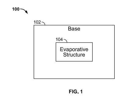
[0077] Fig. 1 illustrates one non-limiting example of a medical device 100
according to one
aspect of the present disclosure. The medical device 100 may be configured to
provide cooling to
a tissue region, or an array of tissue regions, either noninvasively (e.g., at
a surface of the tissue
region) or invasively (e.g., at a predetermined depth into the tissue region).
The medical device
100 includes a base 102 and an evaporative structure 104. The base 102 is
configured to contact a
tissue region to facilitate the removal of heat from the tissue region,
thereby cooling the tissue
region to a predetermined temperature. In some non-limiting examples, the
medical device 100
may be configured to cool the tissue region to a desired temperature profile
as a function of time.
[0078] In some non-limiting examples, the base 102 may be a noninvasive
implement designed
to continuously contact a surface of a tissue region to cool the tissue region
at the surface and/or to
a predetermined depth into the tissue region. In some non-limiting examples,
the base 102 may be
a noninvasive implement designed to discretely contact a surface of a tissue
region in a desired
fractional pattern to provide fractional cooling over the tissue region and/or
to a predetermined
depth into the tissue region. In some non-limiting examples, the base 102 may
be a noninvasive
implement designed to provide thermal management (i.e., cooling) adjacent to
or around a
fractional heating pattern to minimize damage to non-target tissue between the
fractional heating
areas. In some non-limiting examples, the base 102 may be an invasive
implement configured to
-8-
CA 03054037 2019-08-19
WO 2018/136830 PCT/US2018/014578
penetrate into a tissue region, or an array of tissue regions, to provide
cooling to the tissue
region(s) at a predetermined depth, or a range of depths.
[0079] In some non-limiting examples, the evaporative structure 104 may be
in contact with or
integrated into the base 102. The evaporative structure 104 is configured to
receive a working
fluid to facilitate cooling of a tissue region via a two-phase heat transfer
process. The evaporative
structure 104 is designed to include one or more of cavities and/or one or
more flow paths formed
therein, each configured to be filled with the working fluid. Once filled with
the working fluid,
liquid menisci are formed within each of the cavities or paths due to the
combined effect of
capillary and short range forces. The liquid menisci act as evaporation sites
that provide
significant heat removal potential from the evaporative structure 104 and the
base 102 due to the
large enthalpy of vaporization of liquids. Thus, the medical device 100 is
operable to provide
cooling to a tissue region via a two-phase heat transfer process, which
provides a heat removal
capacity as high as several orders of magnitude greater than conventional
medical cooling
technologies (e.g., single-phase cooling, thermoelectric cooling, Joule-
Thompson cooling, etc.).
[0080] In the illustrated non-limiting example, the evaporative structure
104 may not be
required to receive a continuous flow of fluid. Rather, the evaporative
structure 104 may be filled
or charged with a predetermined quantity of the working fluid. Alternatively
or additionally, the
evaporative structure 104 may be initially brought into fluid communication
with a source of the
working fluid, and the capillary forces provided by the design of the
evaporative structure 104
cause the working fluid to flow into the evaporative structure 104 without the
requirement of an
externally induced pressure differential (e.g., a pump). In some non-limiting
examples, the
evaporative structure 104 may be coated with a material that possesses a high
surface tension (e.g.,
a single layer of graphite, or graphene). Once filled, the evaporative
structure 104 may be
removed from fluid communication with the source of working fluid. The
evaporative structure
104 may be designed to initiate and maintain evaporation of the working fluid,
once the base 102
is brought into contact with a tissue region. That is, heat transferred from
the tissue region through
the base 102 and to the evaporative structure 104 is sufficient to initiate
and maintain the
evaporation of the working fluid and, thereby, the cooling of the tissue
region. Thus, the medical
device 100 is operable to provide cooling to a tissue region without the
requirement of an external
power supply, or heat source, to facilitate the evaporation of the working
fluid therein. In some
non-limiting examples, this enables the medical device 100 to operate as a
passive device (i.e.,
-9-
CA 03054037 2019-08-19
WO 2018/136830 PCT/US2018/014578
does not require an external source of energy to operate) and to possess
increased mobility over
conventional medical cooling system, which require wires, power supplies,
etc., to operate.
Alternatively or additionally, the evaporative structure 104 may be designed
such that the capillary
forces maintain the working fluid within the evaporative structure 104
regardless of the orientation
of the medical device 100. That is, the capillary forces within the
evaporative structure 104, once
filled, may be greater than the force of gravity enabling the medical device
100 to be used in any
orientation without the threat of leakage of the working fluid or partially
dry areas in the
evaporative structure 104.
[0081] Due to the thermodynamic operation of the of the medical device 100,
an amount of
working fluid needed to cool a tissue region to a desired temperature for a
desired amount of time
is known. That is, the rate of evaporation of the working fluid from the
evaporative structure 104
may be known based on the heat input from the tissue region. In this way, the
medical device 100
may be tailored to provide a desired amount of cooling for a desired amount of
time. Alternatively
or additionally, the known amount of time for a given mass of working fluid to
evaporate may be
utilized to determine when the working fluid needs to be re-filled and/or when
a different working
fluid may be communicated to the evaporative structure 104 to control the
temperature of the
tissue region.
[0082] The medical device 100 may be operable with a variety of different
working fluids. For
example, water, a liquid hydrocarbon or alcohol, halogenated hydrocarbons,
ammonia, carbon
dioxide, to name a few. In some non-limiting examples, the working fluid may
be chosen based
on a specific medical application, desired heat exchange rate, and/or range of
operating
temperatures. Since evaporative processes are substantially isothermal, the
desired temperature
range and heat transfer characteristics may be governed by the thermophysical
properties of the
working fluid. That is, the boiling point of the working fluid is known for a
given pressure and
temperature and, thus, an equilibrium temperature achieved by the medical
device 100 may be
determined based at least in part by the chemical composition of the working
fluid. Table 1 below
provides various non-limiting examples of the properties and operational
characteristics of the
medical device 100.
Table 1: Properties and Operating Characteristics of the Medical Device 100
Operating Range -220 C to 200 C
-10-
CA 03054037 2019-08-19
WO 2018/136830 PCT/US2018/014578
Operating Pressure 0.01bar to 10 bar
Working Fluids Hydrocarbons, Hydrofluorocarbons,
Hydrofluoroolefins,
Alcohols, Water, Aqueous Solution, Nobel Gases, Binary
Mixtures, Nanoparticle laden Fluids, Cryogenic Fluids: N2,
02, etc.
Substrate Material Metals, Polymers, Composite Materials, Non-metallic
elements
E.G., Copper, Aluminum, Graphite, etc.
Evaporative Structure Microgrooved, Fractional Pattern Microchannels,
Nano-
spheres
E.G., Aluminum, Copper, Carbon, Steel, etc.
Evaporative Pore Size 100nm-2000[tm
Coatings Wetting or non-wetting coating, Gold, Teflon,
Anodized
Nano-Layers, Nano-structured coatings
Fluid Flow Control Thermo-capillary, Piezo-electric, Expansion Valve,
Capillary
Tube, Electroosmotic Driven Flow, Electromotive Force
Temperature Control Thermocouple, RTD, Embedded Contact Micro-wires
[0083] When implementing the medical cooling device 100 to cool a desired
tissue region, the
thermal characteristics of the desired tissue region combined with its
structural-mechanics
response to change in temperature may play a role in energy-based medical
applications. For
example, in the case of cryolipolysis, water and fat containing tissues may
undergo a phase change
as their temperature drops below the melting point for water and/or fat. This
phase change (i.e.,
crystallization) is accompanied with two events in the thermal characteristics
of the tissue as well
as the energy balance during the cooling process. First, the thermal
conductivity for the solid
phase is higher than the liquid phase, therefore, the conduction heat transfer
may be improved
significantly as the frozen front moves into the non-frozen section of the
desired tissue region. For
example, water possesses a thermal conductivity of approximately four times
higher in the solid
phase (i.e., ice) when compared to liquid water. This increase in thermal
conductivity may induce
a cascade of accelerating affects as long as the heat removal capacity is not
exceeded and while the
distance from the cold surface is not imposing a large resistance on the heat
flow. Second, the
latent heat of the phase change released at the interface between the frozen
and non-frozen sections
of tissue may add a significant load to the total heat that should be removed
from the desired tissue
-11-
CA 03054037 2019-08-19
WO 2018/136830 PCT/US2018/014578
region. If the cooling capacity is limited, the cooling process is slowed down
to match the
maximum heat flow that could be dissipated.
[0084] These dynamic characteristics of the tissue, explained above, may
only be noticed in
circumstances where the heat flow from the desired tissue region to the cold
surface is not limited
by the capacity of the mechanism employed to provide the cooling effect. The
two-phase cooling
leveraged by the medical device 100 provides superior cooling performance and
significantly
increased cooling capacity when compared to conventional cooling mechanism
(e.g., single-phase
cooling, thermoelectric cooling, Joule-Thompson cooling, spray cooling, etc.).
The extremely high
cooling capacity of the medical device 100 turns the dynamic thermal behavior
of the desired
tissue region into an advantage to accelerate the progression of the frozen
front, enhance the
effectiveness of the thermal damage to the desired tissue region by increasing
the energy removed
from the unit volume of tissue per unit time, reduce the duration of a desired
medical procedure,
shrink the footprint of the overall medical device 100 and significantly
reduce the total weight
thereof, reduce the risk of undesired damage to neighboring/non-target tissue,
improve the
reliability of the medical device 100 due to passive operation, increase the
effective range (distance
from the cold surface) that can be cooled, and improve the temporal and
spatial accuracy in
controlling and maintaining the temperature levels.
[0085] In some non-limiting examples, the medical device 100 may be
configured to provide a
step-wise, cyclic, or predetermined temperature profile as a function of time
cooling approach by
inducing cooling waves into the desired tissue region. For example, if a
tissue region is required to
be cooled to -10 C, the medical device 100 may be configured to start at an
operating temperature
of -5 C and stay there for a first predetermined amount of time. After the
first predetermined
amount of time, the medical device 100 may be configured to transition to an
operating
temperature of -15 C for a second predetermined amount of time. The operating
temperature
transition between -5 C and -15 C may be facilitated, for example, by changing
the operating
pressure of system and/or changing the working fluid and/or the flow rate of
the working fluid, to
name a few. Once the second predetermined amount of time has passed, the
medical device 100
may be configured to transition back to an operating temperature of -5 C for a
third predetermined
amount of time. In some non-limiting examples, the medical device 100 may
cyclically continue
to transition between -5 C, -15 C , and -5 C operating temperatures until a
total time for a given
procedure is reached. The step-wise, or cyclic, transitions in operating
temperature may enable the
-12-
CA 03054037 2019-08-19
WO 2018/136830 PCT/US2018/014578
medical device 100 to more efficiently cool the tissue region to the desired -
10 C and reach the
desired -10 C temperature in less time, when compared to providing cooling at
a constant -10 C.
[0086] In accordance with a non-limiting configuration, the use or method
of use of the
medical device 100 does not include a step of treatment of a human or animal
body by surgery or
therapy. It is noted that the skills of a person using a device as described
herein, may not have the
skills of a physician, and that the intended treatment may not be motivated
due to illness of the
treated person, rather for aesthetic reasons.
[0087] In some non-limiting examples, a suction device may be implemented
to adhere the
tissue region to the base 102. The suction device may be in the form of a
vacuum pump, or
another device capable of generating a pressure lower than atmospheric
pressure, to suction the
tissue region onto the base 102.
[0088] Fig. 2 illustrates another non-limiting example of the medical
device 100 according to
one aspect of the present disclosure. As illustrated in Fig. 2, the medical
device 100 may include a
heat source 106. In some non-limiting examples, the heat source 106 may be a
resistive heater, a
thin, transparent heater arranged between the base 102 and the tissue region,
a thermoelectric
heater, a microwave heater, an electromagnetic heater (e.g., infrared), an
ultrasound heater, a radio
frequency heater etc. In some non-limiting examples, the heat source 106 may
leverage waste heat
from another component (e.g., a laser) located externally from the base 102.
[0089] The heat source 106 may be configured to selectively apply heat to
the base 102 and/or
a tissue region. In some non-limiting examples, the heat source 106 may be
integrated into the
base 102 to facilitate the selective heating of the base 102. In some non-
limiting examples, the
heat source 106 may be located remotely from the base 102 and in thermal
communication with
the base 102 and/or a tissue region. In operation, the medical device 100 may
be used to cool a
tissue region for a given medical application, and the heat source 106 may
subsequently heat the
tissue region and/or an adjacent tissue region back to approximately room
temperature. In some
non-limiting application, the medical device 100 may freeze at least a portion
of the tissue region
(e.g., the surface of the tissue region) and the heat source 106 may be used
to prevent sticking
between the base 102 and the surface of the tissue region. For example, a
thin, transparent heat
source 106 may be arranged between the base 102 and the surface of the tissue
region to facilitate
quick removal of the medical device 100 from the surface of the tissue region
after the desired
cooling has been applied thereto.
-13-
CA 03054037 2019-08-19
WO 2018/136830 PCT/US2018/014578
[0090] Fig. 3 illustrates another non-limiting example of the medical
device 100 according to
one aspect of the present disclosure. As illustrated in Fig. 3, the
evaporative structure 104 may be
in fluid communication with a fluid source 108. The fluid source 108 may
include a supply of
working fluid that may be furnished to the evaporative structure 104. In some
non-limiting
examples, the fluid source 108 may be a non-pressurized source (i.e., at
approximately
atmospheric pressure) of working fluid. In these non-limiting example, fluid
contact of between
the fluid source 108 and the evaporative structure 104 may be sufficient
induce capillary forces
that supply the evaporative structure 104 with the working fluid. In some non-
limiting examples,
the fluid source 108 may be configured to induce a pressure drop between the
fluid source 108 and
the evaporative structure 104 to furnish the working fluid into the
evaporative structure 104. In
these non-limiting examples, the fluid source 108 may be configured to
selectively furnish the
working fluid into the evaporative structure 104 (e.g., when it is determined
that the evaporative
structure 104 requires more working fluid).
[0091] Fig. 4 illustrates another non-limiting example of the medical
device 100 according to
one aspect of the present disclosure. As illustrated in Fig. 4, the medical
device 100 may include a
fluid control device 110 in communication with the fluid source 108 and/or the
evaporative
structure 104. In some non-limiting examples, the fluid control device 110 may
be configured to
control a direction of the fluid flow between the fluid source 108 and the
evaporative structure 104,
or the flow rate of the working fluid. For example, the fluid control device
110 may be in the form
of a check valve configured to only allow fluid to flow from the fluid source
108 to the evaporative
structure 104.
[0092] In some non-limiting examples, the fluid control device 110 may be
configured to
control a pressure of the working fluid provided from the fluid source 108 to
the evaporative
structure 104. For example, the fluid control device 110 may be in the form of
a disposable
charged cartridge that is configured to selectively increase the pressure of
the working fluid
flowing to the evaporative structure 104. In this way, the fluid control
device 110 may be utilized
to control a cooling temperature output by the medical device 100 by varying
the pressure of the
working fluid within the evaporative structure 104. Alternatively or
additionally, the fluid control
device 110 may include a pressure regulator configured to increase or decrease
the pressure of the
working fluid, as desired.
-14-
CA 03054037 2019-08-19
WO 2018/136830 PCT/US2018/014578
[0093] In some non-limiting examples, the fluid control device 110 may be
configured to
selectively provide fluid communication between the fluid source 108 and the
evaporative
structure 104. For example, the fluid control device 110 may be in the form of
an on/off valve
configured to selectively provide fluid communication between the fluid source
108 and the
evaporative structure 104 to activate and deactivate the cooling of a tissue
region. It should be
appreciated that the various forms of the fluid control device 100 described
herein may be
combined, and the medical device 100 is not limited to the use of only one of
the described
functi onaliti es.
[0094] Fig. 5 illustrates another non-limiting example of the medical
device 100 according to
one aspect of the present disclosure. As illustrated in Fig. 5, the medical
device 100 may include a
condenser 112 in fluid communication with the evaporative structure 104. The
condenser 112 may
be configured to facilitate the condensation of evaporated working fluid that
flows from the
evaporative structure 104. In some non-limiting examples, the condenser 112
may be configured
to provide a sufficient amount of heat removal or dissipation to the working
fluid to effectuate the
condensation thereof. The condenser 112 may be in fluid communication with the
fluid source
108 to recapture the working fluid and provide it to the fluid source 108. In
other non-limiting
examples, the condenser 112 may be configured to communicate the condensed
working fluid to a
reservoir, as will be described.
[0095] Fig. 6 illustrates another non-limiting example of the medical
device 100 according to
one non-limiting example of the present disclosure. As illustrated in Fig. 6,
the fluid control
device 110 may be remotely in fluid communication (i.e., not arranged in-line
with the fluid source
108) with the working fluid downstream of the fluid source 108 and downstream
of the
evaporative structure 104. This configuration may enable the fluid control
device 110 to
selectively control a pressure of the working fluid flowing into the
evaporative section 104 (e.g., to
control a cooling temperature provided by the medical device 100) and/or
control a pressure of the
evaporated working fluid leaving the evaporative structure 104 (e.g., to
control the condensing of
the evaporated working fluid).
[0096] Fig. 7 illustrates another non-limiting example of the medical
device 100 according to
one aspect of the present disclosure. As illustrated in Fig. 7, the
evaporative structure 104 may be
in fluid communication with the fluid source 108, the condenser 112, and a
reservoir 114. In some
non-limiting examples, the reservoir 114 may be a tank or vessel at
approximately atmospheric
-15-
CA 03054037 2019-08-19
WO 2018/136830 PCT/US2018/014578
pressure that contains working fluid. In some non-limiting examples, the
reservoir 114 may be
tank or vessel either above or below atmospheric pressure that contains
working fluid. The fluid
source 108 may be configured to furnish the working fluid from the reservoir
114 to the
evaporative structure 104 at a predetermined pressure and flow rate. In some
non-limiting
examples, the working fluid may flow continually from the fluid source 108 to
the evaporative
structure 104 through the condenser 112 and back to the reservoir 114. In some
non-limiting
examples, the working fluid may be selectively provided to the evaporative
structure 104, as
needed.
[0097] The thermal and thermodynamic characteristics of the medical device
100 enable the
medical device 100 to be self-adapting, or self-regulating based on the heat
input applied thereto.
That is, the amount of working fluid evaporated within the evaporative
structure 104 and
subsequently condensed by the condenser 112 may be proportional to the heat
input to the medical
device 100 from the tissue. In this way, the medical device 100 may self-
regulate the amount of
evaporation and subsequent condensing of the working fluid to provide
sufficient liquid working
fluid to the reservoir 114 and fluid source 108.
[0098] Fig. 8 illustrates another non-limiting example of the medical
device 100 according to
one aspect of the present disclosure. As illustrated in Fig. 8, the
evaporative structure 104 may be
the form of a patterned evaporative structure 118. The patterned evaporative
structure 118 may be
configured to provide a cooling pattern with varied heat flux to a tissue
region. In some non-
limiting examples, the mechanical structure of the patterned evaporative
structure 118 may be
tailored to spatially vary the heat dissipation flux provided by the medical
device 100. For
example, a porosity of the patterned evaporative structure 118 may be designed
to spatially vary
the heat removal flux or capacity across the medical device 100. Alternatively
or additionally, a
material of the base 102, a coating of the base 102 or the patterned
evaporative structure 118,
and/or an external coating applied between the base 102 and the tissue region
may be designed to
spatially vary the heat dissipation flux or profile of the medical device 100
to define a cooling
pattern. In one non-limiting example, an antifreeze coating, or material, may
be applied between
the base 102 and the tissue region to protect certain areas within the tissue
region from the cooling
effect of the medical device 100 (e.g., to provide protection against
freezing) .
[0099] In some non-limiting examples, the medical device 100 may be
configured to operate
with a spatially varying operating temperature along the base 102. For
example, the base 102 may
-16-
CA 03054037 2019-08-19
WO 2018/136830 PCT/US2018/014578
define a symmetrical operating temperature profile that increases in
temperature from a centerline
of the base 102 to first and second edges of the base 102. In some non-
limiting examples, the base
102 may define a symmetrical operating temperature profile that decreases in
temperature from a
centerline of the base 102 to first and second edges of the base 102. In some
non-limiting
examples, the base 102 may define a varied operating temperature profile that
conforms to any
functional form, as desired.
[00100] Fig. 9 illustrates a non-limiting example of a tiled medical device
200 according to one
aspect of the present disclosure. As illustrated in Fig. 9, the tiled medical
device 200 may include
a plurality of the medical devices 100 arranged in an array, or tiled,
pattern. It should be
appreciated that the medical devices 100 may be arranged in any pattern as
desired. In some non-
limiting examples, the medical devices 100 may be linked via a mesh-like
structure. In other non-
limiting examples, the medical devices 100 may be individually mounted to an
external structure.
In any case, the medical devices 100 may be moveable to enable the tiled
medical device 200 to
conform to any anatomical region and/or to match any anatomical features, as
desired. In some
non-limiting examples, the medical devices 100 may be individually controlled
within the tiled
medical device 200 to enable the tiled medical device 200 to provide a
predetermined cooling
pattern. For example, the medical devices 100 may be provided with various
working fluids to
define different operating temperatures. Alternatively or additionally, the
evaporative structures
104 within the medical devices 100 may be designed to provide different heat
transfer properties.
Alternatively or additionally, the bases 102 of the medical devices 100 may be
coated and/or
insulated to control the output temperature thereof In some non-limiting
examples, selective
groups of the medical devices 100 within the tiled medical device 200 may be
controlled to enable
the tiled medical device 200 to provide a predetermined cooling pattern. For
example, selective
groups of the medical devices 100 within the tiled medical device 200 may be
connected to
different working fluid circuits, which enable the selective groups of the
medical devices 100 to
operate at different cooling temperatures.
[00101] It should be appreciated that the various non-limiting examples of the
medical device
100 described herein are not necessarily separate in nature, and the medical
device 100 may be
adapted to include any combination of the various non-limiting components and
configurations
described herein.
-17-
CA 03054037 2019-08-19
WO 2018/136830 PCT/US2018/014578
[00102] Fig. 10 illustrates one non-limiting example of the evaporative
structure 104 according
to one aspect of the present disclosure. In the non-limiting example of Fig.
10, the evaporative
structure 104 is integrated into the base 102. The base 102 includes a
plurality of fins 400 that
extend from a first surface 402 of the base 102 to form a plurality of
channels 404 therebetween.
The channels 404 are configured to receive the working fluid and are
dimensioned to ensure that
liquid menisci are formed therein. During operation, for example, a second
surface 406 of the base
102 may be brought into engagement with a tissue region. Heat energy from the
tissue region may
travel through the base 102 and the fins 400 to the liquid menisci formed in
the channels 404
where the working fluid is evaporated. The integral effect of evaporation from
all the menisci in
the evaporative structure 104 provides the substantial heat removal capacity
of the medical device
100. The properties of the evaporative structure 104 may affect the rate of
evaporation and thereby
the overall heat removal capacity of the medical device 100. For example, the
number of channels
404, the channel width W, the material of the base 102, a coating applied to
the base 102, and a
material applied to the exterior of the base 102 (i.e., between the base 102
and the tissue region)
may all affect the overall cooling performance of the medical device 100.
[00103] Fig. 11 illustrates another non-limiting example of the evaporative
structure 104
according to the present disclosure. As shown in Fig. 11, the evaporative
structure 104 may be
formed by a porous substrate 500. In some non-limiting examples, the porous
substrate 500 may
be attached to, or removably positioned, on the first surface 402 of the base
102. In some non-
limiting examples, the porous substrate 500 may be attached to the first
surface 402 of the base
102. In any case, the porous substrate 500 includes a plurality of pores 502
that each act as sites to
form menisci, once filled with the working fluid. During operation, for
example, the second
surface 406 of the base 102 may be brought into engagement with a tissue
region. Heat energy
from the tissue region may travel through the base 102 and the porous
substrate 500 to the liquid
menisci where the working fluid is evaporated. The properties of the porous
substrate 500 may
affect the rate of evaporation and thereby the overall heat removal capacity
of the medical device
100. For example, the number of pores 502, the size of the pores 502, the
material of the porous
substrate 500, the material of the base 102, a material applied to the
exterior of the base 102 (i.e.,
between the base 102 and the tissue region), and a coating applied to the base
102 may all affect
the overall cooling performance of the medical device 100.
-18-
CA 03054037 2019-08-19
WO 2018/136830 PCT/US2018/014578
[00104] As described above, the medical device 100 may be in the form of a
noninvasive
medical device. Figs. 12 and 13 illustrate one non-limiting example of a
noninvasive medical
device 600 configured to cool a tissue region via a two-phase heat transfer
process in accordance
with the systems and methods described herein. The noninvasive medical device
600 includes a
base 602 having a first surface 604, a treatment surface 606 arranged opposite
to the first surface
604, and a cooling cavity 608 formed in the base 602. In some non-limiting
examples, the base
602 may be fabricated from a metal material (e.g., aluminum, copper, brass,
etc.). In some non-
limiting examples, the base 602 may be fabricated from a graphite or woven
material (e.g., carbon
fiber).
[00105] In the illustrated non-limiting example, the base 602 includes two
cooling cavities 608.
In other non-limiting examples, the base 602 may include more or less than two
cooling cavities
608. The cooling cavities 608 are formed by recesses that extend into the
first surface 604 toward
the treatment surface 606. In the illustrated non-limiting example, the
cooling cavities 608 define
a generally rectangular shape. In other non-limiting examples, the cooling
cavities 608 may define
another shape (e.g., round, polygonal, etc.), as desired.
[00106] The cooling cavities 608 are configured to receive a porous
substrate 610 therein. In
some non-limiting examples, the porous substrate 610 may be fabricated from a
metal (e.g.,
aluminum or copper), carbon fiber mesh material, or metal foam material. The
porous substrates
610 includes a plurality of pores that each act as sites to form menisci, once
filled with the working
fluid. As described herein, the menisci may act as sites for the working fluid
to evaporate from, in
response to heat input from the desired tissue region in contact with the
treatment surface 606.
[00107] The geometric properties of the porous substrates 610 (e.g., a size of
the pores) may be
designed such that, once filled with working fluid, capillary forces maintain
the working fluid
therein regardless of the orientation of the noninvasive medical device 600.
That is, the capillary
forces within the porous substrates 610, once filled, may be greater than the
force of gravity
enabling the noninvasive medical device 600 to be used in any orientation
without the threat of
leakage of the working fluid.
[00108] The porous substrates 610 may be in engagement with at least a portion
of the base 602.
In the illustrated non-limiting example, one or more posts 611 protrude upward
from the bottom
surface of the cooling cavities 608 to enhance contact between the base 602
and the porous
substrates 610. The posts 611 may be arranged throughout the cooling cavities
608 to aid the
-19-
CA 03054037 2019-08-19
WO 2018/136830 PCT/US2018/014578
conductive heat transfer between the porous substrates 610 and the base 602.
In the illustrated
non-limiting example, each of the cooling cavities 608 include six posts 611
staggered therealong.
In other non-limiting examples, each of the cooling cavities 608 may include
more or less than six
posts 611 arranged in any pattern, as desired.
[00109] In the illustrated non-limiting example, the porous substrates 610 may
be exposed to
the surroundings (i.e., the noninvasive medical device 600 defines an open
circuit with respect to
the working fluid). This may allow the working fluid arranged within the
porous substrates 610 to
evaporate to the surroundings. In these non-limiting examples, the working
fluid may be chosen to
be chemically inert and/or safe for inhalation by the patient and/or the user.
In some non-limiting
examples, the porous substrates 610 may be pre-loaded with the working fluid.
In some non-
limiting examples, the porous substrates 610 may be quasi-open where porous
substrates 610 are
covered by a structure, which is exposed to the surroundings. The evaporated
working fluid may
travel along the structure and subsequently condense therein to enable at
least a portion of the
working fluid to be collected and recirculated, as desired. In some non-
limiting examples, the
porous substrates 610 may be sealed from the surroundings to provide a closed
circuit for the
working fluid. That is, the working fluid may be provided to the noninvasive
medical device 600
from a sealed reservoir, and the evaporated working fluid may be captured from
the sealed cooling
cavities 608 and subsequently condensed either actively (e.g., via a
condenser) or passively (e.g.,
via heat transfer with the surroundings). The condensed working fluid may be
fluidly
communicated back to the sealed reservoir. Thus, in the closed circuit, the
working fluid may not
be exposed to the surroundings thereby enabling the use of chemically active
working fluids that
may be potentially harmful in an open circuit.
[00110] The cooling cavities 608 and thereby the porous substrates 610 are
connected to a port
612 via first and second channels 614 and 616. The port 612 and the first and
second channels 614
and 616 are recessed into the first surface 604. During operation, the port
612 may be configured
to be connected to a supply of working fluid. The working fluid may flow from
the port 612 along
the channels 614 and 616 to the cooling cavities 608 and into the porous
substrates 610.
[00111] In some non-limiting examples, a disposable charged cartridge (not
shown) may be
provided to control a pressure of the working fluid within the noninvasive
medical device 600. For
example, the charged cartridge may be in fluid communication with the working
fluid upstream of
the cooling cavities 608, and configured to selectively increase a pressure of
the working fluid
-20-
CA 03054037 2019-08-19
WO 2018/136830 PCT/US2018/014578
flowing into the cooling cavities 608. Alternatively or additionally, the
charged cartridge may be
in fluid communication with the noninvasive medical device 600 downstream of
the cooling
cavities 608, for example, to effectuate condensation of the working fluid.
[00112] During operation of the closed circuit configuration of the
noninvasive medical device
600, for example, a working fluid may be supplied to the noninvasive medical
device 600 to fill
the porous cavities 608. In some non-limiting examples, the capillary forces
provided by the
design of the porous substrates 610 may cause the working fluid to flow
therein without the
requirement of an externally induced pressure differential. Thus, working
fluid may be supplied to
the port 612 and the working fluid may be naturally (i.e., without external
forces) drawn into the
porous substrates 610.
[00113] Once the porous substrates 610 are filled with the working fluid, the
noninvasive
medical device 600 may be positioned such that the treatment surface 606
engages a desired tissue
region of a patient. The engagement of the treatment surface 606 with the
desired tissue region
initiates heat transfer between the noninvasive medical device 600 and the
desired tissue region.
Specifically, heat from the desired tissue region transfers through the
treatment surface 606 and to
a bottom surface of the cooling cavities 608. From the bottom surface of the
cooling cavities 608,
the heat transfers through the porous substrates 610 to the liquid menisci
where evaporation of the
working fluid due to the heat input from the tissue region. The integral
effect of evaporation from
all the menisci in the porous substrates 610 provides the noninvasive medical
device 600 with
substantial heat removal capacity (i.e., heat flux capacity) when compared to
conventional medical
cooling technologies.
[00114] As described above, in some non-limiting examples, the noninvasive
medical device
600 may define a closed circuit with respect to the working fluid. Figs. 14A-
15 illustrate a
illustrate non-limiting example of the noninvasive medical device 600 that
implements a closed
circuit with respect to the working fluid. As illustrated in Figs. 14A and
14B, the noninvasive
medical device 600 may include a condensing plate 629 and defines a flow path
618 on one side
thereof and a condensing structure 631 on another side thereof The flow plat
618 extends from an
inlet port 620 to an outlet port 622. The flow path 618 may be recessed into
one side of the
condenser plate 629 and define a shape that covers a desired amount of the
surface area of the base
602. In the illustrated non-limiting example of Fig. 14A, the flow path 618
extends from the inlet
port 620 in a generally straight path toward an opposing end of the condensing
plate 629 and,
-21-
CA 03054037 2019-08-19
WO 2018/136830 PCT/US2018/014578
adjacent to the opposing end of the condensing plate 629, the flow path 618
curves in a direction
toward a center of the condensing plate 629. The flow path 618 then extends in
a direction back
toward the end of the condensing plate 629 on which the inlet port 620 is
arranged in a generally
swirl-like pattern. That is, the flow path 618 curves back and forth as it
extends toward the inlet
end of the base 602. Once the swirl section of the flow path 618 reaches the
inlet end of the
condensing plate 629, the flow path 618 curves in a curves in a direction away
from the inlet port
620 and extends in a generally straight path to the outlet port 622.
[00115] It is to be appreciated that the illustrated flow path 618 is but
one non-limiting example
and that the flow path 618 may be shaped to cover a desired amount of the
condensing plate, as
desired. For example, the flow path 618 may be shaped to uniformly cover a
substantial amount of
the total surface area of the condensing plate 629. In other non-limiting
examples, the flow path
618 may be shaped to cover a selected section of the surface area of the
condensing plate 629
where cooling is desired.
[00116] As illustrated in Fig. 14B, the other side of the condensing plate
629 include the
condensing structure 631. In the illustrated non-limiting example, the
condensing structure 631
includes a plurality of ridges 633 arranged along a recessed surface 635. The
plurality of ridges
633 protrude outwardly from the recessed surface 635 and extend in a lateral
direction between
first and second ends 637 and 639 of the condensing plate 629. In some non-
limiting example, the
plurality of ridges 633 may act as fins to promote addition heat transfer from
the fluid flowing
through the flow path 618, which acts to aid in condensing of evaporated
working fluid, as will be
described.
[00117] Fig. 15 illustrates a evaporative plate 641 of the noninvasive medical
device 600 with a
closed circuit. As illustrated in Fig. 15, the evaporative plate 641 includes
an evaporative structure
624 arranged on a side thereof opposite to the treatment surface 606. In the
illustrated non-limiting
example, the evaporative structure 624 may include a plurality of ridges 626
that protrude
outwardly from a recessed surface 643 in a direction away from the treatment
surface 606. The
plurality of ridges 626 may extend in a lateral direction between first and
second ends 628 and 630
of the recessed surface 624, and may be arranged along the recessed surface
624 between the inlet
port 620 and the outlet port 622. In some non-limiting examples, the plurality
of ridges 626 may
extend varying lateral distances between the first and second ends 628 and
630. For example, the
plurality of ridges 626 may alternate between a first ridge 632 and a second
ridge 634 between the
-22-
CA 03054037 2019-08-19
WO 2018/136830 PCT/US2018/014578
inlet port 620 and the outlet port 622. The first ridge 632 may extend from a
center of the recessed
surface 624 to a location between the center and each of the first and second
ends 628 and 630
(i.e., the first ridge 632 does not extend completely between the first and
second ends 628 and
630). The second ridge 634 may extend completely between the first and second
ends 628 and
630. In some non-limiting examples, a distance between adjacent ridges 632 and
634 may ensure
that capillary forces maintain the working fluid therebetween. Therefore, the
different lateral
extensions between the first ridge 632 and the second ridge 634 may maintain
the working fluid
along a centerline of the evaporative plate 641. In other non-limiting
examples, the design of the
evaporative structure 641 may be altered to accommodate any desired cooling
pattern, for
example, by manipulating the arrangement and orientation of the ridges 632,
634. In some non-
limiting examples, the evaporative structure 641 may be in the form of a
porous structure, as
described herein. In some non-limiting examples, the evaporative structure 641
may be in the
form of one or more microchannels that extend along the recessed surface 624.
[00118] Both of the condenser plate 629 and the evaporative plate 641 may
include a recessed
notch 636 that extends therein and surrounds the flow path 618, the condensing
structure 631, and
the evaporative structure 624. The recessed notches 636 may be configured to
receive a seal (e.g.,
an o-ring or gasket) therein to facilitate forming a seal between a cover
plate attached to the each
of the sides of the condenser plate 629 and the non-treatment side of the
evaporative plate 641.
[00119] As illustrated in Figs. 16 and 17, the noninvasive medical device 600
may, when
assembled, include an insulated layer 640 arranged around a periphery thereof.
In some non-
limiting examples, the insulated layer 640 may aid in inhibiting heat from
dissipating from the
noninvasive medical device 600 to the atmosphere, and may provide protection
for a user
manipulating the noninvasive medical device 600. Additionally, the noninvasive
medical device
600 may include a charging port that enables the working fluid to be charged
into the device (i.e.,
flow into the area between the evaporative structure 641 and the condensing
structure 631. When
assembled, the condenser plate 629 may be attached to the condenser plate 641
such that the
evaporative structure 624 faces the condensing structure 631. Thus, when
assembled, one side of
the device includes the treatment surface 606, which is thermally coupled to
the evaporative
structure 624, and a cover plate may be arranged on the other side, which
covers the flow path 618.
[00120] During operation of the closed circuit configuration of the
noninvasive medical device
600, for example, working fluid may be charged into a cavity formed between
the evaporative
-23-
CA 03054037 2019-08-19
WO 2018/136830 PCT/US2018/014578
structure 624 and the condensing structure 631. Once charged, this cavity may
be sealed off,
thereby closing the working fluid off from the surroundings. When the
treatment surface 606 is
placed in contact with a desired tissue region, heat transfer initiates
between the desired tissue
region and the evaporative structure 624. Specifically, heat from the desired
tissue region transfers
through the treatment surface 606 and to the working fluid flowing within the
evaporative structure
624. The heat input from the desired tissue region facilitates the evaporation
of the working fluid
which can come into contact with the condensing structure 631. Evaporation of
the working fluid
flowing within the evaporative structure 624 enables the noninvasive medical
device 600 to
leverage the advantages of two-phase heat transfer processes described herein.
Thus, the
noninvasive medical device 600 provides substantial heat removal capacity
(i.e., heat flux
capacity) when compared to conventional medical cooling technologies.
[00121] During operation of the device, cooling fluid may be flown through the
flow path 618,
which is isolated from the working fluid. Thus, as the evaporated working
fluid builds up around
the condensing structure 631, the cooling provided by the fluid flowing
through the flow path 618
may provide the necessary heat removal to facilitate condensing of the
evaporated working fluid
and result in the condensed working fluid "raining down" onto the evaporative
structure 624.
[00122] Fig. 18 another non-limiting example of the noninvasive medical device
600
implemented in a thermal treatment application. As illustrated in Fig. 18, the
noninvasive medical
device 600 may define an open system with respect to the working fluid (i.e.,
the working fluid is
provided to the device and recovered from the device) and the device does not
include a
condensing stage within the base 602 (i.e., the condensing stage happens
remotely from the base
602, which is in contact with the tissue region). The noninvasive medical
device 600 may be
configured to receive working fluid from a tank 603. A inlet line 613 extends
between the tank
603 and an inlet to the evaporative structure 610, 624 to provide fluid
communication
therebetween. A flow control device 110, 605 may be arranged on the inlet line
613 between the
tank 603 and the inlet to the evaporative structure 610, 624 In some non-
limiting examples, the
flow control device 110, 605 may be configured to control a direction of fluid
flow, a pressure of
fluid flow, and/or a flow rate. From the inlet to the evaporative structure
610, 624, the working
fluid can flow along the evaporative structure 624 and remove heat from the
tissue region, which
results in evaporation of the working fluid. Thus, the noninvasive medical
device 600 provides
-24-
CA 03054037 2019-08-19
WO 2018/136830 PCT/US2018/014578
substantial heat removal capacity (i.e., heat flux capacity) when compared to
conventional medical
cooling technologies.
[00123] The evaporated working fluid may flow through an outlet to the
evaporative structure
610, 624 and into an outlet line 615. From the outlet line 615, the evaporated
working fluid may
be condensed in a condenser 609 and subsequently stored in a tank 611.
[00124] In the illustrated non-limiting example, a controller 607 is in
communication with one
or more temperature sensors arranged to measure temperature adjacent to or on
a surface of the
desired tissue region. The controller 607 may instruct the flow control device
110, 605 to adjust
the operating parameters of the noninvasive medical device 600 based at least
in part on the
measurement of the temperature sensors. Several parameters may be used to
control the thermal
output parameters of the noninvasive cooling 600, as described herein.
[00125] In some non-limiting examples, the noninvasive medical deice 600 may
be utilized with
a flexible blanket 645 that is convers and seals around the noninvasive device
600. A space
between the flexible blanket 645 and the noninvasive medical device 600 may be
in
communication with a vacuum 647 that is configured to reduce a pressure within
this space. Due
to the form factor of the noninvasive medical device 600 (e.g., thin), the
connection to the vacuum
647 may maintain thermal contact between the tissue surface and the treatment
surface 606,prevent
thermal disturbance from the surroundings (i.e., insulation), suppress blood
flow in the tissue
region, and accelerate cooling or heating of the target tissue.
[00126] In some non-limiting examples, as illustrated in Fig. 20, the
noninvasive medical device
600 may be provided with an adhesive layer 621 that is adhesively attached to
the treatment
surface 606. An anti-freeze 623 layer may be provided between the adhesive
layer 621 and a
removable sheet 625. The anit-freeze layer 623. The removable sheet 625 may be
a disposable
component that is applied to a desired tissue region and disposed of after a
desired medical
treatment is performed. In this way, the sterility of the noninvasive medical
device 600 may be
maintained.
[00127] In some non-limiting examples, as illustrated in Fig. 21, a thin
heater 627 may be
integrated into the removable sheet 625 to melt any frozen sticking between
the treatment surface
606 and the tissue surface 606 and enable the detachment of the noninvasive
medical device 600
from the tissue surface. In some non-limiting examples, as illustrated in Fig.
22, the thin heater
-25-
CA 03054037 2019-08-19
WO 2018/136830 PCT/US2018/014578
627 may be not be a disposable component and may be arranged between the
adhesive layer 621
and the anti-freeze layer 623.
[00128] In all of the configurations of the noninvasive medical device 600,
the treatment surface
606 may be configured to conform to a specific tissue region on a patient. In
some non-limiting
examples, the treatment surface 606 may be coated with a coating. The coating
applied to the
treatment surface 606 may fabricated from a material configured to correspond
with the
thermophysical properties of the base 602 and/or the working fluid within the
porous substrates
610. In the non-limiting example of Fig. 13, the treatment surface 606 defines
a generally arcuate
or curved surface with a generally rectangular profile. In the non-limiting
example of Fig. 17, the
treatment surface 606 defines a generally flat surface with a generally
rectangular profile. In other
non-limiting examples, the treatment surface 606 may define a generally convex
shape, as shown
in Fig. 23. In some non-limiting examples, the treatment surface 606 may
define a generally
smooth, or uninterrupted profile. In some non-limiting examples, the treatment
surface 606 may
define a rough, or interrupted, profile. For example, the treatment surface
606 may include a
structural pattern arranged thereon to increase a surface area thereof Fig. 24
illustrates one non-
limiting example of a structural pattern of the treatment surface 606 that
includes a plurality of
alternating peaks and valleys. Fig. 25 illustrates one non-limiting example of
the treatment surface
606 including a plurality of protrusions, or pins, 642 extending therefrom.
[00129] In some non-limiting examples, the base 602 or treatment surface 606
may define a
generally horseshoe shape, as shown in Fig. 26. In some non-limiting,
examples, the base 602 or
treatment surface 606 may define a generally banana, or crescent moon, shape,
as shown in Fig.
27. In some non-limiting examples, the base 602 or treatment surface 606 may
define a generally
annular shape, as shown in Fig. 28. In these non-limiting examples, a suction
device may be
configured to draw a tissue region into a central aperture defined by the base
602.
[00130] In some non-limiting examples, the noninvasive medical device 600 may
be operable to
provide cooling to an uneven, or non-uniform, tissue surface. For example, as
shown in Fig. 29, a
tissue surface 644 may include one or more recesses 646 arranged thereon. In
these non-limiting
examples, a material (e.g., a gel or foam) 648 may be applied to the tissue
surface within the recess
646 to fill the recesses 646. The material may be configured to selectively
protect, or insulate, the
tissue recesses 646. In some non-limiting examples, the material 648 may
define a thermal
conductivity that is less than or equal to a thermal conductivity defined by
the tissue surface 644
-26-
CA 03054037 2019-08-19
WO 2018/136830 PCT/US2018/014578
(e.g., skin). By applying the material, for example, to the skin to fill, and
insulate, the recesses
646, the treatment surface 606 of the noninvasive medical device 600 may only
provide cooling to
the areas between, or around, the recesses 646, which contact the treatment
surface 606.
[00131] In some non-limiting examples, as shown in Fig. 30, a tissue surface
650 may include
one or more protrusions 652 arranged thereon. The material (e.g., a gel or
foam) 648 may be
applied in a pool around the protrusion 652 to selectively protect, or
insulate, the tissue surface 650
adjacent to the protrusion 652. By applying the material 648 in a pool around
the protrusion 652,
the treatment surface 606 of the noninvasive medical device 600 may only
provide cooling to the
protrusion 652.
[00132] In accordance with a non-limiting configuration, the use or method of
use of the
noninvasive medical device 600 does not include a step of treatment of a human
or animal body by
surgery or therapy. It is noted that the skills of a person using a device as
described herein, may
not have the skills of a physician, and that the intended treatment may not be
motivated due to
illness of the treated person, rather for aesthetic reasons.
[00133] Various parameters of the noninvasive medical device 600 may be
altered to control the
heat removal capacity and operating temperature range based on the
application. For example, the
material of the base 602, the material of the porous substrates 610, the size
of the pores, porosity in
the porous substrates 610, the geometry of the fluid path 618, the
thermophysical properties of the
working fluid, the geometric properties of the cooling cavities 608, etc.
Table 2 below provides
various non-limiting examples of the properties and operational
characteristics of the noninvasive
medical device 600.
Table 2: Properties and Operating Characteristics of the Noninvasive Medical
Device 600
Operating Range -200 C to 200 C
Operating Pressure 0.01bar to 10 bar
Working Fluids Hydrocarbons, Hydrofluorocarbons, Alcohols, Water,
Aqueous Solution, Nobel Gases, Binary Mixtures,
Nanoparticle laden Fluids
Substrate Material Metals, Polymers, Composite Materials
Examples: Copper, Aluminum, Graphite
Porous Substrate Material Aluminum, Copper, Carbon, Steel
-27-
CA 03054037 2019-08-19
WO 2018/136830 PCT/US2018/014578
Porous Substrate Pore Size 100nm-200011.m
Coatings Wetting or non-wetting coating, Gold, Teflon,
Anodized
Nano-Layers, Nano-structured coatings
Fluid Flow Control Thermo-capillary, Piezo-electric, Expansion Valve,
Capillary
Tube
Temperature Control Thermocouple, RTD, Embedded Contact Micro-wires
[00134] As described above, the medical device 100 may be in the form of an
invasive medical
device. Fig. 31 illustrates one non-limiting example of an invasive medical
device 700 configured
to cool or heat a tissue region via a two-phase heat transfer process in
accordance with the systems
and methods described herein. In the illustrated non-limiting example, the
invasive medical device
700 may be in the form a needle or an arrangement of needles (either fixed or
expandable), which
may include an introducer (not shown) to control a penetration depth of the
invasive medical
device 700. In other non-limiting examples, the invasive medical device 700
may be in the form
of a catheter based device.
[00135] The invasive medical device 700 includes a proximal end 702, a distal
end 704, an
inner surface 706, and an outer surface 708. The proximal end 702 may be
coupled to the
introducer (not shown). The distal end 704 includes a needle tip 710 to
facilitate penetration into a
desired tissue region of a patient. The inner surface 706 includes one or more
channels 712
formed therein. In some non-limiting examples, the inner surface 706 may be
coated with a
material (e.g., a single layer of graphite, or graphene) that possesses a
desired surface
characteristics such as wetting properties, high surface tension, etc.
[00136] The invasive medical device 700 may define an insulated length Li that
may include,
for example, an insulated coating to inhibit heat transfer to and from
surrounding tissue. The
insulated length Li may be defined at any axial length along the outer surface
708, as desired. In
some non-limiting examples, the insulated length Li may extend axially from
the proximal end 702
to a location between the proximal end 702 and the distal end 704 to insulate
tissue adjacent to a
surface of the desired tissue region.
[00137] The invasive medical device 700 may define a thermally active length
LT that is
configured to be exposed to the desired tissue region at a desired depth
within the tissue region to
facilitate cooling of the desired tissue region at the desired depth. The
thermally active length LT
-28-
CA 03054037 2019-08-19
WO 2018/136830 PCT/US2018/014578
may define any length along the outer surface 708, as desired. In some non-
limiting examples, the
thermally active length may extend axially from the distal end 704 to a
location between the distal
end 704 and the proximal end 702 to cool tissue below a surface of the desired
tissue region.
[00138] With specific reference to Figs. 32-35, the channels 712 are
recessed radially into the
inner surface 706 and extend axially along the inner surface 706 between the
proximal end 702 and
the distal end 704. The inner surface 706 defines a generally hollow cavity
714 configured to
receive a working fluid therein. During operation, for example, the hollow
cavity 714 of the
invasive medical device 700 may be filled with a working fluid. The invasive
medical device 700
may then be inserted into a desired tissue region to a desired depth within
the tissue region. In
some non-limiting examples, the axial arrangements of the insulated length Li
and the thermally
active length LT may determine a treatment depth to which the cooling extends
within the desired
tissue region.
[00139] Once the outer surface 708 is brought into contact with and/or
inserted into the desired
tissue region, the working fluid within the cavity 714 starts to evaporate
thereby initiating the
cooling of the desired tissue region to a target temperature. As the working
fluid evaporates, vapor
V flows out of the cavity 714 while working fluid flow L is maintained within
the channels 712 to
facilitate the continuous cooling of the desired tissue region. The design of
the channels 712
within the inner surface 706 is configured to maintain working fluid flow
within at least a portion
of the channels 712 toward the distal end 704, and prevent dry-out, against
the friction to fluid
flow within the channels 712. The driving force to induce working fluid to
flow into the channels
712 is maintained by the gradient in liquid pressure along the channels 712 as
they extend axially
along the inner surface 706. The pressure gradient is induced by the change in
capillary pressure
that results from the change in the structure of the channels 712 as they
extend axially along the
inner surface 706 toward the distal end 704. Specifically, as shown in Figs.
33-35, the average
radius for liquid meniscus within the channels 712 decreases step-wise as the
channels 712 extend
axially along the inner surface 706 toward the distal end 704. In the
illustrated non-limiting
example, the decrease in the meniscus radius within the channels 712 may be
facilitated by an
increase in the circumferential distribution of the channels 712 as the
channels 712 extend axially
along the inner surface 706. That is, the number of channels 712 arranged
circumferentially
around the inner surface 706 may increase step-wise as the channels 712 extend
axially toward the
distal end 704. Thus, the design of the invasive cooling device 700 ensures
that working fluid flow
-29-
CA 03054037 2019-08-19
WO 2018/136830 PCT/US2018/014578
is maintained within at least a portion of the channels 712 along the inner
surface 706 to provide
evaporative cooling to the desired tissue region throughout the cooling
process.
[00140] In some non-limiting examples, the channels 712 may define a
continuous flow path as
they increase in circumferential disbursement axially along the invasive
medical device 700. That
is, the channels 712 adjacent to the proximal end 702 (Fig. 33) may branch
into the channels 712
in between the proximal and distal ends 702 and 704 (Fig. 34), which may then
branch into the
channels 712 adjacent to the distal end 704 (Fig. 35). In some non-limiting
examples, the channels
712 may be at least partially discontinuous as they increase in
circumferential disbursement axially
along the invasive medical device 700.
[00141] In some non-limiting examples, the invasive medical device 700 may,
instead of the
channels 712, include a plurality of microspheres arranged within an inner
cavity defined by the
inner surface. The microspheres with varying diameters may be arranged at
different locations
axially along the inner cavity. For example, microspheres with the smallest
diameter may be
provide axially along a portion of the inner cavity adjacent to the needle tip
710, microspheres with
the largest diameter may be provided axially along a portion of the inner
cavity adjacent to the
proximal end 702, and microspheres with a medium diameter may be provided
between the
smallest diameter and largest diameter microspheres. In this way, the varying
diameters may draw
the working fluid into the inner cavity by capillary forces and enable the
evaporation of the
working fluid therein.
[00142] In some non-limiting examples, the invasive medical device 700 may be
combined with
a heating to provide varying thermal characteristics axially therealong. For
example, the a top
portion of the invasive medical device 700 may be provided with one of the
various evaporative
structures described herein and a bottom portion of the invasive medical
device may be provided
with a heat source (e.gõ RF heating) to provide a combined heating a cooling
effect. For example,
the cooling effect may mitigate pain associated with the heating effect.
[00143] In some non-limiting examples, the invasive medical device 700 may be
arranged into
an array to, for example, to be implemented in a fractional medical treatment.
[00144] In accordance with a non-limiting configuration, the use or method of
use of the
invasive medical device 700 does not include a step of treatment of a human or
animal body by
surgery or therapy. It is noted that the skills of a person using a device as
described herein, may
-30-
CA 03054037 2019-08-19
WO 2018/136830 PCT/US2018/014578
not have the skills of a physician, and that the intended treatment may not be
motivated due to
illness of the treated person, rather for aesthetic reasons.
[00145] Various parameters of the invasive medical device 700 may be varied to
control the
heat removal capacity and operating temperature range based on the
application. For example, the
pattern of the channels 712 on the inner surface 706, the thermophysical
properties of the working
fluid, the material of the noninvasive medical device 700, and the coatings on
the inner surface 706
and the outer surface 708. It should be appreciated that the control of the
cooling capacity of the
invasive medical device 700 may be more constrained than the noninvasive
medical devices
described herein. That is, the significantly increased cooling capacities
provided by the two-phase
heat transfer process leveraged by the systems and methods described herein
may require specific
attention to the cooling capacity of the invasive medical device 700 to
prevent tissue damage.
Table 3 below provides various non-limiting examples of the properties and
operational
characteristics of the invasive medical device 700.
Table 3: Properties and Operating Characteristics of the Invasive Medical
Device 700
Operating Range -220 C to 200 C
Operating Pressure 0.1bar to 10 bar
Working Fluids Hydrocarbons, Hydrofluorocarbons,
Hydrofluoroolefins,
Water, Aqueous Solution, Binary Mixtures, Cryogenic Fluids:
N2, 02
Needle Material Metals, Non-metallic elements, Composite Materials
Examples: Copper, Aluminum, Graphite
Needle-Wall-Internal Microgrooved, Fractional Pattern Microchannels,
Nano-
Structure Spheres
Porous Substrate Pore Size 100nm-201.tm
Needle Internal Wall Wetting or non-wetting coating, Gold, Anodized Nano-
Layers
Coatings
Fluid Flow Control Electroosmotic Driven Flow, EMF, Piezo-electric,
Capillary
Tube
Fluid Injection Direct Heat Exchange, Micro-nozzles for enhanced
mixing
Temperature Control Thermocouple, RTD, Embedded Contact Microwires
-31-
CA 03054037 2019-08-19
WO 2018/136830 PCT/US2018/014578
[00146] As described above, the medical device 100 may be in the form of a
noninvasive
medical device. Figs. 36 and 37 illustrate another non-limiting example of a
noninvasive medical
device array 900 configured to cool a tissue region via a two-phase heat
transfer process in
accordance with the systems and methods described herein. In the illustrated
non-limiting
example, the noninvasive medical device array 900 may be implemented to
provide cooling
adjacent to locations subjected to a fractional damage or injury pattern. In
some non-limiting
examples, the fractional damage or injury pattern may be created through the
use of
electromagnetic energy (e.g., a laser), radiofrequency needle, coring need, or
other device that
causes tissue damage either through heating, mechanical disruption, ultrasound
or other methods
of causing tissue damage. The noninvasive medical device array 900 may be
comprised of a base
901 having a plurality of openings 912 arranged therein to accommodate a
fractional treatment
pattern. In the illustrated non-limiting example, the base 901 includes a
plurality of array tiles 902.
Each of the array tiles 902 includes a plurality of array units 904 that are
configured to provide
cooling to a tissue region adjacent to the fractionally heated tissue.
[00147] The array units 904 include a proximal end 906, a distal end 908, and
a plurality of
channels 910 arranged thereon. The proximal end 906 is configured to be
arranged adjacent to the
fractionally heated tissue. When assembled, the proximal ends 906 are
configured to combine to
create an opening 912 through which the fractional treatment may be performed.
That is, the
openings 912 formed by the assembled proximal ends 906 of the array units 904
provides access to
the tissue region in a desired fractional pattern. The number of openings 912
and orientation of the
openings 912 formed by the noninvasive medical device array 900 is not meant
to be limiting in
any way, and the array tiles 902 may be modularly arranged to create any
fractional pattern, as
desired.
[00148] The distal ends 908 may be in fluid communication with a fluid source
914. In the
illustrated non-limiting example, the fluid source 914 may be an accumulation,
or pool, of working
fluid. The working fluid may be naturally drawn into the channels 910 and flow
therethrough
based on the capillary pressure induced by the design of the channels 910. The
channels 910 may
extend varying lengths along the array unit 904 from the proximal end 906 to a
location between
the proximal end 906 and the distal end 908. In this way, number of channels
910 for the working
fluid to flow through increases as the fluid is drawn from the distal end 908
to the proximal end
906 on each of the array units 904. Since the array units 904 define a fixed
width, as the number
-32-
CA 03054037 2019-08-19
WO 2018/136830 PCT/US2018/014578
of channels 910 for the working fluid to flow through increases, a channel
width experienced by
the fluid flowing through the channels 910 along the array units 904 may
decrease. This decrease
in channel width may induce the capillary pressure necessary to draw the
working fluid from the
fluid sources 914 to the proximal ends 906, thereby filling the channels 910
of each array unit 904
with working fluid. Once filled with working fluid, each of the channels 910
within array units
904 may form menisci to facilitate the evaporation of the working fluid. The
evaporation of the
working fluid from the channels 910 may remove heat conductively from the
array units 904.
[00149] In operation, the noninvasive medical device array 900 may be placed
in contact with a
tissue region that will be subjected to a fractional medical treatment that
will result in heating of
the tissue in a fractional pattern. The noninvasive medical device array 900
is modularly
constructed to enable the array tiles 902 to be arranged in any fraction
pattern to conform to the
desired medical treatment. Once constructed in the desired fraction pattern,
the fluid source 914
may be placed in fluid communication with the distal ends 908 of the array
units 904 to fill the
channels 910 with working fluid. The working fluid within each of the channels
910 can form
menisci along the channels 910 to promote the evaporation of the working fluid
within the
channels 910. Heat may be absorbed from the tissue region and transferred
through the array units
904 to the working fluid within the channels 910 where the heat input may
facilitate the
evaporation of the working fluid at the menisci formed therein. The heat
absorbed from the tissue
region may cool the tissue region in the areas where the array units 904
contact the tissue region.
The openings 912 formed by the array tiles 902 enable the fractional medical
or cosmetic treatment
(e.g., incident laser light) to be performed on the tissue region, while the
tissue adjacent to, or
around, the openings 912 are cooled by the noninvasive medical device array
900. As is known in
the art, it is imperative to ensure that tissue between regions of fractional
treatment remain
undamaged to promote healing. Furthermore, the cooling provided by the
noninvasive medical
device array 900 may provide an anesthetic effect. Thus, the noninvasive
medical device array
900 may add to the efficacy, safety, comfort, and/or tolerability of
fractional medical treatments.
[00150] Figs. 38 and 39 illustrate another non-limiting example of the
noninvasive medical
device array 900 that may be implemented to provide cooling adjacent to
locations subjected to a
fractional damage or injury pattern using a two-phase heat transfer process in
accordance with the
systems and methods described herein. As illustrated in Figs. 38 and 39, the
noninvasive medical
device array 900 includes the plurality of openings 912 arranged in a desired
fractional pattern.
-33-
CA 03054037 2019-08-19
WO 2018/136830 PCT/US2018/014578
The plurality of openings 912 may be dimensioned to enable, for example, a
laser beam 916 to
propagate therethrough and subject a tissue region 918 to a fractional
treatment (e.g., ablation).
[00151] In general, the individual areas on the surface of the tissue region
918 subjected to the
laser beam 916 may be very small. In addition, the laser beam 916 may deliver
large amounts of
energy in short bursts of time. Thus, the neighboring areas of the tissue
region 918 not subjected
to the laser beam 916 require large amounts of heat to be dissipated in a
short amount of time to
prevent the formation of hot spots, which may undesirably damage the
neighboring tissue. Due to
the two-phase heat transfer process leveraged by the noninvasive medical
device array 900, any
growing temperature gradients forming in the tissue neighboring the plurality
of openings 912 may
be decayed rapidly by localized high flux evaporation of a working fluid
within the noninvasive
medical device array 900.
[00152] In the illustrated non-limiting example, the noninvasive device array
900 may include a
top plate 920, a bottom plate 922, and an evaporative structure 924 arranged
between the top plate
920 and the bottom plate 922. The top plate 920 and the bottom plate 922 may
be fabricated from
a metal material (e.g., aluminum) and may provide a seal around the plurality
of openings 912. In
some non-limiting examples, the evaporative structure 924 may be open to the
atmosphere along
the sides thereof to facilitate the introduction of the working fluid therein.
In some non-limiting
examples, the evaporative structure 924 may comprise a plurality of
microchannels or a porous
substrate (e.g., a metal foam). In any case, the evaporative structure 924 is
configured to be filled
with a working fluid (e.g., by placing the working fluid in fluid
communication with the
evaporative structure 924 and allowing capillary forces to draw the working
fluid into the
evaporative structure 924). Once filled with the working fluid, the
evaporative structure 924 may
be in its thermodynamic equilibrium with its own pure vapor.
[00153] During a fractional ablation procedure, for example, a high flux is
introduced by the
laser beam 916 at the onset of ablation. As the laser beam 916 drills deeper
into the tissue region
918, the high temperature area on the surface of the tissue region 918 begins
to spread radially
through the tissue region 918 at each of the fractional sites (i.e., adjacent
to each of the plurality of
openings 912). This heat spread continues to propagate through the tissue
region 918 at each of
the fractional sites long after the laser beam 916 has been removed. Without
sufficient cooling
applied to the areas neighboring the fractional sites, the thermal damage area
may grow quickly
into the neighboring areas, as an undesirable side effect.
-34-
CA 03054037 2019-08-19
WO 2018/136830 PCT/US2018/014578
[00154] The noninvasive medical device array 900 illustrated in Figs. 38 and
39 provides a heat
transfer path with an extremely small resistance compared to the alternative
path (i.e., through the
tissue). Therefore, heat is conducted to from the tissue region 918 to the
bottom plate 922 and into
the evaporative structure 924. The heat in the desired tissue region 918 in
contact with the bottom
plate 922 is rapidly removed and spread by the immediate evaporation of the
working fluid within
the evaporative structure. The evaporative structure 924 may maintain the
working fluid in liquid
form over the entire surface of the bottom plate 922 to ensure uniform
evaporative cooling
capacity as the laser beam 916 encounters the surface of the tissue region
918. For example,
evaporated working fluid may condense once it contacts the top plate 920 and
the condensed
working fluid may fall back into the evaporative structure 924. In the
illustrated non-limiting
example, the noninvasive medical device array 900 may operate passively and
may not include any
moving parts, which provides an advantage over conventional medical cooling
technologies.
[00155] Various parameters of the noninvasive medical device array 900 may be
altered to
control the heat removal capacity and operating temperature range based on the
application. For
example, the material of the array units 904, the number and arrangement of
the array units 904,
the width of the channels 910, the thermophysical properties of the working
fluid, etc. It should be
appreciated that the properties and operating characteristics of the
noninvasive medical device 600
in Table 2 may apply to the noninvasive medical device array 900.
[00156] In accordance with a non-limiting configuration, the use or method of
use of the
noninvasive medical device array 900 does not include a step of treatment of a
human or animal
body by surgery or therapy. It is noted that the skills of a person using a
device as described
herein, may not have the skills of a physician, and that the intended
treatment may not be
motivated due to illness of the treated person, rather for aesthetic reasons.
[00157] In some non-limiting examples, the design and properties of the
noninvasive medical
device array 900 provide several advantages in addition to the significantly
increased cooling
capacity described herein. For example, the noninvasive medical device array
900 may be
fabricated from an opaque mesh structure that protects tissue oriented under
the mesh structure
from being subjected to the electromagnet energy. While the mesh is opaque to
the
electromagnetic energy, the openings within the mesh do not create any losses
in the transmission
of the electromagnetic energy to the surface of the tissue region, which is
not true when sprays or
sapphire cooling systems are used. In some non-limiting examples, the
noninvasive medical
-35-
CA 03054037 2019-08-19
WO 2018/136830 PCT/US2018/014578
device array 900 provides a framework for delivering a distributed but
localized pressure to the
surface of the tissue region. This substantially increases the pressure
applied to a given location
subjected to the mesh. To this end, the framework may provide constriction of
blood flow due to
the pressure applied, and constriction of nerve signals from tissue oriented
distally from the brain
with the framework located more proximally.
[00158] In addition to the non-limiting examples of the noninvasive medical
device array 900
described herein, the noninvasive medical device array 900 may be formed of
tubes that facilitate
evaporative cooling with the tubes extending to form a mesh across the surface
of the tissue region.
In this case, the diameter and distribution of the tubes may be selected to
create a mesh having
parameters in ratios that are selected to optimize parameters, such as
cooling, pressure, protected
tissue surface area, amount of time dedicated to pre-cooling of the tissue
before laser application,
and the like.
[00159] In some non-limiting applications, the noninvasive medical device
array 900 may be
used with other therapeutic systems, such as needles or surgical devices
(e.g., biopsy systems and
the like). Cooling, tissue protection, and pressure application to constrain
blood flow, or nerve
signal conduction may be used in conjunction with needle application through
the openings in the
mesh or surgical devices extended through the openings in the mesh, such as
biopsy devices
extended through the device or the like.
[00160] Regardless of the particular clinical application being performed, the
noninvasive
medical device array 900 may be utilized with negative pressure or
suction/vacuum systems,
where the tissue arranged in the opening of the array may be subjected to a
negative pressure as
part of a larger therapeutic procedure.
[00161] Figs. 40 and 41 illustrate a non-limiting example of a noninvasive
medical device 1000
that may be implemented to provide cooling adjacent to locations subjected to
a fractional damage
or injury pattern using a two-phase heat transfer process in accordance with
the systems and
methods described herein. The noninvasive medical device 1000 may also be
implemented in
other medical cooling applications other than fractional treatments (e.g.,
photo-dynamic treatments
and tumor ablation).
[00162] As illustrated in Figs. 40, the noninvasive medical device 1000
includes a top plate
1002 and a bottom plate 1004. The top plate 1002 and the bottom plate 1004 may
be fabricated
from a transparent, or optically transmissive, material. The material that the
top plate 1002 and
-36-
CA 03054037 2019-08-19
WO 2018/136830 PCT/US2018/014578
bottom plate 1004 are fabricated from may be chosen to provide desired optical
characteristics.
For example, the noninvasive medical device 1000 may be used to cool non-
target tissue in
applications where electromagnetic energy is delivered to a tissue region.
Thus, the material for
the top plate 1002 and the bottom plate 1004 may be chosen to be transparent
in the wavelength
range that corresponds with a given electromagnetic treatment.
[00163] The top plate 1002 may include an inlet port 1006 and an outlet port
1008 that both
extend through the top plate 1002. When assembled, the inlet port 1006 is
configured to align with
an inlet reservoir 1010 formed in the bottom plate 1004 and the outlet port
1008 is configured to
align with an outlet reservoir 1012 formed in the bottom plate 1004. The
bottom plate 1004
includes a plurality of microchannels 1014 that extend between the inlet
reservoir 1010 and the
outlet reservoir 1012. In some non-limiting examples, a porous substrate may
be arranged
between the top plate 1002 and the bottom plate 1004. Each of the inlet
reservoir 1010, the outlet
reservoir 1012, and the plurality of microchannels 1014 are recessed into the
bottom plate 1004.
[00164] In some non-limiting examples, a ratio of the projected area used by
the plurality of
microchannels 1014 to the total contact surface area (i.e., the bottom surface
1011 of the bottom
plate 1004) may be less than 10%. In some non-limiting examples, a ratio of
the projected area
used by the plurality of microchannels 1014 to the total contact surface area
(i.e., the bottom
surface 1011 of the bottom plate 1004) may be less than 5%. In any case, the
projected area
occupied by the plurality of microchannels 1014 is very small relative to the
contact surface area.
Thus, the substantial majority of the bottom plate 1004 may be uninterrupted
by the plurality of
microchannels 1014, which leaves significant space for the electromagnetic
energy to pass through
the noninvasive medical device 1000 without interruption.
[00165] In the illustrated non-limiting example, each of the plurality of
microchannels 1014
defines a generally constant width, rectangular cross-section. In other non-
limiting examples, the
plurality of microchannels 1014 may define an alternative shape and/or pattern
on the bottom plate
1004. For example, the spacing between the microchannels 1014 and shape of the
path traversed
by the microchannels 1014 between the inlet reservoir 1010 and the outlet
reservoir 1012 may be
designed to prevent interference with any incoming electromagnetic energy
(e.g., a fractional laser
patter, a single laser beam, etc.). In some non-limiting examples, the spacing
between the
microchannels 1014, the pattern of the microchannels, 1014, and/or the
geometry of the cross-
section defined by the microchannels 1014 may be tuned to provide a fast
cooling response and
-37-
CA 03054037 2019-08-19
WO 2018/136830 PCT/US2018/014578
steady cooling, while avoiding interference with incoming electromagnetic
energy. In some non-
limiting examples, the inner surface of the microchannels 1014 may be covered
with a coating
[please list a few], or patterned to enhance fluid flow and lower friction
losses.
[00166] In the illustrated non-limiting example, the noninvasive medical
device 1000 defines a
generally round shape. In other non-limiting examples, the noninvasive medical
device 1000 may
define another shape (e.g., curved, polygonal, etc.). For example, the contact
surface 1011 of the
bottom plate 1004 and/or the noninvasive medical device 1000 may take any of
the various
geometries described herein with reference to the base 602 and the treatment
surface 606 of the
noninvasive medical device 600.
[00167] With reference to Figs. 40 and 41, during operation of the noninvasive
medical device
1000, for example, the contact surface 1011 of the bottom plate 1004 may be
brought into contact
with the surface of a desired tissue region 1016 to be subjected to an
electromagnetic-based
treatment. The working fluid may be drawn from an external reservoir 1018 and
enter the inlet
reservoir 1010 of the bottom plate 1004 through the inlet port 1006 of the top
plate 1002. The
working fluid may be distributed over the plurality of microchannels 1014
passively by surface
tension and intermolecular forces (e.g., capillary forces).
[00168] Electromagnetic energy may be transmitted through the noninvasive
medical device
1000, without interference, in a desired treatment pattern. The working fluid
flowing through the
plurality of microchannels 1014 may absorb incoming thermal energy from the
tissue region 1016
and evaporate. The evaporation of the working fluid as is flows along the
plurality of
microchannels 1014 toward the outlet reservoir 1012 induces a direct and
uniformly distributed
cooling effect over the entire contact area of the contact surface 1011. The
working fluid may exit
through the outlet port 1008 in the gas phase (e.g., vapor), and the vapor
leaving the outlet port
1008 may be collected, condensed in a condenser 1021, and returned to the
reservoir 1018.
[00169] As illustrated in Fig. 41, the cooling effect provided by the
noninvasive medical device
1000 may protect a non-target tissue region 1020 from heat generated by the
incoming
electromagnetic energy, and ensure that a target tissue region 1022 is
subjected to the desired
medical treatment provided by the electromagnetic energy. Flow of the working
fluid to the
noninvasive medical device 1000 may be controlled via one or more feedback
signals acquired
from the tissue region 1016 and/or the noninvasive medical device 1000. For
example, the
temperature at one or more locations along the bottom plate 1004, a contact
force between the
-38-
CA 03054037 2019-08-19
WO 2018/136830 PCT/US2018/014578
contact surface 1011 and the surface of the tissue region 1016, and/or a
temperature at one or more
locations at the interface between the contact surface 1011 and the surface of
the tissue region
1016.
[00170] Various parameters of the noninvasive medical device 1000 may be
altered to control
the heat removal capacity and operating temperature range based on the
application. For example,
the material of the top plate 1002 and the bottom plate 1004, the number and
arrangement of the
plurality of microchannels 1014, the geometry and pattern of the channels
1014, the
thermophysical properties of the working fluid, etc. It should be appreciated
that the properties
and operating characteristics of the noninvasive medical device 600 in Table 2
may apply to the
noninvasive medical device 1000.
[00171] In accordance with a non-limiting configuration, the use or method of
use of the
noninvasive medical device 1000 does not include a step of treatment of a
human or animal body
by surgery or therapy. It is noted that the skills of a person using a device
as described herein, may
not have the skills of a physician, and that the intended treatment may not be
motivated due to
illness of the treated person, rather for aesthetic reasons.
[00172] As described herein, the present disclosure provides various non-
limiting examples of
noninvasive medical devices 100, 600, 900, and 1000, which may be implemented
to selectively
cool a tissue region in medical applications. Due to the noninvasive nature of
these devices, it may
be desirable to acquire the feedback signals, which may be used to control
these devices,
noninvasively. Some of the feedback signals that may be used to control the
noninvasive medical
devices 100, 600, 900, and 1000 disclosed herein are the temperature within
the tissue region are
various locations and depths within the tissue region. Additionally, in some
medical applications,
it is desired to determine the temporal and spatial distribution of
temperature within a tissue region
to ensure that a target tissue region is treated, while other tissue regions
remain untreated. For
example, it may be necessary to track a cold front penetrating into a tissue
region and to control a
medical cooling device based on a desired location or depth of this cold
front. Clearly, obtaining
temperature information within a tissue region would require an invasive
technique. Thus, the
present disclosure provides an approach to noninvasively determine a spatial
and temporal
temperature distribution at various depths within a tissue region, for
example, based on a
temperature distribution measured at the surface of the tissue region.
-39-
CA 03054037 2019-08-19
WO 2018/136830 PCT/US2018/014578
[00173] Figs. 42 and 43 are a graphs depicting one non-limiting example of a
progression of
isothermal surfaces that illustrate penetration of a cold front in a medium
(e.g., tissue). By
measuring the temperature at any point and direction of the isothermal
surfaces, the temperature
distribution in other points and directions may be extracted. The present
disclosure provides an
approach to determine the temperature distribution within this tissue region
based on information
gathered from the surface of the tissue region. In some non-limiting examples,
a plurality of
temperature measurements gathered at the surface of a tissue region may be
related to the actual
existing temperature profiles within the tissue region (e.g., in the fat
layer) at different depths at a
given time.
[00174] Fig. 44 illustrates one non-limiting example of a test setup utilized
to develop the
noninvasive temperature determination approach according to the systems and
methods of the
present disclosure. In the illustrated non-limiting example, the test was
performed on a cubic
sample of pig tissue with a skin layer 1100 and a fat later 1102. The pig skin
was at an initial
temperature of 24 C and a flat cooling applicator 1104 with an initial
temperature of -15 C, which
covered half of the skin. Four thermocouples (numbered 1-4 in Fig. 44) were
placed are varying
depths Yl, Y2, Y3, an Y4 within the tissue sample under the cooling applicator
1104. In addition,
four thermocouples (numbered 5-8 in Fig. 44) were placed along the surface of
the skin at different
locations Xl, X2, X3, and X4 next to the cooling applicator 1104. By
interpolating between these
four data points (i.e., X,Y pairs) at a given time, a function that describes
the temperature at any
given point on the skin for that given time may be calculated. This
information may provide the
temperature profile distribution for the surface of the skin in that specific
time. Thus, the
temperature at any given point inside the fat or on the surface of the skin
may be a function of its
coordinates and time (i.e., T(t) = f(x, y, t), where T is temperature, x is
the distance along the skin
surface from an origin, and y is the depth into the tissue from the origin,
and t is time).
[00175] Figs. 45-48 are graphs illustrating the temperature as a function of
time for the eight
thermocouples depicted in Fig. 44 for one thousand seconds after the cooling
applicator was
brought into contact with the skin. The interpolation of the temperature of
the four horizontal
thermocouples for each fifty second interval from the start till the end of
the one thousand second
experiment verses the horizontal distance from the origin are illustrated in
the graph of Fig. 49. By
equating each of the extracted equations to a desired temperature, the
location of the points with
that temperature can be extracted. For example, a thermocouple on the surface
of the skin can be
-40-
CA 03054037 2019-08-19
WO 2018/136830 PCT/US2018/014578
represented by T(t) = f(x, 0, t). By solving this equation for 0 C, the
equation
becomes: f(x, 0, t) = 0. Then for t=1:50:1000, the corresponding values for
x(t) can be extracted
from the extracted equations.
[00176] The interpolation of the temperature of the four vertical (depth)
thermocouples for each
fifty second interval from the start till the end of the one thousand second
experiment versus the
depth relative to the skin surface are illustrated in the graph of Fig. 49.
This data gives a second
function T(t) = f(0, y, t). By solving this equation for 0 C, the equation
becomes f(0, y, t) = 0.
Then for t=1:50:1000, the corresponding values for y(t) can be extracted.
[00177] With the gathered information, now x and y pairs are known at the same
time for
T=0 C. For example, at the time that the temperature at a location five
millimeters from the origin
is going to be 0 C, the corresponding depth into the tissue region that the
temperature is also 0 C
can be determined. Thus, the pairs for identical temperatures at difference
depths into the tissue
region can be determined. For example, for any isotherms at 0 C, an equation y
= f(x) can be
developed to determine the temperature at various depths into the tissue
region when the option to
invasively measure the temperature within the tissue is unavailable or
undesirable to implement.
By placing the X coordinate at its corresponding temperature equation, a
corresponding depth for
that temperature can be determined within the tissue region. Eventually, a
function can be
developed that equates the depth within the tissue region to a length on the
surface of the skin for
the exact same temperature.
[00178] Fig. 51 is a graph illustrating the x, y pairs for the 0 C isotherm in
the experiment
described above. Using the above-described approach, the x, y, pairs of the 10
C isotherm can
also be determined and are plotted with the 0 C isotherm in Fig. 52. As
illustrated, the x, y pairs
for any temperature isotherm can be determined based on a corresponding
temperature at the
surface of the tissue region. Thus, the present disclosure provides an
approach to noninvasively
determine a spatial and temporal temperature profile within a tissue region
based on a temperature
at a surface of the tissue region. In real world applications, the option of
providing the
thermocouples at various depths within the tissue region may not be practical,
however, the
equations described above have already been developed, which may act as
virtually having the
thermocouples within the tissue region. Thus, the need to invasively measure a
temperature at
depth within a region may be eliminated and the approach described herein may
act to relate the
temperature at the surface of a tissue region to the temperature at depth.
-41-
CA 03054037 2019-08-19
WO 2018/136830 PCT/US2018/014578
[00179] In some non-limiting applications, one or more temperatures may be
measured on a
tissue surface adjacent to a medical device configured to provide a thermal
effect to a tissue region.
For example, one or more temperatures may be measured at a predetermined
intervals to the side
or adjacent to a medical device configured to provide a thermal effect to a
tissue region. In this
way, the temperature profile at the tissue surface may be determined and,
using the approach
described herein, correlate this profile at the surface to a profile within,
or at depth into, the tissue
region. It should be appreciated that the approach for noninvasively measuring
a temperature
profile within a tissue region may be equally applicable to medical cooling
technologies and
medical heating technologies.
[00180] The various medical devices 100, 600, 700, 900, and 1000 described
herein that may be
implemented to cool a tissue region described herein may be controlled by
varying one or more
control parameters. For example, fluid flow into the devices 100, 600, 700,
900, and 1000 can be
adjusted to control the temperature and cooling rates applied to the tissue
region. The fluid flow
rates may be controlled either passively or actively. For passive control,
fluid flow is controlled by
the pressure in the device 100, 600, 700, 900, and 1000 and, if present, the
condenser (e.g., the
condenser 112, 1022). The pressure in the device 100, 600, 700, 900, and 1000
and, if present, the
condenser may be determined by the incoming heat flux, heat loss, and the
geometry and
orientation of the device 100, 600, 700, 900, and 1000 and, if present,
condenser as well as the
liquid and vapor transport lines between them. Some advantages of passive
fluid flow control are
the simplicity of the system, straight forward integration, and higher
reliability.
[00181] For active control, a control valve, flow control device, or a
capillary tube may control
the fluid flow to the device 100, 600, 700, 900, and 1000 and/or the vapor
pressure in the
condenser, if present. The control system response may be tuned based on the
feedback parameters
obtained by a monitoring system. Some advantages of the active fluid flow
control are the
flexibility in responding to sharp temperature fluctuations and user defined
cooling/heating
procedures.
[00182] As described herein, another control parameter to adjust the operating
characteristics of
the devices 100, 600, 700, 900, and 1000 may be the thermo-physical properties
of the working
fluid used. The thermophysical properties may determine the performance,
operating ranges for
temperature, pressure, and cooling rates, and the geometrical parameters of
the device 100, 600,
700, 900, and 1000 and, if present, the condenser. Several substances can be
employed as working
-42-
CA 03054037 2019-08-19
WO 2018/136830 PCT/US2018/014578
fluid for each particular application. Each fluid determines its own operating
condition and design
parameters based on its equilibrium pressures, latent heat of evaporation,
density, etc. Therefore,
selection of the cooling fluid(s) is essential in the design, operation, and
specifically optimization
and control of a phase-change heating/cooling system.
[00183] In addition to flow control and working fluid selection, the
temperature of the
treatment/contact surface for the devices 100, 600, 700, 900, and 1000
described herein may be
controlled directly using electrical heating, and/or convective
heating/cooling. Each of these
methods can be integrated into the phase-change system as an auxiliary system
for ultra-fast
response or reversing the temperature change direction quickly, if needed.
[00184] In some non-limiting examples, the temperature and cooling/heating
rates of a the
devices 100, 600, 700, 900, and 1000 described herein may also be controlled
using two or more
substances employed as working fluid. For example, in a two-fluid system,
Fluid A may be
introduced to lower the temperature quickly from initial tissue temperature to
an intermediate
temperature. The thermos-physical properties of the fluid determine/assure a
fixed minimum
temperature for the step 1 of the cooling. The system can then switch to using
Fluid B to cool the
target tissue to the final temperature. This process may be reversed to then
bring the tissue back to
the intermediate temperature, if desired. The multi-fluid process may be
expanded to implement
more than two working fluids to define as many temperature "steps" as desired.
[00185] In some non-limiting examples, the quality of the thermal/mechanical
contact between
the tissue and the devices 100, 600, 700, 900, and 1000 described herein may
be important in
controlling the heat exchange rates across the cooling/heating interface. The
local normal force,
the presence and thickness of interfacial materials such gels, pastes, etc.
and an applied vacuum
level are among crucial factors affecting the thermal resistance and thermal
contact quality. Each
of these parameters can be used to adjust and control the heat flow and
cooling rates across the
ti s sue/hot/col d plate interfaces.
EXAMPLES
[00186] The following examples set forth, in detail, ways in which the various
medical devices
described herein that leverage a two-phase heat transfer process to cool a
tissue region may be
used or implemented, and will enable one of skill in the art to more readily
understand the
-43-
CA 03054037 2019-08-19
WO 2018/136830 PCT/US2018/014578
principles thereof. The following examples are presented by way of
illustration and are not meant
to be limiting in any way.
[00187] Fig. 53 illustrates a test setup utilized to test the two-phase
heat transfer process
leveraged for cooling a tissue region according to the systems and methods
described herein. A
porous substrate was arranged within a metal applicator that includes a
contact surface. The metal
applicator was fabricated from 6061 aluminum and includes a contact surface of
15 millimeters
(mm), a wall thickness of 4 mm, and an outer diameter (of the portion
receiving the porous
substrate) of 24 mm. The porous substrate was fabricated from aluminum and
included a variable
pore size that decreased as the porous substrate progressed toward the contact
surface. The
decreasing pore size enabled a working fluid to be naturally drawn into the
porous substrate due to
capillary forces in accordance with the systems and methods described herein.
The working fluid
for the test was iso-butane (C4E110), and the working pressure was 1 bar. The
boiling point of iso-
butane is -11.7 C at 1 bar. The tissue sample tested included an average skin
thickness of 2.5 mm
and a fat thickness of 20 mm. For the test, the tissue sample was inserted
into a plastic enclosure
with dimensions slightly larger than the sample itself to reduce heat exchange
between the tissue
sample and the surroundings. The enclosure covered all but the top surface
(i.e., the skin surface)
of the tissue sample where it was in contact with the contact surface of the
applicator. The tissue
sample tested was porcine.
[00188] Initially, all of the components including the tissue sample, the
enclosure, and the
applicator were at thermal equilibrium with the ambient air. The contact
surface was brought into
engagement with the surface of the tissue sample and the liquid iso-butane was
injected into the
applicator. The temperature was measured as a function of time at varying
depths into the tissue
for both the illustrated two-phase device and thermoelectric cooling. The
temperature was
measured using Omega Hypodermic Type-E thermocouples HYP-1.
[00189] As illustrated in Fig. 54, once the iso-butane was injected into
the porous substrate of
the applicator, the two-phase cooling penetrated more rapidly into the
simulated tissue. For
example, after 200 seconds (s) the thermoelectric cooling had only cooled from
approximately
24 C to approximately 20 C at a depth of 5.5 mm into the tissue sample, while
the two-phase
cooling cooled the tissue sample to approximately 13 C in the same time. In
addition, the two-
phase cooling device maintained the surface of the tissue sample at a higher
temperature when
compared to the thermoelectric cooling, while simultaneously maintaining a
colder temperature at
-44-
CA 03054037 2019-08-19
WO 2018/136830 PCT/US2018/014578
a depth of 5.5 mm into the tissue sample. Thus, the two-phase cooling, when
compared to
thermoelectric cooling, provides more rapid cooling that can penetrate to
greater depths within the
tissue and maintain the surface of the tissue at warmer temperatures.
[00190] Figs. 55-57 illustrates a model of a tissue region after being
subjected to a laser pulse
during an ablation procedure. The noninvasive medical device array 900 is
placed in contact with
the tissue surrounding the laser beam. As illustrated in Figs. 55 and 56, the
heat input from the
laser is rapidly dissipated by the noninvasive medical device array 900. Fig.
57 illustrates the
temperature as a function of time at various locations radially outward from
the laser beam. After
the delivery of the laser energy (i.e., the peaks illustrated in the graph),
the noninvasive medical
device array 900 almost instantaneously (e.g., less than 0.2 seconds)
decreases the temperature in
the regions radially outward from the laser beam to a non-damaging
temperature.
[00191] Fig. 58 illustrates a setup used to model the cooling performance of
the noninvasive
medical device 1000 against the cooling performance of a conventional cooling
device, which was
modeled using the setup of Fig. 59. Fig. 60 is a graph illustrating the
temperature a temperature at
the skin surface two seconds after the cooling was initiated. As illustrated
in Fig. 60, the
conventional cooling system provided an extremely uneven temperature profiles
and failed to
uniformly cool the tissue. Conversely, the two-phase heat transfer leveraged
by the noninvasive
medical device 1000 provides significantly increased cooling capacity and was
able to uniformly
reduce the temperature of the skin to a temperature well below the
conventional cooling device.
[00192] Thus, while the invention has been described above in connection with
particular
embodiments and examples, the invention is not necessarily so limited, and
that numerous other
embodiments, examples, uses, modifications and departures from the
embodiments, examples and
uses are intended to be encompassed by the claims attached hereto. The entire
disclosure of each
patent and publication cited herein is incorporated by reference, as if each
such patent or
publication were individually incorporated by reference herein.
-45-