Note: Descriptions are shown in the official language in which they were submitted.
CA 03054467 2019-08-23
WO 2018/158250 PCT/EP2018/054812
MULTIPLE DRESSING NEGATIVE PRESSURE WOUND THERAPY
SYSTEM
RELATED APPLICATIONS
[0001] This application claims priority to U.S. Provisional Application
No. 62/464988, filed February 28, 2017, entitled "MULTIPLE DRESSING NEGATIVE
PRESSURE WOUND THERAPY SYSTEM," U.S. Provisional Application No. 62/464992,
filed February 28, 2017, entitled "MULTIPLE DRESSING NEGATIVE PRESSURE
WOUND THERAPY SYSTEM," and U.S. Provisional Application No. 62/465011, filed
February 28, 2017, entitled "MULTIPLE DRESSING NEGATIVE PRESSURE WOUND
THERAPY SYSTEM," each of which is hereby incorporated by reference in its
entirety.
BACKGROUND
Technical Field
[0002] Embodiments described herein relate to apparatuses, systems, and
methods
for the treatment of wounds, for example using multiple wound dressings in
combination with
negative pressure wound therapy.
Description of the Related Art
[0003] Negative pressure wound therapy (NPWT) promotes wound healing by
facilitating the formation of granulation tissue at the wound site and by
assisting the body's
normal inflammatory process while simultaneously removing excess fluid, which
may contain
adverse cytokines and/or bacteria. However, existing NPWT systems are
typically limited at
least because they are able to treat only one wound at a time. When existing
NPWT systems
are used for treating more than one wound, this results in ineffective and
imprecise treatment.
Accordingly, further improvements in NPWT are needed to fully realize the
benefits of
treatment.
-1-
CA 03054467 2019-08-23
WO 2018/158250 PCT/EP2018/054812
SUMMARY
[0004] In some embodiments, a negative pressure would therapy apparatus
includes a negative pressure source, a plurality of pressure sensors, and a
controller. The
negative pressure source includes a plurality of inlets configured to couple
via a plurality of
fluid flow paths to a plurality of wound dressings and provide negative
pressure to the
plurality of wound dressings. The plurality of fluid flow paths include a
first fluid flow path
configured to fluidically connect a first wound dressing to a first inlet of
the plurality of inlets,
and a second fluid flow path configured to fluidically connect a second wound
dressing to a
second inlet of the plurality of inlets. The plurality of pressure sensors are
configured to
measure pressure in the plurality of fluid flow paths. The plurality of
pressure sensors include
a first pressure sensor configured to measure pressure in the first fluid flow
path, and a second
pressure sensor configured to measure pressure in the second fluid flow path.
The controller is
configured to operate the negative pressure source and provide, based on
pressure measured
by at least one of the first or second pressure sensors, indication of at
least one operating
condition associated with at least one of the first or second fluid flow
paths.
[0005] The apparatus of the preceding paragraph may also include any
combination of the following features described in this paragraph, among
others described
herein. At least one operating condition can include a blockage, a leakage, an
overpressure, or
a dressing full condition. The apparatus can further include a housing
configured to support
the negative pressure source and the first and second inlets. The first fluid
flow path can
include a first identifier configured to indicate to a user a fluidic
connection between the first
wound dressing and the negative pressure source. The second fluid flow path
can include a
second identifier configured to indicate to the user a fluidic connection
between the second
wound dressing and the negative pressure source. The first and second
identifiers can include
at least one of a printed glyph, a printed icon, an embossed glyph, an
embossed icon, a braille
character, or a color coding. The first and second identifiers can be
positioned proximate the
inlet manifold branching attachment. The controller can be further configured
to provide a
first indication associated with an operating condition in the first fluid
flow path and a second
indication associated with an operating condition in the second fluid flow
path. The first and
second indications can be one or more of visual or audio indications.
-2-
CA 03054467 2019-08-23
WO 2018/158250 PCT/EP2018/054812
[0006] In some embodiments, a negative pressure wound therapy apparatus
can
include a negative pressure source, a pressure sensor, and a controller. The
negative pressure
source can include a plurality of inlets configured to be coupled via a
plurality of fluid flow
paths to a plurality of wound dressings and provide negative pressure to the
plurality of
wound dressings. The plurality of fluid flow paths can include a first fluid
flow path
configured to fluidically connect a first wound dressing to a first inlet of
the plurality of inlets
and a second fluid flow path configured to fluidically connect a second wound
dressing to a
second inlet of the plurality of inlets. The first fluid flow path can include
a flow restrictor or a
flow enlarger. a pressure sensor configured to measure pressure in at least
one of the plurality
of fluid flow paths. The controller can be configured to operate the negative
pressure source
and provide, based on pressure measured by the pressure sensor, indication of
at least one
operating condition associated with at least one of the first or second fluid
flow paths.
[0007] The apparatus of the preceding paragraph may also include any
combination of the following features described in this paragraph, among
others described
herein. The at least one operating condition can include one or more of a
blockage, a leakage,
an overpressure, or a dressing full condition. The controller can be
configured to provide the
indication of the at least one operating condition based on pressure changes
over time.
Pressure changes over time in the first fluid flow path can be different from
pressure changes
over time in the second fluid flow path. The controller can be further
configured to detect a
blockage in the first or second fluid flow path based on the difference in the
pressure changes
over time in the first and second fluid flow paths. The apparatus can further
include a housing
configured to support the negative pressure source and the first and second
inlets. The first
fluid flow path can include a first identifier configured to indicate to a
user a fluidic connection
between the first wound dressing and the negative pressure source. The second
fluid flow path
can include a second identifier configured to indicate to the user a fluidic
connection between
the second wound dressing and the negative pressure source. The first and
second identifiers
can include at least one of a printed glyph, a printed icon, an embossed
glyph, an embossed
icon, a braille character, or a color coding. The controller can be further
configured to provide
a first indication associated with an operating condition in the first fluid
flow path and a
second indication associated with an operating condition in the second fluid
flow path. The
first and second indications can be one or more of visual or audio
indications.
-3-
CA 03054467 2019-08-23
WO 2018/158250 PCT/EP2018/054812
[0008] In some embodiments, a negative pressure therapy apparatus can
include a
negative pressure source, a pressure sensor and a controller. The negative
pressure source can
be configured to couple via a plurality of fluid flow paths to a plurality of
wound dressings
and provide negative pressure to the plurality of wound dressings. The
plurality of fluid flow
paths can include a first fluid flow path and a second fluid flow path. The
first fluid flow path
can be configured to fluidically connect a first wound dressing to the
negative pressure source
The first fluid flow path can have a first valve configured to block passage
of fluid in the first
fluid flow path. The second fluid flow path can be configured to fluidically
connect a second
wound dressing to the negative pressure source. The second fluid flow path can
have a second
valve configured to block passage of fluid in the second fluid flow path. The
pressure sensor
can be configured to measure pressure in the plurality of fluid flow paths.
The controller can
be configured to operate the negative pressure source and to detect an
operating condition
associated with at least one of the first or second fluid paths based on the
measured pressure.
[0009] The apparatus of the preceding paragraph may also include any
combination of the following features described in this paragraph, among
others described
herein. The controller can be configured to detect an operating condition in
the first fluid flow
path when the first valve is open to allow passage of fluid in the first fluid
flow path and the
second valve is closed to block passage of fluid in the second fluid flow
path. The operating
condition in the first fluid flow path can include blockage in the first fluid
flow path. The
plurality of fluid flow paths further can include a third fluid flow path
configured to fluidically
connect a third wound dressing to the negative pressure source. The third
fluid flow path can
include a third valve configured to block passage of fluid in the third fluid
flow path. The
controller can be configured to detect an operating condition in the first
fluid flow path when
the first valve is open to allow passage of fluid in the first fluid flow
path, the second valve is
closed to block passage of fluid in the second fluid flow path, and the third
valve is closed to
block passage of fluid in the third fluid flow path.
[0010] The apparatus of any of the two preceding paragraphs may also
include any
combination of the following features described in this paragraph, among
others described
herein. The controller can be further configured to close the first valve to
block passage of
fluid in the first fluid flow path, close the second valve to block passage of
fluid in the second
fluid flow path, open the third valve to allow passage of fluid in the third
fluid flow path,
-4-
CA 03054467 2019-08-23
WO 2018/158250 PCT/EP2018/054812
based on comparing the measured pressure to a first threshold, determine
presence of a
blockage in the third fluid flow path, and in response to determining that the
blockage is
present in the third fluid flow path, provide indication of the blockage to a
user. The controller
can be further configured to in response to determining blockage in the third
fluid flow path
open the first valve to allow passage of fluid in the first fluid flow path,
open the second valve
to allow passage of fluid in the second fluid flow path, close the third valve
to block passage
of fluid in the third fluid flow path, based on comparing the measured
pressure to a second
threshold, determine presence of a blockage in one or more of the first and
second fluid flow
paths, and in response to determining that the blockage is not present in the
first and second
fluid flow paths, provide indication to replace the third wound dressing. The
controller is
further configured to, in response to determining that the blockage is present
in at least one of
the first or second fluid flow paths, provide indication of the blockage to
the user.
[0011] In some embodiments, a method of operating a negative pressure
wound
therapy device includes closing a first valve associated with a first fluid
flow path. The first
fluid flow path can be configured to provide fluidic connection between a
negative pressure
source and a first wound dressing. Closing the first valve can block flow of
fluid in the first
fluid flow path. The method can further include opening a second valve
associated with a
second fluid flow path. The second fluid flow path can be configured to
provide fluidic
connection between the negative pressure source and a second wound dressing.
Opening the
second valve can allow flow of fluid in the second fluid flow path. The method
can further
include determining an operating condition associated with the second fluid
flow path based at
least in part on a measured pressure in the second fluid flow path. The method
can further
include providing indication of the operating condition.
[0012] The method of the preceding paragraph may also include any
combination
of the following features or steps described in this paragraph, among others
described herein.
The operating condition associated with the second fluid flow path can include
blockage in the
second fluid flow path. The method can further include, in response to
determining blockage
in the second fluid flow path, closing the second valve and opening the first
valve; and
providing an indication to replace the second dressing. The method can further
include
determining an operating condition associated with the first fluid flow path.
The method can
further include a third fluid flow path configured to provide fluidic
connection between the
-5-
CA 03054467 2019-08-23
WO 2018/158250 PCT/EP2018/054812
negative pressure source and a third wound dressing. The third fluid flow path
can include a
third valve configured to provide fluidic connection between the negative
pressure source and
the third wound dressing. Closing the third valve blocks flow of fluid in the
third fluid flow
path.
[0013] In some embodiments, a method of operating a negative pressure
wound
therapy device includes opening a first valve associated with a first fluid
flow path. The first
fluid flow path can be configured to provide fluidic connection between a
negative pressure
source and a first wound dressing. Closing the first valve blocks fluid flow
in the first fluid
flow path. The method can further include closing a second valve associated
with a second
fluid flow path. The second fluid flow path can be configured to provide
fluidic connection
between the negative pressure source and a second wound dressing. Opening the
second valve
allows fluid flow in the second fluid flow path. The method can further
include closing a third
valve associated with a third fluid flow path. The third fluid flow path can
be configured to
provide fluidic connection from a negative pressure source to a third wound
dressing. Closing
the third valve blocks fluid flow in the third fluid flow path. The method can
further include
determining presence of a blockage in the first fluid flow path based at least
in part on a
measured pressure in the first fluid flow path. The method can further include
upon a
determination of the blockage in the first fluid flow path, closing the first
valve (e.g., closing
the first valve blocks the flow of fluid in the first fluid flow path),
opening the second and third
valves (e.g., opening the second and third valves allows flow of fluid in the
second and third
fluid flow paths), determining presence of a blockage in at least one of the
second or third
fluid flow paths, in response to determining that there is no blockage in the
second and third
fluid flow paths, providing an indication to a user to replace the first wound
dressing, and in
response to determining that there is blockage in at least one of the second
or third fluid flow
paths, provide indication to the user.
[0014] In some embodiments, a negative pressure therapy apparatus can
include a
wound dressing. The wound dressing can include a substantially stretchable
wound contact
layer including a wound facing side and a non-wound facing side opposite the
wound facing
side. The wound facing side of the wound contact layer can be configured to be
positioned in
contact with a wound. The wound facing side of the wound contact layer can
support a
plurality of electronic components and a plurality of electronic connections
that connect at
-6-
CA 03054467 2019-08-23
WO 2018/158250 PCT/EP2018/054812
least some of the plurality of the electronic components. The wound facing
side of the wound
contact layer can include a first region of substantially non-stretchable
material that supports
at least one electronic component from the plurality of electronic components.
The at least
one electronic component can be attached to the first region of substantially
non-stretchable
material with adhesive material.
[0015] The apparatus of the preceding paragraph may also include any
combination of the following features described in this paragraph, among
others described
herein. The wound facing side of the wound contact layer can include a second
region of
substantially non-stretchable material that supports at least one electronic
connection from the
plurality of electronic connections. The wound contact layer can include a
substrate
supporting the plurality of electronic components and the plurality of
electronic connections
and a conformal coating covering at least the plurality of electronic
components and the
plurality of electronic connections. The conformal coating can be configured
to prevent fluid
from coming into contact with the plurality of electronic components and the
plurality of
electronic connections. The substrate can be formed from thermoplastic
polyurethane and the
conformal coating is formed from urethane. The wound contact layer can include
a plurality of
perforations configured to allow fluid to pass through the wound contact layer
when negative
pressure is applied to the wound. The plurality of perforations can be further
configured to
allow substantially unidirectional flow of fluid through the wound contact
layer to prevent
fluid removed from the wound from flowing back toward the wound.
[0016] The apparatus of any of the two preceding paragraphs may also
include any
combination of the following features described in this paragraph, among
others described
herein. The wound facing side of the wound contact layer can include a region
of additional
adhesive material configured to position the at least one electronic component
in the wound.
The wound facing side of the wound contact layer can include a third region of
substantially
non-stretchable material that encloses the at least one electronic component.
The at least one
electronic component can include one or more of a sensor, a light emitter, a
processor, or a
communications controller. The plurality of electronic connections can include
a plurality of
electrical traces. The apparatus can further include a negative pressure
source configured to
be fluidically connected to the wound dressing. The wound dressing can further
include an
absorbent layer positioned over the non-wound facing side of the wound contact
layer and a
-7-
CA 03054467 2019-08-23
WO 2018/158250 PCT/EP2018/054812
backing layer positioned over the absorbent layer. The wound contact layer can
be sealed to
the backing layer. The apparatus can further include a port on the backing
layer. The port can
be configured to fluidically connect the wound dressing to a negative pressure
source. The
adhesive material can be thermally curable.
[0017] The apparatus of any of the preceding paragraphs may also
include any
combination of the following features described in this paragraph, among
others described
herein. The apparatus can further include an indicator configured to alert a
user to check at
least one of the plurality of wound dressings; a processor configured to
periodically activate
the indicator; and a button configured permit the user to reset the alert for
the user to check at
least one of the plurality of wound dressings.
[0018] In some embodiments, a method of manufacturing a wound dressing
includes providing a substantially stretchable wound contact layer including a
wound facing
side and a non-wound facing side opposite the wound facing side. The wound
facing side of
the wound contact layer can be configured to be positioned in contact with a
wound. The
method can further include positioning a first region of substantially non-
stretchable material
on the wound facing side of the wound contact layer and positioning a
plurality of electronic
components and a plurality of electronic connections on the wound facing side
of the wound
contact layer. The at least one electronic component from the plurality of
electronic
components can be supported by the first region of substantially non-
stretchable material, and
at least one electronic component can be attached to the first region of
substantially non-
stretchable material with adhesive material.
[0019] The method of the preceding paragraph may also include any
combination
of the following features or steps described in this paragraph, among others
described herein.
The wound contact layer can include a substrate. The method can further
include perforating
the substrate around the plurality of electronic components and the plurality
of electronic
connections; and applying conformal coating over at least the plurality of
electronic
components and the plurality of electronic connections. The conformal coating
can be
configured to prevent fluid from coming into contact with the plurality of
electronic
components and the plurality of electronic connections. The method can further
include
identifying a plurality of locations of the plurality of electronic components
and the plurality of
electronic connections on the substrate prior to perforating the substrate
around the plurality
-8-
CA 03054467 2019-08-23
WO 2018/158250 PCT/EP2018/054812
of electronic components and the plurality of electronic connections.
Identifying the plurality
of locations can include identifying one or more of: a location of an RFID
chip or antenna
positioned on the substrate or a location of an electronic connection
configured to be
connected to an electronic component external to the substrate.
[0020] The method of any of the two preceding paragraphs may also
include any
combination of the following features or steps described in this paragraph,
among others
described herein. The method can further include applying a region of
additional adhesive
material to the wound facing side of the wound contact layer. The additional
adhesive material
can be configured to position the at least one electronic component in the
wound. The method
can further include identifying a location of the at least one electronic
component prior to
applying the region of additional adhesive material. The wound contact layer
can include a
substrate. The method can further include applying conformal coating over at
least the
plurality of electronic components and the plurality of electronic
connections. The conformal
coating can be configured to prevent fluid from coming into contact with the
plurality of
electronic components and the plurality of electronic connections. The method
can further
include applying a region of adhesive material to the wound facing side of the
wound contact
layer, the adhesive material configured to position the at least one
electronic component in the
wound; and perforating the substrate around the plurality of electronic
components and the
plurality of electronic connections. The method can further include
identifying a plurality of
locations of the plurality of electronic components and the plurality of
electronic connections
on the substrate prior to perforating the substrate around the plurality of
electronic
components and the plurality of electronic connections.
[0021] The method of any of the three preceding paragraphs may also
include any
combination of the following features or steps described in this paragraph,
among others
described herein. The method can further include identifying a location of the
at least one
electronic component prior to applying the region of adhesive material.
Identifying the
plurality of locations can include identifying one or more of: a location of
an RFID chip or
antenna positioned on the substrate or a location of an electronic connection
configured to be
connected to an electronic component external to the substrate. The method can
further
include positioning a second region of substantially non-stretchable material
on the wound
facing side of the wound contact layer; and supporting at least one electronic
connection from
-9-
CA 03054467 2019-08-23
WO 2018/158250 PCT/EP2018/054812
the plurality of electronic connections on the second region. The method can
further include
enclosing the at least one electronic component by a third region of
substantially non-
stretchable material positioned on the wound facing side of the wound contact
layer. The
method can further include cutting the wound contact layer along at least one
cutting line to
separate a region of the wound contact layer including the plurality of
electronic components
and the plurality of electronic connections; and attaching the region of the
wound contact
layer to one or more of an absorbent layer or a backing layer to form a wound
dressing. The
substrate can be formed thermoplastic polyurethane and the conformal coating
is formed from
urethane. The adhesive material can be thermally curable.
[0022] Any of the features, components, or details of any of the
arrangements or
embodiments disclosed in this application, including without limitation any of
the pump
embodiments and any of the negative pressure wound therapy embodiments
disclosed below,
are interchangeably combinable with any other features, components, or details
of any of the
arrangements or embodiments disclosed herein to form new arrangements and
embodiments.
BRIEF DESCRIPTION OF THE DRAWINGS
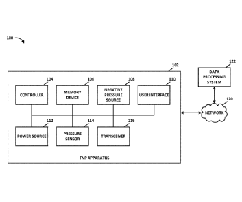
[0023] Figure 1 illustrates a negative pressure therapy system that
includes a TNP
apparatus and a remote data processing system according to some embodiments.
[0024] Figure 2 illustrates a negative pressure therapy system that
includes the
TNP apparatus of Figure 1, as well as an inlet manifold branching attachment,
pressure sensor
and a plurality of fluid flow paths, wound dressings positioned over wounds
according to
some embodiments.
[0025] Figure 3 illustrates some embodiments of negative pressure
therapy system
200 of Figure 2.
[0026] Figures 4A-C illustrate the inlet manifold branching attachment
of Figure 3
according to some embodiments.
[0027] Figure 5 illustrates a diagram of a negative pressure wound
treatment
system according to some embodiments.
[0028] Figure 6 illustrates a diagram of a negative pressure wound
treatment
system according to some embodiments.
-10-
CA 03054467 2019-08-23
WO 2018/158250 PCT/EP2018/054812
[0029] Figure 7 illustrates a diagram of a negative pressure wound
treatment
system according to some embodiments.
[0030] Figures 8A-8B illustrates diagrams of a TNP apparatus according
to some
embodiments.
[0031] Figure 9 illustrates a diagnostics process performed by a
negative pressure
wound treatment system according to some embodiments.
[0032] Figure 10 illustrates a diagnostics process performed by a
negative pressure
wound treatment system according to some embodiments.
[0033] Figure 11 illustrates a diagnostics process performed by a
negative pressure
wound treatment system according to some embodiments.
[0034] Figures 12A-12C illustrate portable negative pressure
apparatuses
according to some embodiments.
[0035] Figures 12D-12G illustrate user interfaces for a portable
negative pressure
apparatus according to some embodiments.
[0036] Figure 13 illustrates a wound dressing according to some
embodiments.
[0037] Figure 14 illustrates a cross section of an embodiment of a
fluidic
connector connected to a wound dressing.
[0038] Figures 15A-15D illustrate embodiments of a wound dressing
incorporating negative pressure indicators according to some embodiments.
DETAILED DESCRIPTION
[0039] Embodiments disclosed herein relate to apparatuses and methods
of
treating a plurality of wounds with reduced pressure, including a source of
negative pressure
and wound dressing components and apparatuses. The apparatuses and components
including
a wound overlay and packing materials, if any, may collectively be referred to
as dressings.
[0040] Embodiments disclosed herein relate to wound therapy for a human
or
animal body. Therefore, any reference to a wound herein can refer to a wound
on a human or
animal body, and any reference to a body herein can refer to a human or animal
body. The
term "wound" as used herein, in addition to having its broad ordinary meaning,
includes any
body part of a patient that may be treated using negative pressure. It is to
be understood that
the term wound is to be broadly construed and encompasses open and closed
wounds in
-11-
CA 03054467 2019-08-23
WO 2018/158250 PCT/EP2018/054812
which skin is torn, cut or punctured or where trauma causes a contusion, or
any other
superficial or other conditions or imperfections on the skin of a patient or
otherwise that
benefit from reduced pressure treatment. A wound is thus broadly defined as
any damaged
region of tissue where fluid may or may not be produced. Examples of such
wounds include,
but are not limited to, abdominal wounds or other large or incisional wounds,
either as a result
of surgery, trauma, sterniotomies, fasciotomies, or other conditions, dehisced
wounds, acute
wounds, chronic wounds, subacute and dehisced wounds, traumatic wounds, flaps
and skin
grafts, lacerations, abrasions, contusions, bums, diabetic ulcers, pressure
ulcers, stoma,
surgical wounds, trauma and venous ulcers or the like.
[0041] Treatment of such wounds can be performed using negative
pressure
wound therapy, wherein a reduced or negative pressure can be applied to the
wound to
facilitate and promote healing of the wound. It will also be appreciated that
the wound
dressing and methods as disclosed herein may be applied to other parts of the
body, and are
not necessarily limited to treatment of wounds.
[0042] It will be understood that embodiments of the present disclosure
are
generally applicable to use in topical negative pressure (TNP) therapy
systems. Briefly,
negative pressure wound therapy assists in the closure and healing of many
forms of "hard to
heal" wounds by reducing tissue oedema; encouraging blood flow and granular
tissue
formation; removing excess exudate and may reduce bacterial load (and thus
infection risk). In
addition, the therapy allows for less disturbance of a wound leading to more
rapid healing.
TNP therapy systems may also assist on the healing of surgically closed wounds
by removing
fluid and by helping to stabilize the tissue in the apposed position of
closure. A further
beneficial use of TNP therapy can be found in grafts and flaps where removal
of excess fluid is
important and close proximity of the graft to tissue is required in order to
ensure tissue
viability.
[0043] As is used herein, reduced or negative pressure levels, such as -
X mmHg,
represent pressure levels relative to normal ambient atmospheric pressure,
which can
correspond to 760 mmHg (or 1 atm, 29.93 inHg, 101.325 kPa, 14.696 psi, etc.).
Accordingly,
a negative pressure value of, for example, -X mmHg reflects pressure that is X
mmHg below
760 mmHg or, in other words, a pressure of (760-X) mmHg. In addition, negative
pressure
that is "less" or "smaller" than X mmHg may correspond to pressure that is
closer to
-12-
CA 03054467 2019-08-23
WO 2018/158250 PCT/EP2018/054812
atmospheric pressure (such as, -40 mmHg is less than -60 mmHg). Negative
pressure that is
"more" or "greater" than -X mmHg may correspond to pressure that is further
from
atmospheric pressure (such as, -80 mmHg is more than -60 mmHg). In some
embodiments,
local ambient atmospheric pressure is used as a reference point, and such
local atmospheric
pressure may not necessarily be, for example, 760 mmHg.
[0044] The negative pressure range for some embodiments of the present
disclosure can be approximately -80 mmHg, or between about -20 mmHg and -200
mmHg.
Note that these pressures are relative to normal ambient atmospheric pressure,
which can be
760 mmHg. Thus, -200 mmHg would be about 560 mmHg in practical terms. In some
embodiments, the pressure range can be between about -40 mmHg and -150 mmHg.
Alternatively, a pressure range of up to -75 mmHg, up to -80 mmHg or over -80
mmHg can
be used. Also in other embodiments, a pressure range of below -75 mmHg can be
used.
Alternatively, the negative pressure apparatus can supply a pressure range of
over
approximately -100 mmHg, or even -150 mmHg.
[0045] In some embodiments of wound closure devices described herein,
increased
wound contraction can lead to increased tissue expansion in the surrounding
wound tissue.
This effect may be increased by varying the force applied to the tissue, for
example by varying
the negative pressure applied to the wound over time, possibly in conjunction
with increased
tensile forces applied to the wound via embodiments of the wound closure
devices. In some
embodiments, negative pressure may be varied over time for example using a
sinusoidal wave,
square wave, and/or in synchronization with one or more patient physiological
indices (such
as, heartbeat).
[0046] Figure 1 illustrates a negative pressure therapy system 100 that
includes a
TNP apparatus 102 and a remote data processing system 122 according to some
embodiments. The TNP apparatus 102 can be used to treat a wound using a wound
dressing
that is in fluidic communication with the TNP apparatus 102 via a fluid flow
path. The TNP
apparatus 102 can include a controller 104, a memory device 106, a negative
pressure source
108, a user interface 110, a power source 112, a pressure sensor 114, and a
transceiver 116
that are configured to electrically communicate with one another. The power
source 112 can
provide power to one or more components of the TNP apparatus 102.
-13-
CA 03054467 2019-08-23
WO 2018/158250 PCT/EP2018/054812
[0047] The controller 104 can control operations of one or more other
components of the TNP apparatus 102 according at least to instructions stored
in the memory
device 106. The controller 104 can, for instance, control operations of and
supply of negative
pressure by the negative pressure source 108. The negative pressure source 108
can include a
pump, such as, without limitation, a rotary diaphragm pump or other diaphragm
pump, a
piezoelectric pump, a peristaltic pump, a piston pump, a rotary vane pump, a
liquid ring pump,
a scroll pump, a diaphragm pump operated by a piezoelectric transducer, a pump
operated by
a voice coil actuator, or any other suitable pump or micropump or any
combinations of the
foregoing. The user interface 110 can include one or more elements that
receive user inputs or
provide user outputs to a patient or caregiver. The one or more elements that
receive user
inputs can include buttons, switches, dials, touch screens, or the like.
[0048] The pressure sensor 114 can be used to monitor pressure
underneath a
wound dressing, such as (i) pressure in a fluid flow path connecting the TNP
apparatus 102
and the wound dressing, (ii) pressure at the wound dressing, or (iii) pressure
at or in the TNP
apparatus 102. In some implementations, the pressure sensor 114 can include at
least two or
more pressure sensors that are positioned to measure the pressure of multiple
fluid flow paths,
such as multiple flow paths connecting the TNP apparatus 102 to multiple wound
dressings.
On other implementations, the pressure sensor 114 can include at least two or
more pressure
sensors that are positioned in or fluidically connected to the fluid flow path
to permit
differential measurement of the pressure. For example, a first pressure sensor
can be
positioned upstream of the wound (such as at or near the inlet of the TNP
apparatus 102) and
a second pressure sensor can be positioned to detect pressure at or near the
wound or at or
near a canister or dressing.
[0049] The transceiver 116 can be used to communicate with the data
processing
system 122 via a network 120. The transceiver 116 can, for example, transmit
device usage
data like alarms, measured pressure, or changes to a therapy program
administered by the
TNP apparatus 102 to the data processing system 122. In some examples, the
transceiver 116
communicate with one or more shut-off valves in a negative pressure therapy
system. The
network 120 can be a communication network, such as a wired or wireless
communications
network (for example, a cellular communications network). The memory device
106 can be
used to store the device usage data that may be transmitted by the transceiver
116. In some
-14-
CA 03054467 2019-08-23
WO 2018/158250 PCT/EP2018/054812
embodiments, the data processing system 122 can transmit data, such as
operating parameters,
to the TNP apparatus 102.
[0050] Figure 2 illustrates a negative pressure therapy system 200
according to
some embodiments. The system 200 includes the TNP apparatus 102 of Figure 1,
as well as a
first fluid flow path 208, a first wound dressing 202 configured to be placed
over a first
wound 220, a second fluid flow path 210, a second wound dressing 204
configured to be
placed over a second wound 222, an inlet manifold branching attachment 206,
and a third fluid
flow path 212. The TNP apparatus 102 can be used to treat the first wound 220
using the first
wound dressing 202 that is in fluidic communication with the TNP apparatus 102
via the first
fluid flow path 208, the inlet manifold branching attachment 206, and the
third fluid flow path
212. The TNP apparatus 102 can also be used to treat the second wound 222
using the
second wound dressing 204 that is in fluidic communication with the TNP
apparatus 102 via
the second fluid flow path 210, the inlet manifold branching attachment 206,
and the third
fluid flow path 212.
[0051] The inlet manifold branching attachment 206 is attached between
the TNP
apparatus 102 and the first and second wound dressings, thereby advantageously
enabling the
TNP apparatus 102 to generate and maintain negative pressure in or under both
of the wound
dressings simultaneously. In this example, the inlet manifolds are not
incorporated into the
TNP apparatus. Instead, an inlet manifold branching attachment 206, such as a
Y-shaped
connector, is used to connect the first and second fluid flow paths 208-B to
the third fluid
flow path 212. In other examples, inlet manifolds can be incorporated into the
TNP apparatus
102 (as shown in Figures 12A-12C) such that the first and second fluid flow
paths connect
directly to the TNP apparatus via integrated inlet manifolds.
[0052] A pressure sensor 114 is positioned in the third fluid flow path
212, such as
at or near an inlet of the TNP apparatus 102, to measure pressure in the third
fluid flow path
212. The controller of the TNP apparatus 102 can monitor the pressure measured
by the
pressure sensor 114 and determine whether an operating condition (for example,
a blockage,
leakage, overpressure, or dressing full condition) has occurred in within the
negative pressure
therapy system 200.
[0053] In some instances, the controller can determine that an
operating condition
exists by comparing the measured pressure to an expected measured pressure (or
flow). An
-15-
CA 03054467 2019-08-23
WO 2018/158250 PCT/EP2018/054812
"expected" pressure (or flow) can be the pressure measured by a pressure
sensor in a negative
pressure system operating in a normal state. The expected pressure can be
equivalent or
almost equivalent (for example, within 1, 2, 3, 4, 5, 10, 15, or 20 mmHg) to a
pressure
supplied by the negative pressure source (or a pressure selected by a user).
In contrast, an
"unexpected" pressure (or flow) can be any measured pressure other than the
expected
pressure (or flow). For instance, in some examples, a wound dressing
experiencing a
blockage, overpressure, or dressing full condition, can cause the pressure
sensor to measure a
lower (for example, more positive pressure) than expected pressure. In other
examples, a
wound dressing experiencing a leakage condition can cause the pressure sensor
to measure a
lower than expected pressure. In some examples, an operating condition can
change the
measured pressure (for example, spike, dip, increase, or decrease in measured
pressure). In
some embodiments, measured pressure is compared to one or more thresholds in
order to
determine if it is expected or unexpected.
[0054] In some examples, the TNP apparatus 102 will only function (for
example,
provide negative pressure) when two or more wound dressings are connected.
Additionally,
some indicators or functionality of the TNP apparatus that is available when
only a single
wound dressing is connected may be disabled so as not to confuse the user. For
example, in
some instances the dressing full indicator is not available for TNP systems
having more than
one connected wound dressing. Thus, the dressing full indicator(s) can be
disabled or removed
from the front panel so as not to confuse the user with unavailable
functionality.
[0055] Figure 3 illustrates some embodiments of negative pressure
therapy system
200. The system 200 includes a TNP apparatus 102, a first fluid flow path 208,
a first wound
dressing 202, a second fluid flow path 210, a second wound dressing 204, a
plurality of
integrated inlet manifolds or connectors 302, 304. The plurality of integrated
inlet manifolds
302, 304 are integrated with the TNP apparatus 102 and are fluidically
connected to the first
wound dressing 202 via the first fluid flow path 208 and the second wound
dressing 204 via
the second fluid flow path 210.
[0056] In some instances, a fluid flow path 208 can be lengthy and in a
location
remote from the TNP apparatus 102. As such, it can be desirable for the fluid
flow paths to
include one or more indicators 306, 308 which would be helpful to a user in
identifying which
-16-
CA 03054467 2019-08-23
WO 2018/158250 PCT/EP2018/054812
fluid flow path 208 is connected to a particular inlet of the plurality of
integrated inlet
manifolds 306, 308.
[0057] As shown, the first fluid flow path 208 includes a plurality of
first
identifiers (stars) 306, and the second fluid flow path 208 includes a
plurality of second
identifiers (triangles) 308. In both instances, at least one identifier 306,
308 is located in close
proximity to the inlet manifolds 306, 308 and at least one identifier is
located in close
proximity to a wound dressing. In some examples, a fluid flow path can include
more than two
identifiers 306, 308. For example, identifiers 306, 308 can be located across
the length of the
fluid flow path. Moreover, an identifier 306, 308 can alternatively include a
printed glyph, a
printed icon, an embossed glyph, an embossed icon, a braille character, a
color-coding and the
like. In some examples, an electronically controlled indication (such as an
LED, an indicator
on a display, etc.) is associated with each fluid flow path. This facilitates
the TNP apparatus
102 in indicating an operating condition that may have occurred on the
associated dressing.
[0058] In some embodiments, at least one pressure sensor can be
positioned with
an inlet manifold (either an integrated manifold or attachment manifold) to
measure the
combined pressure of the first and second fluid flow paths. The controller of
the TNP
apparatus 102 monitors the pressure measured by the pressure sensor and
determines whether
an operating condition has occurred in any of the fluid flow paths. In some
aspects, the
controller can be configured to provide a first indication associated with an
operating
condition in the first fluid flow path 208 and a second indication associated
with an operating
condition in the second fluid flow path 210.
[0059] In some examples, a negative pressure therapy system includes
more than
two wound dressings. Accordingly, the number of fluids flow paths and inlets
can correspond
with the number of wound dressings. For instance, a negative pressure therapy
system having
four wound dressings can have at least four fluid flow paths and at least four
inlets manifolds.
In some examples, a single wound dressing can be configured to communicate
with a TNP
apparatus via more than one fluid flow path. In some examples, the negative
pressure therapy
system can include more inlets manifolds than fluid flow paths and/or wound
dressings. In
examples such as these, the additional inlets can be disregarded or plugged.
[0060] Figures 4A-4C illustrate an inlet manifold branching attachment
206
according to some embodiments. In some examples, the inlet manifold branching
attachment
-17-
CA 03054467 2019-08-23
WO 2018/158250 PCT/EP2018/054812
206 can be used in place of the integrated inlet manifold of Figure 3. As
illustrated, the Y-
shaped inlet manifold branching attachment 206 can include three conduit
attachment portions
306, 308, 410. A pump conduit attachment portion 410 can be used to connect to
a conduit or
tubing extending from a pump or TNP apparatus or to connect to the pump
itself. The pump
conduit attachment portion 410 can include a male non-luer connector at a
proximal end of
the Y-shaped inlet manifold branching attachment. The male connector can
attach to a female
connector of a conduit or pump. The pump conduit attachment portion 410 has a
shaft 408
extending from the attachment portion and forming the bottom portion of the Y
shape of the
inlet manifold branching attachment 206.
[0061] The Y-shaped inlet manifold branching attachment also includes
two
dressing conduit attachment portions 306, 308. The dressing conduit attachment
portions 306,
308 can be used to connect to the coupling of the fluid flow path extending
from a wound
dressing. In some embodiments, a conduit or tubing can be used to connect the
inlet manifold
branching attachment to the Y-shaped inlet manifold branching attachment 206.
The conduit
or tubing may be a soft bridge, a hard tube, or any other apparatus that may
serve to transport
fluid. The conduit or tubing can include a coupling at a proximal end and at a
distal end. The
conduit or tubing can be connected to the coupling of the inlet manifold
branching attachment
at the distal end and connected to the conduit attachment portions of the Y-
shaped inlet
manifold branching attachment at the proximal end of the conduit.
[0062] The dressing conduit attachment portion 306, 308 can include a
female
non-luer connector at a distal end of the Y-shaped inlet manifold branching
attachment. The
female connector can attach to a male connector of the coupling of the inlet
manifold
branching attachment or to the coupling of the conduit.
[0063] In some embodiments, the inlet manifold branching attachment 206
or the
conduit can include incorporated valve(s), clamp(s), cap(s), and/or other
closure mechanisms.
Accordingly, flow or passage of fluid to and from one wound dressing can be
blocked while
another wound dressing continues to apply negative pressure. In some
embodiments, the
closure mechanism can be a valve, for example, a non-return valve.
[0064] In some examples, the valves incorporated in the Y-shaped inlet
manifold
branching attachment 206 are manual shut-off valves. For instance, a user can
manually close
a valve associated with conduit attachment portion 306 thereby blocking the
fluid flow to and
-18-
CA 03054467 2019-08-23
WO 2018/158250 PCT/EP2018/054812
from the first wound dressing 202. Similarly, a user can manually close a
valve associated with
conduit attachment portion 308 thereby blocking the fluid flow to and from the
second wound
dressing 204. In some examples, a valve exists in conduit attachment potion
410, wherein
closure of said valve would block fluid flow to and from the first and second
wound dressings
202, 204.
[0065] In some examples, the valves incorporated in the Y-shaped inlet
manifold
branching attachment 206 are electromechanical valves. For instance, a
controller (for
example, the controller of the TNP apparatus as described in Figure 1) can
communicate with
the valves to open and/or close each valve individually or as a unit. The
communication
between the valves and the TNP apparatus 102 can be wired or wireless. For
instance, a
wireless transceiver (e.g., see Figure 1) of TNP apparatus 102 can communicate
with a
wireless transceiver of the valves. The wireless transceiver of the valves can
be positioned
within the inlet manifold branching attachment 206 or within close proximity
to the inlet
manifold branching attachment 206.
[0066] The dressing conduit attachment portions 306, 308 include shafts
404, 402,
respectively, forming the top portions of the Y shape of the connector. The
proximal ends of
shafts 404, 402 and the distal end of shaft 408 meet at a joint 406. In some
embodiments, the
joint 406 can include a hinge that allows rotation of the shafts 404, 402, 408
about the joint
406. In some embodiments, only shafts 404, 402 of the dressing conduit
attachment portions
can move relative to the joint 406 and the shaft 408 of the pump conduit
attachment portion is
fixed. In some embodiments, the whole Y-shaped inlet manifold branching
attachment will be
in two parts that allow 360 rotation. Figure 4C illustrates an embodiment of
the Y-shaped
inlet manifold branching attachment that is formed of two freely rotating
parts that allow
rotation of each part relative to the other. The rotation of the Y-shaped
inlet manifold
branching attachment can allow the user to twist the pump around while the
wound dressings
and conduits extending from the wound dressings remain stationary.
[0067] In some embodiments, the male and female non-luer connectors can
be a
rigid plastic. In some embodiments, the shafts 408, 404, 402 can be a flexible
plastic tubing. In
some embodiments, the Y-shaped inlet manifold branching attachment can be
encased in a soft
silicone sleeve to increase patient comfort and prevent the Y-shaped inlet
manifold branching
attachment from becoming a pressure point.
-19-
CA 03054467 2019-08-23
WO 2018/158250 PCT/EP2018/054812
[0068] Utilizing the Y-shaped inlet manifold branching attachment 206
illustrated
in Figures 4A-4C to attach a single pump to the two wound dressings, the TNP
apparatus 102
can draw pressure in the two wound dressings simultaneously. The performance
and fluid
management of the multisite dressing and Y-connector is equivalent to a
control test of the
standard single wound dressing with single pump set-up. Although the
attachment 206 is
illustrated as being Y-shaped, the attachment 206 can be of any suitable shape
or combination
of shapes in some implementations. In some embodiments, luer, quick release,
or other types
of connectors can be used as one or more connectors of the attachment 206, the
TNP
apparatus 102, and one or more of the fluid flow paths 208, 210.
[0069] In some examples, a negative pressure therapy system can include
more
than two wound dressings and associated fluid flow paths in fluidic
communication with the
inlet manifold branching attachment. As such, in some embodiments, the inlet
manifold
branching attachment is attached to more than one TNP apparatus and/or more
than two fluid
flow paths (such as, one pressure source and three wound dressings ("1:3"),
1:4, 1:5, 2:1, 2:2,
2:3, 2:4, 2:5). The inlet manifold branching attachment can be a separate
attachment, such as
the Y-shaped connector that can connect to the third fluid flow path, or inlet
manifolds can be
incorporated into the TNP apparatus 102. The total number of inlet manifolds
(for example,
the number of "splits" performed by the inlet manifold branching attachment)
that the inlet
manifold branching attachment contains can be the same as the number of
dressings to be
connected. In some instances, one or more inlet manifolds connect to a single
wound dressing.
[0070] Figure 5 illustrates a negative pressure therapy system 500
having pressure
sensors 502, 504, 506 positioned to measure each fluid flow path associated
with wound
dressings. In particular, a first pressure sensor 502 measure pressures in a
first fluid flow path
208; a second pressure sensor 504 measured the pressure in a second fluid flow
path 210; and
a third pressure sensor 506 measures the pressure in a third fluid flow path
212.
[0071] By positioning a sensor within each of the fluid flow paths, the
controller
can monitor the pressure of each fluid flow path to determine whether an
operating condition
has occurred in the negative pressure therapy system 500. Additionally,
because a sensor
measures pressure on each of the fluid flow paths, upon determination of an
operating
condition, the controller can specifically determine which flow path/wound
dressing
combination is experiencing an operating condition. The negative pressure
therapy system 500
-20-
CA 03054467 2019-08-23
WO 2018/158250 PCT/EP2018/054812
provides the capability to monitor the functionality of individual wound
dressings, thereby
enabling the same set of features and functionality offered by a negative
pressure therapy
system utilizing a single wound dressing.
[0072] The pressure sensors 502, 504, 506 can be positioned anywhere in
the fluid
flow paths, such as between the wound dressings and the inlet manifold
branching attachment
206 or at or near a wound dressing. In some examples, to reduce costs, the
number of
pressure sensors is one fewer than the number of wound dressings. For
instance, where the
number of wound dressings is N, only N-1 pressure sensors are employed in the
negative
pressure therapy system. In examples such as these, a controller can perform a
process to
determine whether the dressing without an associated pressure sensor is
experiencing an
operating condition. In some examples, as described above with respect to
Figures 4A-C, one
or more pressure sensors into an inlet manifold branching attachment 206.
[0073] A plurality of shut-off valves 512, 514, 516 (for example, as
illustrated in
Fig. 12C) can be positioned in the negative pressure therapy system 500 such
that the closure
of a valve blocks passage of fluid to and from an associated wound dressing.
The shut-off
valves 512, 514, 516 can be positioned anywhere in the fluid flow path, such
as between the
inlet manifold branching attachment 206 outlet and a corresponding dressing
inlet. In some
examples, as described above with respect to Figures 4A-C, one or more shut-
off valves are
integrated into an inlet manifold branching attachment 206.
[0074] In some examples, the valves 512, 514, 516 are manual shut-off
valves. For
instance, a user can manually close the first valve 512 thereby blocking the
fluid flow to and
from the first wound dressing 202. In other examples, the valves are
electromechanical valves.
For instance, the TNP apparatus 102 can communicate with the valves to open
and/or close
each valve individually or as a unit. The communication between the valves and
the TNP
apparatus 102 can be wired or wireless. For instance, a wireless transceiver
(see e.g., Figure
1) of TNP apparatus 102 can communicate with a wireless transceiver of the
valves. In some
cases, the wireless transceiver of the valves can be positioned within the
inlet manifold
branching attachment 206 or within close proximity to the inlet manifold
branching
attachment.
[0075] In TNP system 500, the controller can efficiently determine
which fluid
flow path/wound dressing combination is experiencing an operating condition
and, in some
-21-
CA 03054467 2019-08-23
WO 2018/158250 PCT/EP2018/054812
embodiments, can close an associated valve to improve overall efficiency of
the TNP
apparatus. For instance, during normal operation in which no wound dressings
are
experiencing an operating condition, the pressure sensors 502, 504, 506 may
measure
approximately the same pressure. When a fluid flow path/wound dressing
experiences an
operating condition, the measured pressure associated with that fluid flow
path/wound
dressing changes such that a controller can determine: (1) which particular
fluid flow
path/wound dressing is experiencing the operating condition and/or (2) which
particular type
of operating condition is being experienced. For example, if pressure is
measured downstream
of a blockage that occurs in the first fluid flow path/wound dressing, the
measured pressure in
the first fluid flow path can increase (for example, become more negative)
because the
blockage restricts the fluid flow and consequently decreases the volume in
which fluid flows.
As another example, if pressure is measure upstream of the blockage in the
first fluid flow
path/wound dressing, the measured pressure in the first fluid flow path can
decrease (for
example, become more positive) because the blockage severely restricts or
blocks fluid flow in
part of the fluid flow path where pressure is measured. Due to this pressure
change, the
controller can determine that an operating condition (blockage) has occurred
on the first fluid
flow path/wound dressing. In some examples, operating conditions cause spike
or spikes in
measured pressure. In other examples, operating conditions cause an increase
or decrease in
measured pressure. The controller can make these determinations for blockage,
overpressure,
pressure leak, dressing full conditions, and the like.
[0076] In some examples, electronically controllable valves are
utilized to shut-off
therapy to specific dressings to prevent loss of pressure and improve overall
efficiency of the
TNP apparatus. This can be effective in a negative pressure therapy system
having a large
number of wound dressings.
[0077] Figure 6 illustrates a negative pressure therapy system 600
according to
some embodiments. The illustrated system differs from the negative pressure
therapy system
200 in that it includes a flow reducer or restrictor 602 in the first fluid
flow path 208 and a
plurality of integrated inlet manifolds 602, 604 have replaced the inlet
manifold branching
attachment 206. In some implementations, the inlet manifolds 602, 604 can be
replaced with a
single inlet and a branching attachment 206 and the flow restrictor 602 can be
integrated into
one of the passageways or branches of the attachment 206. The addition of the
flow restrictor
-22-
CA 03054467 2019-08-23
WO 2018/158250 PCT/EP2018/054812
602 allows the controller to determine which wound dressing is experiencing an
operating
condition despite utilizing a single pressure sensor 114.
[0078] The flow restrictor 602 (such as a small volume receptacle or a
small
orifice) limits the flow through the first fluid flow 208 path such that the
difference in flow
between the first fluid flow path 208 and the second fluid flow path 210 can
be perceived by
the controller. For example, the TNP apparatus 102 can draw pressure in the
two wound
dressings simultaneously. The flow restrictor 602 restricts the pressure in
the first fluid flow
path 208. In some embodiments, during normal operation, flow detected by the
system 600
(for example, a controller) will be the combination of flow through fluid flow
paths 208 and
210, each of which may be known a priori (such as, calculated based on
characteristics of
each fluid flow paths, calculated via calibration, and the like). Upon the
occurrence of an
operating condition, such as blockage, in the first fluid flow path 208,
detected flow will
reduce to equal or nearly equal the flow through the flow path 210. Thus, the
system 600
would not only detect change in flow, but would detect based on the measured
flow that fluid
is flowing through fluid flow path 210 and that fluid flow path 208 is
experiencing a blockage.
Similarly, the system 600 can detect and indicate an operating condition, such
as a blockage,
in fluid flow path 210. Indication of the operating condition can be performed
using any of the
approaches described herein, such as by audio-visual indication using an LED,
display, and the
like. For example, each of the fluid flow paths 208 and 210 may be associated
with a
particular color or symbol and such color or symbol can be displayed and/or
announced. In
some embodiments, measured flow is compared to one or more thresholds.
[0079] In some embodiments, flow (or flow rate) can be monitored or
measured
directly by using a flow meter. In some implementations, flow can be monitored
or measured
indirectly. For example, flow can be determined by monitoring the change in
pressure
measured by the pressure sensor 114. The controller can determine the rate of
flow, for
example, by determining a pressure gradient, rate of change of pressure, or
pressure decay
rate. As another example, in a system having a negative pressure source that
produces variable
flow, flow rate can be determined based on pressure and speed of the negative
pressure source
(such as pump motor). For example, flow rate can be determined according to
Equation 1
below:
Flow Rate = Ci*F*P + C2 (Equation 1)
-23-
CA 03054467 2019-08-23
WO 2018/158250 PCT/EP2018/054812
where F is the pump speed (such as, frequency of a tachometer signal that
measures pump
motor revolutions), P is measured pressure, and C1 and C2 are suitable
constants. Additional
details are described U.S. Patent No. 8,905,985 and U.S. Patent Publication
No. 2012/0001762, each of which is hereby incorporated by reference in its
entirety.
[0080] In some embodiments, the flow restrictor 602 can be replaced
with a flow
enlarger configured to increase flow. In such cases, detection of an operating
condition will be
similar to the foregoing with the exception that the flow in the flow path 208
associated with
the wound 220 is increased by the flow restrictor.
[0081] In some embodiments, the flow restrictor 602 is a permanent
restrictor,
such as an orifice with a smaller diameter than one or more of the conduits in
the fluid flow
path 208. In some implementations, the flow restrictor 602 is a temporary
restrictor, such as
an adjustable valve, that temporarily restricts the fluid flow when
determination of whether an
operating condition is present is made. In some examples, the controller can
control the
temporary flow restrictor. In certain embodiments, a plurality of flow
reducers and/or
enhancers could be utilized in a system that includes more than two wound
dressings and
associated fluid flow paths. For instance, a system with three wound dressings
can include a
flow restrictor in a first fluid flow path and a flow enhancer in a second
fluid flow path.
Similarly, a system with three wound dressings can include a flow restrictor
in a first fluid flow
path and a stronger (or more narrow) flow restrictor in a second fluid flow
path. In any of
these examples, the difference in flow rates among the plurality of fluid flow
paths can permit
the TNP apparatus to determine which fluid flow path/wound dressing is
experiencing an
operating condition.
[0082] In some examples, a canister can be coupled between the TNP
apparatus
102 and/or the plurality of integrated inlet manifolds 602, 604. The canister
can collect
exudate removed from the wounds 220, 222. Alternatively, a canister can be
coupled between
each wound dressing and the inlet manifold branching attachment.
[0083] Figure 7 illustrates a negative pressure therapy system 700
according to
some embodiments. System 700 differs from the negative pressure therapy system
200 in that
system 700 includes a third wound dressing 510, a third wound 518, a fourth
fluid flow path
508, and a plurality of shut-off valves 512, 514, 516. In addition to treating
wounds 220-B as
described in 200, the system 700 can be utilized to treat the third wound 518
using the third
-24-
CA 03054467 2019-08-23
WO 2018/158250 PCT/EP2018/054812
wound dressing 510 that is in fluidic communication with the TNP apparatus 102
via the
fourth fluid flow path 508, the inlet manifold branching attachment 206, and
the third fluid
flow path 212.
[0084] The plurality of shut-off valves 512, 514, 516 are positioned in
the fluid
flow paths such that the closure of a corresponding valve blocks the fluid
flow to and from the
connected fluid flow path/dressing. The shut-off valve 21 can be positioned
anywhere from
the inlet manifold branching attachment 206 outlet to the corresponding
dressing inlet. As
illustrated, a first valve 512 is positioned on the first fluid flow path 208,
a second valve 514 is
positioned on the second fluid flow path 210 and a third valve 516 is
positioned on the fourth
fluid flow path 508.
[0085] In some examples, the plurality of valves 512, 514, 516 are
manual shut-off
valves. For instance, a user can manually close the first valve 512 thereby
blocking the fluid
flow to and from the first wound dressing 202. In other examples, each of the
plurality of
valves is an electromechanical valve. For instance, the TNP apparatus can
communicate with
the valves to open and/or close each valve individually or as a unit. The
communication
between the valves and the TNP apparatus 102 can be wired or wireless. For
instance, a
wireless transceiver (see e.g., Figure 1) of TNP apparatus 102 can communicate
with a
wireless transceiver of the valves. The wireless transceiver of the valves can
be positioned
within the inlet manifold branching attachment 206 or within close proximity
to the inlet
manifold branching attachment.
[0086] Figure 8A illustrate a negative pressure therapy system 800A
according to
some embodiments. In this example, the TNP apparatus 102 includes at least a
controller 104,
a negative pressure source 108, a plurality of pressure sensors 502, 504, and
a plurality of
integrated inlet manifolds 306, 308.
[0087] The integrated inlet manifolds 602, 604 can be combined into a
single unit
(e.g., as depicted in Figures 3 and 12), such that a single negative pressure
passageway is
connected to the negative pressure source 108. Alternatively, each of the
plurality of
integrated inlet manifolds 306, 308 can directly connect to the negative
pressure source
without first combining with another inlet manifold.
[0088] The plurality of pressure sensors 502, 504 are positioned such
that a first
pressure sensor 502 measures the pressure of the first fluid flow path 208
connected to the
-25-
CA 03054467 2019-08-23
WO 2018/158250 PCT/EP2018/054812
first inlet manifold 306 and a second pressure sensor 504 measures the
pressure of the second
fluid flow path 210 connected to the second inlet manifold 308. In some
examples, the
pressure sensors 502, 504 can be positioned within an inlet manifold. In other
examples, the
pressure sensors are located with a housing of the TNP apparatus 102.
[0089] Figure 8B illustrates a negative pressure therapy system 800B
according to
some embodiments. In this example, the system 800B includes an inlet manifold
branching
attachment 206. As described herein, inlet manifolds can include an inlet
manifold branching
attachment 206 (as illustrated in Figures 4A-4B) and/or can include one or
more integrated
inlet manifolds (as illustrated in Figures 12A-12C).
[0090] The inlet manifold branching attachment 206 includes a pressure
sensor
504 on a first branch 308 fluidically connected to the first dressing 202 via
the fluid flow path
208, as well as a wireless transceiver or receiver 802 to communicate with the
wireless
receiver 804 in communication with a controller 104 of the TNP apparatus 102.
The pressure
sensor 504 measures pressure in the fluid flow path 208, while pressure sensor
502 measures
combined pressure in fluid flow paths 208 and 210. Operating conditions, such
as blockages,
in one or more of the fluid flow paths 208 or 210 can be determined based on
pressure
measured by the sensors 502 and 504 using any of the approaches described
herein. In some
examples, the inlet manifold branching attachment 206 can communicate with the
TNP
apparatus 102, for example, to provide pressure data. The communication can be
wired or
wireless (for example, over Bluetooth). In some examples, dressing-full
detection and/or
detection of other operating conditions can be used to provide indication(s)
to the user.
[0091] Figure 9 illustrates a diagnostics process 900 performed by a
negative
pressure wound treatment system 500 (see e.g., Figure 5) according to some
embodiments.
Process 900 can be performed by a controller of the negative pressure wound
treatment
system. As mentioned above, an operating condition can include a blockage,
leakage,
overpressure, dressing full condition, or the like. The process can detect one
of the foregoing
operating conditions by analysing the pressure measured by pressure sensors
502, 504, 506.
[0092] At block 902, the process monitors the pressure sensors 502,
504, 506
which measure pressure in various fluid flow paths 208, 210, and 508. In some
examples, the
controller monitors the pressure sensors 502, 504, 506 continuously, at
predetermined
-26-
CA 03054467 2019-08-23
WO 2018/158250 PCT/EP2018/054812
intervals (such as, 1 minute, 2 minutes, 5 minutes, 10 minutes, 15 minutes, 20
minutes, 30
minutes, or 60 minutes), and/or responsive to input by a user.
[0093] At block 904, the process determines that an operating condition
has
occurred based at least in part on a change in pressure measured by one of the
plurality of
pressure sensors. For instance, an occurrence of a blockage in one of the
wound dressings
may cause a momentary or prolonged spike or dip in measured pressure. As
another example,
the process can determine flow based on measured pressure (or directly if one
or more
flowmeters are utilized). As described with respect to Figure 5, by
positioning a pressure
sensor to monitor conditions in each of the fluid flow paths associated with
the wound
dressings, the process 900 can monitor the pressure sensors 502, 504, 506 and
determine
specifically which fluid flow path/wound dressing is experiencing an operating
condition.
Thus, the negative pressure therapy system 500 provides the capability to
monitor the
functionality of individual wound dressings, thereby enabling the same set of
features offered
by a negative pressure therapy system utilizing a single wound dressing.
[0094] At block 906, the process 900 provides indication of the flow
path/dressing
(or flow paths/dressings) determined to be experiencing an operating
condition. In some
examples, one or more LEDs or other indicators can be used to indicate to a
user or caregiver
that an operating condition has been detected. For example, each wound
dressing can have a
corresponding LED that is ON when no operating condition is detected on the
associated
wound dressing and OFF when an operating condition is detected on an
associated wound
dressing. In some examples, other indicators may be associated with wound
dressing that is
experiencing an operating condition, such as sounds, wireless messages,
display notifications
and/or other signals that may get the attention of a user or caregiver. In
some examples, the
TNP apparatus may additionally or alternatively provide indication by closing
a valve
associated with the wound dressing experiencing the operating condition.
[0095] Figure 10 illustrates a diagnostics process 1000 performed by a
negative
pressure wound treatment system 700 (see e.g., Figure 7) according to some
embodiments.
Process 1000 can be performed by a controller of the negative pressure wound
treatment
system. As mentioned above, an operating condition can include a blockage,
leakage,
overpressure, dressing full condition, or the like. The process can detect one
of the foregoing
operating conditions by analysing the pressure measured by pressure sensor
502. When the
-27-
CA 03054467 2019-08-23
WO 2018/158250 PCT/EP2018/054812
process determines that an operating condition has occurred, it can initiate
diagnostics to
determine which flow path/dressing is experiencing an operating condition. In
some examples,
the process is implemented in firmware or software that incorporates a
diagnostic mode to
determine which flow path/wound dressing is experiencing an operating
condition. In some
examples, an operator can toggle between the operational modes through an
interface of the
TNP apparatus (such as, a touchscreen interface or dedicated buttons or
switches).
[0096] At block 1002, the process 1000 monitors the pressure sensor
502, which
measures pressure at or in the TNP apparatus 102. In some examples, the
process 1000
continuously monitors the pressure sensor 502. In other examples, the process
1000 monitors
the pressure sensor 502 at predetermined intervals (such as, 1 minute, 2
minutes, 5 minutes,
minutes, 15 minutes, 20 minutes, 30 minutes, or 60 minutes). In other
examples, the
process can monitor the pressure sensor responsive to input by a user.
[0097] At block 1004, the process determines that an operating
condition has (or
may have) occurred and initiates a diagnostic mode to determine which fluid
flow
path(s)/wound dressing(s) are experiencing an operating condition. During the
diagnostic
mode, the process can determine that an operating condition has occurred based
at least in
part on a change in pressure measured by the pressure sensor 502, change in
flow, and the
like. For instance, an occurrence of an operating condition on one of the
fluid flow
paths/wound dressings may cause a momentary or prolonged spike or dip in
pressure
measured by the pressure sensor 502.
[0098] In block 1006, a fluid flow path/wound dressing is selected for
testing. In
some examples, the user and/or the process can make this selection. For
instance, the user can
make the selection by providing input to the process. The section can be
arbitrary, one based
on the user's suspicions, or one based on the process's suggestion. In some
examples, the
process can make the selection. The process's selection, for instance, can be
random, based on
user input, or based on data within the controller.
[0099] The valve associated with the selected fluid flow path/dressing
is opened
and the valve(s) associated with the unselected dressing(s) are closed,
thereby likening the
negative therapy system to a negative pressure system having a single wound
dressing
fluidically connected to a negative pressure source. The valve(s) can be
opened or closed
manually by a user or by the controller. In some examples, the controller can
electronically
-28-
CA 03054467 2019-08-23
WO 2018/158250 PCT/EP2018/054812
(such as, through a wired or wireless connection) control the shut-off valves.
For instance, a
wireless transmitter or transceiver of the TNP apparatus can communicate with
a wireless
transceiver or receiver of the valves. In such examples, the wireless
transceiver can
communicate with each of the valves and is capable of controlling each of the
valves
individually or as a unit.
[0100] At block 1008, the process monitors the pressure sensor 502 to
determine
if the selected dressing is experiencing an operating condition. This analysis
may be similar to
a determination made by a process in a negative pressure system having a
single wound
dressing fluidically connected to a negative pressure source. For example, a
lower than
expected negative pressure (or higher than expected flow) can indicate that a
wound dressing
is experiencing leakage and a higher than expected negative pressure (or lower
than expected
flow) can indicate that a wound dressing is experiencing a blockage,
overpressure, or dressing
full condition.
[0101] If the process determines that the selected wound dressing is
not
experiencing an operating condition (for example, the pressure measured by the
pressure
sensor 502 is generally equivalent to the expected pressure), then a different
fluid flow
path/wound dressing is selected for testing. That is, the process 1000 returns
to block 1006).
The newly selected fluid flow path/wound dressing will be a wound dressing
that has not been
tested during the current diagnostic mode.
[0102] At block 1010, the process determines that the previously
selected wound
dressing is experiencing an operating condition. The valve associated with the
selected wound
dressing is closed (along with closing any valves associated with a fluid flow
paths(s)/wound
dressing(s) previously determined to be experiencing an operating condition).
All other valves
are opened. As described above, the valves can be opened or closed manually by
a user or
automatically by the controller.
[0103] At block 1012, the process monitors the pressure sensor to
determine if
any of the fluid flow paths/wound dressings associated with open valves are
experiencing an
operating condition. For example, the pressure sensor may sense an expected
pressure (or
flow) if no operating conditions are present and may sense an unexpected
pressure (or flow) if
operating conditions are present. If the process determines that an operating
condition is
present among the wound dressing(s) associated with the open valve(s), then a
new wound
-29-
CA 03054467 2019-08-23
WO 2018/158250 PCT/EP2018/054812
dressing is selected (block 1006). As described above, the newly selected
wound dressing will
be a wound dressing that has not been tested during the current diagnostic
mode.
[0104] At block 1014, the process has determined which flow paths/wound
dressings of the plurality of wound dressings are experiencing an operating
condition. The
process can provide appropriate indication as described herein, which
facilitates addressing
and remedying the operating condition. For example, if a dressing full
operating condition is
detected, wound dressings can be replaced. In some examples, the user must
manually replace
the wound dressings. In some examples, the wound dressings are replaced
without the help of
the user. At block 1014, all valves can be opened, and the diagnostic mode has
been
completed. As described above, the valves can be opened manually or
electromechanically.
[0105] Figure 11 illustrates a diagnostics process 1100 performed by a
negative
pressure wound treatment system 700 (see Figure 7) according to some
embodiments.
Process 1100 can be performed by a controller of the negative pressure wound
treatment
system. The process can monitor the measured pressure and can determine
whether any of the
fluid flow paths/wound dressings in the negative pressure system are
experiencing an
operating condition.
[0106] At block 1102, the process has determined that no operating
conditions
exist within the negative pressure system. In some examples, one or more LEDs
or other
indicators can be used to indicate to a user or caregiver that no operating
conditions exist. For
example, each wound dressing can have a corresponding LED that is ON when no
operating
condition is detected on the associated wound dressing and OFF when an
operating condition
is detected on the associated wound dressing (or vice versa). In some
examples, when no
operating condition is detected in the negative pressure system, all
associated LEDs are ON.
In some examples, other indicators may be associated with a no fault state
(state in which no
operating conditions are detected), such as sounds, wireless messages, display
notifications
and/or other signals which may get the attention of a user or caregiver. In
some examples, the
TNP apparatus may not provide an indication of no detected operating
condition.
[0107] In some examples, the process continuously monitors the measured
pressure to determine if any of the fluid flow paths/wound dressings within
the negative
pressure system are experiencing an operating condition. In other examples,
the process
monitors the measured pressured at predetermined intervals (such as, 1 minute,
2 minutes, 5
-30-
CA 03054467 2019-08-23
WO 2018/158250 PCT/EP2018/054812
minutes, 10 minutes, 15 minutes, 20 minutes, 30 minutes, or 60 minutes). In
some instances,
the process monitors the pressure sensor responsive to input by a user.
[0108] At block 1104, the process determines that an operating
condition has
occurred. As mentioned above, an operating condition can include a blockage,
leakage,
overpressure, dressing full condition, or the like. The process can detect one
of the foregoing
an operating condition conditions by analysing the pressure measured by
pressure sensor 502.
For example, an operating condition can be determined based on a change in
measured
pressure (such as, spike, dip, increase, or decrease in measured pressure) or
flow rate.
[0109] For instance, in a negative pressure system operating in a
normal state (for
example, where no fluid flow paths/wound dressings are experiencing an
operating condition),
the pressure sensor measures an "expected pressure," that is, a pressure
equivalent (or almost
equivalent) to a selected pressure to be supplied by the negative pressure
source. In contrast,
when the negative pressure system is operating in a state other than normal
(such as one or
more of the fluid flow paths/wound dressings are experiencing an operating
condition), the
pressure sensor measures a pressure different from the expected pressure. In
some examples, a
fluid flow path/wound dressing experiencing a blockage, overpressure, or
dressing full
condition, can cause the pressure sensor to measure a higher than expected
pressure. In other
examples, a fluid flow path/wound dressing experiencing a leakage condition
can cause the
pressure sensor to measure a lower than expected pressure. As described
herein, flow rate can
be used to determine presence of an operating condition in some
implementations.
[0110] At block 1106, and upon a determination that a wound dressing
within the
negative pressure system is experiencing an operating condition, the process
can provide
indication of a detected operating condition. For instance, each wound
dressing can have a
corresponding LED. Upon the detection of an operating condition, the process
can cause each
of the LEDs to flash. In other examples, other indicators are used in place of
or in supplement
to LEDs. For example, a sound, wireless message, display notification and/or
other signal can
be used to indicate that the negative pressure system is experiencing an
operating condition. In
some examples, the process can at least momentarily turn off the negative
pressure therapy
system to indicate the system is experiencing an operating condition.
[0111] At block 1108, the process can suspend or wait to initiate
troubleshooting
until it receives input from a user. For example, the process can detect that
the system is
-31-
CA 03054467 2019-08-23
WO 2018/158250 PCT/EP2018/054812
experiencing an operating condition and provide an indication to the user or
to a caregiver and
wait until the process receives an acknowledgement. In some examples, the
process
immediately begins troubleshooting without waiting for input from a user or
begins
troubleshooting after a delay.
[0112] At block 1110, a user acknowledges the determination that a
fluid flow
path/wound dressing within the negative pressure system is experiencing an
operating
condition and provides input to the process to start troubleshooting. In some
instances, the
user can press a button to start the troubleshooting process. In other
examples, the process
automatically starts the troubleshooting process immediately or after a
predetermined interval
of not receiving input from a user.
[0113] The user continues by selecting a fluid flow path/wound dressing
to
troubleshoot (for example, test for operating conditions). In some examples,
the user and/or
the process can make this selection. The selection of the fluid flow
path/wound dressing can
be based on a variety of factors including the user's suspicions, a suggestion
by the process,
and the like or a fluid flow path/wound dressing can be arbitrarily selection
or selected based
on an algorithm.
[0114] At block 1112, after a fluid flow path/wound dressing has been
selected for
testing, the valve associated with the selected flow path/dressing is opened
and the valve(s)
associated with the unselected flow path(s)/dressing(s) are closed, thereby
likening the
negative therapy system to a negative pressure system having a single wound
dressing with a
single negative pressure source. In some examples, the valve(s) can be opened
or closed
manually. In other examples, the valve(s) can operated by a controller. For
instance, the
controller can wirelessly control the shut-off valves using a wireless
transmitter or transceiver
configured to communicate with a wireless transceiver or receiver of the
valves. In some
examples, a wireless transceiver can communicate with each of the valves and
is capable of
controlling each of the valves individually or as a unit.
[0115] At block 1114, the user can indicate to the process that all
valves
associated with the unselected flow path(s)/dressing(s) are closed. In some
examples, the
process can communicate with the valves to determine their status, and does
not need input
from a user. In some examples, such as when the valves are wirelessly
controlled by the
controller, the process does not wait for input from a user to open to close
the valves.
-32-
CA 03054467 2019-08-23
WO 2018/158250 PCT/EP2018/054812
[0116] At blocks 1116-1118, the process activates an operating
condition
detection scheme to determine whether or not the selected fluid flow
path/wound dressing is
experiencing an operating condition. This analysis is similar to a
determination made by a
process in a negative pressure system having a single wound dressing with a
single negative
pressure source. For example, a lower than expected negative pressure (or
higher than
expected flow) can indicate to that a fluid flow path/wound dressing is
experiencing leakage
and a higher than expected negative pressure (or lower than expected flow) can
indicate that
the fluid flow path/wound dressing is experiencing a blockage, overpressure,
or dressing full
condition. If the process determines that no operating condition is exists,
the process moves to
block 1130. If the process determines that an operating condition exists, then
the process
moves to block 1120.
[0117] At block 1120, responsive to a determination that the selected
fluid flow
path/wound dressing is experiencing an operating condition, the process can
provide
indication to a user. For instance, the process can cause an LED associated
with the selected
wound dressing to turn ON or OFF.
[0118] At block 1122, the valve associated with the selected flow
path/dressing
(and any valves associated with other flow path(s)/wound dressing(s)
previously determined
to be experiencing an operating condition) is closed and all other valves are
opened. As
mentioned above, the valves can be manually opened or closed by a user or the
controller.
During this step, all known fluid flow path(s)/wound dressing(s) experiencing
an operating
condition can be closed off from the negative pressure source and only
untested fluid flow
path(s)/wound dressing(s) can remain in fluidic communication with the
negative pressure
source.
[0119] At block 1124, the process again activates an operating
condition detection
scheme, this time to determine whether any of the fluid flow path(s)/wound
dressing(s) in
fluidic communication with the negative pressure source are experiencing an
operating
condition. For example, the process determines whether the measure pressure
substantially
matches the expected pressure (or flow substantially matches the expected
flow).
[0120] At step 1126, if no operating conditions are detected, the
process continues
to step 1134. If an operating condition is detected, the process continues to
step 1128.
-33-
CA 03054467 2019-08-23
WO 2018/158250 PCT/EP2018/054812
[0121] At step 1128, and upon a determination that at least one of the
unselected
or untested in step 1110 fluid flow path(s)/wound dressing(s) are experiencing
an operating
condition, the process can provide indication to the user. For examples, each
wound dressing
can have a corresponding LED. Upon the detection of an operating condition,
the process can
cause each of the LEDs associated with untested wound dressings to flash. In
addition, the
process can turn OFF each of the LEDs associated with wound dressings
determined to be
experiencing an operating condition and turn ON each of the LEDs associated
with wound
dressings determined not to be experiencing an operating condition. The
process then returns
to step 1108.
[0122] At step 1130, the process has determined that the selected fluid
flow
path/wound dressing is not experiencing an operating condition. The process
can provide
indication that the selected flow path/wound dressing is not experiencing an
operating
condition. For instance, the process and turn the associated LED ON (or
green). In some
examples, however, one of the remaining fluid flow path(s)/wound dressing(s)
is experiencing
an operating condition.
[0123] At step 1132, a new fluid flow path/wound dressing is selected
for testing
and the process returns to step 1112. The newly selected fluid flow path/wound
dressing has
not been tested during the current troubleshooting process (for example, has
not been selected
at block 1110). As mentioned above, the wound dressing selection can be made
by the user
and/or the process.
[0124] At step 1134, no operating conditions are detected on the fluid
flow
path(s)/wound dressing(s) associated with the open valves. As such, the fluid
flow
path(s)/wound dressing(s) associated with closed valves have been determined
to be
experiencing an operating condition. In some embodiments, if the operating
condition is a
dressing full condition, the wound dressing(s) experiencing the operating
condition are
replaced and associated valve(s) are opened. In some examples, the user may
manually replace
the wound dressings. In some examples, the user will know which wound
dressings need to be
replaced based on an indication by the process (for example, any OFF LED). In
some
instances, the user determines which wound dressings should be replaced by
looking to see
which wound dressings are associated with closed valves. In some examples, the
wound
dressings are replaced without the help of the user.
-34-
CA 03054467 2019-08-23
WO 2018/158250 PCT/EP2018/054812
[0125] At step 1136, the diagnostic mode ends. The process can continue
to step
1102 and continue to monitor the measured pressure.
[0126] In some cases, to aid in the troubleshooting process, a fluid
flow path can
be blocked, for example, by closing a valve or clamping the fluid flow path.
The flow path can
be blocked manually by a user or automatically by a negative pressure wound
treatment
system. By closing a fluid flow path, the diagnostics process performed by the
negative
pressure wound treatment system can be simplified, for example, by reducing a
number of
dressing to troubleshoot. In addition or alternatively, closing a fluid flow
path may allow a
user to replace a dressing without turning off the negative pressure wound
treatment system
or otherwise stopping delivery of negative pressure to the other wound
dressings.
[0127] Figures 12A-12C illustrate portable negative pressure
apparatuses
according to some embodiments. As shown, the TNP apparatus 102 can include an
outer
housing 1210 for containing and/or supporting components of the TNP apparatus
102.
[0128] The outer housing 1210 can include a display 1212 which can be
designed
to provide a user with information (for example, information regarding an
operational status
of the TNP apparatus 102). In some embodiments, the display 1212 can include
one or more
indicators, such as icons 1222 (indicating normal operation), 1224 (indicating
presence of one
or more leaks preventing the apparatus from providing negative pressure wound
therapy),
1226 (check dressing), and 1228 (low power reserve), which can alert the user
to one or more
operating and/or failure conditions of the TNP apparatus 102. The indicators
can include
icons for alerting the user to normal or proper operating conditions, pump
failure, power
failure, the condition or voltage level of the batteries, the condition or
capacity of a wound
dressing, detection of a leak within the dressing or fluid flow pathway
between the dressing
and the pump assembly, suction blockage, or any other similar or suitable
conditions or
combinations thereof.
[0129] For example, the display 1212 can include a check dressing
indicator 1226,
which can provide a user with an alert that prompts a user to check the wound
dressing(s). In
some cases, the alert will ensure that full or substantially full dressing(s)
is(are) timely
replaced. For example, a timer or reminder, which can be controlled by a
processor of the
TNP apparatus, can activate the check dressing indicator 1226 after a
predetermined period of
time. For example, the check dressing indicator can be configured to activate
once a day, such
-35-
CA 03054467 2019-08-23
WO 2018/158250 PCT/EP2018/054812
as every 24 hours, or over other suitable duration of time. In some cases, a
daily reminder
might be frequent enough to minimize a risk of wound maceration, but it is not
so frequent
that it would become a nuisance to the user. In some cases, the check dressing
indicator 1226
can be activated at convenient times for a user. For example, the indicator
can be activated at
the time that fits with the user's daily life, such as when the user is
getting dressed, showering,
etc. In some cases, the dressing check indicator 1226 (or other indicators
1222, 1224, or
1228) can be reset by a single or double press of a button 1216 or by some
other manipulation
of the button 1216. For example, the check dressing indicator can deactivate
upon the first
press of the button 1216, which will pause the TNP apparatus 102. Such press
of the button
1216 can signal user's acknowledgment of the check dressing alert. The TNP
apparatus 102
can then reinitiate provision of negative pressure on the second press of the
button 1216.
[0130] In the illustrated embodiment, one or more icons 1222, 1224,
1226, 1228
can be printed directly on the display 1212 of the outer housing 1210. In some
embodiments,
one or more of the icons 1222, 1224, 1226, 1228 can be provided on a label
attached to a
portion of the outer housing 1210. One or more of the icons 1222, 1224, 1226,
1228 can be
illuminated when the status corresponding to that icon exists in the system.
[0131] The TNP apparatus 102 can include one or more user input
features, such
as the button 1216, designed to receive an input from the user for controlling
the operation of
the TNP apparatus 102. In the embodiment shown, a single button is present
which can be
used to activate and deactivate the TNP apparatus 102 and/or control other
operating
parameters of the TNP apparatus 102. For example, in some embodiments, the
button 1216
can be used to activate the TNP apparatus 102, pause the TNP apparatus 102,
clear
indicators, such as any of icons 1222, 1224, 1226, 1228, and/or be used for
any other suitable
purpose for controlling an operation of the TNP apparatus 102 (for example, by
sequentially
pushing on the button 1216). The button can be a push style button that can be
positioned on
an outside, front surface of the housing 1210. In other embodiments, multiple
input features
(for example, multiple buttons) can be provided on the TNP apparatus 102.
[0132] In some embodiments, the TNP apparatus 102 can include a
connector
1202 for connecting a tube or conduit (such as an inlet manifold branching
attachment 206 or
an integrated inlet manifold) to the TNP apparatus 102. As illustrated in
Figure 12B, the
connector 1202 can include two conduits 602 and 604 for fluidically connecting
the system to
-36-
CA 03054467 2019-08-23
WO 2018/158250 PCT/EP2018/054812
two separate wounds. In some embodiments, more than two wounds can be
connected to the
TNP apparatus 102 via conduits conduits 602 and 604 or one or more additional
conduits.
[0133] The system embodiments described herein can have a compact,
small size.
In some embodiments disclosed herein, a pump assembly of the system can have a
diameter
(for example, equivalent diameter) or lateral size between 15 mm and 35 mm,
less than 15
mm, less than 25 mm, less than 35 mm, or less than 50 mm. For example, in some
embodiments, the system can have a diameter or lateral size of 10 mm, 23 mm,
or 40 mm, or
can have a diameter or lateral size in the range of approximately 26 mm to
approximately 27
mm, between approximately 22 mm or smaller and approximately 28 mm. In some
embodiments disclosed herein, the system can have a thickness or height of
approximately 8
mm, between approximately 6 mm and approximately 10 mm, or a thickness or
height of less
than 20 mm. For example, in some embodiments, the thickness or height of the
system can be
mm, 12 mm, or 20 mm.
[0134] In some examples, the TNP apparatus 102 can include a negative
pressure
source configured to apply pressure for up to 7 days, 10 days, 30 days, and
the like. The
negative pressure source can include a motor, voice coil actuator,
piezoelectric actuator, and
the like. In some embodiments, the TNP apparatus 102 can be battery powered
(for instance,
powered off two AA batteries).
[0135] In some embodiments, in addition to or instead of the one or
more
indicators of the display 1212, the apparatus can provide one or more audible,
tactile, haptic,
or the like alerts.
[0136] Figure 12C illustrates a portable negative pressure apparatus
1210 that
incorporates shut-off valves 1242, 1244 (sometimes referred to as a tap) in
each fluid flow
path according to some embodiments. As described herein with respect to
Figures 4A-4C, 5
or 7, a plurality of shut-off valves 1242, 1244 can be positioned in the
negative pressure
therapy system such that the closure of a valve blocks provision of negative
pressure to an
associated wound dressing. The shut-off valves 1242, 1244 can be positioned
anywhere in the
fluid flow path, such as between the outlets of the inlet manifold branching
attachment (e.g.,
conduits 602 and 604) and a corresponding dressing inlet or, in some cases, on
or within the
dressing. In some examples, as described herein with respect to Figures 4A-C,
one or more
shut-off valves 1242, 1244 can be integrated into the inlet manifold branching
attachment.
-37-
CA 03054467 2019-08-23
WO 2018/158250 PCT/EP2018/054812
[0137] In some cases, one or more of the valves 1242, 1244 are manual
shut-off
valves. For instance, a user can manually close the valve 1242 thereby
blocking provision of
negative pressure to the associated wound dressing. In other examples, one or
more of the
valves can be operated by the system. For example, the valves can be
electromechanical
valves. For instance, a TNP apparatus can communicate with the valves to open
and/or close
each valve individually or as a unit. The communication between the valves and
the TNP
apparatus can be wired or wireless. For instance, a wireless transceiver of
the TNP apparatus
can communicate with a wireless transceiver of the valves.
[0138] Figures 12A-12G illustrate user interface displays 1212 for a
portable
negative pressure apparatus according to some embodiments. A display 1212 may
have a
combination of one or more of any of the indicators 1222, 1224, 1226, 1228
illustrated in
Figures 12A-12G. However, fewer, more, or different indicators are
contemplated. For
example, in some cases, a portable negative pressure apparatus may not include
a display
and/or the display may not include any button or indicators.
[0139] Figure 13 illustrates a perspective view of an embodiment of a
wound
dressing 1300 in conjunction with a fluidic connector 1310. The illustrated
wound dressing
can be used with any of the embodiment of negative pressure systems described
herein. As is
illustrated, the wound dressing 1300 has an oval shaped absorbent layer 1320
having multiple
lobes 1322. In some embodiments, the absorbent layer 1320 can have six lobes.
In some
examples, two or more lobes 1322 (such as, six lobes) are provided on the
wound dressing
1300; the lobes 1322, and specifically, the gaps between the lobes 1322, aid
the wound
dressing 1300 in conforming to nonplanar wounds. For example, it may be
advantageous to
use the dressing 1300 to conform around joints such as elbows and knees. The
dressing 1300
can have a rectangular or square shaped backing layer 1324, and in some
embodiments, the
overall dressing 1300 may measure 190mm x 230mm, or 145.5mm x 4100 mm.
[0140] In some examples, the dressing 1300 may also have circular
cutouts 1328
in a central-waisted portion, which may be located along a midline of the
dressing 1300
transverse to a longitudinal axis of the dressing 1300. Such cutouts 1328 may
be, in some
embodiments, 10 mm, or approximately 10 mm, in diameter, or may be in the
range of 5 mm
to 25 mm, or approximately 5 mm to approximately 25 mm, in diameter. As
illustrated, the
circular cutouts 1328 can be symmetrically arranged on opposite sides of a
longitudinal
-38-
CA 03054467 2019-08-23
WO 2018/158250 PCT/EP2018/054812
midline of the dressing 1300, and may form an arc of greater than 180 degrees,
sometimes
between 180 and 270 (or about 180 to 270) degrees.
[0141] As illustrated, the fluidic connector 1310 may include an
elongate conduit,
or a bridge 1320 having a proximal end 1330 and a distal end 1340, and an
applicator 1380 at
the distal end 1340 of the bridge 1320. In some examples, the bridge 1320
provides a soft,
fluidic connection between the tube 1390 and the wound dressing 1300 and can
advantageously distance the tube 1390 from wound dressing 1300, thereby
reducing the
potential for pressure points caused by the tube 1390. In some examples, the
length of the
bridge 1320 can be 20, 30, 45, 60, or 70 centimeters (+/- a few centimeters).
An optional
coupling 1360 can be disposed at the proximal end 1330 of the bridge 1320. In
some
examples, a cap (not shown) can be attached to the coupling 1360 and can be
useful in
preventing fluids from leaking out of the proximal end 1330.
[0142] A negative pressure system (such as the one illustrated in
Figures 12A-12C) may be connected to the coupling 1360 via a tube 1390 (such
as by
connecting the tube 1390 to one of the connectors 602 or 604), or the system
may be
connected directly to the coupling 1360 or directly to the bridge 1320. In
use, the dressing
1300 is placed over a suitably prepared wound, which may in some cases be
filled with a
wound packing material such as foam or gauze. The applicator 1380 of the
fluidic connector
1310 has a sealing surface that is placed over an aperture in the dressing
1300 and is sealed to
the top surface of the dressing 1300. Either before, during, or after
connection of the fluidic
connector 1310 to the dressing 1300, a system is connected via the tube 1390
to the coupling
1360, or is connected directly to the coupling 1360 or to the bridge 1320. The
system is then
activated, thereby supplying negative pressure to the wound. Application of
negative pressure
may be applied until a desired level of healing of the wound is achieved. In
some
embodiments, the system can be miniaturized and portable, although larger
conventional
pumps may also be used with the dressing 1300. In some embodiments, the system
may be
attached or mounted within, onto, or adjacent the dressing 1300.
[0143] In some embodiments, a source of negative pressure (such as a
pump) and
some or all other components of a TNP system, such as power source(s),
sensor(s),
connector(s), user interface component(s) (such as button(s), switch(es),
speaker(s),
screen(s), etc.) and the like, can be integral with the wound dressing 1300.
The wound
-39-
CA 03054467 2019-08-23
WO 2018/158250 PCT/EP2018/054812
dressing 1300 can include a cover layer for positioning over the layers of the
wound dressing.
The cover layer can be the upper most layer of the dressing. In some
embodiments, the wound
dressing 1300 can include a second cover layer for positioning over the layers
of the wound
dressing and any of the integrated components. The second cover layer can be
the upper most
layer of the dressing or can be a separate envelope that encloses the
integrated components of
the topical negative pressure system.
[0144] As
shown in Figure 13, the fluidic connector 1310 includes an enlarged
distal end, or head 1340 that is in fluidic communication with the dressing
1300 as will be
described in further detail below. In one embodiment, the enlarged distal end
has a round or
circular shape. The head 1340 is illustrated here as being positioned near an
edge of the
dressing 1300, but may also be positioned at any location on the dressing. For
example, some
embodiments may provide for a centrally or off-centered location not on or
near an edge or
corner of the dressing 1300. In some embodiments, the dressing 1300 may
include two or
more fluidic connectors 1310, each having one or more heads 1340, in fluidic
communication
therewith. In an embodiment, the head 1340 may measure 30mm along its widest
edge. The
head 1340 forms at least in part the applicator 1380, described above, that is
configured to
seal against a top surface of the wound dressing.101451
Figure 14 illustrates a cross-
section through a wound dressing 1400 similar to the wound dressing 1300 as
shown in
Figure 13 along with fluidic connector 1410. The wound dressing 1400, which
can
alternatively be any wound dressing embodiment disclosed herein or any
combination of
features of any number of wound dressing embodiments disclosed herein, can be
located over
a wound site to be treated. The dressing 1400 can be used with any of the
negative pressure
system embodiment described herein. The dressing 1400 may be placed as to form
a sealed
cavity over the wound site. In an embodiment, the dressing 1400 includes a top
or cover layer,
or backing layer 1420 attached to an optional wound contact layer 1422, both
of which are
described in greater detail below. These two layers 1420, 1422 can be joined
or sealed
together to define an interior space or chamber. This interior space or
chamber may include
additional structures that may be adapted to distribute or transmit negative
pressure, store
wound exudate and other fluids removed from the wound, and other functions
that will be
explained in detail below. Examples of such structures, described below,
include a
transmission layer 1426 and an absorbent layer 1421.
-40-
CA 03054467 2019-08-23
WO 2018/158250 PCT/EP2018/054812
[0146] As used herein the upper layer, top layer, or layer above refers
to a layer
furthest from the surface of the skin or wound while the dressing is in use
and positioned over
the wound. Accordingly, the lower surface, lower layer, bottom layer, or layer
below refers to
the layer that is closest to the surface of the skin or wound while the
dressing is in use and
positioned over the wound.
[0147] As illustrated in Figure 14, the wound contact layer 1422 can be
a
polyurethane layer or polyethylene layer or other flexible layer which is
perforated, for
example via a hot pin process, laser ablation process, ultrasound process or
in some other way
or otherwise made permeable to liquid and gas. The wound contact layer 1422
has a lower
surface 1424 and an upper surface 1423. The perforations 1425 can include
through holes in
the wound contact layer 1422 that enable fluid to flow through the layer 1422.
The wound
contact layer 1422 helps prevent tissue ingrowth into the other material of
the wound
dressing. The perforations can be small enough to meet this requirement while
still allowing
fluid to flow therethrough. For example, perforations formed as slits or holes
having a size
ranging from 0.025 mm to 1.2 mm are considered small enough to help prevent
tissue
ingrowth into the wound dressing while allowing wound exudate to flow into the
dressing. In
some configurations, the wound contact layer 1422 may help maintain the
integrity of the
entire dressing 1400 while also creating an airtight seal around the absorbent
pad in order to
maintain negative pressure at the wound.
[0148] Some embodiments of the wound contact layer 1422 may also act as
a
carrier for an optional lower and upper adhesive layer (not shown). For
example, a lower
pressure sensitive adhesive may be provided on the lower surface 1424 of the
wound dressing
1400 whilst an upper pressure sensitive adhesive layer may be provided on the
upper surface
1423 of the wound contact layer. The pressure sensitive adhesive, which may be
a silicone,
hot melt, hydrocolloid or acrylic based adhesive or other such adhesives, may
be formed on
both sides or optionally on a selected one or none of the sides of the wound
contact layer.
When a lower pressure sensitive adhesive layer is utilized may be helpful to
adhere the wound
dressing 1400 to the skin around a wound site. In some embodiments, the wound
contact
layer may include perforated polyurethane film. The lower surface of the film
may be provided
with a silicone pressure sensitive adhesive and the upper surface may be
provided with an
acrylic pressure sensitive adhesive, which may help the dressing maintain its
integrity. In some
-41-
CA 03054467 2019-08-23
WO 2018/158250 PCT/EP2018/054812
embodiments, a polyurethane film layer may be provided with an adhesive layer
on both its
upper surface and lower surface, and all three layers may be perforated
together.
[0149] A layer 1426 of porous material can be located above the wound
contact
layer 1422. This porous layer, or transmission layer, 1426 allows transmission
of fluid
including liquid and gas away from a wound site into upper layers of the wound
dressing. In
particular, the transmission layer 1426 can ensure that an open air channel
can be maintained
to communicate negative pressure over the wound area even when the absorbent
layer has
absorbed substantial amounts of exudates. The layer 1426 can remain open under
the typical
pressures that will be applied during negative pressure wound therapy as
described above, so
that the whole wound site sees an equalized negative pressure. The layer 1426
may be formed
of a material having a three dimensional structure. For example, a knitted or
woven spacer
fabric (for example Baltex 7970 weft knitted polyester) or a non-woven fabric
could be used.
[0150] In some embodiments, the transmission layer 1426 includes a 3D
polyester
spacer fabric layer including a top layer (that is to say, a layer distal from
the wound-bed in
use) which is a 84/144 textured polyester, and a bottom layer (that is to say,
a layer which lies
proximate to the wound bed in use) which is a 10 denier flat polyester and a
third layer formed
sandwiched between these two layers which is a region defmed by a knitted
polyester viscose,
cellulose or the like mono filament fiber. Other materials and other linear
mass densities of
fiber could of course be used.
[0151] Whilst reference is made throughout this disclosure to a
monofilament fiber
it will be appreciated that a multistrand alternative could of course be
utilized. The top spacer
fabric thus has more filaments in a yarn used to form it than the number of
filaments making
up the yarn used to form the bottom spacer fabric layer.
[0152] This differential between filament counts in the spaced apart
layers helps
control moisture flow across the transmission layer. Particularly, by having a
filament count
greater in the top layer, that is to say, the top layer is made from a yarn
having more filaments
than the yam used in the bottom layer, liquid tends to be wicked along the top
layer more than
the bottom layer. In use, this differential tends to draw liquid away from the
wound bed and
into a central region of the dressing where the absorbent layer 1421 helps
lock the liquid away
or itself wicks the liquid onwards towards the cover layer where it can be
transpired.
-42-
CA 03054467 2019-08-23
WO 2018/158250 PCT/EP2018/054812
[0153] To improve the liquid flow across the transmission layer 1426
(that is to
say perpendicular to the channel region formed between the top and bottom
spacer layers, the
3D fabric may be treated with a dry cleaning agent (such as, but not limited
to, Perchloro
Ethylene) to help remove any manufacturing products such as mineral oils, fats
and/or waxes
used previously which might interfere with the hydrophilic capabilities of the
transmission
layer. In some embodiments, an additional manufacturing step can subsequently
be carried in
which the 3D spacer fabric is washed in a hydrophilic agent (such as, but not
limited to, Feran
Ice 30g/1 available from the Rudolph Group). This process step helps ensure
that the surface
tension on the materials is so low that liquid such as water can enter the
fabric as soon as it
contacts the 3D knit fabric. This also aids in controlling the flow of the
liquid insult
component of any exudates.
[0154] A layer 1421 of absorbent material is provided above the
transmission layer
1426. The absorbent material, which includes a foam or non-woven natural or
synthetic
material, and which may optionally include a super-absorbent material, forms a
reservoir for
fluid, particularly liquid, removed from the wound site. In some embodiments,
the layer 1421
may also aid in drawing fluids towards the backing layer 1420.
[0155] The material of the absorbent layer 1421 may also prevent liquid
collected
in the wound dressing 1400 from flowing freely within the dressing, and can
act so as to
contain any liquid collected within the dressing. The absorbent layer 1421
also helps distribute
fluid throughout the layer via a wicking action so that fluid is drawn from
the wound site and
stored throughout the absorbent layer. This helps prevent agglomeration in
areas of the
absorbent layer. The capacity of the absorbent material must be sufficient to
manage the
exudates flow rate of a wound when negative pressure is applied. Since in use
the absorbent
layer experiences negative pressures the material of the absorbent layer is
chosen to absorb
liquid under such circumstances. A number of materials exist that are able to
absorb liquid
when under negative pressure, for example superabsorber material. The
absorbent layer 1421
may typically be manufactured from ALLEVYNTM foam, Freudenberg 114-224-4
and/or
Chem-PositeTm11C-450. In some embodiments, the absorbent layer 1421 may
include a
composite having superabsorbent powder, fibrous material such as cellulose,
and bonding
fibers. In an embodiment, the composite is an airlaid, thermally-bonded
composite.
-43-
CA 03054467 2019-08-23
WO 2018/158250 PCT/EP2018/054812
[0156] In some embodiments, the absorbent layer 1421 is a layer of non-
woven
cellulose fibers having super-absorbent material in the form of dry particles
dispersed
throughout. Use of the cellulose fibers introduces fast wicking elements which
help quickly
and evenly distribute liquid taken up by the dressing. The juxtaposition of
multiple strand-like
fibers leads to strong capillary action in the fibrous pad which helps
distribute liquid. In this
way, the super-absorbent material is efficiently supplied with liquid. The
wicking action also
assists in bringing liquid into contact with the upper cover layer to aid
increase transpiration
rates of the dressing.
[0157] An aperture, hole, or orifice 1427 can be provided in the
backing layer
1420 to allow a negative pressure to be applied to the dressing 1400. The
fluidic connector
1410 can be attached or sealed to the top of the backing layer 1420 over the
orifice 1427
made into the dressing 1400, and communicates negative pressure through the
orifice 1427. A
length of tubing may be coupled at a first end to the fluidic connector 1410
and at a second
end to a negative pressure system (not shown) to allow fluids to be removed
from the
dressing. Where the fluidic connector is adhered to the top layer of the wound
dressing, a
length of tubing may be coupled at a first end of the fluidic connector such
that the tubing, or
conduit, extends away from the fluidic connector parallel or substantially to
the top surface of
the dressing. The fluidic connector 1410 may be adhered and sealed to the
backing layer 1420
using an adhesive such as an acrylic, cyanoacrylate, epoxy, UV curable or hot
melt adhesive.
The fluidic connector 1410 may be formed from a soft polymer, for example a
polyethylene, a
polyvinyl chloride, a silicone or polyurethane having a hardness of 30 to 90
on the Shore A
scale. In some embodiments, the fluidic connector 1410 may be made from a soft
or
conformable material.
[0158] The absorbent layer 1421 can include at least one through hole
1428
located so as to underlie the fluidic connector 1410. The through hole 1428
may in some
embodiments be the same size as the opening 1427 in the backing layer, or may
be bigger or
smaller. As illustrated in Figure 14 a single through hole can be used to
produce an opening
underlying the fluidic connector 1410. It will be appreciated that multiple
openings could
alternatively be utilized. Additionally should more than one port be utilized
according to
certain embodiments of the present disclosure one or multiple openings may be
made in the
absorbent layer and the obscuring layer in registration with each respective
fluidic connector.
-44-
CA 03054467 2019-08-23
WO 2018/158250 PCT/EP2018/054812
Although not essential to certain embodiments of the present disclosure the
use of through
holes in the super-absorbent layer may provide a fluid flow pathway which
remains unblocked
in particular when the absorbent layer is near saturation.
[0159] The aperture or through-hole 1428 can be provided in the
absorbent layer
1421 beneath the orifice 1427 such that the orifice is connected directly to
the transmission
layer 1426 as illustrated in Figure 14. This allows the negative pressure
applied to the fluidic
connector 1410 to be communicated to the transmission layer 1426 without
passing through
the absorbent layer 1421. This ensures that the absorbent layer does not
inhibit the negative
pressure applied to the wound site as it absorbs wound exudates. In other
embodiments, no
aperture may be provided in the absorbent layer 1421, or alternatively a
plurality of apertures
underlying the orifice 1427 may be provided. In further alternative
embodiments, additional
layers may be provided over the absorbent layer 1421 and beneath the backing
layer 1420.
[0160] The backing layer 1420 can be gas impermeable, but moisture
vapor
permeable, and can extend across the width of the wound dressing 1400. The
backing layer
1420, which may for example be a polyurethane film (for example, Elastollan
SP9109) having
a pressure sensitive adhesive on one side, is impermeable to gas and this
layer thus operates to
cover the wound and to seal a wound cavity over which the wound dressing is
placed. In this
way an effective chamber is made between the backing layer 1420 and a wound
site where a
negative pressure can be established. The backing layer 1420 can be sealed to
the wound
contact layer 1422 in a border region around the circumference of the
dressing, ensuring that
no air is drawn in through the border area, for example via adhesive or
welding techniques.
The backing layer 1420 protects the wound from external bacterial
contamination (bacterial
barrier) and allows liquid from wound exudates to be transferred through the
layer and
evaporated from the film outer surface. The backing layer 1420 can include two
layers; a
polyurethane film and an adhesive pattern spread onto the film. The
polyurethane film can be
moisture vapor permeable and may be manufactured from a material that has an
increased
water transmission rate when wet. In some embodiments, the moisture vapor
permeability of
the backing layer increases when the backing layer becomes wet. The moisture
vapor
permeability of the wet backing layer may be up to about ten times more than
the moisture
vapor permeability of the dry backing layer.
-45-
CA 03054467 2019-08-23
WO 2018/158250 PCT/EP2018/054812
[0161] The absorbent layer 1421 may be of a greater area than the
transmission
layer 1426, such that the absorbent layer overlaps the edges of the
transmission layer 1426,
thereby ensuring that the transmission layer does not contact the backing
layer 1420. This
provides an outer channel of the absorbent layer 1421 that is in direct
contact with the wound
contact layer 1422, which aids more rapid absorption of exudates to the
absorbent layer.
Furthermore, this outer channel ensures that no liquid is able to pool around
the circumference
of the wound cavity, which could seep through the seal around the perimeter of
the dressing
leading to the formation of leaks. As illustrated in Figure 14, the absorbent
layer 1421 may
define a smaller perimeter than that of the backing layer 1420, such that a
boundary or border
region is defined between the edge of the absorbent layer 1421 and the edge of
the backing
layer 1420.
[0162] As shown in Figure 14, one embodiment of the wound dressing 1400
includes an aperture 1428 in the absorbent layer 1421 situated underneath the
fluidic
connector 1410. In use, for example when negative pressure is applied to the
dressing 1400, a
wound facing portion of the fluidic connector may thus come into contact with
the
transmission layer 1426, which can thus aid in transmitting negative pressure
to the wound
site even when the absorbent layer 1421 is filled with wound fluids. Some
embodiments may
have the backing layer 1420 be at least partly adhered to the transmission
layer 1426. In some
embodiments, the aperture 1428 is at least 1-2 mm larger than the diameter of
the wound
facing portion of the fluidic connector 1410, or the orifice 1427.
[0163] In particular for embodiments with a single fluidic connector
1410 and
through hole, the fluidic connector 1410 and through hole can be located in an
off-center
position as illustrated in Figure 13. Such a location may permit the dressing
1400 to be
positioned onto a patient such that the fluidic connector 1410 is raised in
relation to the
remainder of the dressing 1400. So positioned, the fluidic connector 1410 and
the filter 1414
may be less likely to come into contact with wound fluids that could
prematurely occlude the
filter 1414 so as to impair the transmission of negative pressure to the wound
site.
[0164] Turning now to the fluidic connector 1410, somes embodiments
include a
sealing surface 1416, a bridge 1411 (corresponding to bridge 1320) in Figure
13) with a
proximal end 1330 and a distal end 1340, and a filter 1414. The sealing
surface 1416 can form
the applicator previously described that is sealed to the top surface of the
wound dressing. In
-46-
CA 03054467 2019-08-23
WO 2018/158250 PCT/EP2018/054812
some embodiments a bottom layer of the fluidic connector 1410 may include the
sealing
surface 1416. The fluidic connector 1410 may further include an upper surface
vertically
spaced from the sealing surface 1416, which in some embodiments is defined by
a separate
upper layer of the fluidic connector. In other embodiments the upper surface
and the lower
surface may be formed from the same piece of material. In some embodiments the
sealing
surface 1416 may include at least one aperture 1429 therein to communicate
with the wound
dressing. In some embodiments the filter 1414 may be positioned across the
opening 1429 in
the sealing surface, and may span the entire opening 1429. The sealing surface
1416 may be
configured for sealing the fluidic connector to the cover layer of the wound
dressing, and may
include an adhesive or weld. In some embodiments, the sealing surface 1416 may
be placed
over an orifice in the cover layer. In other embodiments, the sealing surface
1416 may be
positioned over an orifice in the cover layer and an aperture in the absorbent
layer 1420,
permitting the fluidic connector 1410 to provide air flow through the
transmission layer 1426.
In some embodiments, the bridge 1411 may include a first fluid passage 1412 in
communication with a source of negative pressure, the first fluid passage 1412
including a
porous material, such as a 3D knitted material, which may be the same or
different than the
porous layer 1426 described previously. The bridge 1411 can be encapsulated by
at least one
flexible film layer 1408, 1410 having a proximal and distal end and configured
to surround the
first fluid passage 1412, the distal end of the flexible film being connected
to the sealing
surface 1416. The filter 1414 is configured to substantially prevent wound
exudate from
entering the bridge.
[0165] Some embodiments may further include an optional second fluid
passage
positioned above the first fluid passage 1412. For example, some embodiments
may provide
for an air leak disposed at the proximal end of the top layer 1408 that is
configured to provide
an air path into the first fluid passage 1412 and dressing 1400.
[0166] The fluid passage 1412 can be constructed from a compliant
material that is
flexible and that also permits fluid to pass through it if the spacer is
kinked or folded over.
Suitable materials for the fluid passage 1412 include without limitation
foams, including open-
cell foams such as polyethylene or polyurethane foam, meshes, 3D knitted
fabrics, non-woven
materials, and fluid channels. In some embodiments, the fluid passage 1412 may
be
constructed from materials similar to those described above in relation to the
transmission
-47-
CA 03054467 2019-08-23
WO 2018/158250 PCT/EP2018/054812
layer 1426. Advantageously, such materials used in the fluid passage 1412 not
only permit
greater patient comfort, but may also provide greater kink resistance, such
that the fluid
passage 1412 is still able to transfer fluid from the wound toward the source
of negative
pressure while being kinked or bent.
[0167] In some embodiments, the fluid passage 1412 may include a
wicking fabric,
for example a knitted or woven spacer fabric (such as a knitted polyester 3D
fabric, Baltex
79700, or Gehring 8790) or a nonwoven fabric. These materials selected can be
suited to
channeling wound exudate away from the wound and for transmitting negative
pressure
and/or vented air to the wound site, and may also confer a degree of kinking
or occlusion
resistance to the fluid passage 1412. In some embodiments, the wicking fabric
may have a
three-dimensional structure, which in some cases may aid in wicking fluid or
transmitting
negative pressure. In certain embodiments, including wicking fabrics, these
materials remain
open and capable of communicating negative pressure to a wound area under the
typical
pressures used in negative pressure therapy, for example between 40 to 150
mmHg. In some
embodiments, the wicking fabric may include several layers of material stacked
or layered
over each other, which may in some cases be useful in preventing the fluid
passage 1412 from
collapsing under the application of negative pressure. In other embodiments,
the wicking
fabric used in the fluid passage 1412 may be between 1.5 mm and 6 mm; or the
wicking fabric
may be between 3 mm and 6 mm thick, and may include either one or several
individual layers
of wicking fabric. In other embodiments, the fluid passage 1412 may be between
1.2-3 mm
thick, for example, thicker than 1.5 mm. Some embodiments, for example a
suction adapter
used with a dressing which retains liquid such as wound exudate, may employ
hydrophobic
layers in the fluid passage 1412, and only gases may travel through the fluid
passage 1412.
Additionally, and as described previously, the materials used in the system
can be conformable
and soft, which may help to avoid pressure ulcers and other complications
which may result
from a wound treatment system being pressed against the skin of a patient.
[0168] The filter element 1414 can be impermeable to liquids, but
permeable to
gases, and is provided to act as a liquid bather and to ensure that no liquids
are able to escape
from the wound dressing 1400. The filter element 1414 may also function as a
bacterial
barrier. Typically the pore size is 0.2 m. Suitable materials for the filter
material of the filter
element 1414 include 0.2 micron GoreTM expanded PTFE from the MMT range, PALL
-48-
CA 03054467 2019-08-23
WO 2018/158250 PCT/EP2018/054812
VersaporeTM 200R, and DonaldsonTM TX6628. Larger pore sizes can also be used
but these
may require a secondary filter layer to ensure full bioburden containment. As
wound fluid
contains lipids, an oleophobic filter membrane can be used, for example 1.0
micron MMT-332
prior to 0.2 micron MMT-323. This prevents the lipids from blocking the
hydrophobic filter.
The filter element can be attached or sealed to the port and/or the cover film
over the orifice.
For example, the filter element 1414 may be molded into the fluidic connector
1410, or may
be adhered to one or both of the top of the cover layer and bottom of the
suction adapter
1410 using an adhesive such as, but not limited to, a UV cured adhesive.
[0169] It will be understood that other types of material could be used
for the filter
element 1414. More generally a microporous membrane can be used which is a
thin, flat sheet
of polymeric material, this contains billions of microscopic pores. Depending
upon the
membrane chosen these pores can range in size from 0.01 to more than 10
micrometers.
Microporous membranes are available in both hydrophilic (water filtering) and
hydrophobic
(water repellent) forms. In some embodiments of the present disclosure, filter
element 1414
includes a support layer and an acrylic co-polymer membrane formed on the
support layer.
The wound dressing 1400 according to certain embodiments of the present
disclosure can use
microporous hydrophobic membranes (MHMs). Numerous polymers may be employed to
form MHMs. For example, the MHMs may be formed from one or more of PTFE,
polypropylene, PVDF and acrylic copolymer. All of these optional polymers can
be treated in
order to obtain specific surface characteristics that can be both hydrophobic
and oleophobic.
As such these will repel liquids with low surface tensions such as multi-
vitamin infusions,
lipids, surfactants, oils and organic solvents.
[0170] MHMs block liquids whilst allowing air to flow through the
membranes.
They are also highly efficient air filters eliminating potentially infectious
aerosols and particles.
A single piece of MHM is well known as an option to replace mechanical valves
or vents.
Incorporation of MHMs can thus reduce product assembly costs improving profits
and
costs/benefit ratio to a patient.
[0171] The filter element 1414 may also include an odor absorbent
material, for
example activated charcoal, carbon fiber cloth or Vitec Carbotec-RT Q2003073
foam, or the
like. For example, an odor absorbent material may form a layer of the filter
element 1414 or
may be sandwiched between microporous hydrophobic membranes within the filter
element.
-49-
CA 03054467 2019-08-23
WO 2018/158250 PCT/EP2018/054812
The filter element 1414 thus enables gas to be exhausted through the orifice.
Liquid,
particulates and pathogens however are contained in the dressing.
[0172] Similar to the embodiments of wound dressings described above,
some
wound dressings include a perforated wound contact layer with silicone
adhesive on the skin-
contact face and acrylic adhesive on the reverse. Above this bordered layer
sits a transmission
layer or a 3D spacer fabric pad. Above the transmission layer, sits an
absorbent layer. The
absorbent layer can include a superabsorbent non-woven (NW) pad. The absorbent
layer can
over-border the transmission layer by approximately 5mm at the perimeter. The
absorbent
layer can have an aperture or through-hole toward one end. The aperture can be
about 10 mm
in diameter. Over the transmission layer and absorbent layer lays a backing
layer. The backing
layer can be a high moisture vapor transmission rate (MVTR) film, pattern
coated with acrylic
adhesive. The high MVTR film and wound contact layer encapsulate the
transmission layer
and absorbent layer, creating a perimeter border of approximately 20 mm. The
backing layer
can have a 10 mm aperture that overlies the aperture in the absorbent layer.
Above the hole
can be bonded a fluidic connector that includes a liquid-impermeable, gas-
permeable semi-
permeable membrane (SPM) or filter that overlies the aforementioned apertures.
[0173] Figures 15A-15D illustrate embodiments of a wound dressing
incorporating negative pressure indicators according to some embodiments.
Figure 15A
illustrates negative pressure indicators 1591 on or within the wound dressing
1500 to indicate
when negative pressure is established under the dressing. The negative
pressure indicator 1591
can be a mechanical indicator. In some embodiments, the negative pressure
indicator 1591 can
be an indicator that does not require direct line of sight from the patient.
For example, the
negative pressure indicator 1591 can be an indicator that can be touched or
felt by a patient or
user. The negative pressure indicator 1591 can be one or more apertures or cut
outs in an
absorbent material of the dressing. In some cases, once negative pressure is
applied under a
cover layer, the dressing can tighten and the cover layer can compress as it
sucks down into
the one or more apertures or cut outs in the absorbent material.
[0174] In some embodiments, the negative pressure indicators 1591 can
be a small
hole array as illustrated in Figure 15A. In some embodiments, there can be
three small holes in
the dressing. In some embodiments, two sets of three small hole arrays can be
used on
opposite sides of the dressing extending longitudinally along the side edges
of the dressing as
-50-
CA 03054467 2019-08-23
WO 2018/158250 PCT/EP2018/054812
shown in Figure 15A. In some embodiments, an individual negative pressure
indicator can be
about 4mm to about 5mm in diameter.
[0175] The negative pressure indicators can be formed from different
types of step
changes or indentations created in the dressing as a result of a cut out or
hole in the absorbent
layer. In some embodiments, the negative pressure indicators can be formed
from the hole or
cut out in the absorbent material with the cover layer covering the hole or
cut out. In some
embodiments, the hole or cut out in the absorbent material can be circular,
rectangular,
triangular, oval, or any other shape. When no vacuum is applied the area can
feel loose, whilst
under negative pressure the area can tighten and the stepped topography or
indentation in the
cover layer can be apparent. The stepped topography can be visualized and/or
felt by the user.
A small hole in the absorbent material as illustrated in Figure 15A can be
used. In other
embodiments, a large hole in the absorbent material coupled with another film
material or a
rectangular strip in the absorbent material coupled with another film material
can be used.
[0176] The small hole cut in the absorbent material can be used in
combination
with the adhesive coated top film. The interaction between the two behave as
described
previously. Under pressure the absorbent material compresses and the film
tightens revealing a
film covered hole. This hole can be felt when the system is under negative
pressure. When the
system returns to ambient pressure, the film "relaxes" or "springs" back to
its original state
and the hole cannot be as easily felt through the top film material. Figures
15B-15C illustrate
cross sectional views of the holes before (Figure 15B) and during (Figure 15C)
negative
pressure application. The small hole (about 4mm to about 5mm in diameter)
negative pressure
indicators can allow for a tight step change topography when negative pressure
is applied
whilst hiding the stepped hole area when the dressing is returned to ambient
pressure.
[0177] In other embodiments, a large hole with a non-adhesive film can
be used as
a negative pressure indicator. The large hole can be an aperture or cut out as
described with
the small holes. However, since the cover layer can be coated with an adhesive
material, a
non-adhesive film 1592 can be used within the large hole in the absorbent
material 1522 to
prevent the cover layer 1513 from remaining fixed to the lower layers of the
dressing after the
cover layer 1513 has been compressed down into the large hole and then
returned to ambient
pressure.
-51-
CA 03054467 2019-08-23
WO 2018/158250 PCT/EP2018/054812
[0178] Figure 15D illustrates a cross sectional view of an embodiment
of a wound
dressing with a negative pressure indicator 1591 with a large hole aperture in
the absorbent
material 1522. When the system is under negative pressure, the cover layer
1513 can stick to
the non-adhesive film material 1592 and tighten around the absorbent material
1522 creating
the step change topography in the dressing defming the negative pressure
indicator 1591.
Once the dressing returns to ambient pressure, the cover layer 1513 can relax
back to its
original state. In some embodiments, the large hole can be a circular hole of
12mm (about
12mm) in diameter. In some embodiments, more than one large hole can be used.
In some
embodiments, an array of large holes can be used. In some embodiments, the
holes can be less
than 3mm, 3mm (about 3mm), 4mm (about 4mm), 5mm (about 5mm), 6mm (about 6mm),
7mm (about 7mm), or greater than 7mm in diameter.
Terminology
[0179] Depending on the embodiment, certain operations, acts, events,
or
functions of any of the processes described herein can be performed in a
different sequence,
can be added, merged, or left out altogether (such as not all are necessary
for the practice of
the processes). Moreover, in certain embodiments, operations, acts, functions,
or events can
be performed concurrently, such as through multi-threaded processing,
interrupt processing,
or multiple processors or processor cores or on other parallel architectures,
rather than
sequentially.
[0180] The processing of the various components of the illustrated
systems can be
distributed across multiple machines, networks, and other computing resources.
In addition,
two or more components of a system can be combined into fewer components.
Various
components of the illustrated systems can be implemented in one or more
virtual machines,
rather than in dedicated computer hardware systems and/or computing devices.
Likewise, the
data repositories shown can represent physical and/or logical data storage,
including, for
example, storage area networks or other distributed storage systems. Moreover,
in some
embodiments the connections between the components shown represent possible
paths of data
flow, rather than actual connections between hardware. While some examples of
possible
connections are shown, any of the subset of the components shown can
communicate with any
other subset of components in various implementations.
-52-
CA 03054467 2019-08-23
WO 2018/158250 PCT/EP2018/054812
[0181] Any patents and applications and other references noted above,
including
any that may be listed in accompanying filing papers, are incorporated herein
by reference.
Aspects of the disclosure can be modified, if necessary, to employ the
systems, functions, and
concepts of the various references described herein to provide yet further
implementations.
[0182] Features, materials, characteristics, or groups described in
conjunction with
a particular aspect, embodiment, or example are to be understood to be
applicable to any
other aspect, embodiment or example described herein unless incompatible
therewith. All of
the features disclosed in this specification (including any accompanying
claims, abstract and
drawings), or all of the steps of any method or process so disclosed, may be
combined in any
combination, except combinations where at least some of such features or steps
are mutually
exclusive. The protection is not restricted to the details of any foregoing
embodiments. The
protection extends to any novel one, or any novel combination, of the features
disclosed in
this specification (including any accompanying claims, abstract and drawings),
or to any novel
one, or any novel combination, of the steps of any method or process so
disclosed.
[0183] While certain embodiments have been described, these embodiments
have
been presented by way of example only, and are not intended to limit the scope
of protection.
Indeed, the novel methods and systems described herein may be embodied in a
variety of other
forms. Furthermore, various omissions, substitutions and changes in the form
of the methods
and systems described herein may be made. Those skilled in the art will
appreciate that in
some embodiments, the actual steps taken in the processes illustrated or
disclosed may differ
from those shown in the figures. Depending on the embodiment, certain of the
steps described
above may be removed, others may be added. For example, the actual steps or
order of steps
taken in the disclosed processes may differ from those shown in the figure.
Depending on the
embodiment, certain of the steps described above may be removed, others may be
added. For
instance, the various components illustrated in the figures may be implemented
as software or
firmware on a processor, controller, ASIC, FPGA, or dedicated hardware.
Hardware
components, such as processors, ASICs, FPGAs, and the like, can include logic
circuitry.
Furthermore, the features and attributes of the specific embodiments disclosed
above may be
combined in different ways to form additional embodiments, all of which fall
within the scope
of the present disclosure.
-53-
CA 03054467 2019-08-23
WO 2018/158250 PCT/EP2018/054812
[0184] Although the present disclosure includes certain embodiments,
examples
and applications, it will be understood by those skilled in the art that the
present disclosure
extends beyond the specifically disclosed embodiments to other alternative
embodiments or
uses and obvious modifications and equivalents thereof, including embodiments
which do not
provide all of the features and advantages set forth herein. Accordingly, the
scope of the
present disclosure is not intended to be limited by the described embodiments,
and may be
defined by claims as presented herein or as presented in the future.
[0185] Conditional language, such as "can," "could," "might," or "may,"
unless
specifically stated otherwise, or otherwise understood within the context as
used, is generally
intended to convey that certain embodiments include, while other embodiments
do not
include, certain features, elements, or steps. Thus, such conditional language
is not generally
intended to imply that features, elements, or steps are in any way required
for one or more
embodiments or that one or more embodiments necessarily include logic for
deciding, with or
without user input or prompting, whether these features, elements, or steps
are included or are
to be performed in any particular embodiment. The terms "comprising,"
"including," "having,"
and the like are synonymous and are used inclusively, in an open-ended
fashion, and do not
exclude additional elements, features, acts, operations, and so forth. Also,
the term "or" is
used in its inclusive sense (and not in its exclusive sense) so that when
used, for example, to
connect a list of elements, the term "or" means one, some, or all of the
elements in the list.
Likewise the term "and/or" in reference to a list of two or more items, covers
all of the
following interpretations of the word: any one of the items in the list, all
of the items in the
list, and any combination of the items in the list. Further, the term "each,"
as used herein, in
addition to having its ordinary meaning, can mean any subset of a set of
elements to which the
term "each" is applied. Additionally, the words "herein," "above," "below,"
and words of
similar import, when used in this application, refer to this application as a
whole and not to
any particular portions of this application.
[0186] Conjunctive language such as the phrase "at least one of X, Y,
and Z,"
unless specifically stated otherwise, is otherwise understood with the context
as used in
general to convey that an item, term, etc. may be either X, Y, or Z. Thus,
such conjunctive
language is not generally intended to imply that certain embodiments require
the presence of
at least one of X, at least one of Y, and at least one of Z.
-54-
CA 03054467 2019-08-23
WO 2018/158250 PCT/EP2018/054812
[0187] Language of degree used herein, such as the terms
"approximately,"
"about," "generally," and "substantially" as used herein represent a value,
amount, or
characteristic close to the stated value, amount, or characteristic that still
performs a desired
function or achieves a desired result. For example, the terms "approximately",
"about",
"generally," and "substantially" may refer to an amount that is within less
than 10% of, within
less than 5% of, within less than 1% of, within less than 0.1% of, and within
less than 0.01%
of the stated amount. As another example, in certain embodiments, the terms
"generally
parallel" and "substantially parallel" refer to a value, amount, or
characteristic that departs
from exactly parallel by less than or equal to 15 degrees, 10 degrees, 5
degrees, 3 degrees, 1
degree, or 0.1 degree.
[0188] Any of the embodiments described herein can be used with a
canister or
without a canister. Any of the dressing embodiments described herein can
absorb and store
wound exudate.
[0189] The scope of the present disclosure is not intended to be
limited by the
description of certain embodiments and may be defmed by the claims. The
language of the
claims is to be interpreted broadly based on the language employed in the
claims and not
limited to the examples described in the present specification or during the
prosecution of the
application, which examples are to be construed as non-exclusive.
-55-