Note: Descriptions are shown in the official language in which they were submitted.
CA 03054990 2019-08-29
WO 2018/158395 PCT/EP2018/055106
1
Device for shearing tissue
Field of the Invention
[0001] The present invention is related to assemblies and
methods for
creating pressure necrosis of wall portions of internal cavities of a human or
animal body,
and possibly creating compression anastomosis of adjacent wall portions.
Background Art
[0002] There exist a number of medical conditions in which a
wall of an
internal body cavity pouches out to form an undesired hollow protrusion,
referred to as
diverticulum. Diverticulum can occur in various body cavities, such as though
not limited
to the gastrointestinal tract (oesophagus, intestine, colon), the bladder and
the heart. In
some instances of diverticulum, fluids or solid substances such as food
present in the
cavity can get trapped in the pouch (diverticulum) and remain stagnant for a
prolonged
period, eventually leading to infection. In other instances, fluid or solid
substances get
blocked, and cannot pursue their natural course. Current treatment techniques
involve
endoscopic stapling of the diverticulum. In other medical conditions, there is
a need to
open or adapt a septum between two cavities.
[0003] On the other hand, methods and apparatuses for creating
compression anastomosis between walls of adjacent body cavities and related
pressure
necrosis are known from EP 0754434 to Cook Incorporated, 22 January 1997 and
WO 2012/007052 to Ethicon Endo-Surgery Inc., 19 January 2012. Magnets are
placed
in each of the two body cavities, which attract one another and attract
adjacent cavity
walls being interposed between the magnets. The magnets will strongly compress
the
wall tissues trapped between them leading to a cut-off from blood supply,
which causes
necrosis of the tissue between the magnets and anastomosis of the surrounding
tissue.
[0004] From US 8828032 to GI Windows Inc., 9 September 2014, it is
known to provide a train of self-assembling magnets which yield a larger
surface on self-
assembly than the size of any single magnet component. These structures are
useful
when a larger opening must be created between adjacent body cavities. The
train of self-
assembling magnets can be delivered successively through a needle. The magnets
may
be preloaded with a suture allowing the proximal and distal magnet elements to
be tied
to one another. The suture continues to run from the needle as the needle is
retracted
through the separating wall between the cavities and is attached to the second
train of
magnets, thereby linking the self-assembly magnet structures in both cavities
through
CA 03054990 2019-08-29
WO 2018/158395 PCT/EP2018/055106
2
the separating wall. One drawback of the above self-assembling magnet trains
is that
the number of components increases with an increase in the size of the opening
that one
wishes to create, and therefore the difficulty of assembling such large number
of
components increases proportionally.
[0005] It is therefore desirable to provide a method and an assembly or
device which allow for creating a lesion between adjacent cavities with a same
difficulty
level of implementation irrespective of the size of the lesion one wishes to
create. It is
desirable to provide such methods and assemblies which overcome the above
drawbacks. It is desirable to provide such methods and devices allowing for
effectively
forming an opening through a tissue wall, in particular tissue walls
separating adjacent
bodily cavities and lumens, in particular through pressure necrosis and/or
anastomosis.
It is desirable to provide such methods and devices allowing for creating a
lesion, such
as tissue shearing, with less trauma, and less complexity. It is desirable to
provide such
methods and devices allowing for effectively treating diverticulum and/or for
effectively
and easily performing anastomosis, in particular along or within the
gastrointestinal tract.
Summary of the Invention
[0006] Some aspects described further herein therefore provide a
device
or assembly as set out in the appended claims. The device or assembly
comprises a first
member comprising a first material, a second member comprising a second
material and
a thread connected to the first member and to the second member. The first and
second
materials show magnetic attraction between one another. The device or assembly
enables to perform methods as described herein. In some aspects described
herein, the
device of assembly comprises means for pulling the thread. The means for
pulling the
thread is advantageously configured to apply a tension on the thread between
the first
member and the second member. Advantageously, the means for pulling the thread
is
configured to reduce a length of the thread between the first member and the
second
member.
[0007] When the thread of the above devices or assemblies are
operating
within the human or animal bodies to shear tissue, the means for pulling the
thread allow
for applying an additional force/pressure on the tissue by the thread, so that
tissue
becomes necrotic due to prolonged action of the pressure and can be sheared
more
effectively, and with increased safety, e.g. reducing risk of infections.
[0008] According to a first aspect, one or both of the first
member and the
second member are each attached to the thread at a fixed position along the
thread.
[0009] According to a second aspect, at least one of the first and second
members are connected to the thread so as to be able to slide along the
thread. By way
CA 03054990 2019-08-29
WO 2018/158395 PCT/EP2018/055106
3
of example, the device or assembly comprises a third member connected to the
thread
at one end thereof. The first member can be attached to the other end of the
thread. The
third member can perform as the means for pulling the thread, e.g. it can
comprise a
mass configured to pull the thread by gravity.
[0010] According to a third aspect, the third member is interposed between
the first member and the second member. Advantageously, the third member
comprises
a material showing magnetic attraction to one or both of the first member and
the second
member. Alternatively, or in addition, a plurality of third members are
connected to the
thread at spaced apart intervals from one another between the first member and
the
second member. The plurality of third members advantageously show magnetic
attraction to one or both of the first member and the second member, so that
they
progressively pull the thread towards the first or second member as the tissue
is being
sheared.
[0011] According to a fourth aspect, the device or assembly
comprises a
thread winding system (traction system) configured to pull the thread in order
to maintain
a tension on the thread between the first member and the second member.
[0012] The first to fourth aspects, and any aspects described
further herein
can be combined in any suitable combination to provide improved effects. By
way of
example, the fourth aspect is an implementation example of the second aspect
as
described above.
[0013] Some aspects described further herein therefore aim to
provide a
method for shearing tissue by means of the devices and assemblies described
herein.
Tissue shearing can be provided alone or in combination with other operations,
such as
though not limited to pressure necrosis and compression anastomosis. In one
aspect, a
method is described for creating pressure necrosis of a tissue wall between a
pouch and
an adjacent cavity in a human or animal body, and possibly anastomosis of
surrounding
tissue. The pouch advantageously opens into the cavity. The tissue wall hence
comprises a periphery forming an edge of the opening between the pouch and the
cavity.
These conditions can refer to diverticulum, such as though not limited to
Zenker's
diverticulum, and epiphrenic diverticulum and present methods advantageously
allow for
treating these. According to the method, a first member is placed into the
pouch, such
that the first member is adjacent the tissue wall. A second member is placed
in the cavity
and proximate the first member. The first member and the second member
comprise
materials which magnetically attract one another. The first member and the
second
member are placed such that there is magnetic attraction between the first
member and
the second member through the wall. Due to the magnetic attraction force, the
first
CA 03054990 2019-08-29
WO 2018/158395 PCT/EP2018/055106
4
member and the second member compress a portion of the wall overlapping the
first
member and the second member for a prolonged time period, which creates
pressure
necrosis of that overlapping wall portion. A thread extends between the first
member and
the second member and is connected thereto, and extends over the edge to form
a loop
over the wall.
[0014] The tissue compressed between the first member and the
second
member becomes necrotic, and collapses, thereby forming an opening through the
wall.
Advantageously, due to the means for pulling the thread, a continuing pressure
can be
applied by the thread on the tissue, to create a local necrosis of the tissue,
which aids in
tissue shearing by the thread, and renders it more effective and safer. The
first member
and the second member, which remain attached to one another due to the
magnetic
attraction force, separate from the tissue wall and become suspended from the
thread.
The first and second members and the thread form a closed loop around the
tissue wall,
enclosing a portion of tissue between the edge and the freshly formed necrotic
opening.
Due to their weight, the first and second members now may further pull on the
thread
loop, such as in the direction of gravity. As the thread is supported by the
tissue edge,
the tension in the thread caused by the weight of the first and second members
may
further assist shearing the tissue from the edge to the necrotic opening. As a
result, the
separating wall between the pouch and the cavity can be opened substantially
over the
entire height of the wall without having recourse to large magnets or large
magnet
assemblies. As a further result, the pouch is now completely open to the
cavity, which
may prevent infections due to stagnancy in the pouch or create a preferential
passage
for liquid and solid substances, e.g. through the GI tract. It will be
convenient to note that
the tension in the thread by the means for pulling the thread on the one hand
and by
gravity due to the suspended masses of the first and second members on the
other hand,
may act simultaneously to provide improved effects.
[0015] Some aspects described further herein aim to provide a
method of
creating compression anastomosis and/or pressure necrosis between one or more
adjacent tissues. Each of the tissues forms a wall of one of adjacent cavities
of a human
or animal body. These cavities can e.g. form part of the gastrointestinal
tract. Possible
anastomosis applications include, but are not limited to gastrojejunal
anastomosis and
jejuno-jejunal anastomosis. According to the method, a first member and a
second
member are placed in a first one of the adjacent cavities, proximate a first
one of the
walls, e.g. through an endoscopic delivery device guided to the cavity. The
first member
and the second member are spaced apart and connected to a first thread. A
third
member and a fourth member are placed in a second one of the adjacent cavities
and
CA 03054990 2019-08-29
WO 2018/158395 PCT/EP2018/055106
proximate a second one of the walls. The third member and the fourth member
are
connected to a second thread and are placed in correspondence of a respective
one of
the first member and the second member. The first, second, third and fourth
members
comprise materials which magnetically attract one another through the adjacent
tissues
5 to create pressure necrosis of an overlapping portion of the wall.
[0016] Due to pressure necrosis, portions of the adjacent
tissues
overlapping the first member and overlapping the second member will collapse,
and form
necrotic openings through the tissues. The tissues adjacent the necrotic
openings will
anastomose. The magnetic members fall through the necrotic opening and remain
suspended by the threads, which now form a loop with the first to fourth
members. The
loop of threads and magnetic members are supported by the tissue portions
between
the necrotic openings. The members will pull on the thread due to their weight
and start
shearing the tissue portions until a lesion extending between the two necrotic
openings
is formed. As a result, a large opening can be created between adjacent body
cavities in
a simple way.
Description of the drawings
[0017] Aspects of the invention will now be described in more
detail with
reference to the appended drawings, which are illustrative and non-limiting,
and wherein
same reference numerals illustrate same features.
[0018] Figure 1 illustrates a device or assembly for creating pressure
necrosis of a tissue interposed between opposing magnets followed by shearing
of the
tissue by a thread.
[0019] Figures 2A-C represent cross sections of a lumen of the
gastrointestinal (GI) tract of a human body along a sagittal plane and
illustrate how the
device of Fig. 1 is used in performing pressure necrosis and tissue shearing
between the
lumen and an adjacent pouch; Fig. 2A illustrates initial placement of the
device; Fig. 2B
illustrates an opening through the wall separating the pouch from the lumen
and which
is formed by pressure necrosis; Fig. 20 illustrates further shearing of the
separating wall
by the thread.
[0020] Figures 3A-B illustrate the situation in Fig. 20 in two other views;
Fig. 3A shows a perspective cut-out view of the lumen of the GI tract and Fig.
3B shows
a view of the hollow parts of the lumen and the adjacent pouch as seen from
section
plane B-B' in a downwards direction as indicated by the arrows of Fig. 3A.
[0021] Figure 4 illustrates another application of the device of
Fig. 1 for
creating an opening between two adjacent cavities.
CA 03054990 2019-08-29
WO 2018/158395 PCT/EP2018/055106
6
[0022] Figure 5 illustrates a device or assembly for creating
pressure
necrosis in an application as shown in Fig. 4, comprising three magnetic
members
connected to a thread.
[0023] Figures 6A-D illustrate different views of a possible
housing of a
member of the device or assembly of Fig. 1 or Fig. 5; Figure 6A illustrates a
top view of
the housing; Figures 6B and 60 illustrate side views taken along perpendicular
directions; Figure 6D illustrates a perspective view of the housing.
[0024] Figures 7A-D illustrate top views, front views and side
views of the
magnetic members of devices or assemblies of Fig. 1 or Fig. 5 having different
orientations for the engagement hole; Fig. 7A: engagement hole arranged
centrally and
extending parallel to the surface of contact; Fig. 7B: engagement hole
arranged eccentric
and extending parallel to the surface of contact; Fig. 70: central engagement
hole
extending perpendicular to the surface of contact with ring-shaped magnet
surrounding
the engagement hole; Fig. 7D: engagement hole extending perpendicular to the
surface
of contact and arranged eccentric to the magnet.
[0025] Figures 8A-G illustrate front views and corresponding
bottom views
of different shapes of the surface of contact and housing of the magnetic
members of
the device of Fig. 1 or Fig. 5; Fig. 8A: planar surface of contact; Fig. 8B:
concave surface
of contact; Fig. 80: planar surface of contact with protruding edge; Fig. 8D:
convex
surface of contact; Fig. 8E: planar surface of contact with intermediate
protrusion; Fig.
8F: planar surface of contact substantially larger than the size of the
magnet; Fig. 8G:
stair stepped surface of contact.
[0026] Figures 9A-0 illustrate top view, front views and
corresponding side
views of magnetic members of devices or assemblies of Fig. 1 or Fig. 5 having
different
arrangements of magnets; Fig. 9A: monolithic magnet of disc or cylindrical
shape; Fig.
9B: array of multiple disc shaped magnets; Fig. 90: multiple magnets arranged
in an
undefined configuration.
[0027] Figure 10 illustrates an alternative embodiment of a
device or
assembly for pressure necrosis, in which tissue shearing is allowed to occur
simultaneously with creating pressure necrosis.
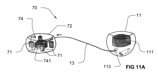
[0028] Figures 11A-B illustrate alternative embodiments of the
device of
Fig. 10, which comprise a traction system (thread winding system) for loading
the thread
between the two members in order to maintain tension in the thread; Fig. 11A
shows an
embodiment in which the traction system is a spiral spring and magnets are
placed
around the spiral spring; Fig. 11B shows an alternative for the second member,
wherein
the spiral spring is coiled around a centrally placed magnet.
CA 03054990 2019-08-29
WO 2018/158395 PCT/EP2018/055106
7
[0029] Figures 12A-C illustrate an alternative embodiment of the
devices
of Fig. 11A-B, wherein the magnet is arranged differently. Fig. 12A represents
a
perspective view of the device. Fig. 12B represents a cross sectional view of
the second
member. Fig. 120 represents a perspective partial cut-out view of the second
member.
Figs. 12B and C show the annular magnet and the thread winding system
including a
pulley for winding the thread.
[0030] Figure 13 illustrates another embodiment of device with
an
alternative means for pulling the thread towards the first and/or second
member.
[0031] Figure 14 illustrates an alternative to Fig. 4, in which
a through
passage is provided in two opposed magnetic members, in order to allow passage
of a
medical tool, such as a needle through the tissue.
[0032] It will be convenient to note that some parts in the
figures have been
drawn in transparency in order to uncover interior components.
Detailed Description
[0033] Referring to Fig. 1, a device 10 for pressure necrosis and
tissue
shearing according to one aspect comprises a first member 11 and a second
member
12 connected to one another by a thread 13. Each of the first member 11 and
second
member 12 can comprise a generally dome shaped housing 110 advantageously made
of a biocompatible material. The housing 110 encapsulates a material 111, 121
respectively. The materials 111, 121 encapsulated within the respective
members 11, 12
show magnetic attraction to one another. Advantageously, both materials 111
and 121
can be permanent magnets. Alternatively, one material can be a permanent
magnet, and
the other one a material which is magnetically attracted to the permanent
magnet, such
as though not limited to a ferromagnetic material, e.g. an iron alloy. Each of
the first
member 11 and the second member comprises a generally planar surface of
contact
112, 122 respectively, advantageously forming a bottom of the housing 110. The
materials 111 and 121 advantageously have magnetic properties such that they
show
magnetic attraction when the first member and the second member are placed
with their
surface of contact 112, 122 against each other, e.g. through suitable
orientation of the
magnetic poles N and S of the permanent magnets within the housing 110. In
use,
therefore, the first and second members will be disposed in such a manner that
the
surfaces of contact 112, 122 are in facing relationship. Each of the first and
second
member further comprises a point of attachment 113 at which the thread 13 is
connected
to the respective member. The thread connection at 113 is advantageously a
fixed
connection, e.g. the thread 13 is fixedly secured, such as through tying or
potting to the
member. Each member 11, 12 may further comprise a suitable structure for
facilitating
CA 03054990 2019-08-29
WO 2018/158395 PCT/EP2018/055106
8
handling of the member, e.g. an eye 114, allowing an endoscopic tool for
engaging the
respective member.
[0034] As a surprising effect, the device 10 allows for creating
an opening
through a tissue wall separating two body cavities. The opening is larger than
the size of
any of the first member 11 and the second member 12. To this end, the members
11, 12
comprising the magnets 111, 121 are used, in a first step, for compressing
tissue
between oppositely arranged magnetically attracting members thereby causing
pressure
necrosis. Subsequently, in a second step, the necrotic tissue collapses and
forms an
opening. The members 11, 12 lack support by the tissue and will fall through
the opening
created by necrosis. The members 11, 12 become suspended by thread 13 and will
pull
on the thread 13 due to gravity. The thread 13, supported by the tissue, will
start shearing
the tissue to create a larger opening.
[0035] A first possible application is shown in Figs. 2A-C and
relates to
treatment of diverticulum, such as though not limited to Zenker's
diverticulum, which is a
pouch 21 protruding from a lumen of the GI tract 20. In the particular case of
Zenker's
diverticulum, the lumen 20 is the hypopharynx. Referring to Fig. 2A, the
device 10 is
introduced into the lumen 20 and the first member 11 placed in the pouch 21
while the
second member 12 placed in the lumen 20 at a corresponding location. The
device 10
can be introduced by any suitable endoscopic means. Since the pouch 21 is open
towards the lumen 20, a delivery device, such as an endoscope with suitable
system for
engaging and positioning the members 11, 12, e.g. a grasper, forceps or
specific
catheter, can be guided to the pouch 21 to place the first member 11. No
tissue needs
to be pierced in order to place member 11. The same endoscope or another
endoscope
can be guided to the adjacent lumen 20 to place the second member 12. It is
alternatively
possible to place the second member 12 first, and to place the first member 11
subsequently. The thread 13 is either connected to the two members 11 and 12
prior to
loading the device 10 in the endoscope, or may be connected to either or both
members
upon placement. Advantageously, the thread 13 connecting the two members 11
and 12
does not pass through the tissue wall 23, but forms a loop over the periphery
24 of the
tissue wall. The placement of the members can be guided with endoscopic
ultrasound
and/or fluoroscopy which are procedures well-known to persons skilled in the
art of
endoscopic procedures.
[0036] Referring to Fig. 2B, due to the magnetic attraction
between the
members 11 and 12, the tissue wall 23 gets trapped between the two members and
is
compressed. By suitable selection of the magnetic attraction force between the
magnets
111 and 121, a compression pressure larger than 5 mm Hg can be created, which
is
CA 03054990 2019-08-29
WO 2018/158395 PCT/EP2018/055106
9
sufficient for stopping blood supply to the trapped tissue and hence causing
necrosis of
the tissue within a few days. The necrotic tissue trapped between the members
11 and
12 collapses and frees the members 11 and 12 which remain attached to one
another
and suspended on the thread 13. The first and second members and the thread
now
form a closed loop around the tissue wall 23, enclosing that portion of tissue
interposed
between the periphery 24 and the freshly formed necrotic opening. Due to their
weight,
the members 11 and 12 now pull on the thread loop 13 in the direction of
gravity.
Referring to Fig. 20, the thread 13 is now supported by the periphery 24 of
the tissue
wall and the tension in the thread 13 caused by the weight of the members 11
and 12
will start shearing the tissue from the periphery 24 until the initial opening
22 formed by
necrosis is reached. The action of the thread 13 shearing the tissue wall 23
is
represented in Figs. 3A-B from other viewing directions. By so doing, a lesion
extending
over the entire depth of the pouch 21 can be created without requiring
recourse to larger
structures and without requiring specific delivery devices.
[0037] The example described above is just one possibility of how device
10 can be used. Referring to Fig. 4, with two such devices 10 and 10', with
each one
device placed in one of two adjacent cavities 40, 41, a lesion can be created
extending
over the distance by which the first member 11 and the second member 12 are
spaced
apart. The magnetically attracting members 11 and 12' of respective devices
10, 10' are
placed in correspondence, each in the respective cavity. Portions of the
tissue walls 42
and 43 of each respective cavity 40, 41 overlapping the members 11 and 12' are
compressed. Likewise, the other members 12 and 11', which also magnetically
attract
each other, are placed in correspondence, each in the respective cavity at a
location
spaced apart from the members 11 and 12'. Portions of the tissue walls 42 and
43
overlapping the members 12 and 11' are compressed. Members 11 and 12 are
spaced
apart from each other, such as by a distance of at least 40 mm, advantageously
at least
50 mm to enable making a larger lesion. The tissue trapped between members 11
and
12' on the one hand and between members 12 and lion the other hand will become
necrotic and collapse, which will free the members towards one cavity 40 or
the other
one 41. Either thread 13 or 13' will be pulled by the freed member couples 11-
12' and
12-11' in the direction of gravity, which will shear the tissue in between the
openings
formed. It will be convenient to note that also in the case of Fig. 4, no
puncture or piercing
through the tissue wall is required for initial placement of the devices 10
and 10', as long
as both cavities 40 and 41 are accessible. A configuration as in Fig. 4 may be
useful for
creating gastrojejunal anastomosis or jejuno-jejunal anastomosis.
CA 03054990 2019-08-29
WO 2018/158395 PCT/EP2018/055106
[0038] It will be convenient to note that devices and assemblies
described
herein advantageously allow to perform tissue shearing assisted by pressure
necrosis
and/or compression anastomosis.
[0039] Even though the devices 10 and 10' in Fig. 4 are shown to
comprise
5 two magnetic members 11,12 and 11', 12' each, it will be clear that
device configurations
comprising more than two members, such as three members or four members
connected
by a thread are similarly applicable. By way of example, device 50 shown in
Fig. 5
comprises three members 11, 12 and 14 connected by thread 13. Two such devices
50
can be used in the same way as the example shown in Fig. 4, each device being
placed
10 in a different one of the adjacent cavities 40 and 41, as with the
devices 10 and 10'. The
third member 14 may comprise its proper magnet 141 or magnetically active
material.
[0040] The shape of the housing 110 of the members 11, 12 and
possibly
14, and the shape of the magnets or magnetically active materials 111, 121 as
well as
of the surface of contact 112, 122 is not particularly limited. Suitable
shapes and
configurations may be chosen depending on any particular application. By way
of
example, instead of dome-shaped, such housing may be cylindrical,
advantageously
with planar top and bottom base. There is no preference for using the top or
the bottom
as surface of contact in the latter case.
[0041] Each member can comprise a suitable handling structure
for
manipulating the member. In a first example, referring to Fig. 5, the members
11, 12 and
14 can comprise one or more thread loops 117 which facilitate handling of the
members
by a forceps. The thread loops 117 project from the housing of the member and
can be
provided in addition or in alternative to other handling structures, such as
the
engagement eye. In another example, referring to Figs. 6A-D, the engagement
eye or
hole 114, or the thread loop 117 as in Fig. 5, can be replaced by, or be
supplemented
with an advantageously ribbed projecting wall 115 allowing engagement by a
forceps,
e.g. of the types commonly used in endoscopic procedures. The magnet, or
magnetically
active material is received in interior and completely closed recess 116.
Referring to Figs.
7A-D, the engagement hole 114 may be oriented parallel to the surface of
contact 112,
perpendicular thereto, or oblique.
[0042] Referring to Fig. 70, it may be useful to provide the
hole 114 as a
through-hole and the magnet 111 advantageously surrounding the through-hole
114.
The through-hole is advantageously arranged to traverse the surface of contact
112 of
the member. One advantage of such an arrangement is shown in Fig. 14. Fig. 14
differs
with respect to Fig. 4 only in the shape of the second members 12, 12' which
comprise
such a through-hole 114 and in which the magnets are advantageously annular
CA 03054990 2019-08-29
WO 2018/158395 PCT/EP2018/055106
11
surrounding the through-hole 114. Referring to Fig. 14, the through-hole 114
allows to
pass a medical tool, such as a needle, through the respective member 12, 12'.
Since the
magnets of members 12 and 12' surround the through-hole 114, a necrotic
tissue, which
may be anastomosed, is created surrounding the location of the through-hole
114,
allowing a needle or other tool to pass through the tissues 42 and 43 safely.
[0043] Referring to Figs. 8A-G, the surface of contact 112, 122
which
defines the contact being made between the member and the tissue, can be, but
need
not be, planar. Other kinds of shapes, such as concave, convex, stepped or
staircase-
like can be contemplated. It will be convenient to note that the surface of
contact of
members placed at opposite sides of the tissue wall, such as members 11 and 12
in Fig.
2, and members 11 and 11' in Fig. 4, may have a complementary shape. By way of
example, the shapes shown in Fig. 80, 8E and 8G may be complementary. In
another
example, two mushroom-shaped members as shown in Fig. 8G may be used. These
mushroom-shaped members may form plugs or anchors preventing the magnetically
attached members to fall through the tissues once the tissue becomes necrotic
and
collapses.
[0044] Referring to Figs. 7A-D and Figs. 9A-C, the magnets 111,
121 or,
as the case may be, magnetically active materials can have any suitable shape.
They
may be monolithic within the housing 110, or made up of separate parts
arranged
adjacent one another, as shown in Figs. 9B-C. Other shapes than disc or
cylindrical are
possible, such as ring, oval or parallelepiped.
[0045] It will be convenient to note that the shearing by the
thread 13 can
be facilitated by suitable selection of the mass (weight) of any of the
members 11 and
12. Generally, a tension on the thread of at least 5 mm Hg may be sufficient
for causing
tissue shearing, with tensions of at least 10 mm Hg, at least 20 mm Hg, at
least 50 mm
Hg, or at least 100 mm Hg being advantageous. In some instances, it may be
useful to
maintain the thread 13 under a constant tension, as from the onset,
particularly even
prior to the collapse of the tissue due to pressure necrosis performed by the
members
11 and 12. One possible way of accomplishing is by making one of the members
slidable
relative to the thread. Referring to Fig. 10, device 60 differs from device 10
in that the
second member 62 is not fixedly attached to the thread 13, but is allowed to
slide along
the thread. The first member 11 is attached to one end of the thread 13, and a
mass
member 63 is attached to the opposite end of thread 13, with the second member
62
being slidingly attached to thread 13 in between the first member 11 and the
mass
member 63. To this end, the second member 62 comprises a through hole 621
slidingly
receiving thread 13. Mass member 63 has a suitable weight in order to provide
a desired
CA 03054990 2019-08-29
WO 2018/158395 PCT/EP2018/055106
12
tension in thread 13, but will advantageously not be magnetically attracted to
either one
of the members 62 and 11. In particular, mass member 63 may not comprise any
magnet
or magnetically active material. Mass member may have a mass at least equal to
50%,
75% or 100% of the mass of any one of the members 11 and 12. When members 11
and
62 are placed as shown in Fig. 2, the mass member 63 is freely suspended and
will pull
on the thread 13 and put it under tension to immediately start shearing
tissue. It will
therefore be clear that device 60 allows for simultaneous pressure necrosis
and tissue
shearing. A yet alternative embodiment is obtained by adapting the arrangement
10 of
Fig. 1 to include the mass member 63 interposed between the first member 11
and the
second member 12. In this case, mass member 63 may, but need not be slidingly
attached to the thread 13.
[0046] Fig. 11A-B show alternative embodiments to the device 60,
in which
tension in the thread 13 is created through a thread winding system integrated
within one
of the members, or both. Referring to Fig 11A, device 70 differs from device
10 in that
the second member 72 comprises a thread winding system 74 for loading thread
13 in
order to maintain tension in the thread. Thread winding system 74 can comprise
a spiral
spring 741 which is preloaded to pull on thread 13 according to a desired
preload force
acting in the direction of the arrow. Magnets 71 may be placed peripherally to
thread
winding system 74, as shown in Fig. 11A, or in any other suitable
configuration. Referring
to Fig. 11B, an alternative to Fig. 11A is shown, which differs from device 70
in that the
thread winding system 75 in the second member 73 is a spiral spring which is
coiled
around magnet 76, i.e., spring 73 and magnet 76 are concentric. In an
alternative
embodiment, the arrangement of Fig. 10 is adapted to incorporate the thread
winding
system in the mass member 63.
[0047] Yet an alternative embodiment to the devices 70 of Figs. 11A-B is
shown in Figs. 12A-C, representing a device 80 comprising a thread winding
system 84
incorporated in the second member 82. Device 80 differs from device 70 in the
disposition of magnet 81 (see Fig. 12B) which is annular and arranged above or
below
the thread winding system 84. Alternatively, possibly annular magnets may be
arranged
above and below the thread winding system, e.g. in a symmetrical fashion. The
thread
winding system 84 comprises a spiral spring operably connected to a pulley 86.
Thread
13 is wound on pulley 86. Spiral spring 85, or any other suitable spring, is
advantageously
preloaded to apply a suitable torque on pulley 86 to pull on thread 13. In the
latter design,
the thread winding system 84 and the magnet 81 can be kept separated from one
another
without interference.
CA 03054990 2019-08-29
WO 2018/158395 PCT/EP2018/055106
13
[0048] Referring to Fig. 120; a blocking system 89 can be
provided in the
second member 82 for blocking rotation of the thread winding system 84. By way
of
example, blocking system 89 comprises a blocking member 88 which blocks the
pulley
86 and/or the spiral spring 85, e.g. by engagement. The blocking system 89 is
releasable,
e.g. by pulling on thread 87 attached to blocking member 88, the blocking
member 88 is
released and the pulley and spiral spring can turn and wind the thread 13.
[0049] Referring to Fig. 13, a yet alternative embodiment is
shown, which
differs from device 10 in that a plurality of third members 91 are attached
along the thread
13, between the first member 11 and the second member 12. The third members
may
be arranged with advantageously uniform spacing between one another and they
are
advantageously fixed to thread 13. The third members advantageously show
magnetic
attraction to the first member 11 and/or the second member 12 and act here as
thread
pulling means. As the tissue is further sheared, the third member 91 closest
to the first
member 11 will progressively move to the first member due to the magnetic
attraction.
The third members 91 can have any suitable shape, e.g. beads, and
advantageously
have dimensions substantially smaller than those of the first member and
second
member, e.g. half or less of the size of the first member and/or second
member. It will
be convenient to note that neither the thread winding system in Figs. 11A-B
and 12A-C,
nor the third members in Fig. 13 prejudice the position and shape of the
magnet or
magnetically active material, of the housing, or of the surface of contact,
and these
features may assume any suitable shape, e.g. as illustrated in Figs. 7A-D
through 9A-C.
[0050] Yet another alternative to the systems shown in Fig. 10
through 13
is to use an elastic or resilient thread to couple the members to one another.
This thread
is advantageously preloaded prior to placement of the device, e.g. by
providing an
appropriate structure which maintains the members at a distance larger than
the length
of the thread at rest, which structure is removed upon installing the device.
The thread
is stretched and thereby preloaded. In one example, the thread may be made of,
or
comprise a shape memory material. Suitable shape memory materials are shape
memory polymers or (metal) alloys, which e.g. can retract at a body
temperature level.
.. One example is DiAPLEX commercialised by Mitsubishi Corporation Fashion
Co., Ltd.,
Japan. The use of a mass member pulling on the thread, of a traction system,
or of elastic
or resilient thread can be advantageously used in the application referred to
in Fig. 4.
That is, either one or both the devices 10 and 10' in Fig. 4 may be provided
with means
for (pre)loading the thread 13, 13'. In the example of Fig. 4, (pre)loading
the threads 13,
.. 13' with tensile stress enhances fusion between the tissues 42 and 43.
CA 03054990 2019-08-29
WO 2018/158395 PCT/EP2018/055106
14
[0051] The thread is advantageously made of a non-resorbable
material,
and is advantageously a monofilament thread, e.g. made of polyamide. The shape
of the
housing 110 is advantageously atraumatic. While in the above embodiments, only
one
thread is described extending between the first member and the second member,
it will
be convenient to note that a plurality of such threads, e.g. two, three or
more can be
provided to extend between, and be connected to the first member and second
member.
The means for pulling the thread may be operable to pull some or all of these
threads.
Multiple threads advantageously allow for shearing tissue across multiple,
spaced apart
sections.
[0052] Each of the members has a diameter D (see Fig. 6B) or largest
size
advantageously smaller than or equal to 40 mm, advantageously smaller than or
equal
to 30 mm, advantageously smaller than or equal to 20 mm, advantageously
smaller than
or equal to 15 mm, advantageously smaller than or equal to 20 mm. The member
11 or
12 can have a diameter or size of at least 5 mm, advantageously at least 10
mm. The
thread 13 extending between the members has a length which in an initial
placement
position is advantageously at least 80 mm, advantageously at least 100 mm,
advantageously at least 120 mm. The mass of the member 11 or 12 generally
depends
on the application, and may be determined on the basis of e.g. contact length
of the
thread with the tissue in order to arrive at a desired tension on the thread.
[0053] Some aspects of the present invention as described herein are
set
out in the following numbered clauses.
1. Method of creating pressure necrosis of a tissue wall
between
a pouch and an adjacent cavity in a human or animal body, wherein the tissue
wall
comprises a periphery forming an edge of an opening between the pouch and the
cavity,
the method comprising:
placing a first member into the pouch, such that the first member is
adjacent the wall,
placing a second member in the cavity and proximate the first
member,
wherein the first member and the second member comprise
materials which magnetically attract one another,
wherein the first member and the second member are placed such
that there is magnetic attraction between the first member and the second
member
through the wall and wherein the first member and the second member compress
an
overlapping portion of the wall to create pressure necrosis,
CA 03054990 2019-08-29
WO 2018/158395 PCT/EP2018/055106
wherein the first member and the second member are connected
through a thread, wherein the thread extends over the edge.
2. Method of clause 1, wherein, following pressure necrosis, the
first member and the second member are suspended by the thread forming a loop
over
5 the wall.
3. Method of clause 2, wherein the thread shears the wall by
action of a weight of the first and second members.
4. Method of clause 2, wherein the thread shears the wall while
the thread is under tension by action of a mechanism that induces a traction
force in the
10 thread.
5. Method of clause 2, comprising exerting a pulling force on the
thread while the thread shears the wall.
6. Method of clause 2, wherein the wall is cut by the thread in a
direction of gravity.
15 7. Method of clause 1, wherein the thread does not pierce
through the wall until pressure necrosis is created.
8. Method of clause 1, wherein at least one of the first member
and the second member comprises an encapsulated magnet.
9. Method of clause 1, comprising inserting a delivery device
having an internal lumen into the cavity, and
guiding the delivery device through the opening to the pouch and
delivering the first member to the pouch by sliding the first member through
the internal
lumen;
delivering the second member to the cavity by sliding the second
member through the internal lumen;
wherein the first member and the second member are connected by
the thread in the internal lumen.
10. Method of clause 1 for treating diverticulum, wherein the
pouch is a diverticulum.
11. Method of clause 10, wherein the cavity is a segment of a
gastrointestinal tract of the human or animal body.
12. Method of creating compression anastomosis between
adjacent tissues, each of the tissues forming a wall of one of adjacent
cavities of a human
or animal body, the method comprising:
CA 03054990 2019-08-29
WO 2018/158395 PCT/EP2018/055106
16
placing a first member and a second member in a first one of the
adjacent cavities, proximate a first one of the walls, wherein the first
member and the
second member are spaced apart and connected to a first thread;
placing a third member and a fourth member in a second one of the
adjacent cavities and proximate a second one of the walls, wherein the third
member
and the fourth member are connected to a second thread and are placed in
correspondence of a respective one of the first member and the second member;
wherein the first, second, third and fourth members comprise
materials which magnetically attract one another through the adjacent tissues
to create
pressure necrosis of an overlapping portion of the wall.
13. Method of clause 12, wherein the first thread does
not extend
to the second one of the adjacent cavities upon placement of the first and
second
members and wherein the second thread does not extend to the first one of the
adjacent
cavities upon placement of the third and fourth members.
14. Method of clause 12, comprising a step of shearing the
adjacent tissues by the first thread or the second thread, wherein the
shearing opens a
lesion extending between a necrotic opening through the adjacent tissues
formed at a
location of the first member and a necrotic opening through the adjacent
tissues formed
at a location of the second member.
15. Method of clause 14, wherein one of the first thread and the
second thread shears the adjacent tissues while the respective thread is under
tension
by action of a mechanism that induces a traction force in the respective
thread.