Note: Descriptions are shown in the official language in which they were submitted.
METHOD AND APPARATUS FOR WIDE-BAND PHASE GRADIENT SIGNAL
ACQUISITION
[0001]
TECHNICAL FIELD
[0002] The present disclosure generally relates to a biosignal
acquisition apparatus
that differentially acquires wide-band phase gradient signals that are used to
non-invasively
estimate functions of the body such as heart functions, as well as to pinpoint
and distinguish
disease (e.g., to predict presence or non-presence of a disease).
BACKGROUND
[0003] Conventional electrocardiographic instruments are configured to
acquire and
record biosignals such as biopotential signals relating to electrical
activities of the heart. It is
conventionally accepted that a large fraction of the total signal collected by
such instruments
is considered devoid of biological information. However, hidden within the
full spectrum of
physiologic signals emitted from the human body are information that can be
used to pinpoint
and distinguish disease.
[0004] Because these information can be captured in physiologic signals
having
signal power comparable to, or lower than, the noise floor of conventional
electrocardiographic instruments, such information are difficult to extract,
or not discernible,
from the measured signals of these instruments. In some instances, the signal
of interests has
an order of magnitude of a few micro-Volts, and in other instances, even
smaller. At such
Page 1
Date Recue/Date Received 2021-03-19
CA 03055157 2019-08-30
WO 2018/158749
PCT/IB2018/051358
levels, interference from external energy sources such as man-made
radiofrequency
transmission and those that occur naturally as well as those from internal
circuitries of the
measurement instrument itself can affect the acquisition and recording of such
information.
[0005] What are needed are devices, systems and methods that overcome
challenges
in the present art, some of which are described above.
SUMMARY
[0006] The present disclosure facilitates capture (e.g., bipolar capture)
of
differentially-acquired wide-band phase gradient signals (e.g., wide-band
cardiac phase
gradient signals, wide-band cerebral phase gradient signals) that are
simultaneously sampled,
in some embodiments, having a temporal skew among the channels of less than
about 1
and in other embodiments, having a temporal skew of not more than about 10
femtoseconds.
Notably, the exemplified system minimizes non-linear distortions (e.g., those
that can be
introduced via certain filters such as phase distortions) in the acquired wide-
band phase
gradient signals so as to not affect the information therein that can non-
deterministically
affect analysis of the wide-band phase gradient signal in the phase space
domain.
[0007] The bipolar capture operation, for use in differential measurements,
increases
the dynamic range of the differential measurement input so as to reduce, or
eliminate, a need
for filtering (e.g., low frequency filtering), thereby improving acquisition
of the acquired
wide-band phase gradient signals by further minimizing potential non-linear
distortions that
maybe introduced from additional hardware circuitry associated with such
filtering. The
bipolar capture operation, of a differential measurement, also reduce or
eliminate common
mode noise, via use of a single amplifier, as compared to a pair of amplifiers
that capture
unipolar signals where common mode noise reduction is based on tolerances of
resistors and
capacitors and symmetry of the amplifiers (e.g., op-amps).
2
CA 03055157 2019-08-30
WO 2018/158749
PCT/IB2018/051358
[00081 Further, a shield drive circuit and shield-drive voltage plane may
be used to
facilitate low noise and low interference operation of the acquisition system.
In some
embodiments, the acquisition system has a noise performance of better than 10
[IV.
[0009] In an aspect, an apparatus (e.g., a BioSignal Acquisition Instrument
(a "BSA
instrument")) is disclosed. The apparatus includes a plurality of biosignal
acquisition
channels (e.g., three channels) in which each biosignal acquisition channel
comprises a gain
amplifier configured to, by bipolar sensing for each input (of a differential
input pair),
differentially amplify biopotential signals received from a pair of associated
surface
electrodes placed on a patient (including mammals such as human and test
animals) to
generate a differentially-acquired wide-band phase gradient signal (e.g.,
differential wide-
band cardiac gradient signal), wherein each differential biopotential signal
is amplified
without filtering that causes distortion in the generated differential wide-
band cardiac phase
gradient signal above about lkHz, wherein each output of the biosignal
acquisition channels
feeds an analog-to-digital conversion circuit that simultaneously samples
(e.g., having a
temporal skew among the channels of less than about 1 tts or having a temporal
skew of not
more than about 10 femtoscconds) each of the biosignal acquisition channels
(e.g., having at
a sampling frequency above about 10KHz, e.g., about 40Khz, about 80KHz, about
500Khz,
or higher) to generate a differential wide-band cardiac phase gradient signal
data stream.
[0010] In some embodiments, the apparatus further includes a potential
biasing circuit
that actively applies a varying potential to a patient so as to shunt
environmental noise
currents flowing over or in the patient. In some embodiments, the potential
biasing circuit
applies a constant positive potential to the patient. In some embodiments, the
potential
biasing circuit drives the patient to a constant negative potential. In some
embodiments, the
potential biasing circuit drives the patient to a varying potential.
3
CA 03055157 2019-08-30
WO 2018/158749
PCT/IB2018/051358
[0011] In some embodiments, the apparatus includes a potential biasing
circuit that
actively applies a potential (e.g., a constant potential, e.g., about 1.5 VDC
or a varying
potential that varies about -1.5 VAc,n,$) to the patient so as to shunt
environmental noise
currents flowing over or in the patient. In some embodiments, the applied
varying potential
has a value of about 2.0 VAC_rms, about 1.8 VAC_rms, about 1.6 VAC_rms, about
1.4 VAC_rms,
about 1.2 VAC_ ims, about 1.0 VAc_rms, about 0.8 VAC_ rms, about 0.6 VAc_ims,
about 0.4 VAc_rms,
about 0.2 VAC_ims, about -0.2 VAC_rms, about -0.4 VAC_Ims, about -0.6 VAC_
ims, about -0.8
VAC rms, about -1.0VAc rms, about -1.2 VAC rms, about -1.4 VAc rms, about -1.6
VAC rms, about -
1.8 VAC_rms, and about -2.0 VAc_rins. In some embodiments, the applied
potential has a value
of about +0.5 Vric, about +1.0 VDC, about +1.5 VDC, about +2.0 VDc, +2.5 VDc,
about +3.0
VDc, about +3.5 VDC, about +4.0 VDC, about +4.5 VDC, about +5.0 VDC, about -
0.5 VDC, about
-1.0 VDC, about -1.5 VDC, about -2.0 VDC, -2.5 VDC, about -3.0 VDC, about -3.5
VDC, about -
4.0 VDC, about -4.5 VDC, about -5.0 VDC.
[0012] In some embodiments, the potential biasing circuit includes a
waveform
generator (e.g., a configurable waveform generator); and a drive circuit
(e.g., a common
mode amplifier) that couples to the waveform generator to actively apply an
alternating
potential (e.g., between about -1.0VDC and about -2.0VDC or between about +1.0
and about
+2.0 VDC) to the patient so as to shunt environmental noise currents flowing
in the patient.
[0013] In some embodiments, the potential biasing circuit actively applies
an
alternating potential having a minimum magnitude greater than a DC bias value
associated
with one or more of the surface electrodes placed on the patient (e.g.,
wherein the one or
more surface electrodes have a half-cell potential).
[0014] In some embodiments, the apparatus includes a potential biasing
circuit that
actively applies a varying potential on a patient so as to shunt environmental
noise currents
4
CA 03055157 2019-08-30
WO 2018/158749
PCT/IB2018/051358
flowing on or in the patient, wherein a substantial portion (e.g., greater
than about 75%) of
the varying potential is negative.
[0015] In some embodiments, the apparatus includes a potential biasing
circuit that
actively applies a constant potential to a patient so as to shunt
environmental noise currents
flowing on or in the patient.
[0016] In some embodiments, the apparatus includes a terminal block (e.g..
for a
given cable) comprising at least one connector configured to couple to a cable
associated with
a given surface electrode, wherein the cable comprises a shield layer that
encapsulates one or
more signal wires that carries a given biopotential signal received from the
given surface
electrode (e.g., wherein the shield layer does not terminate at or connect to
the surface
electrode); and a noise-rejection circuit (e.g., a unity gain amplifier) that
applies a potential of
the potential biasing circuit to the shield layer of the cable and to a cable-
shield drive voltage
plane to allow for return pass for noisy current induced on the shield layer.
[0017] In some embodiments, the apparatus includes one or more terminal
blocks
each of which individually couples to a shield of a cable associated with a
surface electrode;
and a shield-equalizing circuit that injects a signal carried in the cable to
the shield of the
cable such that the injected signal approximately matches (e.g., within at
least about 90%) the
signal carried in the cable.
[0018] In some embodiments, the gain amplifier of each of the biosignal
acquisition
channels directly couples to a terminal block (e.g., for a given cable)
comprising a plurality of
connectors, each of which couples a cable associated with a given surface
electrode.
[0019] In some embodiments, each of the biosignal acquisition channels
comprises a
gain amplifier configured to amplify the received biopotential signal with a
gain that provides
a measurement resolution, with the analog-to-digital circuit, greater than
about 0.3 V per bit
(e.g., wherein the analog-to-digital circuit provides a bit resolution of at
least about 12 bits).
CA 03055157 2019-08-30
WO 2018/158749
PCT/IB2018/051358
[0020] In some embodiments, the gain amplifier is powered by a single
voltage
supply (e.g., about +1.5 VDC, about +3 VDC, about +3.3 VDC, about +5 VDC,
about +12 VDC,
and about +15 VDC, about -1.5 VDC, about -3 VDC, about -3.3VDc, about -5 VDC,
about -12
VDC, and about
-15 VDC).
[0021] In some embodiments, the biopotential channels comprises a number of
channels selected from the group consisting of 2, 3, 4, 5, 6, 7, 8, 9, 10, 11,
and 12 (e.g.,
wherein the number of cables and surface electrodes corresponds to one-half
the number of
channels plus one, e.g., a common mode reference cable and surface electrode).
[0022] In some embodiments, the analog-to-digital circuit of each biosignal
acquisition channel is configured to sample a wide-band cardiac phase gradient
signal over a
pre-defined voltage range of at least about 5 milli-Volt (mV) at a resolution
of less than about
2 micro-Volt (RV) per bit and at a rate greater than about 5000 Hertz, wherein
the biosignal
acquisition channels are simultaneously sampled with a temporal skew between
channels less
than 1 micro-seconds (i.ts), and wherein each biosignal acquisition channel
comprises a
signal-to-noise ratio of greater than about 15dB (e.g., greater than 20dB).
[0023] In some embodiments, each biosignal acquisition channel comprises a
gain
amplifier circuit (e.g., a gain amplifier circuit board or flex circuit) that
directly couples to
given surface electrode within an electrode housing.
[0024] In some embodiments, each gain amplifier circuit associated with a
given
electrode housing feeds a corresponding analog-to-digital circuit located in a
second housing,
the second housing being connected to the given electrode housing via a cable.
[0025] In some embodiments, the apparatus further comprising: a plurality
of analog-
to-digital circuits, each corresponding to a bio-signal acquisition channel,
wherein each
output of the each bio-signal acquisition channel feeds a corresponding analog-
to-digital
6
CA 03055157 2019-08-30
WO 2018/158749
PCT/IB2018/051358
circuit, and wherein the analog-to-digital circuits simultaneously sample to
generate a
plurality of wide-band cardiac phase gradient signal data streams each
associated with a
given differential wide-band cardiac phase gradient signal.
[0026] In another aspect, a method is disclosed of generating wide-band
cardiac phase
gradient signal data. The method includes differentially amplifying (e.g., a
gain amplifier
circuit) acquired biopotential signals received from a plurality of surface
electrodes each
placed on a patient to generate a wide-band cardiac phase gradient signal,
wherein each
differential biopotential signal is amplified without filtering that causes
distortions in the
generated differential wide-band cardiac phase gradient signal above about l
kHz, and
wherein each input of the paired differential input is configured for hi-polar
sensing; and
simultaneously sampling (e.g., AD converters), at a sampling frequency greater
than about 50
KHz, each of the amplified differential wide-band cardiac phase gradient
signals to generate
differential wide-band cardiac phase gradient signal data streams, wherein the
amplified
differential wide-band cardiac phase gradient signals are simultaneous sampled
so as to have
a temporal skew among each of the amplified wide-band cardiac phase gradient
signals less
than about 1 us.
[0027] In another aspect, a signal acquisition board is disclosed. The
signal
acquisition board includes a multi-layer printed circuit board comprising: a
first layer that
serves as a reference ground plane; a second layer co-planar to the first
layer that serves as a
cable-drive voltage plane (e.g., having a potential of about +1.5V); and one
or more signal
layers having a pair of conductive traces (e.g., low-impedance traces) running
substantially
therethrough and across one or more regions coincident and coplanar to the
second layer,
wherein the pair of conductive traces electrically couple, across a connector
directly or
indirectly affixed to the multi-layer printed circuit, ends of at least two
signal-carrying
conductors to differential input pins of an analog-to-digital conversion and
amplifier stage
7
CA 03055157 2019-08-30
WO 2018/158749
PCT/IB2018/051358
mounted on a surface of the multi-layer printed circuit, wherein a first
signal-carrying
conductor of the at least two signal-carrying conductors is associated with a
first cable and a
second signal-carrying conductor of the at least two signal-carrying
conductors is associated
with a second cable; wherein the second layer electrically couples, over the
at least one
connector, i) a first outer conductor that serves as an outer shield of the
first cable and ii) a
second outer conductor that serves as an outer shield of the second cable, so
as to drive
potentials of the first outer conductor and the second outer conductor to that
of the cable-
drive voltage plane.
[0028] In some embodiments, the first cable and the second cable terminate
at a
single cable-pin connector, the single cable-pin connector having a coupling
element
configured to releasably mate to the connector of the signal acquisition
board.
[0029] In some embodiments, the pair of conductive traces are arranged, on
a same
set of signal layers of the one or more signal layers, and in close proximity
to one another
such that substantial lengths of each conductive trace of the pair of
conductive traces are
substantially parallel to one another.
[0030] In some embodiments, each conductive trace of the pair of conductive
traces
has a length and have a same number of via so as to have a substantially
similar impedance
characteristics as one another.
[0031] In some embodiments, each conductive trace of the pair of conductive
traces
includes an impedance element (e.g., a single 101(52 resistor) arranged
between a respective
pin of the connector and a respective differential input pins of the analog-to-
digital
conversion circuit, and wherein the pair of conductive traces has a
capacitance element
coupled therebetween to form, with the impedance elements, an antialiasing
filter.
[0032] In some embodiments, the multi-layer printed circuit board further
comprises
a conductive housing that serves as a grounded shield cage, wherein the
conductive housing
8
CA 03055157 2019-08-30
WO 2018/158749
PCT/IB2018/051358
spans a portion of the second layer so as to encapsulate a substantial portion
of the pair of
conductive traces, and wherein the conductive housing is affixed to the
surface of the multi-
layer printed circuit and is electrically coupled to the reference ground
plane.
[0033] In some embodiments, the analog-to-digital conversion and amplifier
stage
comprises a single integrated circuit having one or more analog-to-digital
converters (ADCs)
with built-in programmable gain amplifiers (PGAs).
[0034] In some embodiments, the analog-to-digital conversion and amplifier
stage for
the pair of conductive traces comprises an analog-to-digital converters (ADCs)
integrated
circuit coupled to an amplifier circuit.
[0035] In some embodiments, the multi-layer printed circuit board further
comprises:
one or more processors and one or more memory components coupled to the one or
more
processors, wherein the one or more processors and the one or more memory
components are
arranged on a portion of the surface of the multi-layer printed circuit that
do not coincide or
overlap with the cable-drive voltage plane of the second layer.
[0036] In some embodiments, the pair of conductive traces forms a part of a
first
differential input channel of the signal acquisition board.
[0037] In some embodiments, the signal acquisition hoard of claim further
comprises
a second differential input channel and a third differential input channel,
wherein each of the
second differential input channel and the third differential input channel
comprises a pair of
conductive traces running substantially through the one or more signal layers
across the one
or more regions coincident and coplanar to the cable-drive voltage plane of
the second layer,
wherein each of the second differential input channel and the third
differential input channel
connects to a pair of cables having at least one signal-carrying conductor and
an outer
conductor that serves as an outer shield of the signal-carrying conductor, and
wherein the
cable-drive voltage plane electrically couples, over the at least one
connector, to the outer
9
CA 03055157 2019-08-30
WO 2018/158749
PCT/IB2018/051358
conductors of the pair of cables so as to drive potentials of the outer
conductors to that of the
cable-drive voltage plane.
BRIEF DESCRIPTION OF DRAWINGS
[0038] Embodiments of the present invention may be better understood from
the
following detailed description when read in conjunction with the accompanying
drawings.
Such embodiments, which are for illustrative purposes only, depict novel and
non-obvious
aspects of the invention. The drawings include the following figures:
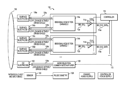
[0039] Fig. 1 is a diagram of an example apparatus configured to
differentially
acquire wide-band cardiac phase gradient signals in accordance with an
embodiment.
[0040] Fig. 2 is a diagram of a time series representation of a wide band
cardiac
gradient signal (unipolar) in accordance with an illustrative embodiment.
[0041] Fig. 3 is a diagram of the example differentially-acquired wide-band
cardiac
gradient signal data of FIG. 2 shown in the frequency domain, in accordance
with an
embodiment.
[0042] Fig. 4A is a detailed diagram of a biosignal acquisition channel of
Fig. 1 with
bipolar sensing in accordance with an illustrative embodiment.
[0043] Fig. 4B is a detailed diagram of a biosignal acquisition channel of
Fig. 1 with
bipolar sensing in accordance with another illustrative embodiment.
[0044] Fig. 5 is a diagram of a method of matching potential of a signal-
carrying
conductor and a shield-conductor in accordance with an embodiment.
[0045] Fig. 6 is a diagram of an example system in accordance with an
illustrative
embodiment.
[0046] Fig. 7 is a diagram of an example instrumentation amplifier
configured for one
channel of bipolar sensing operation.
CA 03055157 2019-08-30
WO 2018/158749
PCT/IB2018/051358
[0047] Fig. 8 is a diagram of an example integrated circuit with
instrumentation
amplifiers configured for multiple channels of bipolar sensing operations.
[0048] Figs. 9A, 9B, 9C, 9D, 9E, 9F, 9G, 9H, 91, 9J, 9K, 9L, 9M, 9N, 90,
9P, 9Q,
9R, 9S, and 9T are circuit diagrams of a differentially-acquired wide-band
cardiac phase
gradient signal acquisition system in accordance with an illustrative
embodiment.
[0049] Fig. 10A is a diagram of an example biosignal acquisition ("BSA")
board that
includes the differentially-acquired wide-band cardiac phase gradient signal
acquisition
system of Fig. 9 in accordance with an embodiment.
[0050] Fig. 10B is a diagram showing details of tracing of the example
hiosignal
acquisition board in accordance with an embodiment.
[0051] Figs. 10C and 10D shows additional views of the biosignal
acquisition board.
[0052] Fig. 11A is a photograph of an example BSA instrument that includes
the BSA
board of Fig. 10A in accordance with an embodiment.
[0053] Fig. 11B is a diagram with an exploded unassembled view of an
example BSA
instrument that includes the BSA board of Fig. 10A in accordance with an
embodiment.
[0054] Figs. 12A and 12B are diagrams of an example placement of the
surface
electrodes at the chest and back of a patient to acquire hio-potential signals
associated with
differentially-acquired wide-band cardiac phase gradient signal data in
accordance with an
illustrative embodiment.
[0055] Fig. 13 is an example operation of a BSA instrument in accordance
with an
illustrative embodiment.
DETAILED DESCRIPTION
[0056] The components in the drawings are not necessarily to scale relative
to each
other and like reference numerals designate corresponding parts throughout the
several views.
11
CA 03055157 2019-08-30
WO 2018/158749
PCT/IB2018/051358
[0057] Fig. 1 is a diagram of an example apparatus 100 configured to
differentially
acquire wide-band cardiac phase gradient signals in accordance with an
embodiment. As
shown in Fig. 1, apparatus 100 includes a number of biosignal acquisition
channels 104
(shown as "biosignal acquisition channel 0" 104a and "biosignal acquisition
channel 1" 104b)
that is each operatively coupled to a corresponding pair of surface electrodes
106 (shown as
surface electrodes 106a, 106b, 106c, 106d, and etc.) to differentially acquire
wide-band
cardiac phase gradient signals from a patient's chest area and/or back area
108. In some
embodiments, apparatus 100 includes three biosignal acquisition channels 104
for XYZ lead
measurements.
[0058] In some embodiments, the biosignal acquisition channels 104 are
configured
to differentially acquire wide-band phase gradient signals (e.g., wide-band
cerebral phase
gradient signal) at other locations, for example, at a patient's head. In
other embodiments,
wide-band phase gradient signals are differentially acquired from other areas
of the body,
e.g., in proximity to a target organ.
[0059] Bipolar sensing provides true differential XYZ lead measurements of
wide-
band cardiac phase gradient signals in which vectorcardiograms (VCG) derived
therefrom are
stable on any choice of reference positions (i.e., the measurements are not
sensitive to lead
positions). Leads of apparatus 100 have polarity and are placed at specific
locations on the
body surface. A reference lead (shown as "CM Electrode" 122) is used to reduce
noise.
[0060] Bipolar sensing facilitate differential measurements that reduce, or
eliminate,
common mode noise based on internal symmetry of the analog to digital
converters (ADCs)
and only amplifies potential differences between two points with very high
common mode
rejection. Bipolar sensing facilitate differential measurements that also
provide high static
gain accuracy.
12
CA 03055157 2019-08-30
WO 2018/158749
PCT/IB2018/051358
[0061] Referring still to Fig. 1, each biosignal acquisition channel 104
includes one or
more amplifier circuits 110 (e.g., instrumentation class amplifiers) (not
shown ¨ see Fig. 4A
or 4B) that amplifies, via bipolar inputs, differential biopotential signals
received at a given
amplifier circuit to generate a differential amplified biopotential signal 112
(shown as
"BIO_SIGo" 112a, "BIO_SIGi" 112b, and etc.) corresponding to wide-band cardiac
phase
gradient signal having little or no non-linear distortions introduced into the
signal path.
[0062] Example of such non-linear distortions includes phase distortions
that may
affect the signal at different frequencies which can distort the wide-band
cardiac phase
gradient signal in the phase space domain. In addition, non-linear distortions
includes
variability in the signal paths among the different acquisition channels.
[0063] As shown in Fig. 1, the biosignal acquisition channels 104 are
coupled to a
corresponding analog-to-digital conversion circuit 114 (shown as 114a, 114b,
and etc.) that
are simultaneously sampled such that a temporal skew among each of the sampled
signal is
less than about 1 gs, to convert the amplified differential biopotential
signals 112a, 112b to
time-series data (shown as "BIO_SIG_DATA0" 116a,"1310_SIG_DATA1" 116b, and
etc.)
associated with the differentially-acquired wide-band cardiac phase gradient
signal and that
are received by a controller 118 for subsequent analysis (e.g., in phase space
domain). In
some embodiments, the biosignal acquisition channels 104 are configured to
simultaneously
sample the acquired signal with a temporal skew of not more than about 10
femtoseconds.
[0064] The controller 118 manages the acquisition and recording of the
biosignal
from the patient and, in some embodiments, manages the transmission of
recorded
information (including, e.g., biosignals, instrument identification, and
patient identification)
to a remote data storage location (e.g., a storage area network). In some
embodiments, the
controller 118 manages the acquisition and recording of the biosignal from the
patient and
interfaces with a computing device to transmit recorded information
(including, e.g.,
13
CA 03055157 2019-08-30
WO 2018/158749
PCT/IB2018/051358
biosignals, instrument identification, and patient identification) to a remote
data storage
location. In some embodiments, processing is performed on the stored data set
to determine
cardiac performance, including but not limited to, predicting Ejection
Fraction (in
percentage), assessing ischemic burden, and/or detecting coronary artery
disease, from the
differentially-acquired wide-band cardiac phase gradient signals generated
from the acquired
biopotential signals. In some embodiments, the controller 118 manages the
acquisition and
recording of the biosignal from the patient and manages the processing, e.g.,
locally or
remotely, of the biosignal to present results on a graphical user interface
operatively
connected to the controller.
[0065] In some embodiments, the system 100 includes a pulse oximeter
circuit 128
that operates with a pulse oximeter (P02) sensor 130 to collect oxygen
saturation readings.
The collected oxygen saturation readings may be used to augment analyses of
the
differentially-acquired wide-band cardiac phase gradient signal data. In some
embodiments,
data associated with oxygen saturation readings are collected concurrently
with the
acquisition of the wide-band cardiac phase gradient signal data. In other
embodiments, data
associated with oxygen saturation readings are independently collected. Other
sensors or
features may also be included.
[0066] Referring still to the embodiment of Fig. 1, each analog-to-digital
conversion
circuit 114a or 114b includes a high-speed sigma-delta converter that is
configured to sample
simultaneously to have a temporal skew of less than about 1 us (e.g., not more
than about 10
fs (femtosecond)) with the other biosignal acquisition channels. The output of
the analog-to-
digital conversion circuit 114 is preferably a serial data stream (serial
digital stream) that is
provided to the controller 118. The controller 118, in some embodiments, is
configured to
aggregate the acquired data 116a, 116b (associated with a differentially-
acquired wide-band
cardiac phase gradient signal) over a pre-defined period and transmit the
collected data to a
14
CA 03055157 2019-08-30
WO 2018/158749
PCT/IB2018/051358
repository (e.g., a storage area network). In some embodiments, the acquired
data 116a, 116b
are transmitted as time series data in a file. In some embodiments, the
transmission is only
performed in between acquisition events. In some embodiments, the file
includes one or
more, e.g., time series data, instrument identification data, instrument
performance data,
and/or patient identification data.
[0067] In other embodiments, the controller 118 is configured to store the
acquired
data 116a, 116b, which is then processed locally. In some embodiments, the
acquired data is
processed by the acquisition system and is then transmitted as collected data
(e.g., as a time-
series data) to the repository. Each differentially-acquired wide-band cardiac
phase gradient
signal data sets may have a duration period between about 100 seconds and
about 200
seconds.
[0068] The differentially-acquired wide-band cardiac phase gradient signal
data
comprises a wide range of frequencies, in some embodiments, having a sampling
greater than
1 KHz (Kilo-Hertz). In some embodiments, the differentially-acquired wide-band
cardiac
phase gradient signal data comprises a sampling frequency greater than about 5
KHz. In
some embodiments, the wide-band cardiac phase gradient signal data comprises a
sampling
frequency greater than about 10 KHz. In some embodiments, the differentially-
acquired
wide-band cardiac phase gradient signal data comprises a sampling frequency
greater than
about 40 KHz. In some embodiments, the wide-band cardiac phase gradient signal
data
comprises a sampling frequency greater than about 80 KHz. In some embodiments,
the
differentially-acquired wide-band cardiac phase gradient signal data comprises
a sampling
frequency greater than about 500 KHz. In various embodiments, the
differentially-acquired
wide-band cardiac phase gradient signal data has little or no non-linear
distortion within its
range of sampled frequencies.
CA 03055157 2019-08-30
WO 2018/158749
PCT/IB2018/051358
[0069] In addition, the differentially-acquired wide-band cardiac phase
gradient
signal data has a range of at least about 5 mV (millivolt) at a resolution of
less than about 2
V (microvolt) per bit. In some embodiments, the differentially-acquired wide-
band cardiac
phase gradient signal data has a resolution of about, or less than, 1/2 V per
bit. Other such
ranges and resolutions may be used.
[0070] Because '1/2 p.V is below the thermal noise associated with most
conventional
circuitries, the system 100 includes several features to reduce interference
from its own
circuitries as well as from external energy sources such as from
radiofrequency
transmissions. It is observed that noise level of a differentially-acquired
wide-hand cardiac
phase gradient signal, when implemented with such techniques, are generally
less than about
V.
[0071] FIG. 2 is a diagram of an example unipolar wide-band cardiac phase
gradient
signal data (shown as 202a, 202b, 202c, and etc.) shown as a time series data,
in accordance
with an embodiment. The differentially-acquired wide-band cardiac phase
gradient signal
data shows a difference between two of these signals (e.g., 202a and 202b;
202c and 202d,
and etc.). In some embodiments, the patient is actively driven to a common
mode potential
and the acquired biopotential signal includes that common mode potential. In
such
embodiments, the differentially-acquired wide-band cardiac phase gradient
signal data is the
remaining signal with the common-mode reference removed, e.g., via
differential acquisition
scheme or via computation. In some embodiments, the differentially-acquired
wide-band
cardiac phase gradient signal data has been amplified and normalized with the
common-mode
reference removed via hardware circuitry.
[0072] Fig. 3 is a diagram of the example differentially-acquired wide-band
cardiac
phase gradient signal data of Fig. 2 shown in the frequency domain, in
accordance with an
embodiment.
16
CA 03055157 2019-08-30
WO 2018/158749
PCT/IB2018/051358
[0073] It is discovered that wide-band biopotential signals and
differential signals
thereof, having energy and frequency components beyond those of conventional
electrocardiography (ECG) and traditionally perceived to be random noise,
includes
measurable data of the heart physiology that can be discriminated by genetic
algorithms (and
other machine learning algorithms) to assess regional flow characteristics of
the heart,
including an estimated value for stenosis an identification of ischemia, a
fractional flow
reserve (FFR) of specific arteries and branches thereof. Noise removal (e.g.,
by applying
cleaning techniques to the data resulting in the same amount of data as prior
to noise
removal) is a fundamental step in signal processing. However, the exemplified
method and
system processes the entire obtained biopotential signals without any noise
removal
operations in the wide-band region of the signal. What has heretofore been
perceived and/or
classified as unwanted noise in the wide-band data is, in many cases, the
signal of interest.
Examples of noise removal that is not performed include, but not limited to,
analog-based
low-pass filters, band-pass filters, high-pass filters and well as digital-
based filters such as
FIR filters, Butterworth filters, Chcbyshey filters, median filters, among
others.
[0074] In addition to removing information of interest from the acquired
wide-band
signals, certain circuit elements can introduce non-linear distortions that
can affect analyses
in phase space of the differentially-acquired wide-band phase gradient signals
and are not
included, or minimized, in the signal path of the exemplified system. For
example, certain
analog pass filters (e.g., analog-based low-pass filters, band-pass filters,
high-pass filters and
well as digital-based filters such as FIR filters, Butterworth filters,
Chebyshev filters, median
filters, among others, as discussed above) may introduce phase distortions
which may result
in non-linear group delays among the multiple acquisition channels or
introduce frequency-
dependent distortions in individual acquisition channels. In addition, certain
circuit elements
such as field-effect transistors (e.g., MOSFET) may introduce unnecessary
capacitance and
17
CA 03055157 2019-08-30
WO 2018/158749
PCT/IB2018/051358
gate-field effect noise to the signal path. In addition, certain semiconductor
and insulating
materials with avalanche breakdown effects (e.g., in Zener diodes) may
introduce avalanche
noise to the signal path.
[0075] In some embodiments, the signal may be processed via phase linear
operations
to allow for analyses of specific aspects of the high-frequency wide-band
data. In some
embodiments, the signal may be processed via operations or circuitries that
affect frequencies
completely outside the band of interest. In some embodiments, these
frequencies that are
filtered are in the radiofrequency range or above.
[0076] As shown in Fig. 3, the wide-band cardiac gradient signal has a
frequency
component greater than about 1 kHz, which is significantly higher than
convention
electrocardiogram measurements. In some embodiments, the differential wide-
band cardiac
gradient signal has a frequency component up to about 2 kHz (e.g., about 0 Hz
to about 2
kHz). In some embodiments, the different wide-band cardiac gradient signal has
a frequency
component up to about 4 kHz (e.g., about 0 Hz to about 4 kHz). In some
embodiments, the
differential wide-band cardiac gradient signal has a frequency component up to
about 5 kHz
(e.g., about 0 Hz to about 5 kHz). In some embodiments, the differential wide-
band cardiac
gradient signal has a frequency component up to 6 kHz (e.g., about 0 Hz to
about 6 kHz). In
some embodiments, the differential wide-band cardiac gradient signal has a
frequency
component up to about 7 kHz (e.g., about 0 Hz to about 7 kHz). In some
embodiments, the
differential wide-band cardiac gradient signal has a frequency component up to
about 8 kHz
(e.g., about 0 Hz to about 8 kHz). In some embodiments, the differential wide-
band cardiac
gradient signal has a frequency component up to 9 kHz (e.g., about 0 Hz to
about 9 kHz). In
some embodiments, the differential wide-band cardiac gradient signal has a
frequency
component up to 10 kHz (e.g., about 0 Hz to about 10 kHz). In some
embodiments, the
18
CA 03055157 2019-08-30
WO 2018/158749
PCT/IB2018/051358
differential wide-band cardiac gradient signal has a frequency component up to
50 kHz (e.g.,
about 0 Hz to about 50 kHz).
[0077] Fig. 4A is a diagram of a biosignal acquisition channel 104 with
bipolar
sensing in accordance with an illustrative embodiment. The biosignal
acquisition channel
104 includes an operational amplifier 110 (e.g., an instrumentation class
amplifier) having a
first differential input 402a and a second differential input 402b that each
directly couples to
a terminal (shown as 404a, 404b) to operatively couple to the surface
electrodes 106a, 106b.
The biosignal acquisition channel 104 is configured such that little, or no,
non-linear
distortions (e.g., such as those discussed herein) are introduced into the
signal path. To this
end, active and passive filters are preferably not placed in the signal path,
or are minimized,
to reduce distortions that they may be introduced during operation. In some
embodiments, a
single anti-aliasing filter is included in the signal path (that also servers
a protection to the
input of the channel). The operational amplifier 110 preferably provides a
gain greater than
about 15 dB (decibel) to generate the differentially-acquired wide-band phase
gradient signal.
In some embodiments, the operational amplifier 110 provides a gain greater
than about 20
dB. The output 412 of the operational amplifier 110, in some embodiments, is
coupled to the
analog-to-digital conversion circuit 114 (e.g., sigma-delta ADC). In some
embodiments, the
operational amplifier 110 and the analog-to-digital conversion circuit 114 are
part of a single
integrated circuit. In addition, though shown as two terminals, terminals
404a, 404b may be
part of a common terminal housing.
[0078] In some embodiments, and as shown in Fig. 4A, each biosignal
acquisition
channel 104 electrically couples to a respective set of paired surface
electrodes 106a, 106b
over a pair of cables 124a, 124b (e.g., a co-axial cable)) that employs an
active noise
reduction system. In some embodiments, the active noise reduction system is
used to actively
19
CA 03055157 2019-08-30
WO 2018/158749
PCT/IB2018/051358
shield signal-carry conductors used to carry signal across multiple circuit
boards prior to the
acquired signal being digitized.
[0079] In Fig. 4A, the biosignal acquisition channel 104 include an active
noise
reduction system that actively shields the signal-carrying conductors 408a,
408b in cable
124a, 124b arranged between the surface electrode 106a, 106b and the
operational amplifier
110. The cables 124a, 124b include a set of first conductors 408a, 408b (e.g.,
pair of twisted
wires) and a set of second conductive layers 406a, 406b (i.e., outer shield)
that surrounds the
respective first conductors 408a, 408b. The active noise reduction system
includes a shield-
equalizing circuit (also referred to as a shield-drive circuit or cable-drive
circuit) comprising
operational amplifiers 410a, 410b that injects the signal carried in the
conductors 408a, 408b
to the shield 406a, 406b of the cables 124a, 124b such that the injected
signal approximately
matches (e.g., within at least about 90%) the signal carried in the cable. Put
another way, the
active noise reduction system drives the shield 406a, 406b to about the same
electric potential
as the conductor 408a, 408b which reduces the electrical leakage between the
conductors
408a, 408b and the shield 406a, 406b. In another aspect, the outer shields
(e.g., 406a, 406b)
of the cables (e.g., 124a, 124b) are electrically coupled to a shield-drive
voltage plane 416
(also referred to as a cable-drive voltage plane) to provide a return pass for
noisy current
induced on the outer shield (e.g., 406a, 406b).
[0080] In some embodiments, the operational amplifier 410 is configured as
a unity
gain amplifier. In other embodiments, non-unity gain is used. The inputs 414a,
414b of the
operational amplifiers 410a, 410b are coupled to the input of the gain
amplifier 110, which is
also coupled to the terminals 404a, 404b. The outputs of the operational
amplifiers 410a,
410b are coupled to the second conductive layers 406a, 406b of the cables
124a, 124b.
[0081] Fig. 4B is a diagram of a biosignal acquisition channel 104 with
bipolar
sensing in accordance with another illustrative embodiment. In Fig. 4B, an
active noise
CA 03055157 2019-08-30
WO 2018/158749
PCT/IB2018/051358
reduction system is used in which an average potential from all, or most, of
the signal-
carrying conductors (e.g., 408a, 408b) is used to drive the outer shield 406a,
406b of the
cables (e.g., 124a, 124b) for each of the biosignal acquisition channels. As
shown in Fig. 4B,
an operational amplifiers 410a is coupled to an averaging circuit 418 that is
coupled to each
of the signal-carrying conductors (e.g., 408a, 408b). The signal-carrying
conductors (e.g.,
408a, 408b) are coupled to the gain amplifier 110 that coupled to the analog-
to-digital
conversion circuit 114. In Fig. 4B, the gain amplifier 110 and the analog-to-
digital conversion
circuit 114 are arranged on the same printed circuit board. In some
embodiments, the gain
amplifier 110 and the analog-to-digital conversion circuit 114 are combined in
a single
integrated circuit. Other components may be arranged with the gain amplifier
110 to provide
a desired gain output for the amplifier.
[0082] In another embodiment, the operational amplifiers 410a is coupled to
output of
an amplifier output of a microcontroller that generates an analog output
signal by averaging
the inputs of the acquired differential wide-band cardiac gradient signal.
[0083] In some embodiments, the outer shields (e.g., 406a, 406b) arc
electrically
coupled to a shield-drive voltage plane 416 to provide a return pass for noisy
current induced
on the outer shield (e.g., 406a, 406h).
[0084] In some embodiments, the active noise reduction system uses the
potential of a
single signal-carrying conductor (e.g., 408a or 408b) to drive the outer
shields for all the
cables (408a, 408b, etc.) of all the biosignal acquisition channels.
[0085] Fig. 5 is a diagram illustrating operations of the shield-equalizing
circuit in
accordance with an illustrative embodiment. As shown in Fig. 5, a shield
conductor 406 of a
cable 124 surrounds a signal conductor 408 and is driven by an operational
amplifier (e.g.,
410a) to a potential that matches, or nearly matches, that of the signal
conductor 408. For
example, where the signal conductor 408 carries a potential of about +1.5V,
and the
21
CA 03055157 2019-08-30
WO 2018/158749
PCT/IB2018/051358
operational amplifier (e.g. 410a) drives the shield conductor 406 also to
about +1.5V.
Because the potential between the signal conductor 408 and shield conductor
406 matches, or
nearly matches, the dielectric electric field between them is minimized. To
this end, a
perturbation introduced to the signal-conductor 408 by the shield-conductor
406 due to
perturbation of the shield-conductor 406 from external interference is
dampened.
[0086] Example Noise Rejection Subsystem
[0087] To improve the signal quality of the differentially-acquired wide-
band cardiac
phase gradient signal 112, the exemplified system 100 (e.g., as shown in Fig.
1), in some
embodiments, includes a noise rejection system 120 that eliminates, or
reduces,
environmental noise currents flowing in the patient's body that might
interfere with the
biopotential measurement. The noise rejection system 120 is configured to
actively drive the
patient's body to a potential that shunts environmental noise currents during
normal
operation. Environmental noise may be generated from a variety of
environmental sources
including nearby electronics, transmission devices, and local AC power
systems, among
others. Any or all of these sources may generate voltages at the measurement
electrodes that
can render a patient's biopotential un-measurable or reduce the resolution of
the
measurement.
[0088] As shown in Fig. 1, the noise rejection system 120 is operatively
coupled to a
surface electrode 122 that is in electrical contact (e.g., directly or via a
conductive gel or
paste) with a surface of the body 108. In some embodiments, the noise
rejection system 120
actively applies a varying potential to the body 108, e.g., a potential that
varies between two
negative potential values.
[0089] In some embodiments, the surface electrode (e.g., 106a, 106b, 106c,
106d,
122) may be used in conjunction with gels or other coupling media or devices
that can form a
half-cell potential in the signal path when measuring the differentially-
acquired wide-band
22
CA 03055157 2019-08-30
WO 2018/158749
PCT/IB2018/051358
cardiac phase gradient signal. For example, silver chloride gel may introduce
a 300 mV
biased in the signal path. In some embodiments, the noise rejection system 120
actively
drives the body 108 to a varying potential that varies between two negative
potential values
such that the magnitudes of negative potential values are greater than the
expected half-cell
potential DC bias value associated with the surface electrodes.
[0090] Referring still to Fig. 1, noise rejection system 120 is
electrically coupled, via
a cable 124e, to a common-mode electrode 122 that is placed on the body 108.
In some
embodiments, an active noise reduction system, e.g., similar to that used in
the biosignal
acquisition channels, is used to actively shield the signal-carrying conductor
in the cable 124e
between the common-mode surface electrode 122 and the noise rejection system
120. In
other embodiments, a passive shield is used in which the shield-conductor of
the cable 124e
is coupled to the ground plane of the system 100.
[0091] The noise rejection system 120, in some embodiments, includes a
waveform
generator and an operational amplifier. In some embodiments, the waveform
generator is a
fixed-frequency oscillator. In other embodiments, the waveform generator is a
microcontroller that is electronically programmable to generate an analog
output that can
vary in frequency and amplitude range, e.g., based on control signals
outputted from the
controller 118. In Fig. 1, the noise rejection system 120 is shown operatively
coupled to the
controller 118 via control line 126.
[0092] In some embodiments, the noise rejection system 120 actively drives
the body
108 to a varying potential that varies between a negative potential value and
a positive
potential value.
[0093] In some embodiments, the noise reduction system 120 actively drives
the body
108 to a varying potential that varies between two positive potential values.
23
CA 03055157 2019-08-30
WO 2018/158749
PCT/IB2018/051358
[0094] In other embodiments, the noise reduction system 120 actively drives
the body
to a constant potential (e.g., a value between about -1.5 VDC and about +1.5
VDC or a value
between about -3.0 VDC and about +3 VDC).
[0095] Example BSA System
[0096] Fig. 6 is a diagram of an example system 100 in accordance with an
illustrative embodiment. As shown in Fig. 6, the system 100 includes a first
stage mixed-
signal board 602 that includes the biosignal acquisition channels 104 as
described in relation
to Fig. 1. The first stage mixed-signal board 602 is operatively coupled to a
second stage
mixed-signal board 604 over one or more cables 418 that carries the amplified
biopotential
signals 112. The second stage mixed-signal board 604 includes the analog-to-
digital
conversion circuit 114 and the controller 118, as described in relation to
Fig. 1. The second
stage mixed-signal board 604 communicates to a third stage controller board
606 that
provides communication and interface functionality for apparatus 100.
[0097] As shown in Fig. 6, the second stage mixed-signal board 604 includes
memory
608 and interface circuit 610. The memory 608 locally stores the acquired
biopotcntial signal
data 116 associated with the differentially-acquired wide-band cardiac phase
gradient signal
data for a given measurement prior to the data 116 being sent to the third
stage controller
board 606 to be transmitted to remote storage. The interface circuit 610, in
some
embodiments, includes communication isolation circuitries such as optical
isolators and other
isolation circuitries such as, but not limited to, for power and ground. The
third stage
controller board 606 includes a processor 612, a memory 614, a communication
transceiver
616, and an interface circuit 618 that, collectively, is configured to operate
with the second
stage mix-signal board 604 to offload the differential wide-band cardiac phase
gradient signal
data acquired thereat to transmit, e.g., via wireless communication to remote
storage (e.g.,
repositories in the cloud). In some embodiments, the third stage controller
board 606 is
24
CA 03055157 2019-08-30
WO 2018/158749
PCT/IB2018/051358
configured to analyze the differentially-acquired wide-band cardiac phase
gradient signal data
acquired thereat and present outputs of the analyses at a graphical user
interface associated
therewith. In some embodiments, the third stage controller board 606 is a part
of a custom
computing device. In other embodiments, the third stage controller board 606 a
part of a
general computing device.
[0098] In some embodiments, the first stage mixed-signal board 602, the
second stage
mixed-signal board 604, and the third stage controller board 606 are part of a
single printed
circuit board.
[0099] Fig. 7 is a diagram of an example instrumentation amplifier
configured for one
channel of bipolar sensing operation. The instrumentation amplifier is a zero-
drift,
instrumentation amplifier (e.g., INA 188 integrated circuit, manufactured by
Texas
Instruments, Inc. (Dallas, TX)).
[0100] Fig. 8 is a diagram of an example integrated circuit with
instrumentation
amplifiers configured for multiple channels of bipolar sensing operations. The
integrated
circuit is a 6-channel, 24-bit ADC with an integrated ECG front end (e.g., ADS
1296
integrated circuit, manufactured by Texas Instruments. Inc. (Dallas, TX)). The
integrated
circuit has delta-sigma analog-to-digital converters with built-in
programmable gain
amplifiers (PGAs).
[0101] Example BioSignal Acquisition Circuit
[0102] Figs. 9A, 9B, 9C, 9D, 9E, 9F, 9G, 9H, 91, 9J, 9K, 9L, 9M, 9N, 90,
9P, 9R, 9S,
9T, and 9V, are circuit diagrams of a prototype wide-band cardiac phase
gradient signal
acquisition system 900 with bipolar operations in accordance with an
illustrative
embodiment.
[0103] Specifically, Fig. 9A shows a high-level diagram of the system 900.
As
shown in Fig. 9, the system 900 includes a main controller 910 that couples to
a biopotential
CA 03055157 2019-08-30
WO 2018/158749
PCT/IB2018/051358
acquisition circuit 902 that acquires the biopotential signal data associated
with differentially-
acquired wide-band cardiac phase gradient signals. The main controller 910 may
perform the
function of controller 118 as described in relation to Fig. 1. The main
controller 910 couples
to a pulse oximetry circuit 904 that acquires oximetry data. The system 900
further includes
a USB interface circuit 906 configured to provide communication to the main
controller 910
for testing and development. The system 900 includes an MFi interface circuit
908 that
provides connectivity to a computing device (e.g., device 606 as described in
relation to Fig.
6). The system 100 further includes a power system 912 to provide power to the
various
circuits and also to provide reference voltage for the analog-to-digital
conversion.
[0104] Figs. 9B, 9C, and 9D show detailed diagrams of power circuits 912.
In Fig.
9B, a power circuit 912a to supply power to the system 900 from batteries is
shown. The
power circuit includes a monitoring and charging circuit. In Fig. 9C, a power
circuit 912B
for the biosignal acquisition channel is shown. In Fig. 9D, a power circuit
912C for digital
circuits is shown.
[0105] Fig. 9E shows a detailed diagram of a controller circuit for main
controller 910
(shown as device "EFM32GG880" 910). The controller circuit includes a memory
module
912 (shown as device "S23MLOG1") that couples to the main controller 910. The
main
controller 910 is an ARM Cortex CPU platform manufactured by Silicon
Laboratories
(Austin, TX), part no. "EFM32GG880". The memory "S23MLOG1" is an 8 GB
(gigabyte)
NAND Flash memory manufactured by Cypress Semiconductor Corporation (San Jose,
CA).
The main controller 910 operates with the biosignal acquisition channel (e.g.,
104) to receive
the biopotential signal data and to locally store the data to the NAND Flash
memory (e.g.,
912) for each acquisition.
[0106] Fig. 9F shows a detailed diagram of the MFi circuit 908. The MFi
circuit 908
includes a microcontroller 914 (shown as device "SiM3U167") that provides an
interface to
26
CA 03055157 2019-08-30
WO 2018/158749
PCT/IB2018/051358
an external computing device. The main controller 910 of Fig. 9E may be
configured by
computer readable instructions stored in memory to retrieve, between
acquisition of the
differentially-acquired wide-band cardiac phase gradient signal data (e.g.,
biosignal data and
instrument identification data) stored in the NAND Flash memory and transfers
the data to an
external computing device through the MFi circuit 908. In some embodiments,
the MFi
circuit 908 may be powered down during the acquisition of wide-band cardiac
phase gradient
signal data so as to minimize interference during the signal acquisition.
[0107] The SiM3U167 is an ARM Cortex-M3 based microcontroller (MCU),
manufactured by Silicon Laboratories (Austin, TX). The SiM3U167 may he part of
a USB
MCU family of energy friendly devices configured with low energy operation,
fast wake-up
times and energy saving modes.
[0108] Fig. 9G shows a detailed diagram of the USB communication circuit
906 that
is used to access the main controller 910, e.g., for testing and development.
The circuit may
not be available for access by a user during normal runtime operation.
[0109] Figs. 9H, 91, 9J, and 9K show detailed diagrams of the biopotential
acquisition
circuit 902. The biopotential acquisition circuit 902 includes an analog-to-
digital converter IC
916 (shown as device "AD51294" 916) configured with an integrated ECG front
end circuit
comprising a programmable gain amplifier. To this end, analog-to-digital
converter IC 916
includes both the gain amplifier 110 and the analog-to-digital conversion
circuit 114 in a
single integrated circuit. Other configuration of the analog-to-digital
conversion circuit may
be used, though the analog-to-digital conversion circuit has at least about 17
bits of
resolution, preferably about 24 bits.
[0110] Specifically, Fig. 9H shows the wiring of the analog-to-digital
converter IC
916, to the main controller 910 and the biopotential channel circuit 922 of
Fig. 90 via the
control lines and data lines. Further, in Fig. 9H, a single cable terminal
block 924
27
CA 03055157 2019-08-30
WO 2018/158749
PCT/IB2018/051358
(corresponding to terminals 404a, 404b) is provided to couple to a cable
assembly comprising
cables 124a-124e that couples to the electrodes 106a-106e. The cable terminal
block 924
includes i) pins (shown as pin 1, 3, 5, 7, and 9 of J500) for 3 pairs of
differential inputs and ii)
a pin 926 (shown as pin 4 of J500) for the outer shield drive. Each of pins 1,
3, 5, 7, and 9 of
J500 connect to respective inputs 928a-928f of respective biopotential channel
922. The
biopotential channels 922 are repeated 6 times to provide outputs 930a-930f to
the inputs
932a-932f of the analog-to-digital converter IC 916. The analog-to-digital
converter IC 916
provides the acquired signal 112 over a digital bus to main controller 910 via
lines 934 (see
Figs. 9A and 9E).
[0111] Fig. 90 shows a detailed diagram of an example biosignal acquisition
channel
922, as shown in connection with Fig. 9H. Notably, there are no active
components or low-
pass filtering in the signal path 940 between the input 950 and output 952 of
the channel 922.
To this end, there is a lack of active filters and/or circuit elements that
can introduce non-
linear distortions into the signal path. In Fig. 90, components 928 are shunts
serving as a
jumper and components 938 are not placed and are provided as optional
components within
the prototype printed circuit board. Indeed, only a single anti-aliasing
circuit is included in
the signal path 939. The anti-aliasing circuit includes two resisters 940 from
two channels
922 connected by a capacitor (shown as 942a, 942b, and 942c in Fig. 9H). The
number of
components (e.g., resisters 940 and capacitor 942a-942b) is preferably
minimized to improve
noise performance, though more than one of each of these components may be
used. The
resisters 940 for a channel pairs are 10 k-Ohm and also serves to protect the
input of the
analog-to-digital converter IC 916.
[0112] One or more ferrite 928 (e.g., ferrite bead) may be placed in the
signal path to
suppress high frequency noise (e.g., radio-frequency noise). It is noted that
radio-frequency
28
CA 03055157 2019-08-30
WO 2018/158749
PCT/IB2018/051358
signals are generally in the MHz range which is several orders of magnitude
higher than the
biopotential signals of interest, which are in the KHz to hundreds of KHz.
[0113] To provide defibrillation protection, a defibrillator protection
circuit, or
equivalent thereof, is placed in the signal path 940. As shown in Fig. 9L, a
combined
defibrillation, surge, and ESD protector circuit is used. Fig. 9L shows a
detailed diagram of a
defibrillator protection circuit (shown as 948a, 948b). An example combined
defibrillation,
surge, and ESD protector circuit is the MAX30034 protection devices,
manufactured by
Maxim Integrated (San Jose, CA). In Fig. 9H, a limiting resister (R520, R519,
R524, R517,
R518, and R521) is shown placed in the signal path 940 used with the ESD
protector circuit.
[0114] Figs. 91, 9J, and 9K each shows the detailed diagram of the
capacitive
decoupling and filtering of the power plane and ground plane of the analog-to-
digital
conversion circuit.
[0115] Fig. 9M and Fig. 9N show detailed diagrams of power conditioning
circuits
that provide reference voltages to the biopotential amplifier circuits as
shown in Fig. 9L and
to the biopotcntial amplifier circuit as shown in Fig. 9H.
[0116] Noise Reduction Circuit
[0117] Fig. 9P shows a detailed diagram of an example noise rejection
circuit that
applies a common-mode voltage reference to the body.
[0118] The goal of the noise rejection system is to eliminate environmental
noise
currents flowing in the patient's body that might interfere with biopotential
measurement.
Noise may be generated from a variety of environmental sources; including
consumer
electronics, cell phones, and the local AC power system. Any or all of these
may generate
voltages at the measurement electrodes that will render a patient's
biopotential un-measurable
or more difficult to measure.
29
[0119] To combat environmental noise, the BSA Instrument hardware
employs a
common mode amplifier to actively applies the patient's body with a varying
potential (e.g.,
between -1.0VDC and -2.0VDC or +1.0 and +2.0 VDC) or a constant potential
(e.g., a value
between +1.5 VDC or -1.5VDC) and thus shunt environmental noise currents
during normal
operation. In Fig. 9P, the common mode amplifier is connected to an internal
amplifier output
946 of the analog-to-digital converter IC 916 (Fig. 9H). In other embodiments,
a separate
amplifier stage may be used to drive the patient's body to other potentials.
[0120] The BSA Instrument hardware further includes an operational
amplifier, U501
(shown as "LMV2011" 410a) that drives the outer shields 406a-406f of the
cables 124a-124f
with the same potential as that of the common mode amplifier. As shown in Fig.
9P, the input
944 of the operational amplifier 410a is also coupled to the internal
amplifier output 946 of
the analog-to-digital converter IC 916 (Fig. 9H). The analog-to-digital
converter IC 916 is
configured to generate constant potential (e.g., 1.5VDc). In other
embodiments, the analog-to-
digital converter IC 916 is configured to generate an average output of the
readings of the
inputs 932a-932f of the analog-to-digital converter 916.
[0121] Figs. 9Q, 9R, 9S, and 9T, are detailed diagrams of components of
the oximetry
circuit (shown as 904a, 904b, 904c, and 904d). The oximetry circuit 904 is
configured to
operate with a pulse oximeter (P02) sensor to collect oxygen saturation
readings. In some
embodiments, the oxygen saturation readings is collected with at least 12 bits
of resolution
and at a minimum rate of 200 samples per second.
[0122] Another example of the wide-band cardiac phase gradient signal
acquisition
system is described in W02017/033164.
[0123] Example BSA Board
Page 30
Date Recue/Date Received 2021-03-19
CA 03055157 2019-08-30
WO 2018/158749
PCT/IB2018/051358
[0124] Fig. 10A is a diagram of an example biosignal acquisition ("BSA")
board
1000 comprising multi-layer printed circuit board that includes the wide-band
cardiac phase
gradient signal acquisition system of Fig. 9 in accordance with an embodiment.
The BSA
board 1000, in some embodiments, includes a conductive shield 1004 (e.g., a
grounded shield
cage) that surrounds the mixed-signal front-stage circuitries of the biosignal
acquisition
channel 104 arranged between the cable terminal block 924 and the analog-to-
digital
converter IC 916. The conductive shield 1004, in some embodiments, is
electrically coupled
to reference ground plane.
[0125] Fig. 10B shows a diagram of a detailed view 1002 of Fig. 10A of the
mixed-
signal front-stage circuitries of the biosignal acquisition channel 104
arranged between the
cable terminal block 924 and the analog-to-digital converter IC 916.
[0126] In Fig. 10B, three sets of tracing pairs for the three-differential
channels are
shown, including tracings 1006a, 1006b, tracings 1006c, 1006d, and tracings
1006e, 1006f in
which tracings 1006a, 1006b are connected to biopotential channel inputs 928a
and 928b;
tracings 1006c, 1006d arc connected to biopotential channel inputs 928c and
928d; and
tracings 1006e, 1006f are connected to biopotential channel inputs 928e and
928f. The
tracings 1006a-1006f are arranged across two layers (shown in solid and in
dash) connected
by vias 1008a-1008f.
[0127] As noted above, only a single anti-aliasing circuit is included in
the signal path
940 (and, in some embodiments, a defibrillation protection circuit). The anti-
aliasing circuit
includes two resisters 940 from two channels 922 connected by a capacitor
(shown as 942a,
942b, and 942c in Fig. 9H). The number of components (e.g., resisters 940 and
capacitor
942a-942b) is minimized to improve noise performance. The resisters 940a-940f
for a given
channel pair are 10 k-Ohm and serve to protect the input of the analog-to-
digital converter IC
31
CA 03055157 2019-08-30
WO 2018/158749
PCT/IB2018/051358
916 by increasing thea common mode rejection ratio for the inputs of the
analog-to-digital
converter IC 916.
[0128] Cable-Drive Voltage Plane
[0129] In another aspect, a shield drive voltage circuit is used to
facilitate low noise
and low interference operation of the acquisition system. Fig. 10B further
shows an example
shield-drive voltage plane 416. The shield-drive voltage plane 416 is
connected to a shield-
drive amplifier 410a that drives the outer shields 406a-406f of the cables
124a-124f and
provides a return pass for noisy current induced on the outer shield 406a-
406f. The shield-
drive voltage plane 416 is electrically coupled to terminal 924 through vias
1010 that
connects to a pin of the terminal 924 that connects to outer shields 406-406f
of the cables
124a-124f. In some embodiments, the cable includes a trunk segment having an
outer shield
and includes a set of branch segments comprising multiple branch cables
extending from the
trunk segment. Each of the branch cables includes an outer shield that
connects to the outer
shield of the trunk segment.
[0130] In some embodiments, the multi-layer printed circuit board comprises
seven
layers in which the top "first" layer and "third" layer are designated for
signal tracings, the
"second" layer and bottom "seventh" layer has a reference ground plane, and
the "fourth"
layer includes the cable-drive voltage plane 416. Indeed, the "second" and
"seventh" layer of
the board serve as a reference ground plane, and the "fourth" layer serves as
the cable-drive
ground plane. Layer "five" may be used as a power layer.
[0131] The top `first" layer and "third layer" comprise signal layers
having pairs of
conductive traces (e.g., low-impedance traces) running substantially through
the layers and
across one or more regions coincident and coplanar to the cable-drive voltage
plane 416. In
some embodiments, the conductive traces arc 0.254 mm wide (0.001 inch wide).
Other trace
thicknesses may be used depending on the material to facilitate low-impedance
operations.
32
CA 03055157 2019-08-30
WO 2018/158749
PCT/IB2018/051358
The pair of conductive traces electrically couples, across the connector
(e.g., terminal 924)
directly or indirectly affixed to the multi-layer printed circuit, to the ends
of signal-carrying
conductors of the cables 124a-124f and also to the differential input pins of
the analog-to-
digital converter IC 916 (having an analog-to-digital conversion circuit and
amplifier stage).
The cable-drive voltage plane 416 (as the second ground layer) electrically
couple, over
terminal 924, to the outer shield 406a of cable 124a, the outer shield 406b of
cable 124b, the
outer shield 406c or cable 124c, the outer shield 406d of cable 124d, the
outer shield 406e of
cable 124e, and the outer shield 406f of cable 124f. The cable-drive voltage
plane 416
overlaps with a substantial length of the tracings 1006a-1006f and overlaps in
part (shown as
1008) over the footprint of the analog-to-digital converter IC 916. Though
shown being
routed across two layers, in other embodiments, the tracings 1006a-1006f may
be routed over
a single layer of the multi-layer printed circuit board.
[0132] In some embodiments, the cables 124a-124f terminate at a single
cable-pin
connector (shown in Fig. 11A) that is configured to releasably mate to the
connector (e.g.,
terminal 924) of the signal acquisition board 1000.
[0133] To allow for even more improved low-noise operation, each conductive
trace
of the pair of conductive traces 1006a-1006f is arranged with a similar length
and has a same
number of via as the corresponding trace (as, for example, shown in Fig. 10B)
so as to have a
substantially similar impedance characteristics with the corresponding trace
of the differential
pair. Further, each pair of conductive traces are arranged, on each layer that
they are routed,
in close proximity (as, for example, shown in Fig. 10B) to one another such
that substantial
lengths of each conductive trace of the pair of conductive traces are
substantially parallel to
one another.
[0134] Further, the conductive traces 1006a-1006f and cable-drive voltage
plane 416
are arranged on a portion of the board 1000 that is, in essence, isolated from
the processing
33
CA 03055157 2019-08-30
WO 2018/158749
PCT/IB2018/051358
and communication components (e.g., 910, 912, 914) to minimize interference
and noise
generated from such circuits.
[0135] Referring still to Fig. 10A, the geometric configuration of the
conductive
shield 1004 serving as a grounded shield cage is shown. The conductive shield
1004 spans a
substantial portion of the cable-drive voltage plane 416 (as the second ground
layer) so as to
encapsulate a substantial portion of the pair of conductive traces 1006a-
1006f.
[0136] Indeed, the pair of conductive traces 1006a, 1006b forms a part of a
first
differential input channel of the signal acquisition board of a set of three
differential input
channels. As shown in Fig. 10B, the second differential input channel also
comprises a pair of
conductive traces 1006c, 1006d (and the third differential input channel
comprises a pair of
conductive traces 1006e, 10061) running substantially through signal layers
across a region
coincident and coplanar to the cable-drive ground plane.
[0137] Referring back to Fig. 10B, the BSA board 1000 is connected, via
connector
1014, to a battery that provides power to the acquisition circuit. The BSA
board 1000
includes a USB connector 1012 that provides an interface to the
microcontrollcr.
[0138] Figs. 10C and 10D shows additional views of the biosignal
acquisition board
1000. In Fig. 10C, trace routings and plane boundaries of layers 1, 3, and 4
are shown. In Fig.
10D, trace routings and plane boundaries of layers 1, 3, 4, and 6 are shown.
The routings as
shown in Figs. 10C and 10D corresponds to the component placements described
in relation
to Fig. 10A.
[0139] Fig. 11A is a photograph of an example BSA instrument 1100 that
includes
the BSA board 1000 of Fig. 10A in accordance with an embodiment. The BSA
system 1100
includes a housing 1102 that houses a computing device 1104 (e.g., a portable
computing
device) that interfaces with the BSA board 1000 (sec Fig. 10A). The housing
1102 further
includes a connector 1106 that connects to the cables 124a-124f associated
with the surface
34
CA 03055157 2019-08-30
WO 2018/158749
PCT/IB2018/051358
electrodes 106a-106g. As shown in Fig. 11A, surface electrodes 106a-106f are
used for the
acquisition of the wide-band cardiac phase gradient signals and surface
electrode 106g is the
common-mode reference electrode.
[0140] Fig. 11B is a diagram with an exploded unassembled view of an
example BSA
instrument 1100 that includes the BSA board of Fig. 10A in accordance with an
embodiment.
[0141] Table 1 shows example components of the BSA instrument 1100.ffig. 12
Item No. Description
(Fig. 11B)
1 Enclosure Base
2 BSA Enclosure Middle
3 BSA Enclosure Top
4 Computing Device (e.g., Apple IPad Mini2,
Cellular)
Nicolay Mini-12 Instrument Socket, Straight
6 Sp02 Flex PCA
7 HiRose Sp02 Connector
8 Lead Flex PCA
9 Biosignal Acquisition PCA
Lightning-Micro USB Cable
11 BSA Instrument Label
12 3.7 V(Nominal) Lithium Ion Rechargeable Battery
13 Computing Device Interface (e.g., iPad Menu
Button)
14 Tempered Glass Screen Protector
CA 03055157 2019-08-30
WO 2018/158749
PCT/IB2018/051358
15 7 Channel Lead Set
16 Sp02 Sensor
17 Enclosure Sensor-Lead Safety Door
18 BSA Enclosure Handle 1
19 BSA Enclosure Handle 2
20 Enclosure Sensor-Lead Safety Door Label
21 Top Light Pipe
23 Battery Double Sided Foam Tape
[0142] The biopotential signal data, in some embodiments, are normalized as
time
series data and with the common mode potential removed.
[0143] The wide-band cardiac phase gradient signal data are generated as
differentials
of the acquired biopotential signal data.
[0144] Phase gradient signals are generated from two or more biopotential
signals
acquired from the body, for example, as a differential between two
biopotential signals
acquired at two locations on the body. To this end, phase gradient signals can
be generated
for any given pairing of biopotential signals acquired at various electrodes,
in addition to
those shown herein, for subsequent analysis in phase space.
[0145] It should be appreciated that non-linear phase distortions, among
other things,
as described herein can generate errors in the differential signals, which
shows as non-linear
noise in the data in phase space. To this end, acquisition of wide-band phase
gradient signals
without non-linear phase distortions can significantly improve the accuracy
and precision of
subsequent analysis of the wide-band phase gradient signals in phase space.
[0146] Examples of the phase space techniques and analyses that can be
performed on
the wide-band cardiac phase gradient signal are described in U.S. Publication
No.
36
2016/0378936, entitled "Methods and Systems Using Mathematical Analysis and
Machine
Learning to Diagnose Disease"; U.S. Publication No. 2015/0216426, entitled
"Method and
System for Characterizing Cardiovascular Systems From Single Channel Data";
U.S. Patent
No. 9,597,021, entitled "Noninvasive Method for Estimating Glucose,
Glycosylated
Hemoglobin and Other Blood Constituents"; U.S. Publication No. 2015/0133803,
entitled
"Noninvasive Electrocardiographic Method for Estimating Mammalian Cardiac
Chamber
Size and Mechanical Function"; U.S. Patent No. 9,737,229, entitled
"Noninvasive
Electrocardiographic Method for Estimating Mammalian Cardiac Chamber Size and
Mechanical Function"; U.S. Patent No. 9,408,543, entitled "Non-invasive Method
and
System for Characterizing Cardiovascular Systems and All-Cause Mortality and
Sudden
Cardiac Death Risk"; U.S. Patent No. 9,655,536, entitled "Non-invasive Method
and System
for Characterizing Cardiovascular Systems"; U.S. Patent No. 9,289,150,
entitled "Non-
invasive Method and System for Characterizing Cardiovascular Systems"; U.S.
Patent No.
8,923,958, entitled "System and Method for Evaluating an Electrophysiological
Signal"; U.S.
Publication No. 2017/0119272, entitled "Method and Apparatus for Wide-Band
Phase
Gradient Signal Acquisition"; U.S. Publication No. US 2018-0000371 Al,
entitled "Non-
invasive Method and System for Measuring Myocardial Ischemia, Stenosis
Identification,
Localization and Fractional Flow Reserve Estimation"; and U.S. Publication No.
US 2018-
0078146 Al, entitled "Method and System for Visualization of Heart Tissue at
Risk".
[0147] The wide-band phase gradient signal data generated by the
exemplified
embodiments may be used, as noted above, as inputs for various phase space
techniques and
analyses that may in turn be used and performed to generate clinically useful
information for
assessing the state of the patient's health as well as to, e.g., pinpoint and
distinguish disease
states and their status as well as for predicting possible disease onset,
whether it be in the
Page 37
Date Recue/Date Received 2021-03-19
cardiac or brain fields (such as when wide-band cardiac or cerebral phase
gradient signals are
used), the oncological field, the prenatal field, or any other medical field
in which all or a
portion of full spectrum of physiologic signals emitted from the human or
other mammalian
body could be so used. For example, such clinically useful information may be
then further
analyzed and transformed into any number of reports, data sets, presentations,
etc. (in any
number of formats including but not limited to digital formats for
presentation via a
smartphone or computer, paper report formats, presentation slide formats, or
other) for
review by a physician and/or presentation to a patient. Such data may be used,
for example,
by the physician to recommend further testing and/or treatment for the
patient. Such data may
be used, for example, by the physician to recommend further testing and/or
treatment for the
patient. Examples of methods and systems that could be used to collect and
process
physiologic signals as discussed herein may be found in PCT Publication No.
W02017/033164. As such, the present invention contemplates methods and systems
for
utilizing the biosignal acquisition instruments described herein to acquire
and process any
type of mammalian physiological signal into wide-band phase gradient signal
data that may
be then further processed using various phase space techniques and analyses
described herein
and for in turn generating data and/or reports based on such techniques and
analyses, in any
number of formats, that include clinically relevant and useful information for
the patient and
his/her physician.
[0148] Figs. 12A and 12B are diagrams of an example placement of the
surface
electrodes 106a-106g at the chest and back of a patient to acquire bio-
potential signals
associated with wide-band cardiac phase gradient signals in accordance with an
illustrative
embodiment. Fig. 12A shows a front view of placement of the surface electrodes
106a-106g
to the chest and back of the patient. Fig. 12B shows a side view of placement
of the surface
Page 38
Date Recue/Date Received 2021-03-19
CA 03055157 2019-08-30
WO 2018/158749
PCT/IB2018/051358
electrodes 106a-106g to the same. As shown, the surface electrodes are
positioned at i) a first
location proximal to a Right anterior axillary line corresponding to a 5th
intercostal space; ii)
a second location proximal to a Left anterior axillary line corresponding to
the 5th intercostal
space; iii) a third location proximal to a Left sternal border corresponding
to a 1st intercostal
space; iv) a fourth location proximal to the Left sternal border below the
sternum and lateral
to a xiphoid process; v) a fifth location proximal to the Left sternal border
corresponding to a
3rd intercostal space; vi) a sixth location proximal to a Back directly
opposite of the fifth
location and left of a spine; and viii) a seventh location proximal to a Right
upper quadrant
corresponding to a 2nd intercostal space along a Left axillary line. A common
lead (shown
as "CMM") is also shown.
[0149] Figs. 12A and 12B also show example acquisition points of the
differential
measurements that is acquired by the BSA instrument.
[0150] It is contemplated that in addition to acquisition of wide-band
cardiac phase
gradient signals, the exemplified system 100 may be used to acquire wide-band
cerebral
phase gradient signals.
[0151] Fig. 13 is an example operation of BSA instrument or device 1100
(shown as
"BioSignal Acquisition Device" 100) in accordance with an illustrative
embodiment. As
shown in Fig. 13, the BSA instrument 1100 is configured to acquire a wide-band
cardiac
phase gradient signal 116 from a patient 108. Each BSA instrument 1100 is
operatively
coupled a wireless communication device 1302 that is configured to transmit
the acquired
wide-band cardiac phase gradient signal data 116 to a data repository 1304
(shown as
"MDDS 1304" (Medical Device Data System)) that is connected to a plurality of
BSA
instrument 100. The wide-band cardiac phase gradient signal data 116 of each
BSA
instrument 1100 is stored at the repository 1304 and is subsequently analyzed,
e.g., by a
processing center 1306. The output of the analysis is stored in a diagnosis
repository 1308
39
CA 03055157 2019-08-30
WO 2018/158749
PCT/IB2018/051358
that is accessible to clinicians, via client devices 1310, from a portal 1312
operatively
coupled to the diagnosis repository 1308.
[0152] Having thus described several embodiments of the present disclosure,
it will
be rather apparent to those skilled in the art that the foregoing detailed
disclosure is intended
to be presented by way of example only, and is not limiting. Many advantages
for non-
invasive method and system for location of an abnormality in a heart have been
discussed
herein. Various alterations, improvements, and modifications will occur and
are intended to
those skilled in the art, though not expressly stated herein. These
alterations, improvements,
and modifications are intended to be suggested hereby, and are within the
spirit and the scope
of the present disclosure.
[0153] In some embodiments, acquisition of biopotential signals associated
with
wide-band phase gradient signals may be performed at other parts of the body
to diagnose
various disease and conditions. For example, the exemplified system may be
used to acquire
biopotential signals associated with wide-band phase gradient signals for
oncology. The
exemplified system may be used to acquire biopotential signals associated with
wide-band
phase gradient signals for monitoring pre-natal development.
[0154] It is contemplated that the exemplified methods and systems can be
used to
acquire biosignals from any type of mammals and animals including test animals
for research
and clinical purposes as well as for the treatment of animals in veterinary
purposes.
[0155] Additionally, the recited order of the processing elements or
sequences, or the
use of numbers, letters, or other designations therefore, is not intended to
limit the claimed
processes to any order except as may be specified in the claims. Accordingly,
the present
disclosure is limited only by the following claims and equivalents thereto.
[0156] The exemplary analyses can be used to identify various pathologies
and
conditions including, but not limited to: heart disease, cardiac arrhythmia,
diabetic autonomic
CA 03055157 2019-08-30
WO 2018/158749
PCT/IB2018/051358
neuropathy, Parkinson's disease, forms of epilepsy, brain injury, altered
states of cognition,
stability of a heart at different heart rates, effectiveness of medication,
ischemia, silent
ischemia, atrial fibrillation, ventricular fibrillation, ventricular
tachycardia, blood vessel
blockages, attention deficit disorder, etc.
[0157] Unless otherwise expressly stated, it is in no way intended that any
method set
forth herein be construed as requiring that its steps be performed in a
specific order.
Accordingly, where a method claim does not actually recite an order to be
followed by its
steps or it is not otherwise specifically stated in the claims or descriptions
that the steps are to
be limited to a specific order, it is no way intended that an order be
inferred, in any respect.
This holds for any possible non-express basis for interpretation, including:
matters of logic
with respect to arrangement of steps or operational flow; plain meaning
derived from
grammatical organization or punctuation; the number or type of embodiments
described in
the specification.
[0158] These various components discussed herein are merely examples of
components that could work in these embodiments and other components may also
be used.
41